Note: Descriptions are shown in the official language in which they were submitted.
CA 02995616 2018-02-13
WO 2017/030966 PCT/US2016/046792
ANTI-MICROBIAL COATINGS AND DEVICES
RELATED APPLICATIONS
[001] This application claims the benefit of and priority to U.S. Application
Serial No. 15/235,778,
filed on August 12, 2016 and to U.S. Provisional Application Serial No.
62/205,206, filed on August
14, 2015, both of which are incorporated herein by reference in their
entireties.
REFERENCE TO SEQUENCE LISTING
[002] This specification includes a sequence listing provided on a compact
disc, submitted
herewith, which includes the file entitled 110697-013302 ST25.txt having the
following size: 3,400
bytes, which was created August 12, 2016, the contents of which are
incorporated by reference
herein.
STATEMENT OF GOVERNMENT SUPPORT
[003] This invention was made with Government Support under Grant Numbers NSF
DGE
1144804 and NSF-TIP 1439177 awarded by the National Science Foundation (NSF).
The
Government has certain rights in the invention.
FIELD
[004] The disclosure relates generally to anti-microbial peptides and medical
device coatings
including such peptides.
BACKGROUND
[005] Increasing bacterial resistance due to the overuse of conventional
antibiotics has led to an
alarming rate of nosocomial infection and a desperate call for the development
alternative
antibiotics. The CDC estimates that these infections kill at least 23,000
people per year. Among the
most promising alternatives are anti-microbial peptides (AMPs). AMPs are short
peptide sequences
(10-50 amino acids) and most are amphipathic and cationic (+2 to +9 charge).
They are associated
with the innate immunity of several species. Continuous exposure to a diverse
array of pathogens
requires AMPs to have broad spectrum activity against bacteria, fungi,
viruses, and protozoa. AMPs
are recognized for their potent activity against common gram negative and gram
positive microbes,
such as Escherichia colt (E. coli) and Staphylococcus aureus (S. aureus),
which are common in
hospitals and community environments. The development of anti-microbial
resistance to current
1
CA 02995616 2018-02-13
WO 2017/030966 PCT/US2016/046792
antibiotics has been identified as a serious threat.
[006] AMPs use different mechanisms to kill bacteria than traditional
antibiotics, making the
development of bacterial resistance much less likely; however, these
mechanisms differ between
peptides and are not fully understood. Most proposed mechanisms involve
physical AMP-membrane
interactions, with AMPs using their positive charge and hydrophobicity to
target anionic bacterial
membranes, create pores, cause cell leakage, and eventually promote cell
lysis. In some cases the
AMP may be nonspecifically cytotoxic, which poses a hurdle for their clinical
implementation. For
example, human-derived LL-37 negatively affects keratinocyte viability at 10
melittin displays
significant toxicity against epithelial cells at only 1.2
and FDA-approved nisin demonstrates
toxicity to epithelial cells at 89.9 M. To further understand how to reduce
AMP toxicity for their
use in the clinic, AMP mechanisms of action must be better understood.
[007] Nosocomial infection plays a major role in causing failure of some
biomedical devices. Each
year in the United States, 5-10% out of 2 million fracture fixation devices
and 1-2% of the 600,000
implanted joint prostheses become infected, leading to extended hospital stays
and revision
surgeries, ultimately costing over $250 million. The best way to combat these
infections is to prevent
bacterial colonization in the first place. Due to their broad spectrum
activity and ability to be
chemically manipulated, AMPs may provide a solution for a wide variety of
applications including
preventing the infection of medical devices. Tethering AMPs to medical devices
may offer a
promising way to prevent infection. However, there is still a need for a
method of tethering AMPs
to medical device that facilitates the anti-microbial function of the tethered
AMP.
SUMMARY
[008] The present disclosure relates generally to anti-microbial peptides and
medical device
coatings including such peptides. In some embodiments, a coating composition
comprises a tether
covalently attached to an anti-microbial peptide, the tether having sufficient
length to permit the
anti-microbial peptide to at least partially penetrate a membrane of a
bacteria, upon contact of the
anti-microbial peptide with the bacteria. The anti-microbial peptide is
derived from Chrysophsin-1,
Chrysophsin-2, or Chrysophsin-3 by altering a terminal amino acid residue to
covalently bind to the
tether, for example peptides with the amino acid sequence of SEQ ID NO: 2, 3,
5, 6, 8 or 9. The
tether can be a hetero-bifunctionalized polymer, with a first end being
terminated with a N-
hydroxysuccinimide (NETS) and a second end being terminated with a maleimide
group to covalently
2
CA 02995616 2018-02-13
WO 2017/030966 PCT/US2016/046792
bind the second end of the tether to the anti-microbial peptide. In some
embodiments, the tether can
have a length between about 15nm and about 75nm.
[009] In some embodiments, a medical device comprises a surface; and an anti-
microbial
coating deposited on the surface, wherein the anti-microbial coating
comprises: a tether covalently
bound to the surface at a first end and covalently bound to an anti-microbial
peptide at a second end,
the tether having sufficient length to permit the anti-microbial peptide to at
least partially penetrate a
membrane of a bacteria, upon contact of the anti-microbial peptide with the
bacteria. The surface of
the medical device may be functionalized to facilitate binding of the tether
to the surface.
[0010] In some embodiments, a method for preparing an anti-microbial medical
device
comprises applying a tether to a surface of a medical device to covalently
attach the tether to the
surface at a first end of the tether; and binding an anti-microbial peptide to
a second end of the
tether, where the tether is selected to have characteristics allowing the
tether to maintain or promote
an anti-microbial activity of the anti-microbial peptide. The surface of the
medical device can be
functionalized to enhance its binding properties.
BRIEF DESCRIPTION OF THE DRAWINGS
[0011] The present disclosure is further described in the detailed description
which follows, in
reference to the noted plurality of drawings by way of non-limiting examples
of exemplary
embodiments, in which like reference numerals represent similar parts
throughout the several views
of the drawings, and wherein:
[0012] FIG. 1 illustrates an AMP attached to a tether that has been bound to a
surface;
[0013] FIG. 2 illustrates a typical quartz crystal microbalance with
dissipation monitoring (QCM-D)
response to grafting an anti-microbial peptide to a functionalized surface (in
this case, silicon
dioxide) via a tether where frequency (Af) and dissipation (AD) are depicted
on the primary and
secondary y-axes, respectively, versus time, at the 3rd, 5th, /-th,
9th and 11th overtones, and a-i
represent time stamps according to changes in material flow through the QCM-D;
a=tether flow,
b=tether incubation, 30 min; c=buffer rinse, d=peptide flow, e=peptide
incubation, 30 min, f=buffer
rinse, g=bacteria flow, h=bacteria incubation, 1 hr, i=buffer rinse;
[0014] FIG. 3 shows the mass attachment (ng) of the tether molecules versus
tether size according to
QCM-D software models at different overtones;
[0015] FIG. 4 shows the thickness of the tether plus anti-microbial peptide
layer (nm) according to
3
CA 02995616 2018-02-13
WO 2017/030966 PCT/US2016/046792
QCM-D software models, compared to physically-adsorbed peptides;
[0016] FIGS. 5A-5B show the areal mass (ng/cm2) versus tether length
calculated using the Voigt-
Kelvin Model for tethered peptides via different tether sizes (Fig. 5A), and
these areal mass values
(solid bars) compared to the estimated values for each overtone using the
Sauerbrey estimation
(cross-hatched bars representing each overtone), (FIG. 5B);
[0017] FIG. 6 demonstrates bacterial mortality in relation to tether length
against Gram-negative E.
coil and Gram-positive S. aureus, calculated using the counts of live and dead
fluorescent cells after
a 1 hour exposure to peptides covalently bound to the surface for each tether
length;
[0018] FIG. 7 demonstrates the proposed mechanism of activity of physically-
adsorbed (A) and
covalently bound (B-D) and anti-microbial peptides attached to various length
tethers;
[0019] FIGS. 8A-8B present diagrams of water molecules in association with
different PEG layers
for PEG 2000 (FIG 8A) and PEG 7500 (FIG 8B);
[0020] FIG. 9 demonstrates one embodiment of a tethering process using the QCM-
D method, over
a surface (any surface) and depicts a flow order of the different components
flown through the
QCM-D instrument and over QCM-D sensors starting, from the top of the diagram
to the bottom;
[0021] FIG. 10 shows data of turbidity (indicating degree of bacterial
growth), measured via
OD(590) demonstrating activity of a C-CHY1 (SEQ ID NO:1) coating on 1-cm
segments, sectioned
in cylindrical cross-sections, of Foley urinary catheters using PEG 866 and
PEG 7500 tethers
compared to bare surfaces and surfaces with only the APTMS functionalization,
versus
Pseudomonas aeruginosa (ATCC 29260), with increased OD(590) indicating
increased bacterial
growth; and
[0022] FIG. 11 shows additional OD(590) turbidity data, demonstrating activity
of a C-CHY1
coating on Foley urinary catheter segment that has been prepared using an
oxygen plasma cleaner to
clean the surface and deposit hydrophilic oxygen molecules on the surface
prior to functionalization
and peptide attachment, with PEG 866 and PEG 7500 tethers compared to bare
surfaces and surfaces
with only the APTMS functionalization versus P. aeruginosa (ATCC 29260), with
increased
OD(590) indicating increased bacterial growth;
[0023] FIG. 12 shows the minimum inhibitory concentrations (MIC) of CHY1 (SEQ
ID NO: 1), C-
CHY1 (SEQ ID NO:2) and M-CHY1 (SEQ ID NO: 3) against several strains of
bacteria, defined as
the minimum concentration (tM) required to inhibit 100% visible growth of the
microbe;
[0024] FIGS. 13A and FIG. 13B demonstrate the cytotoxicity profile of M-CHY1
(SEQ ID NO: 3)
4
CA 02995616 2018-02-13
WO 2017/030966 PCT/US2016/046792
anti-microbial peptide at different concentrations against a human primary
fibroblast cell line, at 8
hours (FIG. 13A) and 30 hours (FIG. 13B) of exposure by measuring the
fluorescence at 590 nm
indicating the reduction of AlamarBlue by healthy cells seeded at 4,000 cells
per 96-well;
[0025] FIG. 14 shows the cytotoxicity profile of C-CHY1 (SEQ ID NO: 2) against
a human primary
fibroblast cell line after 12 hours of exposure to various concentrations of C-
CHY1, measured using
the OD(590) signal of the reduction of MTT into formazan after solubilizing in
dimethylsulfoxide,
and the dotted line indicates the average level of MTT reduction into formazan
of cells without
peptides;
[0026] FIGS. 15A-15B demonstrate the morphological characteristics of human
primary fibroblasts
when exposed to 5 i.tM (top), 10 i.tM (middle) and 30 i.tM (bottom)
concentrations of M-CHY1 (SEQ
ID NO: 3) (left panels) compared to untreated samples (right panels) at 10
hours' exposure (FIG.
15A) and 30 hours' exposure (FIG. 15B), taken using the 10x magnification of a
Leica light
microscope;
[0027] FIG. 16 demonstrates the optimization process of PEG 7500 linker
molecules on the surface,
by demonstrating the mass of PEG bound to the surface as a function of
temperature (x-axis) and salt
concentration ("high salt" is twice the salt concentration of normal PBS
buffer), since PEG
optimization is hypothesized to be directly related to resulting anti-
microbial activity.
[0028] FIG. 17 demonstrates an example calculation of the percent dead S.
aureus bacteria
calculated after 1 hour exposure to C-CHY1 anti-microbial peptide tethered via
PEG 7500 after
temperature optimization of the PEG attachment, at 55 degrees C.
[0029] FIG. 18 illustrates a typical QCM-D response to grafting a M-CHY1 (SEQ
ID NO: 3) anti-
microbial peptide to a functionalized surface via a tether of PEG 7500, where
frequency and
dissipation are depicted on different axes versus time, at the 3rd, 5th, 7th,
9th and 11th overtones,
and a-c represent major time stamps according to changes in material flow
through the QCM-D;
a=tether flow, b=peptide flow, c=bacteria flow.
[0030] FIG. 19 demonstrates a comparison of the killing ability of PEG 7500-
tethered C-CHY1 and
M-CHY1 anti-microbial peptides tethered to a surface against Gram-negative E.
coil and Gram-
positive S. aureus bacteria.
[0031] While the above-identified drawings set forth presently disclosed
embodiments, other
embodiments are also contemplated, as noted in the discussion. This disclosure
presents illustrative
embodiments by way of representation and not limitation. Numerous other
modifications and
CA 02995616 2018-02-13
WO 2017/030966 PCT/US2016/046792
embodiments can be devised by those skilled in the art which fall within the
scope and spirit of the
principles of the presently disclosed embodiments.
DETAILED DESCRIPTION
[0032] In reference to FIG. 1, in some embodiments, there is provided an anti-
microbial composition
comprising a tethering molecule or tether 12 attached (such as by a covalent
bond) to an anti-
microbial peptide (AMP) 14. The composition 10 may be applied as a coating to
a surface 16 of a
device, such as, for example, a medical device, in need of anti-microbial
protection.
[0033] In some embodiments, there is provided a composition comprising a
tether covalently
attached to an anti-microbial peptide, wherein the tether comprises a polymer
that has sufficient
flexibility and length allowing the polymer to maintain or promote an anti-
microbial activity of the
anti-microbial peptide when the tether is covalently attached to a surface. In
some embodiments, the
anti-microbial peptide may have a modified terminal end to facilitate binding
of the anti-microbial
peptide to the tether without interfering with the anti-microbial activity. In
some aspects, there is
disclosed an anti-microbial device comprising a surface with a silicone
coating, a tether covalently
attached to the surface, and an anti-microbial peptide covalently grafted to
the tether. In some
embodiments, the surface may be functionalized. In some embodiments, the anti-
microbial peptide
has a functional domain with an anti-microbial activity, and a tether binding
domain, the tether
binding domain being modified to allow for grafting to the tether. The tether
comprises a polymer
that has sufficient flexibility and a length allowing the polymer to maintain
or promote an anti-
microbial activity of the anti-microbial peptide when the tether is covalently
attached to a surface.
In some embodiments, the tether comprises polyethylene glycol (PEG). In some
embodiments, the
tether comprises maleimide polyethylene glycol N-hydroxysuccinimide ester
molecules. In some
embodiments, the anti-microbial peptide is selected from a group of peptides
of SEQ ID NO: 1- SEQ
ID NO: 9.
[0034] In some embodiments, there is disclosed an anti-microbial device
comprising of a surface
with a silicone coating, anti-microbial peptides, which may be chemically
linked to the tether
molecule and physically adsorbed onto the surface by means of natural
diffusion and non-covalent
interactions. In some embodiments, the surface retains the peptide and
demonstrates effective anti-
microbial activity. In some aspects, the anti-microbial peptide prevents the
formation of biofilms. In
In some embodimentsõ biofilms are prevented by bactericidal action of the anti-
microbial peptide on
6
CA 02995616 2018-02-13
WO 2017/030966 PCT/US2016/046792
the surface. In some embodiments biofilms are prevented by the choice of
tether molecule that
prevents initial adhesion of bacteria to the surface.
[0035] AMPS.
[0036] Anti-microbial peptide (AMP) may be any peptide having an anti-
microbial activity. In
some embodiments, AMPs may be short peptide sequences (8-50 amino acids) and
most are
amphipathic and cationic, having a positive charge between about 2 and about
10. They are
associated with the innate immunity of several species. Due to a continuous
exposure to a diverse
array of pathogens, AMPs of the present disclosure may have broad spectrum
activity against
bacteria, fungi, viruses, and protozoa. In some embodiments, AMPs may be
selected for their potent
activity against common Gram-negative and Gram-positive microbes, such as
Escherichia coil (E.
coil), Staphylococcus epidermic/is, Pseudomonas aeruginosa (P. aeruginosa),
Proteus mirabilis and
Staphylococcus aureus (S. aureus), and activity against resistant organisms
such as Methicillin-
resistant S. aureus (MRSA), which are common in hospitals and community
environments. The
AMPs can act against the bacterial membrane with physical interaction,
typically forming pores and
causing cell lysis.
[0037] In some embodiments, AMPs will have a functional domain with an anti-
microbial activity,
and a tether binding domain opposite the functional domain where a terminal
amino acid residue
may be modified to allow for covalent bonding to the tether. In some
embodiments, the
modification may be the addition of a cysteine or methionine residue to either
the N- or C- terminal
end of the peptide. AMPs may be made without regard to stereochemistry, as the
tethering process
introduces natural resistance to proteolysis, but AMPs may be made in either
the all D- or all L-
isomeric forms if necessary to further inhibit (or even promote) proteolysis.
D-isomeric amino acids
are less susceptible to proteolytic degradation then their L-isomeric
counterparts. Negatively charged
amino acids such as aspartic acid (D) and glutamic acid (E) can be replace
with positively charged
amino acids such as arginine (R) histidine (H) or lysine (K). Higher positive
charge is associated
with more anti-microbial activity. Hydrophobic residues may be strategically
substituted in order to
change the helicity of the peptide which may affect the ability of the peptide
to insert itself into
bacterial membranes. The AMP can be between at least 70%, and in some
embodiments, at least
85% pure, as determined by HPLC, otherwise the impurities may affect binding
and anti-microbial
activity. In some aspects, the anti-microbial peptide prevents the formation
of biofilms by
preventing initial bacterial adhesion and by its bactericidal action.
7
CA 02995616 2018-02-13
WO 2017/030966 PCT/US2016/046792
[0038] In some embodiments, AMPs will comprise an amino acid sequence derived
from
Chrysophsin-1 (CHY1; FFGWLIKGAIHAGKAIHGLIHRRRH; SEQ ID NO: 1). In some
embodiments, CHY1 can be modified at the C-terminus or the N-terminus to
facilitate covalent
binding of the AMP to the tether, without effecting anti-microbial activity of
CHY1. In some
embodiments, AMPs of the present disclosure may include peptides having a
modified terminal end
while retaining the anti-microbial activity of CHY-1, and in some embodiments,
the derivative may
comprise an amino acid sequence with at least 95%, 90%, 85%, 80%, or 75%
identity to SEQ ID
NO: 1. In some embodiments, the N-terminus cysteine-modified Chrysophsin-1 (C-
CHY1;
CFFGWLIKGAIHAGKAIHGLIHRRRH; SEQ ID NO: 2) and the N-terminus methionine-
modified
Chrysophsin-1 (M-CHY1; NIFFGWLIKGAIHAGKAIHGLIHRRRH; SEQ ID NO: 3) can be used.
In some aspects, the modified peptides, including M-CHY1 and C-CHY1 may be
toxic in high
concentrations to human fibroblasts, above 10 M concentrations. In some
embodiments, these toxic
concentrations may influence the appearance and morphology of the cells that
have been exposed as
a function of exposure time. In some embodiments, the modification, adding the
cysteine (SEQ ID
NO: 2) or methionine (SEQ ID NO: 3), is at the N-terminal end as the C-
terminal end may include
charged arginine residues. The charged arginine resides may allow for the anti-
microbial activity of
the Chrysophsin-1 peptide. Cysteine has a sulfhydryl group which allows for
binding of the peptide
to the spacer molecule. Methionine also has a sulphur group that allows for
covalent binding to the
tether. Chysophsin-1 and the modified Chysophsin-1 peptides, C-CHY1 and M-
CHY1, are salt
tolerant peptides helping retain their activity in the in vivo environment.
Chysophsin-1 and the
modified Chysophsin-1 peptides, C-CHY1 and M-CHY1, have significant positive
charges at their
C-terminal end due to the arginine (R) and histidine (H) localized at this
end. Additional
modifications having similar characteristics are possible. In some
embodiments, isoforms of
Chrysophsin-1 and their derivatives, Chrysophsin-2 (CHY2, SEQ ID NO: 4;
FF GWLIRGAIHAGKAIHGLIHRRRH, C-CHY2, SEQ ID NO:
5;
CFF GWLIRGAIHAGKAIHGLIHRRRH, M-CHY2, SEQ ID NO:
6;
NIFFGWLIRGAIHAGKAIHGLIHRRRH) or Chrysophsin-3 (CHY3, SEQ ID NO: 7;
FIGLLISAGKAIHDLIRRRH, C-CHY3, SEQ ID NO: 8; CFIGLLISAGKAIHDLIRRRH, M-CHY3,
SEQ ID NO: 9; MFIGLLISAGKAIHDLIRRRH) may be used. In some embodiments, AMPs of
the
present disclosure may include peptides having an amino acid sequence with at
least 95%, 90%,
85%, 80%, or 75% identity to peptides having one of amino acid sequences SEQ
ID NO: 1 - SEQ
8
CA 02995616 2018-02-13
WO 2017/030966 PCT/US2016/046792
ID NO: 9.
[0039] In some embodiments, the AMPs may comprise one or more AMPs selected
from SMAP-29
(Sheep myeloid anti-microbial peptide), LL-37 (human derived cathelicidin
peptide), EPI-1
(Epinecidin; derived from Orange Spotted Grouper), TP 3 (Tilapia piscidin 3),
TP 4 (Tilapia piscidin
4), BmKn2 (derived from Scorpion), TET213 (synthetic), Cecropin P1 (derived
from pigs), Cecropin
A (derived from pigs), and Dermaseptin-59 (derived from Phyllomedusa
sauvagei). In some
embodiments, the AMPs may comprise derivatives of SMAP-29, LL-37, EPI-1, TP 3,
TP 4, BmKn2,
TET213, Cecropin P1, Cecropin A, and Dermaseptin-59.
[0040] Tethers.
[0041] The tether can be any substance capable of covalently binding an AMP to
a surface of a
device without interfering with the anti-microbial activity of the AMP. In
some aspects, tethering
the anti-microbial peptides covalently to surfaces may decrease their ability
to interact with
mammalian membranes but retain their anti-microbial activity. In some
embodiments, the act of
tethering may also increase peptide stability. The tether may be a polymer,
and in some
embodiments the polymer will have a flexibility and a length that will promote
or maintain the anti-
microbial activity of the AMP. In some embodiments, the tether is selected to
have characteristics
(flexibility, length, charge etc.) or other dominant factors that promote or
maintain the peptide
mechanism of action against bacteria. Flexibility of the tether may allow for
the peptides to
aggregate into clusters in order to form pores in the bacterial membrane. The
flexibility of the tether
may allow for the peptide to change its conformation to promote bacterial
killing. The peptide may
adopt a different mechanism of action when tethered. In some embodiments, the
length of the tether
is sufficient to allow for pore formation of the membrane. Flexible tethers
typically do not have large
pendant groups such as benzene, and the backbone is primarily composed of but
not exclusively
comprised of carbon-carbon single bonds. The tether may have sufficient length
to allow for AMP
interaction with the bacterial membrane as well as the ability of AMP to
penetrate the membrane.
Tether length could be used to control the specificity of anti-microbial
action. Higher charges of the
tether molecule may increase the affinity of the peptide for bacterial
membranes, thus increasing the
likelihood of the AMP to impart bactericidal action.
[0042] The tether may be a hydrophilic polymer such as PEG, a zwitterionic
polymer, or a
hydrophobic polymer, as long as such tethers are sufficiently flexible. For
zwiterionic polymers the
overall charge of the polymer is neutral although the backbone components may
carry a net positive
9
CA 02995616 2018-02-13
WO 2017/030966 PCT/US2016/046792
or negative charge. In some embodiments, tethers may carry a net positive or
net negative charge
and which can be dependent upon the number of monomer units. For the monomer
the charge can be
between -3 and +3 at neutral pH.
[0043] In some embodiments, flexibility of a polymer refers to the ability of
the polymer chain to
bend and flex to allow for lateral movement of the peptide relative to the
surface and aggregate
especially in the presence of bacteria. Flexible polymers generally have a low
glass transition
temperature as well as a backbone structure that typically does not include
large pendant groups or a
benzene ring in the main chain without sufficient carbons in-between, which
may increase stiffness.
This flexibility may be desirable especially for longer chains. In some
embodiments, the flexibility
of the tether may allow the AMP to aggregate on the bacterial cell membrane
and adopt their native
pore-forming mechanism.
[0044] In some embodiments, length of a polymer may refer to the maximum
possible extension of
the tether and can be directly related to the thickness of the anti-microbial
coating. For example for
PEG 866, its maximum length is 5.2 nm and PEG 7500 is 58 nm. Higher molecular
weight (longer)
PEG chains are more desirable as they allow for better interaction and
penetration of the bacterial
membrane. The length of the tether may be sufficient to allow for anti-
microbial action of the
tethered peptide. The molecular weight of the tether will vary depending upon
the polymer. For
example, in some embodiments, a tether having a length between about 5 nm and
about 100 nm can
be used. In some embodiments, a tether having a length between about 25 nm and
75 nm may be
used. Depending of the length of the AMP 1 a tether having a length between
about 15 nm and 75
nm can be sufficient.
[0045] If a tether has a linear, non-branched chain, length is directly
related to the molecular weight
of the chain. In some embodiments, polymers with molecular weight between
about 800 g/mol and
about 15000 g/mol can be used as a tether. For branched or hyper-branched
polymers this would not
be the case. Examples of such polymers include, but is not limited to, PEG
with 3 or more functional
arms, one for binding to the surface and two or more for the peptide, which
can allow for increased
AMP binding, leading to higher AMP densities which can improve anti-microbial
activity, PEG
based dendrimers with functionality depending on the number of generations, in
some embodiments,
between 1 and 4 generations. For such polymers, higher molecular weights are
desirable, between
5000 g/mol and 100,000 g/mol. The use of branched or hyper branched polymers
as tethering
molecules may allow for additional functional groups and potentially higher
peptide surface
CA 02995616 2018-02-13
WO 2017/030966 PCT/US2016/046792
densities. This allows for more functional groups at the end of the branches
to which the modified
AMPs can bind. Increased AMP density can improve anti-microbial efficacy.
[0046] In some embodiments, length of the polymer may be sufficient for the
attached AMP to
interact with the bacterial membrane and form pores. The molecular weight of
the tether will vary
depending upon the polymer. For example, in some embodiments, a tether having
a length between
about 5 nm and about 100 nm can be used. In some embodiments, a tether having
a length between
about 25 nm and 75 nm may be used. Depending of the length of the AMP a tether
having a length
between about 15 nm and 75 nm can be sufficient.
[0047] In some embodiments, the tether may have a shorter length where charge
density is more
important than length for the peptide mechanism of action and is the dominant
factor for anti-
microbial activity. The importance of charge density depends upon the AMP. For
example for C-
CHY1 the charge density may be more important for anti-microbial activity than
tether length at
PEG with a molecular weight of less than 2000 g/mol and less than 20nm in
length. For other
peptides and tethers, this will vary.
[0048] In some embodiments the tether will be biocompatible and the length and
composition may
be selected to minimize fouling. Hydrophilic polymers such as PEG have been
shown to reduce
protein and bacterial adsorption onto surfaces. This may be due to their
strong interaction with water
molecules making it energetically unfavourable for proteins to bind to the
surface to the exclusion
volume, entropic effects and steric repulsion. There needs to be a sufficient
density of the PEG, or
hydrophilic polymer, this density varies by length of the polymer.
Zwitterionic and hydrophobic
polymers work by minimizing surface energy. By combining the protein and
bacterial resistant
properties of the tether with the anti-microbial activity of the AMP, an
antifouling surface may be
prepared.
[0049] In some embodiments, the tether can be hetero-bifunctionalized, with
one end of this being
terminated with a N-hydroxysuccinimide (NHS) and the other end being
terminated with a
maleimide group. The maleimide end group allows for attachment of the N- or C-
terminal modified
anti-microbial peptide by formation of a thioether bond. Such tether can also
bind to a medical
device surface via displacement of the NHS group by the amide group. In some
embodiments, the
tether may comprise maleimide PEG N-hydroxysuccinimide ester molecules (MAL-
PEG-NHS).
The NHS binds to a surface and the maleimide end group allows for attachment
of the N- or C-
terminal modified anti-microbial peptide by formation of a thioether bond. MAL-
PEG-NHS may
11
CA 02995616 2018-02-13
WO 2017/030966 PCT/US2016/046792
have molecular weights (MW) between about 800 Da and about 15,000 Da, for
example, 866, 2000,
or 7500 (referred to as PEG 866, PEG 2000 and PEG 7500, respectively, and
collectively PEG). In
some embodiments the PEG will have a MW of from about 750 to about 10,000. In
some
embodiments, the PEG will have a MW from about 800 to about 8,000.
[0050] In some embodiments, the polymer can be a polycation or polycation
functionalized with the
maleimide and NHS chemistry described herein. In some embodiments, the
polymers may be
polyethylene, polyurethane/s, polylactide-co-glycolic acid, dendritic polymers
or zwitterionic
compounds. The molecular weight and length of these polymers, polycations or
polyanions may be
adjusted to provide desired properties, such as, for example, flexibility and
length. In some
embodiments a tether may not be needed and the modified peptides C-CHY1 and M-
CHY1 can be
physically adsorbed to the device surface.
[0051] In some aspects, tethers may be reacted with the anti-microbial peptide
prior to addition onto
a functionalized surface, or prior to its physical adsorption onto the surface
by means of natural
diffusion and non-covalent interactions. In some embodiments, the surface
retains the peptide and
demonstrates effective anti-microbial activity. In some embodiments, the
peptide comprises of one
or more AMPs having the amino acid sequence of SEQ ID NO: 1, 2, 3, 4, 5, 6, 7,
8, 9, and 10.
[0052] In some embodiments biofilms are prevented by the choice of tether
molecule that prevents
initial adhesion of bacteria to the surface.
[0053] Coatings.
[0054] In some embodiments, the above compositions of AMPs bound to a tether
may be prepared
as a coating. In some embodiments, the tethers and AMPS may be provided in a
solvent, such as,
for example, phosphate buffered saline (PBS) or PBS supplemented with between
1 and 5 mM
ethylenediaminetetraacetic acid (PBS/EDTA). Other solvents can be used, as
well as additional
additives.
[0055] In some embodiments the coating may be supplied separately and may be
applied as a
coating to a surface of a device at the point of use. In some embodiments, the
coating may be
applied to the device at the time of manufacturing of the device. In some
embodiments, the surface
may be functionalized. In some embodiments, device is a medical device. In
some embodiments
the medical device may be, for example, a catheter such as a urinary, central
venous, peripherally-
inserted or central catheter. In some embodiments the medical device may be an
orthopaedic device
such as an artificial hip or knee, or may be spinal implants, orthopaedic pin
or screw. In some
12
CA 02995616 2018-02-13
WO 2017/030966 PCT/US2016/046792
embodiments, the surface may be on a surgical instrument such as scalpels,
forceps, tweezers, and
sutures. In some embodiments, the coating may be applied to any surface where
inhibition of
bacterial growth in a medical or health care setting is desires.
[0056] In some embodiments, the coating may be applied to the surface of a
commercial device,
consumer product or article of manufacture in order to inhibit biofilm
formation or bacterial growth.
The commercial device or article of manufacture may be used for food storage
and preparation or
packaging, such as, for example, plastic wrap or meat packaging. In some
embodiments the surface
is a surface found on cutting boards and counters, or on processing equipment
such as batch mixers,
homogenizers, and grinders. In some embodiments the consumer product may be
baby and
children's toys such as pacifiers and rattles, or paints that may be used on
boats, pools, and decks.
[0057] In another aspect of the present disclosure, a device is provided
wherein the coating has
already been applied to the device.
[0058] The device may be a medical device. In some embodiments, the medical
device will be, for
example, a catheter such as a urinary, central venous, peripherally-inserted
or central catheter. In
some embodiments the medical device is an orthopaedic device such as an
artificial hip or knee, or
may be spinal implants, orthopaedic pin or screw. In some embodiments, the
surface may be on a
surgical instrument such as scalpels, forceps, tweezers, and sutures.
[0059] In certain embodiments, biofilms are prevented by bactericidal action
of the anti-microbial
peptide coated on the surface.
[0060] In some aspects, there is disclosed a method for preparing an anti-
microbial medical device,
the method comprising covalently attaching a tether to the functionalized
surface; and binding the
tether to an anti-microbial peptide, where the tether is selected to have
characteristics allowing the
tether to maintain or promote an anti-microbial activity of the anti-microbial
peptide. In some
embodiments, the method may further comprise a step of functionalizing the
surface of the medical
device to facilitate covalent binding of the tether.
[0061] In some embodiments the surface may be functionalized to enhance the
covalent binding of
the coating to the surface of a device. The tether provides a covalent link
between the AMP and the
functionalized surface with the tether separating the two. The functionalized
surface may be a
surface of a medical device, such as, for example, a Foley catheter. The
surface to be functionalized
is cleaned, using ethanol, deionized water, sodium dodecyl sulphate (SDS)
follow by deionized
water and then oxygen plasma cleaned. The surface to be functionalized may be
functionalized using
13
CA 02995616 2018-02-13
WO 2017/030966 PCT/US2016/046792
3-aminopropyltrimethoxysilane (APTMS), by coating the surface with APTMS, onto
which a tether
is attached. In some embodiments, the tether can be hetero-bifunctionalized,
with one end of this
being terminated with a N-hydroxysuccinimide (NHS) and the other end being
terminated with a
maleimide group. Such tether can bind to the APTMS via displacement of the NHS
group by the
amide group. The maleimide end group allows for attachment of the N- or C-
terminal modified anti-
microbial peptide by formation of a thioether bond. This process works with
any anti-microbial
peptide with a cysteine or methionine end group. Other processes for
functionalizing the surface
may be utilized, such as spray deposition, electrospinning, free radical
initiation (grafting from),
with the initiator being a peroxide (as an example benzyol peroxide (BPO))
using a grafting from
technique then capping with an end group that has a maleimide group.
[0062] The functionalizing step may comprise submerging the medical device in
a 10% (v/v) 3-
(aminopropyl)trimethoxysilane in methanol solution and rinsing the medical
device with methanol
and deionized water to yield a functionalized medical device. Other functional
groups may be used
such as 3-(aminopropyl)triethoxysilane, or those that allow deposition of free
amine groups on the
surface. The grafting step further comprises incubating the medical device
with the tether for a
period of time sufficient to attach the tether covalently to the
functionalized surface to yield a coated
medical device. The grafting of the tether can be performed between 20 C and
70 C, with 23 C, 37
0C, , 450c¨ 50 C and 55 C optimally used for binding. The optimal binding
temperature may vary
depending on the tether molecular weight (MW), PEG MW=866, MW=2000 and
MW=7500, named
PEG 866, PEG 2000 and PEG 7500, respectively, and surface functionalization.
The number of
monomer PEG molecules that comprise each is defined by this molecular weight,
and the length of
each PEG molecule is defined by the number of PEG monomers. The tether can be
adhered to the
surface for a period of 30 minutes up to 1 hour. The tether comprises a
polymer that has a flexibility
and a length allowing the polymer to maintain or promote an anti-microbial
activity and anti-biofilm
activity, of an anti-microbial peptide when the tether is covalently attached
to the surface.
[0063] In some embodiments, the device will have a surface that may be
silicone-coated and
functionalized to allow for the grafting of a tether to the surface, where the
tether has a flexibility
and a length to promote or maintain an anti-microbial function of an AMP, and
an AMP will be
attached to the tether where the AMP has an anti-microbial domain and a tether
binding domain. In
some embodiments, the present method include the steps of providing a medical
device with a
silicone-coated surface, cleaning the medical device with ethanol,
functionalizing the silicone-coated
14
CA 02995616 2018-02-13
WO 2017/030966 PCT/US2016/046792
surface, covalently grafting a tether to the functionalized medical device,
rinsing the coated medical
device with a buffer, and incubating the coated medical device with the anti-
microbial peptide
wherein the incubating allows the tether to covalently bind to the anti-
microbial peptide.
[0064] Determining Thickness of Coating.
[0065] The overall thickness of the coating may be determined using quartz-
crystal microbalance
with dissipation monitoring (QCM-D). QCM-D may allow calculation of mass
deposition, provided
that the deposited material is sufficiently ridged, according to the
specifications in the Sauerbrey or
Voigt-Kelvin models. QCM-D may allow determination of the thickness of the
coating to be
determined in real time. The overall thickness may be determined using QCM-D
using a
combination of parameters such as mass deposition or peptide surface density
and theoretical
calculations. When thickness is measured using the QCM-D, this allows
calculation of grafted layer
thickness (first the tether layer thickness followed by the tether plus
peptide layer thickness). This
allows calculation of the tether length when it is tethered to the surface in
real-time. In some
embodiments, the proper thickness of the coating may be determined using
alternative methods, such
as ellipsometry and atomic force microscopy.
[0066] The range of desired thicknesses for the tether molecule may be more
than about 5 nm, for
example, between about 15 nm to about 75 nm, or about 20 nm to about 75 nm. In
some
embodiments, it may be between about 40 nm and about 65 nm. The overall
thickness of the system
combined with the AMP can depend on the number of amino acids and folding
properties (i.e.
globular versus linear versus helical) of the peptide when tethered on the
surface.
[0067] To accommodate several peptide mechanisms, specificities, and desired
orientations of
molecules within the coating, various combinations of tethers and/or peptides
may be combined
within the same coating. This will allow for tailoring of the surface to
polymicrobial infiltration (i.e.
mixtures of Gram-positive and Gram-negative bacteria or species of fungi),
which may allow for the
adoption of several peptide mechanisms with one coating. The thickness of such
a heterogeneous
surface can vary with location along the device, and thus the average
thickness may be calculated
using several methods, such as QCM-D, ellipsometry and atomic force
microscopy.
[0068] Further the QCM-D can allow calculation of a surface density of the
material grafted onto the
surface.
[0069] Bacteria can be introduced into the QCM-D in real-time to study the
anti-microbial action of
covalently tethered anti-microbial peptides. Killing percent (also called
bacterial mortality) can be
CA 02995616 2018-02-13
WO 2017/030966 PCT/US2016/046792
determined using microscopy, ex situ from the QCM-D, to determine mechanisms,
and related back
to surface density.
[0070] The QCM-D is a flow process and thus can have a step-by-step procedure
to study the
covalent tethering of various molecules.
[0071] QCM-D measures changes in frequency (4j) and dissipation (4D) that
correspond to changes
in mass deposition (4m) and film rigidity, respectively, on the surface of
oscillating piezoelectric
quartz crystal sensors with nanogram-level sensitivity. It is a non-
destructive flow technique,
allowing for coupling to other experiments. QCM-D can be used to show
appropriate tethering of
AMP to the surface of a device.
[0072] Based on QCM-D data, layer thickness (nm) and grafting density (ng/cm2)
can be calculated
using the Voigt-Kelvin viscoelastic model. Thickness of layers is positively
correlated with
increasing tether length. For example, PEG 866 and 7500 demonstrate the
shortest and longest layer
thicknesses, respectively. The layer with PEG 7500 is only 38% extended
compared with layers
formed with PEG 866 and PEG 2000, which are both greater than 75% extended.
This is consistent
with the formation of a "mushroom like monolayer" of PEG 7500, where the
chains are tangled. In
some embodiments, the mushroom-like monolayer provides a slightly porous
network for swelling
caused by water and surrounding media, allowing for the incorporation of other
small molecules that
can be released from the monolayer, such as, for example, ionic groups,
antibiotics and other small
molecule drugs, or growth factors.
[0073] In some embodiments, the monolayer may prevent protein adhesion. In
some embodiments,
the density of the molecules on the surface of the device will lead to
tangling, providing less physical
space for debris or bacteria to reach the device surface and begin colonizing
it. In some
embodiments, the tether and AMP density will be high enough to reduce this
type of fouling.
Further, the mushroom-like monolayer provides a slightly porous network for
swelling caused by
water and milieu. This polymer swelling could allow for the incorporation and
release of other small
molecules.
[0074] Larger (i.e. longer or increased molecular weight) tethers may
interpenetrate due to the
favourable thermodynamics of entanglement and self-interaction. In some
embodiments, the larger
tethers may take up more space because neighbouring chains intertwine. In some
embodiments, the
larger tether length (for example, PEG 7500) allows the AMP to have higher
activity than the
medium tether length (for example, PEG 2000).
16
CA 02995616 2018-02-13
WO 2017/030966 PCT/US2016/046792
[0075] Grafting.
[0076] In some embodiments, tether and AMP layers are assembled using a
"grafting to" technique.
"Grafting to" techniques allow strict control of tether length by the addition
of whole tether versus
"grafting from" techniques, where the addition of single monomers at a time
leaves a relatively
heterogeneous brush in terms of length. Dead bacteria, or the bacterial
membrane debris, may
remain adhered to the tethered anti-microbial peptide surface due to charge
interactions. The bacteria
may also remain adhered if there is no flow (in applications not in the blood
stream or urine which
may help rinse the surface). In some embodiments, the tether may act like a
brush and help remove
bacterial debris mechanically from the surface extending the useful life of
the coating. In some
embodiments, grafting the longest tether may lead to steric hindrance that
limits not only tether
grafting to the surface but also the number of binding sites available for the
AMP, which may
enhance the activity of the tethered AMP. Other techniques for assembling the
layer may be
utilized, such as such as free radical initiation, with the initiator being a
peroxide (as an example
benzyol peroxide (BPO)) using a grafting from technique then capping with an
end group that has a
maleimide group.
[0077] Tether optimization.
[0078] Changing temperature of the tether incubation may reduce steric
hindrance of the longer
tether spacers (for example, PEG 2000 and PEG 7500) to allow for higher
density binding on the
surface. This can allow for more peptide binding including (C-CHY1) binding to
the surface. For
example, temperatures between about 20 C and about 70 C may be used to to
increase binding of the
heterobifuntionalized PEG molecules, which can increase the anti-microbial
activity of the result
tethered anti-microbial peptide.
[0079] In some embodiments, M-CHY1 may be adsorbed and bound to the surface
using QCM-D to
demonstrate similar broad-spectrum activity as C-CHY1 bound via the same
length of linker
molecule.
[0080] In some embodiments, AMPs can be covalently tethered with the tether to
indwelling
medical devices, such as Foley urinary catheters, to provide a potential
solution to preventing
medical device-associated infection due to their broad spectrum activity, low
likelihood of bacterial
resistance, and anti-microbial activity. The tether length, AMP surface
density, and AMP flexibility
on tethered may influence the anti-microbial activity of the present
compositions. In some
embodiments described herein tethers of various lengths are used to graft AMPs
to functionalized
17
CA 02995616 2018-02-13
WO 2017/030966 PCT/US2016/046792
devices. QCM-D is used to calculate thickness (nm) and density (ng/cm2) of a
tethered AMP, which
may be CHY-1 or C-CHY1, with different PEG tether lengths, and to relate these
properties to anti-
microbial activity.
[0081] The systems and methods of the present disclosure are described in the
following Examples,
which are set forth to aid in the understanding of the disclosure, and should
not be construed to limit
in any way the scope of the disclosure as defined in the claims which follow
thereafter. The
following examples are put forth so as to provide those of ordinary skill in
the art with a complete
disclosure and description of how to make and use the embodiments of the
present disclosure, and
are not intended to limit the scope of what the inventors regard as their
invention nor are they
intended to represent that the experiments below are all or the only
experiments performed. Efforts
have been made to ensure accuracy with respect to numbers used (e.g. amounts,
temperature, etc.)
but some experimental errors and deviations should be accounted for.
[0082] EXAMPLES
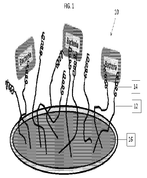
[0083] Referring to FIG. 1, the surface is functionalized with the anti-
microbial peptide via a
flexible linker molecule and exposed to bacteria in the QCM-D using QCM-D
sensors as the
substrate. Referring to FIG. 2 QCM-D was used to monitor covalent attachment
in real-time of the
PEG spacer molecule followed by introduction of the C-CHY1 peptide to the
functionalized Si02
surface (FIG. 2). According to the Sauerbrey calculation, the Am due to PEG
spacers is between 2
and 100 ng per crystal (Table 1, below; FIG. 3). The mass of PEG attached
decreases with increasing
tether length (FIG. 3). PEGylated surfaces show a rigid surface (AD values
from +1x10⁻⁶ to +2x10⁻⁶
Hz) compared to the bulk fluid baseline, validating the use of Eqn. 1 and 2
for PEG layer
calculations. When C-CHY1 is introduced, there is a rapid decrease of Af
corresponding to a Am of
0.8 to 2.2 (Table 1) bound to the surface. This step is accompanied by a
large increase in AD
from near +2x10⁻⁶ to +20x10-6 Hz after the introduction of C-CHY1.
Comparatively, CHY1
introduction leads to a Af decrease corresponding a Am of about 0.4 tg of CHY1
being physically-
adsorbed on the surface. During the rinse, a slightly higher increase in Af of
+5 Hz suggests some of
the CHY1 is removed, consistent with its non-covalent nature, but about 50% of
the peptide remains
adsorbed. The AD does not deviate from the baseline value, suggesting that a
rigid surface is
maintained throughout the entire CHY1 experiment.
18
CA 02995616 2018-02-13
WO 2017/030966
PCT/US2016/046792
Table 1: Sauerbrey and Voigt-Kelvin Calculations of Mass Attachment, Layer
Thickness and Areal Mass for each PEG Tether Length
'Type PEG PEG/C-
Chrysophsin-1 attachment C-CHY1 areal mass
of attachment CHY1
ng (ng/cm2)
tether ng layer
molecul thickness
Nauerbrey Nauerbrey Noigt-Kelvin bSauerbrey
(nm)c
Kelvin
No
Tether
204.9
(PHYS- 373.3 58.6 2.50 0.49
260.9 10.2 475.3 74.6
7.99
ADSOR
B)
PEG
98.1 7.88 874.0 48.8 835.2 168 6.59
1.14 1113 62.2 1063 215
866
PEG
41.8 6.77 2238 60.4 2284 113 17.6
1.32 2850 76.9 2909 144
2000
PEG
2.46 4.58 1753 43.9 1870 176 21.9
1.86 2232 55.0 2381 224
7500
'All values represent the mean for the 3rd through 11th overtones. And error
reported
represents standard error for n>3 surfaces total, corrected for sample size by
dividing
standard deviation by the square root of the number of replicates.
[0084] Once anti-microbial peptide is grafted, the layer becomes significantly
more dissipative as
demonstrated by raw QCM-D data (FIG. 2), which has been seen in previous work.
Thus,
19
CA 02995616 2018-02-13
WO 2017/030966 PCT/US2016/046792
parameters of the system were modeled using the Voigt-Kelvin extended
viscoelastic model in Q-
Tools software (Biolin Scientific, Stockholm, Sweden). This model corrects the
Sauerbrey
estimations for higher energy dissipation by adding terms to the Af relation
to mass (Eqn. 3) and AD
relation to film rigidity (Eqn. 4).
TIL mf 2 co2 d'
Af ¨ 1- (Eqn. 3)
27c6Lmg mg pf ) G'2
G"2]
TIL mf [4 002 d
AD ¨ 2 (Eqn. 4)
nafg6Lmg mg pf d+C2]
Where riL is the viscosity of the bulk liquid assumed to be water (kg/ms);
61_, and 6f are the decay
lengths of the acoustic wave in the bulk liquid and film (m), respectively; mg
and mf are the (kg),
respectively; pf is the film density (kg/m3). The layer was modeled to get
numerical outputs for layer
viscosity, density, and shear modulus (rim, pin, and PA and film thickness.
All overtones were
modeled at once. The bulk liquid, predominantly PBS or PBS/EDTA, was assumed
to have the same
viscosity and density as water at 23 C. Model step size and output ranges were
changed based on
calculated theoretical values using estimated extended molecule size, and the
lowest chi square value
(x2) was taken. Once values of thickness (nm) and density (kg/m3) were found
with the model, the
two were multiplied to calculate areal mass in ng/cm2.
[0085] FIG. 4 demonstrates a measure of the thickness (nm) of the covalently-
bound tether and anti-
microbial peptide (C-CHY1) film on the crystal surface modelled using the
Voigt-Kelvin model
(Table 1; FIG. 4; Eqns 3 and 4). A theoretical maximum thickness using the
assumption of the full
extension of PEG and C-CHY1 was calculated using molecular bond lengths and
found to be 8.8 nm
28, 17.8 nm and 58.05 nm for PEG 866, 2000 and 7500, respectively. Tethered C-
CHY1 via PEG
866 is approximately 6.59 1.14 nm thick. As expected this thickness
increases with increasing
PEG size, from 17.6 1.32 nm for PEG 2000 and 21.9 1.86 nm for PEG 7500.
These numbers
suggest that the PEG was almost fully extended for PEG 866 and 2000 because of
their similarity to
the calculated values. For PEG 7500, the modelled thickness is 38% of
theoretical maximum
thickness, suggesting that the PEG is not fully extended. This could be due to
PEG 7500 chains
interacting with neighbouring chains and peptides, entangling with each other.
Thickness of the
physically-adsorbed CHY1 film was similarly modelled. The thickness is 2.5
0.5 nm.
[0086] Referring to FIG. 5A and FIG. 5B for each of the PEG spacer lengths,
the areal mass was
CA 02995616 2018-02-13
WO 2017/030966 PCT/US2016/046792
determined using the density (kg/m3) obtained by the Voigt-Kelvin viscoelastic
model. The areal
masses calculated for n=3 replicates were 1063 215 ng/cm2, 2909 144
ng/cm2, and 2381 223
ng/cm2 for PEG 866, 2000 and 7500, respectively (Table 1; FIG. 5A). This areal
mass includes the
mass of the PEG spacer, attached C-CHY1, and the trapped buffer solution.
Under the Voigt-Kelvin
model, the following assumptions are made: a Newtonian bulk fluid, a laterally
homogenous and
evenly distributed film, a soft and viscoelastic film (high AD), and an
adsorbed layer is coupled to
the sensor. This model utilizes the AD and Af values contributed by the entire
film on the surface,
including associated buffer molecules, to determine thickness and density
(Eqns 3 and 4 above).
[0087] For comparison, the Sauerbrey equation was used to calculate the mass
addition between
PEG flow and bacteria flow to find the overall grafted mass at each overtone.
Then, this was divided
by the crystal surface area (FIG. 5B). This estimation was a good fit for the
areal mass of grafted
PEG, peptide and associated water. To ensure this good fit, the model and
estimated areal masses
were compared (FIG. 5B). The dense packing of PEG 866 demonstrated in FIG. 3
limits its trapped
buffer molecules to those associated with PEG monomers only. Thus, using the
total system mass
and the mass of PEG monomer-associated water molecules was also a good fit for
lower tether
lengths (FIG. 5B).
[0088] FIG. 6 demonstrates that longer tether length shows highest
antimicrobial activity. For E.
coil, the highest activity was achieved with the longest spacer length used,
PEG 7500 (FIG. 6). For
the shortest spacer PEG 866, 46 2.3% mortality of E. coil was achieved.
Similarly, the highest
activity of C-CHY1 against S. aureus was achieved with the longest spacer
length, PEG 7500, at 64
4.5% mortality (FIG. 6) at 23 C. Using PEG 7500 binding at 55 C, changing the
temperature back
to 23 C for C-CHY1 binding and S. aureus incubation, 91 6.9% mortality was
achieved (FIG. 17).
[0089] The physically-adsorbed CHY1 (FIG. 6) caused 52.3 1.2% and 56.7
1.1% mortalities of
E. coil and S. aureus, respectively.
[0090] Sauerbrey calculations suggest that fewer PEG molecules attach as
tether length increases,
implying that there is also increased spacing between them. With increased
spacing, more water
molecules may be trapped within PEG chains and thus, more contributed mass.
Indeed, Voigt-Kelvin
modeling of raw QCM-D data demonstrated higher areal masses for both PEG 2000
and PEG 7500
than PEG 866 (Table 1). Areal mass due to PEG 2000 was higher than that of PEG
7500. Even
though there are considerably more water molecules associated with one PEG
7500 molecule
compared with one PEG 2000 molecule (FIG. 8), there are significantly more PEG
2000 molecules
21
CA 02995616 2018-02-13
WO 2017/030966 PCT/US2016/046792
(FIG. 8A, 42 ng total) compared to PEG 7500 (FIG. 8B; 2 ng total) leading to a
higher areal mass.
[0091] In solution, CHY1 has been shown to act against bacterial membranes
through pore
formation, as demonstrated by QCM-D and other techniques. Tethered C-CHY1
activity was not
seen to be dependent on bacteria type, but was influenced by tether length and
peptide surface
density, as similar trends with respect to PEG properties were seen for E.
coil and S. aureus. This
agrees with the identical MICs determined of C-CHY1 against both strains. The
ability of QCM-D to
characterize layer thickness and density in a non-destructive manner allowed
coupling of these
results with the results of anti-microbial assays to determine C-CHY1
mechanisms of anti-microbial
action for physically adsorbed peptide (FIG. 7A) and each tether length (FIG.
7B-D).
[0092] In some embodiments described herein, CHY1 is physically adsorbed to a
device, in which
there was no tether. A high local charge density could cause an alternative
mechanism of anti-
microbial activity to pore formation, as seen in other studies, involving the
displacement of positive
cations from the membranes of both E. coil and S. aureus (FIG. 7A). The
calculated areal mass
suggests that there is 5 times more CHY1 on the surface than what has
previously been observed
(Table 1). It is possible that the lower density of CHY1 is close to the
minimum charge density
threshold for activity, where increasing charge density increases anti-
microbial activity.
[0093] For the shortest tethered system with PEG 866, its thickness is only
6.49 1.14 nm which
does not physically allow the peptide to span the E. coil or S. aureus
membranes, which are 23 and
80 nm thick, respectively. Despite this, there is still anti-microbial
activity, suggesting an alternative
mechanism to pore formation (FIG. 7B). This mechanism is also consistent with
the QCM-D
findings. The high density of PEG 866 molecules (FIG. 3) leads to a high
density of C-CHY1 on the
surface and thus a high local charge density. Higher charge densities cause
stronger ionic
displacement in bacterial membranes; thus, PEG 866-tethered C-CHY1 causes
membrane
destabilization due to displacement of positive cations from bacterial
membranes, ultimately leading
to cell death (FIG. 7B).
[0094] FIG. 7 further demonstrates that C-CHY1 tethered to PEG 2000 has the
lowest ability to kill
either bacterial strain, suggesting that the PEG tethering interferes with
anti-microbial activity. The
thickness of this system, 17.6 1.3 nm, suggests that the spacer is 98%
extended but is still not long
enough to fully penetrate either membrane. Partial insertion of C-CHY1 into
the membrane is
possible, but this would limit the extent to which lysis and cell death occur.
Despite the inability of
PEG 2000-tethered C-CHY1 to adequately form pores, some activity still
results, likely from a
22
CA 02995616 2018-02-13
WO 2017/030966 PCT/US2016/046792
different mechanism. The comparatively low bacterial mortality of C-CHY1 with
PEG 2000 versus
PEG 866 tethers is due to lower charge density of C-CHY1 on the surface, since
bactericidal activity
has been shown to increase with increasing charge density (FIG. 7C). The lower
charge density is
due to steric hindrance between grafted PEG 2000 molecules leading to fewer
binding sites available
for C-CHY1 (FIG. 3).
[0095] In some embodiments, C-CHY1 is tethered to PEG 7500. This grafting
exhibits the most
efficient bactericidal activity against both strains of bacteria, showing that
longer tethers demonstrate
higher activity. Despite the lowest amount of PEG on the surface, lowest
binding site availability,
and the least peptide grafted onto the surface (FIG. 3 and FIG. 5), its long
length provides enough
thickness (FIG. 4) to fully penetrate the bacterial membranes and form pores,
similar to how the
peptide acts in solution. This thickness, however, is only 38% of its maximum
theoretical extension,
suggesting that the PEG chains in the system are entangled when there are no
bacteria present. It is
possible that C-CHY1 extension changes in the presence of bacteria due to
changes in local charge
allowing it to aggregate and then form pores. Many studies suggest that pore
formation does not
begin until a sufficient peptide-to-lipid ratio of aggregated peptides on the
surface of the membrane
is achieved. Thus, the proposed mechanism of action is pore formation followed
by lysis and cell
death, thus allowing for the highest activity across all tether lengths (FIG.
7D).
[0096] FIG. 9 demonstrates a tethering process using the QCM-D instrument and
its sensors as the
substrate. However, methods of tethering other than QCM-D, for example, dip-
coating, may be
utilized in this order. First a surface is cleaned, then the surface is
silanized by submerging the
surface in a 10% (v/v) (3-aminopropy1)-trimethoxysilane (APTMS) in methanol
solution for 20 min.
Following that the surface is then rinsed with methanol and DI water yielding
a functionalized
surface. A bi-functionalized polyethylene glycol (PEG) with maleimide and N-
hydroxysuccinimide
ester functional end groups is then introduced (MAL-PEG-NHS) to the
functionalized surface. The
NHS binds to the functionalized surface and the maleimide end group allows for
attachment to an N
or C- terminally modified anti-microbial peptide, such as Chrysophsin-1, by
formation of a thioether
bond. MAL-PEG-NHS may have molecular weights (MW), 866, 2000, or 7500
(referred to as PEG
866, PEG 2000 and PEG 7500, respectively, and collectively PEG).
[0097] Materials and Methods
[0098] Bacterial Strains and Culturing. Escherichia colt HB101 (ATCC 33694)
and Staphylococcus
aureus (ATCC 48366) were cultured overnight in Luria-Bertani broth (20 g/L).
For QCM-D and
23
CA 02995616 2018-02-13
WO 2017/030966 PCT/US2016/046792
toxicity studies, cells were harvested at the late logarithmic phase in their
growth curve, as
confirmed by absorbance measurements (0D(600) = 0.7-1.0 arbitrary units)
(Thermo Scientific
USA, Waltham, MA USA). The cells were centrifuged at 1284 x g (Centrific
Thermo Scientific,
Waltham, MA USA) and re-suspended in 0.01 M, pH 7.2 phosphate buffered saline
(PBS) (Sigma
Aldrich, St. Louis, MO USA) twice, and then diluted 100-fold to approximately
108 cfu/mL.
[0099] Peptides. Chrysophsin-1 (CHY1; FFGWLIKGAIHAGKAIHGLIHRRRH), the N-
terminus
cysteine-modified Chrysophsin-1 (C-CHY1; CFFGWLIKGAIHAGKAIHGLIHRRRH) and the N-
terminus methionine-modified Chrysophsin-1 (M-CHY1;
MFFGWLIKGAIHAGKAIHGLIHRRRH)
were purchased from Bachem Americas, Inc. (Torrance, CA USA). The peptides
were received as a
lyophilized trifluoroacetate salt at greater than 85% purity confirmed by high
performance liquid
chromatography. Solutions of 5 g/L CHY1 and C-CHY1 were made in PBS (pH 7.2)
and PBS
supplemented with between 1 and 5 mM ethylenediaminetetraacetic acid
(PBS/EDTA; pH 7.2) as a
chelating agent respectively, and stored at -20 C. All buffer solutions for
storage, dilutions and
experimentation were degassed by sonication under vacuum for 30 min prior to
their use. The
minimum inhibitory concentrations (MIC) of these peptides against several
common microbes were
found prior to tethering (FIG. 12).
[00100] QCM-D: Covalent Linking of C-CHY1. 5i02-coated quartz crystal
sensors from
Biolin Scientific (Stockholm, Sweden) were used as immobilization platforms
for the modified C-
CHY1. Before use, 5i02 sensors were cleaned in the QCM-D at 40 C using
ethanol, DI water, 2%
sodium dodecyl sulfate (w/v), DI water again, and then nitrogen dried. Lastly,
sensor surfaces were
treated for 2 min using an oxygen plasma cleaner (SPI Supplies, PA USA) to
both clean and
functionalize the surface. The 5i02 crystals were then silanized by submerging
in a 10% (v/v) (3-
Aminopropy1)-trimethoxysilane (APTMS) in methanol solution for 20 min. Each
sensor was then
rinsed twice thoroughly with methanol and DI water and placed in each QCM-D
chamber.
[00101] Changes in frequency (Af, Hz) and dissipation (AD, x10-6 Hz) at
the 3rd, 5th, 7th,
9th and 11th overtones were monitored at a constant 23 C in all chambers using
a Q-Sense E4
QCM-D system (Biolin Scientific, Stockholm, Sweden). All flow rates for the
solutions were at 0.1
mL/min unless otherwise noted, and all volumes given are on a per chamber
basis. PBS/EDTA (pH
7.2) buffer was used to establish a stable baseline measurement. Maleimide PEG
N-
hydroxysuccinimide ester molecules (MAL-PEG-NHS) with molecular weights (MW)
of 866
(ThermoScientific, Waltham MA, USA), 2000, or 7500 (JenKem Technology USA
Inc., Allen, TX
24
CA 02995616 2018-02-13
WO 2017/030966 PCT/US2016/046792
USA) were purchased. These will be referred to as PEG 866, PEG 2000 and PEG
7500, respectively.
One mL of 100 [tM MAL-PEG-NHS was flowed through the QCM-D and subsequently
incubated
for 30 min. Crystals were then rinsed with 1.2 mL of the PBS/EDTA (pH 7.2)
buffer. Similarly, 1.25
mL of 10 [tM C-CHYI solution was then flown over the sensors and allowed to
incubate for 30 min.
To rinse excess C-CHYI off the surface and prepare for the introduction of 2
mL bacteria, a 45 min
PBS rinse at 0.3 mL/min was first flowed through the QCM-D. The dilute
bacterial solution was
allowed to incubate for 1 hour, followed by a final 2 mL PBS (pH 7.2) rinse.
The crystals were
removed from the chambers and placed in individual petri dishes containing
0.85% (w/v) NaC1
solution for bacterial viability testing.
[00102] QCM-D Control Experiments: Physical Adsorption of CHYI .
Similarly, 5i02-coated
sensors were used for physical adsorption of unmodified CHYI and were prepared
as described
above. For this experiment there was no APTMS functionalization or PEG flow,
thus, after cleaning,
the crystals were placed immediately into the QCM-D, PBS (pH 7.2) was used to
establish a baseline
and 1.25 mL of 10 [tM CHYI solution was introduced. Then, flow was stopped and
CHYI was
incubated for 30 min. The rest of the protocol described in the previous
section was then followed,
including the 0.3 mL/min PBS rinse, bacterial flow, bacterial incubation and
final PBS rinse. The
crystals were removed from the chambers and placed in individual petri dishes
containing 0.85%
(w/v) NaC1 solution for bacterial viability testing. For the next type of
control, crystals that had
never been coated with APTMS were cleaned and placed into the QCM-D chambers
without
functionalization. Starting with a PBS rinse, the crystals were exposed to
bacteria solution for 1 hour
and then rinsed before imaging. For the APTMS control, crystals were cleaned
and functionalized
with APTMS. Then the procedure continued starting with a PBS rinse (pH 7.2),
bacteria
introduction, incubation, and a final rinse before imaging. For the PEG
control, the crystals were
cleaned and functionalized with APTMS. A baseline was established, PEG was
flown and then
incubated. Peptide was not introduced in this type of control experiment.
Separate PEG control
experiments were performed for each PEG size. All experiments were repeated at
least three times.
[00103] Bacterial Mortality. Bacterial mortality was determined
immediately after the final
rinse of the QCM-D experiment. Crystals were stained using a LIVE/DEAD
BacLight Bacterial
Viability Kit (Life Technologies Corp, NY USA) in 2 mL of 0.85% (w/v) NaC1
solution with 5 [tM
SYTO 9 and 30 [tM propidium iodide for 15 min. The crystals were rinsed once
using 0.85% (w/v)
NaC1 solution to remove any excess dye and then kept in 1 mL saline to keep
hydrated for imaging.
CA 02995616 2018-02-13
WO 2017/030966 PCT/US2016/046792
The crystals were imaged at 20x objective using fluorescein isothiocyanate
(526 nm) and Texas Red
filters (620 nm) under a Nikon Eclipse E400 fluorescence microscope (Melville,
NY USA). A
minimum of 5 locations on each crystal were examined for live and dead
bacteria, totaling at least 10
images. Images were analyzed using ImageJ Software (http://rsbweb.nih.gov/ij/)
to produce a
merged image from which the percent mortality (or killing percent) of the
cells was determined.
[00104] QCM-D: Data Modelling. The viscoelasticity of deposited material
demonstrated by
the QCM-D raw data was used to determine which model to use in estimating
parameters of the
system such as mass of attachment (ng), layer thicknesses (nm) and peptide
areal mass (ng/cm2).
The brush may be thought to be made up of two layers, first is APTMS plus PEG
and second is C-
CHY1. The former demonstrates near-zero dissipation values, thus, the
Sauerbrey equation for rigid
surfaces (Eqn. 1) applies, where Am is inversely related to Af. For the
Sauerbrey model, AD and film
rigidity are related using Eqn. 2.
Af ¨ 2e Am (Eqn. 1)
A\Ipci[tci
AD ¨ (Eqn. 2)
27EG
[00105] Where f0 is the fundamental frequency of the quartz crystal, 5
MHz; A is the active
crystal surface area; pq is the density of quartz, 2.648 g/cm3; and nq is the
shear modulus of the
crystal, 2.947x10¹¹ g/cm=s2; G" is the loss modulus and G' is the storage
modulus of the film
attached to the crystal surface. Thus, decreases in Af demonstrate an addition
of mass and higher AD
values indicate a softer film. The Sauerbrey model was applied to time points
from the flow of PEG
through the flow of C-CHY1. A "maximum" PEG attachment was also calculated
using the
minimum frequency value between the two time points for comparison.
[00106] Once C-CHY1 is grafted, the layer becomes significantly more
dissipative as
demonstrated by raw QCM-D data, which has been seen in previous work. Thus,
parameters of the
system were modelled using the Voigt-Kelvin extended viscoelastic model in Q-
Tools software
(Biolin Scientific, Stockholm, Sweden). This model corrects the Sauerbrey
estimations for higher
energy dissipation by adding terms to the Af relation to mass (Eqn. 3) and AD
relation to film
rigidity (Eqn. 4).
Af = - -fon' [1¨ 2 CL)2 2G (Eqn. 3)
27Esong MCI pf 6+6,2
26
CA 02995616 2018-02-13
WO 2017/030966 PCT/US2016/046792
AD= + [ (L)22 (Eqn. 4)
'Info 601114 mg Pf G' +C 2
[00107] Where TIL is the viscosity of the bulk liquid assumed to be water
(kg/ms); 6L and 6f
are the decay lengths of the acoustic wave in the bulk liquid and film (m),
respectively; mq and mf
are the (kg), respectively; pf is the film density (kg/m3). The layer was
modelled to get numerical
outputs for layer viscosity, density, and shear modulus (rim, pm, and pm), and
film thickness. All
overtones were modelled at once. The bulk liquid, predominantly PBS or
PBS/EDTA, was assumed
to have the same viscosity and density as water at 23 C. Model step size and
output ranges were
changed based on calculated theoretical values using estimated extended
molecule size, and the
lowest chi square value (x2) was taken. Once values of thickness (nm) and
density (kg/m3) were
found with the model, the two were multiplied to calculate areal mass in
ng/cm2. The Sauerbrey
estimate (Eqn. 1 and 2) for the QCM-D crystal surface area for the grafted C-
CHY1 layer was used
to compare with modelled values.
[00108] Because the long grafted layers are thought to be associated with
water and the QCM-
D modelling does not account for this, reported literature values of
associated water molecules with
PEG monomers were used to model the mass of the system accounting for water.
[00109] FIG. 10 shows data of turbidity (indicating degree of bacterial
growth), measured via
OD(590) demonstrating activity of a C-CHY1 coating on Foley urinary catheter
segments using
PEG 866 and PEG 7500 tethers versus Pseudomonas aeruginosa (ATCC 29260); and
[00110] FIG. 11 shows additional data demonstrating activity of a C-CHY1
coating on Foley
urinary catheter segment that has been prepared using an oxygen plasma cleaner
to clean the surface
and deposit hydrophilic oxygen molecules on the surface prior to
functionalization and peptide
attachment, with PEG 866 and PEG 7500 versus P. aeruginosa (ATCC 29260). To
gather the data
for FIG. 10 and FIG. 11, Foley catheter segments were sectioned in cylindrical
segments, 1-cm long
under sterile conditions. Segments were functionalized using a dip-coating
method according to the
procedure in FIG. 9, entered into 24-well plates and exposed to 2 mL MHB with
2x10⁵ CFU/mL
bacteria at 37 degrees C at 200 RPM agitation. At several time points between
30 minutes and 20
hours, 1 mL aliquots were taken from the each well and the OD(590) was
measured. The 1 mL
aliquots were returned to the respective wells and the plate was returned to
37 degrees C and
incubated until the next time point.
[00111] FIG. 12 shows Table 2 with the minimum inhibitory concentrations
(MIC) of CHY1
27
CA 02995616 2018-02-13
WO 2017/030966 PCT/US2016/046792
(SEQ ID NO: 1), C-CHY1 (SEQ ID NO: 2) and M-CHY1 (SEQ ID NO: 3) against
several strains of
bacteria, at 16 hours after inoculation, defined as the minimum concentration
(.iM) required to
inhibit 100% visible growth of the microbe. All values represent the mean from
three experimental
replicates. The MICs were determined using previously established protocols.
[00112] FIGS. 13A-13B demonstrate the cytotoxicity profile of M-CHY1 (SEQ
ID NO: 3)
anti-microbial peptide at different concentrations against a human primary
fibroblast cell line, at 8
hours (FIG. 13A) and 30 hours (FIG. 13B) of exposure using the reduction of
AlamarBlue by
healthy cells seeded at 4,000 cells per 96-well. The fluorescent signal at 590
nm was determined
after seeding the cells overnight (12-16 hours), exposing the cells to peptide
concentartions for 4
hours, and the addition of 10% v/v AlamarBlue reagent for an additional 4
hours. (Total incubation
with peptides, 8 hours), followed by medium and peptide refreshing and another
addition of
AlamarBlue at 30 hours' incubation. All values represent mean S.D. from at
least three
experimental replicates. The dotted line represents the average cell-only
fluorescence at 590 nm. The
AlamarBlue procedure was followed according to manufacturer's instructions.
[00113] FIG. 14 show the cytotoxicity profile of C-CHY1 (SEQ ID NO: 2)
against a human
primary fibroblast cell line after 12 hours of exposure to various
concentrations of C-CHY1,
measured using the reduction of 3-(4,5-dimethylthiazol-2-y1)-2,5-
diphenyltetrazolium bromide
(MTT) into formazan solubilized by dimethylsulfoxide. Average optical density
at 590 nm of MTT
reduced by 4,000 cells per well of CT 1005 primary human fibroblasts, when
exposed to various C-
CHY1 peptides concentrations in solution after 12 hours of exposure. All
values represent mean
S.D. from at least three experimental replicates. The dotted line represents
the average cell-only MT
reduction signal at 590 nm. The MTT procedure over 4,000 cells per well was
followed according to
manufacturer's instructions.
[00114] FIGS. 15A-15B demonstrate the morphological characteristics of
human primary
fibroblasts when exposed to 5 i.tM (top), 10 tM (middle) and 30 i.tM (bottom)
concentrations of M-
CHY1 (SEQ ID NO: 3) (left panels) compared to untreated samples (right panels)
at 10 hours'
exposure (FIG. 15A) and 30 hours exposure (FIG. 15B), taken using the 10x
magnification of a
Leica light microscope. Photographs were taken during AlamarBlue treatment
described and
depicted in FIG. 13A and FIG. 13B.
[00115] FIG. 16 demonstrates the optimization process of PEG 7500 linker
molecules on the
surface, by demonstrating the mass of PEG bound to the surface as a function
of temperature (x-
28
CA 02995616 2018-02-13
WO 2017/030966 PCT/US2016/046792
axis) and salt concentration ("high salt" is twice the salt concentration of
normal PBS buffer), since
PEG optimization is hypothesized to be directly related to resulting anti-
microbial activity. As
temperature increases, the amount of PEG 7500 increases. This may be due to
the change of
conformation of PEG in solution and on the surface due to the higher
temperatures reducing steric
hindrance allowing for more binding of the PEG. Higher than physiological salt
concentrations
("high salt") alters the binding ability of PEG 7500 linker to bind.
[00116] FIG. 17 included table 3, which is an example calculation of the
percent dead S.
aureus bacteria calculated after 1 hour exposure to C-CHY1 anti-microbial
peptide tethered via PEG
7500 after temperature optimization of the PEG attachment, at 55 degrees C.
Temperature was
reduced to room temperature prior to C-CHY1 binding and bacteria introduction,
and bacterial
mortality was calculated by counting the number of live and number of dead
bacteria in different
areas on the QCM-D sensor (columns 2 and 3), merging the images and counting
the live bacteria
(column 4) and calculating the percent dead (column 5).
[00117] FIG. 18 illustrates a typical QCM-D response to grafting a M-CHY1
(SEQ ID NO: 3)
anti-microbial peptide to a functionalized surface via a tether of PEG 7500,
where frequency and
dissipation are depicted on different axes versus time, at the 3rd, 5th, 7th,
9th and 11th overtones,
and a-c represent major time stamps according to changes in material flow
through the QCM-D;
a=tether flow, b=peptide flow, c=bacteria flow. The interaction of M-CHY1 with
a functionalized
surface and PEG linking molecule is similar to that of C-CHY1 peptide
attaching to the surface via
PEG 7500 as monitored using QCM-D.
[00118] FIG. 19 includes Table 4, which is a comparison of the killing
ability of PEG 7500-
tethered C-CHY1 and M-CHY1 anti-microbial peptides tethered to a surface
against Gram-negative
E. coil and Gram-positive S. aureus bacteria. Table 4 demonstrates the percent
dead bacteria (also
called killing percent), Gram-positive S. aureus and Gram-negative E. coil, as
a result of a 1 hour
incubation with C-CHY1 and M-CHY1 covalently bound via PEG 7500 that had been
incubated at
room temperature (e.g. non-optimized system). Bacterial mortality was
calculated by counting the
number of live and number of dead bacteria in different areas on the QCM-D
sensor and calculating
the percent dead cells, for n=6 replicates and n=4 replicates for C-CHY1 and M-
CHY1, respectively.
[00119] All patents, patent applications, and published references cited
herein are hereby
incorporated by reference in their entirety. It should be emphasized that the
above-described
embodiments of the present disclosure are merely possible examples of
implementations, merely set
29
CA 02995616 2018-02-13
WO 2017/030966 PCT/US2016/046792
forth for a clear understanding of the principles of the disclosure. Many
variations and modifications
may be made to the above-described embodiment(s) without departing
substantially from the spirit
and principles of the disclosure. It can be appreciated that several of the
above-disclosed and other
features and functions, or alternatives thereof, may be desirably combined
into many other different
systems or applications. All such modifications and variations are intended to
be included herein
within the scope of this disclosure, as fall within the scope of the appended
claims.