Note: Descriptions are shown in the official language in which they were submitted.
CA 02996595 2018-02-26
WO 2017/035648
PCT/CA2016/051025
1
TITLE: METHOD FOR 3D IMAGING OF MECHANICAL ASSEMBLIES
TRANSPLANTED INTO MAMMALIAN SUBJECTS
TECHNICAL FIELD
The present invention relates to the field of medical imaging and, in
particular,
to 3D medical imaging of implanted joint replacement components.
BACKGROUND
Osteoarthritis (OA) is the most common cause of arthritis, and is one of the
leading causes of disability. OA significantly affects an individual's ability
to work
and decreases their quality of life. OA is a degenerative joint disease where
the
cartilage of a joint, such as the knee or hip, is compromised resulting in
swelling,
stiffness and pain. Joint replacement surgery using an orthopedic implant is
the typical
course of treatment when pain and/or loss of function become severe.
In the United States, the cost of joint replacement surgery has been reported
to
total nearly 50 billion USD in 2009, surpassing 1 million hip and knee
replacements
annually in recent years. The continued growth of arthroplasty procedures will
also
increase the burden of revision surgeries due to prosthesis problems,
including implant
loosening and assembly failures.
Stereo radiography is a technique that uses two x-ray systems with
intersecting
beams and taking two x-ray images simultaneously of an object placed in the
beam
intersection. Stereo radiography has traditionally been used to accurately
measure
migration which is the micromotion of an implant over time relative to bone.
Accuracy
and precision of 0.1 mm can be achieved using stereo radiography. Excessive
migration within the first year or two has been demonstrated to be able to
predict the
need for revision surgery due to implant loosening as much as 10 years later
and well
before symptoms occur. This enables stereo radiography to detect problems with
specific implants earlier and with fewer patients than other methods.
The assessment and monitoring of implants using stereo radiography methods
such as radio stereometric analysis (RSA) requires an imaging setup capable of
high
CA 02996595 2018-02-26
WO 2017/035648
PCT/CA2016/051025
2
measurement accuracy and precision. In
addition to knowing the imaging
configuration to a high degree of accuracy and precision, 3D computer models
of the
implant being measured are also necessary for the analysis. Current analysis
methods
assume an implant is made of one component or a fixed and known configuration
of
components, or otherwise each component must be measured independently.
However,
in the case where an implant is an assembly consisting of multiple components,
the
precise configuration of the components making up the implant assembly may not
be
known and even be patient-specific due to tolerance stack-up within the
assembly.
An assessment may be further complicated by a limited field of view or
occlusion of part of the assembly caused by radio-opaque components of the
assembly
itself or other implant components, such as a radiopaque cup occluding the
head on the
femoral stem of a hip replacement implant. In such cases, it may be impossible
to
accurately localize specific components of the assembly in the traditional
manner.
That is, there may not be enough image information available to resolve all 6
degrees
of freedom describing the pose, comprised of the position (x-coordinate, y-
coordinate,
z-coordinate) and orientation (i.e., rotations about the x-axis, y-axis, and z-
axis) of the
component. The loss of accuracy and precision because of this missing
information
can be prohibitive in assessing and monitoring implants using stereo
radiography.
SUMMARY
The exemplary embodiments of the present disclosure relate to methods for
measuring the 3D configuration of an orthopaedic implant assembly, its 3D
position
and orientation relative to bone as well as relative to another implant or
implant
component using stereo radiography.
One exemplary embodiment relates to a method for measuring implant location
in a patient, wherein the method comprises: (a) 3D computer models of the
components
which make up an orthopedic implant assembly, (b) defined kinematic
relationships of
the implant assembly's components, wherein a principal component is defined
and the
position and orientation of all other secondary components are described
relative to the
principal component or the preceding component in the kinematic chain, (c) the
acquisition of stereo radiographic imaging data, and (d) accurate measurement
of the
CA 02996595 2018-02-26
WO 2017/035648
PCT/CA2016/051025
3
configuration of the implant's assembly as well as position and orientation of
the
implant using the constraints of the kinematic relationships of its
components.
According to some exemplary embodiments, the method further comprises: (e)
using
the assembly configuration and 3D pose obtained from at least two time points
to
measure changes in assembly configuration and/or pose relative to bone or to
another
implant or implant component.
The method disclosed herein may use location(s) of the clearly visible
component(s) of an implant assembly, combined with knowledge of the kinematic
relationship between the implant components and the limited information from
the
partially occluded components, to accurately determine the configuration of
the
assembly and 3D location of the occluded component(s) within the patient
wherein the
implant assembly is installed.
BRIEF DESCRIPTION OF THE DRAWINGS
These and other features of the invention will become more apparent in the
following detailed description in which reference is made to the appended
drawings.
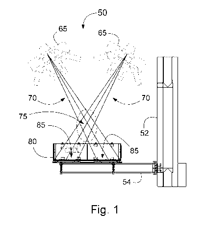
Fig. 1 is a schematic illustration of a stereo radiography system in a 60
degree
inter-beam configuration that may be used in an exemplary method, according to
an
embodiment of the present disclosure.
Fig. 2 is a schematic illustration of an image registration and creation of a
common reference frame (coordinate system) based on the sets of markers
provided by
the reference box of the exemplary dynamic stereo radiography system shown in
Fig.
1;
Fig. 3 is a display illustrating implant tracking between a three-dimensional
model and a pair of radiographic images to optimize position and orientation
for each
component of the implant assembly, according to an exemplary embodiment of the
present disclosure;
CA 02996595 2018-02-26
WO 2017/035648
PCT/CA2016/051025
4
Fig. 4 is a flowchart illustrating the workflow leading to and including the
optimization of the position and pose of the components making up an implant
assembly;
Fig. 5 is a schematic illustration of an exemplary prismatic kinematic between
the femoral stem (principal component) and femoral head (secondary component),
according to an embodiment of the exemplary methods disclosed herein; and
Fig. 6 is a is a schematic illustration showing a representation of a wear
measurement in an acetabular cup liner using the configuration and pose of the
components of an implant assembly, according to an exemplary method disclosed
herein.
DETAILED DESCRIPTION OF THE INVENTION
Imaging-based measurements of orthopaedic implants in vivo with
stereoradiography enable the assessment and monitoring of implant loosening
and
provide data predictive of revision surgery and patient outcome.
The embodiments of the present disclosure describe methods based on stereo
radiography that allow the configuration of the individual components of an
implant
assembly to be quantitatively determined in 3D. Specifically, the embodiments
of the
present disclosure include adding additional degrees of freedom to the pose
optimization of an implant assembly per the kinematic relationship between the
components resulting in the implant assembly's configuration, position and
orientation.
Some exemplary embodiments of the present disclosure pertain to methods in
which the position and orientation of the implant assembly's components are
used to
measure metrics of interest such as settling of assembly components onto each
other,
bedding in, creep, and wear in implants with liners or spacers, migration of
the implant
within the bone into which it has been installed.
For purposes of illustration, the devices and methods of the invention are
described below with reference to the in vivo measurement of the femoral
components
of a human hip implant. However, as will be appreciated by those skilled in
the art, the
CA 02996595 2018-02-26
WO 2017/035648
PCT/CA2016/051025
methods can be employed with other types of implant assemblies for example
knee
implants, shoulder implants, other joints, in vitro or in situ, and for any
mammal.
The exemplary embodiments of the present disclosure relate to the 3D
determination of the configuration of an implant assembly installed into a
mammalian
5 subject, as well as the position and orientation of the implant
assembly's components.
Specifically, 3D computer models of the implant assembly's components are
obtained
and their assembly and pose determined based on a stereo pair of radiographic
images
of a patient's implant. By comparing measured positions and orientations at
multiple
time points, metrics of interest such as migration, creep and wear, and
component
settling can be measured. A person skilled in the art will also recognize that
a series of
radiographic images can be obtained in a dynamic manner or a series of
progressive
static radiographic images, with or without a prescribed voluntary motion
performed by
the patient. A person skilled in the art will also recognize that the methods
described
herein may also be used in single plane x-ray images at a likely expense of
accuracy
and precision.
Stereo Radiography Imaging
Persons of ordinary skill in this art will recognize that there are a variety
of
stereo-radiography techniques that may be used to obtain the radiographic
images of
the implant assembly. For example, biplane or dual-plane fluoroscopy of
radiostereometric analysis (RSA). Some exemplary embodiments of the present
disclosure relate to a stereo-radiographic imaging method for obtaining three-
dimensional measurements of an implant's position and orientation within a
target
region of a patient's anatomy that comprises capturing stereo x-ray exposures
of a
patient who is upright or lying on a table. According to further embodiments,
as is
readily understood by those skilled in the art, weights, rubber bands, and the
like, can
be used to load the joint which contains the implant.
Persons of skill in the art will recognize that a variety of methods may be
used
to obtain the 3D position and orientation of the implant assembly's components
from
the radiographic images. Without limiting the foregoing, reference objects may
be
included in the field of view to allow the calculation of the imaging
configuration.
CA 02996595 2018-02-26
WO 2017/035648
PCT/CA2016/051025
6
Moreover, the image information used to calculate the 3D position and
orientation may
be based on the use of edge detection of the radiographic images, gradient
information
obtained from the image, feature recognition and extraction or digitally
reconstructed
radiography combined with image matching.
Measurement of the position and orientation of an implant assembly's
components
The three-dimensional measurement of the position and orientation of the
implant assembly's components consists of establishing a geometric relation
between
the implant's representation in the stereo radiographic images and a 3D
computer
model of the implant assembly's components. According to some exemplary
embodiments of the present disclosure, methods for the 3D measurement involve
fitting the projection of the 3D computer model to edge or gradient data of
the implant
assembly's components visible in the radiographic images. In this way, the
position
and orientation of the 3D computer model of the implant assembly's components
are
derived from the radiographic images thereby resolving the configuration of
the
implant assembly (Fig. 3).
Image registration is performed either through known information about the
imaging configuration or by determining the imaging configuration using the
radiographic images. According to an exemplary embodiment, this involves
determining x-ray foci positions from the stereo radiographic images and
consolidating
all image information into a common reference frame. According to an exemplary
embodiment of the present disclosure, a registration element exemplified by a
reference
box (Fig. 2) is positioned between the patient and the detector panels. The
registration
element has a series of fiducial and control beads that provide reference
markers from
which x-ray foci can be calculated and all image information can be
consolidated in a
common reference frame (Fig. 4).
Image feature extraction, according to embodiments of the present disclosure,
includes filtering of the images for improved image quality, the robust
detection of
edges in the images, and the creation of component-specific edge maps.
CA 02996595 2018-02-26
WO 2017/035648
PCT/CA2016/051025
7
The 3D computer models of the components of the implant assembly can be
obtained using a variety of methods known to those skilled in the art.
According to
embodiments of the present disclosure, the 3D computer models can be generated
from
CAD software. According to other embodiments, the 3D computer model can be
generated by optical scanning. According to other embodiments, the 3D computer
model can be represented by a parametrized geometric model. According to other
embodiments, the 3D computer model can be generated from a CT or MRI scan.
It is to be noted that the 3D computer models of the components of the implant
assembly are defined separately. A principal component, from which the
position and
orientation is assigned to the entire assembly, is chosen from the assembly
and from
which the kinematic chain of secondary components is defined. Further,
kinematic
relationships between each of the secondary components and the principal
component
are defined, thereby constraining the possible configurations of the assembly
and
reducing the degrees of freedom needed to solve the configuration of the
assembly. It
should be noted that for the special case of a component being independent
from all
other components, no secondary components are linked. According to another
embodiment of the present disclosure, more than one kinematic chain can be
defined
and measured concurrently.
The main optimizer involves fitting the general three-dimensional position and
orientation of the assembly and configuration of the components to establish a
best-fit
(Fig. 4). These iterations involve optimizing the absolute position and
orientation of the
principal component of the implant assembly, along with the relative positions
of the
secondary components as allowed by the kinematic relationships of the implant
assembly. The steps in the main optimizer are repeated for each image pair to
obtain
the optimized positions and orientations; in absolute terms for the principal
component
and in relative terms for each secondary component of the implant assembly.
For each
secondary component, the resulting output can be converted to absolute
positions and
orientations (Fig. 4).
Another exemplary embodiment of the present disclosure pertains to updating
of the edge data from the edge map at each iteration based on goodness of fit
with the
projected 3D computer models.
CA 02996595 2018-02-26
WO 2017/035648
PCT/CA2016/051025
8
Measuring changes in configuration, pose and relative pose
According to exemplary embodiments of the present disclosure, the optimized
3D computer model of the components of the implant assembly provides the basis
for
accurate quantitative measurement of metrics of interest in the assessment or
monitoring of an orthopedic implant. In particular, migration of the implant
assembly
relative to bone as in traditional stereo radiography can be determined. When
varying
loading conditions, changes in assembly configuration suggest a loosening of
one or
more components within the assembly. According to particular embodiments, the
change in the relative three-dimensional position and orientation of the
femoral head
relative to the acetabular cup between two time points can be used to
calculate wear of
the acetabular cup's liner.
EXAMPLES
Example 1
Imaging Apparatus
A stereo orthopaedic radiography system 50 (Halifax Imaging Suite; Halifax
Biomedical Inc., Mabou, NS, Canada) was used. The stereo orthopaedic
radiography
system 50 comprised two radiography systems 65 exposing simultaneously to
obtain
stereo radiographic images (Fig. 1). Each radiography system 65 comprised an x-
ray
source (RAD-92 Sapphire X-Ray Tube; Varian Medical Systems, Palo Alto, CA,
USA), a generator (Hydravision SHF635RF DR X-Ray Generator, SEDECAL USA
Inc., Buffalo Grove, IL, USA), an x-ray detector panel 85, a digital imaging
system
(CDXI 50RF, Canon USA Inc., Melville, NY, USA), and a computer system to link
the
components together, to retrieve the imaging data, and to reconstruct the
imaging data.
The two x-ray imaging systems 65 are positioned at an angle to each other such
that
their x-ray beams 70 overlap in part to create a 3D viewing volume 75.
A 60-degree reference box 80 (SR Reference Box; Halifax Biomedical Inc.,
Mabou, NS, Canada) was placed into the image field of both systems 65 (Figs.
1, 2).
The reference box 80 was constructed from carbon fiber to insure rigidity, to
resist
CA 02996595 2018-02-26
WO 2017/035648
PCT/CA2016/051025
9
deformations resulting from temperature fluctuations during operation, and for
its
radiolucency. The reference box 80 housed two digital detector plates 85 in
the bottom
(away from the patient and x-ray source) in a uniplanar configuration,
immediately
behind a fiducial plane which contained a series of equidistantly spaced radio
opaque
tantalum beads. The top of the box 80 formed the control plane which contained
radio-
opaque tantalum beads also. The fiducial beads allowed the captured images to
be
transformed to a common reference frame, while the control beads allowed the
calculation of the foci (i.e., the x-ray sources) locations to enable the
analysis. The
images were captured on two digital detector plates 85 (CDXI 50RF, Canon USA
Inc.,
Melville, NY, USA) as greyscale images with relative intensity values in
standard
medical DICOM format. The overlap of the two radiography systems' fields of
view
made up the 3D viewing volume 75 (Fig. 2). The registration element has a
series of
fiducial and control beads that provide reference markers from which x-ray
foci can be
calculated and all image information can be consolidated in a common reference
frame
90. The reference box 80 is securely mounted onto a beam 54 that is pivotably
engaged
with a vertical support column 52 whereby the beam 54 can be controllably
raised
upward and downward and additionally controllably rotated on the vertical
support
column 52 (Fig. 1).
Image Data Acquisition
Images were acquired with the patients in supine and standing positions. For
each image, the patients were positioned and instructed by a technologist on
how to
hold the position. Each of the image pairs were reviewed by the technologist
to ensure
image quality and the regions of interest were captured. The images were then
transferred using tele-radiology technology to the image analysis center for
analysis.
Definition of Implant Assembly and Kinematic Relationship
An orthopaedic implant designed for total hip replacement installed into a
patient was imaged post-operatively as described above. The components making
up
the hip implant are a femoral stem 10 and femoral head 20 installed into their
femur 32,
and an acetabular cup and a polyethylene liner (not shown) installed into the
socket 34
of their pelvis (Fig. 3). A 3D computer model (C) of these components was
calculated
CA 02996595 2018-02-26
WO 2017/035648
PCT/CA2016/051025
from the two radiographic images (A), (B) concurrently captured by the two
radiography systems 65 (Fig. 3) following the steps outlined in Fig. 4. In
this example,
the femoral head comprised ceramic material which is relatively radio-lucent
while the
acetabular cup was made from tantalum which is radio-opaque, thereby rendering
the
5 femoral head significantly occluded in one or both radiographic images
(A), (B) (Fig.
3). The degree and location of occlusion depended on patient positioning and
could
not be predicted. The purpose for imaging was to measure cup liner wear which
is
defined for this purpose as the penetration of the head into the cup, in the
proximal
direction, at multiple time points. The degree of occlusion in most image
sequences
10 prohibited this calculation using the standard techniques known in the
art. However,
the femoral stem was visible in its entirety in all image sequences.
Therefore, the
femoral stem was chosen as a principal component of the implant assembly with
the
femoral head as the secondary component. The kinematic relationship between
the
femoral stem and the femoral head was defined, as prismatic coupling with the
axis of
symmetry of the neck of the stem and the axis of symmetry of the head set to
be
collinear (Fig. 5). A reasonable starting location was set for these two
components.
Thus, the assembly of the femoral component of the hip implant was described
as a 7
degrees of freedom system with the pose of the femoral stem described by 6
degrees of
freedom (three translations and three rotations) and the position of the
femoral head
onto the stem as the seventh degree of freedom. The seventh degree of freedom
was
relative to the femoral stem and described the translation of the femoral head
along the
collinear symmetry axes, from the initial position. The acetabular cup was
clearly
visible in all images and was defined as an independent component of the
implant and
described by all 6 degrees of freedom (Fig. 5). The polyethylene liner of the
hip
implant was not visible in the x-rays (Fig. 3(A), (B)) and could not be
measured.
Determination of Imaging Configuration
The radiographic images were loaded onto a computer system for calculation of
the parameters that described the detailed configuration of the imaging
system. The
fiducial beads in the reference box were located in the images and their
locations
tabulated. Based on the known locations of these beads, a projective
transformation
was calculated that matched the bead locations to the tabulated locations from
the
CA 02996595 2018-02-26
WO 2017/035648
PCT/CA2016/051025
11
images following the process steps outlined in Fig. 4. The control beads of
the
reference box were located in the images and their locations tabulated. Based
on the
known locations of the fiducial beads and the control beads, the locations of
the two
foci were calculated.
Extraction of Image Features
The radiographic images were filtered using a Canny edge detection filter.
Using a graphical user interface, a trained user selected all the edges
belonging to the
femoral stem, head and acetabular cup separately. An initial position and
orientation
for the femoral stem (with the coupled head) and cup were set by the user,
also using a
graphical user interface.
Determination of Implant Assembly Configuration and Component Pose
The location of the foci and the parameters describing the projective
transform
were used to calculate the projected contours onto the fiducial plane for any
given
position and orientation of the components making up the implant. An objective
function was made available to the optimizer which calculated a goodness-of-
fit score
between the projected contours and user-selected component-specific edge maps,
given
the pose of the stem, the relative translation of the head along the symmetry
axis and
the pose of the cup. The goodness of fit score was based on a sum of squared
distance
metric and was calculated separately for the femoral stem and femoral cup.
The optimizer used the objective function to find the configuration of the
implant assembly which provided the best fit to the radiographic images,
within a
predefined search space. In this example, the optimizer first used Particle
Swarm
Optimization as a global optimization method. A second round of optimization
attempted to further increase the goodness-of-fit with a local, gradient-based
optimizer.
The initial position of the particles was uniformly distributed along the
predefined
search space and centered on the user initialized estimates. The optimizer
returned the
final pose of the stem 110, neck 115 of the stem 110, and translation of the
femoral
120a, 120b relative to the stem 110 along the axis of symmetry 90, and, the
pose of the
cup (Fig. 5).
CA 02996595 2018-02-26
WO 2017/035648
PCT/CA2016/051025
12
Calculation of Cup Liner Wear
Cup liner wear was defined as proximal penetration of the head into the cup.
With the implant configuration determined by the pose of the femoral neck 115
and the
relative position of the femoral head 120 to the femoral stem 115, the
absolute pose of
the head was calculated for each time point, i.e., "120c" at 1 year and "120d"
at 2 years
(Fig. 6). To calculate the displacement of the head relative to the cup, the
pose of the
cup 120d at 2 years was transformed to be coincide with the pose of the cup at
1 year
120c; thus using the 1-year pose as the reference. The same transform was
applied to
the head's pose at 2 years. In this way, a displacement vector could be
determined
describing the motion of the head relative to the cup between the two time
points. The
component of this displacement generally aligned with the proximal anatomical
direction and was reported was cup liner wear.