Note: Descriptions are shown in the official language in which they were submitted.
WO 2017/049213
PCT/US2016/052317
METHODS AND COMPOSITIONS FOR GENOMIC TARGET ENRICHMENT
AND SELECTIVE DNA SEQUENCING
CROSS-REFERENCE TO RELATED APPLICATIONS
This application claims priority to U.S.S.N. 62/219,332, filed on September
16,
2015.
REFERENCE TO SEQUENCE LISTING
The Sequence Listing submitted September 16, 2016 as a text file named
"PETOM_100_ST25.txt," created on September 14, 2016, and having a size of
7,047
bytes.
FIELD OF THE INVENTION
The disclosed invention is generally related to methods for sequence-specific
capture of fragments of double-stranded DNA from a mixture or library of
fragments,
specifically for preserving the native quantity, structure, methylation
status, or a
combination thereof, of gcnomic DNA molecules greater than 2 kilobases in
length.
BACKGROUND OF THE INVENTION
Using thousands of distinct DNA probes bound to the surface of microarrays, it
was possible to isolate most of the exon sequences of the human genome (Hodges
et al.,
2007), as well as thousands of specific genomic intervals of biological
interest (Hodges
et al., 2009). More recently, there has been increased interest in isolating
and sequencing
long DNA reads to enable construction of phased haplotypes, which consist of
sequence
assemblies corresponding to a single pure paternal or maternal DNA strand. A
phased
haplotype will contain an ordered set of single nucleotide polymorphisms
(SNPs) that
contain valuable genetic information about the genetic linkage structure of
genetically
determined variability over long distances in the human genome.
A large amount of literature summarizes recent advances in sequence-specific
DNA capture and genomic sequencing methods (Tewhey, et al., Genome Biology,
10:R116 (2009); Wang, et al. BMC Genomics 16:214, (2015); Orum, Current Issues
Molec. Biol. 1(2): 105-110(1999)). The most widely used technology for genomic
sequence capture is solution DNA capture, using either DNA or RNA probes
complementary to genomic regions of interest (Gnirke et al., 2009, Tewhey et
al., 2009).
However, DNA capture is difficult to achieve when target molecules consist of
long,
single stranded DNA which rapidly undergo intermolecular re-association via
CA 2998886 2019-07-12
CA 02998886 2018-03-15
WO 2017/049213
PCT/US2016/052317
hybridization of mutually complementary, repetitive sequences that are
ubiquitous in
almost all eukaryotic genomes. Through this re-association process, partially
double-
stranded complexes are rapidly formed that bring together many unrelated
genomic
domains via interaction with multiple repetitive DNA segments present in the
vast
majority of long DNA molecules. These multiple events of inter-molecular re-
association
lead to the formation of DNA polymer networks that make it difficult to
isolate specific
DNA target sequences from long, single stranded DNA.
Alternative methods aimed at selectively enriching long genomic DNA domains
consist of molecular cloning using fosmid vectors (Burgtorf et al., 2003).
However,
fosmid cloning is time consuming and has the disadvantage of eliminating DNA
methylation information present in the DNA of the cells of interest.
Sequence capture of long DNA, followed by DNA sequencing has also been
reported by PACIFIC BIOSCIENCES and Nimblegen (subsidiary of Roche, Inc.) in
a
collaborative effort with an academic group (Wang, et al., 2015). The final
product, a
large insert capture library with PacBio SMRT bell adaptors ligated to both
ends of the
inserts, is loaded onto the PacBio platform for long read-length sequencing.
However,
this method is time-consuming and utilizes ligation-mediated (LM) PCR,
resulting in
potential imbalances in the ratio of maternal and paternal alleles in the
final DNA library.
The most efficient method yet reported for the construction of whole-genome
phased haplotypes is Statistically Aided Long Read Haplotyping (SLRH,
Kuleshov, et
al., 2014). Using SLRH, Kuleshov et al. (2014) demonstrated the phasing of 99%
of
single-nucleotide variants in three human genomes into long haplotype blocks
0.2-1
Mbp in length. However, genome-wide association studies, which are based on
the
underlying principle of linkage disequilibrium (LD) in which a disease
predisposing
allele co-segregates with a particular allele of a SNP, have been hampered by
the lack of
whole-genome genotyping methodologies.
Just like SNPs can be ordered by phasing of long DNA sequencing reads, it is
possible, in theory, to assemble phased "hepitypes," containing an ordered set
of
positions of variable cytosine methylation status (i.e., methylated or
unmethylated) that
contains valuable epigenetic information about the epigenetic linkage
structure of
epigenetically determined variability, over relatively long distances in the
human
genome. However, DNA methylation sequencing technologies yield sequencing
reads no
longer than 250 bases, which are unsuitable for construction of phased
haplotypes.
2
CA 02998886 2018-03-15
WO 2017/049213
PCT/US2016/052317
Thus, there remains a lack of suitable methods for isolating and sequencing
large
double-stranded DNA fragments for the construction of phased haplotypes that
preserve
the cytosine methylation status of the organism (Guo, et al., Genome Res.,
23(12):2126-
35 (2013)).
Accordingly, improved methods for sequence-specific capture and sequencing of
long double-stranded genomic DNA fragments are needed.
Therefore, it is an object of the invention to provide sensitive and/or
efficient
methods for enrichment of one or more long DNA sequence domains (greater than
2,000
bases in size) selected from the genome of eukaryotic cells.
It is also an object of the invention to provide sensitive and efficient
methods for
enrichment of a large multiplicity of long DNA sequence domains (each 2,000 to
40,000
bases in size) selected from the genome of eukaryotic cells.
It is also an object of the invention to provide methods for genomic target
enrichment to generate DNA fragments that preserve mutations, insertions,
deletions,
methylation status, or a combination thereof, of long DNA sequences.
It is also an object of the invention to provide methods for sequencing of DNA
obtained by genomic target enrichment that yields long DNA fragments, whereby
the
DNA sequencing data contains information that enables identification of short
insertions
and short deletions that are very difficult to identify when DNA is enriched
by
conventional methods that yield short DNA fragments.
It is also an object of the invention to provide methods for sequencing of DNA
obtained by genomic target enrichment that yields long DNA fragments, whereby
the
DNA sequencing data contains base modification information that enables
identification
of long patterns of variation in long DNA methylation patterns among different
samples,
said variation in patterns of DNA methylation being impossible to identify
when DNA is
enriched by conventional methods that yield short DNA fragments.
It is also an object of the invention to provide methods for isolating,
accessing,
and processing large genomic DNA fragments that enable the phasing of DNA
methylation reads across large target sequence domains.
It is also an object of the invention to provide methods for isolating,
accessing,
and processing large genomic DNA fragments that enable the phasing of DNA
methylation reads across large paternal or maternal sequence domains.
3
CA 02998886 2018-03-15
WO 2017/049213
PCT/US2016/052317
It is also an object of the invention to provide methods for isolating,
accessing,
and processing large genomic DNA fragments that enable the phasing of DNA
methylation reads in the range of 60,000 to 1,000,000 bases.
It is also an object of the invention to provide methods to rapidly screen
probes to
identify probes of high specificity for improved sequence-specific enrichment.
It is also an object of the invention to provide methods to rapidly screen
probes
that perform with poor specificity and to replace these with probes of higher
specificity
for improved sequence-specific enrichment.
BRIEF SUMMARY OF THE INVENTION
Disclosed are methods and compositions for selectively enriching one or more
nucleic acid fragments from a mixture of nucleic acid fragments. Some forms of
the
disclosed methods and compositions are particularly useful for selectively
enriching
large genomic DNA fragments. Doing so enables linkage analysis of DNA
modifications, such as methylation patterns, that are difficult to perform in
other ways.
In some forms, the method involves (a) bringing into contact one or more sets
of
two or more peptide nucleic acid (PNA) hybridization probes with a first
nucleic acid
sample to form a reaction mix; (b) incubating the reaction mix under
conditions that
allow target-specific strand invasion binding by the PNA probes to their
target sequence
in a nucleic acid fragment, thereby forming nucleic acid fragments bound by
PNA
probes; (c) capturing the nucleic acid fragments bound by PNA probes via the
capture
tag and removing the uncaptured components of the reaction mix from the
captured
nucleic acid fragments bound by PNA probes; and (d) eluting the captured
nucleic acid
fragments from the PNA probes to form an enriched nucleic acid sample. This
form of
the method can thus result in nucleic acid fragments targeted by the PNA
probes being
enriched in the enriched nucleic acid sample as compared to the first nucleic
acid sample.
In this form of the method, the PNA probes in the same set of two or more PNA
probes
are designed to target a different sequence in the same nucleic acid fragment,
the PNA
probes in different sets of two or more PNA probes are designed to target
different
nucleic acid fragments, and the PNA probes each include one or more capture
tags. In
some forms, the step of capturing the nucleic acid fragments bound by PNA
probes via
the capture tag also captures the unbound PNA probes. In some forms, the
method can
also include, following step (b) and prior to step (c), removing unbound PNA
probes
from the reaction mix. In some forms, the method can also include,
simultaneous with
4
CA 02998886 2018-03-15
WO 2017/049213
PCT/US2016/052317
capturing the nucleic acid fragments bound by PNA probes, capturing unbound
PNA
probes via the capture tag.
In some forms, the method involves (a) bringing into contact one or more sets
of
two or more peptide nucleic acid (PNA) hybridization probes with a first
nucleic acid
sample to form a reaction mix; (b) incubating the reaction mix under
conditions that
allow target-specific strand invasion binding by the PNA probes to their
target sequence
in a nucleic acid fragment, thereby forming nucleic acid fragments bound by
PNA
probes; (c) removing unbound PNA probes from the reaction mix; (d) capturing
the
nucleic acid fragments bound by PNA probes via the capture tag and removing
the
uncaptured components of the reaction mix from the captured nucleic acid
fragments
bound by PNA probes; and (e) eluting the captured nucleic acid fragments from
the PNA
probes to form an enriched nucleic acid sample. This form of the method can
thus result
in nucleic acid fragments targeted by the PNA probes being enriched in the
enriched
nucleic acid sample as compared to the first nucleic acid sample. In this form
of the
method, the PNA probes in the same set of two or more PNA probes are designed
to
target a different sequence in the same nucleic acid fragment, the PNA probes
in
different sets of two or more PNA probes are designed to target different
nucleic acid
fragments, and the PNA probes each include one or more capture tags.
In some forms, the method involves (a) bringing into contact one or more sets
of
two or more peptide nucleic acid (PNA) hybridization probes with a first
nucleic acid
sample to form a reaction mix; (b) incubating the reaction mix under
conditions that
allow target-specific strand invasion binding by the PNA probes to their
target sequence
in a nucleic acid fragment, thereby forming nucleic acid fragments bound by
PNA
probes; (c) capturing both the nucleic acid fragments bound by PNA probes via
the
capture tag and the unbound PNA probes via the capture tag and removing the
uncaptured components of the reaction mix from the captured nucleic acid
fragments
bound by PNA probes; and (d) eluting the captured nucleic acid fragments from
the PNA
probes to form an enriched nucleic acid sample. In these forms, the unbound
PNA probes
are separated from the nucleic acid fragments bound by PNA probes by elution
of the
captured nucleic acid fragments but not the captured unbound PNA probes. The
unbound
PNA probes remain captured when the captured nucleic acid fragments are
eluted.
In some forms of the method, the PNA probes each include one or more capture
tags, where at least one of the PNA probes includes one or more peptide
nucleic acid
5
CA 02998886 2018-03-15
WO 2017/049213
PCT/US2016/052317
residues that are derivatized with a charged moiety on the alpha carbon, beta
carbon,
gamma carbon, or combinations thereof and one or more peptide nucleic acid
residues
that are derivatized with a neutral moiety on the alpha carbon, beta carbon,
gamma
carbon, or combinations thereof
In some forms of the method, the PNA probes in at least one of the sets of two
or
more PNA probes has 18 or 19 peptide nucleic acid residues, where at or
between three
to five of the peptide nucleic acid residues of the PNA probes in the at least
one of the
sets of two or more PNA probes are derivatized with the charged moieties,
where the
charged moieties are selected from the group consisting of gamma-L-lysine PNA,
gamma-L-thialysine PNA, and combinations thereof, where at or between two to
six of
the peptide nucleic acid residues of the PNA probes in the at least one of the
sets of two
or more PNA probes that are not derivatized with the charged moieties are
derivatized
with diethylene glycol, and where the capture tag of the PNA probes in at
least one of the
sets of two or more PNA probes is biotin.
In some forms of the method, in one or more of the PNA probes there are
independently at or between one to three peptide nucleic acid residues that
are not
derivatized with a charged moiety between every peptide nucleic acid residue
that is
derivatized with a charged moiety. In some forms of the method, in all of the
PNA
probes there are independently at or between one to three peptide nucleic acid
residues
that are not derivatized with a charged moiety between every peptide nucleic
acid residue
that is derivatized with a charged moiety. In some forms of the method, in one
or more of
the PNA probes there is an average of at or between 1.0 to 5.0 peptide nucleic
acid
residues that are not derivatized with a charged moiety between every peptide
nucleic
acid residue that is derivatized with a charged moiety. In some forms of the
method, in
all of the PNA probes there is an average of at or between 1.0 to 5.0 peptide
nucleic acid
residues that are not derivatized with a charged moiety between every peptide
nucleic
acid residue that is derivatized with a charged moiety.
In some forms of the method, in one or more of the PNA probes there are
independently at or between zero to two peptide nucleic acid residues that are
not
derivatized with a moiety between every peptide nucleic acid residue that is
derivatized
with a moiety. In some forms of the method, in all of the PNA probes there are
independently at or between zero to two peptide nucleic acid residues that are
not
derivatized with a moiety between every peptide nucleic acid residue that is
derivatized
6
CA 02998886 2018-03-15
WO 2017/049213
PCT/US2016/052317
with a moiety. In some forms of the method, in one or more of the PNA probes
there is
an average of at or between 0.5 to 1.5 peptide nucleic acid residues that are
not
derivatized with a moiety between every peptide nucleic acid residue that is
derivatized
with a moiety. In some forms of the method, in all of the PNA probes there is
an average
of at or between 0.5 to 1.5 peptide nucleic acid residues that are not
derivatized with a
moiety between every peptide nucleic acid residue that is derivatized with a
moiety.
In some forms, at least one of the PNA probes includes (a) one or more peptide
nucleic acid residues that are derivatized with a charged moiety on the alpha
carbon, beta
carbon, gamma carbon, or combinations thereof, (b) one or more peptide nucleic
acid
residues that are derivatized with a neutral moiety on the alpha carbon, beta
carbon,
gamma carbon, or combinations thereof, or (c) combinations thereof In some
forms, the
reaction mix can further include a single-strand binding protein. In some
forms, the first
nucleic acid sample has high sequence complexity. In some forms, the first
nucleic acid
sample includes double stranded DNA. In some forms, the first nucleic acid
sample
includes genomic DNA.
In some forms, the enriched nucleic acid fragments have an average length of
at
least 2,000 base pairs. In some forms, the enriched nucleic acid fragments
have an
average length of at least 10,000 base pairs. In some forms, the enriched
nucleic acid
fragments have an average length of at least 15,000 base pairs. In some forms,
each of
.. the enriched nucleic acid fragments has a length of at least 2,000 base
pairs. In some
forms, each of the enriched nucleic acid fragments has a length of at least
10,000 base
pairs. In some forms, each of the enriched nucleic acid fragments has a length
of at least
15,000 base pairs. In some forms, the nucleic acid fragments targeted by the
PNA probes
are enriched to constitute at least 90% of the enriched nucleic acid sample.
Also disclosed are peptide nucleic acid (PNA) hybridization probes. In some
forms, the PNA probe is designed to target a sequence in a nucleic acid
fragment. In
some forms, the PNA probe includes one or more capture tags. In some forms,
the PNA
probe is designed to target a sequence in a nucleic acid fragment. In some
forms, the
PNA probe includes (a) one or more peptide nucleic acid residues that are
derivatized
with a charged moiety on the alpha carbon, beta carbon, gamma carbon, or
combinations
thereof, (b) one or more peptide nucleic acid residues that are derivatized
with a neutral
moiety on the alpha carbon, beta carbon, gamma carbon, or combinations
thereof, or (c)
combinations thereof
7
CA 02998886 2018-03-15
WO 2017/049213
PCT/US2016/052317
In some forms, the PNA probe includes two to six peptide nucleic acid residues
that independently are derivatized with a charged moiety on the alpha, beta,
or gamma
carbon. In some forms, one or more of the peptide nucleic acid residues that
are
derivatized with the charged moiety are derivatized with the charged moiety on
the
gamma carbon. In some forms, all of the peptide nucleic acid residues that are
derivatized with the charged moiety are derivatized with the charged moiety on
the
gamma carbon. In some forms, one or more of the charged moieties are lysine.
In some
forms, all of the charged moieties are lysine. In some forms, one or more of
the charged
moieties are L-lysine. In some forms, all of the charged moieties are L-
lysine.
In some forms, the PNA probe includes one or more peptide nucleic acid
residues
that are derivatized with a short-chain oligoethylene moiety on the alpha,
beta, or gamma
carbon. In some forms, the PNA probe includes one to nineteen peptide nucleic
acid
residues that independently are derivatized with the short-chain oligoethylene
moiety on
the alpha, beta, or gamma carbon. In some forms, one or more of the peptide
nucleic acid
residues that are derivatized with the short-chain oligoethylene moiety are
derivatized
with the short-chain oligoethylene moiety on the gamma carbon. In some forms,
all of
the peptide nucleic acid residues that are derivatized with the short-chain
oligoethylene
moiety are derivatized with the short-chain oligoethylene moiety on the gamma
carbon.
In some forms, one or more of the short-chain oligoethylene moieties are
diethylene
glycol. In some forms, all of the short-chain oligoethylene moieties are
diethylene glycol.
In some forms, the capture tag is biotin or streptavidin. In some forms, the
PNA
probe is derivatized with one or more charged moieties on at least one of the
terminal
PNA residues. In some forms, the charged moiety derivatizing the terminal PNA
probe is
one or more amino acids. In some forms, the charged moiety derivatizing the
terminal
PNA probe is two or more lysine residues.
Also disclosed are sets of peptide nucleic acid (PNA) hybridization probes. In
some forms, a set includes two or more PNA probes, where each of the PNA
probes in
the set are designed to target a different sequence in the same nucleic acid
fragment. In
some forms, multiples of these sets are used. In some forms, the PNA probes in
different
sets of two or more PNA probes are designed to target different nucleic acid
fragments.
In some forms, one or more of the PNA probes in a set includes one or more
capture
tags. In some forms, each of the PNA probes in a set includes one or more
capture tags.
In some forms, one or more of the PNA probes includes (a) one or more peptide
nucleic
8
CA 02998886 2018-03-15
WO 2017/049213
PCT/US2016/052317
acid residues that are derivatized with a charged moiety on the alpha carbon,
beta carbon,
gamma carbon, or combinations thereof, (b) one or more peptide nucleic acid
residues
that are derivatized with a neutral moiety on the alpha carbon, beta carbon,
gamma
carbon, or combinations thereof, or (c) combinations thereof In some forms,
each of the
PNA probes in a set includes (a) one or more peptide nucleic acid residues
that are
derivatized with a charged moiety on the alpha carbon, beta carbon, gamma
carbon, or
combinations thereof, (b) one or more peptide nucleic acid residues that are
derivatized
with a neutral moiety on the alpha carbon, beta carbon, gamma carbon, or
combinations
thereof, or (c) combinations thereof In some forms, all of the PNA probes
include (a)
one or more peptide nucleic acid residues that are derivatized with a charged
moiety on
the alpha carbon, beta carbon, gamma carbon, or combinations thereof, (b) one
or more
peptide nucleic acid residues that are derivatized with a neutral moiety on
the alpha
carbon, beta carbon, gamma carbon, or combinations thereof, or (c)
combinations
thereof.
In some forms, one or more of the PNA probes independently include two to six
peptide nucleic acid residues that independently are derivatized with the
charged moiety
on the alpha, beta, or gamma carbon. In some forms, all of the PNA probes
independently include two to six peptide nucleic acid residues that
independently are
derivatized with the charged moiety on the alpha, beta, or gamma carbon. In
some forms,
independently in one or more of the PNA probes one or more of the peptide
nucleic acid
residues that are derivatized with the charged moiety are derivatized with the
charged
moiety on the gamma carbon. In some forms, in one or more of the PNA probes
all of
the peptide nucleic acid residues that are derivatized with the charged moiety
are
derivatized with the charged moiety on the gamma carbon. In some forms, in all
of the
PNA probes one or more of the peptide nucleic acid residues that are
derivatized with the
charged moiety are derivatized with the charged moiety on the gamma carbon. In
some
forms, in all of the PNA probes all of the peptide nucleic acid residues that
are
derivatized with the charged moiety are derivatized with the charged moiety on
the
gamma carbon.
In some forms of the probe, the PNA probe has at or between 10 to 26 peptide
nucleic acid residues. In some forms of the probe, the PNA probe is designed
to target a
sequence in a nucleic acid fragment. In some forms of the probe, the PNA probe
includes
one or more peptide nucleic acid residues that are derivatized with a charged
moiety on
9
CA 02998886 2018-03-15
WO 2017/049213
PCT/US2016/052317
the alpha, beta, or gamma carbon or combinations thereof, and one or more
peptide
nucleic acid residues that are derivatized with or a neutral moiety on the
alpha, beta, or
gamma carbon, or combinations thereof In some forms of the probe, the PNA
probe
includes one or more capture tags.
In some forms of the probe, the probe includes at or between 16 to 22 peptide
nucleic acid residues. In some forms of the probe, the probe includes 18 or 19
peptide
nucleic acid residues. In some forms of the probe, at or between three to five
of the
peptide nucleic acid residues are derivatized with the charged moieties, where
the
charged moieties are selected from the group consisting of gamma-L-lysine PNA,
gamma-L-thialysine PNA, and combinations thereof, where at or between two to
six of
the peptide nucleic acid residues that are not derivatized with the charged
moieties are
derivatized with diethylene glycol, and where the capture tag is biotin. In
some forms of
the probe, four of the peptide nucleic acid residues are gamma-L-lysine PNA,
where four
of the peptide nucleic acid residues that are derivatized with diethylene
glycol, and where
the capture tag is biotin. In some forms of the probe, four of the peptide
nucleic acid
residues are gamma-L-thialysine PNA, where four of the peptide nucleic acid
residues
that are derivatized with diethylene glycol, and where the capture tag is
biotin.
In some folins of the probe, independently at or between one to three peptide
nucleic acid residues that are not derivatized with a charged moiety between
every
peptide nucleic acid residue that is derivatized with a charged moiety. In
some forms of
the probe, there is an average of at or between 1.0 to 5.0 peptide nucleic
acid residues
that are not derivatized with a charged moiety between every peptide nucleic
acid residue
that is derivatized with a charged moiety. In some forms of the probe, there
are
independently at or between zero to two peptide nucleic acid residues that are
not
derivatized with a moiety between every peptide nucleic acid residue that is
derivatized
with a moiety. In some forms of the probe, there is an average of at or
between 0.5 to 1.5
peptide nucleic acid residues that are not derivatized with a moiety between
every
peptide nucleic acid residue that is derivatized with a moiety. In some forms
of the
probe, every peptide nucleic acid residue is derivatized with a moiety.
In some forms, one or more of the charged moieties are lysine. In some forms,
all
of the charged moieties are lysine. In some forms, one or more of the charged
moieties
are L-lysine. In some forms, all of the charged moieties are L-lysine.
CA 02998886 2018-03-15
WO 2017/049213
PCT/US2016/052317
In some forms, one or more of the PNA probes independently include one or
more peptide nucleic acid residues that are derivatized with a short-chain
oligoethylene
moiety on the alpha, beta, or gamma carbon. In some forms, one or more of the
PNA
probes independently include one to nineteen peptide nucleic acid residues
that
independently are derivatized with the short-chain oligoethylene moiety on the
alpha,
beta, or gamma carbon. In some forms, all of the PNA probes independently
include one
to nineteen peptide nucleic acid residues that independently are derivatized
with the
short-chain oligoethylene moiety on the alpha, beta, or gamma carbon. In some
forms,
independently in one or more of the PNA probes one or more of the peptide
nucleic acid
residues that are derivatized with the short-chain oligoethylene moiety are
derivatized
with the short-chain oligoethylene moiety on the gamma carbon. In some forms,
in one
or more of the PNA probes all of the peptide nucleic acid residues that are
derivatized
with the short-chain oligoethylene moiety are derivatized with the short-chain
oligoethylene moiety on the gamma carbon. In some forms, in all of the PNA
probes one
or more of the peptide nucleic acid residues that are derivatized with the
short-chain
oligoethylene moiety are derivatized with the short-chain oligoethylene moiety
on the
gamma carbon. In some forms, in all of the PNA probes all of the peptide
nucleic acid
residues that are derivatized with the short-chain oligoethylene moiety are
derivatized
with the short-chain oligoethylene moiety on the gamma carbon. In some forms,
one or
more of the short-chain oligoethylene moieties are diethylene glycol. In some
forms, all
of the short-chain oligoethylene moieties are diethylene glycol.
In some forms, one or more of the PNA probes can independently include one or
more peptide nucleic acid residues having a pseudo-complementary nucleobase as
the
base moiety of the peptide nucleic acid residue. In some forms, one or more of
the PNA
probes can independently include one to twenty-two peptide nucleic acid
residues having
a pseudo-complementary nucleobase as the base moiety of the peptide nucleic
acid
residue. In some forms, all of the PNA probes can independently include one to
twenty-
two peptide nucleic acid residues having a pseudo-complementary nucleobase as
the
base moiety of the peptide nucleic acid residue.
In some forms, the pseudo-complementary nucleobases are independently
selected from the group consisting of pseudouridine (5-ribosyluracil); 7-Deaza-
2'-
deoxyguanosine; 2,6-Diaminopurine-2'-deoxyriboside; N4-Ethyl-2'-deoxycytidine;
2-
11
CA 02998886 2018-03-15
WO 2017/049213
PCT/US2016/052317
thiothymidine; 2-aminoadenine; 2-aminopurine-riboside; 2,6-diaminopurine-
riboside; 2'-
deoxyisoguanosine; and 5-hydroxymethy1-2'-deoxycytidine.
In some forms, the one or more of the PNA probes that include one or more
peptide nucleic acid residues having a pseudo-complementary nucleobase as the
base
moiety of the peptide nucleic acid residue is a subset of the PNA probes in
the one or
more sets of PNA probes. In some forms, the subset of the PNA probes in the
one or
more sets of PNA probes includes a subset of the PNA probes in the one or more
sets of
PNA probes that are predicted to be capable of interacting with one or more of
the other
PNA probes in the one or more sets of PNA probes. In some forms, the subset of
the
PNA probes in the one or more sets of PNA probes is a subset of the PNA probes
in the
one or more sets of PNA probes that are predicted to be capable of interacting
with one
or more of the other PNA probes in the one or more sets of PNA probes.
In some forms, the capture tag is biotin or streptavidin. In some forms, one
or
more of the PNA probes are derivatized with one or more amino acids on at
least one of
the terminal PNA residues. In some forms, one or more of the PNA probes are
derivatized with two or more lysine residues on at least one of the terminal
PNA
residues.
In some folins, the method can also include amplifying one or more of the
nucleic acid fragments in the enriched nucleic acid sample. In some forms,
substantially
all of the nucleic acid fragments in the enriched nucleic acid sample are
amplified. In
some forms, the nucleic acid fragments are amplified by whole genome
amplification.
Methods for the sequence-specific capture of long nucleic acid sequences
(i.e.,
between 2,000 and 40,000 base pairs in length, or more than 40,000 base pairs
in length)
have been developed using multiple PNA molecules with modified backbones. Such
modifications can include a mixture of neutral and positive chemical groups.
Particularly
PNA molecules have gamma-modified chiral backbones that include a mixture of
neutral
and positive chemical groups. Some forms of PNA molecule have alpha-modified
chiral
backbones that include a mixture of neutral and positive chemical groups.
Two or more PNA probes with covalently bound haptens are used to target each
nucleic acid of interest for capture, isolation, and subsequent sequencing
analysis of all
the targets enriched by sequence capture, including DNA methylation
sequencing.
Single-strand binding proteins (SSB) can be employed to enhance binding
specificity.
These principles have been utilized to develop a number of methods useful for
12
CA 02998886 2018-03-15
WO 2017/049213
PCT/US2016/052317
enrichment of a multiplicity of genomic DNA regions by capturing very long (2-
40 kb)
double-stranded DNA molecules.
Methods of selectively enriching nucleic acids from a nucleic acid sample
include
the steps of (a) bringing into contact one or more sets of two or more peptide
nucleic acid
(PNA) probes with a first nucleic acid sample to form a reaction mix; (b)
incubating the
reaction mix under conditions that allow target-specific strand invasion
binding by the
PNA probes to a target sequence in a nucleic acid, thereby forming nucleic
acid bound
by PNA probes; (c) capturing the nucleic acid bound by PNA probes via a
capture tag
and removing the uncaptured components of the reaction mix from the captured
nucleic
acid bound by PNA probes; and (d) eluting the captured nucleic acids from the
PNA
probes to form an enriched nucleic acid sample. In some forms, the nucleic
acid sample
includes a multiplicity of complex nucleic acid sequences, such as nuclear DNA
and
mitochondrial DNA. In some forms, the step of capturing the nucleic acids
bound by
PNA probes via the capture tag also captures the unbound PNA probes. For such
forms
the capture medium preferably includes enough capturing components (such as
capture
docks) to capture all of the PNA probes, both bound and unbound.
In some forms, the method involves (a) bringing into contact one or more sets
of
two or more peptide nucleic acid (PNA) hybridization probes with a first
nucleic acid
sample to form a reaction mix; (b) incubating the reaction mix under
conditions that
allow target-specific strand invasion binding by the PNA probes to their
target sequence
in a nucleic acid, thereby forming nucleic acids bound by PNA probes; (c)
removing
unbound PNA probes from the reaction mix; (d) capturing the nucleic acids
bound by
PNA probes via the capture tag and removing the uncaptiu-ed components of the
reaction
mix from the captured nucleic acids bound by PNA probes; and (e) eluting the
captured
nucleic acids from the PNA probes to form an enriched nucleic acid sample.
This form
of the method can thus result in nucleic acids targeted by the PNA probes
being enriched
in the enriched nucleic acid sample as compared to the first nucleic acid
sample.
In some forms, the method involves (a) bringing into contact one or more sets
of
two or more peptide nucleic acid (PNA) hybridization probes with a first
nucleic acid
sample to form a reaction mix; (b) incubating the reaction mix under
conditions that
allow target-specific strand invasion binding by the PNA probes to a target
sequence in a
nucleic acid, thereby forming nucleic acid bound by PNA probes; (c) capturing
both the
nucleic acid bound by PNA probes via the capture tag and unbound PNA probes
via the
13
CA 02998886 2018-03-15
WO 2017/049213
PCT/US2016/052317
capture tag and removing the uncaptured components of the reaction mix from
the
captured nucleic acids bound by PNA probes; and (d) eluting the captured
nucleic acids
from the PNA probes to form an enriched nucleic acid sample. In these forms,
the
unbound PNA probes are separated from the nucleic acids bound by PNA probes by
elution of the captured nucleic acids but not the captured unbound PNA probes.
The
unbound PNA probes remain captured when the captured nucleic acids are eluted.
Therefore, the methods include selectively enriching large genomic DNA
fragments from a genomic DNA sample. In some forms, the genomic DNA fragment
is a
large, double-stranded genomic DNA fragment of between 2,000 and 40,000 base
pairs
.. in length.
In an exemplary method, the invasion-capture reaction is incubated for up to
16
hours and the reaction mixture is then passed through a purification matrix
twice in
succession to remove approximately 99.75%, or more than 99.75% of the unbound
biotinylated probes. Eluted material can be recovered and mixed with an
affinity tag-
specific capture dock immobilized onto a matrix such as Streptavidin-coated
paramagnetic beads. Preferably the final concentration of unbound (free)
biotinylated
PNA probes in the reaction is less than 0.5 M. Paramagnetic beads capable of
binding a
maximum of 1.5 M biotin can be used. Typically, the DNA fragments targeted by
the
PNA probes are enriched in the enriched DNA sample as compared to the first
DNA
sample.
In some forms, the PNA probes in the same set of two or more PNA probes are
designed to target a different sequence in the same DNA fragment. The PNA
probes in
different sets of two or more PNA probes can be designed to target different
DNA
fragments. In some forms the PNA probes each include one or more peptide
nucleic acid
residues derivatized with a charged moiety. The charged moiety can be on the
alpha,
beta, or gamma carbon. In some forms the PNA probes each include one or more
capture
tags.
Typically, the first DNA sample has high sequence complexity, for example, a
genomic DNA sample. The enriched DNA fragments can have an average length of
at
least 2,000 base pairs, an average length of at least 10,000 base pairs, an
average length
of at least 15,000 base pairs, or an average length of more than 40,000 base
pairs. Each
of the enriched DNA sequences can have a length of at least 2,000 base pairs,
a length of
at least 10,000 base pairs, a length of at least 15,000 base pairs or a length
of more than
14
CA 02998886 2018-03-15
WO 2017/049213
PCT/US2016/052317
40,000 base pairs. In some forms, the first and enriched nucleic acid samples
include
intact double-stranded nucleic acid fragments, such as nucleic acid that is
not fully
denatured or substantially denatured. The methods do not require denaturation
of the
target DNA. Therefore, in some forms, when the first nucleic acid sample
includes target
nucleic acid that is intact double-stranded nucleic acid that is never fully
denatured or
never substantially denatured, the enriched sample will also include intact
double-
stranded nucleic acid that is never fully denatured or never substantially
denatured.
In some forms, one or more of the PNA probes independently include two to six
peptide nucleic acid residues that independently are derivatized with the
charged moiety
on the alpha, beta, or gamma carbon. In some forms, all of the PNA probes
independently include two to six peptide nucleic acid residues that
independently are
derivatized with the charged moiety on the alpha, beta, or gamma carbon. For
example,
one or more of the PNA probes can include one or more peptide nucleic acid
residues
that are derivatized with the charged moiety on the gamma carbon; derivatized
with the
charged moiety on the alpha carbon; or derivatized with the charged moiety on
the beta
carbon. Within a single probe molecule, the position for backbone modification
is
preferably always the same. For example, one or more of the PNA probes can
include
one or more peptide nucleic acid residues that are derivatized with the
charged moiety
solely on the gamma carbon; derivatized with the charged moiety solely on the
alpha
carbon; or derivatized with the charged moiety solely on the beta carbon. The
preferred
chemical composition within a PNA probe molecule includes chiral modifications
of a
single type, for example, a probe with all modifications in the gamma
position, or a
probe with all modifications in the alpha position.
In some forms, one or more of the charged moieties is lysine, for example, all
of
the charged moieties can be lysine. In some forms, one or more of the charged
moieties
in is L-lysine, for example, all of the charged moieties can be L-lysine. It
is preferred that
when L-lysine is used, the peptide nucleic acid residues are derivatized at
the gamma
carbon. It is preferred that when D-lysine is used, the peptide nucleic acid
residues are
derivatized at the alpha carbon. The choice between dextro (D) and levo (L)
amino acids
introduced in the PNA backbone can be informed or directed by the ability of
each
enantiomer to induce a right-handed conformation in the PNA backbone. This is
affected
by the position of the derivatizations of the peptide nucleic acid residues,
with
derivatizations at the gamma carbon favoring a right-handed conformation in
the PNA
CA 02998886 2018-03-15
WO 2017/049213
PCT/US2016/052317
backbone when used with L amino acids and with derivations at the alpha carbon
favoring a right-handed conformation in the PNA backbone when used with D
amino
acids. For similar reasons, and on the same terms, the choice between
derivatizations on
the gamma carbon or the alpha carbon in the PNA backbone can be informed or
directed
by the ability of each enantiomer to induce a right-handed conformation in the
PNA
backbone. This is affected by the chiral form of the amino acid, with dextro
(D) amino
acids favoring a right-handed conformation in the PNA backbone when
derivatized at the
alpha carbon and with levo (L) amino acids favoring a right-handed
conformation in the
PNA backbone when derivatized at the gamma carbon.
In some forms, one or more of the PNA probes utilized by the methods
independently include one or more peptide nucleic acid residues derivatized
with a short-
chain oligo-ethylene moiety on the alpha, beta, or gamma carbon. For example,
one or
more of the PNA probes can independently include one to nineteen peptide
nucleic acid
residues that independently are derivatized with the short-chain oligoethylene
moiety on
the alpha, beta, or gamma carbon. Therefore, in a particular form, all of the
PNA probes
independently include one to nineteen peptide nucleic acid residues that
independently
are derivatized with the short-chain oligoethylene moiety on the alpha, beta,
or gamma
carbon. In some forms, in one or more of the PNA probes utilized by the
methods one or
more of the peptide nucleic acid residues is derivatized with a short-chain
oligoethylene
.. moiety on the gamma carbon, for example, all of the PNA probes are
derivatized with
the short-chain oligoethylene moiety on the gamma carbon.
In some forms, one or more of the short-chain oligoethylene moieties is
diethylene glycol, for example, all of the short-chain oligoethylene moieties
can be
diethylene glycol. When the PNA monomer modification is to be placed in the
gamma
position, the short-chain oligoethylene moiety, such as diethylene glycol, is
preferably
synthesized starting with L-serine. When the PNA monomer modification is to be
placed
in the alpha position, the short-chain oligoethylene moiety, such as
diethylene glycol, is
preferably synthesized starting with D-serine. The choice of serine enantiomer
used for
synthesis of PNA monomers can be informed or directed by the desire to induce
a right-
handed conformation on the backbone of the PNA probe.
Within the backbone of a single PNA probe, the gamma carbon modifications
with short-chain oligoethylene moieties, such as diethylene glycol, based on
monomer
synthesis starting from L-serine, can be combined with additional backbone
16
WO 2017/049213
PCT/US2016/052317
modifications based on a charged L-lysine on the gamma carbon. Conversely,
within the
backbone of a single PNA probe, the alpha carbon modifications with short-
chain
oligoethylene moieties, such as diethylene glycol, based on monomer synthesis
starting
from D-serine, can be combined with additional backbone modifications based on
a
charged D-lysine on the alpha carbon. The choice of compatible enantiomers can
be
informed or directed by the desire to induce a right-handed conformation in
the backbone
of the PNA probe. In further forms the capture tag is biotin or streptavidin.
Additional advantages of the disclosed method and compositions will be set
forth
in part in the description which follows, and in part will be understood from
the
description, or may be learned by practice of the disclosed method and
compositions.
The advantages of the disclosed method and compositions will be realized and
attained
by means of the elements and combinations particularly pointed out in the
appended
claims. It is to be understood that both the foregoing general description and
the
following detailed description are exemplary and explanatory only and are not
restrictive
of the invention as claimed.
BRIEF DESCRIPTION OF THE DRAWINGS
The accompanying drawings illustrate several embodiments of the disclosed
method and compositions and together with the description, serve to explain
the
principles of the disclosed method and compositions.
Figures 1A-1D are schematic representations of four modes of PNA oligomer
interaction with double-stranded DNA (dsDNA). PNA oligomers are shown in bold.
Figure IA shows a single PNA oligomer that recognizes a single strand of dsDNA
to
form a triplex PNA-DNA complex. Figure 1B shows a stable triplex invasion
complex
formed by interaction of two PNA oligomers with the same DNA strand, in which
the
.. unbound strand of DNA has been displaced. Figure 1C shows a duplex invasion
complex
formed by a single PNA oligomer, resulting in displacement of a single DNA
strand.
Figure 1D shows a double duplex invasion complex formed by pseudo-
complementary
PNA oligomers.
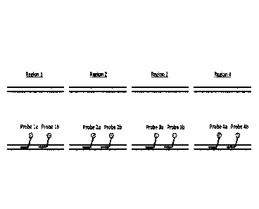
Figure 2 is a schematic representation of PNA probes targeting four different
regions of genomic DNA. Each fragment is targeted by two probes. Each PNA
probe is
covalently attached to a hapten, preferably biotin.
17
CA 2998886 2019-07-12
CA 02998886 2018-03-15
WO 2017/049213
PCT/US2016/052317
Figure 3 is a schematic representation of the methodology for strand invasion
and
capture of a specific double-stranded DNA fragment from a sequencing library.
Figures 4A-4D are histograms showing the comparative number of copies of
DNA fragments in solutions of no PNA control supernatant (control sup), no PNA
control elution (control elu), 5K/2MP PNA supernatant (5K sup) and 5K/2MP PNA
elution (5K elu) respectively, for each of four genomic amplicons analyzed via
quantitative real-time PCR, 18S 50w/75e (Figure 4A); 5S 50w/75e (Figure 4B);
CCR
50w/75e (Figure 4C); and AR 50w/75e (Figure 4D), respectively. Numerical
values of
copies of DNA fragments in each solution are indicated above each bar.
Figure 5 is a histogram showing the enrichment ratio of target (CCR+AR) to
Non-target (18S+5S) comparative number of copies of DNA fragments in solutions
of
control eluate (control), and using the 5K/2MP PNA set targeting the CCR5 and
AR1
regions, respectively. Numerical values of ratios in each solution are
indicated above
each bar.
DETAILED DESCRIPTION OF THE INVENTION
The disclosed methods and compositions may be understood more readily by
reference to the following detailed description of particular embodiments and
the
Example included therein and to the Figures and their previous and following
description.
It is to be understood that the disclosed method and compositions are not
limited
to specific synthetic methods, specific analytical techniques, or to
particular reagents
unless otherwise specified, and, as such, may vary. It is also to be
understood that the
terminology used herein is for the purpose of describing particular
embodiments only
and is not intended to be limiting.
It has been discovered that one or more large nucleic acid fragments (each
between 2,000 base pairs in length and 40,000 base pairs in length) can be
targeted and
enriched from a mixture of nucleic acid fragments using sets of two or more
sequence-
specific PNA hybridization probes. For example, one or more large double-
stranded
DNA fragments can be targeted and enriched from a mixture of genomic DNA
fragments
.. using sets of two or more sequence-specific PNA hybridization probes.
Definitions
As used herein, "enrich" and "enrichment" refer to an increase in the
proportion
of a component relative to other components present or originally present. In
the context
18
CA 02998886 2018-03-15
WO 2017/049213
PCT/US2016/052317
of nucleic acids, enrichment of nucleic acids in a sample refers to an
increase in the
proportion of the nucleic acids in the sample relative to other molecules in
the sample.
"Selective enrichment" is enrichment of particular components relative to
other
components of the same type. In the context of nucleic acid fragments,
selective
enrichment of a particular nucleic acid fragment refers to an increase in the
proportion of
the particular nucleic acid fragment in a sample relative to other nucleic
acid fragments
present or originally present in the sample. The measure of enrichment can be
referred to
in different ways. For example, enrichment can be stated as the percentage of
all of the
components that is made up by the enriched component. For example, particular
nucleic
acid fragments can be enriched in an enriched nucleic acid sample to at least
90% of the
enriched nucleic acid sample.
As used herein, -nucleic acid fragment" refers to a portion of a larger
nucleic acid
molecule. A "contiguous nucleic acid fragment" refers to a nucleic acid
fragment that
represents a single, continuous, contiguous sequence of the larger nucleic
acid molecule.
A "naturally occurring nucleic acid fragment" refers to a nucleic acid
fragment that
represents a single, continuous, contiguous sequence of a naturally occurring
nucleic acid
sequence.
As used herein, "DNA fragment" refers to a portion of a larger DNA molecule. A
"contiguous DNA fragment" refers to a DNA fragment that represents a single,
continuous, contiguous sequence of the larger DNA molecule. A -naturally
occurring
DNA fragment" refers to a DNA fragment that represents a single, continuous,
contiguous sequence of a naturally occurring DNA sequence.
As used herein, "denatured nucleic acid" or "denatured DNA" refers to a
nucleic
acid that is denatured relative to a prior existing "native" or "non-
denatured" state. For
example, double-stranded nucleic acids, such as naturally-occurring dsDNA
strands are
completely denatured when separated into two corresponding single-stranded
nucleic
acid strands. Denaturation of nucleic acids can occur by chemical or physical
means,
such as exposure to salts or increased temperatures above the melting
temperature of the
dsDNA, or by interaction of dsDNA with a denaturing molecule, such as an
antibody or
enzyme. Denaturation can be partial, for example, resulting in partially or
substantially
denatured DNA, or complete, resulting in completely denatured DNA. Nucleic
acid that
has never been subjected to partial or complete denaturation is referred to as
"never-
denatured nucleic acid", such as never-denatured dsDNA.
19
CA 02998886 2018-03-15
WO 2017/049213
PCT/US2016/052317
As used herein, "naturally occurring" refers to a molecule that has the same
structure or sequence as the corresponding molecule as it exists in nature. A
naturally
occurring molecule or sequence can still be considered naturally occurring
when it is
coupled to or incorporated into another molecule or sequence.
As used herein, "nucleic acid sample" refers to a composition, such as a
solution,
that contains or is suspected of containing nucleic acid molecules. An
"enriched nucleic
acid sample" is a nucleic acid sample in which nucleic acids, particular
nucleic acid
fragments, or a combination thereof, are enriched.
As used herein, "DNA sample- refers to a composition, such as a solution, that
contains or is suspected of containing DNA molecules. An "enriched DNA sample"
is a
DNA sample in which DNA, particular DNA fragments, or a combination thereof,
are
enriched.
References in the specification and concluding claims to parts by weight, of a
particular element or component in a composition or article, denotes the
weight
relationship between the element or component and any other elements or
components in
the composition or article for which a part by weight is expressed. Thus, in a
compound
containing 2 parts by weight of component X and 5 parts by weight component Y,
X and
Y are present at a weight ratio of 2:5, and are present in such ratio
regardless of whether
additional components are contained in the compound.
A weight percent of a component, unless specifically stated to the contrary,
is
based on the total weight of the formulation or composition in which the
component is
included.
As used herein, a "residue" of a chemical species refers to the moiety that is
the
resulting product of the chemical species in a particular reaction scheme or
subsequent
formulation or chemical product, regardless of whether the moiety is actually
obtained
from the chemical species. Thus, an ethylene glycol residue in a polymer
refers to one or
more -0CF2CH20- units in the polymer, regardless of whether ethylene glycol
was used
to prepare the polyester. As another example, in a polymer of monomer
subunits, the
incorporated monomer subunits can be referred to as residues of the un-
polymerized
monomer.
As used herein, the term "nucleotide" refers to a molecule that contains a
base
moiety, a sugar moiety and a phosphate moiety. Nucleotides can be linked
together
through their phosphate moieties and sugar moieties creating an inter-
nucleoside linkage.
CA 02998886 2018-03-15
WO 2017/049213
PCT/US2016/052317
The base moiety of a nucleotide can be adenin-9-y1 (A), cytosin-1-yl(C),
guanin-9-y1
(G), uracil-1-y1 (U), and thymin-l-yl (T). The sugar moiety of a nucleotide is
a ribose or
a deoxyribose. The phosphate moiety of a nucleotide is pentavalent phosphate.
A non-
limiting example of a nucleotide would be 3'-AMP (3'-adenosine monophosphate)
or 5'-
GMP (5'-guanosine monophosphate). There are many varieties of these types of
molecules available in the art and available herein.
As used herein, the term "nucleotide analog" refers to a nucleotide which
contains some type of modification to the base, sugar, or phosphate moieties.
Modifications to nucleotides are well known in the art and would include for
example,
5-methylcytosine (5-me-C), 5-hydroxymethyl cytosine, xanthine, hypoxanthine,
and
2-aminoadenine as well as modifications at the sugar or phosphate moieties.
There are
many varieties of these types of molecules available in the art and available
herein.
As used herein, the term "nucleotide substitute" refers to a nucleotide
molecule
having similar functional properties to nucleotides, but which does not
contain a
phosphate moiety. An exemplary nucleotide substitute is peptide nucleic acid
(PNA).
Nucleotide substitutes are molecules that will recognize nucleic acids in a
Watson-Crick
or Hoogsteen manner, but which are linked together through a moiety other than
a
phosphate moiety. Nucleotide substitutes are able to conform to a double helix
type
structure when interacting with the appropriate target nucleic acid. There are
many
varieties of these types of molecules available in the art and available
herein. It is also
possible to link other types of molecules (conjugates) to nucleotides or
nucleotide
analogs to enhance for example, interaction with DNA. Conjugates can be
chemically
linked to the nucleotide or nucleotide analogs. Exemplary conjugates include
but are not
limited to lipid moieties such as a cholesterol moiety. (Letsinger, et al.,
Proc. Natl. Acad.
Sci. USA, 86:6553-6556 (1989)). There are many varieties of these types of
molecules
available in the art and available herein.
As used herein, the term "Watson-Crick interaction" refers to at least one
interaction with the Watson-Crick face of a nucleotide, nucleotide analog, or
nucleotide
substitute. The Watson-Crick face of a nucleotide, nucleotide analog, or
nucleotide
substitute includes the C2, Ni, and C6 positions of a purine based nucleotide,
nucleotide
analog, or nucleotide substitute and the C2, N3, C4 positions of a pyrimidine
based
nucleotide, nucleotide analog, or nucleotide substitute.
21
CA 02998886 2018-03-15
WO 2017/049213
PCT/US2016/052317
As used herein, the term "Hoogsteen interaction" refers to the interaction
that
takes place on the Hoogsteen face of a nucleotide or nucleotide analog, which
is exposed
in the major groove of duplex DNA. The Hoogsteen face includes the N7 position
and
reactive groups (NH2 or 0) at the C6 position of purine nucleotides.
As used herein, the terms "oligonucleotide" or a "polynucleotide- are
synthetic or
isolated nucleic acid polymers including a plurality of nucleotide subunits.
As used herein, the term "non-natural amino acid" refers to an organic
compound
that has a structure similar to a natural amino acid so that it mimics the
structure and
reactivity of a natural amino acid. The non-natural amino acid as defined
herein
generally increases or enhances the properties of a peptide (e.g.,
selectivity, stability)
when the non-natural amino acid is either substituted for a natural amino acid
or
incorporated into a peptide.
As used herein, the term "peptide" refers to a class of compounds composed of
amino acids chemically bound together. In general, the amino acids are
chemically
bound together via amide linkages (CONH); however, the amino acids may be
bound
together by other chemical bonds known in the art. For example, the amino
acids may be
bound by amine linkages. Peptide as used herein includes oligomers of amino
acids and
small and large peptides, including polypeptides.
The term "modified" is often used herein to describe polymers and means that a
particular monomeric unit that would typically make up the pure polymer has
been
replaced by another monomeric unit that shares a common polymerization
capacity with
the replaced monomeric unit. Thus, for example, it is possible to substitute
diol residues
for glycol in poly (ethylene glycol), in which case the poly (ethylene glycol)
will be
"modified" with the diol. If the poly (ethylene glycol) is modified with a
mole
percentage of the diol, then such a mole percentage is based upon the total
number of
moles of glycol that would be present in the pure polymer but for the
modification. Thus,
in a poly (ethylene glycol) that has been modified by 50 mole % with a diol,
the diol and
glycol residues are present in equimolar amounts.
The terms homology and identity mean the same thing as similarity. Thus, for
example, if the use of the word homology is used between two non-natural
sequences it
is understood that this is not necessarily indicating an evolutionary
relationship between
these two sequences, but rather is looking at the similarity or relatedness
between their
nucleic acid sequences. Many of the methods for determining homology between
two
22
WO 2017/049213 PCT/US2016/052317
evolutionarily related molecules are routinely applied to any two or more
nucleic acids or
proteins for the purpose of measuring sequence similarity regardless of
whether they are
evolutionarily related or not.
In general, it is understood that one way to define any known variants and
derivatives or those that might arise, of the disclosed oligonucleotides,
nucleotide
analogs, or nucleotide substitutes thereof and proteins disclosed herein, is
through
defining the variants and derivatives in terms of homology to specific known
sequences.
This identity of particular sequences disclosed herein is also discussed
elsewhere herein.
In general, variants of oligonucleotides, nucleotide analogs, or nucleotide
substitutes
thereof and proteins disclosed herein typically have at least, about 70, 71,
72, 73, 74, 75,
76, 77, 78, 79, 80, 81, 82, 83, 84, 85, 86, 87, 88, 89, 90, 91, 92, 93, 94,
95, 96, 97, 98, or
99 percent homology to the stated sequence or the native sequence. Those of
skill in the
art readily understand how to determine the homology of two proteins or
nucleic acids,
such as genes. For example, the homology can be calculated after aligning the
two
sequences so that the homology is at its highest level. Another way of
calculating
homology can be performed by published algorithms. Optimal alignment of
sequences
for comparison can be conducted by the local homology algorithm of Smith and
Waterman Adv. Appl. Math. 2: 482 (1981), by the homology alignment algorithm
of
Needleman and Wunsch, J. MoL Biol. 48: 443 (1970), by the search for
similarity
method of Pearson and Lipman, Proc. Natl. Acad. Sci. U.S.A. 85: 2444 (1988),
by
computerized implementations of these algorithms (GAP, BESTFIT, FASTA, and
TFASTA in the Wisconsin Genetics Software Package, Genetics Computer Group,
575
Science Dr., Madison, WI), or by inspection. The same types of homology can be
obtained for nucleic acids by for example the algorithms disclosed in Zuker,
M. Science
244:48-52, 1989, Jaeger et al. Proc. Natl. Acad. Sci. USA 86:7706-7710, 1989,
Jaeger et
al. Methods Enzymol. 183:281-306, 1989. It is understood that any of the
methods
typically can be used and that in certain instances the results of these
various methods
can differ, but the skilled artisan understands if identity is found with at
least one of these
methods, the sequences would be said to have the stated identity, and be
disclosed
herein. For example, as used herein, a sequence recited as having a particular
percent
homology to another sequence refers to sequences that have the recited
homology as
calculated by any one or more of the calculation methods described above.
23
CA 2998886 2019-07-12
CA 02998886 2018-03-15
WO 2017/049213
PCT/US2016/052317
For example, a first sequence has 80 percent homology, as defined herein, to a
second
sequence if the first sequence is calculated to have 80 percent homology to
the second
sequence using the Zuker calculation method even if the first sequence does
not have 80
percent homology to the second sequence as calculated by any of the other
calculation
methods. As another example, a first sequence has 80 percent homology, as
defined
herein, to a second sequence if the first sequence is calculated to have 80
percent
homology to the second sequence using both the Zuker calculation method and
the
Pearson and Lipman calculation method even if the first sequence does not have
80
percent homology to the second sequence as calculated by the Smith and
Waterman
calculation method, the Needleman and Wunsch calculation method, the Jaeger
calculation methods, or any of the other calculation methods. As yet another
example, a
first sequence has 80 percent homology, as defined herein, to a second
sequence if the
first sequence is calculated to have 80 percent homology to the second
sequence using
each of calculation methods (although, in practice, the different calculation
methods will
.. often result in different calculated homology percentages).
As used herein, reference to there being some number of residues of a first
description (such as residues not derivatized with a moiety) "between every
residue" of a
second description (such as residues derivatized with a moiety) means that,
between
every two residues of the second description that do not have any other
residue of the
second description between them, the specified number of residues of the first
description are present. Thus, for example, the probe T*gTgC*cTccC*gTtTT*gTcC*
(SEQ ID NO:6) is an example of a probe where, at different locations, zero,
one, or two
residues are not derivatized with a moiety between the residues that are
derivatized with
a moiety. If a residue of the second description is the last residue of the
second
description before the end of the probe (which can be referred to as an end-
proximal
residue of the second description), the reference to there being some number
of residues
of the first description between every residue of the second description does
not apply to
the residues between the end-proximal residue and the end of the probe. Thus,
the
average spacing between residues of the second description counts only the
internal
spacings without considering residues of the first description between each
end and their
respective end-proximal residue of the second description.
The residues of a first description between the end-proximal residue of a
second
description and the end of the probe can be referred to as flanking residues
of the first
24
CA 02998886 2018-03-15
WO 2017/049213
PCT/US2016/052317
description. For example, the probe T*gTgC*cTccC*grft.TT*gTcC* (SEQ ID NO:6)
has
a total of zero residues not derivatized with a moiety between both of the end-
proximal
derivatized residues and their respective ends of the probe and so has zero
flanking
residues not derivatized with a moiety. As another example, the probe
.. cT*tCaT*CtCgT*cTaC*aaT*a (SEQ ID NO:10) has a total of two residues not
derivatized with a moiety between both of the end-proximal derivatized
residues and
their respective ends of the probe and so has two flanking residues not
derivatized with a
moiety. As another example, the probe agT*CgTtC*tTcT*aTCaT*cT (SEQ ID NO:20)
has a total of two residues not derivatized with a moiety between both of the
end-
proximal derivatized residues and their respective ends of the probe and so
has two
flanking residues not derivatized with a moiety.
As another example, the probe T*gTgC*cTccC*gTtTT*gTcC* (SEQ ID NO:6)
has a total of zero residues not derivatized with a charged moiety between
both of the
end-proximal residues derivatized with a charged moiety and their respective
ends of the
.. probe and so has zero flanking residues not derivatized with a charged
moiety. As
another example, the probe cT*tCaT*CtCgT*cTaC*aaT*a (SEQ ID NO:10) has a total
of one residue not derivatized with a charged moiety between both of the end-
proximal
residues derivatized with a charged moiety and their respective ends of the
probe and so
has one flanking residue not derivatized with a charged moiety. As another
example, the
probe agT*CgTtC*tTcT*aTCaT*cT (SEQ ID NO:20) has a total of four residues not
derivatized with a charged moiety between both of the end-proximal residues
derivatized
with a charged moiety and their respective ends of the probe and so has four
flanking
residues not derivatized with a charged moiety.
Materials
Disclosed are materials, compositions, and components that can be used for the
disclosed methods. These and other materials are disclosed herein, and it is
understood
that when combinations, subsets, interactions, groups, etc. of these materials
are
disclosed that while specific reference of each various individual and
collective
combinations and permutation of these compounds may not be explicitly
disclosed, each
is specifically contemplated and described herein. For example, if a matched
set of
peptide nucleic acid (PNA) hybridization probes is disclosed and discussed and
a number
of modifications that can be made to a number of molecules including the
peptide
nucleic acids of each of the probes are discussed, each and every combination
and
CA 02998886 2018-03-15
WO 2017/049213
PCT/US2016/052317
permutation of peptide nucleic acids and the modifications that are possible
are
specifically contemplated unless specifically indicated to the contrary. Thus,
if a class of
modifications A, B, and C are disclosed as well as a class of molecules D, E,
and F and
an example of a combination molecule, A-D is disclosed, then even if each is
not
individually recited, each is individually and collectively contemplated.
Thus, is this
example, each of the combinations A-E, A-F, B-D, B-E, B-F, C-D, C-E, and C-F
are
specifically contemplated and should be considered disclosed from disclosure
of A, B,
and C; D. E, and F; and the example combination A-D. Likewise, any subset or
combination of these is also specifically contemplated and disclosed. Thus,
for example,
the sub-group of A-E, B-F, and C-E are specifically contemplated and should be
considered disclosed from disclosure of A, B, and C; D, E, and F; and the
example
combination A-D. Further, each of the materials, compositions, components,
etc.
contemplated and disclosed as above can also be specifically and independently
included
or excluded from any group, subgroup, list, set, etc. of such materials. These
concepts
apply to all aspects of this application including, but not limited to, steps
in methods of
making and using the disclosed compositions. Thus, if there are a variety of
additional
steps that can be performed it is understood that each of these additional
steps can be
performed with any specific embodiment or combination of embodiments of the
disclosed methods, and that each such combination is specifically contemplated
and
should be considered disclosed.
A. Compounds
1. PNA hybridization probes
PNA hybridization probes (PNA probes) are oligomers of nucleic acid base
pairing residues that include at least one peptide nucleic acid residue and
are designed to
and are capable of invading double-stranded DNA and hybridizing to a target
sequence
via Watson-Crick base pairing. In some forms, PNA probes include one or more
capture
tags. In some forms, the PNA probe is designed to target a sequence in a
nucleic acid
fragment. In some forms, the PNA probe includes one or more capture tags. In
some
forms, the PNA probe is designed to target a sequence in a nucleic acid
fragment. In
some forms, the PNA probe includes (a) one or more peptide nucleic acid
residues that
are derivatized with a charged moiety on the alpha carbon, beta carbon, gamma
carbon,
or combinations thereof, (b) one or more peptide nucleic acid residues that
are
26
CA 02998886 2018-03-15
WO 2017/049213
PCT/US2016/052317
derivatized with a neutral moiety on the alpha carbon, beta carbon, gamma
carbon, or
combinations thereof, or (c) combinations thereof.
In some forms, the PNA probe includes two to six peptide nucleic acid residues
that independently are derivatized with a charged moiety on the alpha, beta,
or gamma
carbon. In some forms, one or more of the peptide nucleic acid residues that
are
derivatized with the charged moiety are derivatized with the charged moiety on
the
gamma carbon. In some forms, all of the peptide nucleic acid residues that are
derivatized with the charged moiety are derivatized with the charged moiety on
the
gamma carbon. In some forms, one or more of the charged moieties are lysine.
In some
forms, all of the charged moieties are lysine. In some forms, one or more of
the charged
moieties are L-lysine. In some forms, all of the charged moieties are L-
lysine.
In some forms, the PNA probe includes one or more peptide nucleic acid
residues
that are derivatized with a short-chain oligoethylene moiety on the alpha,
beta, or gamma
carbon. In some forms, the PNA probe includes one to nineteen peptide nucleic
acid
residues that independently are derivatized with the short-chain oligoethylene
moiety on
the alpha, beta, or gamma carbon. In some forms, one or more of the peptide
nucleic acid
residues that are derivatized with the short-chain oligoethylene moiety are
derivatized
with the short-chain oligoethylene moiety on the gamma carbon. In some forms,
all of
the peptide nucleic acid residues that are derivatized with the short-chain
oligoethylene
moiety are derivatized with the short-chain oligoethylene moiety on the gamma
carbon.
In some forms, one or more of the short-chain oligoethylene moieties are
diethylene
glycol. In some forms, all of the short-chain oligoethylene moieties are
diethylene glycol.
In some forms, one or more of the PNA probes can independently include one or
more peptide nucleic acid residues having a pseudo-complementary nucleobase as
the
base moiety of the peptide nucleic acid residue. In some forms, one or more of
the PNA
probes can independently include one to twenty-two peptide nucleic acid
residues having
a pseudo-complementary nucleobase as the base moiety of the peptide nucleic
acid
residue. In some forms, all of the PNA probes can independently include one to
twenty-
two peptide nucleic acid residues having a pseudo-complementary nucleobase as
the
base moiety of the peptide nucleic acid residue.
In some forms, the pseudo-complementary nucleobases are independently
selected from the group consisting of pseudouridine (5-ribosyluracil); 7-Deaza-
2'-
deoxyguanosine; 2,6-Diaminopurine-2'-deoxyriboside; N4-Ethyl-2'-deoxycytidine;
2-
27
CA 02998886 2018-03-15
WO 2017/049213
PCT/US2016/052317
thiothymidine; 2-aminoadenine; 2-aminopurine-riboside; 2,6-diaminopurine-
riboside; 2'-
deoxyisoguanosine; and 5-hydroxymethy1-2'-deoxycytidine.
In some forms, the one or more of the PNA probes that include one or more
peptide nucleic acid residues having a pseudo-complementary nucleobase as the
base
moiety of the peptide nucleic acid residue is a subset of the PNA probes in
the one or
more sets of PNA probes. In some forms, the subset of the PNA probes in the
one or
more sets of PNA probes includes a subset of the PNA probes in the one or more
sets of
PNA probes that are predicted to be capable of interacting with one or more of
the other
PNA probes in the one or more sets of PNA probes. In some forms, the subset of
the
PNA probes in the one or more sets of PNA probes is a subset of the PNA probes
in the
one or more sets of PNA probes that are predicted to be capable of interacting
with one
or more of the other PNA probes in the one or more sets of PNA probes.
In some forms, the capture tag is biotin or streptavidin. In some forms, the
PNA
probe is derivatized with one or more amino acids on at least one of the
terminal PNA
residues. In some forms, the PNA probe is derivatized with two or more lysine
residues
on at least one of the terminal PNA residues.
In some forms the hybridization probes include peptide nucleic acid (PNA)
oligomers that combine PNA monomers modified at the gamma position with
neutral
and charged moieties.
Sets of two or more PNA hybridization probes including a combination of
charged and neutral gamma modifications can be designed to target any nucleic
acid
sequence (such as DNA or RNA sequence). For example, PNA probes can be
designed
to be complementary to a target nucleotide sequence unique to a particular
gene, nucleic
acid fragment, or DNA fragment from a highly complex nucleic acid sample, such
as a
whole genomic DNA sample. The target nucleic acid sequence can be any suitable
length. For example, the target nucleic acid sequence can be between 8 and 30
nucleotides in length, typically between 15 and 25 nucleotides. A preferred
nucleic acid
target sequence is between 18 and 22 nucleotides in length, inclusive, for
example, 20
nucleotides in length.
In some forms, PNA probes are designed to combine PNA monomers with
gamma Mini-PEG modifications and PNA monomers with gamma L-Lysine
modifications for optimal solubility, rapid hybridization kinetics, high
melting
temperature after DNA hybridization, as well as good mismatch discrimination.
The
28
CA 02998886 2018-03-15
WO 2017/049213
PCT/US2016/052317
positively-charged Lysine residues undergo charge repulsion when contacting
other PNA
molecules. For this reason. PNA probes with 2 or more gamma-L-Lysine
modifications
are less likely to undergo intermolecular hybridization associations with
other probes of
different sequence present in a mixture containing thousands of different PNA
.. sequences, designed to invade different DNA targets. Exemplary PNA probes
are
provided in Table 1. Each hybridization probe includes one or more capture
tags, such as
a biotin moiety, to enable isolation of the target nucleic acid fragments by,
for example,
affinity chromatography. Each hybridization probe optionally includes amino-
acid
adducts to enhance aqueous solubility, for example, two lysine residues.
PNA hybridization probes can be readily synthesized using techniques generally
known to synthetic organic chemists.
1. Target nucleic acid sequences
Short PNA probes can be designed and used as capture probes for enrichment of
a specific nucleic acid target sequence. The design of hybridization probes
for sequence-
specific nucleic acid capture according to the disclosed methods requires
knowledge of
two or more target sequences within each different target nucleic acid
fragment.
Typically, multiple distinct target sequences for the short PNA hybridization
probes are
prevalent in large nucleic acid molecules.
The term "k-mers" refers to short nucleic acid sequences, where "k" denotes
the
number of positions in a short string of nucleotide bases. Typically, each
probe in a set of
probes designed for use according to the disclosed methods should be
complementary to
a short (preferably 18 to 22 bases) nucleotide sequence that is unique in the
sequences
present in the nucleic acid sample. For example, for enrichment of genomic DNA
fragments the probe should be complementary to a short (preferably 18 to 22
bases)
.. nucleotide sequence that is unique in the sequences present in the genome.
Typically, the hybridization probes are designed as matched sets of two or
more
probes that target nucleotide sequences within the same desired DNA fragment.
The
optimal number of different hybridization probes designed to target a nucleic
acid
fragment by the described methods can vary depending upon the size of the
nucleic acid
fragment being targeted. Preferably, two or more probes may be used to target
fragments
up to 20,000 base pairs in length, three or more probes may be used to target
fragments
up 30,000 base pairs in length, and four or more probes may be used to target
fragments
up to 40,000 base pairs in length.
29
CA 02998886 2018-03-15
WO 2017/049213
PCT/US2016/052317
It is possible to design PNA probes that work in pairs by hybridizing to each
strand of the target DNA. Therefore, although not preferred, the two or more
target
sequences can be overlapping, partially overlapping or non-overlapping, for
example,
adjacent or contiguous sequences in the target nucleic acid fragment. In some
forms,
target sequences that are overlapping, partially overlapping, or both can be
excluded. In
some forms, two or more target sequences are separated by one or more
nucleotides. In
some forms, the hybridization probes are designed to induce duplex invasion or
triplex
invasion of the target nucleic acid. Therefore, although not preferred,
hybridization
probes can include two or more target sequences that are partially
overlapping, or non-
overlapping on the target nucleic acid fragment. In some forms, hybridization
probes that
are capable of inducing triplex invasion of the target nucleic acid are
designed to induce
triplex invasion of the target nucleic acid, or both, can be excluded. In some
forms,
although not preferred, a matched pair of two hybridization probes include
palindromic
(self-complementary) sequences can be used in methods for double-duplex
invasion of a
target DNA fragment.
Hybridization probes haying target sequences that are not unique can target,
invade and capture multiple sequences in the genome. Therefore, in some forms,
a set of
two or probes designed for use according to the disclosed methods perfoims
multiplexed
double stranded DNA sequence capture most specifically when each probe in the
set is
complementary to a DNA sequence for which the number of k-mers in the genome
that
differ by only one base is zero. Capture by each probe in a probe set is more
specific
when the number of k-mers in the genome that differ by only two bases is zero.
Capture
by each probe in a probe set is even more specific when the number of k-mers
in the
genome that differ by only three bases is zero. Bioinformatics tools can be
used to
identify in the genome candidate probe sequences that meet the desired
uniqueness
requirement: absence at other genomic positions of closely related sequences
that differ
by one or two or even three mismatches.
Bioinformatics tools for sequence information of the human genome is available
from multiple sources, for example, the UCSC database (version hg12; June 28,
2002)
(intemet site genome.ucsc.edu/goldenPath/28jun2002) developed by the
International
Human Genome Mapping Consortium.
Preferably, probe candidates do not include k-mers capable of self-folding to
form a stable secondary structure. These k-mers have a lower probability of
interacting
CA 02998886 2018-03-15
WO 2017/049213
PCT/US2016/052317
with a target sequence, since they are trapped into a thermodynamically stable
self-
folding configuration.
Table 1: Examples of PNA probes.
Total Total
number of number of
PNA PNA Charged
Total residues
residues residues
number of Total Total number derivatized derivatized on
base- number of with a with a
terminal
Probe Capture containing of PNA underivatized charged neutral PNA
No. Tag residues residues PNA residues moiety
moiety residue
1 Yes 20 20 0 2 18 2
2 Yes 20 20 0 3 17 2
3 Yes 20 20 0 4 16 2
4 Yes 20 20 0 5 15 2
Yes 20 20 0 6 14 2
6 Yes 20 20 1 2 17 2
7 Yes 20 20 1 3 16 2
8 Yes 20 20 1 4 15 2
9 Yes 20 20 1 5 14 2
Yes 20 20 1 6 13 2
11 Yes 20 20 2 2 16 2
12 Yes 20 20 2 3 15 2
13 Yes 20 20 2 4 14 2
14 Yes 20 20 2 5 13 2
Yes 20 20 2 6 12 2
16 Yes 20 20 3 2 15 2
17 Yes 20 20 3 3 14 2
18 Yes 20 20 3 4 13 2
19 Yes 20 20 3 5 12 2
Yes 20 20 3 6 11 2
21 Yes 20 20 4 2 14 2
22 Yes , 20 20 , 4 3 13 , 2 ,
23 Yes , 20 20 , 4 4 12 , 2 ,
24 Yes 20 20 4 5 11 2
Yes 20 20 4 6 10 2
26 Yes 20 20 5 2 13 2
27 Yes 20 20 5 3 12 2
28 Yes 20 20 5 4 11 2
29 Yes 20 20 5 5 10 2
Yes 20 20 5 6 9 2
31 Yes 20 20 6 2 12 2
32 Yes 20 20 6 3 11 2
33 Yes 20 20 6 4 10 2
34 Yes 20 20 6 5 9 2
Yes 20 20 6 6 8 2
36 Yes 20 20 7 2 11 2
37 Yes 20 20 7 3 10 2
38 Yes 20 20 7 4 9 2
39 Yes 20 20 7 5 8 2
31
CA 02998886 2018-03-15
WO 2017/049213
PCT/1JS2016/052317
Total Total
number of number of
PNA PNA Charged
Total residues
residues residues
number of Total Total number derivatized derivatized on
base- number of with a with a
terminal
Probe Capture containing of PNA underivatized charged neutral PNA
No. Tag residues residues PNA residues
moiety moiety residue
40 Yes 20 20 7 6 7 2
41 Yes 20 20 8 2 10 2
42 Yes 20 20 8 3 9 2
43 Yes 20 20 8 4 8 2
44 Yes 20 20 8 5 7 2
45 Yes 20 20 8 6 6 2
46 Yes 20 20 9 2 9 2
47 Yes 20 20 9 3 8 2
48 Yes 20 20 9 4 7 2
49 Yes 20 20 9 5 6 2
50 Yes 20 20 9 6 5 2
51 Yes 20 20 10 2 8 2
52 Yes 20 20 10 3 7 2
53 Yes 20 20 10 4 6 2
54 Yes 20 20 10 5 5 2
55 Yes 20 20 10 6 4 2
56 Yes 20 20 11 2 7 2
57 Yes 20 20 11 3 6 2
58 Yes 20 20 11 4 5 2
59 Yes 20 20 11 5 4 2
60 Yes 20 20 11 6 3 2
61 Yes 20 20 12 2 6 2
62 Yes 20 20 12 3 5 2
63 Yes 20 20 12 4 4 2
64 Yes 20 20 12 5 3 2
65 Yes 20 20 12 6 2 2
66 Yes 20 20 13 2 5 2
67 Yes 20 20 13 3 4 2
68 Yes 20 20 13 4 3 2
69 Yes 20 20 13 5 2 2
70 Yes 20 20 13 6 1 2
71 Yes , 20 20 , 14 2 4 , 2 ,
72 Yes 20 20 14 3 3 2
73 Yes 20 20 14 4 2 2
74 Yes 20 20 14 5 1 2
75 Yes 20 20 14 6 0 2
76 Yes 20 20 15 2 3 2
77 Yes 20 20 15 3 2 2
78 Yes 20 20 15 4 1 2
79 Yes 20 20 15 5 0 2
80 Yes 16 16 6 6 4 2
81 Yes 17 17 7 6 4 2
82 Yes 18 18 8 6 4 2
83 Yes 19 19 9 6 4 2
32
CA 02998886 2018-03-15
WO 2017/049213
PCT/1JS2016/052317
Total Total
number of number of
PNA PNA Charged
Total residues
residues residues
number of Total Total number derivatized derivatized on
base- number of with a with a terminal
Probe Capture containing of PNA underivatized charged neutral PNA
No. Tag residues residues PNA residues
moiety moiety residue
84 Yes 21 21 11 6 4 2
85 Yes 22 22 12 6 4 2
86 Yes 23 23 13 6 4 2
87 Yes 24 24 14 6 4 2
88 Yes 25 25 15 6 4 2
89 Yes 26 26 16 6 4 2
90 Yes 16 16 8 6 2 2
91 Yes 17 17 9 6 2 2
92 Yes 18 18 10 6 2 2
93 Yes 19 19 11 6 2 2
94 Yes 21 21 13 6 2 2
95 Yes 22 22 14 6 2 2
96 Yes 23 23 15 6 2 2
97 Yes 24 24 16 6 2 2
98 Yes 25 25 17 6 2 2
99 Yes 26 26 18 6 2 2
100 Yes 16 16 9 6 1 2
101 Yes 17 17 10 6 1 2
102 Yes 18 18 11 6 1 2
103 Yes 19 19 12 6 1 2
104 Yes 21 21 14 6 1 2
105 Yes 22 22 15 6 1 2
106 Yes 23 23 16 6 1 2
107 Yes 24 24 17 6 1 2
108 Yes 25 25 18 6 1 2
109 Yes 26 26 19 6 1 2
Computer programs are available to identify those undesirable self-folding
k-mers. Short k-mer sequences, typically 18 to 22 bases in length, that are
unique and
also suitable for specific targeting and capture by strand invasion can occur
at a
frequency that is less than 1,000 in 10,000 base pairs.
Typically. DNA target sequences of hybridization probes designed for use
according to the disclosed methods are characterized by having a melting
temperature
that is relatively low. For example, for a sequence of 20 contiguous
nucleotides in a
genome, the expected melting temperature can be calculated using values for
entropy and
enthalpy characteristic of each dinucleotide, as described by Santa Lucia,
Proc. Natl.
Acad. Sci. USA, Vol. 95, pp. 1460-1465 (1998). Therefore, in an exemplary
genomic
33
CA 02998886 2018-03-15
WO 2017/049213
PCT/US2016/052317
domain of 30,000 base pairs, 29,980 k-mers each of 20 base pairs can be
enumerated. A
computer-based algorithm can be used to calculate the predicted melting
temperature of
all 29,980 k-mers in this genomic interval.
A useful 20-base DNA target sequences according to the disclosed methods is
characterized by having a melting temperature that belongs to the lowest half
(50%) of
all computed 20-base DNA melting temperatures. A particularly 20-base DNA
target
sequences according to this invention is characterized by having a melting
temperature
that belongs to the lowest one-third (33%) of all computed 20-base DNA melting
temperatures.
In order to use a multiplicity of hybridization probes in a single reaction
volume
it is preferred that all the probe sequences in the set are unable to
hybridize with each
other. This requirement is satisfied when each possible PNA sequence alignment
between all possible combinations of all probe pairs has at least 3 mismatched
bases, or
more preferably at least 4 mismatches, or more preferably at least 5
mismatches, or even
more preferably at least 6 mismatches. Any computer programs known in the art
can be
used to examine the likelihood of cross-reactivity amongst all PNA probe
candidates in a
set of several thousand probe candidates, to make sure that the preferred
condition of no
inter-probe cross-hybridization is met by all probe pairs.
a. Exemplary Targets
Exemplary target sequences for target-specific enrichment include one or more
components of a specific genome, for example, the human genome. Exemplary
human
genomic DNA that can be targeted and enriched includes DNA located in the MHC
region. For example, in particular forms, target sequences include genetic
elements of
human genomic DNA located in the MHC region of chromosome 6.
In some forms, target sequences for target-specific enrichment include genomic
components of the MHC known to be associated with one or more specific
immunological features or phenotypes. Exemplary immunological features or
phenotypes include having predisposition to autoimmune diseases, or showing
symptoms
of autoimmune diseases. Therefore, in some forms, target sequences enrich
regions of
genomic DNA where sequence variation is associated with immunological features
such
as autoimmune diseases. Exemplary genes associated with sequence variation
relating to
autoimmune diseases include, among others, the DRB1 and DQA1 genes. Therefore,
in
some forms, targeted genomic DNA fragments include the DRB1 gene, or fragments
of
34
CA 02998886 2018-03-15
WO 2017/049213
PCT/US2016/052317
the DRB1 gene. In some forms, targeted genomic DNA fragments include the DQA1
gene, or fragments of the DQAI gene. In some forms, targeted DNA fragments
include
the DQA1 gene, or fragments of the DQA1 gene and the DRB I gene, or fragments
of the
DRBI gene. An exemplary genomic target region is 90,000 bases in length and
spans the
genomic co-ordinates chr6:32522981-32612981 (coordinates based on human genome
build hg19). In some forms, targeted human genomic DNA is located in the Major
Histocompatibility Complex (MEC) region of chromosome 6, for example, the DRB1
and DQA1 genes.
In some forms, targeted genomic DNA includes a 40,000 base window that spans
a region starting at -22,000 bases upstream of the human FOXP3 (Forkhead Box
P3,
expressed in regulatory T-cells) promoter, and ending 18,000 bases downstream
of the
FOXP3 promoter. Therefore, in some forms the targeted genomic DNA includes the
human FOXP3 gene, or fragments of the FOXP3 gene. An exemplary genomic target
region is the sequence spanning the genomic coordinates chrX:49103288-49143288
(coordinates based on human genome build hgl 9). Exemplary targeted genomic
DNA
from this region includes seven sequences, separated from each other by an
average of
5,714 base pairs in the genome.
In some folms, target sequences include genetic elements associated with one
or
more diseases or conditions, or having a known correlation with development of
one or
more disease or conditions (i.e., associated with disease risk). Exemplary
diseases are
autoimmune diseases, diabetes, and the metabolic syndrome, and cancer. For
example, in
a particular form, target sequences include genetic elements from more than 40
or 50
mega-bases of human genomic DNA located within enhancer elements associated
with
disease risk for autoimmune diseases, or enhancer elements associated with
disease risk
for diabetes and the metabolic syndrome. For example, in some forms, targeted
DNA
includes enhancer clusters associated with important diseases, such as Type II
diabetes.
3,677 enhancer clusters have been identified which mapped near genes with
strong
pancreatic islet-enriched expression (Pasquali et al., Nat Genet. 2014
Feb;46(2):136-43
(2014)). Therefore, in some forms, targeted DNA includes genomic DNA windows
of
30,000 to 150,000 base pairs to encompass all of the enhancers within a
cluster. For
example, targeted sequences can be of unique sequence at an average distance
of 5,000
to 7,000 bases from each other within each cluster.
CA 02998886 2018-03-15
WO 2017/049213
PCT/US2016/052317
Other target sequences include enhancer elements associated with the
differentiation of different subsets of white blood cells.
In some forms, target sequences include entire subsets of genomic DNA from a
single genome, or mixtures of two or more genomes from the same or different
species,
such as mitochondria' DNA. For example, in a particular form, target sequences
include
components of the human mitochondrial genome. In some forms, target sequences
include the dog mitochondrial genome, or the cat mitochondrial genome.
In further forms, target sequences include genomic DNA of one or more species
of bacteria, archaea, fungi, protozoa, or mixtures of two or more of these.
Therefore,
target sequences can be sequences of genomic DNA of one or more species of
bacteria
present in the human oral cavity, one or more species of bacteria present in
the human
airway, or present in the human urogenital tract, or known to exist in human
blood or
feces.
Peptide Nucleic Acid (PNA)
Peptide nucleic acid (PNA) is a nucleic acid mimic where the native nucleic
acid
sugar-phosphate backbone is replaced by an N-(2-aminoethyl) glycine unit.
Thus, unlike
DNA and other DNA analogs, PNAs do not include phosphate groups or pentose
sugar
moieties. A methyl carbonyl linker connects natural as well as unusual (in
some cases)
nucleotide bases to this backbone at the amino nitrogens. Un-modified PNAs are
non-
ionic, achiral, neutral molecules and are not susceptible to hydrolytic
(enzymatic)
cleavage. The term "un-modified PNA residues" refers to a PNA residue
including an N-
(2-aminoethyl)-glycine backbone (see Formula III). The term "derivatized PNA-
or
"modified PNA" refers to a PNA residue having one or more substitutions or
derivatized
groups at one or more positions of the un-modified PNA structure.
PNA can be synthesized and modified by any means known in the art. Typically,
the procedures for PNA synthesis are similar to those employed for peptide
synthesis,
using standard solid-phase manual or automated synthesis. Suitable
experimental
methods for making and derivatizing compounds including PNA and modified PNA
are
described in Bahal, et al., Current Gene Therapy, Vol. 14, No. 5 (2014);
Bahal, et al.,
Artificial DNA: PNA & XNA 4:2, 49-57 (2013); De Costa, et al., PLOS One, Vol.
8, (3)
e58670 (2013); Dragulescu-Andrasi, J. Am. Chem. Soc. 128, 10258-10267 (2006);
Englund, et al., Org. Left., Vol. 7, No. 16, 3465-3467 (2005); Ishizuka, et
al., Nucleic
acids Research, Vol. 36, No. 5, 1464-1471 (2008); Huang, et al., Arch Pharm
Res Vol
36
WO 2017/049213 PCT/US2016/052317
35, No 3, 517-522, (2012); Kuhn, etal., Artificial DNA: PNA & XNA 1:1, 45-53
(2010);
Sugiyama, et al., Molecules ,18, 287-310 (2013); Sahu, et al., J Org Chem. 15;
76(14):
5614-5627 (2011); and Yeh, etal., J Am Chem Soc.; 132(31): 10717-10727(2010).
Despite variations from natural nucleic acids, PNA is still capable of
sequence-
specific binding to DNA as well as RNA obeying the Watson-Crick hydrogen
bonding
rules. PNA shows potential in many applications, including bio-sensing and
therapeutics,
due to its high binding affinity and selectivity for DNA and RNA. PNA forms
highly
stable complexes with target DNA and PNA-DNA complexes have a higher thermal
melting temperature (Tm), as compared to the corresponding DNA¨DNA or DNA¨RNA
duplexes formed by the same nucleotide sequence. In addition, hybridization of
PNAs
with target DNA can occur virtually independent of salt concentration and the
Tin of
PNA¨DNA duplex is generally not affected by low ionic strength. Therefore,
PNAs can
hybridize to DNA or RNA sequences involved in secondary structures, which are
destabilized by low ionic strength.
In contrast to DNA, PNA can bind in either a parallel or antiparallel manner
and
PNA hybridization probes will bind to either single-stranded DNA or to double-
stranded
DNA.
PNA hybridization probes are capable of invading complementary target
sequences in DNA duplexes in vitro, as well as in living cells. Strand
invasion of double
stranded DNA by peptide nucleic acids (PNA) has been extensively described in
the
literature (Ito et al., 1992a; Ito, Smith, Cantor (1992b)). Early published
examples of the
use of PNA for DNA capture rely on the ability of PNA molecules to engage in
DNA
triplex interactions that are readily formed in DNA sequences that contain
homo-purine-
rich sequences. For example, PNAs were used to isolate specific sequence
repeats from a
human genomic library, as well as for isolation of a single copy clone from a
yeast
genomic library. However, PNA triplex interactions are not preferred because
it is
difficult to design sufficiently specific triplex probes for capture of a
multiplicity of
different loci in the genome.
Strand invasion by PNA is more efficient in vitro, at low salt concentrations,
and
slower at physiological salt concentrations. Several methods have been applied
to
sequence-specific enrichment of DNA by PNA-based capture. For example, PNAs
containing diaminopurine-thiouracil base pairs bind with high specificity and
efficiency
37
CA 2998886 2019-07-12
CA 02998886 2018-03-15
WO 2017/049213
PCT/US2016/052317
to complementary targets in double- stranded DNA by a mechanism termed "double
duplex invasion" in which the duplex is unwound and both DNA strands are
targeted
simultaneously-, each by a different PNA containing pseudo-complementary bases
(Lohse et. al, 1999) (see Figure 1). When two PNA probes, each containing
pseudo-
complementary bases are used to target a specific DNA sequence, the two PNAs
are
unable to hybridize with each other due to the steric clashes of the pseudo-
complementary bases. By contrast, the interactions with each of the DNA
strands are
highly stable. Double duplex invasion has been used successfully for targeted
correction
of a thalassemia-associated beta globin mutation (Lonkar, et al., 2009).
a. Modifications of PNA
PNA probes can include PNA modified by any means known in the art to change
the structural and functional features of the probes. In some forms, chemical
modifications of PNA change one or more structural characteristics of the PNA.
PNA monomers including any of the modifications described herein can be
incorporated into oligomers. Therefore, PNA probes can be PNA oligomers
including
modified PNA monomers, unmodified PNA monomers, and combinations thereof In
some forms, PNA probes include a multiplicity of variously modified PNA
monomers.
For example, matched pairs of self-complementary PNA probes can be modified to
reduce the thermal stability of the PNA-PNA duplex formed by the probes in
each pair.
In addition, PNA probes can include PNA monomers modified to enhance sequence-
specificity and affinity of DNA-PNA duplexes; and to reduce non-specific
interactions.
Although not preferred, PNA oligomers can include bis-PNA oligomers. Bis-
PNA binds specific target sequences to form a looped-out single strand and an
internal,
triple-stranded invaded complex. Bis-PNA can be prepared in a continuous
synthesis
process by connecting two PNA segments via a flexible linker composed of
multiple
units of either 8-amino-3,6-dioxaoctanoic acid or 6-aminohexanoic acid (Ray
and
Norden, The FASEB Journal, vol. 14 no. 9 1041-1060 (2000)). In some forms, bis-
PNA
oligomers can be excluded.
(A) Pseudo-complementary Bases
Pseudo-complementary (PC) nucleobases are non-standard bases that have
significantly reduced affinity for forming duplexes with each other due to
chemical
modification, but retain strong base pairs with natural DNA or RNA targets and
can
readily hybridize to unmodified nucleic acids. Therefore, the differential
hybridization
38
CA 02998886 2018-03-15
WO 2017/049213
PCT/US2016/052317
properties of pc-nucleic acids provides for efficient sequence-specific
targeting of duplex
DNA by double duplex invasion strategies. When pseudo-complementary invading
PNA
pairs are utilized for DNA strand invasion, the total number of probes used
for DNA
capture is effectively doubled, as compared to a single invading PNA.
A non-limiting list of pseudo-complementary nucleobases includes Pseudouridine
(5-Ribosyluracil); 7-Deaza-2'-deoxyguanosine; 2,6-Diaminopurine-2'-
deoxyriboside;
N4-Ethyl-21-deoxycytidine; 2-Thiothymidine; 2-aminoadenine; 2-Aminopurine-
riboside;
2,6-Diaminopurine-riboside; 2'-Deoxyisoguanosine; and 5-Hydroxymethy1-2'-
deoxycytidine (see Formula I). Pseudo-complementary invading PNA pairs form
stable
Watson: Crick interactions with natural DNA bases, but are not capable of
stable
hydrogen bonding among themselves (Lohse et al 1999), as depicted in Formula
II. For
example, Diaminopurine can form an extra hydrogen bond with thymine, whereas a
steric clash occurs between diaminopurine and thiouracil.
In some forms, PNA probes are designed for use in a capture method based on
pseudo-complementary invading PNA pairs. For example, PNA probes can be
designed
for double duplex invasion by pseudo-complementary PNA to achieve sequence-
specific
capture of a multiplicity of double-stranded DNA domains from eukaryotic
genomes.
N
,A0
1.1
2 ,6-dia minopu ne m inopu rine pseudoisocytosine
0
N (LI
1
thiouracil
E- base
Formula I: Chemical structures of non-standard nucleobases
39
CA 02998886 2018-03-15
WO 2017/049213
PCT/1JS2016/052317
N
Thyrrsim
H
Menine
Drammrsne
H., H' 14 El'
11/4r "`"Pr''
ii
N
4, S 5 '
= -
iµr)r- 4-11
Aden
Diatninwfrine.
Formula 11: adenine-thymidine; diaminopurine-thymidine; adenine-thiouracil,
and
adenine-thiouracil base pairs.
Pseudo-complementary bases can be useful for incorporating into PNA probes
when numerous different PNA probes in a single capture reaction. For example,
pseudo-
complementary bases can be useful when thousands of PNA probes are used
together to
capture numerous target sequences. In certain such forms, the pseudo-
complementary
bases can be incorporated, for example, just into a particular subset of PNA
probes. For
example, the pseudo-complementary bases can be incorporated into a subset of
PNA
probes that computer analysis predicts to be capable of interacting with each
other. Use
of such PNA probes can reduce or eliminate undesired probe-probe interactions.
In some
forms, use of pseudo-complementary bases in PNA probes can be excluded.
In some forms, one or more of the PNA probes can independently include one or
more peptide nucleic acid residues having a pseudo-complementary nucleobase as
the
.. base moiety of the peptide nucleic acid residue. In some forms, one or more
of the PNA
probes can independently include one to twenty-two peptide nucleic acid
residues having
a pseudo-complementary nucleobase as the base moiety of the peptide nucleic
acid
residue. In some forms, all of the PNA probes can independently include one to
twenty-
two peptide nucleic acid residues having a pseudo-complementary nucleobase as
the
base moiety of the peptide nucleic acid residue.
CA 02998886 2018-03-15
WO 2017/049213
PCT/US2016/052317
In some forms, the pseudo-complementary nucleobases are independently
selected from the group consisting of pseudouridine (5-ribosyluracil); 7-Deaza-
2'-
deoxyguanosine; 2,6-Diaminopurine-2'-deoxyriboside; N4-Ethyl-2'-deoxycytidine;
2-
thiothymidine; 2-aminoadenine; 2-aminopurine-riboside; 2,6-diaminopurine-
riboside; 2'-
deoxyisoguanosine; and 5-hydroxymethy1-2'-deoxycytidine.
In some forms, the one or more of the PNA probes that include one or more
peptide nucleic acid residues having a pseudo-complementary nucleobase as the
base
moiety of the peptide nucleic acid residue is a subset of the PNA probes in
the one or
more sets of PNA probes. In some forms, the subset of the PNA probes in the
one or
more sets of PNA probes includes a subset of the PNA probes in the one or more
sets of
PNA probes that are predicted to be capable of interacting with one or more of
the other
PNA probes in the one or more sets of PNA probes. In some forms, the subset of
the
PNA probes in the one or more sets of PNA probes is a subset of the PNA probes
in the
one or more sets of PNA probes that are predicted to be capable of interacting
with one
or more of the other PNA probes in the one or more sets of PNA probes.
(B) Chiral backbone modifications of PNA
In some forms, chemical modifications in the structure of the PNA backbone can
give rise to changes in functional characteristics of PNA. Functional
characteristics of
PNA that can be modified include binding affinity, binding specificity,
aqueous
solubility, thermal stability, and combinations thereof For example, addition
of side
chains at the gamma-position of the PNA backbone can pre-organize the backbone
to
increase binding affinity, and enable a diverse range of chemical
functionalities to be
incorporated via addition of amino acid building blocks. A large number of
chemical
modifications of the original aminoethyl glycine PNA backbone are known. Some
are
shown in Formula III.
41
CA 02998886 2018-03-15
WO 2017/049213 PCT/1JS2016/052317
il.zm
13aft. 0.k..."9 5ase
Oki) Qzkei ,--) I ?
Ni-r'-'''' "--''' ' - 'µ. NI=ONN/0=..
) /
PNA C),,,clohexyi PNA Aminopr4,4int PNA
Ethyiamint
õlow
Nos
0,1,- 3456 Y Qyr: tk
1 µyI.,.. 0
,
,............õ,,,,.
r.-et4=,,,NH,,. ."Nt-rs"--'11\'--94,
0
Amino Acid Retro-inctrso Phosphono Propianyi
Formula III: Chemical structures of a selection of PNA monomer units
PNA can be modified by substitution of the glycine moiety of the PNA backbone
with a chiral moiety. Therefore, in some forms, modified PNA monomers are
chiral PNA
monomers. The modification can be at the alpha (a), beta (f3) or gamma (y)
positions of
the PNA monomer (see Formula IV). For example, the glycine moiety of the PNA
backbone can be substituted by alanine (Nielsen et al., 1994).
Base
Base
p
0 7
H
DNA PNA
ase ase ease
0 0 L---.::?
'NJ 14 y
H H H
n n
e.-PNA p-PNA y-P NA
Formula IV: Chemical structures of DNA, achiral PNA, and alpha, beta, and
gamma-
PNAs.
42
CA 02998886 2018-03-15
WO 2017/049213
PCT/US2016/052317
Modified chiral monomers can be synthesized from L- or D- forms of chiral
amino acids and incorporated into oligomers. Therefore, chiral PNA monomers
can be in
the form of L-PNA or D- PNA monomers (Sugiyama and Kitatta, 2013).
Different chiral isoforms of PNA monomers can have distinct functional
properties. Therefore, the thermal stability of a PNA-DNA duplex containing D-
form or
L-form PNA monomers can be the same as, similar or different to that of the
original
PNA with a glycine backbone. For example, the thermal stability of a PNA-DNA
duplex
containing D-form monomers can be similar to that of the original PNA with a
glycine
backbone, whereas the thermal stability of a PNA-DNA duplex containing L-form
monomers can be reduced relative to a PNA-DNA duplex containing the original
PNA.
(C) Modifications of PNA Charge
Chemical substitutions at the backbone of PNA monomers can introduce negative
or positive charges. For example, PNA having positively charged side-chains
shows
higher selectivity with DNA, while PNA having negatively charged side-chains
shows
higher selectivity with RNA (De Costa & Heemstra, 2013, 2014).
Charged moieties can be introduced to defined positions in PNA probes. For
example, the modification can be at the alpha (a), beta (13) or gamma (y)
positions of the
PNA monomer (see chemical structures of Fofinula IV). In some forms the net
charge of
the backbone is the prevailing factor influencing duplex stability as a
function of ionic
strength. In some forms, charge-modified PNA strands provide sufficient local
perturbation to account for the observed differences in selectivity. For
example, aspartic
acid and lysine monomers have slightly different side chain lengths, with the
lysine
placing the charged atom two carbons farther away from the PNA backbone
relative to
the aspartic acid (De Costa & Heemstra, 2014).
PNA probes including chiral PNA with modifications of the backbone
introducing a positive charge (for example, gamma-Lysine) have improved double-
stranded DNA invasion properties due to induction of helical pre-organization
in the
polyamide backbone, as well as electrostatic interactions with the negatively
charged
backbone of natural DNA. Thus, PNA probes designed to include charge-modified
PNAs
show superior binding selectivity with DNA as compared to equivalent,
unmodified
PNA strands. Therefore, in some forms, PNA probes include one or more PNA
monomers with modifications of the backbone introducing a charge.
43
CA 02998886 2018-03-15
WO 2017/049213
PCT/US2016/052317
PNA duplex stability with DNA or RNA targets can vary with changes in salt
concentrations. At low salt concentrations, positively charged PNA probes bind
more
strongly to DNA and RNA than do negatively charged PNA probes. However, at
medium to high salt concentrations, this trend is reversed, and negatively
charged PNA
probes show higher affinity for DNA and RNA than do positively charged PNA
probes.
Thus, charge screening by counter ions in solution enables negatively charged
side
chains to be incorporated into the PNA backbone without reducing duplex
stability with
DNA and RNA. Thus, introduction of negatively charged side chains, such as
aspartic
acid, is not significantly detrimental to PNA binding affinity at
physiological ionic
strength and PNA probes can be designed to incorporate a negative charge
without
reducing binding affinity.
Sequence-selectivity for charge modified PNAs having positively or negatively
charged gamma side chains can be directly compared using any means known in
the art.
For example, circular dichroism (CD) studies can reveal whether side chain
modifications significantly alter the overall structure of the PNA:DNA
duplexes.
In some forms PNA probes include PNA monomers modified by the addition of a
chiral charged side-chain at the gamma (y) position (y-PNA) (Formula V).
0
R y- 0
1Nr-k"'NjliN=
l's"Stib9tittited PNA
Formula V: chiral PNA containing a charged group modification (R) in the gamma
position of the backbone (De Costa & Heemstra, 2013).
The first gamma-chiral PNA monomer was reported in 1994, and oligomers
carrying y-chiral units was reported in 2005 (Tedeschi et al., 2005, Englund
et al., 2005).
Spectroscopic studies of serine- or alanine-based y-PNAs established that
gamma-
backbone modification pre-organize single-stranded PNA oligomers into a right-
handed
helical structure that is very similar to that of PNA-DNA duplex (Dragulescu,
et al
(2006)). Helical induction is sterically driven and stabilized by base
stacking. Thus,
gamma-PNAs can bind DNA with very high affinity and high sequence selectivity.
For
example, a fully gamma-modified decameric PNA formed an exceptionally stable
PNA-
DNA duplex with an increase of 19 C of the melting temperature compared to the
44
CA 02998886 2018-03-15
WO 2017/049213 PCT/US2016/052317
unmodified PNA (Dragulescu, et al (2006)). The crystal structure of a PNA-DNA
duplex
with complete gamma-backbone modification of the PNA illustrates that gamma-
PNA
possesses conformational flexibility while maintaining sufficient structural
integrity to
adopt the P-helical conformation on hybridization with DNA (Yeh, et al.,
2010).
Gamma-PNAs in the single-strand state (determined by NMR) and in the hybrid
duplex
state (determined by X-ray crystallography) adopt a very similar conformation.
Thus, it is possible to use PNA molecules with chiral backbones to target
double
stranded DNA for strand invasion mediated by Watson-Crick base paring, not
depended
on the formation of DNA triplex structures. For example, gamma-PNAs with a
length of
15-20 nucleotides were shown to invade duplex DNA without the need to attach
any
ancillary agents to PNAs (He et al., 2009).
Exemplary PNA monomers that are charged at neutral pH are PNA monomers
modified by the addition of a positively-charged side-chain lysl ((CCH2)4NH2)
group
(i.e., a lysine side-chain), or a thialysine side chain. In some forms the
lysine side-chain
is added at the gamma position of the PNA backbone (gamma-lysine). The
preferred
lysine isomer at the gamma position for optimal PNA:DNA hybrid stability is
the L-
isomer (i.e., Gamma-L-Lysine PNA Formula VI).
B = Nucleobases
NH2
+
NH3
I
NH
I ' INNNH2
Adenine Guanine
1
eiy) õ, 1
: = 40 NH2 0
NH
11
H
N 0
/
PNA unit Cytosine Thymine
Formula VI: Chemical structure of chiral PNA containing a gamma-L-lysine
modification in the backbone. "B" in the PNA unit denotes a nucleobase.
The chirality of the side-chain moiety can influence the structure of the PNA.
For
example, for gamma-lysine-PNA, the side chain with L configuration is oriented
along
CA 02998886 2018-03-15
WO 2017/049213
PCT/US2016/052317
the periphery of the duplex whereas the D configuration is directed to the
interior of the
duplex.
In some forms, charged PNA monomers are PNA monomers modified with
alpha-Lysine. A D-Lysine isomer at the alpha position yields stable PNA:DNA
hybrids,
but forms a PNA-like helical structure with 16 residues per turn (i.e., alpha-
D-lysine
PNA; Formula VI).
Lo
()
NH3+
u-D-Lys PNA
Formula VII: Chemical structure of chiral PNA containing an alpha-D-lysine
modification in the backbone.
In forms utilizing alpha-D-Ly sine, the simultaneous use, in the same PNA
probe
molecule, of chiral PNA monomers with short chain oligo ethylene glycols
preferably
uses this modification in the alpha position of the PNA backbone, in order to
be
compatible with the chiral alpha-Lysine.
Preferred charged amino acid side chains include gamma-L-Lysine and gamma-
L-thialysine (also known as S-aminoethyl-L-cysteine or thiosine or
Aminoethylcysteine).
L-thialysine is a toxic analog of the amino acid lysine, in which the second
carbon of the
amino acid R-group (side chain) is substituted with a sulfur atom.
0
H2 N
S H
N 2
Formula VIII: Chemical structure of L-thialysine
A key property of L-thialysine is that the pK of the amino R-group is
approximately 9.5, as opposed to approximately 10.5 for lysine. The lower pK
of L-
thialysine can be of utility in devising a more efficient elution method. By
utilizing a
buffer capable of maintaining the pH at 9.75 during the elution step, it is
possible to
obtain release of the captured DNA molecules at a lower temperature than that
required
for release of the equivalent DNA molecules captured using a buffer capable of
46
CA 02998886 2018-03-15
WO 2017/049213
PCT/US2016/052317
maintaining the pH at or above 10.5. This is the case because the L-thialysine
moieties in
the PNA probe undergo de-protonation at pH 9.75, losing their positive charge,
with
consequent weakening of ionic interactions that stabilize PNA probe binding to
the
negatively charged DNA backbone.
(D) Gamma-MiniPEG backbone modifications of
PNA
In some forms PNA probes include PNA with chiral modifications of the
backbone introducing neural, uncharged mini-Polyethylene-glycol (PNA-Mini-PEG)
(Formula IX).
Typically, the mini-PEG modification includes a short-chain oligo-ethylene
glycol. Exemplary oligo-ethylene glycols include di-ethylene glycol, tri-
ethylene glycol,
tetra-ethylene glycol, penta-ethylene glycol, hexa-ethylene glycol, etc.
Formula IX: Repeating unit of a short chain oligo-ethylene glycol (n=1-6).
PNA-Mini-PEG monomers induce helical pre-organization in the polyamide
backbone. Therefore, PNA probes including PNA-Mini-PEG monomers have improved
double-strand DNA invasion properties. For example, Gamma-PNA probes with a
length
of 15-20 nucleotides were shown to invade duplex DNA without the need to
attach any
ancillary agents to PNAs (He et al., 2009). Short polyethylene glycol (Mini-
PEG)-
containing gamma-PNA was reported that possessed further improved DNA binding
properties by reducing non-specific binding to mismatched sequences (Bahal, et
al.,
2012) (see Formula X).
B (A, C, G, T)
1
OH O J.)
f
, N.
N
Formula X: Chemical structure of chiral PNA containing a neutral gamma-Mini-
PEG
modification in the backbone.
Practical applications of chiral PNA probes with gamma-MiniPEG modifications
of the backbone have been reported in the field of antisense inhibition of
transcription of
47
CA 02998886 2018-03-15
WO 2017/049213
PCT/US2016/052317
the CCR5 gene (Bahal et al., 2013) as well as in the field of genome editing
to correct
genetic defects (Bahal et. al., 2014). In spite of these advances, the most
recent review on
the applications of chiral PNA (Sugiyama et al., 2013) fails to mention any
potential
applications of these chiral PNA molecules for DNA enrichment by sequence
capture.
Typically, the mini-PEG modification includes a short-chain oligo-ethylene
glycol. Exemplary oligo-ethylene glycols include di-ethylene glycol, tri-
ethylene glycol,
tetra-ethylene glycol, penta-ethylene glycol, hexa-ethylene glycol, etc.
Useful PNA probes include PNA modified to include chiral backbone
modifications, for example, chiral backbone modifications at the gamma-
position. In
some forms the modification introduces a positive charge. The PNA probes can
also
include residues having a backbone modified by a neutral oligomeric moiety,
such as a
short-chain oligo-ethylene glycol. A preferred short-chain oligoethylene
moiety is
diethylene glycol.
Capture Tags
The disclosed PNA hybridization probes can include one or more capture tags. A
capture tag is any compound that can be used to separate compounds or
complexes
having the capture tag from those that do not. Preferably, a capture tag is a
compound,
such as a ligand or hapten, which binds to or interacts with another compound,
such as
ligand-binding molecule or an antibody. It is also preferred that such
interaction between
the capture tag and the capturing component be a specific interaction, such as
between a
hapten and an antibody or a ligand and a ligand-binding molecule.
Preferred capture tags, described in the context of nucleic acid probes, are
described by Syvnen et al., Nucleic acids Res., 14:5037 (1986). A preferred
capture tag
is biotin, which can be incorporated into nucleic acids.
In the disclosed method, capture tags incorporated into adaptor-indexers or
second adaptors can allow sample fragments (to which the adaptors have been
coupled)
to be captured by, adhered to, or coupled to a substrate. Such capture allows
simplified
washing and handling of the fragments, and allows automation of all or part of
the
method.
Capturing sample fragments on a substrate may be accomplished in several ways.
In some forms, capture docks are adhered or coupled to the substrate. Capture
docks are
compounds or moieties that mediate adherence of a sample fragment by binding
to, or
interacting with, a capture tag on the fragment. Capture docks immobilized on
a substrate
48
CA 02998886 2018-03-15
WO 2017/049213
PCT/US2016/052317
allow capture of the fragment on the substrate. Such capture provides a
convenient
means of washing away reaction components that might interfere with subsequent
steps.
Substrates for use in the disclosed method can include any solid material to
which
components of the assay can be adhered or coupled. Examples of substrates
include, but
are not limited to, materials such as acrylamide, cellulose, nitrocellulose,
glass,
polystyrene, polyethylene vinyl acetate, polypropylene, polymethacrylate,
polyethylene,
polyethylene oxide, polysilicates, polycarbonates, teflon, fluorocarbons,
nylon, silicon
rubber, polyanhydrides, polyglycolic acid, polylactic acid, polyorthoesters,
polypropylfumerate, collagen, glycosaminoglycans, and polyamino acids.
Substrates can
have any useful form including thin films or membranes, beads, bottles,
dishes, fibers,
woven fibers, shaped polymers, particles and microparticles. Some forms of
substrates
are plates and beads. A useful form of beads is magnetic beads.
In some forms, the capture dock is an oligonucleotide. Methods for
immobilizing
and coupling oligonucleotides to substrates are well established. For example,
suitable
attachment methods are described by Pease et al., Proc. Natl. Acad. Sci. USA
91(10:5022-5026 (1994), and Khrapko et al., Mol Biol (Mosk) (USSR) 25:718-730
(1991). A method for immobilization of 3'-amine oligonucleotides on casein-
coated
slides is described by Stimpson et al., Proc. Natl. Acad. Sci. USA 92:6379-
6383 (1995).
A preferred method of attaching oligonucleotides to solid-state substrates is
described by
Guo et al., Nucleic acids Res. 22:5456-5465 (1994).
In some forms, the capture dock is the anti-hybrid antibody. Methods for
immobilizing antibodies to substrates are well established. Immobilization can
be
accomplished by attachment, for example, to aminated surfaces, carboxylated
surfaces or
hydroxylated surfaces using standard immobilization chemistries. Examples of
attachment agents are cyanogen bromide, succinimide, aldehydes, tosyl
chloride, avidin-
biotin, photocrosslinkable agents, epoxides and maleimides. A preferred
attachment
agent is glutaraldehyde. These and other attachment agents, as well as methods
for their
use in attachment, are described in Protein immobilization: fundamentals and
applications, Richard F. Taylor, ed. (M. Dekker, New York, 1991), Johnstone
and
Thorpe, Immunochemistry In Practice (Blackwell Scientific Publications,
Oxford,
England, 1987) pages 209-216 and 241-242, and Immobilized Affinity Ligands,
Craig T.
Hermanson et al., eds. (Academic Press, New York, 1992). Antibodies can be
attached to
a substrate by chemically cross-linking a free amino group on the antibody to
reactive
49
CA 02998886 2018-03-15
WO 2017/049213
PCT/US2016/052317
side groups present within the substrate. For example, antibodies may be
chemically
cross-linked to a substrate that contains free amino or carboxyl groups using
glutaraldehyde or carbodiimides as cross-linker agents. In this method,
aqueous solutions
containing free antibodies are incubated with the solid-state substrate in the
presence of
glutaraldehyde or carbodiimide. For crosslinking with glutaraldehyde the
reactants can
be incubated with 2% glutaraldehyde by volume in a buffered solution such as
0.1 M
sodium cacodylate at pH 7.4. Other standard immobilization chemistries are
known by
those of skill in the art.
iv. Labels
Any of the PNA molecules and PNA hybridization probes described can
routinely be labelled. PNA probes are compatible with a wide range of reporter
molecules. For example, to aid in detection and quantitation of ligator-
detectors coupled
to detector probes, labels can be incorporated into, coupled to, or associated
with, ligator-
detectors, detector probes, and/or adaptor-indexers. It is preferred that the
ligator-
detector be labeled. A label is any molecule that can be associated with
ligator-detectors,
directly or indirectly, and which results in a measurable, detectable signal,
either directly
or indirectly. A label is associated with a component when it is coupled or
bound, either
covalently or non-covalently, to the component. A label is coupled to a
component when
it is covalently coupled to the component. Many suitable labels for
incorporation into,
coupling to, or association with nucleic acid are known. Examples of labels
suitable for
use in the disclosed method are radioactive isotopes, fluorescent molecules,
phosphorescent molecules, bioluminescent molecules, enzymes, antibodies, and
ligands.
Examples of suitable fluorescent labels include fluorescein (FITC), 5,6-
carboxymethyl fluorescein, Texas red, nitrobenz-2-oxa-1,3-diazol-4-y1 (NBD),
coumarin, dansyl chloride, rhodamine, 4'-6-diamidino-2-phenylinodole (DAPI),
and the
cyanine dyes Cy3, Cy3.5, Cy5, Cy5.5 and Cy7. Preferred fluorescent labels are
fluorescein (5-carboxyfluorescein-N-hydroxysuccinimide ester) and rhodamine
(5,6-
tetramethyl rhodamine). Preferred fluorescent labels for simultaneous
detection are FITC
and the cyanine dyes Cy3, Cy3.5, Cy5, Cy5.5 and Cy7. The absorption and
emission
maxima, respectively, for these fluors are: FITC (490 nm, 520 nm), Cy3 (554
nm; 568
nm), Cy3.5 (581 nm; 588 nm), Cy-5 (652 nm: 672 nm), Cy5.5 (682 nm; 703 nm) and
Cy7
(755 nm; 778 nm), thus allowing their simultaneous detection. The fluorescent
labels can
CA 02998886 2018-03-15
WO 2017/049213
PCT/US2016/052317
be obtained from a variety of commercial sources, including Molecular Probes,
Eugene,
OR and Research Organics, Cleveland, Ohio.
Labeled nucleotides are a useful form of label since they can be directly
incorporated into ligator-detectors during synthesis. Examples of labels that
can be
incorporated into DNA or RNA include nucleotide analogs such as BrdUrd (Hoy
and
Schimke, Mutation Research 290:217-230 (1993)), BrUTP (Wansick etal., J. Cell
Biology 122:283-293 (1993)) and nucleotides modified with biotin (Langer et
at., Proc.
Natl. Acad. Sci. USA 78:6633 (1981)) or with suitable haptens such as
digoxygenin
(Kerkhof, Anal. Biochem. 205:359-364 (1992)). Suitable fluorescence-labeled
nucleotides are Fluorescein-isothiocyanate-dUTP, Cyanine-3-dUTP and Cyanine-5-
dUTP (Yu etal., Nucleic acids Res., 22:3226-3232 (1994)). A preferred
nucleotide
analog detection label for DNA is BrdUrd (BUDR triphosphate, Sigma), and a
preferred
nucleotide analog detection label for RNA is Biotin-16-uridine-5'-triphosphate
(Biotin-
16-dUTP, Boehringher Mannheim). Fluorescein, Cy3, and Cy5 can be linked to
dUTP
for direct labeling. Cy3.5 and Cy7 are available as avidin or anti-digoxygenin
conjugates
for secondary detection of biotin- or digoxygenin-labeled probes.
Labels that are incorporated into nucleic acid, such as biotin, can be
subsequently
detected using sensitive methods well-known in the art. For example, biotin
can be
detected using streptavidin-alkaline phosphatase conjugate (Tropix, Inc.),
which is bound
to the biotin and subsequently detected by chemiluminescence of suitable
substrates (for
example, chemiluminescent substrate CSPD: disodium, 3-(4-methoxyspiro-1-1,2,-
dioxetane-3-2'-(5'-chloro)tricyclo 3.3.1.1'71decane]-4-y1) phenyl phosphate;
Tropix,
Inc.).
Other labels include molecular or metal barcodes, mass labels, and labels
detectable by nuclear magnetic resonance, electron paramagnetic resonance,
surface
enhanced raman scattering, surface plasmon resonance, fluorescence,
phosphorescence,
chemiluminescence, resonance raman, microwave, or a combination. Mass labels
are
compounds or moieties that have, or which give the labeled component, a
distinctive
mass signature in mass spectroscopy. Mass labels are useful when mass
spectroscopy is
used for detection. Preferred mass labels are peptide nucleic acids and
carbohydrates.
Combinations of labels can also be useful. For example, color-encoded
microbeads
having, for example, 265 unique combinations of labels, are useful for
distinguishing
51
CA 02998886 2018-03-15
WO 2017/049213
PCT/US2016/052317
numerous components. For example, 256 different ligator-detectors can be
uniquely
labeled and detected allowing mutiplexing and automation of the disclosed
method.
Useful labels are described in de Haas, R. R., et al., -Platinum porphyrins as
phosphorescent label for time-resolved microscopy,"I Histochem. Cytochem.
45(9):1279-92 (1997); Karger and Gesteland, "Digital chemiluminescence imaging
of
DNA sequencing blots using a charge-coupled device camera," Nucleic acids Res.
20(24):6657-65 (1992); Keyes, R. S., et al., "Overall and internal dynamics of
DNA as
monitored by five-atom-tethered spin labels," Biophys. I 72(1):282-90 (1997);
Kirschstein, S., et al., "Detection of the DeltaF508 mutation in the CFTR gene
by means
of time- resolved fluorescence methods," Bioelectrochem. Bioenerg. 48(2):415-
21
(1999); Kricka, L. J., "Selected strategies for improving sensitivity and
reliability of
immunoassays," Clin. Chem. 40(3):347-57 (1994); Kricka, L. J., -
Chemiluminescent and
bioluminescent techniques," Clin. Chem. 37(9):1472-81 (1991); Kumke, M. U., et
al.,
"Temperature and quenching studies of fluorescence polarization detection of
DNA
hybridization," Anal. Chem. 69(3):500-6 (1997); McCreery, T., "Digoxigenin
labeling,"
Mol. Biotechnol. 7(2):121-4 (1997); Mansfield, E. S., et al., -Nucleic acid
detection
using non-radioactive labeling methods,",146/. Cell Probes 9(3):145-56 (1995);
Nurmi,
J., et al., "A new label technology for the detection of specific polymerase
chain reaction
products in a closed tube," Nucleic acids Res. 28(8):28 (2000); Oetting, W.
S., et al.
"Multiplexed short tandem repeat polymorphisms of the Weber 8A set of markers
using
tailed primers and infrared fluorescence detection," Electrophoresis
19(18):3079-
83(1998); Roda, A., et al., "Chemiluminescent imaging of enzyme-labeled probes
using
an optical microscope-videocamera luminograph," Anal. Biochem. 257(1):53-62
(1998);
Siddiqi, A., et al., "Evaluation of electrochemiluminescence- and
bioluminescence-based
assays for quantitating specific DNA," I Clin. Lab. Anal. 10(6):423-31 (1996);
Stevenson, C. L., et al., "Synchronous luminescence: a new detection technique
for
multiple fluorescent probes used for DNA sequencing," Biolechniques 16(6):1104-
11
(1994); Vo-Dinh, T., et al., "Surface-enhanced Raman gene probes," Anal. Chem.
66(20):3379-83 (1994); Volkers, H. H., et al., "Microwave label detection
technique for
DNA in situ hybridization," Eur. I Morphol. 29(1):59-62 (1991).
Metal barcodes, a form of molecular barcode, are 30-300 nm diameter by 400-
4000 nm multilayer multi metal rods. These rods are constructed by
electrodeposition
into an alumina mold, then the alumina is removed leaving these small
multilayer objects
52
CA 02998886 2018-03-15
WO 2017/049213
PCT/US2016/052317
behind. The system can have up to 12 zones encoded, in up to 7 different
metals, where
the metals have different reflectivity and thus appear lighter or darker in an
optical
microscope depending on the metal; this leads to practically unlimited
identification
codes. The metal bars can be coated with glass or other material, and probes
attached to
the glass using methods commonly known in the art; assay readout is by
fluorescence
from the target, and the identity of the probe is from the light dark pattern
of the barcode.
Methods for detecting and measuring signals generated by labels are known. For
example, radioactive isotopes can be detected by scintillation counting or
direct
visualization; fluorescent molecules can be detected with fluorescent
spectrophotometers; phosphorescent molecules can be detected with a
spectrophotometer
or directly visualized with a camera; enzymes can be detected by detection or
visualization of the product of a reaction catalyzed by the enzyme; antibodies
can be
detected by detecting a secondary detection label coupled to the antibody.
Such methods
can be used directly in the disclosed method of amplification and detection.
As used
herein, detection molecules are molecules which interact with amplified
nucleic acid and
to which one or more detection labels are coupled. In some forms of detection,
labels can
be distinguished temporally via different fluorescent, phosphorescent, or
chemiluminescent emission lifetimes. Multiplexed time-dependent detection is
described
in Squire et al., J. Microscopy 197(2):136-149 (2000), and WO 00/08443.
Quantitative measurement of the amount or intensity of a label can be used.
For
example, quantitation can be used to determine if a given label, and thus the
labeled
component, is present at a threshold level or amount. A threshold level or
amount is any
desired level or amount of signal and can be chosen to suit the needs of the
particular
form of the method being performed.
v. Amino acid and peptide adducts
In some forms, amino acids can be added to the termini of the PNA
hybridization
probes. Addition of one or more amino acid residues to the termini of PNA
hybridization
probes can impart structural and functional characteristics to the PNA probes,
including
thermal stability, aqueous solubility, ligand-binding affinity and
combinations thereof
Naturally-occurring amino acids, non-naturally occurring amino acids, and
combinations
thereof can be incorporated onto one or both termini of the PNA probes using
any
technique known in the art. Therefore, PNA probes including naturally-
occurring and
non-naturally occurring amino acids are described. Preferably, the addition of
amino
53
CA 02998886 2018-03-15
WO 2017/049213
PCT/US2016/052317
acids at one or both termini of the PNA does not reduce or otherwise
negatively impact
the specificity or affinity of the probe.
In some forms hydrophilic amino acid residues incorporated to increase the
hydrophilicity or solubility of the probe, or to reduce undesirable
hydrophobic
interactions. For example, addition of one, two or more than two lysine
residues at either
terminus of a PNA probe can enhance the aqueous solubility of the probe
relative to an
equivalent unmodified probe. Therefore, in some forms, PNA hybridization
probes
include terminal poly-lysine adducts.
In some forms, amino acid adducts can be included to assist affinity capture.
Exemplary adducts include one or more repeats of histidine residues. Poly-
histidine
motifs, such as His6 tags, can facilitate PNA capture using nickel-NTA with
very high
efficacy, while maintaining efficient single base pair discrimination.
vi. PNA hybridization probe composition
Examples of alternative PNA probe compositions for DNA capture by invasion
of double-stranded DNA according to this invention are provided in Table 2.
This is not
an exhaustive list, but rather a sampling of the range of possible designs
that can be used
as PNA capture probes according to this invention.
Combinations of multiple PNA modifications within a probe can enhance DNA
capture by invasion of double-stranded DNA. "Probe performance", as determined
by
overall yield of enriched target DNA, can be related to hybridization, for
example, the
specificity and/or affinity of a probe for a specific nucleic acid sequence.
Therefore,
factors that influence inter-molecular interactions between the probe and the
corresponding nucleic acid can influence probe performance, including probe
conformation, probe size and relative charge.
a. Chirality
PNA probes can include both chiral and non-chiral PNA residues. Preferred PNA
probes include chiral PNA monomers in an amount and configuration effective to
promote DNA strand invasion. For example, PNA probes can include chiral,
charged
PNA monomer units that prevent formation of a PNA/PNA duplex by destabilizing
PNA/PNA duplexes, stabilize PNA/DNA duplexes, or both.
The probes can include alternating units of chiral and non-chiral residues. It
may
be that the chirality of PNA residues within a PNA probe results in changes in
the
conformation of the entire probe, or localized changes within one or more
regions of a
54
CA 02998886 2018-03-15
WO 2017/049213
PCT/US2016/052317
probe. Therefore, in some forms, PNA probes having alternative chiral
backbones can
bind to target nucleic acids with different modes of interaction throughout
the probe and
provide higher performance than equivalent, non-chiral probes. Preferred PNA
probes
include at least one chiral PNA residues, more preferably two or more chiral
residues.
In some forms, the performance of a PNA probe can depend upon the relative
content of chiral PNA residues and non-chiral PNA residues within the probe.
As used
herein, a chiral PNA residue is a residue in which the alpha, beta, or gamma
carbon is
derivatized (thus making the derivatized carbon a chiral center). For example.
the
number of chiral residues relative to non-chiral residues can influence the
ability of a
probe to bind a target with high specificity and appropriate affinity,
amenable for use
with the described methods. Therefore, in some forms, the chirality of the
residues in a
PNA probe with respect to the alpha carbon, beta carbon, delta or gamma carbon
can be
the same or different for consecutive PNA residues. PNA probes can be designed
having
residues that have contiguous residues with alternating chirality, or groups
of residues
having regular differences in chirality. In some forms, PNA probes include
chiral
residues every residue, or every other residue, or every third residue, every
fourth reside,
every fifth residue, every sixth residue, every seventh residue, every eighth
residue, or
every ninth residue. In some forms, optimal strand invasion is achieved using
PNA
probes where the residues derivatized on the gamma carbon with a moiety
alternate
every second residue (i.e., 50% derivatized) or every third residue (i.e., 33%
derivatized)
. In some forms, fewer modifications than every third residue result in
reduced probe
performance. Typically, the performance of probes where the gamma-derivatized
residues alternate every second position in the backbone is as good, or better
than when
gamma-derivatized residues are used at every position (i.e., 100% chiral).
Preferred
chiral PNA residues include residues derivatized at the gamma carbon, for
example, by
addition of an amino acid side-chain, or by addition of a miniPEG moiety.
b. Probe size and Relative Charge
Generally, PNA probes include linear oligomers of between 6 and 26 contiguous
PNA residues, inclusive. Typically, the probes have at least two residues
modified with a
charged side-chain. Exemplary charged groups include the side-chains of amino
acid
residues such as lysine, thialysine, arginine, glutamic acid, aspartic acid,
and derivatives
and variants thereof. Preferred charged amino acids include lysine, thialysine
and
derivatives thereof In some forms, PNA hybridization probes include at least
two
CA 02998886 2018-03-15
WO 2017/049213
PCT/US2016/052317
gamma-lysine or thialysine modifications to reduce PNA-PNA interactions.
Preferred
probes include less than 7 charged chiral gamma backbone modifications,
introducing no
more than 7 positive charges in a 20-base PNA probe. These probes can be used
successfully for DNA capture, as they do not give rise to non-specific DNA
binding
artefacts. Therefore, in some forms, PNA probes include at least two residues
modified
by addition of a charged moiety at the gamma-carbon, preferably 3-5 lysines.
In some
forms, probes having more than 6 charged residues have lower performance than
those
having less than 6 charged residues, such as 2, 3, 4. or 5 charged residues.
Highly-charged probes (e.g. probes having 7 or more gamma-L-Lysine backbone
modifications, introducing 7 or more positive charges in a 20-base PNA probe)
can be
used successfully for DNA capture, but are less preferred, as they sometimes
show non-
specific DNA binding artefacts. Therefore, in some forms, PNA hybridization
probes
contain a ratio of less than 7 positive charges for every 20 residues. In some
forms, the
number of non-charged residues is approximately one third of the total number
of
residues. Regardless of the total number of residues within a PNA probe, the
relative
proportion of charged derivatives is generally between 10% and 50%, such as
between
10% and 40%, for example, between 11.5% and 37.5%, between 15% and 40%õ 15%,
15.4%, 18.8%, 19.2%, 20%, 23.1%, 25%, 309,/, 31% or 33.3%. A preferred range
for the
percentage of charged moieties (e.g., % charged PNA residues) within a given
PNA
probe is between 15% and 45%, more preferably between 15% and 35%, for example
between 15% to 25%, inclusive.
The probes provided in Table 2, combine gamma Mini-PEG modifications and
gamma L-Lysine modifications. These probes have good solubility, rapid
hybridization
kinetics, and high melting temperature after DNA hybridization, as well as
good
mismatch discrimination.
Generally, probe performance is also a function of the efficacy of release
from
the target DNA following capture. Therefore, because the melting temperature
of the
PNA:DNA hybrid is proportional to the overall strength of the interaction,
probes that
bind with less affinity and are slightly less-efficient in capture, are easier
to release, and
may more result in a greater yield of target DNA, and/or produce an enriched
DNA
sample having greater conservation of non-denatured dsDNA.
The positively-charged Lysine residues undergo charge repulsion when
contacting other PNA molecules. For this reason, PNA probes with 2 or more
gamma-L-
56
CA 02998886 2018-03-15
WO 2017/049213 PCT/US2016/052317
Lysine modifications are less likely to undergo intermolecular hybridization
associations
with other probes of different sequence present in a mixture containing
thousands of
different PNA sequences, designed to invade different DNA targets.
The last 2 probes in Table 2, each with 19 consecutive gamma modifications in
the backbone can work well for DNA capture, but the chemical synthesis
yield is lower
than for probes with 10 or fewer gamma modifications.
Table 2: Exemplary PNA probe compositions for capture of long, double stranded
DNA
SEQ.
ID PROBE
NO.
1 biotin-B-gkB-B-B-gkB-B-B-gkB-B-B-gkB-B-B-gkB-B-B-gkB-B-B-B-Lys-Lys
2 biotin-B-gkB-B-B-gkB-B-B-gkB-B-B-gPB-B-B-gkB-B-B-gkB-B-B-gkB-Lys-Lys
3 biotin-B-gkB-B-B-gPB-B-B-gkB-B-B-gkB-B-B-gkB-B-B-gPB-B-B-gkB-Ly s-Ly s
4 biotin-B-gkB-B-B-gPB-B-B-gkB-B-B-gPB-B-B-gkB-B-B-gPB-B-B-gkB-Lys-Lys
biotin-B-gkB-B-gPB-B-gPB-B-gkB-B-gPB-B-gPB-B-gkB-B-gPB-B-gPB-B-gkB-Ly s-
- Lys
6
biotin-B-gPB-B-gkB-B-gPB-B-gPB-B-gPB-B-gkB-B-gPB-B-gPB-B-gkB-B-gPB-Ly s-
Ly s
b iotin-B-gPB-B-gPB-B-gkB-B-gPB-B-gPB-B-gPB-B-gPB-B-gkB-B-gPB-B-gPB-Ly s-
7 Lys
biotin-B-gPB-gPB-gPB-gPB-gkB-gPB-gPB-gPB-gPB-gPB-gPB-gPB-gPB-gkB-gPB-
8 gPB-gPB-gPB-gPB-Lys-Lys
9
biotin-B-gPB-gPB-gPB-gPB-gPB-gPB-gPB-gPB-gPB-gPB-gPB-gPB-gPB-gPB-gPB-
gPB-gPB-gPB-gPB-Lys-Lys
: any base, A, G, C, T or a base analog, such as D (2,6-diaminopurine) or
others
Biotin : biotin chemical group
gkB : Base with gamma-Lysine backbone modification (in gamma-position)
gk : gamma Lysine backbone modification; introduces one positive
charge; the gk
monomers for synthesis of PNA (Huang et al., 2012)
gPB : Base with gamma-MiniPEG backbone modification (in gamma-position)
gP : gamma MiniPEG backbone modification; gP monomers for chemicals
synthesis
of PNA (Sahu et al., 2011)
PNA length: 20 bases
Lys-Lys : terminal Lysine dipeptide to increase solubility of PNA
57
CA 02998886 2018-03-15
WO 2017/049213
PCT/US2016/052317
In some forms, the PNA probe is not composed solely of alpha-D-Lysine PNA
residues with no other chiral PNA residues. In some forms, the PNA probe has
more than
PNA residues. In some forms, the PNA probe is not composed solely of alpha-D-
Lysine PNA residues with no other chiral PNA residues and has more than 10 PNA
5 residues.
c. PNA Probe Optimization
Optimal composition can be customized to an application. For example, in some
forms, when the goal is to obtain the maximum absolute yield of captured DNA
sequences, 18 base PNA probes with 5 gamma-L-Lysine residues are preferred. In
some
10 forms, when the application demands not the highest yield but instead
the highest
enrichment (the highest possible ratio of target DNA relative to non-target
DNA),
preferred probes are those that generate a lower level of nonspecific sequence
capture.
Therefore, in some forms, 18-base PNA probes with only 4 gamma-L-Lysine
residues
are preferred.
In some forms of the probe, the PNA probe has at or between 10 to 26 peptide
nucleic acid residues. In some forms of the probe, the PNA probe is designed
to target a
sequence in a nucleic acid fragment. In some forms of the probe, the PNA probe
includes
one or more peptide nucleic acid residues that are derivatized with a charged
moiety on
the alpha, beta, or gamma carbon or combinations thereof, and one or more
peptide
nucleic acid residues that are derivatized with or a neutral moiety on the
alpha, beta, or
gamma carbon, or combinations thereof In some forms of the probe, the PNA
probe
includes one or more capture tags.
In some forms of the probe, the probe includes at or between 16 to 22 peptide
nucleic acid residues. In some forms of the probe, the probe includes 18 or 19
peptide
nucleic acid residues. In some forms of the probe, at or between three to five
of the
peptide nucleic acid residues are derivatized with the charged moieties, where
the
charged moieties are selected from the group consisting of gamma-L-lysine PNA,
gamma-L-thialysine PNA, and combinations thereof, where at or between two to
six of
the peptide nucleic acid residues that are not derivatized with the charged
moieties are
derivatized with diethylene glycol, and where the capture tag is biotin. In
some forms of
the probe, four of the peptide nucleic acid residues are gamma-L-lysine PNA,
where four
of the peptide nucleic acid residues that are derivatized with diethylene
glycol, and where
the capture tag is biotin. In some forms of the probe, four of the peptide
nucleic acid
58
CA 02998886 2018-03-15
WO 2017/049213
PCT/US2016/052317
residues are gamma-L-thialy sine PNA, where four of the peptide nucleic acid
residues
that are derivatized with diethylene glycol, and where the capture tag is
biotin.
In some forms of the probe, independently at or between one to three peptide
nucleic acid residues that are not derivatized with a charged moiety between
every
peptide nucleic acid residue that is derivatized with a charged moiety. In
some forms of
the probe, there is an average of at or between 1.0 to 5.0 peptide nucleic
acid residues
that are not derivatized with a charged moiety between every peptide nucleic
acid residue
that is derivatized with a charged moiety. In some forms of the probe, there
are
independently at or between zero to two peptide nucleic acid residues that are
not
.. derivatized with a moiety between every peptide nucleic acid residue that
is derivatized
with a moiety. In some forms of the probe, there is an average of at or
between 0.5 to 1.5
peptide nucleic acid residues that are not derivatized with a moiety between
every
peptide nucleic acid residue that is derivatized with a moiety. In some forms
of the
probe, every peptide nucleic acid residue is derivatized with a moiety.
In some forms of the probe, the PNA probe includes (i) one or more peptide
nucleic acid residues that are derivatized with a charged moiety on the alpha
carbon, beta
carbon, gamma carbon, or combinations thereof and (ii) one or more peptide
nucleic acid
residues that are derivatized with a neutral moiety on the alpha carbon, beta
carbon,
gamma carbon, or combinations thereof. In some forms of the probe, at or
between 15%
to 28% of the peptide nucleic acid residues of the PNA probe are derivatized
with a
charged moiety. In some forms of the probe, at or between 2 to 7 of the
peptide nucleic
acid residues of the PNA probe are derivatized with a charged moiety. In some
forms of
the probe, 3, 4, 5, or 6 of the peptide nucleic acid residues of the PNA probe
are
derivatized with a charged moiety. In some forms of the probe, 4 or 5 of the
peptide
nucleic acid residues of the PNA probe are derivatized with a charged moiety.
In some
forms of the probe, there are at least two peptide nucleic acid residues that
are not
derivatized with a charged moiety between every peptide nucleic acid residue
that is
derivatized with a charged moiety.
In some forms of the probe, one or more of the peptide nucleic acid residues
that
are derivatized with the charged moiety are independently derivatized with the
charged
moiety on the alpha, beta, or gamma carbon, or combinations thereof In some
forms of
the probe, one or more of the peptide nucleic acid residues that are
derivatized with the
charged moiety are derivatized with the charged moiety on the gamma carbon. In
some
59
CA 02998886 2018-03-15
WO 2017/049213
PCT/US2016/052317
forms of the probe, all of the peptide nucleic acid residues that are
derivatized with the
charged moiety are derivatized with the charged moiety on the gamma carbon.
In some forms of the probe, one or more of the peptide nucleic acid residues
that
are derivatized with the charged moieties are L- or D-lysine peptide nucleic
acid
residues. In some forms of the probe, one or more of the peptide nucleic acid
residues
that are derivatized with the charged moieties are L-thialysine peptide
nucleic acid
residues. In some forms of the probe, all of the peptide nucleic acid residues
that are
derivatized with the charged moieties are L- or D-lysine peptide nucleic acid
residues. In
some forms of the probe, all of the peptide nucleic acid residues that are
derivatized with
the charged moieties are L-thialysine peptide nucleic acid residues. In some
forms of the
probe, one or more the peptide nucleic acid residues that are derivatized with
the charged
moieties are L-lysine peptide nucleic acid residues. In some forms of the
probe, all of the
peptide nucleic acid residues that are derivatized with the charged moieties
are L-lysine
peptide nucleic acid residues.
In some forms of the probe, at or between 4% to 85% of the peptide nucleic
acid
residues of the PNA probe are derivatized with a neutral moiety. In some forms
of the
probe, at or between 4% to 50% of the peptide nucleic acid residues of the PNA
probe
are derivatized with a neutral moiety. In some forms of the probe, at or
between 4% to
35% of the peptide nucleic acid residues of the PNA probe are derivatized with
a neutral
moiety. In some forms of the probe, at or between 1 to 19 of the peptide
nucleic acid
residues of the PNA probe are derivatized with a neutral moiety. In some forms
of the
probe, at or between 1 to 15 of the peptide nucleic acid residues of the PNA
probe are
derivatized with a neutral moiety. In some forms of the probe, at or between 1
to 10 of
the peptide nucleic acid residues of the PNA probe are derivatized with a
neutral moiety.
In some forms of the probe, 1, 2, 3, or 4 of the peptide nucleic acid residues
of the PNA
probe are derivatized with a neutral moiety. In some forms of the probe, 1 or
2 of the
peptide nucleic acid residues of the PNA probe are derivatized with a neutral
moiety.
In some forms of the probe, one or more of the peptide nucleic acid residues
that
are derivatized with a neutral moiety are derivatized on the alpha, beta, or
gamma
carbon. In some forms of the probe, all of the peptide nucleic acid residues
that are
derivatized with a neutral moiety are derivatized on the alpha, beta, or gamma
carbon. In
some forms of the probe, one or more of the peptide nucleic acid residues that
are
derivatized with a neutral moiety are derivatized on the gamma carbon. In some
forms of
CA 02998886 2018-03-15
WO 2017/049213
PCT/US2016/052317
the probe, all of the peptide nucleic acid residues that are derivatized with
a neutral
moiety are derivatized on the gamma carbon.
In some forms of the probe, one or more of the neutral moieties is a short-
chain
oligoethylene moiety. In some forms of the probe, all of the neutral moieties
are short-
chain oligoethylene moieties. In some forms of the probe, one or more of the
short-chain
oligoethylene moieties are diethylene glycol. In some forms of the probe, all
of the short-
chain oligoethylene moieties are diethylene glycol. In some forms of the
probe, the
capture tag is biotin or streptavidin.
In some forms of the probe, the PNA probe is derivatized with one or more
amino acids on at least one of the terminal peptide nucleic acid residues. In
some forms
of the probe, the PNA probe is derivatized with two or more lysine residues on
at least
one of the terminal peptide nucleic acid residues. In some forms of the probe,
one or
more peptide nucleic acid residues have a pseudo-complementary nucleobase as
the base
moiety of the peptide nucleic acid residue. In some forms of the probe, the
pseudo-
complementary nucleobases are independently selected from the group consisting
of
pseudouridine (5-ribosyluracil); 7-Deaza-2'-deoxyguanosine; 2,6-Diaminopurine-
2'-
deoxyriboside; N4-Ethyl-2'-deoxycytidine; 2-thiothymidine; 2-aminoadenine; 2-
aminopurine-riboside; 2,6-diaminopurine-riboside; 2'-deoxyisoguanosine; and 5-
hydroxymethy1-2'-deoxycytidine.
The PNA probes are generally used together in sets of two or more PNA probes.
In some forms of the set, the PNA probes in the same set of two or more PNA
probes are
designed to target a different sequence in the same nucleic acid fragment,
where the PNA
probes in different sets of two or more PNA probes are designed to target
different
nucleic acid fragments.
In some forms of the set. at least one of the PNA probes is a PNA probe as
described herein. In some forms of the set, all of the PNA probes are
independently PNA
probes of any one of claims 11 to 49. In some forms of the set, at least one
of the PNA
probes includes (i) one or more peptide nucleic acid residues that are
derivatized with a
charged moiety on the alpha carbon, beta carbon, gamma carbon, or combinations
.. thereof, (ii) one or more peptide nucleic acid residues that are
derivatized with a neutral
moiety on the alpha carbon, beta carbon, gamma carbon, or combinations
thereof, or (iii)
combinations thereof
61
CA 02998886 2018-03-15
WO 2017/049213
PCT/US2016/052317
In some forms of the set, in one or more of the PNA probes there are
independently at or between one to three peptide nucleic acid residues that
are not
derivatized with a charged moiety between every peptide nucleic acid residue
that is
derivatized with a charged moiety. In some forms of the set, in all of the PNA
probes
there are independently at or between one to three peptide nucleic acid
residues that are
not derivatized with a charged moiety between every peptide nucleic acid
residue that is
derivatized with a charged moiety. In some forms of the set, in one or more of
the PNA
probes there is an average of at or between 1.0 to 5.0 peptide nucleic acid
residues that
are not derivatized with a charged moiety between every peptide nucleic acid
residue that
is derivatized with a charged moiety. In some forms of the set, in all of the
PNA probes
there is an average of at or between 1.0 to 5.0 peptide nucleic acid residues
that are not
derivatized with a charged moiety between every peptide nucleic acid residue
that is
derivatized with a charged moiety.
In some forms of the set, in one or more of the PNA probes there are
independently at or between zero to two peptide nucleic acid residues that are
not
derivatized with a moiety between every peptide nucleic acid residue that is
derivatized
with a moiety. In some forms of the set, in all of the PNA probes there are
independently
at or between zero to two peptide nucleic acid residues that are not
derivatized with a
moiety between every peptide nucleic acid residue that is derivatized with a
moiety. In
some forms of the set, in one or more of the PNA probes there is an average of
at or
between 0.5 to 1.5 peptide nucleic acid residues that are not derivatized with
a moiety
between every peptide nucleic acid residue that is derivatized with a moiety.
In some
forms of the set, in all of the PNA probes there is an average of at or
between 0.5 to 1.5
peptide nucleic acid residues that are not derivatized with a moiety between
every
peptide nucleic acid residue that is derivatized with a moiety.
In some forms of the set, one or more of the PNA probes independently include
at or between two to six peptide nucleic acid residues that independently are
derivatized
with the charged moiety on the alpha, beta, or gamma carbon. In some forms of
the set,
one or more of the PNA probes independently include at or between three to
five peptide
nucleic acid residues that independently are derivatized with the charged
moiety on the
alpha, beta, or gamma carbon. In some forms of the set, all of the PNA probes
independently include at or between two to six peptide nucleic acid residues
that
independently are derivatized with the charged moiety on the alpha, beta, or
gamma
62
CA 02998886 2018-03-15
WO 2017/049213
PCT/US2016/052317
carbon. In some forms of the set, all of the PNA probes independently include
at or
between three to five peptide nucleic acid residues that independently are
derivatized
with the charged moiety on the alpha, beta, or gamma carbon.
In some forms of the set, independently in one or more of the PNA probes one
or
more of the peptide nucleic acid residues that are derivatized with the
charged moiety are
derivatized with the charged moiety on the gamma carbon. In some forms of the
set, in
one or more of the PNA probes all of the peptide nucleic acid residues that
are
derivatized with the charged moiety are derivatized with the charged moiety on
the
gamma carbon. In some forms of the set, in all of the PNA probes one or more
of the
peptide nucleic acid residues that are derivatized with the charged moiety are
derivatized
with the charged moiety on the gamma carbon. In some forms of the set, in all
of the
PNA probes all of the peptide nucleic acid residues that are derivatized with
the charged
moiety are derivatized with the charged moiety on the gamma carbon.
In some forms of the set, in one or more of the PNA probes one or more of the
peptide nucleic acid residues that are derivatized with the charged moieties
are L- or D-
lysine peptide nucleic acid residues. In some forms of the set, in one or more
of the PNA
probes one or more of the peptide nucleic acid residues that are derivatized
with the
charged moieties are L-thialysine peptide nucleic acid residues. In some forms
of the set,
in one or more of the PNA probes all of the peptide nucleic acid residues that
are
derivatized with the charged moieties are L- or D-lysine peptide nucleic acid
residues. In
some forms of the set, in one or more of the PNA probes all of the peptide
nucleic acid
residues that are derivatized with the charged moieties are L-thialysine
peptide nucleic
acid residues. In some forms of the set, in one or more of the PNA probes one
or more of
the peptide nucleic acid residues that are derivatized with the charged
moieties are L-
lysine peptide nucleic acid residues. In some forms of the set, in one or more
of the PNA
probes all of the peptide nucleic acid residues that are derivatized with the
charged
moieties are L-lysine peptide nucleic acid residues. In some forms of the set,
in all of the
PNA probes one or more of the peptide nucleic acid residues that are
derivatized with the
charged moieties are L- or D-lysine peptide nucleic acid residues. In some
forms of the
set, in all of the PNA probes one or more of the peptide nucleic acid residues
that are
derivatized with the charged moieties are L-thialysine peptide nucleic acid
residues. In
some forms of the set, in all of the PNA probes all of the peptide nucleic
acid residues
that are derivatized with the charged moieties are L- or D-lysine peptide
nucleic acid
63
CA 02998886 2018-03-15
WO 2017/049213
PCT/US2016/052317
residues. In some forms of the set, in all of the PNA probes all of the
peptide nucleic acid
residues that are derivatized with the charged moieties are L-thialysine
peptide nucleic
acid residues. In some forms of the set, in all of the PNA probes one or more
of the
peptide nucleic acid residues that are derivatized with the charged moieties
are L-lysine
peptide nucleic acid residues. In some forms of the set, in all of the PNA
probes all of the
peptide nucleic acid residues that are derivatized with the charged moieties
are L-lysine
peptide nucleic acid residues.
In some forms of the set, one or more of the PNA probes independently include
one or more peptide nucleic acid residues that are derivatized with a short-
chain
.. oligoethylene moiety on the alpha, beta, or gamma carbon. In some forms of
the set, one
or more of the PNA probes independently include at or between one to nineteen
peptide
nucleic acid residues that independently are derivatized with the short-chain
oligoethylene moiety on the alpha, beta, or gamma carbon. In some forms of the
set, all
of the PNA probes independently include at or between one to nineteen peptide
nucleic
acid residues that independently are derivatized with the short-chain
oligoethylene
moiety on the alpha, beta, or gamma carbon. In some forms of the set,
independently in
one or more of the PNA probes one or more of the peptide nucleic acid residues
that are
derivatized with the short-chain oligoethylene moiety are derivatized with the
short-chain
oligoethylene moiety on the gamma carbon. In some forms of the set, in one or
more of
the PNA probes all of the peptide nucleic acid residues that are derivatized
with the
short-chain oligoethylene moiety are derivatized with the short-chain
oligoethylene
moiety on the gamma carbon. In some forms of the set, in all of the PNA probes
one or
more of the peptide nucleic acid residues that are derivatized with the short-
chain
oligoethylene moiety are derivatized with the short-chain oligoethylene moiety
on the
gamma carbon. In some forms of the set, in all of the PNA probes all of the
peptide
nucleic acid residues that are derivatized with the short-chain oligoethylene
moiety are
derivatized with the short-chain oligoethylene moiety on the gamma carbon.
In some forms of the set, in one or more of the PNA probes one or more of the
short-chain oligoethylene moieties are diethylene glycol. In some forms of the
set, in one
or more of the PNA probes all of the short-chain oligoethylene moieties are
diethylene
glycol. In some forms of the set, in all of the PNA probes one or more of the
short-chain
oligoethylene moieties are diethylene glycol. In some forms of the set, in all
of the PNA
probes all of the short-chain oligoethylene moieties are diethylene glycol.
64
CA 02998886 2018-03-15
WO 2017/049213
PCT/US2016/052317
In some forms of the set, one or more of the PNA probes independently include
one or more peptide nucleic acid residues having a pseudo-complementary
nucleobase as
the base moiety of the peptide nucleic acid residue. In some forms of the set,
one or more
of the PNA probes independently include at or between one to twenty-two
peptide
nucleic acid residues having a pseudo-complementary nucleobase as the base
moiety of
the peptide nucleic acid residue. In some forms of the set, all of the PNA
probes
independently include at or between one to twenty-two peptide nucleic acid
residues
having a pseudo-complementary nucleobase as the base moiety of the peptide
nucleic
acid residue. In some forms of the set, the pseudo-complementary nucleobases
are
independently selected from the group consisting of pseudouridine (5-
ribosyluracil); 7-
Deaza-2'-deoxyguanosine; 2,6-Diaminopurine-2'-deoxyriboside; N4-Ethy1-2'-
deoxycytidine; 2-thiothymidine: 2-aminoadenine; 2-aminopurine-riboside; 2,6-
diaminopurine-riboside; 21-deoxyisoguanosine; and 5-hydroxymethy1-2'-
deoxycytidine.
In some forms of the set, the one or more of the PNA probes including one or
more
peptide nucleic acid residues having a pseudo-complementary nucleobase as the
base
moiety of the peptide nucleic acid residue is a subset of the PNA probes in
the one or
more sets of PNA probes.
In some forms of the set, the subset of the PNA probes in the one or more sets
of
PNA probes includes a subset of the PNA probes in the one or more sets of PNA
probes
that are predicted to be capable of interacting with one or more of the other
PNA probes
in the one or more sets of PNA probes. In some forms of the set, the subset of
the PNA
probes in the one or more sets of PNA probes consists of a subset of the PNA
probes in
the one or more sets of PNA probes that are predicted to be capable of
interacting with
one or more of the other PNA probes in the one or more sets of PNA probes.
In some forms of the set. in one or more of the PNA probes, the capture tag is
biotin or streptavidin. In some forms of the set, in all of the PNA probes,
the capture tag
is biotin or streptavidin.
In some forms of the set, one or more of the PNA probes are derivatized with
one
or more amino acids on at least one of the terminal peptide nucleic acid
residues. In some
forms of the set, one or more of the PNA probes are derivatized with two or
more lysine
residues on at least one of the terminal peptide nucleic acid residues.
In some forms of the set, one or more or all of the PNA probes target
sequences
in human genomic DNA located in the MHC region of chromosome 6. In some forms
of
CA 02998886 2018-03-15
WO 2017/049213
PCT/US2016/052317
the set, one or more or all of the PNA probes target sequences in human
genomic DNA
associated with one or more diseases or conditions or having a known
correlation with
development of one or more disease or conditions, where the diseases or
conditions are
selected from the group consisting of autoimmune diseases, diabetes, and the
metabolic
.. syndrome, and cancer. In some forms of the set, one or more or all of the
PNA probes
target sequences in human genomic DNA at different positions that map to a
multiplicity
of enhancer elements associated with disease risk for autoimmune diseases. In
some
forms of the set, one or more or all of the PNA probes target sequences in
human
genomic DNA at different positions that map to a multiplicity of enhancer
elements
.. associated with disease risk for diabetes and the metabolic syndrome. In
some forms of
the set, one or more or all of the PNA probes target sequences in human
genomic DNA
at different positions that map to a multiplicity of enhancer elements
associated with the
differentiation of different subsets of white blood cells. In some forms of
the set, one or
more or all of the PNA probes target sequences in human mitochondria' DNA. In
some
forms of the set, one or more or all of the PNA probes target sequences in dog
mitochondrial DNA. In some forms of the set, one or more or all of the PNA
probes
target sequences in genomic DNA of one or more parasites selected from the
group
consisting of bacteria, archaea, fungi, protozoa, or mixtures thereof In some
forms of the
set, one or more or all of the parasite is one or more species of bacteria
present in human
oral cavity, human airway, human urogenital tract, human blood, or human
feces.
In any set, group, mixture, or collection of PNA probes, all or some of the
PNA
probes in the set, group, mixture, or collection can have a specified
characteristic. That
is, when a feature or characteristic of PNA probes are specified, all of the
probes in a set
group, mixture, or collection need not have the specified feature or
characteristic.
.. Generally, when a feature or characteristic is specified for a set, group,
mixture, or
collection of PNA probes, all or substantially all of the PNA probes will have
the
specified feature of characteristic. However, some fraction of the PNA probes
can lack or
have a different value for the specified feature or characteristic. For
example, in any set,
group, mixture, or collection of PNA probes, 80%, 81%, 82%, 83%, 84%, 85%,
86%,
88%, 89%, 90%, 91%, 92%, 93%, 94%, 95%, 96%, 97%, 98%, 99%, or 100% can have
the specified feature or characteristic. Such diversity can be either by
accident or design.
This applies to any feature or characteristic, or combination of features and
characteristics, of PNA probes.
66
CA 02998886 2018-03-15
WO 2017/049213
PCT/US2016/052317
In some forms of the PNA probes, the PNA probes can be characterized by, in
combination, two or more of the disclosed features or characteristics. For
example, a
PNA probe can be characterized as having any two or more specific values,
ranges, or
both of residues in the PNA probe, residues derivatized with a moiety,
residues
derivatized with a charged moiety, residues derivatized with a neutral moiety,
residues
not derivatized with a moiety, average of the residues in the probe that are
derivatized
with a moiety, average of the residues in the probe that are derivatized with
a charged
moiety, average of the residues in the probe that are derivatized with a
neutral moiety,
average of the residues in the probe that are not derivatized with a moiety,
flanking
residues not derivatized with a moiety, flanking residues not derivatized with
a charged
moiety, flanking residues not derivatized with a neutral moiety, residues not
derivatized
with a moiety between every residue derivatized with a moiety, residues not
derivatized
with a charged moiety between every residue derivatized with a charged moiety,
residues
not derivatized with a neutral moiety between every residue derivatized with a
neutral
moiety, average of the residues not derivatized with a moiety between every
residue
derivatized with a moiety, average of the residues not derivatized with a
charged moiety
between every residue derivatized with a charged moiety, average of the
residues not
derivatized with a neutral moiety between every residue derivatized with a
neutral
moiety, percentage of residues in the probe that are derivatized with a
moiety, percentage
of residues in the probe that are derivatized with a charged moiety,
percentage of
residues in the probe that are derivatized with a neutral moiety, percentage
of residues in
the probe that are not derivatized with a moiety, percentage of residues in
the probe that
are not derivatized with a charged moiety, percentage of residues in the probe
that are not
derivatized with a neutral moiety. In is understood that such combinations are
limited to
features and values that are not inconsistent with each other.
For example, a PNA probe or set of PNA probes can be characterized by a
combination of specific a values, ranges, or both of for example, residues in
the PNA
probe, residues derivatized with a charged moiety, and residues derivatized
with a neutral
moiety; residues in the PNA probe, residues derivatized with a charged moiety,
residues
derivatized with a neutral moiety, and residues not derivatized with a charged
moiety
between every residue derivatized with a charged moiety; residues in the PNA
probe,
residues derivatized with a charged moiety, residues derivatized with a
neutral moiety,
and average of the residues not derivatized with a charged moiety between
every residue
67
CA 02998886 2018-03-15
WO 2017/049213
PCT/US2016/052317
derivatized with a charged moiety; residues in the PNA probe, average of the
residues in
the probe that are derivatized with a charged moiety, and average of the
residues in the
probe that are derivatized with a neutral moiety; residues in the PNA probe,
average of
the residues in the probe that are derivatized with a charged moiety, average
of the
residues in the probe that are derivatized with a neutral moiety, and residues
not
derivatized with a charged moiety between every residue derivatized with a
charged
moiety; or residues in the PNA probe, average of the residues in the probe
that are
derivatized with a charged moiety, average of the residues in the probe that
are
derivatized with a neutral moiety, and average of the residues not derivatized
with a
charged moiety between every residue derivatized with a charged moiety.
In some forms of the PNA probes, there can be ten, eleven, twelve, thirteen,
fourteen, fifteen, sixteen, seventeen, eighteen, nineteen, twenty, twenty-one,
twenty-two,
twenty-three, twenty-four, twenty-five, or twenty-six residues in the PNA
probe. In some
forms of the PNA probes, there can be eleven, twelve, thirteen, fourteen,
fifteen, sixteen,
seventeen, eighteen, nineteen, twenty, twenty-one, twenty-two, twenty-three,
twenty-
four, twenty-five, or twenty-six residues in the PNA probe. In some forms of
the PNA
probes, there can be ten, eleven, twelve, thirteen, fourteen, fifteen,
sixteen, seventeen,
eighteen, nineteen, twenty, twenty-one, twenty-two, twenty-three, twenty-four,
or
twenty-five residues in the PNA probe. In some forms of the PNA probes, there
can be
twelve, thirteen, fourteen, fifteen, sixteen, seventeen, eighteen, nineteen,
twenty, twenty-
one, twenty-two, twenty-three, twenty-four, twenty-five, or twenty-six
residues in the
PNA probe. In some forms of the PNA probes, there can be ten, eleven, twelve,
thirteen,
fourteen, fifteen, sixteen, seventeen, eighteen, nineteen, twenty, twenty-one,
twenty-two,
twenty-three, or twenty-four residues in the PNA probe. In some forms of the
PNA
probes, there can be thirteen, fourteen, fifteen, sixteen, seventeen,
eighteen, nineteen,
twenty, twenty-one, twenty-two, twenty-three, twenty-four, twenty-five, or
twenty-six
residues in the PNA probe. In some forms of the PNA probes, there can be ten,
eleven,
twelve, thirteen, fourteen, fifteen, sixteen, seventeen, eighteen, nineteen,
twenty, twenty-
one, twenty-two, or twenty-three residues in the PNA probe. In some forms of
the PNA
probes, there can be fourteen, fifteen, sixteen, seventeen, eighteen,
nineteen, twenty,
twenty-one, twenty-two, twenty-three, twenty-four, twenty-five, or twenty-six
residues in
the PNA probe. In some forms of the PNA probes, there can be ten, eleven,
twelve,
thirteen, fourteen, fifteen, sixteen, seventeen, eighteen, nineteen, twenty,
twenty-one, or
68
CA 02998886 2018-03-15
WO 2017/049213
PCT/US2016/052317
twenty-two residues in the PNA probe. In some forms of the PNA probes, there
can be
fifteen, sixteen, seventeen, eighteen, nineteen, twenty, twenty-one, twenty-
two, twenty-
three, twenty-four, twenty-five, or twenty-six residues in the PNA probe. In
some forms
of the PNA probes, there can be ten, eleven, twelve, thirteen, fourteen,
fifteen, sixteen,
-- seventeen, eighteen, nineteen, twenty, or twenty-one residues in the PNA
probe. In some
forms of the PNA probes, there can be sixteen, seventeen, eighteen, nineteen,
twenty,
twenty-one, twenty-two, twenty-three, twenty-four, twenty-five, or twenty-six
residues in
the PNA probe. In some forms of the PNA probes, there can be ten, eleven,
twelve,
thirteen, fourteen, fifteen, sixteen, seventeen, eighteen, nineteen, or twenty
residues in
-- the PNA probe. In some forms of the PNA probes, there can be seventeen,
eighteen,
nineteen, twenty, twenty-one, twenty-two, twenty-three, twenty-four, twenty-
five, or
twenty-six residues in the PNA probe. In some forms of the PNA probes, there
can be
ten, eleven, twelve, thirteen, fourteen, fifteen, sixteen, seventeen,
eighteen, or nineteen
residues in the PNA probe. In some forms of the PNA probes, there can be
eighteen,
-- nineteen, twenty, twenty-one, twenty-two, twenty-three, twenty-four, twenty-
five, or
twenty-six residues in the PNA probe. In some forms of the PNA probes, there
can be
ten, eleven, twelve, thirteen, fourteen, fifteen, sixteen, seventeen, or
eighteen residues in
the PNA probe. In some forms of the PNA probes, there can be eleven, twelve,
thirteen,
fourteen, fifteen, sixteen, seventeen, eighteen, nineteen, twenty, twenty-one,
twenty-two,
-- twenty-three, twenty-four, or twenty-five residues in the PNA probe. In
some forms of
the PNA probes, there can be twelve, thirteen, fourteen, fifteen, sixteen,
seventeen,
eighteen, nineteen, twenty, twenty-one, twenty-two, twenty-three, or twenty-
four
residues in the PNA probe. In some forms of the PNA probes, there can be
thirteen,
fourteen, fifteen, sixteen, seventeen, eighteen, nineteen, twenty, twenty-one,
twenty-two,
-- or twenty-three residues in the PNA probe. In some forms of the PNA probes,
there can
be fourteen, fifteen, sixteen, seventeen, eighteen, nineteen, twenty, twenty-
one, or
twenty-two residues in the PNA probe. In some forms of the PNA probes, there
can be
fifteen, sixteen, seventeen, eighteen, nineteen, twenty, or twenty-one
residues in the PNA
probe. In some forms of the PNA probes, there can be sixteen, seventeen,
eighteen,
-- nineteen, or twenty residues in the PNA probe. In some forms of the PNA
probes, there
can be seventeen, eighteen, or nineteen residues in the PNA probe. In some
forms of the
PNA probes, there can be eighteen or nineteen residues in the PNA probe. In
some forms
69
CA 02998886 2018-03-15
WO 2017/049213
PCT/US2016/052317
of the PNA probes, there can be eighteen residues in the PNA probe. In some
forms of
the PNA probes, there can be nineteen residues in the PNA probe.
In some forms of the PNA probes, there can be four, five, six, seven, eight,
nine,
ten, eleven, twelve, thirteen, fourteen, fifteen, sixteen, seventeen,
eighteen, or nineteen
residues derivatized with a moiety. In some forms of the PNA probes, there can
be five,
six, seven, eight, nine, ten, eleven, twelve, thirteen, fourteen, fifteen,
sixteen, seventeen,
eighteen, or nineteen residues derivatized with a moiety. In some forms of the
PNA
probes, there can be four, five, six, seven, eight, nine, ten, eleven, twelve,
thirteen.
fourteen, fifteen, sixteen, seventeen, or eighteen residues derivatized with a
moiety. In
some forms of the PNA probes, there can be six, seven, eight, nine, ten,
eleven, twelve,
thirteen, fourteen, fifteen, sixteen, seventeen, eighteen, or nineteen
residues derivatized
with a moiety. In some forms of the PNA probes, there can be four, five, six,
seven,
eight, nine, ten, eleven, twelve, thirteen, fourteen, fifteen, sixteen, or
seventeen residues
derivatized with a moiety. In some forms of the PNA probes, there can be
seven, eight,
nine, ten, eleven, twelve, thirteen, fourteen, fifteen, sixteen, seventeen,
eighteen, or
nineteen residues derivatized with a moiety. In some forms of the PNA probes,
there can
be four, five, six, seven, eight, nine, ten, eleven, twelve, thirteen,
fourteen, fifteen, or
sixteen residues derivatized with a moiety. In some forms of the PNA probes,
there can
be eight, nine, ten, eleven, twelve, thirteen, fourteen, fifteen, sixteen,
seventeen, eighteen,
or nineteen residues derivatized with a moiety. In some forms of the PNA
probes, there
can be four, five, six, seven, eight, nine, ten, eleven, twelve, thirteen,
fourteen, or fifteen
residues derivatized with a moiety. In some forms of the PNA probes, there can
be nine,
ten, eleven, twelve, thirteen, fourteen, fifteen, sixteen, seventeen,
eighteen, or nineteen
residues derivatized with a moiety. In some forms of the PNA probes, there can
be four,
five, six, seven, eight, nine, ten, eleven, twelve, thirteen, or fourteen
residues derivatized
with a moiety. In some forms of the PNA probes, there can be ten, eleven,
twelve,
thirteen, fourteen, fifteen, sixteen, seventeen, eighteen, or nineteen
residues derivatized
with a moiety. In some forms of the PNA probes, there can be four, five, six,
seven,
eight, nine, ten, eleven, twelve, or thirteen residues derivatized with a
moiety. In some
forms of the PNA probes, there can be eleven, twelve, thirteen, fourteen,
fifteen, sixteen,
seventeen, eighteen, or nineteen residues derivatized with a moiety. In some
forms of the
PNA probes, there can be four, five, six, seven, eight, nine, ten, eleven, or
twelve
residues derivatized with a moiety. In some forms of the PNA probes, there can
be
CA 02998886 2018-03-15
WO 2017/049213
PCT/US2016/052317
twelve, thirteen, fourteen, fifteen, sixteen, seventeen, eighteen, or nineteen
residues
derivatized with a moiety. In some forms of the PNA probes, there can be four,
five, six,
seven, eight, nine, ten, or eleven residues derivatized with a moiety. In some
forms of the
PNA probes, there can be thirteen, fourteen, fifteen, sixteen, seventeen,
eighteen, or
nineteen residues derivatized with a moiety. In some forms of the PNA probes,
there can
be four, five, six, seven, eight, nine, or ten residues derivatized with a
moiety. In some
forms of the PNA probes, there can be fourteen, fifteen, sixteen, seventeen,
eighteen, or
nineteen residues derivatized with a moiety. In some forms of the PNA probes,
there can
be four, five, six, seven, eight, or nine residues derivatized with a moiety.
In some forms
of the PNA probes, there can be fifteen, sixteen, seventeen, eighteen, or
nineteen residues
derivatized with a moiety. In some forms of the PNA probes, there can be four,
five, six,
seven, or eight residues derivatized with a moiety. In some forms of the PNA
probes,
there can be sixteen, seventeen, eighteen, or nineteen residues derivatized
with a moiety.
In some forms of the PNA probes, there can be four, five, six, or seven
residues
.. derivatized with a moiety. In some forms of the PNA probes, there can be
seventeen,
eighteen, or nineteen residues derivatized with a moiety. In some forms of the
PNA
probes, there can be four, five, or six residues derivatized with a moiety. In
some forms
of the PNA probes, there can be eighteen or nineteen residues derivatized with
a moiety.
In some forms of the PNA probes, there can be four or five residues
derivatized with a
moiety. In some forms of the PNA probes, there can be nineteen residues
derivatized
with a moiety. In some forms of the PNA probes, there can be four residues
derivatized
with a moiety. In some forms of the PNA probes, there can be five, six, seven,
eight,
nine, ten, eleven, twelve, thirteen, fourteen, fifteen, sixteen, seventeen, or
eighteen
residues derivatized with a moiety. In some forms of the PNA probes, there can
be six,
seven, eight, nine, ten, eleven, twelve, thirteen, fourteen, fifteen, sixteen,
or seventeen
residues derivatized with a moiety. In some forms of the PNA probes, there can
be
seven, eight, nine, ten, eleven, twelve, thirteen, fourteen, fifteen, or
sixteen residues
derivatized with a moiety. In some forms of the PNA probes, there can be
eight, nine,
ten, eleven, twelve, thirteen, fourteen, or fifteen residues derivatized with
a moiety. In
.. some forms of the PNA probes, there can be nine, ten, eleven, twelve,
thirteen, or
fourteen residues derivatized with a moiety. In some forms of the PNA probes,
there can
be ten, eleven, twelve, or thirteen residues derivatized with a moiety. In
some forms of
the PNA probes, there can be eleven or twelve residues derivatized with a
moiety. In
71
CA 02998886 2018-03-15
WO 2017/049213
PCT/US2016/052317
some forms of the PNA probes, there can be twelve residues derivatized with a
moiety.
In some forms of the PNA probes, there can be eleven residues derivatized with
a
moiety. In some forms of the PNA probes, there can be ten residues derivatized
with a
moiety. In some forms of the PNA probes, there can be nine residues
derivatized with a
moiety. In some forms of the PNA probes, there can be eight residues
derivatized with a
moiety. In some forms of the PNA probes, there can be seven residues
derivatized with a
moiety. In some forms of the PNA probes, there can be five, six, seven, eight,
nine, ten,
eleven, twelve, thirteen, fourteen, fifteen, sixteen, or seventeen residues
derivatized with
a moiety. In some forms of the PNA probes, there can be five, six, seven,
eight, nine, ten,
eleven, twelve, thirteen, fourteen, fifteen, or sixteen residues derivatized
with a moiety.
In some forms of the PNA probes, there can be six, seven, eight, nine, ten,
eleven,
twelve, thirteen, fourteen, fifteen, or sixteen residues derivatized with a
moiety. In some
forms of the PNA probes, there can be six, seven, eight, nine, ten, eleven,
twelve,
thirteen, fourteen, or fifteen residues derivatized with a moiety. In some
forms of the
PNA probes, there can be seven, eight, nine, ten, eleven, twelve, thirteen,
fourteen, or
fifteen residues derivatized with a moiety. In some forms of the PNA probes,
there can
be seven, eight, nine, ten, eleven, twelve, thirteen, or fourteen residues
derivatized with a
moiety. In some forms of the PNA probes, there can be seven, eight, nine, ten,
eleven,
twelve, or thirteen residues derivatized with a moiety. In some forms of the
PNA probes,
there can be eight, nine, ten, eleven, twelve, or thirteen residues
derivatized with a
moiety. In some forms of the PNA probes, there can be eight, nine, ten,
eleven, or twelve
residues derivatized with a moiety. In some forms of the PNA probes, there can
be nine,
ten, eleven, or twelve residues derivatized with a moiety. In some forms of
the PNA
probes, there can be nine, ten, or eleven residues derivatized with a moiety.
In some
forms of the PNA probes, there can be ten or eleven residues derivatized with
a moiety.
In some forms of the PNA probes, there can be nine or ten residues derivatized
with a
moiety.
In some forms of the PNA probes, there can be two, three, four, five, six,
seven,
eight, or nine residues derivatized with a charged moiety. In some forms of
the PNA
probes, there can be three, four, five, six, seven, eight, or nine residues
derivatized with a
charged moiety. In some forms of the PNA probes, there can be two, three,
four, five,
six, seven, or eight residues derivatized with a charged moiety. in some forms
of the
PNA probes, there can be four, five, six, seven, eight, or nine residues
derivatized with a
72
CA 02998886 2018-03-15
WO 2017/049213
PCT/US2016/052317
charged moiety. In some forms of the PNA probes, there can be two, three,
four, five,
six, or seven residues derivatized with a charged moiety. In some forms of the
PNA
probes, there can be five, six, seven, eight, or nine residues derivatized
with a charged
moiety. In some forms of the PNA probes, there can be two, three, four, five,
or six
residues derivatized with a charged moiety. In some forms of the PNA probes,
there can
be six, seven, eight, or nine residues derivatized with a charged moiety. In
some forms of
the PNA probes, there can be two, three, four, or five residues derivatized
with a charged
moiety. In some forms of the PNA probes, there can be seven, eight, or nine
residues
derivatized with a charged moiety. In some forms of the PNA probes, there can
be two,
three, or four residues derivatized with a charged moiety. In some forms of
the PNA
probes, there can be eight or nine residues derivatized with a charged moiety.
In some
forms of the PNA probes, there can be two or three residues derivatized with a
charged
moiety. In some forms of the PNA probes, there can be nine residues
derivatized with a
charged moiety. In some forms of the PNA probes, there can be eight residues
derivatized with a charged moiety. In some forms of the PNA probes, there can
be seven
residues derivatized with a charged moiety. In some forms of the PNA probes,
there can
be six residues derivatized with a charged moiety. In some forms of the PNA
probes,
there can be five residues derivatized with a charged moiety. In some forms of
the PNA
probes, there can be four residues derivatized with a charged moiety. In some
forms of
the PNA probes, there can be three residues derivatized with a charged moiety.
In some
forms of the PNA probes, there can be two residues derivatized with a charged
moiety.
In some forms of the PNA probes, there can be three, four, five, six, seven,
or eight
residues derivatized with a charged moiety. In some forms of the PNA probes,
there can
be three, four, five, six, or seven residues derivatized with a charged
moiety. In some
forms of the PNA probes, there can be four, five, six, or seven residues
derivatized with
a charged moiety. In some forms of the PNA probes, there can be four, five, or
six
residues derivatized with a charged moiety. In some forms of the PNA probes,
there can
be three, four, or five residues derivatized with a charged moiety. In some
forms of the
PNA probes, there can be four or five residues derivatized with a charged
moiety. In
some forms of the PNA probes, there can be three or four residues derivatized
with a
charged moiety. In some forms of the PNA probes, there can be five or residues
derivatized with a charged moiety. In some forms of the PNA probes, there can
be two,
three, four, five, or six residues derivatized with a charged moiety.
73
CA 02998886 2018-03-15
WO 2017/049213
PCT/US2016/052317
In some forms of the PNA probes, there can be one, two, three, four, five,
six,
seven, eight, nine, ten, eleven, twelve, thirteen, fourteen, fifteen, sixteen,
or seventeen
residues derivatized with a neutral moiety. In some forms of the PNA probes,
there can
be two, three, four, five, six, seven, eight, nine, ten, eleven, twelve,
thirteen, fourteen,
fifteen, sixteen, or seventeen residues derivatized with a neutral moiety. In
some forms
of the PNA probes, there can be one, two, three, four, five, six, seven,
eight, nine, ten,
eleven, twelve, thirteen, fourteen, fifteen, or sixteen residues derivatized
with a neutral
moiety. In some forms of the PNA probes, there can be three, four, five, six,
seven, eight,
nine, ten, eleven, twelve, thirteen, fourteen, fifteen, sixteen, or seventeen
residues
.. derivatized with a neutral moiety. In some forms of the PNA probes, there
can be one,
two, three, four, five, six, seven, eight, nine, ten, eleven, twelve,
thirteen, fourteen, or
fifteen residues derivatized with a neutral moiety. In some forms of the PNA
probes,
there can be four, five, six, seven, eight, nine, ten, eleven, twelve,
thirteen, fourteen,
fifteen, sixteen, or seventeen residues derivatized with a neutral moiety. In
some fofins
of the PNA probes, there can be one, two, three, four, five, six, seven,
eight, nine, ten,
eleven, twelve, thirteen, or fourteen residues derivatized with a neutral
moiety. In some
forms of the PNA probes, there can be five, six, seven, eight, nine, ten,
eleven, twelve,
thirteen, fourteen, fifteen, sixteen, or seventeen residues derivatized with a
neutral
moiety. In some forms of the PNA probes, there can be one, two, three, four,
five, six,
.. seven, eight, nine, ten, eleven, twelve, or thirteen residues derivatized
with a neutral
moiety. In some forms of the PNA probes, there can be six, seven, eight, nine,
ten,
eleven, twelve, thirteen, fourteen, fifteen, sixteen, or seventeen residues
derivatized with
a neutral moiety. In some forms of the PNA probes, there can be one, two,
three, four,
five, six, seven, eight, nine, ten, eleven, or twelve residues derivatized
with a neutral
moiety. In some forms of the PNA probes, there can be seven. eight, nine, ten,
eleven,
twelve, thirteen, fourteen, fifteen, sixteen, or seventeen residues
derivatized with a
neutral moiety. In some forms of the PNA probes, there can be one, two, three,
four, five,
six, seven, eight, nine, ten, or eleven residues derivatized with a neutral
moiety. In some
forms of the PNA probes, there can be eight, nine, ten, eleven, twelve,
thirteen, fourteen,
fifteen, sixteen, or seventeen residues derivatized with a neutral moiety. In
some forms
of the PNA probes, there can be one, two, three, four, five, six, seven,
eight, nine, or ten
residues derivatized with a neutral moiety. In some forms of the PNA probes,
there can
be nine, ten, eleven, twelve, thirteen, fourteen, fifteen, sixteen, or
seventeen residues
74
CA 02998886 2018-03-15
WO 2017/049213
PCT/US2016/052317
derivatized with a neutral moiety. In some forms of the PNA probes, there can
be one,
two, three, four, five, six, seven, eight, or nine residues derivatized with a
neutral moiety.
In some forms of the PNA probes, there can be ten, eleven, twelve, thirteen,
fourteen,
fifteen, sixteen, or seventeen residues derivatized with a neutral moiety. In
some forms
of the PNA probes, there can be one, two, three, four, five, six, seven, or
eight residues
derivatized with a neutral moiety. In some forms of the PNA probes, there can
be eleven,
twelve, thirteen, fourteen, fifteen, sixteen, or seventeen residues
derivatized with a
neutral moiety. In some forms of the PNA probes, there can be one, two, three,
four, five,
six, or seven residues derivatized with a neutral moiety. In some forms of the
PNA
probes, there can be twelve, thirteen, fourteen, fifteen, sixteen, or
seventeen residues
derivatized with a neutral moiety. In some forms of the PNA probes, there can
be one,
two, three, four, five, or six residues derivatized with a neutral moiety. In
some forms of
the PNA probes, there can be thirteen, fourteen, fifteen, sixteen, or
seventeen residues
derivatized with a neutral moiety. In some forms of the PNA probes, there can
be one,
two, three, four, or five residues derivatized with a neutral moiety. In some
forms of the
PNA probes, there can be fourteen, fifteen, sixteen, or seventeen residues
derivatized
with a neutral moiety. In some forms of the PNA probes, there can be one, two,
three, or
four residues derivatized with a neutral moiety. In some forms of the PNA
probes, there
can be fifteen, sixteen, or seventeen residues derivatized with a neutral
moiety. In some
forms of the PNA probes, there can be one, two, or three residues derivatized
with a
neutral moiety. In some forms of the PNA probes, there can be sixteen or
seventeen
residues derivatized with a neutral moiety. In some forms of the PNA probes,
there can
be one or two residues derivatized with a neutral moiety. In some forms of the
PNA
probes, there can be seventeen residues derivatized with a neutral moiety. In
some forms
of the PNA probes, there can be sixteen residues derivatized with a neutral
moiety. In
some forms of the PNA probes, there can be fifteen residues derivatized with a
neutral
moiety. In some forms of the PNA probes, there can be fourteen residues
derivatized
with a neutral moiety. In some forms of the PNA probes, there can be thirteen
residues
derivatized with a neutral moiety. In some forms of the PNA probes, there can
be twelve
residues derivatized with a neutral moiety. In some forms of the PNA probes,
there can
be eleven residues derivatized with a neutral moiety. In some forms of the PNA
probes,
there can be ten residues derivatized with a neutral moiety. In some forms of
the PNA
probes, there can be nine residues derivatized with a neutral moiety. In some
forms of the
CA 02998886 2018-03-15
WO 2017/049213
PCT/US2016/052317
PNA probes, there can be eight residues derivatized with a neutral moiety. In
some forms
of the PNA probes, there can be seven residues derivatized with a neutral
moiety. In
some forms of the PNA probes, there can be six residues derivatized with a
neutral
moiety. In some forms of the PNA probes, there can be five residues
derivatized with a
neutral moiety. In some forms of the PNA probes, there can be four residues
derivatized
with a neutral moiety. In some forms of the PNA probes, there can be three
residues
derivatized with a neutral moiety. In some forms of the PNA probes, there can
be two
residues derivatized with a neutral moiety. In some forms of the PNA probes,
there can
be one residue derivatized with a neutral moiety. In some forms of the PNA
probes, there
can be two, three, four, five, six, seven, eight, nine, ten, eleven, twelve,
thirteen,
fourteen, fifteen, or sixteen residues derivatized with a neutral moiety. In
some forms of
the PNA probes, there can be two, three, four, five, six, seven, eight, nine,
ten, eleven,
twelve, thirteen, fourteen, or fifteen residues derivatized with a neutral
moiety. In some
forms of the PNA probes, there can be two, three, four, five, six, seven,
eight, nine, ten,
eleven, twelve, thirteen, or fourteen residues derivatized with a neutral
moiety. In some
forms of the PNA probes, there can be three, four, five, six, seven, eight,
nine, ten,
eleven, twelve, thirteen, or fourteen residues derivatized with a neutral
moiety. In some
forms of the PNA probes, there can be three, four, five, six, seven, eight,
nine, ten,
eleven, twelve, or thirteen residues derivatized with a neutral moiety. In
some forms of
the PNA probes, there can be three, four, five, six, seven, eight, nine, ten,
eleven, or
twelve residues derivatized with a neutral moiety. In some forms of the PNA
probes,
there can be four, five, six, seven, eight, nine, ten, eleven, or twelve
residues derivatized
with a neutral moiety. In some forms of the PNA probes, there can be four,
five, six,
seven, eight, nine, ten, or eleven residues derivatized with a neutral moiety.
In some
forms of the PNA probes, there can be four, five, six, seven, eight, nine, or
ten residues
derivatized with a neutral moiety. In some forms of the PNA probes, there can
be five,
six, seven, eight, nine, or ten residues derivatized with a neutral moiety. In
some forms
of the PNA probes, there can be five, six, seven, eight, or nine residues
derivatized with a
neutral moiety. In some forms of the PNA probes, there can be five, six,
seven, or eight
residues derivatized with a neutral moiety. In some forms of the PNA probes,
there can
be five, six, or seven residues derivatized with a neutral moiety. In some
forms of the
PNA probes, there can be five or six residues derivatized with a neutral
moiety. In some
forms of the PNA probes, there can be three, four, or five residues
derivatized with a
76
CA 02998886 2018-03-15
WO 2017/049213
PCT/US2016/052317
neutral moiety. In some forms of the PNA probes, there can be two, three,
four, or five
residues derivatized with a neutral moiety. In some forms of the PNA probes,
there can
be two, three, four, five, or six residues derivatized with a neutral moiety.
In some forms of the PNA probes, there can be one, two, three, four, five,
six,
seven, eight, nine, ten, eleven, twelve, thirteen, fourteen, fifteen, sixteen,
or seventeen
residues not derivatized with a moiety. In some forms of the PNA probes, there
can be
two, three, four, five, six, seven, eight, nine, ten, eleven, twelve,
thirteen, fourteen,
fifteen, sixteen, or seventeen residues not derivatized with a moiety. In some
forms of the
PNA probes, there can be one, two, three, four, five, six, seven, eight, nine,
ten, eleven,
twelve, thirteen, fourteen, fifteen, or sixteen residues not derivatized with
a moiety. In
some forms of the PNA probes, there can be three, four, five, six, seven,
eight, nine, ten,
eleven, twelve, thirteen, fourteen, fifteen, sixteen, or seventeen residues
not derivatized
with a moiety. In some forms of the PNA probes, there can be one, two, three,
four, five,
six, seven, eight, nine, ten, eleven, twelve, thirteen, fourteen, or fifteen
residues not
derivatized with a moiety. In some forms of the PNA probes, there can be four,
five, six,
seven, eight, nine, ten, eleven, twelve, thirteen, fourteen, fifteen, sixteen,
or seventeen
residues not derivatized with a moiety. In some forms of the PNA probes, there
can be
one, two, three, four, five, six, seven, eight, nine, ten, eleven, twelve,
thirteen, or fourteen
residues not derivatized with a moiety. In some forms of the PNA probes, there
can be
five, six, seven, eight, nine, ten, eleven, twelve, thirteen, fourteen,
fifteen, sixteen, or
seventeen residues not derivatized with a moiety. In some forms of the PNA
probes,
there can be one, two, three, four, five, six, seven, eight, nine, ten,
eleven, twelve, or
thirteen residues not derivatized with a moiety. In some forms of the PNA
probes, there
can be six, seven, eight, nine, ten, eleven, twelve, thirteen, fourteen,
fifteen, sixteen, or
seventeen residues not derivatized with a moiety. In some forms of the PNA
probes,
there can be one, two, three, four, five, six, seven, eight, nine, ten,
eleven, or twelve
residues not derivatized with a moiety. In some forms of the PNA probes, there
can be
seven, eight, nine, ten, eleven, twelve, thirteen, fourteen, fifteen, sixteen,
or seventeen
residues not derivatized with a moiety. In some forms of the PNA probes, there
can be
one, two, three, four, five, six, seven, eight, nine, ten, or eleven residues
not derivatized
with a moiety. In some forms of the PNA probes, there can be eight, nine, ten,
eleven,
twelve, thirteen, fourteen, fifteen, sixteen, or seventeen residues not
derivatized with a
moiety. In some forms of the PNA probes, there can be one, two, three, four,
five, six,
77
CA 02998886 2018-03-15
WO 2017/049213
PCT/US2016/052317
seven, eight, nine, or ten residues not derivatized with a moiety. In some
forms of the
PNA probes, there can be nine, ten, eleven, twelve, thirteen, fourteen,
fifteen, sixteen, or
seventeen residues not derivatized with a moiety. In some forms of the PNA
probes,
there can be one, two, three, four, five, six, seven, eight, or nine residues
not derivatized
with a moiety. In some forms of the PNA probes, there can be ten, eleven,
twelve,
thirteen, fourteen, fifteen, sixteen, or seventeen residues not derivatized
with a moiety. In
some forms of the PNA probes, there can be one, two, three, four, five, six,
seven, or
eight residues not derivatized with a moiety. In some forms of the PNA probes,
there can
be eleven, twelve, thirteen, fourteen, fifteen, sixteen, or seventeen residues
not
derivatized with a moiety. In some forms of the PNA probes, there can be one,
two,
three, four, five, six, or seven residues not derivatized with a moiety. In
some forms of
the PNA probes, there can be twelve, thirteen, fourteen, fifteen, sixteen, or
seventeen
residues not derivatized with a moiety. In some forms of the PNA probes, there
can be
one, two, three, four, five, or six residues not derivatized with a moiety. In
some forms of
the PNA probes, there can be thirteen, fourteen, fifteen, sixteen, or
seventeen residues
not derivatized with a moiety. In some forms of the PNA probes, there can be
one, two,
three, four, or five residues not derivatized with a moiety. In some forms of
the PNA
probes, there can be fourteen, fifteen, sixteen, or seventeen residues not
derivatized with
a moiety. In some forms of the PNA probes, there can be one, two, three, or
four residues
not derivatized with a moiety. In some forms of the PNA probes, there can be
fifteen,
sixteen, or seventeen residues not derivatized with a moiety. In some forms of
the PNA
probes, there can be one, two, or three residues not derivatized with a
moiety. In some
forms of the PNA probes, there can be sixteen or seventeen residues not
derivatized with
a moiety. In some forms of the PNA probes, there can be one or two residues
not
derivatized with a moiety. In some forms of the PNA probes, there can be
seventeen
residues not derivatized with a moiety. In some forms of the PNA probes, there
can be
sixteen residues not derivatized with a moiety. In some forms of the PNA
probes, there
can be fifteen residues not derivatized with a moiety. In some forms of the
PNA probes,
there can be fourteen residues not derivatized with a moiety. In some forms of
the PNA
probes, there can be thirteen residues not derivatized with a moiety. In some
forms of the
PNA probes, there can be twelve residues not derivatized with a moiety. In
some forms
of the PNA probes, there can be eleven residues not derivatized with a moiety.
In some
forms of the PNA probes, there can be ten residues not derivatized with a
moiety. In
78
CA 02998886 2018-03-15
WO 2017/049213
PCT/US2016/052317
some forms of the PNA probes, there can be nine residues not derivatized with
a moiety.
In some forms of the PNA probes, there can be eight residues not derivatized
with a
moiety. In some forms of the PNA probes, there can be seven residues not
derivatized
with a moiety. In some forms of the PNA probes, there can be six residues not
derivatized with a moiety. In some forms of the PNA probes, there can be five
residues
not derivatized with a moiety. In some forms of the PNA probes, there can be
four
residues not derivatized with a moiety. In some forms of the PNA probes, there
can be
three residues not derivatized with a moiety. In some forms of the PNA probes,
there can
be two residues not derivatized with a moiety. In some forms of the PNA
probes, there
can be one residue not derivatized with a moiety. In some forms of the PNA
probes,
there can be two, three, four, five, six, seven, eight, nine, ten, eleven,
twelve, thirteen,
fourteen, fifteen, or sixteen residues not derivatized with a moiety. In some
forms of the
PNA probes, there can be two, three, four, five, six, seven, eight, nine, ten,
eleven,
twelve, thirteen, fourteen, or fifteen residues not derivatized with a moiety.
In some
forms of the PNA probes, there can be two, three, four, five, six, seven,
eight, nine, ten,
eleven, twelve, thirteen, or fourteen residues not derivatized with a moiety.
In some
forms of the PNA probes, there can be three, four, five, six, seven, eight,
nine, ten,
eleven, twelve, thirteen, or fourteen residues not derivatized with a moiety.
In some
forms of the PNA probes, there can be three, four, five, six, seven, eight,
nine, ten,
eleven, twelve, or thirteen residues not derivatized with a moiety. In some
forms of the
PNA probes, there can be three, four, five, six, seven, eight, nine, ten,
eleven, or twelve
residues not derivatized with a moiety. In some forms of the PNA probes, there
can be
four, five, six, seven, eight, nine, ten, eleven, or twelve residues not
derivatized with a
moiety. In some forms of the PNA probes, there can be four, five, six, seven,
eight, nine,
ten, or eleven residues not derivatized with a moiety. In some forms of the
PNA probes,
there can be four, five, six, seven, eight, nine, or ten residues not
derivatized with a
moiety. In some forms of the PNA probes, there can be five, six, seven, eight,
nine, or
ten residues not derivatized with a moiety. In some forms of the PNA probes,
there can
be five, six, seven, eight, or nine residues not derivatized with a moiety. In
some forms
of the PNA probes, there can be five, six, seven, or eight residues not
derivatized with a
moiety. In some forms of the PNA probes, there can be five, six, or seven
residues not
derivatized with a moiety. In some forms of the PNA probes, there can be five
or six
residues not derivatized with a moiety. In some forms of the PNA probes, there
can be
79
CA 02998886 2018-03-15
WO 2017/049213
PCT/US2016/052317
three, four, or five residues not derivatized with a moiety. In some forms of
the PNA
probes, there can be two, three, four, or five residues not derivatized with a
moiety. In
some forms of the PNA probes, there can be two, three, four, five, or six
residues not
derivatized with a moiety.
In some forms of the PNA probes, an average of at or between about 15% to
100% of the residues in the probe are derivatized with a moiety. In some forms
of the
PNA probes, an average of at or between about 20% to 80% of the residues in
the probe
are derivatized with a moiety. In some forms of the PNA probes, an average of
at or
between about 15%, 16%, 17%, 18%, 19%, 20%, 21%, 22%, 23%, 24%, 25%, 26%,
27%, 28%, 291?/O, 30%, 31%, 32%, 33%, 34%, 35%, 36%, 37%, 38%, 39%, 40%, 41%,
42%, 43%, 44%, 45%, 46%, 47%, 48%, 49%, 50%, 51%, 52%, 53%, 54%, 55%, 56%,
57%, 58%, 59%, 60%, 61%, 62%, 63%, 64%, 65%, or 66% to, independently and in
any
combination, 30%, 31%, 2%, 33%, 34%, 35%, 36%, 37%, 38%, 39%, 40%, 41%, 42%,
43%, 44%, 45%, 46%, 47%, 48%, 49%, 500/0, 51%, 52%, 53%, 54%, 55%, 56%, 57%,
58%, 59%, 60%, 61%, 62%, 63%, 64%, 65%, 66%, 67%, 68%, 69%, 70%, 71%, 72%,
73%, 74%, 75%, 76%, 77%, 78%, 79%, 80%, 81%, 82%, 83%, 84%, 85%, 86%, 88%,
89%, 90%, 91%, 92%, 93%, 94%, 95%, 96%, 97%, 98%, 99%, or 100% of the residues
in the probe are derivatized with a moiety. In some forms of the PNA probes,
an average
of about 15%, 16%, 17%, 18%, 19%, 20%, 21%, 22%, 23%, 24%, 25%, 26%, 27%,
28%, 29%, 30%, 31%, 32%, 33%, 34%, 35%, 36%, 37%, 38%, 39%, 40%, 41%, 42%,
43%, 44%, 45%, 46%, 47%, 48%, 49%, 50%, 51%, 52%, 53%, 54%, 55%, 56%, 57%,
58%, 59%, 60%, 61%, 62%, 63%, 64%, 65%, 66%, 67%, 68%, 69%, 70%, 71%, 72%,
73%, 74%, 75%, 76%, 77%, 78%, 79%, 80%, 81%, 82%, 83%, 84%, 85%, 86%, 88%,
89%, 90%, 91%, 92%, 93%, 94%, 95%, 96%, 97%, 98%, 99%, or 100% of the residues
.. in the probe are derivatized with a moiety. In some forms of the PNA
probes, an average
of at or between about 16% to 100% of the residues in the probe are
derivatized with a
moiety. In some forms of the PNA probes, an average of at or between about 15%
to
90% of the residues in the probe are derivatized with a moiety. In some forms
of the
PNA probes, an average of at or between about 17% to 100% of the residues in
the probe
are derivatized with a moiety. In some forms of the PNA probes, an average of
at or
between about 15% to 85% of the residues in the probe are derivatized with a
moiety. In
some forms of the PNA probes, an average of at or between about 18% to 100% of
the
residues in the probe are derivatized with a moiety. In some forms of the PNA
probes, an
CA 02998886 2018-03-15
WO 2017/049213
PCT/US2016/052317
average of at or between about 15% to 80% of the residues in the probe are
derivatized
with a moiety. In some forms of the PNA probes, an average of at or between
about 19%
to 100% of the residues in the probe are derivatized with a moiety. In some
forms of the
PNA probes, an average of at or between about 15% to 75% of the residues in
the probe
are derivatized with a moiety. In some forms of the PNA probes, an average of
at or
between about 20% to 100% of the residues in the probe are derivatized with a
moiety.
In some forms of the PNA probes, an average of at or between about 15% to 70%
of the
residues in the probe are derivatized with a moiety. In some forms of the PNA
probes, an
average of at or between about 21% to 100% of the residues in the probe are
derivatized
.. with a moiety. In some forms of the PNA probes, an average of at or between
about 15%
to 68% of the residues in the probe are derivatized with a moiety. In some
forms of the
PNA probes, an average of at or between about 22% to 100% of the residues in
the probe
are derivatized with a moiety. In some forms of the PNA probes, an average of
at or
between about 15% to 66% of the residues in the probe are derivatized with a
moiety. In
some forms of the PNA probes, an average of at or between about 23% to 100% of
the
residues in the probe are derivatized with a moiety. In some forms of the PNA
probes, an
average of at or between about 15% to 64% of the residues in the probe are
derivatized
with a moiety. In some forms of the PNA probes, an average of at or between
about 24%
to 100% of the residues in the probe are derivatized with a moiety. In some
forms of the
PNA probes, an average of at or between about 15% to 62% of the residues in
the probe
are derivatized with a moiety. In some forms of the PNA probes, an average of
at or
between about 25% to 100% of the residues in the probe are derivatized with a
moiety.
In some forms of the PNA probes, an average of at or between about 15% to 60%
of the
residues in the probe are derivatized with a moiety. In some forms of the PNA
probes, an
average of at or between about 26% to 100% of the residues in the probe are
derivatized
with a moiety. In some forms of the PNA probes, an average of at or between
about 15%
to 58% of the residues in the probe are derivatized with a moiety. In some
forms of the
PNA probes, an average of at or between about 27% to 100% of the residues in
the probe
are derivatized with a moiety. In some forms of the PNA probes, an average of
at or
.. between about 15% to 56% of the residues in the probe are derivatized with
a moiety. In
some forms of the PNA probes, an average of at or between about 28% to 100% of
the
residues in the probe are derivatized with a moiety. In some forms of the PNA
probes, an
average of at or between about 15% to 54% of the residues in the probe are
derivatized
81
CA 02998886 2018-03-15
WO 2017/049213
PCT/US2016/052317
with a moiety. In some forms of the PNA probes, an average of at or between
about 29%
to 100% of the residues in the probe are derivatized with a moiety. In some
forms of the
PNA probes, an average of at or between about 15% to 52% of the residues in
the probe
are derivatized with a moiety. In some forms of the PNA probes, an average of
at or
between about 30% to 100% of the residues in the probe are derivatized with a
moiety.
In some forms of the PNA probes, an average of at or between about 15% to 50%
of the
residues in the probe are derivatized with a moiety. In some forms of the PNA
probes, an
average of at or between about 31% to 100% of the residues in the probe are
derivatized
with a moiety. In some forms of the PNA probes, an average of at or between
about 15%
to 48% of the residues in the probe are derivatized with a moiety. In some
forms of the
PNA probes, an average of at or between about 32% to 100% of the residues in
the probe
are derivatized with a moiety. In some forms of the PNA probes, an average of
at or
between about 15% to 46% of the residues in the probe are derivatized with a
moiety. In
some forms of the PNA probes, an average of at or between about 33% to 100% of
the
residues in the probe are derivatized with a moiety. In some forms of the PNA
probes, an
average of at or between about 15% to 44% of the residues in the probe are
derivatized
with a moiety. In some forms of the PNA probes, an average of at or between
about 34%
to 100% of the residues in the probe are derivatized with a moiety. In some
forms of the
PNA probes, an average of at or between about 15% to 42% of the residues in
the probe
are derivatized with a moiety. In some forms of the PNA probes, an average of
at or
between about 35% to 100% of the residues in the probe are derivatized with a
moiety.
In some forms of the PNA probes, an average of at or between about 15% to 40%
of the
residues in the probe are derivatized with a moiety. In some forms of the PNA
probes, an
average of at or between about 36% to 100% of the residues in the probe are
derivatized
with a moiety. In some forms of the PNA probes, an average of at or between
about 15%
to 38% of the residues in the probe are derivatized with a moiety. In some
forms of the
PNA probes, an average of at or between about 37% to 100% of the residues in
the probe
are derivatized with a moiety. In some forms of the PNA probes, an average of
at or
between about 15% to 36% of the residues in the probe are derivatized with a
moiety. In
some forms of the PNA probes, an average of at or between about 38% to 100% of
the
residues in the probe are derivatized with a moiety. In some forms of the PNA
probes, an
average of at or between about 15% to 34% of the residues in the probe are
derivatized
with a moiety. In some forms of the PNA probes, an average of at or between
about 40%
82
CA 02998886 2018-03-15
WO 2017/049213
PCT/US2016/052317
to 100% of the residues in the probe are derivatized with a moiety. In some
forms of the
PNA probes, an average of at or between about 15% to 32% of the residues in
the probe
are derivatized with a moiety. In some forms of the PNA probes, an average of
at or
between about 41% to 100% of the residues in the probe are derivatized with a
moiety.
In some forms of the PNA probes, an average of at or between about 15% to 30%
of the
residues in the probe are derivatized with a moiety. In some forms of the PNA
probes, an
average of at or between about 42% to 100% of the residues in the probe are
derivatized
with a moiety. In some forms of the PNA probes, an average of at or between
about 15%
to 28% of the residues in the probe are derivatized with a moiety. In some
forms of the
PNA probes, an average of at or between about 43% to 100% of the residues in
the probe
are derivatized with a moiety. In some forms of the PNA probes, an average of
at or
between about 15% to 26% of the residues in the probe are derivatized with a
moiety. In
some forms of the PNA probes, an average of at or between about 44% to 100% of
the
residues in the probe are derivatized with a moiety. In some forms of the PNA
probes, an
average of at or between about 15% to 24% of the residues in the probe are
derivatized
with a moiety. In some forms of the PNA probes, an average of at or between
about 45%
to 100% of the residues in the probe are derivatized with a moiety. In some
forms of the
PNA probes, an average of at or between about 15% to 22% of the residues in
the probe
are derivatized with a moiety. In some forms of the PNA probes, an average of
at or
between about 46% to 100% of the residues in the probe are derivatized with a
moiety.
In some forms of the PNA probes, an average of at or between about 15% to 20%
of the
residues in the probe are derivatized with a moiety.
For example, 52.6% of the residues of the probe T*gTgC*cTccC*gTtTT*gTcC*
(SEQ ID NO:6) are derivatized with a moiety, 47.4% of the residues of the
probe
cT*tCaT*CtCgT*cTaC*aaT*a (SEQ ID NO:10) are derivatized with a moiety, and
52.6% of the residues of the probe agT*CgTtC*tTcT*aTCaT*cT (SEQ ID NO:20) are
deriNatized with a moiety.
In some forms of the PNA probes, an average of at or between about 15% to 40%
of the residues in the probe are derivatized with a charged moiety. In some
forms of the
PNA probes, an average of at or between about 15% to 35% of the residues in
the probe
are derivatized with a charged moiety. In some forms of the PNA probes, an
average of
at or between about 20% to 33 ,4) of the residues in the probe are derivatized
with a
charged moiety. In some forms of the PNA probes, an average of at or between
about
83
CA 02998886 2018-03-15
WO 2017/049213
PCT/US2016/052317
15%, 16%, 170o, 180o, 199/0, 200o, 21 10, 220o, 230o, 240o, 250o, 260o, 270o,
28%, 290o,
300o, 310, 32%, 33%, 34%, 35%, 36%, 37%, 38%, 39%, or 400o to, independently
and
in any combination, 200o, 210o, 22%, 23%, 24%, 25%, 26%, 27%, 28%, 29%, 300,
310o, 2%, 330, 3400, 350, 36%, 370o, 38%, 39%, or 400o of the residues in the
probe
are derivatized with a charged moiety. In some forms of the PNA probes, an
average of
about 150o, 16%, 17%, 18%, 19%, 200o, 210o, 2294., 23%, 24%, 25%, 26%, 27%,
28%,
29%, 30%, 31%, 32%, 33%, 34%, 35%, 36%, 37%, 38%, 39%, or 40% of the residues
in
the probe are derivatized with a charged moiety. In some forms of the PNA
probes, an
average of at or between about 16% to 350 of the residues in the probe are
derivatized
with a charged moiety. In some forms of the PNA probes, an average of at or
between
about 15% to 34% of the residues in the probe are derivatized with a charged
moiety. In
some forms of the PNA probes, an average of at or between about 17% to 350 of
the
residues in the probe are derivatized with a charged moiety. In some forms of
the PNA
probes, an average of at or between about 15% to 330 of the residues in the
probe are
derivatized with a charged moiety. In some forms of the PNA probes, an average
of at or
between about 18% to 350w of the residues in the probe are derivatized with a
charged
moiety. In some forms of the PNA probes, an average of at or between about 15%
to
32% of the residues in the probe are derivatized with a charged moiety. In
some forms of
the PNA probes, an average of at or between about 19% to 35% of the residues
in the
probe are derivatized with a charged moiety. In some forms of the PNA probes,
an
average of at or between about 15% to 310o of the residues in the probe are
derivatized
with a charged moiety. In some forms of the PNA probes, an average of at or
between
about 20% to 35% of the residues in the probe are derivatized with a charged
moiety. In
some forms of the PNA probes, an average of at or between about 150o to 30% of
the
residues in the probe are derivatized with a charged moiety. In some forms of
the PNA
probes, an average of at or between about 21% to 350 of the residues in the
probe are
derivatized with a charged moiety. In some forms of the PNA probes, an average
of at or
between about 15% to 29% of the residues in the probe are derivatized with a
charged
moiety. In some forms of the PNA probes, an average of at or between about 22%
to
35% of the residues in the probe are derivatized with a charged moiety. In
some forms of
the PNA probes, an average of at or between about 150o to 28% of the residues
in the
probe are derivatized with a charged moiety. In some forms of the PNA probes,
an
average of at or between about 23% to 35% of the residues in the probe are
derivatized
84
CA 02998886 2018-03-15
WO 2017/049213
PCT/US2016/052317
with a charged moiety. In some forms of the PNA probes, an average of at or
between
about 15% to 27% of the residues in the probe are derivatized with a charged
moiety. In
some forms of the PNA probes, an average of at or between about 24% to 35% of
the
residues in the probe are derivatized with a charged moiety. In some forms of
the PNA
probes, an average of at or between about 15% to 26% of the residues in the
probe are
derivatized with a charged moiety. In some forms of the PNA probes, an average
of at or
between about 25% to 35% of the residues in the probe are derivatized with a
charged
moiety. In some forms of the PNA probes, an average of at or between about 15%
to
25% of the residues in the probe are derivatized with a charged moiety. In
some forms of
the PNA probes, an average of at or between about 26% to 35% of the residues
in the
probe are derivatized with a charged moiety. In some forms of the PNA probes,
an
average of at or between about 15% to 24% of the residues in the probe are
derivatized
with a charged moiety. In some forms of the PNA probes, an average of at or
between
about 27% to 35% of the residues in the probe are derivatized with a charged
moiety. In
some forms of the PNA probes, an average of at or between about 15% to 23% of
the
residues in the probe are derivatized with a charged moiety. In some forms of
the PNA
probes, an average of at or between about 28% to 35% of the residues in the
probe are
derivatized with a charged moiety. In some forms of the PNA probes, an average
of at or
between about 15% to 22% of the residues in the probe are derivatized with a
charged
.. moiety. In some forms of the PNA probes, an average of at or between about
29% to
35% of the residues in the probe are derivatized with a charged moiety. In
some forms of
the PNA probes, an average of at or between about 15% to 21% of the residues
in the
probe are derivatized with a charged moiety. In some forms of the PNA probes,
an
average of at or between about 30% to 35% of the residues in the probe are
derivatized
.. with a charged moiety. In some forms of the PNA probes, an average of at or
between
about 15% to 20% of the residues in the probe are derivatized with a charged
moiety. In
some forms of the PNA probes, an average of at or between about 31% to 35% of
the
residues in the probe are derivatized with a charged moiety. In some forms of
the PNA
probes, an average of at or between about 15% to 19% of the residues in the
probe are
.. derivatized with a charged moiety. In some forms of the PNA probes, an
average of at or
between about 32% to 35% of the residues in the probe are derivatized with a
charged
moiety. In some forms of the PNA probes, an average of at or between about 15%
to
18% of the residues in the probe are derivatized with a charged moiety. In
some forms of
CA 02998886 2018-03-15
WO 2017/049213
PCT/US2016/052317
the PNA probes, an average of at or between about 33% to 35% of the residues
in the
probe are derivatized with a charged moiety. In some forms of the PNA probes,
an
average of at or between about 15% to 17% of the residues in the probe are
derivatized
with a charged moiety. In some forms of the PNA probes, an average of at or
between
about 34% to 35% of the residues in the probe are derivatized with a charged
moiety. In
some forms of the PNA probes, an average of at or between about 15% to 16% of
the
residues in the probe are derivatized with a charged moiety. In some forms of
the PNA
probes, an average of at or between about 20% to 34% of the residues in the
probe are
derivatized with a charged moiety. In some forms of the PNA probes, an average
of at or
between about 21% to 34% of the residues in the probe are derivatized with a
charged
moiety. In some forms of the PNA probes, an average of at or between about 21%
to
33% of the residues in the probe are derivatized with a charged moiety. In
some forms of
the PNA probes, an average of at or between about 22% to 33% of the residues
in the
probe are derivatized with a charged moiety. In some forms of the PNA probes,
an
average of at or between about 23% to 33% of the residues in the probe are
derivatized
with a charged moiety. In some forms of the PNA probes, an average of at or
between
about 23% to 32% of the residues in the probe are derivatized with a charged
moiety. In
some forms of the PNA probes, an average of at or between about 24% to 32% of
the
residues in the probe are derivatized with a charged moiety. In some forms of
the PNA
probes, an average of at or between about 25% to 32% of the residues in the
probe are
derivatized with a charged moiety. In some forms of the PNA probes, an average
of at or
between about 25% to 31% of the residues in the probe are derivatized with a
charged
moiety. In some forms of the PNA probes, an average of at or between about 26%
to
31% of the residues in the probe are derivatized with a charged moiety. In
some forms of
the PNA probes, an average of at or between about 26% to 30% of the residues
in the
probe are derivatized with a charged moiety. In some forms of the PNA probes,
an
average of at or between about 27% to 30% of the residues in the probe are
derivatized
with a charged moiety. In some forms of the PNA probes, an average of at or
between
about 27% to 29% of the residues in the probe are derivatized with a charged
moiety. In
some forms of the PNA probes, an average of at or between about 28% to 29% of
the
residues in the probe are derivatized with a charged moiety.
For example, 26.3% of the residues of the probe T*gTgC*cTccC*gTtTT*gTcC*
(SEQ ID NO:6) are derivatized with a charged moiety, 26.3% of the residues of
the
86
CA 02998886 2018-03-15
WO 2017/049213
PCT/US2016/052317
probe cT*tCaT*CtCgT*cTaC*aaT*a (SEQ ID NO:10) are derivatized with a charged
moiety, and 21.1% of the residues of the probe agT*CgTtC*tTcT*aTCaT*cT (SEQ ID
NO:20) are derivatized with a charged moiety.
In some forms of the PNA probes, there can be independently a total of zero,
one,
two, three, four, five, or six flanking residues not derivatized with a moiety
(that is, not
derivatized with a moiety between both of the end-proximal residues
derivatized with a
moiety and their respective ends of the probe). In some forms of the PNA
probes, there
can be independently a total of zero, one, two, three, four, or five flanking
residues not
derivatized with a moiety (that is, not derivatized with a moiety between both
of the end-
proximal derivatized residues and their respective ends of the probe). In some
forms of
the PNA probes, there can be independently a total of zero, one, two, three,
or four
flanking residues not derivatized with a moiety (that is, not derivatized with
a moiety
between both of the end-proximal derivatized residues and their respective
ends of the
probe). In some forms of the PNA probes, there can be independently a total of
zero,
one, two, or three flanking residues not derivatized with a moiety (that is,
not derivatized
with a moiety between both of the end-proximal derivatized residues and their
respective
ends of the probe). In some forms of the PNA probes, there can be
independently a total
of zero, one, or two flanking residues not derivatized with a moiety (that is,
not
derivatized with a moiety between both of the end-proximal derivatized
residues and
their respective ends of the probe). In some forms of the PNA probes, there
can be
independently a total of zero or one flanking residues not derivatized with a
moiety (that
is, not derivatized with a moiety between both of the end-proximal derivatized
residues
and their respective ends of the probe). In some forms of the PNA probes,
there can be a
total of zero flanking residues not derivatized with a moiety (that is, not
derivatized with
a moiety between both of the end-proximal derivatized residues and their
respective ends
of the probe). For example, the probe T*gTgC*cTccC*gTtTT*gTcC* (SEQ ID NO:6)
has a total of zero flanking residues not derivatized with a moiety (that is,
not derivatized
with a moiety between both of the end-proximal derivatized residues and their
respective
ends of the probe), the probe cT*tCaT*CtCgT*cTaC*aaT*a (SEQ ID NO:10) has a
total
of two flanking residues not derivatized with a moiety (that is, not
derivatized with a
moiety between both of the end-proximal derivatized residues and their
respective ends
of the probe), and the probe agT*CgTtC*tTcT*aTCaT*cT (SEQ ID NO:20) has a
total
of two flanking residues not derivatized with a moiety (that is, not
derivatized with a
87
CA 02998886 2018-03-15
WO 2017/049213
PCT/US2016/052317
moiety between both of the end-proximal derivatized residues and their
respective ends
of the probe).
In some forms of the PNA probes, there can be independently zero, one, two,
three, or four residues not derivatized with a moiety between each of the end-
proximal
residues derivatized with a moiety and their respective ends of the probe. In
some forms
of the PNA probes, there can be independently one, two, three, or four
residues not
derivatized with a moiety between each of the end-proximal residues
derivatized with a
moiety and their respective ends of the probe. In some forms of the PNA
probes, there
can be independently zero, one, two, or three residues not derivatized with a
moiety
between each of the end-proximal residues derivatized with a moiety and their
respective
ends of the probe. In some forms of the PNA probes, there can be independently
two,
three, or four residues not derivatized with a moiety between each of the end-
proximal
residues derivatized with a moiety and their respective ends of the probe. In
some forms
of the PNA probes, there can be independently zero, one, or two residues not
derivatized
with a moiety between each of the end-proximal residues derivatized with a
moiety and
their respective ends of the probe. In some forms of the PNA probes, there can
be
independently three or four residues not derivatized with a moiety between
each of the
end-proximal residues derivatized with a moiety and their respective ends of
the probe.
In some forms of the PNA probes, there can be independently two or three
residues not
derivatized with a moiety between each of the end-proximal residues
derivatized with a
moiety and their respective ends of the probe. In some forms of the PNA
probes, there
can be independently one or two residues not derivatized with a moiety between
each of
the end-proximal residues derivatized with a moiety and their respective ends
of the
probe. In some forms of the PNA probes, there can be independently zero or one
residues
not derivatized with a moiety between each of the end-proximal residues
derivatized with
a moiety and their respective ends of the probe. In some forms of the PNA
probes, there
can be independently four residues not derivatized with a moiety between each
of the
end-proximal residues derivatized with a moiety and their respective ends of
the probe.
In some forms of the PNA probes, there can be independently three residues not
derivatized with a moiety between each of the end-proximal residues
derivatized with a
moiety and their respective ends of the probe. In some forms of the PNA
probes, there
can be independently two residues not derivatized with a moiety between each
of the
end-proximal residues derivatized with a moiety and their respective ends of
the probe.
88
CA 02998886 2018-03-15
WO 2017/049213
PCT/US2016/052317
In some forms of the PNA probes, there can be independently one residue not
derivatized
with a moiety between each of the end-proximal residues derivatized with a
moiety and
their respective ends of the probe. In some forms of the PNA probes, there can
be
independently zero residues not derivatized with a moiety between each of the
end-
proximal residues derivatized with a moiety and their respective ends of the
probe.
For example, the probe T*gTgC*cTecC*gTtTT*gTcC* (SEQ ID NO:6) has zero
residues not derivatized with a moiety between the N-terminal end and its end-
proximal
derivatized residue and zero residues not derivatized with a moiety between
the C-
terminal end and its end-proximal derivatized residue, the probe
cT*tCaT*CtCgT*cTaC*aaT*a (SEQ ID NO:10) has one residue not derivatized with a
moiety between the N-terminal end and its end-proximal derivatized residue and
one
residue not derivatized with a moiety between the C-terminal end and its end-
proximal
derivatized residue, and the probe agT*CgTtC*tTcT*aTCaT*cT (SEQ ID NO:20) has
two residues not derivatized with a moiety between the N-terminal end and its
end-
proximal derivatized residue and zero residues not derivatized with a moiety
between the
C-terminal end and its end-proximal derivatized residue.
In some forms of the PNA probes, there can be independently a total of zero,
one,
two, three, four, five, six, seven, eight, or nine flanking residues not
derivatized with a
charged moiety (that is, not derivatized with a charged moiety between both of
the end-
proximal residues derivatized with a charged moiety and their respective ends
of the
probe). In some forms of the PNA probes, there can be independently a total of
zero,
one, two, three, four, five, six, seven, or eight flanking residues not
derivatized with a
charged moiety (that is, not derivatized with a charged moiety between both of
the end-
proximal residues derivatized with a charged moiety and their respective ends
of the
probe). In some forms of the PNA probes, there can be independently a total of
zero,
one, two, three, four, five, six, or seven flanking residues not derivatized
with a charged
moiety (that is, not derivatized with a charged moiety between both of the end-
proximal
residues derivatized with a charged moiety and their respective ends of the
probe). In
some forms of the PNA probes, there can be independently a total of zero, one,
two,
three, four, five, or six flanking residues not derivatized with a charged
moiety (that is,
not derivatized with a charged moiety between both of the end-proximal
residues
derivatized with a charged moiety and their respective ends of the probe). In
some forms
of the PNA probes, there can be independently a total of zero, one, two,
three, four, or
89
CA 02998886 2018-03-15
WO 2017/049213
PCT/US2016/052317
five flanking residues not derivatized with a charged moiety (that is, not
derivatized with
a charged moiety between both of the end-proximal derivatized residues and
their
respective ends of the probe). In some forms of the PNA probes, there can be
independently a total of zero, one, two, three, or four flanking residues not
derivatized
with a charged moiety (that is, not derivatized with a charged moiety between
both of the
end-proximal derivatized residues and their respective ends of the probe). In
some forms
of the PNA probes, there can be independently a total of zero, one, two, or
three flanking
residues not derivatized with a charged moiety (that is, not derivatized with
a charged
moiety between both of the end-proximal derivatized residues and their
respective ends
of the probe). In some forms of the PNA probes, there can be independently a
total of
zero, one, or two flanking residues not derivatized with a charged moiety
(that is, not
derivatized with a charged moiety between both of the end-proximal derivatized
residues
and their respective ends of the probe). In some forms of the PNA probes,
there can be
independently a total of zero or one flanking residues not derivatized with a
charged
moiety (that is, not derivatized with a charged moiety between both of the end-
proximal
derivatized residues and their respective ends of the probe). In some forms of
the PNA
probes, there can be a total of zero flanking residues not derivatized with a
charged
moiety (that is, not derivatized with a charged moiety between both of the end-
proximal
derivatized residues and their respective ends of the probe). In some forms of
the PNA
probes, there can be independently a total of one flanking residues not
derivatized with a
charged moiety (that is, not derivatized with a charged moiety between both of
the end-
proximal residues derivatized with a charged moiety and their respective ends
of the
probe). In some forms of the PNA probes, there can be independently a total of
two
flanking residues not derivatized with a charged moiety (that is, not
derivatized with a
charged moiety between both of the end-proximal residues derivatized with a
charged
moiety and their respective ends of the probe). In some forms of the PNA
probes, there
can be independently a total of three flanking residues not derivatized with a
charged
moiety (that is, not derivatized with a charged moiety between both of the end-
proximal
residues derivatized with a charged moiety and their respective ends of the
probe). In
some forms of the PNA probes, there can be independently a total of four
flanking
residues not derivatized with a charged moiety (that is, not derivatized with
a charged
moiety between both of the end-proximal residues derivatized with a charged
moiety and
their respective ends of the probe). In some forms of the PNA probes, there
can be
CA 02998886 2018-03-15
WO 2017/049213
PCT/US2016/052317
independently a total of five flanking residues not derivatized with a charged
moiety
(that is, not derivatized with a charged moiety between both of the end-
proximal residues
derivatized with a charged moiety and their respective ends of the probe). In
some forms
of the PNA probes, there can be independently a total of six flanking residues
not
derivatized with a charged moiety (that is, not derivatized with a charged
moiety between
both of the end-proximal residues derivatized with a charged moiety and their
respective
ends of the probe). In some forms of the PNA probes, there can be
independently a total
of seven flanking residues not derivatized with a charged moiety (that is, not
derivatized
with a charged moiety between both of the end-proximal residues derivatized
with a
charged moiety and their respective ends of the probe). In some forms of the
PNA
probes, there can be independently a total of eight flanking residues not
derivatized with
a charged moiety (that is, not derivatized with a charged moiety between both
of the end-
proximal residues derivatized with a charged moiety and their respective ends
of the
probe). In some forms of the PNA probes, there can be independently a total of
nine
flanking residues not derivatized with a charged moiety (that is, not
derivatized with a
charged moiety between both of the end-proximal residues derivatized with a
charged
moiety and their respective ends of the probe). For example, the probe
T*gTgC*cTccC*gTtTT*gTcC* (SEQ ID NO:6) has a total of zero flanking residues
not
derivatized with a charged moiety (that is, not derivatized with a charged
moiety between
both of the end-proximal derivatized residues and their respective ends of the
probe), the
probe cT*tCaT*CtCgT*cTaC*aaT*a (SEQ ID NO:10) has a total of two flanking
residues not derivatized with a charged moiety (that is, not derivatized with
a charged
moiety between both of the end-proximal derivatized residues and their
respective ends
of the probe), and the probe agT*CgTtC*tTcT*aTCaT*cT (SEQ ID NO:20) has a
total
of two flanking residues not derivatized with a charged moiety (that is, not
derivatized
with a charged moiety between both of the end-proximal derivatized residues
and their
respective ends of the probe).
In some forms of the PNA probes, there can be independently zero, one, two,
three, four, five, six, or seven residues not derivatized with a charged
moiety between
each of the end-proximal residues derivatized with a charged moiety and their
respective
ends of the probe. In some forms of the PNA probes, there can be independently
one,
two, three, four, five, six, or seven residues not derivatized with a charged
moiety
between each of the end-proximal residues derivatized with a charged moiety
and their
91
CA 02998886 2018-03-15
WO 2017/049213
PCT/US2016/052317
respective ends of the probe. In some forms of the PNA probes, there can be
independently zero, one, two, three, four, five, or six residues not
derivatized with a
charged moiety between each of the end-proximal residues derivatized with a
charged
moiety and their respective ends of the probe. In some forms of the PNA
probes, there
can be independently two, three, four, five, six, or seven residues not
derivatized with a
charged moiety between each of the end-proximal residues derivatized with a
charged
moiety and their respective ends of the probe. In some forms of the PNA
probes, there
can be independently zero, one, two, three, four, or five residues not
derivatized with a
charged moiety between each of the end-proximal residues derivatized with a
charged
.. moiety and their respective ends of the probe. In some forms of the PNA
probes, there
can be independently three, four, five, six, or seven residues not derivatized
with a
charged moiety between each of the end-proximal residues derivatized with a
charged
moiety and their respective ends of the probe. In some forms of the PNA
probes, there
can be independently zero, one, two, three, or four residues not derivatized
with a
charged moiety between each of the end-proximal residues derivatized with a
charged
moiety and their respective ends of the probe. In some forms of the PNA
probes, there
can be independently four, five, six, or seven residues not derivatized with a
charged
moiety between each of the end-proximal residues derivatized with a charged
moiety and
their respective ends of the probe. In some forms of the PNA probes, there can
be
independently zero, one, two, or three residues not derivatized with a charged
moiety
between each of the end-proximal residues derivatized with a charged moiety
and their
respective ends of the probe. In some forms of the PNA probes, there can be
independently five, six, or seven residues not derivatized with a charged
moiety between
each of the end-proximal residues derivatized with a charged moiety and their
respective
ends of the probe. In some forms of the PNA probes, there can be independently
zero,
one, or two residues not derivatized with a charged moiety between each of the
end-
proximal residues derivatized with a charged moiety and their respective ends
of the
probe. In some forms of the PNA probes, there can be independently five, six,
or seven
residues not derivatized with a charged moiety between each of the end-
proximal
residues derivatized with a charged moiety and their respective ends of the
probe. In
some forms of the PNA probes, there can be independently four, five, or six
residues not
derivatized with a charged moiety between each of the end-proximal residues
derivatized
with a charged moiety and their respective ends of the probe. In some forms of
the PNA
92
CA 02998886 2018-03-15
WO 2017/049213
PCT/US2016/052317
probes, there can be independently three, four, or five residues not
derivatized with a
charged moiety between each of the end-proximal residues derivatized with a
charged
moiety and their respective ends of the probe. In some forms of the PNA
probes, there
can be independently two, three, or four residues not derivatized with a
charged moiety
between each of the end-proximal residues derivatized with a charged moiety
and their
respective ends of the probe. In some forms of the PNA probes, there can be
independently one, two, or three residues not derivatized with a charged
moiety between
each of the end-proximal residues derivatized with a charged moiety and their
respective
ends of the probe. In some forms of the PNA probes, there can be independently
zero,
one, or two residues not derivatized with a charged moiety between each of the
end-
proximal residues derivatized with a charged moiety and their respective ends
of the
probe. In some forms of the PNA probes, there can be independently six or
seven
residues not derivatized with a charged moiety between each of the end-
proximal
residues derivatized with a charged moiety and their respective ends of the
probe. In
some forms of the PNA probes, there can be independently five or six residues
not
derivatized with a charged moiety between each of the end-proximal residues
derivatized
with a charged moiety and their respective ends of the probe. In some forms of
the PNA
probes, there can be independently four or five residues not derivatized with
a charged
moiety between each of the end-proximal residues derivatized with a charged
moiety and
their respective ends of the probe. In some forms of the PNA probes, there can
be
independently three or four residues not derivatized with a charged moiety
between each
of the end-proximal residues derivatized with a charged moiety and their
respective ends
of the probe. In some forms of the PNA probes, there can be independently two
or three
residues not derivatized with a charged moiety between each of the end-
proximal
residues derivatized with a charged moiety and their respective ends of the
probe. In
some forms of the PNA probes, there can be independently one or two residues
not
derivatized with a charged moiety between each of the end-proximal residues
derivatized
with a charged moiety and their respective ends of the probe. In some forms of
the PNA
probes, there can be independently zero or one residues not derivatized with a
charged
moiety between each of the end-proximal residues derivatized with a charged
moiety and
their respective ends of the probe. In some forms of the PNA probes, there can
be
independently seven residues not derivatized with a charged moiety between
each of the
end-proximal residues derivatized with a charged moiety and their respective
ends of the
93
CA 02998886 2018-03-15
WO 2017/049213
PCT/US2016/052317
probe. In some forms of the PNA probes, there can be independently six
residues not
derivatized with a charged moiety between each of the end-proximal residues
derivatized
with a charged moiety and their respective ends of the probe. In some forms of
the PNA
probes, there can be independently five residues not derivatized with a
charged moiety
between each of the end-proximal residues derivatized with a charged moiety
and their
respective ends of the probe. In some forms of the PNA probes, there can be
independently four residues not derivatized with a charged moiety between each
of the
end-proximal residues derivatized with a charged moiety and their respective
ends of the
probe. In some forms of the PNA probes, there can be independently three
residues not
derivatized with a charged moiety between each of the end-proximal residues
derivatized
with a charged moiety and their respective ends of the probe. In some forms of
the PNA
probes, there can be independently two residues not derivatized with a charged
moiety
between each of the end-proximal residues derivatized with a charged moiety
and their
respective ends of the probe. In some forms of the PNA probes, there can be
independently one residue not derivatized with a charged moiety between each
of the
end-proximal residues derivatized with a charged moiety and their respective
ends of the
probe. In some forms of the PNA probes, there can be independently zero
residues not
derivatized with a charged moiety between each of the end-proximal residues
derivatized
with a charged moiety and their respective ends of the probe.
For example, the probe T*gTgC*cTccC*gTtTT*gTcC* (SEQ ID NO:6) has zero
residues not derivatized with a charged moiety between the N-terminal end and
its end-
proximal derivatized residue and zero residues not derivatized with a charged
moiety
between the C-terminal end and its end-proximal derivatized residue, the probe
cT*tCaT*CtCgT*cTaC*aaT*a (SEQ ID NO:10) has one residue not derivatized with a
charged moiety between the N-terminal end and its end-proximal derivatized
residue and
one residue not derivatized with a charged moiety between the C-terminal end
and its
end-proximal derivatized residue, and the probe agT*CgTtC*tTcT*aTCaT*cT (SEQ
ID
NO:20) has two residues not derivatized with a charged moiety between the N-
terminal
end and its end-proximal derivatized residue and two residues not derivatized
with a
charged moiety between the C-terminal end and its end-proximal derivatized
residue.
In some forms of the PNA probes, there are independently zero, one, two,
three,
or four residues not derivatized with a moiety between every residue
derivatized with a
moiety. In some forms of the PNA probes, there are independently one, two,
three, or
94
CA 02998886 2018-03-15
WO 2017/049213
PCT/US2016/052317
four residues not derivatized with a moiety between every residue derivatized
with a
moiety. In some forms of the PNA probes, there are independently zero, one,
two, or
three residues not derivatized with a moiety between every residue derivatized
with a
moiety. In some forms of the PNA probes, there are independently two, three,
or four
residues not derivatized with a moiety between every residue derivatized with
a moiety.
In some forms of the PNA probes, there are independently zero, one, or two
residues not
derivatized with a moiety between every residue derivatized with a moiety. In
some
forms of the PNA probes, there are independently three or four residues not
derivatized
with a moiety between every residue derivatized with a moiety. In some forms
of the
PNA probes, there are independently two or three residues not derivatized with
a moiety
between every residue derivatized with a moiety. In some forms of the PNA
probes,
there are independently one or two residues not derivatized with a moiety
between every
residue derivatized with a moiety. In some forms of the PNA probes, there are
independently zero or one residues not derivatized with a moiety between every
residue
derivatized with a moiety. In some forms of the PNA probes, there are four
residues not
derivatized with a moiety between every residue derivatized with a moiety. In
some
forms of the PNA probes, there are three residues not derivatized with a
moiety between
every residue derivatized with a moiety. In some forms of the PNA probes,
there are two
residues not derivatized with a moiety between every residue derivatized with
a moiety.
In some forms of the PNA probes, there is one residue not derivatized with a
moiety
between every residue derivatized with a moiety. In some forms of the PNA
probes,
there are zero residues not derivatized with a moiety between every residue
derivatized
with a moiety.
In some forms of the PNA probes, there are independently at least one, two,
three, or four residues not derivatized with a moiety between every residue
derivatized
with a moiety. In some forms of the PNA probes, there are independently at
least one,
Iwo, or three residues not derivatized with a moiety between every residue
derivatized
with a moiety. In some forms of the PNA probes, there are independently at
least one or
two residues not derivatized with a moiety between every residue derivatized
with a
moiety. In some forms of the PNA probes, there is independently at least one
residue not
derivatized with a moiety between every residue derivatized with a moiety. In
some
forms of the PNA probes, there are independently at least two residues not
derivatized
with a moiety between every residue derivatized with a moiety. In some forms
of the
CA 02998886 2018-03-15
WO 2017/049213
PCT/US2016/052317
PNA probes, there are independently at least three residues not derivatized
with a moiety
between every residue derivatized with a moiety. In some forms of the PNA
probes,
there are independently no more than one, two, three, or four residues not
derivatized
with a moiety between every residue derivatized with a moiety. In some forms
of the
PNA probes, there are independently no more than one, two, or three residues
not
derivatized with a moiety between every residue derivatized with a moiety. In
some
forms of the PNA probes, there are independently no more than one or two
residues not
derivatized with a moiety between every residue derivatized with a moiety. In
some
forms of the PNA probes, there is independently no more than one residue not
derivatized with a moiety between every residue derivatized with a moiety. In
some
forms of the PNA probes, there are independently no more than two residues not
derivatized with a moiety between every residue derivatized with a moiety. In
some
forms of the PNA probes, there are independently no more than three residues
not
derivatized with a moiety between every residue derivatized with a moiety.
For example, the probe T*gTgC*cTecC*gTtTT*gTcC* (SEQ ID NO:6) has, at
different locations, zero, one, or two residues not derivatized with a moiety
between the
residues derivatized with a moiety, the probe cT*tCaT*CtCgT*cTaC*aaT*a (SEQ ID
NO:10) has, at different locations, zero, one, or two residues not derivatized
with a
moiety between the residues derivatized with a moiety, and the probe
agT*CgTtC*tTcT*aTCaT*cT (SEQ ID NO:20) has, at different locations, zero or
one
residue not derivatized with a moiety between the residues derivatized with a
moiety.
In some forms of the PNA probes, there are independently one, two, three,
four,
five, or six residues not derivatized with a charged moiety between every
residue
derivatized with a charged moiety. In some forms of the PNA probes, there are
independently two, three, four, five, or six residues not derivatized with a
charged moiety
between every residue derivatized with a charged moiety. In some forms of the
PNA
probes, there are independently one, two, three, four, or five residues not
derivatized with
a charged moiety between every residue derivatized with a charged moiety. In
some
forms of the PNA probes, there are independently three, four, five, or six
residues not
derivatized with a charged moiety between every residue derivatized with a
charged
moiety. In some forms of the PNA probes, there are independently one, two,
three, or
four residues not derivatized with a charged moiety between every residue
derivatized
with a charged moiety. In some forms of the PNA probes, there are
independently four,
96
CA 02998886 2018-03-15
WO 2017/049213
PCT/US2016/052317
five, or six residues not derivatized with a charged moiety between every
residue
derivatized with a charged moiety. In some forms of the PNA probes, there are
independently one, two, or, three residues not derivatized with a charged
moiety between
every residue derivatized with a charged moiety. In some forms of the PNA
probes, there
are independently four, five, or six residues not derivatized with a charged
moiety
between every residue derivatized with a charged moiety. In some forms of the
PNA
probes, there are independently three, four, or five residues not derivatized
with a
charged moiety between every residue derivatized with a charged moiety. In
some forms
of the PNA probes, there are independently two, three, or four residues not
derivatized
with a charged moiety between every residue derivatized with a charged moiety.
In some
forms of the PNA probes, there are independently one, two, or three residues
not
derivatized with a charged moiety between every residue derivatized with a
charged
moiety. In some forms of the PNA probes, there are independently five or six
residues
not derivatized with a charged moiety between every residue derivatized with a
charged
moiety. In some forms of the PNA probes, there are independently four or five
residues
not derivatized with a charged moiety between every residue derivatized with a
charged
moiety. In some forms of the PNA probes, there are independently three or four
residues
not derivatized with a charged moiety between every residue derivatized with a
charged
moiety. In some forms of the PNA probes, there are independently two or three
residues
not derivatized with a charged moiety between every residue derivatized with a
charged
moiety. In some forms of the PNA probes, there are independently one or two
residues
not derivatized with a charged moiety between every residue derivatized with a
charged
moiety. In some forms of the PNA probes, there are six residues not
derivatized with a
charged moiety between every residue derivatized with a charged moiety. In
some forms
of the PNA probes, there are five residues not derivatized with a charged
moiety between
every residue derivatized with a charged moiety. In some forms of the PNA
probes, there
are four residues not derivatized with a charged moiety between every residue
derivatized with a charged moiety. In some forms of the PNA probes, there are
three
residues not derivatized with a charged moiety between every residue
derivatized with a
charged moiety. In some forms of the PNA probes, there are two residues not
derivatized
with a charged moiety between every residue derivatized with a charged moiety.
In some
forms of the PNA probes, there is one residue not derivatized with a charged
moiety
between every residue derivatized with a charged moiety.
97
CA 02998886 2018-03-15
WO 2017/049213
PCT/US2016/052317
In some forms of the PNA probes, there are independently at least one, two,
three, four, or five residues not derivatized with a charged moiety between
every residue
derivatized with a charged moiety. In some forms of the PNA probes, there are
independently at least one, two, three, or four residues not derivatized with
a charged
moiety between every residue derivatized with a charged moiety. In some forms
of the
PNA probes, there are independently at least one, two, or three residues not
derivatized
with a charged moiety between every residue derivatized with a charged moiety.
In some
forms of the PNA probes, there are independently at least one or two residues
not
derivatized with a charged moiety between every residue derivatized with a
charged
moiety. In some forms of the PNA probes, there is independently at least one
residue not
derivatized with a charged moiety between every residue derivatized with a
charged
moiety. In some forms of the PNA probes, there are independently at least two
residues
not derivatized with a charged moiety between every residue derivatized with a
charged
moiety. In some forms of the PNA probes, there are independently at least
three residues
not derivatized with a charged moiety between every residue derivatized with a
charged
moiety. In some forms of the PNA probes, there are independently at least four
residues
not derivatized with a charged moiety between every residue derivatized with a
charged
moiety. In some forms of the PNA probes, there are independently no more than
one,
two, three, four, five, or six residues not derivatized with a charged moiety
between
every residue derivatized with a charged moiety. In some forms of the PNA
probes, there
are independently no more than one, two, three, four, or five residues not
derivatized
with a charged moiety between every residue derivatized with a charged moiety.
In some
forms of the PNA probes, there are independently no more than one, two, three,
or four
residues not derivatized with a charged moiety between every residue
derivatized with a
charged moiety. In some forms of the PNA probes, there are independently no
more than
one, two, or three residues not derivatized with a charged moiety between
every residue
deriNatized with a charged moiety. In some forms of the PNA probes, there are
independently no more than one or two residues not derivatized with a charged
moiety
between every residue derivatized with a charged moiety. In some forms of the
PNA
probes, there is independently no more than one residue not derivatized with a
charged
moiety between every residue derivatized with a charged moiety. In some forms
of the
PNA probes, there are independently no more than two residues not derivatized
with a
charged moiety between every residue derivatized with a charged moiety. In
some forms
98
CA 02998886 2018-03-15
WO 2017/049213
PCT/US2016/052317
of the PNA probes, there are independently no more than three residues not
derivatized
with a charged moiety between every residue derivatized with a charged moiety.
In some
forms of the PNA probes, there are independently no more than four residues
not
derivatized with a charged moiety between every residue derivatized with a
charged
moiety. In some forms of the PNA probes, there are independently no more than
five
residues not derivatized with a charged moiety between every residue
derivatized with a
charged moiety. In some forms of the PNA probes, there are independently no
more than
six residues not derivatized with a charged moiety between every residue
derivatized
with a charged moiety.
For example, the probe T*gTgC*cTecC*gTtTT*gTcC* (SEQ ID NO:6) has, at
different locations, three or four residues not derivatized with a charged
moiety between
the residues derivatized with a charged moiety, the probe
cT*tCaT*CtCgT*cTaC*aaT*a
(SEQ ID NO:10) has, at different locations, two, three, or four residues not
derivatized
with a charged moiety between the residues derivatized with a charged moiety,
and the
probe agT*CgTtC*tTcT*aTCaT*cT (SEQ ID NO:20) has, at different locations,
three or
four residues not derivatized with a charged moiety between the residues
derivatized
with a charged moiety.
In some folms of the PNA probes, there are independently an average of at or
between about 0.4 to 1.6 residues not derivatized with a moiety between every
residue
derivatized with a moiety. In some forms of the PNA probes, there are
independently an
average of at or between about 0.5 to 1.5 residues not derivatized with a
moiety between
every residue derivatized with a moiety. In some forms of the PNA probes,
there are
independently an average of at or between about 0.4, 0.5, 0.6, 0.7, 0.8, 0.9,
1.0, 1.1, 1.2,
1.3, 1.4, or 1.5 to 0.5, 0.6, 0.7, 0.8, 0.9, 1.0, 1.1, 1.2, 1.3, 1.4, 1.5, or
1.6 residues not
derivatized with a moiety between every residue derivatized with a moiety. In
some
forms of the PNA probes, there are independently an average of at or between
about
0.40, 0.41, 0.42, 0.43, 0.44, 0.45, 0.46, 0.47, 0.48, 0.49, 0.50, 0.51, 0.52,
0.53, 0.54, 0.55,
0.56, 0.57, 0.58, 0.59, 0.60, 0.61, 0.62, 0.63, 0.64, 0.65, 0.66, 0.67, 0.68,
0.69, 0.70, 0.71,
0.72, 0.73, 0.74, 0.75, 0.76, 0.77, 0.78, 0.79, 0.80, 0.81, 0.82, 0.83, 0.84,
0.85, 0.86, 0.87,
0.88, 0.89, 0.90, 0.91, 0.92, 0.93, 0.94, 0.95, 0.96, 0.97, 0.98, 0.99, 1.00,
1.01, 1.02, 1.03,
1.04, 1.05, 1.06, 1.07, 1.08, 1.09, 1.10, 1.11, 1.12, 1.13, 1.14, 1.15, 1.16,
1.17, 1.18, 1.19,
1.20, 1.21, 1.22, 1.23, 1.24, 1.25, 1.26, 1.27, 1.28, 1.29, 1.30, 1.31, 1.32,
1.33, 1.34, 1.35,
1.36, 1.37, 1.38, 1.39, 1.40, 1.41, 01.42, 1.43, 1.44, 1.45, 1.46, 1.47, 1.48,
1.49, 1.50,
99
CA 02998886 2018-03-15
WO 2017/049213
PCT/1JS2016/052317
1.51, 1.52, 1.53, 1.54, 1.55, 1.56, 1.57, 1.58, or 1.5910 0.41, 0.42, 0.43,
0.44, 0.45, 0.46,
0.47, 0.48, 0.49, 0.50, 0.51, 0.52, 0.53, 0.54, 0.55, 0.56, 0.57, 0.58, 0.59,
0.60, 0.61, 0.62,
0.63, 0.64, 0.65, 0.66, 0.67, 0.68, 0.69, 0.70, 0.71, 0.72, 0.73, 0.74, 0.75,
0.76, 0.77, 0.78,
0.79, 0.80, 0.81, 0.82, 0.83, 0.84, 0.85, 0.86, 0.87, 0.88, 0.89, 0.90, 0.91,
0.92, 0.93, 0.94,
0.95, 0.96, 0.97, 0.98, 0.99, 1.00, 1.01, 1.02, 1.03, 1.04, 1.05, 1.06, 1.07,
1.08, 1.09, 1.10,
1.11, 1.12, 1.13, 1.14, 1.15, 1.16, 1.17, 1.18, 1.19, 1.20, 1.21, 1.22, 1.23,
1.24, 1.25, 1.26,
1.27, 1.28, 1.29, 1.30, 1.31, 1.32, 1.33, 1.34, 1.35, 1.36, 1.37, 1.38, 1.39,
1.40, 1.41,
01.42, 1.43, 1.44, 1.45, 1.46, 1.47, 1.48, 1.49, 1.50, 1.51. 1.52, 1.53, 1.54,
1.55, 1.56,
1.57, 1.58, 1.59, or 1.60 residues not derivatized with a moiety between every
residue
derivatized with a moiety.
The average of residues not derivatized with a moiety between every
derivatized
residue (that is, a residue derivatized with a moiety) can be calculated by
adding together
the number of residues in each gap between derivatized residues (including
zero as the
gap between immediately adjacent derivatized residues) and dividing by the
number of
gaps (including zero length gaps between immediately adjacent derivatized
residues).
Thus, for example, the probe T*gTgC*CfccC*gTtTT*gTcC* (SEQ ID NO:6) has nine
gaps between ten derivatized residues (including the gap of zero length
between the
adjacent derivatized Ts), with the gaps between derivatized residues being of
length 1, 1,
1, 2, 1, 1, 0, 1, and 1. This produces an average of residues not derivatized
with a moiety
between every derivatized residue of 9/9 = 1. As another example, the probe
cT*tCaT*CtCgT*cTaC*aaT*a (SEQ ID NO:10) has eight gaps between nine
derivatized
residues (including the gap of zero length between the adjacent derivatized T
and C),
with the gaps between derivatized residues being of length 1, 1, 0, 1, 1, 1,
1, and 2. This
produces an average of residues not derivatized with a moiety between every
derivatized
residue of 8/8 = 1. As another example, the probe agT*CgTtC*tTcT*aTCaT*cT (SEQ
ID NO:20) has nine gaps between ten derivatized residues (including the gaps
of zero
length between the adjacent derivatized Ts and Cs), with the gaps between
derivatized
residues being of length 0, 1, 1, 1, 1, 1, 0, 1, and 1. This produces an
average of residues
not derivatized with a moiety between every derivatized residue of 7/9 = 0.78.
Alternatively, the average of residues not derivatized with a moiety between
every derivatized residue can be calculated by subtracting the number of
underivatized
flanking residues (that is, flanking residues not derivatized with a moiety)
and the
number of derivatized residues from the total number of residues in the probe
and
100
CA 02998886 2018-03-15
WO 2017/049213
PCT/US2016/052317
dividing the result by one less than the number of derivalized residues in the
probe. Thus,
for example, the probe T*gTgC*cTccC*gTt'TT*gTcC* (SEQ ID NO:6) has 19 total
residues, 0 underivatized flanking residues, and 10 derivatized residues. This
produces
an average of residues not derivatized with a moiety between every,
derivatized residue
of (19-0-10)/(10-1) = 1. As another example, the probe
cT*tCaT*CtCgT*cTaC*aaT*a
(SEQ ID NO:10) has 19 total residues, 2 underivatized flanking residues, and 9
derivatized residues. This produces an average of residues not derivatized
with a moiety
between every derivatized residue of (19-2-9)/(9-1) = 1. As another example,
the probe
agT*CgTtC*tTcT*aTCaT*cT (SEQ ID NO:20) has 19 total residues, 2 underivatized
flanking residues, and 10 derivatized residues. This produces an average of
residues not
derivatized with a moiety between every derivatized residue of (19-2-10)1(10-
1) = 0.78.
In some forms of the PNA probes, there are independently an average of at or
between about 0.9 to 6.0 residues not derivatized with a charged moiety
between every
residue derivatized with a charged moiety. In some forms of the PNA probes,
there are
independently an average of at or between about 1.0 to 5.0 residues not
derivatized with
a charged moiety between every residue derivatized with a charged moiety. In
some
forms of the PNA probes, there are independently an average of at or between
about
0.90, 0.91, 0.92, 0.93, 0.94, 0.95, 0.96, 0.97, 0.98, 0.99, 1.00, 1.01, 1.02,
1.03, 1.04, 1.05,
1.06,1.07, 1.08, 1.09, 1.10, 1.11, 1.12, 1.13, 1.14, 1.15, 1.16, 1.17, 1.18,
1.19, 1.20, 1.21,
1.22, 1.23, 1.24, 1.25, 1.26, 1.27, 1.28, 1.29, 1.30, 1.31, 1.32, 1.33, 1.34,
1.35, 1.36, 1.37,
1.38, 1.39, 1.40, 1.41, 1.42, 1.43, 1.44, 1.45, 1.46, 1.47, 1.48, 1.49, 1.50,
1.51, 1.52, 1.53,
1.54, 1.55, 1.56, 1.57, 1.58, 1.59, 1.60, 1.61, 1.62, 1.63, 1.64, 1.65, 1.66,
1.67, 1.68, 1.69,
1.70, 1.71, 1.72, 1.73, 1.74, 1.75, 1.76, 1.77, 1.78, 1.79, 1.80, 1.81, 1.82,
1.83, 1.84, 1.85,
1.86, 1.87, 1.88, 1.89, 1.90, 1.91, 1.92, 1.93, 1.94, 1.95, 1.96, 1.97, 1.98,
1.99, 2.00, 2.01,
2.02. 2.03, 2.04, 2.05, 2.06, 2.07, 2.08, 2.09, 2.10, 2.11, 2.12, 2.13, 2.14,
2.15, 2.16, 2.17,
2.18, 2.19, 2.20, 2.21, 2.22, 2.23, 2.24, 2.25, 2.26, 2.27, 2.28, 2.29, 2.30,
2.31, 2.32, 2.33,
2.34, 2.35, 2.36, 2.37, 2.38, 2.39, 2.40, 2.41, 2.42, 2.43, 2.44, 2.45, 2.46,
2.47, 2.48, 2.49,
2.50, 2.51, 2.52, 2.53, 2.54, 2.55, 2.56, 2.57, 2.58, 2.59, 2.60, 2.61, 2.62,
2.63, 2.64, 2.65,
2.66, 2.67, 2.68, 2.69, 2.70, 2.71, 2.72, 2.73, 2.74, 2.75, 2.76, 2.77, 2.78,
2.79, 2.80, 2.81,
2.82, 2.83, 2.84, 2.85, 2.86, 2.87, 2.88, 2.89, 2.90, 2.91, 2.92, 2.93, 2.94,
2.95, 2.96, 2.97,
2.98, 2.99, 3.00, 3.1, 3.2, 3.3, 3.4, 3.5, 3.6, 3.7, 3.8, 3.9, 4.0, 4.1, 4.2,
4.3, 4.4, 4.5, 4.6,
4.7, 4.8, 4.9, 5.0, 5.1, 5.2, 5.3, 5.4, 5.5, 5.6, 5.7, 5.8, or 5.9 to 0.91,
0.92, 0.93, 0.94, 0.95,
0.96, 0.97, 0.98, 0.99, 1.00, 1.01, 1.02, 1.03, 1.04, 1.05, 1.06, 1.07, 1.08,
1.09, 1.10, 1.11,
101
CA 02998886 2018-03-15
WO 2017/049213
PCT/1JS2016/052317
1.12, 1.13, 1.14, 1.15, 1.16, 1.17, 1.18, 1.19, 1.20, 1.21, 1.22, 1.23, 1.24,
1.25, 1.26, 1.27,
1.28, 1.29, 1.30, 1.31, 132, 1.33, 11.34, 1.35, 1.36, 1.37, 1.38, 1.39, 11.40,
11.41, 1.42, 1.43,
1.44, 1.45, 1.46, 1.47, 1.48, 1.49, 1.50, 1.51, 1.52, 1.53, 1.54, 1.55, 1.56,
1.57, 1.58, 1.59,
1.60, 1.61, 1.62, 1.63, 1.64, 1.65, 1.66, 1.67, 1.68, 1.69, 1.70, 1.71, 1.72,
1.73, 1.74, 1.75,
1.76, 1.77, 1.78, 1.79, 1.80, 1.81, 1.82, 1.83, 1.84, 1.85, 1.86, 1.87, 1.88,
1.89, 1.90, 1.91,
1.92, 1.93, 1.94, 1.95, 1.96, 1.97, 1.98, 1.99, 2.00, 2.01, 2.02, 2.03, 2.04,
2.05, 2.06, 2.07,
2.08, 2.09, 2.10, 2.11, 2.12, 2.13, 2.14, 2.15, 2.16, 2.17, 2.18, 2.19, 2.20,
2.21, 2.22, 2.23,
2.24, 2.25, 2.26, 2.27, 2.28, 2.29, 2.30, 2.31, 2.32, 2.33, 2.34, 2.35, 2.36,
2.37, 2.38, 2.39,
2.40, 2.41, 2.42, 2.43, 2.44, 2.45, 2.46, 2.47, 2.48, 2.49, 2.50, 2.51, 2.52,
2.53, 2.54, 2.55,
2.56, 2.57, 2.58, 2.59, 2.60, 2.61, 2.62, 2.63, 2.64, 2.65, 2.66, 2.67, 2.68,
2.69, 2.70, 2.71,
2.72, 2.73, 2.74, 2.75, 2.76, 2.77, 2.78, 2.79, 2.80, 2.81, 2.82, 2.83, 2.84,
2.85, 2.86, 2.87,
2.88, 2.89, 2.90, 2.91, 2.92, 2.93, 2.94, 2.95, 2.96, 2.97, 2.98, 2.99, 3.00,
3.1, 3.2, 3.3,
3.4, 3.5, 3.6, 3.7, 3.8, 3.9, 4.0, 4.1, 4.2.4.3, 4.4, 4.5, 4.6, 4.7, 4.8, 4.9,
5.0, 5.1, 5.2, 5.3,
5.4, 5.5, 5.6, 5.7, 5.8, 5.9, or 6.0 residues not derivatized with a charged
moiety between
every residue derivatized with a charged moiety.
The average of residues not derivatized with a charged moiety between every
residue derivatized with a charged moiety can be calculated by adding together
the
number of residues not derivatized with a charged moiety in each gap between
residues
derivatized with a charged moiety (including zero as the gap between
immediately
adjacent residues derivatized with a charged moiety) and dividing by the
number of gaps
(including zero length gaps between immediately adjacent residues derivatized
with a
charged moiety). Thus, for example, the probe T*gTgC*cTccC*gTtTT*gTcC* (SEQ ID
NO:6) has four gaps between five residues derivatized with a charged moiety,
with the
gaps between derivatized residues being of length 3, 4, 4, and 3. This
produces an
average of residues not derivatized with a moiety between every derivatized
residue of
14/4 = 3.5. As another example, the probe cT*tCaT*CtCgT*cTaC*aaT*a (SEQ ID
NO:10) has four gaps between five residues derivatized with a charged moiety,
with the
gaps between derivatized residues being of length 3, 4, 3, and 2. This
produces an
average of residues not derivatized with a moiety between every derivatized
residue of
12/4 = 3Ø As another example, the probe agT*CgTtC*tTcT*aTCaT*cT(SEQ ID
NO:20) has three gaps between four residues derivatized with a charged moiety,
with
the gaps between derivatized residues being of length 4, 3, and 4. This
produces an
102
CA 02998886 2018-03-15
WO 2017/049213
PCT/US2016/052317
average of residues not derivatized with a moiety between every derivatized
residue of
11/3 = 3.7.
Alternatively, the average of residues not derivatized with a charged moiety
between every residue derivatized with a charged moiety can be calculated by
subtracting the number of flanking residues not derivatized with a charged
moiety and
the number of residues derivatized with a charged moiety from the total number
of
residues in the probe and dividing the result by one less than the number of
residues
derivatized with a charged moiety in the probe. Thus, for example, the probe
T*gTgC*cTccC*gTtTT*gTcC* (SEQ ID NO:6) has 19 total residues, 0 flanking
residues not derivatized with a charged moiety, and 5 residues derivatized
with a charged
moiety. This produces an average of residues not derivatized with a charged
moiety
between every residue derivatized with a charged moiety of (19-0-5)45-1) =
3.5. As
another example, the probe cT*tCaT*CtCgT*cTaC*aaT*a (SEQ ID NO:10) has 19
total
residues, 2 flanking residues not derivatized with a charged moiety, and 5
derivatized
residues. This produces an average of residues not derivatized with a charged
moiety
between every residue derivatized with a charged moiety of (19-2-5)/(5-1) =
3Ø As
another example, the probe agT*CgTtC*tTcT*aTCaT*cT (SEQ ID NO:20) has 19 total
residues, 4 flanking residues not derivatized with a charged moiety, and 4
residues
derivatized with a charged moiety. This produces an average of residues not
derivatized
with a charged moiety between every residue derivatized with a charged moiety
of (19-4-
4)/(4-1) = 3.7.
In some forms of the PNA probes, independently zero, one, or two purine
residues are derivatized. In some forms of the PNA probes, independently zero
or one
purine residues are derivatized. In some forms of the PNA probes,
independently zero
purine residues are derivatized. In some forms of the PNA probes, no purine
residues are
derivatized.
In addition to PNA probe charge and length, the content of miniPEG modified
chiral residues can also be optimized for any given application. In some forms
an optimal
18-mer PNA probe can have 3, 4, or 5 gamma-mini-PEG modifications. In a
particular
form the use of 4 gamma-mini-PEG residues is the optimal compromise between
yield
and selectivity.
In an exemplary form, PNA probes include 18 or 19 bases. Preferably, the
probes
contain 3 or 4 or 5 residues having charged amino acid side chains, most
preferably 4 or
103
CA 02998886 2018-03-15
WO 2017/049213
PCT/US2016/052317
residues having charged amino acid side chains. Preferably, the probes contain
1 or 2
or 3 or 4 or 5 or 6 or 7 or 8 or 9 or10 or 11 or 12 or 13 or 14 residues
having mini-PEGs.
Probe performance can be related to hybridization, for example, the
specificity and/or
affinity of a probe for a specific nucleic acid sequence. In some forms, the
probe
5 performance is
directly associated with the content of charged amino acids residues,
directly associated the content of residues having mini-PEG modifications, or
directly
associated with the content of residues having charged amino acids and the
content of
residues having mini-PEG modifications. For example, the presence of an
increased
number of residues having charged amino acid side chains can increase specific
hybridization of the probe relative to an equivalent PNA probe having a
reduced number
of residues having charged amino acid side chains. In some forms, the presence
of
residues having charged amino acid side chains has a greater impact upon probe
performance than the presence of residues having mini-PEG modifications.
Non-limiting examples of preferred compositions for 18-base PNA probes
containing several L-lysine or several L-thialysine residues are shown in
Table 3, below.
Table 3: Exemplary PNA probe compositions of amino acid side chain/mini PEGs.
SEQ.
ID Sequence Amino Acid Side Chain /PEG Content
NO.
1 nKnMnKnMnKnnMnKnMn 4 monomers L-
ly sine and 4 mini-PEG monomers
2 nKnMnKnMnKnMnKnMnn 4 monomers L-
lysine and 4 mini-PEG monomers
3 nKnMnKnMnKnMnK_nMnK 5 monomers L-
lysine and 4 mini-PEG monomers
4 nSnMnSnMnSnnMnSnMn 4 monomers L-thialy sine and 4 mini-PEG monomers
nSnMnSnMnSnMnSnMnn 4 monomers L-thialysine and 4 mini-PEG monomers
6 nSnMnSnMnSnMnSnMnS 5 monomers L-thialysinc and 4 mini-PEG monomers
7 nKnMnKnMnKnnMnKnMn 4 monomers L-
ly sine and 4 mini-PEG monomers
8 nKnMnKnMnKnMnKnMnn 4 monomers L-
lysine and 4 mini-PEG monomers
9 nKnMnKriMnKnMnKnMnK 5 monomers L-
lysine and 4 mini-PEG monomers
"K" represents gamma-L-lysine chiral PNA monomer base; "S" represents gamma-L-
thialysine chiral PNA monomer base; "n" represents standard (achiral) PNA
monomer
base; and INF represents gamma mini-PEG chiral PNA monomer base.
104
CA 02998886 2018-03-15
WO 2017/049213
PCT/US2016/052317
d. Exemplary PNA Probes
The probing sequence of nucleic acids within each PNA probe defines the
nucleic
acid sequence to which each probe will hybridize. Therefore, the probing
nucleobase
sequence of each probe defines the complementary nucleic acid sequence(s)
targeted by
the probe region (e.g., genomic DNA fragments) that will be enriched when
hybridized
to the probes as defined by the described methods.
Exemplary nucleobase probing sequences of PNA probes are provided in Table
4.
Table 4: Exemplary Nucleobase probing sequences and compositions of PNA probes
Seq.
Probe
ID Probing Nucleobase Sequence Probe Composition
Name
No.
1 C4902 TCCCATGCACTTTTCSATT Biotin-C-0-tC*ccAtgC*acT*TtT*cgA*tt
2 C5391 CTTTTTACAGCCCGTCTCAC Biotin-0-0-cT*ttT*taCagC*ccGtcT*caC*
3 C8925-6/1 TTTATTTGGCGTTTGTAATT-KK Biotin-0-0-T*ttAkttr*ggCgtT*tgT*aaT*t-KK
3 C8926-4/3 TTTATTTGGCGTTTGTAATT-KK Biotin-C-0-T*ttAttT*ggCgtT*tgTaaT*T-KK
4 A2486 TATCCGTATTACTICTCTGG Biotin-0-0-f*atCcgT*atT*acT*tcT*ctGg
5 A9827 CAGGTATTCCTATCGTCCTT-KK Biotin-C-0-C4-agaktarktcCtaT*cgT*ccT*t-KK
Probes Targeting the human major histocompatibility complex (MTIC); All have 5
gamma-L-lysines
6 32526695 TGTGCCTCCCGTTTTSTCC Biotin-C-0-T*gTgC*cTccC*gTtTT*gTcC*
7 32531919 TGTCCGATTGTTCTTATAC Biotin-C-0-T*gTcC*gaTT*gTtCtT*aTaC*
8 32538455 CTCGGCATGTATTTTSCTC Biotin-0-0-C*tCggC*aTgT*aTTTT*gCtC*
9 32542414 CACTTGACCCTGCTCSCCT Biotin-0-0-C*aCtT*gaCCcT*gCtC*gCcT*
10 32546193 CTTCATCTCGTCTACAATA Biotin-C-0-cT*tCaT*CtCgT*cTaC*aaT*a
11 32550859 CTGCGTTOTTTSTACTATA Biotin-0-0-cT*gCgT*TcT*tTgT*aCTaf*a
12 32553907 TCTCCGTATTTCCTCGCTA BioLin-0-0-T*cTcC*gTaT*LTcC*LCgCrka
13 32560105 ATAGTGTCTCGTTTACTTT Biotin-0-0-aT*agTgT*cTC*gTtT*aCtT*t
14 32564701 CTGTACCAACTTCTCAATC Biotin-C-0-cT*gTaC*CaaC*TtC*tCaaT*c
15 32570978 CGCTGACTGTTACCACCCT Biotin-0 0-C'gCTgaC*TgTT*acC*aCcaAt
16 32576190 CTGATTCACGCTCTACATT Biotin-0-0-cT*gaTtC*aCgC*TCT*aCarkt
17 32580488 TCTCGTATATTTTTCATGT Biotin-C-0-tC*tCgT*aTaTT*tTtC*aTgT*
18 32584472 GTTAACTGTCCGTTTTTCT Biotin-C-0-9T*TaaC*TgTcC*gTtT*tTcT*
19 32592335 GTTAACCGCACCTCTCTTC Biotin-C-0-gr*TaaCC*gCaC*cTcT*cTtC*
20 32592780 AGTCGTTCTTCTATCATCT Biotin-C-0-agT*CgTtC*tTcT*aTC*aTcT*
21 32598489 ATTACTTfTGCCGATSCCT Biotin-0-0-aT*1aCt1*t1gC*Cgaf*gCcT*
22 32604915 ACCCATCCCTCTTGCSACT BioLin-C-0-aC*cCaT*cCcTkcTT*gCgaC*L
23 32609311 CTACAACTCTACCGCTGCT Biotin-C-0-cT*aCaaC*TcT*acC*gCTgC*t
105
CA 02998886 2018-03-15
WO 2017/049213
PCT/US2016/052317
Probcs Targeting the human major histocompatibility complex (MT1C); All have 4
gamma-L-lysincs
6 32526695 TCTGCCTCCCGTTTTSTCC Biotin-C-0-T*gTgC*cTccC*gTtTT*gTcC
7 32531919 TGTCCGATTGTTCTTATAC Biotin-C 0-T)gTcC*gaTT*gTtCtT*aTaC
8 32538455 CTCGGCATGTATTTTSCTC Biotin-0-0-C*tCggC*aTgTkaTTtT*gCtC
9 32542414 CACTTGACCCTSCTCSCCT Biotin-C-0-C*aCtT*gaCCcT*gCtC*gCcT
32546193 CTTCATCTCGTCTACAATA Biotin-C-0-cT*tCaT*CtCgT*cTaC*aaTa
11 32550859 CTGCGTTCTTTGTACTATA Biotin-C-0-cT*gCgT*TcTtT*gTaC*TaTa
12 32553907 TCTCCCTATTTCCTCSCTA Biotin-C-0-T*cTcC*gTaT*tTcC*tCgCTa
13 32560105 ATAGIGTCTCGTTTAGTTT Biotin-C 0-aT*agigT*cTCgi*tTaC*tft
14 32564701 CTGTACCAACTTCTCAATC Biotin-C-0-cT*gTaC*CaaC*TtCtC*aaTc
32570978 CCTGACTGTTACCACCCT Biotin-C-0-C*gCTgaC*TgTT*acCaC*cCt
16 32576190 C T GAT T CAC G C T CIACATT Biotin-0 0-
cT*gaTtC*aCgC*t0TaC*aTt
17 32580488 TCTCGTATATTTTTCATGT BioLin-C-0-LC*LCgT*aTaTT*LTLC*aTgT
18 32584472 CTTAACTGTCCSTTTTTCT Biotin-C-0-gT*TaaC*TgTcC*gTtT*tTcT
19 32592335 GTTAACCGCACCTCTCTTC Biotin-C-0-gT*TaaCC*gCaC*cTcT*cTtC
32592780 AGTCGTTCTTCTATCATCT Biotin-C-0-agT*CgTtaAtTcT*aTCaT*cT
21 32598489 ATTACTTTTGCCGATGCCT Biotin-C-0-aT*TaCtT*tTgC*CgaT*gCcT
22 32604915 ACCCATCCCTCTTGCSACT Biotin-C-0-aC*cCaT*cCcT*cTTgC*gaCt
23 32609311 CTACAACTCTACCGCTGCT Biotin-C-0-cT*aCaaC*TcT*acCgC*TgCt
Probes Targeting the human MHC FOXP3 (Forkhead Box P3, expressed in regulatory
T-cells)
24 49109870 TTACTCCGCTTCTTTTCAA Biotin-C-0-tT*aCtC*cgC*tTcT*tTtC*aa
49114104 CCATTCACCGTCCATACCT Biotin-C-0-cC*aTtC*acCgT*cCaT*aCcT*
26 49119924 ATTCCCGTTCTTTCTCGTT Biotin-C-0-aT*tcC*ggTT*gTttC*tCgT*t
27 49123871 TCCTGACCCGTTTAATCTT Biotin-C-0-tC*cTgaC*cCgT*tTaaT*cT*t
28 49128917 CTTTACTCTTATCCCSTAA Biotin-C-0-cT*tTaC*tCtT*atC*cCgT*aa
29 49132435 ACTTCTCCCGTTCAACTCC Biotin-C-0-aC*tTgT*ccC*gTtC*aaCtC*c
49136588 GTCCCTATGCTAACCCTCT Biotin-C-0-gT*cCcT*aTgC*TaaC*cCtC*t
Probes Targeting the human Mitochondrial gcnome
31 3491 ACCOCCCACATCTACCATC Biotin-C-0-aC*cCgC*CaCa1*cTaC*Ca1*c
32 5467 CACGCTACTCCTACCTATC BioLin-C-0-C*aCgC*TaCLC*cTaC*cTarkc
33 11848 CTCGCTAACCTCGCCTTAC Biotin-C-0-C*tCgC*TaaC*cTcgC*ctTaC*
34 15188 ACTTACTATCCSCCATCCC Biotin-C-0-aC*tTaC*TaT*cCgC*CaTcC*c
For probe composition, standard nucleobase PNA residues are provided in
lowercase
font (a, c, t, g); PNA residues modified with gamma- miniPEg base are provided
in
uppercase font (A, C, T, G); PNA residues modified with gamma-L-Lysine or
gamma-L-
thiolysine are provided in uppercase font followed by asterisks (A*, C*, T*,
G*).
106
CA 02998886 2018-03-15
WO 2017/049213
PCT/US2016/052317
2. Single-strand DNA binding protein
Single-stranded DNA-binding proteins (SSB) are also described. Single strand-
binding protein (SSB) can facilitate double-stranded DNA invasion by a PNA
hybridization probe.
Single-stranded DNA-binding proteins (SSB) can increase stability of a double-
stranded DNA-PNA complex. For example, SSB can facilitate hybridization by a
conventional (achiral) PNA probe (Ishizuka et al., 2009; Ishizuka & Tedeschi,
2009).
The PNA and SSB form a double-stranded DNA-PNA-SSB complex that stabilizes the
single-stranded DNA not bound to PNA. Therefore, the use of a reaction buffer
containing the bacterial single-strand binding protein (SSB) improves the
efficiency and
specificity of PNA strand invasion by PNA probes that hybridize only to one
strand of
the target DNA. Exemplary SSB proteins are derived from organisms including
Escherichia colt (E. coli), and Thermus aquaticus (Taq). Single-stranded DNA
Binding
Protein (SSB) to final concentration of 2 M. The concentration of SSB in
solution can be
optimized according to the needs of the experiment. SSB is commercially
available from
a number of sources, such as from SIGMA (catalogue number: S3917). Typically,
SSB
is present at a concentration from about 0.01uM to 100 uM, inclusive. A
preferred
concentration of SSB is 2-3 uM.
3. Nucleic acid samples
For the disclosed methods, samples generally can be collected and/or obtained
in
any of the manners and modes in which nucleic samples are collected and
obtained.
By "sample- is intended any sampling of nucleic acids. Any nucleic acid sample
can be used with the disclosed methods. Examples of suitable nucleic acid
samples
include genomic samples, mRNA samples, cDNA samples, nucleic acid libraries
(including cDNA and genomic libraries), whole cell samples, environmental
samples,
culture samples, tissue samples, bodily fluids, and biopsy samples. Numerous
other
sources of nucleic acid samples are known or can be developed and any can be
used with
the disclosed method. Preferred nucleic acid samples for use with the
disclosed method
are nucleic acid samples of significant complexity such as genomic samples and
dsDNA
libraries created by enzymatic or mechanical cleavage of genomic DNA.
Methods for collecting various bodily or cellular samples and for extracting
nucleic acids are well known in the art. For example, nucleic acids can be
obtained from
cells, tissues, or bodily fluids containing nucleic acid. Examples of bodily
samples
107
CA 02998886 2018-03-15
WO 2017/049213
PCT/US2016/052317
include, but are not limited to, blood, lymph, urine, gynecological fluids,
and biopsies.
Bodily fluids can include blood, urine, saliva, or any other bodily secretion
or derivative
thereof Blood can include whole blood, plasma, serum, or any derivative of
blood. The
sample can include cells, particularly eukaryotic cells from swabs and
washings or tissue
from a biopsy. Samples can be obtained from a subject by a variety of
techniques
including, for example, by scraping, washing, or swabbing an area, by using a
needle to
aspirate bodily fluids, or by removing a tissue sample (i.e., biopsy).
In some forms, the nucleic acid sample is genomic DNA, such as human genomic
DNA. Human genomic DNA is available from multiple commercial sources (e.g.,
Coriell
#NA23248). Typically, genomic DNA nucleic acid samples include native dsDNA.
Therefore, samples can include non-denatured DNA, including dsDNA that has
never
been completely denatured (i.e., never-denatured DNA) or never been
substantially or
partially denatured (i.e., never substantially denatured DNA), or mixtures of
denatured
and non-denatured DNAs. In some foims, nucleic acid samples include non-
natural
DNA, (i.e., synthetic DNA), that may include mixture of double and single-
stranded
DNA. Nucleic acid fragments are segments of larger nucleic molecules. Nucleic
acid
fragments, as used in the disclosed method, generally refer to nucleic acid
molecules that
have been cleaved. A nucleic acid sample that has been incubated with a
nucleic acid
cleaving reagent is referred to as a digested sample. A nucleic acid sample
that has been
digested using a restriction enzyme is referred to as a digested sample.
Therefore, nucleic
acid samples can be genomic DNA, such as human genomic DNA (including a
mixture
including human nuclear and mitochondria' DNA), or any digested or cleaved
sample
thereof In some forms, the nucleic acid sample contains one or more genomic
DNA
fragments of interest. Exemplary nucleic acid fragments have a length of
approximately
2 kb. approximately10 kb, approximately 15 kb, approximately 20 kb,
approximately 25
kb, approximately 30 kb, approximately 35 kb, or approximately 40 kb.
B. Kits
The materials described above as well as other materials can be packaged
together in any suitable combination as a kit useful for performing, or aiding
in the
performance of, the disclosed method. It is useful if the kit components in a
given kit are
designed and adapted for use together in the disclosed method. For example,
disclosed
are kits for the sequence-specific capture and enrichment of long double
stranded DNA
strands according to the disclosed methods. Typically, kits include one or
more sets of
108
CA 02998886 2018-03-15
WO 2017/049213
PCT/US2016/052317
PNA probes specific to a DNA sequence. For example, kits for the simultaneous
capture
of one or more specific DNA sequences for a genome include a multiplicity of
different
sets of matched PNA hybridization probes, each probe being complementary to a
corresponding target sequence in the genome. In some forms, kits for genomic
DNA
capture can be customized to include one or more sets of PNA hybridization
probes
custom-designed to capture the desired genomic DNA fragments.
Kits can contain any means for fragmenting DNA. Apparatus for DNA
fragmentation is known in the art and includes ultra-sonicators. such as the
Covaris
Focused-ultrasonicator.
The kits also can contain apparatus suitable for capture and affinity-
purification
of the PNA-DNA complexes. Suitable apparatus can include an affinity-binding
column.
The affinity binding column can contain a suitable substrate matrix coupled to
a capture
dock specific for a capture tag on one or more PNA hybridization probes.
Preferably, the
affinity-binding column facilitates simplified washing and handling of the
fragments, and
allows automation of all or part of the method. Kits also can contain any
other apparatus
that provides a convenient means of washing away or otherwise separating
undesirable
reaction components from the target DNA/PNA complexes. An exemplary material
for
separation of PNA/DNA complexes and unbound PNA probes is polyacrylarnide, for
example in the form of beads. Polyacrylamide beads suitable for separation of
unbound
PNA probes are available from multiple commercial sources (e.g., Biogel P100,
available from BioRad catalogue number 150-4170). Therefore, kits can include
a
column containing Biogel P100.
Kits can contain substrates in any useful form, including thin films or
membranes, beads, bottles, dishes, fibers, woven fibers, shaped polymers,
particles and
microparticles. In some forms, kits contain substrates in the form of magnetic
beads, for
example, streptavidin coated paramagnetic beads (e.g., DYNABEADSO M280
streptavidin, available from Thermo-Fisher Life Technologies catalogue number
112.05D; 112.06D or 602.10). Kits can also contain the buffers and reagents
required to
couple nucleic acids, wash the bound complexes and elute nucleic acids from
the
substrates. An exemplary buffer for coupling and washing includes 10 mM Tris-
HC1 (pH
7.5), 1 mM EDTA, 2 M NaCl. Kits can also include other buffers and reagents
that are
commercially available from multiple sources (e.g., DYNABEADS Kilobase BINDER
TM kit, available from Thermo-Fisher Life Technologies catalogue number
60101). When
109
CA 02998886 2018-03-15
WO 2017/049213
PCT/US2016/052317
magnetic beads are used, kits can also include suitable means for isolating
the magnetic
beads, such as a magnet.
Kits also can contain chemical reagents necessary for immobilizing and
coupling
capture docks to substrates according to any method established in the art.
Exemplary attachment agents include cyanogen bromide, succinimide, aldehydes,
tosyl chloride, avidin-biotin, photo-crosslinkable agents, epoxides and
maleimides.
The disclosed kits can also include single stranded binding protein (SSB). The
SSB can be provided as an aliquot in a vessel and can be in an amount
sufficient to
stabilize the complex formed by interaction between a target DNA and the
sequence-
specific PNA.
In some forms, kits are designed to contain one or more sets of reagents
suitable
for the target-specific enrichment of one or more components of a specific
genome, for
example, the human genome. Exemplary human genomic DNA that can be targeted
and
enriched using the described kits includes DNA located in the MHC region. For
example, in particular forms, kits include PNA probe sets designed to capture
up to 7
mega-bases of human genomic DNA located in the Major Histocompatibility
Complex
(MHC) region of chromosome 6.
In some forms, kits include PNA probe sets designed to capture genomic
components of the MHC known to be associated with one or more specific
immunological features or phenotypes. Exemplary immunological features or
phenotypes include having predisposition to autoimmune diseases, or showing
symptoms
of autoimmune diseases. Therefore, in some forms, kits include PNA probes that
selectively enrich genomic DNA including regions where sequence variation is
associated with immunological features such as autoimmune diseases. Exemplary
genes
associated with sequence variation relating to autoimmune diseases include,
among
others, the DRB1 and DQA1 genes. Therefore, in some forms, kits include PNA
probes
that enrich genomic DNA fragments including the DRB1 gene, or fragments of the
DRB1 gene. In some forms, kits include PNA probes that enrich genomic DNA
fragments including the DQA1 gene, or fragments of the DQA1 gene. In some
forms,
kits include PNA probes that enrich genomic DNA fragments including the DQA1
gene,
or fragments of the DQA1 gene and the DRB1 gene, or fragments of the DRB1
gene. An
exemplary genomic target region is 90,000 bases in length and spans the
genomic
coordinates chr6:32522981-32612981 (coordinates based on human genome build
hg19).
110
CA 02998886 2018-03-15
WO 2017/049213
PCT/US2016/052317
In some forms, kits that enrich human genomic DNA located in the Major
Histocompatibility Complex (MHC) region of chromosome 6, for example, kits
targeting
the DRB I and DQA1 genes, include one or more probes having nucleobase probing
sequences of SEQ ID Nos. 6-23.
In some forms, kits target the, a 40,000 base window that spans a region
starting
at -22,000 bases upstream of the human FOXP3 (Forkhead Box P3, expressed in
regulatory T-cells) promoter, and ending 18,000 bases downstream of the FOXP3
promoter. Therefore, in some forms the kits target human genomic DNA including
the
FOXP3 gene, or fragments of the FOXP3 gene. An exemplary genomic target region
is
the sequence spanning the genomic coordinates chrX:49103288-49143288
(coordinates
based on human genome build hg19). An exemplary kit for enriching genomic DNA
from this region uses a total of seven probes, separated from each other by an
average of
5,714 base pairs in the genome. In some forms, kits that enrich human genomic
DNA
located in the region of the human FOXP3 promoter, include one or more probes
having
nucleobase probing sequences of SEQ ID Nos. 24-30. In some forms, kits that
target the
FOX3 gene and components of the FOX3 gene include seven PNA probes having
nucleobase probing sequences of SEQ ID Nos. 24-30.
In some foims, kits include PNA probe sets designed to capture genetic
elements
associated with one or more diseases or conditions, or having a known
correlation with
development of one or more disease or conditions (i.e., associated with
disease risk).
Exemplary diseases are autoimmune diseases, diabetes, and the metabolic
syndrome, and
cancer. For example, in a particular form, kits include PNA probe sets
designed to
capture up to 40 mega-bases of human genomic DNA located at different
positions, and
mapping to a multiplicity of enhancer elements associated with disease risk
for
autoimmune diseases. In some forms, kits include PNA probe sets designed to
capture up
to 40 mega-bases of human genomic DNA located at different positions, and
mapping to
a multiplicity of enhancer elements associated with disease risk for diabetes
and the
metabolic syndrome. In some forms, kits include PNA probe sets designed to
capture up
to 50 mega-bases of human genomic DNA located at different positions, and
mapping to
a multiplicity of enhancer elements associated with the differentiation of
different
subsets of white blood cells. For example, in some forms, kits include PNA
probe sets
designed to capture enhancer clusters associated with important diseases, such
as Type II
diabetes. 3,677 enhancer clusters have been identified which mapped near genes
with
111
CA 02998886 2018-03-15
WO 2017/049213
PCT/US2016/052317
strong pancreatic islet-enriched expression (Pasquali et al., Nat Genet. 2014
Feb;46(2):136-43 (2014)). Therefore, in some forms, kits include PNA probes
that
capture genomic DNA windows of 30,000 to 150,000 base pairs to encompass all
of the
enhancers within a cluster. For example, kits can include PNA probes of unique
sequence at an average distance of 5,000 to 7,000 bases from each other within
each
cluster.
In some forms, kits include PNA probe sets designed to capture entire subsets
of
genomic DNA from a single genome, or mixtures of two or more genomes from the
same or different species, such as mitochondrial DNA. For example, in a
particular form,
kits include PNA probe sets designed to capture the entire human mitochondrial
genome.
In some forms, kits that enrich human genomic DNA corresponding to some or all
of the
human mitochondrial genome include one or more probes having nucleobase
probing
sequences of SEQ ID Nos. 31-34. In some forms, kits that that enrich human
genomic
DNA corresponding to some or all of the human mitochondria' genome include
four
PNA probes having nucleobase probing sequences of SEQ ID Nos. 31-34.
In some forms, kits include PNA probe sets designed to capture the entire dog
mitochondrial genome. In some forms, kits include PNA probe sets designed to
capture
the entire cat mitochondrial genome. In further forms, kits include PNA probe
sets
designed to capture genomic DNA of one or more species of bacteria, archaea,
fungi,
protozoa, or mixtures of two or more of these. Therefore, kits can include PNA
probes
and/or other reagents to capture genomic DNA of one or more species of
bacteria present
in the human oral cavity, one or more species of bacteria present in the human
airway, or
present in the human urogenital tract, or known to exist in human blood or
feces. For
example, in a particular form, kits include PNA probe sets designed to capture
genomic
DNA of 20 or more species of bacteria present in the human oral cavity. In a
further
form, kits include PNA probe sets designed to capture genomic DNA of 20 or
more
species of bacteria present in human feces.
C. Mixtures
It has been established that the use of a high multiplicity of short
hybridization
probe molecules enables capture of many different genomic DNA domains
simultaneously. Disclosed are mixtures formed by performing or preparing to
perform
the disclosed methods.
112
CA 02998886 2018-03-15
WO 2017/049213
PCT/US2016/052317
1. Mixtures of two or more hybridization probes
For example, disclosed are mixtures including one or more sets of
hybridization
probes designed to target a specific DNA sequence. Typically, a set of
hybridization
probes include at least two probes targeting non-identical nucleotide
sequences.
Preferably, each of the hybridization probes is a PNA probe including at least
one PNA
modified with a positive charge, such as a gamma-lysine PNA and at least one
PNA
modified with a neutral short-chain oligomer, such as a gamma-mini-PEG PNA.
Mixtures including at least two different PNA hybridization probes are
provided.
For example, the mixtures can include three or more hybridization probes
complementary to non-overlapping sequences within a single genomic DNA
fragment of
interest. Exemplary dsDNA fragments have a length of approximately 2 kb,
approximately10 kb, approximately 15 kb, approximately 20 kb, approximately 25
kb,
approximately 30 kb, approximately 35 kb, or approximately 40 kb.
In a particular form, mixtures include a multiplicity of hybridization probes
designed to selectively capture genomic regions of interest from a DNA sample
prior
according to the disclosed methods. For example, mixtures including two or
more gene-
specific probes can target one or more specific genes from the human genome.
In some
forms, mixtures include sets of hybridization probes designed to target any
one of the
20,000 genes of the human genome. In some forms, mixtures include sets of
hybridization probes designed to target more than one of the 20,000 genes of
the human
genome. In some forms, the mixture includes approximately 40,000 hybridization
probes, designed to selectively capture all of the 20,000 genes of the human
genome. In
some forms, a set of approximately 18,000 PNA hybridization probes are
designed to
target 6,000 different regions of the human genome that contain enhancers
relevant to a
specific disease. In some forms, a set of approximately 16 different PNA
hybridization
probes is designed to target the human mitochondrial DNA. In this case a high
multiplicity of probes is utilized to ensure the capture of the 16 kb
mitochondrial DNA,
even in the event that multiple mitochondrial DNA mutations are present in the
biological sample.
In order to use a multiplicity of PNA probes in a single mixture it is
preferred that
all the probe sequences in the set are unable to hybridize with each other.
Therefore,
mixtures preferably include combinations of PNA probe pairs having at least 3
113
CA 02998886 2018-03-15
WO 2017/049213
PCT/US2016/052317
mismatched bases, or more preferably at least 4 mismatches, or more preferably
at least 5
mismatches, or even more preferably at least 6 mismatches.
Whenever the method involves mixing or bringing into contact compositions or
components or reagents, performing the method creates a number of different
mixtures.
For example, if the method includes 3 mixing steps, after each one of these
steps a
unique mixture is formed if the steps are performed separately. In addition, a
mixture is
formed at the completion of all of the steps regardless of how the steps were
performed.
The present disclosure contemplates these mixtures, obtained by the
performance of the
disclosed methods as well as mixtures containing any disclosed reagent,
composition, or
.. component, for example, disclosed herein.
D. Systems
Disclosed are systems useful for performing, or aiding in the performance of,
the
disclosed method. Systems generally include combinations of articles of
manufacture
such as structures, machines, devices, and the like, and compositions,
compounds,
.. materials, and the like. Such combinations that are disclosed or that are
apparent from
the disclosure are contemplated. For example, disclosed and contemplated are
systems
including a device for processing nucleic acid samples and enriching for
sequence-
specific dsDNA fragments and a device for determining the nucleic acid
sequence of the
fragment, optionally including and assessing secondary structural
characteristics, such as
detecting the methylation state of the nucleic acids. As another example,
disclosed and
contemplated are systems including an automated device for fragmenting genomic
nucleic acid samples and detecting the sequence and optionally the methylation
state of
specific nucleic acid fragments.
1. Data Structures and Computer Control
Disclosed are data structures used in, generated by, or generated from, the
disclosed method. Data structures generally are any form of data, information,
and/or
objects collected, organized, stored, and/or embodied in a composition or
medium. For
example, the nucleotide sequence of a large dsDNA fragment associated with a
specific
target sequence or hybridization probe(s), and the methylation profile, or set
of
sequences and associated methylation states stored in electronic form, such as
in RAM or
on a storage disk, is a type of data structure. The disclosed method, or any
part thereof or
preparation therefor, can be controlled, managed, or otherwise assisted by
computer
control. Such computer control can be accomplished by a computer controlled
process or
114
CA 02998886 2018-03-15
WO 2017/049213
PCT/US2016/052317
method, can use and/or generate data structures, and can use a computer
program. Such
computer control, computer controlled processes, data structures, and computer
programs are contemplated and should be understood to be disclosed herein.
Uses
The disclosed methods and compositions are applicable to numerous areas
including, but not limited to, the enrichment of a multiplicity of genomic DNA
regions
by capturing very long double-stranded DNA molecules. Other uses include
sequence
analysis of the very long DNA molecules and production of phased haplotypes.
Other
uses include analysis of the native methylation status of the very long DNA
molecules
-- and production of phased hepitypes. Other uses are disclosed, apparent from
the
disclosure, and/or will be understood by those in the art.
Methods for capturing long DNA molecules for harnessing the special utility of
long DNA reads are provided. The sequence-specific capture of long DNA strands
enables construction of phased haplotypes, which consist of sequence
assemblies that
-- correspond to a single DNA strand, either a pure paternal strand, or
alternatively a pure
maternal strand.
Therefore, the methods can include production of phased haplotypes. Phased
haplotypes can include an ordered set of single nucleotide polymorphisms
(SNPs) that
contain valuable genetic information about the genetic linkage structure of
genetically
-- determined variability, over long distances in the human genome.
One of the most efficient methods yet reported for the construction of Whole-
genome phased haplotypes is Statistically Aided Long Read Haplotyping (SLRH,
Kuleshov et al., 2014). SLRH is a form of dilution haplotyping that involves
placing a
small number of large ¨7- to 10-kbp DNA fragments into separate pools. Each
pool is
-- fitted with a unique barcode that identifies its fragments, which are then
recovered from
short-read sequences and assembled into long haplotype blocks using a phasing
algorithm. Libraries of pooled, bar coded DNA fragments are sequenced. The
sequenced
reads are then aligned to the reference genome and mapped back to their
original wells as
specified by the barcode adapters. Mapped reads within each well are clustered
into
-- groups that are believed to come from the same fragment. A haplotyping
algorithm,
Prism, was developed to which augment the efficacy and accuracy of dilution
haplotyping with statistical techniques. Using SLRH, Kuleshov et al. (2014)
115
CA 02998886 2018-03-15
WO 2017/049213
PCT/US2016/052317
demonstrated the phasing of 99% of single-nucleotide variants in three human
genomes
into long haplotype blocks 0.2-1 Mbp in length.
Just as SNPs can be ordered by phasing of long DNA sequencing reads, it is
possible, in theory, to assemble phased "Hepitypes." The term "Hepitype", by
analogy to
.. haplotype, is an ordered set of positions of variable Cytosine methylation
status
(methylated or unmethylated) that contains valuable epigenetic information
about the
epigenetic linkage structure of epigenetically determined variability, over
relatively long
distances in the human genome. Almost all prior technique for DNA methylation
sequencing yield sequencing reads no longer than 250 base pairs.
Utilizing the Roche FLX system, Herrmann et al. (2011) performed a series of
DNA methylation sequencing experiments where the average read length was 204
base
pairs, allowing them to obtain phased methylation information sufficient to
construct
relatively short "Hepitypes¨. These hepitypes provided data of utility in the
study
phylogenetic traces of somatic evolution in colon cancer and in follicular
lymphomas.
.. The phased DNA methylation information was used to construct phylogentic
trees of
cancer developmental changes that resulted in alterations in DNA methylation
patterns in
the cancer cells. Phylogenetic trees were fitted using maximum-parsimony
methods as
implemented in Phylip 3.69 (internet site
evolution.gs.washington.edu/phylip.html) with
de- fault parameters.
A more recent study utilized large scale short-read methylation data from two
cell
lines (human embryonic stem cells and differentiated lung fibroblasts) to
generate phased
hepitypes associated with thousands of different SNP loci across the human
genome
(Chung et al., 2013). This study was based on data obtained by bisulfite
sequencing, and
therefore the phased hepitypes encompassed distances shorter than 100 bp. The
longest
hepitype found in this study was 89 bp in chr12, which included 10 cytosine
positions
that may be methylated or unmethylated differentially as cells replicate.
Another
observed hepitype included 95 base pairs in chr2 and included 6 cytosine
positions which
may be methylated or unmethylated. The reported hepitypes are shorter than
traditionally
defined haplotypes due to the short sequencing reads.
A fundamental property of long, double stranded DNA capture according to the
described methods is the ability to easily substitute a capture target
sequence (and a
corresponding probe) for another, present within the same long DNA genomic
domain,
which typically ranges from 2,000 to 40,000 base pairs in length.
116
CA 02998886 2018-03-15
WO 2017/049213
PCT/US2016/052317
The disclosed methods include the determination, identification, correlation,
etc.
(which can be referred to collectively as "identifications") of nucleic acid
samples, states,
etc., based on measurements, detections, comparisons, analyses, assays,
screenings, etc.
For example, the disclosed methods can be used to generate nucleic acid
sequence information databases for the identification of phased haplotypes and
phased
hepitypes (also called epi-haplotypes) from genomic DNA. Such identifications
are
useful for many reasons. For example, and in particular, such identifications
allow
specific actions to be taken based on, and relevant to, the particular
identification made.
For example, diagnosis of a particular epi-haplotype in a tissue sample. In
certain
.. instances a particular epi-haplotype may be indicative of a disease or
condition in
particular subjects (and the lack of diagnosis of that disease or condition in
other
subjects) has the very useful effect of identifying subjects that would
benefit from
treatment, actions, behaviors, etc. based on the diagnosis. For example,
treatment for a
particular disease or condition in subjects identified is significantly
different from
.. treatment of all subjects without making such an identification (or without
regard to the
identification). Subjects needing or that could benefit from the treatment
will receive it
and subjects that do not need or would not benefit from the treatment will not
receive it.
Accordingly, also disclosed herein are methods including taking particular
actions following and based on the disclosed identifications. For example,
disclosed are
.. methods including creating a record of an identification, such as an
identification based
upon nucleic acid sequence information that includes, for example, base
modification
infomiation over long distances in a maternal or a paternal chromosome (in
physical -
such as paper, electronic, or other - form, for example), or creating a
database, such as an
electronic database. Thus, for example, creating a record of an identification
based on the
disclosed methods differs physically and tangibly from merely performing a
measurement, detection, comparison, analysis, assay, screen, etc. Such a
record is
particularly substantial and significant in that it allows the identification
to be fixed in a
tangible form that can be, for example, communicated to others (such as those
who could
compile, process, catalogue or treat, monitor, follow-up, advise, etc. based
on the
identification); retained for later use or review; used as data to assess sets
of subjects,
treatment efficacy, accuracy of identifications based on different
measurements,
detections, comparisons, analyses, assays, screenings, etc., and the like. For
example,
such uses of records of identifications can be made, for example, by the same
individual
117
CA 02998886 2018-03-15
WO 2017/049213
PCT/US2016/052317
or entity as, by a different individual or entity than, or a combination of
the same
individual or entity as and a different individual or entity than, the
individual or entity
that made the record of the identification. The disclosed methods of creating
a record can
be combined with any one or more other methods disclosed herein, and in
particular,
with any one or more steps of the disclosed methods of identification.
As another example, disclosed are methods including making one or more further
identifications based on one or more other identifications. For example,
particular
diagnosis, treatments, monitorings. follow-ups, advice, etc. can be identified
based on the
other identification. For example, identification of a particular base
modification pattern,
including a DNA methylation pattern that can be indicative of a sample or
subject having
a disease or condition with a high level of a particular component or
characteristic can be
further identified as a subject that could or should be treated with a therapy
based on or
directed to the high level component or characteristic. A record of such
further
identifications can be created (as described above, for example) and can be
used in any
suitable way. Such further identifications can be based, for example, directly
on the other
identifications, a record of such other identifications, or a combination.
Such further
identifications can be made, for example, by the same individual or entity as,
by a
different individual or entity than, or a combination of the same individual
or entity as
and a different individual or entity than, the individual or entity that made
the other
identifications. The disclosed methods of making a further identification can
be
combined with any one or more other methods disclosed herein, and in
particular, with
any one or more steps of the disclosed methods of identification.
As another example, disclosed are methods including treating, monitoring,
following-up with, advising, etc., a subject identified from analysis of
nucleic acids by
the disclosed methods. Accordingly, subjects can be identified as needing
treating,
monitoring, following-up with, advising, etc. by analysis according to any of
the
disclosed methods of nucleic acid samples taken from the subject. For example,
particular treatments, monitorings, follow-ups, advice, etc., can be used
based on
identification and/or based on a record of identification. For example, a
subject identified
as having a disease or condition with a high level of a particular component
or
characteristic (and/or a subject for which a record has been made of such
identification)
can be treated with a therapy based on or directed to the high level component
or
characteristic. An example of a high level component is a high frequency of
118
CA 02998886 2018-03-15
WO 2017/049213
PCT/US2016/052317
heteroplasmy (the presence of different mutated DNA sequences within a single
biological sample) in mitochondria] DNA captured and then sequenced according
to the
disclosed methods. Another example of a high level component is a high level
of
hypomethylation (loss of methylation, often associated with transcriptional
activation) in
captured DNA fragments. Such hypomethylation can be detected, for example, in
captured DNA fragments corresponding to particular Human Endogenous Retrovirus
(HERV) sequences captured and sequenced to reveal base modifications according
to the
disclosed methods. Such treatments, monitoring, follow-ups, advice, etc. can
be based,
for example, directly on identifications, a record of such identifications, or
a
combination. Such treatments, monitoring, follow-ups, advice, etc. can be
performed, for
example, by the same individual or entity as, by a different individual or
entity than, or a
combination of the same individual or entity as and a different individual or
entity than,
the individual or entity that made the identifications and/or record of the
identifications.
The disclosed methods of treating, monitoring, following-up with, advising,
etc., can be
combined with any one or more other methods disclosed herein, and in
particular, with
any one or more steps of the disclosed methods of identification.
Methods
A. Methods for Isolating Large Sequence-Specific fragments of
dsDNA
1. Genomic DNA Capture
Methods to capture, isolate and characterize a multiplicity of long double
stranded DNA regions from genomic DNA, or equally well from a DNA sequencing
library constructed with long DNA fragments have been developed. The methods
enable
purification of specific DNA sequences, or isolation of selected classes of
DNA
sequences from a mixture of DNA fragments, such as a genomic DNA library. The
methods overcome roadblocks for mapping and sequencing genomic DNA such as the
presence of repeated DNA sequences.
As used herein, the term "monitoring" as used herein refers to any method in
the
art by which an activity can be measured.
As used herein, the term -providing" as used herein refers to any means of
adding
a compound or molecule to something known in the art. Examples of providing
can
include the use of pipettes, syringes, needles, tubing, guns, etc. This can be
manual or
automated. It can include transfection by any means or any other means of
providing
nucleic acids to dishes, cells, tissue, cell-free systems and can be in vitro
or in vivo.
119
CA 02998886 2018-03-15
WO 2017/049213
PCT/US2016/052317
As used herein, the term "subject" includes, but is not limited to, animals,
plants,
bacteria, viruses, parasites and any other organism or entity. The subject can
be a
vertebrate, more specifically a mammal (e.g., a human, horse, pig, rabbit,
dog, sheep,
goat, non-human primate, cow, cat, guinea pig or rodent), a fish, a bird or a
reptile or an
amphibian. The subject can be an invertebrate, more specifically an arthropod
(e.g.,
insects and crustaceans). The term does not denote a particular age or sex.
Thus, adult
and newborn subjects, as well as fetuses, whether male or female, are intended
to be
covered. A patient refers to a subject afflicted with a disease or disorder.
The term
"patient- includes human and veterinary subjects.
A cell can be in vitro. Alternatively, a cell can be in vivo and can be found
in a
subject. A "cell" can be a cell from any organism including, but not limited
to, a
bacterium.
In some forms, the method involves (a) bringing into contact one or more sets
of
two or more peptide nucleic acid (PNA) hybridization probes with a first
nucleic acid
sample to form a reaction mix; (b) incubating the reaction mix under
conditions that
allow target-specific strand invasion binding by the PNA probes to their
target sequence
in a nucleic acid fragment, thereby forming nucleic acid fragments bound by
PNA
probes; (c) capturing the nucleic acid fragments bound by PNA probes via the
capture
tag and removing the uncaptured components of the reaction mix from the
captured
nucleic acid fragments bound by PNA probes; and (d) eluting the captured
nucleic acid
fragments from the PNA probes to form an enriched nucleic acid sample. This
form of
the method can thus result in nucleic acid fragments targeted by the PNA
probes being
enriched in the enriched nucleic acid sample as compared to the first nucleic
acid sample.
In this form of the method, the PNA probes in the same set of two or more PNA
probes
are designed to target a different sequence in the same nucleic acid fragment,
the PNA
probes in different sets of two or more PNA probes are designed to target
different
nucleic acid fragments, and the PNA probes each include one or more capture
tags. In
some forms, the step of capturing the nucleic acid fragments bound by PNA
probes via
the capture tag also captures the unbound PNA probes. In some forms, the
method can
also include, following step (b) and prior to step (c), removing unbound PNA
probes
from the reaction mix. In some forms, the method can also include,
simultaneous with
capturing the nucleic acid fragments bound by PNA probes, capturing unbound
PNA
probes via the capture tag.
120
CA 02998886 2018-03-15
WO 2017/049213
PCT/US2016/052317
In some forms, the method involves (a) bringing into contact one or more sets
of
two or more peptide nucleic acid (PNA) hybridization probes with a first
nucleic acid
sample to form a reaction mix; (b) incubating the reaction mix under
conditions that
allow target-specific strand invasion binding by the PNA probes to their
target sequence
in a nucleic acid fragment, thereby forming nucleic acid fragments bound by
PNA
probes; (c) removing unbound PNA probes from the reaction mix; (d) capturing
the
nucleic acid fragments bound by PNA probes via the capture tag and removing
the
uncaptured components of the reaction mix from the captured nucleic acid
fragments
bound by PNA probes; and (e) eluting the captured nucleic acid fragments from
the PNA
probes to form an enriched nucleic acid sample. This form of the method can
thus result
in nucleic acid fragments targeted by the PNA probes being enriched in the
enriched
nucleic acid sample as compared to the first nucleic acid sample. In this form
of the
method, the PNA probes in the same set of two or more PNA probes are designed
to
target a different sequence in the same nucleic acid fragment, the PNA probes
in
different sets of two or more PNA probes are designed to target different
nucleic acid
fragments, and the PNA probes each include one or more capture tags.
In some forms, the method involves (a) bringing into contact one or more sets
of
two or more peptide nucleic acid (PNA) hybridization probes with a first
nucleic acid
sample to form a reaction mix; (b) incubating the reaction mix under
conditions that
allow target-specific strand invasion binding by the PNA probes to their
target sequence
in a nucleic acid fragment, thereby forming nucleic acid fragments bound by
PNA
probes; (c) capturing both the nucleic acid fragments bound by PNA probes via
the
capture tag and the unbound PNA probes via the capture tag and removing the
uncaptured components of the reaction mix from the captured nucleic acid
fragments
bound by PNA probes; and (d) eluting the captured nucleic acid fragments from
the PNA
probes to form an enriched nucleic acid sample. In these forms, the unbound
PNA probes
are separated from the nucleic acid fragments bound by PNA probes by elution
of the
captured nucleic acid fragments but not the captured unbound PNA probes. The
unbound
PNA probes remain captured when the captured nucleic acid fragments are
eluted. In
some forms, the step of eluting the captured nucleic acid fragments from the
PNA probes
is enhanced by the addition of one or more agents or conditions that enhance
the release
of captured dsDNA from the PNA probes.
121
CA 02998886 2018-03-15
WO 2017/049213
PCT/US2016/052317
In some forms, the method can include (a) bringing into contact one or more
sets
of two or more PNA probes of any one of claims 68 to128 with a first nucleic
acid
sample to form a reaction mix; (b) incubating the reaction mix under
conditions that
allow target-specific strand invasion binding by the PNA probes to their
target sequence
in a nucleic acid fragment, thereby forming nucleic acid fragments bound by
invading
PNA probes; (c) capturing the nucleic acid fragments bound by PNA probes via
the
capture tag and removing the uncaptured components of the reaction mix from
the
captured nucleic acid fragments bound by PNA probes; (d) eluting the captured
nucleic
acid fragments from the PNA probes to form an enriched nucleic acid sample,
where
nucleic acid fragments targeted by the PNA probes are enriched in the enriched
nucleic
acid sample as compared to the first nucleic acid sample.
In some forms of the method, the PNA probes each include one or more capture
tags, where at least one of the PNA probes includes one or more peptide
nucleic acid
residues that are derivatized with a charged moiety on the alpha carbon, beta
carbon,
gamma carbon, or combinations thereof and one or more peptide nucleic acid
residues
that are derivatized with a neutral moiety on the alpha carbon, beta carbon,
gamma
carbon, or combinations thereof
In some foiins of the method, the PNA probes in at least one of the sets of
two or
more PNA probes has 18 or 19 peptide nucleic acid residues, where at or
between three
to five of the peptide nucleic acid residues of the PNA probes in the at least
one of the
sets of two or more PNA probes are derivatized with the charged moieties,
where the
charged moieties are selected from the group consisting of gamma-L-lysine PNA,
gamma-L-thialysine PNA, and combinations thereof, where at or between two to
six of
the peptide nucleic acid residues of the PNA probes in the at least one of the
sets of two
or more PNA probes that are not derivatized with the charged moieties are
derivatized
with diethylene glycol, and where the capture tag of the PNA probes in at
least one of the
sets of two or more PNA probes is biotin.
In some forms of the method, in one or more of the PNA probes there are
independently at or between one to three peptide nucleic acid residues that
are not
derivatized with a charged moiety between every peptide nucleic acid residue
that is
derivatized with a charged moiety. In some forms of the method, in all of the
PNA
probes there are independently at or between one to three peptide nucleic acid
residues
that are not derivatized with a charged moiety between every peptide nucleic
acid residue
122
CA 02998886 2018-03-15
WO 2017/049213
PCT/US2016/052317
that is derivatized with a charged moiety. In some forms of the method, in one
or more of
the PNA probes there is an average of at or between 1.0 to 5.0 peptide nucleic
acid
residues that are not derivatized with a charged moiety between every peptide
nucleic
acid residue that is derivatized with a charged moiety. In some forms of the
method, in
all of the PNA probes there is an average of at or between 1.0 to 5.0 peptide
nucleic acid
residues that are not derivatized with a charged moiety between every peptide
nucleic
acid residue that is derivatized with a charged moiety.
In some forms of the method, in one or more of the PNA probes there are
independently at or between zero to two peptide nucleic acid residues that are
not
.. derivatized with a moiety between every peptide nucleic acid residue that
is derivatized
with a moiety. In some forms of the method, in all of the PNA probes there are
independently at or between zero to two peptide nucleic acid residues that are
not
derivatized with a moiety between every peptide nucleic acid residue that is
derivatized
with a moiety. In some forms of the method, in one or more of the PNA probes
there is
an average of at or between 0.5 to 1.5 peptide nucleic acid residues that are
not
derivatized with a moiety between every peptide nucleic acid residue that is
derivatized
with a moiety. In some forms of the method, in all of the PNA probes there is
an average
of at or between 0.5 to 1.5 peptide nucleic acid residues that are not
derivatized with a
moiety between every peptide nucleic acid residue that is derivatized with a
moiety.
In some forms of the method, the reaction mix further includes a single-strand
binding protein. In some forms of the method, the first nucleic acid sample
has high
sequence complexity. In some forms of the method, the first nucleic acid
sample includes
double stranded DNA. In some forms of the method, the double stranded DNA has
never
been completely denatured or never been substantially denatured. In some forms
of the
method, the first nucleic acid sample includes genomic DNA. In some forms of
the
method, the enriched nucleic acid fragments have an average length of at least
2,000 base
pairs. In some forms of the method, the enriched nucleic acid fragments have
an average
length of at least 10,000 base pairs. In some forms of the method, the
enriched nucleic
acid fragments have an average length of at least 15,000 base pairs. In some
forms of the
method, each of the enriched nucleic acid fragments has a length of at least
2,000 base
pairs. In some forms of the method, each of the enriched nucleic acid
fragments has a
length of at least 10,000 base pairs. In some forms of the method, each of the
enriched
nucleic acid fragments has a length of at least 15,000 base pairs.
123
CA 02998886 2018-03-15
WO 2017/049213
PCT/US2016/052317
In some forms of the method, the nucleic acid fragments targeted by the PNA
probes represent at least 90% of the nucleic acid fragments within the
enriched nucleic
acid sample. In some forms of the method, the enriched nucleic acid sample
includes a
molar ratio of targeted to non-targeted nucleic acid fragments that is between
50:1 and
150:1. In some forms, the method further includes, following step (b) and
prior to step
(c), removing unbound PNA probes from the reaction mix. In some forms, the
method
further includes, simultaneous with capturing the nucleic acid fragments bound
by PNA
probes, capturing unbound PNA probes via the capture tag.
In some forms of the method, eluting the bound nucleic acid fragments in step
(d)
is carried out using Herculase II DNA polymerase. In some forms of the method,
eluting
the bound nucleic acid fragments in step (d) is carried out by deprotonation
of the
charged moiety by raising the pH.
In some forms, the method further includes amplifying one or more of the
nucleic
acid fragments in the enriched nucleic acid sample. In some forms of the
method,
substantially all of the nucleic acid fragments in the enriched nucleic acid
sample are
amplified. In some forms of the method, the nucleic acid fragments are
amplified by
whole genome amplification.
In some folins of the method, the nucleic acid sample includes ILLUMINA-
MOLECULOCC adapter-ligated nucleic acid fragments. In some forms of the
method, the
nucleic acid sample includes nucleic acid fragments that have been end-
repaired and
purified according to one or more protocols for PACIFIC BIOSCIENCES Library
Preparation. In some forms of the method, the nucleic acid sample includes
PACBIO0
hairpin adapter-ligated nucleic acid fragments. In some forms; the method
further
includes, following step (c) and prior to step (d), ligating PACBIOt hairpin
adapters to
the captured nucleic acid.
Also disclosed are kits. In some forms, the kit can include a set of PNA robes
as
described here; and instructions for performing a form of the method as
described herein.
In some forms, the kit can further include one of more enzymes or proteins for
performing one or more steps in the method.
The methods can be carried out without the need for conditions that denature
the
targeted nucleic acids. Therefore, in some forms, the methods enrich targeted
DNA that
is non-denatured dsDNA, including DNA that has never been completely denatured
or
never been substantially or partially denatured. When the targeted DNA
includes long
124
CA 02998886 2018-03-15
WO 2017/049213
PCT/US2016/052317
fragments of intact double-stranded DNA (dsDNA), the methods enrich dsDNA that
preserves the native state of the DNA, including native methylation state and
native
conformation of the enriched ds DNA.
Methods for sequence enrichment can be carried out as stand-alone procedures,
or they can be implemented and adapted to be carried out consecutively or
within other
procedures, such as procedures for sequencing of DNA libraries and/or
preparation of
DNA libraries. For example, in some forms, the described methods for sequence
enrichment can be implemented within the work flow of existing technologies
for library
preparation and/or selective sequencing. In some forms, target sequence
enrichment
methods are incorporated into the workflow of DNA library preparation for
sequencing
in standard DNA sequencing instruments.
Typically, the methods enable specific enrichment of at least 75% of the
target
sequence from the nucleic acid sample, such as 80%-100%, preferably 90%-100%,
most
preferably 97%, 98%, 99% or 100% of the target sequence. Typically the methods
provide an enriched sample having a ratio of target to non-target sequences in
excess of
1:50, such as 1:75, 1:100 or greater than 1:100.
Each of the method steps is discussed in greater detail, below.
i. Preparation of nucleic acid samples
Any of the methods described herein can include the step of preparing a
nucleic
acid sample. Methods for preparation of nucleic acid samples are known in the
art.
If the nucleic acid sample is within cells, tissue or bodily fluids,
preparation and
purification of the nucleic acid from the sample can include lysis of cells,
such as cells
within blood. For example, a lysis reaction mixture can contain up to 100 of
whole
blood and 100 ill of lysis buffer containing 100 mM Tris-HC1 (pH 8.5), 50 mM
KC1, 6
mM MgCl2, 0.02% Triton X-100 and 1 mg/ml Proteinase K (Boehringer Mannheim;
added immediately before use). The lysis reaction mixture can be incubated
(e.g., at
55 C for 15 mm., then at 100 C for 10 min) to simultaneously denature the
genomic
DNA and inactivate the proteinase K.
To remove cellular debris the reaction mixture can be centrifuged at a
suitable
speed and time (e.g., 12,000x g for a time between one minute and one hour) to
pellet
cellular debris. The nucleic acid sample can be removed from the pellet of
debris by
decanting. In some forms, it is not necessary to centrifuge mixture to remove
cellular
debris (e.g., when less than 25 il of blood is used this step can be omitted).
125
CA 02998886 2018-03-15
WO 2017/049213
PCT/US2016/052317
Capture of Specific Fragments of Long Genomic DNA
or from a DNA sequencing library
Methods of capturing and sequencing specific fragments of long genomic DNA
using multiple PNA probes are provided. Typically the methods include the
steps of
shearing target DNA into long fragments; targeting the fragments with
hybridization
probes; strand invasion of the DNA fragments: removal of non-specifically
bound DNA;
removal of unbound probes; and isolation, quantitation and characterization of
the
enriched targeted DNA fragments. Methods to capture and sequence a
multiplicity of
long double stranded DNA regions from genomic DNA, or equally well from a DNA
sequencing library constructed with long DNA fragments have been developed.
a. Shearing genomic DNA to generate long
fragments followed by construction of DNA
Library
Sheared DNA fragments can have an average size of 10,000 base pairs, or 15,000
base pairs, or 20,000 base pairs, or 25,000 base pairs, or 30,000 base pairs,
or 35,000
base pairs, or 40,000 base pairs. Where a sequencing library containing long
fragments
of genomic DNA is desired, such as library can be constructed using standard
techniques.
When genomic DNA is used, the genomic DNA can be sheared into fragments of
a desired size using any techniques known in the art. For example, genomic DNA
can be
sheared into fragments having an average size of 10 kb using the Covaris g-
TUBETm
centrifugal device (Covaris Prod#: 520079). Preferably, the desired fragment
size can be
selected (e.g., by adjusting the shearing forces applied to the genomic DNA).
A useful protocol for DNA library construction is described by Wang et al.,
2015.
Exemplary procedures include DNA end-repair, followed by 3'-end adenylation,
and
ligation of ILLUMINAO index paired-end adaptors. Exemplary protocols for each
of
these steps include the following:
126
CA 02998886 2018-03-15
WO 2017/049213
PCT/US2016/052317
(A) DNA end-repair
A) Combine and mix the following components into the sample tubes:
Component Volume (ttL)
Size selected DNA 76.0
End-repair 10x buffer* 9.0
End-repair enzyme mix* 5.0
Total 90.0
*From NEBNext End-Repair Module (Cat. No. E6050L).
B) Incubate the mixture at 25 C for 30 minutes at a bench top thermomixer.
C) Purify with 0.8x SPRI AMPure XP beads and elute the DNA sample in
52 pl nuclease-free H20.
(B) 3'-end adenylation
A) Combine and mix the following components in the sample tubes:
Component Volume (FILL)
End-repaired DNA 51.0
NEBNextIm dA-Tailing Reaction Buffer
6.0
(10X)*
Klenow Fragment (3--5. exo-)* 3.0
Total 60.0
*From NEBNext dA-Tailing Module (Cat. No. E6053L).
B) Incubate the mixture at 37 C thermomixer for 20 min.
C) Purify with 0.8X SPRI AMPure XP beads and elute the DNA sample in 64 p1
nuclease-free H20.
127
CA 02998886 2018-03-15
WO 2017/049213
PCT/US2016/052317
(C) Ligation of Illumina index paired-end
adaptors
A) Combine and mix the following components in the sample tubes:
Component Volume (ut)
Illumina Index Paired-end Adaptor (15 M) 5.0
Quick Ligase 5X buffer* 18.0
A-Tailed DNA 62.0
Quick Ligase Enzyme* 5.0
Total 90.0
*From NEB (Cat. No. E-6056L).
B) Incubate at room temperature for 30 minutes.
C) Purify with 0.8x SPRI AMPure XP beads and elute DNA in 72 I nuclease-
free FI20.
b. Targeting genomic DNA fragments
Targeting of genomic DNA fragments or a library can be initiated by contacting
the long genomic DNA with a multiplicity of PNA probes, each probe containing
a
bindable hapten, such as biotin. Typically, the probes can have a length of 18
bases, 19
bases, 20 bases, 21 bases, or 22 bases. Preferred hybridization probes are 20
bases in
length.
Each targeted genomic DNA molecule is targeted and invaded by 2 or more
different PNA probes. Therefore, contacting the DNA fragments with PNA probes
can
include adding a mixture of DNA fragments to a mixture containing one or more
sets of
PNA probes.
The number of different hybridization probes designed to target a nucleic acid
fragment by the described methods can vary depending upon the size of the
nucleic acid
fragment being targeted. For example, a fragment of 3,581 base pairs in length
required
at least two specific PNA hybridization probes for complete (-99%) recovery
from a
mixture of fragments (as demonstrated in Example 1), and a fragment of 11,970
base
pairs in length required at least two or three specific PNA hybridization
probes for
compete (-99%) recovery from a mixture of fragments (as described in Example
2).
Thus, each genomic domain (3,600 base pairs) is typically targeted by two or
more
different PNA probes that hybridize at two distinct sites according to the
schematic in
Figure 2. For example, two or more probes may be used to target fragments up
to 20.000
128
CA 02998886 2018-03-15
WO 2017/049213
PCT/US2016/052317
base pairs in length, three or more probes may be used to target fragments up
30,000
base pairs in length, and four or more probes may be used to target fragments
up to
40,000 base pairs in length. For example, in a reaction for genomic DNA
capture of a
total of 2,500 different genomic domains, each of approximately 3,600 base
pairs in
length, two different biotinylated PNA probes are used for each domain and the
total
number of different PNA probes in solution is 5,000.
The total concentration of all probes in the reaction can influence the
efficacy of
affinity purification of the PNA-DNA complex, because unbound PNA probes can
compete for binding to an affinity matrix. Each probe can be present at a
concentration
ranging from 0.2 nM to 2.0 M. An exemplary concentration for each probe is
0.08 M.
For example, if a double-stranded capture reaction is carried out in a volume
of 100 IA,
the number of probes is 5,000, and the concentration of each probe is 0.08 M,
the total
concentration of all probes is 400 M.
In some forms the contacting long genomic DNA with a multiplicity of PNA
probes occurs in the presence of single-stranded binding protein (SSB).
c. Strand invasion of genomic DNA fragments
Strand invasion of the double-stranded genomic DNA molecules can be achieved
by incubating the mixture of genomic DNA and a multiplicity of PNA probes
together in
suitable conditions for strand invasion to occur. Conditions that can be
varied and
optimized according to the needs of the experiment include the concentration
of the
target DNA; concentration of the hybridization probes; composition of the
reaction
buffer; the reaction volume, the size and shape of the reaction vessel,
temperature, and
incubation time.
A preferred reaction volume is 100 1. A preferred amount of target DNA is 100
ng. Preferably, the number of PNA probes used to target each DNA fragment is
sufficient to isolate more than 50% of the targeted fragment present in the
reaction.
When each DNA fragment is targeted by 2 biotinylated probes, the total
concentration of
unbound probes should be less than 0.5 M. A preferred amount of target DNA is
100
ng. A preferred concentration of each hybridization probe is 0.08 M.
Preferably, the
number of hybridization probes that target each DNA fragment is sufficient to
isolate
more than 90% of the targeted DNA fragments in the mixture. An exemplary
reaction
buffer contains 20 mM Tris-HC1 (pH 8.0), 30 mM (or 20 mM) NaC1, 0.1 mM EDTA.
An
exemplary reaction temperature is in the range of 37 C to 47 C, for a period
of time
129
CA 02998886 2018-03-15
WO 2017/049213
PCT/US2016/052317
sufficient to achieve strand invasion of the double-stranded genomic DNA
molecules. In
some forms, the reaction is carried out at 46 C, for a period of four hours.
Extended
incubation times can be used. For example, incubation times of 16 hours or
more, up to
and including 36 hours, can be used.
In some forms, single-stranded DNA binding protein (SSB) enables or enhances
strand invasion. SSB can be included in the reaction buffer at a final
concentration in the
range of 0.5 M to 4 M, for example, SSB is included at a final concentration
of 2 M.
d. Release of non-specifically bound PNA probes
The reaction mixture can optionally be incubated for an additional period of
time
at an increased temperature to facilitate release of non-specifically bound
PNA probes.
The increased temperature for incubation can be determined based on the Tm of
the target
DNA and probes. Preferably, the temperature for this step can be in the
interval of Tm ¨
10 C. One base pair mismatch between positively charged PNA and DNA target has
been shown to decrease the Tm of the interaction approximately 10 C (Tilani et
al.,
2014). For example, the reaction can be incubated at 55 C for an additional 5
minutes.
e. Separation of unbound PNA probes
Unbound PNA probes can optionally be separated from the reaction mixture by
any suitable means known in the art.
A preferred mode of separation is size-exclusion chromatography. Unbound PNA
probes can be separated from DNA and DNA/PNA complexes on the basis of size,
by
passage through a gel filtration column containing porous beads which have the
property
of including the free biotinylated PNA probes in their pores, while excluding
all long
DNA molecules. Long DNA molecules and DNA/PNA complexes are collected in the
eluate. An exemplary gel-filtration column is a P100 size exclusion
centrifugation
column (Bio-Rad). The eluate can optionally be passed through a P100 column
one or
more additional times. Following size exclusion, the DNA and PNA-DNA fragments
are
present in the eluate from the column and can be diluted into a suitable
volume.
Alternatively, unbound PNA probes can be captured along with PNA probes
bound to DNA. Subsequent selective elution of the DNA from the PNA probes to
which
the DNA was bound can also serve to separate the DNA from the unbound PNA
probes.
130
CA 02998886 2018-03-15
WO 2017/049213
PCT/US2016/052317
f. Capturing specifically bound PNA probes
Isolation of specifically bound PNA probes can be achieved by contacting the
material excluded from the size-exclusion separation matrix with a surface
containing a
capture dock specific for the capture tag present on the PNA probes.
In some forms, the capture docks are adhered or coupled to a substrate, such
as
paramagnetic beads with a biotin-binding entity, preferably streptavidin. For
example,
biotinylated PNA probes can be captured at the surface of DYNABEADSC M280
streptavidin.
The specifically bound PNA-DNA probes are contacted with the capture docks in
a suitable buffer for a suitable time to allow for saturation of the beads
with the PNA
tagged PNA-DNA complexes. An exemplary incubation is carried out for 2 hours
at
room temperature.
In some forms, the step of capturing the nucleic acid fragments bound by PNA
probes via the capture tag also captures the unbound PNA probes. For such
forms the
capture medium preferably includes enough capturing components (such as
capture
docks) to capture all of the PNA probes, both bound and unbound. This is
useful when a
separate step of separating the unbound PNA probes is not performed.
g= Removal of non-bound PNA probes and non-
hound DNA
Probes bound to capture docks that are adhered or coupled to a substrate can
be
isolated from the solution and washed once or more than once to remove the non-
bound
DNA and non-specifically (weakly) associated probe-DNA complexes.
For example, when magnetic beads are used as a substrate, the beads and bound
DNA
can be separated from the mixture using a magnet. The isolated beads and bound
DNA
can be washed once, twice, or more than twice.
Any suitable washing buffer can be used to remove non-bound PNA probes and
the DNA without any bound PNA probes from the surface. Washing of the DNA-PNA-
substrate complexes is facilitated by use of a column. For example, if the
substrate
includes beads, the beads can be placed into a column and wash buffer passed
through
the column continuously to flush away the reaction mixture. The only remaining
material
bound to the substrate is genomic DNA or DNA library fragments that contain
preferably
two or more bound PNA probes.
131
CA 02998886 2018-03-15
WO 2017/049213
PCT/US2016/052317
h. Eluting the targeted lung DNA fragments
Targeted long DNA fragments free of bound PNA are released from the capture
surface (e.g., magnetic beads) using a suitable denaturing buffer. Suitable
buffers include
20 mM Tris pH 8.0, 400 mM (or 200 mM) NaCl, 0.1 mM EDTA, 20% formamide, and
0.01% Trion X-100 at 65 C for 5 minutes, with agitation. In some forms, the
step of
eluting the bound DNA includes addition of one or more agents or solutions to
enhance
elution from the PNA probes and thereby increase the yield of the enriched
DNA.
Exemplary methods for enhancing elution include methods that displace the PNA
from the bound target DNA. In some forms, the PNA probes are displaced by
primer
extension of the 3' hairpin using enzymes and dNTPs. An exemplary enzyme for
use in
displacement of PNA probes by primer extension of the 3' hairpin is Herculase
DNA
polymerase II. Herculase II DNA polymerase is a fusion protein of Pfu Ultra
and a
DNA-binding domain that is designed to facilitate DNA polymerization on GC-
rich
templates. Herculase II DNA polymerase is available from multiple commercial
sources,
including from Agilent Technologies (Catalog #600675). Enzymes such as
Herculase II
DNA polymerase successfully displace the PNA and create a DNA-DNA duplex
containing a digestion site for the restriction enzyme BccI, whereas the
starting DNA-
PNA duplex cannot be digested by BccI. Therefore, in some forms, when the step
of
PNA probe elution includes use of Herculase II DNA polymerase, the
completeness of
PNA probe displacement can be determined by correlation with the efficiency of
the
restriction digestion of Herculase II products (Budno, et al, 2010).
Methods of increasing the yield of enriched DNA can also include deprotonation-
facilitated release of PNA from bound target DNA when using a PNA probe having
a
charged amino acid composition, at slightly alkaline pH. For example, when
using one or
more PNA probes including residues modified by derivatization with a
thialysine moiety,
the slightly alkaline pH can be used to assist dissociation of probe/captured
nucleic acids.
The eluted DNA, free of the bound PNA probes, consists of the originally
targeted double-stranded DNA fragments, each fragment including the two or
more
targeted nucleotide sequences. Enriched DNA (as well as any unbound DNA
remaining
in the supernatant) can be characterized and quantified using methods known in
the art.
In some forms, the enriched DNA is present in extremely small amounts. For
example, the enriched DNA may be undetectable even after staining with
fluorescent
intercalating dyes. In such forms, the enriched DNA is preferably analyzed by
methods
132
CA 02998886 2018-03-15
WO 2017/049213
PCT/US2016/052317
involving DNA amplification. In some forms, semi-quantitative PCR can be
carried out
to amplify the captured DNA fragments and optionally to amplify any unbound
DNA
remaining in the reaction mixture to determine the % capture.
Sequence determination and analysis of long genomic
DNA
The described methods for sequence-specific DNA capture provide enriched
double-stranded DNA fragments without the need for PCR amplification.
Therefore, the
methods provide large dsDNA fragments in the same proportions and having the
same
methylation status as was present in the organism from which they were
derived.
.. Captured genome fragments can be quantified and sequenced to provide
information
regarding variant type, copy number variation, frequency spectra, population
distributions and population diversity.
For example, following methods for the capture of specific fragments of long
genomic DNA or from a DNA sequencing library according to steps ii.a.-ii.h.
(above),
the released DNA can be packaged into a sequencing library containing all of
the
captured long DNA fragments.
The complete DNA sequence of the captured long dsDNA can be determined
using any suitable DNA sequencing techniques and instrumentation known in the
art. For
example, DNA sequencing can be carried out by the Agencourt Bioscience
Corporation
(Beverly, MA). DNA sequencing data can be analyzed using the multiple sequence
alignment program Clustal W (e.g., see web site ebi.ac.uk/Tools/clustalw/).
If a library has been constructed prior to sequence-specific DNA capture, the
complete DNA sequence of captured long DNA library fragments can be determined
directly by using a DNA sequencing instrument compatible with the library.
Those skilled in the art will be able to decide when it is preferable to
construct a
DNA library prior to sequence-specific DNA capture, as opposed to performing
sequence-specific DNA capture prior to the construction of a library. In
general, when
the objective is to capture a relatively small number of DNA fragments, it is
preferable to
construct a library prior to sequence-specific DNA capture. On the other hand,
when the
number of genomic DNA fragments targeted for capture is large (more than 1000
different DNA fragments being targeted for enrichment) it may be preferable to
construct
the DNA sequencing library after sequence-specific DNA capture has been
performed.
133
CA 02998886 2018-03-15
WO 2017/049213
PCT/US2016/052317
In some forms, the objective of performing target enrichment using the
disclosed
methods for capture of long DNA fragments is not to obtain DNA methylation
information, but only to obtain DNA sequence information without base
modification
information. For these DNA sequencing forms, the captured DNA fragments can be
amplified after release from the capture surface, and prior to DNA sequencing
library
construction. A preferred amplification method for these DNA sequencing forms
is
whole genome amplification (Hasmats et al., 2014). Whole genome amplification
can be
performed using any suitable technique. For example, the GE Healthcare
Illustra
GenomiPhi V2 DNA Amplification kit (GE Healthcare, Waukesha, Wisconsin) can be
used. Alternatively, amplification can be performed using the QIAGEN REPLI-g
Mini
Kit, Catalog No. 150023 (QIAGEN, 27220 Tumberry Lane, Valencia, CA 91355). DNA
amplified using the REPLI-g Mini Kit has been tested with, and is highly
suited for,
numerous downstream analyses, including next-generation sequencing. Since
there is no
requirement for a separate PCR-based amplification step, REPLI-g whole genome
amplification and a subsequent library preparation step will require less
hands-on time
and result in longer read-lengths than PCR-based methods. High-quality,
comparable
next-generation sequencing (NGS) results showing a high percentage of sequence
coverage and vei-y low error rates can be achieved with either the GE
Healthcare Illustra
GenomiPhi or the QIAGEN Repli-g amplification methods.
a. Haplotype analysis
Methods for haplotype analysis of long genomic dsDNA fragments are provided.
For example, the described methods for sequence-specific DNA capture can
optionally
include the additional step of determining the phase of one or more SNPs on a
single
chromosome.
Single nucleotide polymorphisms (SNPs) are markers that have emerged for
whole-genome linkage scans and association studies. SNPs are a common type of
sequence variation and are useful markers due to their stability, abundance,
and relative
ease of scoring. It is estimated that there are over 10 million SNPs, with a
minor allele
frequency of approximately 5% or more. An international consortium to identify
and
characterize human haplotypes (HapMap Project) across four geographically
distinct
human populations identified a standard set of common-allele SNPs.
134
CA 02998886 2018-03-15
WO 2017/049213
PCT/US2016/052317
Therefore, common allele SNPs can be used for the identification and
characterization of underlying genetic bases for complex human diseases,
pathogen
susceptibility, and differential drug responses.
Genotyping of the large genomic DNA fragments enriched by the described
methods can be carried out using any system known in the art. The preferred
method for
genotyping capture DNA fragments is DNA sequencing capable of generating long
reads. Other capture technologies can be used, such as the Affymetrix Genome-
Wide
Human SNP Nsp/Sty 6.0 and Illumina 1.0 Million SNP mass arrays, but these are
not
preferred.
iv. Capture and DNA Methylation Sequencing of Specific
Fragments of Long DNA from a Genomic library using
multiple PNA probes
Disclosed are methods including determining the methylation state of one or
more long dsDNA sequences in a sample. Methods to capture and achieve DNA
methylation sequencing of a multiplicity of long double stranded DNA regions
from a
DNA sequencing library constructed with long DNA fragments are provided.
Capture of
targeted sequence-specific fragments of DNA from any suitable DNA sample using
multiple PNA probes can be achieved using method steps ii.a-ii.h., described
above.
Genomic target enrichment can be utilized to generate DNA sequences containing
long
reads of DNA methylation information, such long reads being enabling for the
phasing
of DNA methylation across large sequence domains, potentially in the range of
40,000 to
1,000,000 base pairs.
Determining the methylation status of a DNA fragment can be carried out by any
means known in the art, for example, by bisulfite sequencing. Sequencing of
genomic
DNA subjected to sodium bisulfite conversion (Methy1C-Seq) can enable single-
base
resolution, strand specific identification of methylated cytosines throughout
the majority
of the genome. Therefore, the described methods can be used to generate high-
coverage
whole-genome mammalian DNA methylomes. Read coverage and bisulfite conversion
rates on distinct alleles can be used to quantify allele-specific DNA
methylation (ASM)
by any methods known in the art, for example, using Fisher's exact test.
Determining the complete DNA methylation sequence of the captured long DNA
library fragments can be achieved using an automated DNA sequencing instrument
capable of reporting DNA sequences, as well as DNA modification information.
An
135
CA 02998886 2018-03-15
WO 2017/049213
PCT/US2016/052317
exemplary instrument is the PACIFIC BIOSCIENCES RSII instrument, used with
Teti
oxidation chemistry (Clark, et al., 2013).
v. Iterative methods for culling PNA probes suspected of
not being optimally specific for enrichment of genomic
DNA domains by means of double-stranded DNA
capture
Methods for the identification and removal of PNA probes suspected of being
sub-optimally-specific have also been developed. The methods can include
specific
capture of double-stranded DNA.
In some forms, a set of PNA probes is designed for capture of different
regions
throughout a genome. For example, a set of 5,000 PNA probes can be designed
for
capture of 2,500 different regions in the human genome. Each region is 20,000
base pairs
in length, and is targeted by 2 specific PNA probes, directed to hybridize
with specific
target sequences within a 3,000 base interval located in the center of each
20,000 base
region. The targeted DNA domains, in total, can correspond to up to 50 million
base
pairs (50 Mb of DNA). The set of 5,000 probes, each probe synthesized with a
biotin
residue at one terminus of the molecule, is used for performing capture and
sequencing
of fragments of long DNA from a genomic library, using method steps ii.a.-
ii.h.,
described above.
For example, sequencing can be performed using a preferred platform capable of
generating long reads, such as the PACIFIC BIOSCIENCES RSII system, and the
theoretical sequence oversampling is calculated to be 100X, based on a 50
Megabase
genome.
The Iterative methods for culling PNA probes suspected of not being optimally
specific for enrichment of genomic DNA domains are subsequently carried out as
follows:
a. Mapping of sequenced DNA
The sequencing reads are mapped to the human genome, and scaffolds are
constructed using appropriate software. More than 86% of the post-filter reads
are
aligned to the human reference genome. Approximately 80% of the aligned sub-
read
scaffolds map to the 50 Mb aggregate of genomic regions originally targeted
for capture,
while approximately 20% of the reads map to other, non-targeted regions of the
genome.
Among the 20% of the reads that do not map to targeted DNA, the bioinformatics
136
CA 02998886 2018-03-15
WO 2017/049213
PCT/US2016/052317
analysis identifies 350 genomic regions, each approximately 20,000 base pairs
long,
where the sub-read scaffolds show an average oversampling of 25 per region.
This result
implies that among the 5,000 PNA probes, there is a subset of underperforming
probes
that effectively capture 350 non-targeted genomic domains.
b. Identification of non-specific hybridization
interactions
Using a suitable sequence alignment and search tool, such as "ublast" (part of
the
USEARCH sequence analysis package. Edgar, 2010) the complete set of 5,000 PNA
probe sequences is sequentially aligned (5,000 independent alignment runs) to
the
sequences of the 350 genomic regions that were captured due to nonspecific
hybridization interactions. Following alignment, 350 PNA probes are identified
that
yield the most significant alignment scores with specific 20-base sequences
located
within the 350 genomic regions that were captured due to non-specific
hybridization
interactions with two or more mismatches.
c. Substitution of non-specific PNA probes
350 PNA probes identified by significant alignment scores with 20-base
sequences located within the 350 non-specifically captured genomic regions are
substituted by 350 new PNA probes, to create a new set of 4,650 existing + 350
new
PNA probes, equal to 5,000 PNA probes, targeting the same original 2,500
regions of the
genome.
d. Determination of enhanced DNA capture
Capture and sequencing of fragments of long DNA from a genomic library, using
method steps ii.a.-ii.h., as described above is repeated. Sequencing is
repeated and
analysis is carried out as in method step i. with the new data set. The
objective of
repeating the experiment with 350 new probes is to ascertain which of the 350
genomic
regions that were previously captured due to non-specific hybridization
interactions can
be identified as having been eliminated in the second iteration of the capture
experiment,
in which the 350 probes suspected to be nonspecific were substituted for new
probes.
Optionally, additional iterations of the culling procedure can be carried out
as necessary.
In some forms, the disclosed methods have one or more of the following
features:
(a) the target nucleic acid is not denatured prior to, or during, binding and
capture; (b) a
multiplicity of long dsDNA fragments are targeted, each by a minimum of two
PNA
probes; (c) the PNA probes used have a chiral backbone favoring a right-handed
helical
137
CA 02998886 2018-03-15
WO 2017/049213
PCT/US2016/052317
conformation (such probes are more capable of strand invasion); (d) the PNA
probes
include chiral monomers modified with short-chain oligoethylene moieties and
chiral
monomers with positively charged amino acids, preferably lysine; and (e) many
thousands or probes can be used in a single capture reaction to capture many
thousands
of different target nucleic acids.
In some forms, the disclosed methods use two alternative types of chiral PNA,
each designed to induce a right-handed helical conformation in the PNA probe.
Most
preferred is chiral PNA that include a mixture of gamma-L-Lysine monomers and
gamma-short-chain oligoethylene PNA monomers (the latter synthesized starting
from
gamma-L-Serine). Also preferred are chiral PNA probes that include a mixture
of alpha-
D-Lysine and alpha-short-chain oligoethylene PNA monomers (the latter
synthesized
starting from alpha-D-serine).
In some forms, the disclosed methods do not use triplex formation and thus
avoids having to target only homopurine-homopyrimidine sequences in DNA. Such
sequences are often not unique in the human genome. In some forms, the
disclosed
methods do not use overlapping nor partially-overlapping probes.
In some forms, the disclosed methods do not use pseudocomplementary PNA
bases in the PNA probes due to their cost. However, the disclosed methods can
use
pseudocomplementary PNA bases, preferably in a small subset of PNA probes, for
the
purpose of reducing the possibility of interactions between particular PNA
probes
(among thousands of different PNA probes used in combination) that happen to
be
partially complementary by chance. In other words, pseudocomplementary PNA
bases
can be used in the PNA probes as an alternative to eliminating all instances
of
complementary sequences between the PNA probes used in a set of PNA probes.
2. Exemplary Protocols
Exemplary protocols for the capture of specific fragments of long genomic DNA
or from a DNA sequencing library according to the described methods are
provided.
Methods for the capture of specific fragments of long genomic DNA can be
carried out
as a stand-alone procedure, or they can be integrated into other protocols for
the
identification and/or manipulation of nucleic acids. Relevant Downstream
applications
include Integration with PACIFIC BIOSCIENCES sequencing library preparation,
Integration with ILLUMINA sequencing library preparation, integration with
Oxford
Nanopore library preparation for nanopore sequencing, integration within
protocols for
138
CA 02998886 2018-03-15
WO 2017/049213
PCT/US2016/052317
kits for isolation of mitochondria' DNA from total DNA (e.g., DNA obtained
from
human tissues), integration within protocols for kits for sequence enrichment
of specific
regions of the genome from DNA obtained from specific subsets of human white
blood
cells, such as CD4+ T-cells, CD8+ T-cells, or any other subset of white cells,
integration
within protocols for kits for enrichment of specific microbial genomes from
DNA
samples obtained from human feces, integration within protocols for kits for
enrichment
of specific DNA sequences from non-human species (e.g., cats, dogs, horses,
cows,
chickens, etc.), and integration within protocols for kits for Kits for
enrichment of
specific DNA sequences from important plant species.
Typically, the precise conditions and reagents used to perform each of the
method
steps can be modified or optimized for specific enrichment of a given target
sequence or
group of target sequences.
i. Exemplary Targeted DNA Enrichment Protocol using
Peta0mics Enrichment Technology
In some forms, the methods are optimized for enrichment of a desired fragment
of double-stranded DNA from a mixture containing multiple restriction
fragments of
phage lambda DNA. An exemplary phage lambda DNA target fragment size is 8.5
Kb.
In some forms the methods are optimized for sequence enrichment of specific
fragments
of double-stranded genomic DNA from total human genomic DNA. An exemplary
genomic DNA target fragment size is 8 Kb.
a. Peta0mics Target Enrichment of DNA library
material
1. Prepare probes by heating at 65 C for 10 minutes, then vortex and spin
down.
2. Combine 1 lig target DNA (sheared to fragments of a desired size), 20
pmoles
each probe, 5X SI buffer, 2.60 pi, SSB, 7.2 u1_, Formamide, and add H20 to a
total
volume of 501.1L. Exemplary final concentrations are 400 nM each probe, 41.7
mM total
NaCl, 2 04 SSB, 14% formamide.
3. Probe concentration 200 nM each; make 2 samples and do not add probe to
one
tube ("control") - 1 no probe samples + 1 samples containing all probes.
4. Briefly vortex each tube and spin down to get all liquid at the bottom.
5. Place tubes in dry bath and incubate at 50 C for 4 hours for strand
invasion (SI),
then incubate at 60 C for 5 minutes.
139
CA 02998886 2018-03-15
WO 2017/049213
PCT/US2016/052317
6. Purify the DNA from the free probe (e.g., using a P100 size exclusion
column).
Spin at 100 x g for 4 minutes.
7. Combine purified SI reaction with BSA passivated Cl magnetic beads + 100
H2O.
8. Incubate capture reactions at room temperature on rotator for 2 hours.
9. Take samples of rotator and put on magnet for 3 minutes. Transfer
supernatant to
new tube.
10. Add 150 ut, 0.02% Tween-20 Wash buffer (e.g., containing TWEENR) to
beads,
re-suspend by pipetting, vortex for 30 sec, put on magnet for 2 mins. Discard
wash
buffer.
11. Repeat wash three times and discard washes.
12. Add 150 !IL 0.02% Tween-20 Wash buffer, re-suspend and incubate in
thermomixer at 50 C x 7 min.
13. Add 100 [IL elution buffer (e.g., 10 mM Tris pH 8, 400 mM NaCl, 0.1 mM
EDTA, 20% formamide) to washed beads, vortex, spin and incubate at 75 C for 7
minutes with agitation in thermomixer.
14. Place tubes on magnet for 3 minutes. Transfer eluate to new tube.
15. Purify supernatants and eluted DNA (e.g., using AMPure XP beads), wash
2X
with ethanol, elute in 40 IA dH20. Purify supernatants and eluted DNA (e.g.,
with
AMPure XP beads), wash 2X with ethanol, elute into suitable volume (e.g., 40
IA) dH20.
16. Prepare qPCR using Control sup, Control eluate, PNA sup and PNA eluates
as
templates.
Incorporation of Peta0mics Enrichment Technology
into PACIFIC BIOSCIENCES library preparation
workflow for DNA Library preparation, including
ligation of hairpin adapters
The following protocols (steps a-c) illustrate how the described methods for
target sequence enrichment can be incorporated into the workflow of DNA
library
preparation for sequencing in standard DNA sequencing instruments. While it is
of
course possible to perform the target enrichment steps prior to DNA sequencing
library
preparation, in some instances it is actually advantageous to merge the target
enrichment
methods of this invention into the DNA library preparation work flow. In an
exemplary
protocol, PNA probes containing PNA residues modified with gamma-L-thialysine
are
140
WO 2017/049213 PCT/US2016/052317
used in a sequence enrichment step embedded in a sequencing library
preparation for a
PACIFIC BIOSCIENCES sequencing instrument. In some forms, PNA residues
modified with L-lysine are used in the PNA probes for the Example based on
ILLUMINA sequencing.
a. Ligation of PACBIOCD Hairpin adapters
1. Shear 3-5 ug of target DNA (e.g., human genomic DNA) to average
fragment
length of 20 kb (e.g., using Covaris g-tubes) and centrifuge (e.g., 4,000 rpm
for 60
seconds).
2. Concentrate sheared DNA sample (e.g., via 0.45 X AMPure PB magnetic
beads);
a. Add volume of AMPure PB beads to 0.45 X volume of DNA sample;
b. Mix to heterogeneity. Shake on vortex mixer at 2,000 rpm for 10 minutes;
c. Place tubes on magnet until beads collect on side of tube and solution
is
clear. Aspirate cleared supernatant with pipette carefully to not disturb
bead pellet;
d. Wash AMPure PB beads twice with 70% ethanol;
e. Remove residual ethanol and air-dry beads for 30-60 seconds;
f. Resuspend beads in 38iut PacBio Elution buffer, vortex at 2,000 rpm for
1 minute. Place tubes on magnet until beads collect and solution is clear;
and
g. Transfer supernatant to new 0.5 mL Eppendorfrm tube.
3. Treat sheared genomic DNA with Exonuclease VII to remove single-
stranded
ends from DNA fragments.
a. Add DNA Damage Repair Buffer, NAD+, ATP high, dNTPs and ExoVII
enzyme from PACB101) Template Preparation Kit to lx; and
b. Incubate at 37 C for 15 minutes. Return reaction to 4 C.
4. Repair DNA Damage by adding 2 1_, of DNA Damage Repair Enzyme Mix
and
incubating at 37 C for 20 minutes. Return reaction to 4 C for 1-5 minutes.
5. Repair ends of DNA sample by adding 2.5 uL of End Repair Enzyme Mix
and
incubating at 25 C for 5 minutes. Return reaction to 4 C.
6. Purify DNA sample via 0.45X AMPure PB magnetic beads as in step #2.
Elute in
20 1_, PacBio Elution Buffer.
7. Ligate PACBIO hairpin adapters via blunt-end ligation.
141
CA 2998886 2019-07-12
CA 02998886 2018-03-15
WO 2017/049213
PCT/US2016/052317
a. Add Annealed Blunt Hairpin Adapters to end-repaired DNA sample
and
mix well;
b. Add Template Prep Buffer and ATP low and mix well;
c. Add Ligase enzyme and dH20 and mix well by pipetting; and
d. Incubate ligation reaction at 25 C overnight.
8. Inactivate ligase by incubating reaction at 65 C for 10 minutes. Return
reaction
to 40 C.
9. Treat ligated DNA with Exonuclease III and Exonuclease VII to remove
failed
ligation products. Incubate reaction at 37 C for 1 hour then return reaction
to 4 C.
10. Purify ligated DNA sample (e.g., via 0.45X AMPure PB magnetic beads) as
in
step #2. Elute in suitable volume (e.g., 30 !.LL) dH20.
b. Peta0mics Target Enrichment of DNA library
material
11. PNA-mediated strand invasion for capture of selected double-stranded
DNA
targets.
a. Add 5X Strand Invasion buffer (e.g., 10 mM Tris pH 8.0, 30 mM NaCl,
0.1 mM EDTA, 0.02% TWEEN-20 ) to IX;
b. Add Taq Single-stranded DNA Binding Protein (SSB) to final
concentration of 2 uM;
c. Add a set of target-directed gamma-PNAs (e.g., 18-mer PNAs with 4
gamma-L-thialysine and 4 gamma-mini-PEG modifications) to final
concentration of 400 nM per PNA;
d. Add formamide to final concentration of 14.4%;
e. Mix well and incubate reaction at 50 C for 3 hours; and
f Incubate reaction at 60 C for 5 minutes for stringency step to melt
imperfect PNA interactions with the DNA.
12. Remove unbound, free gamma-PNA (e.g., via P100 size-exclusion
column).
a. Add strand invasion reaction to column and spin at 100 xg for 4
minutes
at room temperature.
13. Capture biotinylated PNA-bound target DNA (e.g., with BSA-passivated Cl
streptavidin magnetic beads).
a. Resuspend washed and BSA-passivated Cl streptavidin magnetic
beads
in 50 !.LL strand invasion reaction, 100 iL dH20 and 50 iL Wash buffer
142
CA 02998886 2018-03-15
WO 2017/049213
PCT/US2016/052317
(e.g., 10 mM Tris pH 8.0, 0.25M NaCl, 0.1 mM EDTA, 0.05% Tween-
20) to a final volume of 2004 in a 1.5 mL Eppendorf tube; and
b. Incubate capture reaction on rotating platform for 2 hours at
room
temperature.
14. Wash streptavidin beads three times in Wash buffer at room temperature.
Place
on magnet each time until solution is clear. Discard supernatant.
15. Wash streptavidin beads once in Wash buffer by incubating at 50 C
for 7
minutes in thermomixer (agitation = 800 rpm). Place on magnet until solution
is clear.
Discard supernatant.
16. Elute captured target DNA from streptavidin beads by resuspending the
beads in
Elution buffer (e.g., 10 mM CAPSO pH 9.75, 400 mM NaC1, 0.1 mM EDTA, 20%
formamide) to raise the pH above the pKa of the gamma-thialysine groups (pKa =
9.5)
thus decreasing the PNA melting temperature. Incubate at 75 C for 7 minutes
in
thermomixer (agitation = 800 rpm). Addition of supercoiled, circular DNA can
be added
as carrier if capturing very small amounts of DNA.
c. PACIFIC BIOSCIENCES Library
Preparation, steps after hairpin adapter ligation
17. Purify enriched target DNA (e.g., via 0.45X AMPure PB magnetic beads
as in
step #2). Elute in 30 uL PACBIO Elution Buffer.
18. Use Blue Pippin instrument to size-select enriched target DNA.
a. BP start: 8000; BP end: 50000.
19. Purify and concentrate size-selected target DNA (e.g., via 1X AMPure PB
magnetic beads as in step #2). Elute in 10 lid, PACBIO Elution Buffer,
20. Sequence target-enriched DNA using PACIFIC BIOSCIENCES RSII
instrument.
Incorporation of Peta0mics Enrichment Technology
into PACIFIC BIOSCIENCES library preparation
workflow for DNA Sequencing, including On-bead
Hairpin Adapter Ligation.
The following protocols (steps a-d) illustrate how the described methods for
target sequence enrichment can be incorporated into the workflow of DNA
sequencing
including On-bead Hairpin Adapter Ligation. While it is of course possible to
perform
the target enrichment steps prior to DNA sequencing, it is actually
advantageous to
143
CA 02998886 2018-03-15
WO 2017/049213
PCT/US2016/052317
merge the target enrichment methods of this invention into the DNA sequencing
work
flow.
a. PACIFIC BIOSCIENCES Library
Preparation, steps 1 to 6, prior to adapter
ligation
1. Shear 3-5 jag of target DNA (e.g., human genomic DNA) to average
fragment
length of 20 kb (e.g., using Covaris g-tubes). Centrifuge at 4000 rpm for 60
seconds.
2. Concentrate sheared DNA sample (e.g., via 0.45X AMPure PB magnetic
beads);
a. Add volume of AMPure PB beads to 0.45X volume of DNA sample
b. Mix to heterogeneity. Shake on vortex mixer at 2000 rpm for 10 minutes;
c. Place tubes on magnet until beads collect on side of tube and solution
is
clear. Aspirate cleared supernatant with pipette carefully to not disturb
bead pellet;
d. Wash AMPure PB beads twice with 70% ethanol;
e. Remove residual ethanol and air-dry beads for 30-60 seconds;
Resuspend beads in 384, PACBIO Elution buffer. Vortex at 2000 rpm
for 1 minute. Place tubes on magnet until beads collect and solution is
clear;
g. Carefully pipet supernatant and transfer to new 0.5 mL
Eppendorf tube.
3. Treat sheared genomic DNA with Exonuclease VII to remove single-stranded
ends from DNA fragments.
a. Add DNA Damage Repair Buffer, NAD+, ATP high, dNTPs and ExoVII
enzyme from PACBIO Template Preparation Kit to 1X;
b. Incubate at 37 C for 15 minutes. Return reaction to 4 C.
4. Repair DNA Damage by adding 2 1.11_, of DNA Damage Repair Enzyme Mix and
incubating at 37 C for 20 minutes. Return reaction to 4 C for 1-5 minutes.
5. Repair ends of DNA sample by adding 2.5 !IL of End Repair Enzyme Mix and
incubating at 25 C for 5 minutes. Return reaction to 4 C.
6. Purify DNA sample (e.g., via 0.45X AMPure PB magnetic beads) as in step
#2.
Elute in 30 [IL PACBIO Elution Buffer.
144
CA 02998886 2018-03-15
WO 2017/049213
PCT/US2016/052317
b. Peta0mics Target Enrichment of DNA library
material
7. PNA-mediated strand invasion for capture of selected double-stranded
DNA
targets.
a. Add 5X Strand Invasion buffer (e.g., 10 mM Tris pH 8.0, 30 mM NaC1,
0.1 mM EDTA, 0.02% TWEEN-20k) to 1X;
b. Add Taq Single-stranded DNA Binding Protein (SSB) to final
concentration of 2 nM;
c. Add a set of target-directed gamma-PNAs (e.g., 18-mer PNAs with 4
gamma-L-thialysine and 4 gamma-mini-PEG modifications) to final
concentration of 400 nM per PNA;
d. Add formamide to final concentration of 14.4%;
e. Mix well and incubate reaction at 50 C for 3 hours;
f. Incubate reaction at 60 C for 5 minutes for stringency step to melt
imperfect PNA interactions with the DNA.
8. Remove unbound, free gamma-PNA (e.g., via P100 size-exclusion
column).
a. Add strand invasion reaction to column and spin at 100xg for 4
minutes at
room temperature.
9. Capture biotinylated PNA-bound target DNA with BSA-passivated Cl
streptavidin magnetic beads.
a. Resuspend washed and BSA-passivated Cl streptavidin magnetic
beads
in 50 n1_, strand invasion reaction, 100 nL dH20 and 50 1_, Wash buffer
(e.g., 10 mM Tris pH 8.0, 0.25M NaCl, 0.1 mM EDTA, 0.05% TWEEN-
2010 to a final volume of 200 !IL in a 1.5 mL Eppendorf tube; and
b. Incubate capture reaction on rotating platform for 2 hours at room
temperature.
21. Wash streptavidin beads three times in Wash buffer at room temp. Place
on
magnet until solution is clear. Discard supernatant.
22. Wash streptavidin beads once in Wash buffer by incubating at 50 C for
7
minutes in thermomixer (agitation = 800 rpm). Place on magnet until solution
is clear.
Discard supernatant.
145
CA 02998886 2018-03-15
WO 2017/049213
PCT/US2016/052317
c. On-bead PacBio hairpin adapter ligation
23. Ligate PACBIOk hairpin adapters via blunt-end ligation on
streptavidin beads
that contain captured DNA molecules.
a. Resuspend washed streptavidin beads by adding Annealed Blunt Hairpin
Adapters, Template Prep buffer, ATP low, dH20 and ligase enzyme. Mix
well by pipetting; and
b. Incubate on-bead ligation reaction at 25 C overnight on rotating
platform.
24. Inactivate ligase by incubating reaction at 65 C for 10 minutes.
Return reaction
to 4 C.
25. Elute captured, adapter-ligated target DNA from streptavidin beads
by
resuspending the beads in Elution buffer (e.g., 10 mM CAPSO pH 9.75, 400 mM
NaCl.
0.1 mM EDTA, 20% formamide) to raise the pH above the pKa of the gamma-thialy
sine
groups (pKa = 9.5) thus decreasing the PNA melting temperature. Incubate at 75
C for 7
minutes in thermomixer (agitation = 800 rpm). Addition of supercoi1ed,
circular DNA
can be added as carrier if capturing very small amounts of DNA.
d. PACIFIC BIOSCIENCES Library
Preparation and Sequencing
26. Treat eluted DNA sample with Exonuclease III and Exonuclease VII to
remove
failed ligation products. Incubate reaction at 37 C for I hour then return
reaction to 4
C.
27. Purify ligated DNA sample (e.g., via 0.45X AMPure PB magnetic beads
as in
step #2). Elute in 30 uL dH20.
28. Use Blue Pippin instrument to size-select enriched target DNA.
a. BP start: 8000; BP end: 50000
29. Purify and concentrate size-selected target DNA (e.g., via 1X AMPure
PB
magnetic beads) as in step #2. Elute in 10 ittL PACBIOt Elution Buffer.
30. Sequence target-enriched DNA using PACIFIC BIOSCIENCES RSII
instrument.
146
CA 02998886 2018-03-15
WO 2017/049213
PCT/1JS2016/052317
Incorporation of Peta0mics Enrichment Technology
into ILLUMINA library preparation workflow for
DNA Sequencing, including Herculase II-mediated
PNA-displacement and amplification.
The following protocols (steps a-d) illustrate how the described methods for
target
sequence enrichment can be incorporated into the workflow of DNA sequencing
including Herculase II-mediated PNA-displacement and amplification. While it
is of
course possible to perform the target enrichment steps prior to DNA
sequencing, it is
actually advantageous to merge the target enrichment methods of this invention
into the
DNA sequencing work flow.
a. ILLUMINA Libraty Preparation
1. Shear 3-5 jig of target DNA (e.g., human genomic DNA) to average
fragment
length of 20 kb (e.g., using Covaris g-tubes). Centrifuge at 4000 rpm for 60
seconds.
2. Concentrate sheared DNA sample (e.g., via 0.8X AMPure XP magnetic
beads):
a. Add volume of AMPure XP beads to 0.45X volume of DNA sample;
b. Mix to heterogeneity. Shake on vortex mixer at 2,000 rpm for 10 minutes;
c. Place tubes on magnet until beads collect on side of tube and solution
is
clear. Aspirate cleared supernatant with pipette carefully to not disturb
bead pellet;
d. Wash AMPure XP beads twice with 70% ethanol;
e. Remove residual ethanol and air-dry beads for 30-60 seconds;
Resuspend beads in 34 pi, TE buffer. Vortex at 2,000 rpm for 1 minute;
Place tubes on magnet until beads collect and solution is clear; and
g. Carefully pipet supernatant and transfer to new 0.5 mL
Eppendorf tube.
3. Repair sheared DNA ends
a. Add 10X End Repair Buffer, dNTPs, ATP and End Repair Enzyme Mix
and mix well;
b. Incubate reaction at room temperature for 45 minutes; and
c. Incubate at 70 C for 10 minutes to inactivate enzymes.
4. Purify end-repaired DNA (e.g., via 0.8X AMPure XP magnetic beads as in
step
#2). Elute in 42 1t.1_, TE buffer.
5. Ligate A-tails on to DNA ends
147
CA 02998886 2018-03-15
WO 2017/049213
PCT/US2016/052317
a. Add NEB Next dA-Tailing buffer and Klenow fragment to final
volume
of 50 nt. Mix well and incubate at 37 C for 30 minutes.
6. Purify A-tailed DNA fragments via 0.8X AMPure XP magnetic beads as
in step
#2. Elute in 8 !IL TE buffer.
7. Ligate ILLUMINA-MOLECULOO adapters on to DNA ends via T4 ligase.
a. Add 2X Rapid Ligation buffer, 50 n.M annealed Moleculo Adapters
and
T4 ligase to final volume of 20 pt. Mix well and incubate at room
temperature for 10 minutes.
8. Purify ILLUMINA-MOLECULOO adapter-ligated DNA fragments via 0.8X
AMPure XP magnetic beads as in step #2.
9. Elute in 30 nt TE buffer.
b. Peta0mics Target Enrichment of Illumina DNA
library material
10. Gamma-PNA-mediated strand invasion of Target DNA
a. Add 5X Strand Invasion buffer (e.g., 10 mM Tris pH 8.0, 30 mM NaC1,
0.1 nriM EDTA, 0.02% Tween-20) to IX;
b. Add Taq Single-stranded DNA Binding Protein (SSB) to final
concentration of 2 !AM;
c. Add a set of target-directed gamma-PNAs (e.g., 18-mer PNAs with
4
gamma-L-lysine and 4 gamma-mini-PEG modifications) to final
concentration of 400 nM per PNA;
d. Add formamide to final concentration of 14.4%;
e. Mix well and incubate reaction at 50 C for 3 hours; and
Incubate reaction at 60 C for 5 minutes for stringency step to melt
imperfect PNA interactions with the DNA.
11. Remove unbound, free gamma-PNA (e.g., via P100 size-exclusion column).
a. Add strand invasion reaction to column and spin at 100xg for 4
minutes at
room temperature.
12. Capture biotinylated PNA-bound target DNA with BSA-passivated CI
streptavidin magnetic beads.
a. Resuspend washed and BSA-passivated Cl magnetic beads in 50 nt
strand invasion reaction, 100 nt dH20 and 50 [it Wash buffer (e.g., 10
148
CA 02998886 2018-03-15
WO 2017/049213
PCT/US2016/052317
InM Tris pH 8.0, 0.5M NaCl, 0.1 mM EDTA, 0.05% TWEEN-20t) to a
final volume of 2004 in a 1.5 mL Eppendorftube; and
b. Incubate capture reaction on rotating platform for 2 hours at
room
temperature.
31. Wash streptavidin beads three times in Wash buffer at room temperature.
Place
on magnet each time until solution is clear. Discard supernatant.
32. Wash streptavidin beads once in Wash buffer by incubating at 45 C
for 7
minutes in thermomixer (agitation = 800 rpm). Place on magnet until solution
is clear.
Discard supernatant.
c. On-bead Herculase II-mediated PNA
displacement and amplification of target DNA
33. Simultaneously elute target DNA from streptavidin beads and amplify
it via
Herculase II Fusion DNA Polymerase (Agilent). The Herculase enzyme has been
shown
to displace bound PNA from DNA (Brudno, et al, Nature Chemical Biology; 6 (2):
pp.
148-155(2010))
a. Resuspend washed streptavidin beads by adding 5X Herculase 11
reaction
buffer, dNTPs, Illumina-Moleculo adapter-specific Primer, Herculase II
Fusion DNA polymerase and dH20 to 50 pi, final volume. Mix well by
pipetting; and
b. Put reaction in thermocycler with cycling conditions according to
Agilent protocol.
34. Purify amplified target DNA fragments (e.g., via 0.8X AMPure XP
magnetic
beads as in step #2). Elute in 20 iitL TE buffer. Determine DNA concentration
via Qubit
instrument (Life Technologies, Inc.).
d. NEBNext Library Preparation for Illumina
libraries
35. Shear amplified target DNA to ¨400 bp (e.g., via sonication).
36. End Repair of fragmented DNA.
a. Add NEBNext End Repair Reaction buffer 10X, NEBNext End Repair
Enzyme Mix and dH20 to final volume of 100 !IL; and
b. Incubate at 20 C for 30 minutes.
37. Purify end-repaired DNA (e.g., via 1.6X AMPure XP magnetic beads as
in step
#2). Elute in 47 1.1L TE buffer.
149
CA 02998886 2018-03-15
WO 2017/049213
PCT/US2016/052317
38. dA-Tailing of End Repaired DNA
a. Add NEBNext dA-tailing Reaction buffer (10X) and Klenow
fragment to
final volume of 50 pL; and
b. Incubate in a thermal cycler for 30 minutes at 37 C.
39. Purify end-repaired DNA (e.g., via 1.6X AMPure XP magnetic beads as in
step
#2). Elute in 30 jut 1E buffer.
40. Indexed Adapter Ligation of dA-tailed DNA.
a. Add Quick Ligation Reaction Buffer (5X). NEBNext Adaptor and
Quick
T4 DNA Ligase to final volume of 50 !IL.
b. Incubate at 20 C for 15 minutes; and
c. Add USER Enzyme Mix and mix by pipetting. Incubate at 37 C for
15
minutes.
41. Purify end-repaired DNA (e.g., via 1.6X AMPure XP magnetic beads as
in step
#2). Elute in 105 iL TE buffer.
42. Size select Adaptor Ligated DNA using AMPure XP beads per NEBNext
protocol. Elute in 17 pl., TE buffer.
43. PCR enrichment of Adaptor-ligated DNA.
a. Add indexing primer mix of choice and NEBNext Q5 Hot Start HiFi
PCR
Master Mix to 50 tut final volume; and
b. Put reactions in thermal cycler with cycling conditions per NEBNext
protocol.
44. Purify indexed, amplified DNA via 0.9X AMPure XP magnetic beads as in
step
#2. Elute in 30 uL TE buffer.
45. Sequence target-enriched DNA using Illumina MiSeq or NextS eq
instrument.
Examples
Example 1: Use of two biotinylated PCR primers mediates capture of 99% of a
long, double-stranded PCR product
The ability of a covalently-bound biotin hapten to mediate capture of very
long
DNA molecules was evaluated. Since the binding capacity of streptavidin-coated
magnetic beads is limited, a single biotin residue per DNA molecule may not be
sufficient to compete with free biotinylated probes.
To evaluate the capture of biotinylated PCR products, based on the use of one
or
more biotinylated PCR primers, a single biotinylated PCR primer was used to
capture a
150
CA 02998886 2018-03-15
WO 2017/049213
PCT/US2016/052317
long, double-stranded PCR product 3,581 base pairs in length in the presence
of 0.5 p.M
biotinylated probe competitor. The experiment was also carried out using two
biotinylated PCR primers. DNA material remaining in supernatant after capture
of
biotinylated PCR products was visualized and quantified on an agarose gel.
Materials and Methods
Two different biotinylated DNA targets were produced via PCR amplification of
a 3,581 bp region of the human mitochondrial DNA genome using either forward
and
reverse biotinylated primers (2X biotin) or a forward biotinylated primer and
capture
reactions consisted of 100 ng of biotinylated DNA target, 375 ng of
unbiotinylated
LambdaTlindIII DNA and increasing concentrations of competitor biotinylated
probe
("comp") as indicated. The DNA mixture was added to 250 jig of paramagnetic
M280
streptavidin DYNABEADS along with Kilobasebinder Binding Buffer. The mixture
was incubated with rotation for 2 hours at room temperature. DYNABEADSO plus
any
bound biotinylated DNA was separated from the mixture by incubation on a
magnet. The
unbound DNA mixture was electrophoresed on a 0.5% agarose gel for 16 hours at
60 V.
The gel was stained and a digital image was captured. Densitometry was
performed
using ImageJ software. The ratio of the intensity of the biotinylated target
band to the
LambdalflindIII 9416 bp band normalized to input was visualized. A target band
at 3581
bp corresponded to the biotinylated PCR product.
Results
In the single-biotin capture experiment, about 57% of the PCR product remains
in
the supernatant in the presence of 0.5 p.M competitor biotinylated probe, as
determined
by quantitation analysis of gel bands. Bands corresponding to a nucleic acid
fragment
3,581 bp in length could be observed in the gel in the presence of
biotinylated probe
competitor at a concentration of 0.25 p.M and 0.5 M. By contrast, the use of
two
biotinylated PCR primers was sufficient for high yield capture of a long,
double-stranded
PCR product that is 3,581 base pairs in length, even in the presence of 0.5
?AM
biotinylated probe competitor.
Only 1% of the PCR product remained in the supernatant, as observed in the
gel,
corresponding to 99% capture.
151
CA 02998886 2018-03-15
WO 2017/049213
PCT/US2016/052317
Example 2: PNA probes with gamma modifications of the PNA backbone capture
long, double-stranded DNA
The ability of PNA probes including gamma modifications of the PNA backbone
to mediate capture of very long DNA molecules was evaluated. DNA material
remaining
-- in supernatant after strand invasion and capture of target DNA with one or
two
biotinylated PNA probes, each 20 bases long, that contain 6 gamma-Lysine
modifications and 1 gamma Mini-PEG modification was visualized and quantified
on an
agarose gel.
Materials and Methods
Strand invasion reactions consisted of 100 ng of 11,970 bp DNA target (PCR
product capturing a genomic region that contains the human CCR5 gene), 375 ng
of
LambdalflindIII nontarget DNA, 21AM single-stranded DNA binding protein (SSB),
20
mM Tris-HC1 pH 8.0, 20 mM NaCl, 0.1 mM EDTA, and 0.4 p..M PNA(s). Controls
(Cont.) contained no PNA or SSB. Reactions were incubated at 46 C for 4 hours,
then
55 C for 5 minutes. To separate DNA from free PNA probe the reactions were run
over
P100 size exclusion columns (Bio-Rad). Controls and PNA-containing
experimental
lanes were loaded in duplicate. Capture reactions and collection of unbound
DNA were
carried out. Samples were analyzed by gel electrophoresis and densitometry.
To assess the efficiency of capture, three rows of densitometry ratios were
calculated from the intensity of bands observed in the gel. The fraction of
target DNA
not bound was calculated as the relative amount of target DNA remaining in
solution
after capture. The intensity of the target DNA band was normalized to the
LambdalflindIII 2322 bp non-target band. Actual capture was calculated as 1-
(Fraction
of Target Not Bound). Each ratio was determined relative to one of the
controls in that
-- set.
The Fraction of Non-target Not Bound was calculated as a function of the
specificity of capture by the PNA probe(s). This value was determined as the
ratio of the
Lambda/HindIII 9416 bp non-target band to the 2322 bp non-target band. The Non-
target
Normalized Recovery was calculated as the ratio of the "Fraction of Target Not
Bound"
-- value over the "Fraction of Non-target Not Bound" value. This value
provided the
fraction of the total captured material that was specific to the target band
at 11,970 bp
(i.e., DNA specifically targeted by PNA probes).
152
CA 02998886 2018-03-15
WO 2017/049213
PCT/US2016/052317
Results
The use of a single biotinylated PNA capture probe was not sufficient for
double-
stranded DNA capture for a target DNA that is 11,970 base pairs in length. By
contrast,
the use of two (or more) biotinylated PNA capture probes was sufficient for
high-yield
double-stranded DNA capture for a target DNA that is 11,970 base pairs in
length. The
material remaining in the supernatant after capture, visualized in a gel band
at 11,970 kb
ranged from 1.4% to 14.9%. Thus, capture yield from two biotinylated PNA
probes
within the DNA fragment ranged from 98.5% to 85.1%.
Example 3: A single target gene can be captured from a preparation of genomic
DNA with an average size of 10kb.
Capture of long, double stranded DNA by strand-invading PNA probes was
utilized to isolate DNA segments of interest from total genomic DNA. An
experiment
was carried out to determine whether a genomic region containing the CCR5 gene
can be
captured from a preparation of genomic DNA with an average size of 10 kb.
Materials and Methods
A single PNA probe 20 bases long containing 6 gamma-Lysine modifications and
1 gamma Mini-PEG modification was used. Semi-quantitative PCR was carried out
using sheared genomic DNA captured by one PNA probe as template. 3 jug of
human
genomic DNA (Coriell #NA23248) was sheared to an average size of 10 kb using
the
Covaris g-TUBE. Sheared genomic DNA was combined with 2 M single-strand
binding protein (SSB), 20 mM Tris-HC1 pH 8.0,20 mM NaC1, 0.1 mM EDTA and 0.4
jiM CCR 6K PNA and incubated at 46 C for four hours, followed by 55 C for 5
minutes.
A control sample containing no PNA was also included. Size exclusion was
performed
via a P100 column and biotinylated DNA was captured with M280 streptavidin
DYNABEADSk. The DYNABEADS and bound DNA were separated via magnet and
unbound DNA from the supernatant was saved ("supernatant-). DYNABEADSO and
bound DNA were washed twice with wash buffer and bound DNA was eluted from the
beads in 20 mM Tris-HC1, 200 mM NaCl, 0.1 mM EDTA and 20% formamide by
incubating at 65 C for 5 minutes with agitation ("eluate").
Bound DNA "eluate" and "supernatant" samples were concentrated and purified
via AMPure XP beads and eluted in dH20. Alternative methods of concentrating
and
purifying these samples include, but are not limited to, Qiagen PCR
Purification Kit
(catalog # 28104) and traditional phenol-chloroform extraction. Semi-
quantitative PCR
153
CA 02998886 2018-03-15
WO 2017/049213
PCT/US2016/052317
using primers for the specific genomic target (CCR5 gene region, chromosome 3,
"CCR
11055s") and a control non-target genomic region (AR gene region, chromosome
X,
"AR 9827s") was performed with Phusion DNA polymerase. Semi-quantitative PCR
products were electrophoresed on 0.8% agarose gels, stained and a digital
image was
captured. Semi-quantitative PCR using the aforementioned primers and sheared
genomic
DNA starting material as template was also carried out ("Input").
Results
Based on electrophoresis of semi-quantitative PCR products, the "supernatant"
contained about 50% of the CCR5 genomic DNA, indicating PNA-based capture of
the
genomic DNA fragment is incomplete because a single PNA probe was used.
Example 4: PNA probes can isolate DNA segments of interest from genomic
library
constructed using DNA sequencing protocols.
Capture of long, double stranded DNA by strand-invading PNA probes can be
utilized to isolate DNA segments of interest from a genomic library
constructed using
DNA sequencing protocols. For example, this procedure can be carried out using
the
experimental workflow shown in Figure 3.
Materials and Methods
A single PNA probe was used in the experiment. The probe was 20 bases long,
and contained 6 gamma-Lysine modifications and 1 gamma Mini-PEG modification.
Semi-quantitative PCR was carried out using genomic library DNA captured by
one
PNA probe as template.
Briefly, 3 ps of human genomic DNA (Coriell #NA23248) was sheared to an
average size of 10 kb with Covaris g-TUBElm. DNA adapters were ligated onto
repaired
DNA ends. Adapter-ligated genomic DNA was combined with 2 uM single-strand
binding protein (SSB) in 20 mM Tris-HC1 (pH 8.0), 20 mM NaCl, 0.1 mM EDTA and
0.4 uM PNA. A control sample containing no PNA was also included.
Size exclusion was performed via a P100 column and biotinylated DNA was
captured with M280 streptavidin DYNABEADS . The DYNABEADS and bound
DNA were separated via magnet and unbound DNA from the supernatant was saved
("supernatant"). DYNABEADS and bound DNA were washed twice with wash buffer
and bound DNA was eluted from the beads in 20 mM Tris-HC1, 200 mM NaCl, 0.1 mM
EDTA and 20% formamide by incubating at 65 C for 5 minutes with agitation
("eluate").
154
CA 02998886 2018-03-15
WO 2017/049213
PCT/US2016/052317
Bound DNA "eluate" and "supernatant" samples were concentrated and purified
via AMPure XP beads and eluted in dH20. Alternative methods of concentrating
and
purifying these samples include, but are not limited to, Qiagen PCR
Purification Kit
(catalog # 28104) and traditional phenol-chloroform extraction.
Semi-quantitative PCR using primers for the specific genomic target (CCR5 gene
region, chromosome 3, "CCR 11055s") and a control non-target genomic region
(AR
gene region, chromosome X, "AR 9827s") was performed with Phusiont DNA
polymerase. Semi-quantitative PCR products were electrophoresed on 0.8%
agarose
gels, stained and a digital image was captured.
Results
To demonstrate that PNA probes can isolate DNA segments of interest from a
genomic library, a genomic region containing the CCR5 gene was specifically
captured
from a genomic sequencing library that was constructed from fragments of 10
kilobases
in length.
The captured material contains CCR5 genomic DNA, but capture is incomplete
because only a single PNA probe was used. DNA from the AR gene region of the
genome was absent in the "eluate" fraction.
Example 5: Use of three PNA probes in combination yield highly efficient
sequence-
specific capture of genomic library DNA.
Experiments were conducted to determine whether a genomic region containing
the androgen receptor (AR) gene can be specifically captured from a genomic
sequencing library that was constructed from fragments 10 kilobases in length.
The
amount of DNA captured by three PNA probes as template was determined by semi-
quantitative PCR.
Materials and Methods
31.1g of human genomic DNA (Coriell #NA23248) was sheared to an average
size of 10 kb with Covaris g-TUBETh4. DNA adapters were ligated onto repaired
DNA
ends. Adapter-ligated genomic DNA was combined with 2 RM single-strand binding
protein (SSB), 20 mM Tris-HC1 (pH 8.0), 20 mM NaCl, 0.1 mM EDTA and 0.4 i.tM
of
each of three PNAs targeting a region of the human AR gene and incubated at 46
C for
four hours and then at 55 C for 5 minutes.
Each of the three PNA probes used in this experiment was 20 bases long, and
contained 6 gamma-Lysine modifications and 1 gamma Mini-PEG modification.
155
CA 02998886 2018-03-15
WO 2017/049213
PCT/US2016/052317
A control sample containing no PNA was also included. Size exclusion was
performed via a P100 column and biotinylated DNA was captured with M280
streptavidin DYNABEADS . The DYNABEADS and bound DNA were separated via
magnet and unbound DNA from the supernatant was saved ("supernatant").
DYNABEADS and bound DNA were washed twice with wash buffer and bound DNA
was eluted from the beads in 20 mM Tris-HCl, 200 mM NaCl, 0.1 mM EDTA and 20%
formamide by incubating at 65 C for 5 minutes with agitation ("eluate"). Bound
DNA
eluate and supernatant samples were concentrated and purified via AMPure XP
beads
and eluted in dH20. Alternative methods of concentrating and purifying these
samples
.. include, but are not limited to, Qiagen PCR Purification Kit (catalog #
28104) and
traditional phenol-chloroform extraction.
Semi-quantitative PCR using primers for one of the specific genomic targets
(AR
gene region, chromosome X, "AR 9827s-) and two different control non-target
genomic
regions (CCR5 gene region, chromosome 3, "CCR 8925s" and GAPDH gene region,
chromosome 12, "GAPDH 281s") was performed with Phusion DNA polymerase.
Semi-quantitative PCR products were electrophoresed on 0.8% agarose gels,
stained and
a digital image was captured.
Results
Based on semi-quantitative PCR products visualized and quantified on an
agarose
gel, the captured material contained AR genomic DNA. There was no PCR
amplification
of AR genomic material in the supernatant. Thus, the use of three PNA probes
in
combination yields highly efficient capture. Controls included DNA from the
CCR
region as well as DNA from the GAPDH region of the genome, both of which were
absent in the eluate. Thus DNA capture was highly specific.
Example 6: Three PNA probes in combination yield targeted dsDNA that maintains
the original size and double-stranded helical conformation of the DNA.
An experiment was performed to evaluate the size and structural integrity of
double-stranded DNA molecules after they had been subjected to the process of
strand
invasion by two biotinylated PNA probes, A9827 and A2486, captured on
streptavidin-
coated paramagnetic beads, and released under partially denaturing conditions.
Materials and Methods
Capture reactions consisted of 2 different biotinylated PNA probes specific
for
the AR region of the human genome. Each PNA probe was 20 bases long, and
contained
156
CA 02998886 2018-03-15
WO 2017/049213
PCT/US2016/052317
6 gamma-Lysine modifications and 1 gamma Mini-PEG modification. Strand
invasion
reactions consisted of 400 ng of 11,942 bp DNA target (PCR product capturing a
genomic region that contains the human AR gene), 2 M single-stranded DNA
binding
protein (SSB), 20 mM Tris-HC1 pH 8.0, 20 mM NaCl, 0.1 mM EDTA, and 0.4 M
PNA(s). Controls contained no PNA probes. Reactions were incubated at 46 C for
4
hours, followed by 55 C for 5 minutes. To separate DNA from free PNA probe the
reactions were run over P100 size exclusion columns (Bio-Rad). The DNA mixture
was
added to 250 g of paramagnetic DYNABEAD M280 streptavidin along with
Kilobasebinder Binding Buffer. The mixture was incubated with rotation for 2
hours at
room temperature. DYNABEADS plus any bound biotinylated DNA was separated
from the mixture by incubation on a magnet.
The captured DNA was released from the magnetic beads using a denaturing
buffer consisting of 20 mM Tris pH 8.0, 200 mM NaCl, 0.1 mM EDTA, 20%
formamide, at 65 C for 5 minutes. The DNA eluted from the DYNABEADS and the
DNA present in the supernatant were concentrated and purified with AMPure XP
beads
(Agencourt). Alternative methods of concentrating and purifying these samples
include,
but are not limited to, Qiagen PCR Purification Kit (catalog # 28104) and
traditional
phenol-chloroform extraction. Gel electrophoresis analysis was used to compare
the size
of captured DNA to the size of the original long double-stranded DNA material.
DNA
samples were electrophoresed on a 0.7% agarose gel for 3 hours at 125V. The
gel was
stained and a digital image was captured.
Results
The results demonstrated that the AR DNA (a long, double stranded DNA
generated by PCR) migrates at the same position (i.e., a band of 11942 base
pairs) in a
non-denaturing agarose gel as the original DNA. still present in the
supernatant of the
capture reactions.
Thus, the method of DNA enrichment, based on strand invasion and capture of
double-stranded DNA by a multiplicity of PNA probes yields material after
capture and
release that maintains the original size and double-stranded helical
conformation of the
DNA target.
157
CA 02998886 2018-03-15
WO 2017/049213
PCT/US2016/052317
Example 7: Ratios of gamma-modified mini-Peg residues in PNA probes can be
optimized for strand invasion of short double-stranded DNA targets
A simple strand invasion assay was devised, using short PCR products as DNA
strand invasion targets. PNA probes, targeting the same sequence, but having
different
ratios of mini-peg and 1-lysine modifications were tested.
Materials and Methods
DNA target at a concentration of 8 nanoMolar was placed in a 50 IA reaction
volume in a buffer consisting of 20 mM Tris pH 8, 20 mM NaCl, 0.1 mM EDTA. PNA
probes were added at a concentration of 0.3 p.M. The samples were incubated
for 30, 60,
120 or 180 minutes at 52 C. Following incubation samples were chilled, and
separated
in a 1% agarose non-denaturing gel for 3.5 hours at 125V. The 19-base PNA
probes used
in the second gel-shift experiment are as provided in Table 5, below.
Table 5: 19-base PNA probes used in the first gel-shift experiment are as
follows:
Probe ID Probe Sequence y-Lysine y-Mini-PEG
C4902/4K/10MP Biotin-0-0-T*CCCaTgC*aCTTT*TCgaTT* 4 10
C4902/3K/11MP Biotin-0-0-T*CCCaTgCaC*TTITCgaTT* 3 11
C4902/2K/12MP Bio1in-0-0-TCCCaT*gCaCTTTTC*gaTT 2 12
Standard PNA residues are represented by lowercase font; PNA residues modified
with
mini PEG at the gamma-carbon are represented by uppercase font (no asterix;
C*, or T*,
or A*); and PNA residues modified with L-lysine at the gamma-carbon are
represented
by uppercase font (followed by an asterisks; C*, or T*, or A*).
Results
The results of the gel shift analysis using each of the 19-base probes in
Table 2
indicated that the C4902/4K/10MP (21% lysine) probes are more efficient at
invading
DNA and shifting-up the double-stranded DNA band than C4902/3K/11MP (16%
lysine)
probes. The C4902/2K/12MP probes (10.5% lysine) were the least efficient,
producing
no observable strand invasion under these conditions.
Example 8: Content of positively-charged gamma-L-lysine residues in PNA probes
can be optimized for strand invasion
To determine the impact of gamma-lysine versus minipeg content, PNA probes
targeting the same sequence, but having different ratios of mini-peg with the
same or
subtly different content of 1-lysine modifications were tested.
158
CA 02998886 2018-03-15
WO 2017/049213
PCT/US2016/052317
Methods
In a similar experiment as in Example 7, but utilizing a slightly higher probe
concentration, a DNA target at a concentration of 14 nanoMolar was placed in a
50 1
reaction volume in a buffer consisting of 20 mM Tris pH 8, 20 mM NaCl, 0.1 mM
EDTA. PNA probes were added at a concentration of 0.5 p.M. The samples were
incubated for 30, 60, 120 or 180 minutes at 52 C. Following incubation the
samples
were chilled, and separated in a 1% agarose non-denaturing gel for 3.5 hours
at 125V.
The 19-base PNA probes used in the second gel-shift experiment are as provided
in
Table 6, below.
Table 6: 19-base PNA probes used in the first gel-shift experiment
Probe ID Probe Sequence y-Lysine y-Mini-PEG
C4902/4K/10MP Biotin-0-0-T*CCCaTgC*aCTTT*TCgaTT* 4 10
C4902/51c4MP Biotin-0-0-tC*cCaT*gCaC*tTtT*CgaT*t 5 4
C4902/5K/1MP Biotin-0-0-tC*ccAtgC*acT*ttT*cgA*11 5 1
Standard PNA residues are represented by lowercase font; PNA residues modified
with
mini PEG at the gamma-carbon are represented by uppercase font (no asterix;
C*, or T*,
or A*); and PNA residues modified with L-lysine at the gamma-carbon are
represented
by uppercase font (followed by an asterix; C*, or T*, or A*).
Results
The results of the gel shift analysis with the 19-base probes in Table 5
indicated
that 5K/1MP (26% lysine) and 5K/4MP (26% lysine) probes are equally efficient
at
invading DNA and shifting-up the double-stranded DNA band. The 4K/10MP (21%
lysine) probes are somewhat less efficient that the 5K/1Mp and the 5K/4MP
probes.
These results suggest that the content of positively charged gamma-L-lysine
residues in the PNA is at least as influential as the content of the mini-PEG
content for
producing efficient strand invasion. The invasion reaction is essentially
complete after 2
hours of incubation at 52 C.
159
CA 02998886 2018-03-15
WO 2017/049213
PCT/US2016/052317
Example 9: Protocol for Sequence enrichment of a desired fragment of double-
stranded DNA from a mixture containing multiple restriction fragments of phage
lambda DNA
To demonstrate the ability of the methods to enrich a desired fragment of 8500
from a DNA sample containing multiple restriction fragments of phage lambda
DNA
base pairs, the following protocol was designed.
Methods
Pairs of PNA Probes used included either 4 gamma-L-Lysine modifications and 3
gamma-Mini-Peg modifications (C5391 4K/3MP + C8925 4K/3MP, 4K Pair), or 6
gamma-L-Lysine modifications and 1 gamma-Mini-Peg modification (C5391 6K/1MP +
C8925 6K/IMP; 6K Pair).
1. Prepare probes by heating at 65 C for 10 minutes. Vortex and spin down.
2. Combine 375 ng Lambda/HindIII DNA, 200 ng CCR 8500 target, 20 pmol per
probe, 5X SI buffer, 1.95 [iL SSB, 7. 2 [IL Formamide and H20 to a total
volume of 50
1,IL; final concentrations are 400 nM of each probe, 41.7 mM NaC1, 1.5 M
SSB, 14%
formamide.
3. Make 5 samples as above but do not add probe to one tube ("- control")
4. Briefly vortex each tube and spin down to get all liquid at the bottom.
5. Incubate at 46 C or 50 C for 4 hours then incubate at 55 C or 60 C
for 5
minutes.
6. Purify the DNA from the free probe by AMPure XP beads. Elute in 50 p.L
TE.
7. Combine purified reaction with BSA passivated Cl beads + 50 [iL binding
buffer
+ 100 [IL H20.
8. Incubate capture reactions at room temperature on rotator for 2 hours.
9. Take samples off of rotator and put on magnet for 3 minutes. (Transfer
supernatant to new tube.)
10. Add 150 iaL 0.02% TWEENO Wash buffer to beads, resuspend by pipetting,
mix
for 30 seconds, put on magnet for 2 mins. Discard wash buffer.
11. Repeat wash step four times, and discard wash.
12. Add 100 !IL elution buffer (10 mM Tris pH 8, 400 mM NaC1, 0.1 mM EDTA,
20% formamide) to washed beads, vortex, spin, and incubate at 65 C for 5
minutes with
agita-on (800) in thermomixer.
13. Place tubes on magnet for 3 minutes. Transfer eluted material to new
tube.
160
CA 02998886 2018-03-15
WO 2017/049213
PCT/US2016/052317
14. Purify DNA with AMPure XP beads (0.8:1 ratio), wash 2X with 80%
ethanol,
elute in 40 jut df120.
15. In 0.2 rriL plastic tubes, mix 20 1i1_, of supernatant or purified
eluate with 5 pi,
loading dye.
16. Load DNA samples on 0.5% agarose gel and run at 60V for 16 hours with
water
chiller at 5 C.
17. Stain gel with Diamond Nucleic Acid Stain (e.g., for 45 minutes).
18. Rinse gel and visualize (e.g., on Enduro Gel Doc System).
Results
The best enrichment (83:1) was obtained with the 4K/IMP PNA probes.
Although the 6K/1 MP probes were competent for target capture, they also non-
specifically captured the lambda DNA, and bands appeared in the eluate. This
non-
specific capture can be seen clearly on a gel.
Example 10: Protocol for Sequence enrichment of specific fragments of 8 Kb,
double-stranded genomic DNA from total human genomic DNA.
To demonstrate the ability of the methods to enrich a desired fragment of
8,000
base pairs from total human genomic DNA, the following protocol was designed.
Methods
Pairs of PNA Probes used included either 5 gamma-L-Lysine modifications and 1
or 2 gamma-Mini-Peg modifications (C4902 5K/IMP + C5391 5K/2MP + A1767
5K/1MP + A2486 5K/2MP, 5K/2MP Pairs). Controls for non-specific capture were
18S
and 5S ribosomal DNA. The experiment was conducted according to the following
conditions:
1. Prepare probes by heating at 65 C for 10 minutes, then vortex and
spin down.
2. Combine 1 ng NA23248 g DNA (sheared to 15 kb fragments), 1.5 ng CCR8250
target DNA, 1.5 ng AR9127 target, 20 pmoles each probe, 5X SI buffer, 2.60
[1.1_, SSB,
7.2 n1_, Formamide and add H20 to a total volume of 50 ILL. Final
concentrations were
400 nM each probe, 41.7 mM total NaC1, 2 nM SSB, 14% formamide
3. Probe concentration 200 nM each; make 2 samples and do not add probe to
one
tube ("control") - 1 no probe samples + 1 samples containing all probes.
4. Briefly vortex each tube and spin down to get all liquid at the bottom.
5. Place tubes in dry bath and incubate at 50 C for 4 hours then incubate
at 60 C
for 5 minutes.
161
CA 02998886 2018-03-15
WO 2017/049213
PCT/US2016/052317
6. Purify the DNA from the free probe by li xX P100 column. Spin at 100 x g
for 4
minutes.
7. Combine purified SI reaction with BSA passivated Cl magnetic beads +
1001.1.L
8. Incubate capture reactions at room temperature on rotator for 2 hours.
9. Take samples of rotator and put on magnet for 3 minutes. Transfer
supernatant to
new tube.
10. Add 150 !IL 0.02% Tween Wash buffer to beads, re-suspend by pipetting,
vortex
for 30 sec, put on magnet for 2 mins. Discard wash buffer.
11. Repeat wash three times and discard washes.
12. Add 150 !IL 0.02% Tween Wash buffer, re-suspend and incubate in
thermomixer
at 50 C x 7 min.
13. Add 100 [IL elution buffer (10 mM Tris pH 8, 400 mM NaCl, 0.1 mM EDTA,
20% formamide) to washed beads, vortex, spin and incubate at 75 C for 7
minutes with
agitation in thermomixer.
14. Place tubes on magnet for 3 minutes. Transfer eluate to new tube.
15. Purify supernatants and eluted DNA with AMPure XP beads, wash 2X with
ethanol, elute in 40 [IL MA). Purify supernatants and eluted DNA with AMPure
XP
beads, wash 2X with ethanol, elute in 40 [it dH20.
16. Prepare qPCR using Control sup, Control eluate, PNA sup and PNA eluates
as
templates.
Results
In this experiment the human DNA was spiked with 9,000 base PCR products for
the target genes, in order to attain a target gene copy number identical to
the number of
copies of the ribosomal genes.
Results are illustrated as histograms depicting numerical values of copies of
DNA
in each sample in Figures 4A-4D. The histogram bars labeled "control sup"
refer to
material remaining in the supernatant, while "control elu" refers to captured
DNA
detected in the eluate in the experiments where PNA probes are omitted.
The four different PNA probes used, two targeted the Androgen Receptor (AR)
gene, and another two targeting the CCR5 gene. All probes have 5 gamma-L-
lysine
residues and either one or two gamma-mini-PEG residues. The histogram bars
labeled
-5K sup" refer to material remaining in the supernatant, while "5K elu" refers
to
162
CA 02998886 2018-03-15
WO 2017/049213
PCT/US2016/052317
captured DNA detected in the eluate in the experiments where 5K-PNA probes are
present.
The control eluates for 18S and 5S ribosomal DNA contained less than 1,000
captured molecules. By contrast, the 5K eluates contained 96,694 and 74,484
captured
target molecules for the CCR5 and AR gene regions, respectively. These numbers
corresponded to an average target enrichment level of 103.4-fold for both
genes.
REFERENCES
Bahal R, Sahu B, Rapireddy S, Lee CM, Ly D. Sequence-Unrestricted, Watson¨
Crick Recognition of Double Helical B-DNA by (R)-MiniPEG-gPNAs ChemBioChem
2012, 13, 56 ¨ 60.
Bahal R, McNeer NA, Ly DH, Saltzman WM, Glazer PM. Nanoparticle for
delivery of antisense yPNA oligomers targeting CCR5. Artificial DNA PNA XNA.
2013
Apr-Jun;4(2):49-57.
Bahal R, Quijano E, McNeer NA, Liu Y, Bhunia DC, Lopez-Giraldez F, Fields
RJ, Saltzman WM, Ly DH, Glazer PM. Single-stranded yPNAs for in vivo site-
specific
genome editing via Watson-Crick recognition. Curr Gene Ther. 2014;14(5):331-
42.
Brudno Y, Birnbaum ME, Kleiner RE, Liu DR. An in vitro translation, selection
and amplification system for peptide nucleic acids. Nat Chem Biol. 2010
Feb;6(2):148-
155,
Burgtorf CI, Kepper P, Hoehe M, Schmitt C, Reinhardt R, Lehrach H, Sauer S.
Clone-based systematic haplotyping (CSH): a procedure for physical haplotyping
of
whole genomes. Genome Res. 2003 Dec;13(12):2717-24.
Buske FAI, Bauer DC, Mattick JS, Bailey TL. Triplex-Inspector: an analysis
tool
for triplex-mediated targeting of genomic loci. Bioinformatics. 2013 Aug
1;29(15):1895-
7. doi: 10.1093/bioinformatics/btt315. Epub 2013 Jun 5.
Cantor, CR, Smith, CL. Sequence-specific manipulation of DNA. Chapter 14
pp. 470-525 in Genomics: The Science and Technology Behind the Human Genome
Project. Charles R. Cantor, Cassandra L. Smith, authors, Publisher: Wiley-
Interscience; 1
edition (February 2, 1999) ISBN: 978-0-471-59908-1
Chung, WY, Schmitz, RJ, Biorac, T, Ye, D, Dudas, M, Meredith, GD, Adams,
CC Eckel., JR and Zhang, MQ. Constructing Hepitypes: Phasing Local Genotype
and
DNA Methylation. Journal of Neuroscience and Neuroengineering Vol. 2, pp. 1-
12,
2013.
163
CA 02998886 2018-03-15
WO 2017/049213
PCT/US2016/052317
Clark TA, Lu X, Luong K, Dai Q, Boitano M, Turner SW, He C, Korlach J.
Enhanced 5-methylcytosine detection in single-molecule, real-time sequencing
via Teti
oxidation. BMC Biol. 2013 Jan 22;11:4. doi: 10.1186/1741-7007-11-4.
De Costa NT, Heemstra JM. Evaluating the effect of ionic strength on duplex
stability for PNA having negatively or positively charged side chains. PLoS
One.
2013;8(3):e58670. doi: 10.1371joumal.pone.0058670. Epub 2013 Mar 6.
De Costa NT, Heemstra JM. Differential DNA and RNA sequence discrimination
by PNA having charged side chains. Bioorg Med Chem Lett. 2014 May
1524(10):2360-
3. doi: 10.1016/ibmcl.2014.03.059. Epub 2014 Mar 28.
Demidov VV, Bukanov NO, Frank-Kamenetskii D. Duplex DNA capture. Curr
Issues Mol Biol. 2000 Jan;2(1):31-5. Review.
Dueholm, K.L.; Petersen, K.H.; Jensen, D.K.; Egholm, M.; Nielsen, P.E.;
Buchardt, 0. Peptide nucleic acid (PNA) with a chiral backbone based on
alanine.
Bioorg. Med. Chem. Lett. 1994, 4, 1077-1080.
Dragulescu-Andrasi A, Rapireddy S, Frezza BM, Gayathri C, Gil RR, Ly DH. A
simple gamma-backbone modification preorganizes peptide nucleic acid into a
helical
structure. J Am Chem Soc. 2006 Aug 9;128(31):10258-67.
Edgar RC. Search and clustering orders of magnitude faster than BLAST.
Bioinformatics. 2010 Oct 1;26(19):2460-1. doi: 10.1093/bioinformaticsibtq461.
Epub
2010 Aug 12.
Egholm, M., Buchardt, 0., Nielsen, P. E., and Berg, R. H. (1992) Peptide
nucleic
acids (PNA). Oligonucleotide analogues with an achiral peptide backbone. J.
Am. Chem.
Soc. 114:1895-1897.
Englund, E.A.; Appella, D.H. Synthesis of 7-substituted peptide nucleic acids:
A
new place to attach fluorophores without affecting DNA binding. Org. Lett.
2005, 7,
3465-3467.
Gambari R. Peptide nucleic acids: a review on recent patents and technology
transfer, Expert Opinion Ther. Pat. 24(3):267-294 (2014).
Expert Opin Ther Pat. 2014 Mar;24(3):267-94. doi:
10.1517/13543776.2014.863874. Epub 2014 Jan 3. Review.
Gnirke A, Melnikov A, Maguire J, Rogov P, LeProust EM, Brockman W,
Fennell T, Giannoukos G, Fisher S, Russ C, Gabriel S, Jaffe DB, Lander ES,
Nusbaum
164
CA 02998886 2018-03-15
WO 2017/049213
PCT/US2016/052317
C. Solution hybrid selection with ultra-long oligonucleotides for massively
parallel
targeted sequencing. Nat Biotechnol. 2009 Feb;27(2):182-9. doi:
10.1038/nbt.1523.
Hansen ME, Bentin T, Nielsen PE. High-affinity triplex targeting of double
stranded DNA using chemically modified peptide nucleic acid oligomers. Nucleic
acids
Res. 2009 Jul;37(13):4498-507. doi: 10.1093/narigkp437. Epub 2009 May 27.
Hasmats J, Green H, Orear C, Validire P, Huss M,ller M, Lundeberg J.
Assessment of whole genome amplification for sequence capture and massively
parallel
sequencing. PLoS One. 2014 Jan 79(1):e84785. doi: 10.1371.
He G, Rapireddy S, Bahal R, Sahu B. Ly DH. Strand invasion of extended,
mixed-sequence B-DNA by gammaPNAs. J Am Chem Soc. 2009 Sep 2;131(34):12088-
90. doi: 10.1021/ja900228j.
He W, Crawford MJ, Rapireddy S. Madrid M, Gil RR, Ly DH, Achim C. The
structure of a gamma-modified peptide nucleic acid duplex. Mol Biosyst. 2010
Sep;6(9):1619-29. doi: 10.1039/c002254c. Epub 2010 Apr 13.
Herrmann A, Haake A, Ammerpohl 0, Martin-Guerrero 11, Szafranski K,
Stemshorn K, Nothnagel M, Kotsopoulos SK, Richter J, Warner J, Olson J, Link
DR,
Schreiber S, Krawczak M, Platzer M, Nurnberg P, Siebert R, Hampe J. Pipeline
for
large-scale microdroplet bisulfite PCR-based sequencing allows the tracking of
hepitype
evolution in tumors. PLoS One. 2011;6(7):e21332. doi: Epub 2011 Jul 5.
Hodges E, Rooks M, Xuan Z, Bhattacharjee A, Benjamin Gordon D, Brizuela L,
Richard McCombie W, Hannon GJ. Hybrid selection of discrete genomic intervals
on
custom-designed microarrays for massively parallel sequencing. Nat Protoc.
2009;4(6):960-74. doi: 10.1038/nprot.2009.68. Epub 2009 May 28.
Hodges E, Xuan Z, Balija V, Kramer M, Molla MN, Smith SW, Middle CM,
Rodesch MJ, Albert TJ, Hannon GJ, McCombie WR. Genome-wide in situ exon
capture
for selective re-sequencing. Nat Genet. 2007 Dec;39(12):1522-7. Epub 2007 Nov
4.
Huang H, Joe, GH, Choi, SR, Kim, SN, Kim, YT, Pak, HS, Kim, SK, Hong, JH,
Han, HK, Kang, JS, and Lee, W. Preparation and Determination of Optical Purity
of y-
Lysine Modified Peptide nucleic acid Analogues. Arch Marra Res Vol 35, No 3,
517-
522, 2012 DOI 10.1007/s12272-012-0315-4
Ishizuka, T.; Yoshida, J.; Yamamoto, Y.; Sumaoka, J.; Tedeschi, T.; Corradini,
R.; Sforza, S.; Komiyama, M. Chiral introduction of positive charges to PNA
for double-
duplex invasion to versatile sequences. Nucleic acids Res. 2008, 36, 1464-
1471.
165
CA 02998886 2018-03-15
WO 2017/049213
PCT/US2016/052317
Ishizuka T, Otani K, Sumaoka J, Komiyama M. Strand invasion of conventional
PNA to arbitrary sequence in DNA assisted by single-stranded DNA binding
protein.
Chem Commun (Camb). 2009 Mar 14;(10):1225-7. Epub 2009 Jan 14.
Ishizuka T, Tedeschi T, Corradini R, Komiyama M, Sforza S, Marchelli R. SSB-
assisted duplex invasion of preorganized PNA into double-stranded DNA.
Chembiochem, 2009 Nov 2;10(16):2607-12.
Ito T, Smith CL, Cantor CR. Sequence-specific DNA purification by triplex
affinity capture. Proc Natl Acad Sci USA. 1992a Jan 15;89(2):495-8.
Ito T, Smith CL, Cantor CR. Triplex affinity capture of a single copy clone
from
a yeast genomic library. Nucleic acids Res. 1992b Jul 11;20(13):3524.
Kuhn H, Sahu B, Rapireddy S, Ly DH, Frank-Kamenetskii MD. Sequence
specificity at targeting double-stranded DNA with a y-PNA oligomer modified
with
guanidinium G-clamp nucleobases. Artif DNA PNA XNA. 2010 Jul;1(1):45-53.
Kuleshov, V, Xie, D, Chen R, Pushkarev, D, Ma, Z, Blawkamp, T, Kertesz, M,
Snyder, M. Wholoe-genome haplotyping using ong reads and statistical methods.
Nat.
Biotechnology. 2014
Lohse J, Dahl 0, Nielsen PE. Double duplex invasion by peptide nucleic acid: a
general principle for sequence-specific targeting of double-stranded DNA. Proc
Natl
Acad Sci US A. 1999 Oct 12;96(2411804-8.
Lonkar P, Kim KH, Kuan JY, Chin JY, Rogers FA, Knauert MP, Kole R, Nielsen
PE, Glazer PM. Targeted correction of a thalassemia-associated beta-globin
mutation
induced by pseudo-complementary peptide nucleic acids. Nucleic acids Res. 2009
Jun;37(11):3635-44. doi: 10.1093/nar/gkp217. Epub 2009 Apr 13.
Murphy, N.M., Pouton, C.W., Irving, H.R., Human leukocyte antigen haplotype
.. phasing by allele-specific enrichment with peptide nucleic acid probes,
Molecular
Genetics & Genomic Medicine, 2(3):245-253 (2014).
Nielsen, P.E, Apella, D., 2014. Peptide nucleic acids, Methods and Protocols,
2nd
Edition ed. (Eds. P.E. Nielsen, D. Appella). Humana Press, Springer media,
2014.
Nielsen, P. E., Egholm, M., Berg, R. H., and Buchardt, 0. (1991) Sequence
.. selective recognition of DNA by strand displacement with a thymine
substituted
polyamide. Science 254, 1497-1500
Orum H. Purification of nucleic acids by hybridization to affinity tagged PNA
probes. Curr Issues Mol Biol. 1999;1(1-2):105-10.
166
CA 02998886 2018-03-15
WO 2017/049213
PCT/US2016/052317
Ray A, Norden B. Peptide nucleic acid (PNA): its medical and biotechnical
applications and promise for the future. FASEB J. 2000 Jun;14(9):1041-60.
Review.Chem. Soc. 114, 1895-1897
Sahu B, Sacui I, Rapireddy S, Zanotti KJ, Bahal R, Armitage BA, Ly DH.
Synthesis and characterization of confoimationally preorganized, (R)-
diethylene glycol-
containing y-peptide nucleic acids with superior hybridization properties and
water
solubility. J Org Chem. 2011 Jul 15;76(14):5614-27. doi: 10.1021/jo200482d.
Epub 2011
Jun 15.
Santa Lucia J Jr. A unified view of polymer, dumbbell, and oligonucleotide DNA
nearest-neighbor thermodynamics. Proc Natl Acad Sci U S A. 1998 Feb
17;95(4):1460-
5.
Schleifman EB, Glazer PM. Peptide nucleic acid-mediated recombination for
targeted genomic repair and modification. Methods Mol Biol. 2014;1050:207-22.
doi:
10.1007/978-1-62703-553-8_17.
Schleifman EB, McNeer NA, Jackson A, Yamtich J, Brehm MA, Shultz LD,
Greiner DL, Kumar P, Saltzman WM, Glazer PM. Site-specific Genome Editing in
PBMCs With PLGA Nanoparticle-delivered PNAs Confers HIV-1 Resistance in
Humanized Mice. Mol Ther Nucleic acids. 2013 Nov 19;2:e135. doi:
10.1038/mtna.2013.59.
Sugiyama T, Kittaka A. Chiral peptide nucleic acids with a substituent in the
N-
(2-aminoethy)glvcine backbone. Molecules. 2013 Dec 27;18(1):287-310. doi:
10.3390/mo1ecu1es18010287. Review.
Tedeschi, T.; Sforza, S.; Corradini, R.; Marchelli, R. Synthesis of new chiral
PNAs bearing a dipeptide-mimic monomer with two lysine-derived stereogenic
centres.
.. Tetrahedron Left. 2005, 46, 8395-8399.
Tewhey R, Nakano M, Wang X, Pabon-Pefia C, Novak B, Giuffre A, Lin E,
Happe S, Roberts DN, LeProust EM, Topol EJ, Harismendy 0, Frazer KA.
Enrichment
of sequencing targets from the human genome by solution hybridization. Genome
Biol.
2009;10(10):R116. doi: 10.1186/gb-2009-10-10-r116. Epub 2009 Oct 16.
Tilani N, De Costa S, Heemstra J. Differential DNA and RNA sequence
discrimination by PNA having charged side chains. Bioorganic & Medicinal
Chemistry
Left. 2014, 24, 2360-2363.
167
CA 02998886 2018-03-15
WO 2017/049213
PCT/US2016/052317
Totsingan F, Jain V, Green MM. Helix control in polymers: case of peptide
nucleic acids (PNAs) Artif DNA PNA XNA. 2012 Apr-Jun;3(2):31-44. doi:
10.4161/adna.20572. Epub 2012 Apr 1. REVIEW
Wang M, Beck CR, English AC, Meng Q, Buhay C, Han Y, Doddapaneni HV,
Yu F, Boerwinkle E, Lupski JR, Muzny DM, Gibbs RA. PacBio-LITS: a large-insert
targeted sequencing method for characterization of human disease-associated
chromosomal structural variations. BMC Genomics. 2015 Mar 19;16(1):214.
Yeh, J.I.; Boris Shivachev, B.; Rapireddy, S.; Crawford, M.J.: Gil, R.R.; Du,
S.;
Madrid, M.; Ly, D.H. Crystal structure of chiral yPNA with complementary DNA
strand:
Insights into the stability and specificity of recognition and conformational
preorganization. J. Am. Chem. Soc. 2010, 132, 10717-10727.
It is understood that the disclosed method and compositions are not limited to
the
particular methodology, protocols, and reagents described as these may vary.
It is also to
be understood that the terminology used herein is for the purpose of
describing particular
embodiments only, and is not intended to limit the scope of the present
invention which
will be limited only by the appended claims.
Throughout the description and claims of this specification, the word
"comprise"
and variations of the word, such as "comprising- and "comprises," means
"including but
not limited to," and is not intended to exclude, for example, other additives,
components,
integers or steps. Analogously, the word -include" and variations of the word,
such as
"including" and "includes," means "including but not limited to," and is not
intended to
exclude, for example, other additives, components, integers or steps.
"Optional" or "optionally" means that the subsequently described event,
circumstance, or material may or may not occur or be present, and that the
description
includes instances where the event, circumstance, or material occurs or is
present and
instances where it does not occur or is not present.
Ranges may be expressed herein as from "about" one particular value, and/or to
"about another particular value. When such a range is expressed, also
specifically
contemplated and considered disclosed is the range from the one particular
value and/or
to the other particular value unless the context specifically indicates
otherwise. Similarly,
when values are expressed as approximations, by use of the antecedent "about,"
it will be
understood that the particular value forms another, specifically contemplated
embodiment that should be considered disclosed unless the context specifically
indicates
168
WO 2017/049213 PCT/US2016/052317
otherwise. It will be further understood that the endpoints of each of the
ranges are
significant both in relation to the other endpoint, and independently of the
other endpoint
unless the context specifically indicates otherwise. Finally, it should be
understood that
all of the individual values and sub-ranges of values contained within an
explicitly
disclosed range are also specifically contemplated and should be considered
disclosed
unless the context specifically indicates otherwise. The foregoing applies
regardless of
whether in particular cases some or all of these embodiments are explicitly
disclosed.
Unless defined otherwise, all technical and scientific terms used herein have
the
same meanings as commonly understood by one of skill in the art to which the
disclosed
method and compositions belong. Although any methods and materials similar or
equivalent to those described herein can be used in the practice or testing of
the present
method and compositions, the particularly useful methods, devices, and
materials are as
described. Nothing herein is to be construed as an admission that the present
invention is
not entitled to antedate such disclosure by virtue of prior invention. No
admission is
made that any reference constitutes prior art. The discussion of references
states what
their authors assert, and applicants reserve the right to challenge the
accuracy and
pertinency of the cited documents. It will be clearly understood that,
although a number
of publications are referred to herein, such reference does not constitute an
admission
that any of these documents forms part of the common general knowledge in the
art.
Although the description of materials, compositions, components, steps,
techniques, etc. may include numerous options and alternatives, this should
not be
construed as, and is not an admission that, such options and alternatives are
equivalent to
each other or, in particular, are obvious alternatives. Thus, for example, a
list of different
compositions and methods of use thereof does not indicate that the listed
compositions
and methods are obvious one to the other, nor is it an admission of
equivalence or
obviousness.
Those skilled in the art will recognize, or be able to ascertain using no more
than
routine experimentation, many equivalents to the specific embodiments of the
method
and compositions described herein. Such equivalents are intended to be
encompassed by
the following claims.
169
CA 2998886 2019-07-12