Note: Descriptions are shown in the official language in which they were submitted.
CA 02999237 2018-03-20
WO 2017/052241 PCT/KR2016/010604
1
Description
Title of Invention: NOVEL ANTI-MESOTHELIN ANTIBODY
AND COMPOSITION COMPRISING THE SAME
Technical Field
111 The present invention relates to an antibody specifically bound to
mesothelin
(MSLN), a nucleic acid encoding the antibody, a vector and a host cell
including the
nucleic acid, a method for producing the antibody, and a pharmaceutical
composition
for treating cancer or tumor including the antibody as an active ingredient.
[2]
Background Art
131 An antibody is highly effective for treating various cancers or tumors
including solid
tumors. For example, Herceptin has been used successfully in the treatment of
breast
cancer, and Avastin has been used successfully in the treatment of colon
cancer. The
core of the development of cancer or tumor treatment with the antibody is to
develop
an antibody against a membrane surface protein predominantly expressed
(over-expression) in tumor cells.
[4] Mesothelin (MSLN) is a glycoprotein as 69 to 71kDa precursor
polypeptide, and is
expressed as a precursor form of glycophosphatidylinositol (GPI)-bound protein
on a
cell surface. The precursor is separated from a furin site (RPRFRR) in the
precursor,
and forms a 32kDa megakaryocyte potentiating factor (MPF) that is N-terminal
polypeptide released from the cell with a GPI-bound mesothelin membrane
protein that
is 40kDa C-terminal polypeptide (Hassan R. et al., Clin. Cancer Res., 10(12 Pt
1):3937-3942, 2004; Chang, K. et al., Proc. Natl. Acad. Sci. USA, 93(1):136-
40, 1996).
151 The mesothelin was named a megakaryocyte potentiating factor (MPF)
since it had
been purified from a human pancreatic cell line HPC-Y5, and observed to have a
megakaryocyte-potentiating activity (Yamaguchi N. et al., J. Biol. Chem.
269:805-808. 1994).
[6] Function of the mesothelin has not been clearly found yet. Moreover,
fatal re-
productive, hematological or anatomical abnormality has not been observed when
producing a mesothelin gene expression-deficient mouse (Bera, T.K. et al.,
Mol. Cell.
Biol. 20(8):2902-2906, 2000).
171 The mesothelin is a glycoprotein present on a cell surface of a
mesothelial lining of
peritoneal, pleural and pericardial coeloms. The mesothelin is predominantly
expressed
(over-expressed) in mesothelioma which is cancer/tumor cell, ovarian cancer,
pancreatic cancer, stomach cancer, lung cancer and endometrial cancer. On the
contrary, the expression thereof is limited in a normal cell, for example, a
mesothelial
CA 02999237 2018-03-20
WO 2017/052241 PCT/KR2016/010604
2
cell, which may be an ideal target of tumor treatment (Argani, P. et al.,
Clin. Cancer
Res., 7(12):3862-8. 2001; Hassan, R., et al., Clin. Cancer Res., 10(12 Pt
1):3937,
2004).
[8] Further, the mesothelin specifically reacts (interacts) to CA125 (MUC-
16) that is a
mucin-like glycoprotein present on a surface of the tumor cell confirmed as an
antigen
of ovarian cancer. Specifically, it appears that the binding of CA125 to the
membrane
bound mesothelin is able to mediate heterotype cell adhesion and metastasis,
and the
CA125 and the mesothelin are co-expressed in an advanced ovarian
adenocarcinoma
(Rump, A. et al., J. Biol. Chem., 279(10):9190-8, 2004). The expression of the
mesothelin in an endothelium of a peritoneal cavity is correlated with a
preferred part
for forming metastasis of the ovarian cancer, and the mesothelin-CA125 binding
fa-
cilitates peritoneal metastasis of ovarian tumor (Gubbels, J.A. et al., Mol.
Cancer,
5(1):50, 2006).
[9] In recent years, an antibody-based targeted treatment targeting the
lung cancer, the
ovarian cancer, and the pancreatic cancer that express the mesothelin has been
developed. As an example, mAb K1 produced by immunization of the mouse has
been
developed as a primary antibody against a membrane-bound mesothelin
polypeptide
(Chang, K., et al., Int. J. Cancer, 50(3):373, 1992). However, due to low
affinity of the
mAb K1 antibody and poor internalization rate, an immunotoxin consisting of
mAb K1
linked to a truncation type of chemically modified Pseudomonas exotoxin A is
not
suitable for clinical development (Hassan, R., et at., J. lmmunother.,
23(4):473, 2000;
Hassan, R., et at., Clin. Cancer Res., 10(12 Pt 1):3937, 2004). Then, single-
chain an-
tibodies having a higher affinity including SS1-(dsFv)-PE38 have been
developed,
which have an ability to kill tumor cells in vitro (Hassan, R., et al., Clin.
Cancer Res.,
8(11):3520. 2002), and an efficacy in rodent models of human mesothelin-
expression
tumors (Fan, D., et al., Mol. Cancer Ther., 1(8):595, 2002). It may be
appreciated from
the above results that the mesothelin is a target appropriate for
immunotherapy of
multiple cancers. However, it was observed that the SS1-(dsFv)-PE38 has immuno-
genicity in clinical trials, such that a second administration thereof has
been dis-
continued in most patients, and the SS1-(dsFv)-PE38 tends to be rapidly
removed from
the blood, and thus, there is an attempt to induce pegylation of the SS1-
(dsFv)-PE38
into a form of fusion protein, thereby increasing antibody persistence in vivo
(Filpula,
D., et at., Bioconjugate Chem., 18(3):773, 2007).
[10] The clinical trial of the immunotoxin cancer therapy having xenograft
rodent as a
cancer model is often limited by deficiency of cross-reactivity between a
treatment
antibody and a rodent homologue thereof. In addition, a neutralizing anti-
mouse Fv
antibody formed from a patient treated with a rodent-derived antibody or
chimeric
antibody may cause dose limiting toxicity or may reduce therapeutic efficacy.
CA 02999237 2018-03-20
WO 2017/052241 PCT/KR2016/010604
3
Therefore, in order to increase efficacy of the cancer treatment, a targeting
antibody
combined with increased affinity, a reduced dissociation rate, and rodent
cross-re-
activity with regard to a mesothelin antigen is required.
[11] In addition, as an additional property of the novel anti-mesothelin
(MSLN) antibody,
it needs to maintain affinity with regard to the mesothelin expressed on the
cell surface
of different cancer or tumor cells. The mesothelin is a highly variable
protein, and is
subjected to glycosylation as well as proteolysis after translation in
multiple parts
(Hassan, R., et al., Clin Cancer Res., 10(12 Pt 1):3937, 2004). It appears
that a
transcript variant 1 (Genbank NM_005823) represents the major species shown in
tumor cell lines tested to date, but since three different splicing variants
were detected,
variability is extended to a transcription level (Muminova, Z.E., et al., BMC
Cancer,
4:19, 2004; Hellstrom, I., et al., Cancer Epidemiol. Biomarkers Prey.
15(5):1014,
2006). Accordingly, an effective anti-endothelin antibody includes variability
in the
glycosylation pattern that expresses different forms of mesothelins, but it is
required to
be unchangeably bound to a mesothelin epitope expressed on cancer or tumor
cell
surfaces derived from different patients, which is independent from individual
variability.
[12] Therefore, the present inventors made an effort to produce a novel
antibody
specifically bound to MSLN, and as a result, invented the novel antibody
having high
affinity with regard to the MSLN over-expressed in cancer cells, and found a
potential
of the antibody according to the present invention as an effective anti-cancer
therapeutic agent, and completed the present invention.
[13]
Disclosure of Invention
Technical Problem
[14] An object of the present invention is to provide a novel antibody
specifically bound
to mesothelin (MSLN), a nucleic acid encoding the antibody, a vector and a
host cell
including the nucleic acid, a method for producing the same, and a
pharmaceutical
composition for treating cancer or tumor including the antibody as an active
ingredient.
[15] Another object of the present invention is to provide a novel antibody
specifically
bound to the mesothelin (MSLN).
[16]
Solution to Problem
[17] In order to achieve the foregoing objects, the present invention
provides a
mesothelin-specific antibody including: a heavy chain variable region
including a
heavy chain CDR1 having an amino acid sequence of SEQ ID NO: 9, 15, 21, 27, or
59;
a heavy chain CDR2 having an amino acid sequence of SEQ ID NO: 10, 16, 22. 28,
CA 02999237 2018-03-20
WO 2017/052241 PCT/KR2016/010604
4
60, 65, 71, 75, 80, 84, 121, 122, 123 or 125; a heavy chain CDR3 having an
amino
acid sequence of SEQ ID NO: 11, 17, 23, 29, 61, 66, 72, 76, 81, 85, 124 or
126.
[18] In addition, the present invention provides a mesothelin- specific
antibody including:
a light chain variable region including a light chain CDR1 having an amino
acid
sequence of SEQ ID NO: 12, 18, 24, 30, 62, 67, 70, 77, 86 or 117; a light
chain CDR2
having an amino acid sequence of SEQ ID NO: 13, 19, 25, 63, 68, 73, 78 or 82;
a light
chain CDR3 having an amino acid sequence of SEQ ID NO: 14, 20, 26, 64, 69, 74,
79,
83, 87, 118, 119 or 120.
[19] Further, the present invention provides a mesothelin-specific antibody
including: a
heavy chain variable region including an amino acid sequence of SEQ ID NO: 1,
3, 5,
7, 46, 48, 51, 53, 55, 57, 112, 113, 114, 115 or 116 and a light chain
variable region
including an amino acid sequence of SEQ ID NO: 2, 4, 6, 8, 47, 52, 54, 56, 58,
109,
110 or 111.
[20] In addition, the present invention provides a nucleic acid encoding
the MSLN-
specific antibody; and a vector containing the nucleic acid; and a cell into
which the
vector is introduced.
[21] Further, the present invention provides a pharmaceutical composition
for treating
cancer or tumor including the anti-MSLN antibody as an active ingredient.
[22]
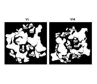
Brief Description of Drawings
[23] FIG. 1 illustrates prediction structures of a light chain variable
region (VL) and a
heavy chain variable region (VH) of clone MS502 among anti-mesothelin (MSLN)
an-
tibodies.
[24] FIG. 2 illustrates relative comparison of light chain variable region
mutants in view
of binding force.
[25] FIG. 3 illustrates relative comparison of heavy chain variable region
mutants in view
of binding force.
[26] FIG. 4 illustrates a vector for expressing the MSLN in a cell line.
1271 FIG. 5 illustrates a precursor form and a mature form of the MSLN on
SDS-PAGE.
[28] FIG. 6 illustrates analysis of an MSLN expression amount of cell lines
and tumor cell
lines expressing the MSLN.
[29] FIG. 7 illustrates selective binding analysis with regard to the cell
lines expressing
the MSLN by using an anti-mesothelin (MSLN) antibody of the present invention.
1301 FIG. 8 illustrates MFI values obtained by confirming whether the anti-
MSLN
antibody is bound to a cell membrane of mesothelioma (H226, H2052) and
pancreatic
cancers (AsPC-1) through FACS.
11311 FIG. 9 illustrates results obtained by confirming whether the anti-
MSLN antibody of
CA 02999237 2018-03-20
WO 2017/052241 PCT/KR2016/010604
the present invention is selectively bound to tumor cells expressing the MSLN.
[32]
Best Mode for Carrying out the Invention
[33] As used herein, term "mesothelin" or "MSLN" refers to any variants,
isoforms and
species homologs of human MSLN that is naturally expressed by cells.
[34] Term "human mesothelin" refers to a human sequence mesothelin such as
a complete
amino acid sequence of human mesothelin having Genbank Accession No.
NP_005814.
[35] In an embodiment of the present invention, monoclonal antibodies that
are
structurally characterized to be specifically bound to the mesothelin
represented by
SEQ ID NO: 127 and separated, such as "clone MI323", "clone MI329", "clone
MI403" and "clone MI407", and "clone MS501", "clone MS502'', "clone MS503",
"clone MS504", "clone MS505" and "clone MS506'', and "clone C2G1", "clone
C2G4'', "clone C3C8", "clone 54", "clone 56", "clone 2-30", "clone 2-73" and
"clone
2-78", and "clone 56-C2G4'', "clone 2-30-C2G4", "clone 2-73-C2G4" and "clone
2-78-C2G4" were produced.
[36] Amino acid sequences with regard to a heavy chain CDR and a light
chain CDR of
each antibody are shown in Tables 2, 5, 12. and 16 below. As shown in Tables
1. 4, 11,
and 15, an anti-MSLN antibody may include an amino acid sequence of a heavy
chain
variable region and a light chain variable region or a sequence having
homology
thereto.
[37] In another embodiment of the present invention, individual antibody
clones of
purified antibodies. i.e., "clone MI323". "clone MI329", "clone MI403", "clone
MS502", and "clone C2G1", "clone C2G4", "clone C3C8", and "clone 56-C2G4",
"clone 2-30-C2G4", "clone 2-73-C2G4" or "clone 2-78-C2G4" with regard to a re-
combinant human MSLN, were selected by using an enzyme linked immunosorbent
assay (ELISA) (data not shown), and quantitative binding force was measured by
using
a Biacore T-200 (GE Healthcare, U.S.A.) biosensor (Example 2-5, Example 3-11,
Examples 3-14). As a result, as shown in Tables 8, 14, and 18 below, all of
the
produced clone antibodies have affinity to the mesothelin even though there is
a slight
difference.
[38] In another embodiment of the present invention, in order to evaluate
whether the
anti-MSLN antibody derived from the immune and synthetic library is
selectively
bound to a MSLN-expres sing cell, an expression amount of the MSLN is measured
in
a cancer cell line, and an antibody binding to each cell is confirmed by FACS
test. As a
result, as illustrated in FIG. 5, it was confirmed that H28, MiaPaCa-2, BxPC-
3, Capan-
1 cell lines are MSLN-negative, and H226, H2452(H2052), AsPC-1 are MSLN-
CA 02999237 2018-03-20
WO 2017/052241 PCT/KR2016/010604
6
positive by measuring whether there are the MSLN having 70kDa precursor form
and
40 to 50kDa mature form from each cancer cell line.
[39] In addition, as a result obtained by performing selective binding
analysis of anti-
MSLN candidate antibodies with regard to the MSLN-expressing cell lines (H226,
H2452(H2052), AsPC-1), as illustrated in FIG. 8 and Table 19, all of the
MI323,
MI329, MI403, MS502 candidate antibodies with regard to the MSLN of
mesothelioma and pancreatic cancer cell lines have significant binding force
even
though there is a slight difference in binding degree. In particular, the
MI323 candidate
antibody has an excellent binding aspect.
[40] Further, as a result obtained by evaluating whether the MI323
candidate antibody
having the excellent binding aspect with regard to the MSLN, MS502 candidate
antibody having a different pattern of Biacore KD(Koff/Kon) value, and a heavy
chain
variable region mutation 2-78-C2G4 candidate antibody produced from the MS502
candidate antibody are selectively bound to MSLN-expressing tumor cells, in
MiaPaCa-MSLN #2 cell that over-expresses the MSLN and MiaPaCa-2 that does not
over-express the MSLN, as illustrated in FIG. 9, the MI323, MS502, and 2-78-
C2G4
candidate antibodies have excellent binding aspect in the MiaPaCa-MSLN #2 cell
that
over-expresses the MSLN as compared to the MiaPaCa-2.
[41] Therefore, the present invention relates to an antibody specifically
bound to
mesothelin (MSLN), preferably, an antibody specifically bound to mesothelin
rep-
resented by SEQ ID NO: 127.
[42] The antibody specifically bound to the mesothelin according to the
present invention
is characterized by containing a heavy chain variable region including a heavy
chain
CDR1 having an amino acid sequence of SEQ ID NO: 9, 15, 21, 27, or 59; a heavy
chain CDR2 having an amino acid sequence of SEQ ID NO: 10, 16, 22, 28, 60, 65,
71,
75, 80, 84, 121, 122, 123 or 125; a heavy chain CDR3 having an amino acid
sequence
of SEQ ID NO: 11, 17, 23, 29, 61, 66, 72, 76, 81. 85, 124 or 126.
[43] The antibody specifically bound to the mesothelin according to the
present invention
is characterized by containing a light chain variable region including a light
chain
CDR1 having an amino acid sequence of SEQ ID NO: 12, 18, 24, 30, 62, 67, 70,
77,
86 or 117; a light chain CDR2 having an amino acid sequence of SEQ ID NO: 13,
19,
25, 63, 68, 73, 78 or 82; a light chain CDR3 having an amino acid sequence of
SEQ ID
NO: 14, 20, 26, 64, 69, 74, 79, 83, 87, 118, 119 or 120.
[44] In the present invention, the antibody specifically bound to the
mesothelin may
contain a heavy chain variable region including a sequence having at least 80%
homology, preferably, at least 90% homology, and more preferably, 100%
homology
to the amino acid sequence of SEQ ID NO: 1, 3, 5, 7, 46. 48, 51, 53, 55, 57,
112, 113,
114, 115 or 116, and the antibody may contain a light chain variable region
including a
CA 02999237 2018-03-20
WO 2017/052241 PCT/KR2016/010604
7
sequence having at least 80% homology, preferably, at least 90% homology, and
more
preferably, 100% homology to the amino acid sequence of SEQ ID NO: 2, 4, 6, 8,
47,
52, 54, 56, 58, 109, 110 or 111.
[45] In the present invention, the antibody specifically bound to the
mesothelin is char-
acterized by containing the heavy chain variable region including the amino
acid
sequence of SEQ ID NO: 1, 3, 5, 7, 46, 48, 51, 53, 55, 57, 112, 113, 114, 115
or 116,
and the light chain variable region including the amino acid sequence of SEQ
ID NO:
2, 4, 6, 8, 47, 52, 54, 56, 58, 109. 110 or 111, and the antibody may be a
human
monoclonal antibody, but is not limited thereto.
[46] The amino acid sequence of the antibody may be substituted by
conservative sub-
stitution. The "conservative substitution" refers to modification of
polypeptide
including substitution of at least one amino acid with an amino acid having
similar bio-
chemical properties to corresponding polypeptide without causing loss of
biological or
biochemical function. "Conservative amino acid substitution" refers to a
substitution in
which an amino acid residue is replaced with an amino acid residue having
similar side
chains. Classes of the amino acid residues having similar side chains are
defined in the
art. These classes include amino acids having basic side chains (e.g., lysine,
arginine,
histidine), amino acids having acidic side chains (e.g., aspartic acid,
glutamic acid),
amino acids having uncharged polar side chains (e.g., glycine, asparagine,
glutamine,
serine, threonine, tyrosine, cysteine), amino acids having non-polar side
chains (e.g.,
alanine, valine, leucine, isoleucine, proline, phenylalanine, methionine,
tryptophan),
amino acids having beta-branched side chains (e.g., threonine, valine,
isoleucine), and
amino acids having aromatic side chains (e.g., tyrosine, phenylalanine,
tryptophan,
histidine). It is anticipated that the antibody of the present invention is
able to still
retain an activity while having the conservative amino acid substitution.
1471 Term "substantial homology" refers that when two kinds of nucleic
acids or two
kinds of polypeptides or designated sequences thereof are optimally aligned
and
compared, the nucleic acids and polypeptides having appropriate nucleotide or
amino
acid insertion or deletion have at least about 80% identity to the nucleotide
or the
amino acid, generally, have at least about 85%, preferably about 90%, 91%,
92%,
93%, 94% or 95%, and more preferably at least about 96%, 97%, 98%, 99%, 99.1%,
99.2%, 99.3%, 99.4% or 99.5% to the nucleotide or the amino acid.
Alternatively,
when a fragment is hybridized with a complementary strand thereof under
selective hy-
bridization conditions, there is substantial homology to the nucleic acid. The
present
invention includes a nucleic acid sequence and a polypeptide sequence having
sub-
stantial homology with regard to the above-described specific nucleic acid
sequence
and amino acid sequence.
1481 In the antibody according to the present invention, for example, the
heavy chain (VH)
CA 02999237 2018-03-20
WO 2017/052241 PCT/KR2016/010604
8
CDR1, 2 and 3 sequences and the light chain (VL) CDR1, 2 and 3 sequences shown
in
Table 2, Table 5, Table 12, and Table 16 may be formed by mixing structurally
similar
heavy chain (VH) and light chain (V1) sequences to be arranged in the CDR1, 2
and 3
of the heavy chain (VH) / light chain (VL) pairs.
[49] As used herein, term "antibody" or "antibody composition" refers to a
preparation of
antibody molecules having single molecule composition. Here, a monoclonal
antibody
composition represents single binding specificity and affinity for a specific
epitope.
Accordingly, term "human monoclonal antibody" refers to an antibody having a
variable region and a constant region derived from a human wiring immunoglobul
in
sequence, and representing single binding specificity. A human antibody of the
present
invention may include amino acid residue that is not encoded by the human
wiring im-
munoglobulin sequence (for example, mutants introduced by in vitro random or
site-
specific mutagenesis, or by in vivo somatic mutation).
[50] The "antibody" used herein is an immunoglobulin molecule which is
immuno-
logically reactive to a specific antigen, and means a protein molecule acting
as a
receptor that specifically recognizes an antigen, and may include all of a
polyclonal
antibody, a monoclonal antibody (single clone antibody), a whole antibody, and
an
antibody fragment. Further, the antibody may include a chimeric antibody
(e.g.,
humanized murine antibody) and a bivalent or bispecific molecule (e.g.,
bispecific
antibody), a diabody, a triabody, and a tetrabody.
[51] The whole antibody has a structure having two full length light chains
and two full
length heavy chains, and each light chain may be linked to a heavy chain via a
disulfide bond. The whole antibody includes IgA, IgD, IgE, IgM, and IgG, and
the IgG
is a subtype, and includes IgG 1 , IgG2, IgG3, and IgG4. The antibody fragment
means
a fragment retaining an antigen-binding function, and includes Fab, Fab',
F(ab')2, scFv,
and Fv, etc.
[52] The Fab has a structure of variable regions of a light chain and a
heavy chain and a
constant region of the light chain and a first constant region (CH1 domain) of
the
heavy chain, and has one antigen-binding site. The Fab' is different from the
Fab in
that the Fab' has a hinge region including one or more cysteine residues at C
terminal
of a heavy chain CH1 domain. The F(ab')2 antibody is produced by achieving the
disulfide bonding of the cysteine residue in the hinge region of the Fab'.
[53] The Fv (variable fragment) refers to the minimum antibody fragment
only having the
heavy chain variable region and the light chain variable region. In double-
stranded
Fv(dsFv), the heavy chain variable region and the light chain variable region
are linked
by the disulfide bond. In the single chain Fv(scFv), the heavy chain variable
region and
the light chain variable region generally are linked by a covalent bond using
a peptide
linker. These antibody fragment may be obtained by using a proteolytic enzyme
(for
CA 02999237 2018-03-20
WO 2017/052241 PCT/KR2016/010604
9
example, the Fab may be obtained by restriction-cutting the whole antibody
with
papain, and F(ab')2 fragment may be obtained by cutting with pepsin), and may
be
constructed by a recombinant DNA technology (for example, amplification by PCR
(Polymerase Chain Reaction) method using DNA encoding the heavy chain of the
antibody or the variable region thereof and DNA encoding the light chain or
the
variable region thereof as a template and using a primer pair, and
amplification with
combination of the DNA encoding the peptide linker of the primer pair allowing
both
ends thereof to link to the heavy chain or the variable region thereof and the
light chain
or the variable region thereof, respectively).
[541 The immunoglobulin has heavy chains and light chains, wherein the
respective heavy
chains and light chains include a constant region and a variable region (these
regions
are also known as domain). The light chain variable region and the heavy chain
variable region include 3 multi-available regions called complementarity-
determining
region (hereinafter, referred to as "CDR"), and four framework regions. The
CDR
mainly acts to bind to an epitope of the antigen. The CDRs of the respective
chains are
sequentially called CDR1, CDR2, and CDR3 generally starting from N-terminal,
and
also identified by the chains in which specific CDRs are located.
[551 The monoclonal antibody (single clone antibody) used herein means an
antibody
molecule of single molecular composition substantially obtained in the same
antibody
population, and may have single binding specificity and affinity for a
specific epitope.
1_56_1 The monoclonal antibody (single clone antibody) used herein is a
molecule derived
from a human immunoglobulin, and all of the amino acid sequences configuring
the
antibody including a complementarity-determining region, a structure region
are
configured of human immunoglobulin amino acid sequences. The human antibody is
typically used in the treatment of human diseases, which is advantageous in
that i) it
more favorably interacts with the human immune system, which more effectively
destroys target cells by complement-dependent cytotoxicity (CDC) or antibody-
dependent cell mediated cytotoxicity (ADCC), ii) the human immune system does
not
recognize the antibody as a foreign material, and iii) even when a smaller
amount of
drug is administered less frequently, a half-life in a human circulatory
system is similar
to that of a naturally occurring antibody.
[571 Terms "clone M1323", "clone MI329", "clone MI403" and "clone MI407"
and "clone
MS501", "clone MS502'', "clone MS503", "clone MS504'', "clone MS505" and
"clone
MS506" and "clone C2G1", "clone C2G4", "clone C3C8", "clone 54", "clone 56",
"clone 2-30", "clone 2-73" and "clone 2-78" and "clone 56-C2G4", "clone
2-30-C2G4", "clone 2-73-C2G4" and "clone 2-78-C2G4" that are antibodies
specifically bound to MSLN used herein mean antibodies bound to the MSLN and
causing inhibition of biological activity of the MSLN, and may be used by
mixing an
CA 02999237 2018-03-20
WO 2017/052241 PCT/KR2016/010604
anti-MSLN antibody.
[58] Here, the "clone MI323", "clone MI329", "clone MI403" and "clone
MI407" are an-
tibodies obtained by immunizing a mouse with recombinant human MSLN, and the
clone MS501". "clone MS502", "clone MS503". "clone MS504", "clone MS505" and
"clone MS506" are antibodies obtained from a phage display from a scFV
library. and
"clone C2G1", "clone C2G4'', "clone C3C8", "clone 54", "clone 56", "clone 2-
30",
"clone 2-73" and "clone 2-78" are antibodies obtained by introducing mutation
into the
"clone MS502" as shown in Table 9, and the "clone 56-C2G4", "clone 2-30-C2G4",
"clone 2-73-C2G4" and "clone 2-78-C2G4" are antibodies produced by combination
between the introduced mutation antibodies.
[59] KD (equilibrium dissociation constant) of the antibody to the MSLN may
be ex-
emplified as follows.
[60] (1) the clone MI323 may have an equilibrium dissociation constant
(K,)) of 1.8 x 10
M or less, preferably, 1.8 x 10⁹M or less, and more preferably. 1.8 x 10¹⁰M or
less,
[61] (2) the clone MI329 may have an equilibrium dissociation constant (KD)
of 3.5 x 10 9
M or less, preferably, 3.5 x 10' M or less, and more preferably, 3.5 x 10"M or
less,
[62] (3) the clone MI403 may have an equilibrium dissociation constant (KB)
of 4.5 x 10
M or less, preferably, 4.5 x 10⁹M or less, and more preferably, 4.5 x 10¹⁰M or
less,
[63] (4) the clone MS502 may have an equilibrium dissociation constant (KD)
of 2.3 x 10 g
M or less, preferably, 2.3 x 10⁹M or less, and more preferably. 2.3 x 10¹⁰M or
less
(see Table 8),
[64] (5) the clone C2G1 may have an equilibrium dissociation constant (KD)
of 9.39 x 10 9
M or less, preferably, 9.39 x 10¹⁰M or less, and more preferably, 9.39 x 10 "M
or less.
[65] (6) the clone C2G4 may have an equilibrium dissociation constant (KD)
of 4.32 x 10 9
M or less, preferably, 4.32 x 10¹⁰M or less, and more preferably, 4.32 x 10¹¹M
or less,
1661 (7) the clone C3C8 may have an equilibrium dissociation constant (KD)
of 1.22 x 10-
8M or less, preferably, 1.22 x 10 9M or less, and more preferably, 1.22 x 10 1
M or less
(see Table 14),
[67] (8) the clone 56 may have an equilibrium dissociation constant (KD) of
1.25 x 10 '1\4
or less, preferably. 1.25 x 10⁹M or less, and more preferably, 1.25 x 10¹⁰M or
less,
1681 (9) the clone 2-30 may have an equilibrium dissociation constant (I(D)
of 1.66 x 10g
M or less, preferably, 1.66 x 10 M or less, and more preferably, 1.66 x 10 1
M or less,
[69] (10) the clone 2-78 may have an equilibrium dissociation constant
(I(D) of 1.63 x 10⁹
M or less, preferably, 1.63 x 10 10M or less, and more preferably, 1.63 x 10
"M or less.
[70] (11) the clone 56-C2G4 may have an equilibrium dissociation constant
(KD) of 1.63
x 108M or less, preferably, 1.63 x 10 9M or less, and more preferably, 1.63 x
10 ' M or
less,
11711 (12) the clone 2-30-C2G4 may have an equilibrium dissociation
constant (KD) of
CA 02999237 2018-03-20
WO 2017/052241
PCT/KR2016/010604
11
2.34 x 10⁸M or less, preferably, 2.34 x 10⁻⁹M or less, and more preferably,
2.34 x 10
1 M or less,
1721 (13) the clone 2-73-C2G4 may have an equilibrium dissociation
constant (KD) of
1.65 x 10⁸M or less, preferably. 1.65 x 10⁹M or less, and more preferably.
1.65 x 10¹⁰
M or less, and
[73] (14) the clone 2-78-C2G4 may have an equilibrium dissociation constant
(1(D) of
3.72 x 10 M or less, preferably. 3.72 x 101 M or less, and more preferably,
3.72 x 10
"M or less (see Table 18).
[74] In another embodiment of the present invention, genes of the heavy
chain variable
region and the light chain variable region of a mouse derived from an immune
library
bound to human MSLN are identified, the heavy chain variable region gene is
linked to
a human immunoglobulin type 1 of heavy chain constant region (IgG1 heavy chain
constant region) gene, and the light chain variable region gene is linked to a
human
kappa light chain constant region, and these genes are inserted into protein
expression
vectors for animal cell, respectively, to produce vectors, followed by
transfection in the
Expi293Fim cell lines and culturing to produce the antibody, and the produced
antibody is purified by protein A to produce the antibody (Example 1-3).
[75] In still another embodiment of the present invention, genes of the
heavy chain
variable region and the light chain variable region derived from a synthetic
scFV
library bound to human MSLN are identified, the heavy chain variable region
gene is
linked to a human immunoglobulin type 1 of heavy chain constant region (IgG1
heavy
chain constant region) gene, and the light chain variable region gene is
linked to a
human kappa light chain constant region, and these genes are inserted into
protein ex-
pression vectors for animal cell, respectively, to produce vectors, followed
by
transfection in the Expi293FTM cell lines and culturing to produce the
antibody, and the
antibody is purified by protein A to produce the antibody (Examples 2-4, 3-10,
3-12,
and 3-13).
[76] Therefore, in another aspect of the present invention, the present
invention provides a
nucleic acid encoding the antibody. The nucleic acid used herein may be
present in a
cell, a cell lysate, or may also be present in a partially purified form or a
substantially
pure form. The nucleic acid is "isolated" or "is substantially pure" when it
is purified
from other cell components or other contaminants, for example, other cell
nucleic acid
or protein by standard techniques including alkaline/SDS treatment, CsC1
banding,
column chromatography, agarose gel electrophoresis, and other techniques well-
known
in the art. The nucleic acid of the present invention may be, for example, DNA
or
RNA, and may include an intron sequence, or may not include the intron
sequence.
[77] In still another aspect of the present invention, the present
invention provides a
vector including the nucleic acid. For expression of the antibody or antibody
fragments
CA 02999237 2018-03-20
WO 2017/052241 PCT/KR2016/010604
12
thereof, DNA encoding the light chain and the heavy chain having a partial
length or a
full length may be obtained by standard molecular biology techniques (for
example,
PCR amplification or cDNA cloning using a hybridoma that expresses a target
antibody), and the DNA may be "operably bound" to transcription and
translation
control sequences to be inserted into the expression vector.
[78] Term "operably bound" used herein may indicate that an antibody gene
is ligated
into the vector so that the transcription and translation control sequences in
the vector
have an intended function to control transcription and translation of the
antibody gene.
The expression vector and an expression control sequence are selected so as to
have
compatibility with a host cell for expression to be used. The light chain gene
of the
antibody and the heavy chain gene of the antibody are inserted into a separate
vector,
or both genes are inserted into the same expression vector. The antibody is
inserted
into the expression vector by a standard method (for example, ligation of an
antibody
gene fragment and a complementary restriction enzyme site on a vector or when
the re-
striction enzyme site is not present at all. blunt end ligation). In some
cases, the re-
combinant expression vector may encode a signal peptide that facilitates
secretion of
the antibody chain from the host cell. The antibody chain gene may be cloned
into the
vector so that the signal peptide is bound to an amino terminal of the
antibody chain
genes according to a frame. The signal peptide may be an immunoglobulin signal
peptide or a heterolo2ous signal peptide (i.e. signal peptide derived from
proteins
except for immunoglobulin). In addition, the recombinant expression vector has
a
regulatory sequence that controls the expression of the antibody chain genes
in the host
cell. The "regulatory sequence" may include a promoter, an enhancer and other
ex-
pression control element (for example, polyadenylation signal) controlling the
tran-
scription or translation of the antibody chain gene. Those skilled in the art
is able to
recognize that design of the expression vector may vary by changing the
regulatory
sequences according to factors such as selection of the host cell to be
transformed, an
expression level of the protein, etc.
[79] In still another aspect of the present invention, the present
invention provides a host
cell including the nucleic acid or the vector. The nucleic acid or the vector
is
transfected into the host cell. For the "transfection", various kinds of
generally used
techniques such as electrophoresis, calcium phosphate precipitation, DEAE-
dextran
transfection, lipofection, etc., may be used to introduce an exogenous nucleic
acid
(DNA or RNA) into a prokaryotic host cell or an eukaryotic host cell. The
antibody
according to the present invention may be expressed in an eukaryotic cell,
preferably,
in a mammalian host cell, in consideration of applicability into a mammalian
cell. The
mammalian host cells suitable for expression of the antibody may include a
Chinese
hamster ovary (CHO) cell (for example, including a dhfr- CHO cell used
together with
CA 02999237 2018-03-20
WO 2017/052241 PCT/KR2016/010604
13
a DHFR selection marker), an NSO myeloma cell, a COS cell, or a SP2 cell,
etc., as
examples.
[80] In still another aspect of the present invention, the present
invention provides a
method for producing an antibody, including culturing a host cell to express
the
antibody. When the recombinant expression vector encoding the antibody gene is
in-
troduced into the mammalian host cell, the antibody may be produced by
culturing the
host cell for a sufficient period of time so that the antibody is expressed in
the host cell,
or more preferably, for a sufficient period of time so that the antibody is
secreted into a
culture medium in which the host cell is cultured.
[81] In some cases, the expressed antibody may be separated from the host
cell and
purified for uniformity. The separation or the purification of the antibody
may be
performed by a separation method, a purification method generally used for
protein,
for example, chromatography. The chromatography may include, for example,
affinity
chromatography, ion exchange chromatography or hydrophobic chromatography
including protein A column and protein G column. In addition to the
chromatography,
the antibody may be separated and purified by additionally combining with
filtration,
ultrafiltration, salting out, dialysis, etc.
[82] In still another aspect of the present invention, the present
invention provides a phar-
maceutical composition for treating cancer or tumor including the antibody as
an active
ingredient.
[83] Term "cancer" or "tumor" typically refers to or describes a
physiological condition of
mammals characterized by cell growth/proliferation that is not controlled.
Examples of
the cancer include carcinoma, lymphoma (e.g., Hodgkin's and non-Hodgkin's
lymphoma), blastoma, sarcoma and leukemia, but are not limited thereto. More
specific examples of the cancer include squamous cell cancer, small-cell lung
cancer,
non-small cell lung cancer, lung adenocarcinoma, lung squamous cell carcinoma,
peritoneal cancer, hepatocellular cancer, gastrointestinal cancer, pancreatic
cancer,
glioma, cervical cancer, ovarian cancer, liver cancer, bladder cancer,
hepatocellular
cancer, breast cancer, colon cancer, colorectal cancer, endometrial or uterine
carcinoma, salivary gland carcinoma, kidney cancer, liver cancer, prostate
cancer,
vulvar cancer, thyroid cancer, liver carcinoma, leukemia and other
lymphoproliferative
disorders, and various types of head and neck cancer. The cancer in the
present
invention is preferably mesothelin-positive cancer, and is selected from the
group
consisting of pancreatic cancer, ovarian cancer, lung cancer, stomach cancer,
en-
dometrial cancer, and mesothelioma.
[84] The present invention provides a pharmaceutical composition including
a thera-
peutically effective amount of anti-MSLN antibody and a pharmaceutically
acceptable
carrier. The "pharmaceutically acceptable carrier" is a material that is able
to be added
CA 02999237 2018-03-20
WO 2017/052241 PCT/KR2016/010604
14
in the active ingredient to help formulation or stabilization of the
preparation, and it
does not cause significant adverse toxicological effects to patients.
[85] The carrier refers to a carrier or diluent that does not inhibit
biological activity and
properties of an administered compound without stimulating the patients. The
pharma-
ceutically acceptable carrier in the composition to be formulated as a liquid
solution is
sterilized and is suitable for a living body. Saline, sterile water. Ringer's
solution,
buffered saline, albumin injection solution, dextrose solution, maltodextrin
solution,
glycerol, ethanol may be used as the carrier, or at least one component
thereof may be
mixed to be used, and other conventional additives such as an antioxidant,
buffer, a
bacteriostatic agent, etc., may be added as needed. In addition, the
composition may be
prepared into formulations for injection, such as an aqueous solution,
suspension,
emulsion, etc., pill, a capsule, a granule or a tablet by further adding
diluent,
dispersant, surfactant, binder and lubricant thereto. Other carriers are
described in, for
example. [Remington's Pharmaceutical Sciences (E. W. Martin)]. The composition
may contain the therapeutically effective amount of at least one anti-MSLN
antibody.
[86] The pharmaceutically acceptable carrier includes sterile aqueous
solution or
dispersion and sterile powder for preparing extemporaneous sterile injectable
solution
or dispersion. The use of such media and agents for pharmaceutical active
materials is
known in the art. The composition is preferably formulated for parenteral
injection.
The composition may be formulated as a solution, a micro-emulsion, a liposome,
or
other ordered structures suitable for high drug concentration. The carrier may
be, for
example, a solvent or dispersion medium containing water, ethanol, polyol (for
example, glycerol, propylene glycol and liquid polyethylene glycol, etc.,) and
suitable
mixtures thereof. In some cases, the composition may include, isotonic agent,
for
example, sugar, polyalcohols such as mannitol, sorbitol, or sodium chloride.
The sterile
injectable solution may be prepared by incorporating a required amount of
active
compound into an appropriate solvent with one kind of the above-described
components or a combination thereof, followed by sterile micro filtration as
needed. In
general, the dispersion is prepared by incorporating the active compound into
a sterile
vehicle containing basic dispersion medium and other required components from
the
above-described components. The sterile powder for preparing the sterile
injectable
solution is obtained by vacuum drying and freeze-drying (lyophilization)
active in-
gredient powder and any additional desirable component powder from previously
sterile-filtered solution.
[87] The pharmaceutical composition may be administered orally or
parenterally in the
dosage and frequency that may vary depending on severity of suffering
patients. The
composition may be administered to a patient as a bolus or by continuous
infusion as
needed. For example, the bolus administration of the antibody of the present
invention
CA 02999237 2018-03-20
WO 2017/052241 PCT/KR2016/010604
which is presented as a Fab fragment may have an amount of 0.0025 to 100mg/kg
body weight, 0.025 to 0.25mg/kg, 0.010 to 0.10mg/kg or 0.10 to 0.50mg/kg. For
the
continuous infusion, the antibody of the present invention which is presented
as the
Fab fragment may be administered at 0.001 to 100 mg/kg kg/min, 0.0125 to
1.25mg/kg/min, 0.010 to 0.75mg/kg/min, 0.010 to 1.0mg/kdmin or 0.10 to
0.50mg/kg/min for 1 to 24 hours, 1 to 12 hours. 2 to 12 hours, 6 to 12 hours,
2 to 8
hours, or 1 to 2 hours. When the antibody of the present invention which is
presented
as a full-length antibody (having a complete constant region is administered,
an admin-
istration amount may be about 1 to 10 mg/kg body weight, 2 to 8 mg/kg, or 5 to
6 mg/
kg. The full-length antibody is typically administered via injection that
lasts for 30
minutes to 35 minutes. An administration frequency depends on the severity of
the
condition. The frequency may be 3 times every week to once in a week or in two
weeks.
[88] In addition, the composition may be administered to a patient via a
subcutaneous
injection. For example, the anti-MSLN antibody having an administration amount
of
10 to 100 mg may be weekly, biweekly, or monthly administered to a patient
through
subcutaneous injection.
[89] As used herein, the "therapeutically effective amount" means an amount
sufficient to
treat diseases at a reasonable benefit/risk ratio applicable for medical
treatment, and an
amount of a combination of the anti-MSLN antibody. The exact amount may vary
depending on a number of factors that include components and physical
characteristics
of a therapeutic composition, intended patient population, individual patient
consid-
erations, etc., but are not limited thereto, and may be easily determined by
those skilled
in the art. When completely considering these factors, it is important to
administer the
minimum amount sufficient to obtain the maximum effect without the side
effect, and
this dosage may be easily determined by an expert in the field.
[90] The dosage of the pharmaceutical composition of the present invention
is not
specifically limited, but is changed according to various factors including a
health state
and weight, severity of the disease of a patient, and a drug type, an
administration
route, and administration time. The composition may be administered in routes
that are
typically allowed in mammals including rat, mouse, cattle, human, etc., for
example,
orally, rectally, intravenously, subcutaneously, intrauterinely or
intracerebrovascularly
in a single dose amount or multidose per day.
[91]
[92] Example
[93] Hereinafter, the present invention will be described in more detail
with reference to
the following Examples. However, the following examples are only for
exemplifying
the present invention, and it will be obvious to those skilled in the art that
the scope of
CA 02999237 2018-03-20
WO 2017/052241 PCT/KR2016/010604
16
the present invention is not construed to be limited to these examples.
[94]
[95] Example 1: Method for producing anti-MSLN antibody from immune library
[96] It is intended to produce an anti-cancer antibody therapeutic agent
using an antibody
against mesothelin (MSLN) over-expressed on a cancer cell surface.
[97] 1-1: Selection of anti-MSLN antibody
[98] A mouse was immunized with a recombinant human MSLN and a spleen was
removed to extract B lymphocyte. Total RNA was separated from the B lymphocyte
and cDNA was synthesized. Various antibody genes of the mouse were cloned from
the synthesized cDNA using polymerase chain reaction (PCR), and inserted into
pComb3X phagemid to produce an antibody library displaying antibody fragments
of
various sequences. In order to find the antibody specifically bound to human
MSLN
from the antibody library, magnetic beads having the MSLN fixed thereto were
mixed
with the antibody library, and clones bound to a target antigen were separated
and
cultured. Then the clones (MI323, MI329, MI403. and MI407) specifically bound
to
the target antigen (human MSLN) were individually identified through an enzyme-
linked immunosorbent assay (ELISA), and antibody gene sequences and amino acid
sequence thereof were identified through base sequence analysis.
[99] As a result, as shown in Table 1, the clones specifically bound to
human MSLN
could be selected, and amino acid sequences thereof were identified.
[100] Table 2 shows CDR amino acid sequences of the clone antibodies of
Table 1 on the
basis of Kabat numbering.
[101]
CA 02999237 2018-03-20
WO 2017/052241 PCT/KR2016/010604
17
[102] [Table 1]
Clone Variable Amino Acid Sequence SEQ ID
Region NO:
MI323 heavy chain EVQLQQSGPELYKPGTSYKISCKASGYSFTS 1
YFIQWVKQRPGQGLEWIGWIFPGSGNTKYN
EMFKGKATLAADTSSSTAYMQLSSLTSEDS
AVYFCARSGGYQYYFDYWGQGTSVTVSS
light chain DIVMTQSHKFMSTSVGDRVSITCKASQDVS 2
TAVAWYQQKPGQSPKLLIYSASYRYPGVPD
RFTGSGSGTDFTFTISSVQAEDLALYYCQQH
YSTPWTFGGGTKLEIKR
MI329 heavy chain EVMLVESGGDLVKPGGSLKLSCAASGFTFS 3
SYAMSWVRRTPEKRLEWVATINSDGSYTF
YPDSVKGRFTISRDNAKNTLYLQMNSLRSE
DTAMYYCARWGENWYFDVWGAGTTVTV
SS
light chain DVVMTQTPLSLPVSLGDQASISCRSSQSLVH 4
SNGNTYLHWYLQKPGQSPKLLIYKVSNRFS
GVPDRFSGSGSGTDFTLKISRVEAEDLGIYF
CSQSTHFPRTFGGATKLELKR
MI403 heavy chain EVQVVESGGGLVKPGGSLKLSCAASGFAFS 5
SYDMSWVRQTPEKRLEWVAYISSGGGSTY
YPDTVKGRFTISRDNAKNTLYLQMNSLKSE
DTAMYYCARQGTAVKNYWYFDVWGAGT
SVTVSS
light chain DIVMTQSPASLAVSLGQRATISCRASQSVST 6
SSSSYVHWYQQRPGQPPKLLIKYASNLESG
VPARFSGSGSGTDFTLNIHPVEEEDTGTYYC
QHSWEIPFTFGSGTKLEIKR
MI407 heavy chain EVKLVESGGGLVKPGGSLKLSCAASGFPFS 7
NYDMSWVRQTPEKRLEWVAY1SSGGGNTY
YPDTVKGRFTISRDNAKNTLYLQMSSLKSE
DTALYFCYRQGTSVESYWYFDYWGAGTTV
TVSS
light chain DIVLTQSPASLAVSLGQRATISCRASQSVSTS 8
SSSYIHWYQQKPGQPPKLLIKYASNLESGVP
CA 02999237 2018-03-20
WO 2017/052241 PCT/KR2016/010604
18
ARFSGSGSGTDFTLNIHPVEEDDTATYYCQ
HSWEIPFTFGSGTELEIKR
[103]
[104] [Table 2]
Clone Variable CDR1 CDR2 CDR3
Region
MI323 heavy chain SYFIQ(SEQ ID WIFPGSGNTKY SGGYQYYFDY(SE
NO: 9) NEMFKG(SEQ Q ID NO: 11)
ID NO: 10)
light chain KASQDVSTA SASYRYP(SEQ QQHYSTPWT(SEQ
VA(SEQ ID ID NO: 13) ID NO: 14)
NO: 12)
MI329 heavy chain SYAMS(SEQ TINSDGSYTFYP WGENWYFDV(SEQ
ID NO: 15) DSVKG(SEQ ID ID NO: 17)
NO: 16)
light chain RSSQSLVHSN KVSNRFS(SEQ SQSTHFPRT(SEQ
GNTYLH(SEQ ID NO: 19) ID NO: 20)
ID NO: 18)
MI403 heavy chain SYDMS(SEQ YISSGGGSTYYP QGTAVKNYWYFD
ID NO: 21) DTVKG(SEQ ID V(SEQ ID NO: 23)
NO: 22)
light chain RASQSVSTSS YASNLES(SEQ QHSWEIPFT(SEQ
SSYVH(SEQ ID NO: 25) ID NO: 26)
ID NO: 24)
MI407 heavy chain NYDMS(SEQ YISSGGGNTYY QGTSVESYWYFDV
ID NO: 27) PDTVKG(SEQ ID (SEQ ID NO: 29)
NO: 28)
light chain SVSTSSSSYIH YASNLES(SEQ QHSWEIPFT(SEQ
(SEQ ID NO: ID NO: 25) ID NO: 26)
30)
[1051
[106] 1-2: IgG gene cloning of MI323, MI329, MI403, and MI407 monoclonal
antibodies
[1071 The pComb3X phagemid containing the genes encoding the heavy chain
variable
regions of the MI323, MI329, MI403, and MI407 clone antibodies was extracted,
and
CA 02999237 2018-03-20
WO 2017/052241 PCT/KR2016/010604
19
used as a template for PCR with a forward primer containing NotI (Table 3: SEQ
ID
NOs: 31 to 34) and a reverse primer containing Apal (Table 3: SEQ ID NO: 35)
by
using Accupower Pfu PCR premix (Bioneer). The PCR was performed by repeating
exposure at 94 C for 10 minutes, and then exposure at 94 C for 15 seconds, at
56 C for
30 seconds, and at 72 C for 90 seconds 30 times, and reacting at 72 C for 10
minutes.
In the amplified genes, DNA bands having an expected size were confirmed on 1%
agarose gel, and were separated using a gel extraction kit, respectively.
Then, the
separated genes reacted with NotI, ApaI restriction enzymes at 37 C for 12
hours or
more, and the genes reacted with the restriction enzymes were separated on 1%
agarose gel again. A pcIW plasmid vector containing human immunoglobulin type
1 of
heavy chain constant region (IgG1 heavy chain constant region) gene was also
cut by
the same method as above and separated on agarose gel. The separated MI323,
MI329,
MI403, and MI407 heavy chain variable region genes were inserted into NotI,
ApaI
sites of a linear pcIW vector containing the human heavy chain constant region
by
using a T4 DNA ligase (Cat.No.M0203S, New England BioLabs (NEB)). The ligation
reaction materials were transformed into XL1-Blue bacteria
(Electroporation-Competent Cells; Cat.No.200228, Stratagene), plated on an LB
plate
(Cat.No.LN004CA, NaraeBiotech) containing carbenicillin, and cultured at 37 C
for
12 hours or more. Then single colonies were chosen and cultured, and plasmids
were
separated by using a plasmid mini kit (Cat.No.27405, QIAGEN), and confirmed by
DNA sequencing.
[108] The pComb3X phagemid containing the genes encoding the light chain
variable
regions of the MI323, MI329, MI403, and MI407 clone antibodies was extracted,
and
used as a template for PCR of the light chain variable regions of the MI323,
MI329,
MI403, and MI407 clone antibodies, wherein the PCR was performed with a
forward
primer containing Notl (Table 3: SEQ ID NO: 36, 38, 40, 42) and a reverse
primer
(Table 3: SEQ ID NO: 37, 39, 41, 43) by using Accupower Pfu PCR premix.
Further,
the human antibody kappa light chain constant region was subjected to PCR with
a
forward primer (Table 3: SEQ ID NO: 44) and a reverse primer containing
HindIII
(Table 3: SEQ ID NO: 45). The PCR was performed by repeating exposure at 94 C
for
minutes, and then exposure at 94 C for 15 seconds, at 56 C for 30 seconds, and
at
72 C for 90 seconds 30 times, and reacting at 72 C for 10 minutes. In the
amplified
genes, DNA bands having a predicted size were confirmed on 1% agarose gel, and
were separated using a gel extraction kit, respectively. Then, the respective
light chain
variable regions and light chain constant regions were mixed, followed by
overlapping
PCR, such that the genes expressing the light chain region were cloned. The
PCR was
performed by repeating exposure at 94 C for 10 minutes, and then exposure at
94 C for
seconds. at 56 C for 30 seconds, and at 72 C for 90 seconds 30 times, and
reacting
CA 02999237 2018-03-20
WO 2017/052241 PCT/KR2016/010604
at 72 C for 10 minutes. In the amplified genes, DNA bands having an expected
size
were confirmed on 1% agarose gel, and were separated using a gel extraction
kit, re-
spectively. Then, the separated genes reacted with NotI, HindIII restriction
enzymes at
37 C for 12 hours or more, and the genes reacted with the restriction enzymes
were
separated on 1% agarose gel again. The pcIVV plasmid vector was also cut by
the same
method as above and separated on agarose gel. The separated MI323, MI329,
MI403,
and MI407 light chain region genes were inserted into NotI, HindIII sites of a
linear
pcIW vector by using a T4 DNA ligase (Cat.No.M0203S, New England BioLabs
(NEB)). The ligation reaction materials were transformed into XL1-Blue
bacteria
(Electroporation-Competent Cells; Cat.No.200228, Stratagene), plated on an LB
plate
(Cat.No.LN004CA, NaraeBiotech) containing carbenicillin, and cultured at 37 C
for
12 hours or more. Then single colonies were chosen and cultured, and plasmids
were
separated by using a plasmid mini kit (Cat.No.27405, QIAGEN), and confirmed by
DNA sequencing.
[109]
CA 02999237 2018-03-20
WO 2017/052241 PCT/KR2016/010604
21
[110] [Table 3]
Name DNA nucleotide sequence SEQ ID
NO:
MI323VH-F GCGGCCGCCATGTACTTGGGACTGAACTATGTATTC 31
ATAGTTTTTCTCTTAAATGGTGTCCAGAGTGAGGTC
CAGCTGCAGCAGTCT
MI329VH-F GCGGCCGCCATGTACTTGGGACTGAACTATGTATTC 32
ATAGTTTTTCTCTTAAATGGTGTCCAGAGTGAGGTG
ATGCTGGTGGAGTCT
MI403VH-F GCGGCCGCCATGTACTTGGGACTGAACTATGTATTC 33
ATAGTTTTTCTCTTAAATGGTGTCCAGAGTGAGGTG
CAGGTGGTGGAGTCT
MI407VH-F GCGGCCGCCATGTACTTGGGACTGAACTATGTATTC 34
ATAGTTTTTCTCTTAAATGGTGTCCAGAGTGAGGTG
AAGTTGGTGGAGTCT
VHApaI-R ACCGATGGGCCCTTGGTGGA 35
MI323VL-F GCGGCCGCCATGGATAGCCAGGCTCAGGTGCTGATG 36
CTGCTGCTGCTGTGGGTGTCAGGGACTTGCGGGGAC
ATTGTGATGACCCAGTCTCACA A A
MI323VLCL- ACACTAGGAGCGGCCACGGTTCGTTTGATTTCCAGT 37
TTGGTCCCT
MI329VL-F GCGGCCGCCATGGATAGCCAGGCTCAGGTGCTGATG 38
CTGCTGCTGCTGTGGGTGTCAGGGACTTGCGGGGAC
GTTGTGATGACCCAGACTCCACTC
MI329VLCL- ACACTAGGAGCGGCCACGGTTCGTTTCAGCTCCAGC 39
TTGGTC
MI403VL-F GCGGCCGCCATGGATAGCCAGGCTCAGGTGCTGATG 40
CTGCTGCTGCTGTGGGTGTCAGGGACTTGCGGGGAT
ATTGTGATGACCCAGTCTCCTGCT
MI403VLCL- ACACTAGGAGCGGCCACGGTTCGTTTTATTTCCAAC 41
TTTGTCCCCGA
MI407VL-F GCGGCCGCCATGGATAGCCAGGCTCAGGTGCTGATG 42
CTGCTGCTGCTGTGGGTGTCAGGGACTTGCGGGGAT
ATTGTGTTGACACAGTCTCCTGCT
CA 02999237 2018-03-20
WO 2017/052241 PCT/KR2016/010604
22
MI407VLCL- ACACTAGGAGCGGCCACGGTTCGTTTTATTTCCAAC 43
TCTGTCCCCG
Ck-F ACCGTGGCCGCTCCTAGTGT 44
CkSHB-R NNNNGGATCCAAGCTTACTAGCACTCCCC 45
[Ill]
[112] 1-3: Production and purification of IgG of MI323. MI329, MI403, and
MI407 clone
antibodies
[113] In order to produce and purify the anti-MSLN antibody MI323, MI329,
MI403, and
MI407 clones obtained by a mouse immune response, Expi293FTM cells were in-
oculated at a concentration of 2.0 x 10⁶ cell/mL the day before transfection.
After in-
cubation (37 C, 8% CO2, 125 rpm) for 24 hours, Expi293TM expression medium
(Cat.No.A1435101, Gibco) was added to prepare a product of 30 mL having a con-
centration of 2.5 x 10⁶ cell/mL (viability = 95%). 30 lig of DNA (pcIW-anti-
MSLN
heavy chain: 15 [kg, pcIW-anti-MSLN light chain: 15 lig) was diluted in an
OptiProTM
SEM medium (Cat.No.12309019, Gibco) so as to have a total volume of 1.5 mL,
and
reacted at room temperature for 5 minutes. 1.5 mL of the OptiProTM SEM medium
(Cat.No.12309019, Gibco) was mixed with 801AL of an ExpiFectamineTM 293
reagent
(Cat.No.A14524, Gibco) so that a total volume is 1.5mL, and reacted at room
tem-
perature for 5 minutes. After the reaction for 5 minutes, 1.5 mL of diluted
DNA and
1.5 mL of diluted ExpiFectamineTM 293 reagent were well-mixed with each other,
and reacted at room temperature for 20 to 30 minutes. 3 mL of the mixture of
DNA
and ExpiFectamineTM 293 reagent was treated in the Expi293FTM cells. After
suspension-culture (37 C, 8% CO), 125 rpm) for 16 to 18 hours. 150 tit of
ExpiFec-
tamineTM 293 Enhancer 1 (Cat.No.A14524, Gibco) and 1.5 mL of ExpiFectamineTM
293 Enhancer 2 (Cat.No.A14524, Gibco) were added thereto, followed by
suspension-
culturing for 5 days. After the culturing, cell debris was removed by
centrifugation at
4000rpm for 20 minutes, and the supernatant passed through 0.22 [tm filter to
be
prepared. MabSelect Xtra (Cat.No.17-5269-02, GE Healthcare) which is protein A
resin having 100 ttL was prepared for each 30 mL of the culture fluid,
followed by cen-
trifugation at 1000 rpm for 2 minutes to remove a storage solution, and the
obtained
product was washed with 400111_, of protein A binding buffer (Cat.No.21007,
Pierce) 3
times. The protein A resin was added to the prepared culture fluid and
rotation-reacted
at room temperature for 30 minutes. The mixture of the culture fluid and the
resin was
put into a pierce spin column snap-cap (Cat.No.69725, Thermo), and then, only
the
resin was left in the column using QIAvac 24 Plus (Cat.No.19413, QIAGEN)
vacuum
manifold. 5 mL of protein A binding buffer was added to wash the resin, 200
piL of a
protein A elution buffer (Cat.No.21009. Pierce) was added thereto. The
resultant
23
material was reacted by resuspension at room temperature for 2 minutes, and
cen-
trifuged at 1000 rpm for I minute, and eluted. Each eluate was neutralized by
adding
2.5 1it of 1.5M Tris-HC1 (pH 9.0). The elution was performed 4 to 6 times, and
each
fraction was quantified by using Nanodrop 200C (Thermo Scientific). The
fractions in
which protein is detected were collected, and exchanged with a PBS
(Phosphate-Buffered Saline) buffer using Zeba Spin Desalting Columns, 7K MWCO,
5
mL (Cat.No.0089892, Pierce). Then, protein clectrophoresis (SDS-PAGE) was
performed under reduction and non-reduction condition to finally verify the
con-
centration quantification and the antibody state, and the antibody was kept at
4 C.
[114]
[115] Example 2: Method for producing anti-MSLN antibody from phage display
synthetic
scFv library
[1161 2-1: Selection of anti-human MSLN scLv antibody using phagc display
[117] For a primary panning, 1 nit of 1013 or more library stock was
reacted in a solid
phase polystyrene tube (Cat.No.444202, Nunc) coated with MSLN at 37 C for 2
hours. At the same time, 10 ILL of XL1-Blue bacteria (electroporation-
competent cells;
Cat.No.200228, Stratagene) were inoculated with 10n1, of SB 10m1/tetracycline
and
grown to an ODax, of 0.8 to 1Ø The library stock obtained after reaction at
37 C for 2
hours was washed with 5m1 of 0.05% Tween 20/PBS four times, and from a
secondary
panning, the number of times of washing with 0.05% Tween 20/PBS increased
according to an increase in the number of times of panning. Then, the
resultant
material was cultured with 1%BSA/0.1M Glycine pH 2,0 at room temperature for
10
minutes to purify the phagemid. The purified phagemid was transferred to a 50
mL
tube and neutralized with 70 ttL of 2M Tris. 9 mL of XL 1-Blue bacteria
(electroporation-competent cells; Cat.No.200228, Stratagene) were treated, and
1 mL
of the bacteria were treated in a washed tube. The bacteria were infected at
room tem-
perature for 30 minutes, and 10 mL of SB, 20 ttli. of tetracycline, 10 ttL of
carbenicillin
were added thereto, followed by suspension-culturing at 37 C and 220 rpm for 1
hour.
Then, the bacteria were treated with 1 mL of VCS M13 helper phage (10" pfu),
and
suspension-cultured at 37 C and at 220 rpm for 1 hour, and treated with 80 mL
of SB,
100 [It of kanamycin, and 100 !AL of carbenicillin, and cultured at 37 C and
at 220 rpm
for 12 hours or more. The bacteria cultured over 12 hours were centrifuged at
3500
rpm and at 4 C for 10 minutes, and the supernatant was transferred to a new
tube. 20
mL of 20% PEG/15% NaCl was added thereto, well-mixed, and reacted in ice for
30
minutes. Then, the supernatant was discarded, and pellets were collected and
re-
suspended with 2 mL of 1% BSA/PBS at 8000 rpm, and at 4 C for 30 minutes, and
centrifuged at 15000 rpm and 4 C for 10 minutes. Here, the collected pellets
were
discarded and 1 mL of the supernatant (2 mL) was stored at -20 C and the
remainder (1
CA 2999237 2019-10-11
24
mL) was used in the following order panning.
[ I 18]
[119] 2-2: Securing individual clones according to ELISA
[120] Single colonies of a phage display synthetic scEv library final
amplified population
were collected, and cultured with 1.5 mL of SB/carbenicillin to an OD600 of
0.8 to 1.0
at 37 C and at 220 rpm, and then cultured with 1 mMIPTG at 30 C and at 200 rpm
for
12 hours or more. The reaction materials were centrifuged at 5500 rpm for 5
minutes,
and only each supernatant was added to EL1SA plates containing underlying MSLN
antigen, and reacted at room temperature for 2 hours. Then, the resultant
materials
were washed with PBST (1XPBS, 0.05% tween 20four times, and HRP/
Anti-hFab-HRP conjugate diluted by 1/5000 with 1% BSA/1XPBS was added thereto,
and reacted at room temperature for 1 hour, and washed with PBST (1XPBS, 0.05%
tween 20) 4 times. Then, a TMB solution was added thereto and reacted for 5 to
10
minutes, and a TMB stop solution was added thereto. Next, 0.D values were
measured
at a measurement wavelength of 450 nm using a TECAN sunrise, and clones having
high O.D. value were secured as individual clones.
[121] As a result, as shown in Table 4, the clones specifically bound to
the human MSLN
were able to be selected, and amino acid sequences thereof were identified.
[122] Table 5 shows CDR amino acid sequences of the clone antibodies of
Table 4 on the
basis of Kabat numbering.
[123]
CA 2999237 2019-10-11
CA 02999237 2018-03-20
WO 2017/052241 PCT/KR2016/010604
[124] [Table 4]
Clone variable region amino acid sequence SEQ ID
NO:
MS501 heavy chain EVQLLESGGGLVQPGGSLRLSCAASGFT 46
FSNYAMSWVRQAPGKGLEWVSGIYPDS
GSTYYADSVKGRFTISRDNSKNTLYLQM
NSLRAEDTAVYYCARNIYTFDYWGQGT
LVTVSS
light chain QSVLTQPPSASGTPGQRVTISCSGSSSNIG 47
SNAVSWYQQLPGTAPKLLIYYNNQRPSG
VPDRFSGSKSGTSASLAISGLRSEDEADY
YCGSWDSSLSGYVFGGGTKLTVLG
MS502 heavy chain EVQLLESGGGLVQPGGSLRLSCAASGFT 48
FSNYAMSWVRQAPGKGLEWVSGIPPDS
GSKYYADSVRGRFTVSRDNSKNTLYLQ
MNSLRAEDTAVYYCAKNMLSFDYWGQ
GTLVTVSS
light chain QSVLTQPPSASGTPGQRVTISCTGSSSNIG 49
SNAVSWYQQLPGTAPKLLIYYNSKRPSG
VPDRFSGSKSGTSASLAISGLRSEDEADY
YCGSWDSSLNGYVFGGGTKVTVLG
MS502-1 heavy chain EVQLLESGGGLVQPGGSLRLSCAASGFT 48
FSNYAMSWVRQAPGKGLEWVSGIPPDS
GSKYYADSVRGRFTVSRDNSKNTLYLQ
MNSLRAEDTAVYYCAKNMLSFDYWGQ
GTLVTVSS
light chain QSVLTQPPSASGTPGQRVTISCICSSSNIG 50
SNAVSWYQQLPGTAPKLLIYYNSKRPSG
VPDRFSGSKSGTSASLAISGLRSEDEADY
YCGSWDSSLNGYVFGGGTKLTVLG
MS 503 heavy chain EVQLLESGGGLVQPGGSLRLSCAASGFT 51
FSNYAMSWVRQAPGKGLEWVSSIYPGD
GSTYYADSVKGRFTISRDNSKNTLYLQM
NSLRAEDTAVYYCAKNAFTFDYWGQGT
LVTVSS
light chain QSVLTQPPSASGTPGQRVTISCSGSSSNIG 52
CA 02999237 2018-03-20
WO 2017/052241
PCT/KR2016/010604
26
SNAVSWYQQLPGTAPKLLIYYNSHRPSG
VPDRFSGSKSGTSASLAISGLRSEDEADY
YCGTWDSSLSGYVFGGGTKLTVLG
MS504 heavy chain EVQLLESGGGLVQPGGSLRLSCAASGFT 53
FSNYAMSWVRQAPGKGLEWVSSIYPNG
SSKYYADSVKGRFTISRDNSKNTLYLQM
NSLRAEDMAVYYCAKNLLTFDYWGQG
TLVTVSS
light chain QSVLTQPPSASGPPGQRVTISCTGSSSNIG 54
NNSVSWYQQLPGTAPKLLIYYDSHRPSG
VPDRFSGSKSGTSASLAIGGLRSEDEADY
YCGAWDDSLNAYVFGGGTKLTVLG
MS505 heavy chain EVQLLESGGGLVQPGGSLRLSCAASGFT 55
FSNYAMSWVRQAPGKGLEWVSAIYPDG
SNKYYADSVKGRFTVSRDNSKNTLYLQ
MNSLRAEDTAVYYCARNAYTFDYWGQ
GTLVTVSS
light chain QSVLTQPPSASGTPGRRVTISCSGSSSNIG 56
SNAVSWYQQLPGTAPKWYYNSQRPSG
VPDRFSGSKSGTSASLAISGLRSEDEADY
YCGSWDSSLNGYVFGGGTKLTVLG
MS506 heavy chain EVQLLESGGGLVQPGGSLRLSCAASGFT 57
FSNYAMSWVRQAPGKGLEWVSSIYPGS
GSTYYADSVKGRFT1SRDNSKNTLYLQM
NSLRAEDTAVYYCARNLYTFDYWGQGT
LVTVSS
light chain QSVLTQPPSASGTPGQRVTISCTGSSSNIG 58
SNAVTWYQQLPGTAPKLLIYYDSHRPSG
VPDRFSGSKSGTSASLAISGPRSEDEADY
YCGAWDSSLSAYVFGGGTKLTVLG
[125]
CA 02999237 2018-03-20
WO 2017/052241 PCT/KR2016/010604
27
[126] [Table 5]
Clone variable region CDR1 CDR2 CDR3
MS501 heavy chain NYAMS(SEQ GIYPDSGSTYYA NIYTFDY(SEQ ID
ID NO: 59) DSVKG(SEQ ID NO: 61)
NO: 60)
light chain SGSSSNIGSNA YNNQRPS(SEQ GSWDSSLSGYV(
VS(SEQ ID NO: ID NO: 63) SEQ ID NO: 64)
62)
MS502 heavy chain NYAMS(SEQ GIPPDSGSKYYA NMLSFDY(SEQ
ID NO: 59) DSVRG(SEQ ID ID NO: 66)
NO: 65)
light chain TGSSSNIGSNA YNSKRPS(SEQ ID GSWDSSLNGYV(
VS(SEQ ID NO: NO: 68) SEQ ID NO: 69)
67)
MS502-1 heavy chain NYAMS(SEQ GIPPDSGSKYYA NMLSFDY(SEQ
ID NO: 59) DSVRG(SEQ ID ID NO: 66)
NO: 65)
light chain ICSSSNIGSNA YNSKRPS(SEQ ID GSWDSSLNGYV(
VS(SEQ ID NO: NO: 68) SEQ ID NO: 69)
70)
MS503 heavy chain NYAMS(SEQ SIYPGDGSTYYA NAFTFDY(SEQ
ID NO: 59) DSVKG(SEQ ID ID NO: 72)
NO: 71)
light chain SGSSSNIGSNA YNSHRPS(SEQ ID GTWDSSLSGYV(
VS(SEQ ID NO: NO: 73) SEQ ID NO: 74)
62)
MS504 heavy chain NYAMS(SEQ SIYPNGSSKYYA NLLTFDY(SEQ
ID NO: 59) DSVKG(SEQ ID ID NO: 76)
NO: 75)
light chain TGSSSNIGNNS YDSHRPS(SEQ ID GAWDDSLNAYV
VS(SEQ ID NO: NO: 78) (SEQ ID NO: 79)
77)
MS505 heavy chain NYAMS(SEQ AIYPDGSNKYYA NAYTFDY(SEQ
ID NO: 59) DSVKG(SEQ ID ID NO: 81)
NO: 80)
CA 02999237 2018-03-20
WO 2017/052241
PCT/KR2016/010604
28
light chain SGSSSNIGSNA YNSQRPS(SEQ ID GSWDSSLNGYV(
VS(SEQ ID NO: NO: 82) SEQ ID
NO: 83)
62)
MS506 heavy chain NYAMS(SEQ SIYPGSGSTYYA NLYTFDY(SEQ
ID NO: 59) DSVKG(SEQ ID ID NO: 85)
NO: 84)
light chain TGSSSN1GSNA YDSHRPS(SEQ ID GAVVDSSLSAYV(
VT(SEQ ID NO: 78) SEQ ID
NO: 87)
NO: 86)
[127]
[128] 2-3: IgG gene cloning of anti-mesothelin antibody
[129] The pComb3X phagemid containing the genes encoding the light chain
variable
regions of the secured MS501, MS502, MS503, MS504, MS505, and MS506 clone an-
tibodies was extracted, and used as a template for PCR of the light chain
variable
regions of the M5501, M5502, M5503, M5504, M5505, and M5506 clone antibodies,
wherein the PCR was performed with a forward primer containing NotI (SEQ ID
NO:
86) and a reverse primer (SEQ ID NO: 87) by using Accupower Pfu PCR premix.
Further, human antibody kappa light chain constant region was subjected to PCR
with
a forward primer (Table 6: SEQ ID NO: 88) and a reverse primer (Table 6: SEQ
ID
NO: 89). The PCR was performed by repeating exposure at 94 C for 10 minutes,
and
then exposure at 94 C for 15 seconds, at 56 C for 30 seconds, and at 72 C for
90
seconds 30 times, and reacting at 72 C for 10 minutes. In the amplified genes,
DNA
bands having an expected size were confirmed on 1 % agarose gel, and were
separated
using a gel extraction kit, respectively. Then, the respective light chain
variable regions
and light chain constant regions were mixed, followed by overlapping PCR, such
that
the genes expressing the light chain region were cloned. The PCR was performed
by
repeating exposure at 94 C for 10 minutes, and then exposure at 94 C for 15
seconds.
at 56 C for 30 seconds, and at 72 C for 90 seconds 30 times, and reacting at
72 C for
minutes. In the amplified genes, DNA bands having an expected size were
confirmed on 1% agarose gel, and were separated using a gel extraction kit, re-
spectively. Then, the separated genes reacted with Nod, HindIII restriction
enzymes at
37 C for 12 hours or more, and the genes reacted with the restriction enzymes
were
separated on 1% agarose gel again. The pcIW plasmid vector was also cut by the
same
method as above and separated on agarose gel. The separated MS501, MS502,
MS503,
M5504, M5505, M5506 light chain region genes were inserted into Nod, Hind111
sites
of the linear pcIW vector by using a T4 DNA ligase (Cat.No.M02035, New England
BioLabs (NEB)). The ligation reaction materials were transformed into XL1-Blue
CA 02999237 2018-03-20
WO 2017/052241 PCT/KR2016/010604
29
bacteria (Electroporation-Competent Cells; Cat.No.200228, Stratagene), plated
on an
LB plate (Cat.No.LN004CA, NaraeBiotech) containing carbenicillin, and cultured
at
37 C for 12 hours or more. Then single colonies were chosen and cultured, and
plasmids were separated by using a plasmid mini kit (Cat.No.27405, QIAGEN),
and
confirmed by DNA sequencing.
[130] The pComb3X phagemid containing the genes encoding the heavy chain
variable
regions of the MS501, MS502, MS503, MS504, MS505, and MS506 clone antibodies
was extracted, and used as a template for PCR with a forward primer containing
NotI
(Table 6: SEQ ID NO: 90) and a reverse primer containing Apal (Table 6: SEQ ID
NO: 91) by using Accupower Pfu PCR premix (Bioneer). The PCR was performed by
repeating exposure at 94 C for 10 minutes, and then exposure at 94 C for 15
seconds.
at 56 C for 30 seconds, and at 72 C for 90 seconds 30 times, and reacting at
72 C for
minutes. In the amplified genes, DNA bands having an expected size were
confirmed on 1% agarose gel, and were separated using a gel extraction kit, re-
spectively. Then, the separated genes reacted with NotI, Apal restriction
enzymes at
37 C for 12 hours or more, and the genes reacted with the restriction enzymes
were
separated on 1% agarose gel again. A pcIW plasmid vector containing human im-
munoglobulin type 1 of heavy chain constant region (IgG1 heavy chain constant
region) gene was also cut by the same method as above and separated on agarose
gel.
The separated MS501, MS502, MS503, MS504, MS505, and MS506 heavy chain
variable region genes were inserted into Notl, Apal sites of the linear pc1W
vector
containing the human heavy chain constant region by using a T4 DNA ligase
(Cat.No.M0203S, New England BioLabs (NEB)). The ligation reaction materials
were
transformed into XL1-Blue bacteria (Electroporation-Competent Cells;
Cat.No.200228. Stratagene), plated on an LB plate (Cat.No.LN004CA,
NaraeBiotech)
containing carbenicillin, and cultured at 37 C for 12 hours or more. Then
single
colonies were chosen and cultured, and plasmids were separated by using a
plasmid
mini kit (Cat.No.27405, QIAGEN), and confirmed by DNA sequencing.
[131]
CA 02999237 2018-03-20
WO 2017/052241
PCT/KR2016/010604
[132] [Table 6]
Name Nucleotide sequence SEQ ID
NO:
Nod-Leader- NNNNGCGGCCGCCATGGATAGCCAGGCTCAG 88
VL-F GTGCTGATGCTGCTGCTGCTGTGGGTGTCAGG
GACTTG CGGGCAGTCTGTGCTGACTCAGCCA
VL-R GGGGTTGGCCTTGGGCTGGCCTAGGACCGTC 89
AGCTTGGT
VL-CL-F CAGCCCAAGGCCAACCCC 90
HindIII-VL-R NNNNGGATCCAAGCTTACTAACATTCTGTAG 91
GGGCCACTGTC
HD-Heavy-F GGTGTCCAGGCGGCCGCCATGTACTTGGGAC 92
TGAACTATGTATTCATAGTTTTTCTCTTAAAT
GGTGTCCAGAGTGAGGTGCAGCTGTTGGAGT
CTG
HD-Heavy-R GGGGGAAGACCGATGGGCCCTTGGTGGAGGC 93
TGAGCTCACGGTGACCAGTGT
[133]
[134] 2-4: Production and purification of IgG of MS501, MS502, MS503,
MS504, MS505,
MS506 clone antibodies
[135] In order to produce and purify the MS501, MS502, MS503, MS504, MS505,
MS506
clone antibodies obtained from the phage display scFv library, Expi293FTM
cells were
inoculated at a concentration of 2.5 x 10⁶ cell/mL the day before
transfection. After in-
cubation (37 C, 8% CO2, 125 rpm) for 24 hours, Expi293TM expression medium
(Cat.No.A1435101, Gibco) was added to prepare a product of 30 mL having a con-
centration of 2.5 x 10⁶ cell/mL (viability = 95%). 30ttg of DNA (pcIw-MS502
heavy
chain variable region: 15 lig, pclw-anti-Mesothelin light chain variable
region: 15 [Ig)
was diluted in an OptiProTM SEM medium (Cat.No.12309019, Gibco) so as to have
a
total volume of 1.5 mL, and reacted at room temperature for 5 minutes. 1.5 mL
of the
OptiProTM SEM medium (Cat.No.12309019, Gibco) was mixed with 80 [tL of an Expi-
FectamineTM 293 reagent (Cat.No.A14524, Gibco) so that a total volume is 1.5
mL,
and reacted at room temperature for 5 minutes. After the reaction for 5
minutes, 1.5
mL of diluted DNA and 1.5 mL of diluted ExpiFectamineTM 293 reagent were well-
mixed with each other, and reacted at room temperature for 20 to 30 minutes. 3
mL of
the mixture of DNA and ExpiFectamineTM 293 reagent was treated in the
Expi293FTM
CA 02999237 2018-03-20
WO 2017/052241 PCT/KR2016/010604
31
cells. After suspension-culture (37 C, 8% CO2, 125 rpm) for 16 to 18 hours,
150 at of
ExpiFectamineTM 293 Enhancer 1(Cat.No.A14524, Gibco) and 1.5 mL of Expi-
FectamineTM 293 Enhancer2 (Cat.No.A14524, Gibco) were added thereto, followed
by
suspension-culturing for 5 days. After the culturing, cell debris was removed
by cen-
trifugation at 4000rpm for 20 minutes, and the supernatant passed through 0.22
[im
filter to be prepared. MabSelect Xtra (Cat.No.17-5269-02, GE Healthcare) which
is
protein A resin having 100 tL was prepared for each 30 mL of the culture
fluid,
followed by centrifugation at 1000 rpm for 2 minutes to remove a storage
solution, and
the product was washed with 400 [it of protein A binding buffer (Cat.No.21007,
Pierce) 3 times. The protein A resin was added to the prepared culture fluid
and
rotation-reacted at room temperature for 30 minutes. The mixture of the
culture fluid
and the resin was put into a pierce spin column snap-cap (Cat.No.69725,
Thermo), and
then, only the resin was left in the column using QIAvac 24 Plus
(Cat.No.19413,
QIAGEN) vacuum manifold. 5 mL of protein A binding buffer was added to wash
the
resin, 200 L of a protein A elution buffer (Cat.No.21009, Pierce) was added
thereto.
The resultant material was reacted by resuspension at room temperature for 2
minutes,
and centrifuged at 1000 rpm for 1 minute, and eluted. Each eluate was
neutralized by
adding 2.5 !IL of 1.5M Tris-HC1 (pH 9.0). The elution was performed 4 to 6
times, and
each fraction was quantified by using Nanodrop 200C (Thermo scientific). The
fractions in which protein is detected were collected, and exchanged with a
PBS
(Phosphate-Buffered Saline) buffer using Zeba Spin Desalting Columns, 7K MWCO,
5
mL (Cat.No.0089892, Pierce). Then, protein electrophoresis (SDS-PAGE) was
performed under reduction and non-reduction condition to finally verify the
con-
centration quantification and the antibody state, and the antibody was kept at
4 C.
[136]
[137] 2-5: Measurement of quantitative binding force of anti-MSLN antibody
with regard
to antigen
[138] Quantitative binding force (affinity) of the purified anti-MSLN
antibodies, i.e.,
MI323, MI329, MI403, MS502 clone antibodies with regard to recombinant human
mesothelin (MSLN) was measured by using a Biacore T-200 (GE Healthcare,
U.S.A.)
biosensor. The MSLN (Cat.No.3265-MS, R&D systems) purified from the HEK293
cells was fixed to a CMS chip (GE Healthcare, U.S.A.) so as to satisfy 200
Rmax by
using an amine-carboxylic reaction. Then, the clone C2G1 antibody, the clone
C2G4
antibody or the clone C3C8 antibody serially diluted with HBS-EP buffer (10mM
HEPES, pH7.4, 150mM NaCl, 3mM EDTA, 0.005% surfactant P20) was allowed to
flow at a concentration range of 0.078nM to 5nM and at a flow rate of
30[tL/min for
association of 120 seconds and dissociation of 1800 seconds. The dissociation
of the
antibody bound to the MSLN was induced by flowing 10 mM Glycine-HC1 pH 1.5 at
a
CA 02999237 2018-03-20
WO 2017/052241 PCT/KR2016/010604
32
flow rate of 30 [IL/min for 30 seconds (Table 7). The affinity was obtained as
movement speed constants (Kon and Koff) and an equilibrium dissociation
constant (KD)
by using a Biacore T-200 evaluation software (Table 8).
[139]
[140] [Table 7]
SPR Biacore T200
Chip CM5
Running Buffer HBS-EP pH7.4
Flow rate 30[tL/min
Association / dissociation time 120 sec / 600 sec
IgG Conc. 0.078-5nM, 1/2 serial dilution
Regeneration 10mM Glycine-HC1 pH1.5, 30 sec
[141]
[142] [Table 8]
Kon Koff K13
M1323 2.7x10⁵ 4.8x10⁵ 1.8x101
M1329 1.4x10⁶ 5.1x10⁵ 3.5x10¹¹
M1403 8.9x10⁴ 4.0x10 5 4.5x10⁻¹
MS502 1.9x10⁷ 4.3x10³ 2.3x101
[143]
[1441 Example 3: Method for producing MS502 clone affinity maturation
antibody
[145] 3-1: Design of MS502 clone affinity maturation library
[146] The amino acid sequence of the anti-mesothelin MS502 clone was
entered to the
Swiss model homepage (http://swissmodel.expasy.org/) to find a template
sequence. 50
sequences were found on the basis of homologous sequences, and modeling was
performed by designating each sequence as the template. 50 sequences were
listed by
priority on the basis of QMEAN4 values (CI3, all atom, solvation, torsion),
and the
template was selected in consideration of sequence identity and resolution in
addition
to the QMEAN4 values. 3g6a.1.B was selected for the heavy chain variable
region,
and 3qhz.1.B (LCDR1,2) and 4o51. 1.A (LCDR3) were selected for the light chain
variable region. The MS502 structure obtained on the basis of the selected
template
was analyzed by pymol program, and paratope was selected on the basis of the
protruding amino acid of the CDR (see FIG. 1). Among them, VL CDR2 Y49, Y50,
K53 were excluded from the mutation candidate amino acids even though it was
an-
CA 02999237 2018-03-20
WO 2017/052241 PCT/KR2016/010604
33
ticipated that they are paratopes since Tyr and Lys are amino acids generally
having a
positive effect on bonding. The introduction of mutation was designed by 50%
preserving the existing sequence, increasing Tyr, Ser, Gly rates, and
controlling the nu-
cleotide sequence using a handmix primer (IDT, U.S.A.) so that hydrophilic, hy-
drophobic, positive charge, and negative charge rates were uniformly included
(Table
9). The heavy chain variable region was divided into each of three fragments
since the
mutation was introduced into all of CDR1, 2, and 3, and they were subjected to
fragment PCR and overlapping PCR to construct a library, and the light chain
variable
region was divided into each of two fragments since the mutation was
introduced into
CDR1 and 3 only, and they were subjected to fragment PCR and overlapping PCR
to
construct a library. A theoretical size of the library according to the
introduction of the
mutation in the case of the heavy chain variable region was 5.83 x 10¹⁰, and
the the-
oretical size of the library according to the introduction of the mutation in
the case of
the light chain variable region was 2.16 x 10⁴.
[147]
CA 02999237 2018-03-20
WO 2017/052241 PCT/KR2016/010604
34
[148] [Table 9]
HCDR1
N31 Y32 A33
Tyr 7 THR 7 Tyr 19 THR 1 Tyr 0.5 THR
7
Ser 8 Gin 0 Ser 8 Gln 0 Ser 7.5 Gln
0.5
Gly , 1 Mn 49 Gly 1 Mn 7 Gly 7 Asn
0.5
hu I Ala 1 Aro, 1 Ala 1 Aiti 1:5 Na
49
lye 0 Val 1 Lye 6 Val 1 lye 0.5 Val
7
I is 7. Leu 1 Tie 7 Lou 1 Tie 0.5 Leu
1.5
Glu 0 Met 0 Glu 0 Met 0 Glu 3.5 Met
0.25
Asp 7 Ile 7 Asp 7 Ile 1 Asp 3.5 Ile
0.75
Phe 1 Pro 1 Phe 7 Pro 1 Phe 0.5 Pro
7
TRP 0 Cys 1 TRP 0 CY 7 TRP 095 Cys
0.5
STOP 0 STOP 0 STOP 0.75
HCDR2
D53 G55 S56
Tyr 7 THR 1 Tyr 0.5 TITR 1 Tyr 17.5 THR
7
Ser 2 Gin 0 Ser 4.5 Gln 0.5 Ser 49
Gin 0
Gly 7 Mn 7 Gly 49 Asn 0.5 Gly 0 Mn
2.5
Am 4 AJa I .Arti 10.5 Ala / No 0 AJa
I
120 0 Val 7 Lyt Ø..5 Val 7 his 0
Val 0.5
1-it 7 Leu 1 Hs 11.5 Lou 1.5 Tts- 2.5
Lou 0.5
G111 0 Met 0 G III 3.5 Met 0.25 Glu 0
Met 0
Asp 49 Ile 1 Asp 3.5 Ile 0.75 Asp 2.5
Ile 0.5
Phe 1 Pro 1 Phe 0.5 Pro 1 Phe 3.5 Pro
7
TRP 0 CYs 1 TRP 1.75 Cys 3.5 TRP 0
CYs 0
SIOP 0 SIOP 2.25 S 10P 0
HCDR3
L98 599 F100
Tyr 0.5 THR 1 Tyr 14 THR 7 Tyr 10.5 THR
1.5
Ser 1.5 Gin 3.5 Ser 49 Gin 0 Ser 7.75 Gin
0
Gly 1 Mn 0.5 Gly 0 Asn 2 Gly 0.25 Mn
2.25
Am. LS Ala 1 Ara 0 Ala 7 *0 6,5 Ala
0.5
Lys 0_5 Val 7 Lye 0 Val 1 Lys 0 Val
3.5
Ile 3.5 Leu 52.5 Its ? Leu 1 lis 1,5, Leu
7
Glu 0.5 Met 1.75 Glu 0 Met 0 Glu 0 Met
0
Asp 0.5 Ile 5.25 Asp 2 Ile 1 Asp 0.75 Ile
10.5
Phe 3.5 Pro 7 Phe 7 Pro 7 Phe 49 Pro
1
TRP 0.25 Cys 0.5 TRP 0 Cys 0 TRP 0 Cys
3.5
STOP 0.75 STOP 0 STOP 0
CA 02999237 2018-03-20
WO 2017/052241
PCT/KR2016/010604
11491 LCDR1
$30
Tyr 18.8 THR 7
Ser 52.5 Gln 0
Gly 0 Mn 2.5
kg 0 ha 7
Lys 0 Val 0.5
Lis 2.5 Lou 0.5
GIL' 0 Met 0
Asp 2.5 Ile 0.5
P he 3.75 Pro 7
TRP 0 Cys 0
STOP 0
LCDR3
W92 594 N95b
Tyr 0 TI IR 2 Tyr 14 TI IR 7 Tyr 7 TI
IR 7
Ser 14 Gln 0 Ser 49 Gln 0 Ser 8 Gln 0
Gly 6 Mn 0 Gly 0 Asn 2 Gly 1 Pen 49
kg 12 ha 2 kg 0 Ala 7 Ng 1 Ala 1
lye 0 Vol 2 lye o Vol 1 lye 0 Vol 1
Lis 0 Lou 16 lie 2 Lou 1 Ils 7 Lou 1
Glu 0 Met 2 Glu 0 Met 0 Glu 0 Met 0
Asp 0 Ile 0 Asp 2 Ile 1 Asp 7 Ile 7
P he 0 Pro 2 P he 7 Pro 7 P he 1 Pro
1
TRP 42 (lye 0 TRP 0 (lye 0 TRP 0 (lye
1
STOP 0 STOP 0 STOP 0
[150]
[151] 3-2: Construction of light chain variable region library
[152] In order to introduce the mutation into the library, first, the light
chain variable
region was divided into 2 parts, and they were subjected to fragment PCR. The
light
chain variable region fragment No. 1 had a light chain variable region gene
sequence
of anti-mesothelin MS502 clone as the template, and a forward primer (SEQ ID
NO:
93) and a reverse primer (SEQ ID NO: 94) were added thereto, and the light
chain
variable region fragment No. 2 had the light chain variable region gene
sequence of
anti-mesothelin MS502 clone as the template, and a forward primer (Table 10:
SEQ ID
NO: 95) and a reverse primer (Table 10: SEQ ID NO: 96) were added thereto, and
then, each fragment was subjected to PCR using a Primestar polymerase premix
(Takara). The PCR was performed by repeating exposure at 98 C for 2 minutes,
and
then exposure at 98 C for 10 seconds, at 60 C for 15 seconds, and at 72 C for
20
seconds 30 times, and reacting at 72 C for 10 minutes. In the amplified genes,
DNA
bands having an expected size were confirmed on 1% agarose gel, and were
separated
using a gel extraction kit (QIAquick Gel Extraction Kit, CAT. NO. 28706,
QIAGEN),
respectively. The light chain constant region had a light chain lambda region
as a
template, and a forward primer (Table 10: SEQ ID NO: 97), and a reverse primer
(Table 10: SEQ ID NO: 98) were added thereto, and each fragment was subjected
to
PCR using a Primestar polymerase premix. The PCR was performed by repeating
exposure at 98 C for 2 minutes, and then exposure at 98 C for 10 seconds, at
60 C for
CA 02999237 2018-03-20
WO 2017/052241 PCT/KR2016/010604
36
15 seconds, and at 72 C for 30 seconds 30 times, and reacting at 72 C for 10
minutes.
In the amplified genes, DNA bands having an expected size were confirmed on 1%
agarose gel, and were separated using a gel extraction kit, respectively. The
secured
light chain variable region fragments 1 and 2, and light chain constant region
at a
molar ratio of 1:1:1 were used as a template, and a forward primer (SEQ ID NO:
92)
and a reverse primer (SEQ ID NO: 98) were added thereto, and each fragment was
subjected to PCR using a Primestar polymerase premix. The PCR was performed by
repeating exposure at 98 C for 2 minutes, and then exposure at 98 C for 20
seconds, at
60 C for 30 seconds, and at 72 C for 60 seconds 30 times, and reacting at 72 C
for 10
minutes. In the amplified genes, DNA bands having an expected size were
confirmed
on 1% agarose gel, and were separated using a gel extraction kit to secure
light chain
variable-constant region affinity maturation gene. The secured gene was
reacted with
NruI and XbaI (NEB) restriction enzyme at 37 C for 4 hours. The genes reacted
with
the restriction enzyme were separated on 1% Agarose gel again. The separated
gene
was inserted into the NruI, XbaI site of the linear pComb3x vector containing
the
M5502 heavy chain variable-constant region by using a T4 DNA ligase
(Cat.No.M02035, NEB). The ligation reaction material was transformed into
XL1-Blue bacteria (Electroporation-competent cells; Cat.No.200228,
Stratagene), and
cultured in 300 mL of LB medium at 37 C and at 220 rpm for 1 hour. Then, the
resultant material was treated with 150 !IL of carbenicillin and 300 jiL of
tetracycline,
and was cultured with shaking at 37 C and at 220 rpm for 1 hour. The resultant
material was treated with VCS M13 helper phage 4.5 mL (1011pfu) and cultured
with
shaking at 37 C and at 220 rpm for 1 hour, and treated with 300 tL of
kanamycin and
300 [it of carbenicillin. and cultured overnight at 37 C and at 220 rpm. The
next day,
the cultured cells were centrifuged at 4000 rpm for 20 minutes. and the
supernatant
was transferred to a new vessel. In order to precipitate the phage, 5X
PEG/NaCl was
used to add 1X PEG/NaCl to the supernatant, and the obtained product allowed
to
stand on ice over 30 minutes. The precipitate phage was centrifuged at 8000
rpm for 30
minutes. The supernatant was discarded and the precipitated phage was re-
suspended
with 10 mL of PBS. In order to remove cell debris, the phage dissolved in 10
mL of
PBS was centrifuged at 14,000 rpm for 10 minutes to separate the supernatant,
and
stored at 4 C. The library size was confirmed by taking 100 uL of culture
fluid after 1
hour of the transformation, plating the culture fluid on the LB plate
(Cat.No.LN004CA, NaraeBiotech) containing carbenicillin in serial dilution
manner,
culturing at 37 C for 12 hours or more, and counting the colonies.
[153]
[154] 3-3: Construction of heavy chain variable region library
11551 In order to introduce the mutation into the library, first, the heavy
chain variable
CA 02999237 2018-03-20
WO 2017/052241 PCT/KR2016/010604
37
region was divided into 3 parts, and they were subjected to fragment PCR. The
heavy
chain variable region fragment No. 1 had a heavy chain variable region gene
sequence
of anti-mesothelin MS502 clone as the template, and a forward primer (Table
10: SEQ
ID NO: 99) and a reverse primer (Table 10: SEQ ID NO: 100) were added thereto,
and
the heavy chain variable region fragment No. 2 had the heavy chain variable
region
gene sequence of anti-mesothelin MS502 clone as the template, and a forward
primer
(Table 10: SEQ ID NO: 101) and a reverse primer (Table 10: SEQ ID NO: 102)
were
added thereto, and the heavy chain variable region fragment No. 3 had the
heavy chain
variable region gene sequence of anti-mesothelin MS502 clone as the template,
and a
forward primer (Table 10: SEQ ID NO: 103) and a reverse primer (Table 10: SEQ
ID
NO: 104) were added thereto, and then, each fragment was subjected to PCR
using a
Primestar polymerase premix (Takara). The PCR was performed by repeating
exposure
at 98 C for 2 minutes, and then exposure at 98 C for 10 seconds, at 60 C for
15
seconds, and at 72 C for 20 seconds 30 times, and reacting at 72 C for 10
minutes. In
the amplified genes, DNA bands having an expected size were confirmed on 1%
agarose gel, and were separated using a gel extraction kit, respectively. The
secured
heavy chain variable region fragments 1, 2, and 3 at a molar ratio of 1:1:1
were used as
a template, and a forward primer (Table 10: SEQ ID NO: 99) and a reverse
primer
(Table 10: SEQ ID NO: 106) were added thereto, and each fragment was subjected
to
PCR using a Primestar polymerase premix. The PCR was performed by repeating
exposure at 98 C for 2 minutes, and then exposure at 98 C for 20 seconds, at
60 C for
30 seconds, and at 72 C for 60 seconds 30 times, and reacting at 72 C for 10
minutes.
In the amplified genes, DNA bands having an expected size were confirmed on 1%
agarose gel, and were separated using a gel extraction kit to secure heavy
chain
variable region affinity maturation gene. The secured gene was reacted with
XhoI and
Apal (NEB) restriction enzyme at 37 C for 4 hours. The genes reacted with the
re-
striction enzyme were separated on 1% Agarose gel again. The separated gene
was
inserted into the XhoI, ApaI site of the linear pComb3x vector containing the
M5502
light chain variable-constant region by using a T4 DNA ligase (Cat.No.M02035,
NEB). The ligation reaction material was transformed into XL1-Blue bacteria
(Electroporation-competent cells; Cat.No.200228, Stratagene), and cultured in
300 mL
of LB medium at 37 C and at 220 rpm for 1 hour. Then, the resultant material
was
treated with 150 [iL of carbenicillin and 300 [IL of tetracycline, and was
suspension-
cultured at 37 C and at 220 rpm for 1 hour. The resultant material was treated
with
VCS M13 helper phage 4.5mL (1011pfu) and cultured with shaking at 37 C and at
220
rpm for 1 hour, and treated with 300 [IL of kanamycin and 300 IL of
carbenicillin, and
cultured overnight at 37 C and at 220 rpm. Next day, the cultured cells were
cen-
trifuged at 4000 rpm for 20 minutes, and the supernatant was transferred to a
new
CA 02999237 2018-03-20
WO 2017/052241 PCT/KR2016/010604
38
vessel. In order to precipitate the phage, 5X PEG/NaCl was used to add IX
PEG/NaC1
to the supernatant, and the obtained product allowed to stand on ice over 30
minutes.
The precipitated phage was centrifuged at 8000 rpm for 30 minutes. The
supernatant
was discarded and the precipitated phage was re-suspended with 10 mL of PBS.
In
order to remove cell debris, the phage dissolved in 10 mL of PBS was
centrifuged at
14,000 rpm for 10 minutes to separate the supernatant, and stored at 4 C. The
library
size was confirmed by taking 100 [IL of culture fluid after 1 hour of the
transformation,
plating the culture fluid on the LB plate (Cat.No.LN004CA, NaraeBiotech)
containing
carbenicillin in serial dilution manner, culturing at 37 C for 12 hours or
more, and
counting the colonies.
[156]
CA 02999237 2018-03-20
WO 2017/052241 PCT/KR2016/010604
39
[157] [Table 101
Name Nucleotide sequence SEQ ID NO:
MS502 VL GC Tcgcgattgcagiggcactggctggnicgctaccgiggc- 94
FR1 Fo cca2gcggcc CAG TCT GTG CTG ACT CAG CCA CCC
NruI TCA
MS502 VL CAG TCT GTG CTG ACT CAG CCA CCC TCA 95
FR1 Fo
MS502 VL GAG CTG CTG GTA CCA GGA GAC AGC ATT 96
FRI Re RX5X4 GCC AAT ATT AGA TGA AGA GCC AGT
ACA AGA
MS502 VL GCC AAT ATT AGA TGA AGA GCC AGT ACA 97
FR2 Fo AGA
MS502 VL ACC TAG GAC GGT CAC CTT GGT GCC TCC GCC 98
FR2 Re GAA GAC ATA ACC RX3X3 CAG GCT RX7X4 ATC
CX8X4 AGA ACC ACA GTA ATA ATC AGC CTC
ATC CTC GGA
MS502 CL GGC ACC AAG GTG ACC GTC CTA GGT CAG CCC 99
Fo AAG GCC AAC CCC ACT GTC
MS502 CL GCT CTA GAA CAT TCT GTA GGG GCC ACT GTC 100
Re TTC TC
M5502 VH gcccatggcc GAG GTG CAG CTG TTG GAG TCT 101
FR1 Fo GGG
NcoI
MS502 VH AGC CTG GCG GAC CCA GCT CAT NX1X2 RX3X4 102
FR1 Re RX3X3 GCT AAA GGT GAA TCC AGA GGC CGC
ACA
MS502 VH atgagctgggtccgccaggct 103
FR2 Fo
MS502 VH GGT GAA CCG ACC TCT TAC AGA ATC AGC GTA 104
FR2 Re ATA TTT RX5X4 NX2X2 ACT RX3X2 AGG AGG
GAT CCC TGA GAC CCA CTC
MS502 VH AAA TAT TAC GCT GAT TCT GTA AGA GGT CGG 105
FR3 Fo TTC ACC
MS502 VH TGA GCT CAC CGT GAC CAG TGT ACC CTG GCC 106
CA 02999237 2018-03-20
WO 2017/052241 PCT/KR2016/010604
FR3 Re CCA GTA GTC RX6X6 RX7X4 NX4X1 CAT ATT
TTT CGC ACA GTA ATA CAC GGC CGT
MS502 VH TGA GCT CAC CGT GAC CAG TGT ACC CTG 107
Re
MS502 VH GCG GGC CCT TGG TGG AGG CTG AGC TCA CCG 108
Re ApaI TGA CCA GTG TAC CCT G
[158]
[159] In Table 10, X codon is a degenerative codon in which each ACGT is
controlled at a
specific rate, and is able to control a rate of the amino acids to be
translated. As an
example, for the NX1X2 codon of SEQ ID NO: 102, N is encoded by A, C, G, T
with
a random ratio, X1 is encoded at 10% of A, 10% of C, 70% of G, and 10% of T,
and
X2 is encoded at 10% of A, 70% of C, 10% of G, and 10% of T. This is designed
for
the reverse primer, and thus, if it is converted to the forward direction, it
is the X2X1N
codon, wherein X2 is encoded at 10% of A, 70% of C, 10% of G, and 10% of T,
and
X1 is encoded at 10% of A, 10% of C, 70% of G, and 10% of T. As a result,
Kabat No.
31 of the heavy chain variable region CDRI is translated into amino acids, Tyr
7%, Ser
8%, Gly 1%, His 7%, Asp 7%, Phe 1%, Thr 7%, Asn 49%, Ala 1%, Val 1%, Leu 1%,
Ile 7%, Pro 1%. Cys 1%.
[160]
[161] 3-4: Selection of light chain variable region mutation antibody
[162] 1 mL of recombinant human protein MSLN having a concentration of 1
m/mL was
put in a solid phase polystyrene tube (Cat.No.444202, Nunc). and the tube
coated at
4 C for 12 hours or more was washed with 5 mL of 0.05% PBST three times. 5 mL
of
1%BSA/PBS was put in the MSLN-coated immuno tube, followed by blocking at
room temperature for 2 hours. A blocking buffer was removed from the immuno
tube,
and then, the light chain variable region phage library was treated in the
tube and
reacted at room temperature for 2 hours. Then, the obtained product was washed
with 5
mL of PBST four times. The immuno tube was treated with 1 mL of glycine (pH
2.0)
elution buffer, and reacted at room temperature for 10 minutes to obtain the
su-
pernatant. After elution, 100 !IL Oof 1.5M Iris-CI (pH 8.8) was added to the
phage and
neutralized. 10 mL of XL1-Blue bacteria (electroporation-competent cells;
Cat.No.200228. Stratagene) cultured for about 2 hours (0D600= 0.8 to 1.0) were
treated with the neutralized phage. After infection at room temperature for 30
minutes,
10 mL of SB, 20 [IL of tetracycline (50 mg/ml), and 10 !IL of carbenicillin
(100 mg/
mL) were added to 10 mL of the infected XL1-Blue bacteria
(electroporation-competent cells; Cat.No.200228, Stratagene), and cultured
with
shaking (200rpm) at 37 C for 1 hour. The bacteria were treated with 1 mL of
VCSM13
CA 02999237 2018-03-20
WO 2017/052241 PCT/KR2016/010604
41
helper phage (> 10' ' pfu/ml), and cultured with shaking (200 rpm) at 37 C for
1 hour.
After 1 hour incubation, the bacteria were treated with 80 mL of SB, 100 [iL
of
kanamycin, and 100 [IL of carbenicillin (100 mg/mL), and cultured overnight
(200
rpm) at 37 C. The overnight cultured library was centrifuged at 4000 rpm for
15
minutes to separate the supernatant only. and 5X PEG/NaC1 was used to add 1X
PEG/
NaCl to the supernatant, and the obtained product allowed to stand on ice over
30
minutes. The supernatant was removed by centrifugation at 8000 rpm for 30
minutes,
and pellets were re-suspended with 2 ml of 1% BSA/PBS and centrifuged at 12000
rpm for 10 minutes. Then, only the supernatant was taken and used in the
following
order panning. This process was repeated four times.
[163]
[164] 3-5: Selection of heavy chain variable region mutation antibody
[165] The supernatant was removed with 50 [IL of streptavidin microbead
(Miltenyl biotec
130-048-101), and 1 mL of PBS was added thereto to perform washing three
times. 1
mL of PBS containing 1% BSA was added to the bead, and the bead was rotated at
room temperature for 2 hours, followed by blocking. 50 nM MSLN was put in 500
!IL
of PBS, and mixed with 500 piL of M5502 VH rational library phage and rotation-
reacted at room temperature for 1 hour. The blocking buffer was removed from
the
bead, and the bead was treated with a solution containing the MSLN and the
phage,
and reacted at room temperature for 15 minutes. Then, the obtained product was
washed with 1 mL of PBST six times. The bead was treated with 1 mL of glycine
(pH
2.0) elution buffer, and reacted at room temperature for 10 minutes to obtain
the su-
pernatant. After elution, 100 !IL of 1.5M Tris-Cl (pH 8.8) was added to the
phage and
neutralized. 10 mL of XL1-Blue bacteria (electroporation-competent cells;
Cat.No.200228. Stratagene) cultured for about 2 to 2.5 hours (0D600= 0.8 to
1.0) were
treated with the neutralized phage. After infection at room temperature for 30
minutes,
mL of SB, 20 tL of tetracycline (50 mg/ml), and 10 itL of carbenicillin (100
mg/
mL) were added to 10 mL of the infected XL 1-Blue bacteria
(electroporation-competent cells; Cat.No.200228, Stratagene), and cultured
with
shaking (200rpm) at 37 C for 1 hour. The bacteria were treated with 1 mL of
VCSM13
helper phage(> 10" pfu/ml), and suspension-cultured (200 rpm) at 37 C for 1
hour.
After 1 hour incubation, the bacteria were treated with 80 mL of SB, 100 iL of
kanamycin, and 100 [IL of carbenicillin (100 mg/mL), and cultured overnight
(200
rpm) at 37 C. The overnight cultured library was centrifuged at 4000 rpm for
15
minutes to separate the supernatant only, and 5X PEG/NaCl buffer was used to
add 1X
PEG/NaCl to the supernatant, and the obtained product allowed to stand on ice
over 30
minutes. The supernatant was removed by centrifugation at 8000 rpm for 30
minutes,
and pellets were re-suspended with 2m of 1% BSA/PBS and centrifuged at 12000
rpm
CA 02999237 2018-03-20
WO 2017/052241 PCT/KR2016/010604
42
for 10 minutes. Then, only the supernatant was taken and used in the following
order
panning. A total of primary, secondary, and tertiary bead panning with regard
to the
MSLN were performed by using MS502 VH rational library phage. As a result of
the
secondary and tertiary pannings, it was confirmed that output titers were
increased,
which showed that the antibodies against the MSLN were amplified.
[166]
[167] 3-6: Secure of individual clones according to ELISA
[168] Single colonies of a final amplified population of each library of
light chain/heavy
chain variable regions were collected, and cultured with 1.5 mL of
SB/carbenicillin up
to an 0D600 of 0.8 to 1.0 at 37 C and at 220 rpm, and then cultured with 1 mM
IPTG at
30 C and at 200 rpm for 12 hours or more. The reaction materials were
centrifuged at
5500 rpm for 5 minutes, and only each supernatant was added to ELISA plates
containing underlying MSLN antigen, and reacted at room temperature for 2
hours.
Then, the resultant materials were washed with PBST (1XPBS, 0.05% tween 20)
four
times, and HRP/Anti-hFab-HRP conjugate diluted by 1/5000 with 1% BSA/1XPBS
was added thereto, and reacted at room temperature for 1 hour, and washed with
PBST
(1XPBS, 0.05% tween 20) 4 times. Then, a TMB solution was added and allowed to
stand for 5 to 10 minutes, and a TMB stop solution was added thereto. Next,
0.D
values were measured at a measurement wavelength of 450 nm using a TECAN
sunrise, and clones having high 0.D value were secured as individual clones.
11691 As a result, as shown in Table 11, the clones specifically bound to
the human MSLN
were able to be selected, and amino acid sequences thereof were identified.
[170] Table 12 shows CDR amino acid sequences of the clone antibodies of
Table 11 on
the basis of Kabat numbering.
[171]
CA 02999237 2018-03-20
WO 2017/052241
PCT/KR2016/010604
43
[172] [Table 111
clone variable region amino acid sequence SEQ ID
NO:
C2G1 heavy chain EVQLLESGGGLVQPGGSLRLSCAAS 49
GFTFSNYAMSWVRQAPGKGLEWVS
GIPPDSGSKYYADSVRGRFTVSRDNS
KNTLYLQMNSLRAEDTAVYYCAKN
MLSFDYWGQGTLVTVSS
light chain QSVLTQPPSASGTPGQRVTISCTGSSS 109
NIGPNAVSWYQQLPGTAPKLLIYYNS
KRPSGVPDRFSGSKSGTSASLAISGLR
SEDEADYYCGSWDSSLSGYVFGGGT
KVTVLG
C2G4 heavy chain EVQLLESGGGLVQPGGSLRLSCAAS 48
GFTFSNYAMSWVRQAPGKGLEWVS
GIPPDSGSKYYADSVRGRFTVSRDNS
KNTLYLQMNSLRAEDTAVYYCAKN
MLSFDYWGQGTLVTVSS
light chain QSVLTQPPSASGTPGQRVT1SCTGSSS 110
NIGSNAVSWYQQLPGTAPKLLIYYNS
KRPSGVPDRFSGSKSGTSASLAISGLR
SEDEADYYCGSWDPSLNGYVFGGGT
KVTVLG
C3C8 heavy chain EVQLLESGGGLVQPGGSLRLSCAAS 48
GFTFSNYAMSWVRQAPGKGLEWVS
GIPPDSGSKYYADSVRGRFTVSRDNS
KNTLYLQMNSLRAEDTAVYYCAKN
MLSFDYWGQGTLVTVSS
light chain QSVLTQPPSASGTPGQRVTISCTGSSS 111
NIGPNAVSWYQQLPGTAPKLLIYYNS
KRPSGVPDRFSGSKSGTSASLAISGLR
SEDEADYYCGSWDSDLRGYVFGGG
TKVTVLG
54 heavy chain EVQLLESGGGLVQPGGSLRLSCAAS 112
GFTFSNYAMSWVRQAPGKGLEWVS
G1YPDSSSTYYADSVKGRFTISRDNS
CA 02999237 2018-03-20
WO 2017/052241
PCT/KR2016/010604
44
KNTLYLQMNSLRAEDTAVYYCARNI
YTFDYWGQGTLVTVSS
light chain QSVLTQPPSASGTPGQRVTISCTGSSS 49
NIGSNAVSWYQQLPGTAPKLLIYYNS
KRPSGVPDRFSGSKSGTSASLAISGLR
SEDEADYYCGSWDSSLNGYVFGGGT
KVTVLG
56 heavy chain EVQLLESGGGLVQPGGSLRLSCAAS 113
GFTFSNYAMSWVRQAPGKGLEWVS
GIPPDSASKYYADSVRGRFTVSRDNS
KNTLYLQMNSLRAEDTAVYYCAKN
MLSFDYWGQGTLVTVSS
light chain QSVLTQPPSASGTPGQRVTISCTGSSS 49
NIGSNAVSWYQQLPGTAPKLLIYYNS
KRPSGVPDRFSGSKSGTSASLAISGLR
SEDEADYYCGSWDSSLNGYVFGGGT
KVTVLG
2-30 heavy chain EVQLLESGGGLVQPGGSLRLSCAAS 114
GFTFSNYAMSWVRQAPGKGLEWVS
GIPPDSNSKYYADSVRGRFTVSRDNS
KNTLYLQMNSLRAEDTAVYYCAKN
MRTFDYWGQGTLVTVSS
light chain QSVLTQPPSASGTPGQRVTISCTGSSS 49
NIGSNAVSWYQQLPGTAPKLLIYYNS
KRPSGVPDRFSGSKSGTSASLAISGLR
SEDEADYYCGSWDSSLNGYVFGGGT
KVTVLG
2-73 heavy chain EVQLLESGGGLVQPGGSLRLSCAAS 115
GFTFSNYAMSWVRQAPGKGLEWVS
GIPPNSDSKYYADSVRGRFTVSRDNS
KNTLYLQMNSLRAEDTAVYYCAKN
MLSFDYWGQGTLVTVSS
light chain QSVLTQPPSASGPPGQRVTISCTGSSS 49
NIGNNSVSWYQQLPGTAPKLLIYYDS
HRPSGVPDRFSGSKSGTSASLAIGGL
RSEDEADYYCGAWDDSLNAYVFGG
CA 02999237 2018-03-20
WO 2017/052241
PCT/KR2016/010604
GTKLTVLG
2-78 heavy chain EV QLLESGGGLVQPGGSLRLSCAAS 116
GFTFSNYAMSWVRQAPGKGLEWVS
GIPPDSGSKYYADSVRGRFTVSRDNS
KNTLYLQMNSLRAEDTAVYYCAKN
MFSFDYWGQGTLVTVSS
light chain QS VLTQPPSASGTPGQRVTISCTGSSS 49
NIGSNAVSWYQQLPGTAPKLLIYYNS
KRPSGVPDRFSGSKSGTSASLAISGLR
SEDEADYYCGSWDSSLNGYVFGGGT
KVTVLG
11731
CA 02999237 2018-03-20
WO 2017/052241 PCT/KR2016/010604
46
[174] [Table 121
clone variable CDR1 CDR2 CDR3
region
C2G1 heavy chain NYAMS(SEQ ID GIPPDSGSKYYAD NMLSFDY(SEQ
NO: 59) SVRG(SEQ ID NO: ID NO: 66)
65)
light chain TGSSSNIGPNAV YNSKRPS(SEQ ID GSWDSSLSGYV
S(SEQ ID NO: NO: 68) (SEQ ID NO:
117) 118)
C2G4 heavy chain NYAMS(SEQ ID GIPPDSGSKYYAD NMLSFDY(SEQ
NO: 59) SVRG(SEQ ID NO: ID NO: 66)
65)
light chain TGSSSNIGSNAV YNSKRPS(SEQ ID GSWDPSLNGYV
S(SEQ ID NO: NO: 68) (SEQ ID NO:
67) 119)
C3C8 heavy chain NYAMS(SEQ ID GIPPDSGSKYYAD NMLSFDY(SEQ
NO: 59) SVRG(SEQ ID NO: ID NO: 66)
65)
light chain TGSSSNIGPNAV YNSKRPS(SEQ ID GSWDSDLRGYV
S(SEQ ID NO: NO: 68) (SEQ ID NO:
117) 120)
54 heavy chain NYAMS(SEQ ID GIPPDSSSKYYAD NMLSFDY(SEQ
NO: 59) SVRG(SEQ ID NO: ID NO: 66)
121)
light chain TGSSSNIGSNAV YNSKRPS(SEQ ID GSWDSSLNGYV
S(SEQ ID NO: NO: 68) (SEQ ID NO: 69)
67)
56 heavy chain NYAMS(SEQ ID GIPPDSASKYYAD NMLSFDY(SEQ
NO: 59) SVRG(SEQ ID NO: ID NO: 66)
122)
light chain TGSSSNIGSNAV YNSKRPS(SEQ ID GSWDSSLNGYV
S(SEQ ID NO: NO: 68) (SEQ ID NO: 69)
67)
2-30 heavy chain NYAMS(SEQ ID GIPPDSNSKYYAD NMRTFDY(SEQ
NO: 59) SVRG(SEQ ID NO: ID NO: 124)
CA 02999237 2018-03-20
WO 2017/052241 PCT/KR2016/010604
47
123)
light chain TGSSSNIGSNAV YNSKRPS(SEQ ID GSWDSSLNGYV
S(SEQ ID NO: NO: 68) (SEQ ID
NO: 69)
67)
2-73 heavy chain NYAMS(SEQ ID GIPPNSDSKYYAD NMLSFDY(SEQ
NO: 59) SVRG(SEQ ID NO: ID NO: 66)
125)
light chain TGSSSNIGSNAV YNSKRPS(SEQ ID GSWDSSLNGYV
S(SEQ ID NO: NO: 68) (SEQ ID
NO: 69)
67)
2-78 heavy chain NYAMS(SEQ ID GIPPDSGSKYYAD NMFSFDY(SEQ
NO: 59) SVRG(SEQ ID NO: ID NO: 126)
65)
light chain TGSSSNIGSNAV YNSKRPS(SEQ ID GSWDSSLNGYV
S(SEQ ID NO: NO: 68) (SEQ ID
NO: 69)
67)
[175]
[176] 3-7: Relative comparison and selection in binding force of individual
clones using
SPR
[177] In order to compare and measure the binding force with culture fluid
of individual
clones secured from confirmation of the individual clones according to ELISA
method
conducted in Example 3-3, first, the human MSLN on the biacore series S CMS
chip
(GE healthcare) was dissolved in pH 4.0 acetate buffer to be 1 m/ml, and
allowed to
flow at a flow rate of 10 IlL/min and fixed to 1000 Ru. The Fab supernatant
expressed
in Example 3-6 was diluted by 1/10 with pH 7.4 HBS-EP buffer, and was allowed
to
flow at a flow rate of 30 IlL/min for association of 120 seconds, and
dissociation of
180 seconds to conduct binding analysis. The regeneration was performed with
10 mM
glycine-HC1 pH 1.5 buffer for 30 seconds. The ELISA and the SPR binding data
were
compared and analyzed to select the final clones of the light chain variable
region and
the heavy chain variable region.
[178] As a result, as illustrated in FIGS. 2 and 3, the binding force of
the light chain
variable region mutations and the heavy chain variable region mutations were
relatively compared, and C2G1, C2G4, and C3C8 were selected for the final
clones of
the light chain variable region and 56, 2-30, 2-58, 2-73, and 2-78 were
selected for the
final clones of the heavy chain variable region.
[179]
CA 02999237 2018-03-20
WO 2017/052241 PCT/KR2016/010604
48
[180] 3-8: IgG gene cloning of clone MS502 light chain variable region
mutation antibody
[181] Each of the secured light chain variable region C2G1, C2G4, and C3C8
genes as the
template was subjected to PCR by using PrimeSTAR HS DNA polymerase
(Cat.No.R010B; Takara) and the forward primer containing NotI (Table 6: SEQ ID
NO: 86) and the reverse primer (Table 6: SEQ ID NO: 87). Further, human
antibody
kappa light chain constant region was subjected to PCR with the forward primer
(Table
6: SEQ ID NO: 88) and the reverse primer (Table 6: SEQ ID NO: 89). The PCR was
performed by repeating exposure at 94 C for 10 minutes, and then exposure at
94 C for
15 seconds, at 56 C for 30 seconds, and at 72 C for 90 seconds 30 times, and
reacting
at 72 C for 10 minutes. In the amplified genes, DNA bands having an expected
size
were confirmed on 1% agarose gel, and were separated using a gel extraction
kit, re-
spectively. Then, the respective light chain variable regions and light chain
constant
regions were mixed, followed by overlapping PCR, such that the genes
expressing the
light chain region were cloned. The PCR was performed by repeating exposure at
94 C
for 10 minutes, and then exposure at 94 C for 15 seconds, at 56 C for 30
seconds, and
at 72 C for 90 seconds 30 times, and reacting at 72 C for 10 minutes. In the
amplified
genes, DNA bands having an expected size were confirmed on 1% agarose gel, and
were separated using a gel extraction kit, respectively. Then, the separated
genes
reacted with NotI, HindIII restriction enzymes at 37 C for 12 hours or more,
and the
genes reacted with the restriction enzymes were separated on 1% agarose gel
again.
The pcIW plasmid vector was also cut by the same method as above and separated
on
agarose gel. The separated C2G1, C2G4, and C3C8 light chain region genes were
inserted into NotI, HindIII sites of the linear pcIW vector by using a T4 DNA
ligase
(Cat.No.M02035, New England BioLabs (NEB)). The ligation reaction materials
were
transformed into XL1-Blue bacteria (Electroporation-Competent Cells;
Cat.No.200228, Stratagene), plated on an LB plate (Cat.No.LN004CA,
NaraeBiotech)
containing carbenicillin, and cultured at 37 C for 12 hours or more. Then
single
colonies were chosen and cultured, and plasmids were separated by using a
plasmid
mini kit (Cat.No.27405, QIAGEN), and confirmed by DNA sequencing.
[182]
[183] 3-9: IgG gene cloning of clone MS502 heavy chain variable region
mutation
antibody
[184] Each of the heavy chain variable region 56, 2-30, 2-58, 2-73, and 2-
78 genes as the
template was subjected to PCR by using PrimeSTAR HS DNA polymerase
(Cat.No.R010B; Takara) and the forward primer containing Nod (Table 6: SEQ ID
NO: 90) and the reverse primer containing Apal (Table 6: SEQ ID NO: 91). The
PCR
was performed by repeating exposure at 98 C for 2 minutes, and then exposure
at 98 C
for 10 seconds, at 58 C for 10 seconds, and at 72 C for 30 seconds 30 times,
and
CA 02999237 2018-03-20
WO 2017/052241 PCT/KR2016/010604
49
reacting at 72 C for 5 minutes. In the amplified genes, DNA bands having an
expected
size were confirmed on 1% agarose gel, and were separated using a gel
extraction kit,
respectively. Then, the three kinds of separated genes were reacted with KpnI
and
ApaI restriction enzyme at 37 C for 4 hours. The genes reacted with the
restriction
enzyme were separated on 1% Agarose gel again. The pcIW plasmid vector was
also
cut by the same method as above and separated on agarose gel. The separated
genes
were inserted into the NotI, ApaI sites of the linear pclw vector containing
the human
heavy chain constant region by using a T4 DNA ligase. The ligation reaction
materials
were transformed into XL1-Blue bacteria (Electroporation-Competent Cells;
Cat.No.200228. Stratagene), plated on an LB plate (Cat.No.LN004CA,
NaraeBiotech)
containing carbenicillin, and cultured at 37 C for 12 hours or more. Then
single
colonies were chosen and cultured, and plasmids were separated by using a
plasmid
mini kit (Cat.No.27405, QIAGEN), and confirmed by DNA sequencing.
[185]
[186] 3-10: Production and purification of IgG of clone MS502 light chain
variable region
mutation antibody
[187] In order to produce and purify the light chain variable region
mutation antibodies
C2G1. C2G4, and C3C8, Expi293FTM cells were inoculated at a concentration of
2.5 x
106 cell/mL the day before transfection. After incubation (37 C, 8% CO2, 125
rpm) for
24 hours, Expi293TM expression medium (Cat.No.A1435101, Gibco) was added to
prepare a product of 30 mL having a concentration of 2.5 x 10⁶ cell/mL
(viability =
95%). 301..ig of DNA (pcIw-MS502 heavy chain variable region: 15 [tg, pcIw-
anti-Mesothelin light chain variable region mutant: 15 [tg) was diluted in an
OptiProTM
SEM medium (Cat.No.12309019, Gibco) so as to have a total volume of 1.5 mL,
and
reacted at room temperature for 5 minutes. 1.5 mL of the OptiProTM SEM medium
(Cat.No.12309019, Gibco) was mixed with 80 iL of an ExpiFectamineim 293
reagent
(Cat.No.A14524, Gibco) so that a total volume is 1.5 mL, and reacted at room
tem-
perature for 5 minutes. After the reaction for 5 minutes, 1.5 mL of diluted
DNA and
1.5 mL of diluted ExpiFectamineTM 293 reagent were well-mixed with each other,
and
reacted at room temperature for 20 to 30 minutes. 3 mL of the mixture of DNA
and
ExpiFectamineim 293 reagent was treated in the Expi293Pm cells. After
suspension-
culture (37 C, 8% CO2, 125 rpm) for 16 to 18 hours, 150 [iL of
ExpiFectamineTM 293
Enhancer 1(Cat.No.A14524, Gibco) and 1.5 mL of ExpiFectamineTM 293 Enhancer2
(Cat.No.A14524, Gibco) were added thereto, followed by suspension-culturing
for 5
days. After the culturing, cell debris was removed by centrifugation at
4000rpm for 20
minutes, and the supernatant passed through 0.22 [km filter to be prepared.
MabSelect
Xtra (Cat.No.17-5269-02, GE Healthcare) which is protein A resin having 100
[..t1_, was
prepared for each 30 mL of the culture fluid, followed by centrifugation at
1000 rpm
CA 02999237 2018-03-20
WO 2017/052241 PCT/KR2016/010604
for 2 minutes to remove a storage solution, and the obtained product was
washed with
400 ttL of protein A binding buffer (Cat.No.21007, Pierce) 3 times. The
protein A
resin was added to the prepared culture fluid and rotation-reacted at room
temperature
for 30 minutes. The mixture of the culture fluid and the resin was put into a
pierce spin
column snap-cap (Cat.No.69725. Thermo), and then, only the resin was left in
the
column using QIAvac 24 Plus(Cat.No.19413, QIAGEN) vacuum manifold. 5 mL of
protein A binding buffer was added to wash the resin, and 200 ItL of a protein
A
elution buffer (Cat.No.21009, Pierce) was added thereto. The resultant
material was
reacted by resuspension at room temperature for 2 minutes, and centrifuged at
1000
rpm for 1 minute, and eluted. Each eluate was neutralized by adding 2.5 [IL of
1.5M
Tris-HC1 (pH 9.0). The elution was performed 4 to 6 times, and each fraction
was
quantified by using Nanodrop 200C (Thermo scientific). The fractions in which
protein
is detected were collected, and exchanged with a PBS (Phosphate-Buffered
Saline)
buffer using Zeba Spin Desalting Columns, 7K MWCO, 5 mL (Cat.No.0089892.
Pierce). Then, protein electrophoresis (SDS-PAGE) was performed under
reduction
and non-reduction condition to finally verify the concentration quantification
and the
antibody state, and the antibody was kept at 4 C.
[188]
[189] 3-11: Measurement of quantitative binding force of MS502 light chain
variable
region mutation antibody with regard to MSLN antigen
[190] Quantitative binding force (affinity) of the purified anti-MSLN
antibodies, i.e., the
MS502 clone light chain variable region mutation antibodies C2G1, C2G4, and
C3C8
with regard to the recombinant human mesothelin (MSLN) was measured by using a
Biacore T-200 (GE Healthcare, U.S.A.) biosensor. The MSLN (Cat.No.3265-MS.
R&D systems. U.S.A.) purified from the HEK293 cells was fixed to a CM5 chip
(GE
Healthcare, CAT. No. BR-1005-30) so as to satisfy 200 Rmax by using an amine-
carboxylic reaction. Then, the clone C2G1 antibody, the clone C2G4 antibody or
the
clone C3C8 antibody serially diluted with HBS-EP buffer (10mM HEPES, pH7.4,
150mM NaCl, 3mM EDTA, 0.005% surfactant P20) was allowed to flow at a con-
centration range of 0.078 nM to 5 nM and at a flow rate of 30 [IL/min for
association
of 120 seconds and dissociation of 1800 seconds. The dissociation of the
antibody
bound to the MSLN was induced by flowing 10 mM Glycine-HC1 pH 1.5 at a flow
rate
of 30 !IL/min for 30 seconds (Table 13). The affinity was obtained as movement
speed
constants (Kon and Koff) and an equilibrium dissociation constant (KD) by
using a
Biacore T-200 evaluation software (Table 14).
[191]
CA 02999237 2018-03-20
WO 2017/052241 PCT/KR2016/010604
51
[192] [Table 131
SPR Biacore T200
Chip CM5
Running Buffer HBS-EP pH7.4
Flow rate 3011L/min
Association / dissociation time 120 sec / 600 sec
IgG Conc. 0.078-5nM, 1/2 serial dilution
Regeneration 10mM Glycine-HC1 pH1.5, 30 sec
[193]
[194] [Table 141
Koff KD
C2G1 7.20x10⁷ 6.76x10³ 9.39x10 "
C2G4 1.40x10 6.05x10' 4.32x10 "
C3C8 5.84x10⁷ 7.11x10³ 1.22x101
[195]
[196] 3-12: Production and purification of IgG of clone MS502 heavy chain
variable region
mutation antibody
[197] In order to produce and purify the heavy chain variable region
mutation antibodies
56, 2-30, 2-58, 2-73, 2-78, Expi293FTM cells were inoculated at a
concentration of 2.5
x 106 cell/mL the day before transfection. After incubation (37 C. 8% CO2, 125
rpm)
for 24 hours, Expi293TM expression medium (Cat.No.A1435101, Gibco) was added
to
prepare a product of 30 mL having a concentration of 2.5 x 10⁶ cell/mL
(viability =
95%). 30 [ig of DNA (pcIw-MS502 heavy chain variable region mutation: 15 lig,
pciw-MS502 light chain variable region: 15 rig) was diluted in an OptiProTM
SEM
medium (Cat.No.12309019, Gibco) so as to have a total volume of 1.5 mL, and
reacted
at room temperature for 5 minutes. 1.5 mL of the OptiPro'm SEM medium
(Cat.No.12309019, Gibco) was mixed with 80 1iL of an ExpiFectamineTM 293
reagent
(Cat.No.A14524, Gibco) so that a total volume is 1.5 mL, and reacted at room
tem-
perature for 5 minutes. After the reaction for 5 minutes, 1.5 mL of diluted
DNA and
1.5 mL of diluted ExpiFectamineTM 293 reagent were well-mixed with each other,
and
reacted at room temperature for 20 to 30 minutes. 3 mL of the mixture of DNA
and
ExpiFectamineTM 293 reagent was treated in the Expi293FTm cells. After
suspension-
culture (37 C, 8% CO2, 125 rpm) for 16 to 18 hours, 150 [tL of
ExpiFectamineTM 293
Enhancer 1 (Cat.No.A14524, Gibco) and 1.5 mL of ExpiFectamineTM 293 Enhancer2
CA 02999237 2018-03-20
WO 2017/052241 PCT/KR2016/010604
52
(Cat.No.A14524, Gibco) were added thereto, followed by suspension-culturing
for 5
days. After the culturing, cell debris was removed by centrifugation at
4000rpm for 20
minutes, and the supernatant passed through 0.22 iim filter to be prepared.
MabSelect
Xtra (Cat.No.17-5269-02, GE Healthcare) which is protein A resin having 100
ILL was
prepared for each 30 mL of the culture fluid, followed by centrifugation at
1000 rpm
for 2 minutes to remove a storage solution, and the obtained product was
washed with
400 iL of protein A binding buffer (Cat.No.21007, Pierce) 3 times. The protein
A
resin was added to the prepared culture fluid and rotation-reacted at room
temperature
for 30 minutes. The mixture of the culture fluid and the resin was put into a
pierce spin
column snap-cap (Cat.No.69725, Thermo), and then, only the resin was left in
the
column using QIAvac 24 Plus(Cat.No.19413, QIAGEN) vacuum manifold. 5 mL of
protein A binding buffer was added to wash the resin, 2004 of a protein A
elution
buffer (Cat.No.21009, Pierce) was added thereto. The resultant material was
reacted by
resuspension at room temperature for 2 minutes, and centrifuged at 1000 rpm
for 1
minute, and eluted. Each eluate was neutralized by adding 2.5 itL of 1.5M Tris-
HCl
(pH 9.0). The elution was performed 4 to 6 times, and each fraction was
quantified by
using Nanodrop 200C (Thermo scientific). The fractions in which protein is
detected
were collected, and exchanged with a PBS (Phosphate-Buffered Saline) buffer
using
Zeba Spin Desalting Columns, 7K MWCO, 5 mL (Cat.No.0089892, Pierce). Then,
protein electrophoresis (SDS-PAGE) was performed under reduction and non-
reduction condition to finally verify the concentration quantification and the
antibody
state, and the antibody was kept at 4 C.
[198]
[199] 3-13: Production and purification of IgG of heavy chain variable
region mutation
antibody combined with final light chain variable region mutation C2G4 clone
of clone
MS502
[200] The C2G4 clone having the most excellent value in the measurement of
affinity
among the light chain variable region mutations was selected and fixed as the
final
clone of the light chain variable region. Then, in order to combine, produce
and purify
the heavy chain variable region mutation antibodies 56, 2-30, 2-58, 2-73, and
2-78,
Expi293P m cells were inoculated at a concentration of 2.5 x 10⁶ cell/mL the
day
before transfection. After incubation (37 C, 8% CO,, 125 rpm) for 24 hours,
Expi293TM expression medium (Cat.No.A1435101, Gibco) was added to prepare a
product of 30 mL having a concentration of 2.5 x 10⁶ cell/mL (viability =
95%). 30 lig
of DNA (pcIw-MS502 heavy chain variable region mutation antibody: 15 tg, pcIw-
light chain variable region mutation C2G4: 15 rig) was diluted in an OptiProim
SEM
medium (Cat.No.12309019, Gibco) so as to have a total volume of 1.5 mL, and
reacted
at room temperature for 5 minutes. 1.5 mL of the OptiProTM SEM medium
CA 02999237 2018-03-20
WO 2017/052241 PCT/KR2016/010604
53
(Cat.No.12309019, Gibco) was mixed with 80 4 of an ExpiFectamineTM 293 reagent
(Cat.No.A14524, Gibco) so that a total volume is 1.5 mL, and reacted at room
tem-
perature for 5 minutes. After the reaction for 5 minutes, 1.5 mL of diluted
DNA and
1.5 mL of diluted ExpiFectamineTM 293 reagent were well-mixed with each other,
and
reacted at room temperature for 20 to 30 minutes. 3 mL of the mixture of DNA
and
ExpiFcctamincTM 293 reagent was treated in the Expi293F1m cells. After
suspension-
culture (37 'V, 8% CO2, 125 rpm) for 16 to 18 hours, 150 iL of ExpiFectamineTM
293
Enhancer 1 (Cat.No.A14524, Gibco) and 1.5 mL of ExpiFectamineTM 293 Enhancer2
(Cat.No.A14524, Gibco) were added thereto, followed by suspension-culturing
for 5
days. After the culturing, cell debris was removed by centrifugation at
4000rpm for 20
minutes, and the supernatant passed through 0.22 [Ina filter to be prepared.
MabSelect
Xtra (Cat.No.17-5269-02, GE Healthcare) which is protein A resin having 100 4
was
prepared for each 30 mL of the culture fluid, followed by centrifugation at
1000 rpm
for 2 minutes to remove a storage solution, and the obtained product was
washed with
400 4 of protein A binding buffer (Cat.No.21007, Pierce) 3 times. The protein
A
resin was added to the prepared culture fluid and rotation-reacted at room
temperature
for 30 minutes. The mixture of the culture fluid and the resin was put into a
pierce spin
column snap-cap (Cat.No.69725. Thermo), and then, only the resin was left in
the
column using QIAvac 24 Plus (Cat.No.19413, QIAGEN) vacuum manifold. 5 mL of
protein A binding buffer was added to wash the resin, 200 4 of a protein A
elution
buffer (Cat.No.21009, Pierce) was added thereto. The resultant material was
reacted by
resuspension at room temperature for 2 minutes, and centrifuged at 1000 rpm
for 1
minute, and eluted. Each eluate was neutralized by adding 2.5 4 of 1.5M Tris-
HC1
(pH 9.0). The elution was performed 4 to 6 times, and each fraction was
quantified by
using Nanodrop 200C (Thermo scientific). The fractions in which protein is
detected
were collected, and exchanged with a PBS (Phosphate-Buffered Saline) buffer
using
Zeba Spin Desalting Columns, 7K MWCO, 5 mL (Cat.No.0089892, Pierce). Then,
protein electrophoresis (SDS-PAGE) was performed under reduction and non-
reduction condition to finally verify the concentration quantification and the
antibody
state, and the antibody was kept at 4 C.
[2011
CA 02999237 2018-03-20
WO 2017/052241 PCT/KR2016/010604
54
[202] [Table 151
Clone variable amino acid sequence SEQ ID
region NO:
56-C2G4 heavy EVQLLESGGGLVQPGGSLRLSCAASGFTFSN 113
chain YAMSWVRQAPGKGLEWVSGIPPDSASKYY
ADS VRGRFTVSRDNSKNTLYLQMNSLRAED
TAVYYCAKNMLSFDYWGQGTLVTVSS
light chain QSVLTQPPSASGTPGQRVTISCTGSSSNIGSN 110
AVSWYQQLPGTAPKLLIYYNSKRPSGVPDRF
SGSKSGTSASLAISGLRSEDEADYYCGSWDP
SLNGYVFGGGTKVTVLG
2-30-C2G heavy EVQLLESGGGLVQPGGSLRLSCAASGFTFSN 114
4 chain YAMSWVRQAPGKGLEWVSGIPPDSNSKYY
ADS VRGRFTVSRDNSKNTLYLQMNSLRAED
TAVYYCAKNMRTFDYWGQGTLVTVSS
light chain QSVLTQPPSASGTPGQRVTISCTGSSSNIGSN 110
AVSWYQQLPGTAPKLLIYYNSKRPSGVPDRF
SGSKSGTSASLAISGLRSEDEADYYCGSWDP
SLNGYVFGGGTKVTVLG
2-73-C2G heavy EVQLLESGGGLVQPGGSLRLSCAASGFTFSN 115
4 chain YAMSWVRQAPGKGLEWVSGIPPNSDSKYY
ADSVRGRFTVSRDNSKNTLYLQMNSLRAED
TAVYYCAKNMLSFDYWGQGTLVTVSS
light chain QSVLTQPPSASGTPGQRVTISCTGSSSNIGSN 110
AVSWYQQLPGTAPKLLIYYNSKRPSGVPDRF
SGSKSGTSASLAISGLRSEDEADYYCGSWDP
SLNGYVFGGGTKVTVLG
2-78-C2G heavy EVQLLESGGGLVQPGGSLRLSCAASGFTFSN 116
4 chain YAMSWVRQAPGKGLEWVSGIPPDSGSKYY
ADSVRGRFTVSRDNSKNTLYLQMNSLRAED
TAVYYCAKNMFSFDYWGQGTLVTVSS
light chain QSVLTQPPSASGTPGQRVTISCTGSSSNIGSN 110
AVSWYQQLPGTAPKLLIYYNSKRPSGVPDRF
SGSKSGTSASLAISGLRSEDEADYYCGSWDP
SLNGYVFGGGTKVTVLG
55
[203]
[204] [Table 161
Clone variable CDR1 CDR2 CDR3
region
56-C2G heavy chain NYAMS(SEQ ID G1PPDSASKYYA NMLSFDY(SEQ ID
4 NO: 59) DSVRG(SEQ ID NO: 66)
NO: 122)
light chain TGSSSNIGSNA YNSKRPS(SEQ ID GSWDPSLNGYV(S
VS(SEQ ID NO: NO: 68) EQ ID NO: 119)
67)
2-30-C2 heavy chain NYAMS(SEQ ID GIPPDSNSKYYA NMRTFDY(SEQ ID
G4 NO: 59) DSVRG(SEQ ID NO: 124)
NO: 123)
light chain TGSSSNICTSNA YNSKRPS(SEQ ID GSWDPSLNGYV(S
VS(SEQ ID NO: NO: 68) EQ ID NO: 119)
67)
2-73-C2 heavy chain NYAMS(SEQ ID GIPPNSDSKYYA NMESEDY(SEQ ID
G4 NO: 59) DSVRG(SEQ ID NO: 66)
NO: 125)
light chain TGSSSN1GSNA YNSKRPS(SEQ ID GSWDPSLNGYV(S
VS(SEQ ID NO: NO: 68) EQ ID NO: 119)
67)
2-78-C2 heavy chain NYAMS(SEQ ID GIPPDSGSKYYA NMFSFDY(SEQ ID
G4 NO: 59) DSVRG(SEQ ID NO: 126)
NO: 65)
light chain TGSSSN1GSNA YNSKRPS(SEQ ID GSWDPSLNGYV(S
VS(SEQ ID NO: NO: 68) EQ ID NO: 119)
67)
[205]
[206] 3-14: Measurement of quantitative binding force of MS502 heavy chain
variable
region mutation antibody and combination of C2G4 and heavy chain variable
region
mutation antibody with regard to MSLN antigen
[207] The quantitative binding force (affinity) of each of the purified
anti-MSLN an-
tibodies, i.e., MS502 clone heavy chain variable region mutation antibodies
56, 2-30,
2-73, 2-78, and the combination antibodies of the C2G4 light chain variable
region and
CA 2999237 2019-10-11
CA 02999237 2018-03-20
WO 2017/052241 PCT/KR2016/010604
56
the heavy chain variable region mutation antibodies including 56-C2G4. 2-30-
C2G4,
2-73-C2G4, 2-78-C2G4 with regard to recombinant human mesothelin (MSLN) was
measured by using a Biacore T-200 (GE Healthcare, U.S.A.) biosensor. The MSLN
(Cat.No.3265-MS, R&D systems, U.S.A.) purified from the HEK293 cells was fixed
to
a CM5 chip (GE Healthcare, CAT. No. BR-1005-30) so as to satisfy 200 Rmax by
using an amine-carboxylic reaction. Then, the clone 56 antibody, the clone 2-
30
antibody, the clone 2-73 antibody, the clone 2-78 antibody, the clone 56-C2G4
antibody, the clone 2-30-C2G4 antibody, the clone 2-73-C2G4 antibody, and the
clone
2-78-C2G4 antibody serially diluted with HBS-EP buffer (10mM HEPES, pH7.4,
150mM NaCl, 3mM EDTA, 0.005% surfactant P20) were allowed to flow at a con-
centration range of 0.078 nM to 5 nM and at a flow rate of 30nL/min for
association of
120 seconds and dissociation of 1800 seconds. The dissociation of the antibody
bound
to the MSLN was induced by flowing 10 mM Glycine-HC1 pH 1.5 at a flow rate of
30
nL/min for 30 seconds (Table 17). The affinity was obtained as movement speed
constants (K. and Koff) and an equilibrium dissociation constant (KD) by using
a
Biacore T-200 evaluation software (Table 18).
[208]
[209] [Table 171
SPR Biacore T200
Chip CMS
Running Buffer HBS-EP pH7.4
Flow rate 30nL/min
Association / dissociation time 120 sec / 600 sec
IgG Conc. 0.078-5nM, 1/2 serial dilution
Regeneration 10mM Glycine-HC1 pH1.5, 30 sec
[210]
CA 02999237 2018-03-20
WO 2017/052241 PCT/KR2016/010604
57
[211] [Table 181
Koõ Koff KD
56 1.26x10⁷ 1.57x10³ 1.25x10⁻¹
2-30 1.92x10⁸ 3.19x10 2 1.66x10'
2-78 1.50x10 2.44x10³ 1.63x10¹¹
56 - C2G4 6.61x10⁷ 1.08x10⁻² 1.63x10⁻¹
2-30 - C2G4 1.15x10⁸ 2.70x10² 2.34x10 10
2-73 - C2G4 8.98x10⁷ 1.48x10 2 1.65x101
2-78 - C2G4 2.10x10' 7.82x10 3.72x10 "
[212]
[213] Example 4: FACS analysis of anti-MSLN antibody binding to MSLN-
expressing
cancer cell
[214] In order to evaluate whether the anti-MSLN antibody derived from the
immune and
synthetic library is selectively bound to the MSLN-expressing cell, an
expression
amount of the MSLN was measured in a cancer cell line, and the antibody
binding
each cell was confirmed by FACS test.
[215]
[216] 4-1: Construction of MSLN-expressing cell line
[217] The plasmid (pCMV/MSLN) containing the MSLN expressing unit and
hygromycin
resistant gene was delivered into MiaPaCa-2 pancreatic cancer cell confirmed
as an
MSLN negative cell line, by using a jetPEI(polyethyleneimine) transfection
system
(Polyplus, 101-40) (FIG. 4). After 48 hours, the cell culture fluid was
replaced with a
culture fluid containing hygromycin B (200 pLg/mL). 10 colonies having
resistance
against the hygromycin were obtained while exchanging the culture fluid every
3 days,
to confirm each MSLN expression amount by a Western blotting method. With
respect
to four kinds of pancreatic cancer cell lines (MiaPaCa-2, BxPC-3, Capan-1,
AsPC-1)
and two kinds of mesothelioma cell lines (H28, H2452). the MSLN expression
amount
in the cells was confirmed by using the anti-MSLN antibody (# 133489, Abcam)
through the Western blotting method. The culturing cells were separated by
adding a
Tryple Express solution and stored in a 15 mL tube, followed by centrifugation
at 2000
rpm at room temperature for 3 minutes to decant the culture fluid, and the
obtained
product was suspended with 100 iL 1xSDS-PAGE sample buffer (50mM Tris(pH6.8),
2% SDS. 100mM DTT (dithiothreitol), 0.1% BPB(bromophenol blue), 10% glycerol)
and heated for 5 minutes. The obtained product was centrifuged to collect the
su-
pernatant, followed by electrophoresis in 4-12% SDS-PAGE, at 20 mA for about 2
CA 02999237 2018-03-20
WO 2017/052241 PCT/KR2016/010604
58
hours, and a transfer unit was used to transfer the separated protein to a
PVDF
membrane, and then, electrophoresis was performed at 300mA with tris-glycine
buffer
(39mM glycine, 48mM tris, 0.037% SDS, 20% methanol) for about 90 minutes. The
PVDF membrane in which the protein was transferred was subjected to blocking
at
room temperature for 1 hour by using a TBS blocking solution. The anti-MSLN
antibody (# 133489, Abcam) as the primary antibody was diluted by 1:2,000 with
5%
skim milk/1xTBST buffer, and reacted at room temperature for about 1 hour, and
washed with 1xTBST buffer 6 times every 5 minutes. The anti-mouse HRP (KPL,
MA,
U.S.A.) as the secondary antibody was diluted by 1:20,000 with 5% skim milk/
1xTBST buffer. and reacted for 30 minutes, and washed with 1xTBST buffer 6
times
every 5 minutes. Then, the MSLN protein band was verified by color development
reaction solution (ECL, Amersham, UK).
[218] As a result, as illustrated in FIG. 5, it was confirmed that H28,
MiaPaCa-2, BxPC-3,
Capan-1 cell lines are MSLN-negative, and H226, H2452(H2052), AsPC-1 are MSLN-
positive by measuring whether there are the MSLN having 70kDa precursor form
and
40 to 50kDa mature form from each cancer cell line.
[219]
[220] 4-2: MSLN expression amount analysis in MSLN-expressing cell lines
and MSLN-
expres sing tumor cell lines
12211 FACS test was performed on the MSLN present in the cell surface with
regard to
MiaPaCa-MSLN which is a cell line in which the MSLN was artificially
expressed.
Cells to be analyzed that were grown in a culture dish were separated by
adding a
Tryple Express solution and stored in a 50 mL tube, followed by centrifugation
at 2000
rpm at room temperature for 3 minutes to decant the culture fluid, and the
obtained
product was washed once with PBS. The cells were suspended with FACS buffer,
and
transferred to a round bottom tube and centrifuged at 2000 rpm at room
temperature
for 3 minutes. The supernatant was discarded and the cells were well-loosened
with the
FACS buffer so as to have a cell density of 4 x 10⁵/mL. Then, 1 [kg of
candidate
antibody was added thereto at 4 C. After 1 hour, the resultant material was
washed
with the FACS buffer twice, and goat-derived anti-human IgG antibody (FITC
junction) was added in an amount of 1 [iL for each sample to be combined at 4
C for
30 minutes. The cells were collected by centrifugation at 2000 rpm for 3
minutes, and
500 iL of fixation buffer was added to resuspend the cells, and the cells were
measured by FACS calibur (FIG. 6).
1222]
12231 4-3: Selective binding analysis of anti-MSLN antibody with regard to
MSLN-ex-
pres sing cell line
112241 The FACS test was performed to evaluate whether the anti-MSLN
antibody was se-
CA 02999237 2018-03-20
WO 2017/052241 PCT/KR2016/010604
59
lectively bound to the MSLN-overexpressing cell line (MiaPaCa-MSLN #2). The
anti-
MSLN candidate antibody was labeled by the method described in Example 4-2
above,
and the cells were measured by FACS calibur (FIG. 7).
[225] The FACS test was performed to evaluate whether the MI323, MI329,
MI403, and
MS502 candidate antibodies having excellent binding force were well-bound even
to
the cell line MSLN of mesothelioma (H226. H2052) and pancreatic cancer (AsPC-
I),
and results were compared with MFI values.
[226]
[227] [Table 191
MSLN Ab Miapaca2 H226 ASPC-1 H2052
MSLN test
MS502
MI403
MI323
MI329
Morab
BAY94-9343 ¨
[228]
[229] As a result, as shown in FIG. 8 and Table 19, all of the MI323,
MI329, MI403, and
MS502 candidate antibodies with regard to the MSLN of mesothelioma and
pancreatic
cancer cell lines had significant binding force even though there is a slight
difference
in binding degree. In particular, the MI323 candidate antibody had an
excellent binding
aspect.
[230] Further, whether the MI323 candidate antibody having the excellent
binding aspect
with regard to the MSLN, MS502 candidate antibody having a different pattern
of
Biacore KD(Koff/Kon) value, and the heavy chain variable region mutation 2-78-
C2G4
candidate antibody produced from the MS502 candidate antibody were selectively
bound to MSLN-expressing tumor cells, was evaluated in MiaPaCa-MSLN #2 cell
that
over-expresses the MSLN and MiaPaCa-2 that does not over-express the MSLN.
[231] As a result, as illustrated in FIG. 9, the MI323. MS502, and 2-78-
C2G4 candidate an-
tibodies had excellent binding aspect in the MSLN-overexpres sing MiaPaCa-MSLN
#2 cell as compared to the MiaPaCa-2.
[232]
Industrial Applicability
[233] The antibody specifically bound to the mesothelin according to the
present invention
CA 02999237 2018-03-20
WO 2017/052241 PCT/KR2016/010604
has high affinity and specificity to an antigen to be effectively usable for
treatment or
diagnosis of cancer or tumor diseases.
[234] The present invention has been described in detail based on
particular features
thereof, and it is obvious to those skilled in the art that these specific
technologies are
merely preferable embodiments and thus the scope of the present invention is
not
limited to the embodiments. Therefore, the substantial scope of the present
invention
will be defined by the accompanying claims and their equivalents.
[235]
Sequence Listing Free Text
[236] Attached electronic file.