Note: Descriptions are shown in the official language in which they were submitted.
CA 03003647 2018-04-27
WO 2017/075259
PCT/US2016/059189
ANTI-FACTOR D ANTIBODY FORMULATIONS
Sequence Listing
The instant application contains a Sequence Listing which has been submitted
electronically in
ASCII format and is hereby incorporated by reference in its entirety. Said
ASCII copy, created on
October 5, 2016, is named GNE-0419-WO_SL.txt and is 66,252 bytes in size.
Field of the Invention
The present invention concerns anti-Factor D antibody formulations. In
particular, the invention
concerns pre-lyophilized, lyophilized and reconstituted stable liquid
formulations of anti-Factor D
antibodies, suitable for intravitreal administration.
Back2round of the Invention
Age Related Macular Degeneration (AIVID)
The complement system plays a central role in the clearance of immune
complexes and the
immune response to infectious agents, foreign antigens, virus-infected cells
and tumor cells. However,
complement is also involved in pathological inflammation and in autoimmune
diseases. Therefore,
inhibition of excessive or uncontrolled activation of the complement cascade
could provide clinical
benefit to patients with such diseases and conditions.
The complement system encompasses three distinct activation pathways,
designated the classical,
mannose-binding lectin and the alternative pathways (V.M. Holers In Clinical
Immunology: Principles
and Practice, ed. R.R. Rich, Mosby Press; 1996, 363-391). The classical
pathway is a
calcium/magnesium-dependent cascade which is normally activated by the
formation of antigen-antibody
complexes. The mannose-binding lectin (MBL) pathway is initiated by the
binding of MBL to
carbohydrate structures on pathogens, resulting in the activation of MBL
protease (MASP) that
cleaves C2 and C4 to form active C2a, C2b, C4a and C4b. The alternative
pathway is a magnesium-
dependent cascade which is activated by deposition and activation of C3 on
certain susceptible surfaces
(e.g. cell wall polysaccharides of yeast and bacteria, and certain biopolymer
materials). Activation of the
complement pathway generates biologically active fragments of complement
proteins, e.g. C3a, C4a and
C5a anaphylatoxins and C5b-9 membrane attack complexes (MAC), which mediate
inflammatory
activities involving leukocyte chemotaxis, activation of macrophages,
neutrophils, platelets, mast cells
and endothelial cells, vascular permeability, cytolysis, and tissue injury.
Factor D is a highly specific serine protease essential for activation of the
alternative complement
pathway. It cleaves factor B bound to C3b, generating the C3b/Bb enzyme which
is the active component
1
CA 03003647 2018-04-27
WO 2017/075259
PCT/US2016/059189
of the alternative pathway C3/C5 convertases. Factor D may be a suitable
target for inhibition, since its
plasma concentration in humans is very low (1.8 gimp, and it has been shown
to be the limiting enzyme
for activation of the alternative complement pathway (P.H. Lesavre and H.J.
Miiller-Eberhard. (1978)1
Exp. Med. 148: 1498-1510; J.E. Volanakis et al. (1985) New Eng. 1 Med. 312:
395-401).
The down-regulation of complement activation has been demonstrated to be
effective in treating
several disease indications in animal models and in ex vivo studies, e.g.
systemic lupus erythematosus and
glomerulonephritis, rheumatoid arthritis, cardiopulmonary bypass and
hemodialysis, hyperacute rejection
in organ transplantation, myocardial infarction, reperfusion injury, and adult
respiratory distress
syndrome. In addition, other inflammatory conditions and autoimmune/immune
complex diseases are
also closely associated with complement activation, including thermal injury,
severe asthma, anaphylactic
shock, bowel inflammation, urticaria, angioedema, vasculitis, multiple
sclerosis, myasthenia gravis,
membranoproliferative glomerulonephritis, and SjOgren's syndrome.
Age-related macular degeneration (AMD) is a progressive chronic disease of the
central retina
with significant consequences for visual acuity. Lim et al. (2012) Lancet
379:1728. Late forms of the
disease are the leading cause of vision loss in industrialized countries. For
the Caucasian population? 40
years of age the prevalence of early AMD is estimated at 6.8% and advanced AMD
at 1.5%. de Jong
(2006)N Engl. I Med. 355: 1474. The prevalence of late AMD increases
dramatically with age rising to
11.8% after 80 years of age. Two types of AMD exist, non-exudative (dry) and
exudative (wet) AMD.
The more common dry form AMD involves atrophic and hypertrophic changes in the
retinal pigment
epithelium (RPE) underlying the central retina (macula) as well as deposits
(drusen) on the RPE.
Advanced dry AMD can result in significant retinal damage, including
geographic atrophy (GA), with
irreversible vision loss. Moreover, patients with dry AMD can progress to the
wet form, in which
abnormal blood vessels called choroidal neovascular membranes (CNVMs) develop
under the retina, leak
fluid and blood, and ultimately cause a blinding disciform scar in and under
the retina.
Drugs targeting new blood vessel formation (neovascularization) have been the
mainstay for
treating wet AMD. Ranibizumab, which is an anti-VEGFA antibody fragment, has
proven to be highly
effective in improving vision for patients afflicted with wet AMD. Recent
studies have implicated an
association between AMD and key proteins in the complement cascade and a
number of therapies
targeting specific complement components are being developed to treat dry AMD.
Treatment of AMD with Anti-Factor D antibodies
Humanized anti-Factor D antibodies are disclosed, for example, in U.S. Patent
No. 8,273,352. A
humanized anti-Factor D Fab fragment (aFD.WT, lampalizumab; FCFD4514S) that
potently inhibits
Factor D and the alternative complement pathway, through binding to an exosite
on factor D is currently
in clinical development for the treatment of GA associated with dry AMD.
Katschke et al. (2012)1 Biol.
2
CA 03003647 2018-04-27
WO 2017/075259
PCT/US2016/059189
Chem. 287:12886. A recent phase II clinical trial has shown that monthly
intravitreal injection of
lampalizumab effectively slowed the progression of GA lesions in patients with
advanced dry AMD.
Two Phase III clinical trials (GX29176 and GX29185) investigating the efficacy
and safety of
lampalizumab intravitreal injections in patients with Geographic Atrophy (GA)
secondary to AMD are
under way.
Formulations for Intravitreal Administration
Drug administration for the treatment of retinal diseases is very challenging.
The anatomical
features of the eye present multiple barriers to any foreign substance,
including the blood¨retinal barrier,
and the blood aqueous barrier (Duvvuri S, et al., Expert Opin Biol Ther. .
2003;3(1):45-56). Such blood-
ocular barriers are defense mechanisms for protecting the eye from infection,
but also make it hard for
drugs to penetrate, especially for diseases in the posterior segments of the
eye. Consequently, the drug
levels achievable relative to other delivery routes, such as topical delivery
to the eye, are limited, and
high-dose administration is often desired to achieve and maintain a drug's
onsite bioavailability (e.g.,
ocular residence time) in order to improve efficacy. In general, invasive drug
delivery strategies requiring
injection directly into the vitreous (intravitreal delivery route) are needed
to deliver drugs to the retina.
However, the intravitreal injection route presents several unique formulation
challenges. The eye
is an extremely sensitive organ, and there is a limited collection of
excipients acceptable for intravitreal
injection compared with other delivery routes. As intravitreal injection is an
invasive route, there is
always a small but significant risk of infection with each new injection,
thus, there is a drive to minimize
the injection frequency (Duvvury et al., supra; Urtti A. et al., Adv Drug
Deliv Rev. 2006;58(11):1131-
11351; Ghate D, et al., Expert Opin Drug Deily. 2006;3(2):275-287).
All these constraints present challenges that are not easily overcome. Low
dosing volumes (<0.1
mL), a limited repertoire of safe excipients for intravitreal injection, and
the unique physical chemical
properties of the drug to be delivered must be addressed. In addition, safety
considerations associated
with intravitreal administration place constraints on the osmolality and pH of
formulations, that, coupled
with stability issues, makes formulation of anti-Factor D antibodies for
intravitreal use particularly
challenging. Stability issues associated with monoclonal antibody Fab
fragments, including isomerization
and racemization of aspartate in Asp-Asp motifs, are discussed, for example,
in Wang et al., J
Pharmaceutical Sci 2013; 102(8):2520-2537; Beckley et al., J Pharmaceutical
Sci 2013; 102(3):947-959;
and Zhang et al., Analytical Biochemistry 2011; 410:234-243.
Lampalizumab is currently in phase III clinical trials for treatment of
geographic atrophy (GA),
an advanced form of dry AMD. The Phase I/II lampalizumab Drug Product (DP) was
formulated as 100
mg/mL lampalizumab in 40 mM L-histidine/L-histidine hydrochloride (histidine
chloride, HisC1), 20 mM
sodium chloride (NaC1), 180 mM sucrose, and 0.04% PS20 at pH 5.5 after
reconstitution. During
3
CA 03003647 2018-04-27
WO 2017/075259
PCT/US2016/059189
development, it was observed that the solubility of lampalizumab in the Phase
I/II DP formulation buffer
was not satisfactory for further clinical development. In order to develop an
anti-Factor D formulation
with improved solubility while maintaining suitable sugar-to-protein ratio to
minimize soluble aggregate
formation in the solid state and tonicity that is appropriate for intravitreal
administration, alternative anti-
Factor D formulations have been investigated.
Summary of the Invention
The present invention is based, at least in part, on the development of anti-
Factor D antibody
formulations that provide for improved solubility of the anti-Factor D
antibody while retaining stability of
the antibody molecule during storage.
In one aspect, the present invention concerns a pharmaceutical formulation
comprising a
therapeutically effective amount of a monoclonal anti-Factor D antibody, a
buffer adjusting the pH to
between 5.0 and 5.4, a lyoprotectant and a surfactant.
In some embodiments, the pH of the formulation is about 5.3.
In some embodiments, the lyoprotectant to antibody ratio in the formulation is
about 60 to 100
mole lyoprotectant: 1 mole antibody, preferably about 80 mole lyoprotectant: 1
mole antibody.
In some embodiments, the buffer used to adjust the pH of the formulation is a
histidine buffer,
which may, for example, be present in an amount of about 5 mM to about 15 mM,
or in an amount of
about 7 mM to about 13 mM.
In some embodiments, the lyoprotectant present in the formulation comprises
one or more
polyols.
In some embodiments, at least one of the polyols is a reducing sugar, such as,
for example, a,a-
trehalose, or a non-reducing sugar, such, as for example, sucrose.
In some embodiments, at least one of the polyols is a disaccharide.
In some embodiments, the surfactant present in the formulation comprises one
or more
polysorbates, e.g. polysorbate 20, and/or poloxamers.
In some embodiments, the monoclonal anti-Factor D antibody present in the
formulation
comprises heavy chain hypervariable regions (HVR-HCs) having at least 98% or
at least 99% sequence
identity to the FIVR sequences of HVR1-HC: GYTFTNYGMN (SEQ ID NO: 3); HVR2-HC:
WINTYTGETTYADDFKG (SEQ ID NO: 4); HVR3-HC: EGGVNN (SEQ ID NO: 5) and/or light
chain
hypervariable regions (HVR-LCs) having at least 98% or at least 99% sequence
identity to the HVR-LC
sequences of HVR1-LC: ITSTDIDDDMN (SEQ ID NO: 8); HVR2-LC: GGNTLRP (SEQ ID NO:
9);
and HVR3-LC: LQSDSLPYT (SEQ ID NO: 10).
4
CA 03003647 2018-04-27
WO 2017/075259
PCT/US2016/059189
In some embodiments, the monoclonal anti-Factor D antibody comprises the HVR-
HCs of
HVR1-HC: GYTFTNYGMN (SEQ ID NO: 3); HVR2-HC: WINTYTGETTYADDFKG (SEQ ID NO:
4); HVR3-HC: EGGVNN (SEQ ID NO: 5) and/or the HVR-LC of HVR1-LC: ITSTDIDDDMN
(SEQ ID
NO: 8); HVR2-LC: GGNTLRP (SEQ ID NO: 9); and HVR3-LC: LQSDSLPYT (SEQ ID NO:
10).
In some embodiments, the monoclonal anti-Factor D antibody comprises a heavy
chain variable
region sequence having at least 85%, or at least 90%, or at least 95%, or at
least 98%, or at least 99%
sequence identity to the variable region sequence of the heavy chain of SEQ ID
NO: 2 and/or a light chain
variable region sequence having at least 85%, or at least 90%, or at least
95%, or at least 98%, or at least
99% sequence identity to the variable region sequence of the light chain of
SEQ ID NO: 7.
In some embodiments, the monoclonal anti-Factor D antibody comprises the
variable region
sequence of the heavy chain of SEQ ID NO: 2 and/or the variable region
sequence of the light chain of
SEQ ID NO: 7.
In some embodiments, the monoclonal anti-Factor D antibody comprises a heavy
chain sequence
comprising SEQ ID NO: 2 and/or a light chain sequence comprising SEQ ID NO: 7.
In some embodiments, the monoclonal anti-Factor D antibody is an IgG antibody,
such as an
IgG1 antibody.
In some embodiments, the monoclonal anti-Factor D antibody is an antibody
fragment, such as a
Fab fragment.
In some embodiments, the monoclonal anti-Factor D antibody is humanized.
In some embodiments, the monoclonal anti-Factor D antibody is lampalizumab.
The pharmaceutical formulations herein may, for example, be for intraocular
administration,
including intravitreal administration.
In various embodiments, the pharmaceutical formulations herein may be sterile
and/or stable
upon freezing and thawing.
In some embodiments, the pharmaceutical formulation is a pre-lyophilized
formulation.
In some embodiments, the pre-lyophilized formulation is stable at a storage
temperature of -20 C
for at least one year, or for at least two years.
In some embodiments, the pharmaceutical formulation is lyophilized.
In some embodiments, the lyophilized pharmaceutical formulation is stable at a
storage
temperature of 5 C for at least one year, or for at least two years.
In another aspect, the invention concerns a reconstituted aqueous liquid
formulation prepared
from any of the pharmaceutical formulations hereinabove described or otherwise
disclosed.
In yet another aspect, the invention concerns a pre-lyophilized or lyophilized
pharmaceutical
formulation comprising a therapeutically effective amount of a monoclonal anti-
Factor D antibody, about
5
CA 03003647 2018-04-27
WO 2017/075259
PCT/US2016/059189
mM to about 15 mM of a histidine buffer adjusting the pH to between 5.0 and
5.4, sodium chloride, a
lyoprotectant and a surfactant.
In some embodiments, the anti-Factor D antibody present in the pre-lyophilized
or lyophilized
pharmaceutical formulation comprises heavy chain hypervariable regions (HVR-
HCs) having at least
5 98% or at least 99% sequence identity to the HVR sequences of HVR1-HC:
GYTFTNYGMN (SEQ ID
NO: 3); HVR2-HC: WINTYTGETTYADDFKG (SEQ ID NO: 4); HVR3-HC: EGGVNN (SEQ ID NO:
5) and/or light chain hypervariable regions (HVR-LCs) having at least 98% or
at least 99% sequence
identity to the HVR-LC sequences of HVR1-LC: ITSTDIDDDMN (SEQ ID NO: 8); HVR2-
LC:
GGNTLRP (SEQ ID NO: 9); and HVR3-LC: LQSDSLPYT (SEQ ID NO: 10).
In some embodiments, the monoclonal anti-Factor D antibody comprises the heavy
chain
hypervariable regions (HVR-HCs) of HVR1-HC: GYTFTNYGMN (SEQ ID NO: 3); HVR2-
HC:
WINTYTGETTYADDFKG (SEQ ID NO: 4); HVR3-HC: EGGVNN (SEQ ID NO: 5) and/or the
light
chain hypervariable regions (HVR-LCs) of HVR1-LC: ITSTDIDDDMN (SEQ ID NO: 8);
HVR2-LC:
GGNTLRP (SEQ ID NO: 9); and HVR3-LC: LQSDSLPYT (SEQ ID NO: 10).
In some embodiments, the monoclonal anti-Factor D antibody comprises a heavy
chain variable
region sequence having at least 85%, or at least 90%, or at least 95%, or at
least 98%, or at least 99%
sequence identity to the heavy chain of SEQ ID NO: 2 and/or a light chain
variable region sequence
having at least 85%, or at least 90%, or at least 95%, or at least 98%, or at
least 99% sequence identity to
the light chain of SEQ ID NO: 7.
In some embodiments, the monoclonal anti-Factor D antibody comprises a heavy
chain sequence
comprising SEQ ID NO: 2 and/or a light chain sequence comprising SEQ ID NO: 7.
The monoclonal anti-Factor D antibody may, for example, be an IgG antibody,
e.g. an IgG1
antibody.
In some embodiments, the monoclonal anti-Factor D antibody is an antibody
fragment, e.g. a Fab
fragment.
In some embodiments, the monoclonal anti-Factor D antibody is humanized.
In some embodiments, the anti-Factor D antibody present in the pre-lyophilized
or lyophilized
pharmaceutical formulations of the present invention is lampalizumab.
In some embodiments, the pre-lyophilized or lyophilized pharmaceutical
formulation comprises
about 25 mg/mL of lampalizumab.
In some embodiments, in the pre-lyophilized or lyophilized pharmaceutical
formulation the
lyoprotectant to antibody ratio is about 60 to 100 mole lyoprotectant: 1 mole
antibody.
In some embodiments, in the lyophilized formulation the lyoprotectant to
antibody ratio is about
80 mole lyoprotectant: 1 mole antibody.
6
CA 03003647 2018-04-27
WO 2017/075259
PCT/US2016/059189
In another aspect, the invention concerns a reconstituted aqueous liquid
formulation prepared
from a lyophilized pharmaceutical formulation hereinabove described or
otherwise disclosed.
In some embodiments, the reconstituted formulation is for intraocular
administration, such as, for
example, for intravitreal administration.
In some embodiments, the reconstituted formulation is sterile.
In some embodiments, the reconstituted formulation comprises about 100 mg/mL
of
lampalizumab.
In a further aspect, the invention concerns a reconstituted aqueous liquid
pharmaceutical
formulation comprising a therapeutically effective amount of a monoclonal anti-
Factor D antibody, about
20 mM to about 60 mM of histidine chloride, a polyol, sodium chloride and a
surfactant.
In some embodiments, the anti-Factor D antibody present in the reconstituted
aqueous liquid
formulation comprises heavy chain hypervariable regions (HVR-HCs) having at
least 98% or at least 99%
sequence identity to the HVR sequences of HVR1-HC: GYTFTNYGMN (SEQ ID NO: 3);
HVR2-HC:
WINTYTGETTYADDFKG (SEQ ID NO: 4); HVR3-HC: EGGVNN (SEQ ID NO: 5) and/or light
chain
hypervariable regions (HVR-LCs) having at least 98% or at least 99% sequence
identity to the HVR-LC
sequences of HVR1-LC: ITSTDIDDDMN (SEQ ID NO: 8); HVR2-LC: GGNTLRP (SEQ ID NO:
9);
and HVR3-LC: LQSDSLPYT (SEQ ID NO: 10).
In some embodiments, the reconstituted formulation comprises a monoclonal anti-
Factor D
antibody, which comprises the heavy chain hypervariable regions (HVR-HCs) of
HVR1-HC:
GYTFTNYGMN (SEQ ID NO: 3); HVR2-HC: WINTYTGETTYADDFKG (SEQ ID NO: 4); HVR3-
HC: EGGVNN (SEQ ID NO: 5) and/or the light chain hypervariable regions (HVR-
LCs) of HVR-LC
sequences of HVR1-LC: ITSTDIDDDMN (SEQ ID NO: 8); HVR2-LC: GGNTLRP (SEQ ID NO:
9);
and HVR3-LC: LQSDSLPYT (SEQ ID NO: 10).
In some embodiments, the monoclonal anti-Factor D antibody present in the
reconstituted
formulation comprises a heavy chain variable region sequence having at least
85%, or at least 90%, or at
least 95%, or at least 98%, or at least 99% sequence identity to the heavy
chain of SEQ ID NO: 2 and/or a
light chain variable region sequence having at least 85%, or at least 90%, or
at least 95%, or at least 98%,
or at least 99% sequence identity to the light chain of SEQ ID NO: 7.
In some embodiments, the monoclonal anti-Factor D antibody present in the
reconstituted
formulation comprises a heavy chain sequence comprising SEQ ID NO: 2 and/or a
light chain sequence
comprising SEQ ID NO: 7.
In some embodiments, the monoclonal anti-Factor D antibody present in the
reconstituted
formulation is an IgG antibody, such as an IgG1 antibody.
7
CA 03003647 2018-04-27
WO 2017/075259
PCT/US2016/059189
In some embodiments, the monoclonal anti-Factor D antibody present in the
reconstituted
formulation is an antibody fragment, such as, for example, a Fab fragment.
In some embodiments, the monoclonal anti-Factor D antibody present in the
reconstituted
formulation is humanized.
In some embodiments, the anti-Factor D antibody present in the reconstituted
formulation is
lampalizumab.
In some embodiments, the reconstituted formulation is for intraocular, such as
intravitreal
administration.
In some embodiments, the reconstituted formulation is sterile.
In some embodiments, the reconstituted formulation comprises about 100 mg/mL
lampalizumab.
In some embodiments, the reconstituted formulation has an ionic strength
equivalent to about 37
to 88 mM sodium chloride, such as an ionic strength equivalent to about 63 mM
sodium chloride.
In a further aspect, the invention concerns a lyophilized formulation
comprising a monoclonal
anti-Factor D antibody, wherein said lyophilized formulation upon
reconstitution yields an aqueous liquid
formulation comprising a therapeutically effective amount of said anti-Factor
D antibody, about 20 mM
to about 60 mM of histidine chloride, a polyol, sodium chloride and a
surfactant.
In some embodiments, in the lyophilized formulation the polyol to antibody
ratio is about 80
mole polyol : 1 mole antibody.
In some embodiments, the anti-Factor D antibody present in the lyophilized
formulation
comprises heavy chain hypervariable regions (HVRs) having at least 98% or at
least 99% sequence
identity to the HVR sequences of HVR1-HC: GYTFTNYGMN (SEQ ID NO: 3); HVR2-HC:
WINTYTGETTYADDFKG (SEQ ID NO: 4); HVR3-HC: EGGVNN (SEQ ID NO: 5) and/or light
chain
hypervariable regions (HVR-LCs) having at least 98% or at least 99% sequence
identity to the HVR-LC
sequences of HVR1-LC: ITSTDIDDDMN (SEQ ID NO: 8); HVR2-LC: GGNTLRP (SEQ ID NO:
9);
and HVR3-LC: LQSDSLPYT (SEQ ID NO: 10).
In some embodiments, the anti-Factor D antibody present in the lyophilized
formulation
comprises the heavy chain hypervariable regions (HVR-HCs) of HVR1-HC:
GYTFTNYGMN (SEQ ID
NO: 3); HVR2-HC: WINTYTGETTYADDFKG (SEQ ID NO: 4); HVR3-HC: EGGVNN (SEQ ID NO:
5) and/or the light chain hypervariable regions (HVR-LCs) of HVR1-LC:
ITSTDIDDDMN (SEQ ID NO:
8); HVR2-LC: GGNTLRP (SEQ ID NO: 9); and HVR3-LC: LQSDSLPYT (SEQ ID NO: 10).
In some embodiments, the monoclonal anti-Factor D antibody present in the
lyophilized
formulation comprises a heavy chain variable region sequence having at least
85%, or at least 90%, or at
least 95%, or at least 98%, or at least 99% sequence identity to the heavy
chain of SEQ ID NO: 2 and/or a
8
CA 03003647 2018-04-27
WO 2017/075259
PCT/US2016/059189
light chain variable region sequence having at least 85%, or at least 90%, or
at least 95%, or at least 98%,
or at least 99% sequence identity to the light chain of SEQ ID NO: 7.
In some embodiments, the monoclonal anti-Factor D antibody present in the
lyophilized
formulation comprises a heavy chain sequence comprising SEQ ID NO: 2 and/or a
light chain sequence
comprising SEQ ID NO: 7.
In some embodiments, the monoclonal anti-Factor D antibody present in the
lyophilized
formulation is an IgG antibody, such as an IgG1 antibody.
In some embodiments, the monoclonal anti-Factor D antibody present in the
lyophilized
formulation is an antibody fragment, e.g. a Fab fragment.
In some embodiments, the monoclonal anti-Factor D antibody present in the
lyophilized
formulation is humanized.
In some embodiments, the anti-Factor D antibody present in the lyophilized
formulation is
lampalizumab.
In some embodiments, the aqueous liquid formulation yielded by reconstitution
of the lyophilized
formulation herein is for intravitreal administration.
In some embodiments, the lyophilized formulation is sterile.
In some embodiments, the lyophilized formulation comprises about 100 mg/mL
lampalizumab.
In some embodiments, the lyophilized formulation is stable at a storage
temperature of 5 C for at
least one year, or for at least two years.
In some embodiments, the aqueous liquid formulation yielded by reconstitution
of the lyophilized
formulation has an ionic strength equivalent to about 37 to 88 mM sodium
chloride.
In some embodiments, the aqueous liquid formulation yielded by reconstitution
of the lyophilized
formulation has an ionic strength equivalent to about 63 mM sodium chloride.
In a further aspect, the invention concerns a syringe for intravitreal
injection comprising any of
the reconstituted formulations hereinabove described, or otherwise disclosed
herein.
In another aspect, the invention concerns a method of making a pharmaceutical
formulation
comprising:
(a) preparing any of the previously described, or otherwise disclosed,
formulations; and
(b) evaluating physical stability, chemical stability, or biological activity
of the monoclonal anti-
Factor D antibody in the formulation.
In yet another aspect, the invention concerns a method for treatment of a
complement-associated
ocular disease comprising administering to a subject in need any of the
foregoing reconstituted
formulations.
9
CA 03003647 2018-04-27
WO 2017/075259
PCT/US2016/059189
In some embodiments, the complement-associated ocular disease is selected from
the group
consisting of age-related macular degeneration (AMD), diabetic retinopathy,
choroidal neovascularization
(CNV), uveitis, diabetic macular edema, pathological myopia, von Hippel-Lindau
disease, histoplasmosis
of the eye, Central Retinal Vein Occlusion (CRVO), corneal neovascularization,
and retinal
neovascularization.
In some embodiments, the AMD is dry AMD.
In some embodiments, the dry AMD is characterized by geographic atrophy.
In some embodiments, the formulation is administered by intravitreal
injection.
In a different aspect, the invention concerns use of any of the reconstituted
formulations herein
for treatment of a complement-associated ocular disease.
In some embodiments, the complement-associated ocular disease is selected from
the group
consisting of age-related macular degeneration (AMD), diabetic retinopathy,
choroidal neovascularization
(CNV), uveitis, diabetic macular edema, pathological myopia, von Hippel-Lindau
disease, histoplasmosis
of the eye, Central Retinal Vein Occlusion (CRVO), corneal neovascularization,
and retinal
neovascularization.
In some embodiments, the AMD is dry AMD.
In some embodiments, the dry AMD is characterized by geographic atrophy.
In some embodiments, the formulation is for intravitreal administration.
In all embodiments, the formulations herein, including pre-lyophilized,
lyophilized. reconstituted
formulations, and liquid formulations, may comprise anti-Factor D antibody
variants.
In some embodiments, the monoclonal anti-Factor D antibody present in the
formulations of this
invention comprises heavy chain hypervariable regions (HVR-HCs) having at
least 90%, or at least 95%,
or at least 98%, or at least 99% sequence identity to the heavy and/or light
chain CDR sequences of anti-
Factor D antibody variants AFD.v1 - AFD.v15 (see FIG. 20).
In some embodiments, the monoclonal anti-Factor D antibody comprises the heavy
and/or light
chain CDR sequence of anti-Factor D antibody variants AFD.v1 - AFD.v15 (see
FIG. 20).
In some embodiments, the monoclonal anti-Factor D antibody comprises a heavy
chain variable
region sequence having at least 85%, or at least 90%, or at least 95%, or at
least 98%, or at least 99%
sequence identity to the variable region sequence of the light chain and/or
heavy chain of anti-Factor D
antibody variants AFD.v1 - AFD.v15 (see FIGs. 21 and 22).
In some embodiments, the monoclonal anti-Factor D antibody comprises the light
chain and/or
heavy chain variable region sequence of anti-Factor D antibody variants AFD.v1
- AFD.v15 (see FIGs. 21
and 22).
CA 03003647 2018-04-27
WO 2017/075259
PCT/US2016/059189
In some embodiments, the C-terminus of the heavy chain of the Fab fragment
ends in the
sequence CDKTHX (SEQ ID NO: 52), wherein X is any amino acid except T. The
present invention
specifically includes formulations comprising anti-Factor D antibodies as
hereinabove described and anti-
Factor D antibody variants (e.g. AFD.v1 - AFD.v15) with the C-terminal
terminus of the heavy chain of a
Fab fragment ending in the amino acids "CDKTHT" (SEQ ID NO: 11), "CDKTHL" (SEQ
ID NO: 12),
"CDKTH" (SEQ ID NO: 13), "CDKT" (SEQ ID NO: 14), "CDK" (SEQ ID NO: 15), or
"CD".
Truncations of the C terminus are able to eliminate AHA-reactivity against the
Fab, without
compromising thermostability or expression. In some embodiments, the C-
terminus of the heavy chain of
a Fab fragment of an anti-Factor D antibody or antibody variant (e.g. AFD.v1 -
AFD.v15) ends in the
amino acids "CDKTHTC" (SEQ ID NO: 16), "CDKTHTCPPC" (SEQ ID NO: 17),
"CDKTHTCPPS"
(SEQ ID NO: 18), "CDKTHTSPPC" (SEQ ID NO: 19), "CDKTHTAPPC" (SEQ ID NO: 20),
"CDKTHTSGGC" (SEQ ID NO: 21), or "CYGPPC" (SEQ ID NO: 22). In some such
embodiments, a
free cysteine in the C-terminal amino acids may be amenable to conjugation,
for example, to a polymer
such as PEG. In some embodiments, a Fab fragment comprises a heavy chain
constant region selected
from SEQ ID NOs: 30 to 51. In some embodiments, a Fab is an IgG2 or IgG4 Fab
(See, e.g. SEQ ID
NOs: 43 to 50) (FIG. 19). In some embodiments, a Fab is an IgG2 Fab fragment
comprising a heavy
chain constant region of SEQ ID NO: 43 (VERK; SEQ ID NO: 23) or IgG2 Fab-C
fragment comprising a
heavy chain constant region of SEQ ID NO: 44 (VERKC; SEQ ID NO: 24). In some
embodiments, a Fab
is an IgG4 fragment comprising a heavy chain constant region selected from SEQ
ID NO: 46 (KYGPP;
SEQ ID NO: 26), SEQ ID NO: 50 (KYGP; SEQ ID NO: 27), SEQ ID NO: 47 (KYG, SEQ
ID NO: 28),
SEQ ID NO: 48 (KY), and SEQ ID NO: 49 (K) or an IgG4 Fab-C fragment comprising
a heavy chain
constant region of SEQ ID NO: 45 (KYGPPC; SEQ ID NO: 25).
As an alternative to truncating and/or mutation at the C terminus, to avoid
pre-existing anti-hinge
antibody (PE-AHA) responses, IgG2or IgG4 Fab fragments can be used, since
these do not show PE-
AHA response.
In some embodiments, the anti-Factor D antibody variant present in the
formulations of the
present invention AFD.v8 or AFD.v14.
Brief Description of the Drawin2s
FIG. 1 illustrates the role of Factor D in the alternative complement pathway.
FIG. 2 shows the dependence of lampalizumab solubility on basic charge variant
levels. Each
dialysis contains lampalizumab at 115 mg/mL in 30 mM HisC1 and 12 mM NaC1 at
pH 5.6 at ambient
temperature. Both cassettes contain lampalizumab from the same lot but the
sample in the cassette on the
11
CA 03003647 2018-04-27
WO 2017/075259
PCT/US2016/059189
right (B) was titrated to pH 5.5 and stressed until it contained 27% basic
peak by IEC. The starting
material contained 7% basic peak by IEC (A).
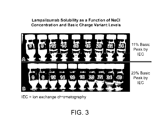
FIG. 3 illustrates lampalizumab solubility as a function of NaC1 concentration
and basic charge
variant levels. Each vial contains lampalizumab at 115 mg/mL in 30 mM HisC1 at
pH 5.6 at ambient lab
temperature. The NaC1 concentration in each vial in mM (0, 6, 12, 14, 16, 18,
20, 22, 24) is shown. 12
mM of NaC1 is required to ensure complete solubility (clear solution with no
turbidity) of lampalizumab
initially (A), but 24 mM of NaC1 is required to ensure complete solubility
(clear solution with no
turbidity) of lampalizumab when higher levels of basic charge variants are
present (B).
FIG. 4 shows Drug Substance size variants by SEC as a function of time at 30
C.
FIG. 5 shows Drug Substance charge variants by IEC as a function of time at 30
C.
FIG. 6 shows Drug Substance size variants by SEC as a function of time at -20
C.
FIG. 7 shows Drug Substance charge variants by IEC as a function of time at -
20 C.
FIG. 8 shows Drug Product size variants by SEC as a function of time at 40
C/75% RH.
FIG. 9 shows Drug Product aggregation rate by SEC 40 C/75% RH as a function of
the sugar-to-
protein ratio in the formulation.
FIG. 10 is an overlay of Drug Product Formulation #1 SEC chromatograms after
storage at
40 C/75% RH for 0, 2, and 4 weeks.
FIG. 11 shows Drug Product charge variants by IEC as a function of time at 40
C/75% RH.
FIG. 12 shows Drug Product size variants by SEC as a function of time at 25
C/60% RH.
FIG. 13 shows Drug Product size variants by SEC as a function of time at 5 C.
FIG. 14 shows Drug Product charge variants by IEC as a function of time at 5
C.
FIG. 15 shows The nucleotide sequence of the heavy chain of lampalizumab
(humanized anti-
Factor D Fab 238-1) (SEQ ID NO: 1). The nucleotide sequence encodes for the
heavy chain of
lampalizumab with the start and stop codon shown in bold and underlined. The
codon corresponding to
the first amino acid in FIG. 18 is bold and italicized.
FIG. 16 shows the amino acid sequence of the heavy chain of lampalizumab
(humanized anti-
Factor D Fab 238-1) (SEQ ID NO: 2). The HVR-HC sequences are bold and
italicized. Variable regions
are regions not underlined while first constant domain CH1 is underlined. HVR-
HC regions are shown
as: HVR1-HC: GYTFTNYGMN (SEQ ID NO: 3); HVR2-HC: WINTYTGETTYADDFKG (SEQ ID
NO: 4); HVR3-HC: EGGVNN (SEQ ID NO: 5). FIG. 16 also discloses FR1-FR4 and CH1
sequences as
SEQ ID NOS 54-57 and 30, respectively.
FIG. 17 shows the nucleotide sequence of the light chain of lampalizumab
(humanized anti-
Factor D Fab 238-1) (SEQ ID NO: 6). The nucleotide sequence encodes for the
light chain of
12
CA 03003647 2018-04-27
WO 2017/075259
PCT/US2016/059189
lampalizumab with the start and stop codon shown in bold and underlined. The
codon corresponding to
the first amino acid in FIG. 20 is bold and italicized.
FIG. 18 shows the amino acid sequence of the light chain of lampalizumab
(humanized anti-
Factor D Fab 238-1) (SEQ ID NO: 7). The amino acid sequence lacks the N-
terminal signal sequence.
The HVR-LC sequences are bold and italicized. Variable regions are regions not
underlined while first
constant domain CL1 is underlined. Framework (FR) regions and HVR regions are
shown as: HVR1-LC:
ITSTDIDDDMN (SEQ ID NO: 8); HVR2-LC: GGNTLRP (SEQ ID NO: 9); HVR3-LC:
LQSDSLPYT
(SEQ ID NO: 10). FIG. 18 also discloses FR1-FR4 and CH1 sequences as SEQ ID
NOS 58-61 and 29,
respectively.
FIG. 19 shows the Fab light chain constant region sequence of an IgG1 anti-
Factor D antibody
Fab fragment (SEQ ID NO: 29), and the heavy chain constant region sequences of
IgGl, IgG2 and IgG4
anti-Factor D antibodies, including heavy chains with C-terminal truncations
(SEQ ID NOs: 30-51).
FIG. 20 shows the light and heavy chain CDR sequences of anti-Factor D
antibody variants
AFD.v1 - AFD.v15. CDR Li sequences disclosed as SEQ ID NOS 8, 62-68, 68-70,
69, 69, 69, 69, 69 and
69, respectively, in order of appearance. CDR L2 sequences "GGNTLRP" and
"AASTLQS" disclosed as
SEQ ID NOS 9 and 71, respectively. CDR L3 sequences "LQSDSLPYT," "QKYNSAPYT"
and
"LQSESLPYT" disclosed as SEQ ID NOS 10, 72 and 73, respectively. CDR H1
sequences "NYGMN"
and "SYAMN" disclosed as SEQ ID NOS 74 and 75, respectively. CDR H2 sequences
"WINTYTGETTYADDFKG," "WINTNTGNPTYAQGFTG," "WINTYTGETTYAEDFKG" and
"WISTYTGETTYAEDFKG" disclosed as SEQ ID NOS 4, 76, 77 and 78, respectively.
CDR H3
sequences "EGGVNN," "EGYFDY," "EGGVDN," "EGGVQN" and "EGGVSN" disclosed as SEQ
ID
NOS 5, 79, 80, 81 and 82, respectively.
FIG. 21 shows the alignment of the light chain variable region sequences of
anti-Factor D
antibody variants AFD.v1 - AFD.v15 in alignment with human framework and
lampalizumab light chain
variable region sequences (SEQ ID NOS 83-94, 92, 92, 92, 92 and 94,
respectively, in order of
appearance). The CDR sequences according to Kabat definition are underlined.
FIG. 22 shows the alignment of the heavy chain variable region sequences of
anti-Factor D
antibody variants AFD.va - AFD.v15 in alignment with human framework and
lampalizumab heavy chain
variable region sequence (SEQ ID NOS 95, 96, 95, 95, 95, 95, 95, 97, 97, 97,
97, 97-101 and 101,
respectively, in order of appearance). The CDR sequences according to Kabat
definition are underlined.
Table 1. Drug Substance Formulations Screened.
Table 2. Stability data for Drug Substance formulations stored at -20 C.
Table 3. Stability data for Drug Substance formulations stored at 5 C.
Table 4. Stability data for Drug Substance formulations stored at 30 C.
13
CA 03003647 2018-04-27
WO 2017/075259
PCT/US2016/059189
Tables 5A and 5B. Stability data for Drug Product formulations stored at 5 C.
Tables 6A and 6B. Stability data for Drug Product formulations stored at 25
C/65% RH.
Tables 7A and 7B. Stability data for Drug Product formulations stored at 40
C/75% RH.
Table 8. ELISA binding data for formulations 1 and 7 at select time points.
Table 9. Stability data for Phase III lampalizumab Drug Substance.
Tables 10A and 10B. Stability data for Phase III lampalizumab Drug Product.
Detailed Description
I. Definitions
Before the present invention is described in greater detail, it is to be
understood that this invention
is not limited to particular embodiments described, as such may, of course,
vary. It is also to be
understood that the terminology used herein is for the purpose of describing
particular embodiments only,
and is not intended to be limiting, since the scope of the present invention
will be limited only by the
appended claims.
Where a range of values is provided, it is understood that each intervening
value, to the tenth of
the unit of the lower limit unless the context clearly dictates otherwise,
between the upper and lower limit
of that range and any other stated or intervening value in that stated range
is encompassed within the
invention. The upper and lower limits of these smaller ranges may
independently be included in the
smaller ranges encompassed within the invention, subject to any specifically
excluded limit in the stated
range.
Unless defined otherwise, technical and scientific terms used herein have the
same meaning as
commonly understood by one of ordinary skill in the art to which this
invention belongs. Singleton et al.,
Dictionary of Microbiology and Molecular Biology 2nd ed., J. Wiley & Sons (New
York, NY 1994),
provides one skilled in the art with a general guide to many of the terms used
in the present application.
All publications mentioned herein are expressly incorporated herein by
reference to disclose and
describe the methods and/or materials in connection with which the
publications are cited.
The term "antibody" is used in the broadest sense, and specifically covers
full length monoclonal
antibodies, polyclonal antibodies, multispecific antibodies (e.g., bispecific
antibodies) and antibody
fragments so long as they exhibit the desired biological activity such as
antigen-binding activity.
Antibodies (Abs) and immunoglobulins (Igs) are glycoproteins having the same
structural characteristics.
While antibodies exhibit binding specificity to a specific target,
immunoglobulins include both antibodies
and other antibody-like molecules which lack target specificity. Native
antibodies and immunoglobulins
are usually heterotetrameric glycoproteins of about 150,000 daltons, composed
of two identical light (L)
chains and two identical heavy (H) chains. Each heavy chain has at one end a
variable domain (VH)
14
CA 03003647 2018-04-27
WO 2017/075259
PCT/US2016/059189
followed by a number of constant domains. Each light chain has a variable
domain at one end (VL) and a
constant domain at its other end. The term "Antibody" as used herein expressly
encompasses antibody
fragments retaining antigen-binding activity.
An "antibody fragment" refers to a molecule other than an intact antibody that
comprises a
portion of an intact antibody that binds the antigen to which the intact
antibody binds. Examples of
antibody fragments include but are not limited to Fv, Fab, Fab', Fab'-SH, Fab'-
C, Fab-SH, Fab-C, Fab-C-
SH, Fab'-C-SH F(ab')2; diabodies; linear antibodies; single-chain antibody
molecules (e.g., scFv); and
multispecific antibodies formed from antibody fragments.
A "Fab-C" refers to a Fab with a C-terminal cysteine, which may be a native
cysteine that occurs
at that residue position (such as a cysteine from the hinge region), or may be
a cysteine added to the C-
terminus that does not correspond to a native cysteine. Nonlimiting exemplary
Fab-C heavy chain
constant regions include the sequences of SEQ ID NOs: 32, 44 and 45.
A "Fab-SH" refers to a Fab with a free thiol group. In some embodiments, the
free thiol group is
located in the last 10 amino acids of the C-terminus of the Fab. Fab-C
antibodies are typically also Fab-
SH antibodies. A further nonlimiting exemplary Fab-SH heavy chain constant
region having the amino
acid sequence of SEQ ID NO: 34. Typically, a Fab comprising an engineered
cysteine (i.e., a Fab that is
a THIOMAB) is a Fab-SH.
As used herein, an "anti-Factor D antibody" means an antibody, as hereinabove
defined, which
specifically binds to Factor D in such a manner so as to inhibit or
substantially reduce complement
activation. In some embodiments, the anti-Factor D antibody is an antibody
fragment (as hereinabove
defined), such as a Fab fragment.
The term "Factor D" is used herein to refer to native sequence and variant
Factor D polypeptides.
In some embodiments the term "Factor D" refers to a native sequence mammalian
polypeptide, more
preferably a native sequence human polypeptide.
The term "variable region" or "variable domain" refers to the domain of an
antibody heavy or
light chain that is involved in binding the antibody to antigen. The variable
domains of the heavy chain
and light chain (VH and VL, respectively) of a native antibody generally have
similar structures, with
each domain comprising four conserved framework regions (FRs) and three
hypervariable regions
(HVRs). (See, e.g., Kindt et al. Kuby Immunology, 6th ed., W.H. Freeman and
Co., page 91 (2007).) A
single VH or VL domain may be sufficient to confer antigen-binding
specificity. Furthermore, antibodies
that bind a particular antigen may be isolated using a VH or VL domain from an
antibody that binds the
antigen to screen a library of complementary VL or VH domains, respectively.
See, e.g., Portolano et al.,
J. Immunol. 150:880-887 (1993); Clarkson et al., Nature 352:624-628 (1991).
CA 03003647 2018-04-27
WO 2017/075259
PCT/US2016/059189
The term "variable" refers to the fact that certain portions of the variable
domains differ
extensively in sequence among antibodies and are used in the binding and
specificity of each particular
antibody for its particular antigen. However, the variability is not evenly
distributed throughout the
variable domains of antibodies. It is concentrated in three segments called
hypervariable regions both in
the light chain and the heavy chain variable domains. The more highly
conserved portions of variable
domains are called the framework regions (FRs). The variable domains of native
heavy and light chains
each comprise four FRs, largely adopting a 13-sheet configuration, connected
by three hypervariable
regions, which form loops connecting, and in some cases forming part of, the
13-sheet structure. The
hypervariable regions in each chain are held together in close proximity by
the FRs and, with the
hypervariable regions from the other chain, contribute to the formation of the
antigen-binding site of
antibodies (see Kabat et al., Sequences of Proteins of Immunological Interest,
5th Ed. Public Health
Service, National Institutes of Health, Bethesda, Md. (1991)). The constant
domains are not involved
directly in binding an antibody to an antigen, but exhibit various effector
functions, such as participation
of the antibody in antibody dependent cellular cytotoxicity (ADCC).
Papain digestion of antibodies produces two identical antigen-binding
fragments, called "Fab"
fragments, each with a single antigen-binding site, and a residual "Fe"
fragment, whose name reflects its
ability to crystallize readily. Pepsin treatment yields an F(ab')2 fragment
that has two antigen-binding
sites and is still capable of cross-linking antigen.
The Fab fragment also contains the constant domain of the light chain and the
first constant
domain (CH1) of the heavy chain. Fab' fragments differ from Fab fragments by
the addition of a few
residues at the carboxyl terminus of the heavy chain CH1 domain including one
or more cysteines from
the antibody hinge region. Fab'-SH is the designation herein for Fab' in which
the cysteine residue(s) of
the constant domains bear at least one free thiol group. F(ab')2 antibody
fragments originally were
produced as pairs of Fab' fragments which have hinge cysteines between them.
Other chemical couplings
of antibody fragments are also known.
As used herein, a "Fab" refers to an antibody that comprises a heavy chain
constant region that
comprises the CH1 domain, or a sufficient portion of the CH1 domain to form a
disulfide bond with the
light chain constant region, but does not contain a CH2 domain or a CH3
domain. As used herein, a Fab
may comprise one or more amino acids of the hinge region. Thus, as used
herein, the term "Fab"
encompasses Fab' antibodies. A Fab may comprise additional non-native amino
acids, such as a C-
terminal cysteine, in which case it may be referred to as a Fab-C. As
discussed below, the term Fab-C
also encompasses Fabs comprising native amino acids of the hinge region,
including a native cysteine at
the C-terminus. In some embodiments, a Fab comprises an engineered cysteine
(i.e., a Fab may be a
THIOMAB).
16
CA 03003647 2018-04-27
WO 2017/075259
PCT/US2016/059189
"Fv" is the minimum antibody fragment which contains a complete antigen-
recognition and
antigen-binding site. This region consists of a dimer of one heavy chain and
one light chain variable
domain in tight, non-covalent association. It is in this configuration that
the three hypervariable regions of
each variable domain interact to define an antigen-binding site on the surface
of the VH-VL dimer.
Collectively, the six hypervariable regions confer antigen-binding specificity
to the antibody. However,
even a single variable domain (or half of an Fv comprising only three
hypervariable regions specific for
an antigen) has the ability to recognize and bind antigen, although at a lower
affinity than the entire
binding site.
The term "hypervariable region" or "HVR," as used herein, refers to each of
the regions of an
antibody variable domain which are hypervariable in sequence and/or form
structurally defined loops
("hypervariable loops"). Generally, native four-chain antibodies comprise six
HVRs; three in the VH
(H1, H2, H3), and three in the VL (L1, L2, L3). HVRs generally comprise amino
acid residues from the
hypervariable loops and/or from the "complementarity determining regions"
(CDRs), the latter being of
highest sequence variability and/or involved in antigen recognition. HVR-H3 is
believed to play a unique
role in conferring fine specificity to antibodies. See, e.g., Xu et al. (2000)
Immunity 13:37-45; Johnson
and Wu (2003) in Methods in Molecular Biology 248:1-25 (Lo, ed., Human Press,
Totowa, N.J.).
"Framework Region" or "FR" residues are those variable domain residues other
than the hypervariable
region residues as herein defined. An HVR region as used herein comprise any
number of residues
located within positions 24-36 (for L1), 46-56 (for L2), 89-97 (for L3), 26-
35B (for H1), 47-65 (for H2),
and 93-102 (for H3). Therefore, an FIVR includes residues in positions
described previously:
A) 24-34 (L1), 50-52 (L2), 91-96 (L3), 26-32 (H1), 53-55 (H2), and 96-101 (H3)
(Chothia and Lesk, J. Mol. Biol. 196:901-917 (1987);
B) 24-34 of Li, 50-56 of L2, 89-97 of L3, 31-35B of H1, 50-65 of H2, and 95-
102 of H3
(Kabat et al., Sequences of Proteins of Immunological Interest, 5th Ed. Public
Health Service, National
Institutes of Health, Bethesda, MD (1991).
C) 30-36 (L1), 46-55 (L2), 89-96 (L3), 30-35 (H1), 47-58 (H2), 93-100a-j (H3)
(MacCallum et al. J. Mol. Biol. 262:732-745 (1996).
Hypervariable regions may comprise "extended hypervariable regions" as
follows: 24-36 or 24-
34 (L1), 46-56 or 50-56 (L2) and 89-97 (L3) in the VL and 26-35B (H1), 50-65,
47-65 or 49-65 (H2) and
93-102, 94-102 or 95-102 (H3) in the VH. The variable domain residues are
numbered according to
Kabat et al., supra for each of these definitions.
With the exception of CDR1 in VH, CDRs generally comprise the amino acid
residues that form
the hypervariable loops. CDRs also comprise "specificity determining
residues," or "SDRs," which are
residues that contact antigen. SDRs are contained within regions of the CDRs
called abbreviated-CDRs,
17
CA 03003647 2018-04-27
WO 2017/075259
PCT/US2016/059189
or a-CDRs. Exemplary a-CDRs (a-CDR-L1, a-CDR-L2, a-CDR-L3, a-CDR-H1, a-CDR-H2,
and a-CDR-
H3) occur at amino acid residues 31-34 of L 1, 50-55 of L2, 89-96 of L3, 31-
35B of H1, 50-58 of H2, and
95-102 of H3. (See Almagro and Fransson, Front. Biosci. 13:1619-1633 (2008).)
An "antibody variant" or "modified antibody" of a reference antibody (also
referred to as
"starting antibody" or "parent antibody") is an antibody that comprises an
amino acid sequence different
from that of the reference/starting antibody, wherein one or more of the amino
acid residues of the
reference antibody have been modified. Generally, an antibody variant will
possess at least 80% sequence
identity, preferably at least 90% sequence identity, more preferably at least
95% sequence identity, and
most preferably at least 98% sequence identity with the reference antibody.
Percentage sequence identity
is determined for example, by the Fitch et al., Proc. Natl. Acad. Sci. USA,
80: 1382-1386 (1983), version
of the algorithm described by Needleman et al., I Mol. Biol., 48: 443-453
(1970), after aligning the
sequences of the reference antibody and the candidate antibody variant to
provide for maximum
homology. Identity or similarity is defined herein as the percentage of amino
acid residues in the
candidate variant sequence that are identical (i.e. same residue) or similar
(i.e. amino acid residue from
the same group based on common side-chain properties, see below) with the
parent antibody residues,
after aligning the sequences and introducing gaps, if necessary, to achieve
the maximum percent sequence
identity. Amino acid sequence variants of an antibody may be prepared by
introducing appropriate
nucleotide changes into DNA encoding the antibody, or by peptide synthesis.
Such variants include, for
example, deletions from, and/or insertions into and/or substitutions of,
residues within the amino acid
sequence of the antibody of interest. Any combination of deletion, insertion,
and substitution is made to
arrive at the final construct, provided that the final construct possesses the
desired characteristics. The
amino acid changes also may alter post-translational processes of the
antibody, such as changing the
number or position of glycosylation sites. Methods for generating antibody
sequence variants of
antibodies are similar to those for generating amino acid sequence variants of
polypeptides described in
U.S. Pat. No. 5,534,615, expressly incorporated herein by reference, for
example.
The term "monoclonal antibody" as used herein refers to an antibody obtained
from a population
of substantially homogeneous antibodies, i.e., the individual antibodies
comprising the population are
identical except for possible naturally occurring mutations that may be
present in minor amounts.
Monoclonal antibodies are highly specific, being directed against a single
antigenic site. Furthermore, in
contrast to conventional (polyclonal) antibody preparations which typically
include different antibodies
directed against different determinants (epitopes), each monoclonal antibody
is directed against a single
determinant on the antigen. The modifier "monoclonal" indicates the character
of the antibody as being
obtained from a substantially homogeneous population of antibodies, and is not
to be construed as
requiring production of the antibody by any particular method. For example,
the monoclonal antibodies
18
CA 03003647 2018-04-27
WO 2017/075259
PCT/US2016/059189
to be used in accordance with the present invention may be made by the
hybridoma method first
described by Kohler et al. (1975) Nature 256:495, or may be made by
recombinant DNA methods (see,
e.g.,U U.S. Patent No. 4,816,567). The "monoclonal antibodies" may also be
isolated from phage antibody
libraries using the techniques described in Clackson et al. (1991) Nature
352:624-628 and Marks et al.
(1991) Mol. Biol. 222:581-597, for example.
The monoclonal antibodies herein specifically include "chimeric" antibodies
(immunoglobulins)
in which a portion of the heavy and/or light chain is identical with or
homologous to corresponding
sequences in antibodies derived from a particular species or belonging to a
particular antibody class or
subclass, while the remainder of the chain(s) is identical with or homologous
to corresponding sequences
in antibodies derived from another species or belonging to another antibody
class or subclass, as well as
fragments of such antibodies, so long as they exhibit the desired biological
activity (U.S. Patent No.
4,816,567; and Morrison etal. (1984) Proc. Natl. Acad. Sci. USA 81:6851-6855).
"Humanized" forms of non-human (e.g., murine) antibodies are chimeric
antibodies which
contain minimal sequence derived from non-human immunoglobulin. For the most
part, humanized
antibodies are human immunoglobulins (recipient antibody) in which residues
from a hypervariable
region of the recipient are replaced by residues from a hypervariable region
of a non-human species
(donor antibody) such as mouse, rat, rabbit or nonhuman primate having the
desired specificity, affinity,
and capacity. In some instances, Fv framework region (FR) residues of the
human immunoglobulin are
replaced by corresponding non-human residues. Furthermore, humanized
antibodies may comprise
residues which are not found in the recipient antibody or in the donor
antibody. These modifications are
made to further refine antibody performance. In general, the humanized
antibody will comprise
substantially all of at least one, and typically two, variable domains, in
which all or substantially all of the
hypervariable loops correspond to those of a non-human immunoglobulin and all
or substantially all of
the FR regions are those of a human immunoglobulin sequence. The humanized
antibody optionally also
will comprise at least a portion of an immunoglobulin constant region (Fc),
typically that of a human
immunoglobulin. For further details, see Jones et al. (1986) Nature 321:522-
525; Riechmann et al.
(1988) Nature 332:323-329; and Presta (1992) Curr. Op. Struct. Biol. 2:593-
596.
A protein including an antibody is said to be "stable" if it essentially
retains the intact
conformational structure and biological activity. Various analytical
techniques for measuring protein
stability are available in the art and are reviewed in, e.g., Peptide and
Protein Drug Delivery, 247-301,
Vincent Lee Ed., Marcel Dekker, Inc., New York, N.Y., Pubs. (1991) and Jones
(1993) Adv. Drug
Delivery Rev. 10: 29-90. An antibody variant with "improved stability" refers
to an antibody variant that
is more stable comparing to the starting reference antibody. Preferably,
antibody variants with improved
stability are variants of the native (wild-type) antibodies in which specific
amino acid residues are altered
19
CA 03003647 2018-04-27
WO 2017/075259
PCT/US2016/059189
for the purpose of improving physical stability, and/or chemical stability,
and/or biological activity,
and/or reducing immunogenicity of the native antibodies. Walsh (2000) Nat.
Biotech. 18:831-3.
The term "isomerization" refers generally to a chemical process by which a
chemical compound
is transformed into any of its isomeric forms, i.e., forms with the same
chemical composition but with
different structure or configuration and, hence, generally with different
physical and chemical properties.
Specifically used herein is aspartate isomerization, a process wherein one or
more aspartic acid (D or
Asp) residue(s) of a polypeptide have been transformed to isoaspartic acid
(IsoAsp) and/or cyclic imide
(Asu) residue(s). Geiger and Clarke (1987) 1 Biol. Chem. 262:785-94;. Wakankar
et al. (2007)
Biochem. 46:1534-44.
The term "deamidation" refers generally to a chemical reaction wherein an
amide functional
group is removed from an organic compound. Specifically used herein is
asparagine deamidation, a
process wherein one or more asparagine (N or Asn) residue(s) of a polypeptide
have been converted to
aspartic acid (D or Asp), i.e. the neutral amide side chain has been converted
to a residue with an overall
acidic property. Xie and Schowen (1999)1 Pharm. Sci. 88:8-13.
Amino acid residues "prone" to certain identified physical or chemical
processes (e.g.,
isomerization or deamidation) refer to those residues within a specific
protein molecule that have been
identified to have the propensity to undergo the identified processes such as
isomerization or deamidation.
Their propensities are often determined by their relative positions within the
primary and/or
conformational structure of the protein. For example, it has been shown that
the first Asp in an Asp-XXX
motif (wherein XXX can be Asp, Gly, His, Ser or Thr) is prone to Asp
isomerization due to the
involvement of its adjacent residue, where some other Asp within the same
protein may not possess such
propensity. Assays for identifying residues to certain process within a
specific protein molecule are
known in the art. See, e.g., Cacia et al (1996) Biochem. 35:1897-1903.
"Active" or "activity" or "biological activity" in the context of an anti-
factor D antibody of the
present invention is the ability to antagonize (partially or fully inhibit) a
biological activity of Factor D.
One example of a biological activity of a Factor D antagonist is the ability
to achieve a measurable
improvement in the state, e.g. pathology, of a Factor D-associated disease or
condition, such as, for
example, a complement-associated ocular condition. The activity can be
determined in in vitro or in vivo
tests, including binding assays, alternative pathway hemolysis assays (e.g.
assays measuring inhibition of
the alternative pathway complement activity or activation), using a relevant
animal model, or human
clinical trials.
The term "complement-associated disorder" is used in the broadest sense and
includes disorders
associated with excessive or uncontrolled complement activation. They include
complement activation
during cardiopulmonary bypass operations; complement activation due to
ischemia-reperfusion following
CA 03003647 2018-04-27
WO 2017/075259
PCT/US2016/059189
acute myocardial infarction, aneurysm, stroke, hemorrhagic shock, crush
injury, multiple organ failure,
hypobolemic shock, intestinal ischemia or other events causing ischemia.
Complement activation has
also been shown to be associated with inflammatory conditions such as severe
burns, endotoxemia, septic
shock, adult respiratory distress syndrome, hemodialysis; anaphylactic shock,
severe asthma, angioedema,
Crohn's disease, sickle cell anemia, poststreptococcal glomerulonephritis and
pancreatitis. The disorder
may be the result of an adverse drug reaction, drug allergy, IL-2 induced
vascular leakage syndrome or
radiographic contrast media allergy. It also includes autoimmune disease such
as systemic lupus
erythematosus, myasthenia gravis, rheumatoid arthritis, Alzheimer's disease
and multiple sclerosis.
Complement activation is also associated with transplant rejection. Complement
activation is also
associated with ocular diseases such as age-related macular degeneration,
diabetic retinopathy and other
ischemia-related retinopathies, choroidal neovascularization (CNV), uveitis,
diabetic macular edema,
pathological myopia, von Hippel-Lindau disease, histoplasmosis of the eye,
Central Retinal Vein
Occlusion (CRVO), corneal neovascularization, and retinal neovascularization.
The term "complement-associated eye condition" or "complement-associated
ocular condition" is
used in the broadest sense and includes all eye conditions the pathology of
which involves complement,
including the classical and the alternative pathways, and in particular the
alternative pathway of
complement. Complement-associated eye conditions include, without limitation,
macular degenerative
diseases, such as all stages of age-related macular degeneration (AMD),
including dry and wet (non-
exudative and exudative) forms, choroidal neovascularization (CNV), uveitis,
diabetic and other
ischemia-related retinopathies, and other intraocular neovascular diseases,
such as diabetic macular
edema, pathological myopia, von Hippel-Lindau disease, histoplasmosis of the
eye, Central Retinal Vein
Occlusion (CRVO), corneal neovascularization, and retinal neovascularization.
In one example,
complement-associated eye conditions includes age-related macular degeneration
(AMD), including non-
exudative (e.g. intermediate dry AMD or geographic atrophy (GA)) and exudative
(e.g. wet AMD
(choroidal neovascularization (CNV)) AMD, diabetic retinopathy (DR),
endophthalmitis and uveitis. In a
further example, nonexudative AMD may include the presence of hard drusen,
soft drusen, geographic
atrophy and/or pigment clumping. In one example, complement-associated eye
conditions include age-
related macular degeneration (AMD), including early AMD (e.g. includes
multiple small to one or more
non-extensive medium sized drusen), intermediate AMD (e.g. includes extensive
medium drusen to one
or more large drusen) and advanced AMD (e.g. includes geographic atrophy or
advanced wet AMD
(CNV). (Ferris et al., AREDS Report No. 18, ; Sallo et al., Eye Res., 34(3):
238-40 (2009); Jager et al.,
New Engl. I Med., 359(1): 1735 (2008)). In a further example, intermediate dry
AMD may include large
confluent drusen. In a further example, geographic atrophy may include
photoreceptor and/or Retinal
Pigmented Epithelial (RPE) loss. In a further example, the area of geographic
atrophy may be small or
21
CA 03003647 2018-04-27
WO 2017/075259
PCT/US2016/059189
large and/or may be in the macula area or in the peripheral retina. In one
example, complement-
associated eye condition is intermediate dry AMD. In one example, complement-
associated eye
condition is geographic atrophy. In one example, complement-associated eye
condition is wet AMD
(choroidal neovascularization (CNV)).
"Geographic Atrophy", also referred to herein as "GA", as used herein is a
disease involving
degeneration of the retinal pigment epithelium (RPE), associated with loss of
photoreceptors. GA is the
advanced form of dry AMD.
"GA Area", as used herein refers to a discrete area representing loss of
retinal anatomy (e.g.
photoreceptors and retinal pigment epithelium (RPE). GA area is measured by
standard imaging
techniques such as fundus autofluorescence (FAF) and digital color fundus
photography (CFP).
"Early AMD", as used herein is a disease characterized by multiple small (<63
[an) or > 1
intermediate drusen (> 63 [ail and < 125 [ari).
"Intermediate AMD", as used herein is a disease characterized by many
intermediate or > 1 large
drusen (> 125 [ail) often accompanied by hyper or hypopigmentation of the
retinal pigment epithelium.
"Advanced AMD", as used herein is a disease characterized by geographic
atrophy (GA) or
neovascular (wet) AMD).
"Treatment" is an intervention performed with the intention of preventing the
development or
altering the pathology of a disorder. Accordingly, "treatment" refers to both
therapeutic treatment and
prophylactic or preventative measures. Those in need of treatment include
those already with the disorder
as well as those in which the disorder is to be prevented. In treatment of an
immune related disease, a
therapeutic agent may directly alter the magnitude of response of a component
of the immune response,
or render the disease more susceptible to treatment by other therapeutic
agents, e.g., antibiotics,
antifungals, anti-inflammatory agents, chemotherapeutics, etc.
The "pathology" of a disease, such as a complement-associated disorder,
includes all phenomena
that compromise the well-being of the patient. This includes, without
limitation, abnormal or
uncontrollable cell growth (neutrophilic, eosinophilic, monocytic, lymphocytic
cells), antibody
production, auto-antibody production, complement production, interference with
the normal functioning
of neighboring cells, release of cytokines or other secretory products at
abnormal levels, suppression or
aggravation of any inflammatory or immunological response, infiltration of
inflammatory cells
(neutrophilic, eosinophilic, monocytic, lymphocytic) into cellular spaces,
etc.
The term "mammal" as used herein refers to any animal classified as a mammal,
including,
without limitation, humans, higher primates, domestic and farm animals, and
zoo, sports or pet animals
such horses, pigs, cattle, dogs, cats and ferrets, etc. In some embodiments of
the invention, the mammal
is a human.
22
CA 03003647 2018-04-27
WO 2017/075259
PCT/US2016/059189
Administration "in combination with" one or more further therapeutic agents
includes
simultaneous (concurrent) and consecutive administration in any order.
"Therapeutically effective amount" is the amount of a "Factor D antibody"
which is required to
achieve a measurable improvement in the state, e.g. pathology, of the target
disease or condition, such as,
for example, a complement-associated eye condition.
"Pharmaceutically acceptable" excipients (vehicles, additives) are those which
can reasonably be
administered to a subject mammal to provide an effective dose of the active
ingredient employed.
A "stable" formulation in one in which the protein, e.g. an anti-Factor D
antibody, therein
essentially retains its physical stability and/or chemical stability and/or
biological activity upon storage.
Various analytical techniques for measuring protein stability are available in
the art and are reviewed in
Peptide and Protein Drug Delivery, 247-301, Vincent Lee Ed., Marcel Dekker,
Inc., New York, N.Y.,
Pubs (1991) and Jones, A. Adv. Drug Delivery Rev. 10: 29-90 (1993), for
example. Stability can be
measured at a selected temperature for a selected time period. In one
embodiment, the formulation is
stable at room temperature or at 40 C for at least 1 month and/or stable at 2-
8 C for at least 1 year and
preferably for at least 2 years. In another embodiment, the pre-lyophilized
formulation (also referred
herein as "Drug Substance" or "DS") is stable at a storage temperature of -20
C for at least one year, or
for at least two years, or for at least three years, or for at least five
years. In a further embodiment, the
lyophilized formulation is stable at a storage temperature of 5 C for at least
one year, or for at least two
years, or for at least three years, or for at least four years, or for at
least five years. Furthermore, the
formulation is preferably stable following freezing (to, e.g., -70 C) and
thawing of the formulation.
A protein, such as an anti-Factor D antibody, "retains its physically
stability" in a pharmaceutical
formulation if it shows no signs of aggregation, precipitation and/or
denaturation upon visual examination
of color and/or clarity, or as measured by UV light scattering or by size
exclusions chromatography.
A protein, e.g. an anti-Factor D antibody, "retains the chemical stability" in
a pharmaceutical
formulation, if the chemical stability at a given time is such that the
protein is considered to still retain its
biological activity as defined below. Chemical stability can be assessed by
detecting and quantifying
chemically altered forms of the protein. Chemical alteration may involve size
modification (e.g. clipping)
which can be evaluated using size exclusion chromatography, SDS-PAGE and/or
matrix-assisted laser
desorption ionization/time-of-flight mass spectrometry (MALDI/TOF MS), for
example. Other types of
chemical alteration include charge alteration (e.g. occurring as a result of
deamidation) which can be
evaluated by ion-exchange chromatography, for example.
An antibody, e.g. an anti-Factor D antibody, "retains its biological activity"
in a pharmaceutical
formulation, if the biological activity of the antibody at a given time is
within about 10% (within the
errors of the assay) of the biological activity exhibited at the time the
pharmaceutical formulation was
23
CA 03003647 2018-04-27
WO 2017/075259
PCT/US2016/059189
prepared as determined in an antigen binding assay, for example. Other
"biological activity" assays for
antibodies are elaborated herein below.
By "isotonic" is meant that the formulation of interest has essentially the
same osmotic pressure
as human blood. Isotonic formulations will generally have an osmotic pressure
from about 250 to 350
mOsm/kg. Isotonicity can be measured using a vapor pressure or ice-freezing
type osmometer for
example.
The term "lyoprotectant" refers to a substance, such as a chemical compound or
molecule, that
protects a protein, e.g. an antibody, from damage resulting from
lyophilization. Preferably, the
lyoprotectant is a polyol.
A "polyol" is a substance with multiple hydroxyl groups, and includes sugars
(reducing and
nonreducing sugars), sugar alcohols and sugar acids. Preferred polyols herein
have a molecular weight
which is less than about 600 D (e.g. in the range from about 120 to about 400
D). A "reducing sugar" is
one which contains a hemiacetal group that can reduce metal ions or react
covalently with lysine and
other amino groups in proteins and a "nonreducing sugar" is one which does not
have these properties of a
reducing sugar. Examples of reducing sugars are fructose, mannose, maltose,
lactose, arabinose, xylose,
ribose, rhamnose, galactose and glucose. Nonreducing sugars include sucrose,
trehalose, sorbose,
melezitose and raffinose. Mannitol, xylitol, erythritol, threitol, sorbitol
and glycerol are examples of sugar
alcohols. As to sugar acids, these include L-gluconate and metallic salts
thereof. Where it desired that the
formulation is freeze-thaw stable, the polyol is preferably one which does not
crystallize at freezing
temperatures (e.g. -20 C.) such that it destabilizes the antibody in the
formulation. Polyols, including
mixtures of polyols, can be used as lyoprotectants in the formulations of the
present invention.
Nonreducing sugars such as sucrose and trehalose are preferred as
lyoprotectants in the anti-Factor D
antibody formulations herein, sucrose is being preferred over trehalose.
As used herein, "buffer" refers to a buffered solution that resists changes in
pH by the action of its
acid-base conjugate components. The buffer of this invention has a pH in the
range from 5.0 to 5.4; and
most preferably has a pH of about 5.3. Examples of buffers that will control
the pH in this range include
histidine, acetate (e.g. sodium acetate), succinate (such as sodium
succinate), gluconate, citrate and other
organic acid buffers. Where a freeze-thaw stable formation is desired, the
buffer is preferably not
phosphate. The term "buffer" specifically includes combinations of two or more
buffers suitable for
providing the desired pH in the formulations herein.
As used herein, a "surfactant" refers to a surface-active agent, typically a
nonionic surfactant. The
formulations of the present invention comprise one or more surfactant. Thus,
the term "surfactant"
specifically includes mixtures of two or more surfactants. Examples of
suitable surfactants include
polysorbate (for example, polysorbate 20 and polysorbate 80); poloxamer (e.g.
poloxamer 188); Triton;
24
CA 03003647 2018-04-27
WO 2017/075259
PCT/US2016/059189
sodium dodecyl sulfate (SDS); sodium laurel sulfate; sodium octyl glycoside;
lauryl-, myristyl-, linoleyl-,
or stearyl-sulfobetaine; lauryl-, myristyl-, linoleyl- or stearyl-sarcosine;
linoleyl-, myristyl-, or cetyl-
betaine; lauroamidopropyl-, cocamidopropyl-, linoleamidopropyl-,
myristamidopropyl-, palmidopropyl-,
or isostearamidopropyl-betaine (e.g. lauroamidopropyl); myristamidopropyl-,
palmidopropyl-, or
isostearamidopropyl-dimethylamine; sodium methyl cocoyl-, or disodium methyl
oleyl-taurate; polyethyl
glycol, polypropyl glycol, and copolymers of ethylene and propylene glycol
(e.g. Pluronics, PF68 etc). In
some embodiments, the surfactant herein is a polysorbate, e.g. polysorbate 20
or a poloxamer.
A "preservative" is a compound which can be included in the formulation to
essentially reduce
bacterial action therein, thus facilitating the production of a multi-use
formulation, for example. Examples
of potential preservatives include octadecyldimethylbenzyl ammonium chloride,
hexamethonium
chloride, benzalkonium chloride (a mixture of alkylbenzyldimethylammonium
chlorides in which the
alkyl groups are long-chain compounds), and benzelthonium chloride. Other
types of preservatives
include aromatic alcohols such as phenol, butyl and benzyl alcohol, alkyl
parabens such as methyl or
propyl paraben, catechol, resorcinol, cyclohexanol, 3-pentanol, and m-cresol.
The term "preservative"
specifically includes mixtures of two or more preservatives. The most
preferred preservative herein is
benzyl alcohol.
The terms "long-acting delivery", "sustained-release" and "controlled release"
are used generally
to describe a delivery mechanism using formulation, dosage form, device or
other types of technologies to
achieve the prolonged or extended release or bioavailability of a therapeutic
drug. It may refer to
technologies that provide prolonged or extended release or bioavailability of
the drug to the general
systemic circulation or a subject or to local sites of action in a subject
including (but not limited to) cells,
tissues, organs, joints, regions, and the like. Furthermore, these terms may
refer to a technology that is
used to prolong or extend the release of the drug from a formulation or dosage
form or they may refer to a
technology used to extend or prolong the bioavailability or the
pharmacokinetics or the duration of action
of the drug to a subject or they may refer to a technology that is used to
extend or prolong the
pharmacodynamic effect elicited by a formulation. A "long-acting formulation,"
a "sustained release
formulation," or a "controlled release formulation" is a pharmaceutical
formulation, dosage form, or other
technology that is used to provide long-acting delivery. In one aspect, the
controlled release is used to
improve drug's local bioavailability, specifically ocular residence time in
the context of ocular delivery.
"Increased ocular residence time" refers to the post-delivery period during
which the delivered ocular
drug remains effective both in terms of quality (stay active) and in terms of
quantity (effective amount).
In addition to or in lieu of high dose and controlled release, the drug can be
modified post-translationally,
such as via PEGylation, to achieve increased in vivo half-life.
CA 03003647 2018-04-27
WO 2017/075259
PCT/US2016/059189
Detailed Description
Anti-Factor D Antibody Formulations
The invention herein pharmaceutical formulations comprising monoclonal anti-
Factor D
antibodies, and their production and use for the treatment of complement-
associated ocular diseases.
In one aspect, the anti-Factor D antibody present in the formulations is a
humanized monoclonal
anti-Factor D antibody. Methods for humanizing non-human antibodies are well
known in the art.
Generally, a humanized antibody has one or more amino acid residues introduced
into it from a source
which is non-human. These non-human amino acid residues are often referred to
as "import" residues,
which are typically taken from an "import" variable domain. Humanization can
be essentially performed
following the method of Winter and co-workers (Jones et al. (1986) Nature
321:522-525; Riechmann et
al. (1988) Nature 332:323-327; Verhoeyen et al. (1988) Science 239:1534-1536),
by substituting rodent
CDRs or CDR sequences for the corresponding sequences of a human antibody.
Accordingly, such
"humanized" antibodies are chimeric antibodies (U.S. Patent No. 4,816,567),
wherein substantially less
than an intact human variable domain has been substituted by the corresponding
sequence from a non-
human species. In practice, humanized antibodies are typically human
antibodies in which some CDR
residues and possibly some FR residues are substituted by residues from
analogous sites in rodent
antibodies.
The choice of human variable domains, both light and heavy, to be used in
making the humanized
antibodies can in some instances be important to reduce antigenicity and/or
HAMA response (human anti-
mouse antibody) when the antibody is intended for human therapeutic use.
Reduction or elimination of a
HAMA response is generally a significant aspect of clinical development of
suitable therapeutic agents.
See, e.g., Khaxzaeli et al. (1988) J Natl. Cancer Inst 80:937; Jotters et al.
(1986) Transplantation 41:572;
Shawler et al. (1985) J Immunol. 135:1530; Sears et al. (1984)1 Biol. Response
Mod. 3:138; Miller et al.
(1983) Blood 62:988; Hakimi et al. (1991)1 Immunol. 147:1352; Reichmann et al.
(1988) Nature
332:323; Junghans et al. (1990) Cancer Res. 50:1495. As described herein, the
invention provides
antibodies that are humanized such that HAMA response is reduced or
eliminated. Variants of these
antibodies can further be obtained using routine methods known in the art,
some of which are further
described below. According to the so-called "best-fit" method, the sequence of
the variable domain of a
rodent antibody is screened against the entire library of known human variable
domain sequences. The
human V domain sequence which is closest to that of the rodent is identified
and the human framework
region (FR) within it accepted for the humanized antibody (Sims et al. (1993)1
Immunol. 151:2296;
Chothia et al. (1987) J Mol. Biol.196:901). Another method uses a particular
framework region derived
from the consensus sequence of all human antibodies of a particular subgroup
of light or heavy chains.
26
CA 03003647 2018-04-27
WO 2017/075259
PCT/US2016/059189
The same framework may be used for several different humanized antibodies
(Carter et al. (1992) Proc.
Natl. Acad. Sci. USA 89:4285; Presta et al. (1993) J Immunol. 151:2623).
For example, an amino acid sequence from an antibody as described herein can
serve as a starting
(parent) sequence for diversification of the framework and/or hypervariable
sequence(s). A selected
framework sequence to which a starting hypervariable sequence is linked is
referred to herein as an
acceptor human framework. While the acceptor human frameworks may be from, or
derived from, a
human immunoglobulin (the VL and/or VH regions thereof), the acceptor human
frameworks may be
from, or derived from, a human consensus framework sequence as such frameworks
have been
demonstrated to have minimal, or no, immunogenicity in human patients. An
"acceptor human
framework" for the purposes herein is a framework comprising the amino acid
sequence of a VL or VH
framework derived from a human immunoglobulin framework, or from a human
consensus framework.
An acceptor human framework "derived from" a human immunoglobulin framework or
human consensus
framework may comprise the same amino acid sequence thereof, or may contain
pre-existing amino acid
sequence changes. Where pre-existing amino acid changes are present,
preferably no more than 5 and
preferably 4 or less, or 3 or less, pre-existing amino acid changes are
present. In some embodiments, the
VH acceptor human framework is identical in sequence to the VH human
immunoglobulin framework
sequence or human consensus framework sequence. In some embodiments, the VL
acceptor human
framework is identical in sequence to the VL human immunoglobulin framework
sequence or human
consensus framework sequence. A "human consensus framework" is a framework
which represents the
most commonly occurring amino acid residue in a selection of human
immunoglobulin VL or VH
framework sequences. Generally, the selection of human immunoglobulin VL or VH
sequences is from a
subgroup of variable domain sequences. Generally, the subgroup of sequences is
a subgroup as in Kabat
et al. In some embodiments, for the VL, the subgroup is subgroup kappa I as in
Kabat et al. In some
embodiments, for the VH, the subgroup is subgroup III as in Kabat et al.
Where the acceptor is derived from a human immunoglobulin, one may optionally
select a human
framework sequence that is selected based on its homology to the donor
framework sequence by aligning
the donor framework sequence with various human framework sequences in a
collection of human
framework sequences, and select the most homologous framework sequence as the
acceptor. The
acceptor human framework may be from or derived from human antibody germline
sequences available
in the public databases.
In some embodiments, human consensus frameworks herein are from, or derived
from, VH
subgroup VII and/or VL kappa subgroup I consensus framework sequences.
27
CA 03003647 2018-04-27
WO 2017/075259
PCT/US2016/059189
In some embodiments, the human framework template used for generation of an
anti-Factor D
antibody may comprise framework sequences from a template comprising a
combination of VI-4.1b+
(VH7 family) and JH4d for VH chain and/or a combination of DPK4 (WI family)
and JK2 for VL chain.
While the acceptor may be identical in sequence to the human framework
sequence selected,
whether that be from a human immunoglobulin or a human consensus framework,
the acceptor sequence
may also comprise pre-existing amino acid substitutions relative to the human
immunoglobulin sequence
or human consensus framework sequence. These pre-existing substitutions are
preferably minimal;
usually four, three, two or one amino acid differences only relative to the
human immunoglobulin
sequence or consensus framework sequence.
Hypervariable region residues of the non-human antibody are incorporated into
the VL and/or
VH acceptor human frameworks. For example, one may incorporate residues
corresponding to the Kabat
CDR residues, the Chothia hypervariable loop residues, the Abm residues,
and/or contact residues.
Optionally, the extended hypervariable region residues as follows are
incorporated: 24-36 or 24-34 (L1),
46-56 or 50-56 (L2) and 89-97 (L3), 26-35B (H1), 50-65, 47-65 or 49-65 (H2)
and 93-102, 94-102, or 95-
102 (H3).
The antibodies herein include all classes and subclasses of immunoglobulin
molecules, including
IgGl, IgG2, IgG3, and IgG4; IgG1 antibodies being preferred.
In some embodiments, the anti-Factor D antibody present in the formulations
herein is an
antigen-binding fragment of a humanized anti-Factor D antibody, such as, for
example, a Fab fragment or
a F(ab')2 fragment, preferably a Fab fragment.
Fab antibody fragments provide the advantage of small size, short serum half-
life, and lack of
effector function, which are beneficial in many therapeutic applications.
Thus, Fab molecules are
advantageous when transient systemic activity that does not persist past
dosing is desired or when
administration and activity are localized to a peripheral compartment such as
the eye. It is known,
however, that several proteases cleave antibodies in the hinge-region of IgG1
antibodies, which results in
anti-hinge antibodies (AHA) towards the neoepitopes. Pre-existing AHA in serum
can act as surrogate Fc
and reintroduce the properties of the Fc lacking in antibody fragments, which
is undesirable.
A Fab molecule typically includes parts of the upper hinge of the antibody.
This upper hinge
region of the antibody serves as the linker between Fab and Fc region but has
no structural or functional
role in a Fab molecule. The recombinant expression of Fab molecules provides
flexibility in defining the
length of the included upper hinge region. It has been found that C-terminal
truncations in the upper hinge
regions and/or mutations of Fab fragments can yield neoepitopes that do not
have detectable pre-existing
AHA, providing a practical route to eliminate related issues.
28
CA 03003647 2018-04-27
WO 2017/075259
PCT/US2016/059189
Constant region sequences of anti-Factor D antibody light and heavy chains,
including heavy
chains with C-terminal truncations are shown in FIG. 19.
The light chain constant region sequence of an anti-Factor D antibody Fab
fragment is shown in
FIG. 19 as SEQ ID NO: 29. In some embodiments, the C-terminus of the heavy
chain of the Fab
fragment ends in the sequence CDKTHX (SEQ ID NO: 52), wherein X is any amino
acid except T. The
present invention specifically includes formulations comprising anti-Factor D
antibodies with the C-
terminal terminus of the heavy chain of a Fab fragment ending in the amino
acids "CDKTHT" (SEQ ID
NO: 11), "CDKTHL" (SEQ ID NO: 12), "CDKTH" (SEQ ID NO: 13), "CDKT" (SEQ ID NO:
14),
"CDK" (SEQ ID NO: 15), or "CD". Truncations and/or mutations at the C terminus
are able to eliminate
AHA-reactivity against the Fab, without compromising thermostability or
expression. In some
embodiments, the C-terminus of the heavy chain of a Fab fragment ends in the
amino acids "CDKTHTC"
(SEQ ID NO: 16), "CDKTHTCPPC" (SEQ ID NO: 17), "CDKTHTCPPS" (SEQ ID NO: 18),
"CDKTHTSPPC" (SEQ ID NO: 19) , "CDKTHTAPPC" (SEQ ID NO: 20), "CDKTHTSGGC" (SEQ
ID
NO: 21), or "CYGPPC" (SEQ ID NO: 22). In some such embodiments, a free
cysteine in the C-terminal
amino acids may be amenable to conjugation, for example, to a polymer such as
PEG. In some
embodiments, a Fab fragment comprises a,IgG1 heavy chain constant region
selected from SEQ ID NOs:
30-42 (FIG. 19). In some embodiments, a Fab is an IgG2 or IgG4 Fab (See, e.g.
SEQ ID NOs: 37-43)
(FIG. 19). In some embodiments, a Fab is an IgG2 Fab fragment comprising a
heavy chain constant
region of SEQ ID NO: 43 (VERK; SEQ ID NO: 23) or IgG2 Fab-C fragment
comprising a heavy chain
constant region of SEQ ID NO: 44 (VERKC; SEQ ID NO: 24). In some embodiments,
a Fab is an IgG4
fragment comprising a heavy chain constant region selected from SEQ ID NO: 46
(KYGPP; SEQ ID NO:
26), SEQ ID NO: 50 (KYGP; SEQ ID NO: 27), SEQ ID NO: 47 (KYG, SEQ ID NO: 28),
SEQ ID NO:
48 (KY), and SEQ ID NO: 49 (K) or an IgG4 Fab-C fragment comprising a heavy
chain constant region
of SEQ ID NO: 45 (KYGPPC; SEQ ID NO: 25). As an alternative to truncating
and/or mutation at the C
terminus, to avoid pre-existing anti-hinge antibody (PE-AHA) responses, IgG1
or IgG4 Fab fragments
can be used, since these do not show PE-AHA response.
Antibodies have a variety of stability issues. The complementarity determining
regions (DCRs)
of antibodies are vulnerable to posttranslational modifications because of
their inflexibility and
accessibility to solvent. Chemical degradation due to Trp oxidation, Asn
deamidation and Asp
isomerization within the CDRs have been reported.
In some embodiments, the anti-Factor D antibody herein is a humanized
monoclonal antibody,
susceptible to isomerization of aspartyl (Asp) residues, such as antibodies
comprising an Asp-Xaa motif,
wherein Xaa is Asp, Gly, His, Ser or Thr, in at least one heavy and/or light
chain hypervariable region
(HVR).
29
CA 03003647 2018-04-27
WO 2017/075259
PCT/US2016/059189
In some embodiments, the monoclonal anti-Factor D antibody present in the
formulations of this
invention comprises heavy chain hypervariable regions (HVR-HCs) having at
least 90%, or at least 95%,
or at least 98%, or at least 99% sequence identity to the HVR sequences of
HVR1-HC: GYTFTNYGMN
(SEQ ID NO: 3); HVR2-HC: WINTYTGETTYADDFKG (SEQ ID NO: 4); HVR3-HC: EGGVNN
(SEQ
ID NO: 5) and/or light chain hypervariable regions (HVR-LCs) having at least
90%, or at least 95%, or at
least 98%, or at least 99% sequence identity to the HVR-LC sequences of HVR1-
LC: ITSTDIDDDMN
(SEQ ID NO: 8); HVR2-LC: GGNTLRP (SEQ ID NO: 9); and HVR3-LC: LQSDSLPYT (SEQ
ID NO:
10).
In some embodiments, the monoclonal anti-Factor D antibody comprises the HVR-
HCs of
HVR1-HC: GYTFTNYGMN (SEQ ID NO: 3); HVR2-HC: WINTYTGETTYADDFKG (SEQ ID NO:
4); HVR3-HC: EGGVNN (SEQ ID NO: 5) and/or the HVR-LC of HVR1-LC: ITSTDIDDDMN
(SEQ ID
NO: 8); HVR2-LC: GGNTLRP (SEQ ID NO: 9); and HVR3-LC: LQSDSLPYT (SEQ ID NO:
10).
In some embodiments, the monoclonal anti-Factor D antibody comprises a heavy
chain variable
region sequence having at least 85%, or at least 90%, or at least 95%, or at
least 98%, or at least 99%
sequence identity to the variable region sequence of the heavy chain of SEQ ID
NO: 2 and/or a light chain
variable region sequence having at least 85%, or at least 90%, or at least
95%, or at least 98%, or at least
99% sequence identity to the variable region sequence of the light chain of
SEQ ID NO: 7.
In some embodiments, the monoclonal anti-Factor D antibody comprises the
variable region
sequence of the heavy chain of SEQ ID NO: 2 and/or the variable region
sequence of the light chain of
SEQ ID NO: 7.
In some embodiments, the monoclonal anti-Factor D antibody comprises a heavy
chain sequence
comprising SEQ ID NO: 2 and/or a light chain sequence comprising SEQ ID NO: 7.
In some embodiments, the monoclonal anti-Factor D antibody present in the
formulations of this
invention comprises heavy chain hypervariable regions (HVR-HCs) having at
least 90%, or at least 95%,
or at least 98%, or at least 99% sequence identity to the heavy and/or light
chain CDR sequences of anti-
Factor D antibody variants AFD.v1 - AFD.v15 (see FIG. 20).
In some embodiments, the monoclonal anti-Factor D antibody comprises the heavy
and/or light
chain CDR sequence of anti-Factor D antibody variants AFD.v1 - AFD.v15 (see
FIG. 20).
In some embodiments, the monoclonal anti-Factor D antibody comprises a heavy
chain variable
region sequence having at least 85%, or at least 90%, or at least 95%, or at
least 98%, or at least 99%
sequence identity to the variable region sequence of the light chain and/or
heavy chain of anti-Factor D
antibody variants AFD.v1 - AFD.v15 (see FIGs. 21 and 22).
CA 03003647 2018-04-27
WO 2017/075259
PCT/US2016/059189
In some embodiments, the monoclonal anti-Factor D antibody comprises the light
chain and/or
heavy chain variable region sequence of anti-Factor D antibody variants AFD.v1
- AFD.v15 (see FIGs. 21
and 22).
In some embodiments, the C-terminus of the heavy chain of the Fab fragment
ends in the
sequence CDKTHX (SEQ ID NO: 52), wherein X is any amino acid except T. The
present invention
specifically includes formulations comprising anti-Factor D antibody variants
(e.g. AFD.v1 - AFD.v15)
with the C-terminal terminus of the heavy chain of a Fab fragment ending in
the amino acids "CDKTHT"
(SEQ ID NO: 11), "CDKTHL" (SEQ ID NO: 12), "CDKTH" (SEQ ID NO: 13), "CDKT"
(SEQ ID NO:
14), "CDK" (SEQ ID NO: 15), or "CD". As discussed above, truncations and/or
mutations at the C
terminus are able to eliminate AHA-reactivity against the Fab, without
compromising thermostability or
expression. In some embodiments, the C-terminus of the heavy chain of a Fab
fragment of an anti-Factor
D antibody variant (e.g. AFD.v1 - AFD.v15) ends in the amino acids "CDKTHTC"
(SEQ ID NO: 16),
"CDKTHTCPPC" (SEQ ID NO: 17), "CDKTHTCPPS" (SEQ ID NO: 18), "CDKTHTSPPC" (SEQ
ID
NO: 19) , "CDKTHTAPPC" (SEQ ID NO: 20), "CDKTHTSGGC" (SEQ ID NO: 21), or
"CYGPPC"
(SEQ ID NO: 22). In some such embodiments, a free cysteine in the C-terminal
amino acids may be
amenable to conjugation, for example, to a polymer such as PEG. In some
embodiments, a Fab fragment
comprises an IgG1 heavy chain constant region selected from SEQ ID NOs: 30 to
42. In some
embodiments, a Fab is an IgG2 or IgG4 Fab (See, e.g. SEQ ID NOs: 43 to 51)
(FIG. 19). Thus, in some
embodiments, a Fab is an IgG2 Fab fragment comprising a heavy chain constant
region of SEQ ID NO:
43 (VERK; SEQ ID NO: 23) or IgG2 Fab-C fragment comprising a heavy chain
constant region of SEQ
ID NO: 44 (VERKC; SEQ ID NO: 24). In some embodiments, a Fab is an IgG4
fragment comprising a
heavy chain constant region selected from SEQ ID NO: 46 (KYGPP, SEQ ID NO:
26), SEQ ID NO: 50
(KYGP; SEQ ID NO: 27), SEQ ID NO: 47 (KYG; SEQ ID NO: 28), SEQ ID NO: 48 (KY),
and SEQ ID
NO: 49 (K) or an IgG4 Fab-C fragment comprising a heavy chain constant region
of SEQ ID NO: 45
(KYGPPC; SEQ ID NO: 25).
As an alternative to truncating and/or mutation at the C terminus, to avoid
pre-existing anti-hinge
antibody (PE-AHA) responses, IgG1 or IgG4 Fab fragments can be used, since
these do not show PE-
AHA response.
In some embodiments the anti-Factor D antibody is lampalizumab.
In some embodiments, the antibody is anti-Factor D antibody variant AFD.v8 or
AFD.v14.
The anti-Factor D antibodies included in the formulations of the present
invention, including the
anti-Factor D variants herein, can also be further covalently modified by
conjugating the antibody to one
of a variety of non-proteinacious polymer molecules. The antibody-polymer
conjugates can be made
using any suitable technique for derivatizing antibody with polymers. It will
be appreciated that the
31
CA 03003647 2018-04-27
WO 2017/075259
PCT/US2016/059189
invention is not limited to conjugates utilizing any particular type of
linkage between an antibody or
antibody fragment and a polymer.
In one aspect, the conjugates of the invention include species wherein a
polymer is covalently
attached to a specific site or specific sites on the parental antibody, i.e.
polymer attachment is targeted to a
particular region or a particular amino acid residue or residues in the
parental antibody or antibody
fragment. Site specific conjugation of polymers is most commonly achieved by
attachment to cysteine
residues in the parental antibody or antibody fragment. In such embodiments,
the coupling chemistry can,
for example, utilize the free sulfhydryl group of a cysteine residue not in a
disulfide bridge in the parental
antibody. The polymer can be activated with any functional group that is
capable of reacting specifically
with the free sulfhydryl or thiol group(s) on the parental antibody, such as
maleimide, sulfhydryl, thiol,
triflate, tesylate, aziridine, exirane, and 5-pyridyl functional groups. The
polymer can be coupled to the
parental antibody using any protocol suitable for the chemistry of the
coupling system selected, such as
the protocols and systems described in US Patent No. 4,179,337; 7,122,636, and
Jevsevar et al. (2010)
Biotech. 1 5:113-128.
In some embodiments, one or more cysteine residue(s) naturally present in the
parental antibody
is (are) used as attachment site(s) for polymer conjugation. In some
embodiments, one or more cysteine
residue(s) is (are) engineered into a selected site or sites in the parental
antibody for the purpose of
providing a specific attachment site or sites for polymer.
In one aspect, the invention encompasses formulations comprising antibody
fragment-polymer
conjugates, wherein the antibody fragment is a Fab, and the polymer is
attached to one or more cysteine
residue in the light or heavy chain of the Fab fragment that would ordinarily
form the inter-chain disulfide
bond linking the light and heavy chains.
In another aspect, the invention encompasses formulations comprising antibody
fragment-
polymer conjugates, wherein the antibody fragment is a Fab' (includes Fab-C),
and the polymer
attachment is targeted to the hinge region of the Fab' fragment (includes Fab-
C). In some embodiments,
one or more cysteine residue(s) naturally present in the hinge region of the
antibody fragment is (are)
used to attach the polymer. In some embodiments, one or more cysteine residues
is (are) engineered into
the hinge region of the Fab' fragment (includes Fab-C) for the purpose of
providing a specific attachment
site or sites for polymer. Cysteine engineered antibodies have been described
previously (U.S. Pat. Pub.
No. 2007/0092940 and Junutula, J. R., et al, J. Immunol Methods, Vol. 332(1-
2), pp. 41-52 (2008), all
herein incorporated by reference in their entirety). In some embodiments,
cysteine engineered antibodies
can be parental antibodies. These are useful for generating antibody fragments
having a free cysteine in a
particular location, typically in a constant region, e.g., CL or CH1. A parent
antibody engineered to
contain a cysteine may be referred to as a "ThioMab" and Fab fragments
produced from such cysteine
32
CA 03003647 2018-04-27
WO 2017/075259
PCT/US2016/059189
engineered antibodies, regardless of the method of production, may be referred
as "ThioMabs" or
"ThioFabs." As described previously (see, e.g., U.S. Pat. Pub. No.
2007/0092940 and Junutula, J. R., et
al, J. Immunol Methods, Vol. 332(1-2), pp. 41-52 (2008)), mutants with
replaced ("engineered") cysteine
(Cys) residues are evaluated for the reactivity of the newly introduced,
engineered cysteine thiol groups.
The thiol reactivity value is a relative, numerical term in the range of 0 to
1.0 and can be measured for
any cysteine engineered antibody. In addition to having a reactive thiol
group, ThioMabs should be
selected such that they retain antigen binding capability. The design,
selection, and preparation of
cysteine engineered antibodies were described in detail previously (see, e.g.,
WO 2011/069104, which is
herein incorporated by reference). Engineered cysteines are preferably
introduced into the constant
domains of heavy or light chains. As such, the cysteine engineered antibodies
will preferably retain the
antigen binding capability of their wild type, parent antibody counterparts
and, as such, are capable of
binding specifically, to antigens. In some embodiments, the anti-Factor D
antibody variant Fab fragment
of the invention is modified by adding one cysteine at the C'-terminal end for
the purpose of providing
one attachment site for polymer conjugation. In another some embodiments, the
anti-Factor D antibody
variant Fab fragment of the invention is modified by adding four additional
residues, Cys-Pro-Pro-Cys
(SEQ ID NO: 53), at the C'-terminal end for the purpose of providing two
attachment sites for polymer
conjugation.
One commonly used antibody conjugation is PEGylation, wherein one or more
polyethylene
glycol (PEG) polymers are covalently attached to the antibody's constant
region. See U.S. Pat. No.
4,179,337; 7,122,636. PEG polymers of different sizes (e.g., from about 500 D
to about 300,000 D) and
shapes (e.g., linear or branched) have been known and widely used in the
field. The polymers useful for
the present invention may be obtained commercially (e.g., from Nippon Oil and
Fats; Nektar
Therapeutics; Creative PEGWorks) or prepared from commercially available
starting materials using
conventional chemical procedures. PEGylation changes the physical and chemical
properties of the
antibody drug, and may results in improved pharmacokinetic behaviors such as
improved stability,
decreased immunogenicity, extended circulating life as well as increased
residence time.
As discussed above, most preferably the anti-Factor D antibody is
lampalizumab.
Lampalizumab is an antigen-binding fragment (Fab) of a humanized anti-Factor D
monoclonal
antibody based on a human IgG1 isotype. Lampalizumab is produced in
Escherichia coil (E. coil) and
consists of one partial heavy chain and one light chain comprising inter- and
intra-chain disulfide bonds.
Lampalizumab is directed against the complement Factor D. Factor D is a highly
specific chymotrypsin-
like serine protease that is a rate-limiting enzyme in the activation of the
alternative complement pathway.
The substrate for Factor D is another alternative pathway serine protease,
Factor B. Following cleavage
by Factor D, Factor B converts into the proteolytically active factor Bb and
initiates the alternative
33
CA 03003647 2018-04-27
WO 2017/075259
PCT/US2016/059189
complement pathway. Increased activation of the alternative complement pathway
has been found in
drusen, cytotoxic deposits present on the Bruch's membrane which are
associated with the development
of age-related macular degeneration (AMD). (Despriet DD, et al., (2006).
Complement factor H
polymorphism, complement activators, and risk of age-related macular
degeneration. JAMA 296 (3):
301-9. A role of alternative pathway complement activation in AMD has further
been supported by
genetics, showing that a mutation in Factor H, a negative regulator of
alternative complement pathway
activation, is strongly correlated with increased risk for developing AMD.
Lampalizumab activity is
specific for the alternative pathway and shows no inhibitory effect on
classical pathway activation.
Lampalizumab inhibits Factor D-mediated cleavage of Factor B, preventing
alternative complement
pathway activation, and thereby inhibiting inflammation and cytotoxic activity
of the activated
complement components (Atkinson JP and Frank MM (2006). Bypassing Complement:
Evolutionary
Lessons and Future Implications. J Clin Invest 116(5):1215-18).
A pharmaceutical composition comprising lampalizumab Drug Product (DP) as a
sterile, white to
off- white, lyophilized powder in a 6-cc USP/Ph. Eur. Type 1 glass vial
intended for ITV administration
is described in W02015/023596. Each glass vial contained nominal 40 mg of
lampalizumab.
Reconstitution of the Drug Product with sterile water for injection (SWFI),
USP/Ph. Eur., was required.
After reconstitution, the Drug Product was formulated as 100 mg/mL
lampalizumab in 40 mM L-histidine
hydrochloride, 20 mM sodium chloride, 180 mM sucrose, 0.04% (w/v) polysorbate
20, pH 5.5. The Drug
Product contained no preservatives and was suitable for single use only.
In one aspect, the present invention concerns improved lampalizumab
formulations, including
pre-lyophilized, lyophilized and reconstituted formulations.
One problem addressed by the present invention is that the pH of the vitreous
is around 7.4 and
this has to be balanced with the highest acceptable pH for lampalizumab
formulations. Indeed,
lampalizumab has limited solubility at the higher end of the acceptable pH
range (around pH 5.8).
Solubility could be improved by increasing the ionic strength or reducing the
pH of the formulation.
However, the pH must stay within relatively narrow limits since injecting
acidic solutions into the human
vitreous raises safety issues. Solubility may also be improved by increasing
the concentration of NaC1 in
the formulation without reducing the pH. However, one additional mM of NaC1 in
the formulation would
remove approximately two mM of sucrose to maintain tonicity. Previous studies
have shown that the
aggregation rate of lyophilized proteins is significantly higher when
formulated with lower sugar-to-
protein ratios. (Cleland, JL et al. (2001). A Specific Molar Ratio of
Stabilizer to Protein is Required for
Storage Stability of a Lyophilized Monoclonal Antibody. J Pharm Sci: 90(3):310-
21). Thus, this
approach would result in a sub-optimal concentration of sugar because the DP
must be approximately
isotonic to be considered safe for intravitreal administration. Therefore, to
maintain a suitable sugar-to-
34
CA 03003647 2018-04-27
WO 2017/075259
PCT/US2016/059189
protein ratio and solubility and to meet safety requirements, other ways had
to be explored for
maintaining DP stability while simultaneously improving lampalizumab
solubility.
The route of administration not only places constraints on the osmolality and
pH of the
formulation, it also limits the excipient species that can be used. For
example, components like histidine
acetate are not suitable for use in the formulations of the present invention,
which undergo lyophilization
prior to reconstitution, because acetic acid is volatile and could be removed
from the formulation during
lyophilization.
The present invention provides improved pharmaceutical formulations of anti-
Factor D antibodies
suitable for intraocular, preferably intravitreal, administration, comprising
an anti-Factor D antibody at a
pH below 5.5 and yet suitable or intravitreal administration. Preferably, the
pH is 5.0, 5.1, 5.2, 5.3 or 5.4.
The formulations can include any buffer which provides the formulation at a
suitable pH, preferably
excluding the use of dual buffers, such as phosphate/citrate buffers.
Exemplary suitable buffers include
sodium citrate, sodium succinate and histidine buffers. For the purpose of the
present invention, a
histidine buffer is preferred, which can not only provide the required pH but
also has lyoprotective
properties.
In some embodiments, the anti-Factor D antibody formulations undergo
lyophilization and are
reconstituted prior to administration. Thus the formulations herein preferably
include one or more
lyoprotactants. Lyoprotectants include polyols (sugars), as defined above,
such as sucrose or trehalose;
an amino acid such as monosodium glutamate or histidine; a methylamine such as
betaine; a lyotropic salt
such as magnesium sulfate; a polyol such as trihydric or higher sugar
alcohols, e.g. glycerin, erythritol,
glycerol, arabitol, xylitol, sorbitol, and mannitol; propylene glycol;
polyethylene glycol; Pluronics; and
combinations thereof. The preferred lyoprotectant is a non-reducing polyol,
such as trehalose or sucrose,
preferably sucrose.
The formulations herein may also include one or more bulking agents, i.e. a
compound which
adds mass to the lyophilized mixture and contributes to the physical structure
of the lyophilized cake (e.g.
facilitates the production of an essentially uniform lyophilized cake which
maintains an open pore
structure). Exemplary bulking agents include mannitol, glycine, polyethylene
glycol and xorbitol.
The formulation herein may further include one or more surfactants (e.g. a
polysorbate) in that it
has been observed herein that this can reduce aggregation of the reconstituted
protein and/or reduce the
formation of particulates in the reconstituted formulation. The surfactant can
be added to the pre-
lyophilized formulation, the lyophilized formulation and/or the reconstituted
formulation (but preferably
the pre-lyophilized formulation) as desired.
Since the reconstituted formulations are not intended for long term storage,
presence of a
preservative is generally not required in the formulations herein. It is,
however, possible to prepare
CA 03003647 2018-04-27
WO 2017/075259
PCT/US2016/059189
formulations comprising a preservative. Examples of potential preservatives
include
octadecyldimethylbenzyl ammonium chloride, hexamethonium chloride,
benzalkonium chloride (a
mixture of alkylbenzyldimethylammonium chlorides in which the alkyl groups are
long-chain
compounds), and benzethonium chloride. Other types of preservatives include
aromatic alcohols such as
phenol, butyl and benzyl alcohol, alkyl parabens such as methyl or propyl
paraben, catechol, resorcinol,
cyclohexanol, 3-pentanol, and m-cresol. The most preferred preservative herein
is benzyl alcohol.
In some embodiments, the formulations herein comprise a monoclonal anti-Factor
D antibody, a
buffer suitable for adjusting the pH in the range of 5.0-5.4, a polyol, and a
surfactant. Preferably, the pH
is about 5.3, the buffer is a histidine buffer, the polyol is sucrose, and the
surfactant is a polysorbate.
In some embodiments, the same ingredients are present in the pre-lyophilized,
lyophilized, and
reconstituted formulations.
In some embodiments, the pre-lyophilized formulation comprises about 25mg/mL
anti-Factor D
antibody.
In some embodiments, the reconstituted formulation comprises about 100 mg/ml
anti-Factor D
antibody.
Use of the anti-Factor D antibody formulations
The formulations of the present invention, which comprise antibodies
recognizing Factor D as
their target, may be used to treat complement-associated ocular disorders.
Complement-associated ocular
disorders include, for example, macular degenerative diseases, such as all
stages of age-related macular
degeneration (AMD), including dry and wet (non-exudative and exudative) forms,
choroidal
neovascularization (CNV), uveitis, diabetic and other ischemia-related
retinopathies, endophthalmitis, and
other intraocular neovascular diseases, such as diabetic macular edema,
pathological myopia, von Hippel-
Lindau disease, histoplasmosis of the eye, Central Retinal Vein Occlusion
(CRVO), corneal
neovascularization, and retinal neovascularization.
In one example, the complement-associated ocular disorders include age-related
macular
degeneration (AMD), including non-exudative (wet) and exudative (dry or
atrophic) AMD, choroidal
neovascularization (CNV), diabetic retinopathy (DR), endophthalmitis, and
uveitis.
In another example, the AMD is dry AMD, including the advanced form
characterized by
geographic atrophy.
In one example, the complement-associated eye condition is geographic atrophy.
In one example,
the complement-associated eye condition is wet AMD (choroidal
neovascularization (CNV)).
The anti-factor D antibody formulations herein are administered by intraocular
administration,
preferably intravitreal injection. A typical dose is about 10 mg per eye,
administered every 4 or 6 weeks,
or by every 2-6 weeks, or by every 2 weeks by intravitreal injection.
36
CA 03003647 2018-04-27
WO 2017/075259
PCT/US2016/059189
In some embodiments, the formulations herein are used to treat geographic
atrophy (GA), the
advanced form of age-related macular degeneration (AMD), a progressive
condition which can result in
blindness. Efficacy can be evaluated by determining a reduction in the rate of
GA disease progression,
defined as the mean change in the GA lesion area of the affected eye from
baseline, as measured by
known techniques, such as fundus autofluorescence (FAF), an imaging technique
used to provide
information about the size and type of GA lesions in the macula. Secondary
efficacy endpoints focus on
assessing the impact of lampalizumab treatment on patients' visual function.
The anti-Factor D formulations of the present invention may be used in
combination with one or
more additional therapeutic agents. In certain embodiments, an additional
therapeutic agent is a
therapeutic agent suitable for treatment of a complement-associated ocular
disease. In some
embodiments, the additional therapeutic agent is suitable for the treatment of
an ocular disorder
associated with undesirable neovascularization in the eye, such as, for
example, wet AMD. In some
embodiments, the additional therapeutic agent is another complement-directed
therapeutic agent,
including another Factor D antagonist, such as another anti-Factor D antibody.
For instance, the anti-Factor D antibody formulations herein may be
administered in combination
with an effective amount of a VEGF antagonist, such as an anti-VEGF antibody
optionally in
combination with another Factor D antagonist, such as another anti-Factor D
antibody. Anti-VEGF
antibodies are described, for example, in US Patent No. 6,884,879 issued Feb.
26, 2015, W098/45331;
W02005/012359; W02005/044853; and W098/45331. In various embodiments, anti-
VEGF drugs to be
administered in combination with the anti-Factor D antibody formulations
herein include AVASTINO
(bevacizumab) and/or LUCENTISO (ranibizumab), optionally in combination with
at least one additional
Factor D antagonist/antibody.
The anti-Factor D antibody formulations herein may also be administered in
combination with an
effective amount of an HTRA1 antagonist, such as, for example, an anti-HTRA1
antibody optionally in
combination with at least one additional Factor D antagonist/antibody. Anti-
HTRA1 antibodies are
described, for example, in WO 2013055998 Al.
The anti-Factor D antibody formulations herein may also be administered in
combination with an
effective amount of an Angiopoietin-2 (Ang2) antagonist, such as an anti-Ang2
antibody optionally in
combination with at least one additional Factor D antagonist/antibody. Anti-
Ang2 antibodies are
disclosed, for example, in US 20090304694 Al.
The anti-Factor D antibody formulations herein may further be administered in
combination with
an effective amount of an TIE2 antagonist, such as an anti-TIE2 antibody
optionally in combination with
at least one additional Factor D antagonist/antibody. Anti-TIE 2 antibodies
are described in US Patent
No. 6,376,653.
37
CA 03003647 2018-04-27
WO 2017/075259
PCT/US2016/059189
Other therapeutic agents suitable for combined administration with the anti-
Factor D antibody
formulations herein are antagonists of various members of the classical or
alternative complement
pathway (complement inhibitors). Thus, the formulations herein may be
administered in combination
with antagonists of one or more of the Cl, C2, C3, C4, C5, C6, C7, C8, and C9
complement components.
In some embodiments, the anti-Factor D formulations herein are combined with
antagonists of the C2
and/or C4 and/or C5 complement components, such as anti-C2 and/or anti-C4
and/or anti-05 antibodies.
Such antibodies are known in the art and/or are commercially available. An
anti-05 antibody eculizumab
(Alexion, Cheshire, CT, USA), has been approved for the treatment of
Paroxysmal nocturnal
hemoglobinuria (PNH) and atypical hemolytic uremic syndrome (aHUS). Other
complement inhibitors
are disclosed, for example, in US Publication No. 20050036991 Al. Thus, the
anti-Factor D antibody
formulations herein may be administered in combination with an effective
amount of one or more
complement inhibitors, including, without limitation, anti-C2 and anti-05
antibodies, optionally in
combination with at least one additional Factor D antagonist/antibody.
The anti-Factor D antibodies may be administered in combination with two or
more of the listed
therapeutic agents, and in general, in combination with two or more
therapeutic agents suitable for
treatment of a complement-associated ocular disease, including an ocular
disorder associated with
undesirable neovascularization in the eye. Bispecific and multi-specific
antibodies binding to two or
more of VEGF, HTRA1, Ang2 and TIE2, or two or more complement components, are
specifically
included in the group of therapeutic agents that can be used in combination
with the anti-Factor D
formulations of the present invention, optionally in combination with another
anti-Factor D
antagonist/antibody.
Combined administration herein includes co-administration, using separate
formulations or a
single pharmaceutical formulation, and consecutive administration in either
order, wherein generally there
is a time period while both (or all) active agents simultaneously exert their
biological activities.
These second medicaments are generally used in the same dosages and with
administration routes
as used hereinbefore or about from 1 to 99% of the heretofore employed
dosages. If such second
medicaments are used at all, preferably, they are used in lower amounts than
if the anti-factor D antibody
or antigen-binding fragment thereof were not present, especially in subsequent
dosings beyond the initial
dosing with antibody, so as to eliminate or reduce side effects caused
thereby.
Where a second medicament is administered in an effective amount with an
antibody exposure, it
may be administered with any exposure, for example, only with one exposure, or
with more than one
exposure. In some embodiments, the second medicament is administered with the
initial exposure. In
some embodiments, the second medicament is administered with the initial and
second exposures. In
some embodiments, the second medicament is administered with all exposures.
38
CA 03003647 2018-04-27
WO 2017/075259
PCT/US2016/059189
The combined administration includes co-administration (concurrent
administration), using
separate formulations or a single pharmaceutical formulation, and consecutive
administration in either
order, wherein preferably there is a time period while both (or all) active
agents simultaneously exert their
biological activities. In some embodiments, after the initial exposure, the
amount of such agent is reduced
or eliminated so as to reduce the exposure of the subject to an agent with
side effects such as prednisone
and cyclophosphamide, especially when the agent is a corticosteroid. In some
embodiments, the amount
of the second medicament is not reduced or eliminated.
Further details of the invention are illustrated by the following non-limiting
examples. The
following examples are offered by way of illustration and not by way of
limitation. Commercially
available reagents referred to in the examples were used according to
manufacturer's instructions unless
otherwise indicated.
In the following examples, the terms Drug Substance (DP) and Drug Product (DP)
are defined as
follows:
Drug Substance (DS): refers to frozen or liquid-state formulation containing
the active
pharmaceutical ingredient, prior to filling and lyophilization.
Drug Product (DP, lyophilized): refers to the lyophilized, solid-state
formulation containing the
active pharmaceutical ingredient in a vial or other container.
Drug Product (DP, reconstituted): refers to liquid-state formulation
containing the active
pharmaceutical ingredient after diluent is added to the vial or other
container.
Drug Product (DP, without qualifiying terms): refers to the lyophilized solid-
state formulation
containing the active pharmaceutical ingredient in a vial or other container.
In the following examples DP is 4X concentrated relative to the DS.
EXAMPLE 1
Materials and Methods
All Drug Substance (DS) and Drug Product (DP) formulations used for the
studies in the
following examples were dialyzed into their respective diafiltration buffers
(containing no sugar or
surfactant) using a Millipore LabscaleTM TFF System equipped with Millipore 10
kDa Pellicon XL 50
Ultrafiltration Cassettes (Cat # PXC010C50). Sugar and surfactant were added
to each formulation via
dilution with conditioning buffer.
A. NaC1 Concentration Determination
A solubility study was conducted in order to determine the appropriate level
of NaC1 to ensure
robust lampalizumab solubility in the DP with an acceptable DP pH range of 5.0-
5.5, protein
39
CA 03003647 2018-04-27
WO 2017/075259
PCT/US2016/059189
concentration range of 90-110 mg/mL, and HisC1 concentration of 40 mM.
Lampalizumab was first
exhaustively dialyzed into 30 mM HisC1 at pH 5.6. Lampalizumab is not
completely soluble in this
solution. Water, NaC1 from a 1 M stock, and 30 mM HisC1 at pH 5.6, was then
combined with the
dialyzed lampalizumab in clear glass HPLC vials (Thermo Scientific Cat # C4010-
V1) in order to
generate samples with a final protein concentration of 115 mg/mL and varying
NaC1 concentrations up to
40 mM. The conditions chosen provide a gap between the sample conditions and
the highest acceptable
pH, highest acceptable protein concentration, and lowest anticipated HisC1
concentration (accounting for
potential Donnan Effect). Samples were held at ambient lab temperature to
investigate the solubility
dependence as a function of the basic charge variant levels.
B. Formulation Screening
After the appropriate target pH and NaC1 levels were determined from the
solubility studies, the
set of formulations to be screened were determined. Formulations 1 and 3 are
identical except for the type
of sugar used as the cryoprotectant/lyoprotectant (sucrose and trehalose,
respectively). Formulations 2
and 4 are included in the screen to investigate the aggregation rates of
Formulations 1 and 3 with lower
sugar concentrations and high protein concentrations, resulting in an inferior
sugar-to-protein ratio.
Formulation 5 is included to investigate DP stability with sodium chloride
removed from the formulation
and the histidine chloride concentration is increased to ensure lampalizumab
solubility in the DP is
equivalent to Formulation 1. Sodium chloride is known to decrease the collapse
temperature of
lyophilized cakes. Formulation 6 is therefore included to investigate the
impact of higher NaC1 levels on
the physical stability of the cake during lyophilization and storage. The
sugar concentration in
Formulations 1, 3, 5, and 6 were chosen such that the target DP osmolality was
approximately 330
mOsm/kg. Formulation 7 was included as a study control.
DS samples were filled in 1 mL aliquots into autoclaved 2 cc glass vials,
stoppered with 13 mm
liquid stoppers, and capped with 13 mm aluminum flip-top caps. DP samples were
filled in 2 mL aliquots
into autoclaved 6 cc glass vials and partially stoppered with 20 mm
lyophilization stoppers prior to
lyophilization. After lyophilization the vials were capped with 20 mm aluminum
flip-top caps. All DP
formulations contained 0.6-0.8% (w/w) moisture following lyophilization. DP
formulations were
reconstituted with purified water to a final volume of 500 pi, such that the
concentration of lampalizumab
and all excipients was four times greater than in the DS prior to
lyophilization. The reconstitution volume
varied for each sample from 440-452 uL depending on the formulation
composition.
C. Assays
Color, Appearance, and Clarity
CA 03003647 2018-04-27
WO 2017/075259
PCT/US2016/059189
Sample appearance was visually assessed against purified water using a light
inspection station
equipped with a white fluorescent light. The appearance of the DP was assessed
prior to and after
reconstitution.
Turbidity
Turbidity (forward scattering) was assessed by averaging the UV absorbance at
340, 345, 350,
355, and 360 nm. Samples were analyzed neat in a 1 cm path length quartz
cuvette using an Agilent
HP8453 spectrophotometer blanked with water.
1)11
The solution pH was measured using a Mettler-Toledo Seven Multi pH meter
standardized with
pH = 4.00 and 7.00 solutions.
Protein concentration via UV Scan (gravimetric dilution)
Lampalizumab concentration was determined via UV Scan using a HP8453 UV
Spectrophotometer. Samples were diluted gravimetrically to approximately 0.5
mg/mL using
lampalizumab DS formulation buffer. Absorption was measured in a quartz
cuvette with a path length of
1 cm. The instrument was blanked with DS formulation buffer. Protein
concentration was calculated
using absorbance at 278 nm (A278), absorbance at 320 nm (A320), dilution
factor (D), and an extinction
coefficient, e, of 1.39 (mg/mL)-1cm-1 according to the following equation:
mg (A278 ¨ A320) x D
Concentration (¨) =
mL c x cell path length (cm)
The dilution factor is calculated according to the following equation, where m
is mass:
D= (1.05 g/mLx m
¨diluted sample)/( 1 . 0 1 g/mL x msampie )
Protein concentration was determined using duplicate dilutions and absorbance
measurements of
each sample.
Molecular Size Distribution via Size Exclusion Chromatography (SEC-HPLC)
The molecular size distribution of lampalizumab samples was determined by
separating size
variants on a TosoHaas TSK G2000SWXL (7.8 mm x 300 mm) size exclusion column
using an Agilent
1200 High Pressure Liquid Chromatography (HPLC) system equipped with UV
detection at 280nm.
Samples were diluted in mobile phase (0.2M potassium phosphate, 0.25M
potassium chloride, pH 6.2) to
a concentration of approximately 2 mg/mL and stored at 2-8 C until injection.
Sample injections of 35 [LL
were analyzed at ambient temperature using a flow rate of 0.7 mL/min.
Lampalizumab Lot FCD508-1
was injected as a reference material and DS formulation buffer was used for
reagent blanks. Peak areas
were integrated with respect to the baseline. Duplicate sample injections were
used to determine the
molecular size distribution.
Charge Heterogeneity via Ion Exchange Chromatography (IEC)
41
CA 03003647 2018-04-27
WO 2017/075259
PCT/US2016/059189
The charge heterogeneity of lampalizumab samples was determined by separating
charge variants
on Thermo Fisher Scientific ProPac0 SAX-10, 4 x 250 mm strong anion exchange
column using an
Agilent 1200 High Pressure Liquid Chromatography (HPLC) system with UV
detection at 280 nm.
Samples were diluted to 2 mg/mL with 20 mM 2-Amino-2-methyl-1,3-propanediol
(AMPD) at pH 8.2
and buffer exchanged into 20 mM AMPD using NAPTM 5 columns and stored at 2-8 C
until injection.
Sample injections of 50 uL were separated on the column at a flow rate of 0.8
mL/min at 40 C using a
linear gradient of 25 mM to 200 mM NaC1 in AMPD at pH 8.2 over 50 minutes. The
column was then
washed with 500 mM NaC1 in AMPD at pH 8.2 for 10 minutes. Lot FCD508-1 was
injected as a
reference material and DS formulation buffer was used for reagent blanks.
Capillary Electrophoresis-Sodium Dodecyl Sulfate (Non-Reduced) (CE-SDS)
The purity of non-reduced lampalizumab samples was determined using a
capillary
electrophoresis (CE) Beckman PA800 plus system with LIF detection. Separation
was obtained by
applying a 15 kV voltage differential across a 31 cm capillary (10 cm to
detector) over a run time of 16
minutes. The capillary temperature was maintained at 20 C. Samples were
denatured with sodium
dodecyl sulfate (SDS) and fluorescently labeled with 3-(2-furoyl)quinolone-2-
carboxaldehyde (FQ dye).
Lampalizumab was injected as a reference material and DS formulation buffer
was used for reagent
blanks. Peak areas were integrated with respect to the baseline and the value
for peak area was divided by
migration time to give a corrected peak area (CPA). Only Formulation 1 samples
were monitored by NR
CE-SDS at select time points to reduce the sample load.
Binding by ELISA
The wells of a high binding polystyrene microtiter plate are coated with
Factor D, washed,
exposed to varying concentrations of lampalizumab in formulation buffer, and
washed. The plates are
then exposed to goat Anti-F(ab')2-HRP antibodies and washed. SureBlue Reserve
solution is then added
to each well and incubated prior to the addition of 0.6 N sulfuric acid.
Optical density values of each well
are then measured at 450 nm (650 nm reference absorbance) to determine the
lampalizumab concentration
in each well.
Subvisible Particles by Light Obscuration
A HIAC 9703 particle counter was used to count the number of subvisible
particulates of sizes
greater than or equal to 2, 5, 10, 25, and 50 um. A total of four injections
of 0.4 mL each were performed
per sample. Reported particle counts indicate the average of the final three
runs (the first run was
discarded).
Moisture
The volumetric Karl Fischer moisture assay was performed as follows. The cake
from a single
DP vial was crushed and placed into 15 mL sample tube and analyzed using
Mitsubishi Model RV 2AJ-
42
CA 03003647 2018-04-27
WO 2017/075259
PCT/US2016/059189
511 TIX robotic titration system filled with Hydranal0 Composite 2 volumetric
Karl Fischer reagent.
The instrument is standardized with sodium tartate dehydrate prior to sample
analysis.
Osmolality
Osmolality of the lampalizumab samples was determined by freezing point
depression in
triplicate using an Advanced Instruments 3300 osmometer.
EXAMPLE 2
Formulation Studies
The lampalizumab formulations contained the following ingredients: histidine
hydrochloride
monohydrate, histidine free base, sodium chloride, sucrose, trehalose
dihydrate and Polysorbate 20. The
list of Drug Substance (DS) formulations screened is set forth in Table 1.
Results
A. Stabilizer Concentration Determination
To verify that the solubility of lampalizumab was a function of basic charge
variant levels, fresh
and stressed lampalizumab were simultaneously dialyzed into 30 mM HisC1 and 12
mM NaC1 at pH 5.6
to a final concentration of 115 mg/mL. The stressed lampalizumab was generated
by titrating the fresh
material to pH 5.5 with 0.1 N HC1 and incubating it at 50 C for 18 hours
before incubating it at 40 C for
18 hours. This resulting sample contained 27% basic charge variants by IEC.
FIG. 2 shows that after dialysis, the fresh material is fully soluble (clear
solution with no
turbidity) at ambient temperature but the stressed material is not (white
solution with turbidity).
FIG. 3 shows that 12 mM of NaC1 is required to maintain solubility (clear
solution with no
turbidity) when lampalizumab containing 11% basic charge variants is
formulated at 115 mg/mL in 30
mM HisC1 at pH 5.6. However, 24mM of NaC1 is required to maintain solubility
when the samples were
stored at room temperature for 23 days until the basic charge variant levels
were at 23%. No change in
acidic charge variants was observed during storage at ambient temperature.
This NaC1 concentration also
allows for sufficiently low sub-visible particle levels to meet the USP<789>
criteria via light obscuration.
B. Formulation Screening
1. DS
The raw data for the DS formulations stored under real-time, accelerated, and
stress conditions in
vials are shown in Tables 2, 3, and 4, respectively. During storage at 30 C
(stress conditions) for up to
four weeks, no difference in the rate of size variant or charge variant
formation was observed between the
DS formulations (FIG. 4 and FIG. 5 respectively). Assuming zero-order
kinetics, the rate of main peak
43
CA 03003647 2018-04-27
WO 2017/075259
PCT/US2016/059189
loss by IEC varied from 12.4-12.9 %/week for Formulations 1-6. The potency of
Formulation 7 DS was
reduced from 98% to 87% binding (Q12713) after storage at 30 C for four weeks.
No difference in the rate of size variant or charge variant formation was
observed between DS
formulations during storage at 5 C (accelerated conditions) for up to eight
weeks. No changes in the
level of size variants by SEC or charge variants were observed in any
formulation during DS storage at -
20 C for up to 24 weeks (FIG. 6 and FIG. 7, respectively). No change in size
variant levels was observed
by NR CE-SDS in Formulation 1 DS after storage at -20 C for 12 weeks (data not
shown). All DS
formulations were found to contain histidine concentrations within 8% of their
target value as determined
by free amino acid analysis.
2. DP
The raw data for the DP formulations stored under real-time, accelerated, and
stress conditions
are shown in Tables 5A and 5B, 6A and 6B, and 7A and 7B, respectively. The
osmolality of
Formulations 1, 3, 5, and 6 at time zero were all 330 10 mOsm/kg, as
expected. The change in size
variants during DP storage at 40 C/75% RH (stress conditions) for up to four
weeks is shown in FIG. 8.
The increase in size variant formation in Formulations 3 and 4 was greater
than in Formulations 1 and 2,
respectively. This indicates that sucrose limits lampalizumab aggregation in
the DP better than trehalose
under stress conditions. FIG. 9 shows that the aggregation rates at 40 C/75%
RH correlate negatively
with the sugar-to-protein ratio in the DP. An overlay of Formulation 1 SEC
chromatograms at time zero
and after storage at 40 C/75% RH for two and four weeks is shown in FIG. 10.
The primary size variant
that formed in the DP under stress conditions was a dimer species; minimal
higher molecular weight
species were formed under stress conditions up to four weeks. The change in
charge variant levels during
DP storage at 40 C/75% RH for up to four weeks is shown in FIG. 11. No clear
trend in the rate of charge
variant formation was observed between formulations. However, the sucrose-
based formulations appear
to have lower levels of charge variants than the trehalose-based formulations.
The change in size variants during DP storage at 25 C/60% RH (accelerated
conditions) for up to
12 weeks is shown in FIG. 12. The aggregation rates at 25 C/60% RH correlate
well with the
aggregation rates at 40 C/75% RH and further demonstrate that trehalose is
inferior to sucrose at limiting
lampalizumab aggregation during DP storage at elevated temperatures. No
difference in the rate of
charge variant formation was observed between all formulations during DP
storage at 25 C/60% RH
(accelerated conditions) for up to 12 weeks (data not shown). No change in
size variants by SEC or
charge variant levels was observed in the DP during storage at 5 C for up to
24 weeks (FIG. 13 and FIG.
14, respectively). No change in size variant levels was observed by NR CE-SDS
in Formulation 1 DP
after storage at 5 C for 12 weeks (data not shown).
44
CA 03003647 2018-04-27
WO 2017/075259
PCT/US2016/059189
Discussion
A. NaC1 Concentration Determination
FIG. 2 shows that the solubility of lampalizumab in a given solution is
reduced as the level of
basic charge variants increases. It is therefore important to control basic
charge variant levels when
determining the NaC1 concentration needed to ensure lampalizumab solubility.
The solubility study
shown in FIG. 3 supports a DP pH up to 5.5 at a protein concentration of 100
10 mg/mL, NaC1
concentration of 28 4 mM, and HisC1 concentration of 40 10 mM to allow for
manufacturing
variability in the DS and ensure robust solubility of lampalizumab containing
up to 22% basic charge
variants.
B. Formulation Screening
No differences in the size or charge variant formation rates were detected
between the candidate
DS formulations after storage at -20 C for six months, 5 C for eight weeks, or
30 C for four weeks.
Formulation selection was therefore based upon assessment of the stability of
the DP formulations.
Lyoprotectant Selection
Based on the aggregation rate of DP Formulations 3 and 4 relative to
Formulations 1 and 2,
respectively, it appears that trehalose is not as effective as sucrose at
minimizing lampalizumab
aggregation under accelerated and stress conditions (FIGs. 8 and 12).
Additionally, the level of charge
variants increased faster in the DP formulations containing trehalose than in
the equivalent formulations
containing sucrose under stress conditions (FIG. 11). Therefore, sucrose was
chosen as the lyoprotectant
species for the lampalizumab formulation. Although lyophilized
protein/trehalose systems have higher
glass transition temperatures than protein/sucrose systems at low water
content (Duddu et al. (1997). The
Relationship Between Protein Aggregation and Molecular Mobility Below the
Glass Transition
Temperature of Lyophilized Formulations Containing a Monoclonal Antibody.
Pharm Research
14(5):596-600; Pikal MJ et al. (2008). Solid State Chemistry of Proteins: II
The Correlation of Storage
Stability of Freeze-Dried Human Growth Hormone (hGH) with Structure and
Dynamics in the Glassy
Solid. J Pharm Sci: 97(12):5106-21), sucrose has been previously shown to be a
superior lyoprotectant for
reducing aggregation and chemical degradation rates (Pikal et al., supra).
Pikal et al. suggest that
chemical degradation and aggregation rates may correlate with fast dynamic
time constants as measure by
neutron scattering rather than the difference between the storage temperature
and the glass transition
temperature of the solid formulation, and sucrose shows greater suppression of
fast dynamics than
trehalose (Pikal et al., supra). The moisture level of all DP formulations
were between 0.6 and 0.8 % w/w
at time zero, so it is unlikely that residual moisture can account for the
difference in degradation rates
between the sucrose- and trehalose-based formulations.
CA 03003647 2018-04-27
WO 2017/075259
PCT/US2016/059189
Formulation Selection
As expected, the rate of aggregation in the DP correlated negatively with the
sugar-to-protein
ratio under stress conditions (FIG. 9). This indicates that it is ideal to
maximize the amount of sucrose in
the formulation. Formulations 1 and 5 had the highest sucrose levels of the
six formulations screened
(not counting the control). No difference in DS or DP stability was observed
between Formulation 1 and
Formulation 5 under all storage conditions investigated. However, at the time
of formulation selection,
there was no known clinical experience with intravitreal administration of
solutions containing greater
than 40 mM of HisCl. Formulation 5 contains 64 mM of HisC1 and therefore
presents additional clinical
risk by increasing the buffering capacity of the DP. The equilibrated pH of
the vitreous would therefore
be lower upon administration of Formulation 5 relative to the other
formulations. Formulation 1 is
therefore preferable to Formulation 5 because it presents a lower clinical
risk. Additionally, no cake
collapse or excessive instability was observed in Formulation 6, which
contained 15mM of NaCl. This
indicates that the 7 mM of NaC1 in Formulation 1 is not likely to result in
any lyo cake collapse or
macroscopic physical instability.
The Drug Substance (pre-lyophilized) and Drug Product (lyophilized) formulated
with the
selected formulation is stable for up to two years under recommended storage
conditions, -20 C for Drug
Substance and 5 C for Drug Product, as attested by the stability data set
forth in Tables 9 and 10A and
10B.
46