Note: Descriptions are shown in the official language in which they were submitted.
CA 03004381 2018-05-04
WO 2017/081154 PCT/EP2016/077269
OCULAR COMPOSITIONS
BACKGROUND OF THE INVENTION
Chronic retinal diseases are the leading contributor to visual impairment and
blindness worldwide.
Loss of sight has a major personal impact on people's daily lives and has a
profound economic
impact on individuals, families, support agencies, society and the state. The
World Health
Organization estimates that globally about 285 million people are visually
impaired, of which 39 million
are blind and 246 million have low vision. Diseases that originate in the
posterior segment (PS) or
back of the eye lead to permanent loss of vision if left untreated and account
for the majority of
blindness, such as in age-related macular degeneration (AMD), diabetic
retinopathy (DR), diabetic
macular edema (DME), cytomegalovirus (CMV) retinitis, retinitis pigmentosa,
uveitis and glaucoma.
The PS of the eye, which includes the retina, choroid, and vitreous body, is
difficult to access due to
the recessed location within the orbital cavity. Therefore, drug delivery to
the PS of the eye has
remained one of the most challenging tasks for pharmaceutical scientists and
retina specialists.
Multiple approaches have been used to deliver drugs to the PS of the eye such
as systemic, topical,
periocular (or transscleral) and intravitreal. Topical (e.g. eye drops) and
systemic (e.g. oral tablets)
routes result in low or sub-therapeutic drug levels due to multiple ocular
barriers, requiring
administration of unnecessarily high concentrations of drug that causes drug-
related toxicity and
producing low treatment efficacy. The periocular route involves injections on
the outer surface of the
eye. Depending upon their area of injection they are termed as
subconjunctival, peribulbar, subtenon
and retrobulbar injections. Following injection, transient diffusion of drug
occurs across the the large
white structural sheath around the circumference of the eye. Drug diffusion
across the scleral
membrane is dependent upon drug's solubility, molecular weight/molecular
radius, charge and
polarity. However, this method has shown low intraocular bioavailability due
to a delay in drug
diffusion through the sclera, systemic clearance and loss of drug before
reaching the target tissues
(e.g. retina/choroid). One of the standard treatments to prevent the above
chronic conditions is by
frequent intravitreal injections (direct injection into the eye) of drug
solutions (e.g. ranibizumab,
bevacizumab, triamcinolone acetonide and dexamethasone) using traditional
hypodermic needles.
Intravitreal injections have the advantage of delivering drugs directly to the
required site, unlike
conventional topical or systemic routes. However, this is not a desirable
method of drug delivery for
several reasons: the need for frequent injections, significant tissue trauma,
short half-lives of injected
drugs, uncomfortable and painful to patients, requires professional training,
causes rise in intraocular
pressure (10P), can result in severe injection-related infections (e.g.
endophthalmitis, hemorrhage,
and cataract), mechanical injury to the lens and retina, high drug-induced
toxicities, and higher costs.
Furthermore, post intravitreal injection patients may require hypotensive
treatment to maintain normal
10P, and may even undergo glaucoma surgery. These risks are dependent upon the
needle type,
where lower gauge needles causes higher damage to the eye. Therefore, reducing
the dosing
frequency is the greatest, realistic unmet need in treating these diseases.
Hence, there is a great
need to develop long-acting controlled release delivery systems. To this end,
limited controlled
release intraocular implants have been engineered that are either injected
with hypodermic needles,
1
CA 03004381 2018-05-04
WO 2017/081154 PCT/EP2016/077269
or surgically sutured to the eye, yet these methods of administration are
highly invasive and cause
excessive tissue trauma. For example: Vitrasert (ganciclovir implant),
Retisert (fluocinolone
acetonide implant), Iluvien (fluocinolone acetonide implant), and OzurdexTM
(dexamethasone
implant) that are commercially available to treat cytomegalovirus retinitis,
uveitis, DME and macular
edema/uveitis, respectively. Other products under development include; l-
VationTM for AMD and DME;
Rh CNTF for retinal pigmentation and AMD; Visulex for uveitis; and VerisomeTM
for AMD and DR
indications. Vitrasert , Retisert , I-VationTM and Rh CNTF are non-
biodegradable and surgically
implanted in the eye, with attendant higher risks for infections, higher cost
of administration, increased
10P and low patience compliance. Also, these implants require a secondary
surgical procedure to
either remove or replace with a new implant. Iluvien (non-biodegradable) &
Ozurdex
(biodegradable) are injected into the eye by using 25G and 22G needles,
respectively, a procedure
that takes ¨20 minutes to accomplish, causing considerable pain/discomfort and
significant morbidity
(subconjunctival haemorrhage, vitreous leak and increased 10P). Alternatively,
non-invasive devices
for sustained transscleral delivery such as iontophoresis, electroporation,
electrophoresis, and
photoacoustic could avoid surgical intervention to significantly improve
patient compliance. However,
wear time of these devices (e.g. 5 to 20 min for iontophoresis), degree of
discomfort to patients,
potential for adequate sustained release, costs and safety of long-term
application are yet to be
established. Furthermore, no long-acting or controlled release protein/peptide-
based therapies have
gained approval in an implant system to date.
Therefore, there is a need for new and improved systems for ocular delivery of
therapeutic agents.
SUMMARY OF THE INVENTION
The present invention provides ocular compositions that can be administered to
the eye in various
forms to achieve controlled release of a therapeutic agent (or drug). The
invention allows the flexibility
to administer a range of small and large drug molecules including proteins,
peptides and gene
therapeutics, and maintain their activity for a controlled period of time. The
invention also provides
methods of treating a number of eye diseases comprising administering the
ocular compositions of
the invention to a subject in need thereof.
According to a first aspect of the invention, there is provided an ocular
composition comprising:
i) 99 to 60 % (w/w) of a photopolymerizable composition selected from the
group of
fragments or monomers consisting of polyalkylene glycol diacrylate and
polyalkylene
glycol dimethacrylate, wherein the photopolymerizable composition has a
molecular
weight in the range of 200 to 20,000 Dalton;
ii) a biodegradable polymer selected from the group consisting of aliphatic
polyester-
based polyurethanes, polylactides, polycaprolactones, polyorthoesters and
mixtures,
copolymers, and block copolymers thereof;
2
CA 03004381 2018-05-04
WO 2017/081154 PCT/EP2016/077269
iii) a photoinitiator; and
iv) a therapeutic agent.
Optionally, the composition is used to form an ocular implant or the
composition is used to coat an
ocular implant.
Optionally, the implant is an in situ formed ocular implant, wherein, further
optionally, the
photopolymerizable composition has a molecular weight in the range of 200 to
1,000 Dalton.
Alternatively, the implant is a pre-formed ocular implant.
Optionally, the biodegradable polymer is selected from the group of collagen,
chitosan,
poly(propylene fumarate), lactide/glycolide copolymer (PLGA), polylactic acid
(PLA), polyglycolic acid
(PGA), polycaprolactone (PCL), lactide/caprolactone copolymer (PLC), poly (L-
Iactide) (PLLA), and
mixtures, copolymers, and block copolymers thereof.
Further optionally, the biodegradable polymer is selected from the group
lactide/glycolide co-polymer
(PLGA), polylactic acid (PLA), polyglycolic acid (PGA), polycaprolactone
(PCL), lactide/caprolactone
copolymer (PLC), poly (L-Iactide) (PLLA) and mixtures, copolymers, and block
copolymers thereof.
Still further optionally, the biodegradable polymer is PLGA.
Alternatively, the biodegradable polymer is selected from the group PCL, PLC,
PLLA, and mixtures,
copolymers, and block copolymers thereof.
Optionally, the photopolymerizable composition is a polyalkylene glycol
diacrylate fragment or
monomer incorporating diacrylate end units selected from the group comprising
polyether fragments
or monomers, polyester fragments or monomers, polycarbonate fragments or
monomers and
mixtures, copolymers, and block copolymers thereof.
Alternatively, the photopolymerizable composition is selected from the group
consisting of
polyethylene glycol diacrylate, diethylene glycol diacrylate, polyethylene
glycol dimethacrylate,
diethylene glycol dimethacrylate, polypropylene glycol diacrylate, dipropylene
glycol diacrylate,
dipropylene glycol dimethacrylate, and polypropylene glycol dimethacrylate.
Optionally, the photopolymerizable composition is polyethylene glycol
diacrylate or polyethylene
glycol dimethacrylate. Further optionally, the photopolymerizable composition
is polyethylene glycol
diacrylate or is PLGA.
Optionally, the molar ratio of lactic acid to glycolic acid in the PLGA is 90%
lactic acid to 10% glycolic
acid, 85% lactic acid to 15% glycolic acid, 75% lactic acid to 25% glycolic
acid, 65% lactic acid to 35%
glycolic acid, 50% lactic acid to 50% glycolic acid, 35% lactic acid to 65%
glycolic acid, 25% lactic
acid to 75% glycolic acid, 15% lactic acid to 85% glycolic acid, and 10%
lactic acid to 90% glycolic
acid.
3
CA 03004381 2018-05-04
WO 2017/081154 PCT/EP2016/077269
An optional ocular composition comprises:
i) 79.5 to 59.5 % (w/w) polyethylene glycol diacrylate or polyethylenene
glycol dimethacrylate;
and
ii) 20 to 40 % (w/w) PLGA, wherein the molar ratio of lactic acid to
glycolic acid in the PLGA is
90% lactic acid to 10% glycolic acid, 85% lactic acid to 15% glycolic acid,
75% lactic acid to 25%
glycolic acid, or 50% lactic acid to 50% glycolic acid.
Further optionally, the ocular composition comprises:
i) 69.5 % (w/w) polyethylene glycol diacrylate or polyethylene glycol
dimethacrylate; and
ii) 30 % (w/w) PLGA wherein the molar ratio of lactic acid to glycolic acid
in the PLGA is 90%
lactic acid to 10% glycolic acid, 85% lactic acid to 15% glycolic acid, 75%
lactic acid to 25% glycolic
acid, or 50% lactic acid to 50% glycolic acid.
An optional ocular composition of the invention comprises:
i) 95.5 to 84.5 % (w/w) polyalkylene glycol diacrylate or polyalkylene
glycol dimethacrylate; and
ii) 4 to 15 % (w/w) PCL.
Another optional ocular composition of the invention comprises:
i) 79.5 to 94.5 % (w/w) polyalkylene glycol diacrylate or polyalkylene
glycol dimethacrylate; and
ii) 20 to 5 % (w/w) PLLA.
Another optional ocular composition of the invention comprises:
i) 95.5 to 84.5 % (w/w) polyalkylene glycol diacrylate or polyalkylene
glycol dimethacrylate; and
ii) 4 to 15 % (w/w) PLC in which lactic acid to caprolactone is in the
range of 90 % lactic acid to
% caprolactone, 80 % lactic acid to 20 % caprolactone, 70 % lactic acid to 30
% caprolactone, 60
% lactic acid to 40 % caprolactone, or 50 % lactic acid to 50 % caprolactone.
The ocular composition of the invention optionally further comprises a solvent
selected from dimethyl
sulfoxide, decylmethyl sulfoxide, 2-pyrrolidone, 1-methyl-2-pyrrolidne, N-
vinyl-pyrrolidine, N-Methyl-2-
pyrrolidone, N-ethyl-pyrrolidone, glycerol formal, glycerol, polyethylene
glycol, propylene glycol,
benzyl alcohol, benzyl benzoate, ethyl benzoate, triacetin, triethyl citrate,
dimethylformamide,
dimethylacetamide and tetrahydrofuran. The solvent may be selected from
dimethyl sulfoxide,
decylmethyl sulfoxide, 2-pyrrolidone, 1-methyl-2-pyrrolidne, N-Methyl-2-
pyrrolidone, and glycerol
formal.
The ocular composition of the invention optionally further comprises a pore-
forming agent. Optionally,
the pore-forming agent is selected from polyethylene glycol, maltose, glucose,
agarose, mannitol,
4
CA 03004381 2018-05-04
WO 2017/081154 PCT/EP2016/077269
gelatin, sodium chloride, magnesium carbonate, magnesium hydroxide, potassium
chloride, sodium
bicarbonate, potassium bicarbonate, and sucrose.
The photopolymerizable composition may be polymerized by irradiating the
composition with light at a
wavelength of between 230 to 550 nm, between 300 to 525 nm, or between 350 to
490 nm for
between 1 second and 60 minutes.
Optionally, the biodegradable polymer is essentially contained within a matrix
of the
photopolymerizable composition.
The photoinitiator may be selected from a hydroxyketone photoinitiator, an
amino ketone
photoinitiator, a hydroxy ketone/benzophenone photoinitiator, a benzyldimethyl
ketal photoinitiator, a
phenylglyoxylate photoinitiator, an acyl phosphine oxide photoinitiator, an
acyl phosphine oxide/alpha
hydroxy ketone photoinitiator, a benzophenone photoinitiator, a ribityl
isoalloxazine photoinitiator, or a
phenylglyoxylate photoinitiator or any combination thereof. Optionally, the
photoinitiator is 14442-
hydroxyethoxyyphenyl]-2-hydroxy-2-methyl-1- propanone, 2,2-dimethoxy-2-
phenylacetophenone
(DMPA) or riboflavin.
The ocular composition of the invention may further comprise a co-initiator.
Optionally, the
photoinitiator is riboflavin and the co-initiator is L-arginine.
The ocular composition of the invention may be a nanoparticle or a
microparticle ocular implant.
Optionally, the nanoparticle ocular implant is less than about 1,000 nm.
Optionally,the microparticle
ocular implant is less than about 1,000 pm.
According to a second aspect of the invention, there is provided a method of
making the ocular
composition of the first aspect of the invention, comprising the steps of:
i) mixing the therapeutic agent, the photopolymerizable composition, the
biodegradable polymer
and the photoinitiator, in any order of addition, to form mixture i);
ii) administering the mixture i) to an ocular area of a subject; and
iii) irradiating the mixture i) with light at a wavelength of between 230
to 550 nm, between 300 to
525 nm, or between 350 to 490 nm for between 1 second and 60 minutes to form
the ocular
composition.
Optionally, the irradiating step is with light at a wavelength of 365 nm or
475 nm for 1 second, 2
minutes, 5 minutes, 10 minutes, 20 minutes, or 30 minutes.
According to a third aspect of the invention, there is provided a method of
making the ocular
composition of the first aspect of the invention, the method comprising the
steps of:
i) mixing the therapeutic agent, the photopolymerizable composition,
the biodegradable
polymer and the photoinitiator, in any order of addition, to form mixture i);
CA 03004381 2018-05-04
WO 2017/081154 PCT/EP2016/077269
ii) irradiating the mixture i) with light at a wavelength of between 230 to
550 nm, between 300 to
525 nm, or between 350 to 490 nm for between 1 second and 60 minutes to form
the ocular
composition; and
iii) administering the composition ii) to an ocular area of a subject.
Optionally, the irradiating step is with light at a wavelength of 365 nm or
475 nm for 1 second, 2
minutes, 5 minutes, 10 minutes, 20 minutes, or 30 minutes.
According to a fourth aspect of the invention, there is provided a method of
making the nanoparticle or
microparticle ocular implant, comprising the steps of:
i) mixing the therapeutic agent, the photopolymerizable composition, the
biodegradable
polymer and the photoinitiator, in any order of addition, to form mixture i);
ii) adding the mixture i) to an aqueous medium to form mixture ii);
iii) sonicating the mixture ii); and
irradiating the mixture ii) with light at a wavelength of between 230 to 550
nm, between 300 to 525
nm, or between 350 350 to 490 nm for between 1 second and 60 minutes to form
the nanoparticles or
microparticles.
BRIEF DESCRIPTION OF THE DRAWINGS
Figure 1: Digital images of blank (without drug) ISPc1 showing (A) in situ
implant formation in PBS
and (B) degradation of implant after 160 days. The ISPc1 were composed of
30%w/w PLGA 75/25,
0.1% w/w Igracure 2959 and 69.9% w/w of PEGDA 700. The gels were in situ
crosslinked in PBS
using UV light (at 365 nm, 3.14 mW/cm2) for 5 min. Mean SD, n=3.
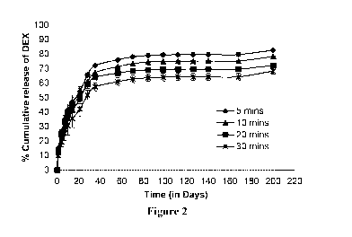
Figure 2: In vitro release of dexamethasone (DEX) from ISPc1 containing 30%w/w
PLGA 75/25,
0.5%w/w DEX, and 0.1%w/w of Igracure 2959 and 69.4% w/w of PEGDA 700. The
gels were in situ
crosslinked, using UV light at 365 nm, for 5, 10, 20 and 30 min, respectively.
Mean SD, n=3
Figure 3: In vitro release of bovine serum albumin (BSA) from ISPc1 containing
30%w/w PLGA 75/25,
0.5%w/w BSA, and 0.1%w/w of Igracure 2959 and 69.4% w/w of PEGDA 700. The
gels were in situ
crosslinked, using UV light at 365 nm 5 min. Mean SD, n=3
Figure 4: In vitro release of bevacizumab (Avastin) from ISPc1 containing 30%
w/w PLGA 75/25,
1.5% w/w bevacizumab, 0.1% w/w Irgacure and 68.4% w/w PEGDA 700 and 30% w/w
PLGA 75/25,
1.5% w/w bevacizumab, 0.05% w/w Irgacure and 68.45% w/w PEGDA 700. In situ
crosslinked using
UV light at 365 nm for either 30 sec or 2.5 min. Mean SD, n=3
Figure 5: In vitro release of ovalbumin (OVA) from ISPc1 containing 30%w/w
PLGA 75/25, 2.5% w/w
OVA and 0.1% w/w Irgacure 2959 and 67.4%w/w of PEGDA 700. In situ crosslinked
for either 30sec,
1 min and 2.5 min. Mean SD, n=3.
6
CA 03004381 2018-05-04
WO 2017/081154 PCT/EP2016/077269
Figure 6: Work of Syringeability (WoS) of 30%w/w PLGA50/50 and 30%w/w
PLGA75/25 and 0.1%
w/w Irgacure 2959 in formulations containing different molecular weights of
69.9% w/w of PEGDAs
(258, 575 and 700). The WoS was calculated from the resultant of force-
distance plots Mean S.D,
n=5.
Figure 7: Percentage cell viability of human retinal pigment epithelial cell
line (ARPE-19) following
exposure of the release media, at different intervals, of the ISPc1 that were
composed of 30%w/w
PLGA 75/25, 0.1%w/w of Igracure 2959 and 69.9% w/w PEGDA 700 and UV
crosslinked for 5 min.
Mean SD, n=3.
Figure 8: (A) Scanning electron microscope image of PPc1 implant (scale bar is
50 pm). (B) Digital
image of a PPc1 adjacent to a ruler.
Figure 9: In vitro release of FITC-Dextran 150kDa and BSA from PPc1 containing
30%w/w of PLGA
75/25, 0.5%w/w FITC-Dextran 150kDa or BSA and 0.1%w/w of Igracure 2959 and
69.4% w/w
PEGDA 700. These PPc1 were cross-linked using UV curing system at wavelength
365 nm for 5 runs
at a lamp intensity of 100%.
Figure 10: In vitro release of TA from PPc1 containing 30%w/w of PLGA 75/25,
2.5%w/w TA, and
0.1%w/w of Igracure 2959 and 67.4%w/w PEGDA 700. These PPc1 were cross-linked
using UV
curing system at wavelength 365nm for 5 runs, at a lamp intensity (LI) of 50%
and 100%.
Figure 11: In vitro release of TA from PPc1 containing 2.5 or 5 %w/w of PLGA
75/25, 2.5%w/w TA,
0.1%w/w of Igracure 2959 in 94.9% w/w or 92.4% w/w of PEGDA 700,
respectively. These PPc1
were cross-linked using UV curing system at wavelength 365nm for 5 runs, at a
lamp intensity (LI) of
25%.
Figure 12: In vitro release of TA from PPc1 containing 2.5 %w/w of PLGA 75/25,
2.5%w/w TA,
0.1%w/w of Igracure 2959 and without pore forming agent in 94.9%w/w PEGDA 700
or with 2%w/w
of pore forming agent (i.e. MgCO3) in 92.9%w/w PEGDA 700. These PPc1 were
cross-linked using UV
curing system at wavelength 365 nm for 10 runs, at a lamp intensity (LI) of
25%.
Figure 13: In vitro release of TA from PPc1 containing 2.5 %w/w of PLGA 75/25,
2.5%w/w TA,
0.1%w/w of Igracure 2959 in 94.9%w/w of PEGDA 700 or PEGDA 6000. These PPc1
were cross-
linked using UV curing system at wavelength 365 nm for 10 runs, at a lamp
intensity (LI) of 25%.
Figure 14: In vivo implant formation of ISPc1 in rat eye following
intravitreal injection (A) Digital image
of rat following administration of fluorescein sodium loaded ISPc1 implant (2
pL), the inset shows
close-up image of the implant in the eye, (B) shows fundus image of control
eye without any implant,
(C-E) fundus images showing the time course of fluorescein sodium release from
ISPc1 implant, taken
on (C) day 1, (D) day 6 and (E) day 18. Implants indicate slow and continuous
release of fluorescein
sodium and degradation over the time.
7
CA 03004381 2018-05-04
WO 2017/081154 PCT/EP2016/077269
DETAILED DESCRIPTION
Photopolymerizable Compositions
The photopolymerizable polymers of the present invention can be used in any of
the compositions
and implants of the invention in combination with any of the other
biodegradable polymers,
therapeutic agents, photoinitiators, solvents, co-solvents, pore forming
agents, and co-initiators
described herein or known in the common general knowledge.
In one embodiment, the photopolymerizable compositions of the invention can be
biodegradable. As
used herein, "biodegradable" is the chemical degradation by biological means.
In some
embodiments, the biodegradation is about 100%, about 98%, about 90%, about
85%, about 80%,
about 60%, about 50%, or about 45% degradation of one or more of the
compositions, monomers,
oligomers, fragments, polymers, photoinitiators, solvents, co-solvents, or co-
initiators. In some
embodiments the biodegradation takes place over about 1 minute, about 10
minutes, about 20
minutes, about 2 hours, about 6 hours, about 12 hours, about 24 hours, about 2
days, about 5 days,
about 1 week, about 1 month, about 2 months, about 5 months, about 6 months,
about 8 months or
about 12 months. In some embodiments the biodegradation takes place between
about 1 month and
about 12 months, between about 6 months and about 12 months, or between about
8 months and
about 12 months.
As used herein, the term "photopolymerizable composition" is a composition
which can form a
crosslinked polymer network upon exposure to light, in particular UV light. As
used herein,
photopolymerizable compositions include photopolymerizable monomers and
oligomers (such as,
dimers, trimers, and tetramers). The terms "oligomers" and "fragments" can be
used interchangeably
to mean between two and twenty monomers, optionally between two and ten
monomers, further
optionally between two and five monomers or between two and four monomers. A
"photopolymerizable monomer" is a single unit of a photopolymerizable polymer
that can bind
chemically to other monomers to form a polymer.
Photopolymerizable compositions of the present invention can be crosslinked
with UV radiation to
form photopolymerized polymers of the present invention.
In one embodiment the photopolymerizable compositions of the present invention
are fragments or
monomers consisting of polyalkylene glycol diacrylate, polyalkylene glycol
dimethacrylate and
mixtures, copolymers, and block copolymers thereof.
In one embodiment, the photopolymerizable compositions are polyalkylene glycol
diacrylate
fragments or monomers incorporating diacrylate end units selected from the
group comprising
polyether fragments or monomers, polyester fragments or monomers,
polycarbonate fragments or
monomers or mixtures, copolymers, or block copolymers thereof. In one
embodiment, the
photopolymerizable composition is a monomers incorporating diacrylate end
units, such as 4-arm or
8-arm PEG acrylate.
8
CA 03004381 2018-05-04
WO 2017/081154 PCT/EP2016/077269
In another embodiment, the photopolymerizable composition is polyethylene
glycol diacrylate,
diethylene glycol diacrylate, polyethylene glycol dimethacrylate, diethylene
glycol dimethacrylate,
polypropylene glycol diacrylate, dipropylene glycol diacrylate, dipropylene
glycol dimethacrylate, and
polypropylene glycol dimethacrylate or mixtures, copolymers, or block
copolymers thereof.
In another embodiment, the photopolymerizable composition is polyethylene
glycol diacrylate or
polyethylene glycol dimethacrylate.
In yet another embodiment, the photopolymerizable composition is polyethylene
glycol diacrylate.
The molecular weight of the photopolymerizable compositions of the present
invention is typically
between about 100 and about 300,000 Da, between about 200 to about 100,000 Da,
between about
200 to 50,000 Da, between about 200 to about 20,000 Da, between about 200 to
about 10,000 Da,
between about 200 and about 8,000 Da, between about 200 and about 5,000 Da, or
between about
200 and about 1,000 Da.
It has been found, for the compositions and implants of the present invention,
that an increase in
molecular weight of the photopolymerizable compositions results in an increase
in drug release rate.
Without wishing to be bound by theory, it is believed that photopolymerizable
compositions with lower
molecular weights have higher crosslinking density and therefore slower drug
release rates.
The photopolymerizable compositions of the present invention typically have
viscocities between
about 0.1 to about 7 dlig, between about 0.2 to about 5 dlig, or between about
0.5 to 2 dL/g.
In another embodiment, the photopolymerizable compositions of the present
invention are
polymerized by irradiating the composition with light at a wavelength of
between about 230 to about
550 nm, between about 300 to about 525 nm, or between about 350 to about 490
nm for between
about 1 second and about 60 minutes, between about 30 seconds and about 30
minutes, between
about 2.5 minutes and about 20 minutes, between about 5 minutes and about 10
minutes. In one
embodiment, the crosslinking is for about 30 seconds, about 1, about 2.5,
about 5, about 10, about 20
or about 30 minutes.
Biodegradable Polymers
The biodegradable polymers of the present invention can be used in any of the
compositions and
implants of the invention in combination with any of the other
photopolymerizable compositions,
therapeutic agents, photoinitiators, solvents, co-solvents, pore forming
agents, and co-initiators
described herein or known in the common general knowledge.
The biodegradable polymers of the present invention are biodegradable but not
photopolymerizable.
In one embodiment of the present invention, the biodegradable polymers are
aliphatic polyester-
based polyurethanes, polylactides, polycaprolactones, polyorthoesters or
mixtures, copolymers, or
9
CA 03004381 2018-05-04
WO 2017/081154 PCT/EP2016/077269
block copolymers thereof. In another embodiment of the present invention the
biodegradable
polymer, is chitosan, poly(propylene fumarate), lactide/glycolide copolymer
(PLGA), polylactic acid
(PLA), polyglycolic acid (PGA), polycaprolactone (PCL), lactide/caprolactone
copolymer (PLC), poly
(L-Iactide) (PLLA), natural biodegradable polymers, such as, collagen and the
like, or mixtures,
copolymers, or block copolymers thereof. In another embodiment, the
biodegradable polymer is
selected from the group lactide/glycolide co-polymer (PLGA), polylactic acid
(PLA), polyglycolic acid
(PGA), polycaprolactone (PCL), lactide/caprolactone copolymer (PLC), poly (L-
Iactide) (PLLA) or
mixtures, copolymers, or block copolymers thereof.
In a particular embodiment, the biodegradable polymer is PLGA. In one
embodiment, the molar ratio
of lactic acid to glycolic acid in the PLGA is 90% lactic acid to 10% glycolic
acid, 85% lactic acid to
15% glycolic acid, 75% lactic acid to 25% glycolic acid, 65% lactic acid to
35% glycolic acid, 50%
lactic acid to 50% glycolic acid, 35% lactic acid to 65% glycolic acid, 25%
lactic acid to 75% glycolic
acid, 15% lactic acid to 85% glycolic acid, and 10% lactic acid to 90%
glycolic acid.
In another particular embodiment, the biodegradable polymer is PCL, PLC, PLLA,
or mixtures,
copolymers, or block copolymers thereof.
Compositions and Implants
In one embodiment, the compositions of the invention comprise combinations of
photopolymerizable
compositions, and biodegradable polymers, as described above, in combination
with a photoinitiator
and a therapeutic agent, which can be delivered to the eye to achieve
controlled drug delivery to treat
a range of eye diseases. The compositions of the invention include:
i) compositions which can be injected into the eye followed by application
of short-term UV light to
induce in situ photocrosslinking, resulting in implant formation, termed as in
situ photocrosslinked
implants (ISPcI); and
ii) compositions which can be photocroslinked prior to application in the
eye to form an implant of
desired shape and size (e.g. film, rod or nano/microparticles) that can be
administered
intraocularly to provide desired period of drug delivery, termed as performed
photocrosslinked
implants (PPc1).
Alternatively, the compositions of the invention can be used to coat ocular
devices, including both in
situ and pre-formed ocular devices.
The implants of the present invention can be any desired shape and size, such
as but not limited to,
for example rectangular, square, cylindrical, circular, oval, films, dumbbell,
rods, beads, etc., as, for
example, macro, micro or nanoparticles.
In one embodiment of the present invention, the ocular implant is a implant,
which is less than about
mm, less than about 5 mm. In one embodiment, the implant is a rectangular
implant of dimensions
10 x 5x 0.5 mm.
CA 03004381 2018-05-04
WO 2017/081154 PCT/EP2016/077269
In one embodiment of the present invention, the ocular implant is a
nanoparticle or a microparticle.
In one embodiment, the nanoparticle ocular implant is less than about 1,000
nm, less than about 900
nm, less than about 750 nm, less than about 500 nm, or less than about 100 nm.
In one embodiment, the microparticle ocular implant is less than about 1,000
pm, less than about 900
pm, less than about 750 pm, less than about 500 pm, or less than about 25 pm.
In situ photocrosslinked implants (ISPc1)
In situ photocrosslinked implants (ISPc1), of the present invention are those
that form and take up
their final localised structure once they are inserted into the body. The
ability of these implants to fill
irregular defects is an advantage of ISPcIs. The ISPcIs of the present
invention also have additional
advantages, which include, site-specific action due to relatively easy and
less invasive application,
localized delivery to specific tissues, prolonged delivery times, reduction in
side effects linked with
systemic delivery and also superior patient comfort and compliance. Additional
advantages of the
ISPcIs of the present invention include, not requiring extreme pH conditions
or elevated temperatures
during processing, which could cause issue when working with temperature or pH
labile drugs (e.g.
proteins, peptides or genetic material). Furthermore, rapid crosslinking at
physiological temperatures
can swiftly entrap drug molecules and can result in an ISPc1 that possesses
the exact required
dimensions for controlled drug release. Photocrosslinking is also beneficial
in comparison to
spontaneous crosslinking (e.g. enzymatic, self-assembled, Michael addition) as
the initiation of the
process is only triggered when exposed to a light source, therefore premature
gelation is not an issue
resulting in excellent control of material formation. Furthermore, short-term
application of UV light will
not cause any safety issues as it is considered safe for ocular applications,
as UV light is clinically
used for corneal crosslinking. Importantly, administration by this method
allows the injection of a
relatively low viscosity material into the body, which then solidifies to form
a semi-solid depot that
controls the drug delivery to provide short or long-term therapeutic action.
In one embodiment, the ISPcIs of the present invention are formed by injection
of a composition of the
invention into a subject in need thereof and subsequent crosslinking using
external source of UV light
that results in formation of a solid implant which controls drug release for
desired period of time.
For ISPcIs of the invention the molecular weight of the photopolymerizable
composition is typically
between about 100 and about 6,000 Da, between about 200 and about 3,000 Da, or
between about
200 and 1,000 Da.
Preformed photocrosslinked implants (PPc1)
11
CA 03004381 2018-05-04
WO 2017/081154 PCT/EP2016/077269
In one embodiment, the present invention is a preformed photocrosslinked
implant (PPc1). These
PPcIs can be inserted in the eye (e.g. in the fornix, subconjunctively,
intracameral,
intrastromal/intracorneal, transsclerally/periocular, intrasclerally or
intravitreally) to treat the front of the
eye or back of the eye diseases. These implants can be fabricated in a variety
of shapes (e.g. rods,
films, cylindrical or circular) and sizes, including in the form of micro or
nanoparticles.
The PPcIs of the present invention have the advantage of high crosslink
density and/or a tight
polymer network structure which can be configured to control drug release
and/or eliminate any burst
release.
The PPcIs of the present invention can be fabricated to have a single and/or
multiple layers, which will
enable loading of more than one drug or the same drug with different release
profiles or rates.
Furthermore, the rate of degradation of the implants can be slower for PPcIs
when compared to
ISPcIs of the invention and can be altered to treat specific diseases or
disorders.
For the PPcIs of the invention the molecular weight of the photopolymerizable
polymers is typically
between about 100 and about 300,000 Da, between about 200 to 100,000 Da,
between about 200 to
50,000 Da, between about 200 to 20,000 Da, or between about 200 to about
10,000 Da.
In one embodiment, the biodegradable polymer is essentially contained within a
matrix of the
photopolymerizable composition. In one embodiment, the biodegradable polymer
is essentially
contained within a matrix of the photopolymerizable composition that forms a
gel upon mixing. In one
embodiment the photopolymerizable polymer is crosslinked in presence of a
photoinitiator and the
biodegradable polymer and therapeutic agent(s). In one embodiment, the
biodegradable polymer is
essentially trapped within the crosslinked photopolymerizable polymer matrix,
and the therapeutic
agent(s) are either dispersed or dissolved between the two phases (i.e.,
photopolymerizable and/or
biodegradable polymer). In one embodiment, the biodegradable polymer is
hydrophobic in nature and
the photopolymerizable polymer is hydrophilic in nature. In one embodiment,
the degree of
crosslinking of the composite implant will govern the rate and extent of
release of the therapeutic
agent(s).
In one embodiment, the present invention is an ocular composition comprising:
i) 99 to 60 % (w/w) of a photopolymerizable composition selected from the
group of
fragments or monomers consisting of polyalkylene glycol diacrylate and
polyalkylene
glycol dimethacrylate, wherein the photopolymerizable composition has a
molecular
weight in the range of 200 to 20,000 Dalton;
ii) a biodegradable polymer selected from the group consisting of aliphatic
polyester-based
polyurethanes, polyglycolide, polylactides, polycaprolactones, polyorthoesters
and
mixtures, copolymers, and block copolymers thereof;
iii) a photoinitiator; and
12
CA 03004381 2018-05-04
WO 2017/081154 PCT/EP2016/077269
iv) a therapeutic agent.
In one embodiment, the photopolymerizable composition is 96.9 % (w/w) and the
biodegradable
polymer is 2.5 % (w/w), the photopolymerizable composition is 94.1 % (w/w) and
the biodegradable
polymer is 5 % (w/w), the photopolymerizable composition is 69.4 % (w/w) and
the biodegradable
polymer is 30 % (w/w).
In another embodiment, the present invention is an ocular composition wherein
i) and ii) are:
i) 79.5 to 59.5 % (w/w) polyethylene glycol diacrylate or polyethylenene
glycol dimethacrylate;
and
ii) 20 to 40 % (w/w) PLGA, wherein the molar ratio of lactic acid to glycolic
acid in the PLGA is
90% lactic acid to 10% glycolic acid, 85% lactic acid to 15% glycolic acid,
75% lactic acid to
25% glycolic acid, or 50% lactic acid to 50% glycolic acid.
In another embodiment, the present invention is an ocular composition wherein
i) and ii) are:
i) 79.5 % (w/w) polyethylene glycol diacrylate or polyethylene glycol
dimethacrylate; and
ii) 30 % (w/w) PLGA wherein the molar ratio of lactic acid to glycolic acid in
the PLGA is 90%
lactic acid to 10% glycolic acid, 85% lactic acid to 15% glycolic acid, 75%
lactic acid to 25%
glycolic acid, or 50% lactic acid to 50% glycolic acid.
In another embodiment, the present invention is an ocular composition wherein
i) and ii) are:
i) 95.5 to 84.5 % (w/w) polyalkylene glycol diacrylate or polyalkylene glycol
dimethacrylate; and
ii) 4 to 15 % (w/w) PCL.
In another embodiment, the present invention is an ocular composition wherein
i) and ii) are:
i) 69.5 to 94.5 % (w/w) polyalkylene glycol diacrylate or polyalkylene glycol
dimethacrylate; and
ii) 20 to 5 % (w/w) PLLA.
In yet another embodiment, the present invention is an ocular composition
wherein i) and ii) are:
i) 95.5 to 84.5 % (w/w) polyalkylene glycol diacrylate or polyalkylene glycol
dimethacrylate; and
ii) 4 to 15 % (w/w) PLC in which lactic acid to caprolactone is in the range
of 90 % lactic acid to
% caprolactone, 80 % lactic acid to 20 % caprolactone, 70 % lactic acid to 30
% caprolactone, 60
% lactic acid to 40 % caprolactone, or 50 % lactic acid to 50 % caprolactone.
In one embodiment, the present invention is an ocular composition wherein i)
is 95.5 to 84.5 % (w/w)
polyalkylene glycol diacrylate or polyalkylene glycol dimethacrylate; and
wherein ii) is 4 to 15 % (w/w)
PCL.
13
CA 03004381 2018-05-04
WO 2017/081154 PCT/EP2016/077269
In one embodiment, the % of the biodegradable polymer is 30% w/w, 5% w/w. 2.5
% w/w, between 4-
10% w/w, or between 5-18% w/w.
In one embodiment, i) and ii) of the compositions of the present invention are
PEGDA and PLGA.
PLGA
PLGA is prepared by polymerisation of lactic acid and glycolic acid monomers.
The glass transition
temperatures (Tg) of PLGA copolymers are above physiological temperatures of
37 C, which imparts
a moderately rigid chain configuration and therefore the mechanical strength
at ambient
temperatures. The use of PLGA in different lactide (LA) to glycolide (GA)
ratio and molecular weight
allows different drug release profiles. An increase in GA content will result
in an increased water
uptake of PLGA and therefore more rapid degradation. The degradation of PLGA
with LA/GA 50/50 is
typically between about 1 and about 3 months.
PEGDA
PEGDA is a synthetic polymer, available in different M. PEGDA is extremely
amenable to
mechanical, structural and chemical alteration and so resulting in hydrogels
with variable properties in
terms of drug delivery and other biomedical applications. PEGDA is formed
through the
functionalization of the end of each PEG molecule with an acrylate group.
PEGDA is also non-toxic,
eliciting only a minimal immunogenic response. PEGDA has double-bond
containing acrylate end
groups which show rapid polymerisation when exposed to light in the presence
of an appropriate
initiator to produce a hydrogel network.
In one embodiment, the present invention is a PLGA/PEGDA PPcl.
In one embodiment, the present invention is a PLGA/PEGDA ISPcl.
Copolymers
All of the copolymers and block copolymers described herein can be used in any
of the compositions
and implants of the invention in combination with any of the other
photopolymerizable compositions,
biodegradable polymers, therapeutic agents, photoinitiators, solvents, co-
solvents, pore forming
agents, and co-initiators described herein.
As used herein "copolymer" is a mixture of two or more different types of
monomer units. As used
herein "block copolymer" is a mixture of two or more homopolymer subunits.
In one embodiment, block or copolymers with PGA, PCL, PLA, PLGA that would
include any other
polymeric component of the polymer e.g. PEG and PEO, for example, PLGA-PEO,
PCL-PEO and
PEG-PLGA, PEG-PCL block copolymers, which include, for example, PEO-PLGA-PEO,
PLGA-PEG,
PLGA-PEO, and PLGA-PEO-PLGA.
14
CA 03004381 2018-05-04
WO 2017/081154 PCT/EP2016/077269
Solvents
All of the solvents described herein can be used in the preparation of any of
the compositions and
implants of the invention in combination with any of the other
photopolymerizable compositions,
biodegradable polymers, therapeutic agents, photoinitiators, pore forming
agents, and co-initiators
described herein.
In one embodiment, the co-solvents used in the present invention can be
selected from
dichloromethane, tetrahydrofuran, ethyl acetate, acetone, dimethylformamide,
acetonitrile, acetic acid,
methanol, ethanol, isopropanol, glycofurol or butanol.
In one embodiment, the solvents used in the present invention are dimethyl
sulfoxide, decylmethyl
sulfoxide, 2-pyrrolidone, N-vinyl-2-pyrrolidone, 1-methyl-2-pyrrolidone, N-
Methyl-2-pyrrolidone, N-
ethyl-pyrrolidone, glycerol formal, glycerol, polyethylene glycol, propylene
glycol, benzyl alcohol,
benzyl benzoate, ethyl benzoate, triacetin, triethyl citrate,
dimethylformamide, dimethylacetamide or
tetrahydrofuran.
In another embodiment, the solvent is dimethyl sulfoxide, decylmethyl
sulfoxide, 2-pyrrolidone, 1-
methyl-2-pyrrolidone, N-Methyl-2-pyrrolidone, or glycerol formal.
In one embodiment, a solvent is used when the biodegradable polymer is PCL,
PLC and/or PLLA. In
one embodiment the solvent is N-Methyl-2-pyrrolidone and N-Vinyl-2-pyrrolidine
when the
biodegradable polymer is PCL, PLC and/or PLLA. In another embodiment, a
solvent is used when the
photomolymerizable composition is PEGDA.
Pore forming agents
In one embodiment, a suitable pore forming agent may be selected in view of
the specific therapeutic
agent and composition of the implant, and the desired elution profile or
release rate. The pore forming
agent may be a naturally occurring agent or polymer or a synthetic agent or
polymer.
In another embodiment, implant porosity can be adjusted by preparing implants
in the presence of
dispersed water-soluble porosigens, which can be removed later by washing with
water to leave an
interconnected meshwork (i.e., porous hydrogels). The pore size of hydrogels
prepared by the
porosigen technique depends on the size of the porosigens.
All of the pore forming agents described herein can be used in any of the
implants and compositions
of the invention in combination with any of the other photopolymerizable
compositions, biodegradable
polymers, therapeutic agents, photoinitiators, solvents, co-solvents, and co-
initiators described herein.
In one embodiment, the compositions of the invention further comprise a pore-
forming agent.
In one embodiment, the pore-forming agent is polyethylene glycol, lactose,
maltose, glucose,
mannitol, gelatin, sodium chloride, magnesium carbonate, magnesium hydroxide,
potassium chloride,
CA 03004381 2018-05-04
WO 2017/081154 PCT/EP2016/077269
sodium bicarbonate, ammonium bicarbonate, potassium bicarbonate, chitosan,
polyvinylpyrrolidone,
polyvinyl alcohol, agarose or sucrose.
Photoinitiators
In one embodiment, the compositions of the invention further comprise a
photoinitiator.
The photoinitiators described herein can be used in any of the compositions
and implants of the
present invention in combination with any of the other photopolymerizable
compositions,
biodegradable polymers, therapeutic agents, photoinitiators, solvents, co-
solvents, and co-initiators
described herein.
In certain embodiments, the photoinitiator is designed to work using light
from about 200 to about 550
nm. In some embodiments, a photoinitiator is designed to work using UV light
from about 200 to about
400 nm.
In certain embodiments, the light source may allow variation of the wavelength
of light and/or the
intensity of the light. Light sources useful in the present invention include,
but are not limited to,
lamps, fiber optics devices, etc.
In one embodiment, the photoinitiator is a ketone (e.g., RCOR'). In one
embodiment, the compound is
an azo compound (e.g., compounds with a ¨N=N¨ group). In one embodiment, the
photoinitiator is
an acylphosphineoxide. In one embodiment, the photoinitiator is a sulfur-
containing compound. In one
embodiment, the initiator is a quinone. In certain embodiments, a combination
of photoinitiators is
used.
In one embodiment, the photoinitiator is a hydroxyketone photoinitiator, an
amino ketone
photoinitiator, a hydroxy ketone/benzophenone photoinitiator, a benzyldimethyl
ketal photoinitiator, a
phenylglyoxylate photoinitiator, an acyl phosphine oxide photoinitiator, an
acyl phosphine oxide/alpha
hydroxy ketone photoinitiator, a benzophenone photoinitiator, a ribityl
isoalloxazine photoinitiator, or a
phenylglyoxylate photoinitiator or any combination thereof.
In one embodiment the photoinitiator is 144-(2-hydroxyethoxy)-pheny1]-2-
hydroxy-2-methy1-1-
propanone, 2,2-dimethoxy-2-phenylacetophenone (DMPA) or riboflavin.
In one embodiment, the compositions of the present invention further comprise
a co-initiator. In one
embodiment, the co-initiator is eosin Y, triethanolamine, camphorquinone, 1-
viny1-2 pyrrolidinone
(NVP), eosin, dimethylaminobenzoate (DMAB), Irgacure 907 (Ciba Geigy),
Irgacure 651 (Ciba
Geigy), Darocur 2959 (Ciba Geigy), or ethyl-4-N,N-dimethylaminobenzoate
(4EDMAB).
In one embodiment, the photoinitiator is riboflavin and the co-initiator is L-
arginine.
Process
16
CA 03004381 2018-05-04
WO 2017/081154 PCT/EP2016/077269
The compositions and implants of the present invention can be made by any
methods know in the art
as well as the methods described herein. The methods described herein are
applicable to all
compositions and implants of the invention.
In one embodiment, polymer M,õ type and copolymer ratio, drug type and
loading, implant size, time
and extent of UV crosslinking and/or amount and type/concentration of
photoinitiator and/or pore
forming agent (porogen) and/or solvent/co-solvent can be altered to control
the rate and extent of
drug release. The alteration of these factors provides compositions of the
invention that can be easily
tailored to produce desired period of drug release to address specific
clinical/patient needs in treating
various ocular diseases.
In one embodiment, the present invention is a method of making the ocular
composition of the
invention, comprising the steps of:
i) mixing a therapeutic agent with a photopolymerizable composition, a
biodegradable polymer,
and a photoinitiator, in any order of addition;
ii) administering the mixture i) to an ocular area of a subject; and
iii) irradiating the mixture i) with light at a wavelength of between about
230 to about 550 nm,
between about 300 to about 525 nm, or between about 350 to about 490 nm for
between about 1
second and about 60 minutes to form the ocular composition.
In another embodiment the present invention is a method of making the ocular
composition of the
present invention, comprising the steps of:
i) mixing a therapeutic agent with a photopolymerizable composition;
ii) adding a biodegradable polymer to the mixture i)
iii) adding a photoinitiator to the mixture ii);
iv) administering a mixture iii) to an ocular area of a subject; and
v) irradiating the mixture iii) with light at a wavelength of between about
230 to about 550 nm,
between about 300 to about 525 nm, or between about 350 to about 490 nm for
between about 1
second and about 60 minutes to form the ocular composition.
In the above embodiments, the irradiating step is with light at a wavelength
of about 365 nm or about
475 nm for about 1 second, about 2 minutes, about 5 minutes, about 10 minutes,
about 20 minutes,
or about 30 minutes.
In another embodiment, the present invention is a method of making the ocular
compositions of the
present invention, the method comprising the steps of:
17
CA 03004381 2018-05-04
WO 2017/081154 PCT/EP2016/077269
i) mixing a therapeutic agent with a photopolymerizable composition, a
biodegradable polymer
and a photoinitiator, in any order of addition;
ii) irradiating the mixture i) with light at a wavelength of between about 230
to about 550 nm,
between about 300 to about 525 nm, or between about 350 to about 490 nm for
between about 1
second and about 60 minutes to form the ocular composition; and
iii) administering the composition ii) to an ocular area of a subject.
In another embodiment, the present invention is a method of making the ocular
composition of the
present invention, the method comprising the steps of:
i) mixing a therapeutic agent with a photopolymerizable composition;
ii) adding a biodegradable polymer to the mixture i)
iii) adding a photoinitiator to the mixture ii);
iv) irradiating the mixture iii) with light at a wavelength of between about
230 to about 550 nm,
between about 300 to about 525 nm, or between about 350 to about 490 nm for
between about 1
second and about 60 minutes to form the ocular composition; and
v) administering the composition iv) to an ocular area of a subject.
In the above embodiments, the irradiating step is with light at a wavelength
of about 365 nm or about
475 nm for about 1 second, about 2 minutes, about 5 minutes, about 10 minutes,
about 20 minutes,
or about 30 minutes.
In another embodiment, the present invention is a method of making the
nanoparticle or microparticle
ocular implant, comprising the steps of:
i) mixing the therapeutic agent, the photopolymerizable composition, the
biodegradable polymer
and the photoinitiator, in any order of addition, to form mixture i);
ii) adding the mixture i) to an aqueous medium to form mixture ii);
iii) sonicating the mixture ii); and
irradiating the mixture ii) with light at a wavelength of between 230 to 550
nm, between 300 to 525
nm, or between 350 to 490 nm for between 1 second and 60 minutes to form the
nanoparticles or
microparticles
In another embodiment, the present invention is a method of making the
nanoparticle or microparticle
ocular implants of the present invention, comprising the steps of:
i) mixing a therapeutic agent with a photopolymerizable composition;
18
CA 03004381 2018-05-04
WO 2017/081154 PCT/EP2016/077269
ii) adding a biodegradable polymer to the mixture i)
iii) adding a photoinitiator to the mixture ii);
iv) adding the mixture iii) to a aqueous medium;
v) sonicating the mixture iv); and
vi) irradiating the mixture v) with light at a wavelength of between about 230
to about 550 nm,
between about 300 to about 525 nm, or between about 350 to about 490 nm for
between about 1
second and about 60 minutes to form the nanoparticles or microparticles.
In one embodiment the aqueous medium is a combination of water and phosphate
buffered saline
(PBS).
In one embodiment, step iv) is carried out in a step-by-step manner to
emulsify the mixture. As used
herein step-by-step means that the mixture is not added all at once, but
rather it is added in stages
with breaks of between the additions.
In another embodiment, i) the therapeutic agent is mixed with a portion of
photopolymerizable
composition and ii) another portion of photopolymerizable composition is mixed
with biodegradable
polymer, then iii) the two portions are mixed together. The mixture iii) is
added to a aqueous medium,
and then sonicated. Finally the mixture is irradiated with light at a
wavelength of between about 230
to about 550 nm, between about 300 to about 525 nm, or between about 350 to
about 490 nm for
between about 1 second and about 60 minutes to form the nanoparticles or
microparticles.
In yet another embodiment, irradiation can be applied during sonication i.e.
sonicating the mixture
under UV light, in other words, the aqueous medium will be under UV light (at
defined wavelength)
and sonication, followed by step-by-step addition of the mixture.
In another embodiment, the sonication time, gel composition, phase ratio (of
the gel vs aqueous
medium), and rate of adding the gel mixture into aqueous medium are adjusted
to form a nanoparticle
or microparticle.
In the compositions of the present invention, varying the UV crosslinking time
can control the rate of
and duration of drug release. In some embodiments, an increase in UV
crosslinking times causes a
decrease in drug release. Additionally, varying the concentration of the
photoinitiator can control the
rate and duration of drug release. Furthermore, varying both the UV
crosslinking time and the
concentration of photoinitiator can control the rate and duration of drug
release. In one embodiment,
decreasing the concentration of the biodegradable polymer (such as PLGA)
increases the drug
release rate. In one embodiment, adding a pore-forming agent (e.g. MgCO3),
increases the drug
release rate. In one embodiment, higher UV crosslinking time and higher
concentration of
photoinitiator can sustain the drug release for longer periods of time. In one
embodiment, the drug
19
CA 03004381 2018-05-04
WO 2017/081154 PCT/EP2016/077269
release can be sustained for a period of greater than about 1 day, about 2
days, about 1 week, about
1 month, about 2 months, about 3 months, or about 6 months.
In one embodiment, the duration of drug release in the ISPcIs of the present
invention can be
considerably extended, for example, providing controlled drug release for a
period of greater than 200
days (>6 months). This duration can be varied by varying the degree of
crosslinking.
In some embodiments, the slow degradation rate of the ISPcIs of the present
invention provide
protection of the sensitive molecules such as peptides and proteins. It has
been shown below, that
the ISPcIs of the present invention are stable and avoid protein degradation
and maintain protein
activity.
In some embodiments, burst release can be eliminated or controlled by varying
the UV crosslinking
time.
In one embodiment, the present invention is a PPclwith no burst release. In
one embodiment, the
present invention is a PPclwith high crosslinking density that significantly
slows drug diffusion.
Methods of use
Any of the implants and compositions described herein are suitable for use in
any of the methods of
the invention described herein.
In one embodiment, the present invention is a method of treating a disease or
disorder of the eye in a
subject in need thereof, comprising administering a composition or implant of
the present invention to
an ocular area of the subject.
In one embodiment, the present invention is a composition or implant of the
present invention for use
in treating a disease or disorder of the eye in a subject in need thereof.
As used herein, an "ocular area" is an area inside, outside or adjacent to the
eye of the subject. In one
embodiment, the ocular area is the sclera (intrascleral), outside the sclera
(transscleral), the vitreous
body, the choroid, the cornea, the stroma, intracameral, the aqueous humor,
the lens, the fornix, or
the optic nerve.
In one embodiment, the compositions and implants can be administered by
injection, including,
intravitreal, subconjunctival, peribulbar, subtenon or retrobulbar injections
and cornea.
In some embodiments, the implants are administered via a surgical procedure.
In some embodiment,
the implants are secured in place, after surgical implantation, via an
adhesive or sutures.
The term "subject" refers to an animal (e.g., a bird such as a chicken, quail
or turkey, or a mammal),
specifically a "mammal" including a non-primate (e.g., a cow, pig, horse,
sheep, rabbit, guinea pig, rat,
cat, dog, and mouse) and a primate (e.g., a monkey, chimpanzee and a human),
and more
CA 03004381 2018-05-04
WO 2017/081154 PCT/EP2016/077269
specifically a human. In one embodiment, the subject is a non-human animal
such as a farm animal
(e.g., a horse, cow, pig or sheep), or a pet (e.g., a dog, cat, guinea pig or
rabbit). In another
embodiment, the subject is a "human".
As used herein, the terms "treat", "treatment" and "treating" refer to
therapeutic treatments includes
the reduction or amelioration of the progression, severity and/or duration of
a disease, disorder or
condition, or the amelioration of one or more symptoms (specifically, one or
more discernible
symptoms) of a disease, disorder or condition, resulting from the
administration of the compositions or
implant of the invention. In specific embodiments, the therapeutic treatment
includes the amelioration
of at least one measurable physical parameter of a disease, disorder or
condition. In other
embodiments the therapeutic treatment includes the inhibition of the
progression of a condition, either
physically by, e.g., stabilization of a discernible symptom, physiologically
by, e.g., stabilization of a
physical parameter, or both. In other embodiments the therapeutic treatment
includes the reduction or
stabilization of a disease, disorder or condition.
Exemplary therapeutic agents include, but are not limited to, polypeptides,
nucleic acids, such as
DNA, RNA, and siRNA, growth factors, steroid agents, antibody therapies,
antimicrobial agents,
antibiotics, antiretroviral drugs, anti-inflammatory compounds, antitumor
agents, anti-angiogeneic
agents, and chemotherapeutic agents.
In one embodiment, the therapeutic agent of the present invention includes,
but is not limited to,
ketorolac, naphazoline, lidocaine, bevacizumab, aflibercept, pegaptanib,
brimonidine, dorzolamide,
azithromycin, rapamycin, bepotastine besilate, diclofenac, besifloxacin,
cysteamine hydrochloride,
fluocinolone acetonide, difluprednate, aflibercept, tasimelteon, ocriplasmin,
enoxaparin sodium,
ranibizumab, latanoprost, timolol, bimatoprost, pegaptanib, ofloxacin,
cephazolin, phenylephrine,
dexamethasone, triamcinolone acetonide, levofloxacin, cyclophosphamide,
melphalan cyclosporine,
methotrexate, azathioprine ketorolac, travoprost, verteporfin, tafluprost,
ketotifen fumarate, foscarnet,
amphotericin B, fluconazole, voriconazole, ganciclovir, acyclovir,
gatifloxacin, vitamin (vitamin A,
vitamin C, and vitamin E), zinc, copper, lutein, zeaxanthin or combinations
thereof.
In one embodiment, the compositions or implants of the present invention can
deliver bioactive agent,
a large molecular weight drug, such as, aflibercept, pegaptanib, or an
antibody therapeutic, such as
ranibizumab, bevacizumab, trastuzumab, rituximab, gentuzumab, ozagamicin or
cetuximab. In some
embodiment, the Mw of the therapeutic agent is greater than about 10 kDa,
about 30 kDa, about 50
kDa, about 75 kDa, about 100 kDa, about 150 kDa, about 200 kDa.
In one embodiment, the disease, or disorder is pain, inflammation, cataracts,
allergies, age-related
macular degeneration (AMD), diabetic retinopathy (DR), macular edema, diabetic
macular edema
(DME), cytomegalovirus (CMV), retinitis, retinitis pigmentosa, uveitis, dry-
eye syndrome, keratitis,
glaucoma, blepharitis, blephariconjunctivtis, ocular hypertension,
conjunctivitis, cystinosis,
vitreomacular adhesion, corneal neovascularisation, corneal ulcers and post-
surgical ocular
inflammations/wound healing.
21
CA 03004381 2018-05-04
WO 2017/081154 PCT/EP2016/077269
EXAMPLES
Materials
The following methods and materials were used in the Examples below
Poly(lactic-co-glycolic acid) (PLGA) 5002A (50% lactic acid, 50% glycolic acid
monomers) and PLGA
7502A (75% lactic acid, 25% glycolic acid) (referred to as PLGA50/50 and
PLGA75/25 respectively
throughout) was purchased from Corbion Purac Biomaterials (Gorinchem, The
Netherlands).
Poly(ethylene glycol) diacrylate (PEGDA) molecular weight (Mw) 258, 575 and
700 Da, ovalbumin
(OVA), bovine serum albumin (BSA), Irgacure 2959 (144-(2-hydroxyethoxy)-
pheny1]-2-hydroxy-2-
methyl-1-propanone), methanol (HPLC grade) and acetonitrile (ACN) (HPLC grade)
were purchased
from Sigma-Aldrich (Dorset, United Kingdom). Triamcinolone acetonide (TA) and
dexamethasone
(DEX) was purchased from Spruyt HiIlen by (Ijsselstein, The Netherlands).
Bevacizumab (BVZ)
(Avastin ) was purchased from local pharmacy (manufactured by Roche,
Switzerland; each vial
consists of 100 mg BVZ in 4 mL i.e, 25mg/m1). Fluorescein
isothiocyanate¨dextran (FITC-Dextran)
(Mw 150kDa) was purchased from TdB Consultancy AB (Uppsala, Sweden). 27G
needles and lml
syringes were purchased from Terumo Europe N.V. (Interleuvenlaan, Belgium).
Rabbit anti-OVA-
biotin conjugate (polyclonal) was purchased from Novus Biologicals (Cambridge,
United Kingdom).
Streptavidin-Horse radish peroxidase conjugate was purchased from BioLegend
(San Diego, United
States). Superblock T20 buffer was purchased from Thermo Scientific Pierce
(Rockford, United
States).
In situ photocrosslinked implants (ISPc1)
Example 1, Preparation of ISPc1 gel formulations
For preparation of ISPcl, the molecules under investigation (DEX, OVA, BSA,
FITC-dextran 150kDa
and BVZ) were firstly added in selected PEGDA (Mw = 700 Da) at a concentration
of 0.5% w/w. Once
the molecules were fully dissolved or suspended, the desired amount of PLGA
75/25 was then added
to the molecule/PEGDA mixture and left to dissolve at room temperature to
produce 30% w/w PLGA
formulations. Prior to photocrosslinking predetermined amount of
photoinitiator Irgacure 2959 (2%
w/v in 70% ethanol in water as stock solution) was added to the formulation
and vortexed for
predetermined time to ensure complete mixing.
Example 2, In vitro drug release study
For drug release studies selected ISPc1 gel formulation were injected (approx.
0.2g or 0.1 g) into
desired amount of PBS (pH 7.3 0.2). Sink conditions were maintained for each
drug type. ISPc1 were
formed by exposing them immediately to 365 nm using bench-top UV light (at 3.1
0.1 mW/cm2,
22
CA 03004381 2018-05-04
WO 2017/081154 PCT/EP2016/077269
CAMAG, Muttenz, Switzerland) for varying periods of time in PBS. The implants
were stored in an
incubator (37 C and 60 rpm) for the duration of the release study. At
predetermined time intervals the
entire PBS medium was removed and replaced with equal amount of fresh PBS. All
the experiments
were carried out in triplicates. The concentration of released drug molecule
in the PBS samples was
analyzed as described below.
Analysis of DEX and TA samples were carried out using reversed-phase HPLC with
UV detection
(Agilent 1260 Infinity Quaternary System) using an Agilent Zorbax Eclipse Plus
250mm C18 column
(250 mm length, 4.6 mm internal diameter and 5 pm particle size) and an
Agilent Zorbax guard
column held at 25 C (Agilent Technologies UK Ltd, Stockport, UK). Analysis
required a mobile phase
of 60% water and 40% acetonitrile with UV absorbance at 270 nm (for DEX) and
236 nm (for TA) at a
flow rate of 1m1/min and 0.8 ml/min respectively. Analysis of FITC-Dextran
150kDa release was
conducted using a fluorescence spectrophotometer. To a black 96-well plate,
150 pL of FITC-dextran
150kDa sample was pipetted. The plate was then analysed using a BMG Labtech
FLUOstar Optima
fluorescence plate reader (BMG Labtech GMBH, Ortenberg, Germany). Fluorescence
excitation
occurred at 480 nm with emission measured at 520 nm. The gain was set at 828
and the plate was
read at 37 C. Fluorescence values were collected and examined using BMG
Labtech OPTIMA
software (version 2.20). BSA, OVA and bevacizumab (Avastin) were assayed using
a PierceTM Micro
BCA protein assay kit (Thermo Scientific, Hampton, UK). To a microwell, 150 pL
of BSA, OVA or BVZ
standard or release sample was pipetted and 150 pL of working reagent was
added. A plate shaker
ensured thorough mixing. The 96-well plate was covered and incubated for 2
hours at 37 C. Once the
plate was allowed to return to room temperature, a UV plate reader measured
the absorbance at 562
nm. The average absorbance reading of the blank was subtracted from those of
the standards/
samples. ELISA test for determining the bioactivity of OVA was tested as per
our in house protocol.
Figure 1A clearly demonstrates that the ISPcIforms an implant when injected
into aqueous
environment and subjected to UV light. Figure 1B indicates that the implants
degrade overtime. They
are numerous factors that govern the extent and rate of drug release and/or
biodegradation of the
implants, factors for example, polymeric composition, polymer M,õ drug type
and loading, implant
size, time and extent of UV crosslinking and amount and type/concentration of
photoinitiator will
determine the rate and extent of drug release. The ability to vary these
factors also means that
implants can be easily tailored to produce desired period of drug release to
address specific
clinical/patient needs in treating various ocular diseases, this is clearly
demonstrated in Figures 2-5.
Figure 2-5 demonstrates in vitro release profiles of various drug molecules
from ISPc1 of different
compositions and crosslinking times. Fig. 2 shows that the percentage release
of DEX is dependent
upon the time of UV crosslinking, where increase in crosslinking time caused
decrease in percentage
drug release. For example, after nearly 140 days the mean percentage DEX
release was 79.62,
75.15, 69.59 and 64.21 from ISPcicrosslinked for 5, 10, 15 and 30 min,
respectively. In all cases low
burst release (<15%) is noted, which is also dependent upon the time of UV
crosslinking. In addition
to small molecules, the mainstay of treatment of PS diseases such as AMD, DR
and DME treatment
are anti-VEGF therapies such as bevacizumab (Avastin ) and ranibizumab
(Lucentie), it was
23
CA 03004381 2018-05-04
WO 2017/081154 PCT/EP2016/077269
paramount to investigate the ability of these ISPcIs to deliver large
molecular weight biologics
molecules for long-term, as these drugs are injected indefinitely in the eye
on monthly basis.
Therefore, we have investigated release of model protein molecule, BSA and OVA
and anti-VEGF
molecule bevacizumab (BVZ). BSA has a Mw of 66kDa. OVA have a Mw of 45kDa
which is nearly
similar in Mw to commercially available anti-VEGF drug ranibizumab (Lucentie).
BVZ has a high Mw
of 149kDa. Figure 3 shows long-term controlled release of BSA from
30%PLGA/69.4% w/w
PEGDA700 crosslinked implants for 5 min, with nearly 86% of BSA released after
200 days. Likewise,
Fig. 4 and 5 shows controlled release of BVZ and OVA from the ISPc1 implants.
It is evident that
either by varying the concentration of the photoinitiator or crosslinking time
the amount of drug
released can be controlled, where higher crosslinking time or higher
concentration of photointiator can
sustain the drug release for longer periods of time. For example,
approximately 41% and 100% BVZ
was released from 30%PLGA ISPc1 implants UV crosslinked for 2.5 min and 30
sec, respectively.
Similar trend was noted with regards to OVA release from ISPcl. It is clear
from the Figures 4 and 5,
that the drug release can be sustained for a period >140 days, by varying the
crosslinking time.
Furthermore, unlike PLGA only implants, which has short degradation time (e.g.
50-60 days for PLGA
50/50 and 3-4 months for PLGA 75/25 alone), drug release ISPcIs can be
considerably extended due
to cross-linked nature of the implant - where controlled drug release for a
period of greater than 200
days (>6 months) as been demonstrated (Fig. 2-3), which can be varied by
varying the degree of
crosslinking.
Due to slow degradation rate of the crosslinked ISPc1s, the drastic drop in
local pH is further delayed
(unlike PLGA only implants) which is especially important and an advantage in
protecting the
sensitive molecules such as peptides and proteins. We have demonstrated that
our novel ISPc1
systems are stable and avoid protein degradation. For example, an ELISA test
was conducted to
determine the bioactivity of released OVA from the ISPcl, at 37 2 C, after 1
and 3 months,
respectively. Nearly 97 2% of the OVA remained active as demonstrated by the
ELISA, which clearly
indicates excellent stability of protein molecules in our delivery systems.
This clearly indicates that the
PLGA/PEGDA implants not only sustain the release of protein molecules but it
also matins protein
activity.
Example 3, Syringeability of ISPc1
Syringeability is a very important parameter in considering whether a
formulation is suitable to be
delivered via a syringe and needle, especially if the needle in question has a
small bore, as would be
required for ocular delivery. Therefore, Work of Syringeability (WoS) was
investigated to determine
the effort that would be required to expel the ISPc1 gel formulations through
27G needle that is
commonly used in intraocular injections. Briefly, 1m1 disposable medical
syringes (Becton, Dickinson
and Company, Oxford, UK) were filled with the ISPc1 gel formulations to a
constant height equivalent
to 0.1 ml. Using the Texture Analyser (Stable Micro Systems, Surrey, UK), the
content of the syringe
was expelled at a rate of 0.5mm/second. The area under the resultant force-
distance plot was used to
24
CA 03004381 2018-05-04
WO 2017/081154 PCT/EP2016/077269
determine the WoS using the Exponent TA.XT software (Version 4.0). The WoS
observed relating to
expelling air from a blank syringe was subtracted from the experimental
results to ensure the data
collected related solely to the formulation under study. An increase in WoS
was conveyed by an
increase in the area under the curve. All measurements were performed in at
least triplicate.
In addition to drug release, it is also important to demonstrate the
injectability of these in situ forming
implant gels, as these are designed to be injected in the eye using hypodermic
needles or
microneedles following short-term application of UV light. Figure 6 represents
the WoS for each ISPc1
formulation that was calculated from the resulting force-distance plots of
Texture-Analysis. The WoS
data indicates that the PLGA/PEGDA formulations for both PLGA 50/50 and PLGA
75/25 require
different forces to expel them from the syringe with 27G needle. In general
the PLGA75/25
formulations are more easily expelled compared to the PLGA50/50 formulations
with a WoS of 43.23
N.mm calculated for the PLGA50/50-PEGDA700 formulation, with 22.55 N.mm
calculated for the
PLGA75/25-PEGDA700 formulation. It would be expected that the highest
molecular weight of
PEGDA would result in the greatest resistance to expulsion. This trend is
followed when considering
the PLGA75/25 formulations but not with the PLGA50/50. The greatest WoS was
seen with the PLGA
50/50-PEGDA258 formulation, 48.24 N.mm, which is significantly greater than
the other PLGA5050
formulations (p < 0.0001). Therefore, the implant forming gels can be injected
and the forces for
injections vary by changing the composition/concentration of the polymers
within the ISPc1
formulation.
Example 4, Preformed Photocrosslinked implants (PPc1)
Similar to ISPc1 the molecule/drug under investigation BSA, TA, OVA, and FITC-
dextran 150kDa was
firstly dissolved/suspended in PEGDA at different concentrations. Following
which desired amount of
PLGA 75/25 or 50/50 was added to the drug/PEGDA mixture and left for mixing at
room temperature
to form a homogenous gels. Finally, desired amount of a photoinitiator
Irgacure 2959 (2% w/v in 70%
ethanol in water) was added to the formulation and vortexed for 1 minute to
ensure complete mixing.
These gels were then casted into molds to form thin films (10 x 5 x 0.5 mm)
and subjected to
photocrosslinking using Fusion UV LightHammer 6 high power UV curing system
(Maryland, USA)
fitted with a "D" class mercury discharge bulb (270W/10nm), with a belt speed
of 10m/min and at
wavelength of 365 nm, at different lamp intensities (LI) and for different
cycles/runs (the expose time
for each run is 3.4 sec) to form PPcl. In vitro drug release studies were
conducted as given for ISPc1
in PBS. Drug samples were collected at a predetermined time intervals and
analyzed using the
techniques as stated above.
In addition to using the ISPc1 as an injectable implant forming ocular
delivery system, the
PLGA/PEGDA composition invented here can also be used as preformed implants.
This offers
additional opportunity for our delivery systems where the preformed
photocrosslinked implants (PPc1)
can be simply inserted in the eye (e.g. in the fornix or subconjunctively) to
treat the front of the eye
CA 03004381 2018-05-04
WO 2017/081154 PCT/EP2016/077269
diseases or can also be administered (e.g. intravitreally) in the eye, using
an applicator, for treating
the back of the eye diseases. The PPc1 are made as detailed above, Fig. 8
shows a digital and SEM
image of these implants. These implants can be fabricated in a variety of
shapes (e.g. rods, films,
cylindrical or circular) and sizes, including in the form of micro-or
nanoparticles.
Using PPcIs we have demonstrated controlled release of BSA, FITC-Dextran
150kDa, and TA over
different periods of time, as shown in the Figure 9-12. FITC-Dextran 150kDa
was selected, as its Mw
is nearly similar to that of commercially available anti-VEGF drug BVZ
(Avastie). As shown in Fig. 9,
the percentage release was dependent upon the Mw, where BSA showed higher
percentage of
release when compared to FITC-Dextran 150kDa over the period of 266 days. For
example, %
release of BSA and FITC-Dextran 150kDa was approx. 72 and 27%, respectively
after 266 days,
which is predicted to continue for another few months. This is due to the fact
that the BSA molecule is
nearly 2.27-times smaller in Mw than FITC-Dextran 150kDa. Here, we have seen
nearly zero-order
release of the molecules with no burst release when compared to the ISPc1s,
this is due to the high
crosslink density or tight network structure of the PPc1 that significantly
slows drug diffusion, when
compared to the ISPcIs. Furthermore, like the ISPcl, the PPcIs are also
biodegradable however the
rate of degradation is slower has compared to ISPcl. Furthermore, the PPcIs
can be fabricated to
have a single and/or multiple layers, which will enable loading of more than
one or more drug
molecule or same drug with different release profiles or rates.
Unlike long-term drug release, drug release for short-term is also
advantageous in treating certain
common ocular diseases of the front of the eye (e.g. glaucoma, dry-eye
syndrome, keratitis,
blepharitis, and other types of bacterial/fungal inflammations) that require
short-term drug delivery. In
this regard Figure 10-13 shows short-term release of a small molecule, TA,
where the TA release can
be for 2 weeks to 9 weeks. Particularly, when PPc1 were fabricated at IL of 50
and 100%, with 5 runs,
nearly 75 and 62% of TA was released within 35 days (Fig. 10). Likewise, when
a low level of PLGA
was used nearly 46% of TA was released within 14 days, which increased to 52%
with decrease in
PLGA concentration (Fig. 11). Furthermore, by adding a pore-forming agent
(e.g. MgCO3), the
amount of drug (TA) released from the implant can be increased. Without
wishing to be bound by
theory it is believed that an increase in pore density of the implant will
allow higher drug release (Fig.
12). On the other hand, when the Mw of PEGDA was varied from 700 Da to 6000 Da
in the
preparation of PPc1s, TA release increased from 11% to 77% after 10 days (Fig.
13). This is due to
the fact that the PPc1 with low Mw PEGDA has higher crosslink density when
compared to high Mw
PEGDA. Overall, these invention suggests that by simply varying the
composition and/or time of
crosslinking of the PPcIs the drug release can be easily tailored, thereby
providing greater degree of
flexibility in designing these implants that can be utilized to address
specific ocular diseases, where
either short-term to long-term drug release is required.
Example 5, Biocompatability of polymeric matrix
In order to gain cytotoxicity data of polymer matrix utilized in preparation
of ISPc1 and PPcl, the
materials were exposed to human retinal epithelial cells line (ARPE-19). For
this a MTT (3-(4,5-
26
CA 03004381 2018-05-04
WO 2017/081154 PCT/EP2016/077269
dimethylthiazol-2-y1)2,5-diphenyl tetrazolium bromide) cleavage assay was used
that was originally
described by Mosmann for measuring cell survival/proliferation12. The assay
detects live cells
therefore this method can be used to measure cytotoxicity of materials. The
MTS assay is often
described as a 'one-step MTT assay' as it allows the addition of the reagent
straight to the cells
without the intermittent steps that are required with the MTT assay. MTS (3-
(4,5- dimethylthiazol-2-y1)-
5-(3-carboxymethoxypheny1)-2-(4-sulfopheny1)-2H- tetrazolium), in the presence
of phenazine
methosulfate (PMS), produces a formazan product that has an absorbance maximum
at 490-500 nm.
The PLGA/PEGDA formulation was produced in a sterile environment with the
PEGDA being filtered
through a sterile 0.2pm syringe filter (VWR , International Ltd,
Leicestershire, UK). The ISPc1
formulation (0.1g) was injected in 5m1 of DMEM/F-12 media (Gibco , Life
TechnologiesTM, Paisley,
UK) in autoclaved glass vials. The formulation was subjected to UV
crosslinking at 365 nm, similar to
in vitro release studies and at predetermined time points (1, 30 and 120 days
after formation), the
entire release media was collected, stored and replaced with fresh media. The
collected media was
then subjected to cytotoxicity studies. All treatments were performed on ARPE-
19 cells seeded in 96-
well plates (Nunc , Denmark) at a cellular density of 1.75x10⁴ cells/well,
which were incubated at
37 C for 24 h in DMEM/F-12. The DMEM/F-12 medium was removed and replaced with
200 pl of the
release media from each time point (fresh media was used as the control).
Subsequently, the cells
were incubated for a further 24 hours. Cell viability was determined using the
cell proliferation assay
where 20plof Promega G3580 MTS assay solution (Promega Corporation, Wisconsin,
USA) was
added to each well. After 2 hours of incubation, the UV absorbance was
determined at 490 nm.
We have also demonstrated that the ISPcIs are biocompatible in nature, as
presented in Figure 7.
When the release samples of the implants was exposed to human retinal pigment
epithelial cell lines
(ARPE-19) over different time periods, it showed compatibility nearly similar
to that of the control
samples (culture media), therefore, indicating biocompatibility with ocular
cell lines.
Example 6, In vivo implant formation
2 pl of ISPc1 gel formulation was injected by intravitreal route in the rat
eye, following by UV light
exposure for 2 min, fundus images were collected to locate the implant
formation and image model
dye release within the eye. Likewise PPc1 were administered by subconjunctival
route and any
surface inflammation monitored by experienced ophthalmologist
In in vivo experiments we have demonstrated that the ISPcIs form an implant
upon injection in the eye
(intravitreal route) and biodegradation over time (Fig. 14). A fluorescent
molecule was used in this
study to show that the drug release occurs over time and implant degradation
without causing any
damage to the ocular tissues, as seen by fundus imaging. This indicates that
this polymeric
composition is biocompatible to ocular tissues and therefore considered safe
for application in the
eye.
27