Note: Descriptions are shown in the official language in which they were submitted.
CA 03004482 2018-05-07
WO 2017/079831
PCT/CA2016/051300
Title: N-terminal epitopes in Amyloid beta and conformationally-selective
antibodies thereto
Related Applications
[0001] This is a PCT application which claims the benefit of priority of
United States Patent
Application Serial Number 62/253044, filed November 9, 2015; United States
Patent Application
Serial Number 62/331,925, filed on May 4, 2016; United States Patent
Application Serial Number
62/365,634, filed on July 22, 2016; and United States Patent Application
Serial Number 62/393,615,
filed on September 12, 2016, each of which are incorporated herein by
reference.
Field
[0002] The present disclosure relates to N-terminal Amyloid beta (A-beta or
Ap) epitopes and
antibodies thereto and more specifically to conformational A-beta epitopes
that are for example
selectively accessible in A-beta oligomers, and related antibody compositions.
Backg round
[0003] Amyloid-beta (A-beta), which exists as a 36-43 amino acid
peptide, is a product
released from amyloid precursor protein (APP) by the enzymes 13 and y
secretase. In Alzheimer's
disease (AD) patients, A-beta can be present in soluble monomers, insoluble
fibrils and soluble
oligomers. In monomer form, A-beta exists as a predominantly unstructured
polypeptide chain. In fibril
form, A-beta can aggregate into distinct morphologies, often referred to as
strains. Several of these
structures have been determined by solid-state NMR.
[0004] For, example, structures for several strains of fibrils are
available in the Protein Data
Bank (PDB), a crystallographic database of atomic resolution three dimensional
structural data,
including a 3-fold symmetric A/3 structure (PDB entry, 2M4J); a two-fold
symmetric structure of A/3-40
monomers (PDB entry 2LMN), and a single-chain, parallel in-register structure
of A/3-42 monomers
(PDB entry 2MXU).
[0005] The structure of 2M4J is reported in Lu et al [8], and the
structure of 2MXU is reported
in Xiao et al [9]. The structure of 2LMN is reported in Petkova et al [10].
[0006] A-beta oligomers have been shown to kill cell lines and neurons
in culture and block a
critical synaptic activity that subserves memory, referred to as long term
potentiation (LTP), in brain
slice cultures and living animals.
[0007] The structure of the oligomer has not been determined to date.
Moreover, NMR and
other evidence indicates that the oligomer exists not in a single well-defined
structure, but in a
conformationally-plastic, malleable structural ensemble with limited
regularity. Moreover, the
concentration of toxic oligomer species is far below either that of the
monomer or fibril (estimates vary
but are on the order of 1000-fold below or more), making this target elusive.
[0008] Antibodies that bind A-beta have been described.
[0009] US Patent 7,780,963 Anti-ADDL Antibodies relates to antibodies that
differentially
recognize multidimensional conformations of A-beta derived diffusible ligands
1
CA 03004482 2018-05-07
WO 2017/079831
PCT/CA2016/051300
[0010] US Patent 9,176,151 describes selective anti-A oligomer antibodies,
kits and an
immunoassay method using a pair of anti-A oligomer antibodies for detecting Ap
oligomers in a
biological sample of a patient.
[0011]
W02003070760 titled ANTI-AMYLOID BETA ANTIBODIES AND THEIR USE is
directed towards antibody molecules capable of specifically recognizing two
regions of the p-A4
peptide, wherein the first region comprises the amino acid sequence AEFRHDSGY
or a fragment
thereof and wherein the second region comprises the amino acid sequence
VHHQKLVFFAEDVG or a
fragment thereof.
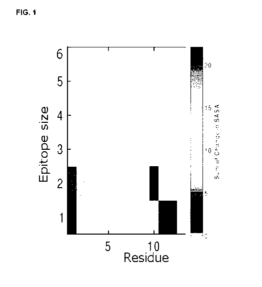
[0012]
W02006066089 titled HUMANIZED AMYLOID BETA ANTIBODIES FOR USE IN
IMPROVING COGNITION is directed to improved agents and methods for treatment
of diseases
associated with beta amyloid and in particular to the identification and
characterization of a
monoclonal antibody, 12A11, that specifically binds to Ap peptide and is
effective at reducing plaque
burden associated with amyloidogenic disorders (e.g., AD).
[0013]
W02007068429 titled ANTIBODIES AGAINST AMYLOID BETA 4 WITH
GLYCOSYLATED IN THE VARIABLE REGION is directed to a purified antibody
molecule preparation
being characterized in that at least one antigen binding site comprises a
glycosylated asparagine
(Asn) in the variable region of the heavy chain (VH).
[0014] Yu
et al. describes a hexavalent foldable Ap1-15 (6Ap15) fused to PADRE or toxin-
derived carrier proteins. Wang et al. 2016 report that peripheral
administration of this antibody
mitigates Alzheimer's disease like pathology and cognitive decline in a
transgenic animal model of
aged Alzheimer Disease [11], [12].
[0015]
Antibodies that preferentially or selectively bind A-beta oligomers over
monomers or
over fibrils or over both monomers and fibrils are desirable.
Summary
[0016]
Described herein are epitopes and more particularly conformational epitopes,
in A-
beta comprising and/or consisting of residues HDSG (SEQ ID NO:1) or related
epitopes, and
antibodies that specifically and/or selectively bind said epitopes. The
epitopes may be selectively
exposed in the oligomeric species of A-beta, in a conformation that
distinguishes oligomeric species
from that in the monomer and/or fibril.
[0017] An
aspect includes a cyclic compound, preferably a cyclic compound, comprising:
an
A-beta peptide the peptide comprising HDS and up to 6 A-beta contiguous
residues, and a linker,
wherein the linker is covalently coupled to the A-beta peptide N-terminus
residue and the A-beta C-
terminus residue.
[0018] In
an embodiment, the A-beta peptide is selected from a peptide having a sequence
of any one of SEQ ID NOS: 1-16, optionally selected from HDSG (SEQ ID NO: 1),
HDSGY (SEQ ID
NO: 4), HDSGYE (SEQ ID NO: 11), RHDSGY (SEQ ID NO: 13), RHDSG (SEQ ID NO: 5),
RHDS
(SEQ ID NO: 6) and DSGY (SEQ ID NO: 14).
2
CA 03004482 2018-05-07
WO 2017/079831
PCT/CA2016/051300
[0019] In another embodiment, the cyclic compound is a cyclic peptide.
[0020] In another embodiment, the cyclic compound described herein
comprises i) a
curvature increase of D and/or S in the compound of at least 10%, at least
20%, or at least 30%
compared to D or S in the context of a corresponding linear compound; ii) at
least one residue
selected from H, D and S, wherein at least one dihedral angle of said residue
is different by at least
30 degrees, at least 40 degrees, at least 50 degrees, at least 60 degrees, at
least 70 degrees, or at
least 80 degrees compared to the corresponding dihedral angle in the context
of a corresponding
linear compound; iii) an O-C-Ca-Cp dihedral angle in D that is different by at
least 30 degrees, at least
40 degrees, at least 50 degrees, at least 60 degrees, at least 70 degrees or
at least 80 degrees
compared to the corresponding dihedral angle in the context of a corresponding
linear compound;
and/or iv) a conformation for H and/or D as measured by entropy that is at
least 10%, at least 20%, at
least 25%, at least 30%, at least 35%, at least 40% more constrained compared
to a corresponding
linear compound.
[0021] In another embodiment, the A-beta peptide is selected from HDSG
(SEQ ID NO: 1),
HDSGY (SEQ ID NO: 4) and RHDSG (SEQ ID NO: 5).
[0022] In another embodiment, the compound further comprises a detectable
label.
[0023] In another embodiment, the linker comprises or consists of 1-8
amino acids and/or
equivalently functioning molecules and/or one or more functionalizable
moieties.
[0024] In another embodiment, the linker amino acids are selected from
A and G, and/or
wherein the functionalizable moiety is C.
[0025] In another embodiment, the linker comprises or consists of amino
acids GCG or
CGC.
[0026] In another embodiment, the linker comprises a PEG molecule.
[0027] In another embodiment, the cyclic compound is selected from the
structures shown in
FIG. 11B.
[0028] An aspect includes an immunogen comprising the cyclic compound
described herein.
[0029] In an embodiment, the compound is coupled to a carrier protein
or immunogenicity
enhancing agent.
[0030] In another embodiment, the carrier protein is bovine serum
albumin (BSA) or the
immunogenicity-enhancing agent is keyhole Keyhole Limpet Haemocyanin (KLH).
[0031] An aspect includes a composition comprising the compound described
herein or the
immunogen described herein.
[0032] In an embodiment, the immunogen comprising compositions
described herein further
comprises an adjuvant.
[0033] In another embodiment, the adjuvant is aluminum phosphate or
aluminum hydroxide.
3
CA 03004482 2018-05-07
WO 2017/079831
PCT/CA2016/051300
[0034] An aspect includes an isolated antibody that specifically binds to
an A-beta peptide
having a sequence of HDSG (SEQ ID NO: 1) or a related epitope sequence,
optionally as set forth in
any one of SEQ ID NOS: 1-16.
[0035] In an embodiment, the antibody specifically binds an epitope on
A-beta, wherein the
epitope comprises at least two consecutive amino acid residues predominantly
involved in binding to
the antibody, wherein the at least two consecutive amino acids are DS embedded
within HDS, or
wherein the at least two consecutive amino acids are HD embedded within HDS.
[0036] In another embodiment, the epitope comprises or consists of
HDS, DSG, HDSG
(SEQ ID NO: 1), HDSGY (SEQ ID NO: 4), HDSGYE (SEQ ID NO: 11), RHDSGY (SEQ ID
NO: 13),
RHDSG (SEQ ID NO: 5), RHOS (SEQ ID NO: 6) and DSGY (SEQ ID NO: 14).
[0037] In another embodiment, the antibody is a conformation specific
and/or selective
antibody that specifically or selectively binds to HDSG (SEQ ID NO: 1) or a
related epitope peptide
presented in a cyclic compound, optionally a cyclic compound described herein,
preferably a cyclic
peptide having a sequence as set forth in SEQ ID NO: 2 or 12.
[0038] In another embodiment, the antibody selectively binds A-beta
oligomer over A-beta
monomer and/or A-beta fibril.
[0039] In another embodiment, the selectivity is at least 2 fold, at
least 3 fold, at least 5 fold,
at least 10 fold, at least 20 fold, at least 30 fold, at least 40 fold, at
least 50 fold, at least 100 fold, at
least 500 fold, at least 1000 fold more selective for A-beta oligomer over A-
beta monomer and/or A-
beta fibril.
[0040] In another embodiment, the antibody does not specifically and/or
selectively bind a
linear peptide comprising sequence HDSG (SEQ ID NO: 1) or a related epitope,
optionally wherein
the sequence of the linear peptide is a linear version of a cyclic compound
used to raise the antibody,
optionally a linear peptide having a sequence as set forth in SEQ ID NO: 2 or
12.
[0041] In another embodiment, the antibody lacks or has negligible
binding to A-beta
monomer and/or A-beta fibril plaques in situ.
[0042] In another embodiment, the antibody is a monoclonal antibody or
a polyclonal
antibody.
[0043] In another embodiment, the antibody is a humanized antibody.
[0044] In another embodiment, the antibody is an antibody binding
fragment selected from
Fab, Fab', F(ab')2, scFv, dsFv, ds-scFv, dimers, nanobodies, minibodies,
diabodies, and multimers
thereof.
[0045] Another embodiment comprises a light chain variable region and
a heavy chain
variable region, optionally fused, the heavy chain variable region comprising
complementarity
determining regions CDR-H1, CDR-H2 and CDR-H3, the light chain variable region
comprising
4
CA 03004482 2018-05-07
WO 2017/079831
PCT/CA2016/051300
complementarity determining region CDR-L1, CDR-L2 and CDR-L3 and with the
amino acid
sequences of said CDRs comprising the sequences:
CDR-Hi GYTFTSYW (SEQ ID NO: 17)
CDR-H2 IDPSDSQT (SEQ ID NO: 18)
CDR-H3 SRGGY (SEQ ID NO: 19)
CDR-L1 QDINNY (SEQ ID NO: 20)
CDR-L2 YTS (SEQ ID NO: 21)
CDR-L3 LQYDNLWT (SEQ ID NO: 22)
[0046] In another embodiment, the antibody comprises a heavy chain
variable region
comprising: i) an amino acid sequence as set forth in SEQ ID NO: 24; ii) an
amino acid sequence with
at least 50%, at least 60%, at least 70%, at least 80% or at least 90%
sequence identity to SEQ ID
NO: 24, wherein the CDR sequences are as set forth in SEQ ID NO: 17, 18 and
19, or iii) a
conservatively substituted amino acid sequence i).
[0047] In another embodiment, the antibody comprises a light chain
variable region
comprising i) an amino acid sequence as set forth in SEQ ID NO: 26, ii) an
amino acid sequence with
at least 50%, at least 60%, at least 70%, at least 80%, or at least 90%
sequence identity to SEQ ID
NO: 26, wherein the CDR sequences are as set forth in SEQ ID NO: 20, 21 and
22, or iii) a
conservatively substituted amino acid sequence of i).
[0048] In another embodiment, the heavy chain variable region amino
acid sequence is
encoded by a nucleotide sequence as set forth in SEQ ID NO: 23 or a codon
degenerate or optimized
version thereof; and/or the antibody comprises a light chain variable region
amino acid sequence
encoded by a nucleotide sequence as set out in SEQ ID NO: 25 or a codon
degenerate or optimized
version thereof.
[0049] In another embodiment, the heavy chain variable region
comprises or consists of an
amino acid sequence as set forth in SEQ ID NO: 24 and/or the light chain
variable region comprises
or consists of an amino acid sequence as set forth in SEQ ID NO: 26.
[0050] In another embodiment, the antibody competes for binding to
human A-beta with an
antibody comprising the CDR sequences as recited in Table 10.
[0051] In an embodiment, the antibody is prepared using a cyclic
compound or immunogen
described herein.
[0052] An aspect includes an immunoconjugate comprising an antibody
described herein
and a detectable label or cytotoxic agent.
[0053] In an embodiment, the detectable label comprises a positron
emitting radionuclide,
optionally for use in subject imaging such as PET imaging.
5
CA 03004482 2018-05-07
WO 2017/079831
PCT/CA2016/051300
[0054] An aspect includes an antibody described herein, or an
immunoconjugate described
herein, optionally with a diluent.
[0055] An aspect includes a nucleic acid molecule encoding a
proteinaceous portion of the
compound or immunogen described herein, the antibody described herein or
proteinaceous
immunoconjugates described herein.
[0056] An aspect includes a vector described herein.
[0057] An aspect includes a cell expressing an antibody described
herein, optionally wherein
the cell is a hybridoma comprising the vector.
[0058] An aspect includes a kit described herein, the immunogen
described herein, the
antibody described herein, the immunoconjugate described herein, the nucleic
acid molecule
described herein, the vector described herein or the cell described herein.
[0059] An aspect includes a method of making the antibody described
herein, comprising
administering the compound or immunogen described herein or a composition
comprising said
compound or immunogen to a subject and isolating antibody and/or cells
expressing antibody specific
or selective for the compound or immunogen administered and/or A-beta
oligomers, optionally lacking
or having negligible binding to a linear peptide comprising the A-beta peptide
and/or lacking or having
negligible plaque binding.
[0060] An aspect includes a method of determining if a biological
sample comprises A-beta,
the method comprising:
a. contacting the biological sample with an antibody described herein or the
immunoconjugate described herein; and
b. detecting the presence of any antibody complex.
[0061] In an embodiment, the method described herein for determining
if the biological
sample contains A-beta oligomer the method comprising:
a. contacting the sample with the antibody described herein or the
immunoconjugate
described herein that is specific and/or selective for A-beta oligomers under
conditions permissive for forming an antibody: A-beta oligomer complex; and
b. detecting the presence of any complex;
wherein the presence of detectable complex is indicative that the sample may
contain
A-beta oligomer.
[0062] In another embodiment, the amount of complex is measured.
[0063] In another embodiment, the sample comprises brain tissue or an
extract thereof,
whole blood, plasma, serum and/or CSF.
[0064] In another embodiment, the sample is a human sample.
6
CA 03004482 2018-05-07
WO 2017/079831
PCT/CA2016/051300
[0065] In another embodiment, the sample is compared to a control,
optionally a previous
sample.
[0066] In another embodiment, the level of A-beta is detected by SPR.
[0067] An aspect includes a method of measuring a level of A-beta in a
subject, the method
comprising administering to a subject at risk or suspected of having or having
AD, an
immunoconjugate comprising an antibody described herein wherein the antibody
is conjugated to a
detectable label; and detecting the label, optionally quantitatively detecting
the label.
[0068] In an embodiment the label is a positron emitting radionuclide.
[0069] An aspect includes a method of inducing an immune response in a
subject,
comprising administering to the subject a compound or combination of compounds
described herein,
optionally a cyclic compound comprising HDSG (SEQ ID NO: 1) or a related
epitope peptide
sequence, an immunogen and/or composition comprising said compound or said
immunogen; and
optionally isolating cells and/or antibodies that specifically or selectively
bind the A-beta peptide in the
compound or immunogen administered.
[0070] An aspect includes a method of inhibiting A-beta oligomer
propagation, the method
comprising contacting a cell or tissue expressing A-beta with or administering
to a subject in need
thereof an effective amount of an A-beta oligomer specific or selective
antibody or immunoconjugate
described herein, to inhibit A-beta aggregation and/or oligomer propagation.
[0071] An aspect includes a method of treating AD and/or other A-beta
amyloid related
diseases, the method comprising administering to a subject in need thereof i)
an effective amount of
an antibody or immunoconjugate described herein, optionally an A-beta oligomer
specific or selective
antibody, or a pharmaceutical composition comprising said antibody; 2)
administering an isolated
cyclic compound comprising HDSG (SEQ ID NO: 1) or a related epitope sequence
or immunogen or
pharmaceutical composition comprising said cyclic compound, or 3) a nucleic
acid or vector
comprising a nucleic acid encoding the antibody of 1 or the immunogen of 2, to
a subject in need
thereof.
[0072] In an embodiment, a biological sample from the subject to be
treated is assessed for
the presence or levels of A-beta using an antibody described herein.
[0073] In an embodiment, more than one antibody or immunogen is
administered.
[0074] In an embodiment, the antibody, immunoconjugate, immunogen,
composition or
nucleic acid or vector is administered directly to the brain or other portion
of the CNS.
[0075] In an embodiment, the composition is a pharmaceutical
composition comprising the
compound or immunogen in admixture with a pharmaceutically acceptable, diluent
or carrier.
[0076] An aspect includes an isolated peptide comprising an A beta
peptide consisting of the
sequence of any one of the sequences set forth in SEQ ID NOS: 1-16.
7
CA 03004482 2018-05-07
WO 2017/079831
PCT/CA2016/051300
[0077] In an embodiment, the isolated peptide is a cyclic peptide
comprising a linker wherein
the linker is covalently coupled to the A-beta peptide N-terminus residue
and/or the A-beta C-terminus
residue.
[0078] In an embodiment, the isolated cyclic peptide comprises a
detectable label.
[0079] An
aspect includes a nucleic acid sequence encoding the isolated peptide
described
herein.
[0080] An aspect includes a hybridoma expressing the antibody
described herein.
[0081]
Other features and advantages of the present disclosure will become apparent
from
the following detailed description. It should be understood, however, that the
detailed description and
the specific examples while indicating preferred embodiments of the disclosure
are given by way of
illustration only, since various changes and modifications within the spirit
and scope of the disclosure
will become apparent to those skilled in the art from this detailed
description.
Brief description of the drawings
[0082] An
embodiment of the present disclosure will now be described in relation to the
drawings in which:
[0083] FIG. 1: Likelihood of exposure as a function of sequence, as
determined by the
collective coordinates method.
[0084]
FIG. 2: Curvature as a function of residue index. Mean curvature in the
equilibrium
ensemble for the cyclic peptide CGHDSGG (SEQ ID No: 2) is shown (solid light
grey), along with the
curvature for the linear peptide (solid dark grey), and the curvature of the
various monomers in the
fibril (dotted line).
[0085]
FIG. 3: Dihedral angle distributions for the side chain heavy atoms of H6.
Schematics
of residue H6 are shown in the insets; the corresponding bond over which the
dihedral angle is taken
is rendered darker than the other bonds, and the four atoms defining the
dihedral angle are shown in
lighter gray. The angles corresponding to the peak values of the dihedral
distributions for all 3
species¨linear peptide, cyclic peptide, and (2M4J) fibril ensemble are
provided in Table 1. The
differences between the peak values are also provided in Table 1. The dihedral
angle distribution for
the fibril ensemble is taken over all 9 chains of A-beta42 in the PDB
structure in the fibril, so the
dihedral distribution observed is generally broader than the distribution for
any single chain taken from
the structure 2M4J.
[0086] FIG. 4: Dihedral angle distribution for the angle 0-C-Ca-Cf3
involving the side chain
heavy atoms of residue D7. Schematics of D7 are shown in the insets; the
corresponding bond over
which the dihedral angle is taken is rendered darker than the other bonds. The
values are provided in
Table 1.
8
CA 03004482 2018-05-07
WO 2017/079831
PCT/CA2016/051300
[0087] FIG. 5: Dihedral angle distributions for angles involving the side
chain heavy atoms of
S8. Schematics of S8 are shown in the insets; the corresponding bond over
which the dihedral angle
is taken is rendered darker than the other bonds. The values are provided in
Table 1.
[0088] FIG. 6: Top panel (A): Side chain entropy change of the linear
and cyclic peptides
relative to the entropy in the fibril, plotted for each residue H, D, and S.
(B) 2nd from top panel: entropy
of the individual dihedral angles in H6. Note for example that CA-CB-CG-ND1
has substantially less
entropy than either the fibril or linear peptides, which can also be seen by
the more sharply peaked
dihedral angle distribution for this particular dihedral in FIG. 3. (C) 2nd
from bottom panel: entropy of
individual dihedral angles in D7. (D) Bottom panel: entropy of individual
dihedral angles in S8.
[0089] FIG. 7: Equilibrium backbone Ramachandran angles for residues H,
D, S, and G, in
both the linear and cyclic forms of the peptide CGHDSGG (SEQ ID NO: 2), along
with the backbone
Ramachandran angles for the residues H, D, S, and G in the context of the
fibril 2M4J.
[0090] FIG. 8: Solubility vs residue index for A-beta42 peptide. HDSG
(SEQ ID NO: 1) has
values of +1.1, +0.14, +1.2, and +0.30 respectively.
[0091] FIG. 9: Plots of the solvent accessible surface area (SASA), the
weighted SASA, ((s, -
<s>)/6s). SASA,, and ((s, -<s>)/6s) = SASA, (((s, -<s>)/6s) = SASANfrd.
[0092] FIG. 10: Two separate views of the root mean squared deviation
(RMSD) values to the
centroids of the three largest clusters of the linear peptide ensemble. Each
point corresponds to a
given conformation taken from the linear peptide, cyclic peptide, or fibril
equilibrium ensembles.
[0093] FIG. 11A: Two views of the cyclic peptide structure CGHDSGG (SEQ
ID NO: 2),
rendered in licorice representation so the orientations of the side chains can
be seen. The light gray
colored conformation is the centroid of the largest cluster, as described
above for FIG. 10, and best
represents the typical conformation of the cyclic peptide. The black side
chains are rendered for a
linear conformation having dihedral angles close to the most likely dihedral
angles of the linear
peptide; the side chains for this linear peptide conformation are superimposed
on the cyclic peptide,
to show that different dihedral angles tend to be preferred for the linear and
cyclic peptides.
[0094] FIG. 11 B: Schematic representations of cyclic peptides
comprising HDSG (SEQ ID
NO: 1), including the cyclic peptide with circular peptide bond, the cyclic
peptide with PEG2 linker
between the G and C residues, and the cyclic peptide with PEG2 linker between
the C and H
residues.
[0095] FIG. 12: Clustering plots by root mean squared deviation (RMSD);
axes correspond
again to the centroids of the three largest clusters of the linear peptide
ensemble, as in FIG. 10.
[0096] FIG. 13: Surface plasmon resonance (SPR) direct binding assay of
antibodies to cyclic
peptide and linear peptide in Panel A, and A-beta oligomer and A-beta monomer
in Panel B.
9
CA 03004482 2018-05-07
WO 2017/079831
PCT/CA2016/051300
[0097] FIG. 14: Primary screening of clones from tissue culture
supernatants using ELISA
and SPR direct binding assay. Plot comparing mAb binding in SPR direct binding
assay versus
ELISA.
[0098] FIG. 15: SPR direct binding assay of select clones to cyclic
peptide (structured
peptide: circles), linear peptide (unstructured peptide, squares), A-beta
monomer (upward pointing
triangles), and A-beta oligomer (downward pointing triangles). Asterisk
indicates a clone reactive to
unstructured linear peptide for control purposes.
[0099] FIG. 16: lmmunohistochemical staining of plaque from cadaveric
AD brain using 6E10
positive control antibody (A) and an antibody (303-25-1B4) raised against
cyclo(CGHDSGG) (SEQ ID
NO:2) (B).
[00100] FIG. 17: Secondary Screening of selected and purified antibodies
using an SPR
indirect (capture) binding assay. SPR binding response of A-beta oligomer to
captured antibody
minus binding response of A-beta monomer to captured antibody (circle); SPR
binding response of
pooled soluble brain extract from AD patients to captured antibody minus
binding response of pooled
brain extract from non-AD controls to captured antibody (triangle); SPR
binding response of pooled
cerebrospinal fluid (CSF) from AD patients to captured antibody minus binding
response of pooled
CSF from non-AD controls to captured antibody (square).
[00101] FIG. 18: Verification of Antibody binding to A-beta oligomers.
SPR sensorgrams and
binding response plots of varying concentrations of commercially-prepared
stable A-beta oligomers
binding to immobilized antibodies. Panel A shows results with the positive
control mAb6E10, Panel B
with the negative isotype control and Panel C with antibody raised against
cyclo (CGHDSGG) (SEQ
ID NO:2). Panel D plots binding of several antibody clones raised against
cyclic peptide comprising
HDSG (SEQ ID No: 1), with A-beta oligomer at a concentration of 1 micromolar.
[00102] FIG. 19: A plot showing propagation of A-beta aggregation in
vitro in the presence or
absence of a representative antibody raised using a cyclic peptide comprising
HDSG (SEQ ID NO:1).
[00103] Table 1 shows the peak values of the dihedral angle distribution
for those dihedral
angles whose distributions show significant differences between the cyclic
peptide and other species.
[00104] Table 2 shows peak values of the Ramachandran backbone phi/psi
angle distributions.
[00105] Table 3 gives the Ramachandran backbone dihedral angles as well
as the side chain
dihedral angles for the cyclic peptide that is the centroid conformation of
the largest conformational
cluster, and for the centroid conformation of the largest cluster taken from
the linear peptide
ensemble.
[00106] Table 4 is a table of mean curvature values for each residue in
the cyclic, linear, and
2M4J fibril ensembles.
[00107] Table 5 shows the binding properties of selected antibodies.
[00108] Table 6 shows the binding properties summary for selected
antibodies.
CA 03004482 2018-05-07
WO 2017/079831
PCT/CA2016/051300
[00109] Table 7 lists the oligomer binding ¨ monomer binding for an
antibody raised against
cyclo(CGHDSGG) (SEQ ID NO:2).
[00110] Table 8 lists properties of antibodies tested on formalin fixed
tissues.
[00111] Table 9 is an exemplary toxicity assay.
[00112] Table 10 lists CDR sequences.
[00113] Table 11 lists heavy chain and light chain variable sequences.
[00114] Table 12 is a table of A-beta epitope sequences and select
sequences with linker.
[00115] Table 13 provides the full A-beta 1-42 human polypeptide
sequence.
Detailed description of the Disclosure
[00116] Provided herein are antibodies, immunotherapeutic compositions and
methods which
may target epitopes preferentially accessible in toxic oligomeric species of A-
beta, including
oligomeric species associated with Alzheimer's disease. A region in A-beta has
been identified that
may be specifically and/or selectively accessible to antibody binding in
oligomeric species of A-beta.
[00117] As
demonstrated herein, generation of oligomer-specific or oligomer selective
antibodies was accomplished through the identification of targets on A-beta
peptide that are not
present, or present to a lesser degree, on either the monomer and/or fibril.
Oligomer-specific epitopes
need not differ in primary sequence from the corresponding segment in the
monomer or fibril,
however they would be conformationally distinct in the context of the
oligomer. That is, they would
present a distinct conformation in terms of backbone and/or side-chain
orientation in the oligomer that
would not be present (or would be unfavourable) in the monomer and/or fibril.
[00118]
Antibodies raised to linear peptide regions may not to be selective for
oligomer, and
thus may bind to monomer or A-beta plaques as well.
[00119] As
described herein, to develop antibodies that may be selective for oligomeric
forms
of A-beta, the inventors sought to identify regions of A-beta sequence that
are prone to disruption in
the context of the fibril, and that may be exposed on the surface of the
oligomer.
[00120] As
described the Examples, the inventors have identified a region they have
determined to be prone to disruption in the context of the fibril. The
inventors designed cyclic
compounds comprising the identified target region to satisfy criteria of an
alternate conformation such
as higher curvature, higher exposed surface area, alternative dihedral angle
distributions, and/or did
not readily align by root mean squared deviation (RMSD) to either the linear
or fibril ensembles.
[00121]
Antibodies could be raised using a cyclic peptide comprising the target
region, that
selectively bound the cyclic peptide compared to a linear peptide of the same
sequence (e.g.
corresponding linear sequence). Experimental results are described and
identify epitope-specific and
conformationally selective antibodies that bind synthetic oligomer selectively
compared to synthetic
11
CA 03004482 2018-05-07
WO 2017/079831
PCT/CA2016/051300
monomers, bind CSF from AD patients preferentially over control CSF and/or
bind soluble brain
extract from AD patients preferentially over control soluble brain extract.
Further staining of AD brain
tissue identified antibodies that show no or negligible plaque binding and in
vitro studies found that
the antibodies inhibited A13 oligomer propagation and aggregation.
I. Definitions
[00122] As used herein, the term A-beta may alternately be referred to as
'amyloid beta',
'amyloid 13, Abeta, A-beta or A13. Amyloid beta is a peptide of 36-43 amino
acids and includes all wild-
type and mutant forms of all species, particularly human A-beta. A-beta40
refers to the 40 amino acid
form; A-beta42 refers to the 42 amino acid form, etc. The amino acid sequence
of human wildtype A-
beta42 is shown in SEQ ID NO: 3
[00123] As used herein, the term "A-beta monomer herein refers to any of
the individual
subunit forms of the A-beta (e.g. 1-40, 1-42, 1-43) peptide.
[00124] As
used herein, the term "A-beta oligomer herein refers to a plurality of any of
the A-
beta subunits wherein several (e.g. at least two) A-beta monomers are non-
covalently aggregated in a
conformationally-flexible, partially-ordered, three-dimensional globule of
less than about 100, or more
typically less than about 50 monomers. For example, an oligomer may contain 3
or 4 or 5 or more
monomers. The term "A-beta oligomer" as used herein includes both synthetic A-
beta oligomer and/or
native A-beta oligomer. "Native A-beta oligomer" refers to A-beta oligomer
formed in vivo, for example
in the brain and CSF of a subject with AD.
[00125] As
used herein, the term "A-beta fibril" refers to a molecular structure that
comprises
assemblies of non-covalently associated, individual A-beta peptides which show
fibrillar structure
under an electron microscope. The fibrillar structure is typically a "cross
beta" structure; there is no
theoretical upper limit on the size of multimers, and fibrils may comprise
thousands or many
thousands of monomers. Fibrils can aggregate by the thousands to form senile
plaques, one of the
primary pathological morphologies diagnostic of AD.
[00126] The term "HDSG" means the amino acid sequence histidine, aspartic
acid, serine, and
glycine as shown in SEQ ID NO: 1. Similarly DSG, DSGG (SEQ ID NO:3), HDSGYE
(SEQ ID NO:11),
HDSGY (SEQ ID NO:4), RHDSG (SEQ ID NO:5), RHDS (SEQ ID NO:6) refer to the
amino acid
sequence identified by the 1-letter amino acid code. Depending on the context,
the reference of the
amino acid sequence can refer to a sequence in A-beta or an isolated peptide,
such as the amino
acid sequence of a cyclic compound.
[00127] The
term "alternate conformation than occupied by an amino acid residue (e.g. H,
D, S
and/or G) in the linear compound, monomer and/or fibril" as used herein means
having one or more
differing conformational properties selected from solvent accessibility,
entropy, curvature (e.g. in the
context of peptide HDSG (SEQ ID NO:1) as compared to for example in the cyclic
peptide described
in the Examples), RMSD structural alignment, and dihedral angle of one or more
backbone or side
chain dihedral angles compared to said property for H, D and/or S in an A-beta
linear compound
12
CA 03004482 2018-05-07
WO 2017/079831
PCT/CA2016/051300
comprising the residue in context, A-beta monomer and/or A-beta fibril
structures as shown for
example in PDBs 2M4J, 2MXU, 2LMN, or 2LMP and shown in FIGS. 1-12 and/or in
the Tables. For
example, FIG. 2 and Table 4 show that the curvature of HDSG (SEQ ID NO:1), for
the cyclic peptide
ensemble is significantly larger than the curvature of HDSG (SEQ ID NO:1), in
the ensemble of fibril
conformations. This is particularly evident for 07, S8, and G9. Moreover for
07 and S8, the curvature
in the cyclic peptide ensemble is substantially higher than that in the linear
peptide ensemble. This
implies conformational selectivity may be particularly conferred by residues
07 and S8. The last two
panels of FIG. 3 show that the dihedral angle distribution for the angles (N-
CA-CB-CG) and (C-CA-
CB-CG) for H6 in the cyclic peptide ensemble do have overlap, but are not the
most common angles
in the linear peptide and fibril ensembles (the probabilities are 36% and 13%
respectively for N-CA-
CB-CG in linear and fibril and 36% and 13% respectively for C-CA-CB-CG in the
linear and fibril). The
last panel of FIG. 4 shows that the dihedral angle distribution for angle (0-C-
CA-CB) involving the
side chain of residue 07 reflects an alternate conformational distribution
compared to either the
monomer or fibril. FIG. 5 shows that the dihedral angle distributions for
angles (N-CA-CB-OG), (C-CA-
CB-0G), and (0-C-CA-CB) involving the side chain of residue S8 reflects an
alternate conformational
distribution compared to either the monomer or fibril. The alternate
conformation can be similarly, less
or more "constrained" than the comparator conformation. For example, FIG. 6
demonstrates that H6 is
more constrained in the cyclic peptide then it is in either the fibril or the
monomer. Residue 07 is more
constrained in the cyclic peptide ensemble then it is in the monomer, but less
than it is in the fibril.
Residue S8 is less constrained in the cyclic peptide ensemble then it is in
the fibril and also marginally
less than it is in the monomer. FIG. 7 demonstrates that the distributions of
the Ramachandran
dihedral angles for the backbone of cyclic peptides are substantially
different than those for either
monomer or fibril for residues 07 and S8. FIG. 8 shows that the residues HDSG
(SEQ ID NO:1) have
a larger solubility than the average solubility present in A-beta peptide,
indicating the likelihood of
exposure of these residues in an oligomeric ensemble of conformations. FIG. 9
shows that residues
HDSG (SEQ ID NO:1) have increased solvent accessible surface area, SASA,
compared to the fibril,
and that, when weighted by solubility, all residues in the cyclic peptide
ensemble show an increase in
weighted SASA over that in the fibril, with residue S8 showing substantial
increase in weighted SASA
over the fibril. FIG. 10 shows that the cyclic peptide equilibrium structures
of HDSG (SEQ ID NO:1)
cluster differently than the equilibrium structures of either the linear
peptide or corresponding
sequence in the fibril, while the linear and fibril ensembles are not clearly
differentiated.
[00128] The term "amino acid" includes all of the naturally occurring
amino acids as well as
modified L-amino acids. The atoms of the amino acid can include different
isotopes. For example, the
amino acids can comprise deuterium substituted for hydrogen nitrogen-15
substituted for nitrogen-14,
and carbon-13 substituted for carbon-12 and other similar changes.
[00129] The term "antibody as used herein is intended to include,
monoclonal antibodies,
polyclonal antibodies, single chain, veneered, humanized and other chimeric
antibodies and binding
fragments thereof, including for example a single chain Fab fragment, Fab'2
fragment or single chain
Fv fragment. The antibody may be from recombinant sources and/or produced in
animals such as
13
CA 03004482 2018-05-07
WO 2017/079831
PCT/CA2016/051300
rabbits, llamas, sharks etc. Also included are human antibodies that can be
produced in transgenic
animals or using biochemical techniques or can be isolated from a library such
as a phage library.
Humanized or other chimeric antibodies may include sequences from one or more
than one isotype or
class or species.
[00130] The
phrase "isolated antibody refers to antibody produced in vivo or in vitro that
has
been removed from the source that produced the antibody, for example, an
animal, hybridoma or
other cell line (such as recombinant insect, yeast or bacteria cells that
produce antibody). The isolated
antibody is optionally "purified", which means at least: 80%, 85%, 90%, 95%,
98% or 99% purity.
[00131] The
term "binding fragment" as used herein to a part or portion of an antibody or
antibody chain comprising fewer amino acid residues than an intact or complete
antibody or antibody
chain and which binds the antigen or competes with intact antibody. Exemplary
binding fragments
include without limitations Fab, Fab', F(ab')2, scFv, dsFv, ds-scFv, dimers,
nanobodies, minibodies,
diabodies, and multimers thereof. Fragments can be obtained via chemical or
enzymatic treatment of
an intact or complete antibody or antibody chain. Fragments can also be
obtained by recombinant
means. For example, F(ab')2 fragments can be generated by treating the
antibody with pepsin. The
resulting F(ab')2 fragment can be treated to reduce disulfide bridges to
produce Fab' fragments.
Papain digestion can lead to the formation of Fab fragments. Fab, Fab' and
F(ab')2, scFv, dsFv, ds-
scFv, dimers, minibodies, diabodies, bispecific antibody fragments and other
fragments can also be
constructed by recombinant expression techniques.
[00132] The
terms "IMGT numbering" or "ImMunoGeneTics database numbering", which are
recognized in the art, refer to a system of numbering amino acid residues
which are more variable
(i.e. hypervariable) than other amino acid residues in the heavy and light
chain variable regions of an
antibody, or antigen binding portion thereof.
[00133]
When an antibody is said to specifically bind to an epitope such as HDSG (SEQ
ID
NO:1), what is meant is that the antibody specifically binds to a peptide
containing the specified
residues or a part thereof for example at least 2 residues of HDSG, with a
minimum affinity, and does
not bind an unrelated sequence or unrelated sequence spatial orientation
greater than for example an
isotype control antibody. Such an antibody does not necessarily contact each
residue of HDSG (SEQ
ID NO:1) and every single amino acid substitution or deletion within said
epitope does not necessarily
significantly affect and/or equally affect binding affinity.
[00134] When an antibody is said to selectively bind an epitope such as a
conformational
epitope, such as HDSG (SEQ ID NO:1), what is meant is that the antibody
preferentially binds one or
more particular conformations containing the specified residues or a part
thereof with greater affinity
than it binds said residues in another conformation. For example, when an
antibody is said to
selectively bind a cyclopeptide comprising HDSG or related epitope relative to
a corresponding linear
peptide, the antibody binds the cyclopeptide with at least a 2 fold greater
affinity than it binds the
linear peptide.
14
CA 03004482 2018-05-07
WO 2017/079831
PCT/CA2016/051300
[00135] As used herein, the term "conformational epitope" refers to an
epitope where the
epitope amino acid sequence has a particular three-dimensional structure
wherein at least an aspect
of the three-dimensional structure not present or less likely to be present in
a corresponding linear
peptide is specifically and/or selectively recognized by the cognate antibody.
The epitope e.g. HDSG
(SEQ ID NO: 1) may be partially or completely exposed on the molecular surface
of oligomeric A-beta
and partially or completely obscured from antibody recognition in monomeric or
fibrillar plaque A-beta.
Antibodies which specifically and/or selectively bind a conformation-specific
epitope recognize the
spatial arrangement of one or more of the amino acids of that conformation-
specific/selective epitope.
For example an HDSG (SEQ ID NO: 1) conformational epitope, refers to an
epitope of HDSG (SEQ
ID NO: 1) that is recognized by antibodies specifically and/or selectively,
for example at least 2 fold, 3
fold, 5 fold, 10 fold, 50 fold, 100 fold, 250 fold, 500 fold or 1000 fold or
greater, more selectively as
compared to linear HDSG (SEQ ID NO: 1).
[00136] The
term "related epitope" as used herein means at least two residues of HDSG
(SEQ ID NO:1) that are antigenic and/or sequences comprising 1or 2 amino acid
residues in a A-beta
either N-terminal or C-terminal to at least two residues of HDSG (SEQ ID NO:
1). For example it is
shown herein HDSG (SEQ ID NO:1), HDSGY (SEQ ID NO:4) and RHDSG (SEQ ID NO:5)
were
identified as regions prone to disorder in an A-beta fibril. HDSGY (SEQ ID
NO:4) and RHDSG (SEQ
ID NO:5) are accordingly related epitopes. Further it is demonstrated through
modelling that residues
07 and S8 in particular exhibit differences in the cyclic compound compared to
the corresponding
linear sequence, accordingly DS, HDS, DSG, DSGY (SEQ ID NO: 13) and RHOS (SEQ
ID NO:6) are
related epitopes. Exemplary related epitopes can include A-beta sequences
included in Table 12.
[00137] The
term "constrained conformation" as used herein with respect to an amino acid
or
a side chain thereof, within a sequence of amino acids (e.g. H or D in HDSG
(SEQ ID NO: 1)), or with
respect to a sequence of amino acids in a larger polypeptide, means decreased
rotational mobility of
the amino acid dihedral angles, relative to a corresponding linear peptide
sequence, or the sequence
or larger polypeptide, resulting in a decrease in the number of permissible
conformations. This can be
quantified for example by finding the entropy reduction for the ensemble of
side chain dihedral angle
degrees of freedom, and is plotted in FIG. 6 for H, D and S. For example, if
the side chains in the
sequence have less conformational freedom than the linear peptide, the entropy
will be reduced.
Such conformational restriction would enhance the conformational selectivity
of antibodies specifically
raised to this antigen. The term "more constrained conformation" as used
herein means that the
dihedral angle distribution (ensemble of allowable dihedral angles) of one or
more dihedral angles is
at least 10% more constrained than in the comparator conformation, as
determined for example by
the entropy of the amino acids, for example H, and/or D (e.g. a more
constrained conformation has
lower entropy). Specifically, the average entropy change relative to the
entropy in the linear peptide,
S(cyclic) ¨ S(linear), of HDS in the overall more constrained cyclic
conformational ensemble is on
average reduced by more than 10% or reduced by more than 20% or reduced by
more than 30% or
reduced by more than 40%, from the unconstrained conformational ensemble, e.g.
of the quantity
S(linear) ¨ S(fibril)/[mean( S(linear) + S(fibril))] for the linear peptide is
approximately 81.7% entropy
CA 03004482 2018-05-07
WO 2017/079831
PCT/CA2016/051300
reduction for H6, 49.8% entropy reduction for 07, and ¨8.83% entropy reduction
for S8 (the negative
number implying that S8 has larger entropy for the cyclic peptide than the
linear peptide).
[00138] The term "no or negligible plaque binding" or "lacks or has
negligible plaque binding"
as used herein with respect to an antibody means that the antibody does not
show typical plaque
morphology staining on immunohistochemistry (e.g. in situ) and the level of
staining is comparable to
or no more than 2 fold the level seen with an IgG negative (e.g. irrelevant)
isotype control
[00139] The term "Isolated peptide" refers to peptide that has been
produced, for example, by
recombinant or synthetic techniques, and removed from the source that produced
the peptide, such
as recombinant cells or residual peptide synthesis reactants. The isolated
peptide is optionally
"purified", which means at least: 80%, 85%, 90%, 95%, 98% or 99% purity and
optionally
pharmaceutical grade purity.
[00140] The term "detectable label" as used herein refers to moieties
such as peptide
sequences (such a myc tag, HA-tag, V5-tag or NE-tag), fluorescent proteins
that can be appended or
introduced into a peptide or compound described herein and which is capable of
producing, either
directly or indirectly, a detectable signal. For example, the label may be
radio-opaque, positron-
emitting radionuclide (for example for use in PET imaging), or a radioisotope,
such as 3H, 13N, 140,
18F, 32p,
35 123 125 131
F, P, S, I, I, I; a fluorescent (fluorophore) or chemiluminescent
(chromophore) compound,
such as fluorescein isothiocyanate, rhodamine or luciferin; an enzyme, such as
alkaline phosphatase,
beta-galactosidase or horseradish peroxidase; an imaging agent; or a metal
ion. The detectable label
may be also detectable indirectly for example using secondary antibody.
[00141] The term "epitope" as commonly used means an antibody binding site,
typically a
polypeptide segment, in an antigen that is specifically recognized by the
antibody. As used herein
"epitope" can also refer to the amino acid sequences or part thereof
identified on A-beta using the
collective coordinates method described. For example an antibody generated
against an isolated
peptide corresponding to a cyclic compound comprising the identified target
region HDSG SEQ ID
NO:1), recognizes part or all of said epitope sequence. An epitope is
"accessible" in the context of the
present specification when it is accessible to binding by an antibody.
[00142]
[00143] The term "greater affinity" as used herein refers to a relative
degree of antibody
binding where an antibody X binds to target Y more strongly (Kon) and/or with
a smaller dissociation
constant (Koff) than to target Z, and in this context antibody X has a greater
affinity for target Y than for
Z. Likewise, the term "lesser affinity herein refers to a degree of antibody
binding where an antibody
X binds to target Y less strongly and/or with a larger dissociation constant
than to target Z, and in this
context antibody X has a lesser affinity for target Y than for Z. The affinity
of binding between an
antibody and its target antigen, can be expressed as KA equal to 1/K0 where K0
is equal to kon/koff.
The kon and koff values can be measured using surface plasmon resonance
technology, for example
using a Molecular Affinity Screening System (MASS-1) (Sierra Sensors GmbH,
Hamburg, Germany).
16
CA 03004482 2018-05-07
WO 2017/079831
PCT/CA2016/051300
An antibody that is selective for a conformation presented in a cyclic
compound optional a cyclic
peptide for example has a greater affinity for the cyclic compound (e.g.
cyclic peptide) compared to a
corresponding sequence in linear form (e.g. the sequence non-cyclized).
[00144]
Also as used herein, the term "immunogenic refers to substances that elicit
the
production of antibodies, activate T-cells and other reactive immune cells
directed against an
antigenic portion of the immunogen.
[00145] The
term "corresponding linear compound" with regard to a cyclic compound refers
to
a compound, optionally a linear peptide, comprising or consisting of the same
sequence or chemical
moieties as the cyclic compound but in linear (i.e. non-cyclized) form for
example having properties as
would be present in solution of a linear peptide. For example, the
corresponding linear compound can
be the synthesized peptide that is not cyclized.
[00146] As
used herein "specifically binds" in reference to an antibody means that the
antibody
recognizes an epitope sequence and binds to its target antigen with a minimum
affinity. For example
a multivalent antibody binds its target with a KID of at least le-6, at least
le-7, at least le-8, at least
le-9, or at least le-10. Affinities greater than at least le-8 may be
preferred. An antigen binding
fragment such as Fab fragment comprising one variable domain, may bind its
target with a 10 fold or
100 fold less affinity than a multivalent interaction with a non-fragmented
antibody.
[00147] The
term "selectively binds" as used herein with respect to an antibody that
selectively
binds a form of A-beta (e.g. fibril, monomer or oligomer) or a cyclic compound
means that the
antibody binds the form with at least 2 fold, at least 3 fold, or at least 5
fold, at least 10 fold, at least
100 fold, at least 250 fold, at least 500 fold or at least 1000 fold or more
greater affinity. Accordingly
an antibody that is more selective for a particular conformation (e.g.
oligomer) preferentially binds the
particular form of A-beta with at least 2 fold etc greater affinity compared
to another form and/or a
linear peptide.
[00148] The
term "linker" as used herein means a chemical moiety that can be covalently
linked to the peptide comprising HDSG (SEQ ID NO: 1) epitope peptide,
optionally linked to HDSG
(SEQ ID NO: 1) peptide N- and C- termini to produce a cyclic compound. The
linker can comprise a
spacer and/or one or more functionalizable moieties. The linker via the
functionalizable moieties can
be linked to a carrier protein or an immunogen enhancing agent such as Keyhole
Limpet Hemocyanin
(KLH).
[00149] The term "spacer" as used herein means any preferably non-
immunogenic or poorly
immunogenic chemical moiety that can be covalently-linked directly or
indirectly to a peptide N- and
C- termini to produce a cyclic compound of longer length than the peptide
itself, for example the
spacer can be linked to the N- and C- termini of a peptide consisting of HDSG
(SEQ ID NO: 1) to
produce a cyclic compound of longer backbone length than the HDSG (SEQ ID NO:
1) sequence
itself. That is, when cyclized, the peptide with a spacer (for example of 3
amino acid residues) makes
a larger closed circle than the peptide without a spacer. The spacer may
include, but is not limited to,
17
CA 03004482 2018-05-07
WO 2017/079831
PCT/CA2016/051300
moieties such as G, A, or PEG repeats, e.g. GHDSG (SEQ ID NO:7) GHDSGG (SEQ ID
NO:8),
GGHDSGG (SEQ ID NO:9), GHDSGGG (SEQ ID NO:10), etc. The spacer may comprise or
be
coupled to one or more functionalizing moieties, such as one or more cysteine
(C) residues, which
can be interspersed within the spacer or covalently linked to one or both ends
of the spacer. Where a
functionalizable moiety such as a C residue is covalently linked to one or
more termini of the spacer,
the spacer is indirectly covalently linked to the peptide. The spacer can also
comprise the
functionalizable moiety in a spacer residue as in the case where a biotin
molecule is introduced into
an amino acid residue.
[00150] The
term "functionalizable moiety" as used herein refers to a chemical entity with
a
"functional group" which as used herein refers to a group of atoms or a single
atom that will react with
another group of atoms or a single atom (so called "complementary functional
group") to form a
chemical interaction between the two groups or atoms. In the case of cysteine,
the functional group
can be ¨SH which can be reacted to form a disulfide bond. Accordingly the
linker can for example be
CCC. The reaction with another group of atoms can be covalent or a strong non-
covalent bond, for
example as in the case of biotin-streptavidin bonds, which can have Kd-1e-14.
A strong non-covalent
bond as used herein means an interaction with a Kd of at least le-9, at least
le-10, at least le-11, at
least le-12, at least le-13 or at least le-14.
[00151]
Proteins and/or other agents may be functionalized (e.g. coupled) to the
cyclic
compound, either to aid in immunogenicity, or to act as a probe in in vitro
studies. For this purpose,
any functionalizable moiety capable of reacting (e.g. making a covalent or non-
covalent but strong
bond) may be used. In one specific embodiment, the functionalizable moiety is
a cysteine residue
which is reacted to form a disulfide bond with an unpaired cysteine on a
protein of interest, which can
be, for example, an immunogenicity enhancing agent such as Keyhole Limpet
Hemocyanin (KLH), or
a carrier protein such as Bovine serum albumin (BSA) used for in vitro
immunoblots or
immunohistochemical assays.
[00152] The term "reacts with" as used herein generally means that there is
a flow of electrons
or a transfer of electrostatic charge resulting in the formation of a chemical
interaction.
[00153] The
term "animal" or "subject" as used herein includes all members of the animal
kingdom including mammals, optionally including or excluding humans.
[00154] A
"conservative amino acid substitution" as used herein, is one in which one
amino acid
residue is replaced with another amino acid residue without abolishing the
protein's desired
properties. Suitable conservative amino acid substitutions can be made by
substituting amino acids
with similar hydrophobicity, polarity, and R-chain length for one another.
Examples of conservative
amino acid substitution include:
Conservative Substitutions
Type of Amino Acid Substitutable Amino Acids
Hydrophilic Ala, Pro, Gly, Glu, Asp, Gln, Asn, Ser,
Thr
18
CA 03004482 2018-05-07
WO 2017/079831
PCT/CA2016/051300
Conservative Substitutions
Sulphydryl Cys
Aliphatic Val, Ile, Leu, Met
Basic Lys, Arg, His
Aromatic Phe, Tyr, Trp
[00155] The
term "sequence identity as used herein refers to the percentage of sequence
identity between two polypeptide sequences or two nucleic acid sequences. To
determine the percent
identity of two amino acid sequences or of two nucleic acid sequences, the
sequences are aligned for
optimal comparison purposes (e.g., gaps can be introduced in the sequence of a
first amino acid or
nucleic acid sequence for optimal alignment with a second amino acid or
nucleic acid sequence). The
amino acid residues or nucleotides at corresponding amino acid positions or
nucleotide positions are
then compared. When a position in the first sequence is occupied by the same
amino acid residue or
nucleotide as the corresponding position in the second sequence, then the
molecules are identical at
that position. The percent identity between the two sequences is a function of
the number of identical
positions shared by the sequences (i.e., % identity=number of identical
overlapping positions/total
number of positions×100%). In one embodiment, the two sequences are the
same length. The
determination of percent identity between two sequences can also be
accomplished using a
mathematical algorithm. A preferred, non-limiting example of a mathematical
algorithm utilized for the
comparison of two sequences is the algorithm of Karlin and Altschul, 1990,
Proc. Natl. Acad. Sci.
U.S.A. 87:2264-2268, modified as in Karlin and Altschul, 1993, Proc. Natl.
Acad. Sci. U.S.A. 90:5873-
5877. Such an algorithm is incorporated into the NBLAST and XBLAST programs of
Altschul et al.,
1990, J. Mol. Biol. 215:403. BLAST nucleotide searches can be performed with
the NBLAST
nucleotide program parameters set, e.g., for score=100, wordlength=12 to
obtain nucleotide
sequences homologous to a nucleic acid molecules of the present application.
BLAST protein
searches can be performed with the XBLAST program parameters set, e.g., to
score-50,
wordlength=3 to obtain amino acid sequences homologous to a protein molecule
described herein. To
obtain gapped alignments for comparison purposes, Gapped BLAST can be utilized
as described in
Altschul et al., 1997, Nucleic Acids Res. 25:3389-3402. Alternatively, PSI-
BLAST can be used to
perform an iterated search which detects distant relationships between
molecules (Id.). When utilizing
BLAST, Gapped BLAST, and PSI-Blast programs, the default parameters of the
respective programs
(e.g., of XBLAST and NBLAST) can be used (see, e.g., the NCBI website).
Another preferred non-
limiting example of a mathematical algorithm utilized for the comparison of
sequences is the algorithm
of Myers and Miller, 1988, CABIOS 4:11-17. Such an algorithm is incorporated
in the ALIGN program
(version 2.0) which is part of the GCG sequence alignment software package.
When utilizing the
ALIGN program for comparing amino acid sequences, a PAM120 weight residue
table, a gap length
penalty of 12, and a gap penalty of 4 can be used. The percent identity
between two sequences can
be determined using techniques similar to those described above, with or
without allowing gaps. In
calculating percent identity, typically only exact matches are counted.
19
CA 03004482 2018-05-07
WO 2017/079831
PCT/CA2016/051300
[00156] For antibodies, percentage sequence identities can be determined
when antibody
sequences maximally aligned by IMGT or other (e.g. Kabat numbering
convention). After alignment, if
a subject antibody region (e.g., the entire mature variable region of a heavy
or light chain) is being
compared with the same region of a reference antibody, the percentage sequence
identity between
the subject and reference antibody regions is the number of positions occupied
by the same amino
acid in both the subject and reference antibody region divided by the total
number of aligned positions
of the two regions, with gaps not counted, multiplied by 100 to convert to
percentage.
[00157] The
term "nucleic acid sequence" as used herein refers to a sequence of nucleoside
or
nucleotide monomers consisting of naturally occurring bases, sugars and
intersugar (backbone)
linkages. The term also includes modified or substituted sequences comprising
non-naturally
occurring monomers or portions thereof. The nucleic acid sequences of the
present application may
be deoxyribonucleic acid sequences (DNA) or ribonucleic acid sequences (RNA)
and may include
naturally occurring bases including adenine, guanine, cytosine, thymidine and
uracil. The sequences
may also contain modified bases. Examples of such modified bases include aza
and deaza adenine,
guanine, cytosine, thymidine and uracil; and xanthine and hypoxanthine. The
nucleic acid can be
either double stranded or single stranded, and represents the sense or
antisense strand. Further, the
term "nucleic acid" includes the complementary nucleic acid sequences as well
as codon optimized or
synonymous codon equivalents. The term "isolated nucleic acid sequences" as
used herein refers to
a nucleic acid substantially free of cellular material or culture medium when
produced by recombinant
DNA techniques, or chemical precursors, or other chemicals when chemically
synthesized. An
isolated nucleic acid is also substantially free of sequences which naturally
flank the nucleic acid (i.e.
sequences located at the 5 and 3' ends of the nucleic acid) from which the
nucleic acid is derived.
[00158]
"Operatively linked" is intended to mean that the nucleic acid is linked to
regulatory
sequences in a manner which allows expression of the nucleic acid. Suitable
regulatory sequences
may be derived from a variety of sources, including bacterial, fungal, viral,
mammalian, or insect
genes. Selection of appropriate regulatory sequences is dependent on the host
cell chosen and may
be readily accomplished by one of ordinary skill in the art. Examples of such
regulatory sequences
include: a transcriptional promoter and enhancer or RNA polymerase binding
sequence, a ribosomal
binding sequence, including a translation initiation signal. Additionally,
depending on the host cell
chosen and the vector employed, other sequences, such as an origin of
replication, additional DNA
restriction sites, enhancers, and sequences conferring inducibility of
transcription may be incorporated
into the expression vector.
[00159] The
term "vector as used herein comprises any intermediary vehicle for a nucleic
acid molecule which enables said nucleic acid molecule, for example, to be
introduced into
prokaryotic and/or eukaryotic cells and/or integrated into a genome, and
include plasmids,
phagemids, bacteriophages or viral vectors such as retroviral based vectors,
Adeno Associated viral
vectors and the like. The term "plasmid" as used herein generally refers to a
construct of
CA 03004482 2018-05-07
WO 2017/079831
PCT/CA2016/051300
extrachromosomal genetic material, usually a circular DNA duplex, which can
replicate independently
of chromosomal DNA.
[00160] By
"at least moderately stringent hybridization conditions" it is meant that
conditions
are selected which promote selective hybridization between two complementary
nucleic acid
molecules in solution. Hybridization may occur to all or a portion of a
nucleic acid sequence molecule.
The hybridizing portion is typically at least 15 (e.g. 20, 25, 30, 40 or 50)
nucleotides in length. Those
skilled in the art will recognize that the stability of a nucleic acid duplex,
or hybrids, is determined by
the Tm, which in sodium containing buffers is a function of the sodium ion
concentration and
temperature (Tm = 81.5 C ¨ 16.6 (Log10 [Na+]) + 0.41(%(G+C) ¨ 600/1), or
similar equation).
Accordingly, the parameters in the wash conditions that determine hybrid
stability are sodium ion
concentration and temperature. In order to identify molecules that are
similar, but not identical, to a
known nucleic acid molecule a 1% mismatch may be assumed to result in about a
1 C decrease in
Tm, for example if nucleic acid molecules are sought that have a >95%
identity, the final wash
temperature will be reduced by about 5 C. Based on these considerations those
skilled in the art will
be able to readily select appropriate hybridization conditions. In preferred
embodiments, stringent
hybridization conditions are selected. By way of example the following
conditions may be employed to
achieve stringent hybridization: hybridization at 5x sodium chloride/sodium
citrate (SSC)/5x
Denhardt's solution/1.0% SDS at Tm - 5 C based on the above equation, followed
by a wash of 0.2x
SSC/0.1% SDS at 60 C. Moderately stringent hybridization conditions include a
washing step in 3x
SSC at 42 C. It is understood, however, that equivalent stringencies may be
achieved using
alternative buffers, salts and temperatures. Additional guidance regarding
hybridization conditions
may be found in: Current Protocols in Molecular Biology, John Wiley & Sons,
N.Y., 2002, and in:
Sambrook et al., Molecular Cloning: a Laboratory Manual, Cold Spring Harbor
Laboratory Press,
2001.
[00161] The term "treating" or "treatment" as used herein and as is well
understood in the art,
means an approach for obtaining beneficial or desired results, including
clinical results. Beneficial or
desired clinical results can include, but are not limited to, alleviation or
amelioration of one or more
symptoms or conditions, diminishment of extent of disease, stabilized (i.e.
not worsening) state of
disease, preventing spread of disease, delay or slowing of disease
progression, amelioration or
palliation of the disease state, diminishment of the reoccurrence of disease,
and remission (whether
partial or total), whether detectable or undetectable. "Treating" and
"Treatment" can also mean
prolonging survival as compared to expected survival if not receiving
treatment. "Treating" and
"treatment" as used herein also include prophylactic treatment. For example, a
subject with early
stage AD can be treated to prevent progression can be treated with a compound,
antibody,
immunogen, nucleic acid or composition described herein to prevent
progression.
[00162] The term "administered" as used herein means administration of a
therapeutically
effective dose of a compound or composition of the disclosure to a cell or
subject.
21
CA 03004482 2018-05-07
WO 2017/079831
PCT/CA2016/051300
[00163] As used herein, the phrase "effective amount" means an amount
effective, at
dosages and for periods of time necessary to achieve a desired result.
Effective amounts when
administered to a subject may vary according to factors such as the disease
state, age, sex, weight of
the subject. Dosage regime may be adjusted to provide the optimum therapeutic
response.
[00164] The
term "pharmaceutically acceptable" means that the carrier, diluent, or
excipient is
compatible with the other components of the formulation and not substantially
deleterious to the
recipient thereof.
[00165]
Compositions or methods "comprising" or "including" one or more recited
elements
may include other elements not specifically recited. For example, a
composition that "comprises" or
"includes" an antibody may contain the antibody alone or in combination with
other ingredients.
[00166] In understanding the scope of the present disclosure, the term
"consisting" and its
derivatives, as used herein, are intended to be close ended terms that specify
the presence of stated
features, elements, components, groups, integers, and/or steps, and also
exclude the presence of
other unstated features, elements, components, groups, integers and/or steps.
[00167] The
recitation of numerical ranges by endpoints herein includes all numbers and
fractions subsumed within that range (e.g. 1 to 5 includes 1, 1.5, 2, 2.75, 3,
3.90, 4, and 5). It is also
to be understood that all numbers and fractions thereof are presumed to be
modified by the term
"about." Further, it is to be understood that "a," "an," and "the" include
plural referents unless the
content clearly dictates otherwise. The term "about" means plus or minus 0.1
to 50%, 5-50%, or 10-
40%, preferably 10-20%, more preferably 10% or 15%, of the number to which
reference is being
made.
[00168]
Further, the definitions and embodiments described in particular sections are
intended
to be applicable to other embodiments herein described for which they are
suitable as would be
understood by a person skilled in the art. For example, in the following
passages, different aspects of
the invention are defined in more detail. Each aspect so defined may be
combined with any other
aspect or aspects unless clearly indicated to the contrary. In particular, any
feature indicated as being
preferred or advantageous may be combined with any other feature or features
indicated as being
preferred or advantageous.
[00169] The
singular forms of the articles "a," "an," and "the" include plural references
unless
the context clearly dictates otherwise. For example, the term "a compound" or
"at least one
compound" can include a plurality of compounds, including mixtures thereof.
Epitopes and binding proteins
[00170] The
inventors have identified an "epitope region" in A-beta HDSG (SEQ ID NO: 1) at
amino acid residues 6 to 9 of A-beta. They have further identified that the
epitope region may be or
22
CA 03004482 2018-05-07
WO 2017/079831
PCT/CA2016/051300
comprise a conformational epitope, and that HDSG (SEQ ID NO: 1) may be
selectively accessible to
antibody binding in oligomeric species of A-beta.
[00171]
Without wishing to be bound by theory, fibrils may present interaction sites
that have a
propensity to catalyze oligomerization. This may only occur when selective
fibril surface not present in
normal individuals is exposed and able to have aberrant interactions with A-
beta monomers.
Environmental challenges such as low pH, osmolytes present during
inflammation, or oxidative
damage may induce disruption in fibrils that can lead to exposure of more
weakly stable regions.
There is interest, then, to predict these weakly-stable regions, and use such
predictions to rationally
design antibodies that could target them. Regions likely to be disrupted in
the fibril may also be good
candidates for exposed regions in oligomeric species.
[00172] Computer based systems and methods to predict contiguous protein
regions that are
prone to disorder are described in US Patent Application serial no. 62/253044,
SYSTEMS AND
METHODS FOR PREDICTING MISFOLDED PROTEIN EPITOPES BY COLLECTIVE
COORDINATE BIASING filed November 9, 2015 which is hereby incorporated by
reference in its
entirety. As described in the Examples, the methods were applied to A-beta and
identified an epitope
that as demonstrated herein is specifically or selectively more accessible in
A-beta oligomers.
[00173] As
described in the Examples, cyclic peptide cyclo(CGHDSGG) (SEQ ID NO:2) may
capture one or more of the conformational differences of the HDSG (SEQ ID NO:
1) epitope in
oligomers relative to the monomer and/or fibril species. For example,
differences in solvent accessible
surface area, curvature, RMSD structural alignment, and the dihedral angle
distributions for several of
the amino acids and dihedral angles in the cyclic 7-mer cyclo (CGHDSGG) (SEQ
ID NO:2) were
found to be substantially different than either the monomer and/or fibril,
suggesting that the cyclic
peptide provides for a conformational epitope that is distinct from the linear
epitope. Antibodies raised
using an immunogen comprising cyclo(CGHDSGG) (SEQ ID NO:2) selectively bound
cyclo(CGHDSGG) (SEQ ID NO:2) over linear CGHDSGG (SEQ ID NO:2) and selectively
bound
synthetic and/or native oligomeric A-beta species compared to monomeric A-beta
and A-beta fibril
plaques. Further antibodies raised to cyclo(CGHDSGG) were able to inhibit in
vitro propagation of A-
beta aggregation.
II. HDSG (SEQ ID NO: 1) "Epitope" Compounds
[00174]
Accordingly, the present disclosure identifies an epitope in A-beta consisting
of amino
acids HDSG (SEQ ID NO: 1) or a part thereof, HDSG (SEQ ID NO: 1) corresponding
to amino acids
residues 6-9 on A-beta. As demonstrated in the Examples, epitopes HDSG (SEQ ID
NO:1), HDSGY
(SEQ ID NO:4) and RHDSG (SEQ ID NO:5) (included in the epitopes collectively
referred to herein as
"HDSG and related epitopes") were identified as regions prone to disorder in
an A-beta fibril. The
residues HDSG (SEQ ID NO: 1) emerged in two predictions using the collective
coordinates method,
while the flanking residues of this epitope, R5 and Y10, each occurred in one
prediction.
23
CA 03004482 2018-05-07
WO 2017/079831
PCT/CA2016/051300
[00175] An aspect includes a compound comprising an isolated A-beta peptide
comprising or
consisting of HDSG (SEQ ID NO:1), sequence of a related epitope and/or part of
any of the foregoing.
[00176] In an embodiment, the A-beta peptide is selected from an amino
acid sequence
comprising or consisting of HDSG (SEQ ID NO:1), HDSGY (SEQ ID NO:4) or RHDSG
(SEQ ID
NO:5). In an embodiment the A-beta peptide has a sequence of an A-beta peptide
as set forth in any
one of the epitopes in Table 12. In an embodiment, the compound comprises the
sequence as set
forth in any of SEQ ID NO: 2, SEQ ID NOs:2, 28 and 29.
[00177] In an embodiment, the compound is a cyclic compound, such as a
cyclopeptide. The
terms cyclopeptide and cyclic peptide are used interchangeably herein.
[00178] In some embodiments, the A-beta peptide comprising HDSG (SEQ ID
NO: 1) (or a
part thereof) can include 1, 2 or 3 additional residues present in A-beta, N-
and/or C- terminus of
HDSG (SEQ ID NO: 1) (or the part thereof), for example the A-beta peptide can
include 1 residue N-
terminal and be RHDSG (SEQ ID NO:5). As shown for example in the A-beta
sequence of SEQ ID
NO: 3, the 3 amino acids N-terminal to HDSG (SEQ ID NO:1) in A-beta are EFR
and the 3 amino
acids C-terminal to HDSG (SEQ ID NO: 1) are YEV. In embodiments, where the
compound
comprising the A-beta peptide is cyclized, the A-beta peptide is or is a
maximum of 8, A-beta
residues, 7 A-beta residues or 6 A-beta residues. In an embodiment, the A-beta
peptide is or is a
maximum of 5 A-beta residues. For example, where the A-beta peptide is 6 amino
acids it may
comprise or consist of the amino acid sequence RHDSGY (SEQ ID NO:13), HDSGYE
(SEQ ID NO:
11), DSGYEV (SEQ ID NO: 15) or FRHDSG (SEQ ID NO: 16).
[00179] In an embodiment, the compound further includes a linker. The
linker comprises a
spacer and/or one or more functionalizable moieties. The linker can for
example comprise 1, 2, 3, 4,
5, 6, 7 or 8 amino acids and/or equivalently functioning molecules such as
polyethylene glycol (PEG)
moieties, and/or a combination thereof. In an embodiment, the spacer amino
acids are selected from
non-immunogenic or poorly immunogenic amino acid residues such as G and A, for
example the
spacer can be GGG, GAG, G(PEG)G, PEG-PEG-GG and the like. One or more
functionalizable
moieties e.g. amino acids with a functional group may be included for example
for coupling the
compound to an agent or detectable label or a carrier such as BSA or an
immunogenicity enhancing
agent such as KLH.
[00180] In an embodiment the linker comprises GC-PEG, PEG-GC, GCG or PEG-
C-PEG.
[00181] In an embodiment, the linker comprises 2, 3, 4, 5, 6, 7 or 8 amino
acids.
[00182] In embodiments wherein the A-beta peptide comprising HDSG (SEQ
ID NO: 1) or a
part thereof includes 1, 2 or 3 additional residues found in A-beta that are N-
and/or C- terminal to
HDSG (SEQ ID NO: 1) the linker is covalently linked to the N- and/or C-
termini of the A-beta residues
(e.g. where the peptide is RHDSG (SEQ ID NO:5), the linker is covalently
linked to R and G residues).
Similarly, where the A-beta peptide is HDSG (SEQ ID NO:1), the linker is
covalently linked to residues
24
CA 03004482 2018-05-07
WO 2017/079831
PCT/CA2016/051300
H and G and where the A-beta peptide is HDSGY(SEQ ID NO:4), the linker is
covalently linked to
residues H and Y.
[00183]
Proteinaceous portions of compounds (or the compound wherein the linker is
also
proteinaceous) may be prepared by chemical synthesis using techniques well
known in the chemistry
of proteins such as solid phase synthesis or synthesis in homogenous solution.
[00184] As mentioned, the compound can be a cyclic compound. Reference to
the "cyclic
peptide" herein can refer to a fully proteinaceous compound (e.g. wherein the
linker is for example 1,
2, 3, 4, 5, 6, 7 or 8 amino acids). It is understood that properties described
for the cyclic peptide
determined in the examples can be incorporated in other compounds (e.g. other
cyclic compounds)
comprising non-amino acid linker molecules. The terms "cyclopeptide" and
"cyclic peptide" are used
interchangeably herein.
[00185] An
aspect therefore provides a cyclic compound comprising peptide HDSG (SEQ ID
NO: 1) (or a part thereof such as DSG) and a linker, wherein the linker is
covalently coupled directly or
indirectly to the peptide comprising HDSG (SEQ ID NO: 1) (e.g. the H and the G
residues when the
peptide consists of HDSG (SEQ ID NO: 1)). In the cyclic compound for example,
at least H, D and/or
S is in an alternate conformation than H, D and/or S in a corresponding linear
peptide, optionally in a
more constrained conformation.
[00186] In
an embodiment, the cyclic compound comprises an A-beta peptide comprising
HDSG (SEQ ID NO: 1) and up to 6 A-beta residues (e.g. 1 or 2 amino acids N
and/or C terminus to
HDSG (SEQ ID NO: 1)) and a linker, wherein the linker is covalently coupled
directly or indirectly to
the peptide N-terminus residue and the C-terminus residue of the A-beta
peptide. In the cyclic
compound for example at least D is in an alternate conformation than D in a
corresponding linear
peptide, or at least S is in an alternate conformation than S in a
corresponding linear peptide and
optionally wherein at least H, or at least D, is in a more constrained
conformation than the
conformation occupied in the corresponding linear peptide comprising.
[00187] The cyclic compound can be synthesized as a linear molecule with
the linker
covalently attached to the N-terminus or C-terminus of the peptide comprising
the A-beta peptide,
optionally HDSG (SEQ ID NO:1) or related epitope, prior to cyclization.
Alternatively part of the linker
is covalently attached to the N-terminus and part is covalently attached to
the C-terminus prior to
cyclization. In either case, the linear compound is cyclized for example in a
head to tail cyclization
(e.g. amide bond cyclization).
[00188] In
an embodiment the cyclic compound comprises an A-beta peptide comprising or
consisting of HDSG (SEQ ID NO:1) and a linker, wherein the linker is coupled
to the N- and C- termini
of the peptide (e.g. the H and the G residues when the peptide consists of
HDSG (SEQ ID NO:1).). In
an embodiment, at least H, D and/or S is in an alternate conformation in the
cyclic compound than
occupied by H, D and/or S in a linear compound, (e.g. linear peptide)
comprising HDSG (SEQ ID NO:
1).
CA 03004482 2018-05-07
WO 2017/079831
PCT/CA2016/051300
[00189] The linear peptide comprising the A-beta sequence, can be comprised
in a linear
compound. The linear compound or the linear peptide comprising HDSG (SEQ ID
NO: 1) is in an
embodiment, a corresponding linear peptide. In another embodiment, the linear
peptide is any length
of A-beta peptide comprising HDSG (SEQ ID NO: 1), including for example a
linear peptide
comprising A-beta residues 1-35, or smaller portions thereof such as A-beta
residues 1-20, 2-20, 3-
20, 1-15, 3-15, 3-12 and the like etc. The linear peptide can in some
embodiments also be a full
length A-beta peptide.
[00190] In an embodiment, at least H, D and/or S is in an alternate
conformation in the cyclic
compound than occupied by a residue, optionally by H and/or D, in the monomer
and/or fibril.
[00191] In an embodiment, at least D, S and/or H is in an alternate
conformation in the cyclic
compound than occupied by a residue, optionally by D and/or S, in the monomer
and/or fibril.
[00192] In an embodiment, the alternate conformation is a constrained
conformation.
[00193] In an embodiment, at least H, optionally alone or in combination
with at least D is in a
more constrained conformation than the conformation occupied in a linear
peptide comprising HDSG
(SEQ ID NO: 1).
[00194] In an embodiment, the conformation of H and/or H in combination
with one or more of
D and/or S is comprised in the compound in an alternate conformation,
optionally in a more
constrained conformation.
[00195] As shown in FIG. 6, residues H and D are in a more constrained
conformation in the
cyclic compound compared to the conformational ensemble present in the linear
peptide. The FIG.
shows that there is approximately a 81.7% entropy reduction for H and
approximately a 49.8%
entropy reduction for D. In an embodiment, the cyclic compound has a
conformation H and/or D that
is at least 10%, at least 20%, at least 25%, at least 30%, at least 35% or at
least 40% more
constrained compared to a corresponding linear compound, as quantified by that
residue's reduction
in entropy.
[00196] For example, the alternate conformation can include one or more
differing dihedral
angles in residues H, and/or D, and/or S, and optionally in D and/or S
differing from the dihedral
angles in the linear peptide and/or peptide in the context of the fibril.
[00197] As shown in FIG. 4, the dihedral angle distribution of 07 is
substantially different in the
cyclic peptide compared to the linear peptide or residue in the context of the
fibril. In an embodiment,
the cyclic compound comprises a D comprising an O-C-Ca-Cp (also referred to as
O-C-CA-CB)
dihedral angle that is at least 30 degrees, at least 40 degrees, at least 50
degrees, at least 60
degrees, at least 70 degrees, at least 80 degrees, at least 90 degrees, at
least 100 degrees, at least
110 degrees, at least 120 degrees, at least 130 degrees, or at least 140
degrees different, than the
corresponding dihedral angle in the context of the linear peptide and/or
fibril. For example, Table 1
indicates that for simulated linear peptides, cyclic peptides, and fibrils,
the difference in this dihedral
angle is about 160 degrees between cyclic and linear, and about 195 degrees
between cyclic and
26
CA 03004482 2018-05-07
WO 2017/079831
PCT/CA2016/051300
fibril. Accordingly in an embodiment, the cyclic compound comprises a D
comprising an O-C-Ca-Cp
(also referred to as O-C-CA-CB) dihedral angle that is at least 140 degrees
different, at least 150
degrees different, at least 160 degrees different, at least 170 degrees
different, than the
corresponding dihedral angle in the context of the fibril.
[00198] Table 1 also identifies differences in the dihedral angle
distributions for other angles,
including those for example in residues H, D and S.
[00199] Accordingly in an embodiment the cyclic compound comprises an A-
beta peptide
residue selected from H, D and S, wherein at least one dihedral angle is at
least 30 degrees, at least
40 degrees, at least 50 degrees, at least 60 degrees, at least 70 degrees, at
least 80 degrees, at least
90 degrees, at least 100 degrees, at least 110 degrees, at least 120 degrees,
at least 130 degrees or
at least 140 degrees different, than the corresponding dihedral angle in the
context of the linear
compound.
[00200] In an embodiment, the cyclic compound comprises a minimum
average side-
chain/backbone dihedral angle difference between the cyclic compound and
linear peptide. For
example, it is demonstrated for the centroid conformations listed in Table 3,
that the average side-
chain/backbone dihedral angle difference between the cyclic and linear peptide
is as follows H: 28.5
degrees, D: 133 degrees, S: 129 degrees, G: 13 degrees. The corresponding
numbers between the
cyclic and fibril are H: 51 degrees, D: 103 degrees, S: 114 degrees, G: 73
degrees.
[00201] Accordingly, in an embodiment, the cyclic compound comprises an
average side-chain
difference compared to the linear peptide of at least: for H, at least 20
degrees; for D and/or S at least
30 degrees, at least 40 degrees, at least 50 degrees, at least 60 degrees, at
least 70 degrees, at least
80 degrees, at least 90 degrees, at least 100 degrees, at least 110 degrees or
at least 120 degrees;
and/or for G, at least 30 degrees, at least 40 degrees, at least 50 degrees or
at least 60 degrees.
[00202] The angle difference can for example be positive or negative,
(+) or (-).
[00203] The alternate conformation can comprise an alternate backbone
orientation. For
example, the backbone orientation that the cyclic epitope exposes for an
antibody differs compared to
linear or fibril form.
[00204] FIG. 7 plots the phi and psi angles sampled in equilibrium
simulations, for residues H6,
07, S8, and G9 in both linear and cyclic peptides consisting of sequence
CGHGSGG, as well as
HDSG (SEQ ID NO: 1) in the context of the equilibrated fibril structure using
initial condition from FOB
2M4J. From FIG. 7 it is seen that the distribution of backbone dihedral angles
(Ramachandran phi/psi
angles) in the cyclic peptide is different from the distribution of
Ramachandran angles sampled for
either the linear peptide, or for the A-beta peptide HDSG (SEQ ID NO: 1) in
the context the fibril
structure 2M4J, particularly for residues 07 and S8. Table 2 lists differences
for peak values of
distributions of backbone phi/psi angles. Similarly Table 3 shows backbone
phi/phi angles and the
differences for the centroid structures (plotted in FIG. 10). For example, for
the centroid conformations
of the largest linear cluster and largest fibril cluster, for which dihedral
angles are listed in Table 3, the
27
CA 03004482 2018-05-07
WO 2017/079831
PCT/CA2016/051300
average backbone Ramachandran angle difference (ACAT) between the cyclic and
linear peptide is
given as follows for HDSG (SEQ ID NO: 1) : (-3.0,-1.1) degrees, (72.6, 164.1)
degrees,: (67.8,195.1)
degrees, (-38.8,13.4) degrees; the average backbone Ramachandran angle
difference (ACAT)
between the cyclic and fibril peptide is given as follows: (-53.7,29.7)
degrees, (4.7,-154.5) degrees,
(86.5,129) degrees, (127,-163.8) degrees.
[00205] Accordingly, in an embodiment, the cyclic compound comprises an A-
beta peptide with
at least one residue wherein backbone phi/psi angles is at least 30 degrees,
at least 40 degrees, at
least 50 degrees, at least 60 degrees, at least 70 degrees, at least 80
degrees different, at least 90
degrees, at least 100 degrees, at least 110 degrees, at least 120 degrees, at
least 130 degrees, at
least 140 degrees, at least 150 degrees, at least 160 degrees, at least 170
degrees or at least 180
degrees compared to the corresponding linear peptide or in a fibril FOB
structure. .
[00206] The
alternate conformation can also include an increase in curvature centered
around
an amino acid or of the cyclic compound comprising HDSG (SEQ ID NO: 1) or a
related epitope
relative to a corresponding linear peptide and/or A-beta fibril.
[00207] In
an embodiment, the alternate conformation HDSG (SEQ ID NO: 1) has an
increased curvature relative to linear HDSG (SEQ ID NO: 1). As shown in the
Examples, the
curvature of the backbone at the positions of 07 and S8 in the cyclic compound
is increased relative
to the curvature at those positions in the linear peptide, or peptide in the
context of the fibril (FIG. 2)
as described in Example 2.
[00208] The
values of the curvature were determined for H, D, S, G in cyclo(CGHDSGG) (SEQ
ID NO:2), linear CGHDSGG (SEQ ID NO:2), and HDSG (SEQ ID NO:1) in the context
of the fibril and
are described in Example 2.
[00209]
Accordingly, the compound comprises an A-beta peptide wherein the curvature of
the
D and/or S in the alternate conformation is increased by at least 0.1, 0.2,
0.3 or more radians
compared to the corresponding linear peptide, or 07 or S8 in the context of
the fibril.
[00210] In
an embodiment, the HD, DS, SG, HDS, DSG, and/or HDSG (SEQ ID NO: 1) are in
an alternate conformation, for example as compared to what is occupied by
these residues in a non-
oligomeric conformation, such as the linear peptide and/or fibril.
[00211]
Further the entropy of the side chains is reduced in the cyclic peptide
relative to the
linear peptide, rendering the side chains in a more structured conformation
than the linear peptide.
[00212] As
demonstrated herein, the curvature of the cyclic compound is for some amino
acids
different than that in the linear peptide or to the peptide in the context of
the fibril (FIG. 2). For
example the curvature of 07 in the context of the cyclic compound CGHDSGG (SEQ
ID NO: 2)
compared to the corresponding linear peptide is increased.
28
CA 03004482 2018-05-07
WO 2017/079831
PCT/CA2016/051300
[00213] Accordingly in one embodiment, the curvature of D and/or S in the
cyclic compound is
increased by at least 10%, at least 20%, or at least 30% compared to a
corresponding linear
compound.
[00214] It is also demonstrated, that one or more of the dihedral angles
in residues H, and/or
D, and/or S, tend to be significantly different from the dihedral angles in
the linear peptide or peptide
in the context of the fibril. For these amino acids, when the solvent
accessible surface area (SASA) is
weighted by the solubility, more emphasis is placed on residue S8. The entropy
of the side chains of
H6 is reduced in the cyclic peptide relative to the linear peptide and even
the fibril, implying the
tendency to have a restricted pose for this residue.
[00215] Cyclic compounds which show similar changes are also
encompassed.
[00216] The cyclic compound in some embodiments that comprises a peptide
comprising
HDSG (SEQ ID NO: 1) or related epitope can include 1, 2, 3 or more residues in
A-beta directly
upstream and/or downstream of HDSG (SEQ ID NO: 1) or the related epitope. In
such cases the
spacer is covalently linked to the N- and C- termini of the ends of the
corresponding residues of the A-
beta sequence.
[00217] In some embodiments, the linker or spacer is indirectly coupled to
the N- and C-
terminus residues of the A-beta peptide.
[00218] In an embodiment, the cyclic compound is a compound shown in
FIG. 11B.
[00219] Methods for making cyclized peptides are known in the art and
include SS-cyclization
or amide cyclization (head-to-tail, or backbone cyclization). Methods are
further described in Example
3. For example, a peptide with "C" residues at its N- and C- termini, e.g.
CGHDSGGC (SEQ ID NO:
2), can be reacted by SS-cyclization to produce a cyclic peptide.
[00220] As described in Example 2, a cyclic compound of FIG. 11B was
assessed for its
relatedness to the conformational epitope identified. The cyclic compound
comprising HDSG (SEQ ID
NO: 1) peptide for example can be used to raise antibodies selective for one
or more conformational
features.
[00221] The epitope HDSG (SEQ ID NO: 1) and/or a part thereof, as
described herein may be
a potential target in misfolded propagating strains of A-beta involved in A-
beta, and antibodies that
recognize the conformational epitope may for example be useful in detecting
such propagating
strains.
[00222] Also provided in another aspect is an isolated peptide comprising
an A-beta peptide
sequence described herein, including linear peptides and cyclic peptides.
Linear peptides can for
example be used for selecting antibodies for lack of binding thereto. The
isolated peptide can
comprise a linker sequence described herein. The linker can be covalently
coupled to the N or C
terminus or may be partially coupled to the N terminus and partially coupled
to the C terminus as in
29
CA 03004482 2018-05-07
WO 2017/079831
PCT/CA2016/051300
CGHDSGG (SEQ ID NO: 2) linear peptide. In the cyclic peptide, the linker is
coupled to the C-
terminus and N-terminus directly or indirectly.
[00223]
Another aspect includes an immunogen comprising a compound, optionally a
cyclic
compound described herein. The immunogen may also comprise for example HDS,
DSG or HDSG
(SEQ ID NO: 1) or additional A-beta sequence. The amino acids may be directly
upstream and/or
downstream (i.e. N-terminal and/or C-terminal) of HDS, DSG or HDSG (SEQ ID NO:
1) or related
epitope sequence. Antibodies raised against such immunogens can be selected
for example for
binding to a cyclopeptide comprising HDSG (SEQ ID NO:1) or a related epitope.
[00224] A
immunogen is suitably prepared or formulated for administration to a subject,
for
example, the immunogen may be sterile, or purified.
[00225] In an embodiment, the immunogen is a cyclic peptide comprising HDSG
or a related
epitope.
[00226] In
an embodiment, the immunogen comprises immunogenicity enhancing agent such
as Keyhole Limpet Hemocyanin (KLH) or a MAP antigen. The immunogenicity
enhancing agent can
be coupled to the compound either directly, such as through an amide bound, or
indirectly through a
functionalizable moiety in the linker. When the linker is a single amino acid
residue (for example with
the A-beta peptide in the cyclic compound is 6 amino acid residues) the linker
can be the
functionalizable moiety (e.g. a cysteine residue).
[00227] The
immunogen can be produced by conjugating the cyclic compound containing the
constrained epitope peptide to an immunogenicity enhancing agent such as
Keyhole Limpet
Hemocyanin (KLH) or a carrier such bovine serum albumin (BSA) using for
example the method
described in Lateef et al 2007, herein incorporated by reference. In an
embodiment, the method
described in Example 3 or 4 is used.
[00228] A
further aspect is an isolated nucleic acid molecule encoding the proteinaceous
portion of a compound or immunogen described herein.
[00229] In embodiment, the nucleic acid molecule encodes any one of the
amino acid
sequences sent forth in SEQ ID NOS: 1-16.
[00230] In
an embodiment, nucleic acid molecule encodes HDSG (SEQ ID NO: 1) or a related
epitope and optionally a linker described herein.
[00231] A
further aspect is a vector comprising said nucleic acid. Suitable vectors are
described elsewhere herein.
Ill. Antibodies, Cells and Nucleic Acids
[00232] The
compounds and particularly the cyclic compounds described above can be used
to raise antibodies that specifically bind DS, HDS or HDSG (SEQ ID NO: 1) in A-
beta (e.g. residues 6-
7, 6-8 or 6-9) and/or which recognize specific conformations of DS, HDSV or
HDSG (SEQ ID NO: 1)
in species of A-beta, for example oligomeric species of A-beta. Similarly
cyclic compounds comprising
CA 03004482 2018-05-07
WO 2017/079831
PCT/CA2016/051300
for example RHDSG (SEQ ID NO: 5), HDSGY (SEQ ID NO: 4), HDSG (SEQ ID NO: 1)
and/or other
related epitope sequences described herein can be used to raise antibodies
that specifically bind
HDSG (SEQ ID NO: 1) etc and/or specific conformational epitopes thereof. As
demonstrated herein,
antibodies were raised to cyclo(CGHDSGG) (SEQ ID NO: 2), which specifically
and/or selectively
bound cyclo(CGHDSGG) (SEQ ID NO: 2) over linear CGHDSGG (SEQ ID NO: 2).
[00233] Accordingly as aspect includes an antibody (including a binding
fragment thereof) that
specifically binds to an A-beta peptide having of a sequence HDSG (SEQ ID NO:
1) or a related
epitope sequence, for example as set forth in any one of SEQ ID NOs: 1 to 16.
[00234] In an embodiment, the A-beta peptide is comprised in a cyclic
peptide and the
antibody is specific or selective for A-beta presented in the cyclic compound.
[00235] In an embodiment, the antibody specially and/or selectively binds
the A-beta peptide of
the cyclic compound, wherein the A-beta has a sequence as set forth in any one
of SEQ ID NOs: 1 to
16.
[00236] In an embodiment, the cyclic compound is a cyclic peptide. In an
embodiment, A-beta
peptide in the cyclic peptide is any one of SEQ ID NO: 1-16. In a further
embodiment, the cyclic
peptide has a sequence as set forth in SEQ ID NO: 2, 12, 28 or 29.
[00237] As described in the examples, antibodies having one or
properties can be selected
using assays described in the Examples.
[00238] In an embodiment, the antibody does not bind a linear peptide
comprising the
sequence HDSG (SEQ ID NO: 1), optionally wherein the sequence of the linear
peptide is a linear
version of a cyclic sequence used to raise the antibody, optionally as set
forth in SEQ ID NO: 2, 12,
28 or 29.
[00239] In an embodiment, the antibody is selective for the A-beta
peptide as presented in the
cyclic compound relative to a corresponding linear compound comprising the A-
beta peptide.
[00240] In an embodiment, the antibody specifically binds an epitope on
A-beta, the epitope
comprising or consisting HDSG (SEQ ID NO: 1) or a related epitope thereof.
[00241] In an embodiment, the epitope recognized specifically or
selectively by the antibody on
A-beta is a conformational epitope.
[00242] In an embodiment, the antibody is isolated.
[00243] In an embodiment, the antibody is an exogenous antibody
[00244] As described in the Examples, H, D, and/or S in the cyclic compound
may be
predominantly accessible or exposed in conformations of A-beta that are
distinct from a
corresponding linear peptide, monomer and/or fibril forms.
[00245] Accordingly a further aspect is an antibody which specifically
binds an epitope on A-
beta, wherein the epitope comprises or consists of at least one amino acid
residue predominantly
31
CA 03004482 2018-05-07
WO 2017/079831
PCT/CA2016/051300
involved in binding to the antibody, wherein the at least one amino acid is H,
D or S embedded within
the sequence HDSG (SEQ ID NO:1). In an embodiment, the epitope comprises or
consists of at least
two consecutive amino acid residues predominantly involved in binding to the
antibody, wherein the at
least two consecutive amino acids are HD or DS or SG embedded within HDSG (SEQ
ID NO:1).
[00246] In
another embodiment, the epitope consists of HDSG (SEQ ID NO:1) or a related
epitope.
[00247] In
an embodiment, the antibody is a conformation selective antibody. In an
embodiment, the antibody selectively binds a cyclic compound comprising an
epitope peptide
sequence described herein compared to the corresponding linear sequence. For
example an antibody
that binds a particular epitope conformation can be referred to as a
conformation specific antibody.
Such antibodies can be selected using the methods described herein. The
conformation selective
antibody can differentially recognize a particular A-beta species or a group
of related species (e.g.
dimers, trimers, and other oligomeric species) and can have a higher affinity
for one species or group
of species compared to another (e.g. to either the monomer or fibril species).
[00248] In
an embodiment, the antibody does not specifically bind monomeric A-beta. In an
embodiment, the antibody does not specifically bind A-beta senile plaques, for
example in situ in AD
brain tissue.
[00249] In
another embodiment, the antibody does not selectively bind monomeric A-beta
compared to native- or synthetic- oligomeric A-beta.
[00250] For
example, the antibody may specifically bind a cyclic compound comprises a
residue selected from H, D and S, wherein at least one dihedral angle is at
least 30 degrees, at least
40 degrees, at least 50 degrees, at least 60 degrees, at least 70 degrees, at
least 80 degrees, at least
90 degrees, at least 100 degrees, at least 110 degrees, at least 120 degrees,
at least 130 degrees or
at least 140 degrees different in the cyclic compound, than the corresponding
dihedral angle in the
context of the linear compound.
[00251] In an embodiment, the antibody selectively binds A-beta peptide in
a cyclic compound,
the A-beta comprising HDSG (SEQ ID NO: 1) or a part thereof, relative to a
linear peptide comprising
HDSG (SEQ ID NO: 1), such as a corresponding sequence. For example, in an
embodiment the
antibody selectively binds HDSG (SEQ ID NO: 1) in a cyclic conformation and
has at least 2 fold, at
least 3 fold, at least 5 fold, at least 10 fold at least 20 fold, at least 30
fold, at least 40 fold, at least 50
fold, at least 100 fold, at least 500 fold, at least 1000 fold greater
selectivity (e.g. greater binding
affinity) for HDSG (SEQ ID NO: 1) in the cyclic conformation compared to HDSG
(SEQ ID NO: 1) in a
linear peptide, for example as measured by ELISA, or optionally a method
described herein.
[00252] In
an embodiment, the cyclic compound is cyclo(CGHDSGG) (SEQ ID NO: 2) or the
cyclic compound with sequence as set forth in SEQ ID NO: 12, 28 or 29.
[00253] In an embodiment, the antibody selectively binds A-beta peptide in
a cyclic compound
and/or oligomeric A-beta. In an embodiment, the selectivity is at least 2
fold, at least 3 fold, at least 5
32
CA 03004482 2018-05-07
WO 2017/079831
PCT/CA2016/051300
fold, at least 10 fold, at least 20 fold, at least 30 fold, at least 40 fold,
at least 50 fold, at least 100 fold,
at least 500 fold, at least 1000 fold more selective for the A-beta peptide in
the cyclic compound
and/or A-beta oligomer over a species of A-beta selected from A-beta monomer
and/or A-beta fibril
and/or a compound comprising a corresponding linear peptide.
[00254] In an embodiment, the antibody lacks A-beta fibril plaque (also
referred to as senile
plaque) staining. Absence of plaque staining can be assessed by comparing to a
positive control such
as A-beta-specific antibodies 6E10 and 4G8 (Biolegend, San Diego, CA), or 2C8
(Enzo Life Sciences
Inc., Farmingdale, NY) and an isotype control. An antibody described herein
lacks or has negligible A-
beta fibril plaque staining if the antibody does not show typical plaque
morphology staining and the
level of staining is comparable to or no more than 2 fold the level seen with
an IgG negative isotype
control. The scale can for example set the level of staining with isotype
control at 1 and with 6E10 at
10. An antibody lacks A-beta fibril plaque staining if the level of staining
on such a scale is 2 or less.
In embodiment, the antibody shows minimal A-beta fibril plaque staining, for
example on the foregoing
scale, levels scored at less about or less than 3.
[00255] In an embodiment, the antibody is a monoclonal antibody.
[00256] To produce monoclonal antibodies, antibody producing cells (B
lymphocytes) can be
harvested from a subject immunized with an immunogen described herein, and
fused with myeloma
cells by standard somatic cell fusion procedures thus immortalizing these
cells and yielding hybridoma
cells. Such techniques are well known in the art, (e.g. the hybridoma
technique originally developed
by Kohler and Milstein (Nature 256:495-497 (1975)) as well as other techniques
such as the human
B-cell hybridoma technique (Kozbor et al., Immunol.Today 4:72 (1983)), the EBV-
hybridoma
technique to produce human monoclonal antibodies (Cole et al., Methods
Enzymol, 121 : 140-67
(1986)), and screening of combinatorial antibody libraries (Huse et al.,
Science 246:1275 (1989)).
Hybridoma cells can be screened immunochemically for production of antibodies
specifically reactive
with the epitope sequences described herein and the monoclonal antibodies can
be isolated.
[00257] Specific antibodies, or antibody fragments, reactive against
particular antigens or
molecules, may also be generated by screening expression libraries encoding
immunoglobulin genes,
or portions thereof, expressed in bacteria with cell surface components. For
example, complete Fab
fragments, VH regions and FV regions can be expressed in bacteria using phage
expression libraries
(see for example Ward et al., Nature 41:544-546 (1989); Huse et al., Science
246:1275-1281 (1989);
and McCafferty et al., Nature 348:552-554 (1990).
[00258] In an embodiment, the antibody is a humanized antibody.
[00259] The humanization of antibodies from non-human species has been
well described in
the literature. See for example EP-B1 0 239400 and Carter & Merchant 1997
(Curr Opin Biotechnol 8,
449-454, 1997 incorporated by reference in their entirety herein). Humanized
antibodies are also
readily obtained commercially (e.g. Scotgen Limited, 2 Holly Road, Twickenham,
Middlesex, Great
Britain.).
33
CA 03004482 2018-05-07
WO 2017/079831
PCT/CA2016/051300
[00260] Humanized forms of rodent antibodies are readily generated by CDR
grafting
(Riechmann et al. Nature, 332:323-327, 1988). In this approach the six CDR
loops comprising the
antigen binding site of the rodent monoclonal antibody are linked to
corresponding human framework
regions. CDR grafting often yields antibodies with reduced affinity as the
amino acids of the
framework regions may influence antigen recognition (Foote & Winter. J Mol
Biol, 224: 487-499,
1992). To maintain the affinity of the antibody, it is often necessary to
replace certain framework
residues by site directed mutagenesis or other recombinant techniques and may
be aided by
computer modeling of the antigen binding site (Co et al. J lmmunol, 152: 2968-
2976, 1994).
[00261] Humanized forms of antibodies are optionally obtained by
resurfacing (Pedersen et al.
J Mol Biol, 235: 959-973, 1994). In this approach only the surface residues of
a rodent antibody are
humanized.
[00262] Human antibodies specific to a particular antigen may be
identified by a phage display
strategy (Jespers et al. Bio/Technology, 12: 899-903, 1994). In one approach,
the heavy chain of a
rodent antibody directed against a specific antigen is cloned and paired with
a repertoire of human
light chains for display as Fab fragments on filamentous phage. The phage is
selected by binding to
antigen. The selected human light chain is subsequently paired with a
repertoire of human heavy
chains for display on phage, and the phage is again selected by binding to
antigen. The result is a
human antibody Fab fragment specific to a particular antigen. In another
approach, libraries of phage
are produced were members display different human antibody fragments (Fab or
Fv) on their outer
surfaces (Dower et al., WO 91/17271 and McCafferty et al., WO 92/01047). Phage
displaying
antibodies with a desired specificity are selected by affinity enrichment to a
specific antigen. The
human Fab or Fv fragment identified from either approach may be recloned for
expression as a
human antibody in mammalian cells.
[00263] Human antibodies are optionally obtained from transgenic animals
(US Patent Nos.
6,150,584; 6,114,598; and 5,770,429). In this approach the heavy chain joining
region (JH) gene in a
chimeric or germ-line mutant mouse is deleted. Human germ-line immunoglobulin
gene array is
subsequently transferred to such mutant mice. The resulting transgenic mouse
is then capable of
generating a full repertoire of human antibodies upon antigen challenge.
[00264] Humanized antibodies are typically produced as antigen binding
fragments such as
Fab, Fab F(ab')2, Fd, Fv and single domain antibody fragments, or as single
chain antibodies in
which the heavy and light chains are linked by a spacer. Also, the human or
humanized antibodies
may exist in monomeric or polymeric form. The humanized antibody optionally
comprises one non-
human chain and one humanized chain (i.e. one humanized heavy or light chain).
[00265] Antibodies, including humanized or human antibodies, are
selected from any class of
immunoglobulins including: IgM, IgG, IgD, IgA or IgE; and any isotype,
including: IgG1, IgG2, IgG3
and IgG4. A chimeric, humanized or human antibody may include sequences from
one or more than
one isotype or class.
34
CA 03004482 2018-05-07
WO 2017/079831
PCT/CA2016/051300
[00266] Additionally, antibodies specific for the epitopes described herein
are readily isolated
by screening antibody phage display libraries. For example, an antibody phage
library is optionally
screened by using cyclic compounds comprising peptides corresponding to
epitopes disclosed herein
to identify antibody fragments specific for conformation specific antibodies.
Antibody fragments
identified are optionally used to produce a variety of recombinant antibodies
that are useful with
different embodiments described herein. Antibody phage display libraries are
commercially available,
for example, through Xoma (Berkeley, California) Methods for screening
antibody phage libraries are
well known in the art.
[00267] A
further aspect is antibody and/or binding fragment thereof comprising a light
chain
variable region and a heavy chain variable region, the heavy chain variable
region comprising
complementarity determining regions CDR-H1, CDR-H2 and CDR-H3, the light chain
variable region
comprising complementarity determining region CDR-L1, CDR-L2 and CDR-L3 and
with the amino
acid sequences of said CDRs comprising the sequences set forth below:
CDR-H1 GYTFTSYW SEQ ID NO: 17
CDR-H2 IDPSDSQT SEQ ID NO: 18
CDR-H3 SRGGY SEQ ID NO: 19
CDR-L1 QDINNY SEQ ID NO: 20
CDR-L2 YTS SEQ ID NO: 21
CDR-L3 LQYDNLWT SEQ ID NO: 22
[00268] In
an embodiment, the antibody is a monoclonal antibody. In an embodiment, the
antibody is a chimeric antibody such as a humanized antibody comprising the
CDR sequences as
recited in Table 10.
[00269]
Also provided in another embodiment, is an antibody comprising the CDRs in
Table 10
and a light chain variable region and a heavy chain variable region,
optionally in the context of a
single chain antibody.
[00270] In yet another aspect, the antibody comprises a heavy chain
variable region
comprises: i) an amino acid sequence as set forth in SEQ ID NO: 24; ii) an
amino acid sequence with
at least 50%, at least 60%, at least 70%, at least 80%, at least 90% sequence
identity to SEQ ID NO:
24, wherein the CDR sequences are as set forth in SEQ ID NO: 17, 18 and 19, or
iii) a conservatively
substituted amino acid sequence i). In another aspect the antibody comprises a
light chain variable
region comprising i) an amino acid sequence as set forth in SEQ ID NO: 26, ii)
an amino acid
sequence with at least 50%, at least 60%, at least 70%, at least 80% or at
least 90% sequence
identity to SEQ ID NO: 26, wherein the CDR sequences are as set forth in SEQ
ID NO: 20, 21 and 22,
or iii) a conservatively substituted amino acid sequence of i). In another
embodiment, the heavy chain
variable region amino acid sequence is encoded by a nucleotide sequence as set
out in SEQ ID NO:
23 or a codon degenerate optimized version thereof. In another embodiment, the
antibody comprises
CA 03004482 2018-05-07
WO 2017/079831
PCT/CA2016/051300
a light chain variable region amino acid sequence encoded by a nucleotide
sequence as set out in
SEQ ID NO: 25 or a codon degenerate or optimized version thereof. In an
embodiment, the heavy
chain variable region comprises an amino acid sequence as set forth in SEQ ID
NO: 24
[00271]
Another aspect is an antibody that specifically binds a same epitope as the
antibody
with CDR sequences as recited in Table 10.
[00272] Another aspect includes an antibody that competes for binding to
human A-beta with
an antibody comprising the CDR sequences as recited in Table 10.
[00273]
Competition between antibodies can be determined for example using an assay in
which an antibody under test is assessed for its ability to inhibit specific
binding of a reference
antibody to the common antigen. A test antibody competes with a reference
antibody if an excess of a
test antibody (e.g., at least a 2 fold, 5, fold, 10 fold or 20 fold) inhibits
binding of the reference
antibody by at least 50%, at least 75%, at least 80%, at least 90% or at least
95% as measured in a
competitive binding assay.
[00274] A
further aspect is an antibody conjugated to a therapeutic, detectable label or
cytotoxic agent. In an embodiment, the detectable label is a positron-emitting
radionuclide. A
positron-emitting radionuclide can be used for example in PET imaging.
[00275] A
further aspect relates to an antibody complex comprising an antibody described
herein and/or a binding fragment thereof and oligomeric A-beta.
[00276] A
further aspect is an isolated nucleic acid encoding an antibody or part
thereof
described herein.
[00277] Nucleic acids encoding a heavy chain or a light chain are also
provided, for example
encoding a heavy chain comprising CDR-H1, CDR-H2 and/or CDR-H3 regions
described herein or
encoding a light chain comprising CDR-L1, CDR-L2 and/or CDR-L3 regions
described herein.
[00278] The
present disclosure also provides variants of the nucleic acid sequences that
encode for the antibody and/or binding fragment thereof disclosed herein. For
example, the variants
include nucleotide sequences that hybridize to the nucleic acid sequences
encoding the antibody
and/or binding fragment thereof disclosed herein under at least moderately
stringent hybridization
conditions or codon degenerate or optimized sequences In another embodiment,
the variant nucleic
acid sequences have at least 50%, at least 60%, at least 70%, most preferably
at least 80%, even
more preferably at least 90% and even most preferably at least 95% sequence
identity to nucleic acid
sequences encoding SEQ ID NOs: 24 and 26.
[00279] In an embodiment, the nucleic acid is an isolated nucleic acid.
[00280]
Another aspect is an expression cassette or vector comprising the nucleic acid
herein
disclosed. In an embodiment, the vector is an isolated vector.
[00281] The
vector can be any vector, including vectors suitable for producing an antibody
and/or binding fragment thereof or expressing a peptide sequence described
herein.
36
CA 03004482 2018-05-07
WO 2017/079831
PCT/CA2016/051300
[00282] The nucleic acid molecules may be incorporated in a known manner
into an
appropriate expression vector which ensures expression of the protein.
Possible expression vectors
include but are not limited to cosmids, plasmids, or modified viruses (e.g.
replication defective
retroviruses, adenoviruses and adeno-associated viruses). The vector should be
compatible with the
host cell used. The expression vectors are "suitable for transformation of a
host cell", which means
that the expression vectors contain a nucleic acid molecule encoding the
peptides corresponding to
epitopes or antibodies described herein.
[00283] In an embodiment, the vector is suitable for expressing for
example single chain
antibodies by gene therapy. The vector can be adapted for specific expression
in neural tissue, for
example using neural specific promoters and the like. In an embodiment, the
vector comprises an
IRES and allows for expression of a light chain variable region and a heavy
chain variable region.
Such vectors can be used to deliver antibody in vivo.
[00284] Suitable regulatory sequences may be derived from a variety of
sources, including
bacterial, fungal, viral, mammalian, or insect genes.
[00285] Examples of such regulatory sequences include: a
transcriptional promoter and
enhancer or RNA polymerase binding sequence, a ribosomal binding sequence,
including a
translation initiation signal. Additionally, depending on the host cell chosen
and the vector employed,
other sequences, such as an origin of replication, additional DNA restriction
sites, enhancers, and
sequences conferring inducibility of transcription may be incorporated into
the expression vector.
[00286] In an embodiment, the regulatory sequences direct or increase
expression in neural
tissue and/or cells.
[00287] In an embodiment, the vector is a viral vector.
[00288] The recombinant expression vectors may also contain a marker
gene which facilitates
the selection of host cells transformed, infected or transfected with a vector
for expressing an antibody
or epitope peptide described herein.
[00289] The recombinant expression vectors may also contain expression
cassettes which
encode a fusion moiety (i.e. a "fusion protein") which provides increased
expression or stability of the
recombinant peptide; increased solubility of the recombinant peptide; and aid
in the purification of the
target recombinant peptide by acting as a ligand in affinity purification,
including for example tags and
labels described herein. Further, a proteolytic cleavage site may be added to
the target recombinant
protein to allow separation of the recombinant protein from the fusion moiety
subsequent to
purification of the fusion protein. Typical fusion expression vectors include
pGEX (Amrad Corp.,
Melbourne, Australia), pMAL (New England Biolabs, Beverly, MA) and pRIT5
(Pharmacia,
Piscataway, NJ) which fuse glutathione 5-transferase (GST), maltose E binding
protein, or protein A,
respectively, to the recombinant protein.
[00290] Systems for the transfer of genes for example into neurons and
neural tissue both in
vitro and in vivo include vectors based on viruses, most notably Herpes
Simplex Virus, Adenovirus,
37
CA 03004482 2018-05-07
WO 2017/079831
PCT/CA2016/051300
Adeno-associated virus (AAV) and retroviruses including lentiviruses.
Alternative approaches for gene
delivery include the use of naked, plasmid DNA as well as liposome¨DNA
complexes. Another
approach is the use of AAV plasmids in which the DNA is polycation-condensed
and lipid entrapped
and introduced into the brain by intracerebral gene delivery (Leone et al. US
Application No.
2002076394).
[00291] Accordingly, in another aspect, the compounds, immunogens, nucleic
acids, vectors
and antibodies described herein may be formulated in vesicles such as
liposomes, nanoparticles, and
viral protein particles, for example for delivery of antibodies, compounds,
immunogens and nucleic
acids described herein. In particular synthetic polymer vesicles, including
polymersomes, can be used
to administer antibodies.
[00292] Also provided in another aspect is a cell, optionally an isolated
and/or recombinant
cell, expressing an antibody described herein or comprising a vector herein
disclosed.
[00293] The
recombinant cell can be generated using any cell suitable for producing a
polypeptide, for example suitable for producing an antibody and/or binding
fragment thereof. For
example to introduce a nucleic acid (e.g. a vector) into a cell, the cell may
be transfected, transformed
or infected, depending upon the vector employed.
[00294]
Suitable host cells include a wide variety of prokaryotic and eukaryotic host
cells. For
example, the proteins described herein may be expressed in bacterial cells
such as E. coli, insect
cells (using baculovirus), yeast cells or mammalian cells.
[00295] In an embodiment, the cell is a eukaryotic cell selected from a
yeast, plant, worm,
insect, avian, fish, reptile and mammalian cell.
[00296] In
another embodiment, the mammalian cell is a myeloma cell, a spleen cell, or a
hybridoma cell.
[00297] In an embodiment, the cell is a neural cell.
[00298]
Yeast and fungi host cells suitable for expressing an antibody or peptide
include, but
are not limited to Saccharomyces cerevisiae, Schizosaccharomyces pombe, the
genera Pichia or
Kluyveromyces and various species of the genus Aspergillus. Examples of
vectors for expression in
yeast S. cerivisiae include pYepSec1, pMFa, pJRY88, and pYES2 (lnvitrogen
Corporation, San
Diego, CA). Protocols for the transformation of yeast and fungi are well known
to those of ordinary
skill in the art.
[00299] Mammalian cells that may be suitable include, among others: COS
(e.g., ATCC No.
CRL 1650 or 1651), BHK (e.g. ATCC No. CRL 6281), CHO (ATCC No. CCL 61), HeLa
(e.g., ATCC
No. CCL 2), 293 (ATCC No. 1573) and NS-1 cells. Suitable expression vectors
for directing
expression in mammalian cells generally include a promoter (e.g., derived from
viral material such as
polyoma, Adenovirus 2, cytomegalovirus and Simian Virus 40), as well as other
transcriptional and
translational control sequences. Examples of mammalian expression vectors
include pCDM8 and
pMT2PC.
38
CA 03004482 2018-05-07
WO 2017/079831
PCT/CA2016/051300
[00300] A further aspect is a hybridoma producing an antibody specific for
an epitope
described herein.
IV. Compositions
[00301] A further aspect is a composition comprising a compound,
immunogen, nucleic acid,
vector or antibody described herein.
[00302] In an embodiment, the composition comprises a diluent.
[00303] Suitable diluents for nucleic acids include but are not limited
to water, saline solutions
and ethanol.
[00304] Suitable diluents for polypeptides, including antibodies or
fragments thereof and/or
cells include but are not limited to saline solutions, pH buffered solutions
and glycerol solutions or
other solutions suitable for freezing polypeptides and/or cells.
[00305] In an embodiment, the composition is a pharmaceutical
composition comprising any of
the peptides, immunogens, antibodies, nucleic acids or vectors disclosed
herein, and optionally
comprising a pharmaceutically acceptable carrier.
[00306] The compositions described herein can be prepared by per se
known methods for the
preparation of pharmaceutically acceptable compositions that can be
administered to subjects,
optionally as a vaccine, such that an effective quantity of the active
substance is combined in a
mixture with a pharmaceutically acceptable vehicle.
[00307] Pharmaceutical compositions include, without limitation,
lyophilized powders or
aqueous or non-aqueous sterile injectable solutions or suspensions, which may
further contain
antioxidants, buffers, bacteriostats and solutes that render the compositions
substantially compatible
with the tissues or the blood of an intended recipient. Other components that
may be present in such
compositions include water, surfactants (such as Tween), alcohols, polyols,
glycerin and vegetable
oils, for example. Extemporaneous injection solutions and suspensions may be
prepared from sterile
powders, granules, tablets, or concentrated solutions or suspensions. The
composition may be
supplied, for example but not by way of limitation, as a lyophilized powder
which is reconstituted with
sterile water or saline prior to administration to the patient.
[00308] Pharmaceutical compositions may comprise a pharmaceutically
acceptable carrier.
Suitable pharmaceutically acceptable carriers include essentially chemically
inert and nontoxic
compositions that do not interfere with the effectiveness of the biological
activity of the pharmaceutical
composition. Examples of suitable pharmaceutical carriers include, but are not
limited to, water,
saline solutions, glycerol solutions, ethanol, N-(1(2,3-
dioleyloxy)propyl)N,N,N-trimethylammonium
chloride (DOTMA), diolesylphosphotidyl-ethanolamine (DOPE), and liposomes.
Such compositions
should contain a therapeutically effective amount of the compound, together
with a suitable amount of
carrier so as to provide the form for direct administration to the patient.
39
CA 03004482 2018-05-07
WO 2017/079831
PCT/CA2016/051300
[00309] The composition may be in the form of a pharmaceutically acceptable
salt which
includes, without limitation, those formed with free amino groups such as
those derived from
hydrochloric, phosphoric, acetic, oxalic, tartaric acids, etc., and those
formed with free carboxyl
groups such as those derived from sodium, potassium, ammonium, calcium, ferric
hydroxides,
isopropylamine, triethylamine, 2-ethylarnino ethanol, histidine, procaine,
etc.
[00310] In an embodiment comprising a compound or immunogen described
herein, the
composition comprises an adjuvant.
[00311] Adjuvants that can be used for example, include intrinsic
adjuvants (such as
lipopolysaccharides) that normally are the components of killed or attenuated
bacteria used as
vaccines. Extrinsic adjuvants are immunomodulators which are typically non-
covalently linked to
antigens and are formulated to enhance the host immune responses. Aluminum
hydroxide, aluminum
sulfate and aluminum phosphate (collectively commonly referred to as alum) are
routinely used as
adjuvants. A wide range of extrinsic adjuvants can provoke potent immune
responses to
immunogens. These include saponins such as Stimulons (QS21, Aquila, Worcester,
Mass.) or
particles generated therefrom such as ISCOMs and (immunostimulating complexes)
and
ISCOMATRIX, complexed to membrane protein antigens (immune stimulating
complexes), pluronic
polymers with mineral oil, killed mycobacteria and mineral oil, Freund's
complete adjuvant, bacterial
products such as muramyl dipeptide (MOP) and lipopolysaccharide (LPS), as well
as lipid A, and
liposomes.
[00312] In an embodiment, the adjuvant is aluminum hydroxide. In another
embodiment, the
adjuvant is aluminum phosphate. Oil in water emulsions include squalene;
peanut oil; MF59 (WO
90/14387); SAF (Syntex Laboratories, Palo Alto, Calif.); and RibiTM (Ribi
lmmunochem, Hamilton,
Mont.). Oil in water emulsions may be used with immunostimulating agents such
as muramyl peptides
(for example, N-acetylmuramyl-L-threonyl-D-isoglutamine (thr-MDP), -acetyl-
normuramyl-L-alanyl-D-
isoglutamine (nor-MOP), N-acetylmuramyl-L-alanyl-D-isoglutamyl-L-alanine-2-(1'-
2'dipalmitoyl-sn-
glycero-3-hydroxyphosphoryloxy)-ethylamine (MTP-PE), N-acetylglucsaminyl-N-
acetylmuramyl-L-Al-
D-isoglu-L-Ala-dipalmitoxy propylamide (DTP-DPP) theramide(TM)), or other
bacterial cell wall
components.
[00313] The adjuvant may be administered with an immunogen as a single
composition.
Alternatively, an adjuvant may be administered before, concurrent and/or after
administration of the
immunogen.
[00314] Commonly, adjuvants are used as a 0.05 to 1.0 percent solution
in phosphate -
buffered saline. Adjuvants enhance the immunogenicity of an immunogen but are
not necessarily
immunogenic themselves. Adjuvants may act by retaining the immunogen locally
near the site of
administration to produce a depot effect facilitating a slow, sustained
release of immunogen to cells of
the immune system. Adjuvants can also attract cells of the immune system to an
immunogen depot
and stimulate such cells to elicit immune responses. As such, embodiments may
encompass
compositions further comprising adjuvants.
CA 03004482 2018-05-07
WO 2017/079831
PCT/CA2016/051300
[00315]
Adjuvants for parenteral immunization include aluminum compounds (such as
aluminum hydroxide, aluminum phosphate, and aluminum hydroxy phosphate). The
antigen can be
precipitated with, or adsorbed onto, the aluminum compound according to
standard protocols. Other
adjuvants such as RIBI (ImmunoChem, Hamilton, MT) can also be used in
parenteral administration.
[00316]
Adjuvants for mucosal immunization include bacterial toxins (e.g., the cholera
toxin
(CT), the E. coli heat-labile toxin (LT), the Clostridium difficile toxin A
and the pertussis toxin (PT), or
combinations, subunits, toxoids, or mutants thereof). For example, a purified
preparation of native
cholera toxin subunit B (CTB) can be of use. Fragments, homologs, derivatives,
and fusion to any of
these toxins are also suitable, provided that they retain adjuvant activity.
Preferably, a mutant having
reduced toxicity is used. Suitable mutants have been described (e.g., in WO
95/17211 (Arg-7-Lys CT
mutant), WO 96/6627 (Arg-192-Gly LT mutant), and WO 95/34323 (Arg-9-Lys and
Glu-129-Gly PT
mutant)). Additional LT mutants that can be used in the methods and
compositions include, for
example Ser-63-Lys, Ala-69-Gly, Glu-110-Asp, and Glu-112-Asp mutants. Other
adjuvants (such as a
bacterial monophosphoryl lipid A (MPLA) of various sources (e.g., E. coli,
Salmonella minnesota,
Salmonella typhimurium, or Shigella flexneri, saponins, or polylactide
glycolide (PLGA) microspheres)
can also be used in mucosal administration.
[00317]
Other adjuvants include cytokines such as interleukins for example IL-1, IL-2
and IL-
12, chemokines, for example CXCL10 and CCL5, macrophage stimulating factor,
and/or tumor
necrosis factor. Other adjuvants that may be used include CpG oligonucleotides
(Davis. Curr Top
Microbiol Immunol., 247:171-183, 2000).
[00318] Oil in
water emulsions include squalene; peanut oil; MF59 (WO 90/14387); SAF
(Syntex Laboratories, Palo Alto, Calif.); and RibiTM (Ribi lmmunochem,
Hamilton, Mont.). Oil in water
emulsions may be used with immunostimulating agents such as muramyl peptides
(for example, N-
acetylmuramyl-L-threonyl-D-isoglutam me
(thr-M DP), -acetyl-norm uramyl-L-alanyl-D-isog lutamine
(nor-MDP), N-
acetylmuramyl-L-alanyl-D-isoglutamyl-L-alanine-2-(1'-2'dipalmitoyl-sn-glycero-
3-
hydroxyphosphoryloxy)-ethylamine (MTP-PE), N-acetylglucsaminyl-N-acetylmuramyl-
L-Al-D-isoglu-L-
Ala-dipalmitoxy propylamide (DTP-DPP) theramide(TM)), or other bacterial cell
wall components.
[00319]
Adjuvants useful for both mucosal and parenteral immunization include
polyphosphazene (for example, WO 95/2415), DC-chol (3 b-(N-(N',N'-dimethyl
aminomethane)-
carbamoyl) cholesterol (for example, U.S. Patent No. 5,283,185 and WO
96/14831) and QS-21 (for
example, WO 88/9336).
[00320] An
adjuvant may be coupled to an immunogen for administration. For example, a
lipid
such as palmitic acid, may be coupled directly to one or more peptides such
that the change in
conformation of the peptides comprising the immunogen does not affect the
nature of the immune
response to the immunogen.
41
CA 03004482 2018-05-07
WO 2017/079831
PCT/CA2016/051300
[00321] In an embodiment, the composition comprises an antibody described
herein. In
another embodiment, the composition comprises an antibody described herein and
a diluent. In an
embodiment, the composition is a sterile composition.
V. Kits
[00322] A
further aspect relates to a kit comprising i) an antibody and/or binding
fragment
thereof, ii) a nucleic acid, iii) peptide or immunogen, iv) composition or v)
recombinant cell described
herein, comprised in a vial such as a sterile vial or other housing and
optionally a reference agent
and/or instructions for use thereof.
[00323] In
an embodiment, the kit further comprises one or more of a collection vial,
standard
buffer and detection reagent.
VI. Methods
[00324]
Included are methods for making and using the compounds, immunogens and
antibodies described herein.
[00325] In
particular, provided are methods of making an antibody specific and/or
selective for
a conformational epitope of HDSG (SEQ ID NO: 1) or related epitope comprising
administering to a
subject, optionally a non-human subject, a conformationally restricted
compound comprising an
epitope sequence described herein, optionally cyclic compound comprising HDSG
(SEQ ID NO: 1) or
related epitope, and isolating antibody producing cells or antibodies that
specifically or selectively bind
the cyclic compound and optionally i) specifically or selectively bind
synthetic and/or native oligomers
and/or that have no or negligible senile plaque binding in situ tissue samples
or no or negligible
binding to a corresponding linear peptide. The cyclic compound can for example
comprise any of the
"epitopes" described herein containing cyclic compounds described herein.
[00326] In
an embodiment, the method is for making a monoclonal antibody using for
example
a method as described herein.
[00327] In
an embodiment, the method is for making a humanized antibody using for example
a method described herein.
[00328] In
an embodiment, the antibody is produced using a cyclic compound, optionally a
cyclic peptide, described herein.
[00329]
Antibodies produced using a cyclic compound are selected as described herein
and in
the Examples. In an embodiment, the method comprises isolating antibodies that
specifically or
selectively bind cyclic peptide over linear peptide, are specific for the
epitope sequence, specifically
bind oligomer and/or lack or negligibly bind plaque in situ and/or
corresponding linear peptide,
optionally using a method described herein.
[00330] A
further aspect provides a method of detecting whether a biological sample
comprises A-beta, the method comprising contacting the biological sample with
an antibody described
herein and detecting the presence of any antibody complex. In an embodiment,
the method is for
42
CA 03004482 2018-05-07
WO 2017/079831
PCT/CA2016/051300
detecting whether a biological sample comprises A-beta wherein at least H, D
and/or S is in an
alternate conformation than occupied by H, D and/or S in a non-oligomeric
conformation.
[00331] In an embodiment the method is for detecting whether the
biologic sample comprises
oligomeric A-beta.
[00332] In an embodiment, the method comprises:
a. contacting the biologic sample with an antibody described herein that is
specific
and/or selective for A-beta oligomer herein under conditions permissive to
produce an antibody:A-
beta oligomer complex; and
b. detecting the presence of any complex;
wherein the presence of detectable complex is indicative that the sample may
contain A-beta
oligomer.
[00333] In an embodiment, the level of complex formed is compared to a
test antibody such as
a suitable Ig control or irrelevant antibody.
[00334] In an embodiment, the detection is quantitated and the amount of
complex produced is
measured. The measurement can for example be relative to a standard.
[00335] In an embodiment, the measured amount is compared to a control.
[00336] In another embodiment, the method comprises:
(a) contacting a biological sample of said subject with an antibody described
herein,
under conditions permissive to produce an antibody-antigen complex;
(b) measuring the amount of the antibody-antigen complex in the test sample;
and
(c) comparing the amount of antibody-antigen complex in the test sample to a
control;
wherein detecting antibody-antigen complex in the biological sample as
compared to the control
indicates that the sample comprises A-beta.
[00337] The control can be a sample control (e.g. from a subject without
AD, or from a subject
with a particular form of AD, mild, moderate or advanced), or be a previous
sample from the same
subject for monitoring changes in A-beta oligomer levels in the subject.
[00338] In an embodiment, an antibody described herein is used.
[00339] In an embodiment, the antibody specifically and/or selectively
recognizes a
conformation of A-beta comprising a HDSG (SEQ ID NO: 1) or related
conformational epitope, and
detecting the antibody antigen complex in the biological sample is indicative
that sample comprises A-
beta oligomer.
[00340] In an embodiment, the sample is a biological sample. In an
embodiment, the sample
comprises brain tissue or an extract thereof and/or CSF. In an embodiment, the
sample comprises
43
CA 03004482 2018-05-07
WO 2017/079831
PCT/CA2016/051300
whole blood, plasma or serum. In an embodiment, the sample is obtained from a
human subject. In
an embodiment, the subject is suspected of, at a risk of or has AD.
[00341] A number of methods can be used to detect an A-beta: antibody
complex and thereby
determine if A-beta comprising a HDSG (SEQ ID NO: 1) or related conformational
epitope and/or A-
beta oligomers is present in the biological sample using the antibodies
described herein, including
immunoassays such as flow cytometry, immunoblots, ELISA, and
immunoprecipitation followed by
SDS-PAGE, and immunocytochemistry.
[00342] As described in the Examples surface plasmon resonance
technology can be used to
assess conformation specific binding. If the antibody is labelled or a
detectably labelled secondary
antibody specific for the complex antibody is used, the label can be detected.
Commonly used
reagents include fluorescent emitting and HRP labelled antibodies. In
quantitative methods, the
amount of signal produced can be measured by comparison to a standard or
control. The
measurement can also be relative.
[00343] A further aspect includes a method of measuring a level of- or
imaging A-beta in a
subject or tissue, optionally where the A-beta to be measured or imaged is
oligomeric A-beta. In an
embodiment, the method comprises administering to a subject at risk or
suspected of having or
having AD, an antibody conjugated to a detectable label; and detecting the
label, optionally
quantitatively detecting the label. The label in an embodiment is a positron
emitting radionuclide which
can for example be used in PET imaging.
[00344] A further aspect includes a method of inducing an immune
response in a subject,
comprising administering to the subject a compound described herein,
optionally a cyclic compound
comprising HDSG (SEQ ID NO:1) or a related epitope peptide sequence, an
immunogen and/or
composition comprising said compound or said immunogen; and optionally
isolating cells and/or
antibodies that specifically or selectively bind the A-beta peptide in the
compound or immunogen
administered. In an embodiment, the composition is a pharmaceutical
composition comprising the
compound or immunogen in admixture with a pharmaceutically acceptable, diluent
or carrier.
[00345] In an embodiment, the subject is a non-human subject such as a
rodent. Antibody
producing cells generated are used in an embodiment to produce a hybridoma
cell line.
[00346] In an embodiment, the immunogen administered comprises a
compound illustrated in
FIG. 11B.
[00347] It is demonstrated herein that antibodies raised against
cyclo(CGHDSGG), can
specifically and/or selectively bind A-beta oligomers and lack A-beta plaque
staining. Oligomeric A-
beta species are believed to be the toxic propagating species in AD. Further
as shown in FIG. 19,
antibody raised using cyclo(CGHDSGG) (SEQ ID NO: 2) and specific for
oligomers, inhibited A-beta
aggregation and A-beta oligomer propagation. Accordingly, also provided are
methods of inhibiting A-
beta oligomer propagation, the method comprising contacting a cell or tissue
expressing A-beta with
or administering to a subject in need thereof an effective amount of an A-beta
oligomer specific or
44
CA 03004482 2018-05-07
WO 2017/079831
PCT/CA2016/051300
selective antibody described herein to inhibit A-beta aggregation and/or
oligomer propagation. In vitro
the assay can be monitored as described in Example 10.
[00348] The antibodies may also be useful for treating AD and/or other A-
beta amyloid related
diseases. For example, variants of Lewy body dementia and in inclusion body
myositis (a muscle
disease) exhibit similar plaques as AD in the brain and muscle respectively,
and A-beta can also form
in aggregates implicated in cerebral amyloid angiopathy.
Moreover, "mixed" pathology in
neurodegenerative diseases (including Parkinson's disease and frontotemporal
dementia) is
recognized in which features of AD pathology can be observed without a frank
AD clinical syndrome.
As mentioned, antibodies raised to cyclo(CGHDSGG) (SEQ ID NO: 2) bind
oligomeric A-beta which is
believed to be a toxigenic species of A¨beta in AD and inhibit formation of
toxigenic A-beta oligomers.
[00349] Accordingly a further aspect is a method of treating AD and/or
other A-beta amyloid
related diseases, the method comprising administering to a subject in need
thereof i) an effective
amount of an antibody described herein, optionally an A-beta oligomer specific
or selective or a
pharmaceutical composition comprising said antibody; or 2) administering an
isolated cyclic
compound comprising HDSG (SEQ ID NO:1) or a related epitope sequence or
immunogen or
pharmaceutical composition comprising said cyclic compound, to a subject in
need thereof.
[00350] In an embodiment, a biological sample from the subject to be
treated is assessed for
the presence or levels of A-beta using an antibody described herein. In an
embodiment, a subject with
detectable A-beta levels (e.g. A-beta antibody complexes measured in vitro or
measured by imaging)
is treated with the antibody.
[00351] The antibody and immunogens can for example be comprised in a
pharmaceutical
composition as described herein, and formulated for example in vesicles for
improving delivery.
[00352] One or more antibodies targeting HDSG (SEQ ID NO:1) and/or
related antibodies can
be administered in combination. In addition the antibodies disclosed herein
can be administered with
one or more other treatments such as a beta-secretase inhibitor or a
cholinesterase inhibitor.
[00353] In an embodiment, the antibody is a conformation specific/selective
antibody,
optionally that specifically or selectively binds A-beta oligomer.
[00354] Also provided are uses of the compositions, antibodies, isolated
peptides,
immunogens and nucleic acids for treating AD.
[00355] The compositions, compounds, antibodies, isolated peptides,
immunogens and
nucleic acids, vectors etc. described herein can be administered for example,
by parenteral,
intravenous, subcutaneous, intramuscular, intracranial, intraventricular,
intrathecal, intraorbital,
ophthalmic, intraspinal, intracisternal, intraperitoneal, intranasal, aerosol
or oral administration.
[00356] In certain embodiments, the pharmaceutical composition is
administered systemically.
[00357] In other embodiments, the pharmaceutical composition is
administered directly to the
brain or other portion of the CNS. For example such methods include the use of
an implantable
CA 03004482 2018-05-07
WO 2017/079831
PCT/CA2016/051300
catheter and a pump, which would serve to discharge a pre-determined dose
through the catheter to
the infusion site. A person skilled in the art would further recognize that
the catheter may be implanted
by surgical techniques that permit visualization of the catheter so as to
position the catheter adjacent
to the desired site of administration or infusion in the brain. Such
techniques are described in Elsberry
et al. U.S. Patent 5,814,014 "Techniques of Treating Neurodegenerative
Disorders by Brain Infusion",
which is herein incorporated by reference. Also contemplated are methods such
as those described
in US patent application 20060129126 (Kaplitt and During "Infusion device and
method for infusing
material into the brain of a patient". Devices for delivering drugs to the
brain and other parts of the
CNS are commercially available (eg. SynchroMed EL Infusion System; Medtronic,
Minneapolis,
Minnesota).
[00358] In another embodiment, the pharmaceutical composition is
administered to the brain
using methods such as modifying the compounds to be administered to allow
receptor-mediated
transport across the blood brain barrier.
[00359] Other embodiments contemplate the co-administration of the
compositions,
compounds, antibodies, isolated peptides, immunogens and nucleic acids
described herein with
biologically active molecules known to facilitate the transport across the
blood brain barrier.
[00360] Also contemplated in certain embodiments, are methods for
administering the
compositions, compounds, antibodies, isolated peptides, immunogens and nucleic
acids described
herein across the blood brain barrier such as those directed at transiently
increasing the permeability
of the blood brain barrier as described in US patent 7012061 "Method for
increasing the permeability
of the blood brain barrier, herein incorporated by reference.
[00361] A person skilled in the art will recognize the variety of
suitable methods for
administering the compositions, compounds, antibodies, isolated peptides,
immunogens and nucleic
acids described herein directly to the brain or across the blood brain barrier
and be able to modify
these methods in order to safely administer the products described herein.
[00362] The above disclosure generally describes the present application. A
more complete
understanding can be obtained by reference to the following specific examples.
These examples are
described solely for the purpose of illustration and are not intended to limit
the scope of the
application. Changes in form and substitution of equivalents are contemplated
as circumstances
might suggest or render expedient. Although specific terms have been employed
herein, such terms
are intended in a descriptive sense and not for purposes of limitation.
[00363] The following non-limiting examples are illustrative of the
present disclosure:
Examples
Example 1
COLLECTIVE COORDINATES PREDICTIONS
46
CA 03004482 2018-05-07
WO 2017/079831
PCT/CA2016/051300
[00364] A method for predicting misfolded epitopes is provided by a method
referred to as
"Collective Coordinates biasing" which is described in US Patent Application
serial no. 62/253044,
SYSTEMS AND METHODS FOR PREDICTING MISFOLDED PROTEIN EPITOPES BY
COLLECTIVE COORDINATE BIASING filed November 9, 2015, and is incorporated
herein by
reference. As described therein, the method uses molecular-dynamics-based
simulations which
impose a global coordinate bias on a protein (or peptide-aggregate) to force
the protein (or peptide-
aggregate) to misfold and then predict the most likely unfolded regions of the
partially unstructured
protein (or peptide aggregate). Biasing simulations were performed and the
solvent accessible
surface area (SASA) corresponding to each residue index (compared to that of
the initial structure of
the protein under consideration). SASA represents a surface area that is
accessible to H20. A
positive change in SASA (compared to that of the initial structure of the
protein under consideration)
may be considered to be indicative of unfolding in the region of the
associated residue index. The
method was applied to three A-beta strains, each with its own morphology: a
three-fold symmetric
structure of A/3-40 peptides (or monomers) (FOB entry 2M4J), a two-fold
symmetric structure of A/3-40
monomers (FOB entry 2LMN), and a single-chain, parallel in-register (e.g. a
repeated beta sheet
where the residues from one chain interact with the same residues from the
neighboring chains)
structure of A/3-42 monomers (FOB entry 2MXU).
[00365] Simulations were performed for each initial structure using the
collective coordinates
method as described in US Patent Application serial no. 62/253044 and the
CHARMM force-field
parameters described in: K. Vanommeslaeghe, E. Hatcher, C.Acharya, S. Kundu,
S. Zhong, J. Shim,
E. Darian, 0. Guvench, P. Lopes, I. Vorobyov, and A. D. Mackerell. Charmm
general force field: A
force field for drug-like molecules compatible with the charmm all-atom
additive biological force fields.
Journal of Computational Chemistry, 31(4):671-690, 2010; and P. Bjelkmar, P.
Larsson, M. A.
Cuendet, B. Hess, and E. Lindahl. Implementation of the CHARMM force field in
GROMACS: analysis
of protein stability effects from correlation maps, virtual interaction sites,
and water models. J. Chem.
Theo. Comp., 6:459-466, 2010, both of which are hereby incorporated herein by
reference, with
T IP3P water.
[00366] Epitopes predicted using this method are described in Example 2.
Example 2
I. COLLECTIVE COORDINATES PREDICTIONS
The epitope HDSG (SEQ ID NO:1) emerges as a predicted epitope from strain 2M4J
from the
collective coordinates approach described in Example 1. In several other
strains of fibril, this region
remains disordered and so has no structure in the PDB entry. For example in
2LMN, HDS is
unstructured and so coordinates for these residues are not present in the PDB
structure, in 2LMP
HDS is unstructured in the PDB, and in 2MXU HDSG (SEQ ID NO: 1) is
unstructured in the PDB. The
corresponding FIG. showing the predicted epitope is in FIG. 1. For fibril
structure 2M4J, 2 sequences
bracketing HDSG (SEQ ID NO: 1) from the left and right, RHDSG (SEQ ID NO: 5)
and HDSGY (SEQ
ID NO: 4), are predicted; residues R5 and Y10 each emerge from one prediction,
while residues
47
CA 03004482 2018-05-07
WO 2017/079831
PCT/CA2016/051300
HDSG (SEQ ID NO: 1) emerge from 2 predictions, and so are treated as a
putative consensus
sequence between these two predictions.
II. CONFORMATION SPECIFIC EPITOPES
[00367] As mentioned herein and shown in FIG. 1, the HDSG (SEQ ID NO: 1)
epitope
emerges as a prediction upon adding denaturing stress to the fibril FOB
structure 2M4J. HDSGY
(SEQ ID NO: 4) and RHSDG (SEQ ID NO: 5) also are predicted.
[00368] Ap is a peptide of length 36-43 amino acids that results from
the cleavage of amyloid
precursor protein (APP) by gamma secretase. In AD patients, it is present in
as multiple conformation
monomers, insoluble fibrils, and in soluble oligomers. Ap fibril is the main
component of the senile
plaques found in the brains of AD patients.
[00369] In monomer form, Ap exists as an unstructured polypeptide chain. In
fibril form, Ap can
aggregate into distinct morphologies, often referred to as strains. Several of
these structures have
been determined by solid-state N MR¨ some fibril structures have been obtained
from in vitro studies,
and others obtained by seeding fibrils using amyloid plaques taken from AD
patients.
[00370] The oligomer is suggested to be a toxic and propagative species
of the peptide.
[00371] A prerequisite for the generation of oligomer-specific antibodies
is the identification of
targets on Ap peptide that are not present on or are less favourable in either
the monomer or fibril
conformations. These oligomer-specific epitopes would not differ in primary
sequence from the
corresponding segment in monomer or fibril, however they would be
conformationally distinct in the
context of the oligomer. That is, they would present a distinct conformation
in the oligomer that would
not be present in the monomer or fibril.
[00372] The structure of the oligomer has not been determined to date,
moreover, NMR
evidence indicates that the oligomer exists not in a single well-defined
structure, but in a
conformationally-plastic, malleable structural ensemble with limited
regularity. Moreover, the
concentration of oligomer species is far below either that of the monomer or
fibril (estimates vary but
on the order of 1000-fold below or more), making this target elusive.
[00373] Antibodies directed either against contiguous strands of primary
sequence (e.g., linear
sequence), or against fibril structures, may suffer from several problems
limiting their efficacy.
Antibodies raised to linear peptide regions tend not to be selective for
oligomer, and thus bind to
monomer as well. Because the concentration of monomer is substantially higher
than that of oligomer,
such antibody therapeutics may suffer from "target distraction", primarily
binding to monomer and
promoting clearance of functional Ap, rather than selectively targeting and
clearing oligomeric
species. Antibodies raised to amyloid inclusions bind primarily to fibril, and
have resulted in amyloid
related imaging abnormalities (ARIA), including signal changes thought to
represent vasogenic
edema and/or microhemorrhages.
48
CA 03004482 2018-05-07
WO 2017/079831
PCT/CA2016/051300
[00374] To develop antibodies selective for oligomeric forms of Ap, a
region that may be
disrupted in the fibril was identified. Without wishing to be bound to theory,
it was hypothesized that
disruptions in the context of the fibril may be exposed on the surface of the
oligomer. On oligomers
however, these sequence regions may be exposed in conformations distinct from
either that of the
monomer and/or that of the fibril. For example, being on the surface, they may
be exposed in turn
regions that have higher curvature, higher exposed surface area, and different
dihedral angle
distribution than the corresponding quantities exhibit in either the fibril or
the monomer.
[00375]
Cyclic compounds comprising HDSG (SEQ ID NO: 1) are described herein and shown
in FIG. 11B. The cyclic compounds have been designed to satisfy one or more of
the above criteria of
higher curvature, higher exposed surface area, and alternative dihedral angle
distributions.
[00376] A potential benefit of identifying regions prone to disruption in
the fibril is that it may
identify regions involved in secondary nucleation processes where fibrils may
act as a catalytic
substrate to nucleate oligomers from monomers [3].Regions of fibril with
exposed side chains may be
more likely to engage in aberrant interactions with nearby monomer,
facilitating the accretion of
monomers; such accreted monomers would then experience an environment of
effectively increased
concentration at or near the surface of the fibril, and thus be more likely to
form multimeric aggregates
including oligomers. Aged or damaged fibril with exposed regions of Ap may
enhance the production
of toxic oligomer, and antibodies directed against these disordered regions on
the fibril could be
effective in blocking such propagative mechanisms.
III. CURVATURE OF THE CYCLIC PEPTIDE
[00377] The curvature profile of the cyclic peptide CGHDSGG (SEQ ID NO:2)
differentiates
cyclic HDSG (SEQ ID NO:1) from either the linear peptide or the fibril. The
curvature profiles are
shown in FIG. 2. The histidine residue at position 6 has a lower curvature
than either the linear
peptide or the fibril. The linear peptide tends to bend the backbone compared
to the conformations
explored by either the cyclic peptide or fibril. On the other hand, the
aspartic acid residue 07 and the
serine residue S8 have a higher curvature in the cyclic peptide compared to
the curvature of those
residues in either the linear peptide or the fibril. The glycine residue (G)
in the cyclic peptide has
somewhat higher curvature than that in the fibril but a somewhat lower
curvature than that in the
linear peptide. These results imply that an antibody directed against the
cyclic peptide could show
selectivity for a species presenting a different conformational ensemble than
that of either the
monomer or fibril.
[00378] For
the plots of curvature, and dihedral angle distributions discussed herein, the
data
are obtained from equilibrium simulations in explicit solvent (TIP3P) using
the Charmm27. The
simulation time and number of configurations for each ensemble are as follows.
Cyclic peptide
ensemble: simulation time 1Ons, containing 1001 frames; linear peptide
ensemble: simulation time
1Ons, containing 1001 frames; 2M4J ensemble: 680ps, containing 69 frames.
49
CA 03004482 2018-05-07
WO 2017/079831
PCT/CA2016/051300
[00379] Because the curvature of the cyclic epitope has a different profile
than either the linear
peptide or fibril, it is expected that the corresponding stretch of amino
acids on an oligomer containing
these residues would have a backbone orientation that is distinct from that in
the fibril or monomer.
However the degree of curvature would not be unphysical¨ values of curvature
characterizing the
cyclic peptide are obtained in several locations of the fibril.
[00380] Numerical values of the curvature for residues H,D,S and G are
given in Table 4. Note
that the curvature for both the linear and cyclic peptides is generally larger
for HDSG (SEQ ID NO: 1)
than the curvature of those residues in the fibril, though not significantly
larger than the curvature in
the fibril overall. This is largely due to the fact that HDSG (SEQ ID NO: 1)
is in a relatively extended
beta strand conformation in the fibril structure 2M4J, and suggests that
antibodies raised to either the
cyclic or linear peptides may be conformationally selective against the
fibril, i.e. with low affinity to the
fibril.
IV. DIHEDRAL ANGLE DISTRIBUTIONS
[00381] Further computational support for the identification of an
oligomer-selective epitope, is
provided by both the side chain dihedral angle distributions, and the
Ramachandran (:) and u'
distributions for the backbone dihedral angles in the cyclic peptide a proxy
for an exposed epitope in
the oligomer¨ are for many angles substantially different from the
corresponding distributions in either
the fibril or monomer.
[00382] The side-chain dihedral distributions were examined for residues
H, D, and S. The
distributions of the C- Ca-Cp-C, and N-Ca-Cp-C, dihedral angles for H6 are
different for the cyclic
peptide than for either the monomer or fibril distributions (FIG. 3). The
probability that the linear
peptide occupies a dihedral within the range of almost all (90%) of the cyclic
peptide dihedral angles
is 36% for either of the two above dihedrals, while the probability that the
peptide in the context of the
fibril occupies a dihedral within the range of almost all (90%) of the cyclic
peptide dihedral angles is
only 13% for either of the two above dihedrals. The other dihedral angles have
corresponding
probabilities of approximately 30% or more for either the fibril or monomer
ensembles. In the following
descriptions and FIG.s CA, Ca, or Ca are alternatively used to describe the C-
alpha atom, and
similarly for CB, Cb, and Cp, and so on.
[00383] The dihedral distributions are shown for 07 in FIG. 4. The
probability that the linear
peptide occupies a dihedral within the range of almost all (90%) of the cyclic
peptide dihedral angles
is as follows for the dihedral angles of 07:
C-CA-CB-CG: 12%
N-CA-CB-CG: 19%
O-C-CA-CB: 15%
[00384] The probability that the peptide in the context of the fibril
occupies a dihedral within the
range of almost all (90%) of the cyclic peptide dihedral angles is as follows
for the dihedral angles of
07:
CA 03004482 2018-05-07
WO 2017/079831 PCT/CA2016/051300
C-CA-CB-CG: 30%
N-CA-CB-CG: 49%
O-C-CA-CB: 6%
[00385] The dihedral distributions are shown for S8 in FIG. 5. One can
see that the dihedral
angle distributions are substantially different for the dihedral angles in the
cyclic conformational
ensemble then they are in either the linear or fibril ensembles. The
probability that the linear peptide
occupies a dihedral within the range of almost all (90%) of the cyclic peptide
dihedral angles is as
follows for the dihedral angles of S8:
C-CA-CB-OG: 92% (this indicates substantial overlap, but the weights of the
peaks having overlap in
FIG. 5 are substantially different, as can be seen from FIG. 5, indicating
that the preferred dihedral
angles may still be different.
N-CA-CB-OG: 84% (a similar comment applies to this dihedral angle as does for
C-CA-CB-OG
above)
O-C-CA-CB: 19%
[00386] The probability that the peptide in the context of the fibril
occupies a dihedral within the
range of almost all (90%) of the cyclic peptide dihedral angles is as follows
for the dihedral angles of
S8:
C-CA-CB-OG: 30%
N-CA-CB-OG: 28%
O-C-CA-CB: 25%
[00387] According to the above analysis of side chain dihedral angle
distributions, 70 AND
8Sare the residues showing the largest discrepancy from the linear peptide and
fibril ensembles. 7D
and/or 8S may be key residues on the epitope conferring conformational
selectivity.
[00388] Based on the data shown in FIG.s 3-5, Table 1 lists the peak
values of the dihedral
angle distributions, for those dihedral angles whose distributions that show
significant differences
between the cyclic peptide and other species. Column 1 in Table 1 is the
specific dihedral considered,
column 2 is the peak value of the dihedral distribution for that angle in the
context of the linear peptide
CGHDSGG (SEQ ID NO: 2), column 3 is the peak value of the dihedral
distribution for that angle in
the context of the cyclic peptide CGHDSGG (SEQ ID NO: 2), column 4 is the
difference of the peak
values of the dihedral distributions for the linear and cyclic peptides, and
column 5 is the peak value
of the dihedral distribution for the peptide HDSG (SEQ ID NO: 1) in the
context of the fibril structure
2M4J.
Table 1: Peak Values of the Dihedral Angle Distributions
Dihedral angle linear cyclic Difference fibril
(linear-cyclic)
6H: O-C-CA-CB -57.5, 97.5 107.5 -165, -10 97.5
6H: CA-CB-CG-CD2 -107.5, 97.5 -82.5 -25, 180 -107.5
51
CA 03004482 2018-05-07
WO 2017/079831 PCT/CA2016/051300
6H: CA-CB-CG-ND1 77.5, -72.5 102.5 -25,-175 77.5, -77.5
6H: N-CA-CB-CG -67.5, 67.5, 180 -62.5 -5, 130, -117.5
67.5, 180
6H: C-CA-CB-CG 62.5, -57.5, 180 -57.5 120, 0, -122.5 -
57.5, 180
7D: C-CA-CB-CG -77.5,72.5,172.5 172.5 97.5, -100, 0
72.5, -77.5,
180
7D: N-CA-CB-CG 57.5,-62.5,-162.5 -62.5 120, 0, -100 -
62.5, 52.5
7D: O-C-CA-CB 102.5 -97.5 -160 97.5
8S: N-CA-CB-OG -172.5 -62.5 -110 62.5
8S: C-CA-CB-OG 67.5 180, 62.5 -112.5, 5 -72.5,67.5
8S: O-C-CA-CB 82.5 -102.5 -175 117.5
V. ENTROPY OF THE SIDE CHAINS
[00389] The side chain entropy of a residue may be approximately
calculated from
Slk.B= j
-E (10 ip(();) lii AO i).
1
Where the sum is over all dihedral angles in a particular residue's side
chain, and p( Qi) is the
dihedral angle distribution, as analyzed above.
[00390] A plot of the increase in residue entropy in the cyclic peptide
ensemble, over the
entropy of the fibril, is shown in FIG. 6. The entropy of H6 is reduced
compared to the linear and fibril,
indicating a more constrained pose for H6. Similarly, the entropy of S8 is
only marginally greater than
the fibril for either the linear or cyclic peptide. The entropy of 07 is
reduced relative to the monomer
but increased relative to the fibril.
Dissection of entropy of residue side-chain moieties
[00391] The entropy of each dihedral angle was investigated in the
respective side chains of H,
D, and S. The entropy of the dihedral angles for H6, 07, and S8 are plotted in
FIG. 6. The entropy for
several dihedrals of H, D and S is reduced relative to the fibril, indicating
a restricted pose for those
angles in a conformation that tends to be distinct from either the fibril or
linear monomer.
[00392] The cyclic peptide is generally more rigid than the linear
peptide, particularly for H6.
Moreover, residue H6 is more rigid in the cyclic peptide than in the fibril
conformations. This suggests
there may be a well-defined antigenic profile particularly around H6. The
profile does have overlap
however with the linear and fibril ensembles: the probability of these
ensembles to be within the top
90% of the H6 distribution is as follows: (36%, 36%, 36%, 35%, 33%) and (13%,
13%, 30%, 34%,
65%) for C-CA-CB-CG, N-CA-CB-CG, CA-CB-CG-N01, CB-CG-N01-CE1, and O-C-CA-CB in
the
cyclic-linear ensembles and cyclic-fibril ensembles respectively Low side
chain conformational
entropy in the cyclic peptide supports a well-defined conformational pose that
could aid in conferring
selectivity.
VI. RAMACHANDRAN ANGLES
52
CA 03004482 2018-05-07
WO 2017/079831
PCT/CA2016/051300
[00393] The backbone orientation that the epitope exposes to an antibody
differs depending on
whether the peptide is in the linear, cyclic, or fibril form. This discrepancy
can be quantified by plotting
the Ramachandran angles phi and psi (or (1) and If), along the backbone, for
residues H,D,S,G in both
the linear and cyclic peptides. FIG. 7 plots the phi and psi angles sampled in
equilibrium simulations,
for residues H6, 07, S8, and G9 in both linear and cyclic peptides consisting
of sequence CGHDSGG
(SEQ ID NO: 2), as well as HDSG (SEQ ID NO: 1) in the context of the fibril
structure 2M4J. From
FIG. 7 it can be seen that the distribution of backbone dihedral angles in the
cyclic peptide is most
different from the distribution of dihedral angles sampled for either the
linear peptide, or for the
peptide HDSG (SEQ ID NO: 1) in the context the fibril structure 2M4J, for the
BB dihedral angles of
residues 07 and S8.
[00394] As a specific example, for residue 07, FIG. 7 shows a distribution
of Ramachandran
CT angles that for the cyclic peptide has peak values (most-likely values) at
(4,T)=((-65.90,-44.50).
For the linear peptide, these most likely values are (4,T)=(-1530,1650), and (-
66 ,143 ) (there are two
peaks) and for the fibril structure 2M4J, these most likely values are (4,T)=(-
60 ,1430) and (-
150 ,143 ). The substantial difference in the peak dihedral angle values
implies that antibodies
selected for the cyclic epitope conformation will likely have lower affinity
for the linear and fibril
epitopes.
[00395] The
peak values (most likely values) of the Ramachandran backbone d),T
distributions
for H6, 07, S8, and G9 are given in Table 2. The first column in Table 2 gives
the residue considered,
which manifests two angles, phi and psi, indicated in parenthesis. The 2nd
column indicates the peak
values of the Ramachandran phi/psi angles for HDSG (SEQ ID NO: 1) in the
context of the linear
peptide CGHDSGG (SEQ ID NO: 2), while the 3rd column indicates the peak values
of the
Ramachandran phi/psi angles for HDSG (SEQ ID NO: 1) in the context of the
cyclic peptide
CGHDSGG (SEQ ID NO: 2), and the last column indicates the peak values of the
Ramachandran
phi/psi angles for HDSG (SEQ ID NO: 1) in the context of the fibril structure
2M4J.See FIG. 7. The
backbone Ramachandran angles are very similar between all 3 species for H6.
For 07, there are a
minority of points in the linear and fibril ensembles that overlap with the
points in the cyclic ensemble.
If an ellipse that encloses 90% of the points of the cyclic ensemble is
considered, only about 16% of
the linear ensemble is inside this ellipse, and only about 10% of the fibril
ensemble is inside this hull.
For analogous measures for H6, the corresponding numbers are 27% for the
linear ensemble and
32% for the fibril ensemble. For S8, the smallest convex hull enclosing 90% of
the points of the cyclic
ensemble contains 37% and 15% of the linear and fibril ensembles respectively.
For G9, the 90%
convex hull is best split into two convex hulls containing 90% of the points.
The fraction of points
contained in these hulls is 79% from the linear ensemble, and 11% from the
fibril ensemble.
Table 2. Peak values of distributions of backbone phi/psi angles
53
CA 03004482 2018-05-07
WO 2017/079831
PCT/CA2016/051300
Peak values of linear cyclic fibril
distributions of
backbone phi/psi
angles
116, (phi, psi) (-98.5,0) (-164.6,157.9) (-147.5,150)
(-77.2, -43.3) (-85,150)
(-162.8,157.4)
D7: (phi, psi) (-153,165) (-65.9,-44.5) (-60.5,143.5)
(-66,143.4) (-149.5, 143.5)
S8: (phi, psi) (-66.2,144) (-70.5,-50) (-156, 174)
(-158.3,151.1) (-156, 12)
(-66.5,-48)
G9: (phi, psi) (85.9,7) (86.1, -7) (-114.3,-14.6) (77.5,
0)
(77.5, 158)
(-62.3, 158)
VII. SOLUBILITY AND ANTIGENICITY OF THE PREDICTED EPITOPE SEQUENCE
[00396] The solubility of the residues of A-beta 42 according to the
CamSol prediction scheme
[4] is shown in the FIG. 8. Residues H6-G9 are denoted by vertical lines.
[00397] The more soluble a residue is, the more likely it is to be
encountered on the surface of
the oligomer. A relative solubility factor Gi for residue i is introduced, as:
¨ Save
o-i ¨ _____
Ss
where si is the solubility of residue i, save is the average solubility of the
42-residue A-beta peptide,
and Ss, as given above, is the standard deviation of the solubility of the 42
residue A-beta peptide.
[00398] A positive solubility indicates residues are more likely to be
more exposed to solvent
and accessible to antibodies; the mean solubility over all residues 1 through
42 in A-beta42 on this
scale is -0.39. In the absence of further structural information, the
increased solubility of this region
implies that it is likely to be exposed to solvent rather than buried. Thus in
an ensemble of candidate
oligomers, this region may tend to be more exposed than average. The CamSol
method [4] employs a
linear combination of specific physicochemical properties of amino acids,
including hydrophobicity,
electrostatic charge of a residue at neutral pH, a-helix propensity, and 3-
strand propensity, which is
smoothed over a window of seven residues to account for the effect of the
neighboring residues.
Solubility scores are computed as dimensionless numbers (A.U. or AU), and the
solubility profiles are
rescaled so that a random polypeptide yields a profile with mean 0 and
standard deviation 1.
Accordingly, amino acids with a solubility score smaller than ¨ 1 are regarded
as poorly soluble and
have a negative impact on the solubility of a protein, while scores larger
than 1 denote highly soluble
regions, yielding a positive contribution to the overall solubility.
54
CA 03004482 2018-05-07
WO 2017/079831
PCT/CA2016/051300
[00399] FIG. 9 plots the solvent accessible surface area (SASA), the SASA
weighted by the
solubility factor for each residue, a, = SASAõ and a , = SASA, minus the value
in the fibril, i.e. the
increase in this quantity in the monomer and cyclic peptide over the fibril, a
, ASASA,. Here s, is the
solubility of residue i taken from FIG. 8, <s> is the average solubility over
all 42 residues of A-beta,
<s>= -0.39, and 6s=\II:v V2i(si¨< s >)2 is the standard deviation of the
solubility across all 42
residues of Af3. The plot of SASA vs residue index indicates that residues
towards the N-terminus
tend to display more antibody-accessible surface in all conformations. When
weighted by the solubility
to indicate a measure of the likelihood that a given residue would expose
surface to solvent in a
context that has not been explicitly determined, residues H6 and S8 are
comparable in the cyclic
peptide ensemble. When SASA is weighted by the solubility as above, and then
subtracted by the
corresponding fibril values to indicate values relative to the fibril, residue
S8 emerges as most
exposed and soluble in the cyclic peptide. This analysis places emphasis on
residues S8 and H6 in
this peptide as potentially particularly important for binding.
[00400] The
SASA of the cyclic and linear peptides are comparable, and both larger than
the
SASA in the fibril.
[00401] Weighting by the solubility results in the residue S8 having the
most likelihood of
differential exposure and availability for antibody binding, as compared to
the conformation of HDSG
(SEQ ID NO: 1) in the fibril structure.
VIII. THE ENSEMBLE OF CYCLIC PEPTIDE CONFORMATIONS CLUSTERS DIFFERENTLY
THAN THE ENSEMBLE OF EITHER LINEAR OR FIBRIL CONFORMATIONS
[00402] Definitive evidence that the sequence HDSG (SEQ ID NO: 1) displays
a different
conformation in the context of the cyclic peptide than in the linear peptide
can be seen by using
standard structural alignment metrics between conformations, and then
implementing clustering
analysis. Equilibrium ensembles of conformations are obtained for the linear
and cyclic peptides
CGHDSGG (SEQ ID NO: 2), as well as the full-length fibril in the 3-fold
symmetric structure
corresponding to PDB ID 2M4J. Snapshots of conformations from these ensembles
for residues
HDSG (SEQ ID NO: 1) are collected and then structurally aligned to the
centroids of 3 largest clusters
of the linear peptide ensemble, and the root mean squared deviation (RMSD)
recorded. The
clustering is performed here by the maxcluster algorithm
(http://www.sba.bio.ic.ac.uk/maxcluster). The
3 corresponding RMSD values for the linear, cyclic, and fibril ensembles are
plotted as a 3-
dimensional scatter plot in FIG. 10.
[00403] The
cyclic peptide ensemble, shown as dark circles, is conformationally distinct
from
either the linear peptide shown as grey crosses or fibril ensembles shown as
grey inverted triangles.
The top plot of FIG. 10 shows that the cyclic peptide, but not the linear
peptide, is differentiated from
the conformations presented by the fibril. This implies that an antibody
raised to the cyclic peptide
may be conformationally selective and may not preferentially bind the fibril
or the monomer, but that
an antibody raised to the monomer may still bind to the fibril. Thus, without
wishing to bound by
CA 03004482 2018-05-07
WO 2017/079831
PCT/CA2016/051300
theory, it may be that if the cyclic peptide is used as a mimic of the
oligomer, an antibody raised to
that mimic is unlikely to bind monomer or fibril.
[00404] It
is evident from FIG. 10 that the 3 ensembles cluster differently from each
other. In
particular the cyclic peptide structural ensemble is distinct from either the
linear or fibril ensembles,
implying that antibodies specific to the cyclic peptide epitope will likely
have low affinity to the
conformations presented in the linear or fibril ensembles.
[00405] Two
views of a representative snapshot, constituting the centroid of the largest
cluster
from the cyclic peptide ensemble of structures, are shown in FIG. 11A. As
well, the side-chain
orientations that are present for a representative conformation in the linear
peptide ensemble, having
dihedral angles near the peak of the dihedral angle distribution for the
linear peptide ensemble, are
shown in black in FIG. 11A, superimposed on the cyclic peptide, to make
explicit their different
orientations. Based on the dihedral angle differences discussed above it is
likely that residue 07, and
to a somewhat lesser extent residue S8, will be differentially exposed.
[00406]
FIG. 12 is a series of clustering plots by root mean squared deviation (RMSD)
and the
axes correspond to the centroids of the three largest clusters of the linear
peptide ensemble, as in
FIG. 10.
[00407]
Each point corresponds to a given conformation taken from either the cyclic
peptide, or
various "strains" of fibril equilibrium ensembles, from FOB IDs 2LMN, 2MXU,
and 2LMP. The cyclic
peptide ensemble, shown as dark circles, is conformationally distinct from all
the fibril ensembles.
These fibrils all have varying degrees of disordered N-termini, so that the
fibril ensembles recapitulate
to some extent the linear ensemble. This implies that an antibody raised to
the cyclic peptide may be
conformationally selective to not bind the fibril conformations for multiple
strains of A-beta
[00408]
Table 3 lists values of the Ramachandran backbone and side chain dihedral
angles
undertaken for the cluster centroid cyclic peptide conformation taken from
FIG. 10, and for the
corresponding centroid conformations from the linear peptide and fibril
ensembles. The centroid
conformation for the largest cluster in the equilibrium fibril ensemble is
also taken here. The
differences of the corresponding dihedral angles between the cyclic and linear
conformations, and
between the cyclic and fibril conformations are also given. The large majority
of dihedral angles in this
table are significantly different, as described herein.
[00409]
Table 3: Table of Ramachandran backbone and side chain dihedral angles shown
for
the cyclic peptide conformation that is the centroid of the largest
conformational cluster plotted in FIG.
10, and for the centroids of the largest conformational clusters of the linear
and fibril ensembles that
are also plotted in FIG. 10.
Cyclic linear 2m4j cyclic-linear cyclic-
2m4j
Rama-6H (-137.5,154.4) (-134.5, 155.5) (-83.8,124.7)
(-3.0,-1.1) (-53.7,29.7)
56
CA 03004482 2018-05-07
WO 2017/079831
PCT/CA2016/051300
Rama-7D (-75.2,-26.4) (-147.8, 137.7) (-79.9,128.1)
(72.6,164.1) (4.7,-154.5)
Rama-8S (-73.5,-50.8) (-141.3,144.3) (-160.0,-179.8)
(67.8,195.1) (86.5,129)
Rama-9G (-123.6,10.3) (-84.8,-3.1) (109.4,174.1) (-
38.8,13.4) (127,-163.8)
6H:0-C-CA-CB 103.2 100.4 65.9 2.8 37.3
6H:C-CA-CB- -68.9 48.9 57.2 -117.8 -
126.1
CG
6H: N-CA-CB- 54.9 178.3 -177.8 -123.4 -
127.3
CG
6H: CA-CB-CG- 93.4 58.2 65.8 35.2 27.6
ND1
6H:CA-CB-CG- -100.3 -127.6 -108.3 27.3 8
CD2
6H:CD2-CG- 170.3 -0.1 -6.23 170.4
176.53
ND1-CE1
6H: CB-CG- 1.62 175 178.8 -173.38 -
177.18
ND1-CE1
6H: NE2-CE1- -0.45 0.2 8.22 -0.65 -
8.67
ND1-CG
6H: NE2-CD2- -2.12 0 2.58 -2.12 -4.7
CG-ND1
6H: NE2-CD2- -169.6 -174.5 177.2 4.9 13.2
CG-CB
6H: ND1-CE1- -0.85 -0.25 -7.06 -0.6 6.21
NE2-CD2
6H: CG-CD2- 1.8 0.16 2.36 1.64 -
0.56
NE2-CE1
7D:0-C-CA-CB -94.9 87.9 68.9 177.2 -
163.8
7D: C-CA-CB- 176.3 -51.2 -180 -132.5 -3.6
CG
7D: N-CA-CB- -63.3 63.3 -60.9 -126.6 -2.4
CG
7D: CA-CB-CG- -33.8 113.3 149.6 -147.1
176.6
OD2
7D: CA-CB-CG- 131.6 -70.8 -37.3 -157.6
168.9
OD1
8S: N-CA-CB- -46.7 -163.2 -164 116.5
117.3
OG
8S: C-CA-CB- -170.5 76.4 69.2 95.1
120.3
OG
8S: O-C-CA-CB -109.7 84.1 125.8 166.2
124.5
[00410]
Table 4: Table of mean curvature values for each residue in the cyclic,
linear, and
2M4J fibril ensembles. Curvature vs residue index is plotted in FIG. 2.
Curvatures Linear cyclic 2M4J
6H 1.19 0.781 1.01
7D 0.99 1.41 1.03
8S 0.95 1.36 0.93
9G 1.40 1.31 0.86
57
CA 03004482 2018-05-07
WO 2017/079831
PCT/CA2016/051300
Example 3
Cyclic compound construction comprising a conformationally constrained epitope
[00411] Peptides
comprising HDSG (SEQ ID NO: 1) such as Cyclo(CGHDSGG) (SEQ ID NO:
2) can be cyclized head to tail.
[00412] A
linear peptide comprising HDSG (SEQ ID NO: 1) and a linker, preferably
comprising
2, 3, or 4 amino acids and/or PEG units, can be synthesized using known
methods such as Fmoc
based solid phase peptide synthesis alone or in combination with other
methods. PEG molecules can
be coupled to amine groups at the N terminus for example using coupling
chemistries described in
Ham ley 2014 [6] and Roberts et al 2012 [7], each incorporated herein by
reference. The linear peptide
compound may be cyclized by covalently bonding 1) the amino terminus and the
carboxy terminus of
the peptide+linker to form a peptide bond (e.g. cyclizing the backbone), 2)
the amino or carboxy
terminus with a side chain in the peptide+linker or 3) two side chains in the
peptide+linker.
[00413] The bonds
in the cyclic compound may be all regular peptide bonds (homodetic cyclic
peptide) or include other types of bonds such as ester, ether, amide or
disulfide linkages (heterodetic
cyclic peptide).
[00414]
Peptides may be cyclized by oxidation of thiol- or mercaptan-containing
residues at the
N-terminus or C-terminus, or internal to the peptide, including for example
cysteine and
homocysteine. For example two cysteine residues flanking the peptide may be
oxidized to form a
disulphide bond. Oxidative reagents that may employed include, for example,
oxygen (air), dimethyl
sulphoxide, oxidized glutathione, cystine, copper (II) chloride, potassium
ferricyanide, thallium(III)
trifluro acetate, or other oxidative reagents such as may be known to those of
skill in the art and used
with such methods as are known to those of skill in the art.
[00415] Methods
and compositions related to cyclic peptide synthesis are described in US
Patent Publication 2009/0215172. US Patent publication 2010/0240865, US Patent
Publication
2010/0137559, and US Patent 7,569,541 describe various methods for
cyclization. Other examples
are described in PCT Publication W001/92466, and Andreu et al., 1994. Methods
in Molecular
Biology 35:91-169.
[00416] More
specifically, a cyclic peptide comprising the HDSG (SEQ ID NO: 1) epitope can
be constructed by adding a linker comprising a spacer with cysteine residues
flanking and/or inserted
in the spacer. The peptide can be structured into a cyclic conformation by
creating a disulfide linkage
between the non-native cysteines residues added to the N- and C-termini of the
peptide. It can also
be synthesized into a cyclic compound by forming a peptide bond between the N-
and C-termini
amino acids (e.g. head to tail cyclization).
[00417]
Peptide synthesis is performed by CPC Scientific Inc. (Sunnyvale CA, USA)
following
standard manufacturing procedures.
58
CA 03004482 2018-05-07
WO 2017/079831
PCT/CA2016/051300
[00418] For
example Cyclo(CGHDSGC)(SEQ ID NO: 12) cyclic peptide comprising the
conformational epitope HDSG (SEQ ID NO: 1) is constructed in a constrained
cyclic conformation
using a disulfide linkage between cysteine residues added to the N- and C-
termini of a peptide
comprising HDSG (SEQ ID NO: 1). Two non-native cysteine residues were added to
GHDSG (SEQ
ID NO: 7) one at the C-terminus and one at the N-terminus. The two cysteines
are oxidized under
controlled conditions to form a disulfide bridge or reacted head to tail to
produce a peptide bond.
[00419] As
described above, the structure of the cyclic peptide was designed to mimic the
conformation and orientation of the amino acid side changes of HDSG (SEQ ID
NO: 1) in A-beta
oligomer.
Cyclo(CGHDSGG) (SEQ ID NO: 2)
[00420]
Cyclo(CGHDSGG) (SEQ ID NO: 2) was synthesized using the following method (CPC
Scientific Inc, Sunnyvale CA). The protected linear peptide was synthesized by
standard conventional
Fmoc-based solid-phase peptide synthesis on 2-chlorotrityl chloride resin,
followed by cleavage from
the resin with 30% HFIP/DCM. Protected linear peptide was cyclized to the
corresponding protected
cyclic peptide by using EDC.HCl/HOBt/DIEA in DMF at low concentration. The
protected cyclic
peptide was deprotected by TFA to give crude cyclic peptide and the crude
peptide was purified by
RP HPLC to give pure cyclic peptide after lyophilize.
[00421]
Cyclo(CGHDSGG) (SEQ ID NO: 2) can be prepared by amide condensation of the
linear peptide CGHDSGG (SEQ ID NO: 2).
[00422] Cyclo(C-
PEG2-HDSGG) can be prepared by amide condensation of the linear
compound C-PEG2-HDSGG (SEQ ID NO: 28).
[00423]
Linear(CGHDSGG) was prepared (CPC Scientific Inc, Sunnyvale CA) The protected
linear peptide was synthesized by standard conventional Fmoc-based solid-phase
peptide synthesis
on Fmoc-Gly-Wang resin, then the protected peptide was cleaved by TFA to give
crude peptide and
the crude peptide was purified by RP HPLC to give pure peptide after
lyophilize, and which was used
to conjugate BSA.
Immunogen Construction
[00424] The
cyclic compound Cyclo(CGHDSGG) (SEQ ID NO: 2) was synthesized as
described above and then conjugated to BSA and/or KLH (CPC Scientific Inc,
Sunnyvale CA). BSA or
KLH was re-activated by SMCC in PBS buffer, then a solution of the pure
peptide in PBS buffer was
added to the conjugation mixture, the conjugation mixture was stirred at room
temperature (RT) for
2h. Then the conjugation mixture was lyophilized after dialysis to give the
conjugation product.
Example 4
Antibody Generation and Selection
59
CA 03004482 2018-05-07
WO 2017/079831
PCT/CA2016/051300
[00425] A conformational constrained compound optionally a cyclic compound
such as a cyclic
peptide comprising HDSG (SEQ ID NO: 1) such as cyclo(CGHDSGG) (SEQ ID NO: 2)
peptide is
linked to Keyhole Limpet Hemocyanin (KLH). The cyclopeptide is sent for mouse
monoclonal antibody
production (ImmunoPrecise Antibodies LTD (Victoria BC, Canada), following
protocols approved by
the Canadian Council on Animal Care. Mouse sera are screened using either the
conformational
peptide used for producing the antibodies or a related peptide e.g.
cyclo(CGHDSGG) (SEQ ID NO: 2)
peptide, linked to BSA.
[00426]
Hybridomas were made using an immunogen comprising cyclo(CGHDSGG) (SEQ ID
NO: 2) as further described in Example 6. Hybridoma supernatants were screened
by ELISA and
SPR for preferential binding to cyclo(CGHDSGG) (SEQ ID NO: 2) peptide vs
linear (unstructured)
peptide as described herein. Positive IgG-secreting clones are subjected to
large-scale production
and further purification using Protein G.
Example 5
Assessing binding or lack thereof to plagues/fibrils
[00427]
lmmunohistochemistry can be performed on fresh frozen human brain sections, or
frozen human brain sections, post fixed in 10% formalin. Endogenous peroxidase
activity can be
quenched using 0.5% hydrogen peroxide in methanol for 20 min. Antigen
retrieval can be achieved
using sodium citrate pH 6.0 and steam heating for 25 min followed by cooling
at room temperature
(RT) for 30 min. After stabilization in TBS for 5-7 min, sections are treated
by 70% formic acid for 15
min at RT, and then washed 3 x 15 min in TBS. In a humidified chamber, non-
specific staining is
blocked by incubation with serum-free protein blocking reagent (Dako Canada
Inc., Mississauga, ON,
Canada) for 1 h.
[00428] For
immunostaining, antibodies described herein, positive control 6E10 (1 gimp
and
isotype controls IgG1, 2a and 2b (1pg/ml, Abcam) are used as primary
antibodies. Sections are
incubated overnight at 4 C, and washed 3 x 5 min in TBS-T. Anti-mouse IgG
Horseradish Peroxidase
conjugated (1:1000, ECL) is applied to sections and incubated 45 min, then
washed 3 x 5 min in TBS-
T. DAB chromogen reagent (Vector Laboratories, Burlington ON, Canada) is
applied and sections
rinsed with distilled water when the desired level of target to background
staining is achieved.
Sections are counterstained with Mayer's haematoxylin, dehydrated and cover
slips were applied.
Slides are examined under a light microscope (Zeiss Axiovert 200M, Carl Zeiss
Canada, Toronto ON,
Canada) and representative images captured at 50, 200 and 400X magnification
using a Leica DC300
digital camera and software (Leica Microsystems Canada Inc., Richmond Hill,
ON).
Example 6
Methods and Materials
lmmunogen
CA 03004482 2018-05-07
WO 2017/079831
PCT/CA2016/051300
[00429] Cyclic and linear peptides were generated at CPC Scientific,
Sunnyvale, CA, USA.
Peptides were conjugated to KLH (for immunizing) and BSA (for screening) using
a trifluoroacetate
counter ion protocol. Peptides were desalted and checked by MS and HPLC and
deemed 95% pure.
Peptides were shipped to IPA for use in production of monoclonal antibodies in
mouse.
Antibodies
[00430] A
number of hybridomas and monoclonal antibodies were generated to
cyclo(CGHDSGG) (SEQ ID NO: 2) linked to Keyhole Limpet Hemocyanin (KLH).
[00431]
Fifty day old female BALB/c mice (Charles River Laboratories, Quebec) were
immunized. A series of subcutaneous aqueous injections containing antigen but
no adjuvant were
given over a period of 19 days. Mice were immunized with 100pg of peptide per
mouse per injection
of a 0.5mg/mL solution in sterile saline of cyclic peptide-KLH. Mice were
housed in a ventilated rack
system from Lab Products. All 4 mice were euthanized on Day 19 and lymphocytes
were harvested
for hybridoma cell line generation.
Fusion / Hybridoma Development
[00432]
Lymphocytes were isolated and fused with murine 5P2/0 myeloma cells in the
presence of poly-ethylene glycol (PEG 1500). Fused cells were cultured using
HAT selection. This
method uses a semi-solid methylcellulose-based HAT selective medium to combine
the hybridoma
selection and cloning into one step. Single cell-derived hybridomas grow to
form monoclonal colonies
on the semi-solid media. 10 days after the fusion event, resulting hybridoma
clones were transferred
to 96-well tissue culture plates and grown in HT containing medium until mid-
log growth was reached
(5 days).
Hybridoma Analysis (Screening)
[00433]
Tissue culture supernatants from the hybridomas were tested by indirect ELISA
on
screening antigen (cyclic peptide-BSA) (Primary Screening) and probed for both
IgG and IgM
antibodies using a Goat anti-IgG/IgM(H&L)-HRP secondary and developed with TMB
substrate.
Clones >0.2 OD in this assay were taken to the next round of testing. Positive
cultures were retested
on screening antigen to confirm secretion and on an irrelevant antigen (Human
Transferrin) to
eliminate non-specific mAbs and rule out false positives. All clones of
interest were isotyped by
antibody trapping ELISA to determine if they are IgG or IgM isotype. All
clones of interest were also
tested by indirect ELISA on other cyclic peptide-BSA conjugates as well as
linear peptide-BSA
conjugates to evaluate cross-reactivity.
[00434]
Mouse hybridoma antibodies were screened by Indirect ELISA using
cyclo(CGHDSGG) (SEQ ID NO: 2) conjugated to BSA.
ELISA Antibody Screening
[00435]
Briefly, the ELISA plates were coated with 0.1ug/well cyclo(CGHDSGG)
¨conjugated
-BSA (SEQ ID NO: 2) at 100uL/well in carbonate coating buffer (pH 9.6) 0/N at
4C and blocked with
61
CA 03004482 2018-05-07
WO 2017/079831
PCT/CA2016/051300
3% skim milk powder in PBS for 1 hour at room temperature. Primary Antibody:
Hybridoma
supernatant at 100 uL/well incubated for 1 hour at 370 with shaking. Secondary
Antibody 1:10,000
Goat anti-mouse IgG/IgM(H+L)-HRP at 100uL/well in PBS-Tween for 1 hour at 370
with shaking. All
washing steps were performed for 30 mins with PBS-Tween. The substrate
3,3',5,5'-
tetramethylbenzidine (TMB) was added at 50uL/well, developed in the dark and
stopped with equal
volume 1M HCI.
[00436] Positive clones were selected for further testing.
Positive clones of mouse
hybridomas were tested for reactivity to cyclo(CGHDSGG) (SEQ ID NO: 2)
conjugated BSA and
human transferrin (HT) by indirect ELISA. Plates were coated with 1)
0.1ug/well cyclo(CGHDSGG) ¨
conjugated -BSA (SEQ ID NO: 2) at 100uL/well in carbonate coating buffer (pH
9.6) 0/N at 40; or 2)
0.25ug/well HT Antigen at 50 uL/well in dH20 0/N at 370. Primary Antibody:
Hybridoma supernatant
at 100 uL/well incubated for 1 hour at 370 with shaking. Secondary Antibody
1:10,000 Goat anti-
mouse IgG/IgM(H+L)-HRP at 100uL/well in PBS-Tween for 1 hour at 370 with
shaking. All washing
steps were performed for 30 mins with PBS-Tween. The substrate 3,3',5,5'-
tetramethylbenzidine
(TMB) was added at 50uL/well, developed in the dark and stopped with equal
volume 1M HCI.
ELISA Cyclo vs linear CGHDSGG (SEQ ID NO: 2) compound selectivity
[00437] ELISA plates were coated with 1) 0.1ug/well
cyclo(CGHDSGG) -conjugated ¨
BSA (SEQ ID N0:2) at 100uL/well in carbonate coating buffer (pH 9.6) 0/N at
40; 2) ) 0.1ug/well
linear CGHDSGG -conjugated ¨BSA (SEQ ID N0:2) at 100uL/well in carbonate
coating buffer (pH
9.6) 0/N at 40; or 3) 0.1ug/well Negative-Peptide at 100uL/well in carbonate
coating buffer (pH 9.6)
0/N at 40. Primary Antibody: Hybridoma supernatant at 100 uL/well incubated
for 1 hour at 370 with
shaking. Secondary Antibody 1:10,000 Goat anti-mouse IgG/IgM(H+L)-HRP at
100uL/well in PBS-
Tween for 1 hour at 370 with shaking. All washing steps were performed for 30
mins with PBS-
Tween. The substrate TMB was added at 50uL/well, developed in the dark and
stopped with equal
volume 1M HCI.
Isotyping
[00438] The hybridoma antibodies were isotyped using antibody
trap experiments.
Trap plates were coated with 1:10,000 Goat anti-mouse IgG/IgM(H&L) antibody at
100uL/well
carbonate coating buffer pH9.6 overnight at 40. No blocking step was used.
Primary antibody
(hybridoma supernatants) was added (100 ug/mL). Secondary Antibody 1:5,000
Goat anti-mouse
IgGy-HRP or 1:10,000 Goat anti-mouse IgMp-HRP at 100uL/well in PBS-Tween for 1
hour at 370
with shaking. All washing steps were performed for 30 mins with PBS-Tween. The
substrate TMB was
added at 50uL/well, developed in the dark and stopped with equal volume 1M
HCI.
SPR Binding Assays - Primary and Secondary Screens
SPR analysis of Antibody binding to Abeta monomers and oligomers
62
CA 03004482 2018-05-07
WO 2017/079831
PCT/CA2016/051300
[00439] A-beta Monomer and Olipomer Preparation Recombinant A-beta40 and
42 peptides
(California Peptide, Salt Lake City UT, USA) were dissolved in ice-cold
hexafluoroisopropanol (HFIP).
The HFIP was removed by evaporation overnight and dried in a SpeedVac
centrifuge. To prepare
monomers, the peptide film was reconstituted in DMSO to 5mM, diluted further
to 100p M in dH20 and
used immediately. Oligomers were prepared by diluting the 5mM DMSO peptide
solution in phenol
red-free F12 medium (Life Technologies Inc., Burlington ON, Canada) to a final
concentration of
100pM and incubated for 24 hours to 7 days at 4 C.
[00440] SPR
Analysis All SPR measurements were performed using a Molecular Affinity
Screening System (MASS-1) (Sierra Sensors GmbH, Hamburg, Germany), an
analytical biosensor
that employs high intensity laser light and high speed optical scanning to
monitor binding interactions
in real time. The primary screening of tissue culture supernatants was
performed using an SPR direct
binding assay, whereby BSA-conjugated peptides, A-Beta42 Monomer and A-beta42
Oligomer are
covalently immobilized on individual flow cells of a High Amine Capacity (HAC)
sensorchip (Sierra
Sensors GmbH, Hamburg, Germany) and antibodies flowed over the surface.
Protein G purified mAbs
were analyzed in a secondary screen using an SPR indirect (capture) binding
assay, whereby the
antibodies were captured on a protein A-derivatized sensorchip (XanTec
Bioanalytics GmbH,
Duesseldorf, Germany) and A-Beta40 Monomer, A-beta42 Oligomer, soluble brain
extracts and
cerebrospinal fluid flowed over the surface. The specificity of the antibodies
was verified in an SPR
direct binding assay by covalently immobilizing A-Beta42 Monomer and A-beta42
Oligomer on
individual flow cells of a HAC sensorchip and flowing purified mAbs.
SPR analysis of soluble brain extracts and CSF samples
[00441]
Soluble brain extract and CSF Preparation Human brain tissues and CSFs were
obtained from patients assessed at the UBC Alzheimer's and Related Disorders
Clinic. Clinical
diagnosis of probable AD is based on NINCDS-ADRDA criteria [5]. CSFs are
collected in
polypropylene tubes, processed, aliquoted into 100 pL polypropylene vials, and
stored at -80 C within
1 hour after lumbar puncture.
[00442]
Homogenization: Human brain tissue samples were weighed and subsequently
submersed in a volume of fresh, ice cold TBS (supplemented with EDTA-free
protease inhibitor
cocktail from Roche Diagnostics, Laval QC, Canada) such that the final
concentration of brain tissue
is 20% (w/v). Tissue is homogenized in this buffer using a mechanical probe
homogenizer (3 x 30 sec
pulses with 30 sec pauses in between, all performed on ice). TBS homogenized
samples are then
subjected to ultracentrifugation (70,000xg for 90 min). Supernatants are
collected, aliquoted and
stored at -80 C. The protein concentration of TBS homogenates is determined
using a BCA protein
assay (Pierce Biotechnology Inc, Rockford IL, USA).
[00443] SPR
Analysis Brain extracts from 4 AD patients and 4 age-matched controls, and
CSF samples from 9 AD patients and 9 age-matched controls were pooled and
analyzed. Purified
mAbs were captured on separate flow cells of a protein A-derivatized sensor
chip and diluted samples
injected over the surfaces for 180 seconds, followed by 120 seconds of
dissociation in buffer and
63
CA 03004482 2018-05-07
WO 2017/079831
PCT/CA2016/051300
surface regeneration. Binding responses were double-referenced by subtraction
of mouse control IgG
reference surface binding and assay buffer, and the different groups of
samples compared
Assessing binding or lack thereof to A-beta monomers
[00444] In
the primary screen of tissue culture supernatants, A-beta42 monomers and A-
beta42 oligomers were used in a direct binding assay. In the secondary screen,
A-beta40 monomers
and A-beta42 oligomers, soluble brain extracts and CSF samples were used in an
indirect (capture)
binding assay.
Primary Screen
[00445]
Tissue culture supernatants were screened for the presence of antibody binding
against their cognate cyclic peptide. Each sample was diluted and injected in
duplicate over the
immobilized peptide and BSA reference surfaces for 120 seconds, followed by
injection of running
buffer only for a 300-second dissociation phase. After every analytical cycle,
the sensor chip surfaces
were regenerated. Sensorgrams were double-referenced by subtracting out
binding from the BSA
reference surfaces and blank running buffer injections, and binding response
report points collected in
the dissociation phase.
Oligomer Binding Assay
Next synthetic A-beta 42 oligomers were generated and immobilized as above,
antibody binding
responses analyzed. Antibody binding responses to A-beta 42 oligomers were
compared to binding
responses to cyclic.
Verifying binding to A-beta oligomers.
To further verify and validate A-beta42 Oligomer binding, antibodies were
covalently immobilized,
followed by the injection over the surface of commercially-prepared stable A-
beta42 Oligomers
(SynAging SAS, Vandceuvre-les-Nancy, France).
Results
[00446]
ELISA testing found that the majority of hybridoma clones bound the
cyclopeptide.
[00447]
Next clones were tested by ELISA for their binding selectivity for cyclo- and
linear- CGHDSGG (SEQ ID NO: 2) compounds. A number of clones preferentially
bound
cyclo(CGHDSGG) -conjugated ¨BSA (SEQ ID NO: 2) compared to linear CGHDSGG -
conjugated ¨
BSA (SEQ ID NO: 2).
[00448] lsotyping revealed that the majority of clones were IgG including
IgG1, IgG2a
and IgG3 clones. Several IgM and IgA clones were also identified, but not
pursued further.
64
CA 03004482 2018-05-07
WO 2017/079831 PCT/CA2016/051300
[00449] A direct binding analysis using surface plasmon resonance was
performed to
screen for antibodies in tissue culture supernatants that bind to the cyclic
peptide of SEQ ID NO: 2.
[00450]
FIG. 14 plots the results of the direct binding assay and the ELISA results
and shows that there is a correlation between the direct binding and ELISA
results.
[00451]
Clones were retested for their ability to bind cyclic peptide, linear peptide,
A-
beta 1-42 monomer and A-beta 1-42 oligomers prepared as described above.
Binding assays were
performed using SPR as described above (Direct binding assays). A number of
clones were selected
based on the binding assays performed as shown in Table 5.
[00452] The
selected clones were IgG mAb. Negative numbers in the primary screen
are indicative of no binding (e.g. less than isotype control).
Table 5
303
Cyclic-Peptide (RU) Linear-Peptide (RU) All 42 Monomer (RU) A13
42 Oligomer (RU)
1B4 136.2 -0.1 56.5 109
2B10 171.9 -6.5 -4.1 69.8
3C2 74.9 -2.9 1.9 116.2
3C5 790.4 795.2 7.8 59
5E10 1334.9 35.7 8.2 60.2
6F1 23.4 -8.7 -11.6 77.9
8B2 310.1 7.6 -2.9 49.4
8E7 386.1 -4.2 -25.1 54.2
9E5 253.5 -3.9 -20.1 50.8
10B9 17 -1.5 -23.2 61.9
10B10 235.2 -4.6 -40.8 45.8
10G2 397.6 -0.7 61 109.8
11F10 148.8 -1.5 8.9 66.9
ELISA Prescreen
[00283] The
ELISA prescreen of hybridoma supernatants identified clones which showed
increased binding to the cyclic peptides compared to the linear peptide. A
proportion of the clones
were reactive to KLH-epitope linker peptide. These were excluded from further
investigation. The
majority of the clones were determined to be of the IgG isotype using the
isotyping procedure
described herein.
Direct Binding Measured by Surface Plasmon Resonance - Primary Screen
[00284] Using surface plasmon resonance the tissue culture supernatants
containing antibody
clones were tested for direct binding to cyclic peptide, linear peptide, A-
beta oligomer and A-beta
monomer.
[00285] The
results for the primary screen are shown in FIG. 13. Panel A shows binding to
cyclic peptide and to linear peptide. Panel B shows binding to A-beta oligomer
and A-beta monomer.
A number of the clones have elevated reactivity to the cyclic peptide and all
clones have minimal or
CA 03004482 2018-05-07
WO 2017/079831
PCT/CA2016/051300
no reactivity to linear peptide. There is a general selectivity for A-beta
oligomer binding. Monomer
reactivity is around or below 0 for most epitopes and most clones.
[00286] For
select clones comparative binding profile is shown in FIG. 15. Each clone is
assessed for direct binding using surface plasmon resonance against specific
epitope in the context
of cyclic peptide (structured), linear peptide (unstructured), A-beta monomer,
and A-beta oligomer. A
clone reactive preferentially to unstructured epitope (e.g. linear epitope)
was chosen as control, as
indicated by an asterisk.
Example 7
Secondary Screen
Immunohistochemistry
[00287]
lmmunohistochemistry was performed on frozen human brain sections, with no
fixation or antigen retrieval. In a humidified chamber, non-specific staining
was blocked by incubation
with serum-free protein blocking reagent (Dako Canada Inc., Mississauga, ON,
Canada) for 1 h. The
following primary antibodies were used for immunostaining: mouse monoclonal
isotype controls IgG1,
IgG2a, and IgG2b, and anti-amyloidf3 6E10, all purchased from Biolegend, and
selected purified
clones reactive to the cyclopeptide. All antibodies were used at 1 g/mL.
Sections were incubated at
room temperature for 1h, and washed 3 x 5 min in TBS-T. Anti-Mouse IgG
Horseradish Peroxidase
conjugated (1:1000, ECL) was applied to sections and incubated 45 min, then
washed 3 x 5 min in
TBS-T. DAB chromogen reagent (Vector Laboratories, Burlington ON, Canada) was
applied and
sections rinsed with distilled water when the desired level of target to
background staining was
achieved. Sections were counterstained with Mayer's haematoxylin, dehydrated
and cover slips were
applied. Slides were examined under a light microscope (Zeiss Axiovert 200M,
Carl Zeiss Canada,
Toronto ON, Canada) and representative images captured at 20 and 40X
magnification using a Leica
DC300 digital camera and software (Leica Microsystems Canada Inc., Richmond
Hill, ON). Images
were optimized in Adobe Photoshop using Levels Auto Correction.
CSF and Brain Extracts
[00288]
Human brain tissues were obtained from the University of Maryland Brain and
Tissue
Bank upon approval from the UBC Clinical Research Ethics Board (C04-0595).
CSFs were obtained
from patients assessed at the UBC Hospital Clinic for Alzheimer's and Related
Disorders. The study
was approved by the UBC Clinical Research Ethics Board, and written consent
from the participant or
legal next of kin was obtained prior to collection of CSF samples. Clinical
diagnosis of probable AD
was based on NINCDS-ADRDA criteria. CSFs were collected in polypropylene
tubes, processed,
aliquoted into 100 pL polypropylene vials, and stored at -80 C within 1 hour
after lumbar puncture.
[00289]
Homogenization: Human brain tissue samples were weighed and subsequently
submersed in a volume of fresh, ice cold TBS and EDTA-free protease inhibitor
cocktail from Roche
66
CA 03004482 2018-05-07
WO 2017/079831
PCT/CA2016/051300
Diagnostics (Laval QC, Canada) such that the final concentration of brain
tissue was 20% (w/v).
Tissue was homogenized in this buffer using a mechanical probe homogenizer (3
x 30 sec pulses
with 30 sec pauses in between, all performed on ice). TBS homogenized samples
were then
subjected to ultracentrifugation (70,000xg for 90 min). Supernatants were
collected, aliquoted and
stored at -80 C. The protein concentration of TBS homogenates was determined
using a BCA protein
assay (Pierce Biotechnology Inc, Rockford IL, USA).
[00290] CSF: CSF was pooled from 9 donors with AD and 9 donors without
AD. Samples
were analyzed by SPR using purified IgG at a concentration of 30 micrograms/ml
for all antibodies
Mouse IgG was used as an antibody control, and all experiments were repeated
at least 2 times.
[00291] Positive binding in CSF and brain extracts was confirmed using
antibody 6E10.
[00292] SPR Analysis: 4 brain extracts from AD patients and 4 brain
extracts from age-
matched controls were pooled and analyzed. Brain samples, homogenized in TBS,
included frontal
cortex Brodmann area 9. All experiments were performed using a Molecular
Affinity Screening
System (MASS-1) (Sierra Sensors GmbH, Hamburg, Germany), an analytical
biosensor that employs
high intensity laser light and high speed optical scanning to monitor binding
interactions in real time as
described in Example 6. Purified antibodies generated for cyclopeptides
described herein were
captured on separate flow cells of a protein A-derivatized sensor chip and
diluted samples injected
over the surfaces for 180 seconds, followed by 120 seconds of dissociation in
buffer and surface
regeneration. Binding responses were double-referenced by subtraction of mouse
control IgG
reference surface binding and assay buffer, and the different groups of
samples compared.
Results
CSF Brain Extracts and Immunohistochemistry
[00293] Several clones were tested for their ability to bind A-beta in
CSF, soluble brain
extracts and tissue samples of cadaveric AD brains are shown in Table 6.
Strength of positivity in
Table 6 is shown by the number plus signs.
[00294] Table 6 and Table 7 provide data for selected clone's binding
selectivity for oligomers
over monomer measured as described herein by SPR.
[00295] IHC results are also summarized in Table 6 where "+/-" denotes
staining similar to or
distinct from isotype control but without clear plaque morphology.
[00296] FIG. 16 shows an example of the lack of plaque staining on
fresh frozen sections
with clone 25-1B4 compared to the positive plaque staining seen with 6E10
antibody.
[00297] FIG.17 shows, antibodies raised to the cyclopeptide comprising
HDSG (SEQ ID NO:
1) bound A-beta oligomer preferentially over monomer and also preferentially
bound A-beta in brain
extracts and/ or CSF of AD patients.
67
CA 03004482 2018-05-07
WO 2017/079831
PCT/CA2016/051300
[00298] As shown in Tables 6, 7 and FIGS. 16 and 17, antibodies raised to
the cyclopeptide
comprising HDSG (SEQ ID NO: 1) bound to A-beta in brain extracts and/or CSF,
but did not
appreciably bind to monomers on SPR, and did not appreciably bind to plaque
fibrils by IHC
Table 6: Summary of binding characteristics
Table 6
Clone # Oligomers/ CSF Brain Extract IHC
¨ Plaque
Monomers AD/Non-AD AD/Non-AD Staining
cyclo(CGHDSGG) 25 (184) +++ +++
(SEQ ID NO: 2)
28(3C5) ++
26 ++ ++
30 ++ N/A
*Scoring is relative to other clones in the same sample category.
Table 7. A-beta Oligomer binding RU values subtracted for monomer binding
Clone tested 303-3C5
RU 227.7
Example 8
Synthetic Oligomer Binding
Serial 2-fold dilutions (7.8 nM to 2000nM) of commercially-prepared synthetic
amyloid beta oligomers
(SynAging SAS, Vandceuvre-les-Nancy, were tested for binding to covalently
immobilized antibodies.
Results for control antibody mAb6E10 is shown in FIG. 18A and for mouse
control IgG is shown in
FIG. 18B. FIG. 18 C shows results using an antibody raised against
cyclo(CGHDSGG) (SEQ ID
NO:2).
Example 9
lmmunohistochemistry on Formalin Fixed Tissues
[00299]
Human brain tissue was assessed using antibodies raised to cyclo(CGHDSGG) (SEQ
ID
NO: 2). The patient had been previously characterized and diagnosed with
Alzheimer's disease with a
tripartite approach: (i) Bielschowsky silver method to demonstrate senile
plaques and neurofibrillary
tangles, (ii) Congo red to demonstrate amyloid and (iii) tau
immunohistochemistry to demonstrate
68
CA 03004482 2018-05-07
WO 2017/079831
PCT/CA2016/051300
tangles and to confirm the senile plaques are "neuritic". This tissue was used
to test plaque reactivity
of selected monoclonal antibody clones. The brain tissues were fixed in 10%
buffered formalin for
several days and paraffin processed in the Sakura VIP tissue processors.
Tissue sections were
probed with 114/m1 of antibody with and without microwave antigen retrieval
(AR). The pan-amyloid
beta reactive antibody 6E10 was included along with selected antibody clones
as a positive control.
Antibodies were diluted in Antibody Diluent (Ventana), color was developed
with OptiView DAB
(Ventana). The staining was performed on the Ventana Benchmark XT IHC stainer.
Images were
obtained with an Olympus BX45 microscope. Images were analyzed blind by a
professional
pathologist with expertise in neuropathology.
[00300] As
shown in Table 8 below, using fixed tissue, the tested antibodies were
negative for
specific staining of senile plaque amyloid with or without antigen retrieval.
6E10 was used as the
positive control.
Table 8
Convincing evidence of specific staining of senile
Epitope Antibodies to test plaque amyloid
Without AR Plus AR
Neg Neg
303
28 Neg Neg
Positive Control 6E10 strongly positive strongly
positive
Example 10
20 Inhibition of Oligomer Propagation
[00301] The
biological functionality of antibodies was tested in vitro by examining their
effects
on propagation of Amyloid Beta (Ap) aggregation using the Thioflavin T (ThT)
binding assay. Ap
aggregation is induced by and propagated through nuclei of preformed small Ap
oligomers, and the
complete process from monomeric Ap to soluble oligomers to insoluble fibrils
is accompanied by
25 concomitantly increasing beta sheet formation. This can be monitored by
ThT, a benzothiazole salt,
whose excitation and emission maxima shifts from 385 to 450nm and from 445 to
482nm respectively
when bound to beta sheet-rich structures and resulting in increased
fluorescence. Briefly, Ap 1-42
(Bachem Americas Inc., Torrance, CA) was solubilized, sonicated, diluted in
Tris-EDTA buffer (pH7.4)
and added to wells of a black 96-well microtitre plate (Greiner Bio-One,
Monroe, NC) to which equal
volumes of cyclopeptide raised antibody or irrelevant mouse IgG antibody
isotype controls were
added, resulting in a 1:5 molar ratio of Ap1-42 peptide to antibody. ThT was
added and plates
incubated at room temperature for 24 hours, with ThT fluorescence measurements
(excitation at
440nm, emission at 486nm) recorded every hour using a Wallac Victor3v 1420
Multilabel Counter
(PerkinElmer, Waltham, MA). Fluorescent readings from background buffer were
subtracted from all
wells, and readings from antibody only wells were further subtracted from the
corresponding wells.
69
CA 03004482 2018-05-07
WO 2017/079831
PCT/CA2016/051300
[00302] As shown in FIG. 19, Ap42 aggregation, as monitored by ThT
fluorescence,
demonstrated a sigmoidal shape characterized by an initial lag phase with
minimal fluorescence, an
exponential phase with a rapid increase in fluorescence and finally a plateau
phase during which the
Ap molecular species are at equilibrium and during which there is no increase
in fluorescence. Co-
incubation of Ap42 with an irrelevant mouse antibody did not have any
significant effect on the
aggregation process. In contrast, co-incubation of Ap42 with the test
antibodies completely inhibited
all phases of the aggregation process. Results obtained with antibody clone 25
(1B4; IgG2a isotype)
are shown in FIG. 19. As the ThT aggregation assay mimics the in vivo
biophysical / biochemical
stages of Ap propagation and aggregation from monomers, oligomers,
protofibrils and fibrils that is
pivotal in AD pathogenesis, the antibodies raised to cyclo CGHDSGG demonstrate
the potential to
completely abrogate this process. lsotype control performed using IgG2a showed
no inhibition.
Example 11
[00303] Achieving the optimal profile for Alzheimer's immunotherapy:
Rational generation of
antibodies specific for toxic A-beta oligomers
[00304] Objective: Generate antibodies specific for toxic amyloid-f3
oligomers (Af30)
[00305] Background: Current evidence suggests that propagating prion-
like strains of A130, as
opposed to monomers and fibrils, are preferentially toxic to neurons and
trigger tau pathology in
Alzheimer's disease (AD). In addition, dose-limiting adverse effects have been
associated with Af3
fibril recognition in clinical trials. These observations suggest that
specific neutralization of toxic ApOs
may be desirable for safety and efficacy.
[00306] Design/Methods: Computational simulations were employed as
described herein,
using molecular dynamics with standardized force-fields to perturb atomic-
level structures of Ap fibrils
deposited in the Protein Data Base. It was hypothesized that weakly-stable
regions are likely to be
exposed in nascent protofibrils or oligomers. Clustering analysis, curvature,
exposure to solvent,
solubility, dihedral angle distribution, and Ramachandran angle distributions
were all used to
characterize the conformational properties of predicted epitopes, which
quantify differences in the
antigenic profile when presented in the context of the oligomer vs the monomer
or fibril. The
candidate peptide epitopes were synthesized in a cyclic format that may mimic
regional AO
conformation, conjugated to a carrier protein, and used to generate monoclonal
antibodies in mice.
Purified antibodies were screened by SPR and immunohistochemistry.
Results:
[00307] Sixty-six IgG clones against 5 predicted epitopes were selected
for purification based
on their ability to recognize the cognate structured peptide and synthetic AO,
with little or no binding
to unstructured peptide, linker peptide, or Ap monomers. Additional screening
identified antibodies
that preferentially bound to native soluble AO in CSF and brain extracts of AD
patients compared to
controls. lmmunohistochemical analysis of AD brain allowed for selection of
antibody clones that do
not react with plaque.
CA 03004482 2018-05-07
WO 2017/079831
PCT/CA2016/051300
[00308] Conclusion: Computationally identified A130 epitopes allowed for
the generation of
antibodies with the desired target profile of selective binding to native AD
Af3Os with no significant
cross-reactivity to monomers or fibrils.
Example 12
Toxicity inhibition assay
[00309] The inhibition of toxicity of A-beta42 oligomers by antibodies
raised to the cyclopeptide
can be tested in a rat primary cortical neuron assay.
[00310] Antibody and control IgG are each adjusted to a concentration
such as 2 mg/mL.
Various molar ratios of A-beta oligomer and antibody are tested along with a
vehicle control, A-beta
oligomer alone and a positive control such as the neuroprotective peptide
humanin (HNG).
[00311] An exemplary set up is shown in Table 9.
[00312] Following preincubation for 10 minutes at room temperature, the
volume is adjusted to
840 microlitres with culture medium. The solution is incubated for 5 min at
37C. The solution is then
added directly to the primary cortical neurons and cells are incubated for
24h. Cell viability can be
determined using the MTT assay.
Table 9
AI30 / AB molar ratio AO (pL) AO (pM) AB (pM) AB (pL) Medium (pL) Final volume
(pL)
5/1 1.68 4.2 0.84 12.73 185.6 200
1/1 1.68 4.2 4.20 63.64 134.7 200
1/2 1.68 4.2 8.4 127.27 71.1 200
AO working solution: 2,2 mg/mL - 500 pM
CTRL vehicle: 1,68 pL of oligomer buffer + 127,3 pL PBS + 711 pL culture
medium
CTRL AO: 1,68 pL of AO + 127,3 pL PBS + 711 pL culture medium
1,68 pL of AO + 8,4 pL HNG (100 nM final) + 127,3 pL PBS + 702,6 pL culture
CTRL HNG: medium
[00313] This test was conducted using other antibodies raised against
other cyclopeptides
comprising other epitopes predicted by the collective coordinates method
described in Example 1.
Inhibition of A-beta oligomer toxicity was observed for these other epitopes.
Antibodies raised against
cyclo(CGHDSGG) (SEQ ID NO: 2) will be tested.
Example 13
In vivo toxicity inhibition assay
71
CA 03004482 2018-05-07
WO 2017/079831
PCT/CA2016/051300
[00314] The inhibition of toxicity of A-beta42 oligomers by antibodies
raised to the cyclopeptide
can be tested in vivo in mouse behavioral assays.
[00315] The
antibody and an isotype control are each pre-mixed with A-beta42 oligomers at
2
or more different molar ratios prior to intracerebroventricular (ICV)
injection into mice. Control groups
include mice injected with vehicle alone, oligomers alone, antibody alone, and
a positive control such
as the neuroprotective peptide humanin. Alternatively, the antibodies can be
administered
systemically prior to, during, and/or after ICV injection of the oligomers.
Starting approximately 4-7
days post ICV injection of oligomers, cognition is assessed in behavioral
assays of learning and
memory such as the mouse spatial recognition test (SRT), Y-Maze assay, Morris
water maze model
and novel object recognition model (NOR).
[00316] The mouse spatial recognition test (SRT) assesses topographical
memory, a measure
of hippocampal function (SynAging). The model uses a two-chamber apparatus, in
which the
chambers differ in shape, pattern and color (i.e. topographical difference).
The chambers are
connected by a clear Plexiglass corridor. Individual mice are first placed in
the apparatus for a 5 min
exploration phase where access to only one of the chambers is allowed. Mice
are then returned to
their home cage for 30 min and are placed back in the apparatus for a 5 min
"choice" phase during
which they have access to both chambers. Mice with normal cognitive function
remember the
previously explored chamber and spend more time in the novel chamber. A
discrimination index (DI)
is calculated as follows: DI = (TN ¨ TF)/(TN + TF), in which TN is the amount
of time spent in the
novel chamber and TF is the amount of time spent in the familiar chamber.
Toxic A-beta oligomers
cause a decrease in DI which can be partially rescued by the humanin positive
control. Performance
of this assay at different time points post ICV injection can be used to
evaluate the potential of
antibodies raised to the cyclopeptide to inhibit A-beta oligomer toxicity in
vivo.
[00317] The
Y-maze assay (SynAging) is a test of spatial working memory which is mainly
mediated by the prefrontal cortex (working memory) and the hippocampus
(spatial component). Mice
are placed in a Y-shaped maze where they can explore 2 arms. Mice with intact
short-term memory
will alternate between the 2 arms in successive trials. Mice injected ICV with
toxic A-beta oligomers
are cognitively impaired and show random behavior with alternation close to a
random value of 50%
(versus ¨70% in normal animals). This impairment is partially or completely
reversed by the
cholinesterase inhibitor donepezil (Aricept) or humanin, respectively. This
assay provides another in
vivo assessment of the protective activity of test antibodies against A-beta
oligomer toxicity.
[00318] The
Morris water maze is another widely accepted cognition model, investigating
spatial learning and long-term topographical memory, largely dependent on
hippocampal function
(SynAging). Mice are trained to find a platform hidden under an opaque water
surface in multiple
trials. Their learning performance in recalling the platform location is based
on visual clues and video
recorded. Their learning speed, which is the steadily reduced time from their
release into the water
until finding the platform, is measured over multiple days. Cognitively normal
mice require less and
less time to find the platform on successive days (learning). For analyzing
long-term memory, the test
is repeated multiple days after training: the platform is taken away and the
number of crossings over
72
CA 03004482 2018-05-07
WO 2017/079831
PCT/CA2016/051300
the former platform location, or the time of the first crossing, are used as
measures to evaluate long-
term memory. Mice injected ICV with toxic A-beta oligomers show deficits in
both learning and long-
term memory and provide a model for evaluating the protective activity of test
antibodies.
[00319] The
Novel Object Recognition (NOR) model utilizes the normal behavior of rodents
to
investigate novel objects for a significantly longer time than known objects,
largely dependent on
perirhinal cortex function (SynAging). Mice or rats are allowed to explore two
identical objects in the
acquisition trial. Following a short inter-trial interval, one of the objects
is replaced by a novel object.
The animals are returned to the arena and the time spent actively exploring
each object is recorded.
Normal rodents recall the familiar object and will spend significantly more
time exploring the novel
object. In contrast, A-beta oligomer-treated rodents exhibit clear cognitive
impairment and will spend a
similar amount of time investigating both the 'familiar and 'novel' object.
This can be transiently
reversed with known clinical cognitive enhancers (e.g. donepezil). The NOR
assay can be performed
multiple times in longitudinal studies to assess the potential cognitive
benefit of test antibodies.
[00320] In
addition to behavioral assays, brain tissue can be collected and analyzed for
levels
of synaptic markers (PSD95, SNAP25, synaptophysin) and inflammation markers
(IL-1-beta). Mice
are sacrificed at ¨14 days post-ICV injection of oligomers and perfused with
saline. Hippocampi are
collected, snap frozen and stored at -80 C until analyzed. Protein
concentrations of homogenized
samples are determined by BCA. Concentration of synaptic markers are
determined using ELISA kits
(Cloud-Clone Corp, USA). Typically, synaptic markers are reduced by 25-30% in
mice injected with
A-beta oligomers and restored to 90-100% by the humanin positive control.
Concentrations of the IL-
1-beta inflammatory markers are increased approximately 3-fold in mice
injected with A-beta
oligomers and this increase is largely prevented by humanin. These assays
provide another measure
of the protective activity of test antibodies at the molecular level.
Example 14
In vivo propagation inhibition assay
[00321] In vivo propagation of A-Beta toxic oligomers and associated
pathology can be studied
in various rodent models of Alzheimer's disease (AD). For example, mice
transgenic for human APP
(e.g. APP23 mice) or human APP and PSEN1 (APPPS1 mice) express elevated levels
of A-beta and
exhibit gradual amyloid deposition with age accompanied by inflammation and
neuronal damage.
Intracerebral inoculation of oligomer-containing brain extracts can
significantly accelerate this process
(13, 14). These models provide a system to study inhibition of A-beta oligomer
propagation by test
antibodies administered intracerebrally or systemically.
Example 15
CDR sequencing
[00322] Clone 25 (303-25) which was determined to have an IgG2A heavy chain
and a kappa
light chain was selected for CDR and variable regions of the heavy and light
chains.
73
CA 03004482 2018-05-07
WO 2017/079831 PCT/CA2016/051300
[00323] RT-PCR was carried out using 5 RACE and gene specific reverse
primers which
amplify the appropriate mouse immunoglobulin heavy chain (IgG1/IgG3/IgG2A) and
light chain
(kappa) variable region sequences.
[00324] The specific bands were excised and cloned into pCR-Blunt II-
TOPO vector for
sequencing, and the constructs were transformed into E. coil
[00325] At least 8 colonies of each chain were picked & PCR screened for
the presence of
amplified regions prior to sequencing. Selected PCR positive clones were
sequenced.
[00326] The CDR sequences are in Table 10. The consensus DNA sequence
and protein
sequences of the variable portion of the heavy and light chain are provided in
Table 11
Table 10
Chain CDR Sequence SEQ ID NO
Heavy CDR-H1 GYTFTSYW 17
CDR-H2 IDPSDSQT 18
CDR-H3 SRGGY 19
Light CDR-L1 QDINNY 20
CDR-L2 YTS 21
CDR-L3 LQYDNLWT 22
Table 11
Consensus DNA sequence and translated protein sequences of the variable
region. The
complementarity determining regions (CDRs) are underlined according to I
MTG/LIGM-DB.
lsotype Consensus DNA Sequence Protein sequence
IgG2a AT GGGAT GGAGC T GTAT CAT CC TC T T CT T GGTAGCAACAGC TACA MGWS
C I IL FLVATATG
SE'.'GGT GT CCAC T CC CAGGT C CAAC T GCAGCAGCC T GGGGC T GAGC T G VHS QVQL QQ
P GAELVR
ID
GT GAGGC C T GGGGC T T CAGT GAAGC T GT CC T GCAAGGC T TC T GGC PGASVKL S
CKASGYTF
NO:23,24 TACAC CT TCACCAGCTAC TGGAT GAAC T GGGTGAAGCAGAGGC CT
TSYWMNTWVKQRPGQGL
GGACAAGGCCTTGAATGGAT TGGTAT GAT TGATCCTTCAGACAGT EWI GMIDP SD SQTHYN
CAAAC T CAC TACAAT CAAAT GT TCAAGGACAAGGCCACATTGACT QMF KDKAT L T VD KS S S
GTAGACAAAT CC T C CAGCACAGCC TACC T GCAGC T CAGCAGCC T G TAYLQL S S LT SE DSAV
ACAT C T GAGGAC T C T GC GGT C TAT TAC T GT TCAAGAGGGGGCTAC YYC SRGGYWGQGT T L
T
T GGGGCCAAGGCAC CAC T C T CACAGT C T CC T CA VS S
Kappa ATGAGACCGTCTATTCAGTTCCTGGGGCTCTTGTTGTTCTGGCTT MRPSIQFLGLLLFWLH
SE'.'CAT GGT GC T CAGT GT GACAT CCAGAT GACACAGT C T C CAT C C T CA GAQC D I
QMT QS PS S L S
ID
C T GT C T GCAT CT CT GGGAGGCAAAGT CACCAT CAC T T GCAAGGCA AS LGGKVT IT CKAS
QD
NO:25, 26 AGCCAAGACATTAACAACTATATAGCTTGGTACCAACACAAGCCT INNY IAWYQHKPGKGP
GGAAAAGGTCCTAGGCAGCTCATATATTACACATCTACATTGCAG RQL I YYT S TL QP GI PS
C CAGGCAT CC CAT CAAGGT T CAGT GGAAGT GGGT C T GGGAGAGAT RF S GS GS GRD YS FT
IS
TAT TC CT T CACCAT CAGC GACC T GGAGC C T GAAGATAT T GCAAC T DL E P ED
IATYYCLQYD
TAT TAT T GTCTACAGTAT GATAATCT GT GGACGT T CGGTGGAGGC NLWT FGGGTKLE IK
AC CAAGC T GGAAAT CAAA
Table 12 A-beta "Epitope" Sequences and select A-beta sequences with linker
74
CA 03004482 2018-05-07
WO 2017/079831
PCT/CA2016/051300
HDSG (SEQ ID NO: 1)
CGHDSGG, cyclo(CGHDSGG) (SEQ ID NO: 2)
HDSGY (SEQ ID NO: 4)
RHDSG (SEQ ID NO: 5)
RHOS (SEQ ID NO: 6)
GHDSG (SEQ ID NO: 7)
GHDSGG (SEQ ID NO: 8)
GGHDSGG (SEQ ID NO: 9)
GHDSGGG (SEQ ID NO: 10)
HDSGYE (SEQ ID NO: 11)
CGHDSGGC (SEQ ID NO: 12)
RHDSGY (SEQ ID NO: 13)
DSGY (SEQ ID NO: 14)
DSGYEV (SEQ ID NO: 15)
FRHDSG (SEQ ID NO: 16)
Cyclo(CGHDSG-PEG2); CGHDSG-PEG2(SEQ ID NO: 27)
Cyclo(C-PEG2-HDSGG); C-PEG2-HDSGG (SEQ ID NO: 28)
Table 13
DAEFRHDSGYEVHHQKLVFFAEDVGSNKGAIIGLMVGGVVIA (SEQ ID NO: 3)
[00327]
While the present application has been described with reference to what are
presently
considered to be the preferred examples, it is to be understood that the
application is not limited to the
disclosed examples. To the contrary, the application is intended to cover
various modifications and
equivalent arrangements included within the spirit and scope of the appended
claims.
[00328] All publications, patents and patent applications are herein
incorporated by reference
in their entirety to the same extent as if each individual publication, patent
or patent application was
specifically and individually indicated to be incorporated by reference in its
entirety. Specifically, the
sequences associated with each accession numbers provided herein including for
example accession
numbers and/or biomarker sequences ( e.g. protein and/or nucleic acid)
provided in the Tables or
elsewhere, are incorporated by reference in its entirely.
[00329] The
scope of the claims should not be limited by the preferred embodiments and
examples, but should be given the broadest interpretation consistent with the
description as a whole.
CA 03004482 2018-05-07
WO 2017/079831
PCT/CA2016/051300
CITATIONS FOR REFERENCES REFERRED TO IN THE SPECIFICATION
[1] Gabriela A. N. Cresol, Stefan J. Hermans, Michael W. Parker, and Luke A.
Miles. Molecular basis for
mid-region amyloid-b capture by leading Alzheimer's disease immunotherapies
SCIENTIFIC REPORTSI
5 : 9649, 20151D01: 10.1038/srep09649
[2] Vincent J. Hilser and Ernesto Freire. Structure-based calculation of the
equilibrium folding
pathway of proteins, correlation with hydrogen exchange protection factors. J.
Mol. Biol. 262:756-
772, 1996. The COREX approach.
[3] Samuel I. A. Cohen, Sara Linse, Leila M. Luheshi, Erik Hellstrand, Duncan
A. White, Luke Rajah,
Daniel E. Otzen, Michele Vendruscolo, Christopher M. Dobson, and Tuomas P. J.
Knowles.
Proliferation of amyloid-1342 aggregates occurs through a secondary nucleation
mechanism. Proc.
Nat1.1 Acad. Sci. USA, 110(24):9758-9763, 2013.
[4] Pietro Sormanni, Francesco A. Aprile, and Michele Vendruscolo. The camsol
method of rational
design of protein mutants with enhanced solubility. Journal of Molecular
Biology, 427(2):478-490,
2015.
[5] Deborah Blacker, MD, ScD; Marilyn S. Albert, PhD; Susan S. Bassett, PhD;
Rodney C. P. Go,
PhD; Lindy E. Harrell, MD, PhD; Marshai F. Folstein, MD Reliability and
Validity of NINCDS-ADRDA
Criteria for Alzheimer's Disease The National Institute of Mental Health
Genetics Initiative. Arch
Neurol. 1994;51(12):1198-1204. doi:10.1001/archneur.1994.00540240042014.
[6] Hamley, I.W. PEG-Peptide Conjugates 2014; 15, 1543-1559;
dx.doi.org/10.1021/bm500246w
[7] Roberts, MJ et al Chemistry for peptide and protein PEGylation 64: 116-
127.
[8] J.X.Lu, W.Qiang, W.M.Yau, C.D.Schwieters, S.C.Meredith, R.Tycko, MOLECULAR
STRUCTURE
OF BETA-AMYLOID FIBRILS IN ALZHEIMER'S DISEASE BRAIN TISSUE. CELL Vol. 154
p.1257
(2013)
[9] Y.Xiao, B.MA, D.McElheny, S.Parthasarathy, F.Long, M.Hoshi, R.Nussinov,
Y.Ishii, A BETA (1-42)
FIBRIL STRUCTURE ILLUMINATES SELF-RECOGNITION AND REPLICATION OF AMYLOID IN
ALZHEIMER'S DISEASE. NAT.STRUCT.MOL.BIOL. Vol. 22 p.499 (2015).
[10] A.Petkova,W.Yau,R.Tycko EXPERIMENTAL CONSTRAINTS ON QUATERNARY STRUCTURE
IN ALZHEIMER'S BETA-AMYLOID FIBRILS BIOCHEMISTRY V. 45 498 2006.
[11] Yu YZ, Wang WB, Chao A, Chang Q, Liu S, Zhao M, et al. Strikingly reduced
amyloid burden and
improved behavioral performance in Alzheimer's disease mice immunized with
recombinant chimeric
vaccines by hexavalent foldable A _1-15 fused to toxin-derived carrier
proteins. J Alzheimers Dis
2014;41:243-60.
[12]Wang, HC; Yu, YZ; Liu, S; Zhao, M and Q Xu, Peripherally administered sera
antibodies
recognizing amyloid-beta oligomers mitigate Alzheimer's disease-like pathology
and cognitive decline
in aged 3x Tg-AD mice, Vaccine 2016.
[13] Franziska Langer, Yvonne S Eisele, Sarah K Fritschi, Matthias
Staufenbiel, Lary C Walker,
Mathias Jucker (2011) Soluble Afbetal Seeds Are Potent Inducers of Cerebral
{beta}-Amyloid
Deposition. J Neurosci 31: 41. 14488-14495 Oct.
76
CA 03004482 2018-05-07
WO 2017/079831
PCT/CA2016/051300
[14] Sarah K Fritschi, Franziska Langer, Stephan A Kaeser, Luis F Maia, Erik
Portelius, Dorothea
Pinotsi, Clemens F Kaminski, David T Winkler, Walter Maetzler, Kathy Keyvani,
Philipp Spitzer, Jens
Wiltfang, Gabriele S Kaminski Schierle, Henrik Zetterberg, Matthias
Staufenbiel, Mathias Jucker
(2014) Highly potent soluble amyloid-p seeds in human Alzheimer brain but not
cerebrospinal fluid.
Brain: a journal of neurology 137: Pt 11. 2909-2915 Nov.
77