Note: Descriptions are shown in the official language in which they were submitted.
CA 03005391 2018-05-15
WO 2017/084616
PCT/CN2016/106399
Methods and Compositions for Binding VEGF
CROSS-REFERENCE
[0001] This application claims the benefit of U.S. Provisional Application No.
62/257,673, filed on
November 19, 2015, which disclosure is hereby incorporated by reference in its
entirety.
BACKGROUND OF THE INVENTION
[0002] Vascular Growth Endothelial Factor (VEGF) is a polypeptide ligand that
stimulates
vasculogenesis and angiogenesis. Overexpression of VEGF can play a role in
diseases such as
cancer and diseases of the retina. VEGF signaling is mediated by several VEGF
receptors that
contain an extracellular domain capable of binding VEGF, and a tyrosine kinase
domain responsible
for initiating a signaling cascade. VEGF inhibitors can halt or slow the
progression of diseases
mediated by neovascularization by preventing VEGF receptor signaling. Improved
inhibition of
VEGF receptor signaling can result in better patient outcomes, for example by
further slowing the
progressive loss of sight due to wet macular degeneration.
SUMMARY OF THE INVENTION
[0003] There exists a considerable need for alternative therapeutics that
inhibit VEGF signalling.
The present invention addresses this need and provides additional advantages.
In one aspect, the
present invention provides for a fusion polypeptide comprising two or more
VEGF receptor
immunoglobulin-like-type 2 domains fused with a dimerization polypeptide. In
some embodiments
provided herein, at least one of the two or more VEGF receptor immunoglobulin-
like-type 2
domains is fused to an N-terminus of the dimerization polypeptide and at least
another of the two or
more VEGF receptor immunoglobulin-like-type 2 is fused to a C-terminus of the
dimerization
polypeptide. In some embodiments provided herein, the dimerization polypeptide
is a fragment
crystallizable (Fc) domain. In some embodiments provided herein, the
dimerization domain
comprises a first cysteine residue capable of forming a disulfide bond to a
second cysteine residue.
[0004] In some embodiments provided herein, the fusion polypeptide further
comprises at least one
hinge region between the dimerization polypeptide and the two or more VEGF
receptor
immunoglobulin-like-type 2 domains. In some embodiments provided herein, the
two or more
VEGF receptor immunoglobulin-like-type 2 domains are each at least 80%
identical to a human
VEGF receptor immunoglobulin-like-type 2 domain selected from the group
consisting of SEQ ID
NOs 4-6. In some embodiments provided herein, the two or more VEGF receptor
immunoglobulin-
-1-
CA 03005391 2018-05-15
WO 2017/084616
PCT/CN2016/106399
like-type 2 domains are each at least 90% identical to a human VEGF receptor
immunoglobulin-like-
type 2 domain selected from the group consisting of SEQ ID NOs 4-6. In some
embodiments
provided herein, the two or more VEGF receptor immunoglobulin-like-type 2
domains are each at
least 95% identical to a human VEGF receptor immunoglobulin-like-type 2 domain
selected from
the group consisting of SEQ ID NOs 4-6. In some embodiments provided herein,
the two or more
VEGF receptor immunoglobulin-like-type 2 domains are each a human VEGF
receptor
immunoglobulin-like-type 2 domain selected from the group consisting of SEQ ID
NOs 4-6.
[0005] In some embodiments provided herein, the two or more VEGF receptor
immunoglobulin-
like-type 2 domains are each SEQ ID NO: 4. In some embodiments provided
herein, the two or more
VEGF receptor immunoglobulin-like-type 2 domains are each SEQ ID NO: 5. In
some embodiments
provided herein, the two or more VEGF receptor immunoglobulin-like-type 2
domains are each SEQ
ID NO: 6. In some embodiments provided herein, the two or more VEGF receptor
immunoglobulin-
like-type 2 domains are not identical. In some embodiments provided herein,
the two or more VEGF
receptor immunoglobulin-like-type 2 domains are at least two distinct human
VEGF receptor
immunoglobulin-like-type 2 domains. In some embodiments provided herein, the
at least two
distinct human VEGF receptor immunoglobulin-like-type 2 domains are selected
from the group
consisting of SEQ ID NOs: 4-6.
[0006] In some embodiments provided herein, the fusion polypeptide
substantially lacks a VEGF
receptor immunoglobulin-like-type 3 domain selected from the group consisting
of SEQ ID NOs 1-
3. In some embodiments provided herein, the fusion polypeptide comprises no
more than 60 amino
acids of a VEGF receptor immunoglobulin-like-type 3 domain selected from the
group consisting of
SEQ ID NOs 1-3. In some embodiments provided herein, the VEGF receptor
immunoglobulin-like-
type 3 domain is at least 80% identical to a human VEGF receptor
immunoglobulin-like-type 3
domain selected from the group consisting of SEQ ID NOs 1-3. In some
embodiments provided
herein, the VEGF receptor immunoglobulin-like-type 3 domain is at least 90%
identical to a human
VEGF receptor immunoglobulin-like-type 3 domain selected from the group
consisting of SEQ ID
NOs 1-3. In some embodiments provided herein, the VEGF receptor immunoglobulin-
like-type 3
domain is at least 95% identical to a human VEGF receptor immunoglobulin-like-
type 3 domain
selected from the group consisting of SEQ ID NOs 1-3. In some embodiments
provided herein, the
human VEGF receptor immunoglobulin-like-type 3 domain is SEQ ID NO: 1. In some
embodiments
provided herein, the human VEGF receptor immunoglobulin-like-type 3 domain is
SEQ ID NO: 2.
In some embodiments provided herein, the human VEGF receptor immunoglobulin-
like-type 3
-2-
CA 03005391 2018-05-15
WO 2017/084616
PCT/CN2016/106399
domain is SEQ ID NO: 3. In some embodiments provided herein, the fusion
polypeptide does not
comprise SEQ ID NO: 8.
[0007] In some embodiments provided herein, a homodimer of the fusion
polypeptide exhibits high-
affinity binding to VEGF. In some embodiments provided herein, the homodimer
binds to VEGF
with a higher affinity than the polypeptide shown in SEQ ID NO: 9. In some
embodiments provided
herein, the homodimer binds to VEGF with a higher affinity than a human VEGF
receptor.
[0008] In one aspect, the present invention provides a fusion polypeptide
comprising two or more
vascular endothelial growth factor (VEGF) receptor immunoglobulin-like-type 2
domains fused to a
dimerization polypeptide, wherein a first homodimer of the fusion polypeptide
binds VEGF with a
higher affinity than a second homodimer of aflibercept (SEQ ID NO: 9) when
measured by:
incubating the first homodimer and the second homodimer separately with
immobilized VEGF to
produce bound complexes; washing the bound complexes to remove non-specific
binding; and
performing enzyme-linked immunosorbent assay (ELISA) to assess an amount of
the bound
complexes after step (b).
[0009] In one aspect, the present invention provides an isolated
polynucleotide molecule encoding
the fusion polypeptide of any of the foregoing embodiments.
[0010] In one aspect, the present invention provides an isolated
polynucleotide molecule encoding a
fusion polypeptide that forms a homodimer capable of binding vascular
epithelial growth factor
(VEGF), wherein the polynucleotide comprises SEQ ID NO: 7.
[0011] In one aspect, the present invention provides a vector comprising a
sequence of the isolated
polynucleotide molecule of any of the foregoing aspects and embodiments.
[0012] In one aspect, the invention provides a cell comprising any of the
foregoing vectors or
polynucleotides. In some embodiments provided herein, the cell is a mammalian
cell. In some
embodiments provided herein, the cell is a bacterial cell. In some embodiments
provided herein, the
cell is a fungal cell. In some embodiments provided herein, the cell is an
insect cell.
[0013] In one aspect, the invention provides a method for inhibiting
angiogenesis in a subject in
need thereof comprising: administering to the subject in need thereof a
therapeutically effective
amount of a homodimer of any of the foregoing fusion polypeptides. In some
embodiments provided
herein, the administering is effected by a local administration or a systemic
administration to the
subject. In some embodiments provided herein, the administration is to an eye
of the subject. In
some embodiments provided herein, the administration is to a tumor tissue of
the subject. In some
embodiments provided herein, the administration is intravenous injection. In
some embodiments
-3-
CA 03005391 2018-05-15
WO 2017/084616
PCT/CN2016/106399
provided herein, the administration is intraperitoneal injection. In some
embodiments provided
herein, the administration is intravitreal injection.
[0014] In some embodiments provided herein, in any of the foregoing methods,
the angiogenesis is a
manifestation of a condition selected from the group consisting of age-related
macular degeneration,
diabetic retinopathy, choroidal neovascularization, cystoid macular edema,
diabetic macular edema,
retinal vascular occlusion, corneal neovascularization, corneal
transplantation, neovascular
glaucoma, pterygium chronic conjunctivitis, angiogenesis related therapy
failure such as laser
coagulation, and surgical retinal transplantation. In some embodiments
provided herein, the
condition is AMD. In some embodiments provided herein, the condition is
diabetic retinopathy.
[0015] In some embodiments provided herein, the administering results in one
or more improved
symptoms of the condition, wherein the symptoms are selected from the group
consisting of a
decrease in mean choroidal neovascularization (CNV) leakage, improved mean
visual acuity, a
reduction in mean foveal retinal thickness, a reduction in mean macular size,
and a reduction in
mean lesion size. In some embodiments provided herein, the one or more
improved symptoms of the
condition remains improved for at least 1 month following the administration.
[0016] In some embodiments provided herein, the homodimer is administered by
intravitreal
injection at an amount from about 1 mg to about 3 mg. In some embodiments
provided herein, the
homodimer is administered by intravitreal injection of an amount of about 2
mg. In some
embodiments provided herein, the angiogenesis is a manifestation of a tumor.
In some embodiments
provided herein, the fusion polypeptide is administered by intravenous
injection. In some
embodiments provided herein, the fusion polypeptide is administered by an
intravenous injection
comprising an amount of from about 0.1 to about 30 mg/kg, or from about 1 to
about 8 mg/kg.
INCORPORATION BY REFERENCE
[0017] All publications, patents, and patent applications mentioned in this
specification are herein
incorporated by reference to the same extent as if each individual
publication, patent, or patent
application was specifically and individually indicated to be incorporated by
reference.
BRIEF DESCRIPTION OF THE DRAWINGS
[0018] The novel features of the invention are set forth with particularity in
the appended claims. A
better understanding of the features and advantages of the present invention
will be obtained by
reference to the following detailed description that sets forth illustrative
embodiments, in which the
principles of the invention are utilized, and the accompanying drawings of
which:
-4-
CA 03005391 2018-05-15
WO 2017/084616
PCT/CN2016/106399
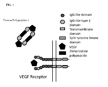
[0019] FIG. 1 illustrates the structure of an exemplary VEGF-binding dimer of
a polypeptide
described herein.
[0020] FIGS. 2A-C illustrate dot blots of CHO cells expressing fusion
polypeptide-1 and Western
blot analysis of the purified protein.
[0021] FIGS. 3A-D illustrate the neutralization of VEGF by fusion polypeptide-
1 in vitro and in
culture.
[0022] FIG. 4 illustrates a comparison of in vitro VEGF neutralization by
Aflibercept (Eylea),
Conbercept, and fusion polypeptide-1.
[0023] FIGS. 5A-D illustrate the pharmacokinetics of fusion polypeptide-1 by
demonstrating its
retention and effectiveness after injection into mice.
[0024] FIGS. 6A-B illustrate the neutralization of VEGF using sera extracted
from mice injected
with fusion polypeptide-1.
[0025] FIGS. 7A-I illustrate the in vivo neutralization of laser injury-
induced neovascularization by
fusion polypeptide-1.
DETAILED DESCRIPTION OF THE INVENTION
[0026] The systems and methods of this disclosure as described herein may
employ, unless
otherwise indicated, conventional techniques and descriptions of molecular
biology (including
recombinant techniques), cell biology, biochemistry, microarray and sequencing
technology, which
are within the skill of those who practice in the art. Such conventional
techniques include polymer
array synthesis, hybridization and ligation of oligonucleotides, sequencing of
oligonucleotides, and
detection of hybridization using a label. Specific illustrations of suitable
techniques can be had by
reference to the examples herein. However, equivalent conventional procedures
can, of course, also
be used. Such conventional techniques and descriptions can be found in
standard laboratory
manuals such as Green, et al., Eds., Genome Analysis: A Laboratory Manual
Series (Vols. I-IV)
(1999); Weiner, et al., Eds., Genetic Variation: A Laboratory Manual (2007);
Dieffenbach,
Dveksler, Eds., PCR Primer: A Laboratory Manual (2003); Bowtell and Sambrook,
DNA
Microarrays: A Molecular Cloning Manual (2003); Mount, Bioinformatics:
Sequence and Genome
Analysis (2004); Sambrook and Russell, Condensed Protocols from Molecular
Cloning: A
Laboratory Manual (2006); and Sambrook and Russell, Molecular Cloning: A
Laboratory Manual
(2002) (all from Cold Spring Harbor Laboratory Press); Stryer, L.,
Biochemistry (4th Ed.) W.H.
Freeman, N.Y. (1995); Gait, "Oligonucleotide Synthesis: A Practical Approach"
IRL Press, London
(1984); Nelson and Cox, Lehninger, Principles of Biochemistry, 3rd Ed., W.H.
Freeman Pub., New
-5-
CA 03005391 2018-05-15
WO 2017/084616
PCT/CN2016/106399
York (2000); and Berg et al., Biochemistry, 5th Ed., W.H. Freeman Pub., New
York (2002), all of
which are herein incorporated by reference in their entirety for all purposes.
Before the present
compositions, research tools and systems and methods are described, it is to
be understood that this
disclosure is not limited to the specific systems and methods, compositions,
targets and uses
described, as such may, of course, vary. It is also to be understood that the
terminology used herein
is for the purpose of describing particular aspects only and is not intended
to limit the scope of the
present disclosure, which will be limited only by appended claims.
[0027] The term "about" or "approximately" means within an acceptable error
range for the
particular value as determined by one of ordinary skill in the art, which will
depend in part on how
the value is measured or determined, i.e., the limitations of the measurement
system. For example,
"about" can mean within 1 or more than 1 standard deviation, per the practice
in the art.
Alternatively, "about" can mean a range of up to 20%, up to 10%, up to 5%, or
up to 1% of a given
value. Alternatively, particularly with respect to biological systems or
processes, the term can mean
within an order of magnitude, preferably within 5-fold, and more preferably
within 2-fold, of a
value. Where particular values are described in the application and claims,
unless otherwise stated
the term "about" meaning within an acceptable error range for the particular
value should be
assumed.
[0028] The terms "polynucleotide," "nucleic acid," and "oligonucleotide" are
used interchangeably.
As used herein, they generally refer to a polymeric form of nucleotides of any
length, either
deoxyribonucleotides or ribonucleotides, or analogs thereof. Polynucleotides
may have any three
dimensional structure, and may perform any function, known or unknown. Non-
limiting examples of
polynucleotides are coding or non-coding regions of a gene or gene fragment,
intergenic DNA, loci
(locus) defined from linkage analysis, exons, introns, messenger RNA (mRNA),
transfer RNA,
ribosomal RNA, short interfering RNA (siRNA), short-hairpin RNA (shRNA), micro-
RNA
(miRNA), small nucleolar RNA, ribozymes, cDNA, recombinant polynucleotides,
branched
polynucleotides, plasmids, vectors, isolated DNA of any sequence, isolated RNA
of any sequence,
nucleic acid probes, adapters, and primers. A polynucleotide may comprise
modified nucleotides,
such as methylated nucleotides and nucleotide analogs. If present,
modifications to the nucleotide
structure may be imparted before or after assembly of the polymer. The
sequence of nucleotides may
be interrupted by non-nucleotide components. A polynucleotide may be further
modified after
polymerization, such as by conjugation with a labeling component.
[0029] The terms "polypeptide", "peptide" and "protein" are used
interchangeably herein to refer to
polymers of amino acids of any length. The polymer may be linear or branched,
it may comprise
-6-
CA 03005391 2018-05-15
WO 2017/084616
PCT/CN2016/106399
modified amino acids, and it may be interrupted by non amino acids. The terms
also encompass an
amino acid polymer that has been modified; for example, disulfide bond
formation, glycosylation,
lipidation, acetylation, phosphorylation, or any other manipulation, such as
conjugation with a
labeling component. As used herein the term "amino acid" includes natural
and/or unnatural or
synthetic amino acids, including glycine and both the D or L optical isomers,
and amino acid analogs
and peptidomimetics.
[0030] A "control" is an alternative subject or sample used in an experiment
for comparison
purpose.
[0031] The terms "subject," "individual," and "patient" are used
interchangeably herein to refer to a
vertebrate, preferably a mammal, more preferably a human. Mammals include, but
are not limited
to, murines, simians, humans, farm animals, sport animals, and pets. Tissues,
cells, and their
progeny of a biological entity obtained in vivo or cultured in vitro are also
encompassed.
[0032] The terms "determining", "measuring", "evaluating", "assessing,"
"assaying," and
"analyzing" can be used interchangeably herein to refer to any form of
measurement, and include
determining if an element is present or not (for example, detection). These
terms can include both
quantitative and/or qualitative determinations. Assessing may be relative or
absolute. "Detecting
the presence of' can include determining the amount of something present, as
well as determining
whether it is present or absent.
[0033] In general, "sequence identity" refers to an exact nucleotide-to-
nucleotide or amino acid-to-
amino acid correspondence of two polynucleotides or polypeptide sequences,
respectively.
Typically, techniques for determining sequence identity include determining
the nucleotide sequence
of a polynucleotide and/or determining the amino acid sequence encoded
thereby, and comparing
these sequences to a second nucleotide or amino acid sequence. Two or more
sequences
(polynucleotide or amino acid) can be compared by determining their "percent
identity." The
percent identity of two sequences, whether nucleic acid or amino acid
sequences, is the number of
exact matches between two aligned sequences divided by the length of the
shorter sequences and
multiplied by 100. Percent identity may also be determined, for example, by
comparing sequence
information using the advanced BLAST computer program, including version
2.2.9, available from
the National Institutes of Health. The BLAST program is based on the alignment
method of Karlin
and Altschul, Proc. Natl. Acad. Sci. USA 87:2264-2268 (1990) and as discussed
in Altschul, et al., J.
Mol. Biol. 215:403-410 (1990); Karlin And Altschul, Proc. Natl. Acad. Sci. USA
90:5873-5877
(1993); and Altschul et al., Nucleic Acids Res. 25:3389-3402 (1997). Briefly,
the BLAST program
defines identity as the number of identical aligned symbols (i.e., nucleotides
or amino acids), divided
-7-
CA 03005391 2018-05-15
WO 2017/084616
PCT/CN2016/106399
by the total number of symbols in the shorter of the two sequences. The
program may be used to
determine percent identity over the entire length of the proteins being
compared. Default parameters
are provided to optimize searches with short query sequences in, for example,
with the blastp
program. The program also allows use of an SEG filter to mask-off segments of
the query sequences
as determined by the SEG program of Wootton and Federhen, Computers and
Chemistry 17:149-163
(1993). Ranges of desired degrees of sequence identity are approximately 80%
to 100% and integer
values therebetween. In general, an exact match indicates 100% identity over
the length of the
shortest of the sequences being compared (or over the length of both
sequences, if identical).
[0034] A "chimeric" or "fusion" polypeptide contains at least one polypeptide
comprising regions
in a different position in the sequence than that which occurs in nature. The
regions may normally
exist in separate proteins and are brought together in the fusion polypeptide;
or they may normally
exist in the same protein but are placed in a new arrangement in the fusion
polypeptide. A chimeric
or fusion polypeptide protein may be created, for example, by chemical
synthesis, or by creating and
translating a polynucleotide in which the peptide regions are encoded in the
desired relationship.
[0035] "Immunoglobulin-like domain" or "Ig-like domain" or "ligand-binding
domain" refers to
independent and distinct domains that are found in the extracellular ligand-
binding region of
cytokine receptors and it is specifically intended that the term encompass not
only the complete
wild-type domain, but also insertion, deletion and substitution variants
thereof that retain at least a
portion of the binding affinity of the wild-type domain. It will be readily
apparent to those of
ordinary skill in the art that numerous variants of the domains or
combinations of the domains of the
cytokine binding proteins can be obtained which will retain substantially the
same functional
characteristics as the wild type domain.
[0036] The phrase "ophthalmically acceptable" with respect to a formulation,
composition or
ingredient herein means having no persistent effect that is substantially
detrimental to the treated eye
or the functioning thereof, or on the general health of the subject being
treated. It will be recognized
that transient effects such as minor irritation or a "stinging" sensation are
common with topical
ophthalmic administration of drugs and the existence of such transient effects
is not inconsistent with
the formulation, composition or ingredient in question being "ophthalmically
acceptable" as herein
defined. However, preferred formulations, compositions and ingredients are
those that cause no
substantial detrimental effect, even of a transient nature.
-8-
CA 03005391 2018-05-15
WO 2017/084616
PCT/CN2016/106399
[0037] In one aspect, the present disclosure provides compositions comprising
fusion polypeptides
that include VEGF receptor immunoglobulin-like-type 2 domains fused with a
dimerization
polypeptide.
[0038] In another aspect, the present disclosure provides compositions
comprising the fusion
polypeptide comprising two or more vascular endothelial growth factor (VEGF)
receptor
immunoglobulin-like-type-2 domains fused to a dimerization polypeptide,
wherein a first
homodimer of the fusion polypeptide binds VEGF with a higher affinity than a
second homodimer
of aflibercept (SEQ ID NO: 9) when measured by: (a) incubating the first
homodimer and the second
homodimer separately with immobilized VEGF to produce bound complexes; (b)
washing the bound
complexes to remove non-specific binding; and (c) performing enzyme-linked
immunosorbent assay
(ELISA) to assess an amount of the bound complexes after step (b).
[0039] VEGF receptor immunoglobulin-like-type-2 domains are generally found in
all members of
the VEGF receptor family. In some cases, VEGF receptors can be membrane bound.
In some cases,
VEGF receptors can be soluble. In some cases, a VEGF receptor is one of three
subtypes. For
example, the first subtype can be referred to as VEGFR-1. Non-limiting
examples of the first
subtype are mouse and human Fitt. The second subtype can be referred to as
VEGFR-2. Non-
limiting examples of the second subtype are human and mouse Kdr, which can be
referred to as Flk 1
or cluster of differentiation 309 (CD309). The third subtype can be referred
to as VEGFR-3. Non-
limiting examples of the second subtype are human and mouse Flt-4.
[0040] The dimerization polypeptide utilized in the fusion polypeptide can be
a fragment
crystallizable (Fc) region of an antibody. In some cases, the dimerization
polypeptide includes a
hinge region. The dimerization polypeptide can be designed to incorporate a
partial Fc without a
hinge and with a CH2 domain that is truncated but retains FcRn binding in
order to confer longer
terminal half-life on the construct. In yet another embodiment, the binding
fusion polypeptide can
be designed to incorporate a partial Fc without hinge but with a CH2 and CH3
domain, which can
dimerize via the CH3 domain.
[0041] The dimerization polypeptide utilized in the fusion polypeptide can
employ heterodimeric
dimerization domains from a variety of sources. They include but are not
limited to heterodimeric
receptors that bind to growth factors (e.g. heregulin), neurotransmitters
(e.g. y-Aminobutyric acid),
and other organic or inorganic small molecules (e.g. mineralocorticoid,
glucocorticoid). Exemplary
heterodimeric receptors are nuclear hormone receptors, erbB3 and erbB2
receptor complex, and G-
protein-coupled receptors including but not limited to opioid, muscarinic,
dopamine, serotonin,
-9-
CA 03005391 2018-05-15
WO 2017/084616
PCT/CN2016/106399
adenosine/dopamine, and GABAB families of receptors. For majority of the known
heterodimeric
receptors, their C-terminal sequences are found to mediate heterodimer
formation. A compilation of
known dimerization domains is provided on the following websites
http://coiledcoils.chm.bris.ac.uk/ccplus/search/ and has been described in
Testa et al., Nucleic Acid
Research (2009) 37: D315-D322. Additionally, coiled coil structures were
described in Vincent, TL,
et al., Bioinformatics (2013) 29: 69-76; Armstrong et al., Bioinformatics
(2011) 27: 1908-1914;
Woolfson DN, Adv. Prot. Chem. (2005) 70: 79-112; Mason JM and Arndt KM,
ChemBioChem
(2004) 5: 170-176; each of which is incorporated by reference herein as if
fully set forth.
[0042] In an embodiment, the dimerization polypeptide may include coiled coils
from basic leucine
zippers. The other basic leucine zippers may be TF6, CREB1, C/EBPct, Fos, or
Jun, viral fusion
polypeptides influenza hemagglutinin or HIV gp41, or other coiled coil domains
APC or ProP.
Dimerization domains may be thermo- sensitive dimeric coiled coils or more
complex multimeric
coil structures in recombinant proteins as a means to regulate enzyme activity
both in vivo and in
vitro.
[0043] Another exemplary class of heterodimerization sequences consists of
amphiphilic peptides
that adopt a coiled-coil helical structure. Well-characterized coiled-coil-
containing proteins include
members of the cytoskeletal family (e.g. a-keratin, vimentin), cytoskeletal
motor family (e.g.
myosin, kinesins, and dyneins), viral membrane proteins (e.g. membrane
proteins of Ebola or HIV),
DNA binding proteins, and cell surface receptors (e.g. GABAB receptors 1 and
2). Coiled-coil
heterodimerization sequences of the present invention can be broadly
classified into two groups,
namely the left-handed and right-handed coiled coils. The left-handed coiled
coils are characterized
by a heptad repeat denoted "abcdefg" with the occurrence of apolar residues
preferentially located at
the first (a) and fourth (d) position. The residues at these two positions
typically constitute a zig-zag
pattern of "knobs and holes" that interlock with those of the other stand to
form a tight-fitting
hydrophobic core. In contrast, the second (b), third (c) and sixth (f)
positions that cover the periphery
of the coiled coil are preferably charged residues. Examples of charged amino
acids include basic
residues such as lysine, arginine, histidine, and acidic residues such as
aspartate, glutamate,
asparagine, and glutamine. Uncharged or apolar amino acids suitable for
designing a heterodimeric
coiled coil include but are not limited to glycine, alanine, valine, leucine,
isoleucine, serine and
threonine. While the uncharged residues typically form the hydrophobic core,
inter-helical and intra-
helical salt-bridge including charged residues even at core positions may be
employed to stabilize
the overall helical coiled-coiled structure (Burkhard et al. (2000) J. Biol.
Chem. 275:11672-11677).
Whereas varying lengths of coiled coil may be employed, the subject
heterodimerization sequences
-10-
CA 03005391 2018-05-15
WO 2017/084616
PCT/CN2016/106399
preferably contain two to ten heptad repeats. More preferably, the
heterodimerization sequences
contain three to eight heptad repeats, even more preferably contain four to
five heptad repeats.
[0044] In designing optimal coiled-coil heterodimerization sequences, a
variety of existing computer
software programs that predict the secondary structure of a peptide can be
used. An illustrative
computer analysis uses the COILS algorithm which compares an amino acid
sequence with
sequences in the database of known two-stranded coiled coils, and predicts the
high probability
coiled-coil stretches (Kammerer et al.(1999) Biochemistry 38:13263-13269).
[0045] In an embodiment, dimerization polypeptides may include dimerization
domains other than
coiled coils. Dimerization domains may be, but are not limited to, membrane
dimerization domains,
dimerization domains from transcription factors other than leucine zippers, G
protein f3y complexes
from heterotrimeric G protein complexes, TIM, ADH5, 14-3-3 proteins or their
binding partners Bad
or Bax, or other protein dimers. Membrane dimerization domains may be
glycophorin A, receptor
tyrosine kinases, or GPCRs. Dimerization domains from transcription factors
other than leucine
zippers may be nuclear receptors, an estrogen receptor, an androgen receptor,
a glucocorticoid
receptor, basic helix-loop-helix MyoD or c-Myc, helix-turn- helix LuxR, TetR,
or cl.
[0046] Additionally, computer modeling and searching technologies further
facilitates detection of
heterodimerization sequences based on sequence homologies of common domains
appeared in
related and unrelated genes. Non-limiting examples of programs that allow
homology searches are
Blast (http://www.ncbi.nlm.nih.gov/BLAST/), Fasta (Genetics Computing Group
package, Madison,
Wis.), DNA Star, Clustlaw, TOFFEE, COBLATH, Genthreader, and MegAlign. Any
sequence
databases that contains DNA sequences corresponding to a target receptor or a
segment thereof can
be used for sequence analysis. Commonly employed databases include but are not
limited to
GenBank, EMBL, DDBJ, PDB, SWISS-PROT, EST, STS, GSS, and HTGS.
[0047] In some cases, dimerization is mediated by non-covalent interactions
between fusion
polypeptides. In some cases, dimerization is mediated by covalent interactions
between fusion
polypeptides. For example, covalent interactions can comprise one or more
disulfide bonds formed
between cysteine residues on two dimerization polypeptides.
[0048] In one aspect, the invention provides dimers of the disclosed fusion
polypeptides. Dimers can
be homodimers, in which each fusion polypeptide in the dimer is identical.
Dimers can be
heterodimers, in which each fusion polypeptide in the dimer is different.
[0049] VEGF receptors comprise immunoglobulin domains. In some cases, wild-
type VEGF
receptors comprise an extracellular ligand binding domain comprising 7
immunoglobulin domains, a
transmembrane spanning region, and an intracellular domain comprising a split
tyrosine-kinase
-11-
CA 03005391 2018-05-15
WO 2017/084616
PCT/CN2016/106399
domain (see FIG. 1). The extracellular ligand binding domain is defined as the
portion of a receptor
that, in its native conformation in the cell membrane, is oriented
extracellularly where it can contact
with its cognate ligand. The extracellular ligand binding domain does not
include the hydrophobic
amino acids associated with the receptor's transmembrane domain or any amino
acids associated
with the receptor's intracellular domain. Generally, the intracellular or
cytoplasmic domain of a
receptor is usually composed of positively charged or polar amino acids (i.e.
lysine, arginine,
histidine, glutamic acid, aspartic acid). The preceding 15-30, predominantly
hydrophobic or apolar
amino acids (i.e. leucine, valine, isoleucine, and phenylalanine) comprise the
transmembrane
domain. The extracellular domain comprises the amino acids that precede the
hydrophobic
transmembrane stretch of amino acids. Usually the transmembrane domain is
flanked by positively
charged or polar amino acids such as lysine or arginine.
[0050] In some cases, the VEGF receptor immunoglobuline-like-type-2 domain is
from a VEGF
receptor from a mammal, such as a rat, human, mouse, dog, horse, cat, or
sheep. In some cases, the
VEGF receptor immunoglobuline-like-type-2 domain is from a VEGF receptor from
an animal, such
as a nematode, an ant, a bird, a whale, cnidarian, or a fish. In an exemplary
embodiment, the VEGF
receptor is from a human.
[0051] In some cases, the VEGF receptor immunoglobuline-like-type-2 domain is
at least 30%, at
least 35%, at least 40%, at least 45%, at least 50%, at least 55%, at least
60%, at least 65%, at least
70%, at least 75%, at least 80%, at least 85%, at least 90%, at least 95%, at
least 96%, at least 97%,
at least 98%, or at least 99% identical to a VEGF receptor immunoglobuline-
like-type-2 domain
from a wild-type VEGF receptor. In some cases, the VEGF receptor
immunoglobuline-like-type-2
domain is from a VEGF receptor is selected from the group consisting of Fltl,
Kdr, and Flt-4. In
some cases, the VEGF receptor is Fltl. In some cases the VEGF receptor is Kdr.
In some cases the
VEGF receptor is Flt-4. In some cases, the VEGF receptor is a human VEGF
receptor. In some
cases, the VEGF receptor is selected from the group consisting of SEQ IDs NO:
4-6.
[0052] In some cases, a VEGF receptor immunoglobulin-like-type 2 domain of the
fusion
polypeptide can be truncated. In some cases, the VEGF receptor immunoglobulin-
like-type 2
domains of the fusion polypeptide comprise at least 70%, at least 75%, at
least 80%, at least 85%, at
least 90%, at least 95%, at least 96%, at least 97%, at least 98%, or at least
99% the length of a
whole VEGF receptor immunoglobulin-like-type 2 domain. In some cases, the VEGF
receptor
immunoglobulin-like-type 2 domain is a human VEGF receptor. In some cases, the
VEGF receptor
immunoglobulin-like-type 2 domain of the fusion polypeptide is at least 70%,
at least 75%, at least
-12-
CA 03005391 2018-05-15
WO 2017/084616
PCT/CN2016/106399
80%, at least 85%, at least 90%, at least 95%, at least 96%, at least 97%, at
least 98%, or at least
99% the length of a sequence selected from the group consisting of SEQ IDs NO:
4-6.
[0053] In some cases, the fusion polypeptide comprises two or more
immunoglobulin-like-type 2
domains. For example, the fusion polypeptide can comprise two, three, four,
five, six, seven, eight,
nine, ten, or more immunoglobulin-like-type 2 domains. The two or more domains
can be fused on
either side of a dimerization polypeptide. For example, a first immunoglobulin-
like-type 2 domain
can be fused at the N-terminus of a dimerization polypeptide, and a second
immunoglobulin-like-
type 2 domain can be fused at the C-terminus of a dimerization polypeptide.
The two or more
immunoglobulin-like-type 2 domains can be derived from the same VEGF receptor.
The two or
more immunoglobulin-like-type 2 domains can be derived from distinct VEGF
receptors. For
example, a first immunoglobulin-like-type 2 domain can be derived from Fla and
a second from
Kdr.
[0054] The fusion polypeptide may comprise VEGF receptor immunoglobulin-like-
type 2 domains
connected directly to each other or to the dimerization polypeptide. The
fusion polypeptide may
comprise VEGF receptor immunoglobulin-like-type 2 domains connected to each
other or to the
dimerization polypeptide via spacers or linkers. In some cases, the spacers or
linkers include portions
or all of an antibody hinge region.
[0055] In some cases, the fusion polypeptide substantially lacks a VEGF
receptor immunoglobulin-
like-type 3 domain. For example, the fusion polypeptide may comprise no more
than 60 amino
acids, 55 amino acids, 50 amino acids, 45 amino acids, 40 amino acids, 35
amino acids, 30 amino
acids, 25 amino acids, 20 amino acids, 15 amino acids, 10 amino acids, 5 amino
acids of a VEGF
receptor immunoglobulin-like-type 3 domain. In some cases, the fusion
polypeptide lacks a sequence
that is at least 30%, at least 35%, at least 40%, at least 45%, at least 50%,
at least 55%, at least 60%,
at least 65%, at least 70%, at least 75%, at least 80%, at least 85%, at least
90%, at least 95%, at
least 96%, at least 97%, at least 98%, or at least 99% identical to a VEGF
receptor immunoglobulin-
like type 3 domain. In some cases, the VEGF receptor is selected from the
group consisting of Fltl,
Kdr, and Flt-4. In some cases, the VEGF receptor is Fitt. In some cases the
VEGF receptors is Kdr.
In some cases the VEGF receptor is Flt-4. In some cases, the VEGF receptor
immunoglobulin-like
type 3 is a human VEGF receptor immunoglobulin-like type 3 domain. In some
cases, the VEGF
receptor is selected from the group consisting of SEQ IDs NO: 1-3. In some
cases, the fusion
polypeptide lacks SEQ ID NO: 8.
[0056] In some cases, the fusion polypeptide comprising two or more vascular
endothelial growth
factor (VEGF) receptor immunoglobulin-like-type-2 domains fused to a
dimerization polypeptide,
-13-
CA 03005391 2018-05-15
WO 2017/084616
PCT/CN2016/106399
wherein a first homodimer of the fusion polypeptide binds VEGF with a higher
affinity than a
second homodimer of aflibercept (SEQ ID NO: 9) when measured by: (a)
incubating the first
homodimer and the second homodimer separately with immobilized VEGF to produce
bound
complexes; (b) washing the bound complexes to remove non-specific binding; and
(c) performing
enzyme-linked immunosorbent assay (ELISA) to assess an amount of the bound
complexes after
step (b)
[0057] In some cases, the binding affinity of the homodimer of the fusion
polypeptides is higher
than that of the fusion polypeptide oligomer. The homodimer of the fusion
polypeptides can have a
higher binding affinity for VEGF than wild-type VEGFR. The homodimer of the
fusion polypeptides
can have a higher binding affinity for VEGF than the homodimer of aflibercept
(SEQ ID NO: 9).
[0058] Binding affinity can be observed or measured using art-recognized
techniques including but
not limited to ELISA, competitive ELISA, in vitro and in vivo neutralization
assays (see Example 2).
[0059] In one example, binding affinity is measured by an ELISA assay. VEGF
and a polypeptide
can be incubated to achieve equilibrium binding. An antibody, directed to VEGF
or the polypeptide,
comprising an enzyme is incubated with the putative binding partner. After
washing, the amount of
protein complex is determined by incubating with the enzyme substrate and
measuring the product.
Exemplary methods for using ELISA to determine binding affinity can be found
in Gan and Patel,
Enzyme Immunoassay and Enzyme-Linked Immunosorbent Assay, Journal of
Investigative
Dermatology (2013), which is hereby incorporated by reference in its entirety.
[0060] In one example, binding affinity is measured by fluorescence resonance
energy transfer
(FRET). VEGF and the peptide of interest are labeled with a first and second
fluorescent molecule,
respectively. FRET relies on the principle that energy can be transferred
between two light-sensitive
molecules. A donor chromophore in an excited state can transfer energy to an
acceptor chromophore
through nonradiative dipole-dipole coupling. The efficiency of this transfer
is proportional the sixth
power of the distance of the two molecules, making FRET extremely sensitive to
changes in distance
between the two molecules. The equilibrium binding constant Kd can be
determined by setting up a
titration where the acceptor-tagged protein is added to the donor-tagged
protein while the emission
spectra are monitored. The ratio of donor-derived fluorescence to acceptor-
derived fluorescence can
be used to determine the binding affinity of the two proteins. For exemplary
methods see Martin et
al., Quantitative analysis of multi-protein interactions using FRET:
Application to the SUMO
pathway. Protein Science (2008), which is hereby incorporated by reference in
its entirety.
[0061] In a second example, binding affinity can be measured by Surface
Plasmon Resonance
(SPR). SPR measures the change in the angle at which polarized light is
reflected from a surface.
-14-
CA 03005391 2018-05-15
WO 2017/084616
PCT/CN2016/106399
The angle is related to the change in mass or layer thickness of the surface
of a chip. Binding affinity
can be measured using, for example, the ProteON XPR36 protein interaction
array system (Bio-
Rad).
[0062] In one aspect, the disclosure provides for a polynucleotide encoding
any fusion polypeptide
described herein. The polynucleotide can be, for example, the polynucleotide
of SEQ ID NO: 7. A
polynucleotide described herein can be obtained using chemical synthesis,
molecular cloning or
recombinant methods, DNA or gene assembly methods, artificial gene synthesis,
PCR, or any
combination thereof. Methods of chemical polynucleotide synthesis are well
known in the art and
need not be described in detail herein. One of skill in the art can use the
sequences provided herein
and a commercial DNA synthesizer to produce a desired DNA sequence. For
preparing
polynucleotides using recombinant methods, a polynucleotide comprising a
desired sequence can be
inserted into a suitable cloning or expression vector, and the cloning or
expression vector in turn can
be introduced into a suitable host cell for replication and amplification, as
further discussed herein.
Polynucleotides may be inserted into host cells by any means known in the art.
Cells may be
transformed by introducing an exogenous polynucleotide, for example, by direct
uptake,
endocytosis, transfection, F-mating, chemical transformation, or
electroporation. Once introduced,
the exogenous polynucleotide can be maintained within the cell as a non-
integrated expression
vector (such as a plasmid) or integrated into the host cell genome. The
polynucleotide so amplified
can be isolated from the host cell by methods well known within the art.
Alternatively, nucleic acid
amplification methods (e.g., PCR) allow reproduction of DNA sequences.
[0063] For recombinant expression of fusion polypeptide disclosed herein, the
polynucleotide
encoding it is isolated and inserted into a replicable vector for further
cloning (amplification of the
DNA) or for expression. In some embodiments, fusion polypeptide is cloned into
the vector, and the
wild-type sequence is mutated to produce a mutant fusion polypeptide
expression vector, such as by
directed mutation using methods known in the art (e.g. by PCR with a primer
containing the desired
mutation). DNA encoding wild-type and mutant fusion polypeptide is readily
isolated and sequenced
using conventional procedures (e.g., by using oligonucleotide probes and
primers). Many vectors are
available. The vector components generally include, but are not limited to,
one or more of the
following: a signal sequence, an origin of replication, one or more marker
genes, an enhancer
element, a promoter, and a transcription-termination sequence.
[0064] In general, and unless the expression vector is introduced into a host
cell chromosome, both
expression and cloning vectors contain a polynucleotide sequence that enables
the vector to replicate
in one or more selected host cells. Generally, in cloning vectors this
sequence is one that enables the
-15-
CA 03005391 2018-05-15
WO 2017/084616
PCT/CN2016/106399
vector to replicate independently of the host chromosomal DNA, and includes
origins of replication
or autonomously replicating sequences. Such sequences are well known for a
variety of bacteria,
yeast, and viruses. The origin of replication from the plasmid pBR322 is
suitable for most Gram-
negative bacteria, the 2p plasmid origin is suitable for yeast, and various
viral origins (5V40,
polyoma, adenovirus, VSV or BPV) are useful for cloning vectors in mammalian
cells. Generally,
the origin of replication component is not needed for mammalian expression
vectors (the 5V40
origin may typically be used only because it contains the early promoter).
[0065] Expression and cloning vectors may contain a selection gene, also
termed a selectable
marker. Typical selection genes encode proteins that (a) confer resistance to
antibiotics or other
toxins, e.g., ampicillin, neomycin, methotrexate, or tetracycline, (b)
complement auxotrophic
deficiencies, or (c) supply critical nutrients not available from complex
media, e.g., the gene
encoding D-alanine racemase for Bacilli. One example of a selection scheme
utilizes a drug to arrest
growth of a host cell. Those cells that are successfully transformed with a
heterologous gene produce
a protein conferring drug resistance and thus survive the selection regimen.
Examples of such
dominant selection use the drugs neomycin, mycophenolic acid, G418, kanamycin,
and hygromycin.
See U.S. Pat. No. 4,965,199.
[0066] A suitable selection gene for use in yeast is the trp 1 gene present in
the yeast plasmid YRp7
(Stinchcomb et al., Nature, 282:39 (1979)). The trpl gene provides a selection
marker for a mutant
strain of yeast lacking the ability to grow in tryptophan, for example, ATCC
No. 44076 or PEP4-1.
Jones, Genetics, 85:12 (1977). The presence of the trp 1 lesion in the yeast
host cell genome then
provides an effective environment for detecting transformation by growth in
the absence of
tryptophan. Similarly, Leu2-deficient yeast strains (ATCC 20,622 or 38,626)
are complemented by
known plasmids bearing the Leu2 gene. In addition, vectors derived from the
1.6-pm circular
plasmid pl(D1 can be used for transformation of Kluyveromyces yeasts.
Alternatively, an expression
system for large-scale production of recombinant calf chymosin was reported
for K. lactis. Van den
Berg, Bio/Technology, 8:135 (1990). Stable multi-copy expression vectors for
secretion of mature
recombinant human serum albumin by industrial strains of Kluyveromyces have
also been disclosed.
Fleer et al., Bio/Technology, 2: 968-975 (1991).
[0067] In some embodiments, the expression vector also comprises a nucleotide
sequence encoding
a detectable label. A detectable label may include, but is not limited to an
enzyme, a transcription
factor, a radioisotope binding protein, a fluorescent protein, or a
fluorescent protein complex. In
certain aspects, the fluorescent protein is a green fluorescent protein (GFP),
cyan fluorescent protein
(CFP), blue fluorescent protein (BFP), yellow fluorescent protein (YFP), red
fluorescent protein
-16-
CA 03005391 2018-05-15
WO 2017/084616
PCT/CN2016/106399
(RFP), variants thereof, or various combinations thereof. In some embodiments,
the detectable label
is detectable by fluorescence, enzymatic activity, FRET, or NMR.
[0068] Expression and cloning vectors usually contain a promoter that is
recognized by the host
organism and is operably linked to fusion polypeptide-encoding polynucleotide.
Promoters suitable
for use with prokaryotic hosts include the phoA promoter, P-lactamase and
lactose promoter
systems, alkaline phosphatase, a tryptophan (trp) promoter system, and hybrid
promoters such as the
tac promoter. However, other known bacterial promoters are suitable. Promoters
for use in bacterial
systems also will contain a Shine-Dalgarno (S.D.) sequence operably linked to
the DNA encoding
the fusion polypeptide.
[0069] Promoter sequences are known for eukaryotes. Virtually all eukaryotic
genes have an AT-
rich region located approximately 25 to 30 bases upstream from the site where
transcription is
initiated. Another sequence found 70 to 80 bases upstream from the start of
transcription of many
genes is a CNCAAT region where N may be any nucleotide. At the 3' end of most
eukaryotic genes
is an AATAAA sequence that may be the signal for addition of the poly A tail
to the 3' end of the
coding sequence. All of these sequences are suitably inserted into eukaryotic
expression vectors.
[0070] Examples of suitable promoting sequences for use with eukaryotic hosts
include the
promoters for 3-phosphoglycerate kinase or other glycolytic enzymes, such as
enolase,
glyceraldehyde-3-phosphate dehydrogenase, hexokinase, pyruvate decarboxylase,
phosphofructokinase, glucose-6-phosphate isomerase, 3-phosphoglycerate mutase,
pyruvate kinase,
triosephosphate isomerase, phosphoglucose isomerase, and glucokinase. Other
eukaryotic promoters,
which are inducible promoters having the additional advantage of transcription
controlled by growth
conditions, are the promoter regions for alcohol dehydrogenase 2,
isocytochrome C, acid
phosphatase, degradative enzymes associated with nitrogen metabolism,
metallothionein,
glyceraldehyde-3-phosphate dehydrogenase, and enzymes responsible for maltose
and galactose
utilization. Suitable vectors and promoters for use in eukaryotic expression
are further described in
EP 73,657. Eukaryotic enhancers also are advantageously used with eukaryotic
promoters. Non-
limiting examples of eukaryotic cells include yeast cells and mammalian cell
lines.
[0071] Fusion polypeptide protein expression from vectors in mammalian host
cells can be
controlled, for example, by promoters obtained from the genomes of viruses
such as polyoma virus,
fowlpox virus, adenovirus (such as Adenovirus 2), bovine papilloma virus,
avian sarcoma virus,
cytomegalovirus, a retrovirus, hepatitis-B virus and most preferably Simian
Virus 40 (5V40),
heterologous mammalian promoters, e.g., the actin promoter or an
immunoglobulin promoter, and
heat-shock promoters, provided such promoters are compatible with the host
cell systems. The early
-17-
CA 03005391 2018-05-15
WO 2017/084616
PCT/CN2016/106399
and late promoters of the SV40 virus are conveniently obtained as an SV40
restriction fragment that
also contains the SV40 viral origin of replication. The immediate early
promoter of the human
cytomegalovirus is conveniently obtained as a Hin III E restriction fragment.
A system for
expressing DNA in mammalian hosts using the bovine papilloma virus as a vector
is disclosed in
U.S. Pat. No. 4,419,446. A modification of this system is described in U.S.
Pat. No. 4,601,978. See
also Reyes et al., Nature, 297:598-601 (1982) on expression of human 0-
interferon cDNA in mouse
cells under the control of a thymidine kinase promoter from herpes simplex
virus. Alternatively, the
rous sarcoma virus long-terminal repeat can be used as the promoter.
[0072] Transcription of a DNA encoding an fusion polypeptide by higher
eukaryotes is often
increased by inserting an enhancer sequence into the vector. Many enhancer
sequences are now
known from mammalian genes (globin, elastase, albumin, a-fetoprotein, and
insulin). Typically,
however, one will use an enhancer from a eukaryotic cell virus. Examples
include the 5V40
enhancer on the late side of the replication origin (bp 100-270), the
cytomegalovirus early-promoter
enhancer, the polyoma enhancer on the late side of the replication origin, and
adenovirus enhancers.
See also Yaniv, Nature, 297:17-18 (1982) on enhancing elements for activation
of eukaryotic
promoters. The enhancer may be spliced into the vector at a position 5' or 3'
to the fusion
polypeptide-encoding sequence, but is preferably located at a site 5' from the
promoter.
[0073] Expression vectors used in eukaryotic host cells (for example, yeast,
fungi, insect, plant,
animal, human, or nucleated cells from other multicellular organisms) will
also contain sequences
necessary for the termination of transcription and for stabilizing the mRNA.
Such sequences are
commonly available from the 5' end, occasionally 3' end, of untranslated
regions of eukaryotic or
viral DNAs or cDNAs. These regions contain nucleotide segments transcribed as
polyadenylated
fragments in the untranslated portion of the mRNA encoding the fusion
polypeptide. One useful
transcription termination component is the bovine growth hormone
polyadenylation region. See WO
1994/11026 and the expression vector disclosed therein.
Host Cells
[0074] In one aspect, the disclosure provides a host cell comprising an
expression vector comprising
a nucleotide sequence encoding a fusion polypeptide disclosed herein, such as
expression vectors
described herein. In some embodiments, the expression vector is
extrachromosomal, such as a
plasmid. In some embodiments, the host cell comprises a stably integrated
transgenic nucleotide
sequence encoding the fusion polypeptide. Under suitable conditions, the host
cell actively expresses
the fusion polypeptide. Conditions suitable for expression depend on a number
of factors known in
-18-
CA 03005391 2018-05-15
WO 2017/084616
PCT/CN2016/106399
the art, such as growth conditions for the cells and the activity of the
promoter driving expression,
which may be constitutively active, active in specific cell types, inducible
in response to the
presence of an inducing agent, or any other promoter described herein or known
in the art.
[0075] In some embodiments the host cell expresses a selectable marker.
Examples of selectable
markers are provided herein, and may be expressed from the expression vector
encoding the fusion
polypeptide, or separately, such as from another expression vector which may
or may not be
integrated into the host cell genome. In some embodiments, the host cell
expresses a detectable label.
Examples of detectable labels are provided herein, and may be expressed from
the expression vector
encoding the fusion polypeptide, or separately, such as from another
expression vector which may or
may not be integrated into the host cell genome.
[0076] Suitable host cells for cloning or expressing the DNA in the vectors
herein are the
prokaryote, yeast, or higher eukaryote cells described herein and otherwise
known in the art.
Suitable prokaryotes for this purpose include eubacteria, such as Gram-
negative or Gram-positive
organisms, for example, Enterobacteriaceae such as Escherichia, e.g., E. coli,
Enterobacter, Erwinia,
Klebsiella, Proteus, Salmonella, e.g., Salmonella typhimurium, Serratia, e.g.,
Serratia marcescans,
and Shigella, as well as Bacilli such as B. subtilis and B. licheniformis
(e.g., B. licheniformis 41P
disclosed in DD 266,710 published 12 Apr. 1989), Pseudomonas such as P.
aeruginosa, and
Streptomyces. One exemplary E. coli cloning host is E. coli 294 (ATCC 31,446),
although other
strains such as E. coli B, E. coli X1776 (ATCC 31,537), and E. coli W3110
(ATCC 27,325) are
suitable. These examples are illustrative rather than limiting.
[0077] In addition to prokaryotes, eukaryotic microbes such as filamentous
fungi or yeast are
suitable cloning or expression hosts for fusion polypeptide-encoding vectors.
Saccharomyces
cerevisiae, or common baker's yeast, is the most commonly used among lower
eukaryotic host
microorganisms. However, a number of other genera, species, and strains are
commonly available
and useful herein, such as Schizosaccharomyces pombe; Kluyveromyces hosts such
as, e.g., K.
lactis, K. fragilis (ATCC 12,424), K. bulgaricus (ATCC 16,045), K. wickeramii
(ATCC 24,178), K
waltii (ATCC 56,500), K. drosophilarum (ATCC 36,906), K. thermotolerans, and
K. marxianus;
yarrowia (EP 402,226); Pichia pastoris (EP 183,070); Candida; Trichoderma
reesia (EP 244,234);
Neurospora crassa; Schwanniomyces such as Schwanniomyces occidentalis; and
filamentous fungi
such as, e.g., Neurospora, Penicillium, Tolypocladium, and Aspergillus hosts
such as A. nidulans
and A. niger.
[0078] Suitable host cells for the expression of fusion polypeptide include
cells derived from
multicellular organisms. Examples of invertebrate cells include plant and
insect cells. Numerous
-19-
CA 03005391 2018-05-15
WO 2017/084616
PCT/CN2016/106399
baculoviral strains and variants and corresponding permissive insect host
cells from hosts such as
Spodoptera frugiperda (caterpillar), Aedes aegypti (mosquito), Aedes
albopictus (mosquito),
Drosophila melanogaster (fruitfly), and Bombyx mori have been identified. A
variety of viral strains
for transfection are publicly available, e.g., the L-1 variant of Autographa
californica NPV and the
Bm-5 strain of Bombyx mori NPV, and such viruses may be used as the virus
herein according to
the present invention, particularly for transfection of Spodoptera frugiperda
cells.
[0079] Plant cell cultures of cotton, corn, potato, soybean, petunia, tomato,
and tobacco can also be
utilized as hosts.
[0080] However, interest has been greatest in vertebrate cells, and
propagation of vertebrate cells in
culture (tissue culture) has become a routine procedure. Examples of useful
mammalian host cell
lines are monkey kidney CV1 line transformed by SV40 (COS-7, ATCC CRL 1651);
human
embryonic kidney line (293 or 293 cells subcloned for growth in suspension
culture, Graham et al.,
J. Gen Virol., 36:59 (1977)); baby hamster kidney cells (BHK, ATCC CCL 10);
Chinese hamster
ovary cells/-DHFR (CHO, Urlaub et al., Proc. Natl. Acad. Sci. USA, 77:4216
(1980), including
DG44 (Urlaub et al., Som. Cell and Mol. Gen., 12: 555-566 (1986)) and DP12
cell lines); mouse
sertoli cells (TM4, Mather, Biol. Reprod., 23:243-251 (1980)); monkey kidney
cells (CV1 ATCC
CCL 70); African green monkey kidney cells (VERO-76, ATCC CRL-1587); human
cervical
carcinoma cells (HELA, ATCC CCL 2); canine kidney cells (MDCK, ATCC CCL 34);
buffalo rat
liver cells (BRL 3A, ATCC CRL 1442); human lung cells (W138, ATCC CCL 75);
human liver
cells (Hep G2, HB 8065); mouse mammary tumor (MMT 060562, ATCC CCL51); TR1
cells
(Mather et al., Annals N.Y. Acad. Sci., 383:44-68 (1982)); MRC 5 cells; F54
cells; and a human
hepatoma line (Hep G2).
[0081] A wide variety of additional cell lines for various tissue culture
applications, gene
expression, and assays are known in the art. Examples of cell lines include,
but are not limited to,
C8161, CCRF-CEM, MOLT, mIMCD-3, NHDF, HeLa-53, Huhl, Huh4, Huh7, HUVEC, HASMC,
HEKn, HEKa, MiaPaCell, Pancl, PC-3, TF1, CTLL-2, C1R, Rat6, CV1, RPTE, A10,
T24, J82,
A375, ARH-77, Calul, 5W480, 5W620, SKOV3, SK-UT, CaCo2, P388D1, SEM-K2, WEHI-
231,
HB56, TIB55, Jurkat, J45.01, LRMB, Bcl-1, BC-3, IC21, DLD2, Raw264.7, NRK, NRK-
52E,
MRCS, MEF, Hep G2, HeLa B, HeLa T4, COS, COS-1, COS-6, COS-M6A, BS-C-1 monkey
kidney epithelial, BALB/3T3 mouse embryo fibroblast, 3T3 Swiss, 3T3-L1, 132-d5
human fetal
fibroblasts; 10.1 mouse fibroblasts, 293-T, 3T3, 721, 9L, A2780, A2780ADR,
A2780cis, A172,
A20, A253, A431, A-549, ALC, B16, B35, BCP-1 cells, BEAS-2B, bEnd.3, BHK-21,
BR 293,
BxPC3, C3H-10T1/2, C6/36, Cal-27, CHO, CHO-7, CHO-IR, CHO-K1, CHO-K2, CHO-T,
CHO
-20-
CA 03005391 2018-05-15
WO 2017/084616
PCT/CN2016/106399
Dhfr -/-, COR-L23, COR-L23/CPR, COR-L23/5010, COR-L23/R23, COS-7, COV-434, CML
Ti,
CMT, CT26, D17, DH82, DU145, DuCaP, EL4, EM2, EM3, EMT6/AR1, EMT6/AR10.0, FM3,
H1299, H69, HB54, HB55, HCA2, HEK-293, HeLa, Hepalc1c7, HL-60, HMEC, HT-29,
Jurkat, JY
cells, K562 cells, Ku812, KCL22, KG1, KY01, LNCap, Ma-Mel 1-48, MC-38, MCF-7,
MCF-10A,
MDA-MB-231, MDA-MB-468, MDA-MB-435, MDCK II, MDCK II, MOR/0.2R, MONO-MAC 6,
MTD-1A, MyEnd, NCI-H69/CPR, NCI-H69/LX10, NCI-H69/LX20, NCI-H69/LX4, NIH-3T3,
NALM-1, NW-145, OPCN/OPCT cell lines, Peer, PNT-1A/PNT 2, RenCa, RIN-5F,
RMA/RMAS,
Saos-2 cells, Sf-9, SkBr3, T2, T-47D, T84, THP1 cell line, U373, U87, U937,
VCaP, Vero cells,
WM39, WT-49, X63, YAC-1, YAR, and transgenic varieties thereof. Cell lines are
available from a
variety of sources known to those with skill in the art (see, e.g., the
American Type Culture
Collection (ATCC) (Manassus, Va.)).
[0082] Host cells can be transfected with one or more of the above-described
expression or cloning
vectors for fusion polypeptide expression and cultured in conventional
nutrient media modified as
appropriate for inducing promoters, selecting transformants, or amplifying the
genes encoding the
desired sequences.
[0083] Host cells transfected with expression vectors for the expression of a
fusion polypeptide
herein may be cultured in a variety of media. Commercially available media
such as Ham's F10
(Sigma), Minimal Essential Medium ((MEM), (Sigma), RPMI-1640 (Sigma), and
Dulbecco's
Modified Eagle's Medium ((DMEM), Sigma) are suitable for culturing the host
cells. In addition,
any of the media described, for example, in Ham et al., Meth. Enz. 58:44
(1979); Barnes et al., Anal.
Biochem. 102:255 (1980); U.S. Pat. No. 4,767,704; 4,657,866; 4,927,762;
4,560,655; or 5,122,469;
WO 1990/03430; WO 1987/00195; or U.S. Pat. No. Re. 30,985 may be used as
culture media for the
host cells. Any of these media may be supplemented as necessary with hormones
and/or other
growth factors (such as insulin, transferrin, or epidermal growth factor),
salts (such as sodium
chloride, calcium, magnesium, and phosphate), buffers (such as HEPES),
nucleotides (such as
adenosine and thymidine), antibiotics (such as GENTAMYCINTm drug), trace
elements (defined as
inorganic compounds usually present at final concentrations in the micromolar
range), and glucose
or an equivalent energy source. Any other necessary supplements may also be
included at
appropriate concentrations that would be known to those skilled in the art.
The culture conditions,
such as temperature, pH, and the like, are those previously used with the host
cell selected for
expression, and will be apparent to the ordinarily skilled artisan.
[0084] Several transfection protocols are known in the art, and are reviewed
in Kaufman R. J., et al.,
Nucleic Acids Res. 19:4485, 1991. The transfection protocol chosen will depend
on the host cell
-21-
CA 03005391 2018-05-15
WO 2017/084616
PCT/CN2016/106399
type and the nature of the expression vector, and can be chosen based upon
routine experimentation.
Typically, a transfection protocol includes introducing an expression vector
into a suitable host cell,
and then identifying and isolating host cells which have incorporated the
heterologous DNA in a
stable, expressible manner.
[0085] One commonly used method of introducing heterologous DNA is calcium
phosphate
precipitation, for example, as described by Wigler et al. (Proc. Natl. Acad.
Sci. USA 77:3567, 1980).
DNA introduced into a host cell by this method frequently undergoes
rearrangement, making this
procedure useful for cotransfection of independent genes.
[0086] Polyethylene-induced fusion of bacterial protoplasts with mammalian
cells (Schaffner et al.,
Proc. Natl. Acad. Sci. USA 77:2163, 1980) is another useful method of
introducing heterologous
DNA. Protoplast fusion protocols frequently yield multiple copies of the
plasmid DNA integrated
into the mammalian host cell genome. This technique typically requires the
selection and
amplification marker to be on the same plasmid as the gene of interest.
[0087] Electroporation can also be used to introduce DNA directly into the
cytoplasm of a host cell,
as described by Potter et al. (Proc. Natl. Acad. Sci. USA 81:7161, 1988) or
Shigekawa and Dower
(BioTechniques 6:742, 1988). In general, electroporation does not require the
selection marker and
the gene of interest to be on the same plasmid.
[0088] Several reagents useful for introducing heterologous DNA into a
mammalian cell have been
described. These include Lipofectin@ Reagent and Lipofectaminem Reagent (Gibco
BRL,
Gaithersburg, Md.). Both of these reagents are commercially available reagents
used to form lipid-
nucleic acid complexes (or liposomes) which, when applied to cultured cells,
facilitate uptake of the
polynucleotide into the cells.
[0089] Transfection of cells with heterologous DNA and selection for cells
that have taken up the
heterologous DNA and express the selectable marker results in a pool of
transfected cells. Individual
cells in these pools may vary in the amount of DNA incorporated and in the
chromosomal location
of the transfected DNA. After repeated passage, pools may lose the ability to
express the
heterologous protein. To generate stable cell lines, individual cells can be
isolated from the pools
and cultured (a process referred to as cloning). In some instances, the pools
themselves may be
stable (e.g., production of the heterologous recombinant protein remains
stable). The ability to select
and culture such stable pools of cells would be desirable as it would allow
rapid production of
relatively large amounts of recombinant protein from mammalian cells.
-22-
CA 03005391 2018-05-15
WO 2017/084616
PCT/CN2016/106399
Methods of Administration of Fusion Polypeptides Disclosed Herein
[0090] The invention comprises methods of treatment comprising administering
to a subject an
effective amount of an agent of the invention. In an exemplary aspect, the
agent is substantially
purified (e.g., substantially free from substances that limit its effect or
produce undesired side-
effects). The agent can be a fusion polypeptide disclosed herein. The agent
can be a dimer of a
fusion polypeptide disclosed herein. The subject is preferably an animal,
e.g., such as cows, pigs,
horses, chickens, cats, dogs, etc., and is preferably a mammal, and most
preferably human.
[0091] In some cases, the pharmaceutical compositions of the invention are
administered to the area
in need of treatment by injection or topical administration. Topical drug
delivery is the most
common treatment for diseases or disorders of the anterior segment of the eye,
including, for
example, corneal diseases, uveitis, and glaucoma. Topical delivery can be a
safer and more
convenient delivery method for patients, and can reduce the risk of many side
effects observed in
systemic treatment regimens. Topical administration of an angiogenesis
inhibitor to the eye or
cornea can be an effective treatment for treating neovascularization and/or
inflammation. A method
of administering the pharmaceutical compositions of the invention to the eye
is by eye drops
comprising a fusion polypeptide disclosed herein.
[0092] In various embodiments, the pharmaceutical compositions of the
invention are administered
to the area in need of treatment by subconjunctival administration. One
exemplary method of
subconjunctival administration to the eye is by injectable formulations
comprising a fusion
polypeptide disclosed herein. Another exemplary method of subconjunctival
administration is by
implantations comprising slow releasing fusion polypeptide or dimer disclosed
herein.
[0093] In various embodiments, the pharmaceutical compositions of the
invention are administered
by intravenous or intraperitoneal injection. One exemplary method of
intravenous or intraperitoneal
administration by injectable formulations comprising a fusion polypeptide or
dimer disclosed herein.
[0094] The invention provides methods for treating or preventing an
inflammatory disease,
autoimmune disease, complement-related disease, ocular disease, and cancer. In
some embodiments,
the invention provides a method of treating a subject with an inflammatory
disease, autoimmune
disease, complement-related disease, ocular disease, and/or cancer, comprising
administering to the
subject an effective amount of any fusion polypeptide described herein. In
some embodiments, the
method further comprises administering to the individual an effective amount
of at least one
additional therapeutic agent, e.g., as described below. In some embodiments,
the invention provides
a fusion polypeptide for use in inhibiting binding of a VEGF to a VEGFR. In
some embodiments,
the invention provides a fusion polypeptide for use in inhibiting binding of a
VEGF to a VEGFR in a
-23-
CA 03005391 2018-05-15
WO 2017/084616
PCT/CN2016/106399
subject comprising administering to the subject an effective amount of the
fusion polypeptide to
inhibit binding of a VEGF to a VEGFR. In some embodiments, the invention
provides a fusion
polypeptide for use in inhibiting VEGF signaling pathway (e.g., inhibition of
VEGF activity) in a
subject comprising administering to the subject an effective amount of the
fusion polypeptide to
VEGF signaling pathway (e.g., inhibition of VEGF activity). A "subject"
according to any of the
above embodiments is preferably human.
[0095] An inflammatory disease that can be treated or prevented by the fusion
polypeptides
described herein include, but is not limited to, macular degeneration (e.g.,
age-related macular
degeneration), acute myocardial infarction (AMI), atherosclerosis,
glomernephritis, asthma, and
multiple sclerosis. An autoimmune disease that can be treated or prevented by
the fusion
polypeptides described herein include, but is not limited to, Alzheimer's
disease, autoimmune
uveitis, systemic lupus erythematosus (SLE), lupus nephritis, ulcerative
colitis, inflammatory bowel
disease, Crohn's disease, adult respiratory distress syndrome (ARDS), multiple
sclerosis, diabetes
mellitus, Huntington's disease, Parkinson's disease, rheumatoid arthritis,
juvenile rheumatoid
arthritis, osteoarthritis, psoriatic arthritis, CNS inflammatory disorders,
myasthenia gravis,
glomerulonephritis, and autoimmune thrombocytopenia. A complement-related
disease that can be
treated or prevented by the fusion polypeptides described herein include, but
is not limited to,
aneurysm, atypical hemolytic uremic syndrome, thrombotic thrombocytopenic
purpura, idiopathic
thrombocytopenic purpura, AMD, spontaneous fetal loss, recurrent fetal loss,
traumatic brain injury,
psoriasis, autoimmune hemolytic anemia, hereditary angioedema, stroke,
hemorrhagic shock, septic
shock, complication from surgery such as coronary artery bypass graft (CABG)
surgery, pulmonary
complications such as chronic obstructive pulmonary disease (COPD), ischemia-
reperfusion injury,
organ transplant rejection, and multiple organ failure. In some embodiments,
the cancer that can be
treated or prevented by the fusion polypeptides described herein includes
colorectal cancer, non-
small cell lung cancer, lymphoma, leukemia, adenocarcinoma, glioblastoma,
kidney cancer, gastric
cancer, prostate cancer, retinoblastoma, ovarian cancer, endometrial cancer,
and breast cancer. In a
further embodiment, any of the cancers disclosed herein that can be treated or
prevented by the
fusion polypeptides described herein is metastatic. An ocular disease that can
be treated or prevented
by the fusion polypeptides described herein include, but is not limited to,
wet age-related macular
degeneration, dry age-related macular degeneration, diabetic retinopathy,
diabetic retinal edema,
diabetic macular edema, retrolental fibroplasias, retinal central occlusion,
retinal vein occlusion,
ischemic retinopathy, hypertensive retinopathy, uveitis (e.g., anterior,
intermediate, posterior, or
panuveitis), Behcet's disease, Biett's crystalline dystrophy, blepharitis,
glaucoma (e.g., open-angle
-24-
CA 03005391 2018-05-15
WO 2017/084616
PCT/CN2016/106399
glaucoma), neovascular glaucoma, neovascularization of the cornea, choroidal
neovascularization
(CNV), subretinal neovascularization, corneal inflammation, and complications
from corneal
transplantation.
[0096] The fusion polypeptides and compositions described herein are
particularly useful for
treating macular degeneration such as AMD. AMD is the leading cause of
blindness and visual
impairment among the elderly (>50 years) in the United States and other
developed countries (Bird,
A. C., (2010). J. Clin. Invest., 120(9): 3033-3041). AMD is broadly classified
into two types, a wet
form and a dry form, with the dry form constituting up to 80-90% of all AMD
cases. Dry AMD
(non-exudative) is a form of AMD in which cellular debris called drusen
accumulates between the
retina and the choroid. Dry AMD has three stages, early, intermediate, and
advanced, and is
characterized by the presence of macular drusen. In advanced dry AMD, central
geopraphic atrophy
occurs resulting loss of vision in the center of the eye. The wet (exudative
or neovascular) form
AMD is the more severe form in which abnormal blood vessels (choroidal
neovascularization, CNV)
grow up from the choroid through Bruch's membrane behind the macula, resulting
in rapid vision
loss. In recent years, increasing evidence has indicated that complement
activation plays a major role
in pathogenesis of AMD (Issa, P. C., et al, (2011), Graefes. Arch. Clin. Exp.
Ophthalmol., 249: 163-
174). It is generally accepted that dry AMD can progress to wet AMD. The
present invention
provides methods of treating AMD (such as wet or dry forms of AMD) by
administering an effective
amount of a composition comprising a fusion polypeptide as described herein.
In some
embodiments, the invention provides methods of treating or preventing one or
more aspects or
symptoms of AMD, including, but not limited to, formation of ocular drusen,
inflammation in the
eye or eye tissue, loss of photoreceptor cells, loss of vision (including for
example visual acuity and
visual field), neovascularization, subretinal hemorrhage, retinal detachment,
blood vessel leakage
and any other AMD related aspects.
[0097] In a further aspect, the invention provides for the use of a fusion
polypeptide in the
manufacture or preparation of a medicament. In some embodiments, the
medicament is for treatment
of an inflammatory disease, autoimmune disease, complement-related disease,
ocular disease, and
cancer. In some embodiments, the invention provides a fusion polypeptide for
the manufacture of a
medicament for use in inhibiting binding of a complement protein to a
complement regulating
protein. In some embodiments, the invention provides a fusion polypeptide for
the manufacture of a
medicament for use in inhibiting binding of a VEGF to a VEGFR. In some
embodiments, the
invention provides a fusion polypeptide for the manufacture of a medicament
for use in inhibiting
complement activation and VEGF signaling pathway (e.g., inhibition of VEGF
activity) in a subject
-25-
CA 03005391 2018-05-15
WO 2017/084616
PCT/CN2016/106399
comprising administering to the subject an effective amount of the fusion
polypeptide to inhibit
complement activation and VEGF signaling pathway (e.g., inhibition of VEGF
activity). A "subject"
according to any of the above embodiments is preferably human. In some
embodiments, the
medicament is used for treatment of an inflammatory disease including, but not
limited to, macular
degeneration (e.g., age-related macular degeneration), acute myocardial
infarction (AMI),
atherosclerosis, glomernephritis, asthma, and multiple sclerosis. In some
embodiments, the
medicament is used for treatment of an autoimmune disease including, but not
limited to,
Alzheimer's disease, autoimmune uveitis, systemic lupus erythematosus (SLE),
lupus nephritis,
ulcerative colitis, inflammatory bowel disease, Crohn's disease, adult
respiratory distress syndrome
(ARDS), multiple sclerosis, diabetes mellitus, Huntington's disease,
Parkinson's disease, rheumatoid
arthritis, juvenile rheumatoid arthritis, osteoarthritis, psoriatic arthritis,
CNS inflammatory disorders,
myasthenia gravis, glomerulonephritis, and autoimmune thrombocytopenia. In
some embodiments,
the medicament is used for treatment of a complement-related disease
including, but not limited to,
aneurysm, atypical hemolytic uremic syndrome, thrombotic thrombocytopenic
purpura, idiopathic
thrombocytopenic purpura, AMD, spontaneous fetal loss, recurrent fetal loss,
traumatic brain injury,
psoriasis, autoimmune hemolytic anemia, hereditary angioedema, stroke,
hemorrhagic shock, septic
shock, complication from surgery such as coronary artery bypass graft (CABG)
surgery, pulmonary
complications such as chronic obstructive pulmonary disease (COPD), ischemia-
reperfusion injury,
organ transplant rejection, and multiple organ failure. In some embodiments,
the cancer that can be
treated or prevented by the fusion polypeptides described herein includes
colorectal cancer,
metastatic colorectal cancer, non-small cell lung cancer, lymphoma, leukemia,
adenocarcinoma,
glioblastoma, kidney cancer, metastatic kidney cancer, gastric cancer,
prostate cancer,
retinoblastoma, ovarian cancer, endometrial cancer, and breast cancer. In
other embodiments, the
medicament is used for treatment of an ocular disease including, but not
limited to, wet age-related
macular degeneration, dry age-related macular degeneration, diabetic
retinopathy, diabetic retinal
edema, diabetic macular edema, retrolental fibroplasias, retinal central
occlusion, retinal vein
occlusion, ischemic retinopathy, hypertensive retinopathy, uveitis (e.g.,
anterior, intermediate,
posterior, or panuveitis), Behcet's disease, Biett's crystalline dystrophy,
blepharitis, glaucoma (e.g.,
open-angle glaucoma), neovascular glaucoma, neovascularization of the cornea,
choroidal
neovascularization (CNV), subretinal neovascularization, corneal inflammation,
and complications
from corneal transplantation.
[0098] By way of example, but not limitation, the method of the invention may
be useful in treating
clinical conditions that are characterized by vascular permeability, edema or
inflammation such as
-26-
CA 03005391 2018-05-15
WO 2017/084616
PCT/CN2016/106399
brain edema associated with injury, stroke or tumor; edema associated with
inflammatory disorders
such as psoriasis or arthritis, including rheumatoid arthritis; asthma;
generalized edema associated
with burns; ascites and pleural effusion associated with tumors, inflammation
or trauma; chronic
airway inflammation; capillary leak syndrome; sepsis; kidney disease
associated with increased
leakage of protein; and eye disorders such as age related macular degeneration
and diabetic
retinopathy.
Pharmaceutical Compositions For Ophthalmic Administration
[0099] Pharmaceutical compositions useful in the practice of the method of the
invention include a
therapeutically effective amount of an active agent with a pharmaceutically
acceptable carrier. The
term "pharmaceutically acceptable" means approved by a regulatory agency of
the Federal or a state
government or listed in the U.S. Pharmacopeia or other generally recognized
pharmacopeia for use
in animals, and more particularly, in humans. The term "carrier" refers to a
diluent, adjuvant,
excipient, or vehicle with which the therapeutic is administered. Examples of
suitable
pharmaceutical carriers are described in "Remington's Pharmaceutical Sciences"
by E. W. Martin. In
an exemplary embodiment, the composition is formulated in accordance with
routine procedures as a
pharmaceutical composition adapted for topical administration to human beings.
Such
pharmaceutical compositions may be liquid, gel, ointment, salve, slow release
formulations or other
formulations suitable for ophthalmic administration. The composition comprises
an effective amount
of a fusion polypeptide or dimer disclosed herein and, optionally, at least
one ophthalmically
acceptable excipient, wherein the excipient is able to reduce a rate of
removal of the composition
from the eye by lacrimation, such that the composition has an effective
residence time in the eye of
about 2 hours to about 24 hours.
[0100] In various embodiments, compositions of the invention can comprise a
liquid comprising an
active agent in solution, in suspension, or both. The term "suspension" herein
includes a liquid
composition wherein a first portion of the active agent is present in solution
and a second portion of
the active agent is present in particulate form, in suspension in a liquid
matrix. As used herein, liquid
compositions include gels.
[0101] In some cases, the liquid composition is aqueous. Alternatively, the
composition can take
form of an ointment. In an exemplary embodiment, the composition is an in situ
gellable aqueous
composition, for example an in situ gellable aqueous solution. Such a
composition can comprise a
gelling agent in a concentration effective to promote gelling upon contact
with the eye or lacrimal
fluid in the exterior of the eye. Suitable gelling agents non-restrictively
include thermosetting
-27-
CA 03005391 2018-05-15
WO 2017/084616
PCT/CN2016/106399
polymers such as tetra-substituted ethylene diamine block copolymers of
ethylene oxide and
propylene oxide (e.g., poloxamine 1307); polycarbophil; and polysaccharides
such as gellan,
carrageenan (e.g., kappa-carrageenan and iota-carrageenan), chitosan and
alginate gums. The phrase
"in situ gellable" includes not only liquids of low viscosity that can form
gels upon contact with the
eye or with lacrimal fluid in the exterior of the eye, but also more viscous
liquids such as semi-fluid
and thixotropic gels that exhibit substantially increased viscosity or gel
stiffness upon administration
to the eye or area surrounding the eye.
[0102] Aqueous compositions of the invention have ophthalmically compatible pH
and osmolality.
In some cases, these compositions incorporate means to inhibit microbial
growth, for example
through preparation and packaging under sterile conditions and/or through
inclusion of an
antimicrobially effective amount of an ophthalmically acceptable preservative.
Suitable
preservatives non-restrictively include mercury-containing substances such as
phenylmercuric salts
(e.g., phenylmercuric acetate, borate and nitrate) and thimerosal; stabilized
chlorine dioxide;
quaternary ammonium compounds such as benzalkonium chloride,
cetyltrimethylammonium
bromide and cetylpyridinium chloride; imidazolidinyl urea; parabens such as
methylparaben,
ethylparaben, propylparaben and butylparaben, and salts thereof;
phenoxyethanol;
chlorophenoxyethanol; phenoxypropanol; chlorobutanol; chlorocresol;
phenylethyl alcohol;
disodium EDTA; and sorbic acid and salts thereof.
[0103] The composition can comprise an ophthalmic depot formulation comprising
an active agent
for subconjunctival administration. The ophthalmic depot formulation comprises
microparticles of
essentially pure active agent, e.g., a fusion polypeptide or dimer disclosed
herein. The microparticles
comprising a fusion polypeptide or dimer disclosed herein can be embedded in a
biocompatible
pharmaceutically acceptable polymer or a lipid encapsulating agent. The depot
formulations may be
adapted to release all of substantially all the active material over an
extended period of time. The
polymer or lipid matrix, if present, may be adapted to degrade sufficiently to
be transported from the
site of administration after release of all or substantially all the active
agent. The depot formulation
can be liquid formulation, comprising a pharmaceutical acceptable polymer and
a dissolved or
dispersed active agent. Upon injection, the polymer forms a dot at the
injections site, e.g. by
gelifying or precipitating.
[0104] The composition can comprise a solid article that can be inserted in a
suitable location in the
eye, such as between the eye and eyelid or in the conjunctival sac, where the
article releases the
active agent. Release from such an article is in some cases to the cornea,
either via lacrimal fluid that
bathes the surface of the cornea, or directly to the cornea itself, with which
the solid article is
-28-
CA 03005391 2018-05-15
WO 2017/084616
PCT/CN2016/106399
generally in intimate contact. Solid articles suitable for implantation in the
eye in such fashion
generally comprise polymers and can be bioerodible or non-bioerodible.
Bioerodible polymers that
can be used in preparation of ocular implants carrying a fusion polypeptide or
dimer disclosed herein
in accordance with the present invention include without restriction aliphatic
polyesters such as
polymers and copolymers of poly(glycolide), poly(lactide), poly(E-
caprolactone),
poly(hydroxybutyrate) and poly(hydroxyvalerate), polyamino acids,
polyorthoesters,
polyanhydrides, aliphatic polycarbonates and polyether lactones. Illustrative
of suitable non-
bioerodible polymers are silicone elastomers.
[0105] The active agents of the invention can be formulated as neutral or salt
forms.
Pharmaceutically acceptable salts include those formed with free amino groups
such as those derived
from hydrochloric, phosphoric, acetic, oxalic, tartaric acids, etc., and those
formed with free
carboxyl groups such as those derived from sodium, potassium, ammonium,
calcium, ferric
hydroxides, isopropylamine, triethylamine, 2-ethylamino ethanol, histidine,
procaine, etc.
Pharmaceutical Compositions for Injection.
[0106] In some embodiments, the invention provides a pharmaceutical
composition for injection
containing at least one fusion polypeptide or dimer of the present invention
and a pharmaceutical
excipient suitable for injection. Injection can provide for local or systemic
administration of an
agent.
[0107] The forms in which the novel compositions of the present invention may
be incorporated for
administration by injection include aqueous or oil suspensions, or emulsions,
with sesame oil, corn
oil, cottonseed oil, or peanut oil, as well as elixirs, mannitol, dextrose, or
a sterile aqueous solution,
and similar pharmaceutical vehicles.
[0108] Aqueous solutions in saline are also conventionally used for injection.
Ethanol, glycerol,
propylene glycol, liquid polyethylene glycol, and the like (and suitable
mixtures thereof),
cyclodextrin derivatives, and vegetable oils may also be employed. The proper
fluidity can be
maintained, for example, by the use of a coating, such as lecithin, for the
maintenance of the
required particle size in the case of dispersion and by the use of
surfactants. The prevention of the
action of microorganisms can be brought about by various antibacterial and
antifungal agents, for
example, parabens, chlorobutanol, phenol, sorbic acid, thimerosal, and the
like.
[0109] Sterile injectable solutions are prepared by incorporating the compound
of the present
invention in the required amount in the appropriate solvent with various other
ingredients as
enumerated above, as required, followed by filtered sterilization. Generally,
dispersions are prepared
-29-
CA 03005391 2018-05-15
WO 2017/084616
PCT/CN2016/106399
by incorporating the various sterilized active ingredients into a sterile
vehicle which contains the
basic dispersion medium and the required other ingredients from those
enumerated above. In the
case of sterile powders for the preparation of sterile injectable solutions,
certain desirable methods of
preparation are vacuum-drying and freeze-drying techniques which yield a
powder of the active
ingredient plus any additional desired ingredient from a previously sterile-
filtered solution thereof.
Administration of Pharmaceutical Compositions
[0110] In some embodiments, a given dosing schedule comprising one or more
administrations of a
fusion polypeptide or dimer as described herein may be repeated on a daily,
weekly, biweekly,
monthly, bimonthly, annually, semi-annually, or any other period as may be
determined by a
medical professional. A repeated dosing schedule may be repeated for a fixed
period of time
determined at the start of the schedule; may be terminated, extended, or
otherwise adjusted based on
a measure of therapeutic effect, such as a level of reduction in the presence
of detectable disease
tissue (e.g. a reduction of at least 50%, 60%, 70%, 80%, 90%, 95%, 99%, or
100%); or may be
terminated, extended, or otherwise adjusted for any other reason as determined
by a medical
professional.
[0111] A fusion polypeptide or dimer disclosed herein can be administered as
part of a combination
treatment, wherein fusion polypeptide or dimer is administered with one or
more additional
therapeutic agents. Such one or more additional agents can be administered
simultaneously or
separately with respect to the fusion polypeptide. Administration in
combination utilizing one or
more additional agents includes, for example, simultaneous administration of
two agents in the same
dosage form, simultaneous administration in separate dosage forms, and
separate administration. For
example, multiple therapeutic agents can be formulated together in the same
dosage form and
administered simultaneously. Alternatively multiple therapeutic agents can be
simultaneously
administered, wherein both the agents are present in separate formulations. In
another alternative, a
fusion polypeptide or dimer of the present invention can be administered just
followed by one or
more additional agents, or vice versa. In the separate administration
protocol, a fusion polypeptide or
dimer of the present invention and one or more additional agents may be
administered a few minutes
apart, or a few hours apart, or a few days apart. The term "combination
treatments" also embraces
the administration of the fusion polypeptides as described herein in further
combination with other
biologically active compounds or ingredients and non-drug therapies (e.g.,
surgery or radiation
treatment).
-30-
CA 03005391 2018-05-15
WO 2017/084616
PCT/CN2016/106399
[0112] Administration of the fusion polypeptide or dimer of the present
invention can be effected by
any method that enables delivery of the fusion polypeptide or dimer to the
site of action. These
methods include oral routes, intraduodenal routes, parenteral injection
(including intravenous,
intraarterial, subcutaneous, intramuscular, intravascular, intraperitoneal or
infusion), topical (e.g.,
transdermal application), rectal administration, via local delivery by
catheter or stent. Fusion
polypeptides can also be administered intraadiposally or intrathecally. An
effective amount of a
fusion polypeptide or dimer of the invention may be administered in either
single or multiple doses
by any of the accepted modes of administration of agents having similar
utilities, including rectal,
buccal, intranasal and transdermal routes, by intra-arterial injection,
intravenously, intraperitoneally,
parenterally, intramuscularly, subcutaneously, orally, topically, as an
inhalant, or via an impregnated
or coated device such as a stent, for example, or an artery-inserted
cylindrical polymer. Sequential or
substantially simultaneous administration of a fusion polypeptide, and/or any
additional therapeutic
agent can be effected by any appropriate route as noted above and including,
but not limited to, oral
routes, intravenous routes, intramuscular routes, and direct absorption
through mucous membrane
tissues. The therapeutic agents can be administered by the same route or by
different routes. For
example, a first therapeutic agent of the combination selected may be
administered by intravenous
injection while the other therapeutic agents of the combination may be
administered orally.
Alternatively, for example, all therapeutic agents may be administered orally
or all therapeutic
agents may be administered by intravenous injection.
[0113] Methods of determining the most effective means and dosage of
administration are well
known to those of skill in the art and will vary with the composition used for
therapy, the purpose of
the therapy, the target cell or tissue being treated, and the subject being
treated. Single or multiple
administrations (e.g. about or more than about 1, 2, 3, 4, 5, 6, 7, 8, 9, 10,
11, 12, 13, 14, 15, 16, 17,
18, 19, 20, 25, 30, or more doses) can be carried out with the dose level and
pattern being selected
by the treating physician.
[0114] A fusion polypeptide or dimer may be administered in any suitable
amount, and in the order
disclosed herein. In some embodiments, a fusion polypeptide or dimer is
administered to a subject
within a range of about 0.1 mg/kg-50 mg/kg per day, such as about, less than
about, or more than
about, 1 mg/kg, 2 mg/kg, 3 mg/kg, 4 mg/kg, 5 mg/kg, 6 mg/kg, 7 mg/kg, 8 mg/kg,
9 mg/kg, 10
mg/kg, 11 mg/kg, 12 mg/kg, 13 mg/kg, 14 mg/kg, 15 mg/kg, 16 mg/kg, 17 mg/kg,
18 mg/kg, 19
mg/kg, 20 mg/kg, 25 mg/kg, 30 mg/kg, 35 mg/kg, 40 mg/kg, 45 mg/kg, or 50 mg/kg
per day. In
some embodiments, a fusion polypeptide or dimer is administered to a subject
within a range of
about 0.1 mg/kg-400 mg/kg per week, such as about, less than about, or more
than about 1 mg/kg, 5
-31-
CA 03005391 2018-05-15
WO 2017/084616
PCT/CN2016/106399
mg/kg, 10 mg/kg, 15 mg/kg, 20 mg/kg, 25 mg/kg, 30 mg/kg, 35 mg/kg, 40 mg/kg,
45 mg/kg, 50
mg/kg, 100 mg/kg, 150 mg/kg, 200 mg/kg, 250 mg/kg, 300 mg/kg, 350 mg/kg, or
400 mg/kg per
week. In some embodiments, a fusion polypeptide or dimer is administered to a
subject within a
range of about 0.1 mg/kg-1500 mg/kg per month, such as about, less than about,
or more than about
50 mg/kg, 100 mg/kg, 150 mg/kg, 200 mg/kg, 250 mg/kg, 300 mg/kg, 350 mg/kg,
400 mg/kg, 450
mg/kg, 500 mg/kg, 550 mg/kg, 600 mg/kg, 650 mg/kg, 700 mg/kg, 750 mg/kg, 800
mg/kg, 850
mg/kg, 900 mg/kg, 950 mg/kg, or 1000 mg/kg per month. In some embodiments, a
fusion
polypeptide or dimer is administered to a subject within a range of about 0.1
mg/m2-200 mg/m2 per
week, such as about, less than about, or more than about 5 mg/m2, 10 mg/m2, 15
mg/m2, 20 mg/m2,
25 mg/m2, 30 mg/m2, 35 mg/m2, 40 mg/m2, 45 mg/m2, 50 mg/m2, 55 mg/m2, 60
mg/m2, 65
mg/m2, 70 mg/m2, 75 mg/m2, 100 mg/m2, 125 mg/m2, 150 mg/m2, 175 mg/m2, or 200
mg/m2 per
week. The target dose may be administered in a single dose. Alternatively, the
target dose may be
administered in about or more than about 1,2, 3, 4, 5, 6,7, 8,9, 10, 11, 12,
13, 14, 15, 16, 17, 18, 19,
20, 25, 30, or more doses. For example, a dose of about 20 mg/kg per week may
be delivered weekly
at a dose of about 20 mg/kg, or may be delivered at a dose of about 6.67 mg/kg
administered on each
of three days over the course of the week, which days may or may not be
consecutive. The
administration schedule may be repeated according to any prescribed regimen,
including any
administration schedule described herein. In some embodiments, a fusion
polypeptide or dimer is
administered to a subject in the range of about 0.1 mg/m2-500 mg/m2, such as
about, less than about,
or more than about 5 mg/m2, 10 mg/m2, 15 mg/m2, 20 mg/m2, 25 mg/m2, 30 mg/m2,
35 mg/m2, 40
mg/m2, 45 mg/m2, 50 mg/m2, 55 mg/m2, 60 mg/m2, 65 mg/m2, 70 mg/m2, 75 mg/m2,
100 mg/m2,
130 mg/m2, 135 mg/m2, 155 mg/m2, 175 mg/m2, 200 mg/m2, 225 mg/m2, 250 mg/m2,
300 mg/m2,
350 mg/m2, 400 mg/m2, 420 mg/m2, 450 mg/m2, or 500 mg/m2.
[0115] An exemplary dosing regimen comprises administering an initial dose of
a fusion
polypeptide of about 2 mg/kg, followed by a weekly maintenance dose of about 1
mg/kg every other
week. Other dosage regimens may be useful, depending on the pattern of
pharmacokinetic decay that
the physician wishes to achieve. For example, dosing an individual from one to
twenty-one times a
week is contemplated herein. In certain embodiments, dosing ranging from about
3 pg/kg to about 2
mg/kg (such as about 3 pg/kg, about 10 pg/kg, about 30 pg/kg, about 100 pg/kg,
about 300 pg/kg,
about 1 mg/kg, and about 2/mg/kg) may be used. In certain embodiments, dosing
frequency is three
times per day, twice per day, once per day, once every other day, once weekly,
once every two
weeks, once every four weeks, once every five weeks, once every six weeks,
once every seven
weeks, once every eight weeks, once every nine weeks, once every ten weeks, or
once monthly, once
-32-
CA 03005391 2018-05-15
WO 2017/084616
PCT/CN2016/106399
every two months, once every three months, or longer. Progress of the therapy
is easily monitored by
conventional techniques and assays. The dosing regimen, including the fusion
polypeptide
administered, can vary over time independently of the dose used.
[0116] Dosages for a particular fusion polypeptide may be determined
empirically in individuals
who have been given one or more administrations of fusion polypeptide.
Individuals are given
incremental doses of a fusion polypeptide. To assess efficacy of a fusion
polypeptide, a clinical
symptom of an inflammatory disease (such as AMD) can be monitored.
[0117] Administration of a fusion polypeptide according to the methods of the
invention can be
continuous or intermittent, depending, for example, on the recipient's
physiological condition,
whether the purpose of the administration is therapeutic or prophylactic, and
other factors known to
skilled practitioners. The administration of a fusion polypeptide may be
essentially continuous over a
preselected period of time or may be in a series of spaced doses, e.g., either
during or after
development of an inflammatory disease (such as AMD).
[0118] Guidance regarding particular dosages and methods of delivery is
provided in the literature;
see, for example, U.S. Pat. Nos. 4,657,760; 5,206,344; or 5,225,212. It is
within the scope of the
invention that different formulations will be effective for different
treatments and different diseases
or disorders, and that administration intended to treat a specific organ or
tissue may necessitate
delivery in a manner different from that to another organ or tissue. Moreover,
dosages may be
administered by one or more separate administrations, or by continuous
infusion. For repeated
administrations over several days or longer, depending on the condition, the
treatment is sustained
until a desired suppression of disease symptoms occurs. However, other dosage
regimens may be
useful. The progress of this therapy is easily monitored by conventional
techniques and assays.
[0119] In some embodiments, a fusion polypeptide or dimer and/or any
additional therapeutic
compound of the invention is administered in multiple doses. Dosing may be
about once, twice,
three times, four times, five times, six times, or more than six times per
day. Dosing may be about
once a month, once every two weeks, once a week, or once every other day.
[0120] Administration of the fusion polypeptides of the invention may continue
as long as
necessary. In some embodiments, a fusion polypeptide or dimer of the invention
is administered for
more than 1, 2, 3, 4, 5, 6, 7, 14, or 28 days. In some embodiments, a fusion
polypeptide or dimer of
the invention is administered for less than 28, 14, 7, 6, 5, 4, 3, 2, or 1
day. In some embodiments, a
fusion polypeptide or dimer of the invention is administered chronically on an
ongoing basis, e.g.,
for the treatment of chronic effects.
-33-
CA 03005391 2018-05-15
WO 2017/084616
PCT/CN2016/106399
[0121] When a combination treatment of the invention is administered as a
composition that
comprises one or more agents, and one agent has a shorter half-life than
another agent, the unit dose
forms may be adjusted accordingly.
[0122] The combination treatments according to the invention are effective
over a wide dosage
range. For example, in the treatment of adult humans, dosages from 0.01 to
1000 mg, from 0.5 to
100 mg, from 1 to 50 mg per day, and from 5 to 40 mg per day are examples of
dosages that may be
used. An exemplary dosage is 10 to 30 mg per day. The exact dosage will depend
upon the agent
selected, the route of administration, the form in which the compound is
administered, the subject to
be treated, the body weight of the subject to be treated, and the preference
and experience of the
attending physician.
[0123] A pharmaceutical composition of the present invention typically
contains an active ingredient
(e.g., a fusion polypeptide or dimer disclosed herein, and one or more
pharmaceutically acceptable
excipients, carriers, including but not limited inert solid diluents and
fillers, diluents, sterile aqueous
solution and various organic solvents, permeation enhancers, solubilizers and
adjuvants.
[0124] Described below are non-limiting exemplary pharmaceutical compositions
and methods for
preparing the same.
[0125] In another aspect of the present invention, methods are provided for
treating ophthalmic
disease by applying one or more of the subject combination treatments to the
eye of a subject.
Methods are further provided for administering the combination treatments of
the present invention
via eye drop, intraocular injection, intravitreal injection, topically, or
through the use of a drug
eluting device, microcapsule, implant, or microfluidic device. In some cases,
combination treatments
are administered with a carrier or excipient that increases the intraocular
penetrance of the
compound such as an oil and water emulsion with colloid particles having an
oily core surrounded
by an interfacial film.
[0126] In some cases, the colloid particles include at least one cationic
agent and at least one non-
ionic surfactant such as a poloxamer, tyloxapol, a polysorbate, a
polyoxyethylene castor oil
derivative, a sorbitan ester, or a polyoxyl stearate. In some cases, the
cationic agent is an alkylamine,
a tertiary alkyl amine, a quarternary ammonium compound, a cationic lipid, an
amino alcohol, a
biguanidine salt, a cationic compound or a mixture thereof. In some cases the
cationic agent is a
biguanidine salt such as chlorhexidine, polyaminopropyl biguanidine,
phenformin, alkylbiguanidine,
or a mixture thereof. In some cases, the quaternary ammonium compound is a
benzalkonium halide,
lauralkonium halide, cetrimide, hexadecyltrimethylammonium halide,
tetradecyltrimethylammonium
halide, dodecyltrimethylammonium halide, cetrimonium halide, benzethonium
halide,
-34-
CA 03005391 2018-05-15
WO 2017/084616
PCT/CN2016/106399
behenalkonium halide, cetalkonium halide, cetethyldimonium halide,
cetylpyridinium halide,
benzododecinium halide, chlorallyl methenamine halide, r- myristylalkonium
halide, stearalkonium
halide or a mixture of two or more thereof. In some cases, cationic agent is a
benzalkonium chloride,
lauralkonium chloride, benzododecinium bromide, benzethenium chloride,
hexadecyltrimethylammonium bromide, tetradecyltrimethylammonium bromide,
dodecyltrimethylammonium bromide or a mixture of two or more thereof. In some
cases, the oil
phase is mineral oil and light mineral oil, medium chain triglycerides (MCT),
coconut oil;
hydrogenated oils comprising hydrogenated cottonseed oil, hydrogenated palm
oil, hydrogenate
castor oil or hydrogenated soybean oil; polyoxyethylene hydrogenated castor
oil derivatives
comprising poluoxy1-40 hydrogenated castor oil, polyoxy1-60 hydrogenated
castor oil or polyoxyl-
100 hydrogenated castor oil.
[0127] The compounds or pharmaceutical compositions of the present invention
can be used in
combination with an amount of one or more substances selected from anti-
angiogenesis agents,
signal transduction inhibitors, and antiproliferative agents.
[0128] For in vivo administration of the fusion polypeptides described herein,
normal dosage
amounts may vary from about 10 ng/kg up to about 100 mg/kg of an individual's
body weight or
more per day, in some cases about 1 mg/kg/day to 10 mg/kg/day, depending upon
the route of
administration. For repeated administrations over several days or longer,
depending on the severity
of the disease or disorder to be treated, the treatment is sustained until a
desired suppression of
symptoms is achieved.
[0129] As an example, the fusion polypeptide disclosed herein is administered
to a patient with
Neovascular (Wet) Age-Related Macular Degeneration (AMD). The fusion
polypeptide is
administered by opthalmic intravitreal injection. The dose is between 0.1 and
0.4 mg, preferably
about 2 mg. Administration can be every 2 weeks, every 4 weeks, or every 8
weeks. After one
month, two months, three months, or four months, the dosing frequency can
changed. In some cases,
the dosing frequency is changed to every 2 weeks, every 4 weeks, or every 8
weeks.
Kits
[0130] The invention also provides an article of manufacture comprising
packaging material and a
pharmaceutical agent contained within the packaging material, wherein the
pharmaceutical agent
comprises at least one fusion polypeptide or dimer of the invention and
wherein the packaging
material comprises a label or package insert that indicates that the fusion
polypeptide or dimer can
be used for treating eye injury. The kit can comprise a composition comprising
a fusion polypeptide
-35-
CA 03005391 2018-05-15
WO 2017/084616
PCT/CN2016/106399
or dimer and one or more other components such as, for example, components to
be combined prior
to use either by a health care professional or by the subject. In one
embodiment, the fusion
polypeptide or dimer is combined with one or more components that can
comprise, for example, a
solution included in the kit to reconstitute a fusion polypeptide or dimer in
the form of an
ophthalmical composition suitable for topical or subconjunctival
administration to a human or
animal. Kit components can comprise, for example, normal saline solutions
and/or solutions
comprising one or more suitable pharmaceutical carriers, stabilizers,
additives, or buffers. In some
cases the kit comprises instructions for treatment or administration regimens
and/or instructions for
preparing or reconstituting a fusion polypeptide or dimer for use. The
instructions can be in writing
on paper, on computer media of any suitable type, as audiovisual materials
including, for example,
CD or DVD, or any other suitable format.
[0131] Other features of the invention will become apparent in the course of
the following
descriptions of exemplary embodiments which are given for illustration of the
invention and are not
intended to be limiting thereof.
Examples
Example 1
[0132] Gene synthesis and expression vector construction
[0133] Fusion polypeptide-1 DNA was synthesized by custom DNA synthesis and
inserted into the
multicloning site of pcDNA-3 vector with TOPO cloning kit (Life Technologies).
After introduction
into E. coli, colonies were picked, and the plasmid DNA was prepared and
sequenced. One of the
clones that contained the correct sequence was amplified, and the plasmid DNA
was isolated using
Qiagen Megaprep kit.
[0134] Generation of Chinese Hamster Ovary cell lines stably expressing fusion
polypeptide-1
[0135] The CHO cells were routinely maintained at 37 C in DMED/F12 medium
(Life
Technologies, CA) supplemented with 5% (v/v) fetal bovine serum, 2 mM L-
glutamax (Life
Technologies) in a 5% CO2 humidified incubator. Prior to transfection, CHO
cells (0.5x10⁶ cells)
were seeded in a 6-well culture plate containing 2 mL of the complete DMEM/F12
medium. When
reached to approximate 80% confluent, the cells were transfected with 2 lag of
fusion polypeptide-1
expression plasmid using Lipofectaminem 2000 reagents (Life Technologies)
following
manufacturer's instruction.
-36-
CA 03005391 2018-05-15
WO 2017/084616
PCT/CN2016/106399
[0136] The cells were maintained in transfection for overnight and then
washed, trypsinized, and
replated into 3 new 10-cm plates containing 1/10, 3/10, and 6/10 of total cell
numbers, accordingly.
The cells were cultured in complete DMEM/F12 medium containing 600 la g/ml of
G418. The
selection medium was replenished every 3-4 days until discrete foci of G418-
resistant cells were
evident after 10-14 days of selection. When the individual colony was clearly
formed, 60 individual
clones were isolated and expanded in a 48-well plate in the presence of G418.
When the cells
reached confluence, the culture was gradually adapted into serum-free CHO
medium (Life
Technologies) by addition of 50% CHO medium for 2 days, and then in 100% of
CHO medium for
3-5 days. Expression of fusion polypeptide-1, which comprises an Fc domain,
was checked in each
clone by dot-blot analysis of culture medium using a HRP-conjugated donkey
anti-human Fc
antibody (1:10,000 dilution, Jackson ImmunoResearch Laboratories, PA); and
revealed with ECL
Western blotting substrate (Thermo Scientific, Waltham, MA). There were 40
clones showing
expression of fusion polypeptide-1 as assayed by dot-blotting on the culture
medium, among which
16 clones exhibited strong reactivity (FIG. 3A). The culture medium from
randomly selected
positive clones after second round selection was further analyzed by Western
blotting for protein
size and integrity (FIG. 3B). The fusion polypeptide-1 was shown as a 62 kDa
single band on a
reducing SDS-PAGE gel, which was slightly larger than the theoretically
calculated molecular
weight of 52 kDa, presumably due to post-translational protein modification.
12 clones with strong
reactivity were selected for further cloning by limited dilution in 96-well
plates or by single colony
picking methods. Still positive clones by dot-blotting were used for initial
analysis of VEGF
neutralization activity of the fusion polypeptide. The positive clones were
then subjected to a second
round of cloning by limited dilution in a serial of 96-well plates. The
clones, confirmed by dot-blots,
were used for protein production.
[0137] Large-scale cell culture
[0138] Production of fusion polypeptide-1-Fc fusion polypeptide was performed
using standard
laboratory CHO cell culture system. The fusion polypeptide-1 positive CHO
cells were cultured at
37 C, 8% CO2, in SF-CHO medium with 2 mM L-Glutamax at a seeding density of
0.5 x 10⁶
cells/mL in a shake flask at 80 rpm. The culture medium was added every 2-3
days to keep a cell
density of 0.5 to 1.5 x 10⁶ cells/mL. When culture medium was added to 300 mL
with cell density of
lx106 cells/mL, the L-glutamax was increased to 8 mM. Cell viability was
assessed by the trypan
blue exclusion test (Life Technologies). When the viable cells dropped to
¨80%, the cells were
separated by centrifugation at 1,000x g for 10 min, and the supernatant was
further cleared by high
-37-
CA 03005391 2018-05-15
WO 2017/084616
PCT/CN2016/106399
speed centrifugation of 10,000x g for 1 hr at 4 C. The supernatant was used
for fusion polypeptide-1
purification.
[0139] Purification of fusion polypeptide-1
[0140] The secreted fusion polypeptide-1 from CHO cells was purified by
affinity chromatography
of protein G Sepharosem 4 fast flow (GE Healthcare, Sweden). Cell-free culture
supernatant
containing fusion polypeptide-1 was adjusted to pH 8.5 with 1 M Tris-HC1, pH
9.0, and loaded onto
an Econo column at a flow rate of 50 mL/h. After extensive washing of the
column with 0.025 M
Tris-HC1, 0.15 M NaC1, pH 8.5, fusion polypeptide-1 was eluted with 0.1 M
glycine¨HC1, pH 3.0 at
a flow-rate adjusted to 30 mL/h. The eluted fraction in a collection of 1-mL
serial was rapidly
neutralized with 1 M Tris-HC1, pH 9.5. One and five iaL from each collected
fraction were analyzed
by dot-blotting for the presence of fusion polypeptide-1 (see FIG. 2A). The
positive fractions were
pooled and dialyzed against 1 L of 1xPBS per each for 3 changes. The
concentration of purified
protein was determined by ELISA.
[0141] Detection of fusion polypeptide-1 expression by dot-blotting and
Western blotting
[0142] For dot-blotting of the fusion polypeptide-1 expression in SF-CHO cell
culture medium, 10
lat of culture medium were spotted onto Immobilon-P transfer membrane
(Millipore) and allowed to
air-dry for 30-60 min. The membrane was blocked in 5% dry milk in Tris-
buffered saline containing
0.1% Tween 20 (TBST) for lh, and incubated with HRP-conjugated donkey anti-
human IgG Fcy
fragment (1:10,000 dilution, Jackson ImmunoReseach) for 1 h. The membrane was
then washed
three times in TBST prior to detection with ECL Western blotting substrates
(Thermo Scientific).
For Western analysis of fusion polypeptide-1 expression, the purified protein
was diluted in 1X
NuPAGE LDS sample buffer (Life Technologies) in the presence of serial
concentration of DTT
(see FIG. 2C), and separated by SDS-PAGE electrophoresis, transferred onto an
Immobilon-P
membrane and immunoblotted for human IgGy Fc fragment, as described above (see
FIG. 2B).
[0143] Quantification of fusion polypeptide-1 by ELISA
[0144] The fusion polypeptide-1 was quantified with a sandwich enzyme-linked
immunosorbent
assay (ELISA), based on the capture of the fusion polypeptide-1 to the solid
phase of an ELISA plate
by goat anti-human IgG Fcy (Jackson ImmunoResearch Laboratories), and
detection of the captured
Fc fusion polypeptide via horseradish peroxidase (HRP)-conjugated donkey anti-
human IgG Fcy
fragment (Jackson ImmunoResearch laboratories). Briefly, ELISA plates (Nunc
MaxisorpTm ELISA
plate) were coated with goat anti-human IgG, Fcy at 1 lag/m1 in sodium
carbonate, pH 9.5 for
overnight at 4 C. The wells were washed three times with PBS containing 0.05%
Tween 20 (PBST),
-38-
CA 03005391 2018-05-15
WO 2017/084616
PCT/CN2016/106399
and then blocked with PBS containing 1% BSA for 1 h at room temperature.
Purified fusion
polypeptide-1 was serially diluted with PBS and added to the wells for 2 h at
room temperature.
After five washes with PBST, 100 lat of HRP-conjugated donkey anti-human IgG,
Fcy (1:10,000
dilution) was added for 1 h. After a final 5 washes in PBST, the colorimetric
endpoint was generated
using 3,3',5,5'-Tetramethylbenzidine (TMB)-ELISA substrate (Sigma-Aldrich).
The enzyme
reaction was stopped by adding 100 iaL of 2N sulphuric acid to each well. The
optical density was
read with an ELISA plate reader (Molecular Devices) at 450 nm. ELISA SoftMax
Pro-5 software
was utilized for processing the standard curve and for calculation of the
amount of fusion
polypeptide-1 in the samples.
[0145] In vitro depletion of recombinant human VEGF-165 by fusion polypeptide-
1
[0146] To determine the VEGF binding activity of the expressed fusion
polypeptide-1 fusion
polypeptide, we performed in vitro VEGF-165 depletion assay using either the
fusion polypeptide-1
expressing CHO cell culture medium or the protein-A agarose bead concentrated
fusion polypeptide
from the cultured media. 1.5 mL of the fusion polypeptide-1 expression CHO
cultured media or the
purified fusion polypeptide-1 at a serial concentration were incubated with
200 pM and 350 pM
respectively of human VEGF165 at 4 C for 2 hr with rotating, followed by
addition of 20 iaL of 50%
protein A agarose slurry in 20 mM Tris-HC1, pH8Ø The incubation continued
for one more hour
before spinning down the bound fusion polypeptide-1 -VEGF complex by
centrifugation at 12,000xg
for 5 min. The supernatant were assayed for the concentration of unbound VEGF
by VEGF ELISA
kit (Cat# KHG0112, Life Technologies). Both the protein-A bead concentrated
protein and the direct
culture medium were demonstrated to completely pull-down the input VEGF-165 at
concentration of
350 pg/mL and 200 pg/mL, respectively (Fig. 3A), indicating that the positive
clones, as selected by
dot-blotting using anti-human IgG-Fcy antibody, can produce a fusion
polypeptide possessing the
ligand binding capacity.
[0147] Neutralization of VEGF activity by fusion polypeptide-1 in ligand-
induced receptor
autophosphorylation
[0148] To determine the ligand neutralization function for fusion polypeptide-
1 protein, a VEGF
receptor signal transduction assay was adapted in which the activation of
VEGFR2 by VEGF was
denoted by phosphorylation at tyrosine 1175 [5]. Functional blocking of VEGF-
induced receptor
phosphorylation was examined in human Umbilical Vein Endothelial Cell (Huvec).
The Huvec cell
obtained from ATCC was cultured in F-12K medium supplemented with 0.1 mg/mL of
heparin, 0.03
mg/mL endothelial cell growth supplement (ECGS). When cells grew to 90%
confluent, the culture
media were changed to F-12K medium containing 0.5% PBS but without ECGS and
cultured for
-39-
CA 03005391 2018-05-15
WO 2017/084616
PCT/CN2016/106399
overnight. Then the cells were challenged for 15 min with vehicle, 20 ng/ml of
human VEGF-165
alone or the VEGF-165 that had been pre-incubated at RT for 60 min with 2-fold
molar excess of
fusion polypeptide-1 in F-12K medium. Cells were then lysed, and immunoblotted
with primary
antibody specific for phospho-VEGF receptor 2 (Tyr1175) (Cell signaling
Technology). Results
showed phosphorylation at VEGFR2 tyrosine residue 1175 was rapidly activated
by VEGF 165 in
Huvec cell, but this activation was blocked by preincubation of the ligand
with fusion polypeptide-1
(Fig. 3B), indicating a complete trap of the ligand, VEGF165, by fusion
polypeptide-1.
[0149] Neutralization of VEGF activity by fusion polypeptide-1 - inhibition of
new blood vessel
growth in Choroid sprouting in vitro
[0150] Neutralization of VEGF by fusion polypeptide-1 prompted us to further
test in vitro
inhibition of microvessel sprouting. Choroid/sclera were isolated from
enucleated eyes and cut into
2x1 mm squares, and placed in 24-well plate coated with 30 iaL of growth
factor-reduced MatrigelTM
(BD Biosciences, CA). The Matrigel with choroid was incubated at 37 C for 10
mm, prior to
addition of 500 iaL of medium into each well for matrigel to solidify. The
choroid was cultured in
CSC complete medium (Cell Systems) supplemented with EGM-2 medium (Lonza)
containing
VEGF and 2% FBS, at 37 C, 5% CO2 for 48 h, in the presence or absence of 100
ng/mL of fusion
polypeptide-1. The images were captured on a Zeiss Axio microscope. Comparing
with the control
treatment, fusion polypeptide-1 significantly inhibited the growth of blood
vessels from
choroidal/sclera explants (Fig. 2C and 2D). The inhibition of vessel growth by
fusion polypeptide-1
was estimated to be 90% at the concentration of 300 ng/ml.
[0151] Example 2
[0152] Compare in vitro VEGF binding by fusion polypeptide-1 vs commercial
Aflibercept
and Conbercept
[0153] Aflibercept (commercial name, EYLEA) is an Fc-fusion polypeptide
carrying extracellular
domains of VEGF receptors, being used as decoy receptor to neutralize VEGF for
treating the
patients with Neovascular (Wet) Age-related Macular Degeneration (AMD),
Macular Edema
following Retinal Vein Occlusion (RVO), Diabetic Macular Edema (DME) and
Diabetic
Retinopathy (DR). Conbercept is another Fc-tagged VEGF decoy receptor
developed similar to
Aflibercept, which is sold in Chinese market. To compare VEGF binding
capacity, we performed in
vitro VEGF depletion assay by fusion polypeptide-1, Aflibercept and
Conbercept. 50 pM (100 pg in
100 iaL) of recombinant human VEGF 165 was incubated with each of three
molecules at seven
different amounts of 300, 150, 75, 50, 37.5, 18.8, and 0 pM, which represent
trap receptor-to-VEGF
molarity ratio of 6, 3, 1.5, 1, 0.75, 0.375, and 0, accordingly. At high
concentration of 300 pM
-40-
CA 03005391 2018-05-15
WO 2017/084616
PCT/CN2016/106399
(Trap! VEGF molar ratio of 6:1), all three trap receptors were able to
completely deplete the VEGF
added, but gradually lose their neutralization capacity with decreasing amount
of trap receptors.
Interestingly, at 150, 75, and 50 pM of trap receptors per reaction,
representing the receptor to ligand
molar ratio of 3:1, 1.5:1, and 1:1 accordingly, both Aflibercept and
Conbercept pulled down
significant less amount of VEGF than either one of two fusion polypeptide-1
preparations from two
independent clones (FIG. 4). At 1.5:1 and 1:1 molar ratio conditions, either
Aflibercept or
Conbercept could only pull down approximately one half amount of the VEGF that
was pulled down
by fusion polypeptide-1 of either preparations (FIG. 4), suggesting that
fusion polypeptide-1
possesses higher VEGF depleting activity. The scheme to construct fusion
polypeptide-1 has
overcome the shortcomings of VEGFR1 domain 2 Fc fusion polypeptides in
literature; the latter only
yields 1/60 of VEGF binding affinity of VEGFR1.
[0154] Example 3
[0155] Pharmacokinetics study of fusion polypeptide-1 fusion polypeptide
[0156] To assess in vivo pharmacokinetic of the fusion polypeptide-1, we
injected a low dose of the
protein-G agarose purified protein at 0.4 mg per kg of body weight via tail
vein into either C57BL6
or Balb/C wild type mice. Blood was collected at 1 and 5 hr post injection at
the injection day, and
then at day-2 (D2), -5, -7, -10 and -13 via retro-orbital venous plexus (0.1
mL of blood per mouse
using a Fishbrancirm Micro Blood collecting tube (Fisher Scientific) or a
microvette capillary blood
collection tube (Sarstedt) under anesthesia. The retention of the injected Fc-
tagged fusion
polypeptides in serum was analyzed by Western blotting using anti-human IgG-
Fcy antibody.
Although the blood contents of Fc-fusion polypeptide decreased gradually
within first 7 days after
i.v. injection, the constant existence of the Fc-reactive protein was clearly
evident by day-10 and -13
post-injection (FIGS. 5A, 5B, and 5C), indicating a stable and long lasting
retention of the fusion
polypeptide-1 protein in the body fluid. The results from two separate animals
(#102 and #103) are
shown in the figure.
[0157] To further evaluate the neutralization activity of the plasma fusion
polypeptide-1 after a long
period in circulation, we randomly selected one serum sample isolated from
injected animal on day 7
post-injection for in vitro VEGF depletion assay. Fifty pM (100 pg/0.1 mL) of
free VEGF 165 were
used in each of the binding reaction containing 20 iaL serum at a serial of
1:10 dilutions. It was
clearly evident that, after 7 days in circulation, the fusion polypeptide-1
still possessed high VEGF
binding activity (FIG. 5D). These results demonstrate that fusion polypeptide-
1 protein not only
survives for a long period in circulation, but also retain its ligand binding
activity.
-41-
CA 03005391 2018-05-15
WO 2017/084616
PCT/CN2016/106399
[0158] Pharmacodynamic comparison of fusion polypeptide-1 with ranibizumab
(LUCENTIS)
and Aflibercept (EYLEA)
[0159] Given that the fusion polypeptide-1 sustained its activity for a long
period in circulation, we
then performed PD comparison studies of fusion polypeptide-1 with those of
either ranibizumab or
Aflibercept. In the fusion polypeptide-1 and ranibizumab comparison assay,
both fusion
polypeptide-1 and ranibizumab were intravenously injected with 0.4 mg per kg
of body weight, and
serum was prepared at days-5, -8, -15, and -22 post-injection. 20 L of serum
from each sample was
used to neutralize 100 ng of recombinant human VEGF 165 in 0.1 mL of assay
solution
(approximately equal to 50 pM). While all samples from ranibizumab injected
mice at 8 days or
longer after injection lost most of their VEGF neutralization activity, those
from the fusion
polypeptide-1 injected mice retained most of the neutralization activity at
day-8, and the sera from
two of four fusion polypeptide-1 injected mice retained the neutralization
activity up to 22 days of
post-injection (FIG. 6A), suggesting that the fusion polypeptide-1 has a
longer retention and
functional activity in vivo, as compared to the antibody-based therapeutics.
[0160] In a similar experimental design, we compared PD of fusion polypeptide-
1 with Aflibercept,
another Fc Fusion based decoy trap receptor that is widely used in clinical
medication of the
neovascularization-related ophthalmologic diseases. Since both fusion
polypeptide-1 and Aflibercept
share similar molecular design and molecular weight, we injected equal amount
of protein per
animal. The serum was prepared after 5 and 10 days, and used in VEGF
neutralization assay, as
described above. Both fusion polypeptide-1 and Aflibercept were able to
neutralize VEGF (FIG.
6B). fusion polypeptide-1 injected serum exhibited a trend of stronger VEGF
depleting activity.
[0161] Example 4
[0162] Inhibition of neovascularization by fusion polypeptide-1 in the mouse
models of laser
injury induced choroid neovascularization (CNV) and retinopathy of prematurity
(ROP)
[0163] VEGF plays crucial role in promoting neovascularization in retina,
which is hallmark for a
variety of vision threatening ocular diseases, such as wet age-related macular
degeneration (AMD),
macular edema following retinal vein occlusion (RVO), diabetic macular edema
(DME), diabetic
retinopathy (DR), and retinopathy of prematurity (ROP). To determine the anti-
CNV activity of
fusion polypeptide-1 in vivo, we adopted a laser-induced CNV model [1,3], and
evaluated the
development of new blood vessel complex in the retinas of the mice that had
been treated with laser
burn for 7 days in the presence or absence of fusion polypeptide-1.
-42-
CA 03005391 2018-05-15
WO 2017/084616
PCT/CN2016/106399
[0164] Laser injury induced choroid neovascularization (CNV)
[0165] The laser-induced neovascularization was performed on mice at ages
between 6-10 weeks
[1,2]. The pupils were dilated with a single drop mixture of 0.06% tropicamide
and 0.3%
phenylephrine hydrochloride. Two minutes later, the mice were anesthetized
with intraperitoneal
injection of Ketamine-Xylazine solution (100 mg/kg and 10 mg/kg body weight
for Ketamine and
Xylazine, respectively). Diode laser burns applied with a Novus Spectra
ophthalmic laser (Lumenis,
Inc., Santa Clara, CA) mounted on a slit lamp (Model SL-M; Zeiss, Inc., Tokyo,
Japan), and
generated four lesions symmetrically surrounding the optic nerve of each eye.
A coverslip coated
with 2.5% hypromellose ophthalmic demulcent solution was held on the mouse
cornea, which
served to subtract the optics of the cornea and lens for optimal view of the
retina and spots of the
laser lesions [1,3]. The laser variables were set as 50 gm in spot size, 0.05
seconds duration, and 250
mW in power. The power used was assessed by the ability to produce a blister
indicating rupture of
the Bruch's membrane. 2 lat of 2 gg of fusion polypeptide-1 was intravitreally
injected immediately
after the laser injury. Laser spots were evaluated by isolectin GS-1B4
staining for the presence of
CNV at day 7 after laser treatment
[0166] For laser induced CNV, the laser treated eyes were enucleated and fixed
at 4 C with ice-cold
4% paraformaldehyde in PBS for overnight. The anterior segment, lens, and
retina were removed,
and the remaining eye cups with attached RPE layer were washed with PBS, and
proceeded with
Isolectin-GS-1B4 staining. The eye cup was permeabilized by incubation in 0.5%
Triton X-100 at
4 C for overnight. After wash with PBS, the tissue was incubated in a solution
of 10 gg/mL of Alexa
Fluor 568-conjugated isolectin-GS-1B4 (Life Technologies) by rocking at room
temperature for
overnight. After incubation, the stained eye cups were mounted andexamined
with a fluorescence
microscope. Results showed that CNV formation in the control mouse retinas
(FIG. 7A), but this
was significantly inhibited by injection with fusion polypeptide-1 (FIG. 7B
and C).
[0167] Oxygen-induced retinopathy (OIR)
[0168] Inhibition of neovascularization by fusion polypeptide-1 was further
examined on a mouse
model of retinopathy of prematurity, in which new-born pups at postnatal day 7
were placed in a
hyperoxic environment for 5 days to cause blood vessels constriction and
obliteration of the central
retinal vessels. Upon return to normoxia at p12, the central retinal area
became hypoxic leading to
development of pathological NV, during which time, re-vascularization of the
normal plexuses and
the formation of pathological, pre-laminar neovascular tufts required the
presence of VEGF. To
induce neovascularization in new born babies, neonates at postnatal day 7 (P7)
and mother were
exposed to 75% oxygen in a BioSpherix ProOx Model 110 unit, lasting until P12
when the
-43-
CA 03005391 2018-05-15
WO 2017/084616
PCT/CN2016/106399
maximum Vaso-obliteration was induced [4]. Animals were returned to room air
for a four hour
recovery period before i.p. injection with 2 lag of purified fusion
polypeptide-1 in 20 iaL saline.
When maximal neovascularization occurred five days later at P17, the neonates
were sacrificed and
the eyes were enucleated under deep anesthesia, and fixed in 4% PFA for 1 h at
room temperature.
The retina was removed from eye ball and permeabilized by incubation in 0.5%
Triton X-100 at 4 C
for overnight. After wash with PBS, the retinas were incubated in a solution
of 10 la g/mL of Alexa
Fluor 568-conjugated isolectin-GS-1B4 (Life Technologies) by rocking at room
temperature for
overnight. After incubation, the retinas with radial cuts were flat mounted
for fluorescence
microscopy right after the staining.
[0169] Five days after switching to normoxia condition, neovascularization was
characterized by
fluorescently labeled isolectin IB4 staining in the retina, and massive newly
formed vascular
complex was induced by p17 in the absence of fusion polypeptide-1 (FIG. 7D-F).
Such pathological
NV development and the size of the induced NV were significantly inhibited by
fusion polypeptide-
1 treatment (FIG. 7G-I). These results suggest that fusion polypeptide-1
protein has high potential to
be developed into a potent therapeutic drug for treating NV related
ophthalmologic diseases.
[0170] Statistical analysis
[0171] Data was presented as means standard deviation (SD). The statistical
significance and P
values were calculated by ANOVA multiple comparison, Tukey's HSD analysis for
multiple group
comparison, and by one way ANOVA for two group comparison.
[0172] While preferred embodiments of the present invention have been shown
and described
herein, it will be obvious to those skilled in the art that such embodiments
are provided by way of
example only. Numerous variations, changes, and substitutions will now occur
to those skilled in
the art without departing from the invention. It should be understood that
various alternatives to the
embodiments of the invention described herein may be employed in practicing
the invention. It is
intended that the following claims define the scope of the invention and that
methods and structures
within the scope of these claims and their equivalents be covered thereby.
-44-
CA 03005391 2018-05-15
WO 2017/084616
PCT/CN2016/106399
[0173] Sequence Listing:
[0174] VEGFR1 (FLT1) immunoglobulin-like-type 3 domain SEQ ID NO: 1
[0175] DVQISTPRPVKLLRGHTLVLNCTATTPLNTRVQMTWSYPDKNKRASVRRRIDQSNS
HANIFYSVLTIDKMQNKDKGLYTCRVRSGPSFKSVNTSVH
[0176] VEGFR2 (KDR) immunoglobulin-like-type 3 domain SEQ ID NO: 2
[0177] DVVLSP SHGIELSVGE KLVLNCTART ELNVGIDFNW EYPSSKHQHK
KLVNRDLKTQ SGSEMKKFLS TLTIDGVTRS DQGLYTCAAS SGLMTKKNST
[0178] VEGFR3 (FLT4) immunoglobulin-like-type 3 domain SEQ ID NO: 3
[0179] PFLVHITGNELYDIQLLPRKSLELLVGEKLVLNCTVWAEFNSGVTFDWDYPGKQAER
GKWVPERRSQQTHTELSSILTIHNVSQHDLGSYVCKANNGIQRFRESTEVI
[0180] VEGFR1 (FLT1) immunoglobulin-like-type 2 domain SEQ ID NO: 4
[0181] GRELVIPCRVTSPNITVTLKKFPLDTLIPDGKRIIWDSRKGFIISNATYKEIGLLTCEAT
VNGH
[0182] VEGFR2 (KDR) immunoglobulin-like-type 2 domain SEQ ID NO: 5
[0183] NKNKTVVIPCLGSISNLNVSLCARYPEKRFVPDGNRISWDSKKGFTIPSYMISYAGMV
FCEAKINDE
[0184] VEGFR3 (FLT4) immunoglobulin-like-type 2 domain SEQ ID NO: 6
[0185] KDAMWVPCLVSIPGLNVTLRSQSS VLWPDGQEVVWDDRRGMLVSTPLLHDALYLQ
CETTWGDQ
30
-45-
CA 03005391 2018-05-15
WO 2017/084616
PCT/CN2016/106399
[0186] Polynucleotide that encodes fusion polypeptide-1 SEQ ID NO: 7
[0187] ACG CGT GCC ACC ATG GTC AGC TAC TGG GAC ACC GGG GTC CTG CTG TGC
GCG CTG CTC AGC TGT CTG CTT CTC ACA GGA TCT AGT TCC GGA AGT GAT ACC
GGT AGA CCT TTC GTA GAG ATG TAC AGT GAA ATC CCC GAA ATT ATA CAC ATG
ACT GAA GGA AGG GAG CTC GTC ATT CCC TGC CGG GTT ACG TCA CCT AAC ATC
ACT GTT ACT TTA AAA AAG TTT CCA CTT GAC ACT TTG ATC CCT GAT GGA AAA
CGC ATA ATC TGG GAC AGT AGA AAG GGC TTC ATC ATA TCA AAT GCA ACG TAC
AAA GAA ATA GGG CTT CTG ACC TGT GAA GCA ACA GTC AAT GGG CAT TTG TAT
AAG ACA AAC TAT CTC ACA CAT CGA CAA ACC AAT ACA ATC ATA GAT GGT GGA
GGC GGA TCG GGA GGC GGT GGG TCC CCG TCA GTC TTC CTC TTC CCC CCA AAA
CCC AAG GAC ACC CTC ATG ATC TCC CGG ACC CCT GAG GTC ACA TGC GTG GTG
GTG GAC GTG AGC CAC GAA GAC CCT GAG GTC AAG TTC AAC TGG TAC GTG GAC
GGC GTG GAG GTG CAT AAT GCC AAG ACA AAG CCG CGG GAG GAG CAG TAC AAC
AGC ACG TAC CGT GTG GTC AGC GTC CTC ACC GTC CTG CAC CAG GAC TGG CTG
AAT GGC AAG GAG TAC AAG TGC AAG GTC TCC AAC AAA GCC CTC CCA GCC CCC
ATC GAG AAA ACC ATC TCC AAA GCC AAA GGG CAG CCC CGA GAA CCA CAG GTG
TAC ACC CTG CCC CCA TCC CGG GAT GAG CTG ACC AAG AAC CAG GTC AGC CTG
ACC TGC CTG GTC AAA GGC TTC TAT CCC AGC GAC ATC GCC GTG GAG TGG GAG
AGC AAT GGG CAG CCG GAG AAC AAC TAC AAG ACC ACG CCT CCC GTG CTG GAC
TCC GAC GGC TCC TTC TTC CTC TAC AGC AAG CTC ACC GTG GAC AAG AGC AGG
TGG CAG CAG GGG AAC GTC TTC TCA TGC TCC GTG ATG CAT GAG GCT CTG CAC
AAC CAC TAC ACG CAG AAG AGC CTC TCC CTG TCT CCG GGT AAA ACT CAC ACA
TGC CCA CCG TGC CCA GCA CCT GAA CTC CTG GGG GGA GGA TCC CGG CCA TTT
GTT GAA ATG TAT TCA GAG ATT CCT GAG ATC ATT CAT ATG ACA GAG GGC CGA
GAA TTG GTA ATA CCA TGC AGA GTC ACC AGT CCC AAT ATA ACA GTC ACC CTG
AAG AAA TTC CCT CTC GAT ACG CTC ATT CCA GAT GGC AAA AGG ATC ATA TGG
GAT TCA CGC AAG GGA TTT ATA ATC AGT AAC GCA ACC TAT AAA GAG ATC GGA
TTG CTT ACT TGT GAG GCA ACG GTA AAC GGT CAC CTT TAC AAG ACG AAT TAC
TTG ACT CAC AGG CAA ACG AAC ACT ATA ATT GAC TGA GGA TCC AAA TGA
[0188] Fla immunoglobulin-like-type 3 domain Basic Region SEQ ID NO: 8
[0189] KNKRASVRR
-46-
CA 03005391 2018-05-15
WO 2017/084616
PCT/CN2016/106399
[0190] Aflibercept SEQ ID NO: 9
SDTGRPFVEMYSEIPEIIHMTEGRELVIPCRVTSPNITVTLKKFPLDTLIPDGKRIIWDSRKGFII
SNATYKEIGLLTCEATVNGHLYKTNYLTHRQTNTIIDVVLS PS HGIELS VGEKLVLNCTARTE
LNVGIDFNWEYPSS KHQHKKLVNRDLKTQS GS EMKKFLS TLTIDGVTRSDQGLYTCAASSG
LMTKKNSTFVRVHEKDKTHTCPPCPAPELLGGPS VFLFPPKP KDTLMISRTPEVTCVVVD VS
HEDPEVKFNWYVDGVEVHNAKTKPREEQYNSTYRVVSVLTVLHQDWLNGKEYKCKVSN
KALPAPIEKTISKAKGQPREPQVYTLPPSRDELTKNQVSLTCLVKGFYPSDIAVEWESNGQPE
NNYKTTPPVLD S D GS FFLYS KLTVD KSRWQQGNVFSC S VMHEALHNHYTQ KS LS LS PG
-47-