Note: Descriptions are shown in the official language in which they were submitted.
CA 03005443 2018-05-15
WO 2017/087366 PCT/US2016/061993
DEVICES AND :METHODS FOR MONITORING PHYSIOLOGIC PARAMETERS
CROSS-REFERENCF TO RELATED APPLICATIONS
[0001] This application claims the benefit of priority to US. PliAisieFoal
Application NO:
62/255,915 filed November 16, 2015 and U.S, Provisional Application No.
62/264,734
filed December 8, 2015 and U.S. Provisional Application No. 62/302,684 filed
March 2,
2016 and U.S. Provisional Application No. 62/331,263 filed May 3, 2016 and
U.S.
Provisional Application No. 62/402,244 filed September 30, 2016, each of
Ivhich is
incorporated herein by reference in its entirety.
I' I EL ........................ D OF THE INVENTION
100021 The present invention relates to the field of monitoring cardiac
function,
I NCO R PORA TI ON BY R EF ERT'..NC E
10003] All publications and patent applications mentioned in this
specification are herein
incorporated by reference to the same extent as if each such individual
publication or patent
application were specifically and individually indicated to be so ineoiporated
by reference.
BACKGROUND OF THE INVENTION
[00041 Heart failure (I-IF) is the leading cause of hospitalization among
adults over 65
years of age in the United States. In 2014, more than 5,1 million people in
the United States
were living with a diagnosis of [IF, and as many as one in nine deaths each
year can be
attributed to complications stemming from this disease. Acute decompensation
is a
threatening consequence of HF that occurs when uncontrolled fluid retention in
the thoracic
cavity prevents the heart from maintaining adequate circulation. An important
component
of managing HE patients is maintaining an appropriate fluid volume by
adjusting the
patient's medications in response to his/her cardiac function. Fluid volume
metrics, such as
dyspnea, edema, and weight gain, can be monitored by patients at home as an
indirect
indicator of wOrsening cardiac function, hut are highly non-specific and
cannot predict
&compensation risk with sufficient resolution to affect the hospitalization
rate. Recent
evidence has shown that directly monitoring, cardiac function via an
implantable sensor can
provide clinicians with a remote monitoring tool to determine when medication
CA 03005443 2018-05-15
WO 2017/087366 PCT/US2016/061993
.:adjitstmentscati .prevent. &compensation and the need for hospitalization,
.1HOweverõ:he
cost and invasive nature of these sensOrs .seVerely testier their potential
for Clinical
adoption,.
[00051 Various -mechanisms have been employed to determine cardiac function
and II-tea:M.
These include invasive technologies such as the Swan Ganz catheter and a
pulmonary
artery implant to less invasive technologies such as arterial waveform
monitoring devices,
and ..a.trfrice worn technologies such as bioimpedance monitors and noncontact
technologies
such as scales to monitor weight. The invasive technologies are more accurate
but also
more risky while the noninvasive technologies have less risk but are more
cumbersome and
typically less accurate. The presence of collected fluidõ peripheral edema,
ascitesõ pleural
effusions and weight can also be used to monitor cardiac function in CHF
patients, but
these parameters are merely sy.mptomatic surrogates with poor correlation .to
actual cardiac
output,
[00061 What is needed is a simple., repeatable,. acctiate.Monitor Of cardiac
limetiOn and
other physiologic parameters that allows consistent :measurement of cardiac:
output in the
clinic, hospital andfor home environment The present invention provides an
easy to use,
home-based device and method for the tracking of cardiac output, stroke volume
and
cardiac function. The invention can also be used for monitoring mechanical
phases of the
cardiac cycle, which are useful for diagnosing structural issues such as heart
valve
pathologies.
SUMMARY OF THE :11S:VIF.NTION
[00071 The present invention is a non-invasive respiratory monitor that is
capable of
directly .1mM-taring cardiac function in a. remote setting.. The respiratory
monitorõ or airway
de-vice/controller, detects minor variations in expiratory airflow pressure
known as
eardiogenic oscillations (COS), which are generated by changes in the
pulmonary blood
volume that correspond with the cardiac cycle. The strength, or magnitude, or
variations in
magnitude, of cardiac oscillations is a direct indicator of cardiac function
and is directly
correlated Ivith stroke volume and inversely proportional to pulmonary artery
pressure_
[0008I Minor, cyclic waveforms caused by cardiogenic oscillations, or cardiac
pulses, can
be detected in the bulk pressure and flow measurements of expiration and
.inspiration. The
method and device of the present invention utilizes this ability to detect and
isolate cardiac
oscillations, or pulsations, within the sensed pressure profile in the airway
of an animal or
CA 03005443 2018-05-15
WO 2017/087366 PCT/US2016/061993
human. Pressure measured at :around 100 Liz, or around .80Hz.to .around 120 Hz
within the
airway Of a:subject allows for ekcellent resolution of the pressure signal.
When presStut in
the airway is measured at this frequency, cardiogenic oscillations may be
visible in the
resulting pressure curve. These pulsations are best seen at end expiration, or
during a
breath hold,. but can be seen throughout the breathing cycle. 'This result may
be the result of
the heart beating in close proximity to the lungs, which subsequently
transmits the pressure
fluctuations through the trachea to the mouth and nose. ft may also be the
result of
pulmonary blood flow, which may slightly compress the lungs as the heart
beats.
[00091 The m.agnitude of cardiac OSCillatiOnS is indicated by the standard
deviation, or
variations, of the cardiac oscillation pressure waveform and is a direct
indicator of cardiac
function and is directly correlated with stroke volume and inversely
proportional to
pulmonary artery pressure. The cardiac performance of patients with :heart
.failure is
reduced when compared to that of healthy individuals, which will dampen the
cardiac
oscillation curve relative to healthy subjects.
[00101 hi one embodiment .of an airway de-vice as described herein, the device
may
generally comprise a .mouthpiece section and an opening section defining one
or inore
airway lumens. therethrough. The airway device may further comprise a first
sensor in
-fluid communication .with the one or more airwav lumens and configured to
detect an
airway pressure when a user inhales or exhales through the one or more airway
lumens, a
second sensor positioned upon a hand-piece for contact against a portion of
the user and
configured to detect a physiological signal -from the user, and a conuoller in
communication with the first and second sensors, wherein the con-troller is
programmed to
correlate pressure oscillations in the airway pressure received from the first
sensor -with
heartbeats received from the first sensor, the second sensor, or pressure data
.corresponding
to a rough airway pressure
[00111 In one embodiment of a method of correlating physiologic parameters,
the method
may generally comprise detecting via a first sensor an airway pressure of a
user while
inhaling or exhaling through one or more airway lumens of a. respiration
device having a
.mouthpiece section and an opening section, detecting via a second sensor
positioned -upon a
hand-piece. of the respiration device a. physiological signal sensed from the
user in contact
with the second sensor, and correlating via a. controller pressure
oscillations in the airway
pressure received from the first sensor with a .timing of heartbeats received
from the first
sensor, the second sensor.õ or pressure data coffesponding to a rough airway
pressure.
3
CA 03005443 2018-05-15
WO 2017/087366 PCT/US2016/061993
100121 The present invention senses .pressure and/or flow-within the airway
by.exposing
the airway (Via the patients :nose or mouth): to orte or inore pressure, flow,
and/or :Other
sensor(s), When the epiglottis is opened, this exposure to the airway allows
pressure andlor
flow sensors to detect small pulsations that occur during heart function.
These fluctuations
may also be detected -with a sensitive enough sensor, when the epig:lottis is
closed. With an
appro-priately sensitive sensor sampling at a rapid frequency, waveforms can
be seen in the
airway corresponding to contractions, relaxation and valve :openings in -the
heart, This
phenomenon has been found to be repeatable .and allows not only for tracking
of heart and
lung function and/or conditions (i.e. -pulmonary .edemaõ pleural effu.sions,
congestive heart
1(ì
failure, aortic insufficiency, mitral, pulmonic, tricuspid insufficiency,
etc.) but can be used
to diagnose disease in patients using the airway device. Whereas .ECG is used
to :monitor
and diagnosis heart conditions based on the electrical signal being sent to
the :heart, the
present invention provides additional information based on the actual
mechanical function
of the heart.
IS
[001.31 Preferably, the amplitude and/or area -under the curves for pressure
andfor flow data
can be used to determine relative pulmonary blood flow, relative stroke
volume, andlor
relative pulmonary artery pressure. For example, as pulmonary 'blood flow
increases, the
amplitudes of the flow pulsations in the breath increase. Additional
parameters, such as the
slope of the pressure curve, changes in the curve or standard deviation of the
curve can also
20 be
used to determine relative cardiac function. When tracked over time, these
parameters
provide noninvasive insights into the patient's changing cardiac health and
Call be used to
adjust his/her care accordingly. This is particularly useftd for people who
are being
monitored regularly for changes in their conditions, such as patients with
heart :failure_
Patient pressure/flow curve data can also be compared to those of healthy or
unhealthy
25 patient populations :to asses a particular patient's, or a group of
patients', health
100141 In some embodiments, the patient is prompted by a controller to breathe
in-to the
device naturally for several cycles. This may be done automatically by a
controller.
Further, the airway device may be simply placed in the mouth and worn while
going about
activities of dail.y living to allow for natural sensing of respiratory rate,
another powerful
30
predictor and indicator of progressing illness. In some embodiments, the
airway
de-vice/controller Call calculate the rate of exhalation and capture
cardiogenie oscillations at
the same phase of breathing for each patient to allow for consistent measures
of cardiac
output and lung function, in other :embodiments, the mean or median of the
samples may
4
CA 03005443 2018-05-15
WO 2017/087366 PCT/US2016/061993
be used -as. the representative value ftir that particular .rneasurement For
example,: the
patient may breath regularly fbr'2, 5, or 10 minutes., during which the
pressure, flow; 'and
other signals are captured, and at the end the of the session values such as
the average
amplitude of the signal caused by cardiogenic oscillations may be reported. In
this way
inn-a-measurement -variability is reduced and the signal-to-noise ratio is
improved.
1001.51 Further, in some embodiments, the patient may be prompted by a
controller to
.inhale deeply and hold histher breath (or, if used in conjunction \vith a
ventilator, the
ventilator can be paused at end inhalation, end exhalation, or elsewhere,
either manually or,
preferably, automatically with communication between airway device/controller
and, the
ventilator or incorporation of airway device/controller into the ventilator)
to see the impact
of breathing on the pressure waveform. Variability in the respiratory pulse
pressure
waveform can be used to determine hydration status, as well as volume status..
Dehydrated
or hypovole-mic patients will see a pulse pressure waveform that varies
throughout the
respiratory cycle due to the change in cardiac 1-Unction with the changing
thoracic pressures
found. with respiration.. As .fluid status is restored., this variability is
reduced and lack of
variability can provide a, powerful indicator that fluid status has been
restored. In addition
to pulse pressure variability, heart .rate variability miiy also be used to
assess fluid status.
Variability may be assessed on a continuous basis during natural of mechanical
ventilation
or may be assessed during a respirator pause to look for changes at .0nd-
inspiration and/or
end-expiration over time to track variability. 'the ratio of end-inspiratory
to end-expiratory
pulse amplitude during .respiration or -with a breath hold ma-y be determined,
'Variations in
waveform peak-to-peak period and magnitude, in addition to other parameters,
may be
determined.
[00161 In some embodiments, the patient may be prom.pted by a controller t
exhale
against resistance, while leaving histher .throat open (i.e., leaving the
,ulottis and/or
epiglottis open). This is referred to as a "modified 'Valsalva maneuver" or
MVM. The
patient/user fitly be prompted to exhale within a specific pressure range .and
for a specific
film period. For example, the user may be prompted to exhale at a. pressure of
l MITI lig (3.-.
0,5mm Fig) for at least 5 seconds. "Exhaling" may include .exhaling into a
closed or open
system. Slf the system is open, for example, with a resistance control
orifice, the .resistance
control orifice must be small enough to allow the user to exhale at -the given
pressure for
the given time frame. In other words, the vent, can't allow more than a
breath's air capacity
to escape at the required IVIVM pressure. The user may be prompted by the
controller, or
CA 03005443 2018-05-15
WO 2017/087366 PCT/US2016/061993
inStri/Cted,.,b) perform the MAIM within the proper parameters (Open throat,
pressure, and
[00171 .A respiratoiy pause .may also be used :IQ provide another determinant
of eardiae
output- change in end-tidal CO2 after a respirator. pause, .The use of
:respiratoly pulse
pressure waveform analysis in conjunction Nvith the end tidal CO2 method may
improve
the accuracy of the results and make this .method less susceptible to pulse
pressure
variability.
[04181 In addition, actual, or absolute, cardi4c otAtp41, can be determined
:without
calibration LISiTIU the airway device/controller. .By combining the aitWay
device/controller
with spirometry or a .ventilatorõ the volume of air in the lung can be
accurately estimated.
In addition, actual, or absolute., cardiac output can be determined using a
CO2 sensor to
determine end tidal CO2., as well as an air flow sensor and oxygen sensor_
'The calculations
to determine cardiac output can be -performed as .described in "Noninvasive
Monitoring
Cardiac Output Using Partial CO2 Rebreathing" by Brian P. Youngõ MD, and Lewis
L.
Low, MD. A spirometer andfor -ventilator may be stationary or ambulatory, or
may be
miniature and built into the mouthpiece itself
[00191 hi another embodiment, absolute stroke volume, cardiac output, andfor
pulmonary
artery pressure can be .estimated by= comparing the amplitude of the pressure
or flow curves
in the airway to the volume of ai.r in the lungs and using correlation
coefficients based on
patient based variables such as their gender and height, in a similar manner
to the way
correlation coefficients can be use with pulse-transit-time to estimate 'blood
pressure (see
Gesche, Eleiko, et al_ "Continuous blood pressure measurement by using the
pulse transit
time: comparison to a cuff-based method." (201.1). In this manner, the present
invention
may be used .to estimate -the actual volume displaced in the lung by the
cardiac pulse_ which
represents the true stroke volume. An ECG or pulse oximetry signai may be used
to help
determine the pulse transit time.
1.0020j Furtherroore.
'the .setting ...of low .paise: .pressore variahiíty this technique can also
be used to calculate the dead space íu the lung, 'Thia.ean be .done by
comparing the Cardiac
pulse pressure waveform at end-inhalation and end-exhalation. if tidal volume
is known
with spimmetry or mechanical ventilation), then, assuming the cardiac pulse is
a
constant, one can calculate the dead space in the lung by looking at the
mapitude of the
cardiac pressure pulse and calculating the predicted amplitude of the cardiac
pulse,
measuring the actual amplitude of the cardiac pulse, and determining the dead
space
6
CA 03005443 2018-05-15
WO 2017/087366 PCT/US2016/061993
information. from the difference between the two :(400 :to the extra dead
space = being
compressed also). 'Total lung volume may also be calculated by the application
of a fixed
amount of analyte or a small bolus of gasfair to the lung then calculating the
resulting
concentration of the analyte or the final pressure after delivery of the bolus
of air (assuming
a breath hold at end-inspiration),
[00211 Due to its ease of use and noninvasive...:natare,. the resent invention
leads Asell
veil to home healthcare. monitoring. In a preferred embodiment, the airway
device \ v. be
handheld or body Wom (but does not need to he. The airway device .may
continuously or
nte rmi ttently measure flow rates/volumes, pressure, temperature, and/Or gas
concentrations .41 the airway. Patient .manipulations .may be requested 'by -
the airway
devicelcontroller (i.e. "Breathe deep then hold your breath for 5 seconds')
and the airway
device/controller may be able to automatically or manually communicate the
extracted
information to the patient and/or healthcare provider, or with a mobile
device., computer,
server or .other device. Alerts may be programmed into the airway device
and/or controller,
as e.11, to NV LIM of impending issues or danger, or to guide the user through
its use. By
continuously sensing .the pressure, the airway device/controller may also
provide
continuous feedback on the adequacy of the patient manipulations (i.e. "Slow
down the
speed of your breath") to .optimize the patient manipulations for improved
data capture..
Alerts may be .audible, visual, vibration, etc. Alerts may also be sent to a
physician,
monitor, hospital, :EMR etc. Alerts may be transferred wirelessly -to any
device including a
mobile device, computer, server, etc.
[00221 In temperature-sensing embodiments, the airway :device/controller .roy
sthse
.inhaled and exhaled temperature and the controller, based on flowTheat
exchange
algorithms, reports the patient's temperature.. Alternatively, the airway
d.eviceicontroller
may report fiends in te.mperature based on baseline data. acquired -when the
patient was at a
normal temperature. 'This deviation from baseline data can be utilized with
any of the
sensed parameters thereby- allowing for the determination of a relative change
in .any of the
parameters without knowing the actual value of any of the parameters,
100231 In any of the home health.õ clinic or hospital embodiment of the airway
devicelcontroller of present invention, additional functionality may be
incorporated,
including temperature sensing, respiratory :function monitoring (i.e.
spirometry), acoustic
monitoring (to track wheezing in asthmatics, etc.), detection of analytes
and/or compounds
in the. breath (i.e. urea, markers of infection, 02, C(2, water -vapor, etc.),
.detection of
CA 03005443 2018-05-15
WO 2017/087366 PCT/US2016/061993
analytes the saliva (since the device may be placed inside the mouth ii
some
embodimentS). Additional air Sensors may include aleohol, and/or other drugs
such as
narcotics, marijuana, tobacco, etc,
[00241 In addition, physical sensors in contact. with the body, for example
the lips, fingers,
hands, may include :ECG sensors, pulse sensors, mucosa1 contact sensors, etc.
When ECG
sensors are in place, sampling of the :pulsatile signals in the breath from
the cardiogenic
oscillations mar be synchronized with the ECG signal in order to identify
periodic signals,
evaluate only the relevant portions of the signal and to reduce the amount of
noise. For
example., the inagnitude of change in the pressure and/or flow: signals during
a set amount
of time (such as 200 or 500 ins) may be the variable of interest that is
tracked over time to
monitor the cardiac health of the patient A 2-lead ECG may also be used. The
R. wave, of
the ECG signal may be used for synchronization. Pulse oximetry may also be
used,
100251 The amplitude of cardiac oscillations is directly affected by pulmonary
blood flow
(PBF) in a linear manner, and the amplitude of this cardiac oscillation peak
is likely
correlated to the pulmonary blood -volume variation (PBVV), which is defined
as the
change in the pulmonary blood volume from systole to diastole. PBVV has
previously been
investigated as a metric of cardiac function during heart failure. The PBVV
reflects an
increase in capillary volume that impinges upon the compliant bronchiole
network leading
to the alveoli of the lung and generates high frequency peaks in ainvay
pressure during
systole phase. of the cardiac cycle. These peaks of cardiac oscillations can
be detected.
PI3V-V is proportional to the stroke volume and both values decrease as the
cardiac output
declines during heart failure. P13\TV is also inversely proportional to
increases in vascular
resistance coincident with heart failure, which restrict the ability of the
pulmonary
capillaries to expand into the -pulmonary airways and contribute to pulmonary
hypertension. Thus, the standard deviation of cardiac oscillations (SDC)S) is
directly
proportional to cardiac output and inversely proportional to pulmonary artery
pressure
(PAP):
[00261 SDCOS a*(-APAP) VAPBF
[002711 where a and b are constants representing compliance of the pulmonary
arteries and
bronchioles, respectively_
14:10281 Pulmonary arterial compliance
[0(291 Pulmonary Arterial Compliance (PAC) is related to 'Cardiac Heart
Failure:(CHF)
and is a strong indicator of CI-I1'. As -the pulmonary: artery becoMeS
cOngegted, PAP
8
CA 03005443 2018-05-15
WO 2017/087366 PCT/US2016/061993
inereaaes,:as PAP inereases, the pulmonary artery.stretches_.:811.4 at higher
pressures :(above:
about 25mmlig), the .pulmonary artery becomes less able to streten further
Which leads to
increased pulse pressure within the pulmonary artery (pulmonary artery pulse
pressure, or
PAPP). As a result of the higher pressures within the pulmonary artery, more
work is
required front the .right ventricle., and stroke volume (SV) is increased_
IN301 PAC can be calculated asSV/ PAPP (mUmmttg)
[00311 Pulmonary arterial compliance has been shown to be a strong indicator
of
cardiovascular death or complications. As PAC decreases, the chance of
cardiovascular
complications or death increases. in addition, treatments for heart failure
have been shown
to increase -the PAC. Currently, the .only reliable way to ineasure PAC is
with an ..invasive
catheterization procedure.
100321 Cardiogenic oscillations are generated by: the :Cardiac pulsation in
the pulnionar5,
vasculature and are directly related to PAC. As heart failure worsens, stroke
volume .may
decrease .which leads to a decrease in the PAC amplitude. Also. PAP increases,
the
pulmonary artery stiffens,. and PAPP increases, also leading to a. decrease in
.the .PAC
amplitude. A decrease in PAC or PAC amplitude, is a strong indicator of
worsening heart
health. Amplitude in this instance refers to peak-to-peak amplitude of the
curve.
100331 In one use case example., the airway .devicelcontroller can be used to
tracì . a patient.
with congestive heart failure. lf the patient using the ail-Way
devicelcontroller is found. to
have decreased stoke volume or increased pulmonary artery pressure (via the
pressure.
andlor flow sensors), decreased lung volume and/or decreased respiratory
compliance (WC
to fuid accumulation in the pleura andlor pulmonary spaces (via spirometer or
pressure
sensor) and/or enlargement of the heart, increased pathologic lung sounds (via
the acoustic
sensor/microphone), increased end-tidal CO2 andlor an increased .respiratory
rate (via the
pressure sensor or spirometer) then the healthcare provider or patient may be
alerted that
their COntlitiOn is worsening,
[0(34] In the home Ihealthcare .embodiment., #te. patient Itay then be .Sent
home Ak.ith
networked device or return to the clinic) for repeat measurements. In the
instance where
this device is used in combination with daily weighings on a networked scale,
the airway
deviceicontroller may conununicate with .an existing network provided by the
scale or other
in-home patient monitoring device, or any network, to alert .the user andior
.healthcare
provider. in this way, the patient's cardiac health can be monitored remotely
and
noninvasive ly, This technique may also be used in lieu of radiographic
examination .to look
9
CA 03005443 2018-05-15
WO 2017/087366 PCT/US2016/061993
ifor.pneutno,;:ot benlo-thorax. :fallowing .a.procedirre: Tension petit-n(41
1,u and detection.
Of any other lung pathology :rimy be accomplished with this technology, as
Welk in the
hospital, office, or home setting.
[00351 In an alternative embodiment, the airway device/controller -may record
noises
directly within the respiratory tract. In this embodiment, the airway
device/controller may
incorporate a disposable or reusable microphone attached to the airway device,
or
alternatively, to a ventilator, vent tube or endotracheal tube). The
microphone can track
respiratory sounds and rapidly report the onset of respiratory distress,
pneumonia, rales,
rhonchi or other changes in lungs sounds. In its preferred embodiment the
airway
devicelcontroller may incorporate noise cancellation -functions.. In one such
embodiment,
two microphones may be used within the airway device with one microphone
facing the
airway and the other microphone in a similar position within the airway device
but sealed
off from the airway. The signal fro-m the sealed off microphone may then be
subtracted
.from. the microphone open to the airway thereby cancelling out ambient noise
and allowing
resolution of the physiologic sounds (cardiac, respiratory, gastrointestinal,.
etc.),
[00361 In some embodiments, the airway device/controller .could be used in the
placement
andlor continuous monitoring of an endotracheal tube (ET). ET placement is
related to
causes of infection in ventilator-acquired -pneumonia patients: poor placement
can lead to
pooling of fluid and., within the fluid, bacterial colonization can occur -
which then can
migrate through the ET or around the cuff of the ET and into the lungs. -
Pooling of fluid
and/or changes in -respiratory flow/pressure can be monitored -to obtain an
early onset
indication of infection. Bacteria may also be detected through sensors on the
device.
1-003-71 In yet another embodiment, the airway device/controller can detect
pathologic
behavior of the heart -valves. For example, when used -in combination with an
ECG, the
expected mechanical heart behavior and timing of the cardiac, cycle is known.
By
comparing the .electrical and mechanical signals, improper mechanical !Unction
can be
detected, such as the timing of the contraction of the atria or ventricles and
opening or
closing of the heart valves. Furthermore, the intensity and timing of these
signals can also
be used to diagnosis pathologies .. for exampleõ whether certain phases of the
cardiac cycle
are prolonged or incompleteõ such. as with antral valve. regurgitation.. This
information may
be used alone or in combination with the sound information described above or
with any
other -technique .for diagnosing heart murmurs in order to better understand
he underlying
heart .function or dysfunction.
CA 03005443 2018-05-15
WO 2017/087366 PCT/US2016/061993
[00381 This and any of the embodiments described herein may be utilized in a
continuous:
or intermittent manner. The airway device may be desianed to be SiOrn. by. the
use or
require additional equipment to function and may be applied to the nose and/or
mouth or
applied directly to an endotracheal tube. The airway device/controller and any
or all of its
functions may be used in any setting including: the home, office, dirk,
hospital ward,
ASC or U.
[00391 The airway device/controller may be used to monitor chronic conditions
and/or
detect acute conditions including: COPD, asthma, CHF, cancer, stroke,
pulmonary
embolism, and any other condition that could have an impact on respiratory
rate,
temperature, stroke volume, heart rate, tidal volume:, lung sounds:, :heart
sounds, GI sounds,
p02, pCO2, pH, or any other of the monitored parameters.
[0040] The ainvay device may incorporate a controller to analyze the signals
from the
various sensors. Alternatively, allõ or part, of a controller may exist
separately from the
airway device and communicate with the airway device either wirelessly (via
internet,
intranet, WAN, LAN or other network, or it may be local via Bluetooth,
etc.) or
wired. If the connection is wired, it may be continuous or intermittent. For
example, the
data from the airway device may be periodicall),,, transmitted via a USB
connection or other
type of connection after data has been collected. A wireless connection may
also be
continuous or intermittent. The controller may be, or conmumicate with, one or
more
mobile devices, computers, servers, etc_
BRIEF DESCRIPTION OF THE DRAWING$
[00411 Figure I shows one embodiment of the airway deviceicontroller,
[00421 Figure 2 shows an embodiment of the airway device/controller.
[00431 Figure 3 is a graph showing an ECG overlaid on airway pressure data.
[0044j Figure 4 is a graph showing pressure data from the ventilation tube of
an animal,
100451 Figure 5 shows a graph of the ECG curve as well as corresponding
cardiogenic
oscillations waveforms.
100461 Figure 6 shows an embodiment of the airway &Vice/tont:roller
[0047] Figure 7 shows an embodiment of the airway device/eon:troller
[00481 Figure 8 Shows an embodiment of the airway device/controller
11
CA 03005443 2018-05-15
WO 2017/087366 PCT/US2016/061993
[00491 Figure 9 Shows on embodiment of the airway device used wireiessly it :a
controller in the form of a smart phone,
[00501 Figure 10 shows an embodiment of the airway device 'Connected tO a
controller in
the form of a smart phone using a wired cotineetiOL
[00511 Figure i is a block diagram of a data processing system, which 'nay be
used with
any embodiments of the invention,
100521 Figure 12 shows an embodiment of a mouthpiece, which includes a
restrictor.
100531 Figure 13 shows an embodiment of a mouthpiece which incorporates a
mechanical
[00541 Figure 14 shows an embodiment in which the restrictor and: the sampling
exit are
combined
[00551 Figure 15 shows an embodiment which incorporates a flow filter.
[00561 Figure 16 shows a graph which demonstrates pulse pressure variability.
[00571 Figure 17 shows an eiribodiment of the airway device/controller which
includes a
hand piece and at least some of the controller functions.
100581 Figure 18 shows another embodiment of the airway devicelc.ontroller.
[00591 Figure 19 shows several cardiogenic oscillation pressure curves arid
their average.
100601 Figures 20A-C shows some alternative graphical displays.
[00611 Figures 21A-13 show embodiments of the airway devicelc.ontmller which
include a
hand-piece,
[00621 Figure 22 shows an embodiment of the airway device/controller which has
been
incorporated into a CPAP device.
[00631 Figure 23 shows an embodiment of the airway device/controller which may
be
standalone, or incorporated into a CPAP device,
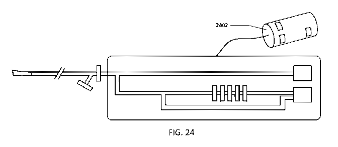
[00641 Figure 24 shows an enibodiment of the airway devicelcontroller which
may be
standalone, or incorporated into a CPAP device.
[00651 Figure 25 shows a. graph of the pressure signal from a rough pressure
sensor, a
sensitive differential pressure sensor and an ECG.
100661 Figures 26A and 26B show 2 embodiments of the mouthpiece area of the
airway
device,
100671 Figure 27 shows a top view of an embodiment of the hand piece.
CA 03005443 2018-05-15
WO 2017/087366 PCT/US2016/061993
[00681 Figure n.shows a..battom yiew.dan'.erribodiment.of die hand piece.
it10691 Figure 2.9 shows how an embodiment of the .airway device controller
and how the
system communicates data.
10070] Figure 30 shows examples of screens that may be shown on the display
[00711 Figure 31 outlines a method. of use of an embotiment of the airway.
device/controller device.
[0072] Figure 32 shows nì embodiment of the airway device which incorporates a
spirometer, and other features.
[00731 Figures 33 and 34 show example screens of a training app.
1007,1] Figures 35 and 36 show example screens of a survey.
DETAILED DESCRIPTION OF THE INVENTION
100751 Figure 1 shows an embodiment .of the airway device wom in the mouth of
a patient.
One .of the advantages of a portable embodiment, such as this oneõ is that it
can be worn by
a subject that is not only awake and not intubated, but upright and active. In
other words,
the use of the airway device is not limited to patients on a ventilator, CPAPõ
.or .other
stationary medical device.. The airway device/controller -may be used on a
patientluser with
no additional ventilation support, or airway pressure support Said another
way, the airway
device/controller 'nay be used on a patient without a ventilator or (TAP
machine or
additional flow source, or any sort .of artificial ventilation or airway
pressure support. The
airway device/controller may be =used by patients/users who are breathing
naturally or
normally, or may be used in a "prompt mode", where the controller .prompts the
user to do
something other than breathe naturally, For example, the controller .m.ay
prompt the :user to
hold his/her breath., hold hisiher breath after inhalation, hold his:her
breath after exhalation,
hold hislher breath "now", perform the MV M at a given pressure for a certain
period of
time, etc.
[00761 The airway device contains one or :More .SeusOrS which .0an: meaSUre
andior
calculate airway pressure, airway now, temperature, sounds, respiratory rate,
stroke
volume, heart rate, tidal volume, lung sounds, heart sounds. GI sounds, p02,
pC.:02, pH,
ECG, pulse rate, pulse pressure,. spirometry, analytes and/or compounds in the
breath (i.e.
urea, markers of infection, 02, CO2, urea, water vapor, alcohol, drugs, etc,)
or analytes
andfor com.pounds in the saliva, such as glucose., etc.
I 3
CA 03005443 2018-05-15
WO 2017/087366 PCT/US2016/061993
f00711 A .controller is either iueorporated into the airway :device or a
separate device Which
communicates with the aii*Ity devite. either Wirelessly or via a Wired.
connection. Ti
controller may be incorporated into a ventilator, a (PAP, a stand-alone device
or
incorporated into, or in communication with, a computer andlor smartphone.
[00781 hi a preferred embodiment, the controller is incorporated into a
smartphone which
communicates wirelessly with the airway device, either on a continuo-us or
intermittent
basis. Data. -transferred from the controller .may also be trannnitted to/from
a remote server,
for example, via the internet or an intranet. Data from the controller may
also be
anonymized. Anonymized data nyay be aggregated across .patients for trends
analysis. Data
.10
collected .m.ay include metadata such as patient 1D, timestamp, patient
medical history,
such as weight, medications., etc. Use of the term "airway device" herein m.ay
include a
controller component.
100791 The airway device may have a portion within the mouth or be .completely
external.,
it may also be over the nose either instead of, or in addition to, .the mouth.
The airway
device .may purpose-1'4111y block the nose. The airway device .may also be
incorporated into
an endotracheal tube.
[00801 Figure 2 shows a detailed view of an embodiment Of airWay.device 200.
This
embodiment includes external .opening section 204., mouthpiece section 206 and
neck
section 208. The mouthpiece de-vice in this embodiment includes at least two
airway
lumens., exhalation airway lumen 210 and inhalation airway lumen .212. In this
embodiment, the two lumens are separated by divider 2.14. Alternatively, only
one lumen
may be present, for example, only an exhalation lumen..
i.0081] Gas outflow vent 216, in the exhalation airway lumen, may include a
spiromeny
function. The .vent may also maintain or cause to be maintained a slight
positive pressure so
that the airway of the subject remains open during breathing, .which aids in
the ability to
sense certain. parameters,
[0082,1 The air in-flow, or inhalation airway lumen,: andfor the exhalation
.airway hunen0.
may include one-way 218 valve to help direct eihated air through the
exhalation airway
lumen during breathing,
[00831 Sensors 222, 224, and 226 may sense any Of the pararnetei's Listed
hereWithiri.
Sensors mar be placed in the .exhalation airway lumen 2.10, the inhalation
airway lumen
212, or on the outside of the airway device, Sensors .222 .on the outside .of
the device will
generally be for contact sensing with the mucosa andlor the lips, such as ECG
sensors.
14
CA 03005443 2018-05-15
WO 2017/087366 PCT/US2016/061993
SeMOM 224 in the exhalation:. airway turnen may: measure parameters
...associated with
exhaled air, including pressure, flOW, .5i) ands, O. CO2, urea, Water vapor;
aleohol, drugs,
etc. SenSOIS 226 in the inhalation airway lumen may measure parameters
associated with
inhaled air, including 02, CO2, urea, water vapor, .alcohol, drug.s, etc.
[00841 Generally, the sensors can be placed anywhere along the length of .the
airway
device, but there may be advantages to certain locations for certain types of
sensors. For.
example, sensors for temperature, water vapor, alcohol, drugs etc. measured in
.exhaled
would likely be better placed closer to the stibject.
10085j How andlor pressure sensors can be placed anywhere along the length of
the airway
device, but there .may be an advantage to placing these sensors in a narrow
andlor constant
diameter section of the airway device such as within neck. 208. A. sensor .or
sensors mav
also be placed on gas outflow vent 216. Sensors may also be remote. For
example, a
pressure sensor, for example a pressure transducer, may be in fluid
communication with the
mouthpiece via. a .mbe with an inner lumen.
[NM] A single use barrier may be -LiSed to cover mouthpiece section. 206 to
maintain
sterility of the airway device. Alternatively., a disposable mouthpiece
section may be
attached to the airway device and removed after use. A laeat-moisture
exchanger may be
used to prevent humidity from the breath .entering into the device.
Alternatively, the airway
device ma),,, be sterilizeable or disposable.
[00871 .Airway device. 202 may incorporate 'hardware andlor software to either
act as a
controller, or communicate with a controller. 'The airway device may also act
as a "partial
controller", where some of the controller activities take place within the
airwa.y device, and
some take place within a separate controller device.
[0088] Airway device may be made out of any suitable material or materials,.
including
polymerõ metal, or any other material or any combination of materials, /kirway
device is
preferably relatively light and portable..
[0089] Flow/pressure sensors .may include orifice plateSõ pressure
txansducerS, cone
devices, Pitot tubes, Venturi tubesõ flow nozzles, Misch cr Lìtly tye.
pneumotachon*tom,
or any other suitable technology. Sensor resolution is generally. high.
Pressure sensor
sensitivity may around +/- 0.5 mmHg. Pressure sensor sensitivity may around +/-
1 mmHg.
Pressure sensor sensitivity may around +I- 2 mmHg. Alternatively, pressure
sensor
sensitivity may around +/- 10 mmHg. Alternatively, pressure. sensor
sensitivity may around
+I- 20 mmHg,
CA 03005443 2018-05-15
WO 2017/087366 PCT/US2016/061993
100901 Figure 3 shows a graph of an ECG= along with simultaneously measured
airway
pressure data. KG data 304 is shown below airway pressure data 102. Within the
airway
pressure, systolic pulse data 306 and diastolic pulse data 308 are clearly
visible. 'ithin the
3-lead ECG data, P wave 310. QRS complex 312, and T wave 314 are all visible.
The
double headed arrow lines show where the QS complex peak lines up with the
valleys of
the pressure data.
100911 Figure 4 shows a detailed view of the pressure data between
respirations shown in
graph 402. Cardiogenic oscillations can be seen in detailed view 404 of
pressure vs. time.
The amplitude or area under the curve for these pulses can be used as an
indicator of
relative cardiac output and/or pulmonary artery pressure. Not shown but also
useful in the
same manner are cardiogenie oscillations in the flow signal.
10092] Figure 5 shows a graph of the ECG CURT, the cardiogenie oscillations
waveform
generated using data from pressure sensor(s), and the cardiogenie oscillations
waveform
generated using data from flow sensor(s). Also shown are the amplitude and the
frequency
of a cardiogenic oscillatiOnS waveform,
[00931 Figure 6 shows another embodiment of the airway device. The neck
portion 602 is
extended so that it also serves as tbe mouthpiece portion, which is more straw-
like than the
previously shown embodiment
100941 Figure 7 shows another embodiment of the airway device, Mouthpiece area
702 is
flat and designed to go over the lips/mouth. Strap 704 may hold the device on
the face of
the subject
[00951 Figure 8 shows another embodiment of the airway device. External
opening section
802 of this embodiment is elongated and more narrow than previously shown
embodiments. Section 802 may be flexible, as in flexible tubing, or may be
rigid, or may be
partially flexible and partially rig,id. Mouthpiece section 804 may include
mouth shield 802
to help keep the device in place. The various sensors and/or valves may be
anywhere along
the length of this embodiment.
10096] Figure 9 shows an embodiment of the airway device and controller where
the
controller is separate from, and may be remote to, at least in part, the
airway device, ln this
embodiment controller 904 is a smart phone and communicates wirelessly with
airway
device 902, which ma-y include a wireless data transmitter.
100971 Figure 10 shows an embodiment of the airway device and controller where
the
controller is separate, at least in part, from the airway device. In this
embodiment,
16
CA 03005443 2018-05-15
WO 2017/087366 PCT/US2016/061993
-controller 1002 is a;smart phone .and communicates with airway -device I 004 -
via .a.Ø.wire.
'or table, for Odmiple, aUSB cable, In this embodiment data May be collected
and stored in
airway device 1004 and periodically uploaded to controller 1002 via the
cable,.
.Alternativelyõ data .may be stored, at least in part, in the controller.
[00981 The .controller, whether it is separate from the airway device, or
incorporated into
the airway device, or some functions are located in th.e airway device and
some located
separately, may function as follows. The contmlier .collects .the data from
the various
sensors and analyzes them to determine cardiac output, stroke volume andfor
cardiac
function and/or other .parameters In addition, the controller may prompt the
subject to help
obtain the data from the sensors,. For example, the .controller .may prompt
the subject to
hold his/her breath. The breath holding prompt may 'happen at certain phases
of the
breathing cycle:, such as before or after inhalation andlor exhalation. The
controller may
prompt the subject to breath at a certain rate or to inhale, exhale, or hold
hisiher breath for a
certain time period., or within a certain goal pressure range. Indicators .may
be present on
the controller andfor the airway device to 'help the subject time certain
activities, or achieve
certain breathing goals, such as exhale pressure. For example,. the controller
may prompt
the subject to hold his/her breath until a light on th.e controller andfor
airway device turns
green, or until an auditory signal is heard.
[00991 The controller may also determine whether the .dataitis-- collecting is
adequate: -1-br
analysis. For example., if the subject's airWay ís dosing between breaths, or
during
exhalation, the data may be more difficult to analyze. Th.e controller can
sense when this is
h.appening either 'by the pressure/flow profile or other parameters and can
prompt the
subject to adjust his/her brc...athing. For example, the controller may prompt
the subject to
breath more slowly, or to sit still. in .addition, the .controller may change
the positive
pressure of the airway device to help keep the airway open. Some possible
prompts .that .the
controller may pro-vide to th.e subject are:
hold your breath for x seconds
101.011 ¨ hold your breath until the indicator does
[01021 Breath normally until the indicator does x
[01031 ------ exhale at a consistent pressure as shown on indicator
[010411 ¨ exhale at a consistent pressure as shown on indicator fOr x seconds
(or until
indicator says x seconds has elapsed)
17
CA 03005443 2018-05-15
WO 2017/087366 PCT/US2016/061993
10105I exhale with throat open at a :consistent :pressure as shown on
indicator for x
Seconds
[01061 --- perform the MVM for x seconds
101071 - exhale and then hold breath
[0108] - inhale and then hold breath
[01091 ¨ breath normally
10110] breath more slowly
[01111 --- Breath more quickly
[0112] --- Breath in slowly
101131 Breath out slowly
Breath in quickly
[011511¨Breath out quickly
IOU 61 testing is complete
[01171¨ begin exercising
[0118] ¨ end exercise
10119] Other prompts are also possible. The prompts may change depending on
the data
being collected. For example:. if the controller determines that the airway is
closing
between breaths, during breathing, or during exhalation, the prompts may tell
the subject to
breathe differently, or the controller may cause the airway device to apply
positive pressure
to the airway. lirt addition, the user may be prompted In certain time(s) of
the day to use the
device, so that the device is used at the same time each day. For example, the
device may
prompt the user to use the device upon -waking.
101201 Other parameters that may be considered in determining whether the
subject's
breathing is optimal for data collection include: variability of peak-to-peak
period and
magnitude, waveform shape, etc.
101211 The controller may analyze the data from the sensors to determine other
conditions,
including COPD, asthma, CE1F, cancer, stroke, pulmonary embolism, dyspnea,
paroxysmal, nocturnal dyspnea, emphysema, and any other condition that could
have an
impact on respiratory rate, temperature, stroke volume, heart rate, tidal
volume,. lung
sounds, heart sounds, GI sounds, p02, pCO2, pH, alcohol, urea, drugs, or any
other of the
monitored parameters.
18
CA 03005443 2018-05-15
WO 2017/087366 PCT/US2016/061993
[01221 -Vagal toneivasovagar syndrome may alSo be determined tisin g the
:present
invention, Slii2sht changes in heart beat paraincterS, including; amplitude,
tate, ViaVefOrin
shape, etc., at different stages of the breathing cycle can be measured and
vagal tone
determined. For example, if the hew rate increases during inhalation, this may
indicate a
high vagal tone.
101231 Iample of Data Processing System
101241 FIG. .11 is a block diagram of a data processing system, which .may be
used with
any embodiment of the invention. :For example, the system 1100 may be used as
part of a
controller, server,. mobile device, hand piece, computer, tablet, etc. Note
that while .F1G, 11
1.0 illustrates various co.mponents of a conipater system., it is .not
intended to represent any
particular architecture or manner of interconnecting the components; as such
details are not
germane to the present inventionõ. It will also be appreciated that network
computers,
handheld computersõ -mobile devices, tablets, cell phones and other data
processing systems
which have fewer components or perhaps more components may also be used with
the
present invention.
[01251 As shol.vnín FIG. 1.1õ, the computer. system IWO,. which is a form of
.4 data
proceSsifig .systern,, inc udes a bus or interOontect 1102 Which is, coupled
to one :or .more
microprocessors 1103 and. a ROM 1107, a. volatile RAM 1105, and a non-volatile
memory
1106. The microprocessor 1.103 is coupled .to cache me.mory 1104. The hus 1102
interconnects these various components together and also interconnects these
components
1103, 1.1.07, 11.05, and 1.106 to a. display controller and display device
1.108, as well as to
inputtoutput (1/0) devices 1110, which may be mice, keyboards, modems, network
interfaces, printers, and other devices which are well-known in the art.
[0126] Typically, -the input/output devices 1.110 are coupled to -the system
through
input/output controllers 1109. The volatile RAM 1105 is typically implemented
as dynamic
R.AM. (DRAM) which requires power continuously in order to refresh or maintain
the data
in the memory. The non-volatile memory 11.06 is typically a -magnetic hard
drive, a.
magnetic optical dri.ve, an optical drive., or a. DVD RAM or other type .of
131(411ot-3r system
which .maintains data even after power is removed from the system. Typically,
the non-
volatile memory will also be a random access memory., although this is not
required.
[0127.1 While FIG, 11. shows that the non-volatile .memory is a local device
coupled
directly to the rest of the components in the data processing system, the
present 41:W11600
may utilize a non-volatile ine.mory which is remote from .the system; such as,
a network
19
CA 03005443 2018-05-15
WO 2017/087366 PCT/US2016/061993
storage device which is :coupled to the data processiag system through :a
,network interface
such as a modem or Ethernet interface. 'The bus 1102 may include one or ittbre
buses
connected to each other through Vali0 US bridges, controllers, andlor
adapters, as is well-
lolown in the art. In one embodiment, the 110 controller 1109 includes a US B
(Universal
Serial I3us) adapter for controlling USB peripherals. Alternatively, I/0
controller 1109 may
include an IEEE-1394 adapter, also known as FireWire adapter, for controlling
FireWire
devices.
101281 Some portions of the preceding detailed descriptions have been
presented in terms
of algorithms and symbolic representations of:operations on data bitS
a computer
memory_ These algorithmic descriptions and representations are the ways used
by those
skilled in the data processing arts to most effectively convey the substance
of their work to
others skilled in the art. An algorithm is here, and generally, conceived to
be a self-
consistent sequence of operations leading to a desired result. The operations
are those
requiring physical manipulations of physical quantities.
[01291 It should be borne in mind, however, that all of these and similar
terms are to be
associated with the appropriate physical quantities and are merely convenient
labels
applied to these quantities. Unless specifically stated otherwise as apparent
from the above
discussionõ it is appreciated that throughout the description, discussions
utilizing terms
such as those set forth in the claims below, refer to the action and processes
of a computer
system, or similar electronic computing device, that manipulates and
transforms data
represented as physical (electronic) quantities within the computer system's
registers and
memories into other data similarly represented as physical quantities within
the computer
system tnemories or registers or other such ill fOrMation storage,
tiartsrnássion or display
devices.
101301 The techniques shown in the figures can be implemented using:code and
data stored
and executed on one or more electronic devices. St4ch electiouic devices store
:and
communicate (internally and/or with other electronic devices over a network)
code and data
using computer-readable media, such as non-transitory computer-readable
storage media
(e.g., magnetic disks; optical disks; random access memory; read only memory;
flash
ITICIllory devices; phase-change Memory) and transitory computer-readable
transmission
media (e.g., electrical, optical, acoustical or other form of propagated
signals such as
carrier waves, infrared signals, digital signals).
CA 03005443 2018-05-15
WO 2017/087366 PCT/US2016/061993
[01311 The :processes or methods depicted in the -figures herein may be.
performed by
processing logit that. comprises hardware (e.g. circuitry, dedicated logic,
ete.), firmware,
software embodied On a non-transitory computer readable medium.), or a
combination
of both. Although the processes or methods are described above in terms of
some
sequential operations., it should be appreciated that some of the operations
described may
be performed in a different order. Moreover, some operations may be performed
in parallel
rather than sequentially.
[0132.1 Figure 12 shows an embodiment of an airway -device. which includes a
restrictor...
The restrictor helps reduce turbulent air flow within the airvit ydevite.
Air:Way device 1202.
in this embodiment has .mouth opening 1.204, which is larger -than .restrictor
Restrietor 1206 is open to ambient air. As the user exhales into the airway
device, restrictor
.1206 restricts the air.flow which increases the laminar nature of the air
flow within the
airway device. ln this embodiment, as the user breathes through opening. 1204,
some air
exits restrictor 1206, however some air, preferably air which is predominantly
flowing in a
laminar manner, exits sampling exit or lumen 1208. Sampling exit 1208 may
connect
directly to a pressure, or other, sensor, or it may connect to a pressure
sensor or other
sensor via connector 1210, -which may be a flexible or rigid tube. The purpose
of restrictor
1206 is to reduce turbulence in the air flow within the aliway device so that
the air .exiting
sampling .exit 12fõ.% is as laminar as possible. Note that this figure is
showing an exhalation
lumen only. A separate inhalation lumen -may be incorporated into the device
andlor the
subject may be asked to inhale separately, either through hisiher nose, or 'by
removing th.e
device from histher mouth. .Alternatively, the patient inay also use the
exhalation lumen for
inhalation.
[0.1331 Figure 13 shows an embodiment .of an airway device which incorporates
a
mechanical filter. ln this e.mbodiment there are at least two sampling
lumens., 1302 and
1304. One of -the sampling lumen includes mechanical high Ilass filter 1306.
The :pressure
sensor in this embodiment is a differential pressure sensor. Differential
pressure sensor
1308 is in fluid communication with at least two sampling lumens or inputs,
and compares
the pressure reading between the two lumens. This configuration produces a
cleaner
pressure signal tbr analysis by circuit board 1310 by filtering out the
pressure from the
breaths and leaving those from the cardiogenic oscillations. Circuit board
1310 may be
incorporated into .the airway device or ma),,, be separate, .for example on a
separate
controller, and communicated with either wirelesSly or via wire. In this
embodiment, th.e
21:
CA 03005443 2018-05-15
WO 2017/087366 PCT/US2016/061993
circuit 'board:iS :it)COrpOrated: into the airwa.y device .and communicates
.A.Vitit a controller. via.
'vireless transmitter-1312, ln this embodiment, circuit board .131:0 and
wirele.ss transmitter
1312 may be considered to be part of the controller as welt for purposes of
defining the
controller. Filter 1306 may be made out of any suitable material including
foam or any
membrane that is semi -permeable to airõ Note that. this figure is showing an
exhalation
lumen only. A separate inhalation lumen may be incorporated into the device
and/or the
subject .may be asked to inhale separately, either through his/her nose, or by
removing .the
device front his/her mouth. Alternatively, the patient may also use the
exhalation lumen for
inhalation.
101341 The .mechanical high-pass filter isolates the higher frequency cardiac
oscillation
signal from the lower frequency pressure.: signal associated with n.atural
breathing. This
filter may employ a. partially-impermeable barrier between differential
sensing and
reference inputs, 'The high-frequency cardiac oscillation signal is seen by
the sensing input,
whereas the pressure changes due to breathing are low frequency enough to
equilibrate
across the membrane and a:re detected at 'both inputs, By breathing, into the
de-vice with a
slight expiratory pause, or .1.1Sing the !VIVA the cardiogenic oscillation
signal can be
reliably. captured. Some embodiments may incorporate an a:dditional, less
sensitive,
pressure sensor to monitor the entire breathing cycle and provide feedback to
the patient
about the size and frequency of the breaths, improving repeatability between
measurements,
[01351 Figure 14 shows an .embodiment in -winch the restrictor and the
sampling exit are
combined. Restrictor 1402 reduces the turbulence in the airflow as air is
breathed in and
out of the airway device. Breathed air exits and may enter via outlet 1404.
Differential
pressure sensor 1308 may allow air to flow through it or .alongside it to exit
the airway
device, or alternatively, the airway device may have an additional air exit
(not shown).
Note that this figure is showing an exhalation lumen only.. A separate
inhalation lumen .may
be incorporated into the device .andlor the subject may be asked to inhale
separately., either
through his/her nose, .or .by removing .the device from his/her mouth,
101.361 Note that the restrictor could be. anything suitable, such as a flow
control valve, a.
pressure control valve, etc.
[0137] Figure 15 shows an erribodiment which incorporates a flow filter. Flow
filter .1502
decreases the turbulence of the airflow coming into the airway device, in this
.embodimentõ
flow .filter IS02 is used instead of a restrictor. The airway device .ma:y
have an additional
CA 03005443 2018-05-15
WO 2017/087366 PCT/US2016/061993
air exit not sitown).. Row niter 1502 ..tray be made .-out:eauy .suitable
..ataterial such as
polymer and in any 'suitable configuration such as a honeycomb or parallel.
capillary
configuration, -Note that this figure is showing. an exhalation lumen only. A
separate
inhalation lumen inlay be incorporated into the device and/or the subject may
be asked to
inhale separately, either through his/her nose, or by removing the device from
hislher
mouth.
[01381 .Any of the embodiments herein can be adapted to be used inside the
mouth, or
partially insícle the mouth. For example, an airway device deeper inside the
mouth may be
advantageous in keeping the airway open for cleaner pressure measurements.
Furthermore,
any of the embodiments herein -may also be adapted to be used with patients
who are
tracheally intubated, in Which case the devices described are attached to or
in.-line., with the
tracheal -tube.
1.01391 Figure 16 shows a graph which demonstrates pulse pressure variability.
As
mentioned earlier, variability in the respiratory pulse pressure -wa.veform
can be used to
determine hydration status, as -well as volume status, and also pulmonary
artery
compliance. The graph in Figure 1.6 shows the pulse pressure at end
inspiration and at end
expiration. Pulse pressure is defined as the difference between the systolic
and diastolic
pressure readings, or the amplitude of the waveform (lowest point to highest
point). The.
difference in amplitude between these two waveforms is the pulse pressure
variability. A
large variability may indicate dehydration, where a decrease in variability
over time may be
an indicator that hydration is being restored or has been restored,
[0140.1 1.7igure 17 shows an embodiment of the airway de-vice/controller which
includes a
hand piece and at least some of the controller -functions_ The airway device
of this
embodiment includes 2 mouthpieces 1702 .and 1704. The user breaths into one of
these
mouthpieces and breath .exits through the other mouthpiece. Hand piece 1706 is
held by the
user or by the user's physician. Display 1708 displays one or more display
areas 1710.
These display areas may include buttons, or links, to more information, such
as settings,
waveforms, including waveforms showing HR (heart rate), SY ("stroke volume),
CO
(cardiac output), PAC (pulmonary arterial .compliance, etc., analytical
results of waveform
analysis, triggers for .alarms/notices, etc. The airway device/controller of
this embodiment
may communicate wirelesslyõ or in a wired manner -with one or -more mobile
devices,
computers, servers, etc.
23
CA 03005443 2018-05-15
WO 2017/087366 PCT/US2016/061993
[01411 Figure 18 shows. another :embodiment Or 'the' :airway
deviceltontroller. This
embodiment includes controller 1802, signal transmisSiOn tubing 1804, beat-
tudistute
exchangerlter 1806 and mouthpiece 1808. In this embodiment, the pressurefflow
sensor
may be in the controller, :In use, during user breathing into the mouthpieceõ
the pressure in
the airway is transmitted to the pressure/flow sensor via. mouthpiece .1808
and tubing 1804.
Note that controller 1802 may be in communication with a computer/mobile
device/networked server etc., which .inav include some of the controller
functions.
[0142.1 Embodiments of the airway device/controller may also be incorporated
with a
standard or specialized inhaler, for example for asthma. The airway
device/controller ìn
these embodiments inav include a feature which track.s .usage of the airway
device andlor
inhaler to monitor use compliance.
10143] Embodiments of the airway devicecontrol ler may include integration
with
electronic 'health records (EMR) or electronic health records or other
systems. For example,
data. from the controller may be transmitted wirelessly (or wired) to a.
server in the interact
which integrates the data with th.at of an FAIR. The patient ID (possibly
anonymized) may
be integrated into .the metadata of the data ..transmitted by the controller
so that the data can
be integrated with the correct patient's medical record.
[0144j Data from multiple .airway devices/controllers .may be collected and
aggregated and
analyzed for trends. 'This data. may be anon),,,mized to compl),,, with
privacy rules.
[014.5[ in some embodiments of the airway devicelcontrollerõ respiratory sinus
arrhythmias
(changes in heart rate due .to breathing) may be tracked as an indicator of
heart heal.th or
heart failure. Deviations th)111 trends may be indicative of heart failure
issues and may
provide an alert, Because the data collected by the airway device may be
continuous, for
example, while the user sleeps, deviations from .the norm. (either for that
patient .or for a
patient population) may indicate changes in health, and in particular, heart
health.
101461 in some embodiments of the airway device/controller, the device is used
in an
ambulatory manner. In other words, the user may use the device while walking
around,
watching TV, working, sleepingõ resting, exercising or while perfoiming
everyday
activities. The riser is not tied. to a stationary device, hospital nor
clinic,
[01471 Sensors connected to the airway devicekontroller -may include a blood
oxygen
saturation sensor or a blood CW saturation sensor or any other type of
oxygen/C.02
sensor. For example., blood oxygen saturation may be determined by a pulse
oximetry
sensor in contact with the lips, tongue, oral mucosa and/or
fingerlextremities. Thi.s signal
24
CA 03005443 2018-05-15
WO 2017/087366 PCT/US2016/061993
and/or art:.EKG íiìaicollected fro rn: one .or more .EKG. sensors :(which may
be in cotict
-with these,. or other, locatiOnS) may be used to determine pulse transit
time.
[0.1481 Tissue .02,42.02 may be (determined using an air tonometry sensor in
contact with,
or in1,3roximity to, the tongue or oral .mucosa or elsewhere. This type of
sensor may include
an air permeable membrane between the sensor and .the body.
[0149.1 Absolute stroke volume may also be determined as follows_ The volume
or air
displaced by each cardiac contraction due to pressure changes is determined by
first
determining the volume of air in the lunt.-zs. This may be done in one .or
more of several
ways:
.10
[0150] 1.) pulsed air .method - A known volume of .:ait is pulsed into .the
lungs and the
change in pressure is measured. :From this, the volume of dead space in the
lungs -may be
determined.
[0151] 2) spirometry - Total lung volume can be estimated front spirometry.
[0152] 3) gas dilution A known quantity and concentration of a target gas is
infused into
the lungs. The concentration of -the target gas is then measured irt the
exhaled air exhaled to
determine how much air has mixed with the target gas, thus providing an
estimate of lung
volume.
[01531 Strol.ce volunte vaiabi ity may ..41so be determineftalculated,. For:
cpuiple, the
tontrolier may prompt the user to breathe in deeply.. The controller may use
data captured
from a sensor/sensors to determine stroke volume measurements at end
inhalation and at
end expiration- potentially to determine stroke volume variability. The
controller may
correct for changes in cardiac pulse size due to chanase in tung volume using
spirometry
(which measures breath volume) or the pulsed air method., or gas dilution
.technioues.
[0154] Spirom.etry tua),,, be used to measure one or more of several
parameters, including:
Vital capacity (VC), Forced vital capacity (F VC), Forced expiratory volume
(FEV) at
timed intervals of 0.5, I .0 (FEV1), 2.0, 3,0 seconds, and other intervals,
forced expiratory
flow 25-75% (FE]; 25-45), maximal voluntary ventilation ('VV), also known as
Maximum breathing capacity,. 'Peak Expiratory Flow (PER and any other
parameters.
Other tests may be performed ................................................
Results may be provided in raw data. (liters, liters per
...................................................................... second)
and percent predicted the test result as a percent .of -the "predicted
values" for the
patients of similar characteristics (height, age, sex, and sometimes race and
weight), or the
results may be provided in other ways.
CA 03005443 2018-05-15
WO 2017/087366 PCT/US2016/061993
[(155.1 1/1::sonie errib.odirnents.of the airway devieelcontroller,..art ECG
signal of the user is.
collected simultaneOuSly-to the cardiogeníe Oscillation data, In this Aiay,
the precise length
and/or timing of a heartbeat can be determined (by the :ECG signal) and the
cardiogenic
oscillation pressure curve can be divided up into precise heartbeats. hi other
wordsõ one or
more cardiogenic oscillation curves, each -relating to one heartbeat, can be
collected and
identified and averaged, because the start and end of the cardiogenic
oscillation curve
relating to each heartbeat is precisely identified by the ECG signal_ This
allows collecting
more than one cardiogenic oscillation curve and averaging them to get more
accurate
cardiogenic oscillation curve data. One or more ECG sensorlelectrode(s) may be
placed .on
the mouthpiece, or handheld portion of the airway device. ECG
sensor/electrode(s) may be
in contact with the user's mouth, finger(s), hand(s), or elsewhere on the
body. Various
features of the ECG curve may be used to "gate," die cardiogenic oscillation
pressure curve.
For example, the R peak, or .alternatively the P, Q, S, T, l.J areas may be
used.
[01551 Alternatively, or in addition,. the signal from a pulse
oximeteephotoplethysmograph
1 S may be used to gate the cardiogenic oscillation pressure curve in the
same -way --- to
determine the precise length/timing of the heartbeat The same way a feature of
the ECG
curve can be used as a gating feature (for .example, -using die time between
subsequent
peaks of the R-wave), a feature., peak, valley, slope, length etc, of the
pulse .oximeter CUTAT
may be used instead of, or in addition to, the ECG curve. Multiple ECG and/or -
multiple
pulse oximeter signals may alternatively be used. For example, the device -may
have
electrodes/sensors for pulse oximeter and FCCì for each hand, resulting in 2
ECG signals in
addition to 2 pulse oximeter signals. This allows the best signal to be used
to gate the
cardiogenic oscillation pressure curve. The best signal may be chosen by
amplitude,
identifiable peak, consistency, etc, In this way, a good signal is likely to
be Obtained even if
the user is not touching, or in contact with, all of the electrodes/sensors
perfectly. Where
redundant SellSOIS are used to gate th.e cardiogenic oscillation pressure
curve, -the redundant
sensors -may be set -with different gains on each, In this way, if one of the
signals maxes
out, or rails, where the peak of the curve is difficult -to identify, another
signal may be
lower and have more identifiable peaks. This situation may OMIT if a user is
pressing the
sensors with a lot of pressure. lIn this way, one device with different
sensors set with
differing gains, may accommodate users with different finger pressures.
[01571 Outliers, or less usefid data, may also be removed -using EC7G and/or
pulse oximetry
signals from th.e analysis to optimize the analysis results.. More th.an one
collected
26
CA 03005443 2018-05-15
WO 2017/087366 PCT/US2016/061993
.ECG/p114e.:Okimetlysignal.avy oiso.lw used in theanalY,Sis One or1110re.ptilw
oximeted
photoPlethysmograph senSotielectrode(s) lay be placed on the -mouthpiece, or
handheld
portion of the airway device. Pulse oximeterfphotoplethysmograph
sensorfelectrode(s) -may
be in contact with the user's mouth, finger(s), hand(s), or elsewhere on the
body.
[01581 Alternatively, or in addition, the signal from the rough pressure
sensor ma),,, be used
to gate th.e cardiogenic pressure curve in the same wa-y --- to determine the
precise
lengthitiming of -the heartbeat Any sensor that determines and communicates
the
lenwhltiming. of a heartbeat to the controller -may be used to Uate the
cardiogenic oscillation
pressure curve.
1 0
[01591 In any of the gating curves, a regularly :repeating feature, or peak',
of the curve may-
be -used to assess the quality or relative quality of the gating curve to be
:used as gating, as
well as for the gating itself.
[0.1.60j Figure 19 shows several gated cardiogenic oscillation pressure curves
layered on
top of one another, along with the averag.e of the curves. The thinner lines
1,902 represent
the individual cardiogenic oscillation curves obtained from several
heartbeats. The
heartbeats are separated, on layered on top of each other and the average
curve 1904 is
calculated by the controller. The beginning and end point for each heartbeat
can vary
slightly over time making it difficult to average the curves without a way of
precisely
identifying the start and end point of .each heartbeat (i.e., gating). Here,
the controller does
this by using the simultaneously obtained ECG signal from the patient. 13y
picking one or
more points in the ECG, for example, the R peak, the beginning and end of each
heartbeat
can be precisely identified and as a result, cardiogenic oscillation curves
from multiple
heartbeats can be averaged. The controller may also be equipped to determine
the best
point in the ECG to use for the heartbeat identification., for example the ECG
curve may be
analyzed for the most consistent area/peak, the most identifiable arealpeak,
the sharpest
peak/area etc.
[0.1,61] in the analysis..,shownin:Figure J. the. :median eurve maybe used
instead of, or in
tOMbilltOiOn Wik the mean cuive. :eOnstruct the medial-I.:Carve. the median
value at each
point in time (for example, at 1, 2, 3, ... ins after detection of the R peak)
is calculated.
This may help to reduce the influence of individual outlier curves, although
other
techniques may be used to accomplish the same goalõ such as .excluding the
points that fail
outside a speci17ied window of deviation when calculating the mean.
CA 03005443 2018-05-15
WO 2017/087366 PCT/US2016/061993
[0162] Analysis of the shape a (11e cardiogenio, oscillation curve nay:
provide information
on the patient. It is alsO patient specific, Meaning that even among, healthy
patients, the
cardiogenic oscillation curve shape is unique to each individual. Iln this
way, the shape of
the curve can be used as a signature to identify a patient. Changes in the
cardiogenic
oscillation curve may indicate specific disease states, or relative disease
gates. For
example, the shape of the curve (or the change in the shape of the curve
compared to
normal or over time) may be analyzed to determine, andlor track over time, any
of -the
following disease states.
[0163] Aortic stenosis
.10 [0164] Aortic insufficiency
10165] Mitrat stenosis
[01661 Mitral insufficiency (regurgitation)
[01671 Pulmonary stenosis
10168] Tricuspid stenosis
i 5 [0169J Tricuspid regurgitation
[01701 Pulmonary hypertension
101711 Pulmonary fibrosis
[0172] congestive heart failure
[0173] Acute respiratory distress syndrome
20 10174] Ventilator-acquired pneumonia
[01751 Pneumonia
[0176] Atrial Septal Defect
10177] Patent Foramen vale
[01781 Single ventricle
25 [01791 Others
[01801 In addition, changes to the shape of the oardiogenie oSeitlatiOn curve
over thve4 or
with the patient in different positions, iiriay indicate other patient
parameters such as
hydration. For example, a eardiogenic oscillation curve which appears to show
improvement in PAC when the patient's legs are raised may be an indicator of
dehydration.
30 Hydration status may also be evaluated by changing the patient's
position andlor breathing
pressure. For example, the user may be prompted by the controller to take a
measurement
CA 03005443 2018-05-15
WO 2017/087366 PCT/US2016/061993
While: standing, supine., sitting,. 'legs raisetL At a specific angle,:
inverted,: :etc. The:
relationship bctween.or 1111-1ong-th&C* re dings niay be used in the data
analysis to determine
patient health. For example, the .ratio between supine -COS data and. sitting
COS data may
be used in the analysis.
[01811 In some embodiments, any cardiovascular andior pulmonary parameter may
be
collected in more than one patient position and the relationship between the
parameters
used to determine the health of -the patient. For example, other available
devices, such as
the CardioMEMSTm device, manufactured by St. Jude Medical, may be used to
collect a
patient .parameter, such as pulmonary artery pressure, when the .patient is in
more than one
position. The data collected a.t these different positions can be used in
conjunction with
each another (for example, a .ratio of data sitting and supine) to determine
patient health.
[0182] In some embodiments of the airway device/controller, the :user is
prompted to
breathe in one or more specific ways to obtain the pressure signal. The user -
may be asked
to breathe (exhale, inhale or both or neither or .MVNI) into the device while
simultaneously
the controller controls a display -which displays feedback on the pressure,
the timeõ or other
parameter, of the user's breathing. For .example, the user may be asked .to
exhale at a steady
pressure, within a pressure range, for a certain duration of time.. and the
display may show
the user feedback on that pressure, and time:, such as lights, a graphic
display:, or
alternatively, the controller may provide audible. feedback.
10183] Figures 0A-C shows some alternative graphical displays which can guide
the user
to breathe at a constant pressure within a pressure range. Figure 20.A shows
controller 2004
with indicator lights 2002. In this example, there are three ranges of Fights
with different
colors, brightness, shape, etc. The lights .indicate when the .exhale pressure
is too high, too
low, or i.n range. In this .example, the middle 3 lights show -that -the
exhale pressure is in .-the
optimal range. The user exhales into the mouthpiece and tries to hold hislher
exhale
pressure within the middle lights. Alternatives to lights may be =used,
including a graphical
image, sounds., a dial, etc.
[0:1841 The graphical (or audible) display nlity a1s show the Wet when heiShe
is at the
lower or higher end of the goal range (such as the pressure range), so that
the user can
make small adjustments to keep within range.
[0.185.1 lin some embodiments the goal exhale pressure is about 7 to about 8
.m.mtlig. In
some embodiments the goal .exhale pressure is from about 9 to about I I mmHg.
In some
embodiments the goal exhale pressure is from about 5 to about 7 mmHg. In some
29
CA 03005443 2018-05-15
WO 2017/087366 PCT/US2016/061993
embodiments die goat exhale pressure: is from about 5 to :aboot 20 mmHg. hi
some
embodiments the goal exhale presktre is from about 5 to about 15 mmHg. lit
some
embodiments the goal exhale pressure is from about 10 to about 20 mmHg. In
some
embodiments the .goal exhale pressure is from about 5 to about 1.0 -mmHg. in
some
embodiments the goal exhale pressure is from about 7.5 to about 12,5 mmHg.
101861 In some embodiments the goal exhale time is about 5 seconds.. In some
embodiments the goal exhale time is about 4-.6 seconds. In some embodiments
the goal
exhale time is about 3-7 seconds. In some embodiments the goal exhale time is
about 2-5
seconds. In some embodiments., the goal exhale time is set anvwhere up to 1.0
seconds.
[01871 Figure 20B shows an alternative arrangement of the graphical display.
In this ease,
lights 2006 are used, but they are in a circular or .oval pattern .on a more
curved controller
2008.
[0.1.88j Figure 20C shows an alternative arrangement of the airway
device/controller. In
this example, mouthpiece 2010 may include sensors,, filters etc., and a
wireless transmitter,
such as a Bluetooth transmitter. The mouthpiece may contain components of .the
controller
as well. At -least some components of the controller are included in portable
device 2012.
The portable device .may be a mobile phone, table, .computer etc., and
includes a wireless
receiver and, a display_ In this embodiment, pressure sensor information is
transmitted
wirelessly to .the controller device which displays or communicates feedback
to the user..
The feedback may include breathing pressure feedback, time, as well as any
other
feedback. The .connection between the mouthpiece and the controller may be
wired as well.
Different components of the con-troller .may be distributed between the
mouthpiece and the
CoDtroller,
[0185] Some components of the controller may also exist remotely, for ex.ample
.on an
intemet connected server, which is in communication with the local controller,
101901 'Figures 21A-13 show embodiments of the airway .device/controller which
include a
"sensor hand-piece". A sensor hand-piece includes sensors and/or electrodes on
the hand-
piece itself for sensing various physiological parameters. The
sensors/electrodes may be on
the surface of the sensor hand-piece -where they are easily in contact with
One or more
fingers andior hands of the user. The sensor hand-piece .may include sensors
such as ECG
sensorsVelectrodesõ and/or pulse oximeterlphotoplethysmograph
sensors/electrodes on its
surface. In this embodiment the sensor hand-piece is an .ergonomic case which
the user
holds so that his/her fingers are in contact with various and appropriate
sensors which are
CA 03005443 2018-05-15
WO 2017/087366 PCT/US2016/061993
on the wface a the .case. In Figure. 2.1.A sensor I-land-piece 2102 includes
.ECCi:
sensOrsielectrodes 2104 and pulse' Oxittieteriphotoplethysitograph
sensor/electrode 2106_
7111e user may hold the sensor 'hand-piece with both hands so that 2
fingers/thumbs are
touching the ECG sensors and another fingerlthumb is touching the pulse
oximeter sensor,
Alternatively, the different types of sensors may be combined so that fewer
fingers are
required.
[01911 Figure 21B shows a sensor hand-piece connected to a controller. This
connection
may be wireless or wired. Alternatively, the sensor hand-piece may be combined
with the
controller ¨ the controller/ sensor hand-piece may include a graphical display
as well as
sensors.
1(192! In some .embodiments the airway device/controller is incorporated into
a. CPAP
(Continuous Positive Airway Pressure) device. In these embodiments the
controller may
control the positive pressure delivered by the CPAP device based on the
controller's
analysis of the cardiogenic oscillation pressure curve.
.15 [al
93] Figure 22 shows an embodiment of the airway device/controller which has
been
incorporated into a CPAP deviceõ. The various sensors may exist in mask 2202,
ventilation
tubing 2204 or controller 220( .,A handheld unit, such as the one shown in
Figures 21A/B
may also be present for use when the user is awake, and may or may not include
sensors
such as ECG and photoplethysmotnaph sensors. :Indicator graphics 2208 may also
be
present for setup, or for when the user is awake. Sensors in the mask .may
communicate
with the controller via wire 2210 embedded in the ventilation tubing or .the
communication
may be wireless. No communication tnay be necessary if the sensors are located
in the
controller rather than the mask. Cardiogenic oscillations and other parameters
(such as
EC(iì, pulse, etc.) .may be sensed via .the CPAP system .via sensors in the
mask., .the
controller, or elsewhere, such as in a sensor hand-piece. The CPAP device may
be run in a
test or setup mode, to obtain readings when the user is awake, and also in a
sleeping mode,
to obtain readings while the user is sleeping. For example, the user may be
prompted to
breathe at a certain pressure for a certain time frame while awake to set a
'baseline.
Readings may then be collected while the user is sleeping arid adjustments to
the CPAP
airway pressure may be automatically adjusted 'based on changes to the
readings. In
addition, or alternatively., an alert may sound to wake the ti.ser when
certain changes in:
readings are detected.
CA 03005443 2018-05-15
WO 2017/087366 PCT/US2016/061993
[01941 In ernhodiments.of the.airway.device which are ...combined with a CPAP
device
other positive pressure device.!, heart health may riot -only be diagnosed,
but treated. Because
the airway device measures the exact timing .of heartbeats (via ECG,
photoplethysmograph,
rough breath pressure, cardiogenic oscillations, etc), positive pressure can
be applied
through the airway -to the lungs in synchrony with the heartbeat. Pulmonary-
pressure can
be increased after ventricular contraction, and decreased before the next
heart contraction,
to offload the work of the heart,
[01951 Figure 23 shows an embodiment of .the airway devitelcontroller which
may: be.
standalone, or incorporated into a CPAP .deViee, .6r Used with additional
controller
components. Case 2302. houses the .niechanisms of the controller...Aimay
.tubing 2304 is in
'fluid communication between the. controller and mouthpiece 2306. In this
embodiment, aZll
of the sensors and the controller are within casing 2302, but other
embodiments may exist
with sensor and. controller functions elsewhere, for example as shown in
:Figure 24 where
sensor hand-piece 2402, including ECG and pulse oximetty sensors, is shown,
and/or in
embodiments where some or all of the controller computational functions are
performed on
a remote server.
[01961 Mechanical .filters 2308 and 231.0 help restrict and controi the flow
of Ø0. through
the airway tubing. Inside the case, rough pressure sensor tubing 2312 is in
fluid
communication with rough pressure sensor 2318 and airway tithing 2304. Fine
differential
pressure sensor 2320 is in fluid communication with tubings 2314 and 2316
which are in
.111dd communication with rough pressure sensor tubing 2312. :Wine -flow
restrictor 23.22 is
incorporated in-to tubing 2314 to restrict .flow through one nibing ári arid
communication
with fine differential pressure sensor 2320. The other tubing, tubing 2316,
either does not.
have a flow restrictorõ or has a flow restrictor with a different restriction
level than flow
restrictor 2322. Flow restrictor 2322 may comprise one or .more mechanical
filters. The
signal .from rough pressure sensor 2318 may be subtracted from the signal from
fine
differential pressure sensor 2320 to remove .artifacts from breathing,
moving., coughing,
etc. See Figure 25. Alternatively, .the pressure signal from the rough sensor
may be used to
determine that the user is exhaling at a constant and targeted pressure.. For
example, the
rough pressure sensor signal may drive the graphic display, such as the lights
to show the
user when heishe is in the target pressure range. in this situation, the
cardiogenic oscillation
signal would be extracted from the .fine differential pressure sensor..
32
CA 03005443 2018-05-15
WO 2017/087366 PCT/US2016/061993
[01971 Fie pressure sensor sensitiYity may be around 41-1 mmHg, Rough pressure
sensor
sensitiVity :May be around -4I; 10 ramHtt-,_ Altemativdy, fine pressure sensor
sensitivity
may be around 41- 0,5 mmHg 41- 3 mmHg. Rough pressure sensor sensitivity may
be
around +/- 5 mmHg ¨ 41.- 15 mmHg_ Fine pressure sensor or rough pressure
sensor may be
differential pressure sensors.
101981 As mentioned elsewhere herein, pulse tranSititim0 inay:be determined by
&alp-Ling
the time between the ECG signal (for example, the R peak) and the heartbeat as
measured
by pulse oximetry/photoplethysmograph. Pulse transit time may also be
determined by
determining the time between ECG signal and a peak in the cardiogenie
oscillation curve.
l0 This
Ina y prOVide different information :MC rig heart valve opening pressure
and/or
openi
10199] Figures 26A and 26B show 2 embodiments of the mouthpiece area of the
airway
device. Mouthpiece 2602 with mouthpiece opening 2604 is in communication with
filter
segment 2608 which is in coimnunication with airway tubing 2610 so that a
Iu.nien runs
from mouthpiece opening 2604 to the controller (not shown), Filter segment
2608 contains
a hydrophobic filter membrane with a pore size ranging from about 2 micron to
about 4
micron. Alternatively, the pore size of the filter ranges from about 1 micron
to about S
micron. Preferably, the filter pore size prevents water vapor from passing
through the filter.
[0200] This embodiment includes resistance control orifice 2606 which controls
the
resistance felt by the user while blowing through the mouthpiece. in some
embodiments,
the user is asked to exhale at a relative constant exhale pressure for about 1
to S or about 5
to 10 seconds, The resistance control orifice can control the exhale
resistance. A smaller
sized resistance control orifice will equate to a higher exhale resistance_ A
larger sized
resistance control orifice, or multiple resistance orifices, will result in a
lower exhale
resistance. Preferably, the resistance control orifice is approximately
circular in shape and
ranges from about 0.3nun to about 05 MID in diameter or longest dimension_
Alternatively,
the longest dimension of the resistance control orifice is from about
0.11111)1 to about 1.0
min. Alternatively, the longest dimension of the resistance control orifice is
from about
0.5min to about 1.0 mm.
[02011 Filter segment 2608 may adapt to mouthpiece 2602 andlor airway tubing
2010 by a:
common lucr type adapter or any other suitable adapter such as lucr-tock,
sore* on, snap
on, glue on adapter etc.
33
CA 03005443 2018-05-15
WO 2017/087366 PCT/US2016/061993
[02021 Figure 26B shows ai . embodiment similar to that .shown in. Figure 26A
with the
addition of orifice sheath 2612. The orifice sheath protects the resistance
c.ontrol orifice
from blockage by the users finger or otherwise. The orifice sheath includes
opening 2614
which allows air to freely flow out of (or into., during inhalation) the
resistance control
orifice even if the user has his/her fingers on the orifice/orifice sheath
area. The orifice
sheath is preferably .made from a rigid material such as a polymer, and may be
integral with
mouthpiece 2602 and/or filter segment 2608..
[02031 Figures 27 and 28 show the =top view and bottom view, respectively, of
an
embodiment of a sensor hand piece of the airway device which includes at least
some of
the controller functions. 'The sensor hand piece includes casing 2702. On the
casing top are
one or more heart pulse sensors/electrodes (photoplethysmographlpulse oximetry
sensors)
2704, airway .tubing connector 2706, power button 2708, breath pressure
indicator or
indicators 2710, breath pressure goal indicator or indicators 2712, displa),,,
2714, .and mode
button 2716,
[0204] Figure 28 shows the bottom view of an embodiment of a sensor hand piece
including ECCi- electrode or electrodes 2802 and battery cover 2804. Note
.that any of .the
sensors/electrodes niay be anywhere .on the case.
[0205j When holding the sensor 'hand piece, a user will preferably have one
.or both thumbs
in contact with pulse sensor(s) 2704, and one or more fingers in contact with
EC:G
electrode(s) 2802. Preferably, before the user places his/her fingersithtunbs
on the sensors,
he./she will place .the mouthpiece in his/her mouth. However, if only one hand
is necessary
for the sensor contacts, the mouthpiece .may be placed in the user's mouth at
any time
before testing begins,
[0206] During data .collectionõ the .user is guided through various steps
either by display
2714, or audibly or both. Alternatively the display may be on the user's
mobile
phone/computer with other functions of the sensor hand piece in communication
with .the
mobile phone/computer either by a wired connection, a wireless connection, or
direct
connection via a port in the -mobile phone/computer. Indicator(s) 2710 ma.y be
lights., a
visual bar, audible sounds, tactile feedback (such as vibration) etc. in this
figure., the
indicators are lights which indicate the exhale pressure as well as the
consistency of the
exhale pressure, and .may also indicate when the exhale pressure has been
consistently in
the proper pressure range for the required time. Preferably., the user is
guided by the
indicators to hold a. steady pressure, in addition Of alternatively, the user
is guided by the
CA 03005443 2018-05-15
WO 2017/087366 PCT/US2016/061993
indicators to holita particular targetpreasure.. A larget.pressure may be
preset or may be set
depending On the individual user and the tikes. COmfortable .exhale pressure
range for
holding a steady. exhale pressure. Pressure goal indicator 2712 .may be fixed
in place or
may be -movable to suit individual users. 'The software may also include the
ability to adjust
indicators 271.0 so that goal indicator 2712 stays in the same location, but
refers to different
exhale pressures for different individuals. :For example, as part of a set up
procedure, the
user may be asked to exhale at a comfortable pressure for x seconds.. A button
on -the
device may be pressed to set that pressure as the "goal" zpressure for the
particular
[0207] Figure 29 shows how an embodiment of the airway device controller and
.the
system communicates data. In this embodiment, the controller functions are
distributed
among sensor hand piece 2902, mobile phone/device/computer 2904,
cloudlinternetlintranet server 2908 and dashboard computer 2910. in general,
data is
collected via sensor hand piece 2902, communicated locally with mobile device
2904 (via
Bluetooth, -wift, or other -wired or wireless connection) which communicates
remotely with
server 2908 (via wiii, mobile network-, wired or wireless connnunication etc.)
-which can
then communicate -with any device, such as a desktop computer., laptop
computer, mobile
device., tablet, client, etc. (via will., mobile network, wired or wireless
communication etc.).
[02081 Data. 2906 transferred from the sensor hand piece includes Device 1.1)
(the. unique
2)
identifier of the hand piece, which may be in the form of a MAC address or
other ID), Data
File ID (the unique identifier of the data file -within the device)õ
'freatment data, Position
data .(sittingõ lying down, etc.), Pass/Fail data (related to whether sensor
data collected is
adequate for analysis, .may also be adequacy level data), Timestainp(s), ECG
data, pulse
oximeterl photoplethysmograph data. Breath data, and other relevant data.
102)91 Sensor hand piece 2902 may incorporate any of the embodiments disclosed
herein.
Server 2908 may be remote or local, and may be on the inter/let, intranet or
local network.
Dashboard. computer 2910 ma.y have different access rights. For example, the
patientlairway device user may have one access level, where a physician or an
insurance
company or a clinical trial administer or another type of administrator may
have another
access level. Data may be displayed in various ways depending on the .user.
FOr example, a.
clinical trial administrator may see data aggregated from more than one user,
but may not
see the identity of the users. The patient .may see .only his/her data and may
see trends,
alerts, suggestions etc. The patient's physician may see the data of several
patients., each
CA 03005443 2018-05-15
WO 2017/087366 PCT/US2016/061993
ii:katifiett itS 41e08, data trends, etc =The .dashboard computer; or server,
alay 'be
integrated with an electronic health record. ..Data. from the airway
devitelnay be integrated
-with a patient's personal electronic health record. Data from the airway
device may be used
to make diagnoses either with or without data from other devices/sources.
Sewer 2908 -may
include algorithms which incorporate and analyze data from the airway de-vice
and
optionally other data collection devices to predict outc.omes.
[02101 Figure 30 shows examples of screens that may be shown on display 2714
shown in
Figure 27, Seseen 3.002 shows a sample welcome screen. Screen 3004 shows a
sample
information screen. Screen 3006 shows a sample start screen, which includes
positional
indicator 3007 indicating in which position the user -will start. For example
here, the user
will start in the sitting position. Other positions may include supine, supine
with legs
raised, standing, etc. A iBluetooth indicator shows that .the user is
connected to his/her
mobile device via Bluetooth and. the user is encouraged to push the mode
button to begin
the testing. The user may see more information including how to hold the hand
piece and
where to place hislher finger(s) and thumb(s). Screen 3008 shows that the
testing has .now
begun. First the system will test to confirm that adequate contact with the
sensors is sensed,
by determining, whether the ECG and pulse signals are adequate. "Adequate." or
"good"
may include measuring the signals over time to determine magnitude,
consistency., shape,
trends, or other attribes of the signals. :ECG signal adequacy indicator 3011
and pulse
signal adequacy indicator 3013 show an inadequate signal until an adequate
signal is
obtained. The indicators may show a red indicator, an open indicator, an X, or
a sound .or
tactile indicator, such as a vibration which tells the user when the signal(s)
is inadequate
andlor adequate. Screen 3012. is a screen showing the user that. -the ECG
signal is
inadequate. This type. of screen may pop up after multiple attempts at an
adequate. signai or
when a signal has been inadequate for a. certain period of time. õAdditional
instruction may
be provided to help the user obtain an adequate signal. Screen 3014 shows a
similar screen
when an adequate pulse signal has not been obtained.
[02111 Screen 3016 shows a. screen that indicates that adequate ECG and pulse
signals are
being obtained, 'fhe user is then instructed to begin breathing. Breathing may
mean natural
breathing or a prolonged steady exhale or inhale. Preferably-, the user is
asked to produce a
steady prolonged exhale, .Again, the system evaluates the signal -to determine
if the breath
signal is adequ.ate or inadequ.ate. The adequacy of a breath signal may
include length of
signal, consistency of signal, magnitude of signal, shape of signal (such as
the pressure
3.6
CA 03005443 2018-05-15
WO 2017/087366 PCT/US2016/061993
..signal),..existence:aregailarpeaks.,...etc. Semen 301$ AlOWS.4=:sampte.
screen -which my be:
displayed if the breath signal is determined to be inadequate. The screen may
prO'Ode
additional information such as length of breath, steadiness of breath,
magnitude of breath
etc. For example, here the user is asked to breath for at least 10 seconds.
The user is also
asked to 'breath in the middle of the "white zone", meaning to breathe so that
indicators
2710 shown in Figure 27 are in the goal range. Keeping the indicator in the
goal range will
control both the amplitude of the breath signal as well as the steadiness of
the breath signal.
All of these prompts may be provided to the user or a different prompt may be
provided
depending on whether length, amplitude., or steadiness of the signal is
lacking.
.10 [021.2] Screen 302.0 Shows the progress of the test: when breath, ECG
and pulse signals are
all adequate. The system may analyze.: the results in real time to determine
.whether the. total
signal is adequate, meaning it can be analyzed properly. If the signal cannot
be analyzed
property, the user m.ay be prom.pted to repeat the test,
[-02131 Screen 3022 shows the beginning of the next test, which differs from
.the previous
test by position. This screen, for :example. shows that the user should. now-
lie down. Once
the user is lying down and presses the mode button, screens similar to .those
shown in
screens 3008 through 3020 -will be shown. Other factors in addition to
position may be
changed for different tests. For example, the user .may be asked to exercise
between tests,
or breathe differently or for a different length of time. Screen 3024 is a
sample end screen
when all. testing, for the session is complete.
[02141 Figure 31 outlines a method of :use. of :up embodiment of tht. airway
device/controller device. 'These are steps that the controller perfOrms in one
embodiment.
Step 1102 represents the controller in standbv, waiting fbr a user to initiate
the testing by
connecting the hand piece to a .mobile device via Blue-tooth or other
technology. Once .the
user has initiated the test, and connected via Bluetooth, the controller
prompts the user to
take a certain position., such as sitting, standing or lying down, as
represented in step 3104.
Step 3106 represents the controller prompting the user to put hislher
finger(s) and thumb(s)
on .the appropriate sensors. More specific instructions may be given as well.
Step 3108
represents the controller checking to see if the signals are adequate, 'if the
signals are
inadequate, the controller prompts .the user in a way to increase the adequacy
of the
signal(s). if the signals are adequate, the controller moves onto step 3110 -
where the user is
prompted to begin testing by blowing into .the mouthpiece of the device. Step
3120
represents the controller gathering data from th.e hand. piece sensors (i.e.
liCG and. pulse) as
CA 03005443 2018-05-15
WO 2017/087366 PCT/US2016/061993
welt as .the breath sensor(s) (i;e: pressure and/et:flow). Step .3:118.
represents tbe end orate
test sequence.. .At this point, the controller determines 'whether the data
collected during .the
test sequence are adequate represented by step 31.16. Ilf the data are
adequate, then the
controller ends the current test session represented by step 3114. :In step
3E14, the
controller prompts the user as to, or communicates to the .user, whether there
are any more
tests that need to be performed, for example, in a different -position. If
there are .more tests
to be performed, the controller returns to step 3.104, lf there are no more
tests to be
performed., the controller shuts down the device as shown in step 3132. lithe
collected data.
are not adequate, then the controller restarts the test session as is
represented by step 3124,
by returning to an .earlier step, such as step 3104, 3106 etc.
10215] Some embodiments of the airway device include capabilities to
communicate .with
caregivers, such as physicians, nurses, family, neighbors, etc. This
COMMUllication may
happen wirelessly via a wiafi connection or via a cell connection.
Alternatively, the
communication may happen in a wired .configuration. This communication may be
with a
network or directly peer-to-peer. The network may be the internet, .intranet
or .other
network. These com.munications may help caregivers monitor a user's health
status. For
example, data .may be communicated which .relates to congestive heart failure,
PAC,
pulmonary issues (such as COPD, .emphyserna, asthma, lung capacity, Julia
sounds,
asthma., shortness of breath, etc.), other cardiac issues (such as atrial
fibrillation, valve
regurgitation or prolapse, plaque buildup, heart -murmurs., heart sounds,
etc.), diabetes,
stroke, .nutrition, medication adherence, routine adherence, physic,all
streng,th, physical
dexterity, hydration, temperature, blood pressure, heart rate, -respiratory
rate, tidal volume,
.1F,C(iõ breathing sounds, saliva chemistry, 'breath chemistry,
steadiness/tremors, hearing,
etc. These conditions may be monitored over time and changes in patterns may
indicate a
problem and may be communicated to one or more .caregivers. .Altem.atively,
certain
thresholds may be predefined or "learned- from the data which trigger an alert
to a
carettiver. Effectiveness of various treatments may also be monitored over
time.
[02161 in situations where data is .transmitted via a network, privacy iS of
=utinost concern.
The data may be encrypted, anonymized etc. to adhere to HIPAA standards, in
addition,
the .identity of the -user may be confirmed based on data consistency (data
for a session is
similar enough to the data of past sessions), fingerprint 11) (may be gathered
from the pads
on the handheld portion of the device or elsewhere, such as on a mobile phone
screen),
DNA H.) (may be gathered from saliva or elsewhere), survey .questions etc.
3..8
CA 03005443 2018-05-15
WO 2017/087366 PCT/US2016/061993
[02171 NC)re specifiCally,...pulmonary,
.diseasm :such ,as.:.COPD, emphysema,: asthma etc,,
ity be monitored by t.spirometer, Or other A:vay of measuring breath flow.
For'emmiple,
breath flow may be measured by ultrasonic, Doppler, .mechanically (as in a
pinwheel type
device) etc. A microphone inay be incorporated into the airway device to
detect lung
sounds such as crackling or µ:vheezing, -which may be an indicator of fluid in
the lungs Of
other problems with the lungs. Adherence andior .effectiveness of medications
may be
monitored by looking at this type of data over time, to see if
symptoms/conditions worsen
or get better. Also, chemical markers in the breath and saliva can be
monitored to confirm
use of certain medications/treatments.
.10
[021.8] In addition -to .monitoring heart failure conditions (described
.extensively herein),
other cardiovascular conditions such as AFIB (atrial fibrillation), valve
issues
(regurgitation, irregularities, etc.), plaque buildup may also be monitored.
For example, a
microphone may be incorporated into the airway device to detect heart sounds
such as
those associated with valve regurgitation, irregularities, and/or atrial
fibrillation. One or
more ECG sensors .may be incorporated into the handheld portion of the airway
de-vice or
elsewhere .on the device to monitor the ECG. In addition,. RAC (described
elsewhere
herein) is also useful in monitoring heart conditions other than 'heart
failure. For example,
PAC may be used "to monitor the buildup of plaque, of .other issues within
"the
cardiovascular arteries.
[0219.1 Figure 32 shows an embodiment of the airway device which incorporates
a
spirometer, or flow meter.. Sensor hand-piece 3202 includes mouthpiece 3204,
codpiece
3.206, inner lumen 3208, Which connects the mouthpiece and the endpiece,
pinwheel 3210,
pressure transducer 32.11, electmdes or sensors 3212 (which .may be on top
and/or bottom
of the sensor hand-piece), display 3214, 'buttons 3216, cap 3218, resistance
control orifice
3219, and transmission capability, 3220. This embodiment may also include
other sensors
3.222, such as a temperature sensor, microphone, analyte sensor, etc. either
inside the
mouthpiecelinner lumenlendpiece, Or on the outside .of the mouthpiece., in
contact with the
inouth. A temperature sensor may be inside .the mouthpiece, or on the outside
of the
mouthpiece, A microphone may be inside the mouthpiece or inner lumen. An
analyte
sensor may- be on the outside of -the mouthpiece.
[02201 The airway device shown in Figure 32 can be used to diagnose andlor
monitor
pulmonary diseases, such as COPD, emphysema, asthma., in addition to
monitoring
cardiovascular health. The device is used with cap 3218 removed to assess
pulmonary
39
CA 03005443 2018-05-15
WO 2017/087366 PCT/US2016/061993
1iMetif01. In this mode,:: the user is. :prompted by the controiler to
'breathe through
Mouthpiece 3204, either regularty, cr ìt a- fOrced exhale, So that air lbws
through
mouthpiece 3204, past sensors 3222 which may assess breath temperature,
sounds., etc.,
past pinwheel 3210, which senses air flow, and through inner lumen 3208 .and
exits the
device via endpiece 3206õ which is open in this mode.
10221.1 The .device is -used with cap 3218 in place to assess cardiovascular
health. 11n this
mode, the user is prompted by the controller to contact sensors 3212 in a.
particular way
(sensor contact may also be requested by the controller in pulmonary function
mode). The
user is also prompted by the .controller to exhale into the mouthpiece with
the throat open,
for example via a !AVM. Indicators .on display 3214, or audible or tactile
indicators -may
help the user perform. the MVM at the required pressure for the required
period of time,
and in the required position(s). Buttons 3216 may serve other functions, such
as oriloff,
display settings, navigation etc. Data -may be transmitted from the device
wirelessly via
Bluetooth, wil, cellular network etc. Data may also be transferred via a wired
connection,
such as USTI Pressure transducer 3211 may be incorporated into cap 3218 or may
be
incorporated into the sensor hand-piece body. Display 3214 may be on the
sensor hand-
piece body, or may be .on a mobile device, such as a mobile phone or tablet,
or the display
may be incorporated into both devices.
[02221 .As mentioned elsewhere herein, cardlogenic-millations :are :detectable
during
"modified Valsalva maneuver" or MAIM, 'here : the user has hìs.ter throat open
leaving, the glottis and/or epiglottis open) during exhalation against
pressure. This is
referred to as a "modified Valsalva maneuver" or MVM. The patient/user -may be
prompted
to exhale within a specific pressure range and for a specific time period. The
:user may be
prompted by the controller, or instructed, to perform the .N4VM within the
proper
parameters (open -throat, pressure, and time).
[02231 To ensure that the user is performing the MVIA properly, the user may
be instructed
to .perform use a training app. The training app may be part of the controller
function (i.e,
an app on a mobile phone, which is in communication with a sensing hand-
piece). The
training app -may prompt the user to place the .mouthpiece in his mouth, and
place his
fingersithunibs on the sensors on the sensor hand-piece. The training app may
then ask the
user to exhale into the mouthpiece while holding the sensor hand-piece. The
user will be
as.ked to exhale with his throat open, at a steady pressure indicated on the
display, for a set
time period. 11;or example, the user may be asked to exhale at a steady
pressure., for 5
CA 03005443 2018-05-15
WO 2017/087366 PCT/US2016/061993
8e001148 so that a light indicator on the display remains within an indicated
range. The app
rtiay then display the reStiltS Of the test aamples of displayed data are
Shown in Figures
33 and 34. Figure 33 shows examples of acceptable ECG data, left
photoplethysmograph
data, right photoplethysmograph data, and cardiogenic oscillation (breath fine
pressure)
data. Fig,ure 34 shows examples of unacceptable ECG data, left
photoplethysmograph
data, right photoplethysmograph data, and cardiogenic oscillation (breath fine
pressure)
data. tri some embodiments, the user (or a physician, assistant, technician or
nurse.) will be
prompted to visually assess the data curves to determine Whether they are
acceptable or
unacceptable. For example, an acceptable ECG curve will have noticeable and
periodic R-
wave spikes 3302 that represent the user's heartbeat. An acceptable
photoplethysmograph
curve will have noticeable and periodic spikes 3304 that represent the user's
heartbeat. An
acceptable COS curve will have noticeable and periodic peaks 3306.
[02241 in some embodiments, the signal curve data for the various sensors is
analyzed for
shape in a similar manner by the controller. In these embodiments, the data
'nay not be
displayed on the display. The controller (or user) may only need one
acceptable gating
signal (ECG or either photoplethysmograph signal) and an acceptable COS signal
to
prompt the user to continue to the actual data collection. For example, if the
left
photoplethysinograph signal and the COS signal are acceptable, the left
photoplethysniograph signal may be. used to gate the COS signal.
[02251 If the ECG signal is unacceptable, the user may be prompted by the
controller to
contact the ECG sensor differently, for example with more pressure, less
pressure, or with
more sensor coverage. If the photoplethysmog,raph signal is unacceptable, the
user may be
prompted by the controller to contact the EC.G sensor differently, for example
with TTIOTC,
less pressure or with more sensor coverage, If the COS signal is unacceptable,
the user may
be prompted bv the controller to breathe differently, for example, to hold
breath more
steady (if rough pressure sensor has determined that the exhale :pressure was
not within
range for long enough, or if COS signal curve does not have identifiable
peaks), to exhale
for longer (-if rough pressure sensor has determined that the exhale pressure
was not within
range for long enough or if not enough identifiable peaks are available), to
exhale with
more or less pressure, to open his throat while exhaling (if COS curve does
not have
identifiable peaks) etc.
[02261 Once the user has obtained acceptable data via the training app, he may
move on to
actual data collection for analysis and trending. The user may periodically be
prompted by
41:
CA 03005443 2018-05-15
WO 2017/087366 PCT/US2016/061993
thecontrolier :to fill out .a..stnyerconcerning his/her health_ The survey
õmay 'be Tequested
once/data collectiOn 'sesSion; .Or. at any other interval, for 'example
onteiday, onceAVeek,
once/month, etc.
[02271 Figures 35 and 36 Show examples of survey screens displayed to the user
by the
controller. One example of a survey -which play be used is the Minnesota.
Living With
Heart Failure. Questionnaire,
102281 Some embodiments. of the airway device may include .one or more
accelerometers
on the handheld portion of the device. Data from accelerometers ma),,, be used
to sense .the
position of the device to ensure proper positioning (for example, is the
subject lying down
or sitting based on the angle), or .the existence or worsening of tremors.
102291 Some .embodiments of the .airway device -may include a speaker to
introduce sounds
into the mouthpiece/tube. Some embodiments may include a hearing test ¨ for
example,
touching certain sensors on the handheld portion in response to auditory tones
created by
the controller.
.15 [02301 Some embodiments of the airway device may include impedance
sensors (for
example on the mouthpiece, or on the handheld portion, contacting one or both
hands). For
example, 2 separate impedance sensors may be on the handheld portion ¨ one for
the left
hand and one for the right hand, so that impedance measurements can be taken
across
hands. Impedance measurements can detennine user hydration. Data from the
impedance
sensors may be used alone or in conjunction with data from other sensors. For
example,
impedance data may be used .in conjtmetion with PAC data .to determine user
hydration.
Impedance measurements may also be used to determine user body weight and/or
tat
composition.
102311 Some embodiments of .the airway device may be able to monitor diabetes,
for.
example by monitoring analytes in saliva., breath or on the skin.
102321 SOITIO embodiments .of the airway device collect data on usage patterns
in addition
to physiological data to determine whether the user is straying from routine.
Straying from
routine may be an indicator of a health issue such as stroke, dementia, etc,
102331 Some embodiments of the airway:device:include ORC or
more.:strainsauges. on the
liandheld portion 'to detennine fiDget pregatimisheSe data may:.be..Used:-.for
4evice.:us40
compliance (is -die user positioning his/her fingers appropriately on the
device) and/or for
physiological data such as finger strength or steadiness. Some embodiments may
include a
CA 03005443 2018-05-15
WO 2017/087366 PCT/US2016/061993
dexterity test, where the user is asked to touch certain sensors quickly, or
in a test pattern,
which is indicated by sounds and/or linhts or other indicators produced by the
controller,
[02341 Some etribodiments of the airway device are calibrated using data
across multiple
users of multiple disease states_ Alternatively or additionally, some
embodiments of the
airway device may be calibrated using data learned from the subject user over
time,
optionally augmented with surveys or other means of obtaining health related
information,
43