Note: Descriptions are shown in the official language in which they were submitted.
CA 03009651 2018-06-22
WO 2017/117243
PCT/US2016/068916
SEQUENCING DEVICE
BACKGROUND OF THE INVENTION
[0001] The determination of nucleic acid sequence information is important in
biological
and medical research. The process of determining sequence information is
commonly called
"sequencing." The sequence information is helpful for identifying gene
associations with
diseases and phenotypes, identifying potential drug targets, and understanding
the
mechanisms of disease development and progress. Sequence information is an
important part
of personalized medicine, where it can be used to optimize the diagnosis,
treatment, or
prevention of disease in a specific subject.
[0002] Given the wide applicability and utility of nucleic acid sequence
information, improved
systems and methods for sequencing are desired, for example to reduce the cost
of obtaining
sequence information.
BRIEF SUMMARY OF THE INVENTION
[0003] Embodiments of the invention provide systems and methods for nucleic
acid
sequencing.
[0004] According to one aspect, a system includes a computerized controller,
and a prism
having an input face, an output face, and a detection face. The system further
includes a flow
cell disposed adjacent the detection face of the prism, a plurality of
reservoirs for holding
respective fluids, a plurality of valves connected respectively with the
plurality of reservoirs,
a mechanism to generate fluid flow, an illumination system positioned to
direct light into the
input face of the prism such that the light reaches the detection face of the
prism, and a
sensing system positioned to image a plane in or adjacent the flow cell. The
reservoirs,
valves, mechanism to generate fluid flow, and flow cell are configured such
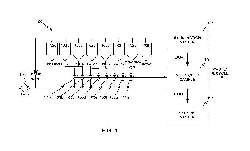
that fluid from
any particular one of the plurality of reservoirs can be individually supplied
to the flow cell
under the impetus of the mechanism to generate fluid flow and under control of
the
computerized controller, by opening of the respective valve of the particular
reservoir and
closing the other valves.
1
CA 03009651 2018-06-22
WO 2017/117243
PCT/US2016/068916
BRIEF DESCRIPTION OF THE DRAWINGS
[0005] FIG. 1 illustrates a block diagram of a system in accordance with
embodiments of
the invention.
[0006] FIG. 2 illustrates nanoballs bound to grid locations of a flow cell, in
accordance
with embodiments of the invention.
[0007] FIG. 3 illustrates an example arrangement of a flow cell, an
illumination system, and a
sensing system, in accordance with embodiments of the invention.
[0008] FIG. 4 illustrates another example arrangement of a flow cell, an
illumination system,
and a sensing system, in accordance with embodiments of the invention.
[0009] FIG. 5 illustrates a system in accordance with other embodiments of the
invention.
[0010] FIGS. 6A and 6B illustrate two trapezoidal prisms in accordance with
embodiments
of the invention.
[0011] FIG. 7A illustrates a flow cell cavity arrangement in accordance with
embodiments
of the invention, and FIG. 7B illustrates the flow of fluids through the flow
cell arrangement
of FIG. 7A.
[0012] FIG. 8A illustrates a flow cell cavity arrangement in accordance with
embodiments
of the invention, and FIG. 8B illustrates the flow of fluids through the flow
cell arrangement
of FIG. 8A.
[0013] FIG. 9A illustrates a flow cell cavity arrangement in accordance with
embodiments
of the invention, and FIG. 9B illustrates the flow of fluids through the flow
cell arrangement
of FIG. 9A.
[0014] FIG. 10 illustrates an instrument in accordance with embodiments of the
invention.
[0015] FIGS. 11A and 11B illustrate the instrument of FIG. 10 with its cover
removed.
[0016] FIG. 12 illustrates a disposable fluidic interface of the instrument of
FIG. 10 in
isolation.
[0017] FIG. 13 is an exploded view of the disposable fluidic interface of FIG.
12.
[0018] FIG. 14A illustrates a cutaway view of the disposable fluidic interface
of FIG. 12,
showing valves in their normally closed positions.
2
CA 03009651 2018-06-22
WO 2017/117243
PCT/US2016/068916
[0019] FIG. 14B shows a valve of FIG. 14A, in its open position.
[0020] FIG. 15 illustrates a cartridge according to embodiments of the
invention.
[0021] FIG. 16A shows an unprocessed surface plasmon resonance (SPR) image of
a
microspotted array on a gold thin film, in accordance with embodiments of the
invention.
[0022] FIG. 16B illustrates a processed SPR image with background subtraction
prior to
exposure to sequencing reagents, in accordance with embodiments of the
invention.
[0023] FIG. 16C shows the change in relative reflected intensity on the
microspotted chip
of FIG. 16B after exposure to sequencing reagents.
[0024] FIG. 17A shows an SPR image of the flow patterned chip in accordance
with
embodiments of the invention.
[0025] FIG. 17B shows raw sequencing data collected from a region of the phiX
bacteriophage genome, in accordance with embodiments of the invention.
[0026] FIG. 17C shows the resulting positive and negative base calls derived
from the raw
data of FIG. 17B.
[0027] FIG. 18 schematically illustrates nanohole sensing, in accordance with
embodiments of the invention.
[0028] FIG. 19 illustrates a module including the nanohole sensing system of
FIG. 18, in
accordance with embodiments of the invention.
[0029] FIG. 20 illustrates a sensogram recorded using nanohole sensing, in
accordance
with embodiments of the invention.
[0030] FIG. 21 illustrates another sensing modality usable in embodiments of
the
invention, configured for utilizing grating waveguide resonance.
[0031] FIGS. 22A and 22B illustrate the effect of grating-waveguide resonance,
in
accordance with embodiments of the invention.
[0032] FIG. 23 illustrates images of a flow-patterned substrate taken using
grating-
waveguide resonance (GWR), in accordance with embodiments of the invention.
[0033] FIG. 24 illustrates averaged intensity readings taken from a
polydopamine
(universal) surface chemistry using GWR, in accordance with embodiments of the
invention.
3
CA 03009651 2018-06-22
WO 2017/117243
PCT/US2016/068916
[0034] FIG. 25 shows a grating used for enhancement of fluorescence, in
accordance with
embodiments of the invention.
[0035] FIG. 26 shows a test system in accordance with embodiments of the
invention.
[0036] FIG. 27A illustrates a digital image taken with the system of FIG. 26.
[0037] FIG. 27B illustrates another digital image taken with the system of
FIG. 26.
[0038] FIG. 27C illustrates a digital slice taken through a portion of the
image of FIG. 27B.
[0039] FIB. 28A illustrates a digital slice taken through a portion of an
image taken using
total internal reflectance fluorescence (TIRF) imaging, in the area of a
particular nanoball.
[0040] FIB. 28A illustrates a digital slice taken through a portion of an
image taken using
surface plasmon enhanced fluorescence (SPEF) imaging, in the area of a
particular nanoball.
DETAILED DESCRIPTION
[0041] The present disclosure provides a device that can be used for a variety
of molecular
analyses, such as nucleic acid sequencing. In some embodiments, sequencing is
carried out
as described in commonly owned US Pat. App. Ser. No. 14/805,381, which is
incorporated
by reference herein in its entirety. Briefly, methods for determining the
sequence of a
template nucleic acid molecule can be based on a repetitive process wherein
each cycle in the
process provides information toward identifying one or more nucleotides in a
target nucleic
acid. The sum of the information from the cycles provides the sequence of
nucleotides for
the target nucleic acid. In particularly useful sequencing protocols each
cycle is carried out
by forming a ternary complex (between polymerase, primed nucleic acid and
cognate
nucleotide) under specified conditions. The method can generally include a
step of
examining the ternary complex prior to a correct nucleotide being incorporated
into the
nucleic acid by covalent attachment to the 3' end of the primer. For example,
the method can
involve providing a template nucleic acid molecule primed with a primer;
contacting the
primed template nucleic acid molecule with a first reaction mixture that
includes a
polymerase and at least one nucleotide molecule; detecting interaction of the
polymerase and
nucleotide with the primed template nucleic acid molecule, without covalent
incorporation of
the nucleotide molecule into the primed template nucleic acid; and identifying
a next base in
the template nucleic acid using the detected interaction of the polymerase and
nucleotide with
4
CA 03009651 2018-06-22
WO 2017/117243
PCT/US2016/068916
the primed template nucleic acid molecule. In this procedure, ternary complex
stabilization
advantageously enhances discrimination between correct and incorrect
nucleotides.
[0042] In particular embodiments, a device of the present disclosure can
detect ternary
complexes formed at each cycle of a sequencing process without the need for
exogenous
labels on one or more of the reactants that would typically be labeled when
carrying out a
sequencing process on other detection platforms. For example, the sequencing
reaction can
be performed using polymerase, nucleotides and primed nucleic acids that all
lack exogenous
labels that are used for detection. However, in some embodiments of the
present disclosure
the polymerase can be labeled with an exogenous moiety. Alternatively or
additionally to
polymerase labeling, the nucleotides can be labeled.
[0043] FIG. 1 illustrates a block diagram of a system 100 in accordance with
embodiments
of the invention. System 100 includes a flow cell 101, in which a sample of
material to be
sequenced can be placed. For example, the sample may be an array of
"nanoballs" 201 of
amplified DNA fragments, bound to a grid of locations within flow cell 101, as
shown in
FIG. 2. While only a few nanoballs 201 are shown in FIG. 2 for ease of
explanation, more or
fewer may be present. Depending on the target application, many, many
receptors may be
present within flow cell 101, for example up to millions, tens of millions, or
more. DNA
nanoballs can be made using methods and compositions as described, for
example, in U.S.
Pat. No. 7,910,354; or US Pat. App. Publ. Nos. 2009/0264299 Al, 2009/0011943
Al,
2009/0005252 Al, 2009/0155781 Al, or 2009/0118488 Al; or Drmanac et al., 2010,
Science
327(5961): 78-81; each of which is incorporated herein by reference.
[0044] Nanoballs are one type of nucleic acid amplification product that can
be used to
form a feature on an array. Other useful amplification products include those
produced by
solid-phase amplification techniques. For example, amplification can be
carried out using
bridge amplification to form nucleic acid clusters on a surface. Useful bridge
amplification
methods are described, for example, in U.S. Pat. Nos. 5,641,658 or 7,115,400;
or US Pat.
App. Pub. Nos. 2002/0055100 Al, 2004/0096853 Al, 2004/0002090 Al, 2007/0128624
Al;
or 2008/0009420 Al, each of which is incorporated herein by reference. Another
useful
method for amplifying nucleic acids on a surface is rolling circle
amplification (RCA), for
example, as described in Lizardi et al., Nat. Genet. 19:225-232 (1998) and US
Pat. App. Pub.
No. 2007/0099208 Al, each of which is incorporated herein by reference.
Emulsion PCR on
beads can also be used, for example as described in Dressman et al., Proc.
Natl. Acad. Sci.
CA 03009651 2018-06-22
WO 2017/117243
PCT/US2016/068916
USA 100:8817-8822 (2003), WO 05/010145, US Pat. App. Pub. No. 2005/0130173 Al
or US
Pat. App. Pub. No. 2005/0064460 Al, each of which is incorporated herein by
reference. A
system or method of the present disclosure can use one or more of the reagents
described in
the above references for making and using nanoballs or other nucleic acid
features.
[0045] Referring again to FIG. 1, system 100 also includes a number of
reservoirs 102a-102h
(collectively reservoirs 102), for holding various buffers, nucleotides, and
other fluids. While
eight reservoirs are shown in the example of FIG. 1, more or fewer reservoirs
may be present in
other embodiments. The reservoirs can contain reagents used for creating
nucleic acid features
and/or reagents for sequencing nucleic acids such as those set forth herein or
in references
incorporated by reference herein.
[0046] Each of reservoirs 102a-102h is connected to respective valve 103a-103h
(collectively
valves 103), such that under the control of a computerized controller (not
shown), fluid from any
one of reservoirs 102 can be individually supplied to flow cell 101 under the
impetus of a
mechanism to generate fluid flow, for example pump 104. In some embodiments,
pump 104 or
other mechanism to generate fluid flow may produce a constant fluid flow, and
in other
embodiments, may produce a variable fluid flow. Flow cell 101 can be
illuminated by an
illumination system 105 and optically sensed by a sensing system 106. Valves
103 may be
arranged either serially, in parallel, or in any combination of
configurations. In some
embodiments, valves 103 are arranged serially to prevent pockets of reagent
that could
contaminate subsequent steps of the sequencing reaction. The wash buffer is
situated at the
position furthest from the flow cell to ensure that all reagents are
thoroughly washed from the
channel and flow cell prior to subsequent sequencing steps. Valves 103 may be
actuated by
pneumatic, mechanical, or electrical means.
[0047] Sensing system 106 may be, for example, a digital camera having an
array light sensor.
Various optical devices such as prisms, lenses, filters, and the like may be
present between flow
cell 101 and sensing system 106, as is explained in more detail below. It
should be recognized
that FIG. 1 is highly schematic, and is not intended to represent specific
component
arrangements. Some specific arrangements are described below.
[0048] In a basic manner of operation of system 100, a sample to be sequenced
is placed in
flow cell 101. The sample may be previously prepared such that nucleic acid
features are present
on the surface prior to introducing the flow cell to the system.
Alternatively, the nucleic acid
features may be constructed in part by system 100 for example, by a solid
phase amplification
technique set forth herein or known in the art. Once the sample is in place, a
test nucleotide may
6
CA 03009651 2018-06-22
WO 2017/117243
PCT/US2016/068916
be delivered to flow cell 101, for example from reservoir 102c. In the
presence of polymerase,
the test nucleotide binds to sites in the sample having cognate nucleotide
positions adjacent to the
3' end of a primer (i.e. the test nucleotide occupies the position of the
"next correct" nucleotide
for primer extension). The binding creates changes in the sites that are
detectable by sensing
system 106. Depending on the sensing technology being used, the detectable
change may be a
change in apparent reflectance due to surface plasmon resonance, may be the
presence of a
fluorescent marker supplied with the nucleotide or polymerase, may be the
presence of
fluorescence excited by illumination system 105 without the need for a marker,
or may be some
other kind of detectable change caused by binding between a primed nucleic
acid, polymerase
and nucleotide to form a ternary complex. A number of sensing technologies
that may be used in
embodiments of the invention are described in U.S. Patent Application No.
14/805,381 filed July
21, 2015 and titled "Nucleic Acid Sequencing Methods and Systems", the entire
disclosure of
which is hereby incorporated by reference herein for all purposes.
[0049] Sensing system 106 then detects the changes in the sample resulting
from the
introduction of the test nucleotide, for example by taking an image of the
area of the flow cell and
analyzing the digital image to detect the locations of any changes. The
changes indicate the
locations at which the supplied test nucleotide attached to the sample via
ternary complex
formation. Because the type of the test nucleotide is known, the nucleotide to
which it attached is
inferable, being the complementary nucleotide of the test nucleotide.
[0050] Preferably, flow cell 101 is washed to remove any unattached reagents
such as
nucleotides, and a second test reagent (e.g. second type of nucleotide) is
supplied to flow cell
101, for example from reservoir 102d. Any changes to the sample are detected
in a similar
manner, and locations where binding of the second test nucleotide (e.g. via
formation of a ternary
complex) are detected are noted as containing primed nucleic acids having a
sequence position
that is complementary to the second test nucleotide.
[0051] This process is repeated so that the nucleotide sequence at each sample
location is
cumulatively determined.
[0052] The above description is highly simplified, and is presented in the
interest of assisting
in the understanding of the specific embodiments described below. More detail
about the
sequencing process may be found below or in U.S. Patent Application
14/805,381, which is
incorporated herein by reference.
[0053] Although the present disclosure exemplifies several aspects of the
systems and
methods set forth herein in the context of nucleic acid sequencing, it will be
understood that a
7
CA 03009651 2018-06-22
WO 2017/117243
PCT/US2016/068916
variety of other analytes can be detected. Analytes that participate in
binding interactions
with probes that can be attached to a surface are particularly useful.
Similarly, binding assays
that have been, or can be, modified to occur on solid-phase supports are also
useful.
Exemplary analytes that can be detected include, but are not limited to,
biological
macromolecules such as proteins, enzymes, receptors, antibodies,
polysaccharides or the like;
analogs of biological macromolecules such as nucleic acid analogs (e.g.
protein nucleic acid),
antibody analogs (e.g. Fab or F(ab')2), mutant enzymes that retain binding
affinity for
substrates or the like; biological particles such as cells, viruses, vesicles,
nanopores,
ribosomes, organelles, nuclei or the like; biological small molecules such as
metabolites,
saccharides, amino acids, nucleotides, enzyme cofactors, or analogs thereof;
or synthetic
analytes such as candidate ligands for target receptors, candidate therapeutic
agents such as
enzyme inhibitors, nanoparticles, beads or the like. Particularly useful
binding assays
include, but are not limited to, immunosorbent assays which can be performed
without the
need for enzyme labels or other labels that are typically used in ELISA
formats, receptor-
ligand binding assays, cell surface receptor biding assays, nucleic acid
hybridization assays,
ribosome binding assays, protein-protein binding assays or the like.
[0054] An advantage of the systems and methods set forth herein is that a
variety of
different types of binding assays can be run on the same system. This is
possible in many
embodiments due to localized detection of different binding events at discrete
surface
features and lack of unwanted background signal from target analytes that
remain in solution.
When using a system of the present disclosure, different types of probe
analytes can be
attached to discrete features on a surface, the location of the probe analytes
can be known or
determined, and different target analytes can be delivered in solution under
conditions that
allow them to bind to probes for which they have an affinity. The different
binding assays
can be run on the same substrate either sequentially or simultaneously (i.e.
in parallel).
[0055] Optical Systems
[0056] FIG. 3 illustrates an example arrangement of flow cell 101,
illumination system 105,
and sensing system 106 in more detail, in accordance with embodiments of the
invention. In
illumination system 105, a light source 301 emits light which is captured and
sufficiently
collimated by a lens 302. In some embodiments, light source 301 may be a light
emitting diode
or an array of light emitting diodes emitting light at a wavelength of about
650 nm, but other
wavelengths may be used on other embodiments, and other kinds of light sources
may be used.
For example, a laser with beam expanding optics may be used. The light
produced by
8
CA 03009651 2018-06-22
WO 2017/117243
PCT/US2016/068916
illumination system 105 may be coherent or non-coherent. In some embodiments,
multiple light
emitting diodes or lasers may be used emitting light in different wavelengths.
[0057] In some embodiments, the light source emits a narrow range of
wavelengths (less
than 10 nm full width at half maximum) centered on a wavelength in the visible
light
spectrum. In other embodiments, the light source emits a broad range of
wavelengths onto a
sample at a fixed angle, and the reflected light is then dispersed by a
diffraction grating onto a
CCD or linear photodiode array to determine the resonant wavelength. In some
embodiments, one or more optical filters may be used to narrow the wavelength
content of
the illumination light.
[0058] Lens 302 may be a simple piano convex element having a focal length of
about 24
mm, or may be a more complex lens such as a multi-element lens. Other focal
lengths may
also be used in other embodiments. Preferably, an aperture 303 limits the size
of the
illumination beam 304. The size of aperture 303 may be selected in accordance
with the
capabilities of the particular embodiment, but in one example, aperture 303
may have a
diameter of about 10 mm.
[0059] Beam 304 enters a prism 305 through an input face 306. Prism 305 may be
a simple
triangular prism made of F2 glass or another suitable glass. In other
embodiments, prism 305
may be molded from a polymer such as polycarbonate or another suitable clear
polymer.
[0060] Flow cell 101 is positioned on a top or detection face 307 of prism
305. A cover
glass 311 may also be present over flow cell 101, opposite detection face 307
of prism 305.
In some embodiments, detection surface 307 is coated with a thin layer of
gold, silver,
aluminum, or another suitable material. The prism can be an integral component
of the flow
cell such that the coating is directly on a surface of the prism and reagents
flow over the
surface of the prism when flowing through the flow cell. In other embodiments,
an optically
transparent window of a flow cell having the coating is coupled with the
prism. As such, the
prism can be an integral part of a flow cell or the prism can be a separate
component that is
removably coupled with a window of the flow cell.
[0061] Illumination system 105 preferably produces plane polarized light (p-
polarization,
where the electric field of the incident photon has a component normal to the
plane of the
gold film), which is then passed through one face of the prism at a defined
angle wherein
some of the light is absorbed by the gold film. Another portion of beam 304
reflects from
detection surface 307, either by total internal reflection or by reflection
from the metal
9
CA 03009651 2018-06-22
WO 2017/117243
PCT/US2016/068916
coating on detection surface 307. Imaging system 106 images an area of
detection surface
307 through output face 308 of prism 305. Imaging system 106 includes a lens
308, which
may be a simple plano convex or aspheric singlet having a focal length of
about 24 mm,
although other kinds of lenses may be used.
[0062] Lens 309 forms an image on an electronic array light sensor 310. Sensor
310 may
be, for example, a complementary metal oxide semiconductor (CMOS) sensor, a
charge
coupled device (CCD) sensor, or another kind of sensor having a number of
light sensitive
areas called pixels arranged in an array. Sensor 310 may be part of a camera,
for example a
DMM24UJO03 board camera available from The Imaging Source of Bremen, Germany.
In
any event, sensor 310 produces signals indicating the intensity of light
received from the
locations on detection face 307 corresponding to the sensor pixels. These
signals may be
compiled into a digital image. Some of the digital image may correspond to one
or more
reference regions to account for changes in background signal due to bulk
refractive index
changes, nonspecific binding of soluble factors, thermal fluctuations, changes
in the surface
of the sensing element, or other effects.
[0063] As is shown in FIG. 3, the plane of sensor 310 may be oblique to the
optical axis of
sensing system 106, in order to correctly image detection face 307, which is
also at an
oblique angle.
[0064] Optionally, detecting a change in refractive index is accomplished in
one or a
combination of means, including, but not limited to, surface plasmon resonance
sensing,
localized plasmon resonance sensing, plasmon-photon coupling sensing,
transmission sensing
through sub-wavelength nanoholes (enhanced optical transmission), photonic
crystal sensing,
interferometry sensing, refraction sensing, guided mode resonance sensing,
ring resonator
sensing, or ellipsometry sensing. Optionally, probe analytes can be localized
to features on
the surface and target analytes can be delivered under conditions wherein the
probes and
targets interact such that a change in local refractive index can be detected
and used to
identify or characterize the interaction. For example, nucleic acid molecules
may be
localized to a surface, wherein the interaction of polymerase with nucleic
acids in the
presence of various nucleotides may be measured as a change in the local
refractive index.
[0065] Optionally, a probe analyte (e.g. a template nucleic acid) is tethered
to or localized
appropriately on or near a surface, such that the interaction of the target
analyte (e.g.
interaction of polymerase and template nucleic acid in the presence of
nucleotides) changes
CA 03009651 2018-06-22
WO 2017/117243
PCT/US2016/068916
the light transmitted across or reflected from the surface. The surface may
contain
nanostructures. Optionally, the surface is capable of sustaining plasmons or
plasmon
resonance. Optionally, the surface is a photonic substrate, not limited to a
resonant cavity,
resonant ring or photonic crystal slab. Optionally, the surface is a guided
mode resonance
sensor. Optionally, the probe analyte (e.g. nucleic acid) is tethered to, or
localized
appropriately on or near a nanohole array, a nanoparticle or a microparticle,
such that the
interaction of target analyte (e.g. interaction of polymerase and template
nucleic acid in the
presence of nucleotides) changes the absorbance, scattering, reflection or
resonance of the
light interacting with the microparticle or nanoparticle.
[0066] Optionally, extraordinary optical transmission (EOT) through a nanohole
array may
be used to monitor probe / target (e.g. nucleic-acid/polymerase) interactions.
Light
transmitted across subwavelength nanoholes in plasmonic metal films is higher
than expected
from classical electromagnetic theory. This enhanced optical transmission may
be explained
by considering plasmonic resonant coupling to the incident radiation, whereby
at resonant
wavelength, a larger than anticipated fraction of light is transmitted across
the metallic
nanoholes. The enhanced optical transmission is dependent on the dimensions
and pitch of
the nanoholes, properties of the metal, as well as the dielectric properties
of the medium on
either side of the metal film bearing the nanoholes. In the context of a
biosensor, the
transmissivity of the metallic nanohole array depends on the refractive index
of the medium
contacting the metal film, whereby, for instance, the interaction of
polymerase with nucleic
acid attached to the metal surface may be monitored as a change in intensity
of light
transmitted across the nanoholes array. The elegance of the EOT/plasmonic
nanohole array
approach is that the instrumentation and alignment requirements of surface
plasmon
resonance may be replaced by very compact optics and imaging elements. For
instance, just
a low power LED illumination and inexpensive CMOS or CCD camera may suffice to
implement robust EOT plasmonic sensors. An exemplary nanohole array-based
surface
plasmon resonance sensing device is described in C. Escobedo et al.,
"Integrated Nanohole
Array Surface Plasmon Resonance Sensing Device Using a Dual-Wavelength
Source,"
Journal of Micromechanics and Microengineering 21, no. 11 (November 1,2011):
115001,
which is herein incorporated by reference in its entirety.
[0067] The plasmonic nanohole array may be patterned on an optically opaque
layer of
gold (greater than 50 nm thickness) deposited on a glass surface. Optionally,
the plasmonic
nanohole array may be patterned on an optically thick film of aluminum or
silver deposited
11
CA 03009651 2018-06-22
WO 2017/117243
PCT/US2016/068916
on glass. Optionally, the nanohole array is patterned on an optically thick
metal layer
deposited on low refractive index plastic. Patterning plasmonic nanohole
arrays on low
refractive index plastics enhances the sensitivity of the device to refractive
index changes by
better matching the refractive indices on the two sides of the metal layer.
Optionally,
refractive index sensitivity of the nanohole array is increased by increasing
the distance
between holes. Optionally, nanohole arrays are fabricated by replication, for
example, by
embossing, casting, imprint-lithography, or template-stripping. Optionally,
nanohole arrays
are fabricated by self-assembly using colloids. Optionally, nanohole arrays
are fabricated by
projection direct patterning, such as laser interference lithography.
[0068] A nano-bucket configuration may be preferable to a nanohole
configuration. In the
nanohole configuration, the bottom of the nano-feature is glass or plastic or
other appropriate
dielectric, whereas in the nano-bucket configuration, the bottom of the nano-
feature
comprises a plasmonic metal. The nano-bucket array configuration may be easier
to fabricate
in a mass production manner, while maintaining the transmission sensitivity to
local
refractive index.
[0069] Optionally, the nanohole array plasmonic sensing is combined with lens-
free
holographic imaging for large area imaging in an inexpensive manner.
Optionally, a
plasmonic biosensing platform comprises a plasmonic chip comprising nanohole
arrays, a
light- emitting diode source configured to illuminate the chip, and a CMOS
imager chip to
record diffraction patterns of the nanoholes, which is modulated by molecular
binding events
on the surface. The binding events may be the formation of a closed-complex
between a
polymerase and a template nucleic acid in the presence of a nucleotide.
[0070] The methods to functionalize surfaces (e.g. for nucleic acid
attachment) for surface
plasmon resonance sensing may be directly applied to EOT nanohole arrays as
both sensing
schemes employ similar metal surfaces to which probes, such as nucleic acids,
can be
attached.
[0071] Optionally, the refractive index changes associated with probe / target
interaction
may be detected or monitored on nanostructured surfaces that do not support
plasmons.
Optionally, guided mode resonance may be used to detect or monitor the probe /
target
interaction. Guided-mode resonance or waveguide-mode resonance is a phenomenon
wherein
the guided modes of an optical waveguide can be excited and simultaneously
extracted by the
introduction of a phase-matching element, such as a diffraction grating or
prism. Such guided
12
CA 03009651 2018-06-22
WO 2017/117243
PCT/US2016/068916
modes are also called "leaky modes", as they do not remain guided, and have
been observed in
one and two-dimensional photonic crystal slabs. Guided mode resonance may be
considered a
coupling of a diffracted mode to a waveguide mode of two optical structured
placed adjacent or
on top of each other. For instance, for a diffraction grating placed on top of
an optical waveguide,
one of the diffracted modes may couple exactly into the guided mode of the
optical waveguide,
resulting in propagation of that mode along the waveguide. For off-resonance
conditions, no light
is coupled into the waveguide, so the structure may appear completely
transparent (if dielectric
waveguides are used). At resonance, the resonant wavelength is strongly
coupled into the
waveguide, and may be coupled out of the structure depending on downstream
elements from the
grating-waveguide interface. In cases where the grating coupler is extended
over the entire
surface of the waveguide, the light cannot be guided, as any light coupled in
is coupled out at the
next grating element. Therefore, in a grating waveguide structure, resonance
is observed as a
strong reflection peak, whereas the structure is transparent to off-resonance
conditions. The
resonance conditions are dependent on angle, grating properties, polarization
and wavelength of
incident light. For cases where the guided mode propagation is not present,
for instance due to a
grating couple to the entire surface of the waveguide, the resonant mode may
also be called
leaky-mode resonance, in light of the strong optical confinement and
evanescent propagation of
radiation in a transverse direction from the waveguide layer. Change in
dielectric properties near
the grating, for instance due to binding of biomolecules affects the coupling
into the waveguide,
thereby altering the resonant conditions. Optionally, where nucleic acid
molecules are attached to
the surface of grating waveguide structures, the polymerase/nucleic-acid
interaction may be
detected or monitored as a change in wavelength of the leaky mode resonance.
[0072] Optionally, a diffraction element may be used directly on a transparent
substrate
without an explicit need for a waveguide element. The change in resonance
conditions due to
probe / target interactions near the grating nanostructure may be monitored as
resonant
wavelength shifts in the reflected or transmitted radiation.
[0073] Optionally, reflected light from a probe-attached, guided mode resonant
sensor may
be used to detect or monitor the probe / target interaction. A broadband
illumination source
may be employed for illumination, and a spectroscopic examination of reflected
light could
reveal changes in local refractive index due to target binding.
[0074] Optionally, a broadband illumination may be used and the transmitted
light may be
examined to identify resonant shifts due to probe / target interaction.
Optionally, a linearly
polarized narrow band illumination may be used, and the transmitted light may
be filtered
13
CA 03009651 2018-06-22
WO 2017/117243
PCT/US2016/068916
through a cross-polarizer; wherein the transmitted light is completely
attenuated due to the
crossed polarizers excepting for the leaky mode response whose polarization is
modified. This
implementation converts refractive index detecting or monitoring to a simple
transmission assay
that may be monitored on inexpensive imaging systems. This exemplary
embodiment is aided by
published material that describe the assembly of the optical components,
Yousef Nazirizadeh et
al., "Low-Cost Label-Free Biosensors Using Photonic Crystals Embedded between
Crossed
Polarizers," Optics Express 18, no. 18 (August 30, 2010): 19120-28, which is
incorporated herein
in its entirety.
[0075] Alongside nanostructured surfaces, plain, un-structured surfaces may
also be used
advantageously for detecting or monitoring refractive index modulations
resulting from probe
/ target interactions. Optionally, interferometry may be employed to detect or
monitor the
interaction of probe and target (e.g. interaction of polymerase with double
stranded nucleic
acid) bound to an un-structured, optically transparent substrate. Optionally,
probe molecules
may be attached to the top surface of a glass slide (by any means known in the
art), and the
system illuminated from the bottom surface of the glass slide. There are two
reflection
surfaces in this configuration, one reflection from the bottom surface of the
glass slide, and
the other from the top surface which has probes (e.g. nucleic acid molecules)
attached to it.
The two reflected waves may interfere with each other causing constructive or
destructive
interference based on the path length differences, with the wave reflected
from the top surface
modulated by the changes in dielectric constant due to the bound probes (and
subsequently by the
interaction of target with the bound probe). With the reflection from the
bottom surface
unchanged, any binding to the probe may be reflected in the phase difference
between the beams
reflected from the top and bottom surfaces, which in turn affects the
interference pattern that is
observed. Optionally, bio-layer interferometry is used to detect or monitor
the probe / target
interaction. Bio-layer interferometry may be performed on commercial devices
such as those sold
by Pall Forte Bio corporation.
[0076] The reflected light from the top surface can be selectively chosen by
using focusing
optics. The reflected light from the bottom surface is disregarded because it
is not in the focal
plane. Focusing only on the probe-attached top surface, the light collected by
the focusing
lens comprises a planar wave, corresponding to the partially reflected
incident radiation, and
a scattered wave, corresponding to the radiations scattered in the collection
direction by
molecules in the focal plane. These two components may be made to interfere if
the incident
14
CA 03009651 2018-06-22
WO 2017/117243
PCT/US2016/068916
radiation is coherent. This scattering based interferometric detection is
extremely sensitive,
and can be used to detect down to single protein molecules.
[0077] In some embodiments, system 100 detects the binding of analytes to the
sample
using the phenomenon of surface plasmon resonance (SPR). Surface plasmon
resonance
sensing is a method for the label-free detection of analytes such as proteins,
enzymes,
macromolecules, nucleic acids, nanoparticles, vesicles, cells, exosomes,
organelles, or other
analytes, due to their interaction with light impinging upon a thin gold film
(the sensing
element or detection surface) at a defined angle and wavelength. The
interaction of light with
the gold film induces a collective oscillation of electrons at the
gold/environment interface
that produces a highly sensitive evanescent field at the interface. The
evanescent field is
highly sensitive to perturbations in refractive index of the surrounding
environment. In some
embodiments, the formation of a ternary complex on a nucleic acid feature
creates slight
changes in the resonance conditions, and can be detected as changes in the
apparent
reflectivity of the gold layer on detection surface 307. In some embodiments,
the angle of
incidence of the illumination light with respect to detection surface 307 can
be varied to
measure changes in reflected intensity. In other embodiments, the angle of
incidence remains
fixed. In any event, changes in reflected intensity are measured as a
sequencing reaction
proceeds on nucleic acid features. The instrument can be configured in such a
way that either
increasing or decreasing intensity can correspond to the detection of a
sequencing step.
[0078] By taking digital images of a sample array of nucleic acid features
after each
application of sequencing reagent (e.g. polymerase and test nucleotides),
features where
reflectivity changes have occurred can be detected and therefore the nucleic
acid to which a
particular nucleotide bound as the next correct nucleotide can be determined.
For example
one can determine which of nanoballs 201 contained a sequence to which the
test nucleotide
bound in a ternary complex.
[0079] FIG. 4 illustrates a system 400 using another example arrangement of
flow cell 101,
illumination system 105 and sensing system 106, in accordance with other
embodiments of the
invention. In the example of FIG. 4, a laser 401 and beam expander 402 are
used to create
illumination beam 403. A trapezoidal prism 404 is used, rather than a
triangular prism.
Trapezoidal prism 404 includes an input surface 405 and an output surface 406
coplanar with
input surface 405. A detection surface 407 is parallel to and spaced apart
from input and output
surfaces 405 and 406. A first angled surface 408 joins one edge of detection
surface 407 and one
edge of input surface 405, and a second angled surface 409 joins the other
edge of detection
CA 03009651 2018-06-22
WO 2017/117243
PCT/US2016/068916
surface 407 with an edge of output surface 406. Prism 404 may be made of any
suitable material,
for example F2 or another glass, or poly(methyl methacrylate) (PMMA) or
another suitable
polymer. As is shown in the embodiment shown in FIG. 4, a portion 411 of prism
404 is
removed, for example to facilitate making prism 404 by injection molding of a
polymer. In other
embodiments, this portion need not be absent, and input and output faces 405
and 406 may join to
form a single face.
[0080] Illumination beam 403 enters input face 405 of prism 404, reflects from
first angled
face 408 and is directed to detection face 407. Light reflecting from
detection face 407
further reflects from second angled face 409 and exits prism 404 via output
face 406. A
camera 410 images a portion of detection face 407. Images captured by camera
410 can be
analyzed as described above to detect attachments of nucleotides to targets
within flow cell
101. A cover glass 412 may also be present.
[0081] The systems of FIGS. 3 and 4 are examples of systems that operate in a
reflection
mode. In these systems, illumination and detection are performed from the same
side of flow
cell 101, and light reaches the sensor by reflection from the detection
surface of the prism.
[0082] FIG. 5 illustrates a system 500 in accordance with other embodiments of
the
invention. Some portions of system 500 are duplicated from system 400 shown in
FIG. 4,
and are given the same reference numbers. In system 500, an additional camera
501 is placed
on the opposite side of flow cell 101 from illumination system 105, and images
a plane at
flow cell 101.
[0083] Camera 501 may sense changes within flow cell 101 (and therefore
bindings of test
nucleotides to the sample) using surface plasmon enhanced fluorescence (SPEF)
imaging.
An optical bandpass filter may be included in camera 501 to block non-
fluorescence
wavelength light and only allow fluorescence light to pass through. In SPEF,
fluorescence of
markers within the sample is excited by the surface plasmon effect. The
fluorescence can be
detected by camera 501, to detect locations where test nucleotides have
attached to the
sample. Camera 410 preferably operates in parallel with camera 501. Thus,
system 500
operates in both a reflection mode and a transmission mode. Camera 401
performs sensing
from the same side of flow cell as illumination system 105 (reflection mode),
while camera
501 senses from the opposite side (transmission mode).
[0084] In other embodiments, detection surface 407 of prism 404 may not be
plated, and
camera 501 may perform total internal reflection fluorescence (TIRF) imaging.
In TIRF, the
16
CA 03009651 2018-06-22
WO 2017/117243
PCT/US2016/068916
fluorescence is excited by an evanescent wave resulting from the total
internal reflection of
the illumination light from the interior surface of the prism. SPEF can
produce images with
better signal-to-noise characteristics than TIRF. A system using TIRF is an
example of a
system operating in a transmission mode, because the sensing is performed from
the opposite
side of flow cell 101 from illumination system 105.
[0085] The surface plasmon effect is very sensitive to the configuration of
the system,
including the materials of the components, the wavelength of light used, and
the angle of
incidence of light on the surface where it is desired to produce plasmons. The
index of
refraction of the prism is an important parameter. FIGS. 6A and 6B illustrate
two trapezoidal
prisms that may be suitable for use in the systems such as those of FIGS. 4
and 5.
[0086] In a system such as those shown in FIGS. 3-5, the detection face of the
prism may
be in direct contact with analytes, for example, being integral to a flow cell
through which
analyte-containing reagents are delivered. As such the detection face of the
prism can be
functionalized with a reactive material such as a mixed alkanethiol monolayer
to provide the
ability to bind avidin, neutravidin, or streptavidin, onto the slide which can
then bind
biotinylated probes such as amplicons, priming sequences, barcode sequences,
or other
capture elements. The detection face may be either unpatterned or patterned.
Patterning of
the sensing element can be achieved by either spotting reagents into an
ordered array of spots
with a microarraying device, or by selectively depositing reagents to the
surface using a flow
cell to create sensing regions.
[0087] In some systems such as those shown in FIGS. 3-5, modification of the
surface
properties of the detection surface can provide the ability to present
chemical moieties that
provide a high level of specificity for sensing. A number of strategies can be
adopted
depending on the material comprising the sensing element.
[0088] For example, in some embodiments, the gold or other thin-film is coated
with a self-
assembled monolayer (SAM) of alkanethiol molecules. The monolayer is comprised
of a
mixture of inter polyethylene glycol (PEG) chains and biotin terminated
alkanethiol chains.
The mixture is tailored to allow optimal spacing between the biotin moieties
for binding of
avidin, neutravidin, or streptavidin. In other embodiments, the gold thin film
can be
modified with amine-terminated, carboxy-terminated, or glycidoxypropyl-
terminated alkane
thiols to allow for derivatization with heterobifunctional cross-linking
agents. These surface
modifications can also serve as an adhesion layer for physically adsorbed
polymers (e.g.
17
CA 03009651 2018-06-22
WO 2017/117243
PCT/US2016/068916
proteins, polylysine, dextrans, polydopamine, etc). Other surface chemistries
for attaching
biomolecules to a surface are well known, and include hydrogels (acrylamide,
agarose, ),
polymers (polylysine, dextran, polydopamine, poly acrylic acid, pHEMA, ),
bifunctional
crosslinkers with reactive endgroups (comprising sulfhydryl, carboxyl,
hydroxyl, amino,
azido, alkyne, phosphonic acid,). Adhesion layer formed by self assembly,
immersion, dip
coating, spin coating, electodeposition, electroless deposition, vapor
deposition, Langmuir-
Blodgett film transfer, reversible addition fragmentation transfer - RAFT,
atom transfer
radical polymerization - ATRP. Surfaces can be activated/cleaned by plasma, UV
ozone,
chemical, radiation, or other means. A scattering label may be conjugated to
the polymerase
molecule and in the presence of the correct base the density of labeled
polymerases can create
a detectable scattering cross section. The scattering label may be comprised
of gold
nanoparticles in the size range of 5 ¨ 100 nm dia. Detection can be
accomplished in
reflectance or transmission mode
[0089] Flow Cell Arrangements
[0090] In some embodiments it is desirable that fluids flowing through flow
cell 101
exhibit uniformity of velocity and cover the array of features on flow cell
101 as nearly
completely as possible.
[0091] FIG. 7A illustrates a flow cell cavity arrangement in accordance with
embodiments
of the invention. The cavity illustrated in FIG. 7A may be covered by a
transparent structure,
and defines a thin, flat recess 701 into which a sample may be placed. An
inlet port 702
allows introduction of fluids to the cavity, and an outlet port 703 provides
an escape route for
the fluids after they have traversed recess 701. In this example arrangement,
fluids are
introduced to flow cell 101 at one corner of the flow cell and exit at the
opposite corner.
[0092] FIG. 7B illustrates the flow of fluids through the flow cell
arrangement of FIG. 7A.
As can be seen, corners 704 and 705 tend not to receive significant fluid
flow.
[0093] FIG. 8A illustrates a flow cell cavity arrangement in accordance with
other
embodiments of the invention. The cavity illustrated in FIG. 8A defines a
thin, flat recess
801 into which a sample may be placed. An inlet port 802 allows introduction
of fluids to the
cavity, and an outlet port 803 provides an escape route for the fluids after
they have traversed
recess 801. In addition, channels 804 and 805 direct fluid from inlet port 802
to locations
near corners 806 and 807, in addition to fluid flowing into the corner nearest
inlet port 802.
18
CA 03009651 2018-06-22
WO 2017/117243
PCT/US2016/068916
[0094] FIG. 8B illustrates the flow of fluids through the flow cell
arrangement of FIG. 8A.
As can be seen, corners 806 and 807 tend to receive somewhat more fluid flow
than in the
cavity arrangement of FIG. 7A.
[0095] FIG. 9A illustrates a flow cell cavity arrangement in accordance with
other
embodiments of the invention. The cavity illustrated in FIG. 9A defines a
thin, flat recess
901 into which a sample may be placed. An inlet port 902 allows introduction
of fluids to the
cavity, and an outlet port 903 provides an escape route for the fluids after
they have traversed
recess 901. In addition, inlet port 902 is at a corner of a triangular lead in
channel 904 that
joins recess 901 at one edge, and carries fluids from inlet port 902 to recess
901. Similarly, a
triangular lead out channel 905 joins recess 901 at one edge, and carries
fluids from recess
901 to outlet port 903 at the triangle corner apart from recess 901.
Preferably, recess 901 is
displaced vertically from lead in and let out channels 904 and 905, so that
the fluids undergo
a vertical shift 906 during incoming flow and an opposite vertical shift 907
upon leaving
recess 901.
[0096] FIG. 9B illustrates the flow of fluids through the flow cell
arrangement of FIG. 9A.
Lead in channel 904 and lead out channel 905 may have only straight edges, may
have only
curved edges, or may have a combination of straight and curved edges. The lead
in and lead
out channels may have constant or varying cross sections.
[0097] The fluidic channel carrying fluids from reservoirs 102 to flow cell
101 may
comprise a simple channel, or may contain more complex structures to enable
mixing,
sorting, switching, or perform other fluidic operations. The order of fluids
entering the
channel are controlled by valves 103. Valves 103 may comprise a thin silicone
layer over an
inlet port. Each valve is normally closed by using either pneumatic or
mechanical force.
When closed, the silicone material is pressed over the opening of the inlet
port with sufficient
force to stop the flow of reagent through the inlet. Additional reagents may
flow around the
valve due to bypass channels that go around the occluded valve.
[0098] The fluidic channel may be contained in a disposable manifold piece
that connects
to reagent containing vessels. The reagents may be contained within sealed
vessels with
tubing connecting the vessels to the fluidic channel, or may be contained in a
separate
disposable reagent pack that attaches to the device manifold.
[0099] The fluids may be driven either by pneumatic pressure or by mechanical
force, for
example by a pump such as pump 104 shown in FIG. 1. In a preferred embodiment,
the
19
CA 03009651 2018-06-22
WO 2017/117243
PCT/US2016/068916
reagent vessels are connected to either an external pressurized gas source, or
an internally
mounted pneumatic pump. Pneumatic pressures utilized may range from 0 psi to
40 psi.
Optionally, fluidics can be simplified by implementing a sipper/dispenser
configuration
where a syringe on an XYZ stage aspirates and dispenses reagents onto and away
from the
flow cell. Capillary forces can also be used to move liquid in and out of the
flow cell.
[0100] Instrument Designs
[0101] FIG. 10 illustrates an instrument 1000 in accordance with embodiments
of the
invention. Instrument 1000 includes a flow cell, an illumination system, and a
sensing
system, for example of the kinds described above. In some embodiments,
instrument 1000 is
a self-contained unit including all necessary subsystems to perform a
sequencing reaction,
and includes an illumination system, a detection systems, a fluidic module, a
disposable
reagent pack, electronics for data acquisition and control, and software for
data acquisition
and control. Additionally, the instrument can include all necessary subsystems
to create
nucleic acid features on the surface of a flow cell. Detection of the
sequencing reaction may
be achieved by label-free optical detection enabled by surface plasmon
resonance sensing
(SPR), or surface plasmon resonance imaging (SPRi), or other methods. It will
be
understood that the instrument can be similarly designed for other detection
purposes in
addition to, or as an alternative to, nucleic acid sequencing. Those skilled
in the art will be
able to modify the design exemplified below in view of the desired detection
purpose.
[0102] Instrument 1000 includes reservoirs 102 for holding the various
reagents used in
operation of the instrument, and valves 103. Fluids containing the reagents
are taken from
reservoirs 102 under control of valves 103 in the correct sequence and
amounts, and supplied
to a disposable fluidic interface 1001 (shown partially disassembled in FIG.
10 and described
in more detail below). The fluid flows are shown schematically in FIG. 10. In
practice
instrument 1000 preferably includes tubing, hoses, or similar conduits for
carrying the fluid
flows.
[0103] FIGS. 11A and 11B illustrate instrument 1000 with its cover removed, so
that
certain interior components are visible. Illumination system 105 directs light
to prism 305,
where it reflects toward sensing system 106 after being affected by the
reactions occurring in
flow cell 101 adjacent the top surface of prism 305. Illumination system 105
may include a
light emitting diode, a laser, or another kind of light source.
CA 03009651 2018-06-22
WO 2017/117243
PCT/US2016/068916
[0104] The angle of the incident light with respect to the surface of the
sensing can be
varied to measure changes in reflected intensity, or remain fixed. Preferably,
the angle of the
incident light is held fixed and changes in reflected intensity are measured
as the sequencing
reaction proceeds. The instrument can be configured in such a way that either
increasing or
decreasing intensity can correspond to the detection of a sequencing step.
[0105] FIG. 12 illustrates disposable fluidic interface 1001 of instrument
1000 in isolation,
and FIG. 13 is an exploded view of disposable fluidic interface 1001.
Disposable fluidic
interface 1001 includes fluidic connection ports 1301, valve cartridge 1302,
flow cell 101,
prism 305, and a prism mount 1303.
[0106] FIG. 14A illustrates a cutaway view of disposable fluidic interface
1001, showing
valves 103 in their normally closed positions. For example, a plunger 1401 of
valve 103a is
in a raised position, closing off channel 1402 so that no fluid flows from
port 1403. FIG. 14B
shows valve 103a in its open position. Plunger 1401 is now in a lowered
position,
unblocking channel 1402, and allowing the flow of fluid from port 1403.
[0107] Power and control signals for the various components of instrument 1000
are
controlled utilizing an internal breakout board, or other data acquisition
(DAQ) and control
device. In a preferred embodiment, a custom breakout board providing a unified
interface for
all subsystems is connected to an external power source or a battery, a DAQ
card, a light
source, and pressure regulation device. This breakout board may be connected
to a computer
by a USB 3.0 cable or another kind of interface. Signals from the computer are
routed
through the breakout board to control subsystems. The sensing system may be a
camera,
which may be connected directly through the computer or via the breakout
board. The
instrument may be controlled via custom written DAQ software. The software
allows for
control of all subsystems of the instrument, collection and saving of data
(e.g. text files,
image files, etc.) as well as real-time analysis of the collected data (e.g.
data manipulation,
base calling, etc.). Instrument 1000 may be especially suited for low-
throughput sequencing
of 1-1000 amplicons for targeted gene panels. In some embodiments, control may
be
accomplished using low cost off-the-shelf control components such as the
Raspberry Pi
computer developed by the Raspberry Pi Foundation or simple controllers
available from
Phidgets, Inc. of Calgary, Alberta, Canada.
[0108] Cartridge
21
CA 03009651 2018-06-22
WO 2017/117243
PCT/US2016/068916
[0109] FIG. 15 illustrates a cartridge 1500 according to embodiments of the
invention.
Cartridge 1500 incorporates several components of an instrument such as
instrument 1000
into a self-contained disposable unit. Cartridge 1500 is shown in FIG. 15 with
its top cover
removed, so that internal details are visible. Cartridge 1500 includes a
housing 1501 defining
a sample well 1502 and a number of reagent wells 1503, holding the sample and
various
reagents needed for a sequencing operation. A prism such as prism 404 is
included, and a
flow cell 101 resides adjacent the prism. Alternatively, the prism and flow
cell can be
integrally formed such that reagents in the flow cell are in direct contact
with a facing surface
of the prism.
[0110] A set of valves is also included (but not visible in FIG. 15), for
controlling flow of
the various sample and reagent fluids to flow cell 101. The valves may be, for
example,
fluidic valves, made from a thin silicone layer over an inlet port. Such a
valve is normally
closed by using either pneumatic or mechanical force. When closed the silicone
material is
pressed over the opening of the inlet port with sufficient force to stop the
flow of reagent
through the inlet. Additional reagents may flow around the valve due to bypass
channels that
go around the occluded valve. Fluid from any particular one of the reservoirs
can be
individually supplied to the flow cell by opening of the respective valve of
the particular
reservoir and closing the other valves.
[0111] Preferably, cartridge 1500 is disposable after being used for one
sequencing task.
The disposability may be facilitated by designing the components of cartridge
1500 for low
cost. For example, housing 1501 may be configured such that it can be
fabricated by
injection molding, and prism 404 may also be fabricated from an injection
molded polymer.
[0112] Cartridge 1500 also includes one or more waste reservoir wells 1504,
for receiving
sample and reagent fluids after they have passed through flow cell 101. For
ease of use and
biocontainment, the cartridge could be self-contained such that all sample and
reagents are
kept on the cartridge. The cartridge could be aseptically sealed to prevent
contamination.
[0113] Surface Modification Strategies
[0114] Modification of the surface properties of the detection surface of a
prism such as
prism 305 or prism 404 can provide the ability to present chemical moieties
that provide a
high level of specificity for sensing. A number of strategies can be adopted
depending on the
material comprising the sensing element.
22
CA 03009651 2018-06-22
WO 2017/117243
PCT/US2016/068916
[0115] For example, in some embodiments, a gold thin-film is coated with a
self-assembled
monolayer (SAM) of alkanethiol molecules. The monolayer is comprised of a
mixture of
inter polyethylene glycol (PEG) chains and biotin terminated alkanethiol
chains. The mixture
is tailored to allow optimal spacing between the biotin moieties for binding
of avidin,
neutravidin, or streptavidin.
[0116] Alternately, the gold thin film may be modified with amine-terminated,
carboxy-
terminated, or glycidoxypropyl-terminated alkane thiols to allow for
derivatization with
heterobifunctional cross-linking agents. These surface modifications can also
serve as an
adhesion layer for physically adsorbed polymers (e.g. nucleic acids, proteins,
polylysine,
dextrans, polydopamine, etc.).
[0117] Array Barcoding Example ¨ Microspotted Array
[0118] FIG. 16A shows an unprocessed surface plasmon resonance (SPR) image of
a
microspotted array on a gold thin film. An image such as FIG. 16A may be
obtained, for
example, using a system such as is shown in FIGS. 3-5. A microspotted array
includes a
microarray of DNA spots created on the gold thin-film substrate by
microspotting of reagents
into an ordered array, with defined spot sizes and lattice spacing.
[0119] In one embodiment, a drop containing 50 ug/m1 of streptavidin is first
placed on an
alkanethiol-modified gold thin-film. The spot is allowed to incubate under 70%
humidity for
2 hours. The same spot is then incubated with a drop of biotinylated-DNA
containing
solution. The DNA can comprise either a piece of template DNA, a primer
sequence, or a
universal bar code sequence for capturing PCR products, or other types of
biomolecules. The
resulting spotted arrays are imaged using SPRi, for example to obtain an image
such as FIG.
16A. This strategy enables performing sequencing reactions on multiple
templates strands
simultaneously.
[0120] FIG. 16B illustrates a processed SPR image with background subtraction
prior to
exposure to sequencing reagents. FIG. 16C shows the change in relative
reflected intensity
on the microspotted chip after exposure to sequencing reagents.
[0121] Array Barcoding Example ¨ Flow Patterned Array
[0122] In some embodiments, the sensing chip can be patterned using a
microfabricated
flow cell to directly expose the desired regions of the chip to different
templates. In one
example, a PDMS flow cell was fabricated using soft lithographic method. The
flow cell was
23
CA 03009651 2018-06-22
WO 2017/117243
PCT/US2016/068916
then placed on top of the sensing chip, and reagents were driven using a
syringe to create a
vacuum at the outlet. A first a solution containing 10 g/ml of streptavidin
in KC1 buffer was
flowed over the chip and allowed to incubate for 15 minutes. The streptavidin
containing
solution was then washed out with buffer. This was then followed by a biotin-
DNA
containing solution in the same buffer and incubated for 25 minutes. The
biotin-DNA
containing DNA solution was then washed out with the buffer. The chip was
immediately
transferred to the instrument for sequencing.
[0123] During sequencing, the DNA patterned regions were exposed to sequencing
reagents. FIG. 17A shows an SPR image of the flow patterned chip.
Streptavidin/DNA
containing regions appear as regions of higher reflectance (brighter) compare
to surrounding
regions. FIG. 17B shows raw sequencing data collected from a region contain a
region of the
phiX bacteriophage genome. FIG. 17C shows the resulting positive (5 taller
bars) and
negative (3 shorter bars) base calls from the raw data showing successful
sequencing.
[0124] Nanohole Array Sensing
[0125] In some embodiments, sensing technologies other than those described
above may
be used, for example nanohole array sensing. FIG. 18 schematically illustrates
nanohole
sensing. A flow cell 101 is in close proximity or contact with a metallized
surface 1801
having a pattern of nanoholes formed through the metal layer. The metal layer
may be made
of gold, silver, aluminum, or another suitable metal. Flow cell 101 is
illuminated by
incoming collimated light 1802. Targets having bound proteins 1803 are near or
attached to
metallized surface 1801.
[0126] The size and spacing of the nanoholes may be selected in accordance
with the
wavelength of light being used in the system, but in one embodiment, the
nanoholes may
each be about 200 nm in diameter and the nanoholes may be arranged in a grid
having rows
and columns spaced about 450-475 nm apart. This example arrangement may be
suitable for
use with light having a wavelength of 650 nm, but other wavelengths, spacings,
or both may
be used. The nanoholes need not be on a rectangular grid.
[0127] In any event, the diameter of the nanoholes is smaller than the light
wavelength. In
a phenomenon known as extraordinary optical transmission (EDT), some of
incoming light
1802 is transmitted through metallized layer 1801 and reaches array light
sensor 1804. Array
light sensor 1804 may be a charge coupled device (CCD) sensor, a complementary
metal
oxide semiconductor (CMOS) sensor, or another kind of sensor. The intensity of
light 1805
24
CA 03009651 2018-06-22
WO 2017/117243 PCT/US2016/068916
reaching sensor 1804 is affected by the binding of proteins 1803, so that
comparison of
'before' and 'after' images taken by sensor 1804 can reveal sites where
proteins have bound.
Specifically the intensity of the light is very sensitive to the bulk and
surface refractive
indexes of the materials at and near surface 1801. By sequentially flowing
test reagents
through flow cell and analyzing images from sensor 1804, sequencing can be
performed as
described above.
[0128] The system of FIG. 18 may perform "lensless" or "contact" imaging.
Because
sensor 1804 is in very close proximity to surface 1801, the effect of
diffraction is minimized,
and image quality can be maintained.
[0129] With a well-collimated beam, the sensing resolution is limited by the
diffraction
from the microarray spots. The angle of diffraction is determined from sin o =
.
spot
Thus, the additional blur diameter due to diffraction in a "contact" imager is
given by
ddif f = L = tan(arcsin('lid )), where L is the distance from the
microarray and the sensor
spot
surface, A is the wavelength of light being used, and dspot is the diameter of
the features on the
microarray causing diffraction. Thus, the blur diameter is smaller with
smaller L. Some
example dimensions and their performance are given in Table 1 below:
L (mm) dspoi (pm) ddiff (pm)
1 30 22 Resolvable
1 100 6.5 Well-resolvable
50 130 Unresolvable
10 100 65 Resolvable
Table 1 ¨ Performance of lensless contact imaging.
[0130] Referring again to FIG. 18, collimated light 1802 may be generated by
any suitable
means, for example using a light emitting diode (LED) with a condenser lens,
using a laser
with a beam expander, or by other methods. In some embodiments designed for
low cost,
collimated light may be derived from ambient light. In some embodiments,
polarizers may
be present, for example one above flow cell 101 and one between surface 1801
and sensor
1804. (The polarizers are not shown in FIG. 18). The polarizers may preferably
be oriented
with their polarization directions orthogonal, so that they perform cross
polarization.
CA 03009651 2018-06-22
WO 2017/117243
PCT/US2016/068916
[0131] In some embodiments, the components of FIG. 18 may be incorporated into
a
disposable module. For example, FIG. 19 illustrates a module 1900 including
the sensing
system of FIG. 18. Similar to cartridge 1500 shown in FIG. 15, cartridge 1900
includes a
housing 1901 defining a sample well 1902 and a number of reagent wells 1903,
holding the
sample and various reagents needed for a sequencing operation. Cartridge 1900
may also
include a waste reservoir 1904.
[0132] Cartridge 1900 also includes a light source 1905 and a collimating lens
1906, for
generating collimated light 1802. Some of light 1802 reaches array light
sensor 1804 after
being affected by reagents within flow cell 101 and passing through a nanohole
array (not
visible in FIG. 19), for imaging as is described above. Various fluidic and
electrical
connections are not shown in FIG. 19 for clarity. Other kinds of light sources
may also be
used, as described above.
[0133] In some embodiments, cartridge 1900 may be disposable, for example,
being
discarded after a single sequencing use.
[0134] Nanohole Sensing Example
[0135] In an example of nanohole sensing, a nanohole array (NHA) was coated
with a
lysine-fixable, biotinylated dextran (Life Technologies, D-1956). The dextran
was
resuspended in deionized water at a concentration of 1 mg/ml. A droplet of the
dextran
solution was then placed on the NHA and allowed to dry. The NHA was cut to a
square of
approximately 4x4 mm and fixed to a 1" diameter, circular glass slide using
double-sided
sticky tape.
[0136] A fluidic cell was fabricated by cutting a channel into a 3 mm thick,
1" diameter
piece of PDMS. The fluidic channel was then placed over the NHA, making sure
the PDMS
was well, but reversibly, adhered to the glass slide. The fluidic cell was
then brought into
contact with a custom fabricated lid with inlet and outlet ports for flowing
reagents. Pressure
was applied to the two pieces to create a fluid tight seal.
[0137] The NHA was illuminated using a 15 mW laser diode with a nominal
emission
wavelength of 670 nm. Light transmitted through the NHA was imaged using a
Grasshopper
3 (Point Grey, Richmond, Canada). Image acquisition was performed using a
custom routine
written in Labview VI. Image analysis and intensity measurements were
performed using
Image J.
26
CA 03009651 2018-06-22
WO 2017/117243
PCT/US2016/068916
[0138] Prior to measurement, the fluidic cell was primed with 1xPBS to ensure
all air
bubbles were removed and the biotin-dextran coating was rehydrated. A solution
containing
50 ug/m1 of Streptavidin in 1xPBS was injected into the flow cell. Binding of
the resulting
streptavidin layer was monitored by measuring the change in light transmission
through the
NHA. Streptavidin was allowed to bind to the biotin-dextran layer for
approximately 100
seconds, followed by washing with excess 1xPBS.
[0139] A 100 nM solution of biotinylated template DNA was prepared with a
suitable
primer sequence in a solution with a final concentration of 2xPBS. Prior to
introduction of
the primer/template DNA, 2xPBS solution was washed through the flow cell to
minimize the
change in bulk dielectric due to ionic strength of the solution.
[0140] Primer/template DNA was then injected into the flow cell and allowed to
bind to the
streptavidin layer for approximately 100 seconds. The primer/template DNA
solution was
washed out with excess 2xPBS. After wash with 2xPBS, the flow cell was washed
with
1xPBS to prepare for the subsequent DNA polymerase solution.
[0141] Four milliliters of a 100 nM solution of the Klenow fragment of DNA
polymerase I
was prepared in a lx solution of taq buffer with 100 nM dATP, which
corresponded to the
conjugate nucleotide of the next correct base of the template sequence. 4 ul
of 1M SrC12 was
added to the solution to stabilize the polymerase/DNA/nucleotide complex
without
incorporating the nucleotide into the growing strand. The polymerase
containing solution
was injected into the flow cell, and allowed to incubate for approximately 100
seconds.
Polymerase binding was detected by monitoring intensity changes in light
transmitted
through the NHA. After the association phase, the polymerase solution was
washed out with
excess 1xPBS.
[0142] A NHA imaging system was used to detect DNA polymerase binding to a
primed
strand of template DNA in the presence of dATP. FIG. 20 shows the sensogram
recorded.
The first binding step involved introduction of a 50 ug/m1 solution of
streptavidin in PBS into
the flow cell. The resulting association of the streptavidin with biotin
groups on the
dextran/biotin coated NHA was detected by a change in transmitted light
intensity. Due to
the high affinity of streptavidin for biotin, no significant change in signal
was measured after
wash with 1xPBS indicating strong binding of the streptavidin layer.
Subsequently,
biotinylated-DNA hybridized with an appropriate primer sequence was introduced
into the
flow cell. The primer/template construct association was detected as
previously described.
27
CA 03009651 2018-06-22
WO 2017/117243
PCT/US2016/068916
Upon wash with 2xPBS the measured signal decreased until a new baseline higher
than initial
baseline was achieved. Finally, a solution of 100 nM DNA polymerase with 100
nM dATP
and SrC12 was introduced into the flow cell. The first nucleotide to be
recognized on the
template strand was thymine, thus the next base to be added to the conjugate
strand is
adenine. Thus, under these conditions, in the presence of the next correct
base the DNA
polymerase will bind to the primer/template complex. The binding was observed
as a change
in transmitted light intensity through the NHA. Upon wash with 1xPBS, the
signal returned to
the initial baseline indicating that the polymerase/dATP complex dissociated
as expected.
[0143] Additional information about nanohole array sensing may be found in C.
Escobedo
et al., "Integrated nanohole Array Surface Plasmon Resonance Sensing Device
Using a Dual-
Wavelength Source," Journal of Micromechanics and Microengineering 21, No. 11
(November 1, 2011): 115001, which is hereby incorporated by reference herein
in its entirety.
[0144] Use of Gratings for Sensing
[0145] FIG. 21 illustrates another sensing modality usable in embodiments of
the
invention. In the example of FIG. 21, a flow cell 101 is placed in proximity
to a resonant
structure including a grating 2101. Collimated light 2102 may be polarized by
a polarizer
2103, and is directed toward flow cell 101. Within flow cell 101, are bound
proteins 2104.
Changing the period of the grating or angle of incidence (0) can bring a
narrow spectral
resonance line to match the wavelength of the source light 2102. In addition,
reducing the
step height (h) of the grating may narrow the resonant peak and increase
sensitivity to the
surface binding (with some possible compromise in the spatial resolution). An
additional
polarizer (not shown) may be present between flow cell 101 and sensor 2105. A
crossed
polarizer transmission configuration allows observation of modes coupled into
slab a
waveguide. This configuration provides near-zero transmission away from
resonant
conditions and thus provide better SNR (ratio of the modes coupled to the
Photonic crystal to
the directly transmitted light). Preferably, the angle of incidence (0) is
adjustable, so that
the correct incident angle can be set for various combinations of grid period
and the indices
of refraction of the materials present.
[0146] The system of FIG. 21 may also be called a grating-waveguide resonance
(GWR)
sensing system.
[0147] As with nanohole sensing, the intensity of the light reaching sensor
2105 is affected
by the binding of proteins 2104, so that 'before' and 'after' images taken by
sensor 2105 can
28
CA 03009651 2018-06-22
WO 2017/117243
PCT/US2016/068916
reveal sites where proteins have bound. By sequentially flowing test reagents
through flow
cell and analyzing images from sensor 2105, sequencing can be performed as
described
above. The system of FIG. 21 may also perform "lensless" or "contact" imaging,
due to the
close proximity of grating 2101 to sensor 2105.
[0148] Other advantages of the system of FIG. 21 may include that the system
is highly
tunable to operate at any particular wavelength from UV to IR, and that the
sensing grating
may be robust in comparison with a thin gold layer.
[0149] Additional information about sensing using gratings may be found in
Block et al.,
Optimizing the Spatial Resolution of Photonic Crystal Label-Free Imaging,"
Applied Optics
48 No. 34 (December 1, 2009), which is hereby incorporated by reference herein
in its
entirety.
[0150] Grating-Waveguide Resonance Examples
[0151] FIGS. 22A and 22B illustrate the effect of grating-waveguide resonance.
A UV-
ozone cleaned 385 nm pitch grating sample was used, with Alexa-647 labeled
bovine serum
albumin (BSA) (diluted at 100 pg/ml). About 10-20 pl of fluorophore solution
was deposited
between a slide and cover glass and illuminated by a 650 nm laser with
polarization parallel
to the grating lines and axis of sample rotation.
[0152] FIG. 22A is an image taken without grating enhancement, and FIG. 22B is
an image
taken with grating enhancement. As is apparent, much more signal is detected
with the
grating enhancement, despite the much shorter exposure time. Without
enhancement, the
mean intensity in FIG. 22A was measured to be about 18.64 (in arbitrary
units), while the
mean intensity in FIG. 22B was measured to be about 43.67 units. Accounting
for the shorter
exposure time, the enhancement to the fluorescent intensity was therefore
>30X.
[0153] FIG. 23 illustrates images of a flow-patterned substrate taken using
grating-
waveguide resonance (GWR). The surface imaged is a biotinylated dextran
surface
chemistry on a UV-ozone cleaned TiO2 surface. The two images were taken at
angles where
dextran and bare TiO2 (respectively) have resonant conditions.
[0154] FIG. 24 illustrates averaged intensity readings taken from a
polydopamine
(universal) surface chemistry functionalization, showing the change in
intensity readings
upon streptavidin-biotin binding and a subsequent KC1 wash.
29
CA 03009651 2018-06-22
WO 2017/117243
PCT/US2016/068916
[0155] Gratings Used as Fluorescence-Enhancing Substrates
[0156] In some embodiments, a grating may be used in a reflection mode, in
order to
enhance fluorescence. FIG. 25 shows a grating 2501 being used in this manner.
Illumination
light is supplied to the grating at an incident angle O. The evanescent tail
of the waveguide
mode excites fluorescence in fluorophores near grating 2501. The fluorescence
can then be
detected by standard imaging techniques.
[0157] At resonance, the incident light is efficiently coupled to the
waveguide mode and
propagates along the surface thus increasing interaction with fluorophore
molecules, this
results in up to a 10X enhancement in excitation efficiency. Additionally,
fluorophore
molecules are resonantly coupled to the same dielectric waveguide and the
resonance angle is
f luor<Oiric (fluorescence directed to small range of angles rather than 4n).
This also
improves the collection efficiency and allows using lower NA (cheaper)
objectives.
Furthermore due to the Purcell effect spontaneous emission rate is higher.
Both effects lead
to an additional > 20X enhancement in emission and collection efficiency of
fluorescence
radiation. The total fluorescence increase is the product of these factors,
and may produce >
300X enhancement compared to the fluorescence on a planar substrate.
[0158] Additional information about the use of gratings to excite fluorescence
may be
found in Block et al., Optimizing the Spatial Resolution of Photonic Crystal
Label-Free
Imaging," Applied Optics 48 No. 34 (December 1, 2009), previously incorporated
by
reference.
[0159] Surface Plasmon Enhanced Fluorescence Example
[0160] In an experimental run, nanoballs similar to nanoballs 201 were
generated using the
technique of rolling circle amplification (RCA). A test system as shown in
FIG. 26 was used
to demonstrate the feasibility of sequencing. The test system of FIG. 26 is
similar to the
system of FIG. 5, but lacks a camera in the position of camera 410.
[0161] In the system of FIG. 26, a laser diode 2601 serves as a light source.
Laser diode
2601 may, for example, transmit light at 650 nm, and the transmitted light may
be filtered
with an excitation filter. A prism 2602 (similar to prism 404) receives the
light, and includes
a gold-plated detection surface 2603 on which a flow cell and cover glass are
placed. In the
system of FIG. 26, prism 2602 is made of PMMA, and the gold plating on
detection surface
2603 is 50 nm thick. A 10 megapixel camera 2604 images a plane within the flow
cell. An
CA 03009651 2018-06-22
WO 2017/117243
PCT/US2016/068916
emission filter 2605 may be present, to filter any fluorescent light emanating
from the flow
cell.
[0162] Detection surface 2603 was treated with streptavidin and incubated for
four hours.
A 30 % biotinylated sequencing primer was applied to detection surface 2603,
and incubated
for 15 minutes at room temperature. Detection surface 2603 was washed with a
low salt
solution. About 10 p1 of the nanoball solution was applied to detection
surface 2603, and
incubated for 30 minutes at room temperature. Detection surface 2603 was again
washed
with low salt solution, and Cy5 labeled dCTP was applied and incubated for
five minutes at
room temperature. In a first test, the Cy5 labeled dCTP was applied without
Bsu polymerase,
and in a second test, the Cy5 labeled dCTP was applied with Bsu polymerase. In
each case,
detection surface 2603 was then washed with a high salt solution (800 mM
NaCl). Prism
2602 was then placed in the system of FIG. 26 for imaging.
[0163] FIG. 27A illustrates a digital image taken by camera 2604 in the case
that no Bsu
polymerase was used. FIG 27A is essentially a featureless dark rectangle.
[0164] FIG. 27B illustrates a digital image taken by camera 2604 in the case
that Bsu
polymerase was used. Significant lightening of the image as compared with FIG.
27A
indicates the detection of significant fluorescence. An inset of FIG. 27B is
magnified to
show that fluorescence from individual nanoballs is resolved. FIG. 27C
illustrates a digital
"slice" (a graph of pixel intensity values along a line segment) taken across
the image in the
region of a particular nanoball, indicating a light intensity peak, which in
turn indicates the
binding of dCTP at that particular nanoball. In these examples, the exposure
time of the
camera was 3 seconds, and 5.82 dB of gain was applied.
[0165] Thus, nucleotide binding is detected at the individual nanoball level.
A test such as
that shown in FIGS. 27A-C may be sufficient for some applications. For
example,
sequencing of one base may be sufficient for a chromosome counting assay
performed in
non-invasive prenatal testing
[0166] Comparison of TIRF and SPEF
101671 In another experiment, the system of FIG. 26 was used to compare the
performance
of total internal reflectance fluorescence (TIRF) imaging with surface plasmon
enhanced
fluorescence (SPEF) imaging, the difference in the two tests being that the
prism used for
SPEF was gold plated on its detection surface, while the prism used for TIRF
did not have a
31
CA 03009651 2018-06-22
WO 2017/117243
PCT/US2016/068916
gold layer. Samples were prepared and the fluorescence from a single nanoball
in each
sample was resolved and measured. FIG. 28A illustrates a digital "slice" (a
graph of pixel
intensity values along a line segment) taken across the TIRF image in the
region of a
particular nanoball. In the arbitrary units of FIG. 28A (resulting from a 10
second exposure
with 3 dB gain applied), the particular imaged nanoball gave 5,340 digital
counts of
brightness, as compared with the brightness of the background field of about
4,200 digital
counts. The ratio of the detected signal to the background (5,340/4,200) is
about 1.3, and is
an indication of the signal-to-noise ratio of the system, and of the ability
of the system to
reliably detect bindings.
[0168] By comparison, FIG. 28B is a digital slice taken across the SPEF image
in the
region of a particular nanoball. For the SPEF image, a five second exposure
and 3 dB of gain
were used. As is shown in FIG. 28B, the particular imaged nanoball gave 11,040
digital
counts of brightness, as compared with the background brightness of about
3,000 digital
counts. The ratio of the detected signal to the background (11,040/3,000) is
about 3.6,
indicating a much better signal-to-noise ration that in the TIRF image.
[0169] While a detailed description of presently preferred embodiments of the
invention
has been given above, various alternatives, modifications, and equivalents
will be apparent to
those skilled in the art without varying from the spirit of the invention.
Therefore, the above
description should not be taken as limiting the scope of the invention, which
is defined by the
appended claims.
[0170] It is to be understood that any workable combination of the features
and capabilities
disclosed above in the various embodiments is also considered to be disclosed.
For example,
any of the sensing modalities discussed above may be partially or fully
incorporated into a
disposable module, and any of the sensing modalities may be performed using a
camera or
other imaging optics, or may be performed using lensless contact imaging. The
sensing
modalities may be used in any workable combination, in any workable
arrangement.
32