Note: Descriptions are shown in the official language in which they were submitted.
CA 03009737 2018-06-26
WO 2017/116239
PCT/NZ2016/050207
1
DIAGNOSIS OF UNSTABLE ANGINA
TECHNICAL FIELD
The present invention provides novel assays, methods and kits to diagnose
unstable
angina in a patient. In addition, the present invention provides novel assays,
methods and
kits to predict complication of stroke and/or heart failure in a patient as a
consequence of
unstable angina.
BACKGROUND OF THE INVENTION
Suspected acute coronary syndromes (ACS) are frequent among hospital emergency
department (ED) presentations and comprise between 5-15% of all attendances
[1]. The
rapid identification of those with genuine myocardial infarction (MI) has been
enhanced via
the use of highly sensitive cardiac troponin biomarker assays, [2-5] but
biomarker assisted
identification of those with non-infarction ischemia (eg. unstable angina
pectoris (UAP)) is
an area of unmet clinical need. UAP is an important clinical substrate for
subsequent
cardiovascular events and its clear, early identification could help reduce
related
cardiovascular morbidity and mortality [6].
Applicants have recently provided the first reports that fragments of the
signal
peptide (sp) regions of the natriuretic hormones B-type natriuretic peptide
(BNPsp), A-type
natriuretic peptide (ANPsp) and C-type Natriuretic Peptide (CNPsp) are present
in the
human circulation [7-9]. Both BNPsp and ANPsp display rapid rises in the
circulation during
ST-elevation MI. BNPsp also shows prompt and significant elevation within 30
minutes in
the setting of dobutamine stress echocardiography [10]. Thus, given the need
for markers
that can discriminate cardiac ischemia, short of tissue necrosis, from other
non-cardiac
causes of chest pain, Applicants sought to determine the potential of BNPsp,
in combination
with other markers such as troponin, NT-proBNP and BNP, to improve the early
identification of true cardiac ischemia in a prospective study of patients
presenting with
chest pain suspicious of ACS. Further, Applicants also assessed the prognostic
potential of
BNPsp alongside troponin, NT-proBNP and BNP in these patients.
SUMMARY OF THE INVENTION
The inventions described and claimed herein have many attributes and
embodiments
including, but not limited to, those set forth or described or referenced in
this Summary of
the Invention. It is not intended to be all-inclusive and the inventions
described and
claimed herein are not limited to or by the features or embodiments identified
in this
Summary of the Invention, which is included for purposes of illustration only
and not
restriction.
CA 03009737 2018-06-26
WO 2017/116239
PCT/NZ2016/050207
2
Applicants have identified that B-type natriuretic peptide signal peptide
fragment
defined by residues 17-26 (BNPsp (17-26)), N-terminal B-type natriuretic
peptide (NTpro-
BNP) and white blood cell count (WCC) is a useful biomarker panel in the
diagnosis of
unstable angina.
In one aspect the present invention provides a method for diagnosing unstable
angina in a patient, the method comprising the steps of:
(i) measuring the level of a Type-B natriuretic peptide signal peptide
(BNPsp)
fragment, the level of N-terminal Type-B natriuretic peptide (NT-proBNP) and
white blood cell count (WCC) in a biological sample obtained from the patient;
and
(ii) comparing the levels of the BNPsp fragment, NT-proBNP and WCC against
reference levels obtained from a control subject,
wherein, a ratio of BNPsp fragment and NT-proBNP to WCC that deviates from a
reference ratio obtained from a control subject is diagnostic that the patient
has unstable
.. angina.
In another aspect the present invention provides a method for predicting a
complication of heart failure and/or stroke in a patient who has previously
been diagnosed
with unstable angina, the method comprising the steps of:
(i) measuring the level of a Type-B natriuretic peptide signal peptide
(BNPsp)
fragment, the level of N-terminal Type-B natriuretic peptide (NT-proBNP) and
white blood cell count (WCC) in a biological sample obtained from the patient;
and
(ii) comparing the levels of the BNPsp fragment, NT-proBNP and WCC against
reference levels obtained from a control subject,
wherein, a ratio of BNPsp fragment and NT-proBNP to WCC that deviates from a
reference ratio obtained from a control subject is predictive that the patient
will develop a
complication of heart failure and/or stroke as a consequence of unstable
angina.
In a further aspect the present invention provides a method for diagnosing
unstable
angina in a patient, the method comprising the steps of:
(i) measuring the level of a Type-B natriuretic peptide signal peptide
(BNPsp)
fragment, the level of N-terminal Type-B natriuretic peptide (NT-proBNP) and
white blood cell count (WCC) in a biological sample obtained from the patient;
and
(ii) comparing the levels of the BNPsp fragment, NT-proBNP and WCC against
reference levels obtained from a control subject,
wherein, a ratio of BNPsp fragment and NT-proBNP to WCC that deviates from a
reference ratio obtained from a control subject is diagnostic that the patient
has unstable
angina, and wherein in the event of a positive diagnosis of unstable angina:
CA 03009737 2018-06-26
WO 2017/116239
PCT/NZ2016/050207
3
(iii) administering an intervention therapy so as to reduce, eliminate,
ameliorate
or treat unstable angina in the patient.
In yet another aspect the present invention provides a test kit for diagnosing
unstable angina in a patient, or for predicting complication of heart failure
and/or stroke in a
patient as a consequence of unstable angina, the test kit comprising:
(i) reagents
specific to measure the levels of a Type-B natriuretic peptide signal
peptide (BNPsp) fragment, the level of N-terminal Type-B natriuretic peptide
(NT-proBNP) and white blood cell count (WCC) in a biological sample obtained
from a patient; and
(ii)
instructions for how to perform the diagnosis of unstable angina in the
patient
or for how to predict a complication of stroke and/or heart failure as a
consequence of
developing unstable angina in the patient.
Applicants have also identified that B-type natriuretic peptide signal peptide
fragment defined by residues 17-26 (BNPsp (17-26)), B-type natriuretic peptide
(BNP) and
white blood cell count (WCC) is a useful biomarker panel in the diagnosis of
unstable
angina.
Accordingly, in another aspect the present invention provides a method for
diagnosing unstable angina in a patient, the method comprising the steps of:
(i) measuring the level of a Type-B natriuretic peptide signal peptide
(BNPsp)
fragment, the level of Type-B natriuretic peptide (BNP) and white blood cell
count (WCC) in a biological sample obtained from the patient; and
(ii) comparing the levels of the BNPsp fragment, BNP and WCC against reference
levels obtained from a control subject,
wherein, a ratio of BNPsp fragment and BNP to WCC that deviates from a
reference
ratio obtained from a control subject is diagnostic that the patient has
unstable angina.
In another aspect the present invention provides a method for predicting a
complication of heart failure and/or stroke in a patient who has previously
been diagnosed
with unstable angina, the method comprising the steps of:
(i) measuring the level of a Type-B natriuretic peptide signal peptide
(BNPsp)
fragment, the level of Type-B natriuretic peptide (BNP) and white blood cell
count (WCC) in a biological sample obtained from the patient; and
(ii) comparing the levels of the BNPsp fragment, BNP and WCC against reference
levels obtained from a control subject,
wherein, a ratio of BNPsp fragment and BNP to WCC that deviates from a
reference
ratio obtained from a control subject is predictive that the patient will
develop a
complication of heart failure and/or stroke as a consequence of unstable
angina.
In a further aspect the present invention provides a method for diagnosing
unstable
angina in a patient, the method comprising the steps of:
CA 03009737 2018-06-26
WO 2017/116239
PCT/NZ2016/050207
4
(i) measuring the level of a Type-B natriuretic peptide signal peptide
(BNPsp)
fragment, the level of Type-B natriuretic peptide (proBNP) and white blood
cell
count (WCC) in a biological sample obtained from the patient; and
(ii) comparing the levels of the BNPsp fragment, BNP and WCC against reference
levels obtained from a control subject,
wherein, a ratio of BNPsp fragment and BNP to WCC that deviates from a
reference
ratio obtained from a control subject is diagnostic that the patient has
unstable angina, and
wherein in the event of a positive diagnosis of unstable angina:
(iii) administering an intervention therapy so as to reduce, eliminate,
mitigate or
.. treat unstable angina in the patient.
In yet another aspect the present invention provides a test kit for diagnosing
unstable angina in a patient, or for predicting complication of heart failure
and/or stroke in a
patient as a consequence of unstable angina, the test kit comprising:
(i) reagents specific to measure the levels of a Type-B natriuretic peptide
signal
peptide (BNPsp) fragment, the level Type-B natriuretic peptide (BNP) and
white blood cell count (WCC) in a biological sample obtained from a patient;
and
(ii) instructions for how to perform the diagnosis of unstable angina in
the patient
or for how to predict a complication of stroke and/or heart failure as a
consequence of developing unstable angina in the patient.
BRIEF DESCRIPTION OF THE FIGURES
Figure 1 shows a schematic diagram outlining the processing of human preproBNP
resulting in generation of free signal peptide, NT-proBNP and BNP peptides;
Figure 2 shows a Clustal W version 1.83 JALVIEW multiple sequence alignment of
the prepoBNP signal peptide sequences. The default Clustal W parameters were
used in this
alignment as follows: DNA Gap Open Penalty=15.0; DNA Gap Extension
Penalty=6.66; DNA
matrix=Identity; Protein Gap Open Penalty=10.0; Protein Gap Extension
Penalty=0.2;
Protein Matrix=Gonnet; Protein/DNA ENDGAP=-1; Protein/DNA GAPDIST=4. The amino
acids were submitted in the Fasta format.
Figure 3 shows receiver operating characteristic (ROC) curve data for the
ability of
presentation TnI, hsTnT, NT-proBNP and BNPsp to diagnose acute myocardial
infarction (MI)
in 505 patients presenting with chest pain. BNPsp had an area under the curve
(AUC) of
0.69 (P<0.001), which was ¨0.3 points less than either troponin and did not
add to either's
ability to diagnose MI.
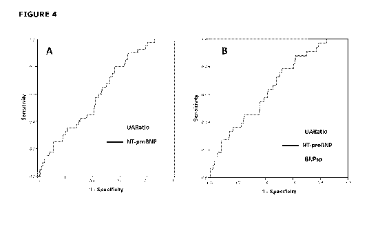
Figure 4 shows (A) ROC curves of UARatio (AUC=0.70) and NT-proBNP (AUC=0.62)
for the identification of UAP (n=40) in 390 non-MI patients at hospital
presentation. The
UARatio AUC was significantly better than the NT-proBNP AUC (p<0.05). (B) ROC
curves of
CA 03009737 2018-06-26
WO 2017/116239
PCT/NZ2016/050207
the ability of UARatio, NT-proBNP and BNPsp to diagnose UAP (n=33) in 328 non-
MI
patients with no abnormalities on ECG. The AUC for UARatio (0.77) was
significantly better
(p<0.05) than both NT-proBNP (0.66) and BNPsp (0.63).
5 SELECTED DEFINITIONS
Unless defined otherwise, all technical and scientific terms used herein have
the
same meaning as commonly understood to one of ordinary skill in the art to
which the
inventions belong. Although any assays, methods, devices and materials
similar or
equivalent to those described herein can be used in the practice or testing of
the invention,
various assays, methods, devices and materials are now described.
It is intended that reference to a range of numbers disclosed herein (for
example 1
to 10) also incorporates reference to all related numbers within that range
(for example, 1,
1.1, 2, 3, 3.9, 4, 5, 6, 6.5, 7, 8, 9 and 10) and also any range of rational
numbers within
that range (for example 2 to 8, 1.5 to 5.5 and 3.1 to 4.7) and, therefore, all
sub-ranges of
all ranges expressly disclosed herein are expressly disclosed. These are only
examples of
what is specifically intended and all possible combinations of numerical
values between the
lowest value and the highest value enumerated are to be considered to be
expressly stated
in this application in a similar manner.
As used in this specification, the words "comprises", "comprising", and
similar words,
are not to be interpreted in an exclusive or exhaustive sense. In other words,
they are
intended to mean "including, but not limited to".
As used in this specification, the term "BNPsp" means B-type Natriuretic
Peptide
signal peptide. Examples of BNPsp include the full length BNPsp molecule
defined by
residues 1-27, as well as fragments thereof. In a particular example, BNPsp
means the
BNPsp fragment defined by residues 17-26 (i.e. BNPsp (17-26; SEQ ID NO:1)).
As used in this specification, the term "BNP" means B-type natriuretic
peptide, which
once processed by proteolytic enzymes includes the N-terminal pro-BNP (NT-
proBNP) and
the cleaved active form of BNP hormone. For the purpose of this specification,
"BNP" refers
to BNP(103-134) and "NT-proBNP" refers to NT-proBNP(27-102) as defined in
Figure 1.
As used in this specification, the acronym "STEMI" means ST-elevation
myocardial
infarction.
As used in this specification, the acronym "NSTEMI" means non ST-elevation
myocardial infarction.
As used in this specification, the acronym "UAP" means unstable angina
pectoris.
As used in this specification, the acronym "ACS" means acute coronary
syndromes.
As used in this specification, the acronym "hsTnT" means highly sensitive
troponin T
assay.
CA 03009737 2018-06-26
WO 2017/116239
PCT/NZ2016/050207
6
As used in this specification, the acronym "WCC" means white cell count and
relates
to the level/number of white blood cells in a sample.
As used in this specification, the acronym "ROC" means receiver operating
characteristic curve.
As used in this specification, the acronym "AF" means atrial fibrillation.
As used in this specification, the term "polypeptide" encompasses amino acid
chains
of any length, including full length sequences in which amino acid residues
are linked by
covalent peptide bonds. Polypeptides useful in the present invention may be
purified
natural products, or may be produced partially or wholly using recombinant or
synthetic
techniques. The term may refer to a polypeptide, an aggregate of a polypeptide
such as a
dimer or other multimer, a fusion polypeptide, a polypeptide fragment, a
polypeptide
variant, or derivative thereof. Polypeptides herein may have chain lengths of
at least 4
amino acids, at least 5 amino acids, or at least 6, at least 7, at least 8, at
least 9, at least
10, at least 11, at least 12, at least 13, at least 14, at least 15, at least
16, at least 17, at
least 18, at least 19, at least 20, at least 21, at least 22, or all 23 amino
acids of the full-
length EPOsp and/or CNPsp. Reference to other polypeptides of the invention or
other
polypeptides described herein should be similarly understood.
As used in this specification, the term "fragment" in relation to a
polypeptide is a
subsequence of a polypeptide that may be detected using a binding agent. The
term may
refer to a polypeptide, an aggregate of a polypeptide such as a dimer or
multimer, a fusion
polypeptide, a polypeptide fragment, a polypeptide variant or derivative
thereof.
The term "isolated" as applied to the polypeptide sequences disclosed herein
is used
to refer to sequences that are removed from their natural cellular or other
naturally-
occurring biological environment. An isolated molecule may be obtained by any
method or
combination of methods including biochemical, recombinant, and synthetic
techniques. The
polypeptide sequences may be prepared by at least one purification step.
The term "purified" as used herein does not require absolute purity. Purified
refers
in various embodiments, for example, to at least about 80%, 85%, 90%, 950/s,
98%, or
99% homogeneity of a polypeptide, for example, in a sample. The term should be
similarly
understood in relation to other molecules and constructs described herein.
As used herein, the term "variant" refers to polypeptide sequences different
from the
specifically identified sequences, wherein 1 to 6 or more or amino acid
residues are deleted,
substituted, or added. Substitutions, additions or deletions of one, two,
three, four, five or
six amino acids are contemplated. Variants may be naturally occurring allelic
variants, or
non-naturally occurring variants. Variants may be from the same or from other
species and
may encompass homologues, paralogues and orthologues.
In certain embodiments,
variants of the polypeptides useful in the invention have biological
activities including signal
peptide activity or antigenic-binding properties that are the same or similar
to those of the
CA 03009737 2018-06-26
WO 2017/116239
PCT/NZ2016/050207
7
parent polypeptides. The term "variant" with reference to polypeptides
encompasses all
forms of polypeptides as defined herein.
Variant polypeptide sequences exhibit at least about 50%, at least about 60%,
at
least about 70%, at least about 71%, at least about 72%, at least about 73%,
at least
about 74%, at least about 75%, at least about 76%, at least about 77%, at
least about
78%, at least about 79%, at least about 80%, at least about 81%, at least
about 82%, at
least about 83%, at least about 84%, at least about 85%, at least about 86%,
at least
about 87%, at least about 88%, at least about 89%, at least about 90%, at
least about
91%, at least about 92%, at least about 93%, at least about 94%, at least
about 95%, at
least about 96%, at least about 97%, at least about 98%, or at least about 99%
identity to
a sequence of the present invention. With regard to polypeptides, identity is
found over a
comparison window of at least 5 to 7 amino acid positions.
Polypeptide variants also encompass those which exhibit a similarity to one or
more
of the specifically identified sequences that is likely to preserve the
functional equivalence of
those sequences, including those which could not reasonably be expected to
have occurred
by random chance. As discussed above, in the case of EPOsp and/or CNPsp
variants
function may be as either a signal polypeptide, or antigenic polypeptide, or
both.
Polypeptide sequence identity and similarity can be determined in the
following
manner. The subject polypeptide sequence is compared to a candidate
polypeptide
.. sequence using BLASTP (from the BLAST suite of programs, version 2.2.18
[April 2008]]) in
b125eq, which is publicly available from NCBI (ftpilitp,ncbi.mh.qav/biast/).
The default
parameters of b125eq are utilized except that filtering of low complexity
regions should be
turned off.
The similarity of polypeptide sequences may be examined using the following
UNIX
command line parameters: b125eq peptideseq1 -j peptideseq2 -F F -p blastp.
The
parameter -F F turns off filtering of low complexity sections. The parameter -
p selects the
appropriate algorithm for the pair of sequences. This program finds regions of
similarity
between the sequences and for each such region reports an "E value" which is
the expected
number of times one could expect to see such a match by chance in a database
of a fixed
reference size containing random sequences. For small E values, much less than
one, this is
approximately the probability of such a random match. Variant polypeptide
sequences
commonly exhibit an E value of less than 1 x 10⁻⁵, less than 1 x 10⁻⁶, less
than 1 x 10⁻⁹, less
than 1 x 10-12, less than 1 x 10-15, less than 1 x 10-18 or less than 1 x 10-
21 when compared
with any one of the specifically identified sequences. Polypeptide sequence
identity may also
be calculated over the entire length of the overlap between a candidate and
subject
polypeptide sequences using global sequence alignment programs.
EMBOSS-needle
(available at http:/www.ebi.ac.uk/emboss/align/) and GAP (Huang, X. (1994) On
Global
Sequence Alignment. Computer Applications in the Biosciences 10, 227-235.) as
discussed
CA 03009737 2018-06-26
WO 2017/116239
PCT/NZ2016/050207
8
above are also suitable global sequence alignment programs for calculating
polypeptide
sequence identity. Use of BLASTP is preferred for use in the determination of
polypeptide
variants according to the present invention.
The term "binding agent" as used herein refers to any solid or non-solid
material
capable of binding a species of AMH, fragment or an antigenic variant thereof.
In one
embodiment the term refers to any natural or non-natural molecule that binds
to a species
of AMH, fragment or antigenic variant thereof. Examples of binding agents
include proteins,
peptides, nucleic acids, carbohydrates, lipids, and small molecule compounds.
One
selective or specific binding agent is an antibody or antigen binding fragment
thereof.
The term "antibody" refers to an immunoglobulin molecule capable of
specifically
binding an antigen, such as, for example, BNPsp, and typically by binding an
epitope or
antigenic determinant of BNPsp, such as, for example, a C-terminal or N-
terminal region of
BNPsp. As used herein, the term "antibody" broadly includes full length
antibodies and
antigen binding fragments or regions thereof. Also included are monoclonal and
polyclonal
antibodies, multivalent and monovalent antibodies, multispecific antibodies
(for example bi-
specific antibodies), chimeric antibodies, human antibodies, humanized
antibodies and
antibodies that have been affinity matured. An antibody binds selectively or
specifically to
BNPsp, if the antibody binds preferentially to a region or domain of BNPsp
which has, e.g.
has less than 25%, or less than 10%, or less than 1% or less than 0.1% cross-
reactivity
with non-BNPsp antigens/epitopes or other non-target BNPsp species, when
appropriate.
Usually, the antibody will have a binding affinity (dissociation constant (Kd)
value), for the
antigen or epitope of about 10-6, or 10-7M, 10-8M, or 10-9M, or 10-19, or 10-
11 or 10-12M.
Binding affinity may be assessed using surface plasma resonance, for example,
or
Scatchard analysis.
As used herein, an "antigen binding fragment" or "antibody fragment" or
"binding
fragment" when used in reference to an antibody, means a portion of the intact
antibody
that preferably retains most or all, or minimally at least one of, the normal
binding functions
of the intact antibody. Examples of antibody fragments include Fab, Fab',
F(ab')2 and Fv
fragments, linear antibodies, diabodies, single chain antibodies (ScFV),
domain antibodies
and multispecific antibodies.
The term "epitope" includes any antigenic (e.g., a protein) determinant
capable of
specific binding to an antibody and/or a T cell receptor. That is, a site on
an antigen to
which B and/or T cells respond. Epitopic determinants usually consist of
chemically active
surface groupings of molecules such as amino acids or sugar side chains, and
usually have
specific three-dimensional structural characteristics, as well as specific
charge
characteristics. An epitope typically includes at least 3, 5 or 8-10 amino
acids. The amino
acids may be contiguous, or non-contiguous amino acids juxtaposed by tertiary
folding.
CA 03009737 2018-06-26
WO 2017/116239
PCT/NZ2016/050207
9
Conformational and non-conformational epitopes are distinguished in that the
binding to the
former but not the latter is lost in the presence of denaturing solvents.
As used herein, the term "antigenic variant" refers to polypeptide sequences
different from the specifically identified sequences, wherein one or more
amino acid
residues are deleted, substituted, or added. Substitutions, additions or
deletions of 1, 2, 3,
4, 5, 6, 10, 15, 20 or more amino acids are specifically contemplated.
Variants may be
naturally-occurring allelic antigenic variants, or non-naturally occurring
antigenic variants.
Variants may be from the same or from other species and may encompass
homologues,
paralogues and orthologues. In certain examples, antigenic variants of the
polypeptides
useful in the invention have biological activities including hormone function
or antigenic-
binding properties that are the same or similar to those of the parent
polypeptides. The
term "antigenic variant" with reference to (poly)peptides encompasses all
forms of
polypeptides as defined herein. The term "antigenic variant" encompasses
naturally
occurring, recombinantly and synthetically produced polypeptides.
For example, an
antigenic variant of human BNP and BNPsp may include the non-human sequences
of BNP
and BNPsp, such as those sequences derived from mouse, rat, sheep, bovine, pig
etc.
In addition to computer/database methods known in the art, polypeptide
antigenic
variants may be identified by physical methods known in the art, for example,
by screening
expression libraries using antibodies raised against polypeptides of the
invention (Sambrook
etal., Molecular Cloning: A Laboratory Manual, 2nd Ed. Cold Spring Harbor
Press, 1987) by
recombinant DNA techniques also described by Sambrook et al. or by identifying
polypeptides from natural sources with the aid of such antibodies.
An "isolated antibody" is an identified antibody that has been separated or
recovered, or both, from a component of its natural environment. For example,
separated
from proteins including enzymes and hormones. In one example, the antibody is
purified to
at least 95%, or 96% or 97% or 98% or 99% by weight of antibody. Purity can be
determined by the Lowry method, for example. Ordinarily the antibody will be
prepared by
at least one purification step.
As used herein, a "monoclonal antibody" means an antibody that is a highly
specific
antibody directed against (or which binds to) a single antigen target. A
monoclonal
antibody may be obtained from a population of homogenous or substantially
homogenous
antibodies wherein each monoclonal antibody is identical and/or bind the same
epitope,
except for natural mutations that may occur in minor amounts. Monoclonal
antibodies are
prepared using methods known the art, such as, for example, in Harlow and Lane
(1988)
.. Antibodies, A Laboratory Manual, Cold Spring Harbor Publications, New York,
and Harlow
and Lane (1999) Using Antibodies: A Laboratory Manual Cold Spring Harbor
Laboratory
Press, Cold Spring Harbor, NY (jointly and individually referred to herein as
Harlow and
Lane).
CA 03009737 2018-06-26
WO 2017/116239
PCT/NZ2016/050207
As used herein, a "polyclonal antibody" means an antibody which may be
directed
against (or which may bind to) multiple antigen targets. Polyclonal antibodies
are prepared
using methods known the art (such as, for example, in Harlow and Lane, ibid).
The term "ELISA" as used herein means an enzyme linked immunosorbent assay, a
5 type of competitive binding assay comprising antibodies and a detectable
label used to
quantitate the amount of an analyte in a sample.
The term "capture antibody" as used herein means an antibody which is
typically
immobilized on a solid support such as a plate, bead or tube, and which
antibody binds to
and captures analyte(s) of interest, for example membrane bound markers
associated with
10 .. an embryonic stem cell population.
The term "detection antibody" as used herein means an antibody comprising a
detectable label that binds to analyte(s) of interest. The label may be
detected using
routine detection means for a quantitative, semi-quantitative or qualitative
measure of the
analyte(s) of interest, for example membrane bound markers associated with an
embryonic
stem cell population.
As used herein, the term "aptamer" refers to single-stranded nucleic acid
molecules
with secondary structures that facilitate high-affinity binding to a target
molecule such as a
polypeptide or protein. In certain examples, the single- stranded nucleic acid
is ssDNA, RNA
or derivatives thereof to improve bioavailability. Aptamer binding affinity to
the target
protein is further described below.
As used herein, the term "marker" or "biomarker" in the context of an analyte
means
any antigen, molecule or other chemical or biological entity that is
specifically found in
circulation or associated with a particular tissue (e.g. heart muscle) that it
is desired to be
identified in or on a particular tissue affected by a disease or disorder, for
example unstable
angina. In specific examples, the marker is a circulating peptide (e.g.) BNPsp
(17-26) or
NT-proBNP. In other examples, the marker is a cell surface antigen or a
nuclear antigen
that is differentially or preferentially expressed by specific cell types. In
other examples the
marker is an intracellular antigen that is differentially or preferrentially
expressed by specific
cell types.
The term "ROC" means Receiver Operating Characteristic and a ROC plot depicts
the
overlap between two distributions by plotting the sensitivity versus 1-
specificity for a
complete range of decision thresholds.
As used herein, the term "effective amount" refers to the amount of a therapy
that is
sufficient to result in the prevention of the development, recurrence, or
onset of a disease
or condition and one or more symptoms thereof, to enhance or improve the
prophylactic
effect(s) of another therapy, reduce the severity, the duration of disease,
ameliorate one or
more symptoms of the disease or condition, prevent the advancement of the
disease or
CA 03009737 2018-06-26
WO 2017/116239
PCT/NZ2016/050207
11
condition, cause regression of the disease or condition, and/or enhance or
improve the
therapeutic effect(s) of another therapy.
As used herein, the terms "manage", "managing", and "management" in the
context
of the administration of a therapy to a subject refer to the beneficial
effects that a subject
derives from a therapy (e.g., a prophylactic or therapeutic agent) or a
combination of
therapies, while not resulting in a cure of the disease or condition. In
certain examples, a
subject is administered one or more therapies (e.g., one or more prophylactic
or therapeutic
agents) to "manage" the disease or condition so as to prevent the progression
or worsening
of the disease or condition.
As used herein, the terms "prevent", "preventing" and "prevention" in the
context of
the administration of a therapy to a subject refers to the prevention or
inhibition of the
recurrence, onset, and/or development of a disease or condition or a symptom
thereof in a
subject resulting from the administration of a therapy (e.g., a prophylactic
or therapeutic
agent), or a combination of therapies (e.g., a combination of prophylactic or
therapeutic
.. agents).
As used herein, the term "prophylactic agent" refers to any molecule,
compound,
and/or substance that is used for the purpose of treating unstable angina.
Examples of
prophylactic agents include, but are not limited to, proteins, immunoglobulins
(e.g., multi-
specific Igs, single chain Igs, Ig fragments, polyclonal antibodies and their
fragments,
monoclonal antibodies and their fragments), antibody conjugates or antibody
fragment
conjugates, peptides (e.g., peptide receptors, selectins), binding proteins,
proliferation
based therapy, and small molecule drugs.
As used herein, the term "therapeutic agent" refers to any molecule, compound,
and/or substance that is used for the purpose of treating and/or managing a
disease or
disorder, such as unstable angina. Examples of therapeutic agents include, but
are not
limited to, proteins, immunoglobulins (e.g., multi-specific Igs, single chain
Igs, Ig
fragments, polyclonal antibodies and their fragments, monoclonal antibodies
and their
fragments), peptides (e.g., peptide receptors, selectins), binding proteins,
biologies,
proliferation-based therapy agents, hormonal agents, radioimmunotherapies,
targeted
agents, epigenetic therapies, differentiation therapies, biological agents,
and small molecule
drugs.
As used herein, the terms "therapies" and "therapy" can refer to any
method(s),
composition(s), and/or agent(s) that can be used in the prevention, treatment
and/or
management of a disease or condition or one or more symptoms thereof.
As used herein, the terms "treat", "treatment" and "treating" in the context
of the
administration of a therapy to a subject refer to the reduction, inhibition,
elimination or
amelioration of the progression and/or duration of (e.g.) unstable angina, the
reduction,
inhibition, elimination or amelioration of the severity of (e.g.) unstable
angina, and/or the
CA 03009737 2018-06-26
WO 2017/116239
PCT/NZ2016/050207
12
amelioration of one or more symptoms thereof resulting from the administration
of one or
more therapies.
The term "sample" or "biological sample" as used herein means any sample taken
or
derived from a subject or patient. Im this specification, the terms "subject"
and "patient"
are used interchangeably. Such a sample may be obtained from a subject, or may
be
obtained from biological materials intended to be provided to the subject. For
example, a
sample may be obtained from blood or heart tissue being assessed, for example,
to
investigate the cardiac status in a subject. Included are samples taken or
derived from any
subjects such as from normal healthy subjects and/or healthy subjects for whom
it is useful
to understand their cardiac status. Preferred samples are biological fluid
samples. The
term "biological fluid sample" as used herein refers to a sample of bodily
fluid obtained for
the purpose of, for example, diagnosis, prognosis, classification or
evaluation of a subject of
interest, such as a patient. The sample may be any sample known in the art in
which
peptide antigens may be detected. Included are any body fluids such as a whole
blood
sample, plasma, serum, ovarian follicular fluid sample, seminal fluid sample,
cerebrospinal
fluid, saliva, sputum, urine, pleural effusions, interstitial fluid, synovial
fluid, lymph, tears,
for example, although whole blood sample, plasma and serum are particularly
suited for use
in this invention. In addition, one of skill in the art would realise that
certain body fluid
samples would be more readily analysed following a fractionation or
purification procedure,
for example, separation of whole blood into serum or plasma components.
The term "purified" as used herein does not require absolute purity. Purified
refers
in one example to at least 90%, or 95%, or 98%, or 99% homogeneity of (e.g.) a
polypeptide or antibody in a sample.
The term "subject" and "patient" are used interchangeably herein. These terms
preferably refer to a mammal and includes human, and non-human mammals such as
cats,
dogs, horses, cows, sheep, deer, mice, rats, primates (including gorillas,
rhesus monkeys
and chimpanzees), possums and other domestic farm or zoo animals. Thus, the
assays,
methods and kits described herein have application to both human and non-human
animals,
in particular, and without limitation, humans, primates, farm animals
including cattle,
sheep, goats, pigs, deer, alpacas, llamas, buffalo, companion and/or pure bred
animals
including cats, dogs and horses. Preferred subjects are humans, and most
preferably
"patients" who as used herein refer to living humans who may receive or are
receiving
medical care or assessment for a disease or condition. Further, while a
subject is preferably
a living organism, the invention described herein may be used in post-mortem
analysis as
well.
As used herein, the term "relating to the presence or amount" of an analyte
reflects
that assay signals are typically related to the presence or amount of an
analyte through the
use of a standard curve calculated using known concentrations of the analyte
of interest. As
CA 03009737 2018-06-26
WO 2017/116239
PCT/NZ2016/050207
13
the term is used herein, an assay is "configured to detect" an analyte if an
assay can
generate a detectable signal indicative of the presence or amount of a
physiologically
relevant concentration of the analyte. Typically, an analyte is measured in a
sample.
A level "higher" or "lower" than a control, or a "change" or "deviation" from
a control
(level) in one embodiment is statistically significant. A higher level, lower
level, deviation
from, or change from a control level or mean or historical control level can
be considered to
exist if the level differs from the control level by about 5% or more, by
about 10% or more,
by about 20% or more, or by about 50% or more compared to the control level.
Statistically significant may alternatively be calculated as 1,0.05. Higher
levels, lower
levels, deviation, and changes can also be determined by recourse to assay
reference limits
or reference intervals. These can be calculated from intuitive assessment or
non-parametric
methods. Overall, these methods may calculate the 0.025, and 0.975 fractiles
as 0.025*
(n+1) and 0.975 (n+1). Such methods are well known in the art. Presence of a
marker
absent in a control may be seen as a higher level, deviation or change.
Absence of a
marker present in a control may be seen as a lower level, deviation or change.
DETAILED DESCRIPTION
Applicants assessed the ability of B-type natriuretic peptide signal peptide
(BNPsp) to
assist with the identification of patients with myocardial infarction (MI) and
unstable angina
pectoris (UAP).
Applicants studied 505 patients who presented to hospital within 4 hours of
onset of
chest pain suspicious of ACS. Blood samples were drawn at 0, 1, 2 and 24 hours
from
presentation and assayed for BNPsp, NT-proBNP, TnI and high sensitivity TnT.
The ability of
BNPsp and other markers to diagnose acute myocardial infarction (MI) and
unstable angina
pectoris (UAP) and predict subsequent events within one year was then
assessed.
Applicants surprisingly discovered that when BNPsp was measured in conjunction
with NT-proCNP and white blood cell count, and the data fitted using Receiver
Operating
Curve analysis, that unstable angina could be diagnosed in a patient
presenting to the
Emergency Department with symptoms of an acute coronary disorder. Further, the
specificity of diagnosis could be enhanced when the levels of potassium (K) in
the patient
were added to the biomarker panel.
Interestingly, receiver operator area under the curve (AUC) data for the
discrimination of myocardial infarction was 0.69 for BNPsp and 0.97 for
troponin, with
BNPsp failing to add to troponin. However, and importantly, in non-MI
patients, BNPsp had
discriminative power for UAP (p<0.05), and when combined with presentation
values of NT-
proBNP, white cell count and potassium into a unique parameter (UARatio), and
generated
an AUC of 0.76 for UAP in patients with normal ECG results (p<0.001). Refer to
Figures 3
and 4, as well as Example 2.
CA 03009737 2018-06-26
WO 2017/116239
PCT/NZ2016/050207
14
Accordingly, in one aspect the present invention provides a method for
diagnosing
unstable angina in a patient, the method comprising the steps of:
(i) measuring the level of a Type-B natriuretic peptide signal peptide
(BNPsp)
fragment, the level of N-terminal Type-B natriuretic peptide (NT-proBNP) and
white blood cell count (WCC) in a biological sample obtained from the patient;
and
(ii) comparing the levels of the BNPsp fragment, NT-proBNP and WCC against
reference levels obtained from a control subject,
wherein, a ratio of BNPsp fragment and NT-proBNP to WCC that deviates from a
reference ratio obtained from a control subject is diagnostic that the patient
has unstable
angina.
Depending on the diagnosis, an intervention therapy may be administered to the
patient to reduce, eliminate, mitigate or treat the unstable angina.
Accordingly, in a further aspect the present invention provides a method for
diagnosing unstable angina in a patient, the method comprising the steps of:
(i) measuring the level of a Type-B natriuretic peptide signal peptide
(BNPsp)
fragment, the level of N-terminal Type-B natriuretic peptide (NT-proBNP) and
white blood cell count (WCC) in a biological sample obtained from the patient;
and
(ii) comparing the levels of the BNPsp fragment, NT-proBNP and WCC against
reference levels obtained from a control subject,
wherein, a ratio of BNPsp fragment and NT-proBNP to WCC that deviates from a
reference ratio obtained from a control subject is diagnostic that the patient
has unstable
angina, and wherein in the event of a positive diagnosis of unstable angina:
(iii)
administering an intervention therapy so as to reduce, eliminate, mitigate or
treat unstable angina in the patient.
Further, in non-MI patients, the UARatio was significantly predictive of
subsequent
stroke (AUC = 0.70, p<0.05) and heart failure (AUC = 0.82, p<0.01) within one
year.
Refer to Example 2.
Accordingly, in a further aspect the present invention provides a method for
predicting a complication of heart failure and/or stroke in a patient who has
previously been
diagnosed with unstable angina, the method comprising the steps of:
(i) measuring the level of a Type-B natriuretic peptide signal peptide
(BNPsp)
fragment, the level of N-terminal Type-B natriuretic peptide (NT-proBNP) and
white blood cell count (WCC) in a biological sample obtained from the patient;
and
(ii) comparing the levels of the BNPsp fragment, NT-proBNP and WCC against
reference levels obtained from a control subject,
CA 03009737 2018-06-26
WO 2017/116239
PCT/NZ2016/050207
wherein, a ratio of BNPsp fragment and NT-proBNP to WCC that deviates from a
reference ratio obtained from a control subject is predictive that the patient
will develop a
complication of heart failure and/or stroke as a consequence of unstable
angina.
Depending on the specificity and sensitivity, the diagnosis may be enhanced by
5
measuring the levels of potassium in the sample. As such, in certain examples
according to
the present invention, the method further comprises measuring the level of
potassium in
the biological sample obtained from the patient.
In other examples of the present invention, the BNPsp fragment is a fragment
defined by residues 17-26 of the full-length/in-tact protein (designated BNPsp
(17-26)).
10
In other examples, the levels of BNPsp, including BNPsp (17-26), and NT-proBNP
may be measured by immunoassay or mass spectroscopy. Further details with
respect to
measurement by immunoassay using antibody- and aptamer-based approaches to
detection
are given below.
The present invention also contemplates commercial kits and articles of
manufacture
15
specific for measuring the levels of, for example, BNPsp fragments, NT-proBNP
and white
blood cells in a biological sample obtained from a patient. As such, in a
further aspect of
the present invention there is provided a kit or article of manufacture
comprising:
(i) reagents specific to measure the levels of a Type-B natriuretic peptide
signal
peptide (BNPsp) fragment, the level of N-terminal Type-B natriuretic peptide
(NT-proBNP) and white blood cell count (WCC) in a biological sample obtained
from a patient; and
(ii) instructions for how to perform the diagnosis of unstable angina in
the patient
or for how to predict a complication of stroke and/or heart failure as a
consequence of developing unstable angina in the patient.
Applicants have also identified that B-type natriuretic peptide signal peptide
fragment defined by residues 17-26 (BNPsp (17-26)), B-type natriuretic peptide
(BNP) and
white blood cell count (WCC) is a useful biomarker panel in the diagnosis of
unstable
angina.
Accordingly, in another aspect the present invention provides a method for
diagnosing unstable angina in a patient, the method comprising the steps of:
(i) measuring the level of a Type-B natriuretic peptide signal peptide
(BNPsp)
fragment, the level of Type-B natriuretic peptide (BNP) and white blood cell
count (WCC) in a biological sample obtained from the patient; and
(ii) comparing the levels of the BNPsp fragment, BNP and WCC against reference
levels obtained from a control subject,
wherein, a ratio of BNPsp fragment and BNP to WCC that deviates from a
reference
ratio obtained from a control subject is diagnostic that the patient has
unstable angina.
CA 03009737 2018-06-26
WO 2017/116239 PCT/NZ2016/050207
16
In another aspect the present invention provides a method for predicting a
complication of heart failure and/or stroke in a patient who has previously
been diagnosed
with unstable angina, the method comprising the steps of:
(i) measuring the level of a Type-B natriuretic peptide signal peptide
(BNPsp)
fragment, the level of Type-B natriuretic peptide (BNP) and white blood cell
count (WCC) in a biological sample obtained from the patient; and
(ii) comparing the levels of the BNPsp fragment, BNP and WCC against reference
levels obtained from a control subject,
wherein, a ratio of BNPsp fragment and BNP to WCC that deviates from a
reference
ratio obtained from a control subject is predictive that the patient will
develop a
complication of heart failure and/or stroke as a consequence of unstable
angina.
In a further aspect the present invention provides a method for diagnosing
unstable
angina in a patient, the method comprising the steps of:
(i) measuring the level of a Type-B natriuretic peptide signal peptide
(BNPsp)
fragment, the level of Type-B natriuretic peptide (proBNP) and white blood
cell
count (WCC) in a biological sample obtained from the patient; and
(ii) comparing the levels of the BNPsp fragment, BNP and WCC against reference
levels obtained from a control subject,
wherein, a ratio of BNPsp fragment and BNP to WCC that deviates from a
reference
ratio obtained from a control subject is diagnostic that the patient has
unstable angina, and
wherein in the event of a positive diagnosis of unstable angina:
(iii) administering an intervention therapy so as to reduce, eliminate,
mitigate or
treat unstable angina in the patient.
In yet another aspect the present invention provides a test kit for diagnosing
unstable angina in a patient, or for predicting complication of heart failure
and/or stroke in a
patient as a consequence of unstable angina, the test kit comprising:
(i) reagents specific to measure the levels of a Type-B natriuretic peptide
signal
peptide (BNPsp) fragment, the level Type-B natriuretic peptide (BNP) and
white blood cell count (WCC) in a biological sample obtained from a patient;
and
(ii) instructions for how to perform the diagnosis of unstable angina in
the patient
or for how to predict a complication of stroke and/or heart failure as a
consequence of developing unstable angina in the patient.
Antibodies and Antigen Binding Fragments
As noted above, antibody or antibodies as used herein refers to a peptide or
polypeptide derived from, modelled after or substantially encoded by an
immunoglobulin
gene or immunoglobulin genes, or fragments thereof, capable of specifically
binding an
antigen or epitope [34-36]. As foreshadowed in the definition section of this
specification,
CA 03009737 2018-06-26
WO 2017/116239
PCT/NZ2016/050207
17
the term antibody includes antigen binding fragments such as, for example,
fragments,
subsequences, complementarity determining regions (CDRs) that retain capacity
to bind to
an antigen, including (i) a Fab fragment, a monovalent fragment consisting of
the VL, VH,
CL and CH1 domains; (ii) a F(ab')2 fragment, a bivalent fragment comprising
two Fab
fragments linked by a disulfide bridge at the hinge region; (iii) a Fd
fragment consisting of
the VH and CH1 domains; (iv) a Fv fragment consisting of the VL and VH domains
of a
single arm of an antibody, (v) a dAb fragment [37], which consists of a VH
domain; and (vi)
an isolated complementarity determining region (CDR). Single chain antibodies
are also
included by reference in the term "antibody."
Further discussion of antibodies and
fragments may be found in references (e.g.) [38-44] all of which are
incorporated herein in
their entirety.
Also included is antiserum obtained by immunizing an animal such as a mouse,
rat
or rabbit with an antigen, such as for example, BNPsp or BNPsp fragments, as
well as
antigenic variants thereof. In brief, methods of preparing polyclonal
antibodies are known
to the skilled artisan. Polyclonal antibodies can be raised in a mammal, for
example, by one
or more injections of an immunizing agent and, if desired, an adjuvant.
Typically, the
immunizing agent and/or adjuvant will be injected in the mammal by multiple
subcutaneous
or intraperitoneal injections. The immunizing agent may include BNPsp or BNPsp
fragments, antigenic variants thereof or a fusion protein thereof. It may be
useful to
conjugate the immunizing agent to a protein known to be immunogenic in the
mammal
being immunized. Examples of such immunogenic proteins include but are not
limited to
keyhole limpet hemocyanin, bovine serum albumin, bovine thyroglobulin, and
soybean
trypsin inhibitor. Examples of adjuvants that may be employed include Freund's
complete
adjuvant and MPL TDM adjuvant (monophosphoryl Lipid A, synthetic trehalose
dicorynomycolate). The immunization protocol may be selected by one skilled in
the art
without undue experimentation.
Monoclonal antibodies may be prepared using hybridoma methods well known in
the
art [e.g. 45-47]. The hybridoma cells may be cultured in a suitable culture
medium,
alternatively, the hybridoma cells may be grown in vivo as ascites in a
mammal. Preferred
immortalized cell lines are murine myeloma lines, which can be obtained, for
example, from
the American Type Culture Collection, Virginia, USA. Immunoassays may be used
to screen
for immortalized cell lines that secrete the antibody of interest. Sequences
of BNPsp or
BNPsp fragments or antigenic variants thereof may be used in screening.
Well known means for establishing binding specificity of monoclonal antibodies
produced by the hybridoma cells include immunoprecipitation, radiolinked
immunoassay
(RIA), enzyme-linked immunoabsorbent assay (ELISA) and Western blot [48].
For
example, as noted above, the binding affinity of the monoclonal antibody can,
for example,
CA 03009737 2018-06-26
WO 2017/116239
PCT/NZ2016/050207
18
be determined by the Scatchard analysis [49]. Samples from immunised animals
may
similarly be screened for the presence of polyclonal antibodies.
Monoclonal antibodies can also be obtained from recombinant host cells. DNA
encoding the antibody can be obtained from a hybridoma cell line. The DNA is
then placed
into an expression vector, transfected into host cells (e.g., COS cells, CHO
cells, E. coli
cells) and the antibody produced in the host cells. The antibody may then be
isolated
and/or purified using standard techniques.
The monoclonal antibodies or fragments may also be produced by recombinant DNA
means (e.g. [50]). DNA modifications such as substituting the coding sequence
for human
heavy and light chain constant domains in place of the homologous murine
sequences [50]
are also possible. The antibodies may be monovalent antibodies. Methods for
preparing
monovalent antibodies are well known in the art (e.g. [51-53]. Production of
chimeric [54],
bivalent antibodies [55] and multivalent antibodies are also contemplated
herein [56].
Other known art techniques for monoclonal antibody production such as from
phage
libraries, may also be used (e.g. [57]).
The monoclonal antibodies secreted by the cells may be isolated or purified
from the
culture medium or ascites fluid by conventional immunoglobulin purification
procedures
such as, for example, reverse phase HPLC, protein A-Sepharose, hydroxyapatite
chromatography, gel electrophoresis, dialysis, or affinity chromatography
[58].
Bispecific antibodies may also be useful. These antibodies are monoclonal,
preferably human or humanized, antibodies that have binding specificities for
at least two
different antigens. Antibodies with greater than two specificities for example
trispecific
antibodies are also contemplated herein.
Antibodies used in the immunoassays described herein specifically bind to
BNPsp or
BNPsp fragments. The term "specifically binds" is not intended to indicate
that an antibody
binds exclusively to its intended target since, as noted above, an antibody
binds to any
polypeptide displaying the epitope(s) to which the antibody binds. Rather, an
antibody
"specifically binds" if its affinity for its intended target is about 5-fold
greater when
compared to its affinity for a non-target molecule which does not display the
appropriate
epitope(s). In certain examples, the affinity of the antibody will be at least
about 5 fold,
preferably 10 fold, more preferably 25-fold, even more preferably 50-fold, and
most
preferably 100-fold or more, greater for a target molecule than its affinity
for a non-target
molecule. In other examples, antibodies bind with affinities of at least about
10-6M, or 10-
7M, or at least about 10-8M, or 10-9M, or 10-10, or 10-11 or 10-12M.
Affinity is calculated as Kd=koff/kon (koff is the dissociation rate constant,
Kon is the
association rate constant and Kd is the equilibrium constant). Affinity can be
determined at
equilibrium by measuring the fraction bound (r) of labelled ligand at various
concentrations
(c). The data are graphed using the Scatchard equation: ric=K(n-r): where
r=moles of
CA 03009737 2018-06-26
WO 2017/116239
PCT/NZ2016/050207
19
bound ligand/mole of receptor at equilibrium; c=free ligand concentration at
equilibrium;
K=equilibrium association constant; and n=number of ligand binding sites per
receptor
molecule. By graphical analysis, r/c is plotted on the Y-axis versus r on the
X-axis, thus
producing a Scatchard plot. Antibody affinity measurement by Scatchard
analysis is well
known in the art [59].
Numerous publications discuss the use of phage display technology to produce
and
screen libraries of polypeptides for binding to a selected analyte [60-63]. A
basic concept of
phage display methods is the establishment of a physical association between
DNA encoding
a polypeptide to be screened and the polypeptide. This physical association is
provided by
the phage particle, which displays a polypeptide as part of a capsid enclosing
the phage
genome that encodes the polypeptide. The establishment of a physical
association between
polypeptides and their genetic material allows simultaneous mass screening of
very large
numbers of phage bearing different polypeptides. Phage displaying a
polypeptide with
affinity to a target binds to the target and these phage are enriched by
affinity screening to
the target. The identity of polypeptides displayed from these phage can be
determined
from their respective genomes. Using these methods a polypeptide identified as
having a
binding affinity for a desired target can then be synthesized in bulk by
conventional means
(e.g. [64]).
The antibodies that are generated by these methods may then be selected by
first
screening for affinity and specificity with the purified polypeptide of
interest and, if required,
comparing the results to the affinity and specificity of the antibodies with
polypeptides that
are desired to be excluded from binding. The screening procedure can involve
immobilization of the purified polypeptides in separate wells of microtiter
plates. The
solution containing a potential antibody or groups of antibodies is then
placed into the
respective microtiter wells and incubated for about 30 min to 2 h. The
microtiter wells are
then washed and a labelled secondary antibody (for example, an anti-mouse
antibody
conjugated to alkaline phosphatase if the raised antibodies are mouse
antibodies) is added
to the wells and incubated for about 30 min and then washed. Substrate is
added to the
wells and a colour reaction will appear where antibody to the immobilized
polypeptide(s) is
present.
The antibodies so identified may then be further analysed for affinity and
specificity
in the assay design selected. In the development of immunoassays for a target
protein, the
purified target protein acts as a standard with which to judge the sensitivity
and specificity
of the immunoassay using the antibodies that have been selected. Because the
binding
.. affinity of various antibodies may differ; certain antibody pairs (e.g., in
sandwich assays)
may interfere with one another sterically, etc., assay performance of an
antibody may be a
more important measure than absolute affinity and specificity of an antibody.
CA 03009737 2018-06-26
WO 2017/116239
PCT/NZ2016/050207
Aptamers
The present invention also contemplates aptamers that selectively bind to
analytes
of interest including, for example, BNP, BNPsp and fragments thereof.
Nucleic acid aptamers are nucleic acid species that have been engineered
through
5
repeated rounds of in vitro selection equivalently, SELEX (systematic
evolution of ligands by
exponential enrichment) to bind to various molecular targets such as small
molecules,
proteins, nucleic acids, and even cells, tissues and organisms. Aptamers offer
molecular
binding and recognition equivalent to antibodies.
In addition to their discriminate
recognition, aptamers offer advantages over antibodies as they can be
engineered
10
completely in vitro, are readily produced by chemical synthesis, possess
desirable storage
properties, and elicit little or no immunogenicity in therapeutic
applications.
According to an example of the present invention, the aptamer is a monomer
(one
unit). According to another example of the invention, the aptamer is a
multimeric aptamer.
The multimeric aptamer may comprise a plurality of aptamer units (mers). Each
of the
15
plurality of units of the aptamer may be identical. In such a case the
multimeric aptamer is
a homomultimer having a single specificity but enhanced avidity (multivalent
aptamer).
Alternatively, the multimeric aptamer may comprise two or more aptameric
monomers, wherein at least two mers of the multimeric aptamer are non-
identical in
structure, nucleic acid sequence or both. Such a multimeric aptamer is
referred to herein as
20
a heteromultimer. The heteromultimer may be directed to a single binding site
i.e.,
monospecific (such as to avoid steric hindrance). The heteromultimer may be
directed to a
plurality of binding sites i.e., multispecific. The heteromultimer may be
directed to a
plurality of binding sites on different analytes, including for example, BNP,
BNPsp and
fragments thereof. Further description of the multimeric aptamer is provided
hereinbelow.
A plurality of multimeric aptamers may be conjugated to form a conjugate of
multimeric aptamers. The multimeric aptamer may comprise, two (dimer), three
(trimer),
four (tetramer), five (pentamer), six (hexamer), and even more units.
Aptamers of the invention can be synthesized and screened by any suitable
methods
in the art.
For example, aptamers can be screened and identified from a random aptamer
library by SELEX (systematic evolution of ligands by exponential enrichment).
Aptamers
that bind to an antigen of interest can be suitably screened and selected by a
modified
selection method herein referred to as cell-SELEX or cellular-SELEX [30-32].
In other
examples, aptamers that bind to a cell surface target molecule (e.g., BNP or
BNPsp) can be
screened by capillary electrophoresis and enriched by SELEX based on the
observation that
aptamer-target molecule complexes exhibited retarded migration rate in native
polyacrylamide gel electrophoresis as compared to unbound aptamers.
CA 03009737 2018-06-26
WO 2017/116239
PCT/NZ2016/050207
21
A random aptamer library can be created that contains monomeric, dimeric,
trimeric,
tetrameric or other higher multimeric aptamers. A random aptamer library
(either ssDNA or
RNA) can be modified by including oligonucleotide linkers to link individual
aptamer
monomers to form multimeric aptamer fusion molecules. In other examples, a
random
oligonucleotide library is synthesized with randomized 45 nt sequences flanked
by defined
20 nt sequences both upstream and downstream of the random sequence, i.e.,
known as
5'-arm and 3'-arm, which are used for the amplification of selected aptamers.
A linking
oligonucleotide (i.e., linker) is designed to contain sequences complementary
to both 5'-arm
and 3'-arm regions of random aptamers to form dimeric aptamers.
For trimeric or
tetrameric aptamers, a small trimeric or tetrameric (i.e., a Holiday junction-
like) DNA
nanostructure is engineered to include sequences complementary to the 3'-arm
region of
the random aptamers, therefore creating multimeric aptamer fusion through
hybridization.
In addition, 3-5 or 5-10 dT rich nucleotides can be engineered into the linker
polynucleotides as a single stranded region between the aptamer-binding
motifs, which
offers flexibility and freedom of multiple aptamers to coordinate and
synergize multivalent
interactions with cellular ligands or receptors. Alternatively, multimeric
aptamers can also
be formed by mixing biotinylated aptamers with streptavidin.
A modified cellular SELEX procedure can be employed to select target-binding
aptamers. Multimeric aptamers may be multivalent but be of single binding
specificity (i.e.,
homomultimeric aptamers). Alternatively, the multimeric aptamer may be
multivalent and
multi- specific (i.e., heteromultimeric aptamers).
Thus, each monomer of the homomultimeric aptamer binds the target protein
(e.g.,
BNP, BNPsp or fragments thereof) in an identical manner. Thus according to an
example of
the invention, all monomeric components of the homomultimeric aptamer are
identical.
Conversely, a heteromultimeric aptamer comprises a plurality of monomeric
aptamers at least two of which bind different sites on a single target protein
or bind at least
two different target proteins.
Selection of RNA-aptamers is well-established using protocols described in the
scientific literature (e.g. [33]).
In certain examples, a suitable nucleotide length for an aptamer ranges from
about
15 to about 100 nucleotide (nt), and in various other examples, 12-30, 14-30,
15-30 nt,
30-100 nt, 30-60 nt, 25-70 nt, 25-60 nt, 40-60 nt, or 40-70 nt in length.
In other examples, the aptamer has affinity at the range of 10-100 nM, which,
after
binding of the aptamer to a tumor cell surface molecule, permits dissociation
of the aptamer
from the target molecule (e.g., BNP or BNPsp), which leads to the release and
recycle of the
aptamer nucleic acid nanostructure to target other tumor cells. T he affinity
of individual
aptamers can be increased by 4-50 fold by constructing multimeric aptamers
linked
CA 03009737 2018-06-26
WO 2017/116239
PCT/NZ2016/050207
22
together by covalent or non-covalent linkages. Methods of multimerizing
aptamers are
further described hereinbelow.
Thus, in certain examples, the desirable affinity of an aptamer to an analyte
of
interets (e.g. BNP or BNPsp) can be fine-tuned by adjusting the multiplexity
of the
.. monomeric aptamer.
Multimerization can be done at the library level as follows.
In certain examples, a linker polynucleotide has a length between about 5
nucleotides (nt) and about 100 nt; in various examples, 10-30 nt, 20-30 nt, 25-
35 nt, 30-
50 nt, 40-50 nt, 50-60 nt, 55-65 nt, 50-80 nt, or 80-100 nt. It is within the
ability of one of
skill in the art to adjust the length of the linker polynucleotide to
accommodate each
monomeric aptamer in the multimeric structure.
In certain examples, the multimeric aptamers can be identified and screened
from a
random multimeric aptamer library as described herein. In other exmaples, the
monomeric
aptamers are linked to each other by one or a plurality of linker
polynucleotides to form
multimeric aptamers. Monomeric aptamers can be linked to form multimeric
aptamers by
any suitable means and in any configurations.
It will be appreciated that the monomeric structures of the invention can be
further
multimerized by post SELEX procedures.
Multimers can be linearly linked by continuous linear synthesis of DNA without
spacers or with nucleic acid spacers. Aptamer synthesis usually relies on
standard solid
phase phosphoramitide chemistry.
Thus, dimers, trimers and tetramers or higher oligomeric structures (e.g.,
pentamers, hexamers, heptamers, octamers etc.) can be linked by a polymeric
spacer.
Methods of generating such polymeric structures are provided in (e.g.) [65].
In certain examples, the aptamers are further modified to protect the aptamers
from
nuclease and other enzymatic activities. The aptamer sequence can be modified
by any
suitable methods known in the art. For example, phosphorothioate can be
incorporated into
the backbone, and 5'-modified pyrimidine can be included in 5 end of ssDNA for
DNA
aptamer. For RNA aptamers, modified nucleotides such as substitutions of the
2'-OH groups
of the ribose backbone, e.g., with 2'-deoxy-NTP or - fluoro-NTP, can be
incorporated into
the RNA molecule using T7 RNA polymerase mutants. The resistance of these
modified
aptamers to nuclease can be tested by incubating them with either purified
nucleases or
nuclease from mouse serum, and the integrity of aptamers can be analyzed by
gel
electrophoresis.
The monomeric or multimeric aptamer of the invention can be further attached
or
conjugated to a detectable or therapeutic moiety (i.e., a pharmaceutical
moiety).
Thus, as noted above, a diagnostic or therapeutic moiety can be attached to an
aptamer embodied herein to provide additional biological activity, such as for
diagnosing,
CA 03009737 2018-06-26
WO 2017/116239
PCT/NZ2016/050207
23
preventing, or treating a condition or disease. In one example a diagnostic
moiety such as
a detectable moiety e.g., label (e.g., His tag, flag tag), fluorescent,
radioactive,
biotin/avidin etc., can be bound to the aptamer, and imaging,
immunohistochemistry, or
other invasive or non-invasive methods used to identify the location(s) and
extend of
binding of the conjugate to locations within the body. For therapeutic uses, a
cytotoxic
agent such as a chemotherapeutic agent, radioactive moiety, toxin, antibody,
nucleic acid
silencing agents e.g., small interfering RNA (siRNA) or other molecule with
therapeutic
activity when delivered to cells expressing a molecule to which the aptamer is
targeted,
may be used to enhance the therapeutic activity of the aptamer or provide a
biological
activity where the aptamer is providing the targeting activity. Moreover,
other conjugates
to the aptamers described herein are contemplated, such as but not limited to
scaffolds,
sugars, proteins, antibodies, polymers, and nanoparticles, each of which have
art-
recognized therapeutic or diagnostic utilities and can be targeted to
particular sites in vivo
using an aptamer embodied herein.
Detection of Binding Agents
The present disclosure includes use of a detection system involving the
binding of
analytes of interest, including but not limited to BNP, BNPsp and fragments
thereof, to a
binding agent and then detecting the amount of bound agent. A similar solution
is to detect
the amount of unbound binding agent in a sample to get an indication of
unbound or bound
peptide or protein of interest. It is intended that such alternative methods
fall within the
scope of the present disclosure as functional alternatives to directly
detecting the amount of
bound binding agent. Persons skilled in the art will appreciate that the
concentration of
BNP, BNPsp and fragments thereof in a sample can be readily calculated from
the amount of
BNP, BNPsp and fragments thereof in a sample when the sample volume is known.
The antibodies according to the present disclosure are particularly useful in
immunoassays for determining the presence and/or amount of BNP, BNPsp and
fragments
thereof in a sample. Due to variable binding affinities of different
antibodies, the person
skilled in the art will appreciate that a standard binding curve of measured
values versus
amount of protein in a sample should be established for a particular antibody
to enable the
amount of protein in a sample to be determined. Such a curve is then used to
determine
the true amount of protein in a sample. In other words, a reference interval
needs to be
determined for each binding agent, including antibody, used.
Sample materials include biological fluids but are not limited thereto. In
terms of the
present disclosure, the biological fluid is typically blood. In one example,
the sample is
tested in vitro.
Immunoassays specific for BNP, BNPsp and fragments thereof require the
production
of antibodies that specifically bind to BNP, BNPsp and fragments thereof.
Antibodies can be
CA 03009737 2018-06-26
WO 2017/116239
PCT/NZ2016/050207
24
used to construct immunoassays with broad specificity, as in competitive
binding assays
below, or used in conjunction with other antibodies described below in
sandwich type assays
to produce assays specific to each of the three peptides or to other peptides
of interest.
The person skilled in the art will appreciate that non-competitive assays are
also possible.
Refer below.
The methods of the present disclosure can be performed using a kit as provided
herein. A kit for measuring the level of BNP, BNPsp and fragments thereof in a
biological
sample is provided. The kit comprises a binding agent that selectively binds
to BNP, BNPsp
and fragments thereof and which can be quantitatively measured upon binding to
BNP,
BNPsp and fragments thereof. Binding agents are as described above.
In another example, indicators may also be used. Indicators may be employed in
ELISA and RIA methods.
Polyclonal and monoclonal antibodies can be used in competitive binding or
sandwich
or dipstick type assays. In one example of this method a liquid sample is
contacted with
the antibody and simultaneously or sequentially contacted with a labelled BNP,
BNPsp and
fragments or modified peptide containing the epitope recognised by the
antibody.
The label can be a radioactive component such as 1251, 131i, 3H, 14C or a
nonradioactive component that can be measured by time resolved fluorescence,
fluorescence, fluorescence polarisation, luminescence, chemiluminescence or
colorimetric
methods. These compounds include europium or other actinide elements,
acrinidium
esters, fluorescein, or radioactive material such as those above, that can be
directly
measured by radioactive counting, measuring luminescent or fluorescent light
output, light
absorbance etc. The label can also be any component that can be indirectly
measured such
as biotin, digoxin, or enzymes such as horseradish peroxidase, alkaline
phosphatase. These
labels can be indirectly measured in a multitude of ways. Horseradish
peroxidase for
example can be incubated with substrates such as o-Phenylenediamine
Dihyhdrochloride
(OPD) and peroxide to generate a coloured product whose absorbance can be
measured, or
with luminol and peroxide to give chemiluminescent light that can be measured
in a
luminometer. Biotin or digoxin can be reacted with binding agents that bind
strongly to
them; e.g. avidin will bind strongly to biotin. These binding agents can in
turn be covalently
bound or linked to measurable labels such as horseradish peroxidase or other
directly or
indirectly measured labels as above. These labels and those above may be
attached to the
peptide or protein: ¨during synthesis, by direct reaction with the label, or
through the use
of commonly available crosslinking agents such as MCS and carbodiimide, or by
addition of
chelating agents.
Following contact with the antibody, usually for 18 to 25 hours at 4 C, or 1
to 240
minutes at 30 C to 40 C, the labelled peptide bound to the binding agent
(for example,
antibody) is separated from the unbound labelled peptide. In solution phase
assays, the
CA 03009737 2018-06-26
WO 2017/116239
PCT/NZ2016/050207
separation may be accomplished by addition of an anti gamma globulin antibody
(second-
antibody) coupled to solid phase particles such as cellulose, or magnetic
material. The
second-antibody is raised in a different species to that used for the primary
antibody and
binds the primary antibody. All primary antibodies are therefore bound to the
solid phase
5 via the second antibody. This complex is removed from solution by
centrifugation or
magnetic attraction and the bound labelled peptide measured using the label
bound to it.
Other options for separating bound from free label include formation of immune
complexes,
which precipitate from solution, precipitation of the antibodies by
polyethyleneglycol or
binding free labelled peptide to charcoal and removal from solution by
centrifugation of
10 filtration. The label in the separated bound or free phase is measured
by an appropriate
method such as those presented above.
Competitive binding assays can also be configured as solid phase assays that
are
easier to perform and are therefore preferable to those above. This type of
assay uses
plates with wells (commonly known as ELISA or immunoassay plates), solid beads
or the
15 surfaces of tubes. The primary antibody is either adsorbed or covalently
bound to the
surface of the plate, bead or tube, or is bound indirectly through a second
anti-gamma
globulin or anti Fc region antibody adsorbed or covalently bound to the plate.
Sample and
labelled peptide (as above) are added to the plate either together or
sequentially and
incubated under conditions allowing competition for antibody binding between
BNP, BNPsp
20 and fragments thereof in the sample and the labelled peptide. Unbound
labelled peptide
can subsequently be aspirated off and the plate rinsed leaving the antibody
bound labelled
peptide attached to the plate. The labelled peptide can then be measured using
techniques
described above.
Sandwich type assays are more preferred for reasons of specificity, speed and
25 greater measuring range. In this type of assay an excess of the primary
antibody to BNP,
BNPsp and fragments thereof is attached to the well of an ELISA plate, bead or
tube via
adsorption, covalent coupling, or an anti Fc or gamma globulin antibody, as
described above
for solid phase competition binding assays. Sample fluid or extract is
contacted with the
antibody attached to the solid phase. Because the antibody is in excess this
binding
reaction is usually rapid. A second antibody to BNP, BNPsp and fragments
thereof is also
incubated with the sample either simultaneously or sequentially with the
primary antibody.
This second antibody is chosen to bind to a site on BNP, BNPsp and fragments
thereof that
is different from the binding site of the primary antibody. These two antibody
reactions
result in a sandwich with the BNP, BNPsp and fragments thereof from the sample
sandwiched between the two antibodies. The second antibody is usually labelled
with a
readily measurable compound as detailed above for competitive binding assays.
Alternatively a labelled third antibody that binds specifically to the second
antibody may be
contacted with the sample. After washing the unbound material the bound
labelled
CA 03009737 2018-06-26
WO 2017/116239
PCT/NZ2016/050207
26
antibody can be measured by methods outlined for competitive binding assays.
After
washing away the unbound labelled antibody, the bound label can be quantified
as outlined
for competitive binding assays.
Immunoassays
In general, immunoassays involve contacting a sample containing or suspected
of
containing a peptide biomarker of interest with at least one antibody that
specifically binds
to the biomarker. A signal is then generated indicative of the presence or
amount of
complexes formed by the binding of peptides in the sample to the antibody. The
signal is
then related to the presence or amount of the peptide biomarker in the sample
(quantitatively, semi-quantitatively or qualitatively). Numerous methods and
devices are
well known to the skilled artisan for the detection and analysis of peptide
biomarkers (e.g.
[66-79].
The assay devices and methods according to the present invention may utilize
labelled molecules in various sandwich, competitive, or non-competitive assay
formats to
generate a signal that is related to the presence or amount of, for example,
BNP or BNPsp,
or fragments thereof in a sample. Suitable assay formats used for the present
invention
include in particular, enzyme-linked immunoassays (ELISA), radioimmunoassays
(RIAs),
competitive binding assays, and the like. Also contemplated are
chromatographic, mass
spectrographic, and protein "blotting" methods. Additionally, certain methods
and devices,
such as biosensors and optical immunoassays, may be employed to determine the
presence
or amount of analytes without the need for a labelled molecule [80, 81]. One
skilled in the
art also recognizes that robotic instrumentation including but not limited to
Beckman
ACCESS TM, Abbott AXSYMC)Tm, Roche ELECSYS TM, Dade Behring STRATUSC)Tm
systems
are among the immunoassay analyzers that are capable of performing
immunoassays
described here, as an example of the present invention.
Antibodies or other polypeptides may be immobilized onto a variety of solid
supports
for use in the assays and methods of the present invention. Solid supports or
phases that
may be used to immobilize specific binding agents include those developed
and/or used as
solid phases in solid phase binding assays. Examples of suitable solid phases
include
membrane filters, cellulose-based papers, beads (including polymeric, latex
and
paramagnetic particles), glass, silicon wafers, microparticles, nanoparticles,
TentaGels,
AgroGels, PEGA gels, SPOCC gels, and multiple-well plates. An assay strip
could be
prepared by coating the antibody or a plurality of antibodies in an array on
solid support.
This strip could then be dipped into the test sample and then processed
quickly through
washes and detection steps to generate a measurable signal, such as a colour
spot.
Antibodies or other polypeptides may be bound to specific zones of assay
devices either by
conjugating directly to an assay device surface, for example, or by indirect
binding. In an
CA 03009737 2018-06-26
WO 2017/116239
PCT/NZ2016/050207
27
example of the latter case, antibodies or other polypeptides may be
immobilized on particles
or other solid supports, and that solid support immobilized to the device
surface.
Biological assays require methods for detection, and one of the most common
methods for quantitation of results is to conjugate a detectable label to a
protein that has
affinity for one of the components in the biological system or sample being
studied. In the
assays and methods of the present invention, the detectable label is typically
conjugated to
a binding agent, such as an antibody. Binding of BNPsp or fragments thereof to
an antibody
to form a complex can be detected directly or indirectly. Detectable labels
may include
molecules that are themselves detectable (e.g., fluorescent moieties,
electrochemical labels,
metal chelates, etc.) as well as molecules that may be indirectly detected by
production of a
detectable reaction product (e.g., enzymes such as horseradish peroxidase,
alkaline
phosphatase, etc.) or by a specific binding molecule which itself may be
detectable (e.g.,
biotin, digoxigenin, maltose, oligohistidine, 2,4-dintrobenzene,
phenylarsenate, ssDNA,
dsDNA, etc.).
By way of illustration, horseradish peroxidase for example can be incubated
with
substrates such as o-Phenylenediamine Dihyhydrochloride (OPD) and peroxide to
generate
a coloured product whose absorbance can be measured, or with luminol and
peroxide to
give chemiluminescent light which can be measured in a luminometer as is known
in the
art. Biotin or digoxin can be reacted with binding agents that bind strongly
to them. For
example, the proteins avidin and streptavidin will bind strongly to biotin.
A further
measurable label is then covalently bound or linked thereto either by direct
reaction with
the protein, or through the use of commonly available crosslinking agents such
as
carbodiimide, or by addition of chelating agents.
Detection also includes fluorescence resonance energy transfer (FRET) between
fluorescent labels, particularly in dual assay formats according to the
present invention for
the simultaneous measurement of, for example, BNP, BNPsp and fragments
thereof.
As such, the present invention also contemplates the analysis of different
species of
BNP and BNPsp, such as the detection and measurement of BNPsp (17-26) for
example,
using multi-site assay formats, as will be known to a person skilled in the
art (e.g. [82]).
Generation of a signal from the label can be performed using various optical,
acoustical, and electrochemical methods well known in the art. As described
herein,
examples of detection modes include fluorescence, radiochemical detection,
reflectance,
absorbance, amperometry, conductance, impedance, interferometry, ellipsometry,
etc. This
list is not meant to be limiting. Antibody-based biosensors may also be
employed to
determine the presence or amount of analytes that optionally eliminate the
need for a
labelled molecule.
CA 03009737 2018-06-26
WO 2017/116239
PCT/NZ2016/050207
28
Immunoassay analysers are also well known and include Beckman Access, Abbott
AxSym, Roche ElecSys and Dade Behring Status systems amongst others that are
well
described.
Preparation of solid phases and detectable label conjugates often comprise the
use of
chemical cross-linkers. Cross-linking reagents contain at least two reactive
groups, and are
divided generally into homofunctional cross-linkers (containing identical
reactive groups)
and heterofunctional cross-linkers (containing non-identical reactive groups).
Homobifunctional cross-linkers that couple through amines, sulfhydryls or
react non-
specifically are available from many commercial sources. Maleimides, alkyl and
aryl halides,
alpha-haloacyls and pyridyl disulfides are thiol reactive groups. Maleimides,
alkyl and aryl
halides, and alpha-haloacyls react with sulfhydryls to form thiol ether bonds,
while pyridyl
disulfides react with sulfhydryls to produce mixed disulfides. The pyridyl
disulfide product is
cleavable. Imidoesters are also very useful for protein-protein cross-links. A
variety of
heterobifunctional cross-linkers, each combining different attributes for
successful
conjugation, are commercially available.
Sandwich type assays (a type of competitive binding assay) have greater
specificity,
speed and greater measuring range. In this type of assay an excess of the
primary
antibody to BNPsp or BNPsp fragment is attached to the well of an ELISA plate,
bead or
tube via adsorption, covalent coupling, or a second antibody, as described
above for solid
phase competition binding assays. Sample fluid or extract is contacted with
the antibody
attached to the solid phase. Because the antibody is in excess this binding
reaction is
usually rapid. A detection antibody to BNP or BNPsp is also incubated with the
sample
either simultaneously or sequentially with the primary antibody. The detection
antibody is
chosen to bind to a site on BNP or BNPsp that is different from the binding
site of the
primary antibody. These two antibody reactions result in a sandwich with the
BNP or BNPsp
or fragment from the sample sandwiched between the two antibodies. The
detection
antibody is usually labelled with a readily measurable compound as detailed
above.
Alternatively a labelled third antibody that binds specifically to the
detection antibody may
be contacted with the sample. After washing away the unbound material the
bound labelled
antibody can be measured and quantified by methods outlined for competitive
binding
assays.
In certain examples of the present invention, various types of immunoassays
are
used, which may include a competitive type of immunoassay. Examples of
competitive
immunoassays include an enzyme immunoassay or enzyme-linked immunosorbent
assay
(EIA or ELISA), a fluorescent immunoassay, a radiometric or radioimmunoassay
(RIA), a
magnetic separation assay (MSA), a lateral flow assay, a diffusion
immunoassay, an
immunoprecipitation assay, an immunosorbent or "antigen-down" assay using an
analyte
bound to a solid support, or an agglutination assay. In one such assay, a
sample contains
CA 03009737 2018-06-26
WO 2017/116239
PCT/NZ2016/050207
29
an unknown amount of analyte to be measured, which may be a protein such as
BNPsp or
BNPsp fragment. The analyte may also be termed an antigen. The sample may be
spiked
with a known or fixed amount of labelled analyte. The spiked sample is then
incubated with
an antibody that binds to the analyte, such as BNP or BNPsp or fragments
thereof, so that
the analyte in the sample and the labelled analyte added to the sample compete
for binding
to the available antibody binding sites. More or less of the labelled analyte
will be able to
bind to the antibody binding sites, depending on the relative concentration of
the unlabelled
analyte present in the sample. Accordingly, when the amount of labelled
analyte bound to
the antibody is measured, it is inversely proportional to the amount of
unlabelled analyte in
the sample. The amount of analyte in the original sample may then be
calculated based on
the amount of labelled analyte measured, using standard techniques known in
the art.
In another type of competitive immunoassay, an antibody that binds to the
analyte,
such as BNP or BNPsp or fragments thereof, may be coupled with or conjugated
to a ligand,
wherein the ligand binds to an additional antibody added to the sample. One
example of
such a ligand includes fluorescein. The additional antibody may be bound to a
solid
support. The additional antibody binds to the ligand coupled with the antibody
that binds in
turn to the analyte or alternatively to the labelled analyte, forming a mass
complex which
allows isolation and measurement of the signal generated by the label coupled
with the
labelled analyte.
In another type of competitive immunoassay, the analyte to be measured may be
bound to a solid support, and incubated with both an antibody that binds to
the analyte and
a sample containing the analyte to be measured. The antibody binds to either
the analyte
bound to the solid support or to the analyte in the sample, in relative
proportions depending
on the concentration of the analyte in the sample. The antibody that binds to
the analyte
bound to the solid support is then bound to another antibody, such as anti-
mouse IgG, that
is coupled with a label. The amount of signal generated from the label is then
detected to
measure the amount of antibody that bound to the analyte bound to the solid
support.
Such a measurement will be inversely proportional to the amount of analyte
present in the
sample. Such an assay may be used in a microtiter plate format.
Examples of the present invention as disclosed herein may be used to perform
immunoassays referred to as immunometric, "two-site" or "sandwich"
immunoassays,
wherein the analyte may be bound to or sandwiched between two antibodies that
bind to
different epitopes on the analyte, such as BNP or BNPsp or fragments thereof.
Representative examples of such immunoassays include enzyme immunoassays or
enzyme-
linked immunosorbent assays (EIA or ELISA), immunoradiometric assays (IRMA),
fluorescent immunoassays, lateral flow assays,
diffusion immunoassays,
immunoprecipitation assays, and magnetic separation assays (MSA). In one such
assay, a
first antibody, which may be described as the "capture" antibody, may be bound
to a solid
CA 03009737 2018-06-26
WO 2017/116239
PCT/NZ2016/050207
support, for which examples have been listed above. The capture antibody may
be bound
to or coated on a solid support using procedures known in the art.
Alternatively, the
capture antibody may be coupled with a ligand that is recognized by an
additional antibody
that is bound to or coated on a solid support. Binding of the capture antibody
to the
5
additional antibody via the ligand then indirectly immobilizes the capture
antibody on the
solid support. An example of such a ligand is fluorescein. The second
antibody, which may
be described as the "detection" antibody, may be coupled with a label, which
may comprise
a chemiluminescent agent, a calorimetric agent, an energy transfer agent, an
enzyme, a
fluorescent agent or a radioisotope. The detection antibody may be coupled
with or
10
conjugated with a label using procedures known in the art. The label may
comprise a first
protein such as biotin coupled with the second antibody, and a second protein
such as
streptavidin that is coupled an enzyme. The second protein binds to the first
protein. The
enzyme produces a detectable signal when provided with substrate(s), so that
the amount
of signal measured corresponds to the amount of second antibody that is bound
to the
15
analyte. Horseradish peroxidase is an example of such an enzyme; possible
substrates
include TMB (3,3', 5,5'-tetramethyl benzidine, OPD (o-phenylene diamine), and
ABTS (2,2'-
azino-bis(3-ethylbenzthiazoline-6-sulfonic acid).
Sandwich immunoassays or sandwich ELISAs are particularly suited for use in
the
present invention.
20
A dipstick type assay may also be used. These assays are well known in the
art.
They may for example, employ small particles such as gold or coloured latex
particles with
specific antibodies attached. The liquid sample to be measured may be added to
one end of
a membrane or paper strip preloaded with the particles and allowed to migrate
along the
strip. Binding of the antigen (such as BNP, BNPsp or fragments thereof) in the
sample to
25
the particles modifies the ability of the particles to bind to trapping sites,
which contain
binding agents for the particles such as antigens or antibodies, further along
the strip.
Accumulation of the coloured particles at these sites results in colour
development that are
dependent on the concentration of competing antigen in the sample. Other
dipstick
methods may employ antibodies covalently bound to paper or membrane strips to
trap
30
antigen in the sample. Subsequent reactions employing second antibodies
coupled to
enzymes such as horse radish peroxidase and incubation with substrates to
produce colour,
fluorescent or chemiluminescent light output will enable quantitation of
antigen in the
sample.
A radioimmunoassay (RIA) may also be used. In one RIA a radiolabelled antigen
and unlabelled antigen are employed in competitive binding with an antibody.
Common
radiolabels include 1251, 131*,
1 3H and 14C. Radioimmunoassays involving precipitation of BNP,
BNPsp or fragments thereof with a specific antibody and radiolabelled antibody
binding
protein can measure the amount of labelled antibody in the precipitate as
proportional to
CA 03009737 2018-06-26
WO 2017/116239
PCT/NZ2016/050207
31
the amount of the BNP or BNPsp in the sample. Alternatively, a labelled BNP,
BNPsp or
fragment thereof is produced and an unlabelled antibody binding protein is
used. A
biological sample to be tested is then added. The decrease in counts from the
labelled BNP,
BNPsp or fragment thereof is proportional to the amount of BNP, BNPsp or
fragment thereof
in the sample.
In RIA it is also feasible to separate bound BNP, BNPsp or fragment thereof
from free
BNP, BNPsp or fragment thereof. This may involve precipitating the
BNP/antibody or
BNPsp/antibody complex with a second antibody. For example, if the
BNP/antibody or
BNPsp/antibody complex contains rabbit antibody then donkey anti-rabbit
antibody can be
used to precipitate the complex and the amount of label counted. For example
in an LKB,
Gammamaster counter [83].
Receiver Operating Characteristic (ROC) Analysis
The clinical performance of a laboratory test depends on its diagnostic
accuracy, or
the ability to correctly classify subjects into clinically relevant subgroups.
Diagnostic
accuracy measures the test's ability to correctly distinguish two different
conditions of the
subjects investigated. Such conditions are for example health and disease or
benign versus
malignant disease.
In each case, a receiver operating characteristic (ROC) plot depicts the
overlap
between the two distributions by plotting the sensitivity versus 1-specificity
for the complete
range of decision thresholds. On the y-axis is sensitivity, or the true-
positive fraction
[defined as (number of true-positive test results)/(number of true-
positive+number of
false-negative test results)]. This has also been referred to as positivity in
the presence of
a disease or condition. It is calculated solely from the affected subgroup. On
the x-axis is
the false-positive fraction, or 1-specificity [defined as (number of false-
positive
results)/(number of true-negative+number of false-positive results)]. It is an
index of
specificity and is calculated entirely from the unaffected subgroup. Because
the true- and
false-positive fractions are calculated entirely separately, by using the test
results from two
different subgroups, the ROC plot is independent of the prevalence of disease
in the sample.
Each point on the ROC plot represents a sensitivity/-specificity pair
corresponding to a
particular decision threshold. A test with perfect discrimination (no overlap
in the two
distributions of results) has an ROC plot that passes through the upper left
corner, where
the true-positive fraction is 1.0, or 100% (perfect sensitivity), and the
false-positive fraction
is 0 (perfect specificity). The theoretical plot for a test with no
discrimination (identical
distributions of results for the two groups) is a 45 diagonal line from the
lower left corner
to the upper right corner. Most plots fall in between these two extremes. If
the ROC plot
falls completely below the 45 diagonal, this is easily remedied by reversing
the criterion for
CA 03009737 2018-06-26
WO 2017/116239
PCT/NZ2016/050207
32
"positivity" from "greater than" to "less than" or vice versa. Qualitatively,
the closer the
plot is to the upper left corner, the higher the overall accuracy of the test.
One convenient objective to quantify the diagnostic accuracy of a laboratory
test is
to express its performance by a single number. The most common global measure
is the
area under the ROC plot. By convention, this area is always 0.5 (if it is
not, one can
reverse the decision rule to make it so). Values range between 1.0 (perfect
separation of
the test values of the two groups) and 0.5 (no apparent distributional
difference between
the two groups of test values). The area does not depend only on a particular
portion of the
plot such as the point closest to the diagonal or the sensitivity at 90%
specificity, but on the
entire plot. This is a quantitative, descriptive expression of how close the
ROC plot is to the
perfect one (area=1.0).
Kits & Articles of Manufacture
The present invention also relates to devices and kits for performing the
assays and
methods described herein. Suitable kits comprise reagents sufficient for
performing an
assay for at least one of the described BNP or BNPsp species, together with
instructions for
performing the described threshold comparisons. For example, kits will be
formatted for
assays known in the art, and in particular, ELISA assays.
In one aspect of the present invention there is a kit or article of
manufacture
comprising:
(i) reagents specific to measure the levels of a Type-B natriuretic
peptide signal
peptide (BNPsp) fragment, the level of N-terminal Type-B natriuretic peptide
(NT-proBNP) and white blood cell count (WCC) in a biological sample obtained
from a patient; and
(ii)
instructions for how to perform the diagnosis of unstable angina in the
patient
or for how to predict a complication of stroke and/or heart failure as a
consequence of developing unstable angina in the patient.
In certain examples, reagents for performing such assays are provided in an
assay
device, and such assay devices may be included in such a kit. For example,
preferred
reagents can comprise one or more solid phase antibodies, the solid phase
antibody
comprising an antibody that detects the BNP or BNPsp species bound to a solid
support.
Accordingly, in certain examples of the present invention, the first binding
agent is
immobilised on a solid support.
In the case of sandwich immunoassays, such reagents can also include one or
more
detectably labelled antibodies, the detectably labelled antibody comprising an
antibody that
detects the intended BNP or BNPsp species bound to a detectable label.
Additional optional
elements that may be provided as part of an assay device are described
hereinafter.
Detectable labels may include molecules that are themselves detectable (e.g.,
fluorescent
CA 03009737 2018-06-26
WO 2017/116239
PCT/NZ2016/050207
33
moieties, electrochemical labels, electrochemical luminescence (ecl) labels,
metal chelates,
colloidal metal particles, etc.) as well as molecules that may be indirectly
detected by
production of a detectable reaction product (e.g., enzymes such as horseradish
peroxidase,
alkaline phosphatase, etc.) or through the use of a specific binding molecule
which itself
may be detectable (e.g., a labelled antibody that binds to the second
antibody, biotin,
digoxigenin, maltose, oligohistidine, 2,4-dintrobenzene, phenylarsenate,
ssDNA, dsDNA,
etc.).
As such, in other examples of the present invention, the second binding agent
comprises a detectable label.
As described herein, the binding agents comprised within the kits of the
present
invention may include an antibody or an antigen binding fragment thereof, for
example, a
monoclonal antibody or antigen binding fragment thereof. A detailed
description with
respect to binding members, including antibodies and antigen binding fragments
is
described elsewhere herein.
In certain aspects, the kit comprises reagents for the analysis of at least
one test
sample. The kit can also include devices and instructions for performing one
or more of the
diagnostic and/or prognostic correlations described herein. Preferred kits
will comprise an
antibody pair for performing a sandwich assay, or a labelled species for
performing a
competitive assay, for an analyte, such as BNP or BNPsp. Preferably, an
antibody pair
comprises a first antibody conjugated to a solid phase and a second antibody
conjugated to
a detectable label, wherein each of the first and second antibodies will bind
different forms
of BNP or BNPsp. Typically, and for the sake of specificity, each of the
antibodies used in
the kits of the present invention include monoclonal antibodies. The
instructions for use of
the kit and performing the correlations can be in the form of labelling, which
refers to any
written or recorded material that is attached to, or otherwise accompanies a
kit at any time
during its manufacture, transport, sale or use.
For example, the term labelling
encompasses advertising leaflets and brochures, packaging materials,
instructions, audio or
video cassettes, computer discs, as well as writing imprinted directly on
kits.
Further encompassed within the scope of the present invention are kits
comprising
dual purpose or multi-site assays for the detection and measurement of
different species of
BNP including BNP and BNPsp. That is, the present invention provides assays
and kits
capable of simultaneously determining the presence and amount of different
species of BNP
and BNPsp, in a biological sample that has been obtained from a subject. In
certain
examples, the present invention provides dual purpose assays and kits
comprising dual
purpose assays for the simultaneous measurement of BNP and BNPsp, as well as
fragments
thereof, wherein the assay comprises any combination of the assays described
herein.
***
CA 03009737 2018-06-26
WO 2017/116239
PCT/NZ2016/050207
34
Any reference to prior art documents in this specification is not to be
considered an
admission that such prior art is widely known or forms part of the common
general
knowledge in the field.
The invention is further described with reference to the following examples.
It will be
appreciated that the invention as claimed is not intended to be limited in any
way by these
examples.
CA 03009737 2018-06-26
WO 2017/116239
PCT/NZ2016/050207
EXAMPLES
EXAMPLE 1: MATERIALS AND METHODS
5 Study population and design
Patients with chest pain suspicious of acute coronary syndromes (ACS) were
prospectively enrolled into our ongoing observational study known as Signal
Peptides in
Acute Coronary Events (SPACE, httpl/www.anatr.org.au, ACTRN12609000057280).
All
patients were enrolled in accord with protocols approved by the Health and
Disabilities
10 Ethics Committee of the Ministry of Health, New Zealand. All
participants gave informed
consent before recruitment and all investigations conformed to the principles
of the
Declaration of Helsinki. Between March 2009 and September 2013, 505 eligible
patients
aged 18 years or older with the primary complaint of acute chest pain
clinically suspicious of
ACS and 4 hours from onset were recruited. More general/atypical symptoms
(such as
15 fatigue, nausea, vomiting, sweating and faintness) were not used as
inclusion criteria.
Patients with end stage renal disease on dialysis were excluded.
Adjudicated diagnosis
The adjudicated diagnosis of acute MI was made in accordance with published
guidelines [1], by two independent cardiologists with access to all clinical
data, but not
20 BNPsp or hsTnT results. In the case of disagreement, an independent
third cardiologist
adjudicated to resolve this. The biochemical component of the diagnosis of MI
was based on
a contemporary TnI assay (not highly sensitive) with 1 value 99th URL (99th
percentile =
0.03 pg/L) within 12 hours of presentation. Atrial fibrillation (AF) during
emergency
department presentation was determined from the ECG, whereas the diagnosis of
UAP was
25 made on the basis of confirmatory provocative investigations (exercise
tolerance testing
(ETT) or dobutamine stress echocardiography testing (DSE)) or angiographic
catheterisation
findings.
Follow up and prognostic end points
30 Within 365 days post-discharge, patients were followed up by telephone
or in
writing. Reported clinical events were identified from the patients themselves
(or their
primary physician) corroborated by the records of the treating institution or
by the
centralised New Zealand Ministry of Health database registry entries on
mortality and
events. The post-discharge end points considered were death, MI, acute
decompensated
35 heart failure and stroke. Events were analysed by ROC analysis for three
groups; all
patients (n=505), MI patients (n=115) and non-MI patients (n=390).
CA 03009737 2018-06-26
WO 2017/116239
PCT/NZ2016/050207
36
Clinical assessment and sample collection
For all patients, initial assessment included clinical history, physical
examination,
ECG recordings, standard blood tests, pulse oximetry and chest radiography.
Patient
management was at the discretion of the attending physicians. Only standard
clinical core
lab TnI (Abbott Architect, non-high sensitive index test available at time of
study initiation)
and other standard blood test results were available to treating staff.
After consent was given, serial blood samples for measurement of BNPsp, NT-
proBNP and
hsTnT (EDTA tubes) and TnI and lipids (Heparin tubes) were taken at 0, 1, 2
and 12-24
hours after presentation. Blood samples (10m1) were drawn into EDTA tubes
chilled on ice,
centrifuged at 2500g for 10 minutes and the plasma frozen at -80 C prior to
assays.
Heparin samples were collected into 5m1 tubes and immediately sent to the
hospital core
biochemistry unit for measurement of cTnI and lipids.
BNPsp assay
BNPsp was measured using our previously reported assay [7-10]. Briefly, the
assay
has a sample detection limit of 5.0 0.6pm01/L, ED50 of 161 8pm01/L and a
sample working
range of 4 - 112pmol/L in which the intra-assay CV is <10%. Inter-assay CVs
are ¨14% at
130pm01/L and ¨13% at 44pm01/L respectively. The 99th percentile upper limit
of the
normal range for BNPsp is 25pm01/L at which the intra-assay CV is 6.2%. Cross-
reactivity
assessment shows no detectable interference with other relevant peptides or
with
medications commonly used in cardiovascular disorders.
Cardiac and other marker assays
NT-proBNP and hsTnT were determined on a Cobas e411 analyser (Roche
Diagnostics). The limit of detection (LOD) for the NT-proBNP assay was 5ng/L
and had an
imprecision co-efficient of variation (CV) of 4.6% at 44ng/L. The LOD for the
hsTnT assay
was 5ng/L with an imprecision CV of <10% at 13ng/L. For the purposes of this
study, an
hsTnT value of 14ng/L was used as the upper limit of normal cut-off and the
clinical
threshold for the diagnosis of MI [11]. All hsTnT results were submitted to
Penzberg during
the worldwide reassessment of hsTnT by Roche and only 3 required adjustment,
all of which
were below 14ng/L. TnI was determined by a contemporary assay (Abbott
Architect) with a
99th percentile cut-off of 30ng/L (0.03ug/L). Cholesterol, HDL, LDL and
Triglycerides were
determined by the core Christchurch hospital lab (Canterbury Health
Laboratories) on an
Abbott Series C analyser.
Statistical analysis
Continuous variables are presented as median (interquartile range, (IQR)) and
categorical variables as numbers and percentages. Bivariate associations
between patient
CA 03009737 2018-06-26
WO 2017/116239
PCT/NZ2016/050207
37
outcomes and continuous variables were analysed using non-parametric Mann-
Whitney U
test and categorical variables using the Pearson x2 test. Analysis of plasma
analyte results
employed Spearman rank order correlation testing and receiver operator
characteristic
curve (ROC) analysis and diagnostic performance (sensitivity, specificity,
positive predictive
value (PPV) and negative predictive values (NPV)) were carried out using SPSS
v22 (IBM).
For ROC curve generation and biomarker panel comparisons, biomarker data were
analysed
as standardised variables (z-scores). In all cases, the standardised variable
was derived
from the maximum biomarker value obtained from the t=0, 1 and 2 hour samples.
Individual biomarkers (BNPsp, NT-proBNP, TnI and hsTnT) were assessed by ROC
analysis for the prediction of index MI and UAP. Combinatorial assessment of
standardised
biomarkers for the detection of index UAP, thus generating a ratio here termed
"UARatio",
was made using analytes according to whether ROC analysis indicated a lower or
higher
value. Thus, the UARatio exploits lower ROC values which have increased
separation from
higher ROC values, compared with neutral performers (-0.5), to predict index
UAP. Higher
ROC analytes function as numerators, whereas lower ROC values function as
denominators.
Iterative analysis identified a minimum core set of best performing
standardised markers,
whose additive nature was confirmed by singular removal and addition, whilst
consistency
was assessed in 3 randomly selected study population halves.
ROC curve comparisons were made using the approach of Hanley and McNeill [12].
In all
analyses, a p-value <0.05 was considered significant.
EXAMPLE 2: RESULTS
Characteristics of patients
The baseline characteristics for the 505 patients recruited are shown in Table
1. One
hundred fifteen (23%) had an adjudicated diagnosis of MI, 40 (8%) had UAP, 324
(64%)
had undifferentiated or non-cardiac chest pain and 26 (5%) had a non-ACS
cardiac disorder
such as atrial fibrillation (AF), heart failure or aortic stenosis. Of these
alternate cardiac
disorders, 19 (4%) were in AF during their emergency department presentation.
Biomarker levels
Presentation BNPsp levels were inversely associated with height (r=-0.13,
p=0.006)
and positively associated with WCC (r=0.17, P<0.001), HDL (r=0.10, p=0.035),
NT-proBNP
(r=0.10, p=0.043), TnI (r=0.11, p=0.01) and hsTnT (r=0.10, p=0.029). Plasma
BNPsp was
significantly higher in MI and other cardiac disorder groups, compared with
other diagnoses
(Table 1). With the MI cases removed, BNPsp levels were significantly higher
in UAP
patients, compared with other diagnoses (Table I). Interestingly, presentation
levels of
BNPsp were significantly higher (p=0.018) in those in AF (25.9 (19.8-
36.0pm01/L), n=19)
CA 03009737 2018-06-26
WO 2017/116239
PCT/NZ2016/050207
38
vs those without AF (22.2 (18.3-25.9pm01/L), n=486). Both hsTnT and TnI were
significantly elevated in MI (as expected) and hsTnT was also significantly
elevated in other
cardiac disorders (Table I). NT-proBNP levels were elevated in all cardiac
disorder groups
compared with non-cardiac cases or those with undifferentiated chest pain.
Diagnosis of MI
The index TnI assay had an ROC AUC of 0.97 for the diagnosis of MI, whereas
the
investigational hsTnT measurement generated an AUC of 0.96 (Table II, Figure
3). Both
assays generated high sensitivity, specificity, PPV and NPV data. In contrast,
BNPsp
generated an AUC = 0.69 and NT-proBNP = 0.64. Addition of BNPsp to TnI, hsTnT
or NT-
proBNP did not improve their respective AUC's, sensitivity or specificity.
Identification of cardiac ischemia and other diagnoses
In the whole study group, no marker AUC fell beyond the line of non-
discrimination
for the detection of UAP, the closest being NT-proBNP with an AUC of 0.58 (95%
CI, 0.50-
0.67, p=0.079). When patients with MI (n=115) were removed, this resulted in
390
patients eligible for analysis. ROC analysis on this group revealed that only
proBNP, BNPsp,
potassium and white cell count (WCC) generated significant AUC's for the
identification of
patients with adjudicated UAP (Table II). The UARatio generated an AUC of 0.70
for the
identification of patients with UAP (Table II) which was significantly better
than the best
individual marker, NT-proBNP (p<0.05, Figure 4A). Accordingly, median
standardised values
of the UARatio were significantly higher in patients with adjudicated UAP
compared with all
other diagnoses (1.19 (0.49-2.92) vs. 0.43 (0.24-1.29), p=0.002). Further
analysis,
focussed on non-MI individuals with no evidence of ECG abnormalities (n=328),
revealed
that the UARatio had an AUC for the identification of UAP of 0.76 (n=33, 950/s
CI, 0.68-
0.83, p<0.001, Figure 4B) with sensitivity, specificity, PPV and NPV of 90%,
50%, 18% and
98%, respectively. This AUC was significantly better (p<0.05) than the
comparative AUC's
for NT-proBNP (AUC=0.66, 95%CI 0.57-0.75) and BNPsp (AUC=0.63, 95%CI, 0.52-
0.74,
Figure 4B). Serial singular removal of individual variables from the UARatio
and repeated
assessment of its performance in 3 randomly selected halves of the study
population
confirmed the consistent test performance of the ratio.
Table 1: Baseline characteristics of patients (median, IQR, percent)
0
t..)
o
,-,
-1
,-,
Myocardial Unstable Other cardiac
Undifferentiated/
o
t..)
infarction (MI) angina disorder Non-cardiac
All patients p-value
o
pectoris chest pain
(UAP)
Patient, no. (%) 115 (23) 40 (8) 26 (5) 324 (64)
505 (100)
Gender, no. (%)
Male 78 (68) 26 (65) 16 (62) 185 (57)
305 (60) <0.001
Female 37 (32) 14 (35) 10 (38) 139 (43)
200 (40) <0.001 P
Age, yrs
.
,
Male 66 (56-76) 64 (58-70) 65 (52-77) 59 (48-70)
62 (51-70) <0.001 o ,
rõ
0
Female 78 (68-86) 66 (59-73) 73(65-80) 68 (58-80)
68 (59-80) <0.001 ,
.3
,
0
' Analytes
Chol (mg.dL-1)(p 181 175 174 187
184
(154-216) (141-213) (149-202) (153-214)
(154-216)
HDL (mg.dL-1)cp 40 39 40 40
40
(34-50) (35-42) (30-58) (37-53)
(36-51)
LDL (mg.dL-1)(ii 116 107 101 116
114 1-d
n
(93-143) (77-139) (85-125) (89-135)
(89-135)
Trig (mg.dL-1) 159 149 142 166
161
N
(97-195) (95-177) (88-181) (126-186)
(107-206)
o
'a
Risk factor (%)
vi
o
Hypertension 79 (69) 33 (83) 20 (77) 195 (60)
327 (65) <0.01 o
-1
Diabetes 19 (4) 9 (23) 4 (15) 43 (13)
75 (15) <0.01 0
t..)
o
Current smoker 16 (3.5) 2 (5) 0 (0) 47 (15)
65 (13) <0.01
--4
,-,
Ever smoker 57 (11) 21(53) 19 (73) 157 (48)
254 (50) <0.01
t..)
BMI (kg.m2) 27.7 27.2 27.7 27.7
27.7 c,.)
yD
(24.7-31.3) (25.1-30.3) (24.6-31.8) (24.9-31.3)
(24.7-31.1)
History (%)
CVD 78 (68) 36 (90) 13 (50) 199 (61)
326 (65) <0.01
MI 36 (31) 20 (50) 10 (38) 99 (31)
165 (33) <0.01
CABG 9 (8) 6 (15) 3 (12) 35 (11)
53 (10) <0.01
Hyperlipidaemia 62 (54) 34 (85) 14 (54) 193 (60)
303 (60) <0.01 P
Angina 48 (42) 30 (75) 17 (65) 158 (49)
253 (50) <0.01 .
-
,
Heart failure 10(9) 4(10) 2(8) 33(10)
49(10) <0.01
,
ECG Results (%)
3 ,
0
,
LBBB 3 (3) 1 (3) 1 (4) 9 (3)
14 (4)
ST-elevation 23 (20) 0 (0) 2 (8) 0 (0)
25 (7) <0.01
ST-depression 10 (9) 1 (3) 3 (12) 2 (1)
16 (4) <0.01
T-wave inversion 20(17) 5(13) 6(23) 30(9)
61 (13) <0.01
No change 59 (51) 33 (83) 14 (54) 283 (9)
389 (72) <0.01
Marker Levels
1-d
n
hsTnT(ng/L) 79* 6 22* 5
8 *<0.01
(37-219) (3-10) (8-36) (3-12)
(3-27) N
TnI (ug/L) 0.25* 0.01 0.02 0.01
0.01 *<0.01
(0.07-1.30) (0.01-0.01) (0.01-0.04) (0.01-0.01)
(0.01-0.03) u,
o
t..)
o
BNPsp (pmol/L) 26.1* 23.1t 25.0* 21.3
22.3 *<0.01 --4
(20.5-36.9) (18.1-28.2) (19.6-34.0) (17.7-24.2)
(18.4-26.2) t<0.05 0
t..)
o
proBNP(ng/L) 758* 436* 1159* 340
392 *<0.01
--4
(279-1447) (288-1445) (445-2013) (157-767)
(183-1063)
,¨,
t..)
UARatio 0.44* 0.23* 0.01 -0.76
-0.35 *<0.01 c,.)
yD
(-0.81-1.96) (-0.50-1.94) (-0.51-1.40) (-1.37-
0.16) (-1.09-0.76)
P
.
,õ
.
.
4=,
d
1¨,
-3
Iv
o
r
0
1
o
0)
1
Iv
0)
.0
n
,-i
z
N
-
u,
=
t..,
=
-4
Table II: ROC curve parameters for diagnosis of MI and definite unstable
angina (UAP) 0
t..)
o
,-,
--4
Diagnosis of MI (n=115/505)
,-,
t..)
Marker AUC 950/0 CI Sens. % Spec. %
PPV % N PV % p-value c,.)
yD
TnI 0.97 0.96-0.99 85 95
83 96 <0.001
hsTnT 0.96 0.94-0.98 94 83
62 98 <0.001
NT-proBNP 0.64 0.58-0.69 56 73
38 85 <0.001
BNPsp 0.69 0.63-0.75 55 84
50 86 <0.001
UARatio 0.64 0.58-0.70 60 68
36 85 <0.001
P
.
Diagnosis of UAP (n=40/390) .
-
,
Marker AUC 95% CI Sens. % Spec. %
PPV % NPV % p-value
,
.3
, TnI 0.54 0.44-0.64 5
85 4 89 0.419 .
,
hsTnT 0.51 0.42-0.59 10 80
5 89 0.910 rõ
NT-proBNP 0.62 0.54-0.70 83 37
13 95 0.012
Potassium 0.61 0.53-0.70 5 97
16 90 0.019
WCC1 0.60 0.52-0.69 5 93
8 90 0.032
BNPsp 0.59 0.49-0.70 83 45
15 96 0.048
UARatio2 0.70* 0.62-0.77 83 51
16 97 <0.001 1-d
n
1-i
N
Table III: ROC performance (AUC, 95% CI) of presentation marker levels to
identify outcomes within 1 year. t = p<0.05, * = ,F2,
0,
p<0.01. nd= not determined
'a
u,
o
t..)
o
--4
Whole study group (n=505)
Marker Mortality MI Stroke
Heart failure 0
t..)
o
(n=21) (n=29) (n=10)
(n=10)
--4
,-,
,-,
t..)
TnI 0.70* 0.64* 0.51
0.72t c,.)
vD
(0.59-0.80) (0.54-0.74) (0.34-0.68)
(0.58-0.86)
hsTnT 0.70* 0.68* 0.68
0.82*
(0.60-0.80) (0.59-0.77) (0.55-0.80)
(0.73-0.90)
NT-proBNP 0.73* 0.70* 0.77*
0.92* P
(0.63-0.74) (0.60-0.79) (0.64-0.90)
(0.84-1.00) . rõ
BNPsp 0.59 0.44 0.48
0.51 ,
.3
,
(0.49-0.69) (0.33-0.55) (0.31-0.64)
(0.34-0.69)
rõ
UARatio 0.61 0.62t 0.63
0.81*
(0.48-0.75) (0.52-0.72) (0.49-0.77)
(0.70-0.92)
1-d
n
,-i
z
N
-
u,
=
t..,
=
-4
MI patients (n=115)
0
w
o
Mortality MI
--4
,-,
Marker (n=7) (n=13)
t..)
yD
NT-proBNP 0.71 0.63 nd
nd
(0.48-0.94) (0.47-0.78)
BNPsp 0.54 0.71t nd
nd
(0.49-0.69) (0.57-0.85)
P
NT-proBNP + BNPsp 0.67 0.70t nd
nd . ,
(0.40-0.93) (0.56-0.84)
rõ
,
.3
,
,
Non-MI patients (n=390)
Mortality MI Stroke
Heart failure
Marker (n=14) (n=16) (n=9)
(n=7)
UARatio 0.62 0.61 0.70t
0.82*
(0.48-0.77) (0.48-0.74) (0.55-0.85)
(0.67-0.96)
1-d
n
,-i
BNPsp 0.59 0.47 0.50
0.61
N
(0.48-0.70) (0.33-0.62) (0.31-0.65)
(0.37-0.85)
'a
u,
o
t..)
o
--4
CA 03009737 2018-06-26
WO 2017/116239
PCT/NZ2016/050207
Prognostic ability of BNPsp and UARatio
Assessment of the whole study group (n=505) for the prognostic performance of
BNPsp to 1 year from index admission revealed that BNPsp at presentation did
not predict
mortality, myocardial infarction, stroke or heart failure. Furthermore, BNPsp
did not add to
5
the predictive abilities of hsTnT, TnI or NT-proBNP with respect to those
outcomes (Table
III). In contrast, the variable UARatio predicted MI (n=29, p=0.029) and heart
failure
(n=10, p=0.001) within one year (Table III).
Focus on MI patients alone (n=115) revealed BNPsp concentrations generated an
AUC of 0.71 (p=0.014) with BNPsp <26pm01/L significantly associated with new
MI (n=13)
10
within one year. Adding BNPsp improved the AUC of NT-proBNP for prediction of
new MI
within one year from 0.63 (p=0.136) to 0.70 (p=0.021, Table III). UARatio did
not predict
any outcomes in MI patients. Analysis of individuals who did not suffer MI
(n=390)
revealed that the UARatio could predict stroke (n=9, P=0.038) and heart
failure (n=7,
p=0.004) within one year (Table III). BNPsp did not predict any events in non-
MI patients.
EXAMPLE 3: DISCUSSION
Applicant's earlier work in identifying BNPsp as a circulating entity with a
rapidly
rising profile in both acute MI [7], and on provocative cardiac testing [10],
provided the
rationale for the study presented here. The major findings of this work are:
i) BNPsp had
similar ROC diagnostic power for acute MI to copeptin (-0.7) in ED patients
with chest pain
[13]. However, unlike copeptin, it did not add diagnostic power to troponin;
ii) BNPsp levels
are significantly elevated in patients presenting with AF, iii) BNPsp, along
with NT-proBNP,
had some discriminative power for UAP in non-MI individuals and the additive
value of these
two combined with WCC and K+ gave rise to a unique ratio that may have
diagnostic
potential in UAP, especially in patients with no change on their ECG, and iv)
BNPsp might
add to the prognostic information from NT-proBNP in patients suffering acute
MI.
The finding that BNPsp levels within two hours of presentation were elevated
in
acute MI is consistent with our previous findings in STEMI patients [7].
Furthermore, this
study has also confirmed the highly dynamic nature of BNPsp elevations in
cardiac ischemia,
in that elevations in this study were rapid in their onset and offset. This
pattern of BNPsp
release might make it useful in terms of detecting repeated ischemic episodes,
but renders
elevations more difficult to detect and requires repeated sampling. Rapid half-
life and
clearance from the circulation is likely one reason why BNPsp did not add to
hsTnT
measurement for the diagnosis of MI.
Elevations of BNPsp in patients with AF is a novel finding. The underlying
mechanism
is unknown but could reflect rapidly changing local mechanical stresses upon
varying
populations of atrial myocytes and/or tachycardia induced ischemia. The
relationship of
BNP/NT-proBNP with AF occurrence and risk prediction is well known [14-16], as
is that of
CA 03009737 2018-06-26
WO 2017/116239
PCT/NZ2016/050207
46
troponin [17,18], and it would be of interest to determine if BNPsp has
similar capabilities in
a larger, appropriately designed study sample.
The potential ability of BNP to detect cardiac ischemia short of infarction
has
evidential backing from experimental [19], clinical [20], and meta-analysis
study [21],
which all suggest that BNP measurement can improve detection of myocardial
ischemia
during provocative testing regimes. Our data that BNPsp was somewhat
discriminative of
UAP in non-MI patients, albeit not as strongly as NT-proBNP, is a positive
finding and whilst
consistent with our previous report in CAD patients undergoing stress
echocardiography
testing [10], was generated from individuals who did not receive any test
stimulus or
provocation. The addition of BNPsp to NT-proBNP was further developed into the
concept of
a "UARatio" in which other variables displaying significant ROC responses in
UAP patients
were also included. The rationale behind the UARatio is an attempt to include
multiple,
potentially useful biomarker values that differentially respond to the
syndrome of interest.
Of the four variables identified here, BNPsp and NT-proBNP are intuitively
appropriate, the
other two less so. The use of WCC on the denominator reflects that fact that
in this study,
WCC levels were significantly lower (P=0.01) in UAP compared with all other
diagnostic
groups. In contrast, WCC values were significantly higher (P<0.01) in acute MI
patients.
There is variation in the literature with respect to WCC levels in UAP
patients with reports
they are elevated [22], unchanged [23], and decreased [24]. This variation is
likely to
reflect time of sampling, prior medication history and smoking status of the
study groups.
In contrast, we found elevations (non-significant) in potassium levels in UAP
patients, which
generated a weak, but significant ROC AUC and it is noteworthy that potassium
combined
positively with each of, and the combination of, NT-proBNP and BNPsp in the
detection of
UAP.
With respect to the UARatio suggested here, the PPV and NPV values generated
in
UAP patients, especially in the equivocal ECG group, are within the ranges
reported for
exercise and echocardiographic testing combined [25]. Furthermore, the UARatio
had
prognostic ability for subsequent MI, stroke and episode of heart failure
within one year.
Future studies might address whether; 1) the ratio described here can improve
the low
diagnostic and therapeutic yields from current provocative cardiac testing
regimes [26], and
2) there is any combining the ratio with other novel potential markers of UAP
such as
microRNAs [27].
***
Although the invention has been described by way of example, it should be
appreciated that variations and modifications may be made without departing
from the
scope of the invention as defined in the claims. Furthermore, where known
equivalents
CA 03009737 2018-06-26
WO 2017/116239 PCT/NZ2016/050207
47
exist to specific features, such equivalents are incorporated as if
specifically referred in this
specification.
CA 03009737 2018-06-26
WO 2017/116239
PCT/NZ2016/050207
48
REFERENCES
1. Thygesen K, Alpert JS, Jaffe AS, Simoons ML, Chaitman BR, White HD;
Joint
ESC/ACCF/AHA/WHF Task Force for the Universal Definition of Myocardial
Infarction.
Third universal definition of myocardial infarction. Circulation 2012;126:2020-
35.
2. Keller T, Zeller T, Peetz D, Tzikas S, Roth A, Czyz E, et al. Sensitive
troponin I assay
in early diagnosis of acute myocardial infarction. N Engl J Med 2009;361:868-
77.
3. Reichlin T, Hochholzer W, Bassetti S, Steuer S, Stelzig C, Hartwiger
S, et al. Early
diagnosis of myocardial infarction with sensitive cardiac troponin assays. N
Engl J
Med 2009;361:858-67.
4. Reichlin T, Twerenbold R, Reiter M, Steuer S, Bassetti S, Balmelli C, et
al.
Introduction of high sensitivity troponin assays: impact on myocardial
infarction
incidence and prognosis. Am J Med 2012;125:1205-13.
5. Thygesen K, Mair J, Giannitsis E, Mueller C, Lindahl B, Blankenberg S,
et al. Study
Group on Biomarkers in Cardiology of ESC Working Group on Acute Cardiac Care.
How to use high-sensitivity cardiac troponins in acute cardiac care. Eur Heart
J
2012;33:2252-57.
6. Hamm CW, Bassand JP, Agewall S, Bax J, Boersma E, Bueno H, et al. ESC
Committee for Practice Guidelines. ESC Guidelines for the management of acute
coronary syndromes in patients presenting without persistent ST-segment
elevation:
The Task Force for the management of acute coronary syndromes (ACS) in
patients
presenting without persistent ST-segment elevation of the European Society of
Cardiology (ESC). Eur Heart J 2011;32:2999-3054.
7. Siriwardena M, Kleffmann T, Ruygrok P, Cameron VA, Yandle TG, Nicholls
MG, et al.
BNP signal peptide circulates in human blood: evaluation as a potential
biomarker of
cardiac ischemia. Circulation 2010;122:255-64.
8. Pemberton CJ, Siriwardena M, Kleffmann T, Ruygrok P, Palmer SC, Yandle
TG, et al.
First identification of circulating preproANP signal peptide fragments in man:
initial
assessment as cardiovascular biomarkers. Clin Chem 2012;58:757-77.
9. Pemberton CJ, Siriwardena M, Kleffmann T, Richards AM. CNP signal
peptide
fragments are present in the human circulation. Biochem Biophys Res Commun
2014;449:301-306.
10. Siriwardena M, Campbell V, Richards AM, Pemberton CJ. Cardiac biomarker
responses to dobutamine stress echocardiography in healthy volunteers and
patients
with coronary artery disease. Clin Chem 2012;58:1492-94.
11. Giannitsis E, Kurz K, Hallermayer K, Jarausch J, Jaffe AS, Katus HA.
Analytical
validation of a high-sensitivity cardiac troponin T assay. Clin Chem
2010;56:254-61.
12. Hanley JA, McNeill BJ. A method of comparing the areas under receiver
operating
characteristic curves derived from the same cases. Radiology 1983;148:839-43.
CA 03009737 2018-06-26
WO 2017/116239
PCT/NZ2016/050207
49
13. Raskovalova T, Twerenbold R, Collinson PO, Keller T, Bouvaist H, Folli C,
et al.
Diagnostic accuracy of combined cardiac troponin and copeptin assessment for
early
rule-out of myocardial infarction: a systematic review and meta-analysis. Eur
Heart J
Acute Cardiovasc Care 2014;3:18-27.
14. Patton KK, Ellinor PT, Heckbert S, Christenson RH, DeFilippi C, Gottdiener
JS, et al.
N-terminal pro-B-type natriuretic peptide is a major predictor of the
development of
atrial fibrillation: the Cardiovascular Health Study. Circulation
2009;120:1768-74.
15. Schnabel RB, Larson MG, Yamamoto JF, Sullivan LM, Pencina MJ, Meigs JB, et
al.
Relations of biomarkers of distinct pathophysiological pathways and atrial
fibrillation
incidence in the community. Circulation 2010;121:200-207.
16. Sinner MF, Stepas KA, Moser CB, Krijthe BP, Aspelund T, Sotoodehnia N, et
al. B-
type natriuretic peptide and C-reactive protein in the prediction of atrial
fibrillation
risk: the CHARGE-AF consortium of community based cohort studies. Europace
2014;16:1426-33.
17. Hussein AA, Bartz TM, Gottdiener JS, Sotoodehnia N, Heckbert SR, Lloyd-
Jones D, et
al. Serial measures of cardiac troponin T levels by a highly sensitive assay
and
incident atrial fibrillation in a prospective cohort of older ambulatory
adults. Heart
Rhythm 2015: http//:dx.doi.org/10.1016.j.hrthm.2015.01.020
18. Filion KB, Agarwal SK, Ballantyne CM, Eberg M, Hoogeveen RC, Huxley RR, et
al.
High sensitivity cardiac troponin T and the risk of incident atrial
fibrillation: the
Atherosclerosis Risk in Communities (ARIC) Study. Am Heart J 2015;169:31-8.
19. Mollmann H, Nef HM, Kostin S, Dragu A, Maack C, Weber M, et al. Ischemia
triggers
BNP expression in the human myocardium independent from mechanical stress. Int
J
Cardiol 2010;141;265-71.
20. Staub D, Jonas N, Zellweger MJ, Nusbaumer C, Wild D, Pfisterer ME, et al.
Use of N-
terminal pro-B-type natriuretic peptide to detect myocardial ischemia. Am J
Med
2005;118:1287.
21. Nadir MA, Witham MD, Szwejkowski BR, Struthers AD. Meta-analysis of B-type
natriuretic peptide's ability to identify stress induced myocardial ischemia.
Am J
Cardiol 2011;107:662-67.
22. Yip HK, Wu CJ, Hang CL, Chang HW, Yang CH, Hsieh YK, et al. Levels and
values of
inflammatory markers in patients with angina pectoris. Int Heart J 2005;46:571-
81.
23. Avramakis G, Papadimitraki E, Papakonstandinou D, Liakou K, Zidianakis M,
Dermitzakis A, et al. Platelets and white blood cell subpopulations among
patients
with myocardial infarction and unstable angina. Platelets 2007;18:16-23.
24. Yang N, Feng JP, Chen G, Kou L, Li Y, Ren P. et al. Variability in lipid
profile among
patients presented with acute myocardial infarction, unstable angina and
stable
angina pectoris. Eur Rev Med Pharmcol Sci 2014;18:3761-66.
CA 03009737 2018-06-26
WO 2017/116239
PCT/NZ2016/050207
25. Cheitlin MD, Alpert JS, Armstrong WF, Aurigemma GP, Beller GA, Bierman FZ,
et al.
ACC/AHA Guidelines for the Clinical Application of Echocardiography. A report
of the
American College of Cardiology/American Heart Association Task Force on
Practice
Guidelines (Committee on Clinical Application of Echocardiography). Developed
in
5 collaboration with the American Society of Echocardiography. Circulation
1997;95:1686-1744.
26. Hermann LK, Newman DH, Pleasant A, Rojanasarntikul D, Lakoff D, Goldberg
SA, et
al. Yield of routine provocative cardiac testing among patients in an
emergency
department based-chest pain unit. JAMA Intern Med 2013;173:1128-33.
10 27. Zeller T, Keller T, Ojeda F, Reichlin T, Twerenbold R, Tzikas S, et
al. Assessment of
microRNAs in patients with unstable angina pectoris. Eur Heart J 2014;35:2106-
14.
28. Pemberton CJ, Johnson ML, Yandle TG, Espiner EA. Deconvolution analysis of
cardiac
natriuretic peptides during acute volume overload. Hypertension 2000;36:355-
59.
29. Zimmermann R, Baki S, Dengler TJ, Ring GH, Remppis A, Lange R, et al.
Troponin T
15 release after heart transplantation. Br Heart J 1993;69:395-98.
30. Dastjerdi et al. 2011 Biotechnology and Applied Biochemistry 58(4):226-230
31. Phillips et al., 2008, Anal Chim Acta 621: 101-108
32. Shamah et al., 2008, Acc Chem Res 41: 130-138
33. Ohuchi et al., 2006. Biochimie 88:897-904
20 34. Fundamental Immunology, 3rd Edition, W.E. Paul, ed., Raven Press,
N.Y. (1993)
35. Wilson (1994)J. Immunol. Methods 175:267-273
36. Yarmush (1992)J. Biochem. Biophys. Methods 25:85-97
37. Ward etal., (1989) Nature 341:544-546
38. Proc. Natl. Acad. Sci. USA 81: 6851-6855 (1984)
25 39. Protein Eng 8(10) 1057-1062 (1995)
40. The Pharmacology of Monoclonal Antibodies, vol. 113, Springer-verlag 1994,
Rosenburg and Moore Eds
41. Proc. Natl. Acad. Sci USA 90: 6444-6448 (1993)
42. Nature 321: 522-525 (1986)
30 43. Nature 332: 323-329 (1988)
44. WO 2005/003154
45. Kohler and Milstein (1975) Nature (5517) 256, 495-497
46. United States Patent No. 4,196,265
47. United States Patent No. 4,816,567
35 48. Lutz etal. (1988) Exp. Cell. Res. 175:109-124
49. Munson etal. (1980) Anal Biochem 107: 220
50. United States Patent No. 4,816,567
51. United States Patent No. 5,334,708
CA 03009737 2018-06-26
WO 2017/116239
PCT/NZ2016/050207
51
52. United States Patent No. 5,821,047
53. United States Patent No. 7,476,724
54. United States Patent No. 4,816,567
55. United States Patent No. 5,843,708
56. United States Patent No. 6,020,153
57. Nature 352: 624-628 (1991)
58. Scopes, Protein Purification: Principles and Practice, Springer-Verlag, NY
(1982).
59. van Erp et al. (1991) J. Immunoassay 12: 425-43; Nelson and Griswold
(1988)
Comput. Methods Programs Biomed. 27: 65-8
60. Cwirla et al. (1990) Proc. Natl. Acad. Sci. USA 87, 6378-82
61. Devlin etal. (1990) Science 249, 404-6
62. Scott and Smith (1990) Science 249, 386-88
63. United States Patent No. 5,571,698
64. United States Patent No. US 6,057,098
65. United States Patent Application No. 20120225088
66. United States Patent No. 6,143,576
67. United States Patent No. 6,113,855
68. United States Patent No. 6,019,944
69. United States Patent No. 5,985,579
70. United States Patent No. 5,947,124
71. United States Patent No. 5,939,272
72. United States Patent No. 5,922,615
73. United States Patent No. 5,885,527
74. United States Patent No. 5,851,776
75. United States Patent No. 5,824,799
76. United States Patent No. 5,679,526
77. United States Patent No. 5,525,524
78. United States Patent No. 5,480,792
79. The Immunoassay Handbook, David Wild, ed. Stockton Press, New York, 1994
80. United States Patent No. 5,631,171
81. United States Patent No. 5,955,377
82. United States Patent No. 7,541,160
83. Hunt et al. Clin. Endocrinol. 1997 47:287-296