Note: Descriptions are shown in the official language in which they were submitted.
CA 03009983 2018-06-27
WO 2016/109433
PCT/US2015/067683
1
COMPOSITIONS AND METHODS FOR DELIVERING LYPOPHILIC AGENTS TO DENTAL PULP
AND FOR ENHANCING DENTIN PRODUCTION
INTRODUCTION
[0001] Many toothaches are the result of chronic bacterial infections that
cause inflammation
of the connective, vascular, lymphatic and nervous tissues occupying a chamber
in the center
of the tooth. When these tissues, collectively referred to as the pulp, become
chronically
inflamed they must be removed in a procedure known as a root canal treatment.
Even when
bacterial infections do not penetrate into the pulp chamber, a root canal may
be required
because bacterial by-products can diffuse through the remaining tooth
structure and cause
chronic pulp inflammation. In an effort to treat these conditions, a century's
old procedure
called pulp capping is often employed, which consists of placing a material
such as calcium
hydroxide on the remaining tooth structure that creates a high pH,
antimicrobial environment.
[0002] Tooth sensitivity affects millions of people and can be traced to an
inadequate amount
of dentin insulating the pulp cavity. Normally, dentin is insulated by enamel
on the crown of
the tooth, and by gum tissue on the roots of the teeth. Tooth sensitivity can
occur when these
insulators deteriorate. For example, tooth sensitivity can arise because of a
deep cavity, a
deep dental restoration (e.g., amalgam, composite, a crown, etc.), periodontal
disease, or
because of aging.
[0003] One goal of regenerative dental medicine is to stimulate the
generation of dentin (e.g.,
from odontoblasts) with the same structural and biological properties of
native dentin. In doing
so, the vitality and function of the existing teeth can be preserved.
Odontoblasts secrete an
extracellular matrix that undergoes mineralization and are trapped within the
pulp chamber.
Unless the pulp cavity has been exposed, delivery of medicants (e.g.,
therapeutic agents) to
pulpal tissues is difficult. The pulp is surrounded by dentin, a mineralized
matrix that protects
the pulp cavity from thermal, chemical and other noxious stimuli. The only
means to access
the pulp is either through mechanical exposure (drilling) or delivery of the
medicant via the
dentinal tubules. Dentinal tubules are small (2.5pm diameter), fluid filled
cannuli that house
the odontoblastic process.
[0004] The present disclosure provides compositions and methods for
delivering lipophilic
agents to pulp tissue, and/or for enhancing dentin production by dental pulp
tissue (e.g., in the
context of pulp exposure, tooth sensitivity, and the like).
[0005] Publications
Han et al., PLoS One. 2014 Feb 10;9(2):e88890; Yang and Liu, Stem Cells In
Oral Medicine.
2012;1(1): 3-8; Arioka et al., Biochem Pharmacol. 2014 Aug 15;90(4):397-405;
Biomaterials.
2015 Jan;39:145-54, Epub 2014 Nov 22; Thesleff and Tummers, StemBook
[Internet].
CA 03009983 2018-06-27
WO 2016/109433
PCT/US2015/067683
2
Cambridge (MA): Harvard Stem Cell Institute; 2008-2009 Jan 31; Minear et al.,
Sci Trans!
Med. 2010 Apr 28;2(29):29ra30; Westendorf et al., Gene. 2004 Oct 27;341:19-39;
Moon et.
al., Nat Rev Genet. 2004 Sep;5(9):691-701; Dhamdhere et al., PLoS One. 2014
Jan
6;9(1):e83650; Zhao et al., Methods Enzymol. 2009;465:331-47; U.S. patent
publication
numbers: 20140371151, 20120115788, 20120329790, 20120231091, and 20080226707;
PCT publication number W02012122081; and U.S. patent number: 8809272.
SUMMARY
[0006] Methods and compositions are provided for enhancing dentin
production, and for
delivering a lipophilic agent to pulp tissue of a tooth of an individual. In
some embodiments, a
subject method includes a step of administering to the pulp of a tooth of an
individual, a Wnt
stimulating composition that includes a Wnt stimulator agent, at a dose
sufficient to enhance
the production of dentin by the pulp. In some cases, the pulp is exposed pulp
and the
administering step includes contacting the exposed pulp with the Wnt
stimulating composition.
In some cases, the administering step includes a step of contacting dentin
with the Wnt
stimulating composition, whereby the Wnt stimulating composition penetrates
the dentin to the
underlying pulp tissue. In some cases, the Wnt stimulating composition
includes a lipophilic
Wnt stimulator agent inserted in the non-aqueous phase of a lipid structure.
In some such
cases, the lipophilic Wnt stimulator agent is a Wnt protein (e.g., a Wnt
protein having a lipid
moiety). In some cases, the Wnt protein is Wnt3A (e.g., human Wnt3A). Thus, in
some cases,
the Wnt stimulating composition includes a liposomal Wnt (L-Wnt), e.g.,
liposomal Wnt3A (L-
Wnt3A).
[0007] In some embodiments, a subject method includes a step of contacting
exposed dentin
of a tooth with a composition that includes a lipophilic agent inserted in the
non-aqueous
phase of a lipid structure (e.g., whereby the lipophilic agent penetrates the
dentin to the
underlying pulp tissue). In some cases, the individual has tooth sensitivity
or is at risk of
developing tooth sensitivity (e.g., following a dental procedure). In some
cases, the pulp of the
tooth is exposed and in some cases, the pulp of the tooth is not exposed. In
some cases, a
subject method includes, prior to the contacting step, a step of exposing
dentin of the tooth. In
some cases, the lipophilic agent is a growth factor having a lipid moiety. In
some cases, the
lipophilic agent is a Wnt stimulator agent having a lipid moiety. In some such
cases, the Wnt
stimulator agent is a Wnt protein (e.g., a Wnt protein having a lipid moiety).
In some cases,
the Wnt protein is Wnt3A (e.g., human Wnt3A). Thus, in some cases, the
lipophilic agent
inserted in the non-aqueous phase of a lipid structure is a liposomal Wnt (L-
Wnt), e.g.,
liposomal Wnt3A (L-Wnt3A).
CA 03009983 2018-06-27
WO 2016/109433
PCT/US2015/067683
3
[0008] In some cases, a Wnt3A protein is delivered to the pulp cavity. The
lipophilic WNT3A
protein can be tethered to a lipid vesicle, which can stabilize the in vivo
biological activity of
the protein. The liposomal Wnt3A (L-Wnt3A) formulation can be applied to
exposed dentin,
whereby the liposomal particles penetrate the dentin through the dentinal
tubules, which
extend from the outside of the tooth to the pulp cavity. There, L-Wnt3A can
enhance both the
survival and proliferation of pulp cells, and can stimulate the formation of
dentin (e.g., tertiary
dentin), which insulates the tooth and protects the pulp from thermal and
chemical insult. By
stimulating new dentin formation, the risk of bacterial infection of the pulp
and overall tooth
sensitivity are reduced and the need for root canal therapies, extensive
prosthetic
replacements, and tooth extraction are reduced. The subject methods can
augment the body's
natural response to tooth sensitivity: topical application of a liposomal
protein therapeutic can
stimulate dental pulp cells to produce more dentin and in doing so, provide
additional
insulation to the teeth. The subject methods have broad applications in
general restorative
dentistry, prosthodontics, and periodontics. Kits are provided for practicing
the methods of the
disclosure.
BRIEF DESCRIPTION OF THE DRAWINGS
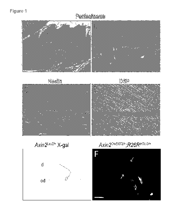
[0009] Figure 1. Odontoblasts are Wnt responsive (A) In skeletally mature
mice,
pentachrome staining identifies dentin (yellow to yellow-green), pulp (purple)
and alveolar
bone (yellow). (B) Higher magnification of the pulpal-dentin complex
illustrates the
organization of pulp cells and odontoblasts (pink) juxtaposed to the pre-
dentin and dentin
(blue and blue-yellow). (C) In the pulp cavity only polarized, secretory
odontoblasts are
positive for Nestin immunostaining. (D) These polarized, secretory
odontoblasts express DSP.
(E) X-gal staining and (F) GFP fluorescence, respectively in adult Axin2Lacz7+
and
Axin2CreERT2/
+,R26RmTmGi+ mice, demonstrates that polarized, secretory odontoblasts and
pulp
cells are Wnt responsive. Abbreviations: ab, alveolar bone; d, dentin; od,
odontoblast; p, pulp;
pd, pre-dentin. Scale bars: 400 pm (A), 25 pm (B-E), 10 pm (F).
[0010] Figure 2. Axin2 deletion does not disrupt odontogenesis or pulpal-
dentin homeostasis.
(A-F) Micro-computed tomography (pCT) reconstructions of the molar region in
skeletally
mature, male (A,C,E) Axin2Lacz7+ and (B,D,F) Axin2LacZ/LacZ mice. Quantified
pCT demonstrate
no differences in (G) dentin volume, or (H) dentin and enamel mineral density
in molars from
age-matched, sex-matched, adult Axin2Laczt+ and Axin2LacZ/LacZ mice.
Pentachrome staining
indicates the cellularity and organization of the pulp cavities from (I)
Axin2Laczt+ and (J)
Axin2Laczn-acz mice. (K) X-gal staining in odontoblasts and sub-odontoblasts
in Axin2Lacz7+ and
(L) Axin2LacZ/LacZ mice; the stronger staining in (L) is due to Axin2LacZ/LacZ
mice carrying two
copies of the LacZ gene. (M) Quantitative RT-PCR analyses of pulp tissues from
Axin2Lacz7+
CA 03009983 2018-06-27
WO 2016/109433
PCT/US2015/067683
4
and Axin2LacZ/LacZ mice, evaluated for the relative expression levels of Lefl
, Axin2 exonl, and
PCNA. (N) Nestin immunostaining in Axin2' z7+ and (0) Axin2LacZ/LacZ mice. (P)
Quantitative
qRT-PCR analyses of pulp tissues from Axin2Laczt+ and Axin2Laczn-acz mice,
evaluated for
expression of Nestin, DSPP, OC, and Coil. Abbreviations: ab, alveolar bone; d,
dentin; p,
pulp. Scale bars: 500 pm (A-F), and 100 pm (I-L and N-0).
[0011] Figure 3. Injury response to an acute pulp exposure in Axin21-
acz/Lacz mice. (A) In
Axin2Lacz7+ mice on day 14, pentachrome staining identifies a pink-colored,
acellular
granulation tissue that occupies the pulp injury site. (B) In Axin2LacZ/LacZ
mice, the pulp injury
site is occupied by a green-yellow mineralized matrix and a dense infiltrate
of cells. (C)
Quantification of the histomorphometric analyses, demonstrating pulp injury
sites in
Axin2Laczn-acz mice. Under polarized light, Picrosirius red staining of (D)
Axin2/a7+ injury sites
and (E) Axin2LacZ/LacZ injury sites. In tissues from Axin2Laczft and Axin21-
acZ/LacZ mice respectively,
immunostaining for (F-G) DSP, and (H-I) Nestin. Pentachrome staining of pulp
injuries on
post-injury day 4 in (J) Axin2''' + and in (K) Axin2LacZ/LacZ mice, quantified
in (L) where Axin2
exonl expression was measured. TUNEL staining indicates programmed cell death
in (M)
Axin2'-' + and (Q) Axin2LacZ/LacZ mice. (0) Quantitative RT-PCR for CASP8
expression. On
post-injury day 7, Ki67 immunostaining in (P) Axin2I-aczi+ and (Q)
Axin2LacZ/LacZ mice.
Abbreviations: ab, alveolar bone; d, dentin; f, furcation; gr, granulation
tissue; p, pulp. Scale
bars: 50 pm (K-L), 100 pm (N-0), 50 pm (H-I), 100 pm (A-B), 100 pm (D-I), 25
pm (N), and 50
pm (0). Single asterisk denotes P<0.05. Double asterisk denotes P<0.01. Error
Bars
represent SEM.
[0012] Figure 4. WNT3A stimulates proliferation and survival of human
dental pulp stem cells
and mouse bone marrow-derived stem cells. (A) Quantitative RT-PCR analyses
following 6,
12, 24 hours of L-PBS or L-WNT3A treatment of human dental pulp stem cells.
(B) Twelve
hours post treatment, the proliferative capacity of hDPSCs was assayed using
the BrdU
incorporation. (C) Quantitative RT-PCR for CASP3 expression. (D) Quantitative
RT-PCR
analyses following 6, 12, 24 hours of L-PBS or L-WNT3A treatment of whole bone
marrow
cells. (E) Twelve hours after whole bone marrow cells were exposed to L-WNT3A
and L-PBS
Ki67 expression and (F) TUNEL activity were evaluated. Scale bars: 100 pm (B,
E, F). Single
asterisk denotes P<0.05. Error Bars represent SEM.
[0013] Figure 5. L-WNT3A treatment induces dentin regeneration. Pentachrome
staining of
pulp injuries on post-injury day 4 in (A) L-PBS treated rats and in (B) L-
WNT3A treated rats.
Post-surgical day 4, TUNEL staining in the pulp cavities of (C,C') L-PBS and
(D,D') L-WNT3A
treated rats. On post-surgical day 4, PCNA expression in the pulp cavities of
(E) L-PBS and
(F) L-WNT3A treated rats. Pentachrome staining of (G) L-PBS and (H) L-WNT3A
treated rats
14 days after injury. (I) Quantification of reparative dentin matrix. Under
polarized light,
Picrosirius red staining of pulp injuries on post-injury day 14 in (J) L-PBS
and (K) L-WNT3A
CA 03009983 2018-06-27
WO 2016/109433
PCT/US2015/067683
treated injury sites. On post-surgical day 14, Nestin expression in (L) L-PBS
and (M) L-
WNT3A treated rats, and DSP expression in (N) L-PBS and (0) L-WNT3A treated
rats.
Abbreviations: dentin, d; in, injury site. Scale bars: 200 pm (A-D), 100 pm
(C', D'), 50 pm (E-
F), 25 pm (G-0), 50 pm. Single asterisk denotes P<0.01. Error Bars represent
SEM.
[0014] Figure 6. In an embryonic 18.5 mouse molar (A) pentachrome staining
identifies the
enamel organ (dotted lines) and dental mesenchyme. (B) X-gal staining of a
molar tooth bud
from an Axin2Lacz7+ embryo. On an adjacent tissue section, (C) Lef1
immunostaining identifies
Wnt responsive cells in the outer enamel organ and in the condensing dental
mesenchyme. At
post-natal day 7, (D) periodic acid schiff staining identifies polarized
odontoblasts and their
newly secreted dentin matrix (pink), which approximates newly secreted enamel
matrix (red)
produced by ameloblasts. (E) These polarized, secretory odontoblasts express
DSP. (F) In
Axin2Lacz7+ mice, X-gal staining demonstrates that polarized, secretory
odontoblasts are Wnt
responsive. Abbreviations: am, ameloblasts; ab, alveolar bone; d, dentin; df,
dental follicle; dp,
dental papilla; e, enamel; eo, enamel organ; m, dental mesenchyme; od,
odontoblast; p, pulp;
pd, pre-dentin. Scale bars: 100 pm (A-C), 100 pm (D), 25 pm (E-F).
[0015] Figure 7. A schematic representation of a healthy tooth (top) and a
tooth in which the
pulp is exposed (bottom).
[0016] Figure 8. (A) A non-penetrating cavity preparation, simulating that
seen in humans,
that cuts through the tubular dentin (yellow) but does not penetrate to the
pulp cavity (pink).
(B) An adjacent section to panel A, stained to identify cells expressing GFP.
These cavity
preparations were generated in transgenic mice that, in the presence of
tamoxifen, undergo a
recombination event where Wnt responsive cells express GFP. Pulp cells are Wnt
responsive,
and in this animal, tamoxifen was delivered via liposomal particles identical
to those used to
delivery WNT3A protein. The GFP positive cells therefore represent Wnt
responsive cells in
the pulp cavity whose only means of tamoxifen exposure was via the dentinal
tubules. B' and
B" show higher magnification of the Wnt-responsive odontoblasts that express
GFP as a
consequence of liposomal tamoxifen delivery. (C-D) The pulp response to the
cavity
preparation (that cuts through the tubular dentin but does not penetrate to
the pulp cavity) and
topical liposomal PBS delivery. A small amount of reparative dentin is
generated (dotted white
line in D), in keeping with the body's natural ability to stimulate a repair
response. (E-F) The
pulp response to a similar cavity preparation as in panels C-D, and topical L-
WNT3A delivery
to exposed dentin. Significantly more reparative dentin is observed in the
pulp chamber.
DETAILED DESCRIPTION
[0017] Methods and compositions are provided for enhancing dentin
production, and for
delivering a lipophilic agent to pulp tissue of a tooth of an individual. In
some cases, a subject
CA 03009983 2018-06-27
WO 2016/109433
PCT/US2015/067683
6
method includes a step of administering to the pulp of a tooth of an
individual, a Wnt
stimulating composition that includes a Wnt stimulator agent, at a dose
sufficient to enhance
the production of dentin by the pulp. In some cases, a subject method includes
a step of
contacting exposed dentin of a tooth with a composition that includes a
lipophilic agent
inserted in the non-aqueous phase of a lipid structure (e.g., whereby the
lipophilic agent
penetrates the dentin to the underlying pulp tissue). Kits are also provided
for practicing the
methods of the disclosure.
[0018] Before the present methods and compositions are described, it is to
be understood
that this invention is not limited to particular method or composition
described, as such may, of
course, vary. It is also to be understood that the terminology used herein is
for the purpose of
describing particular embodiments only, and is not intended to be limiting,
since the scope of
the present invention will be limited only by the appended claims.
[0019] Where a range of values is provided, it is understood that each
intervening value, to
the tenth of the unit of the lower limit unless the context clearly dictates
otherwise, between
the upper and lower limits of that range is also specifically disclosed. Each
smaller range
between any stated value or intervening value in a stated range and any other
stated or
intervening value in that stated range is encompassed within the invention.
The upper and
lower limits of these smaller ranges may independently be included or excluded
in the range,
and each range where either, neither or both limits are included in the
smaller ranges is also
encompassed within the invention, subject to any specifically excluded limit
in the stated
range. Where the stated range includes one or both of the limits, ranges
excluding either or
both of those included limits are also included in the invention.
[0020] Unless defined otherwise, all technical and scientific terms used
herein have the same
meaning as commonly understood by one of ordinary skill in the art to which
this invention
belongs. Although any methods and materials similar or equivalent to those
described herein
can be used in the practice or testing of the present invention, some
potential and preferred
methods and materials are now described. All publications mentioned herein are
incorporated
herein by reference to disclose and describe the methods and/or materials in
connection with
which the publications are cited. It is understood that the present disclosure
supersedes any
disclosure of an incorporated publication to the extent there is a
contradiction.
[0021] As will be apparent to those of skill in the art upon reading this
disclosure, each of the
individual embodiments described and illustrated herein has discrete
components and
features which may be readily separated from or combined with the features of
any of the
other several embodiments without departing from the scope or spirit of the
present invention.
Any recited method can be carried out in the order of events recited or in any
other order
which is logically possible.
CA 03009983 2018-06-27
WO 2016/109433
PCT/US2015/067683
7
[0022] It must be noted that as used herein and in the appended claims, the
singular forms
"a", an, and "the" include plural referents unless the context clearly
dictates otherwise.
Thus, for example, reference to "a cell" includes a plurality of such cells
and reference to "the
peptide" includes reference to one or more peptides and equivalents thereof,
e.g.
polypeptides, known to those skilled in the art, and so forth.
[0023] The publications discussed herein are provided solely for their
disclosure prior to the
filing date of the present application. Nothing herein is to be construed as
an admission that
the present invention is not entitled to antedate such publication by virtue
of prior invention.
Further, the dates of publication provided may be different from the actual
publication dates
which may need to be independently confirmed.
Definitions
[0024] In the description that follows, a number of terms conventionally
used in the field are
utilized. In order to provide a clear and consistent understanding of the
specification and
claims, and the scope to be given to such terms, the following definitions are
provided.
[0025] The terms "polypeptide," "peptide" and "protein" are used
interchangeably herein to
refer to a polymer of amino acid residues. The terms also apply to amino acid
polymers in
which one or more amino acid residue is an artificial chemical mimetic of a
corresponding
naturally occurring amino acid, as well as to naturally occurring amino acid
polymers and non-
naturally occurring amino acid polymer.
[0026] The term "amino acid" refers to naturally occurring and synthetic
amino acids, as well
as amino acid analogs and amino acid mimetics that function in a manner
similar to the
naturally occurring amino acids. Naturally occurring amino acids are those
encoded by the
genetic code, as well as those amino acids that are later modified, e.g.,
hydroxyproline,
gamma-carboxyglutamate, and 0-phosphoserine. Amino acid analogs refers to
compounds
that have the same basic chemical structure as a naturally occurring amino
acid, i.e., an
.alpha. carbon that is bound to a hydrogen, a carboxyl group, an amino group,
and an R
group, e.g., homoserine, norleucine, methionine sulfoxide, methionine methyl
sulfonium. Such
analogs have modified R groups (e.g., norleucine) or modified peptide
backbones, but retain
the same basic chemical structure as a naturally occurring amino acid. Amino
acid mimetics
refers to chemical compounds that have a structure that is different from the
general chemical
structure of an amino acid, but that functions in a manner similar to a
naturally occurring
amino acid.
[0027] The terms "recipient", "individual", "subject", "host", and
"patient", are used
interchangeably herein and refer to any individual for whom diagnosis,
treatment, or therapy is
desired, particularly humans. "Mammal" or "mammalian" for purposes of
treatment refers to
any animal classified as a mammal, including humans, domestic and farm
animals, and zoo,
CA 03009983 2018-06-27
WO 2016/109433
PCT/US2015/067683
8
sports, or pet animals, such as dogs, horses, cats, cows, sheep, goats, pigs,
etc. In some
embodiments, the mammal is human.
Compositions
[0028] The present disclosure provides compositions and methods for
enhancing dentin
production by dental pulp tissue. Such methods include administering to the
pulp of a tooth of
an individual, a Wnt stimulating composition comprising a Wnt stimulator
agent, at a dose
sufficient to enhance the production of dentin by the pulp. In some cases the
Wnt stimulator
agent comprises a lipophilic Wnt stimulator agent (e.g., a Wnt protein, such
as Wnt3A)
inserted in the non-aqueous phase of a lipid structure.
[0029] The present disclosure also provides compositions and methods for
delivering a
lipophilic agent to pulp tissue of a tooth of an individual. In some cases the
lipophilic agent is a
growth factor (e.g., a growth factor having a lipid moiety). In some cases,
the lipophilic agent
is a lipophilic Wnt stimulator agent (e.g., a Wnt protein, such as Wnt3A)
inserted in the non-
aqueous phase of a lipid structure.
[0030] Lipid Structure.
[0031] In some embodiments, the subject agent (e.g., an agent of interest)
is a lipophilic
agent inserted in the non-aqueous phase of a lipid structure. Lipid structures
can be important
for maintaining the activity of lipophilic agents (e.g., Wnt proteins, growth
factors, etc., e.g.,
having a lipid moiety), following in vivo administration. The subject
lipophilic agents (e.g., Wnt
proteins, growth factors, etc., e.g., having a lipid moiety) are not
encapsulated in the aqueous
phase of the lipid structures, but are rather integrated into the lipid
membrane, and may be
inserted in the outer layer of a membrane. Such a structure is not predicted
from conventional
methods of formulating agents (e.g., proteins) in, for example, liposomes. A
Wnt protein
integrated within such lipid structure is referred herein as L-Wnt (e.g.,
Wnt3A integrated into
such a lipid structure can be referred to as L-Wnt3A). The methods used for
tethering
lipophilic agents (e.g., Wnt proteins) to the external surface of a liposome
or micelle can utilize
a moiety (e.g., a protein might have an amino acid sequence) so as to
emphasize the
exoliposomal display of the protein. In some cases, crude liposomes are first
pre-formed and
a lipophilic agent (e.g., a Wnt protein, a growth factors, etc., e.g., having
a lipid moiety) can
then be added to the crude mixture, which will favor addition of exo-liposomal
agent (e.g., Wnt
protein), followed by various formulation steps, which may include size
filtering; dialysis, and
the like. Suitable lipids include fatty acids, neutral fats such as
triacylglycerols, fatty acid
esters and soaps, long chain (fatty) alcohols and waxes, sphingoids and other
long chain
bases, glycolipids, sphingolipids, carotenes, polyprenols, sterols, and the
like, as well as
terpenes and isoprenoids. For example, molecules such as diacetylene
phospholipids may
find use. Included are cationic molecules, including lipids, synthetic lipids
and lipid analogs,
CA 03009983 2018-06-27
WO 2016/109433 PCT/US2015/067683
9
having hydrophobic and hydrophilic moieties, a net positive charge, and which
by itself can
form spontaneously into bilayer vesicles or micelles in water. Liposomes
manufactured with a
neutral charge, e.g. DMPC, can be used. Any amphipathic molecules that can be
stably
incorporated into lipid micelle or bilayers in combination with phospholipids
can be used, with
its hydrophobic moiety in contact with the interior, hydrophobic region of the
micelle or bilayer
membrane, and its polar head group moiety oriented toward the exterior, polar
surface of the
membrane.
[0032] The term "cationic amphipathic molecules" is intended to encompass
molecules that
are positively charged at physiological pH, and more particularly,
constitutively positively
charged molecules, comprising, for example, a quaternary ammonium salt moiety.
Cationic
amphipathic molecules typically consist of a hydrophilic polar head group and
lipophilic
aliphatic chains. Similarly, cholesterol derivatives having a cationic polar
head group may also
be useful. See, for example, Farhood et al. (1992) Biochim. Biophys. Acta
1111:239- 246;
Vigneron et al. (1996) Proc. Natl. Acad. Sci. (USA) 93:9682-9686. Cationic
amphipathic
molecules of interest include, for example, imidazolinium derivatives (WO
95/14380),
guanidine derivatives (WO 95/14381), phosphatidyl choline derivatives (WO
95/35301), and
piperazine derivatives (WO 95/14651). Examples of cationic lipids that may be
used in the
present invention include DOTIM (also called BODAI) (Saladin et al., (1995)
Biochem. 34:
13537-13544), DDAB (Rose et al., (1991) BioTechniques 10(4):520-525), DOTMA
(U.S. Pat.
No. 5,550,289), DOTAP (Eibl and Wooley (1979) Biophys. Chern. 10:261-271),
DMRIE
(Feigner et al., (1994) J. Bioi. Chern. 269(4): 2550-2561), EDMPC
(commercially available
from Avanti Polar Lipids, Alabaster, Ala.), DCC hoi (Gau and Huang (1991)
Biochem. Biophys.
Res. Comm. 179:280-285), DOGS (Behr et al., (1989) Proc. Nat!. Acad. Sci. USA,
86:6982-
6986), MBOP (also called MeB0P) (WO 95/14651 ), and those described in WO
97/00241.
While not required for activity, in some embodiments a lipid structure may
include a targeting
group, e.g. a targeting moiety covalently or non-covalently bound to the
hydrophilic head
group. Head groups useful to bind to targeting moieties include, for example,
biotin, amines,
cyano, carboxylic acids, isothiocyanates, thiols, disulfides, ahalocarbonyl
compounds, a,p-
unsaturated carbonyl compounds, alkyl hydrazines, etc. Chemical groups that
find use in
linking a targeting moiety to an amphipathic molecule also include carbamate;
amide (amine
plus carboxylic acid); ester (alcohol plus carboxylic acid), thioether
(haloalkane plus sulfhydryl;
maleimide plus sulfhydry1), Schiffs base (amine plus aldehyde), urea (amine
plus isocyanate),
thiourea (amine plus isothiocyanate), sulfonamide (amine plus sulfonyl
chloride), disulfide;
hyrodrazone, lipids, and the like, as known in the art. For example, targeting
molecules may
be formed by converting a commercially available lipid, such as DAGPE, a PEG-
PDA amine,
DOTAP, etc. into an isocyanate, followed by treatment with triethylene glycol
diamine spacer
to produce the amine terminated thiocarbamate lipid which by treatment with
the para-
CA 03009983 2018-06-27
WO 2016/109433
PCT/US2015/067683
isothiocyanophenyl glycoside of the targeting moiety produces the desired
targeting
glycolipids. This synthesis provides a water soluble flexible linker molecule
spaced between
the amphipathic molecule that is integrated into the nanoparticle, and the
ligand that binds to
cell surface receptors, allowing the ligand to be readily accessible to the
protein receptors on
the cell surfaces. Further information about liposomal Wnt compositions and
their use is found
in; U.S. patent publication numbers: 20140371151, 20120115788, and
20080226707; and
U.S. patent number: 8809272; all of which are hereby incorporated by reference
in their
entirety.
[0033] In some cases, liposomes or micelles are used as a delivery vehicle. A
liposome is a
spherical vesicle with a membrane composed of a phospholipid bilayer.
Liposomes can be
composed of naturally-derived phospholipids with mixed lipid chains (like egg
phosphatidylethanolamine), or of pure surfactant components like DOPE
(dioleolylphosphatidylethanolamine). Liposomes often contain a core of
encapsulated
aqueous solution; while lipid spheres that contain no aqueous material are
referred to as
micelles. As wnt proteins are present in the lipid phase and not the
encapsulated aqueous
phase, micelles may be used interchangeably with liposome for the compositions
of the
present disclosure. The lipids may be any useful combination of known liposome
or micelle
forming lipids, including cationic lipids, such as phosphatidylcholine, or
neutral lipids, such as
cholesterol, phosphatidyl serine, phosphatidyl glycerol, and the like.
[0034] In some embodiments, the vesicle-forming lipid is selected to
achieve a specified
degree of fluidity or rigidity, to control the stability of the structure in
serum, etc. Liposomes
having a more rigid lipid bilayer, or a liquid crystalline bilayer, are
achieved by incorporation of
a relatively rigid lipid, e.g., a lipid having a relatively high phase
transition temperature, e.g.,
up to 60 C. Rigid, i.e., saturated, lipids contribute to greater membrane
rigidity in the lipid
bilayer. Other lipid components, such as cholesterol, are also known to
contribute to
membrane rigidity in lipid bilayer structures. Lipid fluidity is achieved by
incorporation of a
relatively fluid lipid, typically one having a lipid phase with a relatively
low liquid to liquid-
crystalline phase transition temperature, e.g., at or below room temperature.
[0035] The liposomes may be prepared by a variety of techniques, such as
those detailed in
Szoka, F., Jr., et al., Ann. Rev. Biophys. Bioeng. 9:467 (1980). Typically,
the liposomes are
multilamellar vesicles (MLVs), which can be formed by simple lipid-film
hydration techniques.
In this procedure, a mixture of liposome-forming lipids of the type detailed
above dissolved in
a suitable organic solvent is evaporated in a vessel to form a thin film,
which is then covered
by an aqueous medium. The lipid film hydrates to form MLVs, e.g., in some
cases with sizes
in a range of from 0.1 to 10 microns.
[0036] The liposomes, micelles, etc. of the disclosure may have
substantially homogeneous
sizes in a selected size range, for example, between 0.005 to 0.5 microns
(e.g., 0.01 to 0.5
CA 03009983 2018-06-27
WO 2016/109433
PCT/US2015/067683
11
0.02 to 0.5, 0.025 to 0.5, 0.05 to 0.5, 0.075 to 0.5, 0.1 to 0.5, 0.005 to
0.4, 0.01 to 0.4 0.02 to
0.4, 0.025 to 0.4, 0.05 to 0.4, 0.075 to 0.4, 0.1 to 0.4, 0.005 to 0.3, 0.01
to 0.3 0.02 to 0.3,
0.025 to 0.3, 0.05 to 0.3, 0.075 to 0.3, 0.1 to 0.3, 0.005 to 0.2, 0.01 to 0.2
0.02 to 0.2, 0.025 to
0.2, 0.05 to 0.2, 0.075 to 0.2, 0.1 to 0.2, 0.005 to 0.1, 0.01 to 0.1 0.02 to
0.1, 0.025 to 0.1,
0.05 to 0.1, 0.075 to 0.1, 0.02 to 0.05, or 0.02 to 0.35 microns).
[0037] One effective sizing method for REVs and MLVs involves extruding an
aqueous
suspension of the liposomes through a series of polycarbonate membranes having
a selected
uniform pore size in the range of 0.03 to 0.2 micron, typically 0.05, 0.08,
0.1, or 0.2 microns.
The pore size of the membrane corresponds roughly to the largest sizes of
liposomes
produced by extrusion through that membrane, particularly where the
preparation is extruded
two or more times through the same membrane. Homogenization methods are also
useful for
down-sizing liposomes to sizes of 100 nm or less.
[0038] The pharmaceutical compositions of the present disclosure can also
comprise a
pharmaceutically acceptable carrier. Many pharmaceutically acceptable carriers
may be
employed in the compositions of the present disclosure. Generally, normal
saline will be
employed as the pharmaceutically acceptable carrier. Other suitable carriers
include, e.g.,
water, buffered water, 0.4% saline, 0.3% glycine, and the like, including
glycoproteins for
enhanced stability, such as albumin, lipoprotein, globulin, etc. These
compositions may be
sterilized by conventional, well known sterilization techniques. The resulting
aqueous solutions
may be packaged for use or filtered under aseptic conditions and lyophilized,
the lyophilized
preparation being combined with a sterile aqueous solution prior to
administration. The
compositions may contain pharmaceutically acceptable auxiliary substances as
required to
approximate physiological conditions, such as pH adjusting and buffering
agents, tonicity
adjusting agents and the like, for example, sodium acetate, sodium lactate,
sodium chloride,
potassium chloride, calcium chloride, etc.
[0039] The concentration of lipid structures in the carrier may vary.
Generally, the
concentration can be about 0.1 to 1000 mg/ml, usually about 1-500 mg/ml, about
5 to 100
mg/ml, etc. Persons of skill may vary these concentrations to optimize
treatment with different
lipid components or of particular patients.
[0040] Subject compositions can include a therapeutically effective in vivo
dose of a lipophilic
agent (e.g., a wnt protein), and may comprise a cocktail of one or more
lipophilic agents (e.g.,
one or more wnt proteins, one or more Wnt proteins in addition to one or more
other lipophilic
agents, etc.).
[0041] Wnt signaling pathway! Wnt proteins
[0042] In some embodiments, the subject agent (e.g., an agent of interest)
is a Wnt stimulator
agent. A Wnt stimulator agent increases activity of the Wnt signaling pathway
in a target cell.
A target cell (and/or tissue) that is "Wnt responsive" is a cell/tissue that
can respond to the
CA 03009983 2018-06-27
WO 2016/109433
PCT/US2015/067683
12
extracellular presence of a Wnt protein by triggering the Wnt signaling
pathway. A Wnt
responsive cell includes components of the Wnt signaling pathway (described in
more detail
below), including a receptor (e.g., a Frizzled receptor) that can bind to Wnt
proteins. Not all
cells are Wnt responsive. In some embodiments, the target cell/tissue is Wnt
responsive. In
some embodiments the target cell is not Wnt responsive. In some embodiments,
it is unknown
whether the target cell is Wnt responsive. In some embodiments, it is known
whether the
target cell is Wnt responsive. In some embodiments, the target cell is part of
a heterogeneous
population of target cells (i.e., a heterogeneous target cell population) in
which some cells are
Wnt responsive and some cells are not Wnt responsive (e.g., in some cases a
target tissue,
such as dental pulp, includes cells that are Wnt responsive as well as cells
that are not Wnt
responsive). In some embodiments, it is known which cells of a heterogeneous
target cell
population are Wnt responsive (e.g., stem cells). In some embodiments, it is
unknown which
cells of a heterogeneous target cell population are Wnt responsive.
[0043] The misregulation of Wnt signaling components at various stages
during
embryogenesis leads to catastrophic developmental defects while misregulation
in adults
leads to various disease states, including cancer. There are two main branches
of the Wnt
signaling pathway: (1) the canonical [3-Catenin dependent Wnt signaling
pathway and (2) the
non-canonical [3-Catenin independent pathways, which include planar cell
polarity (PCP)
signaling as well as Calcium signaling (Gao, et. al, Cell Signal. 2010
May;22(5):717-27. Epub
2009 Dec 13). As used herein, the terms "Wnt signaling" and "Wnt/[3-Catenin
signaling" are
used interchangeably to refer to the canonical [3-Catenin dependent Wnt
signaling pathway.
As such, a Wnt signaling stimulator, also referred to as a "Wnt stimulator
agent" (i.e., agonist)
(e.g., Wnt3A) increases output from the [3-Catenin dependent Wnt signaling
pathway while a
Wnt signaling inhibitor (i.e., antagonist) decreases output from the [3-
Catenin dependent Wnt
signaling pathway.
[0044] Activation of the Wnt pathway culminates when the protein [3-Catenin
enters the cell
nucleus (for recent review of the canonical [3-Catenin dependent Wnt signaling
pathway see
Clevers et. al., Cell. 2012 Jun 8;149(6):1192-205: Wnt/[3-catenin signaling
and disease).
However, in the absence of Wnt signaling, free cytosolic [3-Catenin is
incorporated into a
complex, known in the art as the [3-Catenin destruction complex, which
includes the proteins
Axin, Adenomatous Polyposis Coli (APC), and glycogen synthase kinase (GSK-
3[3).
Phosphorylation of [3-Catenin by GSK-3[3 designates [3-Catenin for the
ubiquitin pathway and
degradation (e.g., via TRCP).
[0045] Transduction of the [3-Catenin dependent Wnt signaling pathway
(i.e., the Wnt
signaling pathway) is triggered by the binding of secreted Wnt ligands to two
distinct families
of cell-surface receptors: the Frizzled (Fz) receptor family and the LDL-
receptor-related
protein (LRP) family (Akiyama, Cytokine Growth Factor Rev. 11:273-82 (2000)).
This binding
CA 03009983 2018-06-27
WO 2016/109433
PCT/US2015/067683
13
leads to the activation of Dishevelled (DA) proteins, which inhibit glycogen
synthase kinase-3[3
(GSK-3[3) activity (i.e., phosphorylation of [3-Catenin), leading to the
cytosolic stabilization of p-
Catenin. Stabilized [3-Catenin then enters the nucleus and associates with the
TCF/LEF (T
Cell-specific transcription Factor / Lymphoid Enhancer Factor) family of
transcription factors to
induce transcription of downstream target genes.
[0046] In the absence of Wnt signaling, cytosolic (and therefore nuclear)
levels of [3-Catenin
are kept low by negative regulatory components of the pathway while in the
presence of Wnt
signaling, cytosolic (and therefore nuclear) levels of [3-Catenin are
stabilized by positive
regulatory components of the pathway. For this reason, [3-Catenin levels
(e.g., monitored via
Western blot) can provide insight into whether the Wnt signaling pathway of a
cell has been
stimulated or inhibited (e.g., increased levels of [3-Catenin indicate
increased signaling and
decreased levels indicate decrease signaling). Likewise, [3-Catenin levels in
the nucleus (e.g.,
monitored via fluorescence microscopy, Western blot, etc.) can also be
monitored to
determine increased or decreased signaling.
[0047] By "positive regulatory components" of the Wnt pathway, it is meant
proteins that
function by enhancing (i.e., stimulating) the Wnt pathway, thus resulting in
increased Wnt
pathway signaling activity (i.e., increased Wnt pathway signaling output,
e.g., increased target
gene expression, increased reporter activity, increased levels of [3-Catenin,
etc.). Examples of
known positive regulatory components of the Wnt pathway include, but are in no
way limited
to: Wnt (secreted, extracellular), Norrin (secreted, extracellular), R-spondin
(secreted,
extracellular), PORCN, Wls, Frizzled, LRP5 and LRP6, Tspan12, Lgr4, Lgr5,
Lgr6, Dv!, p-
Catenin, and TCF/LEF. A secreted positive regulatory component of the Wnt
pathway (e.g.,
Wnt, Norrin, R-spondin, and the like) is referred to herein as a "Wnt
stimulator polypeptide". In
some cases a Wnt stimulator polypeptide is a Wnt protein.
[0048] Suitable Wnt polypeptides (i.e., Wnt proteins) include, but are in
no way limited to
human Wnt polypeptides. Human Wnt proteins of interest in the present
application include
the following (accession numbers are for mRNAs encoding the associated Wnt
protein): Wnt-1
(GenBank Accession No. NM_005430); Wnt-2 (GenBank Accession No. NM_003391);
Wnt-
2B (Wnt-13) (GenBank Accession No. NM_004185 (isoform 1), NM_024494.2 (isoform
2)),
Wnt-3 (RefSeq.: NM_030753), Wnt3a (GenBank Accession No. NM_033131), Wnt-4
(GenBank Accession No. NM_030761), Wnt-5A (GenBank Accession No. NM_003392),
Wnt-
5B (GenBank Accession No. NM_032642), Wnt-6 (GenBank Accession No. NM_006522),
Wnt-7A (GenBank Accession No. NM_004625), Wnt-7B (GenBank Accession No.
NM_058238), Wnt-8A (GenBank Accession No. NM_058244), Wnt-8B (GenBank
Accession
No. NM_003393), Wnt-9A (Wnt-14) (GenBank Accession No. NM_003395), Wnt-9B (Wnt-
15)
(GenBank Accession No. NM_003396), Wnt-10A (GenBank Accession No. NM_025216),
Wnt-10B (GenBank Accession No. NM_003394), Wnt-11 (GenBank Accession No.
CA 03009983 2018-06-27
WO 2016/109433
PCT/US2015/067683
14
NM_004626), Wnt-16 (GenBank Accession No. NM_016087)). Although each member
has
varying degrees of sequence identity with the family, all encode small (i.e.,
39-46 kD),
acylated, palmitoylated, secreted glycoproteins that contain 23-24 conserved
cysteine
residues whose spacing is highly conserved (McMahon, A P et al., Trends Genet.
1992; 8:
236-242; Miller, J R. Genome Biol. 2002; 3(1): 3001.1-3001.15). Other Wnt
polypeptides of
interest in the present invention include orthologs of the above from any
mammal, including
domestic and farm animals, and zoo, laboratory or pet animals, dogs, cats,
cattle, horses,
sheep, pigs, goats, rabbits, rats, mice, frogs, zebra fish, fruit fly, worm,
etc.
[0049] Wnt proteins form a family of highly conserved secreted signaling
molecules that
regulate cell-to-cell interactions during embryogenesis. The terms "Wnts" or
"Wnt gene
product" or "Wnt protein" or "Wnt polypeptide" are used interchangeable and
encompass
native sequence Wnt polypeptides, Wnt polypeptide variants, Wnt polypeptide
fragments and
chimeric Wnt polypeptides. In some embodiments of the invention, the Wnt
protein comprises
palmitate covalently bound to a cysteine residue. A "native sequence"
polypeptide is one that
has the same amino acid sequence as a Wnt polypeptide derived from nature,
regardless of
the method used for its production. Such native sequence polypeptides can be
isolated from
cells producing endogenous Wnt protein or can be produced by recombinant or
synthetic
means. Thus, a native sequence polypeptide can have the amino acid sequence
of, e.g.
naturally occurring human polypeptide, murine polypeptide, or polypeptide from
any other
mammalian species, or from non-mammalian species, e.g. Drosophila, C. elegans,
and the
like.
[0050] The term "native sequence Wnt polypeptide" includes, without
limitation, human and
murine Wnt polypeptides. Human Wnt proteins include the following: Wnt1,
Genbank
reference NP005421.1; Wnt2, Genbank reference NP003382.1, which is expressed
in brain in
the thalamus, in fetal and adult lung and in placenta; two isoforms of Wnt2B,
Genbank
references NP004176.2 and NP078613.1. Isoform 1 is expressed in adult heart,
brain,
placenta, lung, prostate, testis, ovary, small intestine and colon. In the
adult brain, it is mainly
found in the caudate nucleus, subthalamic nucleus and thalamus. Also detected
in fetal brain,
lung and kidney. Isoform 2 is expressed in fetal brain, fetal lung, fetal
kidney, caudate nucleus,
testis and cancer cell lines. Wnt 3 and Wnt3A play distinct roles in cell-cell
signaling during
morphogenesis ofthe developing neural tube, and have the Genbank references
NP11 0380.1
and X56842 (Swiss-Prot P56704), respectively.
[0051] The native human Wnt3A amino acid sequence (NP_149122.1) is
specifically
disclosed as SEQ ID NO: 19. Wnt 4 has the Genbank reference NP11 0388.2. Wnt
5A and
Wnt 5B have the Genbank references NP003383.1 and AK013218. Wnt 6 has the
Genbank
reference NP006513.1; Wnt 7A has the Genbank reference NP004616.2. Wnt 7B has
the
Genbank reference NP478679.1. Wnt 8A has two alternative transcripts, Genbank
references
CA 03009983 2018-06-27
WO 2016/109433
PCT/US2015/067683
NP114139.1 and NP490645.1. Wnt 8B has the Genbank reference NP003384.1. Wnt
10A has
the Genbank reference NP079492.2. Wnt 10B has the Genbank reference
NP003385.2. Wnt
11 has the Genbank reference NP004617 .2. Wnt 14 has the Genbank reference
NP003386.1. Wnt 15 has the Genbank reference NP003387.1. Wnt 16 has two
isoforms, Wnt-
16a and Wnt-16b, produced by alternative splicing, Genbank references are
NP057171.2 and
NP476509.1. All GenBank, SwissProt and other database sequences listed are
expressly
incorporated by reference herein.
[0052] The term "native sequence Wnt protein" or "native sequence Wnt
polypeptide"
includes the native proteins with or without the initiating N-terminal
methionine (Met), and with
or without the native signal sequence. The terms specifically include the 352
amino acids long
native human Wnt3a polypeptide, without or without its N terminal methionine
(Met), and with
or without the native signal sequence.
[0053] A "variant" polypeptide means a biologically active polypeptide as
defined below
having less than 100% sequence identity with a native sequence polypeptide.
Such variants
include polypeptides wherein one or more amino acid residues are added at the
N- or C-
terminus of, or within, the native sequence; from about one to forty amino
acid residues are
deleted, and optionally substituted by one or more amino acid residues; and
derivatives of the
above polypeptides, wherein an amino acid residue has been covalently modified
so that the
resulting product has a non-naturally occurring amino acid. Ordinarily, a
biologically active
Wnt variant will have an amino acid sequence having at least about 90% amino
acid
sequence identity with a native sequence Wnt polypeptide, preferably at least
about 95%,
more preferably at least about 99%.
[0054] A "chimeric" Wnt polypeptide is a polypeptide comprising a Wnt
polypeptide or
portion (e.g., one or more domains) thereof fused or bonded to heterologous
polypeptide. The
chimeric Wnt polypeptide will generally share at least one biological property
in common with
a native sequence Wnt polypeptide. Examples of chimeric polypeptides include
immunoadhesins, combine a portion of the Wnt polypeptide with an
immunoglobulin
sequence, and epitope tagged polypeptides, which comprise a Wnt polypeptide or
portion
thereof fused to a "tag polypeptide". The tag polypeptide has enough residues
to provide an
epitope against which an antibody can be made, yet is short enough such that
it does not
interfere with biological activity of the Wnt polypeptide. Suitable tag
polypeptides generally
have at least six amino acid residues and usually between about 6-60 amino
acid residues.
[0055] A "functional derivative" of a native sequence Wnt polypeptide is a
compound having a
qualitative biological property in common with a native sequence Wnt
polypeptide. "Functional
derivatives" include, but are not limited to, fragments of a native sequence
and derivatives of a
native sequence Wnt polypeptide and its fragments, provided that they have a
biological
activity in common with a corresponding native sequence Wnt polypeptide. The
term
CA 03009983 2018-06-27
WO 2016/109433
PCT/US2015/067683
16
"derivative" encompasses both amino acid sequence variants of Wnt polypeptide
and covalent
modifications thereof.
[0056] One may determine the specific activity of a Wnt protein in a
composition by
determining the level of activity in a functional assay, for example in an in
vitro assay, or after
in vivo administration in a test model, e.g. accelerating bone regeneration,
upregulation of
stem cell proliferation, etc., quantitating the amount of Wnt protein present
in a non-functional
assay, e.g. immunostaining, ELISA, quantitation on Coomasie or silver stained
gel, etc., and
determining the ratio of in vivo biologically active Wnt to total Wnt.
[0057] The effective dose of the Wnt protein may vary depending on the
source, purity,
preparation method, etc. Where the Wnt protein is L-Wnt3A, the effective dose
is usually at
least 0.1 ug/ml, at least 0.5 ug/ml, at least 1 ug/ml, at least 2.5 ug/ml, at
least 5 ug/ml, at least
7.5 ug/ml, at least 10 ug/ml, at least 15 ug/ml, and may be at least 25 ug/ml,
at least 50 ug/ml,
or at least 100 ug/ml.
[0058] As discussed above, in some embodiments, a Wnt protein (e.g., Wnt3A,
e.g., human
Wnt3A) is inserted in the non-aqueous phase of a lipid structure, e.g. in the
surface of a
liposome, micelle, lipid raft, etc., in an emulsion, and the like. In some
embodiments the Wnt
protein is presented in its active conformation on an outer liposome membrane
or micelle.
Where the lipid structure is a liposome it can be desirable that the Wnt
protein not be
encapsulated within the liposome, e.g. in an aqueous phase. The lipid-
containing particles
typically display the Wnt protein, the particles comprising at least one copy
of a wnt protein
bearing at least one lipid moiety, where the composition contains at least 50%
of the Wnt
polypeptides displayed on the exterior surface of the particle. In some cases,
R-spondin can
be included in the aqueous core of a liposomal WNT3A (L-Wnt3A) and in so
doing, amplify
and extend the Wnt dependent activation of pulp cells.
[0059] For example, see Dhamdhere et al., PLoS One. 2014 Jan 6;9(1):e83650;
and Zhao et
al., Methods Enzymol. 2009;465:331-47, both of which are hereby incorporated
by reference
in their entirety.
[0060] A subject Wnt stimulator agent is any molecule (e.g., a chemical
compound; a non-
coding nucleic acid, e.g., a non-coding RNA; a polypeptide; a nucleic acid
encoding a
polypeptide, etc.) that results in increased output (i.e., increased target
gene expression) from
the Wnt signaling pathway. For example, a Wnt stimulator agent can function by
stabilizing,
enhancing the expression of, or enhancing the function of a positive
regulatory component of
the pathway or by destabilizing, decreasing the expression of, or inhibiting
the function of a
negative regulatory component of the pathway. Thus, a Wnt stimulator agent can
be a positive
regulatory component of the pathway (e.g., a Wnt protein), or a nucleic acid
encoding one or
more positive regulatory components of the pathway. A Wnt stimulator agent can
also be a
CA 03009983 2018-06-27
WO 2016/109433
PCT/US2015/067683
17
small molecule or nucleic acid that stabilizes a positive regulatory component
of the pathway
either at the level of mRNA or protein.
[0061] In some embodiments, a Wnt stimulator agent functions by stabilizing
p- Cate n in , thus
allowing nuclear levels of p-C ate n n to rise. p-Caten in can be stabilized
in multiple different
ways. As multiple different negative regulatory components of the Wnt
signaling pathway
function by facilitating the degradation of p-Caten n , a subject Wnt
stimulator agent can be a
small molecule or nucleic acid inhibitor (e.g., microRNA, shRNA,
etc.)(functioning at the level
of mRNA or protein) of a negative regulatory component of the pathway. For
example, in
some embodiments, the Wnt stimulator agent is an inhibitor of GSK-3[3. In some
such
embodiments, the inhibitor of GSK-3[3 is a small molecule chemical compound
(e.g., TWS119,
BIO, CHIR-99021, SB 216763, SB 415286, CHIR-98014 and the like).
[0062] TWS119: 3-(6-(3-aminophenyI)-7H-pyrrolo[2,3-d]pyrimidin-4-
yloxy)phenol is described
by Ding et. al, Proc Natl Acad Sci U S A. 2003 Jun 24;100(13):7632-7. BIO: 6-
bromo-3-[(3E)-
1,3-dihydro-3-(hydroxyimino)-2H-indo1-2-ylidene]-1,3-dihydro-(3Z)-2H-indo1-2-
one or (2'Z,3'E)-
6-Bromoindirubin-3'-oxime is described by Meijer et. al, Chem Biol. 2003
Dec;10(12):1255-66.
CHIR-99021: 6-[[2-[[4-(2,4-dichloropheny1)-5-(5-methy1-1H-imidazol-2-y1)-2-
pyrimidinyl]amino]ethyl]amino]-3-pyridinecarbonitrile is described by Bennett
et al., J Biol
Chem. 2002 Aug 23;277(34):30998-1004. SB 216763: 3-(2,4-dichloropheny1)-4-(1-
methy1-1H-
indo1-3-y1)-1H-pyrrole-2,5-dione is described by Cross et al., J Neurochem.
2001 Apr;77(1):94-
102. SB 415286: 3-(3-chloro-4-hydroxyphenylamino)-4-(2-nitropheny1)-1H-pyrrole-
2,5-dione
is described by Cross et al., J Neurochem. 2001 Apr;77(1):94-102. CHIR-98014:
N2-(2-(4-
(2,4-dichloropheny1)-5-(1H-imidazol-1-yhpyrimidin-2-ylamino)ethyl)-5
nitropyridine-2,6-diamine
is described by Ring et al., Diabetes. 2003 Mar;52(3):588-95. Each reference
is herein
specifically incorporated by reference.
[0063] In some cases, a Wnt stimulator agent is a lipophilic agent. For
those agents above
that are not lipophilic, a Wnt stimulator agent can be such an agent
conjugated to a lipid
moiety.
[0064] The effective dose of a Wnt stimulator agent can be at least 0.1 uM,
at least 1 uM, at
least 2.5 uM, at least 5 1.1M, and usually not more than 500 uM, not more than
250 uM, not
more than 100 uM, or not more than 50 M.
[0065] By "negative regulatory components" of the Wnt pathway, it is meant
proteins that
function by antagonizing (i.e., inhibiting) the Wnt pathway, thus resulting in
decreased
pathway output (i.e., decreased Wnt pathway signaling output, e.g., decreased
target gene
expression, decreased reporter activity, decreased levels of p- Cate n in ,
etc.). Examples of
known negative regulatory components of the Wnt pathway include, but are in no
way limited
to: WIF, sFRP, DKK, Wnt5, Wnt11, Notum, WISE/SOST, Axin, APC, GSK-3[3, CK1y,
VVTX,
CA 03009983 2018-06-27
WO 2016/109433
PCT/US2015/067683
18
and [3TrCP. A secreted negative regulatory component of the Wnt pathway is
referred to
herein as a "Wnt inhibitor polypeptide".
[0066] Wnt inhibitor polypeptides (i.e., secreted negative regulatory
components of the Wnt
signaling pathway) include members of the WIF (Wnt inhibitory factor), sFRP
(Secreted
Frizzled Related Protein), DKK (Dickkopf), Notum, and WISE/SOST families,
which interfere
with the appropriate interactions among Wnt, Frizzled, and LRP proteins
(Melkonyan et al.,
1997, Proc Natl Acad Sci U S A 94(25):13636-41; Moon et al.,1997, Cell
88(6):725-8; Fedi et
al., 1999, J Biol Chem 274(27):19465-72; Nusse, 2001, Nature 411(6835):255-6;
Clevers et.
al., Cell. 2012 Jun 8;149(6):1192-205: Wnt/[3-catenin signaling and disease).
Although most
Wnt polypeptides are Wnt stimulator polypeptides, certain Wnt polypeptides
(e.g., Wnt5 and
Wnt11) are Wnt inhibitor polypeptides. Wnt5 and Wnt11 have been demonstrated
to stimulate
non-canonical (non13-catenin dependent) Wnt signaling and have also been
demonstrated to
inhibit canonical ([3-catenin dependent) Wnt signaling. Thus, the term "Wnt
polypeptide"
encompasses some Wnt stimulator polypeptides as well as some Wnt inhibitor
polypeptides.
[0067] The above described agents can be prepared in a variety of ways. For
example, a
subject Wnt stimulating composition and/or a subject liphophilic agent (e.g.,
a Wnt protein)
agent can be prepared (together or separately): as a dosage unit, with a
pharmaceutically
acceptable excipient, with pharmaceutically acceptable salts and esters, etc.
Compositions
can be provided as pharmaceutical compositions.
Pharmaceutical Compositions
[0068] Suitable agents can be provided in pharmaceutical compositions
suitable for
therapeutic use, e.g. for human treatment. In some embodiments, pharmaceutical
compositions of the present disclosure include one or more therapeutic
entities of the present
disclosure (e.g., a subject Wnt stimulating composition and/or a subject
lipophilic agent
inserted in the non-aqueous phase of a lipid structure) and include a
pharmaceutically
acceptable carrier, a pharmaceutically acceptable salt, a pharmaceutically
acceptable
excipient, and/or esters or solvates thereof. In some embodiments, the use of
a subject Wnt
stimulating composition and/or a subject lipophilic agent inserted in the non-
aqueous phase of
a lipid structure includes use in combination with another therapeutic agent
(e.g., a dentin-
stimulating agent, a pulp survival agent, an anti-infection agent, and the
like). Therapeutic
formulations that include a subject Wnt stimulating composition and/or a
subject lipophilic
agent inserted in the non-aqueous phase of a lipid structure can be prepared
by mixing the
agent(s) having the desired degree of purity with a physiologically acceptable
carrier, a
pharmaceutically acceptable salt, an excipient, and/or a stabilizer
(Remington's
Pharmaceutical Sciences 16th edition, Osol, A. Ed. (1980)) (e.g., in the form
of lyophilized
formulations or aqueous solutions). A composition having a subject Wnt
stimulating
composition and/or a subject lipophilic agent inserted in the non-aqueous
phase of a lipid
CA 03009983 2018-06-27
WO 2016/109433
PCT/US2015/067683
19
structure can be formulated, dosed, and administered in a fashion consistent
with good
medical practice. Factors for consideration in this context include the
particular disorder being
treated, the particular mammal being treated, the clinical condition of the
individual patient, the
cause of the disorder, the site of delivery of the agent, the method of
administration, the
scheduling of administration, and other factors known to medical
practitioners.
[0069] "Pharmaceutically acceptable excipient" means an excipient that is
useful in preparing
a pharmaceutical composition that is generally safe, non-toxic, and desirable,
and includes
excipients that are acceptable for veterinary use as well as for human
pharmaceutical use.
Such excipients can be solid, liquid, semisolid, or, in the case of an aerosol
composition,
gaseous.
[0070] "Pharmaceutically acceptable salts and esters" means salts and
esters that are
pharmaceutically acceptable and have the desired pharmacological properties.
Such salts
include salts that can be formed where acidic protons present in the compounds
are capable
of reacting with inorganic or organic bases. Suitable inorganic salts include
those formed with
the alkali metals, e.g. sodium and potassium, magnesium, calcium, and
aluminum. Suitable
organic salts include those formed with organic bases such as the amine bases,
e.g.,
ethanolamine, diethanolamine, triethanolamine, tromethamine, N-
methylglucamine, and the
like. Such salts also include acid addition salts formed with inorganic acids
(e.g., hydrochloric
and hydrobromic acids) and organic acids (e.g., acetic acid, citric acid,
maleic acid, and the
alkane- and arene-sulfonic acids such as methanesulfonic acid and
benzenesulfonic acid).
Pharmaceutically acceptable esters include esters formed from carboxy,
sulfonylonr, and
phosphonoxy groups present in the compounds, e.g., C1_6 alkyl esters. When
there are two
acidic groups present, a pharmaceutically acceptable salt or ester can be a
mono-acid-mono-
salt or ester or a di-salt or ester; and similarly where there are more than
two acidic groups
present, some or all of such groups can be salified or esterified. Compounds
named in this
invention can be present in unsalified or unesterified form, or in salified
and/or esterified form,
and the naming of such compounds is intended to include both the original
(unsalified and
unesterified) compound and its pharmaceutically acceptable salts and esters.
Also, certain
compounds named in this invention may be present in more than one
stereoisomeric form,
and the naming of such compounds is intended to include all single
stereoisomers and all
mixtures (whether racemic or otherwise) of such stereoisomers.
[0071] The terms "pharmaceutically acceptable", "physiologically tolerable"
and grammatical
variations thereof, as they refer to compositions, carriers, diluents and
reagents, are used
interchangeably and represent that the materials are capable of administration
to or upon a
human without the production of undesirable physiological effects to a degree
that would
prohibit administration of the composition.
CA 03009983 2018-06-27
WO 2016/109433
PCT/US2015/067683
[0072] "Dosage unit" refers to physically discrete units suited as unitary
dosages for the
particular individual to be treated. Each unit can contain a predetermined
quantity of active
compound(s) calculated to produce the desired therapeutic effect(s) in
association with the
required pharmaceutical carrier. The specification for the dosage unit forms
can be dictated by
(a) the unique characteristics of the active compound(s) and the particular
therapeutic
effect(s) to be achieved, and (b) the limitations inherent in the art of
compounding such active
compound(s).
[0073] In some embodiments, pharmaceutical compositions can include large,
slowly
metabolized macromolecules such as proteins, polysaccharides such as chitosan,
polylactic
acids, polyglycolic acids and copolymers (such as latex functionalized
SepharoseTM, agarose,
cellulose, and the like), polymeric amino acids, amino acid copolymers, and
lipid aggregates
(such as oil droplets or liposomes).
[0074] Acceptable carriers, excipients, or stabilizers are non-toxic to
recipients at the dosages
and concentrations employed, and include buffers such as phosphate, citrate,
and other
organic acids; antioxidants including ascorbic acid and methionine;
preservatives (such as
octadecyidimethylbenzyl ammonium chloride; hexamethonium chloride;
benzalkonium
chloride, benzethonium chloride; phenol, butyl or benzyl alcohol; alkyl
parabens such as
methyl or propyl paraben; catechol; resorcinol; cyclohexanol; 3-pentanol; and
m-cresol); low
molecular weight (less than about 10 residues) polypeptides; proteins, such as
serum
albumin, gelatin, or immunoglobulins; hydrophilic polymers such as
polyvinylpyrrolidone;
amino acids such as glycine, glutamine, asparagine, histidine, arginine, or
lysine;
monosaccharides, disaccharides, and other carbohydrates including glucose,
mannose, or
dextrins; chelating agents such as EDTA; sugars such as sucrose, mannitol,
trehalose or
sorbitol; salt-forming counter-ions such as sodium; metal complexes (e.g., Zn-
protein
complexes); and/or non-ionic surfactants such as TWEENTM, PLURONICSTM or
polyethylene
glycol (PEG). Formulations to be used for in vivo administration must be
sterile. This is readily
accomplished by filtration through sterile filtration membranes.
[0075] Ingredients may also be entrapped in microcapsule prepared, for
example, by
coacervation techniques or by interfacial polymerization, for example,
hydroxymethylcellulose
or gelatin-microcapsule and poly-(methylmethacylate) microcapsule,
respectively, in colloidal
drug delivery systems (for example, liposomes, albumin microspheres,
microemulsions, nano-
particles and nanocapsules) or in macroemulsions. Such techniques are
disclosed in
Remington's Pharmaceutical Sciences 16th edition, Osol, A. Ed. (1980).
[0076] Compositions can be prepared as injectables, either as liquid
solutions or
suspensions; solid forms suitable for solution in, or suspension in, liquid
vehicles prior to
injection can also be prepared. The preparation also can be emulsified or
encapsulated in
liposomes or micro particles such as polylactide, polyglycolide, or copolymer
for enhanced
CA 03009983 2018-06-27
WO 2016/109433
PCT/US2015/067683
21
adjuvant effect, as discussed above. Langer, Science 249: 1527, 1990 and
Hanes, Advanced
Drug Delivery Reviews 28: 97-119, 1997. The agents of this invention can be
administered in
the form of a depot injection or implant preparation which can be formulated
in such a manner
as to permit a sustained or pulsatile release of the active ingredient. The
pharmaceutical
compositions can be formulated as sterile, substantially isotonic and in full
compliance with all
Good Manufacturing Practice (GMP) regulations of the U.S. Food and Drug
Administration.
Methods
[0077] The present disclosure provides compositions and methods for
enhancing dentin
production by dental pulp tissue. Such methods can include administering to
the pulp of a
tooth of an individual, a Wnt stimulating composition that includes a Wnt
stimulator agent, at a
dose sufficient to enhance the production of dentin by the pulp. In some
cases, the pulp is
exposed pulp and said administering includes contacting the exposed pulp with
the Wnt
stimulating composition. In some cases, said administering includes contacting
dentin with the
Wnt stimulating composition, whereby the Wnt stimulating composition
penetrates the dentin
to the underlying pulp tissue. As discussed above, the Wnt stimulator agent
can be a lipophilic
Wnt stimulator agent. In some cases, the Wnt stimulaor agent is a Wnt protein
(e.g., a Wnt
protein having a lipid moiety)(e.g., a Wnt3A protein). In some cases, the Wnt
stimulating
composition includes a lipophilic Wnt stimulator agent (e.g., a Wnt protein,
e.g., having a lipid
moiety; a Wnt3A protein, e.g., having a lipid moiety; and the like) inserted
in the non-aqueous
phase of a lipid structure. Thus, in some cases, a lipophilic agent inserted
in the non-aqueous
phase of a lipid structure is an L-Wnt (e.g., L-Wnt3A).
[0078] The present disclosure provides compositions and methods for
delivering a lipophilic
agent to pulp tissue of a tooth of an individual. Such methods can include
contacting dentin of
the tooth (e.g., exposed dentin) with a composition that includes a lipophilic
agent inserted in
the non-aqueous phase of a lipid structure (e.g., a liposomes, micelles, and
the like), whereby
the lipophilic agent penetrates the dentin to the underlying pulp tissue. In
some cases, the
individual has tooth sensitivity. In some cases, the pulp tissue of said tooth
is not exposed. In
some cases, the pulp tissue of said tooth is exposed. In some cases, the
method includes,
prior to contacting the dentin, a step of exposing the dentin. In some cases,
the lipophilic
agent is a growth factor (e.g., a growth factor having a lipid moiety). In
some cases, the
lipophilic agent is a lipophilic Wnt stimulator agent. In some cases, the Wnt
stimulaor agent is
a Wnt protein (e.g., a Wnt protein having a lipid moiety)(e.g., a Wnt3A
protein). Thus, in some
cases, a lipophilic agent inserted in the non-aqueous phase of a lipid
structure is an L-Wnt
(e.g., L-Wnt3A).
[0079] In some cases, the method includes, prior to contacting dentin
(e.g., prior to contacting
dentin with a composition having a lipophilic agent inserted in the non-
aqueous phase of a
CA 03009983 2018-06-27
WO 2016/109433
PCT/US2015/067683
22
lipid structure), a step of exposing dentin. For example, when practicing a
subject method,
(e.g., as part of a dental procedure), existing metal or plastic restorations,
carious dentin, or
other medicants (e.g., pulp-lining materials) can be removed, thus exposing
dentin. In some
cases, exposed dentin is cleaned (e.g, with a mild solvent such as
ethylenediaminetetraacetic
acid (EDTA), e.g., 15-17%) prior to contacting the dentin, e.g., to remove a
smear layer. In
some cases, the dentin surface can be rinsed prior contacting the dentin
(e.g., prior to
contacting the dentin with a composition comprising a lipophilic agent
inserted in the non-
aqueous phase of a lipid structure). In some cases, the dentin surface can be
rinsed and then
gently air-dried prior to contacting dentin (e.g., prior to contacting the
dentin with a
composition comprising a lipophilic agent inserted in the non-aqueous phase of
a lipid
structure). In some cases, (e.g., after removal of decayed enamel, and for
example if the
preparation extends into the dentin), a subject composition having a
lipophilic agent inserted
in the non-aqueous phase of a lipid structure (e.g, L-Wnt, L-Wnt3A, and the
like) may be
applied to the dentin (e.g., the dentin can be contacted with the subject
composition) to
stimulate pulp cells to produce additional dentin.
[0080] In some cases, the individual (e.g., the individual to be treated
using the subject
methods and/or compositions) has tooth sensitivity. When an individual has
sensitive teeth,
certain activities, such as brushing, flossing, eating and drinking, can cause
sharp, temporary
pain in the teeth. Sensitive teeth can be the result of worn tooth enamel or
exposed tooth
roots. Tooth sensitivity can be caused by factors such as a cavity, a carious
lesion, a cracked
or chipped tooth, a recently placed filling or a side effect of other dental
procedures (e.g.,
dental restoration, bleaching, and the like), periodontal disease, and/or as a
result of aging. In
some cases, the pulp of the tooth to be treated is exposed (e.g., via
chipping, cavity, a dental
procedure, etc.)(e.g., see Figure 7). In some cases, the pulp of the tooth to
be treated is not
exposed. In some cases, the individual (e.g., the individual to be treated
using the subject
methods and/or compositions) has one or more of: a cavity, a carious lesion, a
cracked or
chipped tooth, a recently placed filling, a side effect of a dental procedure
(e.g., dental
restoration, bleaching, and the like), and periodontal disease.
[0081] For example, in some cases, a carious lesion may appear (e.g.,
radiographically
appear) to be near or to impinge upon the pulp cavity. The subject methods
(e.g., the
application of L-WNT3A) can be used to activate stem cells, progenitor cells,
and/or
odontoblasts within the viable pulp cavity and in doing so stimulate/enhance
dentin formation.
The dentin can act as an insulator and can protect the remaining pulp tissue
(e.g., from
trauma).
[0082] In some cases, pulp tissue of a tooth is exposed and the method is a
method of
administering to the pulp of the tooth of an individual, a Wnt stimulating
composition that
includes a Wnt stimulator agent, at a dose sufficient to enhance the
production of dentin by
CA 03009983 2018-06-27
WO 2016/109433
PCT/US2015/067683
23
the pulp. In some cases, the pulp tissue can be exposed due to injury, a
carious lesion,
etc.(e.g., a chipped tooth, a cavity, and the like). In some cases, the pulp
tissue can be
exposed intentionally (e.g., by a person performing a dental procedure). For
example, the
pulp tissue can be exposed during a preparation of the tooth for the subject
methods, and/or
for some other dental procedure. In some cases, a subject method includes a
step of
exposing pulp of a tooth to produce the exposed pulp. In some cases, the
administering
includes contacting the exposed pulp with a Wnt stimulating composition, which
includes a
Wnt stimulator agent. In some cases, the Wnt stimulator agent is a lipophilic
Wnt stimulator
agent. In some cases, the Wnt stimulaor agent is a Wnt protein (e.g., a Wnt
protein having a
lipid moiety)(e.g., a Wnt3A protein, e.g., having a lipid moiety). In some
cases, the Wnt
stimulating composition includes a lipophilic Wnt stimulator agent (e.g., a
Wnt protein, e.g.,
having a lipid moiety; a Wnt3A protein, e.g., having a lipid moiety; and the
like) inserted in the
non-aqueous phase of a lipid structure. Thus, in some cases, a Wnt stimulating
composition
includes L-Wnt (e.g., L-Wnt3A). In some cases, the pulp tissue can be exposed
unintentionally
(e.g., by a person performing a dental procedure).
[0083] The terms "treatment", "treating", "treat" and the like are used
herein to generally refer
to obtaining a desired pharmacologic and/or physiologic effect. The effect can
be prophylactic
in terms of completely or partially preventing a disease or symptom(s) thereof
and/or may be
therapeutic in terms of a partial or complete stabilization or cure for a
disease and/or adverse
effect attributable to the disease. The subject methods are useful for both
prophylactic and
therapeutic purposes. Thus, as used herein, the term "treating" is used to
refer to both
treatment of a pre-existing condition (e.g., tooth sensitivity, pulp exposure,
and the like), and
to preventative treatments (e.g., to increase dentin of a tooth prior to
symptoms associated
with reduced dentin; e.g., as a way to prevent, reduce the likelihood of, or
reduce the severity
of future tooth sensitivity following a dental procedure). Evidence of
therapeutic effect may be
any diminution in the severity of the condition relative to the pre-existing
condition or relative
to the expected outcome in the absence of treatment. The therapeutic effect
can be
measured in terms of clinical outcome or can be determined by immunological or
biochemical
tests (e.g., tests to determine if Wnt signaling activity was induced).
Individuals to be treated
can be mammals, e.g. primates, including humans, may be laboratory animals,
e.g. rabbits,
rats, mice, etc., particularly for evaluation of therapies, horses, dogs,
cats, farm animals, etc.
[0084] A therapeutic treatment is one in which the subject is inflicted
prior to administration
and a prophylactic treatment is one in which the subject is not inflicted
prior to administration.
In some embodiments, the subject has an increased likelihood of becoming
inflicted or is
suspected of being inflicted prior to treatment. In some embodiments, the
subject is suspected
of having an increased likelihood of becoming inflicted.
CA 03009983 2018-06-27
WO 2016/109433
PCT/US2015/067683
24
[0085] As used herein, the term "pulp exposure" or "dental pulp exposure"
refers to exposure
of the dental pulp tissue. In healthy teeth, pulp resides within the dentin
(e.g., see Figure 7),
but pulp exposure can lead to inflammation, infection, abscess formation, etc.
Pulp exposure
can result from traumatic injury (e.g., a cracked tooth), decay, cavity
formation, and the like. In
some cases, pulp is exposed during (e.g., as a result of) a dental procedure
(which may be
intentional or unintentional exposure). Pulp exposure can be acute (e.g., a
cracked tooth), or
can occur over a period of time (e.g., via formation of a cavity). In some
cases, a subject
method includes a step of exposing pulp of a tooth prior to contacting the
pulp with a subject
Wnt stimulatory composition.
[0086] In some cases, the subject methods are performed after placement of
a deep
restoration (amalgam, composite resin, crown) that radiographically appears to
be near to, or
impinges upon, the pulp cavity. In some cases, the subject methods are
performed after direct
pulp capping in young patients, or in patients in which the pulp cavity is
inadvertently exposed.
In some cases, the subject methods are performed after placement of any tooth
restoration
(amalgam, composite resin, crown), to prevent and/or to reduce the likelihood
of tooth
sensitivity. In some cases, a subject Wnt stimulating composition is topically
applied to root
surfaces after root planning and scaling for periodontal disease patients. In
some cases, a
subject Wnt stimulating composition (e.g., L-Wnt, L-Wnt3A, and the like) is
applied to teeth
with incipient carious lesions for which removal of the decayed enamel is
generally considered
sufficient. All of the methods described herein have broad applications in
general restorative
dentistry, prosthodontics, and periodontics.
[0087] In some cases, the subject methods are a treatment of sensitive
teeth (tooth
sensitivity), e.g., caused by a dental restoration, a carious lesion,
periodontal disease, or as a
result of aging. For example, in teeth where a carious lesion radiographically
appears to be
near to or impinges upon the pulp cavity, the application of a subject
composition (e.g., L-
WNT3A) can be used to activate stem cells, progenitor cells, and/or
odontoblasts within the
viable pulp cavity and in doing so stimulate tertiary dentin formation. This
tertiary dentin can
act as an insulator and protects the remaining pulp tissue from trauma.
[0088] In some cases, a Wnt stimulating composition and/or a lipophilic
agent (e.g., a Wnt
protein, such as Wnt3A) inserted in the non-aqueous phase of a lipid structure
(e.g., L-
WNT3A) is applied to the dentin (or in some cases applied to exposed pulp). In
other words, in
some cases, dentin and/or exposed pulp is contacted with a Wnt stimulating
composition
and/or a lipophilic agent (e.g., a Wnt protein, such as Wnt3A) inserted in the
non-aqueous
phase of a lipid structure. A subject composition can be re-applied over the
course of 1 to 30
minutes (e.g., 1 to 15 minutes, 2 to 15 minutes, 2 to 12 minutes, 4 to 15
minutes, 4 to 12
minutes, 5 to 15 minutes, 5 to 12 minutes, 5 to 10 minutes, 7 to 15 minutes, 7
to 12 minutes,
or 8 to 12 minutes). In some cases, after contacting the denting and/or
exposed pulp with a
CA 03009983 2018-06-27
WO 2016/109433
PCT/US2015/067683
subject composition, the tooth is closed using standard tooth replacement
materials
(composite resin, amalgam, glass ionomer cement, etc). In some cases, a
subject method
includes a step of closing the tooth. In some cases, dentin and/or exposed
pulp is contacted
with a subject composition two more times (e.g, 3 or more times, 4 or more
times, 5 or more
times) over a period of time in a range of from 1 to 30 minutes (e.g., 1 to 15
minutes, 2 to 15
minutes, 2 to 12 minutes, 4 to 15 minutes, 4 to 12 minutes, 5 to 15 minutes, 5
to 12 minutes, 5
to 10 minutes, 7 to 15 minutes, 7 to 12 minutes, or 8 to 12 minutes).
[0089] The dosage of the therapeutic formulation will vary widely,
depending upon the nature
of the condition, the frequency of administration, the manner of
administration, the clearance
of the agent from the host, and the like. The initial dose can be larger,
followed by smaller
maintenance doses. The dose can be administered as infrequently as weekly or
biweekly, or
more often fractionated into smaller doses and administered daily, semi-
weekly, or otherwise
as needed to maintain an effective dosage level.
[0090] In some embodiments, administration of a subject composition (e.g.,
a wnt stimulator
composition, a composition that includes a lipohilic agent, etc.) is performed
by local
administration. Local administration, as used herein, may refer to topical
administration, but
can also refer to injection or other introduction into the body at a site of
treatment (e.g., at or
near the sight of a dental injury, tooth sensitivity, etc.).
[0091] In some embodiments, a subject composition is administered on a short
term basis, for
example a single administration, or a series of administration performed over,
e.g. 1, 2, 3 or
more days, up to 1 or 2 weeks. The size of the dose administered can be
determined by a
physician and will depend on a number of factors, such as the nature and
gravity of the
disease, the age and state of health of the patient and the patient's
tolerance to the procedure
and/or to the composition.
[0092] The terms "co-administration", "co-administer", and "in combination
with" include the
administration of two or more therapeutic agents (e.g., a subject Wnt
stimulating composition
and/or a subject lipophilic agent inserted in the non-aqueous phase of a lipid
structure; and/or
another dentin-stimulating agent, a pulp survival agent, an anti-infection
agent, and the like)
either simultaneously, concurrently or sequentially within no specific time
limits. In one
embodiment, the agents are contacted with the target tissue (e.g., pulp and/or
dentin) or in the
subject's body at the same time or exert their biological or therapeutic
effect at the same time.
In some embodiments, the therapeutic agents are in the same composition or
unit dosage
form. In some embodiments, the therapeutic agents are in separate compositions
or unit
dosage forms. In certain embodiments, a first agent can be administered prior
to (e.g.,
minutes, 15 minutes, 30 minutes, 45 minutes, 1 hour, 2 hours, 4 hours, 6
hours, 12 hours, 24
hours, 48 hours, 72 hours, 96 hours, 1 week, 2 weeks, 3 weeks, 4 weeks, 5
weeks, 6 weeks,
8 weeks, or 12 weeks before), concomitantly with, or subsequent to (e.g., 5
minutes, 15
CA 03009983 2018-06-27
WO 2016/109433
PCT/US2015/067683
26
minutes, 30 minutes, 45 minutes, 1 hour, 2 hours, 4 hours, 6 hours, 12 hours,
24 hours, 48
hours, 72 hours, 96 hours, 1 week, 2 weeks, 3 weeks, 4 weeks, 5 weeks, 6
weeks, 8 weeks,
or 12 weeks after) the administration of a second therapeutic agent.
In some cases, a subject Wnt stimulating composition and/or a subject
lipophilic agent
inserted in the non-aqueous phase of a lipid structure (e.g., formulated as a
pharmaceutical
composition) is co-administered with one or more additional agents (e.g., a
dentin-stimulating
agent, a pulp survival agent, an anti-infection agent, and/or a growth factor,
etc.). Such
administration may involve concurrent (i.e. at the same time), prior, or
subsequent
administration of the additional agent with respect to the administration of
an agent or agents
of the disclosure. In some embodiments, treatment is accomplished by
administering a
combination (co-administration) of a subject Wnt stimulating composition
and/or a subject
lipophilic agent inserted in the non-aqueous phase of a lipid structure with
another agent (e.g.,
a dentin-stimulating agent, a pulp survival agent, an anti-infection agent,
and/or a growth
factor, etc.).
[0093] Treatment may be combined with other active agents, such as
antibiotics, cytokines,
etc. Classes of antibiotics include penicillins, e.g. penicillin G, penicillin
V, methicillin, oxacillin,
carbenicillin, nafcillin, ampicillin, etc.; penicillins in combination with
8¨lactamase inhibitors,
cephalosporins, e.g. cefaclor, cefazolin, cefuroxime, moxalactam, etc.;
carbapenems;
monobactams; aminoglycosides; tetracyclines; macrolides; lincomycins;
polymyxins;
sulfonamides; quinolones; cloramphenical; metronidazole; spectinomycin;
trimethoprim;
vancomycin; etc. Cytokines may also be included, e.g. interferon y, tumor
necrosis factor cc,
interleukin 12, etc.
[0094] A "therapeutically effective dose" or "therapeutic dose" is an
amount sufficient to effect
desired clinical results (i.e., achieve therapeutic efficacy). A
therapeutically effective dose can
be administered in one or more administrations. For purposes of this
disclosure, a
therapeutically effective dose of a subject Wnt stimulating composition and/or
a subject
lipophilic agent inserted in the non-aqueous phase of a lipid structure is an
amount that is
sufficient to palliate, ameliorate, stabilize, reverse, prevent, slow or delay
the progression of
the disease and/or injury state (e.g., pulp exposure, tooth sensitivity,
etc.). Thus, a
therapeutically effective dose of a subject Wnt stimulating composition and/or
a subject
lipophilic agent inserted in the non-aqueous phase of a lipid structure can
increase the amount
of dentin produced by the tooth pulp, can decrease cell death in the tooth
pulp (e.g., which
can be detected using techniques such as TUNEL staining, Casp8 expression,
CASPASE 3
expression, and the like); can increase cell proliferation in the tooth pulp
(e.g., which can be
detected using techniques such as Ki67 immunostaining, BrdU incorporation, and
the like);
can increase the number of differentiated odontoblasts in the tooth pulp
(e.g., which can be
detected using expression markers such as Nestin and/or the extracellular
matrix protein
CA 03009983 2018-06-27
WO 2016/109433
PCT/US2015/067683
27
DSP); can increase levels of highly organized tubular dentin matrix in the
tooth pulp; and can
increase levels of a collagenous matrix with a linear organization (suggestive
of tubular
orthodentin) in the tooth pulp (e.g., as can be detected by picrosirius red
staining and
polarized light).
[0095] As such, in some cases, the subject methods include a step of
evaluating tooth pulp
for increased dentin produced by the tooth pulp, decreased cell death in the
tooth pulp (e.g.,
which can be detected using techniques such as TUNEL staining, Casp8
expression,
CASPASE 3 expression, and the like); increased cell proliferation in the tooth
pulp (e.g., which
can be detected using techniques such as Ki67 immunostaining, BrdU
incorporation, and the
like); increased number of differentiated odontoblasts in the tooth pulp
(e.g., which can be
detected using expression markers such as Nestin and/or the extracellular
matrix protein
DSP); increased levels of highly organized tubular dentin matrix in the tooth
pulp; and/or
increased levels of a collagenous matrix with a linear organization
(suggestive of tubular
orthodentin) in the tooth pulp (e.g., as can be detected by picrosirius red
staining and
polarized light). The "increase" and/or "decrease" can be relative to any
convenient control
(e.g., a pre-determined value, an untreated control tooth, a sample from the
same patient
evaluated prior to treatment; a control treated with a placebo (e.g., a saline
solution); and the
like).
[0096] In some cases, the subject methods include a step of evaluating
whether a treatment
stimulated (e.g., increased) Wnt signaling (i.e., activity of the Wnt
signaling pathway). Any
convenient method can be used to detect such activity (e.g., expression of a
target gene of
the Wnt signaling pathway, increase of a Wnt reporter, etc.). The "increase"
and/or
"decrease" can be relative to any convenient control (e.g., a pre-determined
value, an
untreated control tooth, a sample from the same patient evaluated prior to
treatment; a control
treated with a placebo (e.g., a saline solution); and the like).
[0097] It will be understood by one of skill in the art that guidelines for
parameters such as
dosage and frequency can be adjusted for various factors such as molecular
weight of the
active agent, type of administration, e.g. intranasal, inhalation, topical,
injection, systemic (e.g.
i.m., i.p., i.v.), and the like. A subject composition/agent can be
administered by any suitable
means, including topical, oral, parenteral, intrapulmonary, and intranasal.
Parenteral infusions
include intramuscular, intravenous (bollus or slow drip), intraarterial,
intraperitoneal,
intrathecal or subcutaneous administration. Administration may include
injection, parenteral
routes such as intravenous, intravascular, intraarterial, subcutaneous,
intramuscular,
intratumor, intraperitoneal, intraventricular, intraepidural, or others as
well as oral, nasal,
ophthalmic, rectal, or topical. Sustained release administration is also
specifically included in
the disclosure (e.g., such as depot injections or erodible implants).
Localized delivery is
contemplated, e.g., topical administration to dentin and/or contact with
exposed pulp tissue.
CA 03009983 2018-06-27
WO 2016/109433
PCT/US2015/067683
28
[0098] As noted above, a subject composition/agent can be formulated with a
pharmaceutically acceptable carrier (one or more organic or inorganic
ingredients, natural or
synthetic, with which a subject agent is combined to facilitate its
application). A suitable carrier
includes sterile saline although other aqueous and non-aqueous isotonic
sterile solutions and
sterile suspensions known to be pharmaceutically acceptable are known to those
of ordinary
skill in the art. An "effective amount" refers to that amount which is capable
of ameliorating or
delaying progression of the diseased, degenerative or damaged condition. An
effective
amount can be determined on an individual basis and will be based, in part, on
consideration
of the symptoms to be treated and results sought.
Kits
[0099] Also provided are kits for use in the methods (e.g., pharmaceutical
pack or kit including
one or more containers having one or more of the ingredients of the
pharmaceutical
compositions of the invention. The subject kits can include a Wnt stimulating
composition that
includes a Wnt stimulator agent (e.g., at a dose sufficient to enhance the
production of dentin
by dental pulp) and/or a lipophilic agent inserted in the non-aqueous phase of
a lipid structure.
In some cases, a Wnt stimulator agent is a lipophilic agent inserted in the
non-aqueous phase
of a lipid structure. In some embodiments, a kit comprises two or Wnt
stimulator agents and/or
two or more lipophilic agents inserted in the non-aqueous phase of a lipid
structure (e.g., two
or more lipophilic agents each inserted in the non-aqueous phase of separate
lipid structures;
and/or two or more lipophilic agents inserted in the non-aqueous phase of the
same lipid
structure). In some embodiments, a Wnt stimulator agent and/or a lipophilic
agent inserted in
the non-aqueous phase of a lipid structure is provided in a dosage form (e.g.,
a therapeutically
effective dosage form). In the context of a kit, a Wnt stimulator agent and/or
a lipophilic agent
inserted in the non-aqueous phase of a lipid structure can be provided in
liquid or sold form in
any convenient packaging (e.g., stick pack, dose pack, etc.). The agents of a
kit can be
present in the same or separate containers. For example, a kit may have a Wnt
stimulator
agent in one container and in another container, a lipophilic agent inserted
in the non-aqueous
phase of a lipid structure. As another example, a kit may have a Wnt
stimulator agent in one
container and in another container, another Wnt stimulatory agent. As yet
another example, a
kit may have in one container a lipophilic agent inserted in the non-aqueous
phase of a lipid
structure, and in another container, a different a lipophilic agent inserted
in the non-aqueous
phase of a lipid structure. In some cases, the agents of a subject kit are
present in the same
container. The above kits may include a reagent and/or component for a dental
procedure
associated with the subject methods (e.g., a reagent and/or component for
capping a tooth,
for exposing dentin, for exposing pulp, and the like).
CA 03009983 2018-06-27
WO 2016/109433
PCT/US2015/067683
29
[00100] In addition to the above components, the subject kits may further
include (in certain
embodiments) instructions for practicing the subject methods. These
instructions may be
present in the subject kits in a variety of forms, one or more of which may be
present in the kit.
One form in which these instructions may be present is as printed information
on a suitable
medium or substrate, e.g., a piece or pieces of paper on which the information
is printed, in
the packaging of the kit, in a package insert, and the like. Yet another form
of these
instructions is a computer readable medium, e.g., diskette, compact disk (CD),
flash drive, and
the like, on which the information has been recorded. Yet another form of
these instructions
that may be present is a website address which may be used via the internet to
access the
information at a removed site.
[00101] The invention now being fully described, it will be apparent to one
of ordinary skill in
the art that various changes and modifications can be made without departing
from the spirit
or scope of the invention.
EXPERIMENTAL
[00102] The following examples are put forth so as to provide those of
ordinary skill in the art
with a complete disclosure and description of how to make and use the present
invention, and
are not intended to limit the scope of what the inventors regard as their
invention nor are they
intended to represent that the experiments below are all or the only
experiments performed.
Efforts have been made to ensure accuracy with respect to numbers used (e.g.
amounts,
temperature, etc.) but some experimental errors and deviations should be
accounted for.
Unless indicated otherwise, parts are parts by weight, molecular weight is
weight average
molecular weight, temperature is in degrees Centigrade, and pressure is at or
near
atmospheric.
[00103] All publications and patent applications cited in this
specification are herein
incorporated by reference as if each individual publication or patent
application were
specifically and individually indicated to be incorporated by reference.
[00104] The present invention has been described in terms of particular
embodiments found or
proposed by the present inventor to comprise preferred modes for the practice
of the
invention. It will be appreciated by those of skill in the art that, in light
of the present
disclosure, numerous modifications and changes can be made in the particular
embodiments
exemplified without departing from the intended scope of the invention. For
example, due to
codon redundancy, changes can be made in the underlying DNA sequence without
affecting
the protein sequence. Moreover, due to biological functional equivalency
considerations,
changes can be made in protein structure without affecting the biological
action in kind or
CA 03009983 2018-06-27
WO 2016/109433
PCT/US2015/067683
amount. All such modifications are intended to be included within the scope of
the appended
claims.
Examples
Example 1
[00105] The experiments below show a liposome-reconstituted form of WNT3A
produces a
stable form of the protein that when used as a biomimetic compound activates
cells in the
injured tooth pulp, stimulating dentin regeneration. A Wnt-amplified
environment was found to
be associated with superior pulp healing after pulp injury, for example, the
number of cells
undergoing apoptosis was significantly reduced, resulting in significantly
better survival of
injured odontoblasts and an increase in tertiary dentin. Pulp cells responded
to elevated Wnt
stimulus by differentiating into dentin-secreting odontoblasts. Thus,
transiently amplifying a
Wnt response resulted in improved pulp vitality.
[00106] Materials and Methods
[00107] Animals
[00108] The Stanford Committee on Animal Research and the Animal Care
Committee of the
University Paris Descartes (agreement CEEA34.CC.016.11, Comite d'ethique pour
l'experimentation animale n 34, Paris, France) approved all experimental
procedures. Rats
were purchased from Janvier Labs. Axin2LacZ/LacZ (#11809809) and Axin2CreERT2/-
,R26RmTmGA
(#018867 and #007576, respectively) mice were purchased from Jackson Labs. For
Axin2creERT2A;R26RmTmG/i- mice, tamoxifen was delivered IP (4.0 mg/25 mg body
weight) for 5
consecutive days.
[00109] Animal surgeries
[00110] Adult male mice (3-5 months old) were anesthetized with an
intraperitoneal injection of
ketamine (80 mg/kg) and xylazine (16 mg/kg). In total, 72 mice (36 Axin2Lacz7+
and 36
Axin2Laczn-acz mice) were used. A cavity was created with a 0 0.3 mm diameter
round bur
(E0123, Dentsply Maillefer, Ballaigues, Switzerland) then a #6 k-file was used
to expose the
dental pulp. Glass ionomer cement (3M) was used to cap the injury. Mice were
sacrificed at
times indicated in the experiments.
[00111] In rats, a cavity was created with a 0 0.2-mm-diameter round bur
then a root-canal-
shaping rotary nickel-titanium file system (Protaper, Dentsply) was used to
expose the dental
pulp. After pulp exposure, beads treated either with L-WNT3A (N=18), or L-PBS
(N=18) were
implanted into the pulp chamber using a blunt steel probe (see below for
details on the
preparation of the beads). Biodentine cement (Septodont, Saint-Maur des
Fosse's, France)
was used to cap the injury. Rats were sacrificed at times indicated in the
experiments.
[00112] Preparation and delivery of L-WNT3A
CA 03009983 2018-06-27
WO 2016/109433
PCT/US2015/067683
31
[00113] Purified recombinant human WNT3A protein was incubated with
liposome vesicles as
described (13). L-WNT3A (10 ng, see (14)) or L-PBS (N = 18 for each condition)
was
delivered on Affi-Gel agarose beds (Bio-Rad Laboratories) that had been soaked
overnight at
37 C in the relevant solutions (15).
[00114] Sample preparation, processing, histology, histomorphometrics, and
cellular assays
[00115] Maxillae were harvested, the skin and outer layers of muscle were
removed, and the
tissues were fixed. Tissues were sectioned at a thickness of 8 pm and
processed using
established procedures (16). Histologic staining was performed as described
(16). A minimum
of six sections were used to quantify the amount of new dentin.
Histomorphometric
measurements were performed as described (17).
[00116] X-gal staining was performed as described (18). TUNEL staining was
performed as
described by the manufacturer (In Situ Cell Death Detection Kit, Roche).
lmmunostaining was
performed using standard procedures (10). For cell proliferation analysis,
BrdU labeling
reagent (Invitrogen, Carlsbad, CA) was either injected IP, or added to culture
media according
to the manufacturers instructions; animals were sacrificed 12 hours later and
both bone
marrow-derived stem cells and dental pulp cells were fixed 12 hours later.
[00117] Primary antibodies and their dilutions were as follows: anti-
biotinylated BrdU (1:200),
anti-Nestin (1:300), anti-Ki67 (1:200), anti-PCNA (1:1000), anti-DSP (1:1000).
[00118] Dental pulp stem cells and bone marrow treatments
[00119] Human dental pulp stem cells were isolated as described (19), in
accordance with the
Ethics committee of the Pirkanmaa Hospital District, Tampere, Finland
(R06009). Cells were
cultured in Dulbecco modified Eagle Medium (DMEM) containing Nutrient Mixture
F-12 with
10% fetal bovine serum. Cells were treated with L-PBS or L-WNT3A (effective
concentration =
0.06 pg/mL) at 37 C for 6, 12, and 24 hours. RNA was isolated afterwards and
analyzed by
qRT-PCR (see below) and BrdU incorporation (below).
[00120] Bone marrow was harvested from the femora and tibiae of adult mice,
aliquoted to
produce similar sized samples. DNA content was measured to ensure that
variation between
samples was <10% (20). Each aliquot was incubated with 20 pL of DMEM with 10%
fetal
bovine serum containing L-PBS or L-WNT3A (effective concentration=0.15 pg/mL)
at 37 C for
4 hours. RNA was isolated afterwards and analyzed by qRT-PCR, or the tissues
were fixed in
4% PFA at 4 C then processed into OCT for cryosectioning. TUNEL activity and
Ki67
expression were analyzed using 10 pm sections (see above).
[00121] Quantitative RT-PCR
[00122] Total RNA was extracted using TRIzol (Invitrogen). cDNA was
synthesized by using
SuperScript III First-Strand Synthesis Kit (Invitrogen) according to the
instructions of the
manufacturer. RT-PCR and quantitative PCR (ABI Prism 7900 HT Sequence
Detection
System) were performed as described (10). All reactions were performed in
triplicate.
CA 03009983 2018-06-27
WO 2016/109433
PCT/US2015/067683
32
[00123] The following primer sets were used:
Axin2, 5'-ACCCTGGGCCACTTTAAAG-3' (sense) (SEQ ID NO: 1) and
5'-CCTTCATACATCGGGAGCAC-3' (antisense) (SEQ ID NO: 2);
Axin2 exon 1, 5'-TCAGTAACAGCCCAAGAACC-3 (sense) (SEQ ID NO: 3) and
5'-GAGCCTCCTCTCTTTACAGC-3' (antisense) (SEQ ID NO: 4);
CASP3, 5'-GCACTGGAATGTCATCTCGCT-3' (sense) (SEQ ID NO: 5) and
5'-GGCCCATGAATGTCTCTCTGAG-3' (antisense) (SEQ ID NO: 6);
Lefl , 5'-ACACCCTGATGAAGGAAAGC-3' (sense) (SEQ ID NO: 7) and
5'-GACCCATTTGACATGTACGG-3' (antisense) (SEQ ID NO: 8);
PCNA, 5'-CTTGGAATCCCAGAACAGGA-3' (sense) (SEQ ID NO: 9) and
5'-CAGCATCTCCAATGTGGCTA-3 (antisense) (SEQ ID NO: 10);
Nestin: 5'-CTCGGGAGAGTCGCTTAGAG-3' (sense) (SEQ ID NO: 11) and
5'-CACAGCCAGCTGGAACTTT-3' (antisense) (SEQ ID NO: 12);
Dentin sialophosphoprotein (DSPP): 5'-GGAATGGAGAGAGGACTGCT-3' (sense)
(SEQ ID NO: 13) and 5'-AGGTGTTGTCTCCGTCAGTG-3' (antisense) (SEQ ID NO: 14);
Osteocalcin: 5'-TGTGACGAGCTATCAAACCAG-3' (sense) (SEQ ID NO: 15) and 5'-
GAGGATCAAGTTCTGGAGAGC-3' (antisense) (SEQ ID NO: 16); and
Collagen type I: 5'-AAGGACAAGAGGCACGTCTG-3' (sense) (SEQ ID NO: 17) and 5'-
CGCTGTTCTTGCAGTGGTAG-3' (antisense) (SEQ ID NO: 18).
[00124] Dentin volume and mineral density micro-CT analysis
[00125] Micro-computed topographies of the maxillae were performed using a
SkyScan 1176
scanner (SkyScan, Bruker, Belgium) at a 5 pm resolution. Scanning was done at
45 kV, 556
mA. Reconstruction of sections was achieved using a modified Feldkamp cone-
beam
algorithm with beam hardening correction set to 50%. CTAnalyzer software
(version 1.02;
SkyScan) was employed for morphometric quantification.
[00126] Reparative dentin histomorphometry
[00127] Sections from rat molars were examined morphometrically at a
constant magnification
(250x) with a semi-automatic image analyzer coupling the microscope to a video
camera and
a computer (21). Six sections per sample (N = 6 molars per group) were taken
at the center of
the pulp exposure site. At day 14, the porosity of the dentin bridge was
determined on
Masson's trichrome-stained sections by measuring the percentage of space
containing cells
within the reparative dentin.
[00128] Statistical analyses
[00129] Results are presented as mean standard error values of
independent replicates.
Student's t test was used to quantify differences described in this article.
P0.05 was
considered to be significant.
CA 03009983 2018-06-27
WO 2016/109433
PCT/US2015/067683
33
[00130] Results
[00131] When Odontoblasts are Wnt responsive
[00132] Odontoblasts are distinguished from pulp cells by the expression of
the intermediate
filament protein Nestin (Figure 1C (22)), and dentin sialoprotein, DSP (Fig.1A-
D) (23)). Using
X-gal staining of tissues from Axin2'zt+ mice, in which the promoter of the
Wnt target gene
Axin2 drives LacZ expression (24,25), the odontoblasts lining the inner
surface of the pulp
cavity were Wnt responsive (Figure 1 E). A second, inducible Axin2 reporter
strain
oxin2CreERT2/+ ,R26RmTmG/i,
) verified that odontoblasts respond to an endogenous Wnt signal:
GFP immunofluorescence was readily apparent in odontoblast cell bodies and the
processes
that extended into the dentin (Figure 1F). Analyses of embryonic and early
post-natal dental
tissues (Figure 6) showed that odontoblasts were also Wnt responsive,
indicating that these
cells maintain a Wnt responsive status throughout their lifetime.
[00133] Deletion of Axin2 does not affect the dentin/pulp complex
[00134] Using Axin2LacZ/LacZ mice
in which the negative Wnt regulator Axin2 is deleted (24,25),
and Wnt responsiveness is elevated (10,26), the gross morphology of the teeth
from
Axin2'' + and Axin2LacZ/LacZ mice was evaluated and no significant differences
were found in
the size of the pulp cavities, or the thickness and density of the alveolar
bone, and the size of
the pulp chambers was unaffected by Axin2 deletion (Figure 2A-F). The dentin
volume (Figure
2G), and the mineral densities of enamel and dentin were also equivalent in
Axin2'' + and
Axin2Leczn-acz mice (Figure 2H). Histologic examination showed that the
Axin2Laczn-acz pulp was
indistinguishable from heterozygous and wild-type littermates (Figure 2I,J;
I\120 for each
genotype). The distribution of X-gale cells in Axin2Laczn-acz and Axin2Lacz7+
mice was
unchanged; the only difference of note was the intensity of X-gal staining in
Axin2LacZ/LacZ mice,
which is expected since the homozygous mice carry two copies of the LacZ gene
(Figure
2K, L).
[00135] Axin2 is a ligand-dependent inhibitor of Wnt signaling;
consequently, it is anticipated
that in the absence of a Wnt stimulus Axin2LacZ/LacZ mice should show baseline
Wnt signaling,
equivalent to that seen in Axin2 Lacz7+ and wild type mice (10,24).
Quantitative RT-PCR verified
that baseline Wnt signaling, as measured by Lefl and Axin2 (exon 1) expression
was
equivalent in Axin2'' + and Axin2Laczn-acz mice (Figure 2M). Markers of cell
proliferation
(Figure 2M), and the odontogenic proteins Nestin (Figure 2N,0), DSPP,
Osteocalcin, and
Collagen type I (Figure 2P) showed no significant differences in expression
levels between
Axin2'''+ and Axin2LacZ/LacZ mice.
[00136] Axin2Laczn-acz mice exhibit a superior reparative response
following acute pulp exposure
[00137] The response of Axin2Laczn-acz mice and their Axin2
control littermates to an acute
pulp exposure was tested. By post-op day 14, the pulp cavities in Axin2Lecz7+
mice were largely
necrotic (N=6; Figure 3A). A distinctly different response was observed in
Axin2LacZ/LacZ mice,
CA 03009983 2018-06-27
WO 2016/109433
PCT/US2015/067683
34
where instead of necrotic pulp tissue the cavity was occupied by reparative
dentin (N=6;
Figure 3B; quantified in C). The organization of this matrix was examined
using picrosirius red
staining and with visualization under polarized light. In Axin2' z7+ controls,
no organized
collagenous network was evident in the injury site (Figure 3D); in contrast,
in Axin2LacZ/LacZ
mice a dense and packed collagen fiber network forming the bridge was obvious
(Figure 3E).
In Axin2Lacz/LacZ mice but not in controls, dentin-secreting cells were
immunopositive for DSP
(Figure 3G) and Nestin (Figure 31).
[00138] On post-op day 4, granulation tissue filled the pulp chambers in
Axin2Laczt+controls
(N=6; Figure 3J). Axin2LacZ/LacZ mice showed minimal granulation tissue (N=6;
Figure 3K).
Quantitative RT-PCR revealed that the endogenous Wnt response, as measured by
Axin2
exonl expression, was significantly elevated in Axin2LacZ/LacZ mice compared
to controls
(Figure 3L).
[00139] Exposure of the pulp causes extensive cellular necrosis (28); there
is also a period of
latent apoptosis when pulp cells damaged by the injury can either die or
recover (29). In
Axin2Lacz7+ controls, abundant TUNEL staining identified these dying cells
(Figure 3M). In
Axin2Laczn-acz mice, very few TUNEL+ve cells were evident, even on post-op day
4 (Figure 3N).
Apoptosis is largely controlled by caspase activity (30) and as anticipated by
the TUNEL
staining, Casp8 expression in Axin2LacZ/LacZ mice was significantly lower than
its expression in
Axin2LacziEcontrols (Figure 30). Cell proliferation, as indicated by Ki67
immunostaining, was
greater in Axin2LacZ/LacZ mice compared to Axin2Lacz7+ controls (Figure 3P,Q).
Thus, in response
to an acute pulp injury that caused a significant elevation in endogenous Wnt
signaling,
Axin2Laczn-acz mice fared better than their heterozygous littermates. The
elevated Wnt
environment was correlated with reduced cell death, enhanced cell
proliferation, and an
overall improvement in the repair response of the pulp.
[00140] Wnt signaling regulates apoptosis and proliferation in dental pulp
stem cells
[00141] It was tested whether a Wnt stimulus alone was sufficient to reduce
cell death and
enhance cell proliferation in pulp cells. Dental pulp stem cells were isolated
from human teeth
(19) and analyzed first for their responsiveness to WNT3A protein (13). Within
6 hours of
treatment, dental pulp stem cells exhibited a 4.8-fold increase in Axin2
expression that
persisted for at least 24h (Figure 4A). The mitotic activity of dental pulp
stem cells was
significantly increased by L-WNT3A treatment (Figure 4B). Human CASPASE 3
expression
was significantly reduced by L-WNT3A treatment (Figure 4C).
[00142] In their undifferentiated state, pulp and bone marrow have been
considered equivalent
tissues (31,32). Whether freshly harvested bone marrow responded to L-WNT3A in
a manner
similar to the human dental pulp stem cells was then tested. Whole bone marrow
from mice
was harvested and treated with L-WNT3A or L-PBS and within 24h a significant
increase in
Wnt responsiveness was detected (Figure 4D). The elevation in Wnt
responsiveness occurred
CA 03009983 2018-06-27
WO 2016/109433
PCT/US2015/067683
simultaneous with an increase in cell proliferation (Figure 4E) and a
reduction in cell death
(Figure 4F). Thus, exposure to a WNT stimulus is sufficient to activate Wnt
signaling, enhance
mitotic activity, and reduce apoptosis in two stem cell populations.
[00143] L-WNT3A treatment preserves pulp vitality after an acute pulp
exposure
[00144] Given the ability of L-WNT3A to reduce apoptosis and promote cell
proliferation in
vitro, it was then tested whether L-WNT3A could elicit similar effects in pulp
tissue after an
acute injury. Acute pulp exposures were generated in wild-type rats and
treated with L-
WNT3A or a liposomal formulation of PBS (L-PBS) then sealed to prevent
bacterial
contamination. Histological analyses verified that the size and extent of the
injury was
equivalent between the treatment groups (N=6 for both treatment groups; Figure
5A,B).
[00145] By post-op day 4, L-PBS controls exhibited extensive pulp necrosis
and apoptosis
(N=6, Figure 5C,C'); in L-WNT3A treated cases, TUNEL staining was minimal
(N=6; Figure
5D). The TUNEL staining that was observed in the L-WNT3A treated samples was
generally
restricted to the roof of the pulp cavity, near to the site of exposure (N=6;
Figure 5D').
[00146] In an elevated Wnt environment such as is observed in
Axin2LacZ/LacZ m= _ _
ice cell
proliferation is significantly elevated after an injury (Figure 3); this
suggests a more vigorous
repair response. The same effect following L-WNT3A treatment of the injured
pulp was
observed: relative to L-PBS treated pulp exposures, proliferating cell nuclear
antigen (PCNA)
immunostaining was much more extensive in the L-WNT3A treated samples (compare
Figure
5E, F).
[00147] Reduced apoptosis and increased proliferation in the L-WNT3A
treated pulps
culminated in a superior repair response. In L-PBS cases, the pulp was largely
occupied on
post-op day 14 by a amorphous, bone-like tissue, called atubular osteodentin
((33); Figure
5G) whereas in L-WNT3A treated cases the pulp cavities were filled with a
highly organized,
tubular dentin matrix (Figure 5H). Similar to previous quantitative analyses
(Figure 3C) the
dentin appeared to be denser in the L-WNT3A treated samples compared to the L-
PBS
controls (Figure 51).
[00148] The reparative dentin matrix, as visualized by picrosirius red
staining and polarized
light, was distinctly different between the two groups: in L-PBS samples the
collagenous
matrix exhibited a basket-weaved pattern, characteristic of bone (Figure 5J);
in L-WNT3A
samples, the collagenous matrix had a linear organization, suggestive of
tubular orthodentin
(Figure 5K).
[00149] Differentiated odontoblasts express Nestin (34); few Nestin+ve
cells were detected in L-
PBS treated samples compared to the L-WNT3A samples (Figure 5L,M).
Differentiated
odontoblasts also express the extracellular matrix protein DSP (23) and DSP+ve
cells were
largely absent from the L-PBS treated pulp compared to the L-WNT3A treated
pulp (Figure
5N,0).
CA 03009983 2018-06-27
WO 2016/109433
PCT/US2015/067683
36
[00150] Discussion
[00151] When confronted with noxious stimuli the human pulp is capable of
mounting a robust
repair response- at least in young patients (35,36). In older individuals, the
pulp responds to
the same noxious stimuli by undergoing necrosis (35).
[00152] Previous reports indicated that after postnatal day 15, molar
odontoblasts and
odontoblasts at the incisor tip lose their Wnt responsiveness (44). The
question of whether
polarized, secretory odontoblasts maintain their dependence upon a Wnt signal
into adulthood
was addressed herein. Two separate approaches were used: first, cryo-sectioned
tissues from
adult Axin2''+ Wnt reporter mice were analyzed and both polarized odontoblasts
and pulp
cells were found to be X-garve (Figure 1E, and Figure 2K). Second, tissues
from adult
Axin2creERT2A,R26RmTmG/+ Wnt reporter mice also illustrated that polarized
odontoblasts were
GFP+ve (Figure 1F). Thus, adult odontoblasts and pulp cells maintain a Wnt
responsive status
in adulthood.
[00153] In the absence of a Wnt stimulus Axin2LacZ/LacZ cells behave the
same as wild-type cells
(10). Because Axin2 represses Wnt signaling in a ligand-dependent manner
(24,25), the
removal of Axin2 results in an amplified Wnt response (10,24). The response to
pulpal injury
can be enhanced by elevating Wnt signaling by either removing a negative Wnt
regulator
(Figure 3), or by providing exogenous WNT3A protein (Figure 5), which is
sufficient to
significantly improve the pulp cavity's repair response.
[00154] The mechanism of WNT action in the pulp may be due in part to the
response of
stem/progenitor cells within this tissue. Human dental pulp stem cells
responded to human
WNT3A protein by strongly up activating the Wnt pathway, by becoming
mitotically active, and
by down-regulating caspase activity (Figs. 4,5), an enzyme that mediates the
execution phase
of apoptosis (50). Collectively, these biological responses are valuable in
therapeutic
strategies that seek to improve a healing response.
[00155] In addition to these biological responses, a difference was noted
in the type of
reparative mineralized tissue that formed after L-PBS and L-WNT3A treatment
(Figure 5). In
L-PBS treated samples, a bone-like mineralized matrix, osteodentin, forms.
Compared to
dentin, osteodentin is porous and consequently its appearance in the injured
pulp represents
a sub-optimal healing response. In L-WNT3A cases the reparative matrix
resembled native
dentin (Figure 5H,K). This dentin matrix was produced by native DSP+ve
secretory
odontoblasts (Figure 5M) and it formed a dentin bridge that effectively
separated the viable
pulp cavity from the external environment. No such dentin bridge was evident
in controls.
[00156] The present disclosure provides an approach (e.g., to root canal
therapy) that exploits
the reliance of pulp cells on endogenous Wnt signaling (Figs. 3,4). A liposome-
reconstituted
form of WNT3A protein effectively protected pulp cells from death and
stimulated proliferation
of undifferentiated cells in the pulp, which together significantly improved
pulp healing. The
CA 03009983 2018-06-27
WO 2016/109433
PCT/US2015/067683
37
strategy of activating endogenous stem cells via L-WNT3A to improve healing
represents a
viable means to achieving pulp regeneration in humans.
[00157] References
1. Love RM, Jenkinson HF 2002 Invasion of dentinal tubules by oral bacteria.
Crit Rev
Oral Biol Med 13(2):171-83.
2. Herman B 1928 Ein weiterer Beitrag zur Frage der Pulpenbehandlung. .
Zahnarztliche
Rundschau 37). :1327-76.
3. Goldberg M, Farges JC, Lacerda-Pinheiro S, Six N, Jegat N, Decup F, Septier
D,
Carrouel F, Durand S, Chaussain-Miller C, Denbesten P, Veis A, Poliard A 2008
Inflammatory and immunological aspects of dental pulp repair. Pharmacol Res
58(2) : 137-47.
4. Chen S, Gluhak-Heinrich J, Wang YH, Wu YM, Chuang HH, Chen L, Yuan GH, Dong
J, Gay I, MacDougall M 2009 Runx2, osx, and dspp in tooth development. J Dent
Res
88(10):904-9.
5. Komori T 2010 Regulation of bone development and extracellular matrix
protein genes
by RUNX2. Cell Tissue Res 339(1):189-95.
6. Sognnaes RF 1959 Dentistry at its centennial crossroads. Science
130(3390):1681-8.
7. Arany PR, Cho A, Hunt TD, Sidhu G, Shin K, Hahm E, Huang GX, Weaver J, Chen
AC, Padwa BL, Hamblin MR, Barcellos-Hoff MH, Kulkarni AB, D JM 2014
Photoactivation of endogenous latent transforming growth factor-beta1 directs
dental
stem cell differentiation for regeneration. Sci Trans! Med 6(238):238ra69.
8. Huang GT 2011 Dental pulp and dentin tissue engineering and regeneration:
advancement and challenge. Front Biosci (Elite Ed) 3:788-800.
9. Thesleff I, Tummers M 2008 Tooth organogenesis and regeneration StemBook,
Cambridge (MA).
10. Minear S, Leucht P, Jiang J, Liu B, Zeng A, Fuerer C, Nusse R, Helms JA
2010 Wnt
proteins promote bone regeneration. Sci Trans! Med 2(29):29ra30.
11. Westendorf JJ, Kehler RA, Schroeder TM 2004 Wnt signaling in osteoblasts
and bone
diseases. Gene 341:19-39.
12. Moon RT, Kohn AD, De Ferrari GV, Kaykas A 2004 WNT and beta-catenin
signalling:
diseases and therapies. Nat Rev Genet 5(9):691-701.
13. Dhamdhere GR, Fang MY, Jiang J, Lee K, Cheng D, Olveda RC, Liu B, Mulligan
KA,
Carlson JC, Ransom RC, Weis WI, Helms JA 2014 Drugging a stem cell compartment
using Wnt3a protein as a therapeutic. PLoS One 9(1):e83650.
14. Zhao L, Rooker SM, Morrell N, Leucht P, Simanovskii D, Helms JA 2009
Controlling
the in vivo activity of Wnt liposomes. Methods Enzymol 465:331-47.
CA 03009983 2018-06-27
WO 2016/109433
PCT/US2015/067683
38
15. Salmon B, Bardet C, Khaddam M, Naji J, Coyac BR, Baroukh B, Letourneur F,
Lesieur
J, Decup F, Le Denmat D, Nicoletti A, Pollard A, Rowe PS, Huet E, Vital SO,
Linglart
A, McKee MD, Chaussain C 2013 MEPE-derived ASARM peptide inhibits odontogenic
differentiation of dental pulp stem cells and impairs mineralization in tooth
models of X-
linked hypophosphatemia. PLoS ONE 8(2):e56749.
16. Lim WH, Liu B, Cheng D, Hunter DJ, Zhong Z, Ramos DM, Williams BO, Sharpe
PT,
Bardet C, Mah SJ, Helms JA 2013 Wnt signaling regulates pulp volume and dentin
thickness. J Bone Miner Res.
17. Minear S, Leucht P, Miller S, Helms JA 2010 rBMP represses Wnt signaling
and
influences skeletal progenitor cell fate specification during bone repair. J
Bone Miner
Res 25(6):1196-207.
18. Brugmann SA, Goodnough LH, Gregorieff A, Leucht P, ten Berge D, Fuerer C,
Clevers
H, Nusse R, Helms JA 2007 Wnt signaling mediates regional specification in the
vertebrate face. Development 134(18):3283-95.
19. Khanna-Jain R, Mannerstrom B, Vuorinen A, Sandor GK, Suuronen R, Miettinen
S
2012 Osteogenic differentiation of human dental pulp stem cells on beta-
tricalcium
phosphate/poly (I-lactic acid/caprolactone) three-dimensional scaffolds. J
Tissue Eng
3(1):2041731412467998.
20. Leucht P, Jiang J, Cheng D, Liu B, Dhamdhere G, Fang MY, Monica SD, Urena
JJ,
Cole W, Smith LR, Castillo AB, Longaker MT, Helms JA 2013 Wnt3a reestablishes
osteogenic capacity to bone grafts from aged animals. J Bone Joint Surg Am
95(14):1278-88.
21. Bataille C, Mauprivez C, Hay E, Baroukh B, Brun A, Chaussain C, Marie PJ,
Saffar JL,
Cherruau M 2012 Different sympathetic pathways control the metabolism of
distinct
bone envelopes. Bone 50(5):1162-72.
22. Farahani RM, Simonian M, Hunter N 2011 Blueprint of an ancestral
neurosensory
organ revealed in glial networks in human dental pulp. J Comp Neurol
519(16):3306-
26.
23. Begue-Kirn C, Krebsbach PH, Bartlett JD, Butler VVT 1998 Dentin
sialoprotein, dentin
phosphoprotein, enamelysin and ameloblastin: tooth-specific molecules that are
distinctively expressed during murine dental differentiation. Eur J Oral Sci
106(5):963-
70.
24. Lustig B, Jerchow B, Sachs M, Weiler S, Pietsch T, Karsten U, van de
Wetering M,
Clevers H, Schlag PM, Birchmeier W, Behrens J 2002 Negative feedback loop of
Wnt
signaling through upregulation of conductin/axin2 in colorectal and liver
tumors. Mol
Cell Biol 22(4):1184-93.
CA 03009983 2018-06-27
WO 2016/109433
PCT/US2015/067683
39
25. Jho EH, Zhang T, Domon C, Joo CK, Freund JN, Costantini F 2002 Wnt/beta-
catenin/Tcf signaling induces the transcription of Axin2, a negative regulator
of the
signaling pathway. Mol Cell Biol 22(4):1172-83.
26. Liu B, Hunter DJ, Rooker S, Chan A, Paulus YM, Leucht P, Nusse Y, Nomoto
H,
Helms JA 2013 Wnt signaling promotes Muller cell proliferation and survival
after
injury. Invest Ophthalmol Vis Sci 54(1):444-53.
27. Fancy SP, Harrington EP, Yuen TJ, Silbereis JC, Zhao C, Baranzini SE,
Bruce CC,
Otero JJ, Huang EJ, Nusse R, Franklin RJ, Rowitch DH 2011 Axin2 as regulatory
and
therapeutic target in newborn brain injury and remyelination. Nat Neurosci
14(8):1009-
16.
28. Stanley HR, Weisman MI, Michanowicz AE, Bellizzi R 1978 Ischemic
infarction of the
pulp: sequential degenerative changes of the pulp after traumatic injury. J
Endod
4(11):325-35.
29. Baume LJ 1980 The biology of pulp and dentine. In: Myers HM (ed.)
Monographs in
Oral Science, vol. 8. Karger, Basel, Switzerland.
30. Cohen GM 1997 Caspases: the executioners of apoptosis. Biochem J 326 ( Pt
1):1-16.
31. Shi S, Gronthos S 2003 Perivascular niche of postnatal mesenchymal stem
cells in
human bone marrow and dental pulp. J Bone Miner Res 18(4):696-704.
32. Shi S, Robey PG, Gronthos S 2001 Comparison of human dental pulp and bone
marrow stromal stem cells by cDNA microarray analysis. Bone 29(6):532-9.
33. Goldberg M, Kulkarni AB, Young M, Boskey A 2011 Dentin: structure,
composition and
mineralization. Front Biosci (Elite Ed) 3:711-35.
34. Fujita S, Hideshima K, Ikeda T 2006 Nestin expression in odontoblasts and
odontogenic ectomesenchymal tissue of odontogenic tumours. J Clin Pathol
59(3):240-5.
35. Lin LM, Ricucci D, Huang GT 2013 Regeneration of the dentine-pulp complex
with
revitalization/revascularization therapy: challenges and hopes. Int Endod J.
36. Murray PE, About I, Lumley PJ, Franquin JC, Windsor LJ, Smith AJ 2003
Odontoblast
morphology and dental repair. J Dent 31(1):75-82.
37. Miura M, Gronthos S, Zhao M, Lu B, Fisher LW, Robey PG, Shi S 2003 SHED:
stem
cells from human exfoliated deciduous teeth. Proc Natl Acad Sci U S A
100(10):5807-
12.
38. Ruch JV 1987 Determinisms of odontogenesis. Revis Biol Celular 14:1-99.
39. Smith AJ, Cassidy N, Perry H, Begue-Kirn C, Ruch JV, Lesot H 1995
Reactionary
dentinogenesis. Int J Dev Biol 39(1):273-80.
40. Smith AJ, Lesot H 2001 Induction and regulation of crown dentinogenesis:
embryonic
events as a template for dental tissue repair? Crit Rev Oral Biol Med
12(5):425-37.
CA 03009983 2018-06-27
WO 2016/109433
PCT/US2015/067683
41. Goldberg M 2011 Pulp healing and regeneration: more questions than
answers. Adv
Dent Res 23(3):270-4.
42. Goldberg M, Six N, Chaussain C, DenBesten P, Veis A, Poliard A 2009 Dentin
extracellular matrix molecules implanted into exposed pulps generate
reparative
dentin: a novel strategy in regenerative dentistry. J Dent Res 88(5):396-9.
43. Tziafas D 2004 The future role of a molecular approach to pulp-dentinal
regeneration.
Caries Res 38(3):314-20.
44. Lohi M, Tucker AS, Sharpe PT 2010 Expression of Axin2 indicates a role for
canonical
Wnt signaling in development of the crown and root during pre- and postnatal
tooth
development. Dev Dyn 239(1):160-7.
45. Seifert AW, Kiama SG, Seifert MG, Goheen JR, Palmer TM, Maden M 2012 Skin
shedding and tissue regeneration in African spiny mice (Acomys). Nature
489:561-5.
46. Stoick-Cooper CL, Moon RT, Weidinger G 2007 Advances in signaling in
vertebrate
regeneration as a prelude to regenerative medicine. Genes Dev 21(11):1292-315.
47. Beers MF, Morrisey EE 2011 The three R's of lung health and disease:
repair,
remodeling, and regeneration. J Clin Invest 121(6):2065-73.
48. Duan J, Gherghe C, Liu D, Hamlett E, Srikantha L, Rodgers L, Regan JN,
Rojas M,
Willis M, Leask A, Majesky M, Deb A 2012 Wnt1/betacatenin injury response
activates
the epicardium and cardiac fibroblasts to promote cardiac repair. EMBO J
31(2):429-
42.
49. Whyte J, Smith A, Liu B, Manzano WR, Evans ND, Dhamdhere G, Fang M, Chang
HY,
Oro AE, Helms JA 2013 Augmenting endogenous Wnt signaling improves skin wound
healing PLoS ONE In press.
50. Chowdhury I, Tharakan B, Bhat GK 2008 Caspases - an update. Comp Biochem
Physiol B Biochem Mol Biol 151(1):10-27.
Example 2
[00158] The example provided herein demonstrates penetration of L-Wnt3A
through dentinal
tubules and activation of pulp cells.
[00159] Rodent dentinal tubules and human dentinal tubules have similar
diameters.
Therefore, a rodent model was used to demonstrate penetration of the L-WNT3A
through the
dentinal tubules to the pulp (Figure 8). Topical application of the liposomal
mixture penetrates
the dentinal tubules. In a Wnt reporter Axin2CreERT2/+; R26RmTmG/+ mouse
strain, the
delivery of the hydrophobic tamoxifen molecule, in association with the same
liposome used
to package WNT3A, results in Cre-mediated recombination event in pulp cells
located
immediately beneath the cavity preparation. L-WNT3A exposure acts as a
survival signal for
odontoblasts. Typically after exposure to a toxic (e.g., heat, chemical)
agent, most
CA 03009983 2018-06-27
WO 2016/109433
PCT/US2015/067683
41
odontoblasts die; this eventually necessitates the removal of the necrotic
pulp and its
replacement with an inert filling material, through a process known as a root
canal. Cell death
is significantly decreased provided odontoblasts are exposed to the pro-
survival signal L-
WNT3A.
[00160] Liposomes were fabricated from dimyristoyl-phosphatidylcholine
lipids. Liposomes
were made with tamoxifen and used in combination with a tamoxifen inducible
mouse strain
Axin2CreERT2/+; R26RmTmG/+ and their penetration were monitored over the
course of 14d.
The liposomes were effective at penetrating to the cells below the deep cavity
preparation and
inducing genetic recombination of the cell. The ability of L-WNT3A to
stimulate proliferation in
dental pulp stem and progenitor cells, and in odontoblasts was demonstrated
above. The
ability of L-WNT3A to curtail programmed cell death through a caspase 8-
mediated
mechanism was demonstrated above. The ability of L-WNT3A to enhance expression
of
molecules associated with dentin secretion including dentin sialoprotein (DSP)
and Nestin has
also been demonstrated herein.