Note: Descriptions are shown in the official language in which they were submitted.
CA 03014001 2018-08-08
WO 2017/144681
PCT/EP2017/054367
1
BINDING MEMBERS TO PD-Li
FIELD OF THE INVENTION
Provided is a binding member against PD-L1, such as a humanized antibody
fragment, in
particular a monovalent, highly potent and stable anti-PD-Li scFv, applicable
for therapeutic and
diagnostic uses. Provided is also a nucleic acid molecule encoding such a
binding member, a
vector containing the sequence of a respective nucleic acid molecule, a host
cell containing the
vector or the nucleic acid sequence of a respective nucleic acid molecule, a
pharmaceutical and a
diagnostic composition containing the binding member or the nucleic acid
molecule, as well as a
use thereof.
BACKGROUND
Programmed cell death protein 1 (PD-1) is a cell surface receptor expressed on
activated T cells,
B cells and myeloid cells. PD-1 binds two ligands, PD-Li (Dong H, et al. Nat
Med.
1999;5:1365-1369) and PD-L2 (Latchman Y. et al Nat Immunol. 2001;2:261-8).
Upon binding of either ligand PD-Li or PD-L2 to PD-1, an inhibitory signalling
cascade is
triggered within the T cell which inhibits TCR-mediated activation of IL-2
production and T cell
proliferation. PD-Li (programmed death-ligand 1) is a type 1 transmembrane
protein which is
constitutively expressed or induced by IFNy on the surface of most human
cancer cells and
antigen presenting cells (APCs).
Further to PD-1, PD-Li binds to CD80 (Butte M.J. et al (2007) 27:111-122), a
membrane
receptor that is capable of binding CD28 and CTLA-4. However, PD-Li interacts
more strongly
with PD-1 than with CD80. Like PD-1, CD80 is a membrane receptor expressed on
T cells and B
cells. PD-Li binding to either PD-1 or CD80 transmits inhibitory signals to T-
lymphocytes,
suppressing T-cell migration, proliferation and secretion of cytotoxic
mediators, and reducing
tumor cell killing. However, while PD-1/PD-L1 interaction drives T cell
exhaustion, PD-
Ll/CD80 interaction drives T cell anergy. These are distinct processes as
exhaustion is
progressive over a period of weeks or months and depends on the chronic
antigen stimulus,
while anergy is induced rapidly after antigen stimulation in the absence of
appropriate
costimulation.
Consequently, PD-Li expression protects tumor cells from T cell-mediated
destruction (Haile
S.T. et al (2011), J Immunol.;186(12):6822-9; Haile S.T. et al (2013), J
Immunol.; 191(5): 2829¨
CA 03014001 2018-08-08
WO 2017/144681
PCT/EP2017/054367
2
2836). Up-regulated levels of PD-Li correlate with increased tumor
aggressiveness and an
increased risk of death. Animal studies demonstrated that blocking of the PD-
Ll:PD-1
interaction via monoclonal antibodies improves T cell activation and reduces
tumor progression.
Moreover, antibody blocking of PD-Li signalling through T cell-expressed CD80
prevents T cell
anergy.
Monoclonal antibodies that block either PD-1 or PD-Li have demonstrated
impressive activity
across a broad set of cancer subtypes, even at advanced and metastatic stages
of disease (Maute
et al (2015), PNAS, 112(47): E6506¨E6514). Although earlier studies suggested
that blockade of
PD-Li interactions with PD-1 or CD80 alone may be more beneficial, in terms of
augmenting
immunity while minimizing the risk of immunopathology (Butte MJ (2008), Mol
Immuno1;45(13):3567-72), recent clinical trials with monoclonal antibodies
blocking PD-Li
interaction with both PD-1 and CD80 showed notable clinical success in several
cancers and
they are less toxic than traditional chemotherapy. Although only a subset of
patients respond to
checkpoint blockade, the duration of such response due to immunological memory
is remarkable
and is longer than would be expected with any other agent in refractory
disease (Janakiram M et
al (2016), Immunotherapy; 8(7):809-19).
Atezolizumab (MPDL3280A, e.g., described in US 8,217,149) is a humanized IgG1
antibody
targeting PD-Li such that receptor binding to PD-1 and CD80 is blocked. The
antibody was
engineered to have a reduced Fc-effector function and therewith reduced
depletion of cells
expressing PD-Li. In October 2016, the FDA approved atezolizumab for the
treatment of
patients with metastatic non-small cell lung cancer (NSCLC) who have disease
progression
during or following platinum-containing chemotherapy. If the tumor has EGFR or
ALK genomic
aberrations, patients should have disease progression on FDA-approved therapy
for these
aberrations prior to receiving the antibody. The underlying clinical studies
enrolled patients
regardless of their PD-Li status and included both squamous and non-squamous
disease types.
In May 2016, the FDA approved atezolizumab for the treatment of patients with
locally
advanced or metastatic urothelial carcinoma, who have disease progression
during or following
platinum containing chemotherapy.
Durvalumab (MEDI4736; see, e.g., US 8,779,108, W02010077634) is a human IgG1
monoclonal anti-PD-Li antibody that blocks both PD-1 and CD80 interaction upon
PD-Li
binding. The antibody was generated by immunizing IgG2 and IgG4 XenoMouse
animals and
exchanging the constant domain for a human IgG1 triple-mutant domain. This
constant domain
contains three point mutations that reduce binding to Clq and the Fc gamma
receptors, resulting
in reduced antibody-dependent cellular cytotoxicity and complement-dependent
cytotoxicity.
CA 03014001 2018-08-08
WO 2017/144681
PCT/EP2017/054367
3
The antibody is in clinical trials as a monotherapy for a number of
indications, including locally
advanced or metastatic NSCLC, urothelial cancer, Head and Neck Cancer,
cervical, colorectal,
esophageal, ovarian, breast, SCLC and gastric cancers and recurrent or
metastatic PD-L1-
positive Squamous Cell Carcinoma of the Head and Neck (SCCHN). Combination
therapy
clinical trials are ongoing.
A further antibody targeting PD-1 and blocking both PD-1 and CD80 interaction
with PD-Li is
avelumab (MSB0010718C, described in W02013079174). The fully human IgG1
monoclonal
antibody retains a native Fc-region and may therefore induce antibody-
dependent cell-mediated
cytotoxicity (ADCC). The antibody is in clinical trials for solid tumors,
gastric cancers, Merkel
cell carcinoma, and NSCLC.
There is still the need for improved compounds targeting immune checkpoint
inhibitors and to
provide safe and effective therapeutic methods to treat immune system-related
disorders, such as
cancer, immune deficiency, autoimmune diseases, allergies, inflammatory
disorders, transplant
rejection, and other disorders.
SUMMARY OF THE INVENTION
The present invention provides for binding members binding PD-L1, including
nucleic acids and
vectors encoding, host cells expressing and compositions containing such
binding members as
well as for their use in therapy.
Such a binding member has one or more of the following properties:
(a) has high affinity to PD-L1, both as immunoglobulin as well as in a
monovalent antibody
fragment format such as an scFv.
(b) binds human PD-Li with a binding dissociation equilibrium constant (KD)
of lower than
10 pM as measured by Kinetic Exclusion Assay under the conditions indicated in
Example 4 for
the monovalent or the conditions indicated in Example 9 for the bivalent
format;
(c) binds to an epitope on PD-Li which impedes human PD-Li interaction with
both human
PD-1 and human CD80;
(d) cross-reacts with monkey PD-Li;
(e) binds monkey PD-L1, with a binding affinity at least as strong, more
preferably at least
twice as strong for monkey PD-Li as for human PD-Li;
(f) does not bind to human PD-L2 or human B7-H3;
(g) inhibits tumor growth in an HCC827 human lung cancer model; and
CA 03014001 2018-08-08
WO 2017/144681
PCT/EP2017/054367
4
(h) forms less than 3% of dimers after 1 or 2 weeks of storage at 37 C
at a concentration of 10
mg/ml in PBS at pH 7.2 in the scFv format.
Such binding members preferably comprise (i) at least one of the variable
heavy chain CDR-H1,
CDR-H2 and CDR-H3 sequences as set forth in SEQ ID NOs: 6, 7 and 8,
respectively; and/or
(ii) at least one of the variable light chain CDR-L1, CDR-L2 and CDR-L3
sequences as set forth
in SEQ ID NOs: 3, 4 and 5, respectively; or a variant thereof.
Such binding members may be used in the treatment of cancer and inflammatory
diseases as well
as in diagnostics. Also provided are related nucleic acids, vectors, cells,
compositions, methods
and kits.
BRIEF DESCRIPTION OF THE DRAWINGS
Figure 1 shows that scFv1 blocks the recombinant human (rh) PD-Li and rhPD-1
mediated
immune checkpoint inhibitory signal in a cell based system.
Figure 2 shows that scFv1 blocks the interaction between rhPD-L1 and rhPD-1 in
ELISA.
Background level was determined in the absence of scFv and PD-1.
Figure 3 shows that scFv1 blocks the interaction between rhPD-L1 and rhCD80 in
ELISA.
Background level was determined in the absence of PD-Li.
Figure 4 shows that the ability of scFv1 to bind to rhPD-L1, measured by
ELISA, is unaffected
after storage at 37 C in human serum.
Figure 5 shows that scFv1 binds to rhPD-L1 in a kinetic exclusion assay.
Figure 6 shows that scFv1 binds to recombinant human and monkey PD-L1, but not
to rat PD-
L1, by binding ELISA. Background level is shown in the absence of scFv, and
functionality of
proteins is confirmed by use of a positive control antibody as defined in
example 5.
Figure 7 shows that scFv1 binds to recombinant monkey PD-Li in a kinetic
exclusion assay.
Figure 8 shows that scFv1 binds to the human natural form of PD-Li on the
surface of cells in a
kinetic exclusion assay.
Figure 9 shows that scFv1 produced from E.Coli inclusion bodies or secreted by
CHO cells show
similar inhibition of the interaction between PD-Li and PD-1 in a cell based
system.
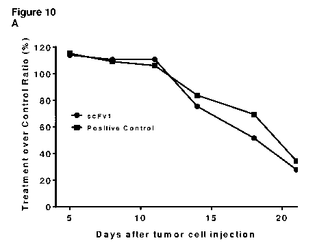
Figure 10 shows that scFv1 promotes tumor shrinkage in a HCC827 human lung
cancer model in
nude mice which have been administered with human peripheral blood mononuclear
cells
(PBMCs). A: Treatment (scFv1 or positive control IgG) over control (non-
binding scFv2) ratio
as defined in example 8. B: Tumor growth inhibition (scFv1 or positive control
IgG compared to
non-binding scFv2) as defined in example 8.
CA 03014001 2018-08-08
WO 2017/144681
PCT/EP2017/054367
Figure 11 shows that IgG_1 and IgG_2 are more effective than IgG_3 and IgG_4
in the
inhibition of the interaction between rhPD-L1 and rhPD-1. Background level was
determined in
the absence of IgG and PD-1.
Figure 12 shows that IgG_1 (A) has a tighter affinity than IgG_2 (B) in the
interaction between
5 IgG and PD-Li.
DETAILED DESCRIPTION
In order that the explanations on the binding members, nucleic acids, vectors,
host cells,
compositions, methods and uses disclosed herein may be more readily
understood, certain terms
are first defined.
Definitions
Unless otherwise defined, all other scientific and technical terms used in the
description, figures
and claims have their ordinary meaning as commonly understood by one of
ordinary skill in the
art. Although similar or equivalent methods and materials to those described
herein can be used
in the practice or testing of the binding members, nucleic acids, vectors,
host cells, compositions,
methods and uses disclosed herein, suitable methods and materials are
described below. All
publications, patent applications, patents, and other references mentioned
herein are incorporated
by reference in their entirety. In case of conflict, the present
specification, including definitions,
will prevail. The materials, methods, and examples are illustrative only and
not intended to be
limiting.
The term "administering", as used herein, refers to any mode of transferring,
delivering,
introducing, or transporting matter such as a compound, e.g. a pharmaceutical
compound, or
other agent such as an antigen, to a subject. Modes of administration include,
without being
limited to, parenteral administration, oral, rectal, systemic, intravenous,
subcutaneous,
urogenital, topical, intravitreal, intraocular, otic, intranasal, transdermal,
intradermal, dermal,
intraperitoneal, intramuscular, sublingual, or buccal administration.
Administration "in
combination with" further matter such as one or more therapeutic agents
includes simultaneous
(concurrent) and consecutive administration in any order.
As used herein, the terms "conservative modification" and "conservative
substitution" refer to a
modification and a substitution, respectively, that maintains physically,
biologically, chemically
CA 03014001 2018-08-08
WO 2017/144681
PCT/EP2017/054367
6
and/or functionally the properties with regard to the corresponding reference.
A molecule that
includes a sequence with conservative substitution for instance has a similar
size, shape, electric
charge, chemical properties, including a comparable ability to form covalent
or hydrogen bonds,
and/or comparable polarity. Such conservative modifications include, but are
not limited to, one
or more nucleobases and amino acid substitutions, additions and deletions.
For example, conservative amino acid substitutions include those in which the
amino acid
residue is replaced with an amino acid residue having a similar side chain.
For example, amino
acid residues being non-essential with regard to binding to an antigen can be
replaced with
another amino acid residue from the same side chain family, e.g. serine may be
substituted for
threonine. Amino acid residues are usually divided into families based on
common, similar side-
chain properties, such as:
1. nonpolar side chains (e.g., glycine, alanine, valine, leucine, isoleucine,
methionine),
2. uncharged polar side chains (e.g., asparagine, glutamine, serine,
threonine, tyrosine,
proline, cysteine, tryptophan),
3. basic side chains (e.g., lysine, arginine, histidine, proline),
4. acidic side chains (e.g., aspartic acid, glutamic acid),
5. beta-branched side chains (e.g., threonine, valine, isoleucine) and
6. aromatic side chains (e.g., tyrosine, phenylalanine, tryptophan,
histidine).
A conservative substitution can be taken to be a substitution of a first amino
acid within one of
the six groups above by a further amino acid within the same group of the six
groups. Preferred
conservative substitutions include:
1. Substituting alanine (A) by valine (V);
2. Substituting arginine (R) by lysine (K);
3. Substituting asp aragine (N) by glutamine (Q);
4. Substituting aspartic acid (D) by glutamic acid (E);
5. Substituting cysteine (C) by serine (S);
6. Substituting glutamic acid (E) by aspartic acid (D);
7. Substituting glycine (G) by alanine (A);
8. Substituting histidine (H) by arginine (R) or lysine (K);
9. Substituting isoleucine (I) by leucine (L);
10. Substituting methionine (M) by leucine (L);
11. Substituting phenylalanine (F) by tyrosine (Y);
12. Substituting serine (S) by threonine (T);
13. Substituting tryptophan (W) by tyrosine (Y);
CA 03014001 2018-08-08
WO 2017/144681
PCT/EP2017/054367
7
14. Substituting phenylalanine (F) by tryptophan (W);
and/or
15. Substituting valine (V) by leucine (L)
and vice versa. Other substitutions such as substituting proline (P) by
alanine (A) are also
permissible and can be determined empirically or in accord with other known
conservative or
non-conservative substitutions. A conservative substitution may also involve
the use of a non-
natural amino acid.
Non-conservative substitutions, i.e. exchanging members of one family against
members of
another family, may lead to substantial changes, e.g. with respect to the
charge, dipole moment,
size, hydrophilicity, hydrophobicity or conformation of the binding member,
which may alter the
binding activity, in particular if amino acids are affected that are essential
for binding to the
target molecule. A non-conservative substitution may also involve the use of a
non-natural
amino acid.
Conservative and non-conservative modifications can be introduced into
parental binding
members by a variety of standard techniques known in the art, such as
combinatorial chemistry,
site-directed DNA mutagenesis, PCR-mediated and/or cassette mutagenesis,
peptide/protein
chemical synthesis, introducing appropriate modifications into or constructing
a new nucleic acid
sequence encoding the binding member and/or a chemical reaction specifically
modifying
reactive groups in the parental binding member. The variants can be tested by
routine methods
for their chemical, biological, biophysical and/or biochemical properties.
Preferably, the
conservative amino acid substitution does not substantially change the
functional, and generally
also the structural characteristics of the parental sequence. Accordingly, the
binding
characteristics of a binding member that includes a conservative substitution
are at least
essentially unaltered. Furthermore, a conservative amino acid substitution
generally does not
substantially modify or disrupt a secondary structure of the parental
sequence.
The term "label" is used herein to refer to any substance the detection or
measurement of which,
either directly or indirectly, by physical or chemical means, is indicative of
the presence of a
selected target bioentity in a sample. Representative examples of useful
detectable labels
include, but are not limited to, molecules or ions directly or indirectly
detectable based on light
absorbance, fluorescence, reflectivity, light scatter, phosphorescence, or
luminescence
properties, molecules or ions detectable by their radioactive properties or
molecules or ions
detectable by their nuclear magnetic resonance or paramagnetic properties. A
label may in some
embodiments be a molecule that can be indirectly detected based on light
absorbance or
CA 03014001 2018-08-08
WO 2017/144681
PCT/EP2017/054367
8
fluorescence, for example, various enzymes which cause appropriate substrates
to convert, e.g.,
from non-light absorbing to light absorbing molecules, or from non-fluorescent
to fluorescent
molecules.
An "effective amount" or a "therapeutically effective amount" of an item such
as a compound,
including a binding member disclosed herein, is an amount ¨ either as a single
dose or as part of
a series of doses ¨ which at the dosage regimen applied yields the desired
therapeutic effect, i.e.,
to reach a certain treatment goal. A therapeutically effective amount is
generally an amount
sufficient to provide a therapeutic benefit in the treatment or management of
the relevant
pathological condition, or to delay or minimize one or more symptoms
associated with the
presence of the condition. The dosage will depend on various factors including
patient and
clinical factors (e.g., age, weight, gender, clinical history of the patient,
severity of the disorder
and/or response to the treatment), the nature of the disorder being treated,
the particular
composition to be administered, the route of administration, and other
factors.
The term "essentially consists of' is understood to allow the presence of
additional components
in a sample or a composition that do not affect the properties of the sample
or a composition. As
an illustrative example, a pharmaceutical composition may include excipients
if it essentially
consists of an active ingredient.
Within the scope of the present disclosure, the term "antibody" refers to a
full-length
immunoglobulin as well as to fragments thereof. Such a full-length
immunoglobulin may be
monoclonal, polyclonal, chimeric, humanized, veneered and/or a human antibody.
A chimeric
antibody may e.g. include a constant region of a different species and/or a
different isotype or be
an artificial bispecific or multispecific construct, such as e.g. a quadroma,
a knob-into-hole
(KIH) or CrossMab or a DuoBody. The term also encompasses constructs where
full-length
immunoglobulins are fused to an antibody fragment or a non-antibody scaffold.
Exemplary
examples thereof, without being limited to, include BslAb, Bs2Ab, Bs3Ab,
Bs4Ab, TslAb and
Ts2Ab as described by Dimasi N. et al (2009), JMB 393, 672-692. Further
chimeric antibodies
include DVD-Ig, IgG-scFab, scFab-dsscFv, Fv2-Fc, scFv-KIH, FynomABs, or BiTE-
KIH. An
antibody as disclosed herein may in some embodiments be glycosylated in other
embodiments,
the antibody is not glycosylated.
By "fragment" in reference to a polypeptide such as an antibody or a
proteinaceous binding
molecule is meant any amino acid sequence present in a corresponding
polypeptide, as long as it
CA 03014001 2018-08-08
WO 2017/144681
PCT/EP2017/054367
9
is shorter than the full-length immunoglobulin sequence and as long as it is
capable of
performing the function of interest of the protein - in the case of an
antibody specifically binding
to the desired target, e.g. antigen (such as PD-L1). The term "antibody
fragment" refers to a
portion of an antibody, often the hypervariable region and portions of the
surrounding heavy and
light chains, that displays specific binding affinity for a particular target,
typically a molecule. A
hypervariable region is a portion of an antibody that physically binds to the
polypeptide target.
An antibody fragment thus includes or consists of one or more portions of a
full-length antibody
retaining the targeting specificity of the antibody. Such antibody fragment
may for instance lack
at least partially the constant region (Fc region) of the full-length
antibody. In some
embodiments, an antibody fragment is produced by digestion of the full-length
antibody. An
antibody fragment may also be a synthetic or recombinant construct that
contains one or more
parts of the antibody (see e.g., Holliger P and Hudson J. Engineered antibody
fragments and the
rise of single domains. Nature Biotechnol. 2005, vol. 23, 9, p.1126). Examples
of an antibody
fragment include, but are not limited to, an scFv, a Fab, a Fv, a Fab', a
F(ab')2 fragment, a scFab,
a dAb, a VHH, a nanobody, a V(NAR) or a so called minimal recognition unit, a
diabody, a
single-chain diabody (scDb), a tandem scDb (Tandab), a linear dimeric scDb (LD-
scDb), a
circular dimeric scDb (CD-scDb), a BiTE (also called bispecific T-cell
engager, tandem scFv or
tandem di-scFv), a DART, a tandem tri-scFv, a tri(a)body, bispecific Fab2, di-
miniantibody,
tetrabody, di-diabody, or scFab-dsscFv.
A "single chain variable fragment" or a "single chain antibody" or a "scFv"
are examples of a
type of antibody fragment. A scFv is a fusion protein that includes the VH and
VL domains of
an antibody connected by a linker. It thus lacks the constant Fc region which
is present in a full-
length antibody.
A "binding member" as used herein refers to a proteinaceous binding molecule
comprising one
or more CDRs and optionally the variable light and/or heavy chains as
disclosed herein. As such,
the term "binding member" comprises antibodies (i.e. full-length
immunoglobulins and antibody
fragments as defined above), proteinaceous non- antibody scaffolds and/or
other binding
compounds. In some embodiments, the non-antibody scaffolds comprise one or
more CDR
sequences as disclosed herein. Such binding member can be monovalent or
multivalent, i.e.
having one or more antigen binding sites. Non-limiting examples of monovalent
binding
members include scFv, Fab, scFab, dAb, VHH, V(NAR) (or a so called minimal
recognition
unit), DARPins, affilins and nanobodies. A multivalent binding member can have
two, three,
CA 03014001 2018-08-08
WO 2017/144681
PCT/EP2017/054367
four or more antigen binding sites. Full-length immunoglobulins, F(ab')2
fragments, bis-scFv (or
tandem scFvor BiTE), DART, diabodies, scDb, DVD-Ig, IgG-scFab, scFab-Fc-scFab,
IgG-scFv,
scFv-Fc, scFv-fc-scFv, Fv2-Fc, FynomABs, quadroma, CrossMab, DuoBody,
triabodies and
tetrabodies are non-limiting examples of multivalent binding members; in the
exemplary
5 multivalent binding members, two binding sites are present, i.e. the
binding member is bivalent.
In some embodiments, the multivalent binding member is bispecific, i.e. the
binding member is
directed against two different targets or two different target sites on one
target molecule.
Bispecific antibodies are, e.g., reviewed in Muller D. and Kontermann R.E.
Bispecific
antibodies. Edited by Diibel S. Weinheim: Wiley-VCH, 2007. ISBN 3527314539. p.
345. In
10 some embodiments, the multivalent binding member includes more than two,
e.g., three or four
different binding sites for three or four, respectively, different antigens.
Such binding member is
multivalent and multispecific, in particular tri- or tetra-specific,
respectively.
"Non-antibody scaffolds" are antigen-binding polypeptides which are e.g.
described in Fielder
M. and Skerra A. Non-antibody scaffolds. Edited by Diibel S. Weinheim: Wiley-
VCH, 2007.
ISBN 3527314539. p. 467; or Gilbreth R.N. and Koide S. Structural insights for
engineering
binding proteins based on non-antibody scaffolds. Curr. Opin. Struct. Biol.
2012, vol. 22, p.413.
Non-limiting examples include affibodies, affilin molecules, AdNectins,
muteins based on
polypeptides of the lipocalin family (Anticalins ), DARPins, Knottins, Kunitz-
type domains,
Avimers, fynomers, Tetranectins and trans-bodies. Avimers contain so called A-
domains that
occur as strings of multiple domains in several cell surface receptors
(Silverman J., et al., Nature
Biotechnol. 2005, vol. 23, p.1556). Tetranectins, derived from the respective
human
homotrimeric protein, likewise contain loop regions in a C-type lectin domain
that can be
engineered for desired binding (ibid.).
A binding member as disclosed herein may be PEGylated or hyperglycosylated if
desired, see
also below. In some embodiments, a binding member is a fusion protein of one
of the exemplary
proteinaceous binding molecules above and an albumin-binding domain, for
instance an
albumin-binding domain of streptococcal protein G. In some embodiments, a
binding member is
a fusion protein of an antibody fragment, such as a single-chain diabody, and
an antibody
binding domain, for instance a bacterial antibody binding domain. As an
illustrative example, a
single-chain diabody may be fused to domain B of staphylococcal protein A as
described by
Unverdorben et al. (Protein Eng., Design & Selection, 2012, vol. 25, p.81).
CA 03014001 2018-08-08
WO 2017/144681
PCT/EP2017/054367
11
The "IC50" or "half-maximum inhibitory concentration" is a measure of
antagonist potency and
describes quantitatively the effectiveness of a compound to inhibit a
biological or biochemical
function. This value accordingly indicates how much of a certain item, such as
a binding
member, is needed to inhibit by 50% a certain biological or biochemical
process or function.
Although no direct indicator of affinity, the IC50 and the K values are
correlated and can be
determined via the Cheng-Prusoff equation (Cheng Y. and Prusoff W.H.
Relationship between
the inhibition constant (Ki) and the concentration of inhibitor which causes
50 per cent inhibition
(IC50) of an enzymatic reaction. Biochem. Pharmacol. 1973, vol. 22, p.3099;
Rammes G., et al.,
PLOSONE 2009, vol. 4, p. 1-14; Zhen J., et al., Concentration of receptor and
ligand revisited in
a modified receptor binding protocol for high-affinity radioligands: [3H]
spiperone binding to D2
and D3 dopamine receptors. J. Neurosci. Meth. 2010, vol. 188, p.32).
The term "framework" (FR) refers to the scaffold of the variable antibody
domain, either the
variable light chain (VL) or variable heavy chain (VH), embedding the
respective CDRs. A VL
and/or VH framework typically includes four framework sections, FR1, FR2, FR3
and FR4,
flanking the CDR regions. Thus, as known in the art, a VL has the general
structure: (FR-L1) ¨
(CDR-L1) ¨ (FR-L2) ¨ (CDR-L2) ¨ (FR-L3) ¨ (CDR-L3) ¨ (FR-L4), whereas a VH has
the
general structure: (FR-H1) ¨ (CDR-H1) ¨ (FR-H2) ¨ (CDR-H2) ¨ (FR-H3) ¨ (CDR-
H3) ¨ (FR-
H4).
The term "CDR" refers to the hypervariable regions of the antibody which
mainly contribute to
antigen binding. Typically, an antigen binding site includes six CDRs,
embedded into a
framework scaffold. Herein, the CDRs of the VL are referred to as CDR-L1, CDR-
L2 and CDR-
L3 whereas the CDRs of the VH are referred to as CDR-H1, CDR-H2 and CDR-H3.
These can
be identified as described in KABAT, E.A., et al. Sequences of Proteins of
Immunological
Interest. 5th edition. Edited by U.S. DEPARTMENT OF HEALTH AND HUMAN SERVICES.
NIH Publications, 1991. p. 91-3242. CDR-H1 as used herein, however, differs
from the Kabat
definition in that it starts with position 27 and ends prior to position 36
(AHo positions 28 to 42,
inclusive).
As used herein, the numbering system to identify amino acid residue positions
in the VH and VL
of the antibody corresponds to the "AHo"-system described by Honegger A. and
Pliickthun A.
Yet another numbering scheme for immunoglobulin variable domains: An automatic
modelling
and analysis tool. J. Mol. Biol. 2001, vol. 309, p.657. The publication
further provides
CA 03014001 2018-08-08
WO 2017/144681
PCT/EP2017/054367
12
conversion tables between the AHo and the Kabat system (Kabat E.A. et al.,
Sequences of
Proteins of Immunological Interest. 5th edition. Edited by U.S. Department of
Health and Human
Services. NIH Publications, 1991. No. 91-3242).
"Humanized" antibodies refer to antibodies that include one or more, typically
all six CDR
regions of a non-human parent antibody or variants thereof or synthetic CDRs,
and of which the
framework is, e.g., (i) a human framework, potentially including one or more
framework
residues of the non-human parent antibody, or (ii) a framework from a non-
human antibody
modified to increase similarity to naturally produced human frameworks.
Methods of
humanizing antibodies are known in the art, e.g. Leger 0., and Saldanha J.
Antibody Drug
Discovery. Edited by Wood C. London: Imperial College Press, 2011. ISBN
1848166281. p.1-
23.
The term "isolated" indicates that matter such as a peptide, a nucleic acid
molecule or a cell has
been removed from its normal physiological environment, e.g. a natural source,
or that a peptide
or nucleic acid is synthesized. Use of the term "isolated" indicates that a
naturally occurring
sequence has been removed from its normal cellular (e.g., chromosomal)
environment. Thus, the
sequence may be in a cell-free solution or placed in a different cellular
environment. "Isolated"
in reference to a polypeptide or nucleic acid molecule means a polymer of two
or more amino
acids or nucleotides coupled to each other, including a polypeptide or nucleic
acid molecule that
is isolated from a natural source or that is synthesized. The term "isolated"
does not imply that
the sequence is the only amino acid chain or nucleotide chain present, but
that it is essentially
free of, e.g., non-amino acid material and/or non-nucleic acid material,
respectively, naturally
associated with it. An "isolated cell" refers to a cell that is separated from
the molecular and/or
cellular components that naturally accompany the cell.
The term "identity" as used herein refers to the sequence match between two
proteins or nucleic
acids. The protein or nucleic acid sequences to be compared are aligned for
maximum
correspondence over a comparison window, for example using bioinformatics
tools such as
EMBOSS Needle (pair wise alignment; available at www.ebi.ac.uk or by manual
alignment and
visual inspection. When the same position in the sequences to be compared is
occupied by the
same nucleobase or amino acid residue, then the respective molecules are
identical at that very
position. Accordingly, the "percent identity" is a function of the number of
matching positions
divided by the number of positions compared and multiplied by 100%. For
instance, if 6 out of
10 sequence positions are identical, then the identity is 60%. Aligning
sequences for maximum
CA 03014001 2018-08-08
WO 2017/144681
PCT/EP2017/054367
13
correspondence may require introducing gaps. The percent identity between two
protein
sequences can, e.g., be determined using the Needleman and Wunsch algorithm
(Needlemann
S.B. and Wunsch C.D. A general method applicable to the search for
similarities in the amino
acid sequence of two proteins. J. Mol. Biol. 1970, vol. 48, p.443) which has
been incorporated
into EMBOSS Needle, using a BLOSUM62 matrix, a "gap open penalty" of 10, a
"gap extend
penalty" of 0.5, a false "end gap penalty", an "end gap open penalty" of 10
and an "end gap
extend penalty" of 0.5, or a method of aligning sequences manually introducing
gaps in a manner
which maximises identity can be used. Thus, in one embodiment, sequences
disclosed herein are
aligned by manually introducing gaps in a manner which maximises sequence
identity. Two
molecules having the same primary amino acid or nucleic acid sequence are
identical
irrespective of any chemical and/or biological modification. For example, two
antibodies having
the same primary amino acid sequence but different glycosylation patterns are
identical by this
definition. In case of nucleic acids, for example, two molecules having the
same sequence but
different linkage components such as thiophosphate instead of phosphate are
identical by this
definition. A sequence being longer than any of the sequences provided herein,
for example
because it comprises several variable domains or one or more constant domains,
shall
nevertheless be identical to the reference sequence disclosed herein if
sequence identity over a
comparison window is given. A comparison window as used herein includes the
entire sequence
as claimed. Similarly, nucleobases that differ only because of exocyclic
modifications, for
example cytosine and 5-methyl-cytosine, are identical by this definition.
The term "nucleic acid molecule" as used herein refers to any nucleic acid in
any possible
configuration, such as single stranded, double stranded or a combination
thereof. Examples of
nucleic acids include for instance DNA molecules, RNA molecules, analogues of
the DNA or
RNA generated using nucleotide analogues or using nucleic acid chemistry,
locked nucleic acid
molecules (LNA), protein nucleic acids molecules (PNA), alkylphosphonate and
alkylphosphotriester nucleic acid molecules and tecto-RNA molecules (e.g. Liu
B. et al., J. Am.
Chem. Soc. 2004, vol. 126, 4076). LNA has a modified RNA backbone with a
methylene bridge
between C4' and 02', providing the respective molecule with a higher duplex
stability and
nuclease resistance. Alkylphosphonate and alkylphosphotriester nucleic acid
molecules can be
viewed as a DNA or an RNA molecule, in which phosphate groups of the nucleic
acid backbone
are neutralized by exchanging the P-OH groups of the phosphate groups in the
nucleic acid
backbone to an alkyl and to an alkoxy group, respectively. DNA or RNA may be
of genomic or
synthetic origin and may be single or double stranded. Such nucleic acid can
be e.g. mRNA,
CA 03014001 2018-08-08
WO 2017/144681
PCT/EP2017/054367
14
cRNA, synthetic RNA, genomic DNA, cDNA synthetic DNA, a copolymer of DNA and
RNA,
oligonucleotides, etc. A respective nucleic acid may furthermore contain non-
natural nucleotide
analogues and/or be linked to an affinity tag or a label.
Many nucleotide analogues are known and can be used in nucleic acids used in
the methods
disclosed in this specification. A nucleotide analogue is a nucleotide
containing a modification at
for instance the base, sugar, or phosphate moieties. As an illustrative
example, a substitution of
2'-OH residues of siRNA with 2'F, 2'0-Me or 2'H residues is known to improve
the in vivo
stability of the respective RNA. Modifications at the base moiety may be a
natural or a synthetic
modification of A, C, G, and T/U, a different purine or pyrimidine base, such
as uracil-5-yl,
hypoxanthin-9-yl, and 2-aminoadenin-9-yl, as well as a non-purine or a non-
pyrimidine
nucleotide base. Other nucleotide analogues serve as universal bases. Examples
of universal
bases include 3-nitropyrrole and 5-nitroindole. Universal bases are able to
form a base pair with
any other base. Base modifications often can be combined with for example a
sugar
modification, such as for instance 2'-0-methoxyethyl, e.g. to achieve unique
properties such as
increased duplex stability.
As used in this document, the expression "pharmaceutically acceptable" refers
to those active
compounds, materials, compositions, carriers, and/or dosage forms which are,
within the scope
of sound medical judgment, suitable for use in contact with the tissues of
human beings and
animals without excessive toxicity, irritation, allergic response, or other
problems or
complications, commensurate with a reasonable benefit/risk ratio.
The term "preventing" in the medical/physiological context, i.e. in the
context of a physiological
state, refers to decreasing the probability that an organism contracts or
develops an abnormal
condition.
"Similar" protein sequences are those which, when aligned, share similar amino
acid residues
and most often, but not mandatorily, identical amino acid residues at the same
positions of the
sequences to be compared. Similar amino acid residues are grouped by chemical
characteristics
of the side chains into families. These families are described above for
"conservative amino acid
substitutions". The "percent similarity" between sequences is the number of
positions that
contain identical or similar residues at the same sequence positions of the
sequences to be
compared divided by the total number of positions compared and multiplied by
100%. For
CA 03014001 2018-08-08
WO 2017/144681
PCT/EP2017/054367
instance, if 6 out of 10 sequence positions have identical amino acid residues
and 2 out of 10
positions contain similar residues, then the sequences have 80% similarity.
The similarity
between two sequences can e.g. be determined using EMBOSS Needle. A sequence
being longer
than any of the sequences provided herein, for example because it comprises
several variable
5 domains or one or more constant domains, shall nevertheless be similar to
the reference sequence
disclosed herein if sequence similarity over a comparison window is given. A
comparison
window as used herein includes the entire sequence as claimed.
The term "specific" as used in this document is understood to indicate that a
binding member or
10 a binding compound binds to a defined target such as PD-Li with an
equilibrium binding
constant KD of < 10-6 molar. This constant can be determined, e.g. using
Quartz Crystal
Microbalance (QCM) in an Attana instrument, Surface Plasmon Resonance (SPR)
technology in
a BIACORE instrument or Kinetic Exclusion Assay (KinExA ).
15 The terms "stratifying" and "stratification" as used herein indicate
that an individual is assigned
to a certain group according to characteristics matching the respective group
such as a
corresponding probability of responding to a binding member disclosed herein.
The groups may
be used, for example, for testing, prescribing, adjusting dosing, suspending
or abandoning a
binding member. Accordingly, in some embodiments of a method or use according
to the
invention a subject may be stratified into a subgroup of a clinical trial of a
therapy.
The term "subject" as used herein, also addressed as an individual, refers to
a human or non-
human animal, generally a mammal. A subject may be a mammalian species such as
a rabbit, a
mouse, a rat, a guinea pig, a hamster, a dog, a cat, a pig, a cow, a goat, a
sheep, a horse, a
monkey, an ape or a human. Thus, the methods, uses and compositions described
in this
document are applicable to both human and veterinary disease. As explained in
more detail
below, the sample may be obtained from the subject. It is thus understood that
conclusions
drawn from expression levels in the sample and decisions based thereon concern
the subject
from whom/which the sample has been taken. Further, while a subject is
typically a living
organism, a method or use described in this document may also be used in post-
mortem analysis.
Where the subject is a living human who is receiving medical care for a
disease or condition, it is
also addressed as a "patient".
CA 03014001 2018-08-08
WO 2017/144681
PCT/EP2017/054367
16
The terms "treatment" and "treating" as used herein, include a prophylactic or
preventative
measure having a therapeutic effect and/or preventing, slowing down (lessen),
or at least
partially alleviating or abrogating an abnormal, including pathologic,
condition in the organism
of a subject. Treatment according to the present disclosure involves the
administration of a
pharmaceutically effective amount of a molecule as described herein, i.e.
inter alia, the binding
member (such as an antibody), nucleic acid, vector or cell disclosed herein,
to a subject in need
thereof to prevent, cure, delay the onset and/or progression, reduce the
severity of, stabilize,
modulate, cure or ameliorate one or more symptoms of a PD-Li-related disorder.
Typically, the
binding member, nucleic acid, vector or host cell is provided in a
pharmaceutical composition
including those described herein. Those in need of treatment include those
already with the
disorder as well as those prone to having the disorder or those in whom the
disorder is to be
prevented (prophylaxis). Generally, a treatment reduces, stabilizes, or
inhibits progression of a
symptom that is associated with the presence and/or progression of a disease
or pathological
condition.
As used herein, "PD-Li" refers to the protein also known as "programmed cell
death ligand 1,"
"cluster of differentiation 274 (i.e., CD274)" or "B7 homolog 1 (i.e., B7-
H1)". The native
protein comprises two extracellular domains, a transmembrane domain, and a
cytoplasmic
domain. The term encompasses full-length and/or unprocessed PD-Li as well as
any
intermediate resulting from processing in the cell. PD-Li can exist as a
transmembrane protein
or as a soluble protein; thus, the term as used herein may refer to the full
length or the
extracellular domain of the protein. The term also encompasses naturally
occurring variants of
PD-L1, e.g., splice variants or allelic variants. The protein may additionally
contain a tag, such
as a his tag or Fc tag. The amino acid sequence of exemplary human full-length
PD-Li protein
can e.g. be found under NCBI protein database accession number NP_054862.The
term "hPD-
Ll" refers to human PD-Li and comprises natural hPD-L1 and recombinant human
rhPD-Li.
"rPD-Li" refers to recombinant PD-Li. Recombinant PD-Li may or may not have an
amino
terminal methionine residue, depending upon the method by which it is
prepared. "rhPD-Li"
refers to recombinant human PD-Li. Likewise, PD-Li may also be obtained by
isolation from
biological samples of human or non-human origin. rhPD-L1 may, e.g., be
obtained from RnD
Systems, USA, cat. no. 156-B7, or from Peprotech, USA, cat. no. 310-35.
"Monkey PD-Li"
refers to PD-Li of Rhesus macacque (Macaca mulatta). The amino acid sequence
of exemplary
monkey PD-Li protein can e.g. be found under NCBI protein database accession
number
NP_001077358. Monkey PD-Li may, e.g., be obtained from Sino Biological, China,
cat. no.
CA 03014001 2018-08-08
WO 2017/144681
PCT/EP2017/054367
17
90251-0O2H. "Rat PD-Li" refers to PD-Li of Rattus norvegicus (Norway rat). The
amino acid
sequence of exemplary rat PD-Li protein can e.g. be found under NCBI protein
database
accession number NP_001178883 Rat PD-Li may, e.g., be obtained from Sino
Biological,
China, cat. no. 80450-RO2H. "Mouse PD-Li" refers to PD-Li of Mus musculus. The
amino acid
sequence of exemplary mouse PD-Li protein can e.g. be found under NCBI protein
database
accession number NP_068693Mouse PD-Li may, e.g., be obtained from Sino
Biological, China,
cat. no. 50010-MO3H or from RnD Systems, USA, cat. no. 1019-B7-100.
"PD-1" is the programmed cell death protein 1, also known as CD279 is a cell
surface receptor
for PD-Li. PD-1 binds two ligands, PD-Li and PD-L2. PD-1 is a transmembrane
protein
including an extracellular domain followed by a transmembrane region and an
intracellular
domain. The term encompasses full-length and/or unprocessed PD-1 as well as
any intermediate
resulting from processing in the cell. PD-1 can exist as a transmembrane
protein or as a soluble
protein; thus, the term as used herein may refer to the full length or the
extracellular domain of
the protein. The term also encompasses naturally occurring variants of PD-1,
e.g., splice variants
or allelic variants. The protein may additionally contain a tag, such as a his
tag or Fc tag. The
amino acid sequence of exemplary human PD-1 protein can e.g. be found under
NCBI protein
database accession number NP_005009 The term "hPD-1" refers to human PD-1 and
comprises
its natural form (hPD-1) as well as the recombinant human form (rhPD-1). "rPD-
1" refers to
recombinant PD-1.
"CD80" refers to the cluster of differentiation 80, also known as B7-1, B7.1,
BB1, CD28LG,
CD28LG1, LAB7. It is a membrane receptor for CD28 and CTLA-4 as well as PD-Li
and
comprises extracellular domain followed by a transmembrane region and an
intracellular
domain. The term encompasses full-length and/or unprocessed CD80 as well as
any intermediate
resulting from processing in the cell. CD80 can exist as a transmembrane
protein or as a soluble
protein; thus, the term as used herein may refer to the full length or the
extracellular domain of
the protein. The term also encompasses naturally occurring variants of CD80,
e.g., splice
variants or allelic variants. The protein may additionally contain a tag, such
as a his tag or Fc tag.
The amino acid sequence of exemplary human CD80 protein can e.g. be found
under NCBI
protein database accession number NP_005182. CD80 may, e.g., be obtained from
RnD
Systems, USA, cat. no. 9050-B1-100.". The term "hCD80" refers to human CD80
and comprises
its natural form (hCD80) as well as the recombinant human form (rhCD80).
"rCD80" refers to
recombinant CD80.
CA 03014001 2018-08-08
WO 2017/144681
PCT/EP2017/054367
18
"PD-L2" refers to the protein also known as "Programmed cell death 1 ligand
2", "B7-DC", or
"CD273" (cluster of differentiation 273). The term as used herein encompasses
full-length and/or
unprocessed PD-L2 as well as any intermediate resulting from processing in the
cell. PD-L2 can
exist as a transmembrane protein or as a soluble protein; thus, the term as
used herein may refer
to the full length or the extracellular domain of the protein. The term also
encompasses naturally
occurring variants of PD-L2, e.g., splice variants or allelic variants. The
protein may additionally
contain a tag, such as a his tag or Fc tag. The amino acid sequence of
exemplary human full-
length PD-L2 protein can e.g. be found under NCBI protein database accession
number
NP_079515. PD-L2 may, e.g., be obtained from RnD Systems, USA, cat. no. 1224-
PL. The term
"rhPD-L2" refers to recombinant human PD-L2.
"B7-H3" refers to the protein also known as CD276 (Cluster of Differentiation
276). The term as
used herein encompasses full-length and/or unprocessed B7-H3 as well as any
intermediate
resulting from processing in the cell. B7-H3 can exist as a transmembrane
protein or as a soluble
protein; thus, the term as used herein may refer to the full length or the
extracellular domain of
the protein. The term also encompasses naturally occurring variants of B7-H3,
e.g., splice
variants or allelic variants. The protein may additionally contain a tag, such
as a his tag or Fc tag.
The amino acid sequence of exemplary human full-length B7-H3 protein can e.g.
be found under
NCBI protein database accession number NP_079516. B7-H3 may, e.g., be obtained
from RnD
Systems, USA, cat. no. 1027-B3. The term "rhB7-H3" refers to recombinant human
B7-H3.
A "variant" refers to an amino acid or nucleic acid sequence which differs
from the parental
sequence by virtue of addition (including insertions), deletion, modification
and/or substitution
of one or more amino acid residues or nucleobases while retaining at least one
desired activity of
the parent sequence disclosed herein. In the case of antibodies such desired
activity may include
specific antigen binding. Similarly, a variant nucleic acid sequence may be
modified when
compared to the parent sequence by virtue of addition, deletion and/or
substitution of one or
more nucleobases, but the encoded antibody retains the desired activity as
described above.
Variants may be naturally occurring, such as allelic or splice variants, or
may be artificially
constructed.
Nucleic acid hybridization reactions can be performed under conditions of
different stringency.
"Stringent conditions" are widely known and published in the art. Typically,
during the
CA 03014001 2018-08-08
WO 2017/144681
PCT/EP2017/054367
19
hybridization reaction a SSC-based buffer can be used in which SSC is 0.15 M
NaC1 and 15 mM
citrate buffer having a pH of 7Ø Increasing buffer concentrations and the
presence of a
denaturing agent increase the stringency of the hybridization step. For
example, high stringency
hybridization conditions can involve the use of (i) 50% (vol/vol) formamide, 5
x SSC (0.75 M
NaCl, 0.075 M sodium citrate), 50 mM sodium phosphate (pH 6.8), 0.1 % sodium
pyrophosphate, 5 x Denhardt's solution, sonicated salmon sperm DNA (50
mcg/m1), 0.1% SDS,
and 10% dextran sulfate at 42 C with washes at 42 C in 0.2 x SSC and 0.1% SDS;
(ii) 50%
(vol/vol) formamide with 0.1% bovine serum albumin/0.1% fico11/0.1%
polyvinylpyrrolidone/50
mM sodium phosphate buffer at pH 6.5 with 750 mM sodium chloride, 75 mM sodium
citrate at
42 C, or (iii) 10% dextran sulfate, 2 x SSC, and 50% formamide at 55 C,
followed by a high-
stringency wash consisting of 0.1 x SSC containing EDTA at 55 C. Additionally
or
alternatively, one, two or more washing steps using wash solutions of low
ionic strength and
high temperature can be included in the hybridization protocol using, for
example, 0.015 M
sodium chloride/0.0015 M sodium citrate/0.1% sodium dodecyl sulfate at 50 C.
The scope and meaning of any use of a term will be apparent from the specific
context in which
the term is used. Certain further definitions for selected terms used
throughout this document are
given in the appropriate context of the detailed description, as applicable.
The terms "comprising", "including," containing", "having" etc. shall be read
expansively or
open-ended and without limitation. Singular forms such as "a", "an" or "the"
include plural
references unless the context clearly indicates otherwise. Thus, for example,
reference to a
"vector" includes a single vector as well as a plurality of vectors, either
the same - e.g. the same
operon - or different. Likewise reference to a "cell" includes a single cell
as well as a plurality
of cells. Unless otherwise indicated, the term "at least" preceding a series
of elements is to be
understood to refer to every element in the series. The terms "at least one"
and "at least one of'
include for example, one, two, three, four, or five or more elements. It is
furthermore understood
that slight variations above and below a stated range can be used to achieve
substantially the
same results as a value within the range. Also, unless indicated otherwise,
the disclosure of
ranges is intended as a continuous range including every value between the
minimum and
maximum values.
Any embodiments specifically and explicitly recited herein may form the basis
of a disclaimer
either alone or in combination with one or more further embodiments.
CA 03014001 2018-08-08
WO 2017/144681
PCT/EP2017/054367
Unless defined otherwise, all technical and scientific terms used herein have
the same meaning
as commonly understood to one of ordinary skill in the art to which the
inventions described
herein belong. All publications and patents mentioned herein are incorporated
herein by
5 reference in their entirety for the purpose of describing and disclosing,
for example, the
constructs and methodologies that are described in the publications, which
might be used in
connection with the presently described inventions.
Various aspects of the disclosure are described in further detail in the
following subsections. It is
10 understood that the various embodiments, preferences and ranges may be
combined at will.
Further, depending of the specific embodiment, selected definitions,
embodiments or ranges may
not apply.
Binding Member Characterization
15 The binding members provided herein specifically bind PD-Li. The binding
specificity of the
binding member may be verified using techniques well known in the art. In some
embodiments,
the PD-Li is human PD-Li.
Binding of the binding member to PD-Li blocks the interaction of PD-Li with PD-
1 and/or
20 CD80, preferably with both PD-1 and CD80.
In some embodiments, the binding member provided herein is bivalent and binds
hPD-L1 with a
KD of lower than 10 pM as measured by KinExA@, preferably lower than 5 pM,
more preferably
about 3 pM, e.g., 2.9 pM, 2.8 pM or 2.7 pM. In some embodiments, such bivalent
binding
member is a full-length immunoglobulin. In one embodiment, said KinExA@
measurements for
bivalent binding members are done at room temperature. In one embodiment, the
binding
member is bivalent and the conditions as specified in Example 9 are used for
the KinExA@
measurements.
In some embodiments, the binding member provided herein is monovalent and
binds hPD-L1
with a KD of lower than 50 pM as measured by KinExA . Said KD is preferably
lower than 10
pM, such as about 9 pM, e.g., 9.0 pM, 8.9 pM, 8.8 pM or 8.7 pM. In one
embodiment, said
KinExA@ measurements for monovalent binding members are done at room
temperature. In one
embodiment, the binding member is monovalent and the conditions as specified
in Example 4
are used for the KinExA@ measurements.
CA 03014001 2018-08-08
WO 2017/144681
PCT/EP2017/054367
21
In some embodiments, said monovalent binding member is a scFv. In some
embodiments, said
monovalent binding member is an antibody fragment having a molecular weight of
about 60 kDa
or lower, such as about 55 kDa, 50 kDa, 45 kDa, 40 kDa, 35 kDa, 30 kDa or 27
kDa or lower. In
one embodiment, the molecular weight of the binding member is about 26 kDa,
such as 23, 24,
25, 26, or 27 kDa. In particular for cancer treatment, antibody fragments may
have advantages
over full length antibodies when targeting the PD-1 :PD-L1 signaling pathway
(Maute et al
(2015), PNAS, Nov 24; 112(47): E6506¨E6514). Due to their smaller size,
antibody fragments
are believed to penetrate deeper into tumors than is the case with full-length
antibodies, which
typically have a molecular weight of about 150 kDa, or any other antibody
format having a
similar molecular weight or higher. Another drawback associated with full-
length antibodies, in
particular IgGs, is their ability to mediate cytotoxic immune responses
through their Fc region
(e.g., ADCC/ADCP or CDC). This inhibition may be undesirable when targeting
the PD-1:PD-
Ll axis as both proteins are expressed on the surface of antitumor cytotoxic T
cells. Hence,
administering full-length monoclonal antibodies with functional Fc parts may
result in the
depletion of the very lymphocytes they are intended to activate. Treatment
with anti¨PD-1
antibodies was found to correlate with lower circulating T-cell numbers in
patients. Therefore,
antibody fragments having a small molecular weight (e.g., 60 kDa or lower,
such as about 55
kDa, 50 kDa, 45 kDa, 40 kDa, 35 kDa, 30 kDa or 27 kDa or lower) may offer a
more effective
alternative to full-length antibody therapeutics in the treatment of cancer.
Thus, in preferred
embodiments, the binding member is an antibody fragment selected from the
group consisting of
Fab, Fab', scFab, scFv, Fv fragment, nanobody, VHH, dAb, minimal recognition
unit, diabody,
single-chain diabody (scDb), BiTE or DART. Said formats have a molecular
weight below 60
kDa and do not comprise a Fc domain.
The size and/or architecture of the binding member has implications on its
half-life. To decrease
side-effects in a therapeutic setting, it may be advantageous to use binding
members with a short
half-life. This may e.g. be achieved by using a binding member lacking an Fc
part or having a
modified Fc part.
In certain applications it may be advantageous to induce cytotoxic immune
responses and/or
activate complement and therefore, presence of a Fc domain may be desired.
Thus, in one
embodiment, the binding member comprises an Fc domain which is capable of
mediating
cytotoxic immune responses. Non-limiting examples of binding members including
an Fc
domain are full-length immunoglobulins, DVD-Ig, scFv-Fc and scFv-Fc.scFv
fusions, IgG-
CA 03014001 2018-08-08
WO 2017/144681
PCT/EP2017/054367
22
scFab, scFab-dsscFv, Fv2-Fc, IgG-scFv fusions (such as e.g., bsAb, Bs lAb,
Bs2Ab, Bs3Ab,
TslAb, Ts2Ab, Knob-into-Holes (KiHs)), DuoBody, CrossMab.
In one embodiment, the binding member comprises an Fc domain and/or hinge
which is
modified such that it does not induce cytotoxic immune responses and/or or
does not activate
complement. Such inactivated Fc domain and/or hinge can be created by
introducing one or
more substitutions as thought in the art. Such binding member has the
advantage of increased
half-life when compared to antibody fragments having a molecular weight below
60 kDa,
without mediating mediate cytotoxic immune responses.
In one embodiment, the binding member derivative lacks an Fc domain. Exemplary
binding
member lacking an Fc domain are Fab, Fab', scFab, scFv, Fv fragment, nanobody,
VHH,
minimal recognition unit, diabody, single-chain diabody (scDb), tandem scDb
(Tandab), a linear
dimeric scDb (LD-scDb), circular dimeric scDb (CD-scDb), BiTE (also called
tandem di-scFv or
tandem scFv), tandem tri-scFv, tri(a)body, bispecific Fab2, di-miniantibody,
di-diabody, scFab-
dsscFv or DART.
In one embodiment, the binding member comprises a constant region selected
from the group
consisting of human IgGl, IgG2, IgG3 or IgG4 isotype.
In one embodiment, the binding member comprises a constant region selected
from the group
consisting of murine IgGl, IgG2A, IgG2B, IgG3 isotype.
In one aspect, the invention provides a binding member against PD-L1,
comprising
(a) at least one of the VH CDR sequences CDR-H1, CDR-H2 or CDR-H3 as set forth
in SEQ ID
NOs: 6, 7 and 8, respectively, or variants thereof; and/or
(b) at least one of the VL CDR sequences CDR-L1, CDR-L2 or CDR-L3 as set forth
in SEQ ID
NOs: 3, 4 and 5, respectively, or variants thereof. In some embodiments, the
binding member
includes at least CDR-L3 of SEQ ID NO: 5 and/or CDR-H3 of SEQ ID NO: 8, or
variants
thereof. In some embodiments, the binding member includes two CDR sequences
selected from
the group consisting of SEQ ID NOs: 6, 7 and 8, or variants thereof. In some
embodiments, the
binding member includes two CDR sequences selected from the group consisting
of SEQ ID
NOs: 3, 4 and 5, or variants thereof. In some embodiments, the binding member
comprises all
three CDRs of SEQ ID Nos: 6, 7 and 8 or variants thereof. In some embodiments,
the binding
CA 03014001 2018-08-08
WO 2017/144681
PCT/EP2017/054367
23
member comprises all three CDRs of SEQ ID Nos: 3, 4 and 5 or variants thereof.
Preferably, a
binding member includes all CDRs as set forth in SEQ ID NOs: 3-8, or variants
thereof.
The binding members provided herein possess a strong binding affinity for
human PD-Li. For
example, such binding member is capable of binding human PD-Li with an
equilibrium binding
constant KD of lower than 100 pM, preferably lower than 75 pM, 50 pM, 25 pM,
15 pM, most
preferably the KD is about 10 pM or lower, such as about 9 pM (e.g. 9.0 pM,
8.9 pM, 8.8 pM or
8.7 pM), 8 pM, 7 pM, 6 pM, 4 pM, 3 pM (2.9 pM, 2.8 pM or 2.7 pM) or lower.
Affinities can be
determined as described in the example section below or other methods
available in the art. In a
preferred embodiment, the affinity is determined by Kinetic Exclusion Assay
(KinExA ) at room
temperature, more preferably under the conditions indicated in Example 4 for
monovalent
binding members or Example 9 for bivalent binding members.
The binding member described herein may be, essentially consist of, or include
an antibody
(such as full-length immunoglobulin) or an antibody fragment (such as a Fab,
Fab', F(ab')2,
scFab, scFv, Fv fragment, nanobody, VHH or minimal recognition unit) or a non-
antibody
scaffold. Some binding members include one or more copies of variable light
and/or heavy
chains as disclosed herein, e.g., a format selected from the group consisting
of tandem scFvs,
diabodies or a single chain diabodies (scDb), tandem scDb, linear dimeric
scDb, circular dimeric
scDb, a BiTE; a tandem tri-scFv, a tri(a)body, bispecific Fab2, di-
miniantibody, IgGs, triabody,
tetrabody, scFv-Fc-scFv fusion, di-diabody, DVD-lg, IgG-scFab, scFab-dsscFv,
Fv2-Fc, or a
IgG-scFv fusion (including, without being limited to, BslAb, Bs2Ab, Bs3Ab,
Bs4Ab, TslAb
and Ts2Ab), quadroma, knob-into-hole (KIH), bispecific antibodies, CrossMabs
and DuoBodies.
In some embodiments, the binding member and in particular the monovalent
antibody fragment
above is a scFv. The VH and VL domains can be connected in either orientation,
VL-linker-VH
or VH-linker-VL, by a flexible linker. In a preferred embodiment, the
orientation is VL-linker-
VH, i.e. the light chain variable region being at the N-terminal end and the
heavy chain variable
region being at the C-terminal end of the polypeptide.
The binding member is preferably a humanized binding member, such as a
humanized antibody,
in particular a humanized antibody fragment, such as an scFv. The binding
member can be
monoclonal and/or chimeric.
CA 03014001 2018-08-08
WO 2017/144681
PCT/EP2017/054367
24
Thus, in some embodiments, the binding member includes a variable heavy chain
region of
subtype VH3 and/or a variable light chain region of subtype Vkappal.
In a preferred embodiment, the binding member comprises the VH sequence of SEQ
ID NO: 2 or
a variant thereof. Such variant has at least 85%, more preferably at least
90%, 91%, 92%, 93%,
94%, 95%, 96%, 97%, 98%, 99% or most preferably 100% sequence identity to SEQ
ID NO: 2.
Differently put, in one embodiment, the binding member comprises a VH sequence
having at
least 85%, more preferably at least 90%, 91%, 92%, 93%, 94%, 95%, 96%, 97%,
98%, 99% or
most preferably 100% sequence identity to SEQ ID NO: 2.
Additionally or alternatively, the binding member disclosed herein comprises
the VL sequence
of SEQ ID NO: 1, or a variant thereof. Such variant has at least 85%, more
preferably at least
90%, 91%, 92%, 93%, 94%, 95%, 96%, 97%, 98%, 99% or most preferably 100%
sequence
identity to SEQ ID NO: 1. Differently put, in one embodiment, the binding
member comprises a
VL sequence having at least 85%, more preferably at least 90%, 91%, 92%, 93%,
94%, 95%,
96%, 97%, 98%, 99% or most preferably 100% sequence identity to SEQ ID NO: 1.
In one embodiment, such binding member comprises a VH sequence having at least
85%, more
preferably at least 90%, 91%, 92%, 93%, 94%, 95%, 96%, 97%, 98%, 99% or most
preferably
100% sequence similarity to SEQ ID NO: 2. Additionally or alternatively, the
binding member
comprises a VL sequence having at least 85%, more preferably at least 90%,
91%, 92%, 93%,
94%, 95%, 96%, 97%, 98%, 99% or most preferably 100% sequence similarity to
SEQ ID NO:
1.
In a much preferred embodiment, the binding member comprises the VL as set
forth in to SEQ
ID NO: 1 and the VH as set forth in SEQ ID NO: 2. The framework sequences of
both SEQ ID
NO: 1 and SEQ ID NO: 2 are derived from a human immunoglobulin described in WO
03/097697 A (ESBATech AG). Its VH and VL framework sequences have been
modified for
humanization and stabilization of rabbit antibodies, see, e.g., WO 2009/155726
A (ESBATech,
AN ALCON BIOMEDICAL RESEARCH UNIT LLC); Borras, L., et al., JBC 2010, vol.
285(12), p. 9054.
In some embodiments, the binding member comprises one or more, preferably all
VL framework
sequences selected from the group consisting of SEQ ID Nos: 12 to 15.
CA 03014001 2018-08-08
WO 2017/144681
PCT/EP2017/054367
In some embodiments, the binding member comprises one or more, preferably all
VH framework
sequences selected from the group consisting of SEQ ID Nos: 16 to 19.
5 The binding member, for example in the case of a scFv or a bispecific
molecule such as a tandem
scFv, a diabody or a single chain diabody, may comprise a linker sequence. In
the case of a scFv,
such linker sequence typically has ten to about 25 amino acids. Usually, a
linker peptide is rich
in glycines, which confer flexibility, as well as serines and/or threonines
for improved solubility.
In a preferred embodiment, a (GGGGS)4 linker (SEQ ID NO: 10) or a variant
thereof is used.
10 Variations of said motif having two to five repeats may also be used.
Further suitable linkers are
described, e.g., in Alfthan, K., Protein Eng 1995, vol. 8(7), p. 725.
Thus, in one embodiment, such binding member comprises, has, essentially
consists of or
consists of an amino acid sequence that includes SEQ ID NO. 9. In some
embodiments, the
15 binding member comprises, has, essentially consists of or consists of an
amino acid sequence
that includes SEQ ID NO. 11.
In certain embodiments variants of the binding member provided herein are
contemplated. For
example, it may be desirable to improve antigen binding, antibody-dependent
cell-mediated
20 cytotoxicity (ADCC), complement-dependent cytotoxicity (CDC), to reduce
susceptibility to
proteolysis and/or susceptibility to oxidation, to increase stability or
solubility, to decrease
immunogenicity and/or to alter other biological, biochemical or biophysical
properties of the
binding member. In some embodiments, the variant does not show any improvement
over the
parent binding member. A variant may in some embodiments be a proteinaceous
molecule that
25 differs from a given binding member, in one, two, three, four, five or
more positions of its amino
acid sequence. Such difference may e.g., be a substitution, addition,
modification or deletion.
Variants of the binding members provided herein may be prepared by protein
and/or chemical
engineering, introducing appropriate modifications into the nucleic acid
sequence encoding the
binding member, or by protein/peptide synthesis. Any combination(s) of
deletions, substitutions,
additions, modifications and insertions can be made to the framework or to the
CDRs, provided
that the generated binding member possesses the desired characteristics for
which it can be
screened using appropriate methods. Of particular interest are substitutions,
preferably
conservative substitutions as described above.
CA 03014001 2018-08-08
WO 2017/144681
PCT/EP2017/054367
26
The binding member described herein may comprise one or more, such as two,
three, four, five,
six, seven, eight, nine, ten, eleven, twelve or more of such conservative
substitutions.
Non-conservative substitutions may lead to more substantial changes, e.g.,
with respect to the
charge, dipole moment, size, hydrophilicity, hydrophobicity or conformation of
the polypeptide.
In one embodiment, the binding member comprises one or more, such as two,
three, four, five,
six, seven, eight, nine, ten, eleven, twelve or more of such non-conservative
substitutions.
Modifications may be present in the CDRs and/or in the framework sequences.
For example, the
CDRs provided herein may comprise one, two, three, four, five or even more
modifications. For
example, the CDR-L1, CDR-L2 and CDR-L3 sequences taken as a whole are at least
75%,
preferably at least 76%, 77%, 78%, 79%, 80%, 85%, 90%, 91%, 92%, 93%, 94%,
95%, 96%,
97%, 98% or more preferably 99% identical to the CDRs provided herein, in
particular to SEQ
ID NOs: 3, 4, and 5. Additionally or alternatively, the CDR-H1, CDR-H2 and CDR-
H3
sequences taken as a whole are at least 80%, preferably at least 81%, 82%,
83%, 84%, 95%,
90%, 91%, 92%, 93%, 94%, 95%, 96%, 97%, 98% or more preferably 99% identical
to the
CDRs provided herein, in particular to SEQ ID NOs: 6, 7 and 8.
In one embodiment the CDR-L1, CDR-L2, CDR-L3, CDR-H1, CDR-H2 and CDR-H3 taken
as a
whole are at least 85%, preferably at least 90%, 91%, 92%, 93%, 94%, 95%, 96%,
97%, 98% or
more preferably 99% similar to the CDRs provided herein, in particular to SEQ
ID NOs: 3, 4 and
5. Additionally or alternatively, the CDR-L1, CDR-L2, CDR-L3, CDR-H1, CDR-H2
and CDR-
H3 taken as a whole are at least 85%, preferably at least 90%, 91%, 92%, 93%,
94%, 95%, 96%,
97%, 98% or more preferably 99% similar to the CDRs provided herein, in
particular to SEQ ID
NOs: 6, 7 and 8.
In one embodiment, a variant comprises one, two, three, or four substitutions
in any one of
sequence SEQ ID NOs: 1 to 19. In one embodiment, a variant comprises five,
six, seven, eight,
nine, ten, eleven or twelve substitutions in any one of sequence SEQ ID NOs:
1, 2, 9 or 11.
A particularly preferred type of variant is one where one or more entire CDRs
are replaced.
Typically, the CDR-H3 and CDR-L3 contribute most significantly to antigen
binding. For
example, the entire CDR-L1, CDR-L2, CDR-H1 and/or CDR-H2 may be replaced by a
different
CA 03014001 2018-08-08
WO 2017/144681
PCT/EP2017/054367
27
CDR of natural or artificial origin. In some embodiments, one or more CDRs are
replaced by an
alanine-cassette.
Additionally or alternatively, the VH of the antibody comprises solubility
enhancing point
mutations. W02009/155725 (ESBATech, a Novartis Company) describes a motif,
which has
proven to increase the overall solubility of the antibody. The residues are
placed at positions
located in the interface of the variable domain and the constant domain of an
antibody and
stabilize in particular antibody fragments such as scFv, lacking the constant
domain. In some
embodiments, in a variant of the binding member as disclosed herein one, two
or all three of the
following residues are present:
(i) serine (S) at heavy chain amino acid position 12 (according to AHo
numbering);
(ii) serine (S) or threonine (T) at heavy chain amino acid position 103
(according to AHo
numbering); and/or
(iii) serine (S) or threonine (T) at heavy chain amino acid position 144
(according to AHo
numbering). In a preferred embodiment, such variant has a serine at VH
position 12; a serine
at VH position 103; and a threonine at VH position 144 (all AHo numbering).
Additionally or alternatively, variants may include one or more point
mutations as claimed in
EP2158315B1, incorporated herein by reference.
Variants may e.g. include modifications as described in W02014/206561,
incorporated herein by
reference, in particular including VL framework sequences SEQ ID NOs. 15 to 22
of
W02014/206561.
Preferably, a variant binding member as described herein
(i) retains specific binding to PD-L1, in particular to hPD-Li; and/or
(ii) has a KD to human PD-Li of lower than 100 pM, preferably lower than 75
pM, 50 pM, 40
pM, 30 pM, 20 pM, more preferably of lower than 10 pM as measured by KinExA
(the
measurement preferably being made using the conditions described in Example 4
for monovalent
binding members or Example 9 for bivalent binding members); and/or
(iii) is not cross-reactive with mouse PD-Li and/or;
(iv) is cross-reactive to monkey PD-Li; and/or
(v) competes with the binding member disclosed herein for binding to PD-Li;
and/or
(vi) has at least 80%, preferably at least 85%, 90%, 95% or 97% sequence
identity to the
sequences disclosed herein.
CA 03014001 2018-08-08
WO 2017/144681
PCT/EP2017/054367
28
Variants may also be prepared by chain shuffling of light and heavy chains. A
single light chain
can be combined with a library of heavy chains to yield a library of variants.
In one embodiment,
said single light chain is selected from the group of VL sequences recited
above and/or said
library of heavy chains comprises one or more of the VH sequences recited
above. Likewise, a
single heavy chain can be combined with a library of light chains. Preferably,
said single heavy
chain is selected from the group of VH sequences recited above and/or said
library of light
chains comprises one or more of the VL sequences recited above.
A binding member can comprise any of the VL and/or the VH sequences mentioned
above.
Binding members having a single domain format, such as a nanobody or a VHH,
comprise only
one of either the VL or VH sequences mentioned above, preferably the VH
sequence.
Multivalent binding members, in particular F(ab')2 fragments, bis-scFv (also
known as tandem
scFv), diabodies, scDb, triabodies or tetrabodies and the like, preferably
bispecific binding
members, may comprise one or more of the VL sequences mentioned above and/or
one or more
of the VH sequences mentioned above.
The binding members of the instant invention, preferably the monovalent
antibody fragments,
more preferably the scFvs, are particularly stable. As used herein the term
"stability" refers to the
biophysical property of the polypeptide to remain monomeric in solution after
prolonged
incubation and/or incubation at elevated temperature. Unstable polypeptides
tend to dimerize or
oligomerize and even precipitate, thereby decreasing shelf-life and becoming
less suitable for
pharmaceutical applications.
The binding members provided herein and in particular the monovalent antibody
fragment above
remain monomeric at least to 85%, preferably at least to 90%, 91%, 92%, 93%,
94%, and most
preferably to 95% after being incubated at a concentration of 10 mg/ml in PBS
at pH 7.2 for 2
weeks at a temperature of 4 C, additionally or alternatively also when
incubated under the same
conditions at 22 C or 37 C. In some embodiments, the binding member and in
particular the
monovalent antibody fragment above remains monomeric at least to 85%,
preferably at least to
90%, 91%, 92%, 93%, 94%, 95%, 96% and most preferably to 97% after being
incubated at a
concentration of 10 mg/ml in PBS at pH 7.2 for 3 weeks at a temperature of 4
C, additionally or
alternatively also when incubated under the same conditions at 22 C or 37 C.
In some embodiments, the binding member is a scFv and forms less than 3% of
dimers after 1
week or after 2 weeks of storage at 37 C at a concentration of 10 mg/ml in PBS
at pH 7.2.
CA 03014001 2018-08-08
WO 2017/144681
PCT/EP2017/054367
29
The degree of monomers can, e.g., be determined by SE-HPLC (Size Exclusion
HighPerformance Liquid Chromatography). A suitable mobile phase for such
testing is, e.g.,
PBS at pH 7.2. The monomer content can be quantified by peak integration of
the UV280 signal
measured during the protein chromatography. A suitable system is, e.g., a
Dionex Summit HPLC
controlled by Chromeleon 6.8 software that also allows for subsequent
chromatogram analysis
and peak quantification.
The binding member, preferably the monovalent antibody fragment above, more
preferably the
scFv, may have a theoretical isoelectric point (pI) in the range of 4 to 10,
preferably 4 to 9, most
preferably about 7.6. The theoretical pI can, for example, be calculated by
using the ProtParam
tool on the ExPASy Server (available at http://web.expasy.org/protparam/; see
also
GASTEIGER E. et al. Protein Identification and Analysis Tools on the ExPASy
Server. (In) The
Proteomics Protocols Handbook. Edited by Walker J.M. Totowa: Humana Press
Inc., 2005.
ISBN 9781588295934. p. 571-607).
The binding member can be cross-reactive with PD-Li from non-human species
which has
advantages for testing the binding member in animal models. Preferably, the
binding member is
cross-reactive with monkey PD-Li. In some embodiments, the KD of a monovalent
binding
member at room temperature in scFv format to monkey PD-Li is about 3.3 pM as
measured by
KinExA , e.g. measured under the conditions indicated in Example 5. In some
embodiments,
the affinity of the binding member is at least as strong, more preferably at
least twice as strong
for monkey PD-Li as for human PD-Li. In some embodiments, the binding member
is not
cross-reactive to mouse PD-Li. Often, antibodies against a given human target
have lower
affinities to rodent orthologs which renders rodent in vivo animal data less
valuable. As the
binding members disclosed herein have comparable KD values for human and
monkey PD-L1,
in vivo animal data are expected to be more reflective of the disease in
humans. Additionally,
cross reactivity to monkey enables the use of monkey as a toxicology species.
In preferred embodiments, the binding member is not cross-reactive with other
members of the
B7 family, such as PD-L2 and/or B7-H3. Both proteins have high sequence
similarity to PD-Li
and therefore, binding to these B7 family members would raise safety concerns.
Thus, in some embodiments, a binding member specifically binding to PD-Li is
provided,
comprising at least one variable light chain of SEQ ID NO: 1 and at least one
variable heavy
CA 03014001 2018-08-08
WO 2017/144681
PCT/EP2017/054367
chain of SEQ ID NO: 2, wherein said binding member has an equilibrium binding
constant KD to
human PD-Li of lower than 10 pM. Preferably, said binding member remains
monomeric to at
least 95% in a scFv format after incubation for 1 week or 2 weeks at 37 C in
PBS at a
concentration of 10 mg/ml. More preferably, said binding member is not cross-
reactive to mouse
5 PD-Li.
The invention also provides a binding member competing with the binding
members disclosed
herein for binding to human PD-Li. For example, such competing (or cross-
blocking) binding
member may be neutralizing. Preferably, such competing binding member has an
equilibrium
10 binding constant (KD) for binding to human PD-Li of 250 pM or lower,
such as lower than about
100 pM, 40 pM, 30 pM, 20 pM 10 pM or lower than about 5 pM. Thus, in one
embodiment, the
binding member has a KD of less than about 5 pM.
As used herein, the term "competing" refers to the competition between binding
members for
15 binding to the same target. Competition can be determined by competitive
binding assays in
which the binding member of interest prevents or inhibits or reduces specific
binding of the
binding members disclosed herein to a common antigen (here, PD-Li or a
fragment thereof,
respectively). Such competitive binding assays are known in the art and
include, without being
limited to, solid phase direct or indirect radioimmunoassay (RIA) and solid
phase direct or
20 indirect enzyme immunoassay (ELISA). Typically, such assay involves the
use of purified
antigen bound to a solid surface, a binding member to be tested and the
reference binding
member as described herein. Competitive inhibition is measured by determining
the amount of
either (i) the reference binding member bound to the solid surface in the
presence of the binding
member to be tested, or (ii) the binding member to be tested bound to the
solid surface in the
25 presence of the reference binding member. A competing binding member may
bind (i) to the
same epitope as the reference binding member, (ii) to an overlapping epitope,
or (iii) to a
different epitope on the same target molecule but sterically hindering binding
of the reference
binding member to its target.
30 Usually, when a competing binding member is present in excess, it will
reduce specific binding
of the binding member as described herein to PD-L1, i.e. it cross-blocks
binding, by at least 40-
45%, 45-50%, 50-55%, 55-60%, 60-65%, 65-70%, 70-75% or 75% or more.
Preferably, binding
of the binding members described herein in presence of the competing binding
member is
reduced by at least 80-85%, 85-90%, 90-95%, 95-97%, or 97% or more.
CA 03014001 2018-08-08
WO 2017/144681
PCT/EP2017/054367
31
In one embodiment, the binding member is monovalent, such as a scFv or a Fab
fragment. In
another embodiment, the binding member is multivalent. Such multivalent
molecule can be
bivalent (such as a full-length antibody or a F(ab')2 fragment) or comprises
at least three target
binding sites. The multivalent binding member can be a bispecific antibody
such as, e.g. a
diabody, a single-chain diabody, a bis-scFv or a DART (see, e.g. Kontermann
R.E. Methods in
Mol. Biol. Edited by LO, B. Totowa, N.J.: Humana Press, 2004. ISBN 1588290921.
p. 227).
Said bispecific antibodies may well use shorter linkers then those described
above for scFv, i.e.,
having only one to three repeats of the basic motif of SEQ ID No.: 10 (see,
e.g., Holliger, P., et
al., PNAS, 1993, vol. 90(14), p.6444). In another embodiment, the multivalent
binding member
is a triabody, a minibody or tetrabody. Other examples of multivalent binding
members include,
without being limited to, single-chain diabodies, tandem scDb, linear dimeric
scDb, circular
dimeric scDb, BiTEs, tandem tri-scFv, a tri(a)bodies, bispecific Fab2, di-
miniantibodies, scFv-
Fc-scFv fusions, di-diabodies, DVD-Igs, IgG-scFab, scFab-dsscFv, Fv2-Fcs, or
IgG-scFv fusions
(including, without being limited to, BslAb, Bs2Ab, Bs3Ab, Bs4Ab, TslAb and
Ts2Ab,
quadroma, knob-into-hole (KIH), bispecific antibodies, CrossMabs and
DuoBodies).
A binding member according to the present disclosure may in some embodiments
include a
capture moiety such as a streptavidin binding tag, e.g. the STREP-TAGS
described in US
patent application US 2003/0083474, US patent 5,506,121 or 6,103,493. Further
examples of a
capture moiety include, but are not limited to, maltose-binding protein,
glutathione-S-transferase
(GST), calmodulin binding peptide (CBP), FLAG-peptide (e.g. of the sequence
Asp-Tyr-Lys-
Asp-Asp-Asp-Asp-Lys-Gly), the T7 epitope (Ala-Ser-Met-Thr-Gly-Gly-Gln-Gln-Met-
Gly),
maltose binding protein (MBP), the HSV epitope of the sequence Gln-Pro-Glu-Leu-
Ala-Pro-
Glu-Asp-Pro-Glu-Asp of herpes simplex virus glycoprotein D, the Vesicular
Stomatitis Virus
Glycoprotein (VSV-G) epitope of the sequence Tyr-Thr-Asp-Ile-Glu-Met-Asn-Arg-
Leu-Gly-Lys,
the hemagglutinin (HA) epitope of the sequence Tyr-Pro-Tyr-Asp-Val-Pro-Asp-Tyr-
Ala and the
"myc" epitope of the transcription factor c-myc of the sequence Glu-Gln-Lys-
Leu-Ile-Ser-Glu-
Glu-Asp-Leu.
A further example of a capture moiety is a metal chelator, which is capable of
binding a metal
ion. A respective capture moiety may be ethylenediamine,
ethylenediaminetetraacetic acid
(EDTA), ethylene glycol tetraacetic acid (EGTA), diethylenetriaminepentaacetic
acid (DTPA),
N,N-bis(carboxymethyl)glycine (also called nitrilotriacetic acid, NTA), 1,2-
bis(o-
CA 03014001 2018-08-08
WO 2017/144681
PCT/EP2017/054367
32
aminophenoxy)ethane-N,N,N,Nt-tetraacetic acid (BAPTA), 2,3-dimercapto-1-
propanol (-
dimmercaprol), porphine or heme. In line with the standard method of
immobilised metal
affinity chromatography used in the art, for example an oligohistidine tag is
capable of forming a
complex with copper (Cu2 ), nickel (Ni2 ), cobalt (Co2 ), or zink (Zn2 ) ions,
which can for
instance be presented for chromatography purposes by means of the chelator
nitrilotriacetic acid
(NTA).
In some embodiments, the binding member disclosed herein is less immunogenic
than a known
binding member against PD-Li. In some embodiments, the binding member
disclosed herein
binds to a different epitope than a known binding member against PD-Li. In
some embodiments,
the binding member disclosed herein has a different clearance rate than a
known binding
member against PD-Li. In some embodiments, the binding member disclosed herein
has an
increased resistance towards aggregations and/or protease degradation than a
known binding
member against PD-Li. In some embodiments, the binding member disclosed herein
has an
improved IC50 and/or EC50 than a known binding member against PD-Li. In some
embodiments,
the binding member disclosed herein has improved binding parameters such as
k., koff or KD
than a known binding member to PD-Li. In some embodiments, the binding member
disclosed
herein has a different species cross-reactivity pattern than a known binding
member against PD-
Ll. In some embodiments, the binding member has a different pH stability than
a known binding
member against PD-Li. In some embodiments, the binding member has a different
long term
stability at indicated temperatures than a known binding member against PD-Li.
In some
embodiments, the binding member shows a different tissue penetration
capability than a known
binding member against PD-Li. In some embodiments, the binding member has a
different
blocking efficacy of PD-Li interactions with its receptors PD-1 and/or CD80
than a known
binding member against PD-Li.
Also contemplated are binding members competing with the binding members
disclosed herein
for binding to PD-Li.
Nucleic Acids, vectors, host cells and method of production
A binding member as described herein may be encoded by a single nucleic acid
sequence or by a
plurality of nucleic acid sequences. In the case of a plurality of nucleic
acid sequences each
sequence may encode one variable region. In some embodiments, a nucleic acid
sequence may
encode two or more variable regions. Generally, a plurality of nucleic acid
sequences encodes
CA 03014001 2018-08-08
WO 2017/144681
PCT/EP2017/054367
33
the variable regions of a binding member. Typically, each variable region is
encoded by one
distinct nucleic acid sequence. The respective nucleic acid sequences encoding
the variable
regions may be included in a single nucleic acid molecule. In some embodiments
two or more
nucleic acid sequences encoding the variable regions are included in a single
nucleic acid
molecule. In some embodiments, each nucleic acid sequence encoding a variable
region is
included in a single distinct nucleic acid molecule. Accordingly, a plurality
of nucleic acid
molecules may be used in the production of a binding member, for example each
encoding at
least one variable region. A respective nucleic acid molecule may in some
embodiments define
an expression cassette. As indicated above, an expression cassette is a
nucleic acid molecule
capable of directing expression of a particular nucleotide sequence in an
appropriate host cell.
An expression cassette includes a promoter operatively linked to the
nucleotide sequence of
interest, which is operatively linked to one or more termination signals. It
may also include
sequences required for proper translation of the nucleotide sequence. The
coding region can
encode a polypeptide of interest and can also encode a functional RNA of
interest, including but
not limited to, antisense RNA or a non-translated RNA, in the sense or
antisense direction. The
expression cassette comprising the nucleotide sequence of interest can be
chimeric, meaning that
at least one of its components is heterologous with respect to at least one of
its other
components. The expression cassette can also be one that is naturally
occurring but has been
obtained in a recombinant form useful for heterologous expression. In some
embodiments,
however, the expression cassette is heterologous with respect to the host;
i.e., the particular
nucleic acid sequence of the expression cassette does not occur naturally in
the host cell and was
introduced into the host cell or an ancestor of the host cell by a
transformation event. The
expression of the nucleotide sequence in the expression cassette can be under
the control of a
constitutive promoter or of an inducible promoter that initiates transcription
only when the host
cell is exposed to some particular external stimulus. In the case of a
multicellular organism such
as a plant or an animal, the promoter can also be specific to a particular
tissue, organ, or stage of
development.
Knowing the sequence of the binding member or of its parts, cDNAs encoding the
polypeptide
sequence can be generated by methods well known in the art, e.g. by gene
synthesis. These
cDNAs can be cloned by standard cloning and mutagenesis techniques into a
suitable vector
such as an expression vector or a cloning vector. Optionally, the variable
light chain is encoded
by a separate vector than the variable heavy chain of the antibody. Further,
additional sequences
CA 03014001 2018-08-08
WO 2017/144681
PCT/EP2017/054367
34
such as a tag (e.g., a His-tag), a constant domain for the production of a Fab
or a full-length
antibody, a linker, the coding sequence of a second binding specificity or
another functional
polypeptide such as an enzyme to generate a fusion construct or a bispecific
molecule may be
included into the genetic construct.
Based on the cloning strategy chosen, genetic constructs may generate a
binding member having
one or more additional residues at the N-terminal or C-terminal end. For
example, an N-terminal
methionine derived from the start codon or an additional alanine may be
present in an expressed
polypeptide, unless it has been clipped off post-translationally. It is
therefore to be understood
that the antibodies disclosed herein comprise the disclosed sequences rather
than consist of them.
Thus, in one embodiment, the binding member comprises the sequence of SEQ ID
NO: 9. In
another embodiment, the binding member comprises the sequence of SEQ ID NO:
11. If the
binding member is a scFv having the orientation VH-linker-VL or any other
antibody fragment
where the VH is placed N-terminally, the VH sequence part of the molecule may
be N-
terminally methylated. Thus, in one embodiment, SEQ ID NO: 2 has an N-terminal
methionine.
Basic protocols of standard cloning, mutagenesis and molecular biology
techniques are described
in, e.g., Molecular Cloning, A Laboratory Manual (Green M. and Sambrook, J.
Molecular
Cloning: a Laboratory Manual. 4th edition. Cold Spring Harbor Laboratory,
2012. ISBN
1936113422.).
Further contemplated are isolated nucleic acids hybridizing with the nucleic
acids described
herein under stringent conditions.
Also contemplated are cells recombinantly expressing the binding members
disclosed herein.
Appropriate host cells for the expression of the genetic constructs can be
prokaryotic or
eukaryotic. Suitable prokaryotic host cells are gram-negative or gram-positive
and include
species of the Escherichia, Ervinia, Enterobacter, Klebsiella, Pseudomonas or
Bacillus families.
In some embodiments, the host cell is Escherichia coli, such as one or more of
E. coli strains
BL21 (DE3) (for example Invitrogen, USA, cat. no. C600003) and Origami Th4
2(DE3) (for
example Novagen, USA, cat. no. 71345-3).
If post-translational modifications such as glycosylation or phosphorylation
are desired, it may
be advantageous to use a eukaryotic host cell. For example, eukaryotic
microbes such as
CA 03014001 2018-08-08
WO 2017/144681
PCT/EP2017/054367
commonly used Saccharomyces cerevisiae or Pichia pastoris strains may serve as
a host cell.
Suitable examples of a host cells also include a plant or an animal cell, in
particular insect or
mammalian cells. Suitable mammalian cells include, without being limited to,
Chinese Hamster
Ovary Cells (CHO), Human Embryonic Kidney Cells (HEK), Human Umbilical Vein
5 Endothelial Cells (HUVEC) or NSO myeloma cells. Glycosylation in
prokaryotic host cells as
also been reported, see e.g. Jaffe S.R.P. et al., Curr. Opin. Biotechnol.
2014, vol. 30, p. 205.
The binding member can be produced by way of expression in a suitable host
cell. For example,
the expression vectors described above are introduced into a host cell by
standard techniques
10 such as electroporation or chemical transformation. The transformed
cells are then cultivated
under conditions adequate for recombinant protein expression, typically in
appropriate
nutritional media, optionally modified for inducing promotors, selecting
transformants, or
amplifying encoding sequences of interest. The binding member is recovered
from the culture
and optionally purified using standard techniques in the art. The yield of
recombinant protein
15 may be improved by optimizing media and culture conditions such as
temperature or oxygen
supply. In prokaryotes, the binding member can be produced in the periplasm,
intracellularly as
inclusion bodies or be secreted into the medium. Animal cells will typically
secrete the binding
member into the medium. Upon harvest, the protein can be purified using
methods well known
in the art such as gel filtration, ion exchange chromatography, reversed phase
chromatography,
20 hydrophobic interaction, mixed mode chromatography and/or affinity
chromatography.
In one embodiment, the binding member is produced in a cell-free system. This
typically
involves in vitro transcription followed by in vitro translation of nucleic
acid product templates
encoding a protein as described herein, e.g., plasmid DNA or PCR product
templates. For
25 example, crude lysates from growing cells are used, providing the
necessary enzymes as well as
the cellular protein synthesis machinery. The necessary building blocks such
as amino acids or
nucleobases as well as energy delivering molecules and others can be
exogenously supplied.
Cell-free expression systems can, for example, be based on lysed rabbit
reticulocytes (e.g.,
Rabbit Reticulocyte Lysate System, Promega, cat. no. L4540), HeLa cells (e.g.,
1-Step Human In
30 Vitro Translation Kit, 88881, Thermo Scientific), insect cells (e.g.,
EasyXpress Insect Kit II,
32561, Qiagen), wheat germs (e.g., Wheat Germ Extract, L4380, Promega), or
E.coli cells (e.g.,
PURExpress In Vitro Protein Synthesis Kit, E68005, NEB). Also, optimized cell-
free antibody
expression systems for improved disulfide bond generation can be used for
production.
Commercially available kits include insect cell lysates (e.g., EasyXpress
Disulfide Insect Kit,
CA 03014001 2018-08-08
WO 2017/144681
PCT/EP2017/054367
36
32582, Qiagen) or E.coli cell lysates (e.g., EasyXpress Disulfide E. coli Kit,
32572, Qiagen).
Cell-free protein synthesis has, e.g., the advantage of being fast, achieving
high product yields,
allowing for easy modification of reaction conditions, forming a low degree of
or even no
byproducts. Cell-free protein synthesis may involve biological and/or chemical
steps which
cannot be conducted in purely biological or chemical production systems. For
example, non-
natural or chemically-modified amino acids can be incorporated into the
protein at desired
positions. ScFv- toxin fusion proteins have been successfully produced in cell-
free systems
(Nicholls, P. J., et al., JBC 1993, vol. 268, pp. 5302-5308). Thus, in one
embodiment a method
of producing the binding member described herein is provided, which includes
the steps of (a)
providing a cell-free system, (b) providing a nucleic acid product template
encoding the binding
member above above, (c) allowing for transcription and translation of the
nucleic acid product
template; (d) recovering; and optionally (e) purifying the binding member,
respectively.
Additionally or alternatively, a method of producing the binding member
described herein
includes at least one step of chemical synthesis. For example, the method may
be entirely
chemical. In another embodiment, the cell-based or the cell-free production
systems described
above include such at least one step of chemical synthesis.
In some embodiments, a binding member as described herein is produced in a
cell-based system
using an expression vector for intracellular expression in E. coli. Upon
expression, the
polypeptide is generated as an inclusion body within the host cell which is
separated from further
cell particles followed by solubilisation in a denaturing agent such as
guanidine hydrochloride
(GndHC1) and refolded by renaturation procedures well known to the skilled
person.
The desired binding member may also be produced in a transgenic animal. A
suitable transgenic
animal may be obtained according to standard methods, for example including
the steps of (i)
making the transgenic embryo, e.g. by micro injecting DNA constructs that
include the coding
sequence of the binding members as well as suitable control sequences into
eggs; (ii) transferring
the eggs into pseudo-pregnant recipient females; (iii) monitoring gestation or
pregnancy; and (iv)
selecting a descendant expressing the desired antibody.
It is to be understood that the nucleic acids, vectors, host cells and method
of production
described above also apply to the binding members (insofar as they are a
protein) described
herein.
CA 03014001 2018-08-08
WO 2017/144681
PCT/EP2017/054367
37
Further contemplated herein are cells expressing chimeric antigen receptors
(CARs). CAR
expressing cells have found ample use in cancer treatment. Such cells, either
of autologous or
allogeneic origin, are genetically modified to express CARs, e.g. by traducing
the cells with
lentiviral vectors. Cells are commonly T cells, whereas NK cells have also
found use. A CAR
typically has several sections, comprising an antigen binding domain, a
spacer, a transmembrane
domain, a costimulatory signaling domain, and a signaling domain.
The extracellular antigen-binding domain specifically recognizes a given
target protein, usually
on a cancer cell. Upon binding to the target, the CAR cell is activated and
also kept in proximity
to the cancer cell. The antigen-binding domain is connected via a spacer to a
transmembrane
domain which in turn is connected to the intracellular costimulatory signaling
domain. The
length of the spacer may have to be optimized, depending on the
characteristics of the antigen-
binding domain and its target protein. Binding of the target to the cancer
cell triggers as
conformational change that leads to an activation signal by the signaling
domain, e.g., a CD3
zeta signaling domain. The costimulatory signaling domain, typically located
between the
transmembrane domain and the signaling domain, serves in amplifying the
activation signal.
Exemplary embodiments of costimulatory signaling domains are CD28 or 4-1BB.
In some embodiments, the antigen binding domain comprises the VL and/or VH
sequences as
described herein. In some embodiments, the antigen binding domain comprises a
scFv as
described herein.
In some embodiments, the CAR expressing cell is an "armored CAR" cell, i.e. a
CAR expressing
cell which secretes soluble proteins to modify the immune response within the
tumor
microenvironment of the subject to which the CAR cells were administered. In
some
embodiments, such cell excretes a binding member as described herein, in
particular a scFv.
Chemical and/or biological modifications
In one aspect, the binding member disclosed herein is chemically and/or
biologically modified.
Such modification may include, but is not limited to, glycosylation,
PEGylation, HESylation,
Albumin fusion technology, PASylation, labelling with dyes and/or
radioisotopes, conjugation
with enzymes and/or toxins, phosphorylation, hydroxylation and/or sulfation.
Likewise, any
CA 03014001 2018-08-08
WO 2017/144681
PCT/EP2017/054367
38
binding member, the nucleic acid sequence, the vector and/or the host cell
described above can
be modified accordingly.
Chemical and/or biological modifications may be conducted to optimize
pharmacodynamics or
water solubility of the protein or to lower its side effects. For example,
PEGylation, PASylation
and/or HESylation may be applied to slow down renal clearance and thereby
increase plasma
half-life time of the binding member. Additionally or alternatively, a
modification may add a
different functionality to the protein, e.g. a toxin to more efficiently
combat cancer cells, or a
detection molecule for diagnostic purposes.
Glycosylation refers to a process that attaches carbohydrates to proteins. In
biological systems,
this process is performed enzymatically within the cell as a form of co-
translational and/or post-
translational modification. A protein, here the binding member such as an
antibody, can also be
chemically glycosylated. Typically, but not limited to, glycosylation is (i) N-
linked to a nitrogen
of asparagine or arginine side-chains; (ii) 0-linked to the hydroxy oxygen of
serine, threonine,
tyrosine, hydroxylysine, or hydroxyproline side-chains; (iii) involves the
attachment of xylose,
fucose, mannose, and N-acetylglucosamine to a phospho-serine; or (iv) in form
of C-
mannosylation wherein a mannose sugar is added to a tryptophan residue found
in a specific
recognition sequence. Glycosylation patterns can, e.g., be controlled by
choosing appropriate
cell lines, culturing media, protein engineering manufacturing modes and
process strategies
(HOSSLER, P. Optimal and consistent protein glycosylation in mammalian cell
culture.
Glycobiology 2009, vol. 19, no. 9, p. 936-949.). In some embodiments, the
glycosylation patterns
of the binding members described herein are modified to enhance ADCC and CDC
effector
function.
Protein engineering to control or alter the glycosylation pattern may involve
the deletion and/or
the addition of one or more glycosylation sites. The creation of glycosylation
sites can
conveniently be accomplished by introducing the corresponding enzymatic
recognition sequence
into the amino acid sequence of the binding member or by adding or
substituting one or more of
the above enumerated amino acid residues.
It may be desirable to PEGylate the binding member. PEGylation may alter the
pharmacodynamic and pharmacokinetic properties of a protein. Polyethylene-
glycol (PEG) of
an appropriate molecular weight is covalently attached to the protein backbone
(see, e.g., Pasut
CA 03014001 2018-08-08
WO 2017/144681
PCT/EP2017/054367
39
G. and Veronese F. State of the art in PEGylation: the great versatility
achieved after forty years
of research. J. Control Release, 2012, vol. 161, no. 2, p.461). PEGylation may
additionally
reduce the immunogenicity by shielding the PEGylated protein from the immune
system and/or
alter its pharmacokinetics by, e.g. increasing the in vivo stability of the
binding member,
protecting it from proteolytic degradation, extending its half-life time and
by altering its
biodistribution.
Similar effects may be achieved by PEG mimetics, e.g., HESylating or
PASylating the antibody.
HESylation utilizes hydroxyethyl starch ("HES") derivatives, whereas during
PASylation the
antibody becomes linked to conformationally disordered polypeptide sequences
composed of the
amino acids proline, alanine and serine. These PEG mimetics and related
compounds are, e.g.,
described in Binder U. and Skerra, A. Half-Life Extension of Therapeutic
Proteins via Genetic
Fusion to Recombinant PEG Mimetics, in Therapeutic Proteins: Strategies to
Modulate Their
Plasma Half-Lives. Edited by Kontermann R., Weinheim, Germany: Wiley-VCH,
2012. ISBN:
9783527328499. p. 63.
The binding member may include an epitope such as a salvage receptor binding
epitope. Such
salvage receptor binding epitope typically refers to an epitope of the Fc
region of an IgG
molecule (e.g., IgGl, IgG2, IgG3, or IgG4) and has the effect of increasing
the in vivo half-life
of the molecule.
Additionally or alternatively, the binding member is labelled with or
conjugated to a second
moiety which ascribes ancillary functions following target binding. The second
moiety may,
e.g., have an additional immunological effector function, be effective in drug
targeting or useful
for detection, without being limited thereto. The second moiety can, e.g., be
chemically linked
or fused genetically to the binding member using known methods in the art.
Molecules which may serve as second moiety include, without being limited to,
a radionuclide,
also called a radioisotope, an apoenzyme, an enzyme, a co-factor, a peptide
moiety such as a
HIS-tag, a protein, a carbohydrate such as a mannose-6-phosphate tag, a
fluorophore such as
fluorescein isothiocyanate (FITC), phycoerythrin, a green/blue/red or other
fluorescent protein,
allophycocyanin (APC), a chromophore, a vitamin such as biotin, a chelator, an
antimetabolite
such as methotrexate, a liposome, a toxin such as a cytotoxic drug, or a
radiotoxin. Illustrative
examples of a radionuclide are 35S, 32P, 14,-,,
l_. 18F, and 1251. Examples of suitable enzymes include,
CA 03014001 2018-08-08
WO 2017/144681
PCT/EP2017/054367
but are not limited to, alkaline phosphatase, horseradish peroxidase, beta-
galactosidase and
angiogenin. An illustrative example of a suitable protein is a lectin.
Examples of suitable
cytotoxic drugs include, but are not limited to, taxol, gramicidin D and
colchicine.
5 A labelled binding member is particularly useful for in vitro and in vivo
detection or diagnostic
purposes. For example, a binding member labelled with a suitable radioisotope,
enzyme,
fluorophore or chromophore can be detected by radioimmunoassay (RIA), enzyme-
linked
immunosorbent assay (ELISA), or flow cytometry-based single cell analysis
(e.g., FACS
analysis), respectively. Similarly, the nucleic acids and/or vectors disclosed
herein can be used
10 for detection or diagnostic purposes, e.g. using labelled fragments
thereof as probes in
hybridization assays. Labelling protocols may, e.g., be found in Johnson I.
and Spence, M. T.Z.
Molecular Probes Handbook, A Guide to Fluorescent Probes and Labelling
Technologies. Life
Technologies, 2010. ISBN: 0982927916.
15 Compositions
A binding member, a nucleic acid sequence and/or a vector as disclosed herein
may be provided
in a composition which further includes a suitable carrier, excipient or
diluent. In typical
embodiments, a respective composition includes an antibody described herein.
20 Such composition can, e.g., be a diagnostic, a cosmetic or a
pharmaceutical composition. For
therapeutic or cosmetic purposes, the composition is a pharmaceutical
composition including a
pharmaceutically acceptable carrier, excipient or diluent, i.e. not being
toxic at the dosages and a
concentration employed.
25 Suitable "carrier", "excipients" or "diluents" include, without being
limited to: (i) buffers such as
phosphate, citrate, or other, organic acids; (ii) antioxidants such as
ascorbic acid and tocopherol;
(iii) preservatives such as 3-pentanol, hexamethonium chloride, benzalkonium
chloride, benzyl
alcohol, alkyl paraben, catechol, or cyclohexanol; (iv) amino acids, such as
e.g. histidine,
arginine; (v) peptides, preferably up to 10 residues such as polylysine; (vi)
proteins, such as
30 bovine or human serum albumin; (vii) hydrophilic polymers such as
polyvinylpyrrolidone; (viii)
monosaccharides, disaccharides, polysaccharides and/or other carbohydrates
including glucose,
mannose, sucrose, mannitol, trehalose, sorbitol, aminodextran or
polyamidoamines; (ix)
chelating agents, e.g. EDTA; (x) salt-forming ions such as sodium, potassium
and/or chloride;
(xi) metal complexes (e.g. Zn-protein complexes); (xii) ionic and non-ionic
surfactants such as
CA 03014001 2018-08-08
WO 2017/144681
PCT/EP2017/054367
41
TWEENTm, PLURONICSTM or polyethylene glycol (PEG), and/or (xiii)
cryopreservatives such
as dimethyl sulfoxide (DMSO).
Many of the exemplary compounds have different functions and may, e.g., act as
carrier and as
diluent. It is also to be understood that the composition may include more
than one of each
carrier, diluent or excipient.
The binding member, the nucleic acid sequences or the vector may be provided
on solid support
materials such as beads, microparticles or nanoparticles. Typically, a binding
member molecule
is linked to such carrier via a covalent bond (optionally involving a linker),
a non-covalent bond
or both. The beads and microparticles can include, for example, starch,
cellulose, polyacrylate,
polylacetate polyglycolate, poly(lactide-co-glycolide), latex, or dextran.
In one embodiment, a pharmaceutical composition is provided, which includes
the binding
member, the nucleic acid sequences or the vector as described above. The
composition may
furthermore include one or more additional therapeutically active compounds in
a therapeutically
effective amount. The additional therapeutically active compound is in some
embodiments a
compound active against an PD-Li-mediated disease.
Therapeutic applications
A molecule as described herein, in particular the binding member (such as an
antibody), the
nucleic acid molecule, the host cell or the vector, is useful as a medicament.
Typically, such a
medicament includes a therapeutically effective amount of a molecule or cell
as provided herein.
Accordingly, a respective molecule or host cell can be used for the production
of a medicament
useful in the treatment of one or more PD-Li related disorders.
In one aspect, a method of treating a PD-Li related/PD-Li mediated disorder is
provided. The
method includes the steps of administering a pharmaceutically effective amount
of a molecule or
host cell as described herein, in particular the antibody or host cell, to a
subject in need thereof. In
one embodiment, the pharmaceutical composition described above, which includes
such
pharmaceutically effective amount of the binding member, e.g. antibody, or the
host cell is
administered to the subject. The medicament referred to above may be
administered to a subject.
CA 03014001 2018-08-08
WO 2017/144681
PCT/EP2017/054367
42
The subject in need of a treatment can be a human or a non-human animal.
Typically, the
subject is a mammal, e.g., a mouse, a rat, rabbit, a hamster, a dog, a cat, a
monkey, an ape, a
goat, a sheep, a horse, a chicken, a guinea pig or a pig. In typical
embodiments, the subject is
diagnosed with a PD-Li-related disorder or may acquire such a disorder. In
case of an animal
model, the animal might be genetically engineered to develop a PD-Li related
disorder. In an
animal model an animal may also be genetically engineered in such a way that
it shows the
characteristics of a PD-Li mediated disease.
A variety of PD-Li related disorders are known, in which an antagonist of PD-
Li has shown a
therapeutic effect in, including, without being limited to, NSCLC (non-small
cell lung
carcinoma), urothelial cancer, melanoma, renal cell carcinoma, Hodgkin's
lymphoma, head and
neck squamous cell carcinoma, ovarian cancer, gastrointestinal cancer,
hepatocellular cancer,
glioma, breast cancer, lymphoma, small cell lung carcinoma, myelodysplastic
syndromes,
prostate cancer, bladder cancer, cervical cancer, non-clear cell kidney
cancer, colorectal cancer,
sarcomas, colon cancer, kidney cancer, lung cancer, pancreatic cancer or
gastric cancer, skin
cancer, uterine cancer, glioblastoma, leukemia, carcinoma, Merkel cell
carcinoma or renal cell
carcinoma (RCC), blood cancer, multiple myeloma, lymphoblastic leukemia (ALL),
B cell
leukemia, chronic lymphocytic leukemia, non-Hodgkin's lymphoma, and ovarian
cancer; or
wherein said disease is systemic lupus erythematosus.
The PD-1 pathway has also been shown to be involved in sepsis and related
disorders (see, e.g.
W02015038538). Thus, in one embodiment, the PD-Li related disease is sepsis,
septic shock,
systemic inflammatory response syndrome, or compensatory anti-inflammatory
response
syndrome.
Bodhankar et al. ((2015) Stroke 46(10): 2926-34) demonstrated beneficial
therapeutic effects of
treatment with an anti-PD-Li monoclonal antibody in the middle cerebral artery
occlusion
mouse model of experimental stroke.
PD-1 and PDL-1 are immunohistochemically detectable in primary central nervous
system
lymphomas and may be involved in creating an immunosuppressive
microenvironment
(Berghoff et al., (2014) Clinical Neuropathology 33(1):42-9). Specific immune
checkpoint
inhibitors may be considered for experimental therapy approaches in this
disease.
CA 03014001 2018-08-08
WO 2017/144681
PCT/EP2017/054367
43
The impact of the PD-1/PD-L1 interaction on acute leukaemia in the post-
transplant setting has
been evaluated in both mice and humans. Koestner et al. ((2011), Blood 117(3):
1030-1041)
observed restoration of a graft-versus-lymphoma effect without triggering
graft-versus-host
disease by PD-Li blockade in mouse models: the adoptive transfer of gene-
modified allogeneic
T cells early after transplantation of hematopoietic stem cells provided a
potent graft-versus-
lymphoma effect without graft-versus-host disease, whereas later adoptive
transfer was effective
only with concurrent PD-Li blockade. The T cells were engineered to express T-
cell receptors
(TCRs) against a recipient leukaemia-specific antigen.
The pharmaceutical composition may be applied by one or more of various
suitable routes of
administration. Administration can for instance be conducted parenterally. In
some
embodiments administration is carried out intramuscularly. In some embodiments
administration is carried out intravenously as a bolus or by continuous
infusion. Administration
is in some embodiments conducted intraarticularly, intrasynovially,
subcutaneously, topically
(e.g., to the skin or the eye), parenterally, rectally, intradermally,
subcutaneously, transdermally,
percutanously or locally. Further suitable modes of administration include,
but are not limited to
intracerebrally, intracerebrospinally, intrathecally, epidurally, or
intraperitoneally, orally,
urogenitally, intravitreally, systemically, intravenously, intraperitoneal,
intramuscularly,
intraocularly, oticly, intranasally, by inhalation, sublingually,
intracranially, intramuscularly,
intraperitoneally or buccally, for example. A binding member disclosed herein,
a nucleic acid
sequence, a vector or a host cell disclosed herein can be combined with one or
more further
therapeutically effective compounds. Such a compound may in some embodiments
be capable of
disrupting signalling via a PD-Li receptor. A respective compound may in some
embodiments
be capable of inhibiting one or more additional targets such as, e.g., other
mediators of
inflammatory responses. Such compound(s) can be administered simultaneously or
sequentially.
For therapeutic applications, the binding member may also be radiolabelled or
linked to a toxin
or linked to another effector function as described above.
Generally, therapeutic use of the binding members described herein may be in
combination with
one or more therapies selected from the group of antibody therapy,
chemotherapy, cytokine
therapy, dendritic cell therapy, gene therapy, hormone therapy, laser light
therapy, radiation
therapy or vaccine therapy.
CA 03014001 2018-08-08
WO 2017/144681
PCT/EP2017/054367
44
In some embodiments, the binding member is administered in combination with
one or more
different pharmaceutical compounds. Exemplary examples include CTLA-4
inhibitors (such as
tremelimumab and/or ipilimumab), VEGF inhibitors (such as bevacizumab), EGF
receptor
inhibitors (e.g., erlotinib), cytostatics (e.g., cisplatin, pemetrexed,
carboplatin and/or paclitaxel),
IFN-g, cancer vaccine, soluble CD80 or combinations thereof. An overview of
clinical trials
involving anti-PD-Li antibodies in combination with one or more pharmaceutical
compounds is
given in He Jet al (2015), Nature Scientific Reports; 5:13110; DOT:
10.1038/srep13110.
Chemotherapeutic agents which may be administered in combination include,
without being
limited to, alkylating agents, antimetabolites, antitumor antibiotics,
alkaloids, nitrosourea agents,
topoisomerase inhibitors, hormone or antagonist thereof, aromatase inhibitors,
P-glycoprotein
inhibitors and/or a platinum complex derivative. Exemplary embodiments of
chemotherapeutic
agents are gemcitabine, cyclophsphamine, 5-fluoroucil, oxaliplatin,
Black et al. (2016) Oncotarget 7(9):10557-67 showed in a panel of PD-Li-
expressing human and
mouse breast and prostate cancer cell lines that activation of the PD-1/PD-L1
immune
checkpoint confers tumor cell chemoresistance associated with increased
metastasis. They also
showed that inhibition of the PD-1/PD-L1 axis using anti-PD-1 antibody
enhanced doxorubicin
chemotherapy to inhibit metastasis in a syngeneic mammary orthotopic mouse
model of
metastatic breast cancer. They conclude that combinations of chemotherapy and
immune
checkpoint blockade may limit chemoresistance and progression to metastatic
disease.
In one embodiment, the antibody described herein is administered in
combination with a vaccine
to a subject with persistent viral infection. In murine models, it was shown
that blocking PD-
1/PD-L1 inhibitory signals on exhausted CD8+ T cells, in combination with
therapeutic
vaccination, synergistically enhances functional CD8+ T cell responses and
improves viral
control even in the absence of CD4(+) T cell help (see e.g., Ha SJ et al, J
Exp Med. 2008 Mar
17;205(3):543-55 and EP2079760B1). The subject may e.g. have a persistent
viral infection with
adenovirus, cytomegalovirus, human immondeficiency virus (HIV), Epstein-Barr
virus, hepatitis
virus, herpes virus, papovavirus, papillomavirus, parvovirus, T cell leukemia
virus, T-
lymphotrophic virus (HTLV) and/or varicella-zoster virus.
Also contemplated are methods of inhibiting growth of a tumor or a tumor cell,
comprising the
step of contacting the tumor or tumor cell with a therapeutically effective
amount of the binding
CA 03014001 2018-08-08
WO 2017/144681
PCT/EP2017/054367
member disclosed herein. In one embodiment, the administration educes tumor
growth, in
another embodiment, administration decreases tumor size.
Diagnostic applications and/or detection purposes
5 A binding member as disclosed herein may be used for detection or
diagnostic purposes in vivo
and/or in vitro. For example, a wide range of immunoassays involving
antibodies for detecting
the expression in specific cells or tissues are known to the skilled person.
Likewise, any binding
member, the nucleic acid sequence, the vector and/or the host cell described
in the preceding text
can be used accordingly as detailed in this section.
The expression status of tumoral PD-Li has been shown to be prognostic in
multiple tumor
types, including, without being limited to melanoma, renal cell carcinoma, and
non¨small-cell
lung cancer. PD-Li expression can measured by immunohistochemistry (IHC) for
which anti-
PD-Li antibodies are essential.
For such applications, the binding member (e.g. the antibody), the nucleic
acid sequence, the
vector or the host cell disclosed herein may include a detectable label. In
some embodiments,
the binding member, the nucleic acid sequence, the vector or the host cell
disclosed herein does
not include a detectable label. As an illustrative example, an unlabelled
antibody may be used
and detected by a secondary antibody specifically binding to an epitope on the
binding member,
e.g. antibody, described herein.
In some embodiments, the binding member, nucleic acid sequence, vector and/or
host cell is
coupled to one or more substances that can be recognized by a detector
substance. As an
example, the binding member may be covalently linked to biotin, which can be
detected by
means of its capability to bind to streptavidin. Likewise, the nucleic acids
and/or vectors
disclosed herein can be used for detection or diagnostic purposes, e.g., by
using labelled
fragments thereof as probes in hybridization assays.
In certain embodiments, any of the molecules provided herein, in particular
the antibody, is
useful for detecting the presence of PD-Li in a sample, preferably a sample of
biological origin.
The term "PD-Li" as used in this context includes full-length PD-L1, fragments
thereof and/or
precursors thereof. The term "detecting" encompasses quantitative and/or
qualitative detection.
In certain embodiments, a biological sample includes a cell or tissue from
human patients. Non-
CA 03014001 2018-08-08
WO 2017/144681
PCT/EP2017/054367
46
limiting examples of biological samples include blood, urine, cerebrospinal
fluid, biopsy, lymph
and/or non-blood tissues.
In certain embodiments, the method includes contacting the biological sample
with a binding
member to PD-Li (such as an anti- PD-Li antibody) as described herein under
conditions
permissive for binding of the inhibitor to its target PD-L1, if present, and
detecting the inhibitor-
target complex. Such method may be an in vitro or in vivo method. In one
embodiment, such
binding member is used to select subjects eligible for therapy with the
binding members
described herein, e.g., where PD-Li is a biomarker for selection of patients.
In another aspect, the binding member, e.g. an antibody, is used in cosmetic
applications, e.g.,
for improving the aesthetic appearance of skin.
Likewise, a nucleic acid sequence, a vector and/or a host cell described above
can be used
accordingly as detailed above.
Article of Manufacture
In a further aspect, an article of manufacture (i.e., a kit) is provided. The
article of manufacture
includes matter, e.g. material, useful for (i) the treatment, prevention of
delay of progression of
PD-Li related disorders; for (ii) diagnostic or for (iii) cosmetic purposes.
The article of
manufacture may include instructions for use and one or more containers.
Suitable containers
include, for example, bottles, vials, syringes, cartridges, plates and test
tubes and may be made
from a variety of materials such as glass or plastic. At least one container
holds a composition
that includes a binding member as disclosed herein. The container may have a
sterile access port.
A respective container is typically labelled.
The reagents are typically provided in predetermined amounts of dry powders,
usually
lyophilized, including excipients which after dissolution will provide a
reagent solution having
the appropriate concentration. Other additives such as stabilizers and/or
buffers may also be
included. If the binding member is labelled with an enzyme, the kit will
typically include the
according substrates and cofactors.
The instructions for use may provide indications that the composition is used
for the treatment,
prevention and/or delay of progression of a disorder of choice; or
instructions for performing a
CA 03014001 2018-08-08
WO 2017/144681
PCT/EP2017/054367
47
detection or diagnostic assay. The instructions may be provided on a label
and/or on a package
insert.
SEQUENCES REFERRED TO
The sequences disclosed herein are:
SEQ ID NO: 1 - VL of scFv1
EIVMTQSPSTLSASVGDRVIITCQASEDIYSLLAWYQQKPGKAPKLLIYDASDLA
SGVPSRFSGSGSGAEFTLTISSLQPDDFATYYCQGNYGSSSSSSYGAVFGQGTKL
TVLG
SEQ ID NO: 2¨ VH of scFv1
EVQLVESGGGLVQPGGSLRLSCTVSGIDLSSYTMGWVRQAPGKGLEWVGIISSG
GRTYYASWAKGRFTISRDTSKNTVYLQMNSLRAEDTAVYYCARGRYTGYPYY
FALWGQGTLVTVSS
SEQ ID NO: 3¨ CDR-L1 of scFv1
QASEDIYSLLA
SEQ ID NO: 4¨ CDR-L2 of scFv1
DASDLAS
SEQ ID NO: 5¨ CDR-L3 of scFv1
QGNYGSSSSSSYGAV
SEQ ID NO: 6¨ CDR-H1 of scFv1
IDLSSYTMG
SEQ ID NO: 7 ¨CDR-H2 of scFv1
IISSGGRTYYASWAKG
SEQ ID NO: 8 ¨CDR-H3 of scFv1
GRYTGYPYYFAL
SEQ ID NO: 9 ¨ scFv1
EIVMTQSPSTLSASVGDRVIITCQASEDIYSLLAWYQQKPGKAPKLLIYDASDLA
SGVPSRFSGSGSGAEFTLTISSLQPDDFATYYCQGNYGSSSSSSYGAVFGQGTKL
TVLGGGGGSGGGGSGGGGSGGGGSEVQLVESGGGLVQPGGSLRLSCTVSGIDL
SSYTMGWVRQAPGKGLEWVGIISSGGRTYYASWAKGRFTISRDTSKNTVYLQ
MNSLRAEDTAVYYCARGRYTGYPYYFALWGQGTLVTVSS
SEQ ID NO: 10 ¨ linker
GGGGSGGGGSGGGGSGGGGS
CA 03014001 2018-08-08
WO 2017/144681
PCT/EP2017/054367
48
SEQ ID NO: 11 ¨ methylated scFv1
MEIVMTQSPSTLSASVGDRVIITCQASEDIYSLLAWYQQKPGKAPKLLIYDASDL
ASGVPSRFSGSGSGAEFTLTISSLQPDDFATYYCQGNYGSSSSSSYGAVFGQGTK
LTVLGGGGGSGGGGS GGGGSGGGGSEVQLVESGGGLVQPGGSLRLSCTVSGID
LSSYTMGWVRQAPGKGLEWVGIISSGGRTYYASWAKGRFTISRDTSKNTVYLQ
MNSLRAEDTAVYYCARGRYTGYPYYFALWGQGTLVTVSS
SEQ ID NO: 12 ¨ FR-L1
EIVMTQSPSTLSASVGDRVIITC
SEQ ID NO: 13 ¨ FR-L2
WYQQKPGKAPKLLIY
SEQ ID NO: 14 ¨ FR-L3
GVPSRFSGSGSGAEFTLTISSLQPDDFATYYC
SEQ ID NO: 15 ¨ FR-L4
FGQGTKLTVLG
SEQ ID NO: 16 ¨ FR-H1
EVQLVESGGGLVQPGGSLRLSCTVSG
SEQ ID NO: 17¨ FR-H2
WVRQAPGKGLEWVG
SEQ ID NO: 18¨ FR-H3
RFTISRDTSKNTVYLQMNSLRAEDTAVYYCAR
SEQ ID NO: 19 ¨ FR-H4
WGQGTLVTVSS
SEQ ID NO: 20 ¨ Heavy Chain of IgG_1
EVQLVESGGGLVQPGGSLRLSCTVSGIDLSS YTMGWVRQAPGKGLEWVGIISSG
GRTYYASWAKGRFTISRDTSKNTVYLQMNSLRAEDTAVYYCARGRYTGYPYY
FALWGQGTLVTVSSASTKGPSVFPLAPSSKSTSGGTAALGCLVKDYFPEPVTVS
WNSGALTSGVHTFPAVLQSSGLYSLSSVVTVPSSSLGTQTYICNVNHKPSNTKV
DKKVEPKSCDKTHTCPPCPAPELLGGPSVFLFPPKPKDTLMISRTPEVTCVVVDV
SHEDPEVKFNWYVDGVEVHNAKTKPREEQYNSTYRVVSVLTVLHQDWLNGK
EYKCKVSNKALPAPIEKTIS KAKGQPREPQVYTLPPSREEMTKNQVSLTCLVKG
FYPSDIAVEWESNGQPENNYKTTPPVLDSDGSFFLYSKLTVDKSRWQQGNVFS
CSVMHEALHNHYTQKSLSLSPGK
SEQ ID NO: 21 ¨ Heavy Chain of IgG_2
CA 03014001 2018-08-08
WO 2017/144681
PCT/EP2017/054367
49
EVQLVESGGGLVQPGGSLRLSCAASGFTFSD SWIHWVRQAPGKGLEWVAWISP
YGGS TYYAD SVKGRFTIS ADTS KNTAYLQMNS LRAEDTAVYYCARRHWPGGF
DYWGQGTLVTVS AA STKGPS VFPLAPS S KS TS GGTAALGCLVKDYFPEPVTVS
WNSGALTSGVHTFPAVLQS SGLYS LS SVVTVPS S SLGTQTYICNVNHKPSNTKV
D KKVEPKSCD KTHTCPPCPAPELLGGPS VFLFPPKPKDTLM IS RTPEVTCVVVDV
SHEDPEVKFNWYVDGVEVHNAKTKPREEQYNSTYRVVSVLTVLHQDWLNGK
EYKCKVSNKALPAPIEKTIS KAKGQPREPQVYTLPPSREEMTKNQVSLTCLVKG
FYPSD IAVEWESNGQPENNYKTTPPVLD S DGS FFLYS KLTVD KS RWQQGNVFS
C S VMHEALHNHYTQKS LS LS PGK
SEQ ID NO: 22 ¨ Heavy Chain of IgG_3
EVQLVESGGGLVQPGGSLRLSCAASGFTFSRYWMSWVRQAPGKGLEWVANIK
QDGSEKYYVDSVKGRFTISRDNAKNSLYLQMNSLRAEDTAVYYCAREGGWFG
ELAFDYWGQGTLVTVS S AS TKGPS VFPLAPS S KS TS GGTAALGCLVKDYFPEPV
TVS WNS GALTS GVHTFPAVLQS S GLYS LS SVVTVPS S SLGTQTYICNVNHKPSN
TKVD KKVEPKS CD KTHTCPPCPAPELLGGPS VFLFPPKPKDTLMIS RTPEVTCVV
VD VS HEDPEVKFNWYVDGVEVHNAKTKPREEQYNS TYRVVS VLTVLHQDWL
NGKEYKCKVSNKALPAPIEKTIS KAKGQPREPQVYTLPPSREEMTKNQVSLTCL
VKGFYPS DIAVEWES NGQPENNYKTTPPVLD S DGS FFLYS KLTVD KS RWQQGN
VFS CS VMHEALHNHYTQKS LS LS PGK
SEQ ID NO: 23 ¨ Heavy Chain of IgG_4
EVQLVESGGGVVRPGGS LRLSCAASGFTFDDYGMTWVRQAPGRGLEWVSGIH
WHGKRTGYADSVKGRFTISRDNAKKSLYLQMNS LKGEDTALYHCVRGGMST
GDWFDPWGQGTLVIVS SAKTTAPSVYPLAPVCGDTTGS SVTLGCLVKGYFPEP
VTLTWNS GS LS SGVHTFPAVLQSDLYTLS S SVTVTS STWPS QS ITCNVAHPAS ST
KVDKKIEPRGPTIKPCPPCKCPAPNLLGGPSVFIFPPKIKDVLMIS LSPIVTCVVVD
VS EDDPDVQISWFVNNVEVHTA QTQTHREDYNS TLRVVS ALPIQHQDWM SGK
EFKC KVNNKD LPAPIERTIS KPKGS VRAPQVYVLPPPEEEMTKKQVTLTCMVTD
FMPEDIYVEWTNNGKTELNYKNTEPVLD S D GS YFMYS KLRVEKKNWVERNS Y
S C S VVHEGLHNHHTTKS FS RTPGK
SEQ ID NO: 24¨ Light Chain of IgG_1
EIVMTQS PS TLS AS VGDRVIITCQAS EDIYS LLAWYQQKPGKAPKLLIYDAS D LA
S GVPS RFS GS GS GAEFTLTIS SLQPDDFATYYCQGNYGS SSSSSYGAVFGQGTKL
EIKRTVAAPSVFIFPPSDEQLKSGTASVVCLLNNFYPREAKVQWKVDNALQSGN
CA 03014001 2018-08-08
WO 2017/144681
PCT/EP2017/054367
SQESVTEQDSKDSTYSLSSTLTLSKADYEKHKVYACEVTHQGLSSPVTKSFNRG
EC
SEQ ID NO: 25 ¨ Light Chain of IgG_2
DIQMTQSPSSLSASVGDRVTITCRASQDVSTAVAWYQQKPGKAPKLLIYSASFL
5 YSGVPSRFSGSGSGTDFTLTISSLQPEDFATYYCQQYLYHPATFGQGTKVEIKRT
VAAPSVFIFPPSDEQLKSGTASVVCLLNNFYPREAKVQWKVDNALQSGNSQES
VTEQDSKDSTYSLSSTLTLSKADYEKHKVYACEVTHQGLSSPVTKSFNRGEC
SEQ ID NO: 26 ¨ Light Chain of IgG_3
EIVLTQSPGTLSLSPGERATLSCRASQRVSSSYLAWYQQKPGQAPRLLIYDASSR
10 ATGIPDRFSGSGSGTDFTLTISRLEPEDFAVYYCQQYGSLPWTFGQGTKVEIKRT
VAAPSVFIFPPSDEQLKSGTASVVCLLNNFYPREAKVQWKVDNALQSGNSQES
VTEQDSKDSTYSLSSTLTLSKADYEKHKVYACEVTHQGLSSPVTKSFNRGEC
SEQ ID NO: 27 ¨ Light Chain of IgG_4
DIQMTQSPSSLSASLGDRVTITCRASQSINSYLNWYQQKPGKAPKLLIYVASSLQ
15 SGVPSRFSGSGSGTEFTLTISNLQPEDFATYYCQQSYSTPPITFGQGTRLEIKRAD
AAPTVSIFPPSSEQLTSGGASVVCFLNNFYPKDINVKWKIDGSERQNGVLNSWT
DQDSKDSTYSMSSTLTLTKDEYERHNSYTCEATHKTSTSPIVKSFNRNEC
The following are examples, illustrating the methods and compositions
disclosed herein. It is
20 understood that various other embodiments may be practiced, given the
general description
provided above.
EXAMPLES
25 Example 1 ¨ Identification of PD-Li binding scFvs
Immunization of rabbits: Rabbits were immunized with recombinant human (rh) PD-
Li Fc
fusion (RnD Systems, USA, cat. no. 156-B7). Lymph nodes were extracted after
the final boost
and the cells were cryopreserved.
30 Confirmation of PD-Li specificity: Confirmation of reactivity of rabbit
sera to PD-Li was
carried out by binding ELISA. Briefly, PD-Li-Fc fusion (RnD Systems, USA, cat.
no. 156-B7)
or PD-Li-His (BioVision, USA, cat. no. 7429) were coated at a concentration of
2 mcg/mL in
PBS for one hour at 37 C onto Maxisorp 96-well microplates. After blocking
with 5% non-fat
dry milk and 1% BSA, increasing concentrations of rabbit serum were added, and
bound IgGs
CA 03014001 2018-08-08
WO 2017/144681
PCT/EP2017/054367
51
detected by goat anti-rabbit IgG HRP (Southern Biotech, USA, cat. no. 4050-
05). The ELISA
was developed with TMB ELISA substrate solution (eBioscience, USA, cat. no. 00-
4201-56).
All rabbit sera bound to both Fc fused and His tagged PD-L1, showing that
immunization
successfully induced B cell response against PD-Li.
Flow cytometry sorting of rabbit B cells and culturing: PD-Li-specific memory
B cells were
sorted as single cells into 96-well microplates using FACSAria III (BD
Biosciences). Single B
cell clones were cultured in the presence of feeder cells and conditioned
medium containing 10%
fetal calf serum (FCS).
Over 900 single B cell clones were sorted, cultured and cell culture
supernatants were analyzed
by ELISA for the presence of anti-PD-Li-specific IgGs. Briefly, rhPD-L1 Fc
fusion (RnD
Systems, USA, cat. no. 156-B7) was coated at a concentration of 2 mcg/mL in
PBS overnight at
4 C onto Maxisorp 96-well microplates. After blocking with 5% non-fat dry
milk, 1% BSA and
0.05% Tween-20, cell culture supernatants were added. PD-Li specific IgGs were
detected by
anti-rabbit IgG-HRP (Southern Biotech, cat. no. 4050-05). The ELISA was
developed with TMB
ELISA substrate solution (eBioscience, USA, cat. no. 00-4201-56). PD-Li-
specific IgG-
producing B cell clones were identified and IgG antibodies were further
analyzed for their ability
to block the interaction of PD-Li with PD-1. Briefly, PD-Li expressing CHO
cells (Promega,
USA, cat. no. CS187103) were seeded into 96-well microplates. PD-Li-specific
IgGs were
added and plates incubated for 20 minutes at 37 C, 5% CO2. PD-1 expressing
effector Jurkat
cells (Promega, USA, cat. no. CS187105) were added and the plates incubated
for a further 6
hours at 37 C, 5% CO2. TCR/CD3 activation was measured by luminescent
detection with Bio-
Glo Luciferase Assay System (Promega, G7941). 69 IgG-producing B cell clones
were found to
inhibit the interaction of PD-1 and PD-Li.
Sequencing of PD-Li-neutralizing IgGs: all rabbit B cell clones producing
neutralizing anti-PD-
Li IgG antibodies were subjected to mRNA isolation using the RNeasy Mini Kit
(Qiagen,
Germany, cat. no. 74106). The mRNA was used as a template for reverse
transcription according
to the manufacture's protocol (OneStep RT-PCR kit, Qiagen, Germany, cat. no.
210212).
Subsequently, PCR reactions using oligonucleotides to specifically amplify
rabbit IgG heavy and
light chain encoding sequences were carried out (Biometra Thermocycler T3).
Heavy and light
chain PCR fragments were independently sequenced (ABI, Sanger 3730x1;
Microsynth AG,
Balgach, Switzerland), and obtained nucleotide sequences were translated into
amino acid
CA 03014001 2018-08-08
WO 2017/144681
PCT/EP2017/054367
52
sequences using EMBOSS Transeq (http://www.ebi.ac.uk/Tools/st/) and aligned
using
CLUSTALW2 (http://www.ebi.ac.uk/Tools/msa/clustalw2/).
Construction of anti-PD-Li scFv genes and scFv protein expression: rabbit IgG
CDR regions of
the variable light and the variable heavy chains as defined above were
identified and grafted onto
human light and heavy chain acceptor frameworks. In some, point mutations were
introduced.
Bacterial expression vectors were generated encoding scFv proteins with the N-
terminal variable
light chain linked by the sequence SEQ ID No: 10 to the C-terminal variable
heavy chain. ScFv
proteins were expressed in E.coli BL21 (DE3); Novagen, USA, cat. no. 69450-3)
as inclusion
bodies, which were isolated, solubilized and the proteins were refolded. The
refolded scFvs were
purified by size exclusion chromatography and monomeric peak fractions
corresponding to
approximately 26 kDa were collected.
Humanized scFvs were analyzed for human PD-Li Fc fusion binding by ELISA, as
described
above. By this procedure, out of 47 tested scFvs, 28 scFvs were identified as
binders of human
PD-Li. Humanized scFvs were further analyzed for binding to mouse PD-Li by
ELISA. Briefly,
mouse PD-Li Fc fusion (Sino Biological, China, cat. no. 50010-M03H or RnD
Systems, USA,
cat. no. 1019-B7-100) was coated at a concentration of 5 mcg/mL or 1 mcg/mL
overnight at 4 C
onto Maxisorp 96-well microplates in PBS pH 7.2. After blocking with 1% BSA in
PBS, pH7.2
or 5% non-fat dry milk with 1% BSA in PBS, pH7.2, increasing concentrations of
scFv (0.0016,
0.008, 0.04, 0.2, 1.0 and 5.0 mcg/mL or 0.02, 0.06, 0.19, 0.56, 1.67 and 5.0
mcg/mL) were added
to the wells. Successful coating of mouse PD-Li Fc fusion was confirmed with a
mouse PD-Li
specific antibody (Sino Biological, China, cat. no. 50010-MO8H). Whereas the
scFvs were
detected by Protein L-HRP (Sigma-Aldrich, USA, cat. no. P3226), the full-
length IgG control
antibody was detected by goat anti-rabbit IgG conjugated to HRP (Southern
Biotech, USA, cat.
no. 4050-05). Development was with TMB ELISA substrate solution (eBioscience,
USA, cat.
no. 00-4201-56) and the absorbance was measured at 450 nm. ScFv1 did not cross-
react with
mouse PD-Li up to a concentration of 5 mcg/mL. One tested scFv showed weak
cross-reactivity
to mouse PD-Li. Human PD-Li binding scFvs were further characterized in their
ability to
neutralize the activity of human PD-L1, as shown in Example 2, their
stability, as shown in
Example 3, their affinity for human PD-L1, as shown in Example 4 and their
specificity, as
shown in Example 5. ScFv1 was further characterized by analysis of the binding
to the natural
form of human PD-L1, as shown in Example 6, by analysis of scFv1 secreted from
cells, as
shown in Example 7 and by determination of in vivo efficacy, as shown in
Example 8. After
CA 03014001 2018-08-08
WO 2017/144681
PCT/EP2017/054367
53
conversion to IgG format, the antibody corresponding to scFv1 was further
analysed by ability to
inhibit the interaction between human PD-Li and human PD-1 and by analysis of
affinity to
human PD-L1, as shown in Example 9.
Example 2- Neutralization of Human PD-Li
26 scFvs and one non-binding scFv (scFv2) were further tested for their PD-Li
neutralization
capacity in a PD-1/PD-L1 blockade assay. In this assay, luciferase activity is
promoted by the
activity of T cells. The interaction of PD-Li with PD-Li creates an inhibitory
signal and a
reduction in luciferase activity, which is overcome by treatment of cells with
an inhibitor of PD-
Ll. PD-Li expressing CHO cells (Promega, CS187103) were seeded into 96-well
microplates.
Increasing concentrations of scFvs were added and plates incubated for 20
minutes at 37 C, 5%
CO2. PD-1 expressing effector Jurkat cells (Promega, CS187105) were added and
the plates
incubated for a further 6 hours at 37 C, 5% CO2. TCR/CD3 activation was
measured by
luminescent detection with Bio-Glo Luciferase Assay System (Promega, G7941).
Inhibition
curves were plotted and the IC50 values were calculated using GraphPad Prism
software,
version 6.05. The results for scFv1 and scFv2 are shown in Figure 1. ScFv1
efficiently blocked
the immune checkpoint inhibitory signal with an IC50 of 750 pM. The non-
binding scFv2 did not
show any effect on the immune checkpoint inhibitory signal. ScFv1 showed the
highest potency
of the 27 scFvs tested. The IC50 of the lowest potency scFvs could not be
determined using the
concentration range of up to 10 mcg/mL.
The ability of scFv1, the non-binding scFv2 and three other scFvs to inhibit
the binding of PD-
Li to PD-1 was tested by competition ELISA. rhPD-L1 Fc fusion (RnD Systems,
USA, cat. no.
156-B7) was coated at a concentration of 2 mcg/mL in PBS overnight at 4 C onto
Maxisorp 96-
well microplates. Plates were blocked with 1% BSA and 0.05% Tween-20 in PBS,
pH7.2. A
serial dilution of scFvs was prepared, with eleven 1:3 dilutions starting at 1
mcg/mL, and added
to plates. After one hour at room temperature, half of the scFv dilutions were
removed and
replaced with biotinylated PD-1 Fc fusion (BPS Bioscience, USA, cat. no.
71109) at a final
concentration of 15 ng/mL. Bound PD-1 Fc fusion was detected with streptavidin-
HRP (BD
Pharmingen, USA, cat. no. 554060). Background level was determined in the
absence of PD-1.
The ELISA was developed with TMB ELISA substrate solution (eBioscience, USA,
cat. no. 00-
4201-56). In this assay, the ability of PD-Li to interact with PD-1 generates
an absorbance
signal, which is effectively neutralized by scFv1 but not by the non-binding
scFv2, as shown in
Figure 2. The three other scFvs also neutralized the interaction to an extent
comparable to scFv 1.
CA 03014001 2018-08-08
WO 2017/144681
PCT/EP2017/054367
54
The ability of scFv1 and the non-binding scFv2 to inhibit the binding of PD-Li
to CD80 was
tested by competition ELISA. rhCD80-His (RnD Systems, USA, cat. no. 9050-B1-
100) was
coated at a concentration of 2 mcg/mL in PBS overnight at 4 C onto Maxisorp 96-
well
microplates. Plates were blocked with 1% BSA and 0.05% Tween-20 in PBS, pH7.4.
A serial
dilution of scFvs was prepared with a constant concentration of 50 nM rhPD-L1
Fc fusion (RnD
Systems, USA, cat. no. 156-B7), with eleven 1:3 scFv dilutions starting at 1
mM. This mixture
was incubated with the CD80 coated plates for 2 hours at room temperature. The
background
level corresponding to no binding of PD-Li to CD80 was determined by including
a dilution
series of scFv1 in the absence of any PD-Li-Fc. Bound PD-Li Fc fusion was
detected with goat
anti-human IgG Fc-HRP (Southern Biotech, USA, cat. no. 2048-05). The ELISA was
developed
with TMB ELISA substrate solution (eBioscience, USA, cat. no. 00-4201-56). In
this assay, the
ability of PD-Li to interact with CD80 generates an absorbance signal, which
is effectively
neutralized to background level by scFv1 but not by the non-binding scFv2, as
shown in Figure
3.
Taken together, these results indicate that scFv1 blocks the interaction of PD-
Li with both PD-1
and CD80.
Example 3 ¨ Stability of scFvs
Two different processes can be observed that may affect the stability of
scFvs. Firstly, the scFv
could be prone to dimerization, often followed by oligomerization and further
aggregation and
precipitation. Secondly, scFv degradation, leading to smaller fragments, can
occur over time.
The stability of scFv1 and 4 other scFvs formulated in PBS pH 7.2 was
investigated upon storage
at different temperature conditions. The scFvs were stored at 10 mg/mL
concentration at 4 C,
22 C, 37 C and -20 C in 1.5 mL polypropylene tubes. The samples were analyzed
by SE-HPLC
to determine the levels (%) of monomers, dimers and high molecular weight
oligomers in
relation to the total peak area: a TOSOH TSKgel G2000 SWXL column, phase diol,
L x I.D. 30
cm x 7.8 mm, 5 [tm particle size (Sigma Aldrich, USA, cat no 08540) was used.
5 1AL of scFvs at
10 mg/mL were loaded. As mobile phase PBS pH 7.2 was chosen.
ScFv1 was the most stable of the 5 scFvs tested. The SE-HPLC analysis of scFv1
showed no
detectable low molecular weight degradation products in above described
experimental
conditions. Only a minor amount of dimerization of scFv1 or formation of high
molecule weight
CA 03014001 2018-08-08
WO 2017/144681
PCT/EP2017/054367
molecules was observed upon storage for 2 weeks at 4 C, 22 C and 37 C. ScFv1
formed up to
1.8% and 2.7% of dimers after 1 or 2 weeks of storage at 37 C, respectively
(Table 1).
Table 1
Monomer content (%)
scFvs day 7 day 14
scFv1, 10 mg/ml, 4 C 99.23 99.15
scFv1, 10 mg/ml, 22 C 99.07 98.91
scFv1, 10 mg/ml, 37 C 98.17 97.29
5
The thermal stability of scFv1 was also assessed by differential scanning
fluorimetry (DSF).
scFv1 at 0.4 mg/mL formulated in PBS pH 7.2 was heated from 30 C to 95 C at a
scan rate of
1 C/5 seconds in a real time PCR device (Corbett, Rotor-Gene) in the presence
of 20x SYPRO
Orange (Sigma-Aldrich, USA, cat. no. S5692, 5000x) in PBS pH7.2. The
fluorescence values
10 were measured (excitation wavelength of 470 nm; emission wavelength of
555 nm) during the
gradient run. The midpoint melting temperature (Tm) of scFv1 calculated using
Rotor-Gene
6000 Series Software 1.7. was 81.5 C.
Proteinaceous biologics may become exposed to freeze/thaw stress during
manufacturing,
15 storing and shipping which may cause aggregation and degradation. In
order to assess the
stability of scFv1 during freeze/thaw cycles, it was formulated in PBS pH 7.2
at 10 mg/mL in 1.5
mL polypropylene tubes. The vials were submerged into liquid nitrogen for 1
min, then
incubated in a water bath at room temperature for 5 min. 3, 5, 7 or 10
freeze/thaw cycles were
performed. Samples were centrifuged for 10 minutes at 16,100 x g and the
pellet discarded.
20 Supernatants were analyzed by SE-HPLC as mentioned above, and protein
content determined
by UV spectroscopy. Virtually 100% of scFv1 remained monomeric after 10
freeze/thaw cycles
(Table 2) and no protein loss or precipitation was observed.
Table 2
Freeze Thaw Cycles Monomer Content (%)
0 99.3
3 99.3
5 99.3
7 99.3
CA 03014001 2018-08-08
WO 2017/144681
PCT/EP2017/054367
56
99.3
The stability of scFv1 in human serum (Sigma-Aldrich, USA, cat no H4522) was
assessed by
ELISA after incubation at 10 mcg/mL at 37 C for 0, 4 and 20 hours. The binding
signal was
compared with scFv1 in PBS, pH7.4, with no incubation. Briefly, rhPD-L1 Fc
fusion (RnD
5 Systems, USA, cat. no. 156-B7) was coated at a concentration of 1 mcg/mL
in PBS overnight at
4 C onto Maxisorp 96-well microplates. After blocking with PBS, pH7.4 with, 1%
BSA and
0.05% Tween-20, a series of 1:3 dilutions of 2.5 mcg/ml to 42 ng/mL serum/scFv
samples was
added to the ELISA plates in duplicate. Bound scFv1 was detected with Protein
L-HRP (Sigma-
Aldrich, USA, cat. no. P3226). The ELISA was developed with TMB ELISA
substrate solution
10 (eBioscience, USA, cat. no. 00-4201-56). As shown in Figure 4, there was
no loss of binding
activity of scFv1 after up to 20 hours of incubation with human serum at 37 C.
Example 4¨ Binding to soluble PD-Li
The affinity of scFv1 and three other scFvs to PD-Li -Fc fusion was determined
by Kinetic
Exclusion Assay (KinExA ) with a KinExA 3200 (Sapidyne Instruments, USA, cat.
no. 5001)
including autosampler (Sapidyne Instruments, USA, 5004). The KinExA measures
the
equilibrium binding affinity and kinetics between unmodified molecules in
solution. The
measurement requires the immobilization of one interaction partner on a solid
phase solely to act
as a probe to determine the concentration of the corresponding binding partner
in solution. Here,
PD-Li Fc fusion (RnD Systems, USA, cat. no. 156-B7) was immobilized onto
Poly(methyl
methacrylate) (PMMA) beads (440176, Sapidyne Instruments Inc.) at a
concentration of 30
mcg/ml. PBS with 0.02% NaN3, pH 7.4 was used as running buffer. Affinity of
scFvs to PD-Li
Fc fusion was typically determined using a set of two curves, in which a 2-
fold dilution series of
PD-Li Fc fusion was titrated against a constant amount of scFv. Duplicate
measurements were
prepared for each data point. For scFv1, in the first curve, 20 pM scFv1 was
incubated with 11
different PD-Li Fc fusion concentrations, starting with 5 nM PD-Li Fc fusion.
These mixtures
were incubated for 5 hours. In a second curve, 10 pM scFv1 was incubated with
12 different PD-
Li Fc fusion concentrations, starting with 2.5 nM PD-Li Fc fusion. These
mixtures were
incubated for 9 hours. To detect the amount of unbound scFv present in these
mixtures, the
samples were exposed at a flow rate of 0.25 ml/min to a solid phase containing
immobilized PD-
Li Fc fusion. The captured scFv1 was then detected by injecting 0.5 mL of 250
ng/ml
biotinylated Protein-L (M00097, GenScript), followed by 0.5 mL of 250 ng/mL
streptavidin
DyLight 650 conjugate (Jackson ImmunoResearch), each at a flow rate of 0.25
ml/min. All steps
CA 03014001 2018-08-08
WO 2017/144681
PCT/EP2017/054367
57
were carried out at room temperature. The fluorescence signal, which is
directly proportional to
the concentration of free scFv1 in the equilibrated samples, is converted to a
voltage signal. This
voltage signal is used to calculate the KD value and activity of the scFv
using the "n-curve
analysis" of the KinExA Pro software version 4.1.9 or 4.2.10 (Figure 5) using
the option "titrant
as analysis concentration reference". The KD value calculated for scFv1 was
8.8 pM. The KD
value calculated for other scFvs ranged from 12 to 92 pM.
Example 5 ¨ Selectivity of scFvs
Cross-reactivity of scFv1 and scFv3 to PD-Li from other species was determined
by ELISA.
PD-Li Fc fusions from human (RnD Systems, USA, cat. no. 156-B7), rat (Sino
Biological,
China, cat. no. 80450-R02H) or monkey (Sino Biological, China, cat. no. 90251-
0O2H) were
coated overnight onto Maxisorp 96-well microplates_at a concentration of 1
mcg/mL in PBS, pH
7.2 at 4 C. Plates were blocked with 1% BSA and 0.5% Tween-20 in PBS, pH7.2. A
serial
dilution of scFvs was prepared with concentrations of 1 mcg/mL, 333 ng/mL and
111 ng/mL and
added to plates. As a negative control, PBS with no scFv was used, and as
positive controls, 1
mcg/mL of mouse anti-human PD-Li antibody (BioLegend, USA, cat. no. 329716) or
biotinylated rhPD-1 Fc fusion (BPS Bioscience, USA, cat. no. 71109) was
included. Bound
scFvs were detected with Protein L-HRP (Sigma-Aldrich, USA, cat. no. P3226),
bound mouse
anti-human PD-Li antibody was detected with goat anti-mouse IgG-HRP (Southern
Biotech,
USA, cat. no. 1033-01) and bound biotinylated rhPD-1 Fc fusion was detected
with streptavidin-
HRP (BD Pharmingen, USA, cat. no. 554060). Development was with TMB ELISA
substrate
solution (eBioscience, USA, cat. no. 00-4201-56). The results indicated that
scFv1 and scFv3
specifically bound to human and monkey PD-L1, but not to rat PD-Li (Figure 6).
Cross-reactivity of scFv1 to recombinant human proteins sharing sequence
similarity to PD-Li
was determined by ELISA. rhPD-L1 Fc fusion (RnD Systems, USA, cat. no. 156-
B7), rhPD-L2
Fc fusion (RnD Systems, USA, cat. no. 1224-PL) or rhB7-H3 Fc fusion (RnD
Systems, USA,
cat. no. 1027-B3) were coated overnight onto Maxisorp 96-well microplates_at a
concentration of
1 mcg/mL in PBS, pH 7.2 at 4 C. Plates were blocked with 1% BSA and 0.5% Tween-
20 in
PBS, pH7.2. A serial dilution of scFvs was prepared with concentrations of 5,
1 and 0.2 mcg/mL
and added to plates. As a negative control, the non-binding scFv2 was used,
and as positive
controls, 5, 1 and 0.2 mcg/mL of mouse anti-human B7-H3 antibody (RnD Systems,
USA, cat.
no. MAB1027) or 30 and 15 ng/mL of biotinylated rhPD-1 Fc fusion (BPS
Bioscience, USA,
cat. no. 71109) was included. Bound scFvs were detected with Protein L-HRP
(Sigma-Aldrich,
CA 03014001 2018-08-08
WO 2017/144681
PCT/EP2017/054367
58
USA, cat. no. P3226), bound mouse anti-human B7-H3 antibody was detected with
goat anti-
mouse IgG-HRP (Southern Biotech, USA, cat. no. 1033-01) and bound biotinylated
rhPD-1 Fc
fusion was detected with streptavidin-HRP (BD Pharmingen, USA, cat. no.
554060).
Development was with TMB ELISA substrate solution (eBioscience, USA, cat. no.
00-4201-56).
The results indicated that scFv1 specifically bound to human PD-L1, with no
cross-reactivity to
human PD-L2 or B7-H3.
Crossreactivity of scFv1 to monkey PD-Li was further investigated using KinExA
. The method
was as described in Example 4, except that PMMA beads were coated with 20
mcg/ml of
monkey PD-Li Fc fusion (Sino Biological, China, cat. no. 90251-CO2H) and
affinity was
determined using a set of two curves, in which a 2-fold dilution series of
monkey PD-Li Fc
fusion was titrated against a constant amount of scFv. In the first curve, 50
pM scFv1 was
incubated with 12 different PD-Li Fc fusion concentrations with duplicate
measurements,
starting with 2.5 nM PD-Li Fc fusion. These mixtures were incubated for 6
hours. In a second
curve, 10 pM scFv1 was incubated with 12 different PD-Li Fc fusion
concentrations, starting
with 1 nM PD-Li Fc fusion. These mixtures were incubated for 16 hours to
detect the amount of
unbound scFv present in these mixtures, the samples were exposed at a flow
rate of 0.25 ml/min
to a solid phase containing immobilized PD-Li Fc fusion. All steps were
carried out at room
temperature. The KD value calculated for scFv1 using the "n-curve analysis" of
the KinExA
Pro software version 4.2.10 using the option "titrant as analysis
concentration reference" was 3.3
pM (Figure 7). The results demonstrate that scFv1 binds to monkey PD-Li with
an affinity
around 2.7 times tighter than binding to human PD-Li.
Example 6 ¨ Binding to the natural form of PD-Li
The ability of scFv1 and the non-binding control scFv, scFv2, to bind the
natural form of PD-Li
expressed on the surface of tumor cells was determined by extracellular FACS
analysis. ES-2
cells (ATCC, USA, cat. no. CRL-1978) were stained for 30 minutes on ice with 5
mcg/mL or 1
mcg/mL of scFvs or anti-human PD-Li mouse IgG2 (BioLegend, USA, cat. no.
329716). Bound
scFvs were detected by staining with biotinylated Protein L (Pierce, cat. no.
PI-29997), followed
by staining streptavidin-phycoerythrin (BD Pharmingen, USA, cat. no. 554061).
After washing,
propidium iodide was used to exclude dead cells and cells were analyzed on
FACSAria III (BD
Biosciences). The mean and median fluorescence intensities are shown in Table
3. The results
demonstrate that scFv1 is able to specifically recognise the natural form of
PD-Li expressed on
the surface of ES-2 cells.
CA 03014001 2018-08-08
WO 2017/144681 PCT/EP2017/054367
59
Table 3
Sample Mean Median
fluorescence fluorescence
intensity intensity
Unstained ES-2 Cells 62 50
Positive Control IgG 1172 948
scFv1, 5 mcg/mL 2739 2478
scFv1, 1 mcg/mL 2605 2338
scFv2, 5 mcg/mL 93 78
The binding of scFv1 to cell surface PD-Li was further investigated using
KinExA .The method
was as described in Example 4, except that affinity was determined using
twelve 2-fold serial
dilutions of ES-2 cells (starting with 26.4 million per mL) which were
titrated in duplicates
against a constant amount of scFv1 (50 pM). These mixtures were incubated for
5 hours,
centrifuged for 10 minutes at 3800 x g, and supernatants were transferred to
fresh tubes. To
detect the amount of scFv, the samples were exposed at a flow rate of 0.25
ml/min to a solid
phase containing immobilized PD-Li Fc fusion. All steps were carried out at
room temperature.
Analysis using the KinExA Pro software resulted in a calculated a KD value
for scFv1 binding
to cell surface PD-Li of 12.8 pM (Figure 8).
The results demonstrate that scFv1 binds the natural form of PD-Li expressed
on the surface of
tumor cells.
Example 7 ¨ ScFv Secretion
In order to compare the properties of scFv1 produced in inclusion bodies by E.
coli cells with the
properties of scFv1 secreted from mammalian cells, scFv1 was produced in
suspension-adapted
CHO K1 cells (originally received from ATCC and adapted to serum-free growth
in suspension
culture) by Evitria (Zurich, Switzerland). The seed was grown in a chemically
defined, animal-
component free, serum-free medium. Cells were transfected with a custom-made,
proprietary
transfection reagent, and cells were grown after transfection in an animal-
component free,
serum-free medium. ScFv1 was purified by protein L affinity chromatography
followed by size
exclusion chromatography.
CA 03014001 2018-08-08
WO 2017/144681
PCT/EP2017/054367
The PD-Li neutralization capacity of CHO cell and E.coli cell expressed scFv1
were compared
in a PD-1/PD-L1 blockade assay. In this assay, luciferase activity is promoted
by the activity of
T cells. The interaction of PD-Li with PD-1 creates an inhibitory signal and a
reduction in
luciferase activity, which is overcome by treatment of cells with an inhibitor
of PD-Li. PD-Li
5 expressing CHO cells (Promega, CS187103) were seeded into 96-well
microplates. Increasing
concentrations of scFvs were added and plates incubated for 20 minutes at 37
C, 5% CO2. PD-1
expressing effector Jurkat cells (Promega, CS187105) were added and the plates
incubated for a
further 6 hours at 37 C, 5% CO2. TCR/CD3 activation was measured by
luminescent detection
with Bio-Glo Luciferase Assay System (Promega, G7941). Inhibition curves were
plotted and
10 the IC50 values were calculated using GraphPad Prism software, version
7.02. The results for
CHO cell expressed scFv1, E.coli cell expressed scFv1 and scFv2 are shown in
Figure 9. CHO
cell expressed scFv1 efficiently blocked the PD-Li mediated inhibitory signal
with an IC50 of
602 pM. E.coli cell expressed scFv1 efficiently blocked the immune checkpoint
inhibitory signal
with an IC50 of 874 pM. The non-binding scFv2 did not show any effect on the
immune
15 checkpoint inhibitory signal.
Example 8 ¨ In Vivo Activity
In vivo efficacy of scFv1 was examined using an HCC827 human lung cancer model
in 5-6
week old female NOG mice (Vital River Laboratory Animal Technology Co.,
Beijing, China).
20 Peripheral blood mononuclear cells (PBMC) were isolated from the blood
of four healthy human
donors by density gradient centrifugation using standard procedures. After
centrifugation, cells
were washed with PBS solution and resuspended in PBS. PBMC from each donor
were
transferred to mice by i.p. injection of 5x10⁶ cells in 0.1 ml PBS three days
before HCC827
tumour cell inoculation. Each mouse was then inoculated subcutaneously in the
right flank
25 region with 5 x 10⁶ HCC827 tumor cells in 0.1 ml of PBS. The date of
tumor cell inoculation
was denoted as day 0. Mice were randomised on day 1 and treated twice daily
with
intraperitoneal (i.p.) injections of 15 mg/kg of scFv1 or the non-binding
scFv2, or twice weekly
with intravenous (i.v.) injections of 5 mg/kg of a positive control IgG
antibody (an analogue of
MPDL3280A). Tumor volume was measured at least twice weekly and expressed in
mm3 using
30 the formula V = 0.5 a x b2, where a and b are the length and width of
the tumor, respectively.
Tumour growth inhibition (TGI) is an indication of antitumor effectiveness,
and expressed as
TGI (%) =100 x (1-(mean tumor volume of treated group)/(mean tumor volume of
scFv2
group)). On day 14, the animals with PBMC from the two donors which showed
greatest tumour
growth inhibition were selected for continuation of the study. On day 21, all
animals were
CA 03014001 2018-08-08
WO 2017/144681
PCT/EP2017/054367
61
sacrificed. Group size was n=3 per group per donor, for a total group size of
n=6 with the two
selected donors. The treatment over control ratio (T/C) was calculated as the
ratio of the median
tumor volumes of the scFv1 or positive control IgG treated group compared with
the non-
binding scFv2 control group, using the formula T/C (%) = (median tumor volume
of treated
group / median tumor volume of control group) x 100.
T/C and TGI for the two selected donors (n=6) is shown in Figure 10. Efficacy
of scFv1 and the
positive control IgG antibody was evaluated on day 21 (Table 4). Three of the
six scFv1 treated
mice were tumor free, and TGI for scFv1 treated animals was 47%. The T/C ratio
was 28%. The
effective criteria for the T/C% ratio according to National Cancer Institute
standards is < 42%.
Taken together, this data demonstrates in vivo efficacy of scFv1.
Table 4
Treatment TGI T/C Tumor free
mice
scFv2, 15 mg/kg, i.p. twice not not 0/6
daily applicable applicable
scFv1, 15 mg/kg, i.p. twice 47% 28% 3/6
daily
Positive control IgG, 33% 34% 2/6
5mg/kg, i.v. twice weekly
Example 9 ¨ Characterization in IgG format
ScFv1 was reformatted into IgG format (IgG_1), with heavy chain SEQ ID NO: 20
and light
chain SEQ ID NO: 24. Also prepared were antibodies corresponding to the
published sequence
of YVV243.55.570 as described in U52010/0203056 (IgG_2, with heavy chain SEQ
ID NO: 21
and light chain SEQ ID NO: 25), 2.14H9OPT as described in W02011/066389/A1
(IgG_3, with
heavy chain SEQ ID NO: 22 and light chain SEQ ID NO: 26) and H2M8314N as
described in
W02015/112805A1 (IgG_4, with heavy chain SEQ ID NO: 23 and light chain SEQ ID
NO: 27).
Synthesis was performed by Evitria (Zurich, Switzerland). Suspension-adapted
CHO K1 cells
originally received from ATCC and adapted to serum-free growth in suspension
culture were
used for production. IgG antibodies were purified by Protein A chromatography
followed by size
exclusion chromatography.
CA 03014001 2018-08-08
WO 2017/144681
PCT/EP2017/054367
62
IgG antibodies were first characterized by examining the ability of the
antibodies to inhibit the
interaction of human PD-Li with human PD-1. rhPD-L1 Fc fusion (RnD Systems,
USA, cat. no.
156-B7) was coated at a concentration of 2 mcg/mL in PBS overnight at 4 C onto
Maxisorp 96-
well microplates. Plates were blocked with 1% BSA and 0.05% Tween-20 in PBS,
pH7.4. A
serial dilution of IgGs was prepared, with eleven 1:3 dilutions starting at 1
mcg/mL, and added
to plates. After 30 minutes at room temperature, half of the IgG dilutions
were removed and
replaced with biotinylated PD-1 Fc fusion (BPS Bioscience, USA, cat. no.
71109) at a final
concentration of 15 ng/mL. Bound PD-1 Fc fusion was detected with streptavidin-
HRP (BD
Pharmingen, USA, cat. no. 554060). The ELISA was developed with TMB ELISA
substrate
solution (eBioscience, USA, cat. no. 00-4201-56). The non-binding scFv2 was
included as a
control. In this assay, the ability of PD-Li to interact with PD-1 generates
an absorbance signal,
which is effectively neutralized by IgGs 1 to 4 but not by the non-binding
scFv2, as shown in
Figure 11. The inhibition profiles of the antibodies tested fell into two
groups, with the stronger
potency IgG_1 and IgG_2 having IC50 values of 327 and 267 pM respectively. The
weaker
potency IgG_3 and IgG_4 had IC50 values of 560 and 606 pM respectively. The
IgGs with
stronger potency were taken forward for characterization of binding affinity.
The binding of IgG_1 and IgG_2 to human PD-Li was investigated using KinExA .
Although
published data is available for the binding of IgG_2 to human PD-L1, this data
is typically based
on techniques which involve the immobilization of one interaction partner onto
a solid surface.
These approaches may not reflect the interaction conditions in solution, and
also suffer from
problems of sensitivity when examining very tight interactions. Therefore, a
solution based
method was chosen to compare the binding of the antibodies. PD-Li-His without
an Fc tag was
chosen as interaction partner to avoid measurement of avidity. The method was
as described in
Example 4, except that affinity was determined using a set of two curves, in
which a 2-fold
dilution series of human PD-Li-His (BioVision, USA, cat. no. 7429) was
titrated against a
constant amount of scFv. For both IgGs, in the first curve, 100 pM of IgG was
incubated with 12
different PD-Li Fc fusion concentrations with duplicate measurements, starting
with 5 nM PD-
Li-His. These mixtures were incubated for 5 hours. 500 microlitres of each
sample was injected
onto the human PD-Li Fc fusion coated beads. For IgG_1, in a second curve, 10
pM of IgG was
incubated with 12 different PD-Li-His concentrations, starting with 5 nM PD-Li
His. These
mixtures were incubated for 10 hours. 5 ml of each sample was injected onto
the PD-Li Fc
fusion coated PMMA beads. For IgG_2, in a second curve, 20 pM of IgG was
incubated with 12
different PD-Li-His concentrations, starting with 1.25 nM PD-Li His. These
mixtures were
CA 03014001 2018-08-08
WO 2017/144681
PCT/EP2017/054367
63
incubated for 10 hours. 5 ml of each sample was injected onto the PD-Li Fc
fusion coated
PMMA beads. The KD values calculated for IgGs are reported in Table 5 and n-
curve analysis
shown in Figure 12.
The results demonstrate that IgG_1 (i.e., the scFv1 converted into IgG format)
binds PD-Li with
an affinity around three times tighter than the affinity of scFv1 to PD-Li.
IgG_2 has weaker
affinity to PD-Li when compared to IgG_1.
Table 5
Antibody KD (pM)
IgG_1 2.77
IgG_2 10.06
While presently preferred embodiments of the invention are shown and
described, it is to be
understood that the invention is not limited thereto but may be otherwise
variously embodied and
practiced within the scope of the following claims. Since numerous
modifications and alternative
embodiments of the present invention will be readily apparent to those skilled
in the art, this
description is to be construed as illustrative only and is for the purpose of
teaching those skilled
in the art the best mode for carrying out the present invention. Accordingly,
all suitable
modifications and equivalents may be considered to fall within the scope of
the following claims.