Note: Descriptions are shown in the official language in which they were submitted.
CA 03014301 2018-08-10
WO 2017/137611
PCT/EP2017/053084
1
INTEGRATED CELLS
Technical field of the invention
The present invention relates to the fields of eukaryotic cell culture and
tissue engineering, and provides methods and a cell scaffold material for
culture of eukaryotic cells, wherein a polymer of a silk protein, such as a
fibroin or a spider silk protein, is used as a cell scaffold material.
Background to the invention
The fundamental concept of tissue engineering is to combine different
components, such as living cells, biomaterial and bioactive factors, to form
engineered tissue constructs. Traditional tissue engineering strategies
typically employ a "top-down" approach, in which cells are seeded on a
polymeric scaffold. The material must then contain large pores with high
interconnectivity to allow subsequent cell infiltration. In order to allow a
high
porosity without collapse, the material has to have thick and/or stiff walls,
which leads to poor cell compatibility and low flexibility when the cells are
about to expand.
As alternative, the "bottom-up" tissue engineering approach has been
initiated lately. A bottom-up approach relies on the assembly of a matrix from
smaller components or modules together with the cells. For example, this can
be achieved by 3D printing of hydrogels containing cells. However, one major
drawback of hydrogels is the lack of mechanical strength, which restricts
their
use to soft tissue engineering. The processes used for formulation of stronger
synthetic matrices are typically dependent on harsh conditions such as
melting or organic solvents, and hence not compatible with cell viability.
Moreover, synthetic material typically gets much stiffer than what is suitable
to
match mammalian tissue. The natural extracellular matrix (ECM) that
surrounds mammalian cells in tissue consists of fibers (e.g. collagen and
elastin) composed of modified proteins that are demanding to produce
synthetically, and in vitro mimicry of their mechanical properties has so far
not
been accomplished. Also other organisms use protein fibers as support; the
strongest being silk threads spun by spiders. Apart from outstanding strength,
spider silk has very attractive properties such as elasticity and
biocompatibility.
CA 03014301 2018-08-10
WO 2017/137611
PCT/EP2017/053084
2
Spiders have up to seven different glands which produce a variety of
silk types with different mechanical properties and functions. Dragline silk,
produced by the major ampullate gland, is the toughest fiber, and on a weight
basis it outperforms man-made materials, such as tensile steel. The
properties of dragline silk are attractive in development of new materials for
medical or technical purposes, e.g. as scaffolds for cell culture.
Dragline silk consists of two main polypeptides, mostly referred to as
major ampullate spidroin (MaSp) 1 and 2, but e.g. as ADF-3 and ADF-4 in
Araneus diadematus. These proteins have molecular masses in the range of
200-720 kDa. The genes coding for dragline proteins of Latrodectus hesperus
are the only ones that have been completely characterized, and the MaSp1
and MaSp2 genes encode 3129 and 3779 amino acids, respectively (Ayoub
NA et al. PLoS ONE 2(6): e514, 2007). The properties of dragline silk
polypeptides are discussed in Huemmerich, D. et al. Curr. Biol. 14, 2070-
2074 (2004).
Spider dragline silk proteins, or MaSps, have a tripartite composition; a
non-repetitive N-terminal domain, a central repetitive region comprised of
many iterated poly-Ala/Gly segments, and a non-repetitive C-terminal domain.
It is generally believed that the repetitive region forms intermolecular
contacts
in the silk fibers, while the precise functions of the terminal domains are
less
clear. It is also believed that in association with fiber formation, the
repetitive
region undergoes a structural conversion from random coil and a-helical
conformation to 8-sheet structure. The C-terminal region of spidroins is
generally conserved between spider species and silk types. The N-terminal
domain of spider silks is the most conserved region (Rising, A. et al.
Biomacromolecules 7, 3120-3124 (2006)).
WO 07/078239 and Stark, M. et al., Biomacromolecules 8, 1695-1701,
(2007) disclose a miniature spider silk protein consisting of a repetitive
fragment with a high content of Ala and Gly and a C-terminal fragment of a
protein, as well as soluble fusion proteins comprising the spider silk
protein.
The spider silk protein is spontaneously transformed into a coherent and
water insoluble macrostructure, e.g. an ordered polymer such as a fiber, upon
subjection to an interface such as air:water. The miniature spider silk
protein
unit is sufficient and necessary for the fiber formation. Cells from an
immortalized cell line is added onto the pre-formed, macroscopic spider silk
fiber and allowed to grow.
CA 03014301 2018-08-10
WO 2017/137611
PCT/EP2017/053084
3
Hedhammar, M. et al., Biochemistry 47, 3407-3417, (2008) study the
thermal, pH and salt effects on the structure and aggregation and/or
polymerisation of recombinant N- and C-terminal spidroin domains and a
repetitive spidroin domain containing four poly-Ala and -Gly rich co-blocks.
WO 2011/129756 discloses methods and a cell scaffold material based
on a miniature spider silk protein for eukaryotic cell culture. The protein
may
contain various short (3-5 amino acid residues) cell-binding peptides. Various
cell types are added onto the pre-formed cell scaffold material.
WO 2012/055854 discloses manufacture of a cell scaffold material
comprising a recombinant protein which is a fusion protein between a spider
silk proteins and a longer (>30 amino acid residues), non-spidroin polypeptide
or protein with desirable binding properties. Cells are added onto the pre-
formed cell scaffold material and cultivated.
WO 2015/036619 and Widhe, M. et al., Biomaterials 74:256-266
(2016) disclose further miniature spider silk proteins with useful cell-
binding
peptides. Again, various cell types are added onto the pre-formed cell
scaffold
material.
Johansson et al., PLOS ONE 10(6): e0130169 (2015) discloses
formulation of a spider silk protein into various physical formats.
Subsequently, pancreatic mouse islets were placed on top of the spider silk
matrices and allowed to adhere.
Despite these advances in the field, there is still a need for new cell
scaffolds in the field. In particular, there is a need in the field for a
mechanically robust, three-dimensional scaffold for cultivation of integrated
eukaryotic cells and use in tissue engineering.
Summary of the invention
It is an object of the present invention to provide a cell scaffold with
improved cell compatibility and flexibility when the cells are about to
expand.
It is also an object of the present invention to provide a cell scaffold
which achieves a more tissue-like spreading of cultivated cells.
It is an object of the present invention to provide a cell scaffold with
high seeding efficiency, yielding quickly and viably adhered cells.
It is a further object of the present invention to provide a cell scaffold
with sufficient mechanical strength and suitable stiffness for mammalian
tissue engineering.
CA 03014301 2018-08-10
WO 2017/137611 PCT/EP2017/053084
4
It is also an object of the present invention to provide a process for
providing a cell scaffold under conditions which are compatible with cell
viability.
It is yet another object of the present invention to provide a cell scaffold
wherein cells are integrated throughout the cell scaffold material.
It is also an object of the present invention to provide a method which
allows for co-cultures of several cell types within the cell scaffolds.
For these and other objects that will be evident from the following
disclosure, the present invention provides according to a first aspect a
method for the cultivation of eukaryotic cells, comprising the steps:
(a) providing an aqueous solution of a silk protein capable of assembling into
a water-insoluble macrostructure, wherein the silk protein optionally contains
a cell-binding motif;
(b) preparing an aqueous mixture of a sample of the eukaryotic cells with the
silk protein, wherein the silk protein remains dissolved in the aqueous
mixture;
(c) allowing the silk protein to assemble into a water-insoluble
macrostructure
in the presence of the eukaryotic cells, thereby forming a scaffold material
for
cultivating the eukaryotic cells; and
(d) maintaining the eukaryotic cells within the scaffold material under
conditions suitable for cell culture.
In a preferred variant of the method for the cultivation of eukaryotic
cells, the silk protein is a spider silk protein.
The invention is based on the inventive insight that dispersed
eukaryotic cells can be added to the silk protein solution before assembly of
the silk proteins into a water-insoluble macrostructure, and thereby be
integrated throughout the silk-like material during the mild self-assembly
process. This is in contrast to the prior art cell cultivation methods, where
cells have been added onto pre-formed silk macrostructures.
Advantageously, formulation of macrostructures with integrated cells
provides a high seeding efficiency, yielding quickly and viably adhered cells.
Compared to cultivation in hydrogels, cells attain a more tissue-like
spreading when integrated into silk scaffolds employing the methods
according to the invention.
As demonstrated herein, it is not critical which specific spider silk
protein is utilized in the present invention. The silk protein is preferably a
fibroin, such as a silkworm fibroin, or a spider silk protein.
CA 03014301 2018-08-10
WO 2017/137611
PCT/EP2017/053084
The present invention provides according to a second aspect a
process for manufacturing a cell culture product comprising (i) a scaffold
material for cultivating eukaryotic cells; and (ii) eukaryotic cells, which
are
growing integrated with the scaffold material, comprising the steps:
5 (a) providing an aqueous solution of a silk protein capable of assembling
into
a water-insoluble macrostructure, wherein the silk protein optionally contains
a cell-binding motif;
(b) preparing an aqueous mixture of a sample of the eukaryotic cells with the
silk protein, wherein the silk protein remains dissolved in the aqueous
mixture; and
(c) allowing the silk protein to assemble into a water-insoluble
macrostructure
in the presence of the eukaryotic cells, thereby forming the scaffold material
for cultivating the eukaryotic cells.
In a preferred variant of the process for manufacturing a cell culture
product, the silk protein is a spider silk protein.
According to a third aspect, the present invention provides a cell
culture product comprising (i) a scaffold material for cultivating eukaryotic
cells, which is a water-insoluble macrostructure of a silk protein capable of
assembling into a water-insoluble macrostructure, wherein the silk protein
optionally contains a cell-binding motif; and (ii) eukaryotic cells, which are
growing integrated with the scaffold material.
In a preferred variant of the cell culture product, the silk protein is a
spider silk protein.
In preferred embodiments, the cell culture product is obtainable or
obtained by the manufacturing process according to the invention.
The present invention provides according to a fourth aspect a novel
use of a silk protein capable of assembling into a water-insoluble
macrostructure in the formation of a scaffold material for cultivating
eukaryotic
cells in the presence of said cells; wherein the scaffold material is a water-
insoluble macrostructure of the silk protein; and wherein the silk protein
optionally contains a cell-binding motif.
In a preferred variant of the use, the silk protein is a spider silk protein.
In some preferred embodiments of these and other aspects of the
invention, the macrostructure is brought into a shape selected from fiber,
foam, film, fiber mesh, capsules and nets, preferably fiber or foam.
In certain preferred embodiments of these and other aspects of the
invention, the eukaryotic cells are selected from mammalian cells, preferably
CA 03014301 2018-08-10
WO 2017/137611 PCT/EP2017/053084
6
selected from primary cells and cell lines, such as endothelical cells,
fibroblasts, keratinocytes, skeletal muscle satellite cells, skeletal muscle
myoblasts, smooth muscle cells, umbilical vein endothelial cells, Schwann
cells, pancreatic 13-cells, pancreatic islet cells, hepatocytes and glioma-
forming cells; and stem cells, such as mesenchymal stem cells; or a
combination of at least two different mammalian cell types.
In certain preferred embodiments of the present invention, the silk
protein is a fibroin, such as a silkworm fibroin.
In some preferred embodiments of the present invention, the silk
protein is a spider silk protein. In some preferred embodiments of these and
other aspects of the invention, the spider silk protein is comprising, or
consisting of, the protein moieties REP and CT, wherein
REP is a repetitive fragment of from 70 to 300 amino acid residues, selected
from the group consisting of L(AG)L, L(AG)AL, L(GA)L, and L(GA)GL,
wherein n is an integer from 2 to 10; each individual A segment is an amino
acid sequence of from 8 to 18 amino acid residues, wherein from 0 to 3 of the
amino acid residues are not Ala, and the remaining amino acid residues are
Ala; each individual G segment is an amino acid sequence of from 12 to 30
amino acid residues, wherein at least 40% of the amino acid residues are Gly;
and each individual L segment is a linker amino acid sequence of from 0 to
amino acid residues; and CT is a fragment of from 70 to 120 amino acid
residues, having at least 70% identity to SEQ ID NO: 3 or SEQ ID NO: 68;
and wherein the optional cell-binding motif is arranged either terminally in
the
spider silk protein, or between the moieties, or within any of the moieties,
25 preferably terminally in the spider silk protein.
In certain preferred embodiments of these and other aspects of the
invention, the silk protein contains a cell-binding motif, such as a cell-
binding
motif selected from RGD, IKVAV (SEQ ID NO: 10), YIGSR (SEQ ID NO: 11),
EPDIM (SEQ ID NO: 12), NKDIL (SEQ ID NO: 13), GRKRK (SEQ ID NO: 14),
30 KYGAASIKVAVSADR (SEQ ID NO: 15), NGEPRGDTYRAY (SEQ ID NO:
16), PQVTRGDVFTM (SEQ ID NO: 17), AVTGRGDSPASS (SEQ ID NO: 18),
TGRGDSPA (SEQ ID NO: 19), CTGRGDSPAC (SEQ ID NO: 20) and FNcc
(SEQ ID NO: 9); and preferably from FNcc, GRKRK, IKVAV, RGD and
CTGRGDSPAC, more preferably FN cc and CTGRGDSPAC; wherein FN cc is
C1X1X2RGDX3X4X5C2; wherein each of X1, X2, X3, X4 and X5 are
independently selected from natural amino acid residues other than cysteine;
and Cl and 02 are connected via a disulphide bond.
CA 03014301 2018-08-10
WO 2017/137611
PCT/EP2017/053084
7
Brief description of the drawings
Fig. 1 shows a sequence alignment of spidroin C-terminal domains.
Fig. 2 shows spider silk constructs with cell-binding motifs derived from
fibronectin.
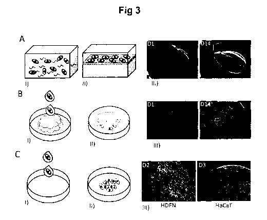
Fig. 3 shows formulation of silk scaffolds with integrated cells.
Fig. 4 shows metabolic activity of cells within silk scaffolds.
Fig. 5 shows viability of cells within silk scaffolds.
Fig. 6 shows spreading of cells within silk scaffolds.
Fig. 7 shows distribution of cells within silk scaffolds.
Fig. 8 shows mechanical properties of silk fibers with cells.
Fig. 9 shows immunofluorescence staining of collagen type I in
fibroblasts grown on silk scaffolds.
Fig. 10 shows immunofluorescence staining of myotube formation in
Hsk cells grown on silk fibers.
Fig. 11 shows presence of several cell types co-cultured within silk
scaffolds.
Fig. 12 shows that islet-like clusters are functional within silk scaffolds.
Fig. 13 shows in vivo imaging of silk scaffolds with cells.
Fig. 14 shows cell distribution within silk fibers.
Fig. 15 shows cell distribution within silk foam.
Fig. 16 shows growth curves of proliferating cells within silk foams.
Fig. 17 shows staining of live cells integrated within silk foams.
Fig. 18 shows growth curves of proliferating cells within silk fibers.
Fig. 19 shows staining of live cells integrated within silk fibers.
Fig. 20 shows growth curves of proliferating cells within silk films.
Fig. 21 shows images of live cells integrated within silk films and
foams.
Fig.22 shows micrographs of cells integrated within silk films and their
crystal violet absorption.
Fig. 23 shows stem cells differentiated into the adipogenic and
osteogenic linages, respectively.
Fig. 24 shows relative gene expression of neuronal progenitor markers
in differentiated stem cells.
CA 03014301 2018-08-10
WO 2017/137611 PCT/EP2017/053084
8
List of appended sequences
SEQ ID NO:
1 RepCT (4RepCT, WT) (DNA)
2 RepCT (4RepCT, WT)
3 CT
4 consensus CT sequence
repetitive sequence from Euprosthenops australis MaSp1
6 consensus G segment sequence 1
7 consensus G segment sequence 2
8 consensus G segment sequence 3
9 FNcc
I KVAV
11 YIGSR
12 EPDIM
13 NKDIL
14 GRKRK
KYGAASIKVAVSADR
16 NGEPRGDTYRAY
17 PQVTRGDVFTM
18 AVTGRGDSPASS
19 TGRGDSPA
CTGRGDSPAC
21 GPNSRGDAGAAS
22 VTGRGDSPAS
23 STGRGDSPAS
24 RGD-4RepCT, Widhe et al. (2013) (DNA)*
RGD-4RepCT, Widhe et al. (2013)*
26 FNcc-4RepCT (DNA)
27 FNcc-4RepCT
28 2RepRGD2RepCT (2R)
29 3RepRGD1RepCT (3R)
GRKRK-4RepCT
31 IKVAV-4RepCT
32 Linker peptide 1
33 Linker peptide 2
34 Linker peptide 3
Linker peptide 4
CA 03014301 2018-08-10
WO 2017/137611
PCT/EP2017/053084
9
SEQ ID NO:
36 CT Euprosthenops sp MaSp1
37 CT Euprosthenops australis MaSp1
38 CT Argiope trifasciata MaSp1
39 CT Cyrtophora moluccensis Sp1
40 CT Latrodectus geometricus MaSp1
41 CT Latrodectus hesperus MaSp1
42 CT Macrothele holsti Sp1
43 CT Nephila clavipes MaSp1
44 CT Nephila pilipes MaSp1
45 CT Nephila madagascariensis MaSp1
46 CT Nephila senegalensis MaSp1
47 CT Octonoba varians Sp1
48 CT Psechrus sinensis Sp1
49 CT Tetragnatha kauaiensis MaSp1
50 CT Tetragnatha versicolor MaSp1
51 CT Araneus bicentenarius 5p2
52 CT Argiope amoena MaSp2
53 CT Argiope aurantia MaSp2
54 CT Argiope trifasciata MaSp2
55 CT Gasteracantha mammosa MaSp2
56 CT Latrodectus geometricus MaSp2
57 CT Latrodectus hesperus MaSp2
58 CT Nephila clavipes MaSp2
59 CT Nephila madagascariensis MaSp2
60 CT Nephila senegalensis MaSp2
61 CT Dolomedes tenebrosus Fb1
62 CT Dolomedes tenebrosus Fb2
63 CT Araneus diadematus AD F-1
64 CT Araneus diadematus ADF-2
65 CT Araneus diadematus ADF-3
66 CT Araneus diadematus ADF-4
67 STGRGDSPAV (FN1011)
68 CT Aranaeus ventricosus MiSp
69 FNcc-RepCTmisp
* Widhe M et al., Biomaterials 34(33): 8223-8234 (2013)
CA 03014301 2018-08-10
WO 2017/137611
PCT/EP2017/053084
Detailed description of the invention
Tissues are built up of cells integrated in a composite material, called
the extracellular matrix (ECM). The ECM provides physical 3D support and
also specific sites for cell anchorage. We have developed a recombinant silk
5 protein functionalized with a motif from the ECM protein fibronectin
(FN),
which enhance the cell supportive capacity of FN-silk formed thereof. A mild
self-assembly process can be used to accomplish various formats of spider
silk scaffolds, including foam, fiber and film. The mild self-assembly process
is surprisingly also useful to accomplish various formats of fibroin silk,
10 .. including foam, fiber and film.
Acute injuries and trauma where tissue loss and failure are large
causes repair process problems due to loss of guiding extracellular matrix.
The healing process is not sufficient and can be life-threatening in case of
life
support organs such as the liver. A liver has a unique ability to self-renewal
.. and if the liver has the chance and time it can regenerate. The recombinant
spider silk could give the support to liver failures by providing a supporting
scaffold for the patients' own liver cells that have survived. This could give
the
liver cells a chance to regenerate and repair and become a personalized liver
transplant.
The co-formulation of silk combined with cells from a specific tissue
(normal or cancer) could also develop a 3D in vitro platform for disease
modeling, drug discovery and toxicology. Cancer treatment is aiming for
personal medicine due to the complexity of the cancer disease. A biomimetic
3D culture of co-formulated cancer and recombinant spider silk is one
example where it could be possible to screen the cancer progress and
develop cancer specific treatment - a personalized method to target and
demolish cancer.
The present invention is based on the insight that dispersed
mammalian cells can be added to a silk protein solution before assembly
.. thereof into water-insoluble ordered polymers or macrostructures, and
thereby be integrated throughout the silk-like material. A collection of
various
mammalian cell types (from mouse and human) have been successfully been
integrated into various silk formats, including fiber, foam and film. The silk
protein is a fibroin or a spider silk protein. The proliferative capacity of
the
.. cells was maintained through more than two weeks within the spider silk
scaffolds, with some variability of when confluence was reached depending
on the cell type. The viability was high (>80%) for all cell types
investigated,
CA 03014301 2018-08-10
WO 2017/137611 PCT/EP2017/053084
11
with confirmed viability in the innermost part of the materials. The observed
cell infiltration is highly advantageous for the formation of engineered
tissue
constructs.
It is demonstrated herein that formulation of macrostructures, preferably
films
and foams, with integrated cells provides a high seeding efficiency, yielding
quickly and viably adhered cells. Elongated cells with filamentous actin and
defined focal adhesion points confirm proper cell attachment within the
scaffolds. Cryosectioning was used to further confirm presence of cells within
the deepest parts of the materials. Tensile testing of cell-containing spider
silk
fibers was performed under physiological-like conditions, to investigate the
mechanical properties. In vivo imaging of cell-containing spider silk
scaffolds
transplanted into the anterior eye chamber confirms maintenance of cells for
4 weeks in vivo.
Compared to cultivation in hydrogels, cells attain a more tissue-like
spreading when integrated into silk scaffolds employing the methods
according to the invention.
Most native tissue types consist of several cell types organized
together in a complex three-dimensional arrangement with extracellular matrix
surrounding the cells and keeping them together. In order to replicate this in
engineered tissue constructs it is therefore of importance to achieve co-
cultures within the scaffolds. With the herein described method for
formulation
of cell containing silk scaffolds it is practically very easy to combine
several
cell types.
According to a first aspect, there is provided a method for the
cultivation of eukaryotic cells. The method is preferably carried out in
vitro.
The method is comprising the steps:
(a) providing an aqueous solution of a silk protein capable of
assembling into a water-insoluble macrostructure, wherein the silk protein
optionally contains a cell-binding motif;
(b) preparing an aqueous mixture of a sample of the eukaryotic cells
with the silk protein, wherein the silk protein remains dissolved in the
aqueous
mixture;
(c) allowing the silk protein to assemble into a water-insoluble
macrostructure in the presence of the eukaryotic cells, thereby forming a
scaffold material for cultivating the eukaryotic cells; and
(d) maintaining the eukaryotic cells within the scaffold material under
conditions suitable for cell culture.
CA 03014301 2018-08-10
WO 2017/137611 PCT/EP2017/053084
12
It is preferred that the eukaryotic cells are mammalian cells, and
preferably human cells, including primary cells, cell lines and stem cells.
Useful examples of primary cells and cell lines include endothelical cells,
fibroblasts, keratinocytes, skeletal muscle satellite cells, skeletal muscle
myoblasts, smooth muscle cells, umbilical vein endothelial cells, Schwann
cells, pancreatic 13-cells, pancreatic islet cells, hepatocytes and glioma-
forming cells. The stem cells are preferably human pluripotent stem cells
(hPSCs), such as embryonic stem cells (ESC) and induced pluripotent cells
(iPS). Useful examples of stem cells include mesenchymal stem cells. The
cells may also preferably be a combination of at least two different
mammalian cell types, such as those set out above.
In the first step, an aqueous solution of a silk protein capable of
assembling into a water-insoluble macrostructure is provided. The
composition of the aqueous solution is not critical, but it is generally
preferred
to use a mild aqueous buffer, e.g. a phosphate buffer with a low or
intermediate ion strength and a pH in the range of 6-8. The aqueous solution
preferably contains no organic solvents, such as hexafluoroisopropanol,
DMSO, and the like.
In certain preferred embodiments of the present invention, the silk
protein is a fibroin. Fibroin is present in silk created by spiders, moths,
such
as silkworms, and other insects. Preferred fibroins are derived from the genus
Bombyx, and preferably from the silkworm of Bombyx mori.
In certain preferred embodiments of the present invention, the silk
protein is a spider silk protein. The terms "spidroins" and "spider silk
proteins"
are used interchangeably throughout the description and encompass all
known spider silk proteins, including major ampullate spider silk proteins
which typically are abbreviated "MaSp", or "ADF" in the case of Araneus
diadematus. These major ampullate spider silk proteins are generally of two
types, 1 and 2. These terms furthermore include non-natural proteins with a
high degree of identity and/or similarity to the known spider silk proteins.
The silk protein optionally contains a cell-binding motif (CBM). The
optional cell-binding motif is arranged either terminally in the silk protein
or
within the silk protein, preferably N-terminally or C-terminally in the silk
protein.
Upon assembly into a macrostructure, the silk protein provides an
internal solid support activity for the cells. For avoidance of doubt, the
term
"macrostructure" refers to a coherent form of the silk protein, typically an
CA 03014301 2018-08-10
WO 2017/137611
PCT/EP2017/053084
13
ordered polymer, such as a fiber, foam or film, and not to unordered
aggregates or precipitates of the same protein. When the silk protein further
contains a cell-binding motif, the resulting macrostructure harbors both a
desired selective cell-binding activity in the cell-binding motif and an
internal
solid support activity in the silk protein fragment. The binding activity of
the
silk protein is maintained when it is structurally rearranged to form
polymeric,
solid structures. These macrostructures also provide a high and predictable
density of the cell-binding motif. The way biomaterials functionalized with
e.g.
RGD stimulate different cell responses is not only affected by the type of RGD
motif used, but also the resulting surface concentrations of ligands. Since
the
rather small silk proteins used in the present study self-assemble into
multilayers where each molecule carries an RGD motif, a dense surface
presentation is expected. However, if a sparser surface concentration is
desired, any possible surface density can be achieved simply by mixing silk
proteins with and without the cyclic RGD cell-binding motif disclosed herein
at
different ratios, thereby directing the cellular response of interest.
The cell-binding motif may for example comprise an amino acid
sequence selected from the group consisting of RGD, IKVAV (SEQ ID NO:
10), YIGSR (SEQ ID NO: 11), EPDIM (SEQ ID NO: 12) and NKDIL (SEQ ID
NO: 13). RGD, IKVAV and YIGSR are general cell-binding motifs, whereas
EPDIM and NKDIL are known as keratinocyte-specific motifs that may be
particularly useful in the context of cultivation of keratinocytes. Other
useful
cell-binding motifs include GRKRK from tropoelastin (SEQ ID NO: 14),
KYGAASIKVAVSADR (laminin derived, SEQ ID NO: 15), NGEPRGDTYRAY
(from bone sialoprotein, SEQ ID NO: 16), PQVTRGDVFTM (from vitronectin,
SEQ ID NO: 17), AVTGRGDSPASS (from fibronectin, SEQ ID NO: 18),
TGRGDSPA (SEQ ID NO: 19) and FN,,, such as CTGRGDSPAC (SEQ ID
NO: 20).
Certain relevant silk constructs with cell binding motifs are illustrated in
Fig. 2. Fig. 2a schematically shows the spider silk protein 4RepCT with
different RGD motifs genetically introduced to its N-terminus. "RGD" in Fig la
denotes the RGD containing peptide (SEQ ID NO 21) used in Widhe M et al.,
Biomaterials 34(33): 8223-8234 (2013). "FNvs" denotes the RGD-containing
decapeptide from fibronectin (SEQ ID NO: 22). "FNcc" in Fig. la denotes the
same peptide with V and S exchanged to C (SEQ ID NO: 20). "FNss" denotes
the same peptide with V and S exchanged to S (SEQ ID NO: 23). Fig. lb
shows the structure of the 9th and 10th domain of fibronectin, displaying the
CA 03014301 2018-08-10
WO 2017/137611
PCT/EP2017/053084
14
turn loop containing the RGD motif. Fig. 1c shows a structure model of the
RGD loop taken from fibronectin, with the residues V and S mutated to C
(adapted from 1FNF.pdb).
In its most general form, FN cc is C1X1X2RGDX3X4X5C2 (SEQ ID NO: 9);
wherein each of X1, X2, X3, X4 and X5 are independently selected from natural
amino acid residues other than cysteine; and Cl and 02 are connected via a
disulphide bond. FN cc is a modified cell-binding motif that imitates the a51-
specific RGD loop motif of fibronectin by positioning cysteines in precise
positions adjacent to the RGD sequence to allow formation of a disulphide-
bridge to constrain the chain into a similar type of turn loop. This cyclic
RGD
cell-binding motif increases the cell adhesion efficacy to a matrix made of a
protein containing the cell-binding motif, such as a recombinantly produced
spider silk protein. The term "cyclic" as used herein refers to a peptide
wherein two amino acid residues are covalently bonded via their side chains,
more specifically through a disulfide bond between two cysteine residues.
The cyclic RGD cell-binding motif FN cc promotes both proliferation of and
migration by primary cells. Human primary cells cultured on a cell scaffold
material containing the cyclic RGD cell-binding motif show increased
attachment, spreading, stress fiber formation and focal adhesions compared
to the same material containing a linear RGD peptide.
In preferred embodiments of FNcc, each of X1, X2, X3, X4 and X5 are
independently selected from the group of amino acid residues consisting of:
G, A, V, S, T, D, E, M, P, N and Q. In other preferred embodiments of FNcc,
each of X1 and X3 are independently selected from the group of amino acid
residues consisting of: G, S, T, M, N and Q; and each of X2, X4 and X5 are
independently selected from the group of amino acid residues consisting of:
G, A, V, S, T, P, N and Q. In certain preferred embodiments of FNcc, X1 is
selected from the group of amino acid residues consisting of: G, S, T, N and
Q; X3 is selected from the group of amino acid residues consisting of: S, T
and Q; and each of X2, X4 and X5 are independently selected from the group
of amino acid residues consisting of: G, A, V, S, T, P and N. In some
preferred embodiments of FNcc, X1 is S or T; X2 is G, A or V; preferably G or
A; more preferably G; X3 is S or T; preferably S; X4 is G, A, V or P;
preferably
G or P; more preferably P; and X5 is G, A or V; preferably G or A; more
preferably A.
In certain preferred embodiments of FNcc, the cell-binding motif is
comprising the amino acid sequence CTGRGDSPAC (SEQ ID NO: 20).
CA 03014301 2018-08-10
WO 2017/137611 PCT/EP2017/053084
Further preferred cyclic RGD cell-binding motifs according to the invention
display at least 60%, such as at least 70%, such as at least 80%, such as at
least 90% identity to CTGRGDSPAC (SEQ ID NO: 20), with the proviso that
position 1 and 10 are always C; position 4 is always R; position 5 is always
G;
5 position 6 is always D; and positions 2-3 and 7-9 are never cysteine. It is
understood that the non-identical positions among positions 2-3 and 7-9 can
be freely selected as set out above.
A preferred group of cell-binding motifs are FNcc, GRKRK, IKVAV, and
RGD, and in particular FNcc, such as CTGRGDSPAC.
The spider silk protein is preferably comprising, or consisting of, the
protein moieties REP and CT. A preferred spider silk protein has the structure
REP-CT. Another preferred spider silk protein has the structure REP-CT. The
optional cell-binding motif is arranged either terminally in the spider silk
protein, or between the moieties, or within any of the moieties, preferably N-
terminally or C-terminally in the spider silk protein.
REP is a repetitive fragment of from 70 to 300 amino acid residues,
selected from the group consisting of L(AG)L, L(AG)AL, L(GA)L, and
L(GA)GL, wherein
n is an integer from 2 to 10;
each individual A segment is an amino acid sequence of from 8
to 18 amino acid residues, wherein from 0 to 3 of the amino acid residues are
not Ala, and the remaining amino acid residues are Ala;
each individual G segment is an amino acid sequence of from
12 to 30 amino acid residues, wherein at least 40% of the amino acid
residues are Gly; and
each individual L segment is a linker amino acid sequence of
from 0 to 30 amino acid residues; and
CT is a fragment of from 70 to 120 amino acid residues, having at least
70% identity to SEQ ID NO: 3 or SEQ ID NO: 68.
The spider silk protein according to the invention is preferably a
recombinant protein, i.e. a protein that is made by expression from a
recombinant nucleic acid, i.e. DNA or RNA that is created artificially by
combining two or more nucleic acid sequences that would not normally occur
together (genetic engineering). The spider silk proteins according to the
invention are preferably recombinant proteins, and they are therefore not
identical to naturally occurring proteins. In particular, wildtype spidroins
are
CA 03014301 2018-08-10
WO 2017/137611
PCT/EP2017/053084
16
preferably not spider silk proteins according to the invention, because they
are not expressed from a recombinant nucleic acid as set out above. The
combined nucleic acid sequences encode different proteins, partial proteins
or polypeptides with certain functional properties. The resulting recombinant
protein is a single protein with functional properties derived from each of
the
original proteins, partial proteins or polypeptides.
The spider silk protein typically consists of from 140 to 2000 amino
acid residues, such as from 140 to 1000 amino acid residues, such as from
140 to 600 amino acid residues, preferably from 140 to 500 amino acid
residues, such as from 140 to 400 amino acid residues. The small size is
advantageous because longer proteins containing spider silk protein
fragments may form amorphous aggregates, which require use of harsh
solvents for solubilisation and polymerisation.
The spider silk protein may contain one or more linker peptides, or L
segments. The linker peptide(s) may be arranged between any moieties of
the spider silk protein, e.g. between the REP and CT moieties, at either
terminal end of the spider silk protein or between the spidroin fragment and
the cell-binding motif. The linker(s) may provide a spacer between the
functional units of the spider silk protein, but may also constitute a handle
for
identification and purification of the spider silk protein, e.g. a His and/or
a Trx
tag. If the spider silk protein contains two or more linker peptides for
identification and purification of the spider silk protein, it is preferred
that they
are separated by a spacer sequence, e.g. His6-spacer-His6-. The linker may
also constitute a signal peptide, such as a signal recognition particle, which
directs the spider silk protein to the membrane and/or causes secretion of the
spider silk protein from the host cell into the surrounding medium. The spider
silk protein may also include a cleavage site in its amino acid sequence,
which allows for cleavage and removal of the linker(s) and/or other relevant
moieties. Various cleavage sites are known to the person skilled in the art,
e.g. cleavage sites for chemical agents, such as CNBr after Met residues and
hydroxylamine between Asn-Gly residues, cleavage sites for proteases, such
as thrombin or protease 30, and self-splicing sequences, such as intein self-
splicing sequences.
The spidroin fragment and the cell-binding motif are linked directly or
indirectly to one another. A direct linkage implies a direct covalent binding
between the moieties without intervening sequences, such as linkers. An
indirect linkage also implies that the moieties are linked by covalent bonds,
CA 03014301 2018-08-10
WO 2017/137611
PCT/EP2017/053084
17
but that there are intervening sequences, such as linkers and/or one or more
further moieties, e.g. 1-2 NT moieties.
The cell-binding motif may be arranged internally or at either end of the
spider silk protein, i.e. C-terminally arranged or N-terminally arranged. It
is
preferred that the cell-binding motif is arranged at the N-terminal end of the
spider silk protein. If the spider silk protein contains one or more linker
peptide(s) for identification and purification of the spider silk protein,
e.g. a His
or Trx tag(s), it is preferred that it is arranged at the N-terminal end of
the
spider silk protein.
A preferred spider silk protein has the form of an N-terminally arranged
cell-bonding motif, coupled by a linker peptide of 0-30 amino acid residues,
such as 0-10 amino acid residues, to a REP moiety. Optionally, the spider silk
protein has an N-terminal or C-terminal linker peptide, which may contain a
purification tag, such as a His tag, and a cleavage site.
The protein moiety REP is fragment with a repetitive character,
alternating between alanine-rich stretches and glycine-rich stretches. The
REP fragment generally contains more than 70, such as more than 140, and
less than 300, preferably less than 240, such as less than 200, amino acid
residues, and can itself be divided into several L (linker) segments, A
(alanine-rich) segments and G (glycine-rich) segments, as will be explained in
more detail below. Typically, said linker segments, which are optional, are
located at the REP fragment terminals, while the remaining segments are in
turn alanine-rich and glycine-rich. Thus, the REP fragment can generally have
either of the following structures, wherein n is an integer:
L(AG)L, such as LA1G1A2G2A3G3A4G4A5G5L;
L(AG)AL, such as LA1G1A2G2A3G3A4G4A5G5A6L;
L(GA)L, such as LG1A1G2A2G3A3G4A4G5A5L; or
L(GA)GL, such as LGiAiG2A2G3A3G4A4G5A5G6L.
It follows that it is not critical whether an alanine-rich or a glycine-rich
segment is adjacent to the N-terminal or C-terminal linker segments. It is
preferred that n is an integer from 2 to 10, preferably from 2 to 8, also
preferably from 4 to 8, more preferred from 4 to 6, i.e. n=4, n=5 or n=6.
In some embodiments, the alanine content of the REP fragment is
above 20%, preferably above 25%, more preferably above 30%, and below
50%, preferably below 40%, more preferably below 35%. It is contemplated
that a higher alanine content provides a stiffer and/or stronger and/or less
extendible fiber.
CA 03014301 2018-08-10
WO 2017/137611
PCT/EP2017/053084
18
In certain embodiments, the REP fragment is void of proline residues,
i.e. there are no Pro residues in the REP fragment.
Turning now to the segments that constitute the REP fragment, it is
emphasized that each segment is individual, i.e. any two A segments, any
two G segments or any two L segments of a specific REP fragment may be
identical or may not be identical. Thus, it is not a general feature of the
spidroin that each type of segment is identical within a specific REP
fragment.
Rather, the following disclosure provides the skilled person with guidelines
how to design individual segments and gather them into a REP fragment,
which is a part of a functional spider silk protein useful in a cell scaffold
material.
Each individual A segment is an amino acid sequence having from 8 to
18 amino acid residues. It is preferred that each individual A segment
contains from 13 to 15 amino acid residues. It is also possible that a
majority,
or more than two, of the A segments contain from 13 to 15 amino acid
residues, and that a minority, such as one or two, of the A segments contain
from 8 to 18 amino acid residues, such as 8-12 or 16-18 amino acid residues.
A vast majority of these amino acid residues are alanine residues. More
specifically, from 0 to 3 of the amino acid residues are not alanine residues,
and the remaining amino acid residues are alanine residues. Thus, all amino
acid residues in each individual A segment are alanine residues, with no
exception or with the exception of one, two or three amino acid residues,
which can be any amino acid. It is preferred that the alanine-replacing amino
acid(s) is (are) natural amino acids, preferably individually selected from
the
group of serine, glutamic acid, cysteine and glycine, more preferably serine.
Of course, it is possible that one or more of the A segments are all-alanine
segments, while the remaining A segments contain 1-3 non-alanine residues,
such as serine, glutamic acid, cysteine or glycine.
In an embodiment, each A segment contains 13-15 amino acid
residues, including 10-15 alanine residues and 0-3 non-alanine residues as
described above. In a more preferred embodiment, each A segment contains
13-15 amino acid residues, including 12-15 alanine residues and 0-1 non-
alanine residues as described above.
It is preferred that each individual A segment has at least 80%,
preferably at least 90%, more preferably 95%, most preferably 100% identity
to an amino acid sequence selected from the group of amino acid residues 7-
19, 43-56, 71-83, 107-120, 135-147, 171-183, 198-211, 235-248, 266-279,
CA 03014301 2018-08-10
WO 2017/137611
PCT/EP2017/053084
19
294-306, 330-342, 357-370, 394-406, 421-434, 458-470, 489-502, 517-529,
553-566, 581-594, 618-630, 648-661, 676-688, 712-725, 740-752, 776-789,
804-816, 840-853, 868-880, 904-917, 932-945, 969-981, 999-1013, 1028-
1042 and 1060-1073 of SEQ ID NO: 5. Each sequence of this group
corresponds to a segment of the naturally occurring sequence of
Euprosthenops australis MaSp1 protein, which is deduced from cloning of the
corresponding cDNA, see W02007/078239. Alternatively, each individual A
segment has at least 80%, preferably at least 90%, more preferably 95%,
most preferably 100% identity to an amino acid sequence selected from the
group of amino acid residues 25-36, 55-69, 84-98, 116-129 and 149-158 of
SEQ ID NO: 2. Each sequence of this group corresponds to a segment of
expressed, non-natural spider silk proteins, which proteins have the capacity
to form silk fibers under appropriate conditions. Thus, in certain embodiments
of the spidroin, each individual A segment is identical to an amino acid
sequence selected from the above-mentioned amino acid segments. Without
wishing to be bound by any particular theory, it is envisaged that A segments
according to the invention form helical structures or beta sheets.
Furthermore, it has been concluded from experimental data that each
individual G segment is an amino acid sequence of from 12 to 30 amino acid
residues. It is preferred that each individual G segment consists of from 14
to
23 amino acid residues. At least 40% of the amino acid residues of each G
segment are glycine residues. Typically, the glycine content of each
individual
G segment is in the range of 40-60%.
It is preferred that each individual G segment has at least 80%,
preferably at least 90%, more preferably 95%, most preferably 100% identity
to an amino acid sequence selected from the group of amino acid residues
20-42, 57-70, 84-106, 121-134, 148-170, 184-197, 212-234, 249-265, 280-
293, 307-329, 343-356, 371-393, 407-420, 435-457, 471-488, 503-516, 530-
552, 567-580, 595-617, 631-647, 662-675, 689-711, 726-739, 753-775, 790-
803, 817-839, 854-867, 881-903, 918-931, 946-968, 982-998, 1014-1027,
1043-1059 and 1074-1092 of SEQ ID NO: 5. Each sequence of this group
corresponds to a segment of the naturally occurring sequence of
Euprosthenops australis MaSp1 protein, which is deduced from cloning of the
corresponding cDNA, see W02007/078239. Alternatively, each individual G
segment has at least 80%, preferably at least 90%, more preferably 95%,
most preferably 100% identity to an amino acid sequence selected from the
group of amino acid residues 1-24, 37-54, 70-83, 99-115 and 130-148 of SEQ
CA 03014301 2018-08-10
WO 2017/137611 PCT/EP2017/053084
ID NO: 2. Each sequence of this group corresponds to a segment of
expressed, non-natural spider silk proteins, which proteins have the capacity
to form silk fibers under appropriate conditions. Thus, in certain embodiments
of the spidroin in the cell scaffold material, each individual G segment is
5 identical to an amino acid sequence selected from the above-mentioned
amino acid segments.
In certain embodiments, the first two amino acid residues of each G
segment are not -Gln-Gln-.
There are three subtypes of the G segment. This classification is based
10 upon careful analysis of the Euprosthenops australis MaSp1 protein
sequence (see W02007/078239), and the information has been employed
and verified in the construction of novel, non-natural spider silk proteins.
The first subtype of the G segment is represented by the amino acid
one letter consensus sequence GQG(G/S)QGG(Q/Y)GG (L/Q)GQGGYGQGA
15 GSS (SEQ ID NO: 6). This first, and generally the longest, G segment
subtype typically contains 23 amino acid residues, but may contain as little
as
17 amino acid residues, and lacks charged residues or contain one charged
residue. Thus, it is preferred that this first G segment subtype contains 17-
23
amino acid residues, but it is contemplated that it may contain as few as 12
or
20 as many as 30 amino acid residues. Without wishing to be bound by any
particular theory, it is envisaged that this subtype forms coil structures or
31-
helix structures. Representative G segments of this first subtype are amino
acid residues 20-42, 84-106, 148-170, 212-234, 307-329, 371-393, 435-457,
530-552, 595-617, 689-711, 753-775, 817-839, 881-903, 946-968, 1043-1059
and 1074-1092 of SEQ ID NO: 5. In certain embodiments, the first two amino
acid residues of each G segment of this first subtype according to the
invention are not -Gln-Gln-.
The second subtype of the G segment is represented by the amino
acid one letter consensus sequence GQGGQGQG(G/R)Y GQG(A/S)G(S/G)S
(SEQ ID NO: 7). This second, generally mid-sized, G segment subtype
typically contains 17 amino acid residues and lacks charged residues or
contain one charged residue. It is preferred that this second G segment
subtype contains 14-20 amino acid residues, but it is contemplated that it may
contain as few as 12 or as many as 30 amino acid residues. Without wishing
to be bound by any particular theory, it is envisaged that this subtype forms
coil structures. Representative G segments of this second subtype are amino
acid residues 249-265, 471-488, 631-647 and 982-998 of SEQ ID NO: 5.
CA 03014301 2018-08-10
WO 2017/137611 PCT/EP2017/053084
21
The third subtype of the G segment is represented by the amino acid
one letter consensus sequence G(R/Q)GQG(G/R)YGQG (A/S/V)GGN (SEQ
ID NO: 8). This third G segment subtype typically contains 14 amino acid
residues, and is generally the shortest of the G segment subtypes. It is
preferred that this third G segment subtype contains 12-17 amino acid
residues, but it is contemplated that it may contain as many as 23 amino acid
residues. Without wishing to be bound by any particular theory, it is
envisaged
that this subtype forms turn structures. Representative G segments of this
third subtype are amino acid residues 57-70, 121-134, 184-197, 280-293,
343-356, 407-420, 503-516, 567-580, 662-675, 726-739, 790-803, 854-867,
918-931, 1014-1027 of SEQ ID NO: 5.
Thus, in preferred embodiments of the spidroin in the cell scaffold
material, each individual G segment has at least 80%, preferably 90%, more
preferably 95%, identity to an amino acid sequence selected from SEQ ID
NO: 6, SEQ ID NO: 7 and SEQ ID NO: 8.
In an embodiment of the alternating sequence of A and G segments of
the REP fragment, every second G segment is of the first subtype, while the
remaining G segments are of the third subtype, e.g.
...A1 GshortA2GlongA3GshortA4GlongA5Gshort... In another embodiment of the REP
fragment, one G segment of the second subtype interrupts the G segment
regularity via an insertion, e.g. ...AiGshortA2GiongA3GmidA4GshortA5Giong...
Each individual L segment represents an optional linker amino acid
sequence, which may contain from 0 to 30 amino acid residues, such as from
0 to 20 amino acid residues. While this segment is optional and not critical
for
the function of the spider silk protein, its presence still allows for fully
functional spider silk proteins and polymers thereof which form fibers, films,
foams and other structures. There are also linker amino acid sequences
present in the repetitive part (SEQ ID NO: 5) of the deduced amino acid
sequence of the MaSp1 protein from Euprosthenops australis. In particular,
the amino acid sequence of a linker segment may resemble any of the
described A or G segments, but usually not sufficiently to meet their criteria
as defined herein.
As shown in W02007/078239, a linker segment arranged at the C-
terminal part of the REP fragment can be represented by the amino acid one
letter consensus sequences ASASAAASAA STVANSVS (SEQ ID NO: 32)
and ASAASAAA (SEQ ID NO: 33), which are rich in alanine. In fact, the
second sequence can be considered to be an A segment according to the
CA 03014301 2018-08-10
WO 2017/137611
PCT/EP2017/053084
22
definition herein, whereas the first sequence has a high degree of similarity
to
A segments according to this definition. Another example of a linker segment
has the one letter amino acid sequence GSAMGQGS (SEQ ID NO: 34),
which is rich in glycine and has a high degree of similarity to G segments
according to the definition herein. Another example of a linker segment is
SASAG (SEQ ID NO: 35).
Representative L segments are amino acid residues 1-6 and 1093-
1110 of SEQ ID NO: 5; and amino acid residues 159-165 of SEQ ID NO: 2,
but the skilled person will readily recognize that there are many suitable
alternative amino acid sequences for these segments. In one embodiment of
the REP fragment, one of the L segments contains 0 amino acids, i.e. one of
the L segments is void. In another embodiment of the REP fragment, both L
segments contain 0 amino acids, i.e. both L segments are void. Thus, these
embodiments of the REP fragments according to the invention may be
schematically represented as follows: (AG)L, (AG)AL, (GA)L, (GA)GL;
L(AG)n, L(AG)A, L(GA)n, L(GA)G; and (AG)n, (AG)A, (GA)n, (GA)G. Any
of these REP fragments are suitable for use with any CT fragment as defined
below.
The CT fragment of the spidroin in the cell scaffold material has a high
degree of similarity to the C-terminal amino acid sequence of spider silk
proteins. As shown in W02007/078239, this amino acid sequence is well
conserved among various species and spider silk proteins, including MaSp1,
MaSp2 and MiSp (minor ampullate spidroin). A consensus sequence of the
C-terminal regions of MaSp1 and MaSp2 is provided as SEQ ID NO: 4. In Fig.
1, the MaSp proteins (SEQ ID NO: 36-66) presented in Table 1 are aligned,
denoted with GenBank accession entries where applicable:
TABLE 1 - Spidroin CT fragments
Species and spidroin Entry
Euprosthenops sp MaSp1 (Pouchkina-Stantcheva*)
Cthyb_Esp
Euprosthenops australis MaSp1 (SEQ ID NO: 3) CTnat_
Eau
Argiope trifasciata MaSp1 AF350266_At1
Cyrtophora moluccensis Sp1
AY666062 Cm1
Latrodectus geometricus MaSp1 AF350273_Lg1
Latrodectus hesperus MaSp1 AY953074 Lh1
Macrothele holsti Sp1 AY666068 Mh1
CA 03014301 2018-08-10
WO 2017/137611 PCT/EP2017/053084
23
Species and spidroin Entry
Nephila clavipes MaSp1 U20329 Nc1
Nephila pilipes MaSp1
AY666076_Np1
Nephila madagascariensis MaSp1
AF350277 Nml
Nephila senegalensis MaSp1
AF350279 Ns1
Octonoba varians Sp1
AY666057 Ov1
Psechrus sinensis Sp1
AY666064 Ps1
Tetragnatha kauaiensis MaSp1
AF350285 Tk1
Tetragnatha versicolor MaSp1
AF350286 Tv1
Araneus bicentenarius Sp2
ABU20328 Ab2
Argiope amoena MaSp2
AY365016_Aam2
Argiope aurantia MaSp2
AF350263 Aau2
Argiope trifasciata MaSp2
AF350267_At2
Gasteracantha mammosa MaSp2
AF350272 Gm2
Latrodectus geometricus MaSp2
AF350275_Lg2
Latrodectus hesperus MaSp2
AY953075 Lh2
Nephila clavipes MaSp2
AY654293 Nc2
Nephila madagascariensis MaSp2
AF350278 Nm2
Nephila senegalensis MaSp2
AF350280 Ns2
Dolomedes tenebrosus Fb1
AF350269 DtFb1
Dolomedes tenebrosus Fb2
AF350270 DtFb2
Araneus diadematus ADF-1 U47853
ADF1
Araneus diadematus ADF-2 U47854
ADF2
Araneus diadematus ADF-3 U47855
ADF3
Araneus diadematus ADF-4 U47856
ADF4
* Comparative Biochemistry and Physiology, Part B 138: 371-376 (2004)
It is not critical which specific CT fragment is present in the spider silk
protein in the cell scaffold material. Thus, the CT fragment can be selected
from any of the amino acid sequences shown in Fig. 1 and Table 1 or
sequences with a high degree of similarity, such as the MiSp CT fragment
SEQ ID NO: 68 from Araneus ventricosus (Genbank entry AFV 31615).. A
wide variety of C-terminal sequences can be used in the spider silk protein.
CA 03014301 2018-08-10
WO 2017/137611
PCT/EP2017/053084
24
The sequence of the CT fragment has at least 50% identity, preferably
at least 60%, more preferably at least 65% identity, or even at least 70%
identity, to the consensus amino acid sequence SEQ ID NO: 4, which is
based on the amino acid sequences of Fig. 1.
A representative CT fragment is the Euprosthenops australis sequence
SEQ ID NO: 3 or amino acid residues 180-277 of SEQ ID NO: 27. Another
representative CT fragment is the MiSp sequence SEQ ID NO: 68. Thus, in
one embodiment, the CT fragment has at least 70%, such as at least 80%,
such as at least 85%, preferably at least 90%, such as at least 95%, identity
to SEQ ID NO: 3, amino acid residues 180-277 of SEQ ID NO: 27, or any
individual amino acid sequence of Fig. 1 and Table 1, or SEQ ID NO: 68. For
example, the CT fragment may be identical to SEQ ID NO: 3, amino acid
residues 180-277 of SEQ ID NO: 27, or any individual amino acid sequence
of Fig. 1 and Table 1, or SEQ ID NO: 68,.
The CT fragment typically consists of from 70 to 120 amino acid
residues. It is preferred that the CT fragment contains at least 70, or more
than 80, preferably more than 90, amino acid residues. It is also preferred
that
the CT fragment contains at most 120, or less than 110 amino acid residues.
A typical CT fragment contains approximately 100 amino acid residues.
The term "(:)/0 identity", as used herein, is calculated as follows. The
query sequence is aligned to the target sequence using the CLUSTAL W
algorithm (Thompson et al, Nucleic Acids Research, 22:4673-4680 (1994)). A
comparison is made over the window corresponding to the shortest of the
aligned sequences. The amino acid residues at each position are compared,
and the percentage of positions in the query sequence that have identical
correspondences in the target sequence is reported as (:)/0 identity.
The term "(:)/0 similarity", as used herein, is calculated as described
above for "(:)/0 identity", with the exception that the hydrophobic residues
Ala,
Val, Phe, Pro, Leu, Ile, Trp, Met and Cys are similar; the basic residues Lys,
Arg and His are similar; the acidic residues Glu and Asp are similar; and the
hydrophilic, uncharged residues Gin, Asn, Ser, Thr and Tyr are similar. The
remaining natural amino acid Gly is not similar to any other amino acid in
this
context.
Throughout this description, alternative embodiments according to the
invention fulfill, instead of the specified percentage of identity, the
corresponding percentage of similarity. Other alternative embodiments fulfill
the specified percentage of identity as well as another, higher percentage of
CA 03014301 2018-08-10
WO 2017/137611
PCT/EP2017/053084
similarity, selected from the group of preferred percentages of identity for
each sequence. For example, a sequence may be 70% similar to another
sequence; or it may be 70% identical to another sequence; or it may be 70%
identical and 90% similar to another sequence.
5 In a preferred spider silk protein according to the invention, the REP-
CT fragment has at least 70%, such as at least 80%, such as at least 85%,
preferably at least 90%, such as at least 95%, identity to SEQ ID NO: 2 or to
amino acid residues 18-277 of SEQ ID NO: 27 or to amino acid residues 18-
272 of SEQ ID NO: 69.
10 In one preferred spider silk protein according to the invention, the
protein has at least 70%, such as at least 80%, such as at least 85%,
preferably at least 90%, such as at least 95%, identity to SEQ ID NO: 25, 27
or 69. In a particularly preferred embodiment, the spider silk protein
according
to the invention is SEQ ID NO: 25, 27 or 69.
15 The cell scaffold material according to the invention preferably
comprises a protein or peptide according to the invention displaying the
cyclic
RGD cell-binding motif. The cyclic RGD cell-binding motif may be exposed
from short synthetic peptides or longer synthetic or recombinant proteins,
which may in turn be attached to or associated with a matrix or support.
20 The cell scaffold material preferably comprises a protein polymer,
which protein polymer in turn is containing the silk protein according to the
invention as a repeating structural unit, i.e. the protein polymer contains or
consists of a polymer of the silk protein according to the invention. This
implies that the protein polymer contains or consists of an ordered plurality
of
25 silk proteins according to the invention, typically well above 100 silk
protein
units, e.g. 1000 silk protein units or more. In a preferred embodiment, the
cell
scaffold material according to the invention consists of the protein polymer.
The magnitude of silk protein units in the polymer implies that the
protein polymer obtains a significant size. In a preferred embodiment, the
protein polymer has a size of at least 0.01 pm in at least two dimensions.
Thus, the term "protein polymer" as used herein relates to silk protein
polymers having a thickness of at least 0.01 pm, such as at least 0.1 pm,
preferably macroscopic polymers that are visible to the human eye, i.e.
having a thickness of at least 1 pm, such as up 10 pm. The term "protein
polymer" does not encompass unstructured aggregates or precipitates. While
monomers/dimers of the spider silk protein are water soluble, it is understood
that the protein polymers according to the invention are solid structures,
i.e.
CA 03014301 2018-08-10
WO 2017/137611 PCT/EP2017/053084
26
not soluble in water. The protein polymers are comprising monomers of the
silk proteins according to the invention as a repeating structural unit.
The protein polymer according to the invention is typically provided in a
physical form selected from the group consisting of fiber, film, coating,
foam,
net, fiber-mesh, sphere and capsule. According to one embodiment, it is
preferable that the protein polymer according to the invention is a fiber,
film or
fiber-mesh. According to certain embodiments, it is preferable that the
protein
polymer has a three-dimensional form, such as a foam or a fiber-mesh. One
preferred embodiment involves thin (typically 0.01-0.1 pm thickness) coatings
made of the protein polymer, which are useful for coating of stents and other
medical devices. The term "foam" is comprising a porous foam with channels
connecting the bubbles of the foam, sometimes to the extent that it can even
be regarded as a three-dimensional net or mesh of fibers.
In a preferred embodiment, the protein polymer is in a physical form of
a free-standing matrix, such as a free-standing film. This is highly useful as
it
allows for transfer of a cell sheet where needed, e.g. in an in vivo situation
where cells need to be transferred as a cell sheet to e.g. a wound area.
The fiber, film or fiber-mesh typically has a thickness of at least 0.1 pm,
preferably at least 1 pm. It is preferred that the fiber, film or fiber-mesh
has a
thickness in the range of 1-400 pm, preferably 60-120 pm. It is preferred that
fibers have a length in the range of 0.5-300 cm, preferably 1-100 cm. Other
preferred ranges are 0.5-30 cm and 1-20 cm. The fiber has the capacity to
remain intact during physical manipulation, i.e. can be used for spinning,
weaving, twisting, crocheting and similar procedures. The film is
advantageous in that it is coherent and adheres to solid structures, e.g. the
plastics in microtiter plates. This property of the film facilitates washing
and
regeneration procedures and is very useful for separation purposes.
The spider silk protein according to the invention harbors an internal
solid support activity in the REP-CT moieties, and optionally also a desired
cell-binding activity in the cell-binding motif, and these activities are
employed
in the cell scaffold material. The cell scaffold material provides a high and
predictable density of the selective interaction activity towards an organic
target. Losses of valuable protein moieties with selective interaction
activity
are minimized, since all expressed protein moieties are associated with the
cell scaffold material.
The polymers which are formed from the silk proteins according to the
invention are solid structures and are useful for their physical properties,
CA 03014301 2018-08-10
WO 2017/137611
PCT/EP2017/053084
27
especially the useful combination of high strength, elasticity and light
weight.
A particularly useful feature is that the REP-CT moieties of the spider silk
protein are biochemically robust and suitable for regeneration, e.g. with
acid,
base or chaotropic agents, and suitable for heat sterilization, e.g.
autoclaving
at 120 C for 20 min. The polymers are also useful for their ability to support
cell adherence and growth.
The properties derived from the REP-CT moieties are attractive in
development of new materials for medical or technical purposes. In particular,
the cell scaffold materials according to the invention are useful as scaffolds
for cell immobilization, cell culture, cell differentiation, tissue
engineering and
guided cell regeneration. They are also useful in preparative and analytical
separation procedures, such as chromatography, cell capture, selection and
culture, active filters, and diagnostics. The cell scaffold materials
according to
the invention are also useful as in medical devices, such as implants and
stents, e.g. as coatings.
In a preferred embodiment, the cell scaffold material comprises a
protein polymer, which is consisting of a silk protein according to the
invention
as a repeating structural unit. And in a further preferred embodiment, the
cell
scaffold material is a protein polymer, which is consisting of a silk protein
according to the invention as a repeating structural unit. The silk protein is
a
fibroin or a spider silk protein.
In the second step, an aqueous mixture of a sample of the eukaryotic
cells with the silk protein is prepared. This can preferably be achieved by
mixing the aqueous solution from the previous step with a liquid cell
suspension or by dispersing a cell pellet. The liquid component of the
aqueous mixture should be suitable for the respective eukaryotic cell in terms
of buffering capacity, ion strength and pH. Suitable media for cell culture
and
cell handling are well-known in the art e.g. DMEM, Ham's Nutrient Mixtures,
Minimal Essential Medium Eagle, and RPMI.
It is preferred that the eukaryotic cells are mammalian cells, and
preferably human cells, including primary cells, cell lines and stem cells.
Useful examples of primary cells and cell lines include endothelical cells,
fibroblasts, keratinocytes, skeletal muscle satellite cells, skeletal muscle
myoblasts, Schwann cells, pancreatic 13-cells, pancreatic islet cells,
hepatocytes and glioma-forming cells. The stem cells are preferably human
pluripotent stem cells (hPSCs), such as embryonic stem cells (ESC) and
induced pluripotent cells (iPS). Useful examples of stem cells include
CA 03014301 2018-08-10
WO 2017/137611 PCT/EP2017/053084
28
mesenchymal stem cells. The cells may also preferably be a combination of
at least two different mammalian cell types, such as those set out above.
In the second step, it is critical that silk protein remains dissolved in the
aqueous mixture. By the term "dissolved" means that the cells are added to
the silk protein before the silk assembly process has been developed, when
the silk proteins predominantly form bonds with the surrounding water
molecules. When the silk assembly process has been developed, irreversible
formation of ordered polymers with predominantly intra- and intermolecular
bonds between the silk proteins occurs. It is understood that the
polymerization is a continuous process, but according to the present
invention, the cells should be added to the dissolved silk protein as early as
possible in view of the desired final format of the final macrostructure. It
is
preferred that the cells are added when at least some, and preferably most of
or even substantially all of the silk proteins remain dissolved. Thus for
instance, if the desired format is a foam, the cells should be added before
foaming or to the wet foam when it is newly made by introduction of air into
the liquid, and not when the foam has polymerized into a silk macrostructure.
Optionally, the aqueous mixture may contain further components which
are desirable to integrate in the macrostructure. For instance, the aqueous
mixture may contain cell-binding proteins and polypeptides, such as laminins.
In the third step, the silk protein is allowed to assemble into a water-
insoluble macrostructure in the presence of the eukaryotic cells. Proteins
structures according to the invention are assembled spontaneously from the
silk proteins according to the invention under suitable conditions, and the
assembly into polymers is promoted by the presence of shearing forces
and/or an interface between two different phases e.g. between a solid and a
liquid phase, between air and a liquid phase or at a hydrophobic/hydrophilic
interface, e.g. a mineral oil-water interface. The presence of the resulting
interface stimulates polymerization at the interface or in the region
surrounding the interface, which region extends into the liquid medium, such
that said polymerizing initiates at said interface or in said interface
region.
Various protein structures can be produced by adapting the conditions during
the assembly. For instance, if the assembly is allowed to occur in a container
that is gently wagged from side to side, a fiber is formed at the air-water
interface. If the mixture is allowed to stand still, a film is formed at the
air-
water interface. If the mixture is evaporated, a film is formed at the bottom
of
the container. If oil is added on top of the aqueous mixture, a film is formed
at
CA 03014301 2018-08-10
WO 2017/137611
PCT/EP2017/053084
29
the oil-water interface, either if allowed to stand still or if wagged. If the
mixture is foamed, e.g. by bubbling of air or whipping, the foam is stable and
solidifies with time. The new macrostructure may be allowed to form in any
suitable cell culture well. Optionally, the culture well surface is pre-coated
with
a silk macrostructure or with other substances, e.g. gelatin.
The assembly into water-insoluble macrostructure results in formation
of a scaffold material for cultivating the eukaryotic cells. Thus, the very
cells to
be cultured are present already during assembly of the scaffold material and
become integrated within the cell material. Thereby, the cells become
surrounded by and embedded in the spider silk macrostructure. This has
advantageous effect in terms of viability, proliferative capacity, cell
spreading
and attachment in the subsequent cell culture. Furthermore, the co-presence
of the cells in the assembly of the macrostructure achieves formation of
cavities and pores in the scaffold material which would otherwise not have
existed.
In the fourth step, the eukaryotic cells are maintained within the
scaffold material under conditions suitable for cell culture, which are well
known to the skilled person and exemplified herein. This advantageously
allows for the cells to grow integrated with the scaffold material. This means
that the cells are not just growing attached to the very surface of the
scaffold
material, but also within cavities and pores in the scaffold material which
have
been formed due to air bubbles and the co-presence of the cells in the
assembly of the macrostructure.
According to a second aspect, the present invention provides a
process for manufacturing a cell culture product comprising (i) a scaffold
material for cultivating eukaryotic cells; and (ii) eukaryotic cells, which
are
growing integrated with the scaffold material. The method is preferably
carried
out in vitro. The method is comprising the steps:
(a) providing an aqueous solution of a silk protein capable of assembling into
a water-insoluble macrostructure, wherein the silk protein optionally contains
a cell-binding motif;
(b) preparing an aqueous mixture of a sample of the eukaryotic cells with the
silk protein, wherein the silk protein remains dissolved in the aqueous
mixture; and
(c) allowing the silk protein to assemble into a water-insoluble
macrostructure
in the presence of the eukaryotic cells, thereby forming the scaffold material
for cultivating the eukaryotic cells.
CA 03014301 2018-08-10
WO 2017/137611
PCT/EP2017/053084
Preferred embodiments and variants of the manufacturing process are
evident from the above disclosure of the method for the cultivation of
eukaryotic cells which is including corresponding steps.
According to a third aspect, the present invention provides a cell
5 culture product comprising (i) a scaffold material for cultivating
eukaryotic
cells, which is a water-insoluble macrostructure of a silk protein capable of
assembling into a water-insoluble macrostructure, wherein the silk protein
optionally contains a cell-binding motif; and (ii) eukaryotic cells, which are
growing integrated with the scaffold material.
10 This means that the cells are not just growing attached to the very
surface of the scaffold material, but also within cavities and pores in the
scaffold material which have been formed e.g. due to the co-presence of the
cells in the assembly of the macrostructure.
Preferred embodiments and variants of the cell culture product are
15 evident from the above disclosure of the method for the cultivation of
eukaryotic cells which is including corresponding features.
In a preferred embodiment, the cell culture product according to the
invention is obtainable or obtained by the manufacturing process according to
the invention. The co-presence of the cells in the assembly of the
20 macrostructure achieves formation of cavities and pores in the scaffold
material which would otherwise not have existed.
According to a fourth and final aspect, the present invention provides a
novel use of a silk protein capable of assembling into a water-insoluble
macrostructure in the formation of a scaffold material for cultivating
eukaryotic
25 cells in the presence of said cells; wherein the scaffold material is a
water-
insoluble macrostructure of the silk protein; and wherein the silk protein
optionally contains a cell-binding motif. The use is preferably carried out in
vitro.
Preferred embodiments and variants of the use are evident from the
30 above disclosure of the method for the cultivation of eukaryotic cells
which is
including corresponding features.
In summary, novel methods for formulation of cell-containing silk
scaffolds have been developed, where the cells are added to the silk protein
before the silk assembly process has been developed. The following
Examples demonstrate how the cells are affected by incorporation into the
silk scaffolds, in terms of viability, proliferative capacity, cell spreading
and
attachment. To survey the generality of the method, a broad repertoire of
CA 03014301 2018-08-10
WO 2017/137611
PCT/EP2017/053084
31
mammalian cells, ranging from stable cell lines to primary cells, of both
mouse and human origin (Table 2) has been analyzed. Maintenance of
specific cell functions for certain cell types, such as production of
extracellular
matrix components, differentiation and responsiveness to glucose stimulation,
has also been confirmed.
Table 2
Tested mammalian cells
Silk Motif Cell Cell Proliferation*** Cell
format type* viability**
integration
Fiber:air FNcc HSkMC +++ +++ Yes,
cryosections
and confocal
Fiber:oil FNcc HSkMC +++ +++ Yes,
cryosections
Fiber:air FNcc HSkMC: +++ ++ Yes,
HDMEC
Cryosections
Fiber:air FNcc HDMEC +++ +++ Yes,
cryosections
Fiber:oil GRKRK HSkMC + ++ Yes,
cryosections
Fiber:oil GRKRK HSkMC: + ++ Yes,
HDMEC
cryosections
Fiber:air FNcc HSMM +++ ++ Yes,
cryosections
Fiber:air FNcc HSMM: +++ ++ Yes,
HDMEC
cryosections
Fiber:air FNcc HDFn: +++ +++ Yes,
HDMEC
cryosections
Fiber:air FNcc HDFn +++ Yes,
cryosections
Fiber:oil FNcc HDFn Yes,
cryosections
Foam FNcc HDFn ++
Fiber:oil FNcc HaCaT Yes,
cryosections
CA 03014301 2018-08-10
WO 2017/137611 PCT/EP2017/053084
32
Silk Motif Cell Cell Proliferation*** Cell
format type* viability** integration
Fiber:air FNcc HaCaT Yes,
cryosections
Foam FNcc HaCaT +++ ++
Foam FNcc mEC +++ ++ Yes,
cryosections
Fiber:air FNcc mEC +++ ++
Foam FNcc mMSC +++ ++ Yes,
cryosections,
confokal
Fiber:air FNcc mMSC +++ ++ Yes,
cryosections
Fiber:oil FNcc mMSC ++ ++ Yes,
cryosections
Foam FNcc hMSC +++ ++
Fiber:air FNcc hMSC +++ ++
Foam FNcc Schwann +++ ++
cells
Foam IKVAV Schwann +++ ++
cells
Foam FNcc:IKVAV Schwann +++ ++
cells
Fiber:air FNcc Schwann +++ ++
cells
Fiber:air IKVAV Schwann +++ ++
cells
Fiber:air FNcc:IKVAV Schwann +++ ++
cells
Foam FNcc Min6m9 ++ ++ Yes,
cryosections
Foam 2R Min6m9 + ++
Foam FN:2R Min6m9 + ++
Foam FN:2R MIP +++ ++
Foam FN Human +
islets
CA 03014301 2018-08-10
WO 2017/137611 PCT/EP2017/053084
33
Silk Motif Cell Cell Proliferation*** Cell
format type* viability** integration
Foam FN:2R Human +
islet cells
Foam FN hESC ++ ++
Film FN hESC ++ ++
Foam FN hIPS ++ ++
Film FN hIPS ++ ++
Film FN SMC ++ ++
Film FN HUVEC ++ ++
Foam FN HUVEC ++ ++
*: HSkMC = Human skeletal muscle satellite cells; HDMEC = Human Dermal
Microvascular Endothelial cells; HSMM = human skeletal muscle myoblasts; HDFn
= Human dermal fibroblasts; HaCaT = human keratinocyte cell line; mEC = mouse
Endothelial cells; mMSC = mouse Mesenchymal stem cells; hMSC = human
Mesenchymal stem cells; Min6m9 = Pancreatic J3-cell line; MIP = Islets from
MIP-
GFP mice; hESC = human embryonal stem cells; hIPS = human induced
pluripotent stem cells; SMC = smooth muscle cells; HUVEC = Human umbilical
vein endothelial cells
**: += 50-70%; ++ =70-90%; +++ = >90%
***: + = cells increase day 1-7; ++ = cells increase day 1-14; +++ = cells
increase
day 1-21
EXAMPLES
Example 1
Materials and Methods
Recombinant spider silk protein preparation
Production of recombinant silk proteins in in E. coli and the following
purification were done essentially as described in Hedhammar M et al.,
Biochemistry 47(11):3407-3417 (2008) and Hedhammar M et al.,
Biomacromolecules 11: 953-959 (2010).
CA 03014301 2018-08-10
WO 2017/137611
PCT/EP2017/053084
34
Briefly, Escherichia coli BL21(DE3) cells (Merck Biosciences) with the
expression vector for the target protein were grown at 30 C in Luria-Bertani
medium containing kanamycin to an OD600 of 0.8-1 and then induced with
isopropyl p-D-thiogalactopyranoside and further incubated for at least 2 h.
Thereafter, cells were harvested and resuspended in 20 mM Tris-HCI (pH
8.0) supplemented with lysozyme and DNase I. After complete lysis, the
supernatants from centrifugation at 15,000 g were loaded onto a column
packed with Ni Sepharose (GE Healthcare, Uppsala, Sweden). The column
was washed extensively before elution of bound proteins with 300 mM
.. imidazole. Fractions containing the target proteins were pooled and
dialyzed
against 20 mM Tris-HCI (pH 8.0). The target protein was released from the
tags by proteolytic cleavage. To remove the released HisTrxHis tag, the
cleavage mixture was loaded onto a second Ni Sepharose column and the
flowthrough was collected. The protein content was determined from the
absorbance at 280 nm.
The protein solutions obtained were purified from lipopolysaccharides
(lps) as described in Hedhammar et al., Biomacromolecules 11:953-959
(2010). The protein solutions were sterile filtered (0.22 pm) before being
used
to prepare scaffolds (film, foam, coatings or fibers).
The recombinant spider silk proteins were successfully expressed in
E coli and purified with similar yield and purity as the original 4RepCT.
The partial spider silk protein 4RepCT (SEQ ID NO: 2) was used as
base for all proteins used. A functionalized version of 4RepCT with the
modified cell binding motif from fibronectin, denoted FNcc-4RepCT in the
experimental section (SEQ ID NO: 27), was used for most of the experiments.
Other versions, 2RepRGD2RepCT ("2R", SEQ ID NO: 28) and
3RepRGD1RepCT ("3R", SEQ ID NO: 29), with the RGD peptide inserted
within the repetitive part, was used for some of the experiments with
endocrine cells and other cells. Another version GRKRK-4RepCT (SEQ ID
NO: 30), with the GRKRK peptide inserted at the N-terminus, was used for
some of the experiments with muscle satellite cells. Another version, IKVAV-
4RepCT (SEQ ID NO: 31) with the IKVAV peptide inserted at the N-terminus,
was used for some of the experiments with Schwann cells.
CA 03014301 2018-08-10
WO 2017/137611
PCT/EP2017/053084
Cell culture
Mesenchymal stem cells (MSC)
Mouse Mesenchymal stem cells (mMSC, Gibco) at a passage of 8-14
were cultured in DMEM F12 HAM supplemented with 10% Fetal Bovine
5 Serum (Mesenchymal Stem Cell Qualified, USDA Approved Regions, Gibco).
Human Mesenchymal stem cells (hMSC) at a passage of 8 (Gibco)
from bone marrow were grown in culture flasks coated with CELLstart (Gibco)
in complete StemPro MSC serum free medium CTS (Gibco) containing 2 mM
Glutamax.
Endothelial cells (EC)
Mouse Endothelial cells (Cell Biologics) were cultured at a passage of
7-9 in complete endothelial cell media MV (PromoCell GmbH, Germany).
Human Dermal Microvascular Endothelial cells (HDMEC) (PromoCell
GmbH, Germany) isolated from dermis from adult donors were cultured
culture flasks coated with gelatin (Sigma Aldrich) in complete endothelial
cell
media MV (PromoCell GmbH, Germany).
Fibroblasts (HDFn)
Human dermal fibroblasts, HDF (ECACC, Salisbury, UK) were used in
passage 8-11. Culture medium, DMEM F12 ham supplemented with 5% FBS
(Sigma), was changed every 2nd-3rd day.
Keratinocytes (HaCaT)
HaCaT (human keratinocyte cell line, spontaneously transformed),
were cultured in DMEM F12 ham supplemented with 5% FBS (Sigma).
Medium was changed every 2nd-3rd day.
Human skeletal muscle satellite cells (Hsk)
Human skeletal muscle satellite cells, HskMSC (ScienCell Research
Laboratories, Carlsbad,CA) and human skeletal muscle myoblasts (HSMM,
Lonza, Belgium) were used from passage 2-6. Skeletal muscle culture
medium, SkMCM (ScienCell Research Laboratories), with skeletal muscle cell
growth supplement, SkMCGS (ScienCell Research Laboratories) or SkGM-2
BulletKit (for HSMM, Lonza) and 5% FBS (ScienCell Research Laboratories
or Lonza, respectively) was changed every second day.
CA 03014301 2018-08-10
WO 2017/137611 PCT/EP2017/053084
36
Schwann cells
Schwann cells (3H Biomedical, Uppsala, Sweden) at a passage of 2-6
were cultured in Schwann cell medium (SCM, 3H Biomedical) supplemented
with 5% FBS and Schwann cell growth supplement (SCGS, 3H Biomedical)
and penicillin/streptomycin solution (3H Biomedical).
Endocrine cells
Pancreatic J3-cell line MIN6m9 at passage 27-35 were cultured in
DMEM (Gibco) supplemented with (3-mercaptoethanol (50pM), penicillin (100
U mL-1), streptomycin (100 ug mL-1), 10% heat-inactivated FBS and glucose
(11 mM).
Islets from MIP-GFP mice, all inbred in the animal core facility at
Karolinska Institutet, were isolated from pancreas by injecting 1.2 mg/ml
collagenase into the bile duct. The pancreas was thoroughly taken out and
put into a flask containing collagenase with same concentration as above.
The flask was then put into a 37 C water bath for 15 min. There after the
islets were washed and handpicked under a stereomicroscope. To disperse
the islets into cells, the islets were first washed two times in PBS without
Ca2+
and Mg2+ and incubated in Accutase (Gibco) for 5 min at 37 C. The cells were
counted and cultured in RPM! 1640 medium (Gibco) supplemented with L-
glutamine (2 mM), penicillin (100 U mL-1), streptomycin (100 ug mL-1) and
10% heat-inactivated fetal bovine serum (FBS).
Human islets were obtained from the unavoidable excess of islets
generated within the Nordic Network for Clinical Islet Transplantation. Only
organ donors who explicitly had agreed to donate for scientific purposes were
included. Informed written consent to donate organs for medical and research
purposes was obtained from donors, or relatives of donors, by the National
Board of Health and Welfare (Socialstyrelsen), Sweden. Experimental
procedures were done according to the approved ethical permit from the
Ethical Committee for Human Research (permit number 2011/14667-32). The
human cells were cultured in CMRL-1066 (ICN Biomedicals) supplemented
with HEPES (10 mM), L-glutamine (2 mM), Gentamycin (50 mg m1-1),
Fungizone (0.25 mg ml-1, Gibco), Ciprofloxacin (20 mg ml-1, Bayer Healthcare
AG), nicotinamide (10 mM), and 10% heat inactivated FBS.
CA 03014301 2018-08-10
WO 2017/137611
PCT/EP2017/053084
37
Hepatocytes
Rodent Hepatocytes (liver cells) are isolated by enzymatic (1,2 mg/ml
Collagenase P in pH 7.4 HBSS buffer supplemented with 25mM Hepes,
0,25% w/v BSA) collagenase treatment of the liver, digested by a continuous
mechanic shaking in 37 C for 20 minutes, separated and cultured in RPM I-
1640 medium supplemented with 10% FBS (Invitrogen).
Glioma forming cell line
Glioma forming cell line GL261 is cultured in 10% FBS containing
DMEM (Invitrogen) with medium change every 2-3rd day.
Co-cultures
Hsk cells in co-culture with EC were cultured in SkMCM culture media.
Endocrine cells in co-culture with MSC and EC cultured in RPM! 1640
medium (Gibco), StemPro MSC serum free medium CTS (Gibco) containing
2mM Glutamax and endothelial cell media MV (PromoCell GmbH, Germany)
at a ratio of 50:25:25.
Formulation of silk scaffolds with integrated cells
Fiber formation
Silk protein (0.5-3 mg) was mixed with 0.5-2 million cells in respective
culture media in a total volume of 2-4 ml. The fiber formation together with
cells was performed in RT under gentle wagging for 1-3 hours. The formed
fibers were then washed in 1xPBS and thereafter transferred into non-tissue
treated 12 or 24-well plates and further kept in culture by adding fresh media
(0.5mL in 24-well plate or 1mL in 12-well plate).
For fiber formation against oil, 3-4 ml of either FC40 (3M), HFE7100 or
HFE7500 (Novec) oils were used.
For pre-made fibers, 70 000 cells were added to each fiber piece
(corresponding to a quarter of what is obtained in each tube), and incubated
in a 96 well for 1 h before transfer to a 24 well with 1 ml fresh media.
Foam formation
The silk foam scaffolds were made with 20-40 pl of silk protein (3
mg/mL) that was placed in the middle of a hydrophobic culture well. Air was
pipetted into the 20 ul protein drop for 30 times. Cell suspensions (0.5-2
CA 03014301 2018-08-10
WO 2017/137611 PCT/EP2017/053084
38
million cells/ml) were prepared in respective culture media containing 25 mM
Hepes but without serum and added dropwise (10-20 pl) either before or after
introduction of air bubbles. The cell containing foam plates were incubated
for
30-60 minutes in the cell incubator before the appropriate cell culture medium
was added.
Film formation
Silk protein (3 mg/mL) was centrifuged after thawing to remove
aggregates. 5 or 10 pL of protein solution was added to a hydrophobic culture
well (Sarstedt suspension cells), to create a drop of liquid on the surface of
the well bottom. Thereafter, an equal volume of cell suspension (HDFn or
HaCaT, 0.5 milj/mL, 1 milj/mL or 2 milj/mL) was added to the drop of protein.
The cell-containing films were incubated 30-60 min in the cell incubator
followed by 30 min (5+5 pL films) or 60 min (10+10 pL films) in the LAF bench
without lid, before 1 mL of culture medium was added. Culture was conducted
for 2 or 3 days before Live/Dead assay (Life Technologies) was performed.
3D Foam formation with hepatocytes and glioma-forming cells
Recombinant spider silk protein is used to prepare a foam of 20 pl of
the protein (3 mg/mL), placed in the middle of a well in a 24 well plate. Air
is
pipetted into the 20 pl protein drop. A cell suspension (1 million cells/ml)
is
prepared in DMEM containing 25 mM Hepes without serum (Invitrogen). A
final amount of 20 000 cells (20 pl) from the prepared cell suspension is
carefully put on top of the foam with small drops. The cell-containing foams
are incubated for 1 h in the cell incubator before more RPMI-1640 medium
supplemented with 10% FBS is added (500p1, Invitrogen).
Analysis of cells within silk scaffolds
Proliferation
Alamar Blue (Invitrogen, Stockholm, Sweden) was used to investigate
viability and proliferation of incorporated cells in the fibers and foam over
a
period of up to 21 days. Alamar Blue was diluted 1/10 in the appropriate cell
culture medium and added to each well containing fibers or foam and
incubated for 2 hours in the cell incubator. After incubation, the
supernatants
were transferred to new 96-well plate (Corning) and OD was measured at
595nm using a multimode plate reader (ClarioStar, LabVision). OD was
CA 03014301 2018-08-10
WO 2017/137611
PCT/EP2017/053084
39
plotted as fluorescent intensity per well. The culture was then, after Alamar
Blue incubation and removal, continued with fresh complete medium.
BrdU (Invitrogen) was added to a final concentration of 10 pM at day 3,
and 14 of culture of the cell-containing silk scaffolds and incubated for 20 h
5 with BrdU before wash, fixation and cryosectioning. DNA denaturation was
performed in 1 N HCI in ice for 10 min, 2 N HCI at RT for 10 min followed by
min at 37 C. Neutralization was done immediately in 0.1 M Borate buffer
pH 8.5 for 10 min at RT. Samples were washed 3 x 5 min in PBS (pH 7.4)
with 0.1% Triton X-100, and blocked 15 min in PBS /1% BSA. Staining was
10 done with BrdU-Mouse Monoclonal Antibody (Clone MoBU-1), Alexa-488
conjugated (Molecular Probes B35130) at 4 pg/mL in PBS/1%BSA for lh at
RT (or overnight at +4 C). Counterstain was done with DAPI. Slides were
mounted in Fluorescence mounting medium (Dako). Micrographs were taken
at 10x and 20x in Nikon inverted fluorescence microscope.
Viability
Live/Dead cell viability assay (Molecular Probes/ Invitrogen,
Stockholm, Sweden) was performed on the cell-containing silk scaffolds at
selected endpoint, after 7-21 days of culture. The silk scaffolds were washed
in PBS before a mixture of Calcein (1/2000) and EthD-1 (1/500) in PBS was
added to the wells and incubated for 30 minutes in RT. Staining was then
analyzed for live (green) and dead (red) cells in a fluorescent inverted
microscope (Eclipse, Nikon, Sweden). Images were taken at 10x
magnification at selected planes of the scaffolds. Using the software NIS-
elements 3 equal areas per image was calculated for (:)/0 viability (amount of
green cells/total amount of cells x 100).
Cell spreading and attachment
After gentle washing, cell-containing silk scaffolds were fixed with 4%
paraformaldehyde, permeabilized with 0.1% Triton X-100 in PBS, and
blocked with 1`)/0 bovine serum albumin (BSA, AppliChem) in PBS. Primary
antibody mouse anti human vinculin (Sigma V9131) was used at a
concentration of 9.5 pg/ml in 1`)/0 BSA. Secondary antibody was
AlexaFlour488 goat anti mouse IgG (H+L), cross adsorbed (Invitrogen), used
at 1:500. Phalloidin-AlexaFluor594 (Life Technologies) was used at 1:40 to
detect filamentous actin. DAPI was used for nuclear staining. Slides were
mounted in fluorescence mounting medium (Dako, Copenhagen). The stained
CA 03014301 2018-08-10
WO 2017/137611
PCT/EP2017/053084
cells were analyzed using an inverted microscope (Nikon Eclipse Ti) at 4x
and 10x magnification.
Cell distribution and morphology
5 At the endpoint, the cell-containing silk scaffolds were fixed in 4%
paraformaldehyde for 15-30 minutes washed, incubated in 20% sucrose until
embedded in Tissue-Tek (Sakura, Japan), cryo-preserved and sectioned in a
cryostat to 12-25 pm thick consecutive sections. Selected sections were
morphological evaluated after following a standard Heamatoxylin and Eosin
10 (HE) staining for frozen tissue.
Differentiation
Selected sections of cell-containing silk scaffolds were permeabilized
in 0.5% Triton x100 for 5 minutes, blocked with 5% normal goat serum in PBS
15 for 30 minutes at RT and stained for Desmin (Anti-Des, Prestige
Antibodies,
Atlas Antibodies, Sigma Aldrich, 1:200). Next the fiber sections were probed
with a secondary antibody raised in goat against rabbit coupled to Alexa488
(Molecular Probes, 1:1000).
20 Collagen production
Selected sections were blocked with 1`)/0 BSA in PBS before staining
with mouse anti collagen type I (clone COL-1, SigmaAldrich) at 3.5 mg/mL in
1`)/0 BSA, followed by AlexaFluor488 goat anti mouse IgG antibody
(Invitrogen). DAPI was used for nuclear staining. Slides were mounted in
25 Fluorescence mounting medium (Dako). Micrographs were taken at 10x in
Nikon inverted fluorescence microscope.
Insulin production and secretion
Silk foam scaffolds with clusters of endocrine cells were washed in
30 PBS and fixed in 1% paraformaldehyde and thereafter permeabilized in PBS
containing 0.3% Triton x100 for 15 min. Blocking was done with 6% fetal calf
serum (FCS) in PBS containing 0.1% Tween for 1 h at room temperature
(RT). The samples were then incubated with antibodies against insulin
(guinea-pig anti-insulin, 1:1000, Dako), rabbit anti-human 0D44 (1:100)
35 and/or mouse anti-human 0D31 (1:100, BD Pharmingen) overnight at 4 C.
The next day the samples were probed with a secondary antibody raised in
CA 03014301 2018-08-10
WO 2017/137611
PCT/EP2017/053084
41
goat against guinea-pig coupled to Alexa488 and rabbit and mouse coupled
to Alexa 594 (Molecular Probes, 1:1000).
Endocrine cell clusters from MIP-GFP transgenic mice were cultured
together with hMSC and HDMEC for 7 days in 24-well plates in foam
consisting of a mixture of 2R and FN protein. The foam was gently put on top
of a 0.5m1 column packed with Bio-Gel P4 polyacrylamide beads (Bio-Rad).
Dynamics of insulin release were studied by perifusing the clusters at 37 C
with the Hepes buffer with 3mM glucose as a basal condition and 11 mM as a
stimulatory glucose concentration for insulin release, followed by 25 mM KCI.
The flow rate was 40 ml/min, and 2 min fractions were collected and analyzed
for insulin with an insulin assay HTRF kit (Cisbio).
Mechanical analysis of cellular silk scaffolds
Stress versus strain (`)/0 length extension) of cell-containing fibers was
measured under physiological-like conditions (37C, 1xPBS) with a custom
built Zwick/Roell Material testing machine using a ramp force of 0.2 N/min.
The fiber ends were mounted with specimen grippers. Fibers with
macroscopic defects or that were obviously maltreated during mechanical
testing were excluded. A circular initial cross section of the fibers was used
for the calculation of stress.
Transplantation and in vivo imaging of cellular silk constructs
Transplantations were done using essentially the same procedure as
previously described in Speier S, et al. Nat Protoc. 3:1278-1286 (2008). Cell-
containing silk matrices were dissected into smaller pieces (-50um) and put
into sterile culture media before aspiration into a 27 gauge eye cannula
(prepared by adapting a blunt ended patch clamp glass capillary) connected
to a 1 mL Hamilton syringe (Hamilton) via 0.4-mm polyethylene tubing
(Portex). B6 Albino A++ (C57BL/6NTac-Atm1.1Arte Tyrtm1Arte, Taconic,
Cologne, Germany) purchased from Jackson Laboratory (Bar Harbor, ME,
USA) were used as recipients after anesthesia with 2% isoflurane (vol/vol).
When the cannula had been stably inserted into the anterior chamber, the
transplants were slowly injected in the smallest volume possible of sterile
saline solution into the anterior chamber, where they settled on the iris.
Analgesia was obtained after surgical procedures with buprenorphine (0.05-
0.1 mg/kg s.c.).
CA 03014301 2018-08-10
WO 2017/137611 PCT/EP2017/053084
42
In vivo imaging of scaffolds in the anterior chamber of the eye of the
transplanted animals was performed essentially as previously reported in
Speier S, et al. Nat Protoc. 3:1278-1286 (2008). Briefly, mice were
anesthetized with 2 (:)/0 isoflurane air mixture and placed on a heating pad,
and
the head was restrained with a head holder. The eyelid was carefully pulled
back and the eye gently supported, Viscotears (Novartis) was used as an
immersion liquid between the eye and the objective. Scanning speed and
laser intensities were adjusted to avoid cellular damage to the mouse eye.
Results
Formulation of silk scaffolds with integrated cells
Fig. 3 shows a schematic description of formulation of silk scaffolds
with integrated cells.
Fiber formation
Fig. 3A shows a schematic description of formulation of cell-containing
silk fibers. The silk protein is mixed with cells suspended in media (I).
During
gentle wagging for 1-3 hours incubation the silk protein assemble at the air-
liquid interface into a fibrous mat with incorporated cells (II). The cell-
containing silk fibers are then easily retrieved and placed in a culture well
(III).
Gentle wagging of silk protein solution mixed with cells in culture media
in a tube resulted in formation of visible fibers within 20 minutes, thus
within
the same time frame as for silk protein alone. The fiber formation was allowed
to continue for 1-3 hours before transferring the fiber bundles to cell
culture
wells with fresh media. To the naked eye the cell-containing fibers look very
similar to ordinary silk fiber bundles at day 1 (Fig. 3A), but continue to
grow in
thickness during the culture period. For some cell types, such as fibroblasts
and skeletal muscle satellite cells, the smaller fiber bundles were typically
curled up after a few days in culture. This could be avoided by mounting them
as elongated fiber bundles between two fixed points using an insert in the
well.
Due to sedimentation, a substantial fraction of the cells was found at
the bottom of the tube during fiber formation. In order to avoid this cell
loss,
an inverted set up was developed, with an oil phase of higher density
underneath the silk/cell solution. Using this approach the cell containing
fibers
were formed at the buffer:oil interface were the cells were trapped, instead
of
CA 03014301 2018-08-10
WO 2017/137611
PCT/EP2017/053084
43
at the air:buffer interface. A higher cell density within the fibers was
obtained
using this method, although at the cost of more irregular morphology.
Foam formation
Fig. 3B shows a schematic description of formulation of cell-containing
silk foam. The silk protein solution with cells in media is transformed into
wet
foam (I) by gently introduction of air. After 30-60 minutes pre-incubation,
additional culture media is added to cover the foam (II). The cellular silk
foam
can then be cultured in the well (III). Scalebar = 1 mm.
Gently introduction of air bubbles into a mixture of silk protein solution
and cells in culture media gave rise to an expanding foam structure, to the
naked eye similar to what is accomplished with only silk protein (Fig. 3B).
When fresh cell culture media is added after a pre-incubation period of 30-60
minutes the foam holds together as a coherent three-dimensional structure.
Throughout the culture period the foam gets increasingly white and less
transparent.
Film formation
Fig. 30 is a schematic description of formulation of cell-containing silk
film. The silk protein solution is placed as a drop into a culture well, where
after cells suspended in media are directly added drop wise (I). After 30-60
minutes of pre-incubation, additional culture media is added to cover the film
(II). The cell-containing silk film can then be cultured in the well and
subjected
to L/D staining (III). Left: 4x magnification of HDFn (20 000cells/film) after
2
days, and right: 4x magnification of HaCaT (10 000 cells/film) after 3 days.
By addition of cells in culture media into a defined drop of silk protein
solution the cells stay together as a coherent film if pre-incubated for 30-60
minutes before fresh culture media is added. Depending on the amount of
cells added, the cell-containing film gets confluent within 1-3 days of
culture.
Cells maintain proliferative capacity within silk scaffolds
Measurements of cell proliferation (using Alamar blue cell viability
assay) confirmed a growth profile of proliferating cells within both foam,
fibers
and films. Fig. 4 shows metabolic activity of cells within silk scaffolds.
Fig. 4A
shows representative growth profiles of individual silk fiber bundles
containing
different cell types (mMSC, mEC, HDFn, Hsk) measured using the Alamar
blue viability assay. Fig. 4B shows representative growth profiles of
individual
CA 03014301 2018-08-10
WO 2017/137611 PCT/EP2017/053084
44
silk foams containing different cell types (mMSC, mEC, HaCaT, MIN6m9)
measured using the Alamar blue viability assay.
The amplitude of the signal varied between samples of fiber bundles,
probably reflecting an uneven distribution of captured cells. This could
partly
be avoided using a higher cell density and quick handling before initiated
fiber
formation. For the foam and film format the growth profiles were more
reproducible between samples, probably due to the fact that here all added
cells are directly captured within the scaffolds.
The slope of the growth curves was affected by both the cell density
and cell type used. Typically, a slower initial phase could be observed,
followed by a steeper curve. Samples which reached a high plateau after two
weeks typically contained confluent cell layers, as could be confirmed with
cellular stain ings (see below).
To examine if cells incorporated within the silk scaffolds (and not just
cells on the surface) are dividing and proliferating, we also performed BrdU
analysis. By adding BrdU to the medium the last 20 h before fixation, cells
that undergo cell division will incorporate BrdU molecules in their genome
during DNA-synthesis. These BrdU molecules can then be detected by
immunofluorescence. In this way, proliferating cells present deep within the
silk fibers could be demonstrated at all time points examined (d4, d11 and
d15). The ratio of dividing cells was higher at the earlier time points, and
decrease during culture period (d4 80%, d11 50%, d15 25%) which is normal
for in vitro culture where the cells get confluent.
The majority of the cells are viable within the silk scaffolds
The viability of the cells within the silk scaffolds were analyzed with
microscopy using a two-color fluorescence viability assay which
simultaneously stains live (green) and dead (red) cells.
Fig. 5 shows viability of cells within silk scaffolds:
A) Live staining of various cell types within cellular silk fibers (10x).
B) Live staining of various cell types within cellular silk foam (10x).
C) Viability of cells within fibers.
D) Viability of cells within foam.
Although the scaffolds to the naked eye looked like ordinary silk
materials, although a bit thicker, it became evident under the fluorescence
microscope that the samples contained a substantial amount of cells, with the
major fraction alive (Fig. 5). The viability within all fibers was above 80%
(Fig.
CA 03014301 2018-08-10
WO 2017/137611
PCT/EP2017/053084
50) and well above 90% for all foam scaffolds (Fig. 5D). For film, the
viability
was very depending on the amount of cells added, with a viability of 80-90% if
cells were added above the number of cells possible to fit in a confluent
layer
(data not shown).
5
The cells spread out and attach via focal adhesions within silk scaffolds
The ability of the cells to stretch and spread out within the silk scaffolds
was evaluated by staining for stress fibers (via actin filament). Fig. 6 shows
spreading of cells within silk scaffolds. Fig. 6A shows f-actin staining of
HDFn
10 cells within fibers (left) and Dapi (round spots represents nuclei) and f-
actin
staining of mMSC in foam (right) (10x). Fig. 6B shows f-actin and Vinculin
(bright spots) staining of HDFn (left) and HDMEC (right) in fibers.
In the fiber format the cells were found along the fiber bundle, with
mostly elongated cell shapes (Fig. 6A, left). In the foam format the cells are
15 typically found stretched out and rambling between the silk
structures (Fig.
6A, right). Well-organized actin stress fibers can be seen in the majority of
the
cells within both fibers and foam.
Cell attachment via formation of focal adhesions was analyzed after
staining for F-actin in combination with vinculin, which is one of the major
20 components of the focal adhesion complex, often situated close to
the cell
membrane. Co-staining of F-actin and vinculin is thus a sign of integrin-
involved, well established binding of cells to the scaffold. Within the cell
containing fibers it was possible to distinguish focal adhesion points as
bright
spot at the edges of elongated cells (Fig. 6B). Within the foam scaffolds the
25 cells were distributed randomly in three dimensions, which complicated
distinction of focal adhesion points, although clear signal from vinculin
staining could be found (data not shown).
Cells are distributed throughout the silk scaffolds
30 In order to confirm that the cells are well distributed within the
silk
scaffolds we performed cryosectioning and H/E staining to locate cells. The
fibers were sectioned both longitudinal and cross the fiber axis (Fig. 7A).
Cells
could be seen throughout the fiber, although some areas were more
populated than others. In accordance with results from the viability assays,
35 the foam scaffolds were more densely populated with cells throughout
the
material (Fig. 7B).
CA 03014301 2018-08-10
WO 2017/137611 PCT/EP2017/053084
46
Fig. 7 shows distribution of cells within silk scaffolds. Fig. 7A shows
H/E staining of longitudinal (left) and cross (right) cryosections of silk
fibers
with HDFn cells. Dark spots represent nuclei. Fig. 7B shows H/E staings of
cryosections of cellular silk foams with HaCaT (left) and mMSC (right). Dark
spots represent nuclei.
Silk scaffolds with cells are mechanically stable
The cell-containing silk scaffolds were stable enough for handling
throughout the culture period and analysis procedures, resembling of ordinary
silk scaffolds in terms of flexibility under humid conditions. In order to
relate
the mechanical properties in comparison to native tissue, the cell containing
fibers were subjected to tensile testing in pre-warmed physiological buffer
(Fig. 8). After an initial elastic phase, the deformation zone was reached and
the fibers were extended to approximately twice its initial length.
Fig. 8 shows mechanical properties of silk fibers with cells by stress
strain curves of two representative silk fibers with fibroblasts (HDFn)
cultured
for two weeks.
Fibroblasts produce collagen within silk scaffolds
As a first step to confirm that the cells maintain their main functions
during culture within the silk scaffolds, it was investigated if fibroblasts
produced collagen type I when growing within the different scaffold types. By
staining intracellular collagen type I, it was evident that a majority of the
cells
produced collagen although within fiber or foam.
Fig. 9 shows immunofluorescence staining of collagen type I. Silk
scaffolds with fibroblasts cultured for two weeks before stained with collagen
type I specific antibodies for the detection of native helical collagen type
I.
The specific antibody detects both intracellular and extracellular collagen.
Round spots represents Dapi staining of nuclei.
Cells within silk scaffolds can be differentiated
In order to confirm that cells within the silk scaffolds are accessible for
differentiation, fibers with human skeletal muscle satellite cells were
transferred to DMEM culture media to promote differentiation. Staining of
Desmin was applied to visualize myotube formation (Fig. 10).
Fig. 10 shows immunofluorescence staining of myotube formation.
Fibers with Hsk cells cultured for two weeks and thereafter kept in
diffentiation
CA 03014301 2018-08-10
WO 2017/137611
PCT/EP2017/053084
47
media for another two weeks, before staining with Desmin. Round spots
represent Dapi staining of nuclei.
Several cell types can be co-cultures within silk scaffolds
Most native tissue types consist of several cell types organized
together in a complex three-dimensional arrangement with extracellular matrix
surrounding the cells and keeping them together. In order to replicate this in
engineered tissue constructs it is therefore of importance to achieve co-
cultures within the scaffolds. With the herein described method for
formulation
of cell containing silk scaffolds it is practically very easy to combine
several
cell types, as long as they can be cultured in a similar media.
We have herein demonstrated an example of co-culture in silk fiber
using human skeletal muscle satellite cells and endothelial cells (Fig. 11A).
The endothelial cells were found distributed with some local clusters within
the fibers, possibly representing an early state of vessel formation.
As example of co-culture within silk foam, we have combined
endocrine cells with supportive mesenchymal stem cells and endothelial cells
(Fig. 11B).
Fig. 11 shows presence of several cell types co-cultured within silk
scaffolds. Fig. 11A shows a section of a silk fiber subjected to co-culture
and
immunostained for EC (upper) and Hsk cells (lower). Fig. 11B shows silk
foam subjected to co-culture and immunostained for MIP (upper) and MSC
(lower).
Endocrine cells within silk scaffolds maintain functional
The endocrine cell islets found within the pancreas, often called islets
of Langerhans, is a typical example of cells which require the right cellular
neighbors as well as a physical three-dimensional support in order to stay
functional.
Fig. 12 shows that islet-like clusters are functional within silk scaffolds.
Fig. 12A shows insulin staining of endocrine cells and a cluster thereof
within
a silk foam. A solution of dispersed endocrine cells, retrieved by cell
dissociation of isolated islets, has a tendency to cluster into islet-like
shapes if
cultured within the silk foam. Staining for insulin confirm that the single
cells
as well as clusters maintain their ability to produce insulin within the silk
foam
(Fig.12A).
CA 03014301 2018-08-10
WO 2017/137611
PCT/EP2017/053084
48
To further elucidate if the islet-like clusters formed within the silk foam
were functional, i.e. produced insulin only upon stimulation, the amount of
insulin was measured after stimulation with physiological concentrations of
glucose. Fig 12B shows a representative curve of dynamic insulin release
after perifusion of islet-like clusters within silk foam. The insulin values
are
normalized for dsDNA, and the insulin values in the chart are presented as %
of basal level. In order to imitate a physiological stimulation as far as
possible,
the clusters were dynamically stimulated with increasing glucose levels. Silk
foam containing islet-like clusters were put into a column that was
dynamically perifused by pumping through buffers with different
concentrations of glucose. Thereby, an increase in insulin release after
stimulation with high concentration (11 mM) of glucose could be measured,
which was reversed when the glucose concentration was brought back to
basal levels (3 mM) (Fig. 12B). Moreover, the clusters within the silk foam
also responded to subsequent KCI stimulation.
In vivo imaging of silk scaffolds with cells
Next, it was investigated how cell-containing silk scaffolds would
persist in vivo. Cells were first cultured within fibers and foam
respectively,
and were after 1 week transplanted into the anterior chamber of the eye of a
mouse. The open window offered by the eye was utilized for evaluation of the
silk scaffold using a camera (Fig. 13, left) and the cells (in vivo traced)
therein
using confocal microscopy (Fig. 13). The macroscopic appearance of the silk
scaffolds was similar for all four weeks in vivo, while the distribution and
.. amount of cells slowly changed, probably due to cell migration as well as
degradation.
Fig. 13 shows in vivo imaging of silk scaffolds with cells. To the left is
shown a picture of an eye with cell-containing (mMSC) fibers (in white)
transplanted into the anterior eye chamber. To the right are representative
confocal micrographs of in vivo traced cells (mMSC) within a silk fiber after
1,
2 and 4 weeks in vivo.
Integration of cells depend on when they are added to the silk protein
Alternative formulation protocols were investigated to determine how
the cells are distributed within the silk scaffolds depending on at which
stage
they are added during the formulation process.
CA 03014301 2018-08-10
WO 2017/137611
PCT/EP2017/053084
49
Fiber formation occurs at a hydrophilic/hydrophobic interface within a
tube put on incubation during gentle rocking. In order to maintain sterile
conditions, the tube has to be closed during incubation, why there are only
two options for cell addition: either to the silk protein solution before
fiber
formation has initiated, or on top of the formed fibers after they have been
retrained and put in a culture well. Since the fibers form as a bundle, there
are
some cell penetrations possible also when cells are added after fiber
formation (Fig. 14, right column). However, if the cells are added to the silk
protein solution before fiber formation, a more even distribution of cells
within
.. the fibers is obtained (Fig.14, left column).
Fig. 14 shows cell distribution within silk fibers. HIE staining of
cryosections of silk fibers with HDFn (upper row) and EC (lower row) added
before (left column) or after (right column) fiber formation. Dark spots
represent cell nuclei.
Foam formation is achieved by gently introduction of air bubbles into a
silk protein solution. The silk scaffold slowly solidifies at the interface in
each
air bubble. If the cells (in media) are added directly to the silk protein
solution
before introducing air bubbles, they get evenly distributed throughout the
silk
foam. If the cells are added dropwise after formation of the foam, the cells
in
media slowly spread through the foam structure as long as the foam is still
wet; with more evenly distribution the earlier the cells are added. If the
cells
are added to dry foam, the foam structure partly collapses, resulting in a
thinner and more net-like structure of the silk.
Foam scaffolds with cells added at different time points were stained
for f-actin (to visualize cells) and imaged using an inverted fluorescence
microscope. Distinct and different cells could be seen in several z-plans of
all
analyzed foam scaffolds were the cells had been added before drying (0-90
min) (Table 3). For foam scaffolds that were allowed to dry before adding the
cells it was only possible to distinguish one z-plan with cells.
CA 03014301 2018-08-10
WO 2017/137611 PCT/EP2017/053084
Table 3
Analysis of silk foam scaffolds with addition of cells at different time
points
Time point Number of z- Number of Time Number of z- Number of
for plan with cells in point for plan with
cells in
addition of different cells layer (H/E addition different cells layer
(H/E
endothelial (fluorescence staining of of fibro- (fluorescence staining
of
cells (min) microscope) cryosection) blasts
microscope) cryosection)
(min)
0 3 10-15 0 3 6-8
10 3 10 10 3
n.a.
3 5 60 2 3-5
90 4 n.a. 90 3 3-5
240 (dry) 1 n.a. 240 (dry) 1 1-2
The foam scaffolds were further investigated by cryosectioning (from
the side) and stained with H/E. For all analyzed foam scaffolds were the cells
5 had been added before drying (0-90 min) the scaffold had a poofy
appearance, with several cells in layers (Fig. 15, left column). Foam
scaffolds
that were allowed to dry before addition of cells, most cells were located as
a
thin and compact line, with one or maximum two cells layers (Fig. 15, right
column).
10 Fig. 15 shows cell distribution within silk foam. H/E staining of
cryosections of silk foam with HDFn (upper row) and EC (lower row) added to
the silk protein solution at time 0 (left column) or after drying for 240
minutes
(right column).
15 Example 2. Integration of cells into foam of minispidroin with an
alternative
C-terminal domain
Production and purification of the silk protein FNcc-RepCTmisp (SEQ
NO: 69) was done as described in Example 1. CTmisp (SEQ ID NO: 68) is a
minor ampullate spider silk protein derived from Aranaeus ventricosus.
20 Primary endothelial cells from capillaries of human origin (HUVEC,
PromoCell) were cultured in Endothelial cell growth medium MV2 (PromoCell)
containing fetal bovine serum (FBS, 5%). The cells were used at passage 6.
CA 03014301 2018-08-10
WO 2017/137611
PCT/EP2017/053084
51
Silk foam scaffolds were made with 20-40 pl of the silk protein (3
mg/mL) that was placed in the middle of a hydrophobic culture well. Air was
pipetted into the 20 pl protein drop 30 times. Cell suspensions (0.5-2 million
cells/m1) were prepared in respective culture media containing 25 mM Hepes
but without serum and added dropwise (10-20 pl) directly after introduction of
air bubbles. The cell containing foam plates were incubated for 30-60 minutes
in the cell incubator before the appropriate cell culture medium was added.
Alamar Blue (Invitrogen, Stockholm, Sweden) was used to investigate
viability and proliferation of incorporated cells. Alamar Blue was diluted
1/10
in the appropriate cell culture medium and added to each well containing
foam and incubated for 2 hours in the cell incubator. After incubation, the
supernatants were transferred to new 96-well plate (Corning) and OD was
measured at 595nm using a multimode plate reader (ClarioStar, LabVision).
OD was plotted as fluorescent intensity per well. The culture was then, after
Alamar Blue incubation and removal, continued with fresh complete medium.
Live/Dead cell viability assay (Molecular Probes/ Invitrogen,
Stockholm, Sweden) was performed on the cell-containing silk foam after 8
days of culture. The silk scaffolds were washed in PBS before a mixture of
Calcein (1/2000) and EthD-1 (1/500) in PBS was added to the wells and
incubated for 30 minutes in RT. Staining was then analyzed for live (green)
and/or dead (red) cells in a fluorescent inverted microscope (Eclipse, Nikon,
Sweden). Images were taken at 4x magnification at selected planes of the
scaffolds.
Fig. 16 shows a growth curve (n=3, SEM) of proliferating cells (20 000
.. HUVEC/well) within foam of FNcc-RePCTmisp (SEQ ID NO: 69; filled
diamonds) and the corresponding FNcc-RepCTmasp (SEQ ID NO: 27; open
squares), confirming similar proliferation.
Fig. 17 shows live cell staining at the end of the culture (Day 8) and
confirms the presence of viable cells integrated within the foams (4x
magnification) of both FNcc-RepCTmisp (left panel) and FNcc-RePCTmasp
(right panel).
Example 3. Integration of cells within matrices of silk fibroin from the silk
worm Bombyx mori
Pieces of silk cocoons from B. mori were degummed in boiling 0.02 M
sodium carbonate, washed properly with distilled water, and dried overnight at
room temperature. Degummed and dried silk were then dissolved in 9.3 M
CA 03014301 2018-08-10
WO 2017/137611
PCT/EP2017/053084
52
LiBr and dialyzed against milli-Q water using dialysis membrane (MWCO 12
kDa) for 3 days with successive water change.
For fiber formation, fibroin protein (0.5-10 mg) was mixed with 0.5-2
million cells in respective culture media in a total volume of 4 ml. The fiber
formation together with cells was performed in RT under gentle wagging for
1-24 hours. The formed fibers were then washed in 1xPBS and thereafter
transferred into non-treated 24-well plates and further kept in culture by
adding fresh media.
For foam formation, 20-40 pl of fibroin protein (3 mg/mL) was placed in
the middle of a hydrophobic culture well. Air was pipetted into the 20 pl
protein drop 30 times. Cell suspensions (0.5-2 million cells/ml) were prepared
in respective culture media containing 25 mM Hepes but without serum and
added dropwise (10-20 pl) either before or after introduction of air bubbles.
The plates were incubated for 30-60 minutes in the cell incubator before the
appropriate cell culture medium was added.
For film formation, 5 or 10 pL of fibroin protein solution (3 mg/mL) was
added to a hydrophobic culture well (Sarstedt suspension cells), to create a
drop of liquid on the surface of the well bottom. Thereafter, an equal volume
of cell suspension was added to the drop of protein. The cell-containing films
were incubated 30-60 min in the cell incubator followed by 30 min in the LAF
bench without lid, before 1 mL of culture medium was added.
Cells were treated and cultured as described under Example 1. The
Alamar blue and Live/dead viability assays were performed as described
under Example 2.
Fig. 18 shows the growth curve of proliferating cells (hDF) within a fiber
of B. mori silk fibroin (open triangles, dotted line), compared to
corresponding
fiber of FNcc-RepCT (SEQ ID NO: 27; filled diamonds, solid line). Fig. 19
shows live staining of fibroblasts (HDFn, ECACC, P7; scale bar 250 pm)
integrated within fibers of B. mori silk fibroin and further confirms presence
of
viable cells at day 15.
The presence of viable HUVECs within a foam of B. mori silk fibroin
was determined after 19 days of culture (data not shown).
Fig. 20 shows growth curves (n=6, SEM; (A): 10 000 HUVEC/well; (B):
3 000 HUVEC/well) of proliferating cells (HUVEC) within a film of B. mori silk
fibroin ("BM", solid diamonds), compared to a corresponding film of FNcc-
RepCT (SEQ ID NO: 27; "FN", open squares). Live staining further confirms
the presence of viable cells, at day 8 within both film types (data not
shown).
CA 03014301 2018-08-10
WO 2017/137611
PCT/EP2017/053084
53
Example 4 - Formulation of silk scaffolds with integrated human pluripotent
stem cells (hPSCs)
Foam formation
A 20 pl droplet of FNcc-RepCT (SEQ ID NO: 27; 3 mg/ml) and laminin
521 (BioLamina, to a final concentration of 10pg/m1) was placed at the center
of a hydrophobic culture well. Air was pipetted into the droplet by quickly
pipetting up and down 20 strokes with a pipette set at 40 pl, to create a
dense
wet foam. 50 000 hPSCs typically at 10 000 cell/p1 concentration in Essential
8TM medium (Life Technologies) was immediately introduced into the foam by
another 10 strokes to disperse the cells throughout the 3D structure. The cell-
containing foam was then stabilized in a cell incubator at 37 C for 20 min
before the addition of 1 ml Essential 8TM containing ROCK inhibitor Y27632,
10 pM, suitable for a 24-well plate. The next day fresh culture medium was
added without ROCK inhibitor, and medium was changed daily.
Film formation
Films were made by adding 10-20 pl of FNcc-RepCT (SEQ ID NO: 27;
3 mg/ml) and lam mm at the center of a hydrophobic well. The silk solution
was formed into the desired shape and size using the pipette tip and typically
000 to 50 000 hPSCs (at least 10 000 cell/pl concentration) were added by
gently dropping the solution into the center of the silk protein, letting the
cells
float out and immerse into the protein mix. The films were then stabilized in
a
cell incubator at 37 C at 20-40 min depending on the size of the film before
25 the addition of 0.5 ml (suitable for a 24-well plate) Essential 8TM
medium
containing ROCK inhibitor, 10 pm. The next day fresh culture medium was
added without ROCK inhibitor and medium was changed daily. PSCs
integrated in silk discs can easily be monitored by bright field microscopy
and
the time point for initiation of differentiation is decided when cells reach
the
30 confluence for the protocol of choice.
Immunostaininq for PSCs included in foam and film
Immunocytochemistry was performed at selected time points after
integration of cells in the silk. The silk scaffolds were washed once in PBS
before the addition of 4 (:)/0 paraformaldehyde for 15 min. Permeabilization
was
carried out for 15 min in PBS with 0.1 (:)/0 Triton X-100 before blocking with
10 (:)/0 donkey serum (Jackson ImmunoResearch). Primary antibodies were
CA 03014301 2018-08-10
WO 2017/137611
PCT/EP2017/053084
54
incubated over night at 4 C in PBS with 0.1 (:)/0 Tween-20 (PBS-T) and 5
(:)/0
serum. Secondary antibodies were incubated for 1 h at RT in PBS-T and 5 (:)/0
serum. Nuclei were counterstained using DAPI (Sigma) and incubated for 30
min. Samples were washed three times with PBS-T between each incubation.
Primary antibodies used: polyclonal goat anti-Nanog, 1:50 dilution
(R&D), polyclonal rabbit anti-laminin, 1:200 dilution (Abcam).
Secondary antibodies used: donkey anti-rabbit 688 (Abcam) and
donkey anti-goat 488 (Jackson ImmunoReasech) both at 1:1000 dilution.
Samples were imaged using a Leica DMI6000 B microscope and the
image software ImageJ.
Fig. 21 shows cultivation of PSCs integrated into silk foam and film:
(A) Example micrographs of foam and film of FNcc-RepCT (SEQ ID NO: 27)
together with laminin 521 (LN521) at 24 h after inclusion of 50 000 human iPS
C5. Cell distribution was visualized by nuclear DAPI stain (blue). Scale bars
represent 1000 pm.
(B) Human embryonic cells, H5980 proliferated well and remained Nanog
positive 72h after integration into foam (upper panel) and film (lower panel)
of
FNcc-RepCT (SEQ ID NO: 27) silk as revealed by ICC. BF is brightfield. The
laminin-coated silk was visualized by an anti-laminin antibody (Abcam) in
green and pluripotency by anti-nanog (R&D) in red. Nuclei were
counterstained with DAPI (blue). Scale bars represent 200 pm.
(C) Representative images of proliferating iPS C5 cells in FNcc-RepCT (SEQ
ID NO: 27) foam and film at 72 h after inclusion visualized with bright field
microscopy.
Conclusions: Human pluripotent stem cells (hPSCs) such as
embryonic stem cells (ESC) and induced pluripotent cells (iPS) survive and
proliferate well after integration into foams and films of silk protein.
Example 5 - Integration of cells into silk films as efficient seeding method
Two different cell types were tested: smooth muscle cells (human
coronary artery, Gibco) and Human Umbilical Vein Endothelial Cells
(Promocell). Cells suspended in respective culture media were mixed 1:1 with
FNcc-RepCT (SEQ ID NO: 27; 3 mg/ml) and then seeded as a drop in culture
wells (either uncoated, or pre-coated with gelatin or silks made of RepCT
("WT", SEQ ID NO: 2) or FNcc-RepCT ("FN", SEQ ID NO: 27).
The amount of cells adhered within 30 min was analyzed after three
subsequent washes before fixation and staining with Crystal violet. Fig. 22,
CA 03014301 2018-08-10
WO 2017/137611
PCT/EP2017/053084
upper row shows absorbance from the crystal violet stained cells after
dissolved from being adhered to the culture well. Significantly more cells
adhered to uncoated wells if seeded within a silk film. Fig. 22, lower row
shows micrographs of the stained cells. The morphology of the adhered cells
5 confirms proper attachment and spreading.
It is concluded that formulation of films with integrated cells provides a
high seeding efficiency, yielding quickly and viably adhered cells.
Example 6 - Differentiation of stem cells integrated into silk scaffolds
10 Fibers and
foam were prepared from FNõ-RepCT (SEQ ID NO: 27)
with integrated human mesenchymal stem cells (hMSC) as set out in
Example 1.
(A) Adipogenic or osteogenic differentiation
15 The macrostructures with integrated hSMC cells were subjected to either
adipogenic or osteogenic differentiation medium (PromoCell) after 7 days of
culture. Media was changed every third day until day 14. The samples were
then subjected to fixation and staining with the lipid marker Red Oil 0 (Sigma
Aldrich) for fat, and the osteogenic marker Alizarin Red S (Sigma Aldrich) for
20 bone, all according to standard protocols.
Fig. 23, upper row shows hMSCs differentiated into the adipogenic
linage contains fat lipids, visualized by Red Oil staining of foams (left) and
fibers (right). (N=2, n=4). Scale bars = 100 pm. Insets show photos of foams
(differentiated (left) and undifferentiated (right), scale bars = 6.6 mm), and
25 fibers (unstained (left), and Red oil stained (right), scale bars 1 mm).
Fig. 23,
lower row shows hMSCs differentiated into the osteogenic linage and probed
with osteogenic marker for calcium content (Alizarin Red S (red)) in foam
(upper left, scale bar = 100 pm) and fiber (upper right, scale bar = 200 pm).
(N=2, n=4). Insets show photos of foams (differentiated (left) and
30 undifferentiated (right), scale bars = 6.6 mm), and fibers (unstained
(left), and
Alizarin Red S stained (right), scale bars = 1 mm).
Lipid droplets were found throughout those silk foam and fibers with
incorporated cells that had been treated with adipocyte induction media (Fig.
23, upper row). Calcium was found deposited throughout scaffolds treated
35 with osteoblast induction media, with accumulation also in the innermost
part
of the fibers (Fig. 23, lower row).
CA 03014301 2018-08-10
WO 2017/137611
PCT/EP2017/053084
56
(B) Neuronal differentiation
The macrostructures with integrated hSMC cells were cultured for 3
days and then subjected to dual-SMAD inhibition (Noggin and SB431542) for
7 days. This protocol yields neural progenitor cells. Thereafter, the medium
was replaced to neuronal progenitor differentiation media, and the culture was
continued for 14 days, followed by RT-qPCR analysis of the neuronal
differentiation markers [3111 tub, MAP2 and GAD1.
Fig. 24 shows relative gene expression analyzed by RT-qPCR of the
neuronal progenitor markers [3111 tub, MAP2 and GAD1 at day 0 and day 21.
All data represent the mean SD for five independent cultivations (n=5).
It is concluded that human mesenchymal stem cells within the silk
scaffolds are accessible for differentiation. Successful differentiation could
be
confirmed after fixation and staining with a lipid marker for fat, and an
osteogenic marker for bone. Successful differentiation could also be
confirmed after RT-qPCR analysis of neuronal differentiation markers.
Example 7 - Cell spreading following integration into silk scaffolds
In order to investigate the effect a fibrillar silk network have on cell
spreading, macrostructures incorporating cells are prepared from FNcc-
RepCT (SEQ ID NO: 27) as set out in Example 1.
For comparison, the same cell type is seeded within a hydrogel of
alginate with covalently coupled RGD motifs (NovaMatrix). The RGD alginate
is prepared as 2% mixture in cell culture media together with cells, and
submersion into CaCl2 (100mM) is used to trigger gelation.
Confocal reflection microscopy is used to collect high resolution 3D
images of the native hydrated state of silk and hydrogel scaffolds with
integrated cells.
The adhesion and spreading of cells integrated within the silk and
hydrogel scaffolds is evaluated using laser scanning confocal microscopy. An
inverted system equipped with fluorescence and phase contrast is used to
allow visualization of both cells and material.
Immunohistochemistry is used to detect the important components
(e.g. integrins, paxillin, vinculin, f-actin) of the various stages of
adhesion
(focal complexes, focal adhesions, fibrillar adhesion, 3D adhesions) at
selected time points.