Note: Descriptions are shown in the official language in which they were submitted.
CA 03014393 2018-08-13
WO 2017/175026
PCT/1B2016/051906
SYSTEM FOR PERFORMING EYE SURGERY WITH SIMULTANEOUS DISPLAY
OF GRAPHICAL INFORMATION FOR FLAP AND ABLATION
TECHNICAL FIELD
The present disclosure relates to a system for performing eye surgery in which
a flap
is cut and tissue underlying the flap is then ablated.
BACKGROUND
Refractive eye surgery is commonly used to correct a variety of vision
problems. One
common such refractive surgery is known as LASIK (laser-assisted in situ
keratomileusis)
and is used to correct myopia, astigmatism, or more complex refractive errors.
Other eye
surgeries may correct corneal defects or other problems. For instance
phototherapeutic
keratectomy (PTK) may be used to remove diseased corneal tissue or corneal
irregularities
either alone or in combination with LASIK. These surgeries may be used alone,
but some are
also compatible with other vision correction surgeries, such as cataract
surgery. For instance,
LASIK to correct astigmatism is often combined with cataract surgery.
During LASIK and other eye surgeries, corrective procedures are commonly
performed on interior parts of the eye, such as the corneal stroma, rather
than on the eye
surface. This practice tends to improve surgical outcomes by allowing the
corrective
procedure to be targeted to the most effective part of the eye, by keeping the
outer, protective
parts of the eye largely intact, and for other reasons.
The interior part of the eye may be accessed in a variety of manners, but
frequently
access involves cutting a flap in the cornea. This is particularly true for
eye surgeries, such as
LASIK, where the corrective procedure is performed on an interior part of the
cornea, such as
the stroma. The flap allows an outer part of the cornea, forming the flap, to
be lifted and
folded out of the way, permitting access to the interior part of the cornea.
The flap is
commonly cut mechanically using a microkeratome or a laser. After the cornea
is cut, the
flap will typically be pulled back over a hinge of corneal tissue that
connects the flap to the
eye to expose an interior part of the cornea. This interior part of the cornea
may be shaped to
correct myopia, astigmatism, or other refractive errors, or to remove
undesirable tissue such
as diseased or irregular tissue. Often, the shaping or tissue removal is done
through corneal
ablation with a laser, such as an excimer laser.
1
NIJMNIARY
The present disclosure relates to a surgery system for performing eye surgery.
The
system includes a cutting device for cutting a flap in a cornea of an eye
undergoing eye surgery,
a shaping device performing ablation of an interior part ofthe cornea, and at
least one display for
displaying a single image that simultaneously presents a graphical
representation of a planned
or actual flap location superimposed with a graphical representation of a
planned or actual area
of ablation.
The present disclosure also relates to a method for performing eye surgery.
The method
includes receiving information derived from an examination of the eye
undergoing surgery into
.. a surgery system, using the information to generate a single image that
simultaneously presents
a graphical representation of a planned or actual flap location superimposed
with a graphical
representation of a planned or actual area of ablation, adjusting a planned
flap location parameter
or a planned area of ablation parameter if an inconsistency in the planned or
actual flap location
and the planned or actual area of ablation is identified and generating a new
single image, cutting
a flap in a cornea of the eye that allows access to an interior part of the
cornea if the planned or
actual flap location and the planned or actual area of ablation are
consistent, and performing
ablation of the interior part of the cornea in the planned or actual area of
ablation.
The above system may be used with the above method and vice versa.
BRIEF DESCRIPTION OF THE DRAWINGS
For a more complete understanding of the present invention and its features
and advantages,
reference is now made to the following description, taken in conjunction with
the accompanying
drawings, in which:
FIG. 1 is a schematic diagram of a system for performing refractive eye
surgery.
FIG. 2 is a schematic diagram of an exemplary interface display of one eye for
planning
and performing refractive eye surgery using the system of FIGURE 1.
FIG. 3 is a schematic diagram of an exemplary interface display of both eyes
for planning
and performing refractive eye surgery using the system of FIGURE 1.
FIG. 4 is a flowchart of a method for performing eye surgery using a single
image that
simultaneously presents graphical information regarding the flap location and
the area of ablation.
DETAILED DESCRIPTION
In the following description, details are set forth by way of example to
facilitate discussion
of the disclosed subject matter. It should be apparent to a person of ordinary
skill
2
Date Regue/Date Received 2022-11-25
CA 03014393 2018-08-13
WO 2017/175026
PCT/1B2016/051906
in the field, however, that the disclosed embodiments are exemplary and not
exhaustive of all
possible embodiments.
In current surgery systems, the device for cutting the flap is typically
separate from
that for performing the ablation. These devices are controlled through
separate interfaces
.. such that a user must plan and cut the flap on one interface and plan and
perform the ablation
on a separate interface. The use of separate interfaces increases the
likelihood of user error
and the likelihood of flaps that are improperly sized or placed for the
planned ablation.
Newer systems are now able to both cut the flap and perform the ablation using
the
same system that combines the devices used. However, in these systems, the
separate stages
of the surgery are still planned separately. For instance, a refractive
profile to be achieved by
ablation is first planned, and then a flap to fit the refractive profile is
designed later.
The flap is cut to accommodate the intended corneal ablation. A flap of the
wrong
size or shape or in the wrong position may interfere with the ablation and may
result in
adverse complications such glare, haze, ghost images, or other distortions of
the visual field.
.. If the flap is determined to be improperly sized or placed, the surgery may
need to be
terminated, with the patient being given multiple months to heal before
another attempt at
refractive surgery is made. In order to avoid such complications, many
surgeons cut a very
large flap, which may give rise to other problems and may increase healing
time.
The present disclosure relates to a system and method for performing eye
surgery,
such as LASIK, in which the same device that is used to plan cutting the flap
or ablation is
also used to perform cutting the flap or ablation. In addition, the system and
method may be
used to provide all graphical information regarding the flap location and the
area of ablation
displayed simultaneously in a single image on a display. This information is
provided before
performing the procedure, although updated information may be provided during
the
.. procedure as well. For instance, the system and method may also display
multiple types or
iterations of a single image that simultaneously presents graphical
information regarding the
flap location and the area of ablation.
In addition, the system and method may display the single image on one
display, or
they may display the same single image on more than one display simultaneously
or at
.. different times. FIG. 1 is a schematic diagram of a surgery system 100 for
performing
refractive surgery. The system 100 includes a support 110 for positioning a
patient, a cutting
device 120 for cutting a flap in the cornea of a patient's eye, and a shaping
device 130 for
performing ablation on an interior part of the cornea. FIG. 1 further includes
cutting device
3
CA 03014393 2018-08-13
WO 2017/175026
PCT/IB2016/051906
displays 140a, which is a microscope display, and 140b, which is a screen, as
well as shaping
device displays 150a, which is a microscope display, and 150b, which is a
screen.
Cutting device 120 may include a laser, such as a femtosecond laser, which
uses short
laser pulses to ablate a series of small portions of corneal tissue to form a
flap that may be
lifted up to expose an interior part of the cornea. The flap may be planned
and cut using one
or both of cutting device displays 140, along with control devices and a
computer.
Shaping device 130 may include a laser, such as an excimer laser, which
ablates
corneal tissue in the area of ablation of the exposed interior part of the
cornea using laser
pulses. The area of ablation may be planned an ablated using one or both of
shaping device
displays 150, along with control devices and a computer.
Cutting device 120 and shaping device 130 may be physically separated as shown
in
FIG. 1. The patient may be moved between cutting device 120 and shaping device
130.
Alternatively, the patient may remain stationary and the cutting device 120 or
the shaping
device 130 may be moved to the patient. In other embodiments, the cutting
device 120 and
shaping device 130 may be physically combined into a single unitary device,
such that
neither the device nor the patient is repositioned when switching from cutting
device 120 and
shaping device 130.
The system 100 also includes one or more control devices for controlling
cutting
device 120 and shaping device 130. The control devices may include an
interactive display,
such as a touchscreen display, a keyboard, a mouse, a touchpad, buttons, a
joystick, a foot
pedal, a heads-up display, virtual-reality glasses, or other devices able to
interact with a user.
System 100 further includes at least one computer able to generate an image
presented
on at least one of displays 140 or 150. The computer may be further connected
to
observational devices, such as a microscope, a camera, an optical coherence
tomography
(OCT) device or display, or another device able to measure the position of the
eye
undergoing surgery. The computer may further be connected to one or more of
the control
devices.
In one example, the same cutting device computer i) is connected to
observational
devices that observe the eye when the patient is positioned with cutting
device 120, ii) sends
graphical information regarding the planned flap location and the planned area
of ablation to
a cutting device display 140, and iii) is connected to cutting device control
devices.
In another example, the same shaping device computer i) is connected to
observational devices that observe the eye when the patient is positioned with
shaping device
4
CA 03014393 2018-08-13
WO 2017/175026
PCT/1B2016/051906
130, ii) sends graphical information regarding the planned flap location and
the planned area
of ablation to a shaping device display 150, and iii) is connected to shaping
device control
devices.
In still another example, the same computer has all of the properties
described above
with respect to both the cutting device computer and the shaping device
computer.
Any computer in system 100 may connect to another part of system 100 via a
wired
connection or wirelessly. One of more of computers of system 100 may also be
connected to
a database, stored locally, on a remote server, or both that store patient
data, treatments plans,
or other information useful in the eye surgery.
System 100 may automatically enter infolination regarding a patient and the
treatment
to be performed on that patient or actually performed on that patient. System
100 may allow
a user to enter and view information regarding a patient and the treatment to
be performed on
that patient. Such data may include information about the patient, such as
identifying
information, the patient's medical history, and information about the eye or
eyes being
treated. Such data may also include information about the treatment plans,
such as the shape
and location of the comeal cut and the location and degree of comeal ablation.
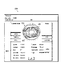
FIG. 2 is an example image 200 of one eye that may be displayed on a screen
display
140b or 150b in system 100. Image 200 includes a simultaneous graphical
representation 220
of the planned flap location, as well as a graphical representation 230 of the
planned area of
ablation in the same eye image 210. As a result, the relative locations of the
planned flap
location and the planned area of ablation may be readily visually compared.
This allows the
user to ensure that the flap is in the correct location for ablation without
having to cut an
overly large flap.
Graphical representation 220 may include other information regarding the flap
in
addition to the flap location. Similarly, graphical representation 230 may
include other
information regarding ablation in addition to the area of ablation. For
instance, in the
example of eye image 210, graphical representation 230 is a heat map with
different colors
represented planned ablation locations.
Eye image 210 may include other graphical information regarding the eye, such
as the
optical zone, location or size of the pupil, or location or size of the iris.
Image 200 may contain information regarding the eye or the planned eye surgery
other than eye image 210. Image 200 may further contain information regarding
the patient,
such as a name 240.
5
CA 03014393 2018-08-13
WO 2017/175026
PCT/1B2016/051906
Image 200, when presented prior to cutting the flap, may include textual flap
information 250 for displaying and editing the parameters of the flap. These
parameters may
include, for example, the side angle of the cut, the diameter and thickness of
the flap, the
position and size of the flap hinge, and the size of a ventilation canal. The
flap and hinge
parameters may be adjusted through one or more input devices, which may be the
same as or
separate from control devices. As the flap and hinge parameters are adjusted,
the graphical
representation 220 of the flap is adjusted to accurately represent the
currently entered
parameters. Alternatively, system 100 may allow adjustments by using an input
device to
manipulate graphical representation 220.
Image 200, when presented prior to ablation, may also include textual
information
260 for displaying and editing the parameters of the ablation. These
parameters may be
presented in terms of the planned correction to near- or far-sightedness or to
astigmatism.
Such planned correction may be presented as sphere, cylinder, and axis
parameters. The
parameters may also include a specification of the size of the area ablation
and may be
.. presented as sizes for a planned optical zone and a planned ablation zone.
The parameters
may be adjusted through one or more input devices, which may be the same as or
separate
from control devices. As the ablation parameters are adjusted, the graphical
representation
230 may be adjusted to accurately represent the currently entered parameters.
Alternatively,
system 100 may allow adjustments by using an input device to manipulate
graphical
.. representation 230.
During eye surgery, between cutting the flap and ablation, an alternative
image
similar to image 200 may be presented. This image may include a graphical
representation of
the actual flap location and, simultaneously, a graphical representation of
the planned area of
ablation. A graphical representation of the planned flap location may also be
presented.
Such an image may allow the user to evaluate whether the actual flap location
is sufficiently
similar to the planned flap location, whether the actual flap location is
sufficient to allow the
planned ablation, or to otherwise evaluate whether it is appropriate to
proceed with ablation.
Similarly, after eye surgery or after ablation, another alternative image
similar to
image 200 may be presented. This image may include a graphical representation
of the actual
area of ablation or other actual ablation information and, simultaneously, a
graphical
representation of the planned or actual flap location. A graphical
representation of the
planned area of ablation or other planned ablation information may also be
presented. Such
an image may allow the user to evaluate whether the surgery proceeded as
planned, whether
6
CA 03014393 2018-08-13
WO 2017/175026
PCT/IB2016/051906
any additional ablation is needed, and any effects of deviations from the
planned flap
location, planned area of ablation, or other planned ablation information. For
instance, some
deviations from the planned area of ablation may be corrected immediately by
performing
additional ablation.
Image 200 or other images as described above may include a schematic,
photographic, or video image of the actual eye and may be overlaid on the eye
image 210.
Although image 200 is a top-down view, images according to the present
disclosure
may be from other angles or points of view. For instance, the image may be a
profile or
cross-sectional view.
By simultaneously presenting graphical information regarding the flap location
and
the area of ablation, system 100 allows the user to more readily identify
inconsistencies
between planned or actual flap location and area of ablation. Particularly
before cutting the
flap or performing ablation, or even after cutting the flap, but before
ablation, identifying
inconsistencies may allow corrective actions to improve surgical outcome. Even
if only
actual inconsistencies are identified, corrective actions may still be
possible, or post-operative
treatments may be improved.
In order to take further advantage of the ability to identify inconsistencies
using an
image presented by system 100, the image or another component of system 100
may alert the
user to potential inconsistencies between flap location, area of ablation, or
other ablation
information. For example, the image presented may alert the user to a
potential
inconsistency using color, icons, dialog boxes, sounds, or other warnings.
FIG. 3 is an example image 300 of both eyes that may be presented on a screen
display 140b or 150b in system 100.
FIG. 4 is a flowchart of a method 400 for performing eye surgery using a
single image
that simultaneously presents graphical information regarding the flap location
and the area of
ablation. In step 410, at least one of the patient's eyes is examined to
identify and determine
the parameters of visual or eye defects.
In step 420, information derived from the examination is entered into a
surgery
system. In step 430, an image of at least one eye containing graphical
information regarding
flap location and the area of ablation in a planned surgery is generated and a
single image
simultaneously presenting this information is produced.
In step 440, the image is evaluated for consistency of the planned flap
location and the
planned area of ablation. If the planned flap location and area of ablation
are not consistent,
7
CA 03014393 2018-08-13
WO 2017/175026
PCT/1B2016/051906
then an adjustment to a planned flap location parameter or a planned area of
ablation
parameter is made in step 450 and a new image is generated.
If the planned flap location and area of ablation are consistent, then a flap
is cut in
step 460.
The flap is them moved to allow access to an interior part of the cornea in
step 470.
Next, in step 480, ablation is performed in the area of ablation.
The process may include other steps in addition to those described above. For
instance, after step 460, the graphical information regarding actual flap
location may be
determined and presented simultaneously with the graphical information
regarding planned
area of ablation. Graphical information regarding the planned flap location
may also be
presented simultaneously.
After step 480, graphical information regarding the actual flap location or
planned
flap location may be presented simultaneously with graphical information
regarding the
actual area of ablation. Graphical information regarding the planned area of
ablation may
also be presented simultaneously.
The images referred to in method 400 may also be presented on one or more
different
displays at the same time or as surgery progresses. For instance, if the
surgery is refractive
eye surgery, the image in step 430 may be presented on a display associated
with a cutting
device, such as a femtosecond laser, while the image may be presented on a
different display
associated with a shaping device, such as an excimer laser, after step 460,
but before or
during step 480.
In addition, in some procedures the steps of method 400 may involve additional
activities. For instance, examination of step 410 may include, for example, a
determination
of the size and shape of the iris and pupil, the thickness and shape of the
cornea, the
identification of diseased or irregular corneal tissue, or the desired vision
correction.
During or following the examination, the user may enter and edit parameters of
the
planned flap position, such as, the side angle of the cut, the diameter and
thickness of the
flap, and the position and size of the flap hinge. During of following the
examination, the
user may also enter and edit parameters regarding ablation such as the desired
correction to
near- or far-sightedness or to astigmatism, and a specification of the size of
the area to be
ablated.
8
CA 03014393 2018-08-13
WO 2017/175026
PCT/1B2016/051906
During step 430 or during or in addition to any later steps, a user may be
alerted to
any inconsistencies in the planned surgery, such as in the planned flap
location and planned
area of ablation.
The disclosed systems and methods may be used in LASIK procedures as well as
other procedures such as LASEK, epi-LASIK, trans-epi-LASIK, PKT,
photorefractive
keratectomy (PRK), cataract surgery, and the like, as well as combinations of
such
procedures.
In addition, variations of the disclosed systems and methods may be used in
other
procedures in which corneal tissue is simply removed, such as PTK with shaping
afterwards.
The above disclosed subject matter is to be considered illustrative, and not
restrictive,
and the appended claims are intended to cover all such modifications,
enhancements, and
other embodiments which fall within the true spirit and scope of the present
disclosure. Thus,
to the maximum extent allowed by law, the scope of the present disclosure is
to be
determined by the broadest permissible interpretation of the following claims
and their
equivalents, and shall not be restricted or limited by the foregoing detailed
description.
9