Note: Descriptions are shown in the official language in which they were submitted.
CA 03014895 2018-08-16
WO 2017/142838
PCT/US2017/017669
APPARATUS AND METHOD FOR RAPID AND COMFORTABLE MAGNETIC IMAGING OF
BREAST TISSUES, WITH CULTURAL SENSITIVITY
Cross Reference and Priority Claim:
[0001] This patent application claims priority to U.S. Provisional Application
Provisional Patent
Application No. Patent Application Serial No. 62/296,344, entitled "APPARATUS
AND METHOD
FOR RAPID COMFORTABLE MAGNETIC IMAGING OF BREAST TISSUES, WITH CULTURAL
SENSITIVITY," filed February 17, 2016, the disclosure of which being
incorporated herein by
reference in its entirety.
Field of Use:
[0002] Disclosed embodiments provide a method and apparatus for clinical
imaging of human
tissue, in particular breast tissue.
Background:
[0003] Conventional breast imaging systems have been used to detect and
characterize breast
lesions. Such systems use various imaging modalities including those based on
x-rays, ultrasound,
Magnetic Resonance Imaging (MRI), and visible and infrared light.
Summary:
[0004] Disclosed embodiments provide an apparatus and method for imaging
breast tissue of a
subject, wherein a subject is positioned on a structure so that at least a
portion of the subject's body is
supported by the structure, magnetic resonance imaging is performed on the
portion of the subject's
body using an MRI system including a plurality of MRI coils positioned in
proximity to the structure,
wherein, while the portion of the subject's body is positioned upon the
structure, breast tissue of the
subject's body is compressed in the proximity of plurality of MRI coils.
Brief Description of the Figures:
[0005] The detailed description particularly refers to the accompanying
figures in which:
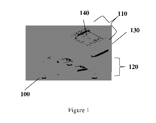
[0006] Figure 1 shows an embodiment of the apparatus, comprising a chair or
other support 100
for a person to sit upon, and an MRI system 110 upon which the person may
place her or his breast
tissues.
[0007] Figure 2 shows a flow chart describing operation of the apparatus and
method.
1
CA 03014895 2018-08-16
WO 2017/142838
PCT/US2017/017669
Detailed Description of the Disclosed Embodiments:
[0008] Figure 1 shows an embodiment of the apparatus comprising a chair or
other support 100
for a person to sit upon, and an MRI system 110 upon which the person may
place her or his breast
tissues. For the purpose of the disclosed embodiments, the term MRI system
comprises components
used to form an image using magnetic resonance or magnetic particle imaging.
The MRI system 110
comprises subunits 130 and 140, where 130 includes coils or magnets (or
electro-permanent magnets)
that polarize protons or other nuclei or electrons in the breast tissues, and
where 140 includes gradient
and/or radiofrequency coils to form an image. Support structure 120 may hold
the MRI system and may
contain other components needed to operate or move the MRI system, for example
wheels and/or
batteries. The display system is not shown in the figure, but is understood to
be present in order to view
images.
[0009] Figure 2 shows a flow chart describing operation of the apparatus and
method. Subject
leans against the MRI system 110 to initiate the process 200. Images are
collected 210, and then the
subject moves away from the MRI 220.
[0010] Disclosed embodiments comprise an apparatus and method for imaging the
mammalian
breast. In accordance with at least one embodiment, a person (typically
female) sits upon a patient
support structure 100 and, with the assistance of gravity places her breast
tissues upon an MRI system
110, thereby partially compressing the breast tissues. Compression may be
helpful in a single-sided
MRI, because in a single-sided MRI, the usable field-of-view does not
typically extend throughout the
patient's body, but only extends a small distance (for example, 15 cm) from
the edge of the MRI system.
Typically, the act of sitting would be with bent knees; however, alternative
structural configurations to
assist a subject's comfort may be provided to facilitate positioning of the
subject and breast tissues.
[0011] It is understood that the term "her" may refer to a male or female
person, and the term
breast may refer to a male breast (as a result of gynecomastia), or to a
female breast including artificial
markers or fillers.
[0012] It should be understood that, in accordance with disclosed embodiments,
images of breast
tissues may be obtained for the breast piecewise, that is by imaging one or
more sections of the breast at
a time, since it is often difficult in a single-sided MRI to obtain very good
uniformity over the entire
breast. Such sectional images can be assembled into an image of the entire
breast with software. It is
understood that the spatial resolution of certain portions of breast tissues
may be different than in other
2
CA 03014895 2018-08-16
WO 2017/142838
PCT/US2017/017669
portions, depending on the gradient applied at the time of image acquisition,
which may be useful in
order to better characterize certain regions of breast tissues.
[0013] In accordance with at least one embodiment, it is not necessary for the
subject to disrobe,
since the MRI signal from the breast tissues is not substantially affected by
the presence of thin layers of
clothing between the breast tissues and the MRI. This lack of requirement to
disrobe has particular
technical utility that is not usually found in other breast imaging
modalities, and is useful in populations
where there are cultural prohibitions against removal of clothing under
certain circumstances.
[0014] In accordance with at least one embodiment, the MRI system 110 may be
suspended or
otherwise attached to a platform 120, which may contain electronics or
batteries or wheels or other
material. It is to be understood that the patient support structure 100 and/or
the MRI support structure
120 may have parts that are adjustable in order to accommodate patients of
different heights and sizes.
[0015] The MRI system 110 may be a single-sided MRI (as depicted in Figure 1),
or may be a 3-
sided system, so long as the person may rest her chest against a portion of
the system 110. The portion
of the MRI system 110 comprises subunits 130 and 140 that are used to form the
MR image. Subunit
130 may comprise electrical coils and/or electro-permanent magnets, in which
said electro-permanent
magnets that are magnetized by a transient current flowing through electrical
coils and stay activated
until the magnetization is removed by other transient currents flowing through
electrical coils. Subunit
140 comprises radiofrequency, gradient, pre-polarizing and/or shimming coils
that may be needed to
form an image. Subunit 130 coils may also have a role to play as gradient and
shim coils. It is to be
understood that a waterproof material or another housing material to prevent
user interference may cover
the MRI system 110 and/or its subunits.
[0016] In an embodiment, ultra-fast and high-magnitude gradient pulses as
described by Irving
Weinberg in U.S. Patent 8,154,286, entitled "APPARATUS AND METHOD FOR
DECREASING
BIO-EFFECTS OF MAGNETIC FIELDS," and related patents and patent applications
(related by
priority claims), all being incorporated by reference, may be used to collect
many sets of data points in
order to achieve high spatial resolution and signal-to-noise ratio, without
causing uncomfortable nerve
stimulation. As taught in patent 8,154,286, the MRI could employ a gradient
transition time of 10
microseconds or less, which is less than the neurological response time for
neurological tissue. The slew
rate (that is, the change of magnetic field per distance per time) is
increased as a result of the reduced
pulse ramp times. The plateau magnitude of the magnetic gradient pulse is
increased, as compared to the
prior art, because of several factors. Firstly, the plateau magnitude may be
increased because of the
3
CA 03014895 2018-08-16
WO 2017/142838
PCT/US2017/017669
improved switching techniques as described above. Secondly, the plateau
magnitude may be increased
because the tissues are depolarized and repolarized within a short period of
time similar to the
neurological response time. As discussed in Patent 8,154,286, the magnitude of
the gradient pulse may
be as high as 1000 T/m.
[0017] Such high magnetic gradient field magnitude may be 400 mT or higher,
with rise-times
of 10 microseconds or less. The gradient pulses may be so rapid as to permit
acquisition in a very short
time, for example 10 seconds or less, so that there is little motion of the
breast during acquisition,
thereby reducing resolution loss from "motion-unsharpness."
[0018] In accordance with at least one embodiment, pre-polarizing coils may be
activated in
order to improve signal-to-noise ratio, as taught in U.S. Patent 8,836,329 by
Weinberg, entitled
"ULTRA-FAST PRE-POLARIZING MAGNETIC RESONANCE IMAGING AND SYSTEM"
(incorporated by reference). As taught in U.S. Patent 8,836,329, a pre-
polarizing magnetic pulse may be
applied to a structure of interest, in which the magnetic pulse has a rise-
time of less than 10
microseconds and a fall time of less than 10 microseconds, or the magnetic
pulse following a pre-
polarizing magnetic pulse has a rise-time of less than 10 microseconds and a
fall time of less than 10
microseconds. As recited in Patent 8,836,329, it is conventionally known that
application of a high
transient magnetic field during the polarization portion of the pulse sequence
results in an improved
signal (see for example, A Macovski, S Conolly: "NOVEL APPROACHES TO LOW-COST
MRI", in
Magnetic Resonance in Medicine 30:221-230, the subject matter of which is
incorporated herein by
reference in its entirety) because more spins are aligned; as a result, the
application of this field
subsequently results in output of a more significant signal as they return to
their equilibrium state.
[0019] In accordance with at least one embodiment, electro-permanent magnets
may be
deactivated in the case of nearby ferromagnetic materials, as taught in U.S.
Provisional Patent
Application number 62/292945 (now filed as a U.S. patent application
15/427,426) by Weinberg and
Nacev, entitled "METHOD AND APPARATUS FOR MANIPULATING ELECTRO-PERMANENT
MAGNETS FOR MAGNETIC RESONANCE IMAGING AND IMAGE GUIDED THERAPY"
(incorporated by reference). As taught in that application, a soft magnetic
material can be in close
proximity to an additional soft magnetic material and a hard magnetic material
and a conductive
material to form one or more electropermanent arrays. Conductive material near
the soft magnetic
material may be energized with current, so that magnetic component from the
one or more
electropermanent arrays will be magnetized in a direction and/or magnitude,
which may be selected by a
4
CA 03014895 2018-08-16
WO 2017/142838
PCT/US2017/017669
user (via controlling equipment) or automated algorithm by a computer (that
provides an automated or
semi-automated controller). The magnetic field produced by one or more
electropermanent arrays can be
reduced or increased by adjusting the magnetization of one or more
electropermanent arrays.
In an embodiment, the ultra-fast gradient pulses may be used to effectively
visualize and/or segment
small calcifications in the breast tissues, which is generally not possible
with MRI because the pulse
sequences of MRI are too slow to catch the rapidly decaying signals from solid-
bound water near
calcifications. This method is similar to that described by Nacev in U.S.
Provisional Patent Application
No. 62/255,843 (and now filed as U.S. Patent application 15/352,164) entitled
"METHOD AND
APPARATUS FOR HIGH SLEW RATE SINGLE POINT MAGNETIC RESONANCE IMAGING OF
MAGNETIZABLE NANOPARTICLES" (incorporated by reference). As taught in those
patent
applications, magnetic gradient pulses are applied with very short durations
(for example, between 10
and 200 microseconds), and/or switched on and/or off quickly (for example,
between 10 and 100
microseconds). The quickly actuated short gradient pulses (see 330 and 340)
allow for polarized species
to be imaged very quickly after an RF excitation pulse (e.g. with very short
TE times). The rapid decay
of signals from protons in the region of microcalcifications may be employed
to segment the
microcalcifications, thereby aiding in diagnosis.
[0020] In accordance with at least one embodiment, the apparatus may be
lightweight enough to
be transported on wheels and may take such little power to operate that it may
be operated in remote
locations using batteries or small generators.
[0021] The magnetic field from electropermanent magnets may be rapidly reduced
through
application of electrical currents, which would be useful in the case of
ferromagnetic objects being
attracted to the electropermanent magnet. In accordance with at least one
embodiment, such rapid
reduction would be actuated by a technologist. In an embodiment, said
reduction could be performed
automatically by a computer that detecting the presence of ferromagnetic
objects approaching the
apparatus. Said detection could include a change in the radio-frequency
signals collected by the
apparatus. An example of such safety feature was described in the U.S. patent
application 15/427,426
(as discussed above) by Weinberg entitled "METHOD AND APPARATUS FOR USING
ELECTROPERMANENT MAGNETS FOR MAGNETIC RESONANCE IMAGING AND IMAGE-
GUIDED THERAPY" (incorporated by reference).
[0022] In accordance with at least one embodiment, the pixel size for images
obtained with the
apparatus may be less than 50 microns, as taught by Nacev and others in the
2014 ISMRM publication
CA 03014895 2018-08-16
WO 2017/142838
PCT/US2017/017669
entitled "A quiet, fast, high-resolution desktop MRI capable of imaging solid-
bound water"
(incorporated by reference).
[0023] In at least one embodiment, fast MRI pulse sequences are used to image
calcium-rich
structures, such as microcalcifications that often accompany breast cancers.
The use of such pulse
sequences without unpleasant nere stimulation are described in the U.S. patent
application 15/352,164
by Nacev entitled "METHOD AND APPARATUS FOR HIGH SLEW RATE SINGLE POINT
MAGNETIC RESONANCE IMAGING OF MAGNETIZABLE NANOPARTICLES" (as discussed
above and incorporated by reference). Conventional MRI systems obtain low
signals from such
structures.
[0024] In accordance with at least one embodiment, the pixel size may be less
than 20 microns.
[0025] In accordance with at least one embodiment, the spatial resolution and
pixel size is
sufficient to perform MRI histology, in which the internal features of cells
(for example, nuclear to
cytoplasm ratio) may be observed in order to characterize whether the cell is
malignant or not. It should
be understood that the MR images obtained with the apparatus may be employed
in order to guide a
biopsy or other intervention. Examples of such interventions may include
destruction of tumor cells via
radiofrequency deposition, or via heating or motion of small magnetic
particles introduced into the body
intravenously or some other means.
[0026] It should be understood that the MRI examination obtained with the
presently disclosed
apparatus may be performed with contrast administered and/or may employ
diffusion-weighted or other
imaging methods to detect and characterize breast lesions. It should be
understood that the images may
be used to guide biopsy, potentially through correlation with other imaging
modalities such as
ultrasound. It should also be understood that an ultrasound transducer may be
incorporated into MRI
system 110 so as to collect co-registered MRI and ultrasound images.
[0027] In accordance with at least one embodiment, images of one or both
breasts may be
obtained in a single session with the system.
[0028] In accodance with at least one embodiment, one or more coils or electro-
permanent
magnets within the MRI system may be fabricated with additive manufacturing,
as taught by Urdaneta et
al in the 2011 IEEE Medical Imaging Proceedings entitled "Good-bye Wires and
Formers: 3-D Additive
Manufacturing and Fractal Cooling Applied to Gradient Coils".
[0029] In accordance with at least one embodiment, the subject may pull
herself towards the
MRI system by using her arms to grab a projection from the apparatus.
6
CA 03014895 2018-08-16
WO 2017/142838
PCT/US2017/017669
[0030] For the purposes of this disclosure, the term "external pressure" is
intended to mean any
force applied to any portion of the subject other than gravity or the
subject's own exertions. As an
example, a subject may lean against the apparatus, using the force of gravity
to compress one or more
portions of a breast against one or more surfaces of the apparatus. In an
alternative embodiment, the
subject may use her arms to grasp a projection as to compress one or more
portions of breast against one
or more surfaces of the apparatus.
[0031] It is understood that the invention may be applied to both men and
women. In the case of
men, the technical utility of the disclosed embodiments may be particularly
useful to provide good
options for breast examination of men.
[0032] It should be understood that the operations explained herein may be
implemented in
conjunction with, or under the control of, one or more general purpose
computers running software
algorithms to provide the presently disclosed functionality and turning those
computers into specific
purpose computers.
[0033] Moreover, those skilled in the art will recognize, upon consideration
of the above
teachings, that the above exemplary embodiments may be based upon use of one
or more programmed
processors programmed with a suitable computer program. However, the disclosed
embodiments could
be implemented using hardware component equivalents such as special purpose
hardware and/or
dedicated processors. Similarly, general purpose computers, microprocessor
based computers, micro-
controllers, optical computers, analog computers, dedicated processors,
application specific circuits
and/or dedicated hard wired logic may be used to construct alternative
equivalent embodiments.
[0034] Moreover, it should be understood that control and cooperation of the
above-described
components may be provided using software instructions that may be stored in a
tangible, non-transitory
storage device such as a non-transitory computer readable storage device
storing instructions which,
when executed on one or more programmed processors, carry out the above-
described method
operations and resulting functionality. In this case, the term non-transitory
is intended to preclude
transmitted signals and propagating waves, but not storage devices that are
erasable or dependent upon
power sources to retain information.
[0035] Those skilled in the art will appreciate, upon consideration of the
above teachings, that
the program operations and processes and associated data used to implement
certain of the embodiments
described above can be implemented using disc storage as well as other forms
of storage devices
including, but not limited to non-transitory storage media (where non-
transitory is intended only to
7
CA 03014895 2018-08-16
WO 2017/142838
PCT/US2017/017669
preclude propagating signals and not signals which are transitory in that they
are erased by removal of
power or explicit acts of erasure) such as for example Read Only Memory (ROM)
devices, Random
Access Memory (RAM) devices, network memory devices, optical storage elements,
magnetic storage
elements, magneto-optical storage elements, flash memory, core memory and/or
other equivalent
volatile and non-volatile storage technologies without departing from certain
embodiments. Such
alternative storage devices should be considered equivalents.
[0036] While certain illustrative embodiments have been described, it is
evident that many
alternatives, modifications, permutations and variations will become apparent
to those skilled in the art
in light of the foregoing description. Accordingly, the various embodiments
of, as set forth above, are
intended to be illustrative, not limiting. Various changes may be made without
departing from the spirit
and scope of the invention.
8