Note: Descriptions are shown in the official language in which they were submitted.
- 1 -
METHOD OF QUANTITATIVE MEASUREMENT OF PARTICLE CONTENT
USING HYDRATED STATE IMAGING
Technical Field
The invention relates to a method of quantitative measurement
of particle content by using a hydrated state imaging method
such as CryoTEM (Cryo Transmission Electron Microscopy).
Background and Summary of the Invention
In the pharmaceutical industry, Virus-Like Particles (VLPs)
and wild-type (wt) or modified viruses, for example, Adeno
Associated Virus (AAV) particles are extensively used as a
carrier for gene delivery. In general, VLPs or replication
deficient AAVs cannot replicate/reproduce as opposed to real
virus particles and are often preferred as a carrier for gene
delivery. The assessment of their content of genetic material
is of prime importance as it is directly linked to the
efficiency of the treatment. Different methods are commonly
used to assess the content of Virus-Like Particles (VLPs) and
AAV particles. One method is real-time polymerase chain
reaction, also known as quantitative polymerase chain reaction
(qPCR). Historically, negative-stain Transmission Electron
Microscopy (nsTEM) has been used as an orthogonal direct
method used as a reference to visualize the content of VLPs
and AAV particles. One reason for this is that nsTEM is fast,
CA 3015803 2018-08-29
,
, .
- 2 -
simple and provides a good resolution so the VLP and AAV
particles can actually be seen. Another is that nsTEM has been
considered accurate and a good method for determining particle
content. However, it was recently discovered that nsTEM has
inherent characteristics that makes it unreliable, not robust,
and even erroneous when it comes to assessing the content of
VLPs, AAV particles and wt virus particles.
In nsTEM, stain is applied to the sample before or after the
sample is applied on the grid to enhance the contrast and
protect the particles. One drawback of nsTEM is that the
stain covers the particles and does not necessarily penetrate
the particles. This prevents the direct native viewing of the
content of the particles. That is, the stain makes the
analysis of the content of particles in nsTEM an indirect
method. In other words, the stain only enters the particles
and creates contrast representing the interior of the
particles when there is an opening in the particle shells, for
example, when the particles are broken. The stain might also
adversely affect the morphology of the sample, and due to the
blotting steps (removing access liquid using a filter paper)
and the low pH of the stain solution, it was surprisingly
realized that particles are often spatially locally affected
by the preparation. It was unexpectedly discovered that the
thickness of the stain layer cannot be fully controlled in the
preparation procedure and in regions with thinner stain the
CA 3015803 2018-08-29
!
- 3 -
particles are not well protected. It turns out that the shape
of empty particles is sometimes affected by the staining and
blotting procedure, even if they are intact. The preparation
procedure can create a dent or invagination on the shell at
the top of the particle where stain can assemble. This is more
likely for empty particles since the interior content then
does not help to retain the shape. This makes it look empty
in the microscope whereas in regions with thicker stain, the
particle shape is intact and there is no visible difference
between an empty and filled intact particle. This makes the
analysis difficult and unreliable.
In nsTEM, the particles are conventionally seen, classified
and counted as empty if they appear with a bright outer fringe
and a dark internal part. The rationale is that once on the
grid, an empty particle collapses and the stain fills the
hollow parts. Full particles are those appearing as bright
disks with slightly brighter parts at the center. The
rationale is that since the particles are filled, they do not
collapse during the preparation and therefore have no hollow
part. There are often a large portion of the particles that
cannot be unambiguously classified. They are so called
uncertain particles.
Another problem is that when an empty particle does not
collapse during preparation, it appears as filled while, on
CA 3015803 2018-08-29
- 4 -
the other hand, when a filled particle collapses, due to high
mechanical constrains on a local level, it may collapse and
appear as empty. An important insight of the present
invention is the realization that the method of using nsTEM is
prone to a high number of false positives and false negatives.
There is a need for a better and a more reliable way of
quantifying particle content. The method of the present
invention is a reliable method by which the content of VLPs,
AAVs and wt viruses can be assessed with good robustness,
accuracy, repeatability and specificity. More particularly,
the method is for quantitative characterization of the content
of VLPs, AAVs and wt virus particles by imaging them in their
native hydrated state such as by using Cryo Transmission
Electron Microscopy (CryoTEM). It was also discovered that
the analysis can reliably be done by using ionic liquids to
prepare the sample for TEM imaging or using special sample
holders for liquid samples (sometimes referred to as liquid
TEM or in situ TEM). The ionic liquid preparation method is
similar to CryoTEM in that the addition of the ionic liquid
keeps the particles in a hydrated state so there is no need to
use stain to enhance the contrast. However, a small amount of
stain or chemical can beneficially be added to preserve the
structure of the particles.
The method of the present invention provides a solution to the
CA 3015803 2018-08-29
- 5 -
above-outlined problems. More particularly, the method is for
the quantitative measurement of the particle content of
particles by using a hydrated state imaging method such as
CryoTEM. A sample of virus or virus-like particles (VLPs),
such as AAV particles, or virus-particles is provided. The
sample is prepared to maintain the sample in a hydrated state.
This may be done in several ways. For CryoTEM in a preferred
embodiment, the sample is rapidly frozen into a cryogenic
liquid at a cryogenic temperature. While at the cryogenic
temperature, a particle content of each VLP in the frozen
sample is observed in the CryoTEM imaging device. For other
hydrated state imaging methods, the imaging is performed in a
TEM imaging device but not at cryogenic temperatures, by using
liquid sample holders, or by adding an ionic liquid to the
sample at preparation. A measurement of the particle content
is determined to assess whether the VLPs are empty or not.
In an alternative embodiment, the method further comprises the
step of automatically or manually detecting particles in the
images and displaying detected particles on a display and
automatically or manually deleting particles that are smaller
than a lower size limit and larger than an upper size limit.
It is also possible to automatically remove or add particles
to the image without first displaying the particles. It is
also possible to display particles and interactively delete or
add particles to the image.
CA 3015803 2018-08-29
i
, .
- 6 -
In another alternative embodiment, the method further
comprises the step of automatically or manually classifying a
particle that has an inner density with no distinct boundary
between a particle shell and a particle core as a filled
particle.
In yet an alternative embodiment, the method further comprises
the step of automatically or manually classifying a particle
that has a distinct outer shell and a minute internal density
as an empty particle.
In an alternative embodiment, the method further comprises the
step of using Cryo Transmission Electron Microscopy to
determine the particle content of the VLPs.
In another alternative embodiment, the method further
comprises the step of determining the particle content of
adeno associated virus (AAV) particles.
In yet an alternative embodiment, the method further comprises
the step of using the AAV particles as a carrier for gene
delivery.
In an alternative embodiment, the method further comprises the
step of classifying AAV particles that contain one or more
CA 3015803 2018-08-29
I
- 7 -
copies of a gene as a filled particle.
In yet another alternative embodiment, the method further
comprises the step of classifying AAV particles that contain
no gene as an empty particle.
In another embodiment, the method further comprises adding an
ionic liquid to the sample to keep the VLPs in a hydrated
state.
In another embodiment, the method further comprises imaging
the VLP particles in their native, liquid and hydrated state
by using a liquid sample holder.
Brief Description of Drawing
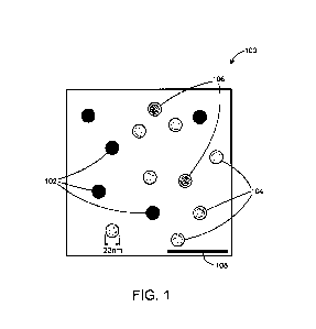
Fig. 1 is a schematic image from an AAV specimen
observed by CryoTEM.
Detailed Description
The present invention relates to a method of using a hydrated
state imaging method such as CryoTEM to assess and
quantitatively measure the degree of content in the interior
of VLPs, AAV particles and wt virus particles. The particle
content measurement could be a metric (number) that
corresponds to how full or empty the particle is. It could
e.g. be a measurement of the overall intensity of the particle
CA 3015803 2018-08-29
- 8 -
interior, or the intensity normalized with the intensity on
the shell of the particle. It could also be a measure of how
much of the area inside the particle that is bright or dark.
As mentioned above, an important aspect of the present
invention is the realization and discovery that nsTEM is not
suitable for the assessment/analysis of filled/empty particles
due to the fact that the particle appearance when imaged on
the grid depends on several parameters such as:
- The thickness of the stain (which varies throughout the
grid);
- The extent to which the specimen is dried (which varies
throughout the grid); and
- The integrity of the particles (which may or may not be
affected by the staining process and preparation process due
to local variations in the mechanical stress the particles
undergo).
Because the stain thickness influences the appearance of the
particles it also affects the result. The stain thickness
varies over the grid and this cannot be reliably controlled.
For example, when the stain is relatively thin, the particles
are more exposed to physical forces in the preparation and
some particles may cave so that the stain is contained in the
cave without penetrating into the inside of the particle.
When the stain remains in the cave of the particle, it gives
an appearance that is very similar to that of empty particles
CA 3015803 2018-08-29
- 9 -
that have been filled by the stain. The particle content
analysis thus depends a lot on whether particles in regions
with thick or thin stain are imaged and analyzed. As a result
of the uneven distribution of the stain on the particles when
nsTEM is used, the particles often appear as having different
amounts of content although they, in reality, have not. This
is an insight that has not been realized in the past.
By using CryoTEM instead, the appearance of the particles
cannot be disadvantageously affected by an uneven distribution
of the stain (since no stain is used) so the particles tend to
look the same in all areas of the viewing area which makes
CryoTEM very effective for the content analysis of the present
invention. In other words, although it is more complicated to
use CryoTEM compared to nsTEM for content analysis, it was
unexpectedly discovered that the advantages of the more
accurate results outweigh the drawbacks of using the more
cumbersome CryoTEM technique. In CryoTEM, according to the
method of the present invention, a small aliquot of a sample
is deposited onto a hydrophilized copper grid covered with a
thin carbon film. The excess of the sample is then blotted-
off by using filter paper. The grid is then rapidly, and
before the sample dries out, plunged into a cryogenic liquid
where the sample of particles is instantly frozen. The rapid
freezing allows the sample/specimen to be embedded in
amorphous ice close to its native (i.e. unstained) hydrated
CA 3015803 2018-08-29
- 10 -
form so the particles has the correct appearance everywhere in
the sample since they are not affected by any stain. The
specimen is then kept at cryogenic temperatures during the
whole process while being inserted and observed in the
transmission electron microscope. One advantage of the method
of the present invention is that it allows a direct
visualization of unaltered particles with the possibility of
seeing their internal features. This makes it possible to
make a more correct assessment of whether a particle is empty
or not. In CryoTEM, empty particles appear as disks that has
a minute internal density. One explanation is that the
internal parts of particles can be seen using CryoTEM and
empty particles have a low internal density. Filled particles
appear as dark homogeneous disks. Again, the internal parts
of particles can be seen using CryoTEM, and filled particles
that contain genetic material inside, have a homogeneous
internal density.
EXAMPLE
Below is a detailed example of how CryoTEM is used to carry
out the method of the present invention.
Grid preparation
Suitable grids, such as 400 mesh copper (Cu) grids, were first
hydrophilized. This was done by glow-discharging the grids.
CA 3015803 2018-08-29
- 11 -
More particularly, the copper grids, covered with a carbon
film, were placed in a glow discharger. Vacuum was applied
until the pressure reached about 0.5 mbar in the chamber. A
current was applied, such as about 20 m7-\, for about 1 minute.
The pressure was then increased to ambient pressure. The
grids were removed and the glow-discharger was turned off.
Grid Freezing
A plunge freezer was turned on. The sample chamber was
equilibrated to the desired temperature and humidity. The
blot paper in the sample chamber was changed. An ethane bath
in the cooling station was prepared. A freshly glow-
discharged grid was loaded on the tweezers. The freezing
process was started. About 3 pL of the sample was deposited
on a grid. After about 10 seconds of wait time, the grid was
blot with filter paper and plunge-frozen. The grid was
transferred in a cryo-grid box and stored in liquid nitrogen.
The ethane and liquid nitrogen were safely thawed and the
plunge-freezer was turned off.
Grid Transfer
The cryo-grid box was transferred from its storage location
into a cryo-work station precooled with liquid nitrogen, into
which a cryo-holder preliminary pump was inserted. The grid
was transferred to the grid slot on the cryo-holder. The
cryo-holder was inserted into the CryoTEM and the liquid
CA 3015803 2018-08-29
i
. .
- 12 -
nitrogen container was filled.
Grid imaging
In the grid imaging step, it was important to make sure the
microscope had been correctly aligned according to the
protocol described by the manufacturer, and that the blank
image from the camera was flat. (It is to be understood that
the grid imaging step may be done automatically where images
are acquired automatically without requiring an operator to be
sitting at the microscope to acquire the images. The grid is
screened until finding a suitable area.) The magnification
was then set with a field of view of about 600 - 1000 nm. The
focus 0 was found before setting the microscope at a slight
defocus of about 6 pm. This defocusing step could have been
done manually or automatically in microscopes that have
autofocus and defocus functionality. The image was acquired
and moved to a nearby area. The step of acquiring the image
was repeated until the desired number of images was acquired.
Subsequent image treatment and analysis
The images were saved and imported by suitable analysis
software such as Vironova Analyzer Software (VAS). The images
to be saved in the microscope were selected and saved in a
suitable format such as in 16bit tiff format, or alternatively
automatically saved after the automatic image acquisition. A
CA 3015803 2018-08-29
I
- 13 -
folder corresponding to the project in VAS was created and all
the required information in the different nodes was completed.
The images were imported in the "Microscopy" node by right
clicking on the node, selecting "Open image(s)" and choosing
the appropriate files prior to clicking on "Open".
Particle detection and classification
In the "Microscopy" node, in the "Particle Type" field, the
"VLP(cryo)" was entered. The images in which particles were
to be detected were selected before right-clicking on one of
them. A "Run detection..." was chosen. The following
parameters were entered:
Detection algorithm Ellipse
segment ion
k4 ______________________________________________________
Recommended Acceptable
Parameter
Nominal Value Range
Clear Content of Detected Yes Yes
Particle
Dark membranes Yes Yes
Divide Large Components Yes Yes
Edge Gap Tolerance 0.2 0.2
Edge Width (nm) 5 4 - 6
CA 3015803 2018-08-29
- 14 -
Maximum Diameter (nm) 24 22 - 30
Minimum Diameter (nm) 18 16 - 22
Minor Axis Ratio 0.2 0.2
Output shape Circular Circular
Post Processing Refinement Yes Yes
Prefer Circular Ellipses Yes Yes
Pre-processing method EdgeDetection EdgeDetection
The detected particles were displayed on the Plot Control by
using the scatterplot display, with "Size" on the x axis, and
"Signal-To-Noise" on the y axis. The detected particles with
a signal to noise <0.1 were first selected before deleting
them.
The detected particles with a size <17 nm and >28 nm were then
selected before deleting them also. The images were visually
assessed on the screen and falsely and incorrectly detected
AAV particles were removed. The correctly detected particles
were accepted by using the verify tool. The AAV particles
that were not detected by the automated detection were
manually boxed.
In more general terms, the following analysis steps were
performed:
1) The particles of interest in the images were detected
either manually or by using a suitable detection algorithm
CA 3015803 2018-08-29
- 15 -
(for example, template matching, circular object detection,
region or border-based detection methods etc.);
2) False detections, based on measures of size, shape and the
signal to noise ratio for each particle, were removed
(automatically or manually or a combination of both); and
3) If necessary, particles that were not detected were added
if an automated detection algorithm was used.
Particle classification
In the particle class node of the Plot control toolbar,
"Content" was chosen. All the detected particles were
displayed by using the RDP PCA tool in the Plot control
toolbar. The plot was rotated in order to obtain a clear
separation between two clusters. The particles of one cluster
were selected and assigned their corresponding class. The
class was determined by using the following parameters:
-AAV particles displaying an inner density with no
distinct boundary between the shell and the core were
classified as filled particles; and
- AAV particles displaying a distinct outer shell and
minute internal density were classified as empty
particles.
The particles from the other cluster were then selected and
assigned their corresponding class. All the images were
CA 3015803 2018-08-29
- 16 -
visually analyzed to assess the classification. The class
"Uncertain" was assigned to all particles in which a
discrepancy was found between the analyst's assessment and the
semi-automated classification.
In more general terms, the following steps were performed:
1) The content of the particles was measured by analysing
the overall intensity and intensity distribution inside the
particles; and
2) The particles were classified based on these
measurements. This could have been done in several ways such
as by manually thresholding each measured feature (e.g. if
darker than a certain intensity Tf then classify as full, if
brighter than another intensity Te then classify as empty and
if between Tf and Te then classify as uncertain). It could
also have been done by marking groups of particles in
scatterplots of the features or by using
automatic/semiautomatic clustering and classification methods.
It is possible to, in a fully automated fashion, discriminate
between filled and empty particles by looking at the internal
density profile of the particles.
Fig. 1 is a schematic illustration of a typical image of VLPs
from an AAV specimen 100 observed by CryoTEM. The specimen
contains filled particles 102 that appear as plain dark disks.
The particles 102 are thus filled with, for example, a
CA 3015803 2018-08-29
- 17 -
pharmaceutical substance or a gene. Empty particles 104
appear as dark circles with a bright internal intensity
corresponding to low internal density because they contain or
carry no gene. Particles 106 for which the classification is
ambiguous appear to display characteristics of in between
filled and empty particles. The analysis thus determines both
the quantity/number of particles that contain the
pharmaceutical substance (gene) and also how much each
particle is filled with the pharmaceutical substance or gene.
Some particles may only be partially filled with a
pharmaceutical substance whereas for genes, the particles
either contain one or more copies of the gene or are empty.
The scale bar 108 of Fig. 1 represents 100nm.
While the present invention has been described in accordance
with preferred compositions and embodiments, it is to be
understood that certain substitutions and alterations may be
made thereto without departing from the spirit and scope of
the following claims.
CA 3015803 2018-08-29