Note: Descriptions are shown in the official language in which they were submitted.
CA 03015913 2018-08-27
WO 2017/151517
PCT/US2017/019733
METHODS OF TREATING CANCER
CROSS REFERENCE TO RELATED APPLICATIONS
This application claims the benefit of U.S. Provisional Application No.
62/301,510, filed
February 29, 2016. The contents of the aforementioned application are hereby
incorporated by
reference in their entirety.
FIELD OF THE INVENTION
The invention relates to methods and compositions for treating cancer such as
melanoma.
BACKGROUND OF THE INVENTION
Agents targeting the programmed death-l/ligand (PD-1/PD-L1) axis release
suppressed anti-
tumor T cell responses, resulting in remarkable clinical activity in numerous
cancers (Wolchok. Cell
162:937, 2015). Nivolumab and pembrolizumab induce clinical responses in 25-
45% of patients with
advanced melanoma and are now widely used following regulatory approval
(Topalian et al. N Engl J
Med 366:2443-54, 2012; Hamid et al. N Engl J Med 369:134-44, 2013; Herbst et
al. Nature 515:563-
7, 2014; Robert et al. N Engl J Med, 372:2521-32, 2015; Robert et al. N Engl J
Med, 372:320-30,
2014; Larkin et al. N Engl J Med, 373:1270-1, 2015). Despite this activity,
clinically-accessible and
validated markers to predict response and guide treatment decision-making have
remained elusive.
Recently, clonal expansion of infiltrating T cells and PD-Li expression by
tumor or immune cells
were implicated as potential markers of treatment response (Topalian et al. N
Engl J Med 366:2443-
54, 2012; Herbst et al. Nature 515:563-7, 2014; Tumeh et al. Nature 515:568-
71, 2014; Weber et al.
Lancet Oncol, 16:375-84, 2015).
Despite these findings, the impact of mutational load and specific oncogenic
mutations in
melanoma in response to agents that inhibit PD-1 or PD-Li has not been
systematically explored.
Furthermore, translating genomic studies to routine clinical practice remains
problematic as whole
exome sequencing (WES) is not widely available and is expensive, time
intensive, and technically
challenging.
Therefore, the need exists for novel therapeutic and diagnostic approaches,
including
immunotherapies and genomic tesings, for treating and diagnosing cancer such
as melanomas.
SUMMARY OF THE INVENTION
The invention is based, at least in part, on the discovery that mutational
load correlates with
therapeutic benefit from a therapy that includes an inhibitor of PD-1 or PD-Li
in melanoma. In one
embodiment, profiling a small fraction of the genome or exome, e.g., using a
hybrid capture-based,
next-generation sequencing (NGS) platform, from a patient sample serves as an
effective surrogate for
1
CA 03015913 2018-08-27
WO 2017/151517
PCT/US2017/019733
the analysis of total mutational load. In another embodiment, an alteration
detected in a single gene
(e.g., an NF1 or LRP1B gene) serves as an alternative or further surrogate for
total mutational burden
to predict a response to the therapy. Using methods that include a targeted
NGS platform for
detecting mutational burden has several advantages, including, but not limited
to, faster, e.g., more
clinically manageable turnaround times (-2 weeks), standardized informatics
pipelines, and more
manageable costs, compared to, e.g., whole genome or whole exome sequencing.
The methods
disclosed herein have other advantages over markers such as PD-Li expression,
since they produce an
objective measure (e.g., mutation load) rather than a subjective measure
(e.g., pathology scoring).
The methods disclosed herein also allow for simultaneous detection of
actionable alterations for
targeted therapies, as well as mutational burden for immune therapies. These
methods can provide
clinically actionable predictors of a response to anti-PD-1 and/or anti-PD-Li
therapies in patients with
advanced melanoma.
Accordingly, the invention provides, at least in part, methods for treating a
subject having, or
at risk of having, a cancer (e.g., a melanoma), by administering to the
subject an effective amount of
an agent (e.g., a therapeutic agent) that targets and/or inhibits the PD-1
pathway. In certain
embodiments, the therapeutic agent is an inhibitor of PD-1 or PD-Li. In
certain embodiments, the
therapeutic agent, e.g., the inhibitor of PD-1 or PD-L1, is administered
responsive to a value of
responder status to the therapeutic agent. In certain embodiments, the value
of responder status
includes a measure of the mutation load in a sample (e.g., a tumor sample)
from the subject. In
certain embodiments, the measure of the mutation load includes a determination
of one or more of:
the level of a somatic alteration in a predetermined set of genes disclosed
herein, the presence of a
somatic alteration in an NF1 gene, the number of a somatic alteration in an
LRP1B gene, or the
number of a C to T transition in a predetermined set of genes disclosed
herein, or any combination
thereof.
Methods of Treatment
In one aspect, the invention features a method of treating a subject having a
cancer, e.g., a
melanoma. The method includes:
(a) acquiring a value of responder status to a therapy, e.g., a therapy
comprising an inhibitor
of PD-1 or PD-L1, for the subject, wherein said value of responder status
comprises a measure of the
mutation load in a sample, e.g., a melanoma sample or a sample derived from a
melanoma, from the
subject; and
(b) responsive to an increased value of responder status, e.g., compared to a
reference value of
responder status, administering to the subject the therapy, thereby treating
the subject.
In certain embodiments, the reference value of responder status is a value of
responder status
for a non-responder to the therapy.
2
CA 03015913 2018-08-27
WO 2017/151517
PCT/US2017/019733
In certain embodiments, the measure of the mutation load comprises a
determination of one,
two, three or all of the following in the sample from the subject:
(i) the level of a somatic alteration (e.g., one or more somatic alterations)
in a predetermined
set of genes set forth in Table 1;
(ii) the presence or absence of a somatic alteration (e.g., one or more
somatic alterations) in
an NF1 gene;
(iii) the number of a somatic alteration (e.g., one or more somatic
alterations) in an LRP1B
gene; or
(iv) the number of a C to T transition (e.g., one or more C to T transitions)
in a predetermined
set of genes set forth in Table 1.
In certain embodiments, the therapy is administered to the subject, responsive
to one, two,
three or all of the following in the sample from the subject:
(i) an increased level of a somatic alteration (e.g., one or more somatic
alterations) in a
predetermined set of genes set forth in Table 1, e.g., compared to a reference
level of a somatic
alteration (e.g., one or more somatic alterations) in the predetermined set of
genes set forth in Table
1;
(ii) the presence of a somatic alteration (e.g., one or more somatic
alterations) in the NF1
gene;
(iii) an increased number of a somatic alteration (e.g., one or more somatic
alterations) in the
LRP1B gene, e.g., compared to a reference number of a somatic alteration
(e.g., one or more somatic
alterations) in the LRP1B gene; or
(iv) an increased number of a C to T transition (e.g., one or more C to T
transitions) in a
predetermined set of genes set forth in Table 1, e.g., compared to a reference
number of a C to T
transition (e.g., one or more C to T transitions) in the predetermined set of
genes set forth in Table 1.
In another aspect, the invention features a method of treating a subject
having a cancer, e.g., a
melanoma. The method includes: administering a therapy, e.g., a therapy
comprising an inhibitor of
PD-1 or PD-L1, to the subject, wherein the subject has, or has been identified
as having, an increased
value of responder status, e.g., compared to a reference value of responder
status, and wherein said
value of responder status comprises a measure of the mutation load in a
sample, e.g., a melanoma
sample or a sample derived from a melanoma, from the subject, thereby treating
the subject.
In certain embodiments, the measure of the mutation load comprises a
determination of one,
two, three or all of the following in the sample from the subject:
(i) the level of a somatic alteration (e.g., one or more somatic alterations)
in a predetermined
set of genes set forth in Table 1;
(ii) the presence or absence of a somatic alteration (e.g., one or more
somatic alterations) in
an NF1 gene;
3
CA 03015913 2018-08-27
WO 2017/151517
PCT/US2017/019733
(iii) the number of a somatic alteration (e.g., one or more somatic
alterations) in an LRP1B
gene; or
(iv) the number of a C to T transition (one or more C to T transitions) in a
predetermined set
of genes set forth in Table 1.
In certain embodiments, the therapy is administered to the subject, responsive
to one, two,
three or all of the following in the sample from the subject:
(i) an increased level of a somatic alteration (e.g., one or more somatic
alterations) in a
predetermined set of genes set forth in Table 1, e.g., compared to a reference
level of a somatic
alteration (e.g., one or more somatic alterations) in the predetermined set of
genes set forth in Table
1;
(ii) the presence of a somatic alteration (e.g., one or more somatic
alterations) in the NF1
gene;
(iii) an increased number of a somatic alteration (e.g., one or more somatic
alterations) in the
LRP1B gene, e.g., compared to a reference number of a somatic alteration
(e.g., one or more somatic
alterations) in the LRP1B gene; or
(iv) an increased number of a C to T transition (e.g., one or more C to T
transitions) in a
predetermined set of genes set forth in Table 1, e.g., compared to a reference
number of a C to T
transition (e.g., one or more C to T transitions) in the predetermined set of
genes set forth in Table 1.
In yet another aspect, the invention features a method of treating a subject
having a cancer,
e.g., a melanoma. The method includes:
(a) determining one or more of the following in a sample, e.g., a melanoma
sample or a
sample derived from a melanoma, from the subject:
(i) the level of a somatic alteration (e.g., one or more somatic alterations)
in a
predetermined set of genes, e.g., set forth in Table 1;
(ii) the presence or absence of a somatic alteration (e.g., one or more
somatic
alterations) in an NF1 gene;
(iii) the number of a somatic alteration (e.g., one or more somatic
alterations) in an
LRP1B gene; or
(iv) the number of a C to T transition (one or more C to T transitions) in a
predetermined set of genes set forth in Table 1; and
(b) responsive to one or more of the following:
(i) an increased level of a somatic alteration (e.g., one or more somatic
alterations) in
a predetermined set of genes set forth in Table 1, e.g., compared to a
reference level of a
somatic alteration (e.g., one or more somatic alterations) in the
predetermined set of genes set
forth in Table 1;
4
CA 03015913 2018-08-27
WO 2017/151517
PCT/US2017/019733
(ii) the presence of a somatic alteration (e.g., one or more somatic
alterations) in the
NF1 gene;
(iii) an increased number of a somatic alteration (e.g., one or more somatic
alterations) in the LRP1B gene, e.g., compared to a reference number of a
somatic alteration
(e.g., one or more somatic alterations) in the LRP1B gene; or
(iv) an increased number of a C to T transition (e.g., one or more C to T
transitions)
in a predetermined set of genes set forth in Table 1, e.g., compared to a
reference number of a
C to T transition (e.g., one or more C to T transitions) in the predetermined
set of genes set
forth in Table 1, administering a therapy, e.g., a therapy comprising an
inhibitor of PD-1 or
PD-L1, to the subject.
Methods of Selecting a Therapy
In an aspect, the invention features a method of selecting a therapy, e.g., a
therapy comprising
an inhibitor of PD-1 or PD-L1, for a subject having a cancer, e.g., a
melanoma. The method includes:
acquiring a value of responder status to the therapy for the subject, wherein
said value of responder
status comprises a measure of the mutation load in a sample, e.g., a melanoma
sample or a sample
derived from a melanoma, from the subject, and wherein an increased value of
responder status, e.g.,
compared to a reference value of responder status, indicates that said subject
is, or is likely to be, a
responder to the therapy, or said subject will, or will likely, respond to the
therapy, thereby selecting
the therapy.
In certain embodiments, the measure of the mutation load comprises a
determination of one,
two, three or all of the following in the sample from the subject:
(i) the level of a somatic alteration (e.g., one or more somatic alterations)
in a predetermined
set of genes set forth in Table 1;
(ii) the presence or absence of a somatic alteration (e.g., one or more
somatic alterations) in
an NF1 gene;
(iii) the number of a somatic alteration (e.g., one or more somatic
alterations) in an LRP1B
gene; or
(iv) the number of a C to T transition (one or more C to T transitions) in a
predetermined set
of genes set forth in Table 1.
In certain embodiments, the therapy is administered to the subject, responsive
to one, two,
three or all of the following in the sample from the subject:
(i) an increased level of a somatic alteration (e.g., one or more somatic
alterations) in a
predetermined set of genes set forth in Table 1, e.g., compared to a reference
level of a somatic
alteration (e.g., one or more somatic alterations) in the predetermined set of
genes set forth in Table
1;
5
CA 03015913 2018-08-27
WO 2017/151517
PCT/US2017/019733
(ii) the presence of a somatic alteration (e.g., one or more somatic
alterations) in the NF1
gene;
(iii) an increased number of a somatic alteration (e.g., one or more somatic
alterations) in the
LRP1B gene, e.g., compared to a reference number of a somatic alteration
(e.g., one or more somatic
alterations) in the LRP1B gene; or
(iv) an increased number of a C to T transition (e.g., one or more C to T
transitions) in a
predetermined set of genes set forth in Table 1, e.g., compared to a reference
number of a C to T
transition (e.g., one or more C to T transitions) in the predetermined set of
genes set forth in Table 1.
Methods of Evaluating a Subject or a Cancer
In still another aspect, the invention features a method of evaluating a
subject having a cancer,
e.g., a melanoma. The method includes:
(a) acquiring a value of responder status to a therapy, e.g., a therapy
comprising an inhibitor
of PD-1 or PD-L1, for the subject, wherein said value of responder status
comprises a measure of the
mutation load in a sample, e.g., a melanoma sample or a sample derived from a
melanoma, from the
subject, and
(b) identifying the subject as a responder (e.g., a complete responder or
partial responder) or
non-responder to the therapy,
wherein a value of responder status equal to or greater than a reference value
of responder
status indicates that said subject is, or is likely to be, a responder, or
said subject will respond, or will
likely respond, to the therapy; or
wherein a value of responder status less than a reference value of responder
status indicates
that said subject is, or is likely to be, a non-responder, or said subject
will not respond, or will likely
not respond, to the therapy.
In certain embodiments, the measure of the mutation load comprises a
determination of one,
two, three or all of the following in the sample from the subject:
(i) the level of a somatic alteration (e.g., one or more somatic alterations)
in a predetermined
set of genes set forth in Table 1;
(ii) the presence or absence of a somatic alteration (e.g., one or more
somatic alterations) in
an NF1 gene;
(iii) the number of a somatic alteration (e.g., one or more somatic
alterations) in an LRP1B
gene; or
(iv) the number of a C to T transition (one or more C to T transitions) in a
predetermined set
of genes set forth in Table 1.
In certain embodiments, the therapy, e.g., the therapy comprising the
inhibitor of PD-1 or PD-
L1, is administered to the subject, responsive to one, two, three or all of
the following in the sample
from the subject:
6
CA 03015913 2018-08-27
WO 2017/151517
PCT/US2017/019733
(i) an increased level of a somatic alteration (e.g., one or more somatic
alterations) in a
predetermined set of genes set forth in Table 1, e.g., compared to a reference
level of a somatic
alteration (e.g., one or more somatic alterations) in the predetermined set of
genes set forth in Table
1;
(ii) the presence of a somatic alteration (e.g., one or more somatic
alterations) in the NF1
gene;
(iii) an increased number of a somatic alteration (e.g., one or more somatic
alterations) in the
LRP1B gene, e.g., compared to a reference number of a somatic alteration
(e.g., one or more somatic
alterations) in the LRP1B gene; or
(iv) an increased number of a C to T transition (e.g., one or more C to T
transitions) in a
predetermined set of genes set forth in Table 1, e.g., compared to a reference
number of a C to T
transition (e.g., one or more C to T transitions) in the predetermined set of
genes set forth in Table 1.
In certain embodiments, the method further comprises sequencing a T cell
receptor (TCR)
gene (e.g., one or more TCR genes). In certain embodiments, the method further
comprises
determining the clonality of a TCR gene.
In a related aspect, a method of evaluating a patient or patient population is
provided. The
method includes: identifying, selecting, or obtaining information or knowledge
that the patient or
patient population has participated in a clinical trial, acquiring a value of
responder status to a therapy,
e.g., a therapy comprising an inhibitor of PD-1 or PD-L1, for the patient or
patient population, and
identifying the patient or patient population as a responder (e.g., a complete
responder or partial
responder) or non-responder to the therapy, or whether the patient or patient
population will respond,
or will likely respond, to the therapy, as described herein.
In another aspect, the invention features a method of evaluating a cancer,
e.g., a melanoma, in
a subject. The method includes:
(a) acquiring a value of responder status to a therapy, e.g., a therapy
comprising an inhibitor
of PD-1 or PD-L1, for the subject, wherein said value of responder status
comprises a measure of the
mutation load in a sample, e.g., a melanoma sample or a sample derived from a
melanoma, from the
subject, and
(b) determining the responsiveness of the cancer to the therapy,
wherein a value of responder status equal to or greater than a reference value
of responder
status indicates that said cancer will respond, or will likely respond, to the
therapy; or
wherein a value of responder status less than a reference value of responder
status indicates
that said cancer will not respond, or will likely not respond, to the therapy.
In certain embodiments, the measure of the mutation load comprises a
determination of one,
two, three or all of the following in the sample from the subject:
7
CA 03015913 2018-08-27
WO 2017/151517
PCT/US2017/019733
(i) the level of a somatic alteration (e.g., one or more somatic alterations)
in a predetermined
set of genes set forth in Table 1;
(ii) the presence or absence of a somatic alteration (e.g., one or more
somatic alterations) in
an NF1 gene;
(iii) the number of a somatic alteration (e.g., one or more somatic
alterations) in an LRP1B
gene; or
(iv) the number of a C to T transition (one or more C to T transitions) in a
predetermined set
of genes set forth in Table 1.
In certain embodiments, the therapy is administered to the subject, responsive
to one, two,
.. three or all of the following in the sample from the subject:
(i) an increased level of a somatic alteration (e.g., one or more somatic
alterations) in a
predetermined set of genes set forth in Table 1, e.g., compared to a reference
level of a somatic
alteration (e.g., one or more somatic alterations) in the predetermined set of
genes set forth in Table
1;
(ii) the presence of a somatic alteration (e.g., one or more somatic
alterations) in the NF1
gene;
(iii) an increased number of a somatic alteration (e.g., one or more somatic
alterations) in the
LRP1B gene, e.g., compared to a reference number of a somatic alteration
(e.g., one or more somatic
alterations) in the LRP1B gene; or
(iv) an increased number of a C to T transition (e.g., one or more C to T
transitions) in a
predetermined set of genes set forth in Table 1, e.g., compared to a reference
number of a C to T
transition (e.g., one or more C to T transitions) in the predetermined set of
genes set forth in Table 1.
In certain embodiments, the method further comprises sequencing a T cell
receptor (TCR)
gene (e.g., one or more TCR genes). In certain embodiments, the method further
comprises
.. determining the clonality of a TCR gene.
Additional aspects or embodiments of the invention include one or more of the
following.
Responder Status and Mutation Load
The invention described herein can include, e.g., acquiring a value of
responder status to a
therapy, e.g., a therapy comprising an inhibitor of PD-1 or PD-L1, for a
subject having a cancer, e.g.,
a melanoma.
In certain embodiments, the therapy is administered to, or selected for, the
subject, responsive
to an increased value of responder status, e.g., compared to a reference value
of responder status. In
certain embodiments, the reference value of responder status is a value of
responder status for a non-
responder to the therapy.
8
CA 03015913 2018-08-27
WO 2017/151517
PCT/US2017/019733
In certain embodiments, the value of responder status comprises a measure of
the mutation
load in a sample, e.g., a melanoma sample or a sample derived from a melanoma,
from the subject.
In certain embodiments, the therapy is administered to, or selected for, the
subject, responsive
to an increased level of mutation load in the sample from the subject,
compared to a reference level of
mutation load. In certain embodiments, the reference level of mutation load is
the level of mutation
load in a sample from a non-responder to the therapy.
In certain embodiments, the subject is identified as a responder to the
therapy, when the
melanoma sample from the subject has one, two, three or all of the following:
(i) an increased level of a somatic alteration in a predetermined set of genes
set forth in Table
1, compared to a reference level of a somatic alteration in the predetermined
set of genes set forth in
Table 1;
(ii) the presence of a somatic alteration in the NF1 gene;
(iii) an increased number of a somatic alteration in the LRP1B gene, compared
to a reference
number of a somatic alteration in the LRP1B gene; or
(iv) an increased number of a C to T transition in a predetermined set of
genes set forth in
Table 1, compared to a reference number of a C to T transition in the
predetermined set of genes set
forth in Table 1.
In certain embodiments, the subject is identified as a non-responder to the
therapy, when the
sample from the subject has one, two, three or all of the following:
(i) a decreased or unchanged level of a somatic alteration in a predetermined
set of genes set
forth in Table 1, compared to a reference level of a somatic alteration in the
predetermined set of
genes set forth in Table 1;
(ii) the absence of a somatic alteration in the NF1 gene;
(iii) a similar, same or decreased number of a somatic alteration in the LRP1B
gene,
compared to a reference number of a somatic alteration in the LRP1B gene; or
(iv) a similar, same or decreased number of a C to T transition in a
predetermined set of genes
set forth in Table 1, compared to a reference number of a C to T transition in
the predetermined set of
genes set forth in Table 1.
In certain embodiments, the predetermined set of genes comprise at least about
50 or more,
about 100 or more, about 150 or more, about 200 or more, about 250 or more,
about 300 or more, or
all of the genes set forth in Table 1.
In certain embodiments, the reference level of a somatic alteration in the
predetermined set of
genes set forth in Table 1 is the level of a somatic alteration in the
predetermined set of genes set
forth in Table 1 in a sample from a non-responder to the therapy.
In certain embodiments, the reference number of a somatic alteration in the
LRP1B gene is
the number of a somatic alteration in the LRP1B gene in a sample from a non-
responder to the
therapy.
9
CA 03015913 2018-08-27
WO 2017/151517
PCT/US2017/019733
In certain embodiments, the reference number of a C to T transition in a
predetermined set of
genes set forth in Table 1 is the number of a C to T transition in a sample
from a non-responder to the
therapy.
In certain embodiments, the level of a somatic alteration in the predetermined
set of genes set
forth in Table 1 is determined by a method comprising sequencing the
predetermined set of genes set
forth in Table 1, e.g., sequencing the coding regions of the predetermined set
of genes set forth in
Table 1. In certain embodiments, the presence or absence of a somatic
alteration in the NF1 gene is
determined by a method comprising sequencing the NF1 gene, e.g., the coding
region of the NF1
gene. In certain embodiments, the number of a somatic alteration in the LRP1B
gene is determined
by a method comprising sequencing the LRP1B gene, e.g., the coding region of
the LRP1B gene. In
certain embodiments, the number of a C to T transition in the predetermined
set of genes set forth in
Table 1 is determined by a method comprising sequencing a predetermined set of
genes set forth in
Table 1.
In certain embodiments, the method further comprises, responsive to measure of
the mutation
load, performing one, two, three or all of the following:
(a) administering an altered dose of the therapy to the subject;
(b) altering a schedule or time course of the therapy for the subject;
(c) administering, e.g., to a non-responder or a partial responder, an
additional agent in
combination with the therapy; or
(d) prognosticating a time course in the progression of the cancer in the
subject.
In certain embodiments, the method further includes acquiring, e.g., directly
or indirectly, a
sample (e.g., a melanoma sample or a sample derived from a melanoma) from the
subject and
evaluating the sample for the mutation load or an alteration, as described
herein.
Somatic Alterations in a Predetermined Set of Genes
A therapy described herein (e.g., a therapy comprising an inhibitor of PD-1 or
PD-L1) can be
administered to, or selected for, a subject having a cancer (e.g., a
melanoma), e.g., responsive to an
increased level of a somatic alteration (e.g., one or more somatic
alterations) in a predetermined set of
genes set forth in Table 1.
In certain embodiments, the determination of the level of a somatic alteration
in the
predetermined set of genes set forth in Table 1 comprises a determination of
the level of a somatic
alteration in about 25 or more, e.g., about 50 or more, about 100 or more,
about 150 or more, about
200 or more, about 250 or more, about 300 or more, or all genes set forth in
Table 1.
In certain embodiments, the determination of the level of a somatic alteration
in the
predetermined set of genes set forth in Table 1 comprises a determination of
the number of a somatic
alteration per a preselected unit, e.g., per megabase in the coding regions of
the predetermined set of
genes, e.g., in the coding regions of the predetermined set of genes
sequenced.
CA 03015913 2018-08-27
WO 2017/151517
PCT/US2017/019733
In certain embodiments, the therapy is administered to, or selected for, the
subject, responsive
an increased level of a somatic alteration in the predetermined set of genes
set forth in Table 1, e.g., at
least about 2-fold, at least about 3-fold, at least about 5-fold, at least
about 10-fold, at least about 15-
fold, at least about 20-fold, at least about 30-fold, at least about 40-fold,
or at least about 50-fold
increase, compared to the reference level of a somatic alteration in the
predetermined set of genes set
forth in Table 1.
In certain embodiments, the therapy is administered to, or selected for, the
subject, responsive
to a determination that the number of somatic alterations in the predetermined
set of genes set forth in
Table 1 is about 3.3 or more, e.g., about 5 or more, about 10 or more, about
15 or more, about 20 or
more, about 25 or more, about 30 or more, about 35 or more, about 40 or more,
about 45 or more, or
about 50 or more, somatic alterations per megabase in the coding regions of
the predetermined set of
genes.
In certain embodiments, the therapy is administered to, or selected for, the
subject, responsive
to a determination that the number of somatic alterations in the predetermined
set of genes set forth in
Table 1 is about 23.1 more, e.g., about 25 or more, about 30 or more, about 35
or more, about 40 or
more, about 45 or more, or about 50 or more, somatic alterations per megabase
in the coding regions
of the predetermined set of genes.
In certain embodiments, the therapy is administered to, or selected for, the
subject, responsive
to a determination that the number of somatic alterations in the predetermined
set of genes set forth in
Table 1 is between about 3.3 and about 23.1, e.g., between about 5 and about
23, between about 10
and about 23, or between about 15 and about 23, somatic alterations per
megabase in the coding
regions of the predetermined set of genes.
In certain embodiments, an increased level of a somatic alteration in the
predetermined set of
genes set forth in Table 1, e.g., at least about 2-fold, at least about 3-
fold, at least about 5-fold, at least
about 10-fold, at least about 15-fold, at least about 20-fold, at least about
30-fold, at least about 40-
fold, or at least about 50-fold increase, compared to the reference level of a
somatic alteration in the
predetermined set of genes set forth in Table 1, indicates that the subject
is, or is likely to be, a
responder, or will respond, or will likely respond, to the therapy.
In certain embodiments, a similar, same or decreased level of somatic
alterations in the
predetermined set of genes set forth in Table 1, compared to the reference
level of somatic alterations
in the predetermined set of genes set forth in Table 1, indicates that the
subject is, or is likely to be a
non-responder, or will not respond, or will likely not respond, to the
therapy.
In certain embodiments, a determination that the number of somatic alterations
in the
predetermined set of genes set forth in Table 1 is about 3.3 or more, e.g.,
about 5 or more, about 10 or
more, about 15 or more, about 20 or more, about 25 or more, about 30 or more,
about 35 or more,
about 40 or more, about 45 or more, or about 50 or more, somatic alterations
per megabase in the
coding regions of the predetermined set of genes set forth in Table 1,
indicates that the subject is, or is
11
CA 03015913 2018-08-27
WO 2017/151517
PCT/US2017/019733
likely to be, a responder, e.g., a complete responder or partial responder, or
will respond, or will likely
respond, to the therapy.
In certain embodiments, a determination that the number of a somatic
alteration in the
predetermined set of genes set forth in Table 1 is less than about 3.3 somatic
alterations per megabase
in the coding regions of the predetermined set of genes set forth in Table 1
indicates that the subject
is, or is likely to be, a non-responder, or will not respond, or will likely
not respond, to the therapy.
In certain embodiments, a determination that the number of somatic alterations
in the
predetermined set of genes set forth in Table 1 is about 23.1 more, e.g.,
about 25 or more, about 30 or
more, about 35 or more, about 40 or more, about 45 or more, or about 50 or
more, somatic alterations
per megabase in the coding regions of the predetermined set of genes set forth
in Table 1, indicates
that the subject is, or is likely to be, a responder, e.g., a complete
responder, or will respond, or will
likely respond, to the therapy.
In certain embodiments, a determination that the number of a somatic
alteration in the
predetermined set of genes set forth in Table 1 is less than about 23.1
somatic alterations per
.. megabase in the coding regions of the predetermined set of genes set forth
in Table 1 indicates that
the subject is, or is likely to be, a partial responder or non-responder (or
will partially respond or will
not respond, or will likely partially respond, or will likely not respond) to
the therapy.
In certain embodiments, a determination that the number of somatic alterations
in the
predetermined set of genes set forth in Table 1 is between about 3.3 and about
23.1, e.g., between
about 5 and about 23, between about 10 and about 23, or between about 15 and
about 23, somatic
alterations per megabase in the coding regions of the predetermined set of
genes set forth in Table 1,
indicates that the subject is, or is likely to be, a partial responder (or
will partially respond, or will
likely partially respond) to the therapy.
Alterations in an NF1 Gene
A therapy described herein (e.g., a therapy comprising an inhibitor of PD-1 or
PD-L1) can be
administered to, or selected for, a subject having a cancer (e.g., a
melanoma), e.g., responsive to a
determination of the number of a somatic alteration in an NF1 gene (e.g., the
coding region of an NF1
gene).
In certain embodiments, the therapy is administered to, or selected for, the
subject, responsive
to a determination that a somatic alteration is present in the NF1 gene (e.g.,
the coding region of the
NF1 gene).
In certain embodiments, the presence of a somatic alteration in the coding
region of the NF1
gene indicates that the subject is, or is likely to be, a responder to the
therapy.
In certain embodiments, the therapy is administered to, or selected for, the
subject, responsive
to a determination that: (a) a somatic alteration is present in the coding
region of the NF1 gene; and
(b) the level of somatic alteration in the predetermined set of genes set
forth in Table 1 is about 23.1
12
CA 03015913 2018-08-27
WO 2017/151517
PCT/US2017/019733
or more, e.g., about 25 or more, about 30 or more, about 35 or more, about
38.5 or more, about 40 or
more, about 45 or more, or about 50 or more somatic alterations per megabase
in the coding regions
of the predetermined set of genes set forth in Table 1.
In certain embodiments, the therapy is administered to, or selected for, the
subject, responsive
.. to a determination of: (a) a presence of a somatic alteration in the coding
region of the NF1 gene; and
(b) an increased, e.g., increased at least about 2-fold, at least about 3-
fold, at least about 5-fold, or at
least about 10-fold, level of somatic alteration in the predetermined set of
genes set forth in Table 1,
compared to a level of somatic alteration in the predetermined set of genes
set forth in Table 1 in a
sample (e.g., a melanoma sample or a sample derived from melanoma) that
comprises a somatic
alteration in an BRAF gene, a somatic alteration in an NRAS gene, or is triple
WT (wild type) for
BRAF, NRAS and NF1 genes.
Alterations in an LRP1B Gene
A therapy described herein (e.g., a therapy comprising an inhibitor of PD-1 or
PD-L1) can be
administered to, or selected for, a subject having a cancer (e.g., a
melanoma), e.g., responsive to a
determination of a presence of a somatic alteration in an LRP1B gene (e.g., in
the coding region of the
LRP1B gene).
In certain embodiments, the therapy is administered to, or selected for, the
subject, responsive
to a determination of a presence of about 1 or more, about 2 or more, about 3
or more, about 4 or
more, or about 5 or more, somatic alterations in the LRP1B gene (e.g., the
coding region of the
LRP1B gene).
In certain embodiments, the presence of about 1 or more, about 2 or more,
about 3 or more,
about 4 or more, or about 5 or more, somatic alterations in the coding region
of the LRP1B gene
indicates that the subject is, or is likely, a responder, or will respond or
will likely respond, to the
therapy.
In certain embodiments, the therapy is administered to, or selected for, the
subject, responsive
to a determination of an increased number of somatic alterations in the coding
region of the LRP1B
gene, e.g., increased by at least about 2-fold, at least about 2.5-fold, at
least about 2.8-fold, at least
about 3-fold, at least about 3.5-fold, at least about 4-fold, or at least
about 5-fold, compared to a
reference number of a somatic alteration in the coding region of the LRP1B
gene.
In certain embodiments, the reference number of a somatic alteration in the
coding region of
the LRP1B gene is the number of a somatic alteration in the coding region of
the LRP1B gene in a
sample (e.g., a melanoma sample or a sample derived from a melanoma) from a
non-responder to the
therapy.
In certain embodiments, the reference number of alterations in the coding
region of the
LRP1B gene is 0 or 1 somatic alteration.
13
CA 03015913 2018-08-27
WO 2017/151517
PCT/US2017/019733
C to T Transitions
A therapy described herein (e.g., a therapy comprising an inhibitor of PD-1 or
PD-L1) can be
administered to, or selected for, a subject having a cancer (e.g., a
melanoma), e.g., responsive to a
determination of the number of a C to T transition in a predetermined set of
genes, e.g., a
predetermined set of genes set forth in Table 1.
In certain embodiments, the therapy is administered to, or selected for, the
subject, responsive
to a determination of a presence of about 20 or more, about 30 or more, about
40 or more, or about 50
or more C to T transitions in a predetermined set of genes set forth in Table
1.
In certain embodiments, a determination of a presence of about 20 or more,
about 24 or more,
about 30 or more, about 40 or more, or about 50 or more C to T transitions in
a predetermined set of
genes set forth in Table 1, indicates that the subject is, or is likely to be,
a responder to the therapy.
In certain embodiments, the therapy is administered to, or selected for, the
subject, responsive
to a determination of an increased number of C to T transitions in a
predetermined set of genes set
forth in Table 1, e.g., increased by at least about 8-fold, at least about 10-
fold, at least about 12-fold,
at least about 15-fold, or at least about 20-fold, compared to the reference
number of C to T
transitions in the predetermined set of genes set forth in Table 1.
In certain embodiments, the reference number of a C to T transition is the
number of C to T
transitions in a sample (e.g., a melanoma sample or a sample derived from a
melanoma) from a non-
responder to the therapy.
In certain embodiments, the reference number of a C to T transition is about 2
C to T
transitions in a predetermined set of genes set forth in Table 1.
Type of Alterations
Various types of alterations, e.g., somatic alterations, can be used to
measure the mutation
load in a sample, as described herein.
In certain embodiments, the alterations, include one or more of the following:
a silent
mutation (e.g., a synonymous alteration), a somatic alteration that has not
been identified as being
associated with a cancer phenotype, a passenger mutation (e.g., an alteration
that has no detectable
effect on the fitness of a clone), a variant of unknown significance (VUS)
(e.g., an alteration, the
pathogenicity of which can neither be confirmed nor ruled out), a point
mutation, a coding short
variant (e.g., a base substitution or an indel), a non-synonymous single
nucleotide variant (SNV), or a
splice variant.
Alternatively, or in combination, in some embodiments, the alterations do not
include one or
more of the following: a rearrangement (e.g., a translocation), a functional
alteration, or a germline
mutation.
In certain embodiments, the somatic alteration is a silent mutation, e.g., a
synonymous
alteration. In certain embodiments, the somatic alteration has not been
identified as being associated
14
CA 03015913 2018-08-27
WO 2017/151517
PCT/US2017/019733
with a cancer phenotype. In certain embodiments, the somatic alteration is a
passenger mutation, e.g.,
an alteration that has no detectable effect on the fitness of a clone. In
certain embodiments, the
somatic alteration is a variant of unknown significance (VUS), e.g., an
alteration, the pathogenicity of
which can neither be confirmed nor ruled out. In certain embodiments, the
somatic alteration is a
point mutation. In certain embodiments, the somatic alteration is other than a
rearrangement, e.g.,
other than a translocation. In certain embodiments, the somatic alteration is
a coding short variant,
e.g., a base substitution or an indel. In certain embodiments, the somatic
alteration is a non-
synonymous single nucleotide variant (SNV). In certain embodiments, the
somatic alteration is a
splice variant. In certain embodiments, the somatic alteration is not a
functional alteration.
In certain embodiments, the alteration is not a germline mutation. In other
embodiments, the
somatic alteration is not identical or similar to (e.g., is distinguishable
from) a germline mutation.In
certain embodiments, an increased level of a somatic alteration is an
increased level of one or more
classes or types of a somatic alteration (e.g., a rearrangement, a point
mutation, an indel, or any
combination thereof). In certain embodiments, an increased level of a somatic
alteration is an
increased level of one class or type of a somatic alteration (e.g., a
rearrangement only, a point
mutation only, or an indel only). In certain embodiments, an increased level
of a somatic alteration is
an increased level of a somatic alteration at a preselected position (e.g., an
alteration described herein,
e.g., a V600 alteration in BRAF). In certain embodiments, an increased level
of a somatic alteration
is an increased level of a preselected somatic alteration (e.g., an alteration
described herein, e.g., a
V600 alteration in BRAF (e.g., a V600K alteration in BRAF)).
Therapeutic Agents and Modalities
A subject having a cancer, as described herein, can be treated with a therapy,
e.g., an
immunotherapy, e.g., a therapy comprising an inhibitor of PD-1 or PD-Li.
In certain embodiments, the inhibitor of PD-1 is an anti-PD-1 antibody. In
certain
embodiments, the inhibitor of PD-1 is chosen from nivolumab (ONO-4538, BMS-
936558, or
MDX1106), pembrolizumab (MK-3475 or lambrolizumab), pidilizumab (CT-011),
MEDI0680
(AMP-514), PDR001, REGN2810, BGB-108, BGB-A317, SHR-1210 (HR-301210, SHR1210,
or
SHR-1210), PF-06801591, or AMP-224.
In certain embodiments, the inhibitor of PD-Li is an anti-PD-Li antibody. In
certain
embodiments, the inhibitor of PD-Li is chosen from atezolizumab (MPDL3280A,
RG7446, or
R05541267), YW243.55.S70, MDX-1105, durvalumab (MEDI4736), or avelumab
(MSB0010718C).
In certain embodiments, the inhibitor of PD-1 or PD-Li is a PD-1 receptor,
e.g., a PD-1
receptor Fc fusion, or a PD-Li receptor, e.g., a PD-Li receptor Fc fusion.
In certain embodiments, the subject is receiving, or has received, a different
therapy
comprising a therapeutic agent or modality other than an inhibitor of PD-1 or
PD-Li.
CA 03015913 2018-08-27
WO 2017/151517
PCT/US2017/019733
In certain embodiments, the different therapy is discontinued, responsive to
the determination
of one, two, three or all of the following:
(i) an increased level of a somatic alteration in a predetermined set of genes
set forth in Table
1, compared to a reference level of a somatic alteration in the predetermined
set of genes set forth in
Table 1;
(ii) the presence of a somatic alteration in an NF1 gene;
(iii) an increased number of a somatic alteration in an LRP1B gene, compared
to a reference
level of a somatic alteration in an LRP1B gene; or
(iv) an increased number of a C to T transtion in a predetermined set of genes
set forth in
Table 1, compared to a reference level of a C to T transition in the
predetermined set of genes set
forth in Table 1.
In certain embodiments, the therapy is administered after cessation of the
different therapy.
In other embodiments, the therapy is administered in combination with the
different therapy.
In certain embodiments, the different therapy is chosen from a chemotherapy, a
radiation
therapy, an immunotherapy, an immunoradiotherapy, an oncolytic virotherapy, a
surgical procedure,
or any combination thereof.
In certain embodiments, the different therapy comprises one or more of:
dacarbazine,
temozolomide, interleukin-2 (IL-2), an interferon, ipilimumab, a BRAF
inhibitor, a MEK inhibitor,
talimogene laherparepvec, an adoptive cell transfer, or any combination
thereof.
In certain embodiments, the interferon is a recombinant interferon alfa-2b or
a peginterferon
alfa-2b. In certain embodiments, the BRAF inhibitor is vemurafenib or
dabrafenib. In certain
embodiments, the MEK inhibitor is cobimetinib or trarnetinib. In certain
embodiments, the adoptive
cell transfer comprises modified T cells or modified dendritic cells.
In certain embodiments, the subject is receiving, or has received, an
immunotherapy, e.g., a
therapy comprising an inhibitor of PD-1 or PD-Li. In certain embodiments, the
subject is not
receiving, or has not received, an immunotherapy, e.g., a therapy comprising
an inhibitor of PD-1 or
PD-Li.
Melanoma
The invention described herein can be used to treat a cancer, e.g., a
melanoma, or to evaluate
a subject having a cancer, e.g., melanoma.
The melanoma can be any stage or risk group of melanoma defined according to
any suitable
melanoma classification system known to those of skill in the art.
In some embodiments, the melanoma is any one of a stage 0, stage IA, stage IB,
stage IIA,
stage IIB, stage IIC, stage III, or stage IV melanoma, e.g., any one of a
stage 0, stage IA, stage IB,
stage IIA, stage IIB, stage IIC, stage III, or stage IV melanoma, as defined
by the American Joint
16
CA 03015913 2018-08-27
WO 2017/151517
PCT/US2017/019733
Committee on Cancer (AJCC) melanoma staging and classification system. In one
embodiment, the
melonoma is staged or classified according to Tables 2A and 2B as provided
herein.
In some embodiments, the melanoma is any one of a melanoma in situ (e.g., a
stage 0
melanoma), a localized melanoma (e.g., a stage I or II melanoma), a regional
metastastic melanoma
(e.g., a stage III melanoma), or a distant metastatic melanoma (e.g., a stage
IV melanoma), e.g., as
described by Balch et al. J Chn Oncol. 2009; 27(36): 6199-6206). In some
embodiments, the
melanoma is an advanced melanoma, e.g., a stage III or IV melanoma.
In other embodiments, the melanoma is any one of a level 1, level 2, level 3,
level 4, or level
5 melanoma, e.g., any one of a level 1 (e.g., melanoma confined to the
epidermis (melanoma in situ)),
level 2 (e.g., invasion into the papillary dermis), level 3 (e.g., invasion to
the junction of the papillary
and reticular dermis), level 4 (e.g., invasion into the reticular dermis), or
level 5 (e.g., invasion into
the subcutaneous fat) melanoma, according to Clark's level (Weedon, Skin
pathology. 2nd Edition.
2002. Sydney: Churchill-Livingstone).
In some embodiments, the melanoma is any one of a stage I, stage II, stage
III, stage IV, or
stage IV melanoma, e.g., any one of a stage I (e.g., depth less or equal to
0.75mm), stage II (e.g.,
depth 0.76 mm - 1.50mm), stage III (e.g., depth 1.51 mm - 2.25mm), stage IV
(e.g., depth 2.26 mm -
3.0mm), or stage V (e.g., depth greater than 3.0 mm) melanoma, according to
Breslow's depth
(Breslow (1970) Annals of Surgery 172 (5): 902-908).
In certain embodiments, the melanoma is an advanced melanoma. In some
embodiments, the
advanced melanoma is a stage III or a stage IV melanoma, e.g., according to
the AJCC staging and
classification system. In one embodiment, the advanced melanoma is a stage III
or stage IV
melanoma according to the staging and classification described in Tables 2A
and 2B. In one
embodiment, the advanced melanoma is a stage III melanoma, e.g., a stage III
melanoma as described
herein. In another embodiment, the advanced melanoma is a stage IV melanoma,
e.g., a stage IV
melanoma as described herein.
In certain embodiments, the melanoma is a metastatic melanoma. In some
embodiments, the
metastatic melanoma is a stage III or a stage IV melanoma, e.g., according to
the AJCC staging and
classification system. In one embodiment, the metastatic melanoma is a stage
III or stage IV
melanoma according to the staging and classification described in Tables 2A
and 2B. In one
embodiment, the metastatic melanoma is a stage III melanoma, e.g., a stage III
melanoma as
described herein. In another embodiment, the metastatic melanoma is a stage IV
melanoma, e.g., a
stage IV melanoma as described herein.
In other embodiments, the melanoma, e.g., the advanced melanoma, comprises, or
is
identified or determined as having, an alteration in one or more of the genes
described herein, e.g.,
one or more of the genes set forth in Table 1, e.g., an alteration as
described herein. In certain
embodiments, the melanoma comprises, or is identified as having, an alteration
in one or more of the
17
CA 03015913 2018-08-27
WO 2017/151517
PCT/US2017/019733
genes chosen from NF1, LRP1B, BRAF, NRAS, TP53, MYC, APC/CTNNB1, IGF1R/HGF,
PTEN,
CDKN2A, CDK4, CDK6, and/or RB1.
In certain embodiments, the melanoma comprises, or is identified as having, an
alteration in
an NF1 gene. In certain embodiments, the melanoma comprises, or is identified
as having, an
alteration in an LRP1B gene. In certain embodiments, the melanoma comprises,
or is identified as
having, an alteration in a BRAF gene. In certain embodiments, the melanoma
comprises, or is
identified as having, an alteration in an NRAS gene. In certain embodiments,
the melanoma
comprises, or is identified as having, an alteration in an NF1 gene and an
alteration in an LRP1B
gene. In certain embodiments, the melanoma comprises, or is identified as
having, an alteration in an
NF1 gene, but does not comprise, or is not identified as having, an alteration
in a BRAF gene, an
alteration in an NRAS gene, or both. In certain embodiments, the melanoma
comprises, or is
identified as having, an alteration in an LRP1B gene, but does not comprise,
or is not identified as
having, an alteration in a BRAF gene, an alteration in an NRAS gene, or both.
In certain
embodiments, the melanoma is identified as a triple wild-type (WT) melanoma,
e.g., the melanoma
does not comprise, or is not identified as having, any of an alteration in an
NF1 gene, an alteration in a
BRAF gene, and an alteration in an NRAS gene.
In certain embodiments, the alteration in the NF1 gene results in decreased
activity of an NF1
gene product (e.g., an NF1 protein), compared to a wild-type activity of NFL
For example, the
alteration can result in an alteration (e.g., a decrease) in a GTPase
activator activity, a
phosphatidylcholine binding activity, and/or a phosphatidylethanolamine
binding activity of an NF1
protein. In one embodiment, the NF1 alteration is, or comprises, a mutation
(e.g., a somatic
mutation), e.g., a substitution (e.g., a base substitution), an insertion, or
a deletion.
In certain embodiments, the alteration in the BRAF gene results in increased
activity of a
BRAF gene product (e.g., a BRAF protein), compared to a wild-type activity of
BRAF. For example,
the alteration can result in an alteration (e.g., an increase) in a kinase
activity of a BRAF protein. In
one embodiment, the BRAF alteration is, or comprises, a mutation (e.g., a
somatic mutation), e.g., a
substitution (e.g., a base substitution), an insertion, or a deletion. In
certian embodiments, the
alteration is a base substitution.
In certain embodiments, the alteration in BRAF is located at codon V600. In
certain
embodiments, the alteration in BRAF is a V600E alteration. In certain
embodiments, the alteration in
BRAF is a V600K alteration. In certain embodiments, the alteration in BRAF is
a V600R alteration.
In certain embodiments, the alteration in BRAF is a V600D alteration.
In certain embodiments, the alteration in BRAF is an alteration other than a
V600 alteration.
In certain embodiments, the alteration in BRAF is a K601 alteration. In
certain embodiments, the
alteration in BRAF is a K601E alteration. In certain embodiments, the
alteration in BRAF is a G469
alteration. In certain embodiments, the alteration in BRAF is a G469E
alteration. In certain
embodiments, the alteration in BRAF is a D594 alteration. In certain
embodiments, the alteration in
18
CA 03015913 2018-08-27
WO 2017/151517
PCT/US2017/019733
BRAF is a D594G alteration. In certain embodiments, the alteration in BRAF is
an L597 alteration.
In certain embodiments, the alteration in BRAF is an L597S alteration. In
certain embodiments, the
alteration in BRAF is an S467 alteration. In certain embodiments, the
alteration in BRAF is an S467L
alteration.
Subjects
In certain embodiments, the subject has a melanoma, e.g., an advanced
melanoma, as
described herein, wherein said melanoma comprises an alteration in e.g., one
or more of the genes set
forth in Table 1, e.g., an alteration as described herein. In certain
embodiments, the melanoma
comprises, or is identified as having, an alteration in one or more of the
genes chosen from NF1,
LRP1B, BRAF, NRAS, TP53, MYC, APC/CTNNB1, IGF1R/HGF, PTEN, CDKN2A, CDK4, CDK6,
and/or RB1. In other embodiments, the subject is identified, or has been
previously identified, as
having a melanoma, e.g., an advanced melanoma, comprising a C to T transition.
The melanoma can be at any stage of disease, e.g., any stage described herein,
including but
not limited to, advanced, recurrent, relapsed, or refractory. Cancer staging
systems for melanomas
include, e.g., the American Joint Committee on Cancer (AJCC) melanoma staging
and classification
system. For example, the melanoma can be any one of a stage 0, stage IA, stage
IB, stage IIA, stage
IIB, stage IIC, stage III, or stage IV melanoma, e.g., any one of a stage 0,
stage IA, stage IB, stage
IIA, stage IIB, stage 'IC, stage III, or stage IV melanoma, e.g., according to
Tables 2A and 2B as
provided herein.
In one embodiment, the subject is a human, e.g., a human patient having a
melanoma, e.g., an
advanced melanoma, as described herein.
In one embodiment, the subject is undergoing or has undergone treatment with a
different
(e.g., non-PD-1 and/or non-PD-L1) therapeutic agent or therapeutic modality.
In one embodiment,
the different therapeutic agent or therapeutic modality is a chemotherapy, a
radiation therapy, an
immunotherapy, an immunoradiotherapy, an oncolytic virotherapy, a surgical
procedure, or any
combination thereof. In one embodiment, the different therapeutic agent or
therapeutic modality
comprises one or more of: dacarbazine, temozolomide, interleukin-2 (IL-2),
interferon (e.g.,
recombinant interferon alfa-2b or peginterferon alfa-2b), ipilimumab, a BRAF
inhibitor (e.g.,
vemurafenib or dabrafenib), a MEK inhibitor (e.g., cobimetinib or trametinib),
talimogene
laherparepvec, and/or adoptive cell transfer (e.g., modified T cells or
dendritic cells).
In one embodiment, responsive to the determination of the mutation load and/or
the presence
of an alteration described herein, the different (e.g., non-PD-1 and/or non-PD-
Li checkpoint)
therapeutic agent or therapeutic modality is discontinued. In yet other
embodiments, the subject has
been identified as being likely or unlikely to respond to the different
therapeutic agent or therapeutic
modality.
19
CA 03015913 2018-08-27
WO 2017/151517
PCT/US2017/019733
In certain embodiments, the subject is a melanoma patient who has participated
in a clinical
trial for an inhitior of PD-1 or PD-Li. In certain embodiments, the subject is
a melanoma patient who
has participated in a clinical trial for a different (e.g., non-PD-1 and/or
non-PD-L1) therapeutic agent
or therapeutic modality.
In one embodiment, the subject is 60 years of age, or older. In another
embodiment, the
subject is between 45 and 60 years of age. In yet another embodiment, the
subject is 45 years of age,
or younger. In still another embodiment, the subject is 30 years of age, or
younger. In one
embodiment, the subject is 45 years of age, or older, and is a male. In
another embodiment, the
subject is 45 years of age, or younger, and is a female. In one embodiment,
the subject is a Caucasian.
In one embodiment, the subject has a family history of melanoma.
Systems
In an aspect, the invention features a system for evaluating a subject having
a cancer, e.g., a
melanoma. The system includes:
at least one processor operatively connected to a memory, the at least one
processor when
executing is configured to:
(a) acquire a value of responder status to a therapy (e.g., a therapy
comprising an inhibitor of
PD-1 or PD-L1) for the subject, wherein said value of responder status
comprises a measure of the
mutation load in a sample (e.g., a melanoma sample or a sample from a
melanoma) from the subject,
and
(b) identify the subject as a responder (e.g., a complete responder or partial
responder) or non-
responder to the therapy,
wherein a value of responder status equal to or greater than a reference value
of responder
status indicates that said subject is, or is likely to be, a responder, or
said subject will respond, or will
likely respond, to the therapy; or
wherein a value of responder status less than a reference value of responder
status indicates
that said subject is, or is likely to be, a non-responder, or said subject
will not respond, or will likely
not respond, to the therapy.
In certain embodiments, the measure of the mutation load comprises a
determination of one,
two, three or all of the following in the sample from the subject:
(i) the level of a somatic alteration (e.g., one or more somatic alterations)
in a predetermined
set of genes set forth in Table 1;
(ii) the presence of a somatic alteration (e.g., one or more somatic
alterations) in an NF1 gene;
(iii) the number of a somatic alteration (e.g., one or more somatic
alterations) in an LRP1B
gene; or
(iv) the number of a C to T transition (e.g., one or more C to T transitions)
in a predetermined
set of genes set forth in Table 1.
CA 03015913 2018-08-27
WO 2017/151517
PCT/US2017/019733
In certain embodiments, the therapy is administered to, or selected for, the
subject, responsive
to one, two, three or all of the following in the melanoma sample from the
subject:
(i) an increased level of a somatic alteration (e.g., one or more somatic
alterations) in a
predetermined set of genes set forth in Table 1, compared to a reference level
of a somatic alteration
(e.g., one or more somatic alterations) in the predetermined set of genes set
forth in Table 1;
(ii) the presence of a somatic alteration (e.g., one or more somatic
alterations) in the NF1
gene;
(iii) an increased number of a somatic alteration (e.g., one or more somatic
alterations) in the
LRP1B gene, compared to a reference level of a somatic alteration (e.g., one
or more somatic
alterations) in the LRP1B gene; or
(iv) an increased number of a C to T transition (e.g., one or more C to T
transitions) in a
predetermined set of genes set forth in Table 1, compared to a reference level
of a C to T transition
(e.g., one or more C to T transitions) in the predetermined set of genes set
forth in Table 1.
Kits, Preparations of Nucleic Acids, and Reaction Mixtures
In an aspect, the invention features a kit. The kit includes:
(a) one or more detection reagents, capable of detecting one or more of:
(i) a somatic alteration (e.g., one or more somatic alterations) in a
predetermined set
of genes set forth in Table 1,
(ii) a somatic alteration (e.g., one or more somatic alterations) in an NF1
gene,
(iii) a somatic alteration (e.g., one or more somatic alterations) in an LRP1B
gene,
(iv) a C to T transition (e.g., one or more C to T transitions); and
(b) instructions for use in determining the mutation load in a melanoma sample
and/or in
treating a melanoma in a subject, and
In certain embodiments, the kit further comprises (c) an inhibitor of PD-1 or
PD-L1, or a
composition thereof.
In another aspect, the invention features a purified or isolated preparation
of a nucleic acid
derived from a sample, e.g., a melanoma sample or a sample derived from a
melanoma. The
preparation includes one or more of:
(i) a somatic alteration (e.g., one or more somatic alterations) in a
predetermined set of genes
set forth in Table 1,
(ii) a somatic alteration (e.g., one or more somatic alterations) in an NF1
gene,
(iii) a somatic alteration (e.g., one or more somatic alterations) in an LRP1B
gene,
(iv) a C to T transition (e.g., one or more C to T transitions).
In certain embodiments, the preparation is used to determine the mutation load
of the
melanoma sample, disposed in a sequencing device, or a sample holder for use
in such a device.
21
CA 03015913 2018-08-27
WO 2017/151517
PCT/US2017/019733
In yet another aspect, the invention features a reaction mixture. The reaction
mixture
includes:
(a) one or more detection reagents, capable of detecting one or more of:
(i) a somatic alteration (e.g., one or more somatic alterations) in a
predetermined set
of genes set forth in Table 1,
(ii) a somatic alteration (e.g., one or more somatic alterations) in an NF1
gene,
(iii) a somatic alteration (e.g., one or more somatic alterations) in an LRP1B
gene, or
(iv) a C to T transition (e.g., one or more C to T transitions); and
(b) a nucleic acid derived from a sample (e.g., a melanoma sample or a sample
derived from a
melanoma) comprising one or more of:
(i) a somatic alteration (e.g., one or more somatic alterations) in a
predetermined set
of genes set forth in Table 1,
(ii) a somatic alteration (e.g., one or more somatic alterations) in an NF1
gene,
(iii) a somatic alteration (e.g., one or more somatic alterations) in an LRP1B
gene, or
(iv) a C to T transition (e.g., one or more C to T transitions).
In certain embodiments, the reaction mixture is used to determine the mutation
load of the
sample, disposed in a sequencing device, or a sample holder for use in such a
device.
In a related aspect, the invention features a method of making a reaction
mixture. The
method includes: combining one or more detection reagents, capable of
detecting one or more of:
(i) a somatic alteration (e.g., one or more somatic alterations) in a
predetermined set of genes
set forth in Table 1,
(ii) a somatic alteration (e.g., one or more somatic alterations) in an NF1
gene,
(iii) a somatic alteration (e.g., one or more somatic alterations) in an LRP1B
gene, or
(iv) a C to T transition (e.g., one or more C to T transitions),
with a nucleic acid derived from a melanoma sample comprising one or more of:
(i) a somatic alteration (e.g., one or more somatic alterations) in a
predetermined set of genes
set forth in Table 1,
(ii) a somatic alteration (e.g., one or more somatic alterations) in an NF1
gene,
(iii) a somatic alteration (e.g., one or more somatic alterations) in an LRP1B
gene, or
(iv) a C to T transition (e.g., one or more C to T transitions).
Unless otherwise defined, all technical and scientific terms used herein have
the same
meaning as commonly understood by one of ordinary skill in the art to which
this invention belongs.
Although methods and materials similar or equivalent to those described herein
can be used in the
practice or testing the invention, suitable methods and materials are
described below. All
22
CA 03015913 2018-08-27
WO 2017/151517
PCT/US2017/019733
publications, patent applications, patents, and other references mentioned
herein are incorporated by
reference in their entirety. In case of conflict, the present specification,
including definitions, will
control. In addition, the materials, methods, and the examples are
illustrative only and not intended to
be limiting.
The details of one or more embodiments featured in the invention are set forth
in the
accompanying drawings and the description below. Other features, objects, and
advantages featured
in the invention will be apparent from the description and drawings, and from
the claims.
BRIEF DESCRIPTION OF THE DRAWING
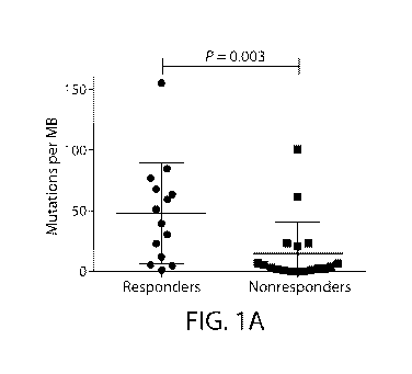
FIG. lA depicts the mutational load in responders vs. non-responders in
initial cohort.
FIG. IB depicts the mutational load in responders vs. non-responders in
validation cohort.
FIG. IC depicts the progression-free survival in patients with high,
intermediate, and low mutational
load.
FIG. 113 depicts the overall survival in patients with high, intermediate, and
low mutational load.
FIG. 2A depicts the performance of mutational load across a range of potential
thresholds. Vertical
bars indicate the thresholds selected based on local performance maxima and
clinical relevance.
FIG. 2B depicts the Receiver Operating Curve (ROC) for mutational loads
cutoffs of 3.3
mutations/MB (low mutational load group) and 23.1 mutations/MB (high
mutational load group).
FIG. 3A depicts the mutational load in tumors with cutaneous/unknown primaries
in responders vs.
non-responders.
FIG. 3B depicts the mutational load in tumors with non-cutaneous primaries
(acral, mucosal, uveal)
in responders vs. non-responders.
FIG. 3C depicts gene amplifications and deletions in responders vs. non-
responders.
FIG. 4A depicts the total number of mutations observed in responders versus
nonresponders.
FIG. 4B depicts the total number of C>T transitions observed in responders
versus nonresponders.
FIG. 4C depicts the types of nucleotide alterations observed in responders
versus nonresponders.
FIG. 4D depicts the mutational load of patients with BRAF mutations, NRAS
mutations, NF1
mutations/loss, and "triple WT" (defined as wild-type for BRAF, NRAS, and
NF1). BRAF non-V600
mutations were included with in the BRAF cohort except for 1 patient with
concurrent NF1 mutation.
One patient with NRASQ61R mutation and concurrent NF1 mutation was included in
the NRAS cohort.
FIG. 5A depicts that the mutational load in TCGA skin cutaneous melanoma
(SKCM) samples using
315 genes included on a hybrid capture NGS panel is highly correlated with
mutations assessed by
whole exome sequencing.
FIG. 5B depicts the mutational load groups and survival in the TCGA using
whole exome sequencing
(WES).
FIG. 5C depicts the mutational load groups and survival in the TCGA using 315
tested (FM) genes.
23
CA 03015913 2018-08-27
WO 2017/151517
PCT/US2017/019733
FIG. 6 depicts the calculated mutational load per sample (top); color-coded
matrix of individual
mutations, copy-number alterations; and clinical characteristics (middle); and
mutation spectra of
individual samples (bottom).
FIG. 7A depicts the LRP1B mutations/variants of unknown significance in
responders vs. non-
responders.
FIG. 7B depicts the total number of mutations among melanomas with and without
LRP1B mutations.
FIG. 7C depicts the association between the number of LRP1B mutations and
total mutations in the
melanoma TCGA.
FIG. 8A depicts the T cell receptor (TCR) clonality in responders vs. non-
responders.
FIG. 8B depicts the T cell fraction in responders vs. non-responders.
FIG. 8C depicts the TCR clonality in responders vs. non-responders in "ideal"
samples, defined as
those obtained within 4 months of anti-PD-1/anti-PD-L1 treatment without other
prior therapies.
FIG. 8D depicts the T cell fraction in these "ideal" samples, defined as those
obtained within 4
months of anti-PD-1/anti-PD-L1 treatment without other prior therapies.
FIG. 9A depicts the correlation between mutation load and T cell receptor
(TCR) clonality.
FIG. 9B depicts the correlation between mutation load and T cell fraction.
DETAILED DESCRIPTION
The invention is based, at least in part, on the discovery that the number of
mutations as
detected in a several hundred gene genes, e.g., by a hybrid capture-based NGS
platform, correlated
with therapeutic benefit from a therapy that includes a PD-1 or a PD-Li. In
certain embodiments,
stratifying patients into groups, e.g., three groups, allowed for accurate
prediction for most patients
into "high" and "low" mutation load cohorts, thus providing a clinically-
feasible marker of response
to an anti-PD-1 and/or an anti-PD-Li therapy in advanced melanoma and other
cancers. In other
embodiments, alterations in several genes (e.g. NF1, LRP1B) also correlated
with total mutational
burden and benefit from an anti-PD-1 or anti-PD-Li therapy.
Without being bound by theory, the likelihood of generating immunogenic tumor
neoantigens
is believed to increase in a probabilistic fashion as mutations develop,
increasing the likelihood of
immune recognition (Gubin and Schreiber. Science 350:158-9, 2015). Assessing
total mutational
load, however, requires whole exome sequencing (WES). This approach
necessitates specialized
tissue processing, a matched normal specimen, and is largely performed as a
research tool currently.
Given the technical and informatics challenges of performing WES in clinical
settings, surrogate
methods of detecting mutational burden are needed. The methods including
validated hybrid capture-
based NGS platform described herein have several pragmatic advantages,
including, for example,
more clinically-feasible turnaround times (-2 weeks), standardized informatics
pipelines, and more
manageable costs. This approach has other advantages over markers such as PD-
Li expression, since
it produces an objective (mutation load) rather than a subjective measure
(immunohistochemical
24
CA 03015913 2018-08-27
WO 2017/151517
PCT/US2017/019733
scoring) (Hansen and Siu. JAMA Oncol 2(1):15-6, 2016). Further, this platform
facilitates
simultaneous detection of actionable alterations relevant for targeted
therapies.
Identifying accurate predictive biomarkers for an anti-PD-1 or anti-PD-Li
therapy has several
direct clinical applications. In melanoma, one could foresee that patients
with high mutational load
could receive, e.g., anti-PD-1 monotherapy, whereas those with
intermediate/low mutational loads
could be treated, e.g., with the more active (but more toxic) combination of
nivolumab and
ipilimumab (Larkin et al. N Engl J Med, 2015). Other cancers (e.g. NSCLC) may
have a lower
overall response rate to anti-PD-1 (Rizvi et al. Lancet Oncol 16:257-65, 2015;
Garon et al. N Engl J
Med, 2015). In these diseases, this approach could stratify patients between
anti-PD-1 and other
active agents such as cytotoxic chemotherapy. Further, identifying genomic and
immune
characteristics of "outliers" (e.g. high mutation load tumors that fail to
respond) is a major need.
Accordingly, the invention provides, at least in part, methods of treating a
subject having, or
at risk of having, a cancer, e.g., a melanoma, by administering to the subject
an effective amount of an
agent (e.g., a therapeutic agent) that targets and/or inhibits the PD-1
pathway. In certain
embodiments, the therapeutic agent is an inhibitor of PD-1 or PD-Li. In
certain embodiments, the
therapeutic agent, e.g., the inhibitor of PD-1 or PD-L1, is administered
responsive to a value of
responder status to the therapeutic agent. In certain embodiments, the value
of responder status
includes a measure of the mutation load in a cancer sample, e.g., a melanoma
sample or a sample
derived from a melanoma, from the subject. In certain embodiments, the measure
of the mutation
load includes a determination of the level of a somatic alteration in a
predetermined set of genes
disclosed herein, the presence of a somatic alteration in an NF1 gene, the
number of a somatic
alteration in an LRP1B gene, the number of a C to T transition in a
predetermined set of genes
disclosed herein, or any combination thereof. Methods and systems for
evaluating a subject or for
selecting a therapy, preparation of nucleic acids, kits, reaction mixtures,
and methods of making a
reaction mixture, are also disclosed.
Certain terms are defined below and throughout the specification.
As used herein, the articles "a" and "an" refer to one or to more than one
(e.g., to at least one)
of the grammatical object of the article.
The term "or" is used herein to mean, and is used interchangeably with, the
term "and/or",
unless context clearly indicates otherwise. The use of the term "and/or" in
some places herein does
not mean that uses of the term "or" are not interchangeable with the term
"and/or" unless the context
clearly indicates otherwise.
"About" and "approximately" shall generally mean an acceptable degree of error
for the
quantity measured given the nature or precision of the measurements. Exemplary
degrees of error are
within 20 percent (%), typically, within 10%, and more typically, within 5% of
a given value or range
of values.
CA 03015913 2018-08-27
WO 2017/151517
PCT/US2017/019733
"Acquire" or "acquiring" as the terms are used herein, refer to obtaining
possession of a
physical entity, or a value, e.g., a numerical value, by "directly acquiring"
or "indirectly acquiring"
the physical entity or value. "Directly acquiring" means performing a process
(e.g., performing a
synthetic or analytical method) to obtain the physical entity or value.
"Indirectly acquiring" refers to
receiving the physical entity or value from another party or source (e.g., a
third party laboratory that
directly acquired the physical entity or value). Directly acquiring a physical
entity includes
performing a process that includes a physical change in a physical substance,
e.g., a starting material.
Exemplary changes include making a physical entity from two or more starting
materials, shearing or
fragmenting a substance, separating or purifying a substance, combining two or
more separate entities
into a mixture, performing a chemical reaction that includes breaking or
forming a covalent or non-
covalent bond. Directly acquiring a value includes performing a process that
includes a physical
change in a sample or another substance, e.g., performing an analytical
process which includes a
physical change in a substance, e.g., a sample, analyte, or reagent (sometimes
referred to herein as
"physical analysis"), performing an analytical method, e.g., a method which
includes one or more of
the following: separating or purifying a substance, e.g., an analyte, or a
fragment or other derivative
thereof, from another substance; combining an analyte, or fragment or other
derivative thereof, with
another substance, e.g., a buffer, solvent, or reactant; or changing the
structure of an analyte, or a
fragment or other derivative thereof, e.g., by breaking or forming a covalent
or non-covalent bond,
between a first and a second atom of the analyte; or by changing the structure
of a reagent, or a
fragment or other derivative thereof, e.g., by breaking or forming a covalent
or non-covalent bond,
between a first and a second atom of the reagent.
"Acquiring a sequence" as the term is used herein, refers to obtaining
possession of a
nucleotide sequence or amino acid sequence, by "directly acquiring" or
"indirectly acquiring" the
sequence. "Directly acquiring a sequence" means performing a process (e.g.,
performing a synthetic
or analytical method) to obtain the sequence, such as performing a sequencing
method (e.g., a Next
Generation Sequencing (NGS) method). "Indirectly acquiring a sequence" refers
to receiving
information or knowledge of, or receiving, the sequence from another party or
source (e.g., a third
party laboratory that directly acquired the sequence). The sequence acquired
need not be a full
sequence, e.g., sequencing of at least one nucleotide, or obtaining
information or knowledge that
identifies a mutation disclosed herein as being present in a subject
constitutes acquiring a sequence.
Directly acquiring a sequence includes performing a process that includes a
physical change
in a physical substance, e.g., a starting material, such as a tissue sample,
e.g., a biopsy, or an isolated
nucleic acid (e.g., DNA or RNA) sample. Exemplary changes include making a
physical entity from
two or more starting materials, shearing or fragmenting a substance, such as a
genomic DNA
fragment; separating or purifying a substance (e.g., isolating a nucleic acid
sample from a tissue);
combining two or more separate entities into a mixture; performing a chemical
reaction that includes
26
CA 03015913 2018-08-27
WO 2017/151517
PCT/US2017/019733
breaking; or forming a covalent or non-covalent bond. Directly acquiring a
value includes performing
a process that includes a physical change in a sample or another substance as
described above.
"Acquiring a sample" as the term is used herein, refers to obtaining
possession of a sample,
e.g., a tissue sample or nucleic acid sample, by "directly acquiring" or
"indirectly acquiring" the
sample. "Directly acquiring a sample" means performing a process (e.g.,
performing a physical
method such as a surgery or extraction) to obtain the sample. "Indirectly
acquiring a sample" refers to
receiving the sample from another party or source (e.g., a third party
laboratory that directly acquired
the sample). Directly acquiring a sample includes performing a process that
includes a physical
change in a physical substance, e.g., a starting material, such as a tissue,
e.g., a tissue in a human
patient or a tissue that was previously isolated from a patient. Exemplary
changes include making a
physical entity from a starting material, dissecting or scraping a tissue;
separating or purifying a
substance (e.g., a sample tissue or a nucleic acid sample); combining two or
more separate entities
into a mixture; performing a chemical reaction that includes breaking or
forming a covalent or non-
covalent bond. Directly acquiring a sample includes performing a process that
includes a physical
change in a sample or another substance, e.g., as described above.
An "alteration" as used herein, of a gene or gene product (e.g., a gene or
gene product set
forth in Table 1, an NF1 gene or gene product, or an LRP1B gene or gene
product) refers to the
presence of a mutation or mutations within the gene or gene product, e.g., a
mutation, which affects
integrity, sequence, structure, amount or activity of the gene or gene
product, as compared to the
normal or wild-type gene. The alteration can be in amount, structure, and/or
activity in a cancer tissue
or cancer cell, as compared to its amount, structure, and/or activity, in a
normal or healthy tissue or
cell (e.g., a control), and is associated with a disease state, such as
cancer. For example, a gene or
gene product which is associated with cancer, or predictive of responsiveness
to anti-cancer
therapeutics, can have or result from an altered nucleotide sequence (e.g., a
mutation), amino acid
sequence, chromosomal translocation, intra-chromosomal inversion, copy number,
expression level,
protein level, protein activity, or epigenetic modification (e.g., methylation
or acetylation status), or
post-translational modification, in a cancer tissue or cancer cell, as
compared to a normal, healthy
tissue or cell. Exemplary mutations include, but are not limited to, point
mutations (e.g., silent,
missense, or nonsense), deletions, insertions, inversions, duplications,
amplifications, translocations,
inter- and intra-chromosomal rearrangements. Mutations can be present in the
coding or non-coding
region of the gene. In certain embodiments, the alterations are associated (or
not associated) with a
phenotype, e.g., a cancerous phenotype (e.g., one or more of cancer risk,
cancer progression, cancer
treatment, or resistance to cancer treatment). In certain embodiments, the
term "alteration" and
"mutation" are used interchangeably herein.
As used herein, "functional alteration" includes an alteration that, compared
with a reference
sequence, e.g., a wild-type or unmutated sequence, has an effect on cell
division, growth or survival,
e.g., promotes cell division, growth or survival. In certain embodiments, the
functional alteration is
27
CA 03015913 2018-08-27
WO 2017/151517
PCT/US2017/019733
identified as such by inclusion in a database of functional alterations, e.g.,
the COSMIC database
(cancer.sanger.ac.uk/cosmic; Forbes et al. Nucl. Acids Res. 2015; 43 (D1):
D805-D811). In certain
embodiments, the functional alteration is an alteration with known functional
status, e.g., occurring as
a known somatic alteration in the COSMIC database. In other embodiments, the
functional alteration
is an alteration with a likely functional status, e.g., a truncation in a
tumor suppressor gene. In certain
embodiments, the functional alteration is a driver mutation, e.g., an
alteration that gives a selective
advantage to a clone in its microenvironment, e.g., by increasing cell
survival or reproduction. In
some embodiments, the functional alteration can cause a clonal expansion. In
certain embodiments,
the functional alteration is an alteration capable of causing one or more of
the following: (a) self-
sufficiency in a growth signal; (b) decreased, e.g., insensitivity, to an anti-
growth signal; (c) decreased
apoptosis; (d) increased replicative potential; (e) sustained angiogenesis; or
(f) tissue invasion or
metastasis. In other embodiments, the functional alteration is not a passenger
mutation, e.g., is an
alteration that has detectable effect on the fitness of a clone of cells. In
other embodiments, the
functional alteration is not a variant of unknown significance (VUS), e.g.,
the pathogenicity of the
variant can neither be confirmed nor ruled out.
"Binding entity" means any molecule to which molecular tags can be directly or
indirectly
attached that is capable of specifically binding to an analyte. The binding
entity can be an affinity tag
on a nucleic acid sequence. In certain embodiments, the binding entity allows
for separation of the
nucleic acid from a mixture, such as an avidin molecule, or an antibody that
binds to the hapten or an
antigen-binding fragment thereof. Exemplary binding entities include, but are
not limited to, a biotin
molecule, a hapten, an antibody, an antibody binding fragment, a peptide, and
a protein.
"Complementary" refers to sequence complementarity between regions of two
nucleic acid
strands or between two regions of the same nucleic acid strand. It is known
that an adenine residue of
a first nucleic acid region is capable of forming specific hydrogen bonds
("base pairing") with a
residue of a second nucleic acid region which is antiparallel to the first
region if the residue is thymine
or uracil. Similarly, it is known that a cytosine residue of a first nucleic
acid strand is capable of base
pairing with a residue of a second nucleic acid strand which is antiparallel
to the first strand if the
residue is guanine. A first region of a nucleic acid is complementary to a
second region of the same
or a different nucleic acid if, when the two regions are arranged in an
antiparallel fashion, at least one
nucleotide residue of the first region is capable of base pairing with a
residue of the second region. In
certain embodiments, the first region comprises a first portion and the second
region comprises a
second portion, whereby, when the first and second portions are arranged in an
antiparallel fashion, at
least about 50%, at least about 75%, at least about 90%, or at least about 95%
of the nucleotide
residues of the first portion are capable of base pairing with nucleotide
residues in the second portion.
In other embodiments, all nucleotide residues of the first portion are capable
of base pairing with
nucleotide residues in the second portion.
28
CA 03015913 2018-08-27
WO 2017/151517
PCT/US2017/019733
The term "cancer" or "tumor" is used interchangeably herein. These terms refer
to the
presence of cells possessing characteristics typical of cancer-causing cells,
such as uncontrolled
proliferation, immortality, metastatic potential, rapid growth and
proliferation rate, and certain
characteristic morphological features.
The term "neoplasm" or "neoplastic" cell refers to an abnormal proliferative
stage, e.g., a
hyperproliferative stage, in a cell or tissue that can include a benign, pre-
malignant, malignant
(cancer) or metastatic stage.
Cancer is "inhibited" if at least one symptom of the cancer is alleviated,
terminated, slowed,
or prevented. As used herein, cancer is also "inhibited" if recurrence or
metastasis of the cancer is
reduced, slowed, delayed, or prevented.
The term "inhibition" or "inhibitor" includes a reduction in a certain
parameter, e.g., an
activity, of a given molecule, e.g., an immune checkpoint inhibitor. For
example, inhibition of an
activity, e.g., an activity of, e.g., PD-1 or PD-L1, of at least 5%, 10%, 20%,
30%, 40%, 50%, 60%, or
more is included by this term. Thus, inhibition need not be 100%. An inhibitor
can inhibit a target
directly (e.g., by binding to the target) or indirectly (e.g., by interfering
an activity associated with the
target). An inhibitor may also inhibit two or more different targets (e.g.,
simultaneously or
sequentially), e.g., having multispecifity (e.g., bispecifity). An inhibitor
of PD-1 or PD-L1, as
described herein, can inhibit PD-1, PD-L1, or both.
The term "Programmed Death 1" or "PD-1" include isoforms, mammalian, e.g.,
human PD-1,
species homologs of human PD-1, and analogs comprising at least one common
epitope with PD-1.
The amino acid sequence of PD-1, e.g., human PD-1, is known in the art, e.g.,
Shinohara T et al.
(1994) Genomics 23(3):704-6; Finger LR, et al. Gene (1997) 197(1-2):177-87.
The term or "PD-Ligand 1" or "PD-Li" include isoforms, mammalian, e.g., human
PD-1,
species homologs of human PD-L1, and analogs comprising at least one common
epitope with PD-
.. Ll. The amino acid sequence of PD-L1, e.g., human PD-L1, is known in the
art.
As used herein, the term "indel" refers to an insertion, a deletion, or both,
of one or more
nucleotides in a nucleic acid of a cell. In certain embodiments, an indel
includes both an insertion and
a deletion of one or more nucleotides, where both the insertion and the
deletion are nearby on the
nucleic acid. In certain embodiments, the indel results in a net change in the
total number of
.. nucleotides. In certain embodiments, the indel results in a net change of
about 1 to about 50
nucleotides.
As used herein, "chemotherapeutic agent" means a chemical substance, such as a
cytotoxic or
cytostatic agent that is used to treat a condition, particularly cancer.
As used herein, "cancer therapy" and "cancer treatment" are synonymous terms.
As used herein, "chemotherapy" and "chemotherapeutic" and "chemotherapeutic
agent" are
synonymous terms.
29
CA 03015913 2018-08-27
WO 2017/151517
PCT/US2017/019733
The terms "homology" or "identity," as used interchangeably herein, refer to
sequence
similarity between two polynucleotide sequences or between two polypeptide
sequences, with identity
being a more strict comparison. The phrases "percent identity or homology" and
"% identity or
homology" refer to the percentage of sequence similarity found in a comparison
of two or more
polynucleotide sequences or two or more polypeptide sequences. "Sequence
similarity" refers to the
percent similarity in base pair sequence (as determined by any suitable
method) between two or more
polynucleotide sequences. Two or more sequences can be anywhere from 0-100%
similar, or any
integer value there between. Identity or similarity can be determined by
comparing a position in each
sequence that can be aligned for purposes of comparison. When a position in
the compared sequence
is occupied by the same nucleotide base or amino acid, then the molecules are
identical at that
position. A degree of similarity or identity between polynucleotide sequences
is a function of the
number of identical or matching nucleotides at positions shared by the
polynucleotide sequences. A
degree of identity of polypeptide sequences is a function of the number of
identical amino acids at
positions shared by the polypeptide sequences. A degree of homology or
similarity of polypeptide
sequences is a function of the number of amino acids at positions shared by
the polypeptide
sequences. The term "substantially identical," as used herein, refers to an
identity or homology of at
least 75%, at least 80%, at least 85%, at least 90%, 91%, 92%, 93%, 94%, 95%,
96%, 97%, 98%,
99%, or more.
"Likely to" or "increased likelihood," as used herein, refers to an increased
probability that an
item, object, thing or person will occur. Thus, in one example, a subject that
is likely to respond to
treatment with an inhibitor of PD-1 or PD-L1, alone or in combination, has an
increased probability of
responding to treatment with the inhibitor alone or in combination, relative
to a reference subject or
group of subjects.
"Unlikely to" refers to a decreased probability that an event, item, object,
thing or person will
occur with respect to a reference. Thus, a subject that is unlikely to respond
to treatment with an
inhibitor of PD-1 or PD-L1, alone or in combination, has a decreased
probability of responding to
treatment with a kinase inhibitor, alone or in combination, relative to a
reference subject or group of
subjects.
"Sequencing" a nucleic acid molecule requires determining the identity of at
least 1
nucleotide in the molecule. In embodiments, the identity of less than all of
the nucleotides in a
molecule is determined. In other embodiments, the identity of a majority or
all of the nucleotides in
the molecule is determined.
"Next-generation sequencing or NGS or NG sequencing" as used herein, refers to
any
sequencing method that determines the nucleotide sequence of either individual
nucleic acid
molecules (e.g., in single molecule sequencing) or clonally expanded proxies
for individual nucleic
acid molecules in a high throughput fashion (e.g., greater than 105 molecules
are sequenced
simultaneously). In one embodiment, the relative abundance of the nucleic acid
species in the library
CA 03015913 2018-08-27
WO 2017/151517
PCT/US2017/019733
can be estimated by counting the relative number of occurrences of their
cognate sequences in the
data generated by the sequencing experiment. Next generation sequencing
methods are known in the
art, and are described, e.g., in Metzker, M. (2010) Nature Biotechnology
Reviews 11:31-46,
incorporated herein by reference. Next generation sequencing can detect a
variant present in less than
5% of the nucleic acids in a sample.
"Sample," "tissue sample," "patient sample," "patient cell or tissue sample"
or "specimen"
each refers to a tissue, a cell, e.g., a circulating cell, obtained from a
subject or patient. The source of
the tissue sample can be solid tissue as from a fresh, frozen and/or preserved
organ, tissue sample,
biopsy, or aspirate; blood or any blood constituents; bodily fluids such as
cerebral spinal fluid,
amniotic fluid, peritoneal fluid or interstitial fluid; or cells from any time
in gestation or development
of the subject. The tissue sample can contain compounds that are not naturally
intermixed with the
tissue in nature such as preservatives, anticoagulants, buffers, fixatives,
nutrients, antibiotics or the
like. In one embodiment, the sample is preserved as a frozen sample or as
formaldehyde- or
paraformaldehyde-fixed paraffin-embedded (FFPE) tissue preparation. For
example, the sample can
be embedded in a matrix, e.g., an FFPE block or a frozen sample.
A "tumor nucleic acid sample" as used herein, refers to nucleic acid molecules
from a tumor
or cancer sample. Typically, it is DNA, e.g., genomic DNA, or cDNA derived
from RNA, from a
tumor or cancer sample. In certain embodiments, the tumor nucleic acid sample
is purified or isolated
(e.g., it is removed from its natural state).
A "control" or "reference" "nucleic acid sample" as used herein, refers to
nucleic acid
molecules from a control or reference sample. Typically, it is DNA, e.g.,
genomic DNA, or cDNA
derived from RNA, not containing the alteration or variation in the gene or
gene product, e.g., not
containing a mutation. In certain embodiments, the reference or control
nucleic acid sample is a wild
type or a non-mutated sequence. In certain embodiments, the reference nucleic
acid sample is
purified or isolated (e.g., it is removed from its natural state). In other
embodiments, the reference
nucleic acid sample is from a non-tumor sample, e.g., a blood control, a
normal adjacent tissue
(NAT), or any other non-cancerous sample from the same or a different subject.
"Adjacent to the interrogation position," as used herein, means that a site
sufficiently close
such that a detection reagent complementary with the site can be used to
distinguish between a
mutation, e.g., an alteration described herein, and a reference sequence,
e.g., a non-mutant or wild-
type sequence, in a target nucleic acid. Directly adjacent, as used herein, is
where 2 nucleotides have
no intervening nucleotides between them.
"Associated mutation," as used herein, refers to a mutation within a
preselected distance, in
terms of nucleotide or primary amino acid sequence, from a definitional
mutation, e.g., a mutant as
described herein. In embodiments, the associated mutation is within n, wherein
n is 2, 5, 10, 20, 30,
50, 100, or 200 nucleotides from the definitional mutation (n does not include
the nucleotides defining
31
CA 03015913 2018-08-27
WO 2017/151517
PCT/US2017/019733
the associated and definitional mutations). In embodiments, the associated
mutation is a translocation
mutation.
"Interrogation position," as used herein, comprises at least one nucleotide
(or, in the case of
polypeptides, an amino acid residue) which corresponds to a nucleotide (or
amino acid residue) that is
mutated in a mutation of interest, e.g., a mutation being identified, or in a
nucleic acid (or protein)
being analyzed, e.g., sequenced, or recovered.
A "reference sequence," as used herein, e.g., as a comparator for a mutant
sequence, is a
sequence which has a different nucleotide or amino acid at an interrogation
position than does the
mutant(s) being analyzed. In one embodiment, the reference sequence is wild-
type for at least the
interrogation position.
Headings, e.g., (a), (b), (i) etc, are presented merely for ease of reading
the specification and
claims. The use of headings in the specification or claims does not require
the steps or elements be
performed in alphabetical or numerical order or the order in which they are
presented.
Various aspects featured in the invention are described in further detail
below. Additional
definitions are set out throughout the specification.
Therapeutic Methods and Agents
"Treat," "treatment," and other forms of this word refer to the administration
of an agent, e.g.,
a therapeutic agent, alone or in combination with a second agent in an amount
effective to impede
growth of a cancer, to cause a cancer to shrink by weight or volume, to extend
the expected survival
time of the subject and or time to progression of the tumor or the like. In
those subjects, treatment can
include, but is not limited to, inhibiting tumor growth, reducing tumor mass,
reducing size or number
of metastatic lesions, inhibiting the development of new metastatic lesions,
prolonged survival,
prolonged progression-free survival, prolonged time to progression, and/or
enhanced quality of life.
A cancer is "treated" if at least one symptom of the cancer is alleviated,
terminated, slowed or
prevented. A cancer is also "treated" if recurrence or metastasis of the
cancer is reduced, slowed,
delayed or prevented.
As used herein, unless otherwise specified, the terms "prevent," "preventing"
and
"prevention" contemplate an action that occurs before a subject begins to
suffer from the re-growth of
the cancer and/or which inhibits or reduces the severity of the cancer.
As used herein, and unless otherwise specified, a "therapeutically effective
amount" of an
agent is an amount sufficient to provide a therapeutic benefit in the
treatment or management of the
cancer, or to delay or minimize one or more symptoms associated with the
cancer. A therapeutically
effective amount of a compound means an amount of therapeutic agent, alone or
in combination with
other therapeutic agents, which provides a therapeutic benefit in the
treatment or management of the
cancer. The term "therapeutically effective amount" can encompass an amount
that improves overall
32
CA 03015913 2018-08-27
WO 2017/151517
PCT/US2017/019733
therapy, reduces or avoids symptoms or causes of the cancer, or enhances the
therapeutic efficacy of
another therapeutic agent.
As used herein, and unless otherwise specified, a "prophylactically effective
amount" of an
agent is an amount sufficient to prevent re-growth of the cancer, or one or
more symptoms associated
with the cancer, or prevent its recurrence. A prophylactically effective
amount of an agent means an
amount of the agent, alone or in combination with other therapeutic agents,
which provides a
prophylactic benefit in the prevention of the cancer. The term
"prophylactically effective amount" can
encompass an amount that improves overall prophylaxis or enhances the
prophylactic efficacy of
another prophylactic agent.
The term "patient" or "subject" includes a human (e.g., a male or female of
any age group),
e.g., a pediatric patient (e.g., infant, child, adolescent); or adult patient
(e.g., young adult, middle-aged
adult or senior adult). In certain embodiments, the subject is an adult
subject (e.g., male or female
adult subject) having, or at risk of having, a melanoma as described herein.
In certain embodiments,
the subject is a subject of or above10, 15, 20, 25, 30, 35, 40, 45, 50, 55,
60, 65, 70, 75, 80, 85, 90
years of age, or more. In certain embodiments, a subject is a subject between
0-10 years of age, 10-
years of age, 20-30 years of age, 30-40 years of age, 40-50 years of age, 50-
60 years of age, 60-70
years of age, 70-80 years of age, or 80-90 years of age. In certain
embodiments, the subject is a
subject between 25 and 29 years of age. In other embodiments, the subject is a
subject between 15
and 29 years of age. In some embodiments, the subject is female and is between
15 and 29 years of
20 age. In certain embodiments, the subject is 65 years of age, or more.
When the term patient" or "subject" is used in conjunction with administration
of a compound
or drug, then the patient has been the object of treatment, observation,
and/or administration of the
compound or drug.
The agents, e.g., the therapeutic agents described herein, can be administered
in combination
with a second therapeutic agent or a different therapeutic modality, e.g.,
anti-cancer agents, and/or in
combination with surgical and/or radiation procedures.
By "in combination with," it is not intended to imply that the therapy or the
therapeutic agents
must be administered at the same time and/or formulated for delivery together,
although these
methods of delivery are within the scope of the invention. The pharmaceutical
compositions can be
administered concurrently with, prior to, or subsequent to, one or more other
additional therapies or
therapeutic agents. In general, each agent will be administered at a dose
and/or on a time schedule
determined for that agent. It will further be appreciated that therapeutic
agents utilized in a
combination can be administered together in a single composition or
administered separately in
different compositions. The particular combination to employ in a regimen will
take into account
compatibility of the first therapeutically active agent with the additional
therapeutically active
agent(s) and/or the desired therapeutic effect to be achieved.
33
CA 03015913 2018-08-27
WO 2017/151517
PCT/US2017/019733
As used herein, the term "responder" includes a subject who has a decrease in
the size of a
tumor, or in the extent of cancer in the body, or shows the disappearance of
one or more (e.g., most or
all) signs of cancer in response to a treatment, e.g., as defined by RECIST
1.1 criteria.
As used herein, the term "partial responder" includes a subject who has a
decrease in the size
of a tumor, or in the extent of cancer in the body, in response to a
treatment, e.g., as defined by
RECIST 1.1 criteria.
As used herein, the term "complete responder" includes a subject who shows the
disappearance of most or all signs of cancer in response to a treatment, e.g.,
as defined by RECIST 1.1
criteria. The cancer needs not to be cured in the complete responder.
As used herein, the term "non-responder" includes a subject who does not have
a decrease in
the size of a tumor, or in the extent of cancer in the body, e.g., as defined
by RECIST 1.1 criteria.
In some embodiments, subjects can be classified as responders (e.g., complete
responders or
partial responders) or non-responders as defined by classical partial or
complete responses (RECIST
1.1 criteria), or atypical immune related responses lasting at least 12
months. In some embodiments,
subjects with mixed responses necessitating additional systemic therapy or
causing clinical
deterioration in <12 months (e.g., as determined by a clinician) are
considered non-responders (e.g.,
before, at or after the additional systemic therapy or clinical
deterioration). In certain embodiments,
the therapy responses are classified based on clinical review of imaging and
clinician notes. RECIST
1.1 criteria is described, e.g., in Eisenhauer et al. Eur J Cancer. 2009;
45(2):228-247.
In some embodiments, the responder (e.g., a complete responder or partial
responder) has a
mutation load in a sample (e.g., a tumor sample) of about 3.3 or more somatic
alterations per a
preselected unit, e.g., per megabase, in a predetermined set of genes, e.g.,
the coding regions of a
predetermined set of genes. In some embodiments, the responder (e.g., a
complete responder) has a
mutation load in a sample (e.g., a tumor sample) of about 23.1 or more somatic
alterations per a
preselected unit, e.g., per megabase, in a predetermined set of genes, e.g.,
the coding regions of a
predetermined set of genes. In some embodiments, the responder (e.g., a
partial responder) has a
mutation load in a sample (e.g., a tumor sample) of about 3.3 or more but
fewer than 23.2 somatic
alterations per a preselected unit, e.g., per megabase, in a predetermined set
of genes, e.g., the coding
regions of a predetermined set of genes. In some embodiments, the non-
responder has a mutation
load in a sample (e.g., a tumor sample) of fewer than about 3.3 somatic
alterations per a preselected
unit, e.g., per megabase, in a predetermined set of genes, e.g., the coding
regions of a predetermined
set of genes.
In some embodiments, the responder (e.g., a complete responder or partial
responder) is a
member of a patient group (e.g., in a clinical trial) that has at least about
29% (e.g., at least about
35%, at least about 40%, at least about 50%, at least about 60%, at least
about 70%, at least about
80%, at least about 85%, at least about 90%, or at least about 95%) objective
response rate (ORR). In
some embodiments, the responder (e.g., a complete responder) is a member of a
patient group that has
34
CA 03015913 2018-08-27
WO 2017/151517
PCT/US2017/019733
at least about 85% (e.g., at least about 90% or at least about 95%) objective
response rate (ORR). In
some embodiments, the responder (e.g., a partial responder) is a member of a
patient group that has
between about 29% and about 85% (e.g., between about 30% and about 80%,
between about 40% and
about 70%, or between about 50% and about 60%) objective response rate (ORR).
In some
embodiments, the non-responder is a member of a patient group that has less
than about 25% (e.g.,
less than about 20% or less than about 10%) objective response rate (ORR).
Exemplary Therapeutic Agents and Modalities
The agent, e.g., therapeutic agent, can be a small molecule, a protein, a
polypeptide, a peptide,
an antibody molecule, a nucleic acid (e.g., a siRNA, an antisense or a micro
RNA), a small molecule,
or an immune cell therapy. Exemplary agents and classes of agents are
described herein.
In one embodiment, the agent, e.g., therapeutic agent, binds and/or inhibits
PD-1 or PD-Li. In
one embodiment, the agent is an antibody molecule. The terms "antibody" and
"antibody molecule"
as used interchangeably herein refer to immunoglobulin molecules and
immunologically active
portions of immunoglobulin molecules, i.e., molecules that contain an antigen
binding site which
specifically binds an antigen, such as a polypeptide featured in the
invention. A molecule which
specifically binds to a given polypeptide featured in the invention is a
molecule which binds the
polypeptide, but does not substantially bind other molecules in a sample,
e.g., a biological sample,
which naturally contains the polypeptide. Examples of immunologically active
portions of
immunoglobulin molecules include F(ab) and F(ab')2 fragments which can be
generated by treating
the antibody with an enzyme such as pepsin. The invention provides polyclonal
and monoclonal
antibodies. The term "monoclonal antibody" or "monoclonal antibody
composition," as used herein,
refers to a population of antibody molecules that contain only one species of
an antigen binding site
capable of immunoreacting with a particular epitope. Antibodies to PD-1 or PD-
Li are known in the
art, as well as techniques for generating antibodies to a polypeptide target,
e.g., PD-1 or PD-Li.
Exemplary PD-1 Inhibitors
In certain embodiments, the inhibitor of PD-1 is an anti-PD-1 antibody. In
certain
embodiments, the inhibitor of PD-1 is chosen from nivolumab (ONO-4538, BMS-
936558, or
MDX1106), pembrolizumab (MK-3475 or lambrolizumab), pidilizumab (CT-011),
MEDI0680
(AMP-514), PDR001, REGN2810, BGB-108, BGB-A317, SHR-1210 (HR-301210, SHR1210,
or
SHR-1210), PF-06801591, or AMP-224.
In other embodiments, the PD-1 inhibitor is an anti-PD-1 antibody chosen from
Nivolumab,
Pembrolizumab or Pidilizumab.
In some embodiments, the anti-PD-1 antibody is Nivolumab (CAS Registry Number:
946414-
94-4). Alternative names for Nivolumab include MDX-1106, MDX-1106-04, ONO-
4538, or BMS-
936558. Nivolumab is a fully human IgG4 monoclonal antibody which specifically
blocks
CA 03015913 2018-08-27
WO 2017/151517
PCT/US2017/019733
PD1. Nivolumab (clone 5C4) and other human monoclonal antibodies that
specifically bind to PD1
are disclosed, e.g., in US 8,008,449 and W02006/121168. In one embodiment, the
inhibitor of PD-1
is Nivolumab, and having a heavy and light chain amino acid sequences
disclosed as SEQ ID NOS: 2
and 3, respectively, in US 8,008,449 and W02006/121168 (or an amino acid
sequence substantially
identical or similar thereto, e.g., a sequence at least 85%, 90%, 95%
identical or higher to the amino
acid sequence specified).
In some embodiments, the anti-PD-1 antibody is Pembrolizumab. Pembrolizumab
(also
referred to as Lambrolizumab, MK-3475, MK03475, SCH-900475 or KEYTRUDAO;
Merck) is a
humanized IgG4 monoclonal antibody that binds to PD-1. Pembrolizumab and other
humanized anti-
PD-1 antibodies are disclosed in Hamid, 0. et al. (2013) New England Journal
of Medicine 369 (2):
134-44, US 8,354,509 and W02009/114335. In one embodiment, the inhibitor of PD-
1 is
Pembrolizumab disclosed in, e.g., US 8,354,509 and WO 2009/114335, and having
a heavy and light
chain amino acid sequences disclosed as SEQ ID NOS: 4 and 5, respectively, in
US 8,354,509 and
WO 2009/114335 (or an amino acid sequence substantially identical or similar
thereto, e.g., a
sequence at least 85%, 90%, 95% identical or higher to the sequence
specified).
In some embodiments, the anti-PD-1 antibody is Pidilizumab. Pidilizumab (CT-
011; Cure
Tech) is a humanized IgG lk monoclonal antibody that binds to PD-1.
Pidilizumab and other
humanized anti-PD-1 monoclonal antibodies are disclosed in W02009/101611.
Other anti-PD-1 antibodies include AMP 514 (Amplimmune), among others, e.g.,
anti-PD1
antibodies disclosed in US 8,609,089, US 2010028330, and/or US 20120114649.
Exemplary PD-Li Inhibitors
In certain embodiments, the inhibitor of PD-Li is an anti-PD-Li antibody. In
certain
embodiments, the inhibitor of PD-Li is chosen from atezolizumab (MPDL3280A,
RG7446, or
R05541267), YW243.55.570, MDX-1105, durvalumab (MEDI4736), or avelumab
(MSB0010718C).
In some embodiments, the PD-Li inhibitor is an antibody molecule. In some
embodiments,
the anti-PD-Ll inhibitor is chosen from YW243.55.570, MPDL3280A, MEDI-4736,
MSB-0010718C,
or MDX-1105.
In some embodiments, the anti-PD-Li antibody is MSB0010718C. MSB0010718C (also
referred to as A09-246-2; Merck Serono) is a monoclonal antibody that binds to
PD-
Ll. MSB0010718C and other humanized anti-PD-Li antibodies are disclosed, e.g.,
in
W02013/079174, and having a heavy and light chain variable region amino acid
sequences disclosed
as SEQ ID NOS: 24 and 25, respectively, in W02013/079174 (or a sequence
substantially identical or
similar thereto, e.g., an amino acid sequence at least 85%, 90%, 95% identical
or higher to the amino
acid sequence specified).
In one embodiment, the PD-Li inhibitor is YW243.55.570. The YW243.55.570
antibody is
an anti-PD-Ll described in WO 2010/077634 (heavy and light chain variable
region amino acid
36
CA 03015913 2018-08-27
WO 2017/151517
PCT/US2017/019733
sequences shown in SEQ ID NOS: 20 and 21, respectively, of WO 2010/077634),
and having a
sequence disclosed therein (or an amino acid sequence substantially identical
or similar thereto, e.g.,
an amino acid sequence at least 85%, 90%, 95% identical or higher to the
sequence specified).
In one embodiment, the PD-Li inhibitor is MDX-1105. MDX-1105, also known as
BMS-
936559, is an anti-PD-Ll antibody described in W02007/005874, and having a
sequence disclosed
therein (or a sequence substantially identical or similar thereto, e.g., a
sequence at least 85%, 90%,
95% identical or higher to the sequence specified).
In one embodiment, the PD-Li inhibitor is MDPL3280A (Genentech / Roche).
MDPL3280A
is a human Fc optimized IgG1 monoclonal antibody that binds to PD-Li.
MDPL3280A and other
human monoclonal antibodies to PD-Li are disclosed in U.S. Patent No.:
7,943,743 and U.S
Publication No.: 20120039906.
Additional Therapies
Treatments described herein can be provided to a patient having had an
unsatisfactory
response to a different therapy, e.g., a non-anti-PD-1 and/or non-anti-PD-Li
therapeutic agent or
therapeutic modality. In one embodiment, the subject is undergoing or has
undergone treatment with
a different therapy, e.g., a non-anti-PD-1 and/or non-anti-PD-Li therapeutic
agent or therapeutic
modality. The different therapy can be a small molecule, a protein, a
polypeptide, a peptide, an
antibody molecule, a nucleic acid (e.g., a siRNA, an antisense or a micro
RNA), or a cell.
In one embodiment, the different therapy is chosen from a chemotherapy, a
radiation therapy,
an immunotherapy, an immunoradiotherapy, an oncolytic virotherapy, a surgical
procedure, or any
combination thereof. In one embodiment, the different therapy comprises one or
more of:
dacarbazine, temozolomide, interleukin-2 (IL-2), an interferon, an inhibitor
of CTLA-4 (e.g., an anti-
CTLA-4 antibody, e.g., ipilimumab), a BRAF inhibitor, a MEK inhibitor,
talimogene laherparepvec,
an adoptive cell transfer, or any combination thereof. In one embodiment, the
different therapy
comprises one or more of: dacarbazine, temozolomide, interleukin-2 (IL-2)
(e.g., a recombinant
interferon alfa-2b or a peginterferon alfa-2b), an interferon, ipilimumab, a
BRAF inhibitor (e.g.,
vemurafenib or dabrafenib), a MEK inhibitor (e.g., cobimetinib or trametinib),
talimogene
laherparepvec, an adoptive cell transfer (e.g., modified T cells or modified
dendritic cells), or any
combination thereof.
An agent, e.g., therapeutic agent, described herein can be administered, alone
or in
combination, e.g., in combination with other chemotherapeutic agents or
procedures, in an amount
sufficient to reduce or inhibit the tumor cell growth, and/or treat or prevent
the cancer(s), in the
subject.
37
CA 03015913 2018-08-27
WO 2017/151517
PCT/US2017/019733
Nuchec Acid Inhibitors
In another embodiment, the agent is a nucleic acid inhibitor selected from an
antisense
molecule, a ribozyme, a double-stranded RNA molecule, a triple helix molecule,
that hybridizes to a
nucleic acid encoding the alteration, or a transcription regulatory region
that blocks or reduces mRNA
expression of the alteration.
In one embodiment, the nucleic acid antagonist is a siRNA that targets mRNA
encoding a
mutation. Other types of antagonistic nucleic acids can also be used, e.g., a
dsRNA, a ribozyme, a
triple-helix former, or an antisense nucleic acid. Accordingly, isolated
nucleic acid molecules that are
nucleic acid inhibitors, e.g., antisense, RNAi, to a mutation-encoding nucleic
acid molecule are
provided.
An "antisense" nucleic acid can include a nucleotide sequence which is
complementary to a
"sense" nucleic acid encoding a protein, e.g., complementary to the coding
strand of a double-
stranded cDNA molecule or complementary to an mRNA sequence. The antisense
nucleic acid can
be complementary to an entire nucleotide sequence encoding one or more
mutations, or to only a
portion thereof. In another embodiment, the antisense nucleic acid molecule is
antisense to a
"noncoding region" of the coding strand of a nucleotide sequence encoding one
or more mutations
(e.g., the 5' and 3' untranslated regions). Anti-sense agents can include, for
example, from about 8 to
about 80 nucleobases (i.e., from about 8 to about 80 nucleotides), e.g., about
8 to about 50
nucleobases, or about 12 to about 30 nucleobases. Anti-sense compounds include
ribozymes, external
guide sequence (EGS) oligonucleotides (oligozymes), and other short catalytic
RNAs or catalytic
oligonucleotides which hybridize to the target nucleic acid and modulate its
expression. Anti-sense
compounds can include a stretch of at least eight consecutive nucleobases that
are complementary to a
sequence in the target gene. An oligonucleotide need not be 100% complementary
to its target
nucleic acid sequence to be specifically hybridizable. An oligonucleotide is
specifically hybridizable
when binding of the oligonucleotide to the target interferes with the normal
function of the target
molecule to cause a loss of utility, and there is a sufficient degree of
complementarity to avoid non-
specific binding of the oligonucleotide to non-target sequences under
conditions in which specific
binding is desired, i.e., under physiological conditions in the case of in
vivo assays or therapeutic
treatment or, in the case of in vitro assays, under conditions in which the
assays are conducted.
Hybridization of antisense oligonucleotides with mRNA can interfere with one
or more of the
normal functions of mRNA. The functions of mRNA to be interfered with include
all key functions
such as, for example, translocation of the RNA to the site of protein
translation, translation of protein
from the RNA, splicing of the RNA to yield one or more mRNA species, and
catalytic activity which
may be engaged in by the RNA. Binding of specific protein(s) to the RNA may
also be interfered with
by antisense oligonucleotide hybridization to the RNA.
Exemplary antisense compounds include DNA or RNA sequences that specifically
hybridize
to the target nucleic acid, e.g., the mRNA encoding a mutation described
herein. The complementary
38
CA 03015913 2018-08-27
WO 2017/151517
PCT/US2017/019733
region can extend for between about 8 to about 80 nucleobases. The compounds
can include one or
more modified nucleobases. Modified nucleobases are known in the art.
Descriptions of modified
nucleic acid agents are also available. See, e.g., U.S. Patent Nos. 4,987,071;
5,116,742; and
5,093,246; Woolf et al. (1992) Proc Natl Acad Sci USA; Anti sense RNA and DNA,
D.A. Melton, Ed.,
Cold Spring Harbor Laboratory, Cold Spring Harbor, N.Y. (1988); 89:7305-9;
Haselhoff and Gerlach
(1988) Nature 334:585-59; Helene, C. (1991) Anticancer Drug Des. 6:569-84;
Helene (1992) Ann.
N.Y. Acad. Sci. 660:27-36; and Maher (1992) Bioassays 14:807-15.
The antisense nucleic acid molecules are typically administered to a subject
(e.g., by direct
injection at a tissue site), or generated in situ such that they hybridize
with or bind to cellular mRNA
and/or genomic DNA encoding a mutation to thereby inhibit expression of the
protein, e.g., by
inhibiting transcription and/or translation. Alternatively, antisense nucleic
acid molecules can be
modified to target selected cells and then be administered systemically. For
systemic administration,
antisense molecules can be modified such that they specifically bind to
receptors or antigens
expressed on a selected cell surface, e.g., by linking the antisense nucleic
acid molecules to peptides
or antibodies which bind to cell surface receptors or antigens. The antisense
nucleic acid molecules
can also be delivered to cells using the vectors described herein. To achieve
sufficient intracellular
concentrations of the antisense molecules, vector constructs in which the
antisense nucleic acid
molecule is placed under the control of a strong pol II or pol III promoter
are preferred.
In yet another embodiment, the antisense nucleic acid molecule is an a-
anomeric nucleic acid
molecule. An a-anomeric nucleic acid molecule forms specific double-stranded
hybrids with
complementary RNA in which, contrary to the usual 13-units, the strands run
parallel to each other
(Gaultier et al. (1987) Nucleic Acids. Res. 15:6625-6641). The antisense
nucleic acid molecule can
also comprise a 2'-o-methylribonucleotide (Inoue et al. (1987) Nucleic Acids
Res. 15:6131-6148) or a
chimeric RNA-DNA analogue (Inoue et al. (1987) FEBS Lett. 215:327-330).
SiRNAs are small double stranded RNAs (dsRNAs) that optionally include
overhangs. For
example, the duplex region of an siRNA is about 18 to 25 nucleotides in
length, e.g., about 19, 20, 21,
22, 23, or 24 nucleotides in length. Typically, the siRNA sequences are
exactly complementary to the
target mRNA. dsRNAs and siRNAs in particular can be used to silence gene
expression in
mammalian cells (e.g., human cells). siRNAs also include short hairpin RNAs
(shRNAs) with 29-
base-pair stems and 2-nucleotide 3' overhangs. See, e.g., Clemens et al.
(2000) Proc. Natl. Acad. Sci.
USA 97:6499-6503; Billy et al. (2001) Proc. Natl. Sci. USA 98:14428-14433;
Elbashir et al. (2001)
Nature. 411:494-8; Yang et al. (2002) Proc. Natl. Acad. Sci. USA 99:9942-9947;
Siolas et al. (2005),
Nat. Biotechnol. 23(2):227-31; U.S. Patent Publication Nos 20040086884;
20030166282;
20030143204; 20040038278; and 20030224432.
In still another embodiment, an antisense nucleic acid featured in the
invention is a ribozyme.
A ribozyme having specificity for a mutation-encoding nucleic acid can include
one or more
39
CA 03015913 2018-08-27
WO 2017/151517
PCT/US2017/019733
sequences complementary to the nucleotide sequence of a mutation cDNA
disclosed herein, and a
sequence having known catalytic sequence responsible for mRNA cleavage (see
U.S. Pat. No.
5,093,246 or Haselhoff and Gerlach (1988) Nature 334:585-591). For example, a
derivative of a
Tetrahymena L-19 IVS RNA can be constructed in which the nucleotide sequence
of the active site is
complementary to the nucleotide sequence to be cleaved in a mutation-encoding
mRNA. See, e.g.,
Cech et al. U.S. Patent No. 4,987,071; and Cech et al. U.S. Patent No.
5,116,742. Alternatively,
mutation mRNA can be used to select a catalytic RNA having a specific
ribonuclease activity from a
pool of RNA molecules. See, e.g., Bartel, D. and Szostak, J.W. (1993) Science
261:1411-1418.
Inhibition of a mutated gene can be accomplished by targeting nucleotide
sequences
complementary to the regulatory region of the mutation to form triple helical
structures that prevent
transcription of the mutated gene in target cells. See generally, Helene, C.
(1991) Anticancer Drug
Des. 6:569-84; Helene, C. i (1992) Ann. N.Y. Acad. Sci. 660:27-36; and Maher,
L.J. (1992) Bioassays
14:807-15. The potential sequences that can be targeted for triple helix
formation can be increased by
creating a so-called "switchback" nucleic acid molecule. Switchback molecules
are synthesized in an
alternating 5'-3', 3'-5' manner, such that they base pair with first one
strand of a duplex and then the
other, eliminating the necessity for a sizeable stretch of either purines or
pyrimidines to be present on
one strand of a duplex.
The invention also provides detectably labeled oligonucleotide primer and
probe molecules.
Typically, such labels are chemiluminescent, fluorescent, radioactive, or
colorimetric.
A mutated nucleic acid molecule can be modified at the base moiety, sugar
moiety or
phosphate backbone to improve, e.g., the stability, hybridization, or
solubility of the molecule. For
non-limiting examples of synthetic oligonucleotides with modifications see
Toulme (2001) Nature
Biotech. 19:17 and Faria et al. (2001) Nature Biotech. 19:40-44. Such
phosphoramidite
oligonucleotides can be effective antisense agents.
For example, the deoxyribose phosphate backbone of the nucleic acid molecules
can be
modified to generate peptide nucleic acids (see Hyrup B. et al. (1996)
Bioorganic & Medicinal
Chemistry 4: 5-23). As used herein, the terms "peptide nucleic acid" or "PNA"
refers to a nucleic
acid mimic, e.g., a DNA mimic, in which the deoxyribose phosphate backbone is
replaced by a
pseudopeptide backbone and only the four natural nucleobases are retained. The
neutral backbone of
a PNA can allow for specific hybridization to DNA and RNA under conditions of
low ionic strength.
The synthesis of PNA oligomers can be performed using standard solid phase
peptide synthesis
protocols as described in Hyrup B. et al. (1996) supra and Perry-O'Keefe et
al. Proc. Natl. Acad. Sci.
93: 14670-675.
PNAs of mutated nucleic acid molecules can be used in therapeutic and
diagnostic
applications. For example, PNAs can be used as antisense or antigene agents
for sequence-specific
modulation of gene expression by, for example, inducing transcription or
translation arrest or
inhibiting replication. PNAs of mutated nucleic acid molecules can also be
used in the analysis of
CA 03015913 2018-08-27
WO 2017/151517
PCT/US2017/019733
single base pair mutations in a gene, (e.g., by PNA-directed PCR clamping); as
'artificial restriction
enzymes' when used in combination with other enzymes, (e.g., Si nucleases
(Hyrup B. et al. (1996)
supra)); or as probes or primers for DNA sequencing or hybridization (Hyrup B.
et al. (1996) supra;
Perry-O'Keefe supra).
In other embodiments, the oligonucleotide may include other appended groups
such as
peptides (e.g., for targeting host cell receptors in vivo), or agents
facilitating transport across the cell
membrane (see, e.g., Letsinger et al. (1989) Proc. Natl. Acad. Sci. USA
86:6553-6556; Lemaitre et al.
(1987) Proc. Natl. Acad. Sci. USA 84:648-652; W088/09810) or the blood-brain
barrier (see, e.g., WO
89/10134). In addition, oligonucleotides can be modified with hybridization-
triggered cleavage
agents (see, e.g., Krol et al. (1988) Bio-Techniques 6:958-976) or
intercalating agents (See, e.g., Zon
(1988) Pharm. Res. 5:539-549). To this end, the oligonucleotide may be
conjugated to another
molecule, (e.g., a peptide, hybridization triggered cross-linking agent,
transport agent, or
hybridization-triggered cleavage agent).
In some embodiments, a nucleic acid inhibitor described herein is provided for
the inhibition
of expression of a nucleic acid comprising the alteration in vitro.
Cancer
The invention described herein can be used to treat a cancer or to evaluate a
subject having a
cancer.
Exemplary cancers include, but are not limited to, melanomas, B cell cancer,
e.g., multiple
myeloma, breast cancer, lung cancer (such as non-small cell lung carcinoma or
NSCLC), bronchus
cancer, colorectal cancer, prostate cancer, pancreatic cancer, stomach cancer,
ovarian cancer, urinary
bladder cancer, brain or central nervous system cancer, peripheral nervous
system cancer, esophageal
cancer, cervical cancer, uterine or endometrial cancer, cancer of the oral
cavity or pharynx, liver
cancer, kidney cancer, testicular cancer, biliary tract cancer, small bowel or
appendix cancer, salivary
gland cancer, thyroid gland cancer, adrenal gland cancer, osteosarcoma,
chondrosarcoma, cancer of
hematological tissues, adenocarcinomas, inflammatory myofibroblastic tumors,
gastrointestinal
stromal tumor (GIST), colon cancer, multiple myeloma (MM), myelodysplastic
syndrome (MDS),
myeloproliferative disorder (MPD), acute lymphocytic leukemia (ALL), acute
myelocytic leukemia
(AML), chronic myelocytic leukemia (CML), chronic lymphocytic leukemia (CLL),
polycythemia
Vera, Hodgkin lymphoma, non-Hodgkin lymphoma (NHL), soft-tissue sarcoma,
fibrosarcoma,
myxosarcoma, liposarcoma, osteogenic sarcoma, chordoma, angiosarcoma,
endotheliosarcoma,
lymphangiosarcoma, lymphangioendotheliosarcoma, synovioma, mesothelioma,
Ewing's tumor,
leiomyosarcoma, rhabdomyosarcoma, squamous cell carcinoma, basal cell
carcinoma,
adenocarcinoma, sweat gland carcinoma, sebaceous gland carcinoma, papillary
carcinoma, papillary
adenocarcinomas, medullary carcinoma, bronchogenic carcinoma, renal cell
carcinoma, hepatoma,
bile duct carcinoma, choriocarcinoma, seminoma, embryonal carcinoma, Wilms'
tumor, bladder
41
CA 03015913 2018-08-27
WO 2017/151517
PCT/US2017/019733
carcinoma, epithelial carcinoma, glioma, astrocytoma, medulloblastoma,
craniopharyngioma,
ependymoma, pinealoma, hemangioblastoma, acoustic neuroma, oligodendroglioma,
meningioma,
neuroblastoma, retinoblastoma, follicular lymphoma, diffuse large B-cell
lymphoma, mantle cell
lymphoma, hepatocellular carcinoma, thyroid cancer, gastric cancer, head and
neck cancer, small cell
cancers, essential thrombocythemia, agnogenic myeloid metaplasia,
hypereosinophilic syndrome,
systemic mastocytosis, familiar hypereosinophilia, chronic eosinophilic
leukemia, neuroendocrine
cancers, carcinoid tumors, and a metastatic lesion thereof.
Melanoma
The invention provides, at least in part, methods for treating a cancer, e.g.,
a melanoma, in a
subject. In certain embodiments, the methods include treatment of a cancer,
e.g., a melanoma,
harboring an alteration described herein (e.g., an alteration in a gene
disclosed in Table 1, e.g., an
alteration in an NF1 gene or an LRP1B gene described herein). The methods
include administering to
the subject a therapeutic agent, e.g., an immunotherapeutic agent, e.g., an
inhibitor of PD-1 or PD-Li.
In certain embodiments, the cancer is a melanoma. In certain embodiments, the
cancer is an
advanced melanoma. In certain embodiments, the cancer is a metastatic
melanoma. In certain
embodiments, the cancer, e.g., melanoma, is at a high risk of recurrence or
replase. In certain
embodiments, the cancer, e.g., melanoma, is at a high risk of metastasis. In
some embodiments, the
melanoma is any stage or risk group melanoma defined according to any suitable
melanoma
classification system known to those of skill in the art.
In certain embodiments, the melanoma is any one of a stage 0, stage IA, stage
IB, stage IIA,
stage IIB, stage IIC, stage III, or stage IV melanoma. In other embodiments,
the melanoma is any one
of a stage 0, stage IA, stage IB, stage IIA, stage IIB, stage IIC, stage III,
or stage IV melanoma, as
defined by the American Joint Committee on Cancer (AJCC) melanoma staging
system. In some
embodiments, the melanoma is any stage melanoma defined according to any
suitable melanoma
classification system known to those of skill in the art. In other
embodiments, the melanoma is any
one of a stage 0, stage IA, stage IB, stage IIA, stage IIB, stage IIC, stage
III, or stage IV melanoma,
set forth in Tables 2A-2B.
In certain embodiments, the melanoma is a stage III or stage IV melanoma. In
certain
embodiments, the melanoma is a stage III melanoma. In certain embodiments, the
melanoma is a
stage III melanoma according to any suitable melanoma classification system
known to those of skill
in the art. In certain embodiments, the cancer is a stage III melanoma set
forth in Tables 2A-2B. In
certain embodiments, the melanoma is a stage IV melanoma. In certain
embodiments, the melanoma
is a stage IV melanoma according to any suitable melanoma classification
system known to those of
skill in the art. In certain embodiments, the cancer is a stage IV melanoma
set forth in Tables 2A-2B.
In certain embodiments, the melanoma is an advanced melanoma. In some
embodiments, the
advanced melanoma is a stage III or stage IV melanoma, e.g., as set forth in
Tables 2A-2B. In certain
42
CA 03015913 2018-08-27
WO 2017/151517
PCT/US2017/019733
embodiments, the melanoma is a meatastic melanoma. In some embodiments, the
metastatic
melanoma is a stage III or stage IV melanoma, e.g., as set forth in Tables 2A-
2B.
Table 2A. TNM Staging Categories for Cutaneous Melanoma*
Classification Thickness (mm) Ulceration Status/Mitoses
Tis NA NA
Ti <i.00 a: Without ulceration and mitosis <
1/mm
b: With ulceration or mitoses > 1/mm2
T2 1.01-2.00 a: Without ulceration
b: With ulceration
T3 2.01-4.00 a: Without ulceration
b: With ulceration
T4 > 4.00 a: Without ulceration
b: With ulceration
No. of Metastatic Nodes Nodal Metastatic Burden
NO 0 NA
Ni 1 a: Micrometastasist
b: Macrometastasis*
N2 2-3 a: Micrometastasist
b: Macrometastasis*
c: In transit metastases/satellites without metastatic nodes
N3 4+ metastatic nodes, or
matted nodes, or in
transit
metastases/satellites with
metastatic nodes
Site Serum LDH
MO No distant metastases NA
M1 a Distant skin, Normal
subcutaneous, or nodal
metastases
Mlb Lung metastases Normal
Mlc All other visceral Normal
metastases
Any distant metastasis Elevated
Abbreviations: NA, not applicable; LDH, lactate dehydrogenase.
*Balch et al. "Final Version of 2009 AJCC Melanoma Staging and
Classification," J Clin Oncol.
2009; 27(36): 6199-6206.
*Micrometastases are diagnosed after sentinel lymph node biopsy.
*Macrometastases are defined as clinically detectable nodal metastases
confirmed pathologically.
43
CA 03015913 2018-08-27
WO 2017/151517
PCT/US2017/019733
Table 2B. Anatomic Stage Groupings for Cutaneous Melanoma*
Clinical Staging* Pathologic Staging*
T N M T N M
0 Tis NO MO 0 Tis NO MO
IA Tla NO MO IA Tla NO MO
IB Tlb NO MO IB T lb NO MO
T2a NO MO T2a NO MO
IIA T2b NO MO IIA T2b NO MO
T3a NO MO T3a NO MO
IIB T3b NO MO IIB T3b NO MO
T4a NO MO T4a NO MO
IIC T4b NO MO IIC T4b NO MO
III Any T N> NO MO IIIA T1-4a Nla MO
T1-4a N2a MO
IIIB T1-4b N1 a MO
T1-4b N2a MO
T1-4a Nib MO
T1-4a N2b MO
T1-4a N2c MO
IIIC T1-4b Nib MO
T1-4b N2b MO
T1-4b N2c MO
Any T N3 MO
IV Any T Any N M1 IV Any T Any N M1
*Balch et al. "Final Version of 2009 AJCC Melanoma Staging and
Classification," J Clin Oncol.
2009; 27(36): 6199-6206.
tClinical staging includes microstaging of the primary melanoma and
clinical/radiologic evaluation
for metastases. By convention, it should be used after complete excision of
the primary melanoma
with clinical assessment for regional and distant metastases.
*Pathologic staging includes microstaging of the primary melanoma and
pathologic information about
the regional lymph nodes after partial (i.e., sentinel node biopsy) or
complete lymphadenectomy.
Pathologic stage 0 or stage IA patients are the exception; they do not require
pathologic evaluation of
their lymph nodes.
In certain embodiments, the melanoma is a cutaneous melanoma. In other
embodiments, the
melanoma is a non-cutaneous melanoma.
In certain embodiments, the cancer, e.g., the melanoma, e.g., the advanced or
metastatic
melanoma, comprises, or is identified or determined as having, an alteration,
e.g., an alteration
44
CA 03015913 2018-08-27
WO 2017/151517
PCT/US2017/019733
described herein, e.g., in a gene described herein. In certain embodiments,
the cancer, e.g., the
melanoma, e.g., the advanced or metastatic melanoma, comprises, or is
identified or determined as
having, an alteration in a gene described in Table 1. In other embodiments,
the cancer, e.g., the
melanoma, e.g., the advanced or metastatic melanoma, comprises, or is
identified or determined as
having, a plurality of alterations in a gene described in Table 1. In other
embodiments, the cancer,
e.g., the melanoma, e.g., the advanced or metastatic melanoma, comprises, or
is identified or
determined as having, a plurality of alterations in a plurality of genes
described in Table 1.
In certain embodiments, the cancer, e.g., the melanoma, e.g., the advanced or
metastatic
melanoma, comprises, or is identified or determined as having, an alteration
in an NF1 gene, e.g., an
alteration in an NF1 gene as described herein.
In certain embodiments, the alteration in the NF1 gene results in decreased
activity of an NF1
gene product (e.g., an NF1 protein), compared to a wildtype activity of NFL
For example, the
alteration can result in an alteration (e.g., a decrease) in a GTPase
activator activity, a
phosphatidylcholine binding activity, and/or a phosphatidylethanolamine
binding activity of NFL In
certain embodiments, the alteration results in an elevated activation of RAS.
In one embodiment, the alteration in the NF1 gene is, or comprises, a mutation
(e.g., a
somatic mutation), e.g., a substitution (e.g., a base substitution), an
insertion, or a deletion.
Exemplary NF1 alterations associated with melanoma are described, e.g., in
Wiesner et al. Am J Surg
Pathol. 2015; 39(10): 1357-62; Krauthammer et al. Nat Genet. 2015; 47(9): 996-
1002. Additional
alterations in the NF1 gene are described, e.g., in the COSMIC database
(cancer.sanger.ac.uk/cosmic;
Forbes et al. Nucl. Acids Res. 2015; 43 (D1): D805-D811).
In certain embodiments, the cancer, e.g., the melanoma, e.g., the advanced or
metastatic
melanoma, comprises, or is identified or determined as having, an alteration
in an LRP1B gene, e.g.,
an alteration in an LRP1B gene as described herein.
In certain embodiments, the alteration in the LRP1B gene results in decreased
activity of an
LRP1B gene product (e.g., an LRP1B protein), compared to a wildtype activity
of LRP1B. For
example, the alteration can result in an alteration (e.g., a decrease) in a
calcium ion binding and/or
low-density lipoprotein receptor activity of LRP1B.
In one embodiment, the alteration in the LRP1B gene is, or comprises, a
mutation (e.g., a
somatic mutation), e.g., a substitution (e.g., a base substitution), an
insertion, or a deletion.
Exemplary LRP1B alterations associated with melanoma are described, e.g., in
Nikolaev et al. Nat
Genet. 2011; 44(2):133-9. Additional alterations in the LRP1B gene are
described, e.g., in the
COSMIC database (cancer.sanger.ac.uldcosmic; Forbes et al. Nucl. Acids Res.
2015; 43 (D1): D805-
D811).
In other embodiments, the cancer, e.g., the melanoma, e.g., the advanced or
metastatic
melanoma, comprises, or is identified or determined as having, an alteration
in a BRAF gene, e.g., an
alteration in a BRAF gene as described herein.
CA 03015913 2018-08-27
WO 2017/151517
PCT/US2017/019733
In certain embodiments, the alteration in the BRAF gene results in increased
activity of a
BRAF gene product (e.g., a BRAF protein), compared to a wild-type activity of
BRAF. For example,
the alteration can result in alteration in one or more of the following
activities of BRAF: ATP binding
calcium, ion binding, identical protein binding, MAP kinase kinase kinase
activity, protein kinase
activity, or protein serine/threonine kinase activity. In certain embodiments,
the alteration results in
an alteration (e.g., an increase) in a kinase activity of BRAF.
In one embodiment, the BRAF alteration is, or comprises, a mutation (e.g., a
somatic
mutation), e.g., a substitution (e.g., a base substitution), an insertion, or
a deletion. In certian
embodiments, the alteration is a base substitution.
In certain embodiments, the alteration in BRAF is located at codon V600. In
certain
embodiments, the alteration in BRAF is a V600E alteration. In certain
embodiments, the alteration in
BRAF is a V600K alteration. In certain embodiments, the alteration in BRAF is
a V600R alteration.
In certain embodiments, the alteration in BRAF is a V600D alteration.
In certain embodiments, the alteration in BRAF is an alteration other than a
V600 alteration.
In certain embodiments, the alteration in BRAF is a K601 alteration. In
certain embodiments, the
alteration in BRAF is a K601E alteration. In certain embodiments, the
alteration in BRAF is a G469
alteration. In certain embodiments, the alteration in BRAF is a G469E
alteration. In certain
embodiments, the alteration in BRAF is a D594 alteration. In certain
embodiments, the alteration in
BRAF is a D594G alteration. In certain embodiments, the alteration in BRAF is
an L597 alteration.
In certain embodiments, the alteration in BRAF is an L597S alteration. In
certain embodiments, the
alteration in BRAF is an S467 alteration. In certain embodiments, the
alteration in BRAF is an S467L
alteration.
In other embodiments, the cancer, e.g., the melanoma, e.g., the advanced or
metastatic
melanoma, comprises, or is identified or determined as having, an alteration
in an NRAS gene, e.g.,
an alteration in an NRAS gene as described herein.
In certain embodiments, the alteration in the NRAS gene results in increased
activity of a
NRAS gene product (e.g., an NRAS protein), compared to a wild-type activity of
NRAS. For
example, the alteration can result in an alteration (e.g., a decrease) in a
GTPase activity of NRAS. In
certain embodiments, the NRAS protein comprises an alteration that decreases
intrinsic GTP
hydrolysis, e.g., resulting in a constitutively active NRAS.
In one embodiment, the NRAS alteration is, or comprises, a mutation (e.g., a
somatic
mutation), e.g., a substitution (e.g., a base substitution), an insertion, or
a deletion. In certian
embodiments, the alteration is a base substitution.
In certain embodiments, the alteration in NRAS is located at codon Q61. In
certain
embodiments, the alteration in NRAS is a Q61H alteration. In certain
embodiments, the alteration in
NRAS is a Q61K alteration. In certain embodiments, the alteration in NRAS is a
Q61L alteration. In
certain embodiments, the alteration in NRAS is a Q61P alteration. In certain
embodiments, the
46
CA 03015913 2018-08-27
WO 2017/151517
PCT/US2017/019733
alteration in NRAS is a Q61Q alteration. In certain embodiments, the
alteration in NRAS is a Q61R
alteration.
In other embodiments, the alteration in NRAS is a G12 alteration. In certain
embodiments,
the alteration in NRAS is a G12A alteration. In certain embodiments, the
alteration in NRAS is a
G12C alteration. In certain embodiments, the alteration in NRAS is a G12D
alteration. In certain
embodiments, the alteration in NRAS is a G12R alteration. In certain
embodiments, the alteration in
NRAS is a G12S alteration. In certain embodiments, the alteration in NRAS is a
G12V alteration. In
certain embodiments, the alteration in NRAS is a G13 alteration. In certain
embodiments, the
alteration in NRAS is a Gl3C alteration. In certain embodiments, the
alteration in NRAS is a Gl3D
alteration. In certain embodiments, the alteration in NRAS is a Gl3R
alteration. In certain
embodiments, the alteration in NRAS is a G13V alteration. In certain
embodiments, the alteration in
NRAS is an S17 alteration. In certain embodiments, the alteration in NRAS is
an Sl7N alteration.
Mutation Load
As used herein, the term "mutation load" or "mutational load" refers to the
level, e.g.,
number, of an alteration (e.g., one or more alterations, e.g., one or more
somatic alterations) per a
preselected unit (e.g., per megabase) in a predetermined set of genes (e.g.,
in the coding regions of the
predetermined set of genes). Mutation load can be measured, e.g., on a whole
genome or exome
basis, or on the basis of a subset of genome or exome. In certain embodiments,
the mutation load
measured on the basis of a subset of genome or exome can be extrapolated to
determine a whole
genome or exome mutation load.
In certain embodiments, the mutation load is measured in a sample, e.g., a
tumor sample (e.g.,
a melanoma sample or a sample acquired or derived from a melanoma), from a
subject, e.g., a subject
described herein. In certain embodiments, the mutation load is measured by
determining one, two,
three or all of the following:
(i) the level of a somatic alteration in a predetermined set of genes set
forth in Table 1,
(ii) the presence of a somatic alteration in an NF1 gene,
(iii) the number of a somatic alteration in an LRP1B gene, or
(iv) the number of a C to T transition.
In certain embodiments, the level of a somatic alteration in a predetermined
set of genes set
forth in Table 1 correlates with the mutation load (e.g., the whole genome or
exome mutation load).
In certain embodiments, the presence of a somatic alteration in an NF1 gene
correlates with the
mutation load (e.g., the mutation load measured based on the level of a
somatic alteration in a
predetermined set of genes set forth in Table 1, or the whole genome or exome
mutation load). In
certain embodiments, the number of a somatic alteration in an LRP1B gene
correlates with the
mutation load (e.g., the mutation load measured based on the level of a
somatic alteration in a
predetermined set of genes set forth in Table 1, or the whole genome or exome
mutation load). In
47
CA 03015913 2018-08-27
WO 2017/151517 PCT/US2017/019733
certain embodiments, the number of a C to T transition correlates with the
mutation load (e.g., the
mutation load measured based on the level of a somatic alteration in a
predetermined set of genes set
forth in Table 1, or the whole genome or exome mutation load).
The terms "mutation load," "mutational load," "mutation burden," and
"mutational burden"
are used interchangeably herein. In the context of a tumor, a mutational load
is also referred to herein
as "tumor mutational burden," "tumor mutation burden," or "TMB."
Exemplary Genes Associated with Cancer
The invention described herein include, e.g., measuring the mutation load,
e.g., by
determining the level of a somatic alteration (e.g., one or more somatic
alterations) in a predetermined
set of genes associated with cancer, e.g., as disclosed herein.
In certain embodiments, the level of a somatic alteration in a gene or gene
product set forth in
Table 1 is determined. In certain embodiments, the level of a somatic
alteration in the coding region
of a gene set forth in Table 1 is determined.
Table 1. Exemplary Genes
Gene List
ABL1 B RAF CHEK1 FANCC GATA3 JAK2 MITF PDCD1LG2 RBM10 STAT4
ABL2 BRCA1 CHEK2 FANCD2 GATA4 JAK3 MLH1 PDGFRA RET STK11
ACVR1B BRCA2 CIC FANCE GATA6 JUN MPL PDGFRB RICTOR SUFU
G1D4 KAT6A
AKT1 BRD4 CREBBP FANCF (C17orf39) (MYST3) MRE 1 1 A PDK1 RNF43
SYK
AKT2 B R1P1 CRKL FANCG Gill KDM5A MSH2 PIK3C2B ROS1 TAF1
AKT3 BTG1 CRLF2 FANCL GNAll KDM5 C MSH6 PIK3CA RPTOR TBX3
ALK BTK CSF1R FAS GNA13 KDM6A MTOR PIK3CB RUNX1 TERC
TERT
AMER1 Cl 1 orf30 (promoter
(FAM123B) (EMS Y) CTCF FAT1 GNAQ KDR MUTYH PIK3CG
RUNX1T1 only)
APC CARD11 CTNNA1 FBXW7 GNAS KEAP1 MYC PIK3R1 SDHA
TET2
MYCL
AR CBFB CTNNB 1 FGF10 GPR124 KEL (MYCL1) PIK3R2
SDHB TGFB R2
ARAF CBL CUL3 FGF14 GR1N2A KIT MYCN PLCG2 SDHC
TNFAIP 3
ARFRP 1 CCND1 CYLD FGF19 GRM3 KLHL6 MYD 88 PMS 2 SDHD TNFRSF14
KMT2A
ARID1 A CCND2 DAXX FGF23 GSK3B (MLL) NF1 POLD1 SETD2
TOP1
KMT2C
ARID1B CCND3 DDR2 FGF3 H3F3A (MLL3) NF2 POLE SF3B 1
TOP2A
KMT2D
ARID2 CCNE1 DICER1 FGF4 HGF (MLL2) NFE2L2 PPP2R1 A SLIT2 TP53
48
CA 03015913 2018-08-27
WO 2017/151517 PCT/US2017/019733
ASXL1 CD274 DNMT3A FGF6 HNFlA KRAS NFKB IA PRDM1 SMAD2 TSC1
ATM CD79A DOT1L FGFR1 HRAS LMO1 NKX2-1 PREX2 SMAD3 TSC2
ATR CD79B EGFR FGFR2 HSD3B1 LRP1B NOTCH1 PRKAR1A SMAD4 TSHR
ATRX CDC73 EP300 FGFR3 HSP9OAA1 LYN NOTCH2 PRKCI SMARCA4 U2AF1
AURKA CDH1 EPHA3 FGFR4 IDH1 LZTR1 NOTCH3 PRKDC SMARCB1 VEGFA
AURKB CDK12 EPHA5 FH IDH2 MAGI2 NPM1 PRS S8 SMO VHL
AX1N1 CDK4 EPHA7 FLCN IGF1R MAP2K1 NRAS PTCH1 SNCAIP WISP3
AXL CDK6 EPHB1 FLT1 IGF2 MAP2K2 NSD1 PTEN SOCS1 WT1
BAP1 CDK8 ERB B2 FLT3 IKBKE MAP2K4 NTRK1 PTPN11 SOX10 XPO1
B ARD1 CDKN1A ERB B3 FLT4 IKZF1 MAP3K1 NTRK2 QKI SOX2 ZBTB2
BCL2 CDKN1B ERBB4 FOXL2 IL7R MCL1 NTRK3 RAC1 SOX9 ZNF217
BCL2L1 CDKN2A ERG FOXP1 INTHB A MDM2 NUP93 RAD50 SPEN ZNF703
BCL2L2 CDKN2B ERRFIl FRS2 1NPP4B MDM4 PAK3 RAD51 SPOP
BCL6 CDKN2C ESR1 FUBP1 IRF2 MED12 PALB 2 RAF1 SPTA1
BCOR CEBPA EZH2 GAB RA6 IRF4 MEF2B PARK2 RANB P2 SRC
BCORL1 CHD2 FAM46C GATA1 IRS2 MEN1 PAX5 RARA STAG2
BLM CHD4 FANCA GATA2 JAK1 MET PB RM1 RB1 STAT3
Select Rearrangements
ALK B RAF BRD4 ETV4 FGFR1 KIT MYC NTRK2 RARA TMPRS S2
BCL2 B RCA1 EGFR ETV5 FGFR2 MSH2 NOTCH2 PDGFRA RET
B CR BRCA2 ETV1 ETV6 FGFR3 MYB NTRK1 RAF1 ROS1
Neurofibromin 1 (NF1)
The invention described herein include, e.g., determining a presence of an
alteration (e.g., a
somatic alteration) in an NF1 gene, or measuring the mutation load, e.g., by
determining a presence of
an alteration (e.g., a somatic alteration) in an NF1 gene.
The neurofibromin 1 (NF1) gene, which is also known as WSS, NFNS, or VRNF,
encodes a
protein that functions as a negative regulator of the Ras signal transduction
pathway. Mutations in
this gene have been linked, e.g., to neurofibromatosis type 1, juvenile
myelomonocytic leukemia and
Watson syndrome. The mRNA for this gene is subject to RNA editing (CGA>UGA-
>Arg1306Term)
resulting in premature translation termination. Alternatively spliced
transcript variants encoding
different isoforms have also been described for this gene.
The nucleotide and amino acid sequences of human NF1 are described, e.g., in
Wallace et al.
Science. 1990; 249: 181-186, Erratum: Wallace et al. Science. 1990; 250: 1749-
1749 (isoform 1);
Marchuk et al. Genomics. 1991; 11: 931-940 (isoform 1); Cawthon et al. Cell.
1990; 62:193-201
(isoform 1); Bernards et al. DNA Cell Biol. 1992; 11: 727-734 (isoforms 1 and
2); Li et al. Genomics.
1995; 25: 9-18 (isoforms 1 and 2); Nishi et al. Oncogene. 1991; 6: 1555-1559
(isoforms 1 and 2);
Andersen et al. Mol. Cell. Biol. 1993; 13: 487-495 (isoform 2); Suzuki et al.
Biochem. Biophys. Res.
Commun. 1991; 181:955-961 (isoform 2); Suzuki et al. Biochem. Biophys. Res.
Commun. 1992; 187:
49
CA 03015913 2018-08-27
WO 2017/151517
PCT/US2017/019733
984-990 (isoform 3); Martin et al. Cell. 1990; 63: 843-849 (isoform 4); Suzuki
et al. Tohoku J. Exp.
Med. 1995; 175: 225-233 (isoform 5); and Xu et al. Cell. 1990; 62: 599-608
(isoforms 1 and 6).
Additional variants, mutations, and polymorphisms of human NF1 are also
described, e.g., in
Upadhyaya et al. Hum. Mutat. 1994; 4: 83-101; Shen et al. J. Med. Genet. 1996;
33: 2-17; Li et al.
Cell. 1992; 69: 275-281(variant Glu-1444); Upadhyaya et al. Hum. Mol. Genet.
1992; 1:735-740
(variants NF1 Met-2164 and Asn-2192); Tassabehji et al. Am. J. Hum. Genet.
1993; 53: 90-95
(variant Gly-His-Glu-Gln-Gln-Lys-Leu-Pro-Ala-Ala-Thr-Leu-Ala-Leu-1733 ins);
Shen et al. Hum.
Mol. Genet. 1993; 2: 1861-1864 (variant Met-991 del); Purandare et al. Hum.
Mol. Genet. 1994; 3:
1109-1115 (variants NF1 Asp-1166 and Arg-1440); Abernathy et al. Hum. Mutat.
1994; 3: 347-352
(variant NF1 2387-Asn-Phe-2388 del); Upadhyaya et al. J. Med. Genet. 1995; 32:
706-710 (variant
NF1 Ala-2631); Gasparini et al. Hum. Genet. 1996; 97: 492-495 (variant NF1 Arg-
629); Wu et al.
Hum. Mutat. 1996; 8: 51-56 (variant NF1 Arg-1035); Upadhyaya et al. Hum.
Genet. 1997; 99: 88-92
(variants NF1 Ser-1412; Gln-1440; Glu-1444 And Gly-1489); Maynard et al. Hum.
Genet. 1997; 99:
674-676 (variants NF1 Arg-844 and Pro-898); Hudson et al. Hum. Mutat. 1997; 9:
366-367 (variant
NF1 Arg-1952); Upadhyaya et al. Hum. Mutat. 1997; 10: 248-250 (variants NF1
GLY-338 and TRP-
1611); Klose et al. Hum. Mol. Genet. 1998; 7: 1261-1268 (variant NF1 Pro-
1276); Krkljus et al. Hum.
Mutat. 1998; 11: 411-411 (variant NF1 Gly-1204, variant His-765); Messiaen et
al. Genet. Med.
1999; 1: 248-253 (variant NF1 Pro-508); Peters et al. Hum. Mutat. 1999; 13:
337-337 (variant NF1
Pro-1446); Fahsold et al. Am. J. Hum. Genet. 2000; 66: 790-818 (variants NF1
Pro-216; Pro-357;
Cys-491; Pro-549; Thr-581; Arg-583; Phe-665; Pro-695; Pro-763; Ser-777; Lys-
780; Pro-781; Pro-
847; Ser-1156; Pro-1250; Gln-1276; Pro-1276; Pro-1446; Val-1605 and Ile-2507,
variant Glu-176);
Ass et al. Hum. Mol. Genet. 2000; 9: 237-247 (variants NF1 Ser-117; Trp-1204;
Pro-1446 and 2387-
Asn-Phe-2388 del), Erratum Ass et al. Hum. Mol. Genet. 2000; 9:659-659;
Boulandet et al. Hum.
Mutat. 2000; 16: 274-275 (variant NF1 Phe-844); Kaufmann et al. Am. J. Hum.
Genet. 2001; 69:
1395-1400 (variant spinal FSNF Pro-2088); Han et al. Hum. Genet. 2001; 109:487-
497 (variants NF1
Lys-780; Cys-784; Pro-1147; Cys-1193; Arg-1444; Ser-1785; Asn-2012 and Lys-
2357); Kluwe et al.
Hum. Mutat. 2002; 19:309-309 (variants NF1 Phe-82; Arg-784 and Glu-1444);
Baralle et al. Am. J.
Med. Genet. A 2003; 119:1-8 (variant NFNS Glu-1459 del); Wang et al. Hum.
Genet. 2003; 112: 117-
123 (variants NF1 Tyr-93; Val-604; Arg-844 and Pro-898, Variants Asp-74; Glu-
176; Arg-712 and
Gln-1276); De Luca et al. Hum. Mutat. 2003; 21: 171-172 (variants NF1 Lys-780;
Pro-847; Glu-848
And Arg-968; Asn-1444; Leu-1953 del and Arg-2001); Kluwe et al. J. Med. Genet.
2003; 40: 368-
371 (variants NF1 Arg-578; Pro-920 and Ala-2221); Zatkova et al. Hum. Mutat.
24:491-501 (variant
NF1 Val-186, characterization of variant NF1 Val-186); De Luca et al. Hum.
Mutat. 2004; 23: 629-
629 (variants NF1 Asn-157; Arg-629; Ser-777; Lys-780; Arg-784; Pro-847; Glu-
848; Arg-968; Asn-
1444; Leu-1953 del and Arg-2001, variant Glu-176); Mattocks et al. J. Med.
Genet. 2004; 41: E48-
E48 (variants NF1 Arg-31; Pro-145; Arg-324; Val-337; Cys-489; Pro-532; Arg-
574; Arg-629; Phe-
665; Phe-844; Pro-844; Met-991 del; Val-1073; Arg-1196; Gly-1276; Gln-1276;
Glu-1430; Glu-1459
CA 03015913 2018-08-27
WO 2017/151517
PCT/US2017/019733
del and Gly-1489, variants Glu-176 and Cys-873); Ferner et al. J. Med. Genet.
2004; 41: 837-841
(variant NF1 Pro-1243); Bertola et al. Am. J. Med. Genet. A 2005; 136: 242-245
(variant NF1 Arg-
844); De Luca et al. Am. J. Hum. Genet. 2005; 77: 1092-1101 (variants NFNS Arg-
194; Glu-1444;
Thr-1451; Leu-1453 and Glu-1459 del); Sjoeblom et al. Science. 2006; 314:268-
274 (variants Ile-
1187; Leu-1951 and Arg-2745); Upadhyaya et al. Am. J. Hum. Genet. 2007; 80:
140-151(variant NF1
Met-991 del); Nystrom et al. Chn. Genet. 76:524-534 (variant NFNS Phe-1411);
Ponti et al. Hered.
Cancer Chn. PraeL 2011; 9: 6-6 (variant NF1 Thr-160); Thomas et al. Eur. J.
Hum. Genet. 2012; 20:
411-419 (variants Glu-176; Thr-330; Asp-393; Leu-393; Pro-519; Thr-776 and Phe-
1484);
Nemethova et al. Ann. Hum. Genet. 2013; 77: 364-379 (variants Nfl Trp-93; Arg-
1048; Arg-1189;
Arg-1661 (isoform 1) and Thr-1918 (isoform 1); and Ben-Salem et al. Childs
Nerv. SysL 2014; 30:
1183-1189 (variant NF1 Pro-2125).
Low Density Lipoprotein Receptor-Related Protein 1B (LRP1B)
The invention described herein includes, e.g., determining the number of an
alteration (e.g., a
somatic alteration) in an LRP1B gene, or measuring the mutation load, e.g., by
determining the
number of an alteration (e.g., a somatic alteration) in an LRP1B gene.
The Low Density Lipoprotein Receptor-Related Protein 1B (LRP1B) gene, which is
also
known as LRP-DIT, LRP-1B, or LRPDIT, belongs to the low density lipoprotein
(LDL) receptor gene
family. These receptors play a wide variety of roles in normal cell function
and development due to
their interactions with multiple ligands.
The nucleotide and amino acid sequences of human LRP1B are described, e.g., in
Liu et al.
Cancer Res. 2000; 60:1961-1967; Lie et al. Genomics 2000; 69: 271-274.
Evaluation of Subjects
Subjects, e.g., patients, can be evaluated for a responder status to a therapy
described herein,
e.g, a cancer immunotherapy, e.g., a therapy comprising an inhibitor of PD-1
or PD-Li. For example,
the evaluation can include determining the mutation load in a cancer, e.g., a
cancer described herein,
and/or the presence of an alteration, e.g., an alteration as described herein
or an alteration in a gene
described herein. A patient can be evaluated, for example, by acquiring
knowledge of the mutation
load in a tumor sample from the patient. Alternatively, or in addition, a
patient can be evaluated, for
example, by determining the genomic sequence of the patient, e.g., by an NGS
method. Alternatively,
or in addition, evaluation of a patient can include directly assaying for the
presence of a mutation in
the patient, such as by an assay to detect a mutated nucleic acid (e.g., DNA
or RNA), such as by,
Southern blot, Northern blot, or RT-PCR, e.g., qRT-PCR. Alternatively, or in
addition, a patient can
be evaluated for the presence of a protein mutation, such as by
immunohistochemistry, Western blot,
immunoprecipitation, or immunomagnetic bead assay.
51
CA 03015913 2018-08-27
WO 2017/151517
PCT/US2017/019733
In one aspect, the results of a clinical trial, e.g., a successful or
unsuccessful clinical trial, can
be repurposed to identify agents for treating cancer, e.g., in a subset
patient population. By one
exemplary method, a candidate agent used in a clinical trial can be
reevaluated to determine if the
agent in the trial is effective to treat a tumor having a particular level of
mutation load or containing a
particular mutation. For example, subjects who participated in a clinical
trial for an agent, such as an
immune checkpoint modulator, can be identified. Patients who experienced an
improvement in
symptoms, e.g., cancer (e.g., a melanoma) symptoms, such as decreased tumor
size, or decreased rate
of tumor growth, can be evaluated for the mutation load and/or the presence of
a mutation. Patients
who did not experience an improvement in cancer symptoms can also be evaluated
for the mutation
load and/or the presence of a mutation. In an embodiment, where patients
having a predetermined
level of mutation load in a tumor sample are found to have been more likely to
respond to the test
agent than patients who do not have such a predetermined level of mutation
load, then the agent is
determined to be an appropriate treatment option for a patient having the
predetermined level of
mutation load. In another embodiment, where patients carrying a mutation are
found to have been
more likely to respond to the test agent than patients who did not carry such
a mutation, then the agent
is determined to be an appropriate treatment option for a patient carrying the
mutation.
"Reevaluation" of patients can include, for example, acquiring knowledge of
the mutation
load in a tumor sample, e.g., a melanoma sample, from the subject.
Alternatively, or in addition,
reevaluation of the patients can include determining the genomic sequence of
the patients, or a subset
of the clinical trial patients, e.g., by an NGS method. Alternatively, or in
addition, reevaluation of the
patients can include directly assaying for the presence of a mutation in the
patient, such as by an assay
to detect a mutated nucleic acid (e.g., RNA), such as by RT-PCR, e.g., qRT-
PCR. Alternatively, or in
addition, a patient can be reevaluated for the presence of a protein mutation,
such as by
immunohistochemistry, Western blot, immunoprecipitation, or immunomagnetic
bead assay.
Methods for Detection of Nucleic Acids and Polyp eptides
Methods for evaluating an altered gene, mutations and/or gene products are
known to those of
skill in the art. In one embodiment, the alteration (e.g., mutation) is
detected in a nucleic acid
molecule by a method chosen from one or more of: nucleic acid hybridization
assay, selective
suppression of PCR (SSP), HPLC or mass-spectrometric genotyping.
Additional exemplary methods include traditional "direct probe" methods such
as Southern
blots and "comparative probe" methods such as comparative genomic
hybridization (CGH), e.g.,
cDNA-based or oligonucleotide-based CGH, can be used. The methods can be used
in a wide variety
of formats including, but not limited to, substrate (e.g., membrane or glass)
bound methods or array-
based approaches.
In certain embodiments, the evaluation methods include probes/primers against
the alterations
described herein.
52
CA 03015913 2018-08-27
WO 2017/151517
PCT/US2017/019733
In one embodiment, probes/primers can be designed to detect a mutation or a
reciprocal
thereof. These probes/primers are suitable, e.g., for PCR amplification.
Probes are used that contain
DNA segments that are essentially complementary to DNA base sequences existing
in different
portions of chromosomes. Examples of probes useful according to the invention,
and labeling and
hybridization of probes to samples are described in two U.S. patents to Vysis,
Inc. U.S. Patent Nos.
5,491,224 and 6,277,569 to Bittner, et al.
Chromosomal probes are typically about 50 to about 105 nucleotides in length.
Longer
probes typically comprise smaller fragments of about 100 to about 500
nucleotides in length. Probes
that hybridize with centromeric DNA and locus-specific DNA are available
commercially, for
example, from Vysis, Inc. (Downers Grove, Ill.), Molecular Probes, Inc.
(Eugene, Oreg.) or from
Cytocell (Oxfordshire, UK). Alternatively, probes can be made non-commercially
from
chromosomal or genomic DNA through standard techniques. For example, sources
of DNA that can
be used include genomic DNA, cloned DNA sequences, somatic cell hybrids that
contain one, or a
part of one, chromosome (e.g., human chromosome) along with the normal
chromosome complement
of the host, and chromosomes purified by flow cytometry or microdissection.
The region of interest
can be isolated through cloning, or by site-specific amplification via the
polymerase chain reaction
(PCR). See, for example, Nath and Johnson, Biotechnic Histochem., 1998,
73(1):6-22, Wheeless et
al., Cytometry 1994, 17:319-326, and U.S. Patent No. 5,491,224.
The probes to be used hybridize to a specific region of a chromosome to
determine whether a
cytogenetic abnormality is present in this region. One type of cytogenetic
abnormality is a deletion.
Although deletions can be of one or more entire chromosomes, deletions
normally involve loss of part
of one or more chromosomes. If the entire region of a chromosome that is
contained in a probe is
deleted from a cell, hybridization of that probe to the DNA from the cell will
normally not occur and
no signal will be present on that chromosome. If the region of a chromosome
that is partially
contained within a probe is deleted from a cell, hybridization of that probe
to the DNA from the cell
can still occur, but less of a signal can be present. For example, the loss of
a signal is compared to
probe hybridization to DNA from control cells that do not contain the genetic
abnormalities which the
probes are intended to detect. In some embodiments, at least 1, 5, 10, 20, 30,
40, 50, 60, 70, 80, 90,
100, 110, 120, 130, 140, 150, 160, 170, 180, 190, 200, or more cells are
enumerated for the presence
of the cytogenetic abnormality.
Cytogenetic abnormalities to be detected can include, but are not limited to,
non-reciprocal
translocations, balanced translocations, intra-chromosomal inversions, point
mutations, deletions,
gene copy number changes, gene expression level changes, and germ line
mutations. In particular,
one type of cytogenetic abnormality is a duplication. Duplications can be of
entire chromosomes, or
of regions smaller than an entire chromosome. If the region of a chromosome
that is contained in a
probe is duplicated in a cell, hybridization of that probe to the DNA from the
cell will normally
53
CA 03015913 2018-08-27
WO 2017/151517
PCT/US2017/019733
produce at least one additional signal as compared to the number of signals
present in control cells
with no abnormality of the chromosomal region contained in the probe.
Chromosomal probes are labeled so that the chromosomal region to which they
hybridize can
be detected. Probes typically are directly labeled with a fluorophore, an
organic molecule that
fluoresces after absorbing light of lower wavelength/higher energy. The
fluorophore allows the probe
to be visualized without a secondary detection molecule. After covalently
attaching a fluorophore to a
nucleotide, the nucleotide can be directly incorporated into the probe with
standard techniques such as
nick translation, random priming, and PCR labeling. Alternatively,
deoxycytidine nucleotides within
the probe can be transaminated with a linker. The fluorophore then is
covalently attached to the
transaminated deoxycytidine nucleotides. See, U.S. Patent No. 5,491,224.
U.S. Patent No. 5,491,224 describes probe labeling as a number of the cytosine
residues
having a fluorescent label covalently bonded thereto. The number of
fluorescently labeled cytosine
bases is sufficient to generate a detectable fluorescent signal while the
individual so labeled DNA
segments essentially retain their specific complementary binding (hybridizing)
properties with respect
to the chromosome or chromosome region to be detected. Such probes are made by
taking the
unlabeled DNA probe segment, transaminating with a linking group a number of
deoxycytidine
nucleotides in the segment, or covalently bonding a fluorescent label to at
least a portion of the
transaminated deoxycytidine bases.
Probes can also be labeled by nick translation, random primer labeling or PCR
labeling.
Labeling is done using either fluorescent (direct)-or haptene (indirect)-
labeled nucleotides.
Representative, non-limiting examples of labels include: AMCA-6-dUTP,
CascadeBlue-4-dUTP,
Fluorescein-12-dUTP, Rhodamine-6-dUTP, TexasRed-6-dUTP, Cy3-6-dUTP, Cy5-dUTP,
Biotin(BIO)-11-dUTP, Digoxygenin(DIG)-11-dUTP or Dinitrophenyl (DNP)-11-dUTP.
Probes also can be indirectly labeled with biotin or digoxygenin, or labeled
with radioactive
isotopes such as 32P and 3H, although secondary detection molecules or further
processing then is
required to visualize the probes. For example, a probe labeled with biotin can
be detected by avidin
conjugated to a detectable marker. For example, avidin can be conjugated to an
enzymatic marker
such as alkaline phosphatase or horseradish peroxidase. Enzymatic markers can
be detected in
standard colorimetric reactions using a substrate and/or a catalyst for the
enzyme. Catalysts for
alkaline phosphatase include 5-bromo-4-chloro-3-indolylphosphate and nitro
blue tetrazolium.
Diaminobenzoate can be used as a catalyst for horseradish peroxidase.
Probes can also be prepared such that a fluorescent or other label is not part
of the DNA
before or during the hybridization, and is added after hybridization to detect
the probe hybridized to a
chromosome. For example, probes can be used that have antigenic molecules
incorporated into the
DNA. After hybridization, these antigenic molecules are detected using
specific antibodies reactive
with the antigenic molecules. Such antibodies can themselves incorporate a
fluorochrome, or can be
detected using a second antibody with a bound fluorochrome.
54
CA 03015913 2018-08-27
WO 2017/151517
PCT/US2017/019733
However treated or modified, the probe DNA is commonly purified in order to
remove
unreacted, residual products (e.g., fluorochrome molecules not incorporated
into the DNA) before use
in hybridization.
Prior to hybridization, chromosomal probes are denatured according to methods
well known
in the art. Probes can be hybridized or annealed to the chromosomal DNA under
hybridizing
conditions. "Hybridizing conditions" are conditions that facilitate annealing
between a probe and
target chromosomal DNA. Since annealing of different probes will vary
depending on probe length,
base concentration and the like, annealing is facilitated by varying probe
concentration, hybridization
temperature, salt concentration and other factors well known in the art.
Hybridization conditions are facilitated by varying the concentrations, base
compositions,
complexities, and lengths of the probes, as well as salt concentrations,
temperatures, and length of
incubation. For example, in situ hybridizations are typically performed in
hybridization buffer
containing 1-2x SSC, 50-65% formamide and blocking DNA to suppress non-
specific hybridization.
In general, hybridization conditions, as described above, include temperatures
of about 25 C to about
55 C, and incubation lengths of about 0.5 hours to about 96 hours.
Non-specific binding of chromosomal probes to DNA outside of the target region
can be
removed by a series of washes. Temperature and concentration of salt in each
wash are varied to
control stringency of the washes. For example, for high stringency conditions,
washes can be carried
out at about 65 C to about 80 C, using 0.2x to about 2x SSC, and about 0.1%
to about 1% of a non-
ionic detergent such as Nonidet P-40 (NP40). Stringency can be lowered by
decreasing the
temperature of the washes or by increasing the concentration of salt in the
washes. In some
applications it is necessary to block the hybridization capacity of repetitive
sequences. Thus, in some
embodiments, tRNA, human genomic DNA, or Cot-I DNA is used to block non-
specific
hybridization. After washing, the slide is allowed to drain and air dry, then
mounting medium, a
counterstain such as DAPI, and a coverslip are applied to the slide. Slides
can be viewed immediately
or stored at -20 C. before examination.
In CGH methods, a first collection of nucleic acids (e.g., from a sample,
e.g., a possible
tumor) is labeled with a first label, while a second collection of nucleic
acids (e.g., a control, e.g.,
from a healthy cell/tissue) is labeled with a second label. The ratio of
hybridization of the nucleic
acids is determined by the ratio of the two (first and second) labels binding
to each fiber in the array.
Where there are chromosomal deletions or multiplications, differences in the
ratio of the signals from
the two labels will be detected and the ratio will provide a measure of the
copy number. Array-based
CGH can also be performed with single-color labeling (as opposed to labeling
the control and the
possible tumor sample with two different dyes and mixing them prior to
hybridization, which will
yield a ratio due to competitive hybridization of probes on the arrays). In
single color CGH, the
control is labeled and hybridized to one array and absolute signals are read,
and the possible tumor
CA 03015913 2018-08-27
WO 2017/151517
PCT/US2017/019733
sample is labeled and hybridized to a second array (with identical content)
and absolute signals are
read. Copy number difference is calculated based on absolute signals from the
two arrays.
Hybridization protocols suitable for use with the methods featured in the
invention are
described, e.g., in Albertson (1984) EMBO J. 3: 1227-1234; Pinkel (1988) Proc.
Natl. Acad. Sci. USA
85: 9138-9142; EPO Pub. No. 430,402; Methods in Molecular Biology, Vol. 33: In
situ Hybridization
Protocols, Choo, ed., Humana Press, Totowa, N.J. (1994), etc. In one
embodiment, the hybridization
protocol of Pinkel, et al. (1998) Nature Genetics 20: 207-211, or of
Kallioniemi (1992) Proc. Natl
Acad Sci USA 89:5321-5325 (1992) is used. Array-based CGH is described in U.S.
Patent No.
6,455,258, the contents of which are incorporated herein by reference.
In still another embodiment, amplification-based assays can be used to measure
presence/absence and copy number. In such amplification-based assays, the
nucleic acid sequences
act as a template in an amplification reaction (e.g., Polymerase Chain
Reaction (PCR)). In a
quantitative amplification, the amount of amplification product will be
proportional to the amount of
template in the original sample. Comparison to appropriate controls, e.g.,
healthy tissue, provides a
measure of the copy number.
Methods of "quantitative" amplification are well known to those of skill in
the art. For
example, quantitative PCR involves simultaneously co-amplifying a known
quantity of a control
sequence using the same primers. This provides an internal standard that can
be used to calibrate the
PCR reaction. Detailed protocols for quantitative PCR are provided in Innis,
et al. (1990) PCR
Protocols, A Guide to Methods and Applications, Academic Press, Inc. N.Y.).
Measurement of DNA
copy number at microsatellite loci using quantitative PCR analysis is
described in Ginzonger, et al.
(2000) Cancer Research 60:5405-5409. The known nucleic acid sequence for the
genes is sufficient
to enable one of skill in the art to routinely select primers to amplify any
portion of the gene.
Fluorogenic quantitative PCR can also be used. In fluorogenic quantitative
PCR, quantitation is based
on amount of fluorescence signals, e.g., TaqMan and sybr green.
Other suitable amplification methods include, but are not limited to, ligase
chain reaction
(LCR) (see Wu and Wallace (1989) Genomics 4: 560, Landegren, et al. (1988)
Science 241:1077, and
Barringer et al. (1990) Gene 89: 117), transcription amplification (Kwoh, et
al. (1989) Proc. Natl.
Acad. Sci. USA 86: 1173), self-sustained sequence replication (Guatelli, et
al. (1990) Proc. Nat. Acad.
Sci. USA 87: 1874), dot PCR, and linker adapter PCR, etc.
Nucleic Acid Samples
A variety of tissue samples can be the source of the nucleic acid samples used
in the present
methods. Genomic or subgenomic DNA fragments can be isolated from a subject's
sample (e.g., a
tumor sample, a normal adjacent tissue (NAT), a blood sample or any normal
control)). In certain
embodiments, the tissue sample is preserved as a frozen sample or as
formaldehyde- or
paraformaldehyde-fixed paraffin-embedded (FFPE) tissue preparation. For
example, the sample can
56
CA 03015913 2018-08-27
WO 2017/151517
PCT/US2017/019733
be embedded in a matrix, e.g., an FFPE block or a frozen sample. The isolating
step can include
flow-sorting of individual chromosomes; and/or micro-dissecting a subject's
sample (e.g., a tumor
sample, a NAT, a blood sample). In certain embodiments, the sample comprises
circulating tumor
DNA (ctDNA).
Protocols for DNA isolation, fragmentation and processing from a tissue sample
are known in
the art as described, e.g., in WO 2012/092426, entitled "Optimization of
Multigene Analysis of
Tumor Samples," incorporated herein by reference in its entirety. Additional
methods to isolate
nucleic acids (e.g., DNA) from formaldehyde- or paraformaldehyde-fixed,
paraffin-embedded (FFPE)
tissues are disclosed, e.g., in Cronin M. et al., (2004) Am J Pathol.
164(1):35-42; Masuda N. et al.,
(1999) Nucleic Acids Res. 27(22):4436-4443; Specht K. et al., (2001) Am J
Pathol. 158(2):419-429;
Ambion RecoverAllTM Total Nucleic Acid Isolation Protocol (Ambion, Cat. No.
AM1975, September
2008); and QIAamp DNA FFPE Tissue Handbook (Qiagen, Cat. No. 37625, October
2007).
RecoverAllTM Total Nucleic Acid Isolation Kit uses xylene at elevated
temperatures to solubilize
paraffin-embedded samples and a glass-fiber filter to capture nucleic acids.
QIAamp DNA FFPE
Tissue Kit uses QIAamp DNA Micro technology for purification of genomic and
mitochondrial
DNA.
Design of Baits
A bait can be a nucleic acid molecule, e.g., a DNA or RNA molecule, which can
hybridize to
(e.g., be complementary to), and thereby allow capture of a target nucleic
acid. In one embodiment, a
bait is an RNA molecule. In other embodiments, a bait includes a binding
entity, e.g., an affinity tag,
that allows capture and separation, e.g., by binding to a binding entity, of a
hybrid formed by a bait
and a nucleic acid hybridized to the bait. In one embodiment, a bait is
suitable for solution phase
hybridization.
Baits can be produced and used by methods and hybridization conditions as
described in US
2010/0029498, Gnirke, A. et al. (2009) Nat Biotechnol. 27(2):182-189, and WO
2012/092426,
entitled "Optimization of Multigene Analysis of Tumor Samples, which are each
incorporated herein
by reference.
Sequencing
The invention also includes methods of sequencing nucleic acids. In one
embodiment, any
of a variety of sequencing reactions known in the art can be used to directly
sequence at least a
portion of a mutation. In one embodiment, the mutated sequence is compared to
a corresponding
reference (control) sequence.
In one embodiment, the sequence of the nucleic acid molecule comprising an
alteration
described herein is determined by a method that includes one or more of:
hybridizing an
oligonucleotide, e.g., an allele specific oligonucleotide for one mutation
described herein to said
57
CA 03015913 2018-08-27
WO 2017/151517
PCT/US2017/019733
nucleic acid molecule; hybridizing a primer, or a primer set (e.g., a primer
pair), that amplifies a
region comprising the mutation of the allele; amplifying, e.g., specifically
amplifying, a region
comprising the mutation of the allele; attaching an adapter oligonucleotide to
one end of a nucleic
acid that comprises the mutation of the allele; generating an optical, e.g., a
colorimetric signal,
.. specific to the presence of the mutation; hybridizing a nucleic acid
comprising the mutation to a
second nucleic acid, e.g., a second nucleic acid attached to a substrate;
generating a signal, e.g., an
electrical or fluorescent signal, specific to the presence of the mutation;
and incorporating a nucleotide
into an oligonucleotide that is hybridized to a nucleic acid that contains the
mutation.
In another embodiment, the sequence is determined by a method that comprises
one or more
of: determining the nucleotide sequence from an individual nucleic acid
molecule, e.g., where a signal
corresponding to the sequence is derived from a single molecule as opposed,
e.g., from a sum of
signals from a plurality of clonally expanded molecules; determining the
nucleotide sequence of
clonally expanded proxies for individual nucleic acid molecules; massively
parallel short-read
sequencing; template-based sequencing; pyrosequencing; real-time sequencing
comprising imaging
the continuous incorporation of dye-labeling nucleotides during DNA synthesis;
nanopore
sequencing; sequencing by hybridization; nano-transistor array based
sequencing; polony sequencing;
scanning tunneling microscopy (STM) based sequencing; or nanowire-molecule
sensor based
sequencing.
Any method of sequencing known in the art can be used. Exemplary sequencing
reactions
include those based on techniques developed by Maxam and Gilbert (Proc. Natl
Acad Sci USA (1977)
74:560) or Sanger (Sanger et al. (1977) Proc. Nat. Acad. Sci 74:5463). Any of
a variety of automated
sequencing procedures can be utilized when performing the assays
(Biotechniques (1995) 19:448),
including sequencing by mass spectrometry (see, for example, U.S. Patent
Number 5,547,835 and
international patent application Publication Number WO 94/16101, entitled DNA
Sequencing by Mass
Spectrometry by H. Koster; U.S. Patent Number 5,547,835 and international
patent application
Publication Number WO 94/21822 entitled DNA Sequencing by Mass Spectrometry
Via Exonuclease
Degradation by H. Koster), and U.S. Patent Number 5,605,798 and International
Patent Application
No. PCT/U596/03651 entitled DNA Diagnostics Based on Mass Spectrometry by H.
Koster; Cohen
et al. (1996) Adv Chromatogr 36:127-162; and Griffin et al. (1993) Appl
Biochem Biotechnol 38:147-
159).
Sequencing of nucleic acid molecules can also be carried out using next-
generation
sequencing (NGS). Next-generation sequencing includes any sequencing method
that determines the
nucleotide sequence of either individual nucleic acid molecules or clonally
expanded proxies for
individual nucleic acid molecules in a highly parallel fashion (e.g., greater
than 105 molecules are
sequenced simultaneously). In one embodiment, the relative abundance of the
nucleic acid species in
the library can be estimated by counting the relative number of occurrences of
their cognate sequences
in the data generated by the sequencing experiment. Next generation sequencing
methods are known
58
CA 03015913 2018-08-27
WO 2017/151517
PCT/US2017/019733
in the art, and are described, e.g., in Metzker, M. (2010) Nature
Biotechnology Reviews 11:31-46,
incorporated herein by reference.
In one embodiment, the next-generation sequencing allows for the determination
of the
nucleotide sequence of an individual nucleic acid molecule (e.g., Helicos
BioSciences' HeliScope
.. Gene Sequencing system, and Pacific Biosciences' PacBio RS system). In
other embodiments, the
sequencing method determines the nucleotide sequence of clonally expanded
proxies for individual
nucleic acid molecules (e.g., the Solexa sequencer, Illumina Inc., San Diego,
Calif; 454 Life Sciences
(Branford, Conn.), and Ion Torrent). e.g., massively parallel short-read
sequencing (e.g., the Solexa
sequencer, Illumina Inc., San Diego, Calif.), which generates more bases of
sequence per sequencing
unit than other sequencing methods that generate fewer but longer reads. Other
methods or machines
for next-generation sequencing include, but are not limited to, the sequencers
provided by 454 Life
Sciences (Branford, Conn.), Applied Biosystems (Foster City, Calif.; SOLiD
sequencer), and Helicos
BioSciences Corporation (Cambridge, Mass.).
Platforms for next-generation sequencing include, but are not limited to,
Roche/454's
Genome Sequencer (GS) FLX System, Illumina/Solexa's Genome Analyzer (GA),
Life/APG's
Support Oligonucleotide Ligation Detection (SOLiD) system, Polonator's G.007
system, Helicos
BioSciences' HeliScope Gene Sequencing system, and Pacific Biosciences' PacBio
RS system. NGS
technologies can include one or more of steps, e.g., template preparation,
sequencing and imaging,
and data analysis as described in WO 2012/092426, entitled "Optimization of
Multigene Analysis of
Tumor Samples, incorporated herein by reference.
Various types of alterations, e.g., somatic alterations and germline
mutations, can be detected
by a method (e.g., a sequencing method) described herein. In certain
embodiments, a germline
mutation is further identified by a method using the SGZ algorighm. The SGZ
algorithm is described
in International Application Publication No. W02014/183078 and U.S.
Application Publication No.
2014/0336996, the contents of which are incorporated by reference in their
entirety.
Data Analysis
After NGS reads have been generated, they can be aligned to a known reference
sequence or
assembled de novo.
For example, identifying genetic variations such as single-nucleotide
polymorphism and
structural variants in a sample (e.g., a tumor sample) can be accomplished by
aligning NGS reads to a
reference sequence (e.g., a wild-type sequence). Methods of sequence alignment
for NGS are
described e.g., in Trapnell C. and Salzberg S.L. Nature Biotech., 2009, 27:455-
457. Examples of de
novo assemblies are described, e.g., in Warren R. et al., Bioinformatics,
2007, 23:500-501; Butler J. et
al., Genome Res., 2008, 18:810-820; and Zerbino D.R. and Birney E., Genome
Res., 2008, 18:821-
829. Sequence alignment or assembly can be performed using read data from one
or more NGS
platforms, e.g., mixing Roche/454 and Illumina/Solexa read data. Algorithms
and methods for data
59
CA 03015913 2018-08-27
WO 2017/151517
PCT/US2017/019733
analysis are described in WO 2012/092426, entitled "Optimization of Multigene
Analysis of Tumor
Samples, incorporated herein by reference.
Reporting
Methods described herein can include providing a report, such as, in
electronic, web-based, or
paper form, to the patient or to another person or entity, e.g., a caregiver,
e.g., a physician, e.g., an
oncologist, a hospital, clinic, third-party payor, insurance company or
government office. The report
can include output from the method, e.g., the identification of a value of
responder status to a therapy,
the identification of a value for mutation load, the identification of the
number of an alteration in a
gene described herein, and/or the indication of the presence or absence of an
alteration described
herein, or wildtype sequence. h) one embodiment, a report is generated, such
as in paper or electronic
form, which identifies the value of responder status to a therapy, the value
for mutation load, the
number of an alteration in a gene described herein, or the presence or absence
of an alteration
described herein, and optionally includes an identifier for the patient from
whom the value, the
number, or the sequence containing the alteration was obtained.
The report can also include information on the role of an alteration as
described herein, or
wildtype sequence, in a cancer, e.g., a melanoma. Such information can include
information on
prognosis, resistance, or potential or suggested therapeutic options, e.g., an
agent as described herein,
e.g., a cancer therapy (e.g., a cancer immunotherapy, e.g., an inhibitor of PD-
1 or PD-L1). The report
can include information on the likely effectiveness of a therapeutic option,
the acceptability of a
therapeutic option, or the advisability of applying the therapeutic option to
a patient, e.g., a patient
having a predetermined value of responder status to a therapy, a predetermined
level of mutation load,
a predetermined number of an alteration in a gene described herein, or a
sequence, alteration or
mutation identified in the test, and in embodiments, identified in the report.
For example, the report
can include information, or a recommendation on, the administration of a drug,
e.g., the
administration at a preselected dosage or in a preselected treatment regimen,
e.g., in combination with
other drugs, to the patient. In one embodiment, not all mutations identified
in the method are
identified in the report. For example, the report can be limited to mutations
in genes having a
preselected level of correlation with the occurrence, prognosis, stage, or
susceptibility of the cancer to
treatment, e.g., with a preselected therapeutic option. The report can be
delivered, e.g., to an entity
described herein, within 7, 14, or 21 days from receipt of the sample by the
entity practicing the
method.
In another aspect, the invention features a method for generating a report,
e.g., a personalized
cancer treatment report, by obtaining a sample, e.g., a tumor sample (e.g., a
melanoma sample), from
a subject, acquiring a value of responder status to a therapy, determining
mutation load in the sample,
determining the number of an alteration in a gene, or detecting a mutation as
described herein in the
sample, and selecting a treatment based on the value of responder status to a
therapy, the value for
CA 03015913 2018-08-27
WO 2017/151517
PCT/US2017/019733
mutation load, the number of alterations in the gene, or the mutation
detected. In one embodiment, a
report is generated that annotates the selected treatment, or that lists,
e.g., in order of preference, two
or more treatment options based on the value of responder status to a therapy,
the value for mutation
load, the number of alterations in the gene, or the mutation detected. In
another embodiment, the
subject, e.g., a patient, is further administered the selected method of
treatment.
Kits
In one aspect, the invention features, a kit, e.g., containing an
oligonucleotide having an
alteration described herein, e.g., an alteration in a gene described herein.
Optionally, the kit can also
contain an oligonucleotide that is the wildtype counterpart of the mutant
oligonucleotide.
In certain embodiments, the kit comprises:
(a) one or more detection reagents, capable of detecting one or more of:
(i) a somatic alteration in a predetermined set of genes set forth in Table 1,
(ii) a somatic alteration in an NF1 gene,
(iii) a somatic alteration in an LRP1B gene, or
(iv) a C to T transition; and
(b) instructions for use in determining the mutation load in a melanoma sample
and/or the
presence or absence of one or more of the aforesaid alterations.
In some embodiments, the kit further comprises an inhibitor of PD-1 or PD-Li
or a
composition thereof. In some embodiments, the kit further instructions for use
in treating a melanoma
in a subject.
A kit can include a carrier, e.g., a means being compartmentalized to receive
in close
confinement one or more container means. In one embodiment the container
contains an
oligonucleotide, e.g., a primer or probe as described above. The components of
the kit are useful, for
example, to diagnose or identify a mutation in a tumor sample in a patient.
The probe or primer of the
kit can be used in any sequencing or nucleotide detection assay known in the
art, e.g., a sequencing
assay, e.g., an NGS method, RT-PCR, or in situ hybridization.
In some embodiments, the components of the kit are useful, for example, to
diagnose or
identify a mutation and/or to evaluate mutation load in a tumor sample in a
patient, and to accordingly
identify an appropriate therapeutic agent to treat the cancer.
A kit featured in the invention can include, e.g., assay positive and negative
controls,
nucleotides, enzymes (e.g., RNA or DNA polymerase or ligase), solvents or
buffers, a stabilizer, a
preservative, a secondary antibody, e.g., an anti-HRP antibody (IgG) and a
detection reagent.
An oligonucleotide can be provided in any form, e.g., liquid, dried, semi-
dried, or lyophilized,
or in a form for storage in a frozen condition.
Typically, an oligonucleotide, and other components in a kit are provided in a
form that is
sterile. An oligonucleotide, e.g., an oligonucleotide that contains a mutation
described herein, or an
61
CA 03015913 2018-08-27
WO 2017/151517
PCT/US2017/019733
oligonucleotide complementary to an alteration described herein, is provided
in a liquid solution, the
liquid solution generally is an aqueous solution, e.g., a sterile aqueous
solution. When the
oligonucleotide is provided as a dried form, reconstitution generally is
accomplished by the addition
of a suitable solvent. The solvent, e.g., sterile buffer, can optionally be
provided in the kit.
The kit can include one or more containers for the composition containing an
oligonucleotide
in a concentration suitable for use in the assay or with instructions for
dilution for use in the assay. In
some embodiments, the kit contains separate containers, dividers or
compartments for the
oligonucleotide and assay components, and the informational material. For
example, the
oligonucleotides can be contained in a bottle or vial, and the informational
material can be contained
in a plastic sleeve or packet. In other embodiments, the separate elements of
the kit are contained
within a single, undivided container. For example, an oligonucleotide
composition is contained in a
bottle or vial that has attached thereto the informational material in the
form of a label. In some
embodiments, the kit includes a plurality (e.g., a pack) of individual
containers, each containing one
or more unit forms (e.g., for use with one assay) of an oligonucleotide. For
example, the kit includes
a plurality of ampoules, foil packets, or blister packs, each containing a
single unit of oligonucleotide
for use in sequencing or detecting a mutation in a tumor sample. The
containers of the kits can be air
tight and/or waterproof. The container can be labeled for use.
For antibody-based kits, the kit can include: (1) a first antibody (e.g.,
attached to a solid
support) which binds to a mutated polypeptide; and, optionally, (2) a second,
different antibody which
binds to either the polypeptide or the first antibody and is conjugated to a
detectable agent.
In one embodiment, the kit can include informational material for performing
and interpreting
the sequencing or diagnostic. In another embodiment, the kit can provide
guidance as to where to
report the results of the assay, e.g., to a treatment center or healthcare
provider. The kit can include
forms for reporting the results of a sequencing or diagnostic assay described
herein, and address and
contact information regarding where to send such forms or other related
information; or a URL
(Uniform Resource Locator) address for reporting the results in an online
database or an online
application (e.g., an app). In another embodiment, the informational material
can include guidance
regarding whether a patient should receive treatment with a particular
chemotherapeutic drug,
depending on the results of the assay.
The informational material of the kits is not limited in its form. In many
cases, the
informational material, e.g., instructions, is provided in printed matter,
e.g., a printed text, drawings,
and/or photographs, e.g., a label or printed sheet. However, the informational
material can also be
provided in other formats, such as computer readable material, video
recording, or audio recording.
In another embodiment, the informational material of the kit is contact
information, e.g., a physical
address, email address, website, or telephone number, where a user of the kit
can obtain substantive
information about the sequencing or diagnostic assay and/or its use in the
methods described herein.
The informational material can also be provided in any combination of formats.
62
CA 03015913 2018-08-27
WO 2017/151517
PCT/US2017/019733
In some embodiments, a biological sample is provided to an assay provider,
e.g., a service
provider (such as a third party facility) or a healthcare provider, who
evaluates the sample in an assay
and provides a read out. For example, in one embodiment, an assay provider
receives a biological
sample from a subject, such as a blood or tissue sample, e.g., a biopsy
sample, and evaluates the
sample using an assay described herein, e.g., a sequencing assay or in situ
hybridization assay, and
determines that the sample contains a mutation. The assay provider, e.g., a
service provider or
healthcare provider, can then conclude that the subject is, or is not, a
candidate for a particular drug or
a particular cancer treatment regimen.
Detection Reagents
In another aspect, the invention features a detection reagent, e.g., a
purified or an isolated
preparation thereof. Detection reagents can distinguish a nucleic acid, or
protein sequence, having an
alteration described herein, e.g., of a nucleic acid molecule comprising an
alteration described herein,
e.g., a somatic alteration in a predetermined set of genes set forth in Table
1; a somatic alteration in
an NF1 gene; a somatic alteration in an LRP1B gene; or a C to T transition.
Detection reagents, e.g., nucleic acid-based detection reagents, can be used
to identify
mutations in a target nucleic acid, e.g., DNA, e.g., genomic DNA or cDNA, or
RNA, e.g., in a
sample, e.g., a sample of nucleic acid acquired or derived from a melanoma,
e.g., an advanced or
metastatic melanoma. Detection reagents, e.g., antibody-based detection
reagents, can be used to
identify mutations in a target protein, e.g., in a sample, e.g., a sample of
protein acquired or derived
from, or produced by, a melanoma cell, e.g., an advanced or metastatic
melanoma cell.
In one embodiment, the detection reagent comprises a nucleic acid molecule,
e.g., a DNA,
RNA or mixed DNA/RNA molecule, comprising sequence which is complementary with
a nucleic
acid sequence on a target nucleic acid (the sequence on the target nucleic
acid that is bound by the
detection reagent is referred to herein as the "detection reagent binding
site" and the portion of the
detection reagent that corresponds to the detection reagent binding site is
referred to as the "target
binding site"). In one embodiment, the detection reagent binding site is
disposed in relationship to the
interrogation position such that binding (or in embodiments, lack of binding)
of the detection reagent
to the detection reagent binding site allows differentiation of mutant and
reference sequences for a
mutant described herein (nucleic acid molecule comprising an alteration
described herein, e.g., a
somatic alteration in a predetermined set of genes set forth in Table 1; a
somatic alteration in an NF1
gene; a somatic alteration in an LRP1B gene; and/or a C to T transition. The
detection reagent can be
modified, e.g., with a label or other moiety, e.g., a moiety that allows
capture.
In one embodiment, the detection reagent comprises a nucleic acid molecule,
e.g., a DNA,
RNA or mixed DNA/RNA molecule, which, e.g., in its target binding site,
includes the interrogation
position and which can distinguish (e.g., by affinity of binding of the
detection reagent to a target
nucleic acid or the ability for a reaction, e.g., a ligation or extension
reaction with the detection
63
CA 03015913 2018-08-27
WO 2017/151517
PCT/US2017/019733
reagent) between a mutation, e.g., an alteration described herein, and a
reference sequence. In
embodiments, the interrogation position can correspond to a terminal, e.g., to
a 3' or 5' terminal
nucleotide, a nucleotide immediately adjacent to a 3' or 5' terminal
nucleotide, or to another internal
nucleotide, of the detection reagent or target binding site.
In embodiments, the difference in the affinity of the detection reagent for a
target nucleic acid
comprising the alteration described herein and that for a target nucleic acid
comprising the reference
sequence allows determination of the presence or absence of the mutation (or
reference) sequence.
Typically, such detection reagents, under assay conditions, will exhibit
substantially higher levels of
binding only to the mutant or only to the reference sequence, e.g., will
exhibit substantial levels of
binding only to the mutation or only to the reference sequence.
In embodiments, binding allows (or inhibits) a subsequent reaction, e.g., a
subsequent
reaction involving the detection reagent or the target nucleic acid. E.g.,
binding can allow ligation, or
the addition of one or more nucleotides to a nucleic acid, e.g., the detection
reagent, e.g., by DNA
polymerase, which can be detected and used to distinguish mutant from
reference. In embodiments,
the interrogation position is located at the terminus, or sufficiently close
to the terminus, of the
detection reagent or its target binding site, such that hybridization, or a
chemical reaction, e.g., the
addition of one or more nucleotides to the detection reagent, e.g., by DNA
polymerase, only occurs,
or occurs at a substantially higher rate, when there is a perfect match
between the detection reagent
and the target nucleic acid at the interrogation position or at a nucleotide
position within 1, 2, or 3
nucleotides of the interrogation position.
In one embodiment, the detection reagent comprises a nucleic acid, e.g., a
DNA, RNA or
mixed DNA/RNA molecule wherein the molecule, or its target binding site, is
adjacent (or flanks),
e.g., directly adjacent, to the interrogation position, and which can
distinguish between a mutation
described herein, and a reference sequence, in a target nucleic acid.
In embodiments, the detection reagent binding site is adjacent to the
interrogation position,
e.g., the 5' or 3' terminal nucleotide of the detection reagent, or its target
binding site, is adjacent, e.g.,
between 0 (directly adjacent) and 1,000, 500, 400, 200, 100, 50, 10, 5, 4, 3,
2, or 1 nucleotides from
the interrogation position. In embodiments, the outcome of a reaction will
vary with the identity of
the nucleotide at the interrogation position allowing one to distinguish
between mutant and reference
sequences. E.g., in the presence of a first nucleotide at the interrogation
position a first reaction will
be favored over a second reaction. E.g., in a ligation or primer extension
reaction, the product will
differ, e.g., in charge, sequence, size, or susceptibility to a further
reaction (e.g., restriction cleavage)
depending on the identity of the nucleotide at the interrogation position. In
embodiments the
detection reagent comprises paired molecules (e.g., forward and reverse
primers), allowing for
.. amplification, e.g., by PCR amplification, of a duplex containing the
interrogation position. In such
embodiments, the presence of the mutation can be determined by a difference in
the property of the
amplification product, e.g., size, sequence, charge, or susceptibility to a
reaction, resulting from a
64
CA 03015913 2018-08-27
WO 2017/151517
PCT/US2017/019733
sequence comprising the interrogation position and a corresponding sequence
having a reference
nucleotide at the interrogation positions. In embodiments, the presence or
absence of a characteristic
amplification product is indicative of the identity of the nucleotide at the
interrogation site and thus
allows detection of the mutation.
In embodiments, the detection reagent, or its target binding site, is directly
adjacent to the
interrogation position, e.g., the 5' or 3' terminal nucleotide of the
detection reagent is directly adjacent
to the interrogation position. In embodiments, the identity of the nucleotide
at the interrogation
position will determine the nature of a reaction, e.g., a reaction involving
the detection reagent, e.g.,
the modification of one end of the detection reagent. E.g., in the presence of
a first nucleotide at the
interrogation position a first reaction will be favored over a second
reaction. By way of example, the
presence of a first nucleotide at the interrogation position, e.g., a
nucleotide associated with a
mutation, can promote a first reaction, e.g., the addition of a complementary
nucleotide to the
detection reagent. By way of example, the presence of an A at the
interrogation position will cause
the incorporation of a T, having, e.g., a first colorimetric label, while the
presence of a G and the
interrogation position will cause the incorporation of a C, having, e.g., a
second colorimetric label. In
one embodiment, the presence of a first nucleotide at the position will result
in ligation of the
detection reagent to a second nucleic acid. E.g., a third nucleic acid can be
hybridized to the target
nucleic acid sufficiently close to the interrogation site if the third nucleic
acid has an exact match at
the interrogation site it will be ligated to the detection reagent. Detection
of the ligation product, or its
absence, is indicative of the identity of the nucleotide at the interrogation
site and thus allows
detection of the mutation.
A variety of readouts can be employed. E.g., binding of the detection reagent
to the mutant or
reference sequence can be followed by a moiety, e.g., a label, associated with
the detection reagent,
e.g., a radioactive or enzymatic label. In embodiments the label comprises a
quenching agent and a
signaling agent and hybridization results in altering the distance between
those two elements, e.g.,
increasing the distance and un-quenching the signaling agent. In embodiments,
the detection reagent
can include a moiety that allows separation from other components of a
reaction mixture. In
embodiments, binding allows cleavage of the bound detection reagent, e.g., by
an enzyme, e.g., by the
nuclease activity of the DNA polymerase or by a restriction enzyme. The
cleavage can be detected by
the appearance or disappearance of a nucleic acid or by the separation of a
quenching agent and a
signaling agent associated with the detection reagent. In embodiments, binding
protects, or renders
the target susceptible, to further chemical reaction, e.g., labeling or
degradation, e.g., by restriction
enzymes. In embodiments binding with the detection reagent allows capture,
separation or physical
manipulation of the target nucleic acid to thereby allow for identification.
In embodiments binding
can result in a detectable localization of the detection reagent or target,
e.g., binding could capture the
target nucleic acid or displace a third nucleic acid. Binding can allow for
the extension or other size
change in a component, e.g., the detection reagent, allowing distinction
between mutant and reference
CA 03015913 2018-08-27
WO 2017/151517
PCT/US2017/019733
sequences. Binding can allow for the production, e.g., by PCR, of an amplicon
that distinguishes
mutant from reference sequence.
In one embodiment the detection reagent, or the target binding site, is
between 5 and 500, 5
and 300, 5 and 250, 5 and 200, 5 and 150, 5 and 100, 5 and 50, 5 and 25, 5 and
20, 5 and 15, or 5 and
10 nucleotides in length. In one embodiment the detection reagent, or the
target binding site, is
between 10 and 500, 10 and 300, 10 and 250, 10 and 200, 10 and 150, 10 and
100, 10 and 50, 10 and
25, 10 and 20, or 10 and 15, nucleotides in length. In one embodiment the
detection reagent, or the
target binding site, is between 20 and 500, 20 and 300, 20 and 250, 20 and
200, 20 and 150, 20 and
100, 20 and 50, or 20 and 25 nucleotides in length. In one embodiment the
detection reagent, or the
target binding site, is sufficiently long to distinguish between mutant and
reference sequences and is
less than 100, 200, 300, 400, or 500 nucleotides in length.
A mutant protein described herein can be distinguished from a reference, e.g.,
a non-mutant
or wild-type protein, by reaction with a reagent, e.g., a substrate, e.g., a
substrate for catalytic activity
or functional activity, or an antibody, that reacts differentially with the
mutant and reference protein.
In one aspect, the invention includes a method of contacting a sample having a
predetermined level of
mutation load and/or comprising a mutant protein described herein with such
reagent and determining
if the mutant protein is present in the sample.
In one embodiment of the reaction mixture, or the method of making the
reaction mixture, the
detection reagent comprises a probe or primer specific for a nucleic acid
containing an alteration
described herein, or an antibody specific for a mutant protein described
herein. In one embodiment of
the reaction mixture, or the method of making the reaction mixture, the
alteration is a somatic
alteration in a gene described herein, e.g., a gene described in Table 1,
e.g., an NF1 gene or an
LRP1B gene.
Preparations of Nucleic Acids and Uses Thereof
In another aspect, the invention features purified or isolated preparations of
a neoplastic or
tumor cell nucleic acid, e.g., DNA, e.g., genomic DNA or cDNA, or RNA,
containing an interrogation
position described herein, useful for determining the mutation load of a
sample or the presence or
absence of an alteration disclosed. The nucleic acid includes the
interrogation position, and typically
additional sequence on one or both sides of the interrogation position. In
addition the nucleic acid can
contain heterologous sequences, e.g., adaptor or priming sequences, typically
attached to one or both
termini of the nucleic acid. The nucleic acid also includes a label or other
moiety, e.g., a moiety that
allows separation or localization.
In certain embodiments, the purified or isolated preparation of a nucleic
acid, e.g., derived
from a melanoma sample, comprises one or more of:
(i) a somatic alteration in a predetermined set of genes set forth in Table 1;
(ii) a somatic alteration in an NF1 gene;
66
CA 03015913 2018-08-27
WO 2017/151517
PCT/US2017/019733
(iii) a somatic alteration in an LRP1B gene; or
(iv) a C to T transition.
In some embodiments, the preparation is used to determine the mutation load of
the sample,
e.g., the melanoma sample. In some embodiments, the preparation is disposed in
a sequencing device,
or a sample holder for use in such a device.
In embodiments, the nucleic acid is between 20 and 1,000, 30 and 900, 40 and
800, 50 and
700, 60 and 600, 70 and 500, 80 and 400, 90 and 300, or 100 and 200
nucleotides in length (with or
without heterologous sequences). In one embodiment, the nucleic acid is
between 40 and 1,000, 50
and 900, 60 and 800, 70 and 700, 80 and 600, 90 and 500, 100 and 400, 110 and
300, or 120 and 200
nucleotides in length (with or without heterologous sequences). In another
embodiment, the nucleic
acid is between 50 and 1,000, 50 and 900, 50 and 800, 50 and 700, 50 and 600,
50 and 500, 50 and
400, 50 and 300, or 50 and 200 nucleotides in length (with or without
heterologous sequences). In
embodiments, the nucleic acid is of sufficient length to allow sequencing
(e.g., by chemical
sequencing or by determining a difference in Tõ between mutant and reference
preparations) but is
optionally less than 100, 200, 300, 400, or 500 nucleotides in length (with or
without heterologous
sequences). Such preparations can be used to sequence nucleic acid from a
sample, e.g., a neoplastic
or tumor sample. In one embodiment the purified preparation is provided by in
situ amplification of a
nucleic acid provided on a substrate. In embodiments the purified preparation
is spatially distinct
from other nucleic acids, e.g., other amplified nucleic acids, on a substrate.
In one embodiment, the purified or isolated preparation of nucleic acid is
derived from a
melanoma, e.g., an advanced melanoma. Such preparations can be used to
determine the mutation
load in the melanoma, and/or the presence or absence of an altered sequence,
e.g., an alteration
described herein, in the melanoma.
In another aspect, the invention features, a method of determining the
sequence of an
interrogation position for an alteration described herein, comprising:
(a) providing a purified or isolated preparation of nucleic acid, e.g., DNA,
e.g., genomic DNA
or cDNA, or RNA, containing an interrogation position described herein; and
(b) sequencing, by a method that breaks or forms a chemical bond, e.g., a
covalent or non-
covalent chemical bond, e.g., in a detection reagent or a target sequence, the
nucleic acid so as to
determine the identity of the nucleotide at an interrogation position. The
method allows determining
if an alteration described herein is present.
In one embodiment, sequencing comprises contacting the nucleic acid comprising
an
alteration described herein with a detection reagent described herein.
In one embodiment, sequencing comprises determining a physical property, e.g.,
stability of a
duplex form of the nucleic acid comprising an alteration described herein,
e.g., Tõ, that can
distinguish mutant from reference sequence.
67
CA 03015913 2018-08-27
WO 2017/151517
PCT/US2017/019733
Reaction Mixtures and Devices
In one aspect, the invention features, reaction mixtures containing a
detection reagent and a
nucleic acid, e.g., DNA, e.g., genomic DNA or cDNA, or RNA, containing an
interrogation position
described herein, useful for determining the mutation load of a sample or the
presence or absence of
an alteration disclosed. In one embodiment, the nucleic acid is acquired or
derived from a melanoma,
e.g., an advanced melanma.
In another aspect, the invention features, reaction mixtures containing a
detection reagent and
a nucleic acid, e.g., DNA, e.g., genomic DNA or cDNA, or RNA, containing an
interrogation position
described herein, useful for determining the mutation load of a sample or the
presence or absence of
an alteration disclosed, disposed in a device for determining a physical or
chemical property, e.g.,
stability of a duplex, e.g., Tõ or a sample holder for use in such a device.
In one embodiment, the
device is a calorimeter. In one embodiment the nucleic acid comprising an
alteration described herein
is acquired or derived from a melanoma, e.g., an advanced melanoma.
In certain embodiments, the reaction mixture described herein comprises:
(a) one or more detection reagents, capable of detecting one or more of:
(i) a somatic alteration in a predetermined set of genes set forth in Table 1,
(ii) a somatic alteration in an NF1 gene,
(iii) a somatic alteration in an LRP1B gene,
(iv) a C to T transition; and
(b) a nucleic acid derived from a melanoma sample comprising one or more of:
(i) a somatic alteration in a predetermined set of genes set forth in Table 1,
(ii) a somatic alteration in an NF1 gene,
(iii) a somatic alteration in an LRP1B gene,
(iv) a C to T transition.
The detection reagents described herein can be used to determine the mutation
load of a
sample or the presence or absence of an alteration disclosed in a sample. In
embodiments, the sample
comprises a nucleic acid that is derived from a melanoma, e.g., an advanced
melanoma. The cell can
be from a neoplastic or a tumor sample, e.g., a biopsy taken from the neoplasm
or the tumor; from
circulating tumor cells, e.g., from peripheral blood; or from a blood or
plasma sample. In one
embodiment, the nucleic acid is derived from e.g., a melanoma, e.g., an
advanced melanoma.
Accordingly, in one aspect, the invention features a method of making a
reaction mixture
comprising combining a detection reagent, capable detecting an alteration
described herein, with a
nucleic acid derived from a sample comprising a sequence having an
interrogation position for an
alteration described herein. In certain embodiments, the method comprises
combining one or more
detection reagents, capable of detecting one or more of: (i) a somatic
alteration in a predetermined set
of genes set forth in Table 1, (ii) a somatic alteration in an NF1 gene, (iii)
a somatic alteration in an
LRP1B gene, or (iv) a C to T transition; with a nucleic acid derived from a
tumor cell or sample, e.g.,
68
CA 03015913 2018-08-27
WO 2017/151517
PCT/US2017/019733
a melanoma cell or sample, comprising one or more of: (i) a somatic alteration
in a predetermined set
of genes set forth in Table 1, (ii) a somatic alteration in an NF1 gene, (iii)
a somatic alteration in an
LRP1B gene, or (iv) C to T transition.
In one embodiment of the reaction mixture, or the method of making the
reaction mixture: the
detection reagent comprises a nucleic acid, e.g., a DNA, RNA or mixed DNA/RNA,
molecule which
is complementary with a nucleic acid sequence on a target nucleic acid (the
detection reagent binding
site) wherein the detection reagent binding site is disposed in relationship
to the interrogation position
such that binding of the detection reagent to the detection reagent binding
site allows differentiation of
mutant and reference sequences for a mutation sequence or event described
herein.
In one embodiment of the reaction mixture, or the method of making the
reaction mixture: the
target nucleic acid sequence is derived from a melanoma, e.g., an advanced
melanoma, as described
herein. In one embodiment of the reaction mixture, or the method of making the
reaction mixture: the
mutation is an alteration described herein, including: a substitution, e.g., a
base substitution described
herein.
An alteration described herein, can be distinguished from a reference, e.g., a
non-mutant or
wildtype sequence, by reaction with an enzyme that reacts differentially with
the mutation and the
reference. E.g., they can be distinguished by cleavage with a restriction
enzyme that has differing
activity for the mutant and reference sequences. E.g., the invention includes
a method of contacting a
nucleic acid comprising an alteration described herein with such an enzyme and
determining if a
product of that cleavage which can distinguish mutant from reference sequence
is present.
In one aspect the inventions provides, a purified preparation of a restriction
enzyme cleavage
product which can distinguish between mutant and reference sequence, wherein
one end of the
cleavage product is defined by an enzyme that cleaves differentially between
mutant and reference
sequence. In one embodiment, the cleavage product includes the interrogation
position.
Detection of Mutated Polypeptide
The activity or level of a mutated polypeptide (e.g., a polypeptide encoded by
a gene set forth
in Table 1, e.g., a mutated NF1 polypeptide or LRP1B polypeptide) can also be
detected and/or
quantified by detecting or quantifying the expressed polypeptide. The mutated
polypeptide can be
detected and quantified by any of a number of means known to those of skill in
the art. These can
include analytic biochemical methods such as electrophoresis, capillary
electrophoresis, high
performance liquid chromatography (HPLC), thin layer chromatography (TLC),
hyperdiffusion
chromatography, and the like, or various immunological methods such as fluid
or gel precipitin
reactions, immunodiffusion (single or double), immunoelectrophoresis,
radioimmunoassay (RIA),
.. enzyme-linked immunosorbent assays (ELISAs), immunofluorescent assays,
Western blotting,
immunohistochemistry (IHC), and the like. A skilled artisan can adapt known
protein/antibody
detection methods.
69
CA 03015913 2018-08-27
WO 2017/151517
PCT/US2017/019733
Another agent for detecting a mutated polypeptide is an antibody molecule
capable of binding
to a polypeptide corresponding to a polypeptide, e.g., an antibody with a
detectable label. Techniques
for generating antibodies are described herein. The term "labeled", with
regard to the probe or
antibody, is intended to encompass direct labeling of the probe or antibody by
coupling (i.e.,
physically linking) a detectable substance to the probe or antibody, as well
as indirect labeling of the
probe or antibody by reactivity with another reagent that is directly labeled.
Examples of indirect
labeling include detection of a primary antibody using a fluorescently labeled
secondary antibody and
end-labeling of a DNA probe with biotin such that it can be detected with
fluorescently labeled
streptavidin.
In another embodiment, the antibody is labeled, e.g., a radio-labeled,
chromophore-labeled,
fluorophore-labeled, or enzyme-labeled antibody. In another embodiment, an
antibody derivative
(e.g., an antibody conjugated with a substrate or with the protein or ligand
of a protein-ligand pair
(e.g., biotin-streptavidin), or an antibody fragment (e.g., a single-chain
antibody, an isolated antibody
hypervariable domain, etc.) which binds specifically with a mutated protein,
is used.
Mutated polypeptides from cells can be isolated using techniques that are
known to those of
skill in the art. The protein isolation methods employed can, for example, be
such as those described
in Harlow and Lane (Harlow and Lane, 1988, Antibodies: A Laboratory Manual,
Cold Spring Harbor
Laboratory Press, Cold Spring Harbor, New York).
Means of detecting proteins using electrophoretic techniques are well known to
those of skill
in the art (see generally, R. Scopes (1982) Protein Purification, Springer-
Verlag, N.Y.; Deutscher,
(1990) Methods in Enzymology Vol. 182: Guide to Protein Purification, Academic
Press, Inc., N.Y.).
In another embodiment, Western blot (immunoblot) analysis is used to detect
and quantify the
presence of a polypeptide in the sample.
In another embodiment, the polypeptide is detected using an immunoassay. As
used herein,
an immunoassay is an assay that utilizes an antibody to specifically bind to
the analyte. The
immunoassay is thus characterized by detection of specific binding of a
polypeptide to an antibody as
opposed to the use of other physical or chemical properties to isolate,
target, and quantify the analyte.
The mutated polypeptide is detected and/or quantified using any of a number of
immunological binding assays (see, e.g., U.S. Patent Nos. 4,366,241;
4,376,110; 4,517,288; and
4,837,168). For a review of the general immunoassays, see also Asai (1993)
Methods in Cell Biology
Volume 37: Antibodies in Cell Biology, Academic Press, Inc. New York; and
Stites & Terr (1991)
Basic and Clinical Immunology 7th Edition.
Screening Methods
In another aspect, the invention features a method, or assay, for screening
for agents that can
be used to treat a tumor having a predetermined level of mutation load and/or
having an alteration
described herein.
CA 03015913 2018-08-27
WO 2017/151517
PCT/US2017/019733
The method includes contacting a cell or tissue, e.g., a tumor cell or tissue,
having a
predetermined level of mutation load and/or having an alteration described
herein, with a candidate
agent; and detecting a change in a parameter associated with the cell or
tissue, e.g., the tumor cell or
tissue, e.g., a change in tumor growth, angiogenesis, apoptosis, or
metastasis. The method can,
optionally, include comparing a value for the parameter to a reference value,
e.g., comparing a value
for a parameter obtained from a tumor sample with the candidate agent to a
value for a parameter
obtained from a tumor sample without the candidate agent. In one embodiment,
if a decrease in a
parameter associated with the tumor, e.g., a change in tumor growth,
angiogenesis, apoptosis, or
metastasis is detected, the candidate agent is identified.
Exemplary parameters evaluated include one or more of: (i) a change in an
activity of a cell,
e.g., a tumor cell, e.g., a change in proliferation, morphology or
tumorigenicity of the cell; (ii) a
change in a tumor present in an animal subject, e.g., size, appearance,
proliferation, of the tumor; or
(iii) a change in the level, e.g., expression level, of a nucleic acid or
polypeptide or protein associated
with an activity of a cell, e.g., a tumor cell.
In one embodiment, the contacting step is effected in a cell in culture, e.g.,
a cell having a
predetermined level of mutation load and/or having an alteration described
herein. In another
embodiment, the contacting step is effected in a cell in vivo (a tumor cell
present in a subject), e.g., an
animal subject (e.g., an in vivo animal model).
In other embodiments, a change in an activity of a cell is detected in a cell
in culture, e.g., a
cell having a predetermined level of mutation load and/or having an alteration
described herein (e.g., a
mammalian cell, a tumor cell or cell line, a recombinant cell). In one
embodiment, the cell is a cell
isolated from a tumor having a predetermined level of mutation load. In one
embodiment, the cell is a
recombinant cell that is modified to express a nucleic acid comprising an
alteration described herein,
e.g., is a recombinant cell transfected with a nucleic acid comprising an
alteration described herein.
The transfected cell can show a change in response to the expressed mutation,
e.g., increased
proliferation, changes in morphology, increased tumorigenicity, and/or
acquired a transformed
phenotype. A change in any of the activities of the cell, e.g., the
recombinant cell, in the presence of
the candidate agent can be detected. For example, a decrease in one or more
of: proliferation,
tumorigenicity, transformed morphology, in the presence of the candidate agent
can be indicative of
the effectiveness of the agent in treating a tumor having a predetermined
level of mutation load and/or
having an alteration described herein.
In yet other embodiment, a change in a tumor present in an animal subject
(e.g., an in vivo
animal model) is detected. In one embodiment, the animal model is a tumor
containing animal or a
xenograft comprising cells having a predetermined level of mutation load
and/or having an alteration
described herein (e.g., tumorigenic cells having a predetermined level of
mutation load and/or having
an alteration described herein). The candidate agent can be administered to
the animal subject and a
change in the tumor is detected. In one embodiment, the change in the tumor
includes one or more of
71
CA 03015913 2018-08-27
WO 2017/151517
PCT/US2017/019733
a tumor growth, tumor size, tumor burden, survival, is evaluated. A decrease
in one or more of tumor
growth, tumor size, tumor burden, or an increased survival is indicative that
the candidate agent is
effective in treating a tumor having a predetermined level of mutation load
and/or having an alteration
described herein.
In certain embodiments, the screening methods described herein can be repeated
and/or
combined. In one embodiment, a candidate agent that is evaluated in a cell-
based assay described
herein can be further tested in an animal subject.
In one embodiment, the candidate agent is a modulator, e.g., an inhibitor, of
an immune
checkpoint molecule, e.g., an inhibitor of PD-1 or PD-Li. In one embodiment,
the candidate agent is
a small molecule compound, a nucleic acid (e.g., antisense, siRNA, aptamer,
ribozyme, microRNA),
or an antibody molecule (e.g., a full antibody or antigen binding fragment
thereof that binds to the
protein encoded by the mutated DNA, e.g., a chimeric, humanized, or human
antibody molecule).
The candidate agent can be obtained from a library (e.g., a commercial library
of inhibitors) or
rationally designed.
The candidate agents can be obtained using any of the numerous approaches in
combinatorial
library methods known in the art, including: biological libraries; peptoid
libraries (libraries of
molecules having the functionalities of peptides, but with a novel, non-
peptide backbone which are
resistant to enzymatic degradation but which nevertheless remain bioactive;
see, e.g., Zuckermann,
R.N. et al. (1994) J. Med. Chem. 37:2678-85); spatially addressable parallel
solid phase or solution
phase libraries; synthetic library methods requiring deconvolution; the "one-
bead one-compound"
library method; and synthetic library methods using affinity chromatography
selection. The
biological library and peptoid library approaches are limited to peptide
libraries, while the other four
approaches are applicable to peptide, non-peptide oligomer or small molecule
libraries of compounds
(Lam (1997) Anticancer Drug Des. 12:145).
Phage display and combinatorial methods for generating antibodies are known in
the art (as
described in, e.g., U.S. Patent No. 5,223,409, International Application
Publication Nos. WO
92/18619, WO 91/17271, WO 92/20791, WO 92/15679, WO 93/01288, WO 92/01047, WO
92/09690, WO 90/02809; Huse et al. (1989) Science 246:1275-1281; Fuchs et al.
(1991)
Bio/Technology 9:1370-1372; Clackson et al. (1991) Nature 352:624-628; Garrad
et al. (1991)
Bio/Technology 9:1373-1377; Hoogenboom et al. (1991) Nuc Acid Res 19:4133-
4137; Barbas et al.
(1991) PNAS 88:7978-7982; Hawkins et al. (1992) J Mol Biol 226:889-896; Hay et
al. (1992) Hum
Antibod Hybridomas 3:81-85; Gram et al. (1992) PNAS 89:3576-3580; and Griffths
et al. (1993)
EMBO J 12:725-734.
Chimeric antibodies can be produced by recombinant DNA techniques known in the
art (see
International Application Publication Nos. WO 86/01533, WO 87/02671, Cabilly
et al. U.S. Patent
No. 4,816,567; Better et al. (1988 Science 240:1041-1043); Liu et al. (1987)
PNAS 84:3439-3443; Liu
et al., 1987, J. Immunol. 139:3521-3526; Sun et al. (1987) PNAS 84:214-218;
Nishimura et al., 1987,
72
CA 03015913 2018-08-27
WO 2017/151517
PCT/US2017/019733
Canc. Res. 47:999-1005; Wood et al. (1985) Nature 314:446-449; and Shaw et
al., 1988, J. Natl
Cancer Inst. 80:1553-1559).
Humanized or CDR-grafted antibodies can be produced by CDR-grafting or CDR
substitution, wherein one, two, or all CDRs of an immunoglobulin chain can be
replaced. See e.g.,
U.S. Patent No. 5,225,539; Jones et al. 1986 Nature 321:552-525; Verhoeyan et
al. 1988 Science
239:1534; Beidler et al. 1988 J. Immunol. 141:4053-4060. Also within the scope
of the invention are
humanized antibodies in which specific amino acids have been substituted,
deleted or added. Criteria
for selecting amino acids from the donor are described in US 5,585,089. Other
techniques for
humanizing antibodies are described in U.S. Patent Nos. US 5,693,761, US
5,693,762; European
Patent No. EP 519596; Morrison, S. L., 1985, Science 229:1202-1207, and Oi et
al., 1986,
BioTechniques 4:214.
Human monoclonal antibodies can be generated using transgenic mice carrying
the human
immunoglobulin genes rather than the mouse system. Splenocytes from these
transgenic mice
immunized with the antigen of interest are used to produce hybridomas that
secrete human mAbs with
specific affinities for epitopes from a human protein (see, e.g.,
International Application Publication
Nos. WO 91/00906, WO 91/10741, WO 92/03918, WO 92/03917; Bruggeman et al. 1991
Eur J
Immunol 21:1323-1326; Bruggeman et al. 1993 Year Immunol 7:33-40; Tuaillon et
al. 1993 PNAS
90:3720-3724; Lonberg, N. et al. 1994 Nature 368:856-859; Green, L.L. et al.
1994 Nature Genet.
7:13-21; and Morrison, S.L. et al. 1994 Proc. Nall. Acad. Sci. USA 81:6851-
6855).
Examples of methods for the synthesis of molecular libraries can be found in
the art, for
example in: DeWitt et al. (1993) Proc. Natl. Acad. Sci. U.S.A. 90:6909; Erb et
al. (1994) Proc. Natl.
Acad. Sci. USA 91:11422; Zuckermann et al. (1994). J. Med. Chem. 37:2678; Cho
et al. (1993)
Science 261:1303; Carrell et al. (1994) Angew. Chem. Int. Ed. Engl. 33:2059;
Carell et al. (1994)
Angew. Chem. Int. Ed. Engl. 33:2061; and Gallop et al. (1994) J. Med. Chem.
37:1233.
Libraries of compounds may be presented in solution (e.g., Houghten (1992)
Biotechniques
13:412-421), or on beads (Lam (1991) Nature 354:82-84), chips (Fodor (1993)
Nature 364:555-556),
bacteria (Ladner, U.S. Patent No. 5,223,409), spores (Ladner U.S. Patent No.
5,223,409), plasmids
(Cull et al. (1992) Proc Natl Acad Sci USA 89:1865-1869) or on phage (Scott
and Smith (1990)
Science 249:386-390; Devlin (1990) Science 249:404-406; Cwirla et al. (1990)
Proc. Natl. Acad. Sci.
87:6378-6382; and Felici (1991) J. Mol. Biol. 222:301-310; Ladner supra.).
Other embodiments of the invention include the following.
The invention also features an isolated nucleic acid molecule, or an isolated
preparation of
nucleic acid molecules, that includes an alteration described herein. Such
nucleic acid molecules or
preparations thereof can include an alteration described herein or can be used
to detect, e.g., sequence,
an alteration.
73
CA 03015913 2018-08-27
WO 2017/151517
PCT/US2017/019733
The invention also features a nucleic acid molecule, e.g., nucleic acid
fragment, suitable as
probe, primer, bait or library member that includes, flanks, hybridizes to,
which is useful for
identifying, or is otherwise based on, an alteration described herein. In
certain embodiments, the
probe, primer or bait molecule is an oligonucleotide that allows capture,
detection or isolation of a
nucleic acid molecule containing an alteration described herein, e.g., an
alteration in a gene set forth
in Table 1, e.g., an NF1 gene or an LRP1B gene.
The oligonucleotide can comprise a nucleotide sequence substantially
complementary to
nucleic acid molecules or fragments of nucleic acid molecules comprising an
alteration described
herein. The sequence identity between the nucleic acid molecule, e.g., the
oligonucleotide, and the
target sequence need not be exact, so long as the sequences are sufficiently
complementary to allow
the capture, detection or isolation of the target sequence. In one embodiment,
the nucleic acid
fragment is a probe or primer that includes an oligonucleotide between about 5
and 25, e.g., between
10 and 20, or 10 and 15 nucleotides in length. In other embodiments, the
nucleic acid fragment is a
bait that includes an oligonucleotide between about 100 to 300 nucleotides,
130 and 230 nucleotides,
or 150 and 200 nucleotides, in length.
In one embodiment, the nucleic acid fragment can be used to identify or
capture, e.g., by
hybridization, a nucleic acid molecule comprising an alteration described
herein, e.g., an alteration in
a gene set forth in Table 1, e.g., an NF1 gene or an LRP1B gene.. For example,
the nucleic acid
fragment can be a probe, a primer, or a bait, for use in identifying or
capturing, e.g., by hybridization,
an alteration described herein.
The probes or primers described herein can be used, for example, in PCR
amplification. In
one exemplary embodiment where detection is based on PCR, amplification of the
mutation can be
performed using a primer or a primer pair, e.g., for amplifying a sequence
flanking an alteration
described herein.
In other embodiments, the nucleic acid fragment includes a bait that comprises
a nucleotide
sequence that hybridizes to a nucleic acid molecules comprising an alteration
described herein, and
thereby allows the capture or isolation of said nucleic acid molecule. In one
embodiment, a bait is
suitable for solution phase hybridization. In other embodiments, a bait
includes a binding entity, e.g.,
an affinity tag, that allows capture and separation, e.g., by binding to a
binding entity, of a hybrid
formed by a bait and a nucleic acid hybridized to the bait.
In other embodiments, the nucleic acid fragment includes a library member
comprising a
nucleic acid molecule described herein. In one embodiment, the library member
includes a mutation,
e.g., a base substitution that results in an alteration described herein.
The nucleic acid fragment can be detectably labeled with, e.g., a radiolabel,
a fluorescent
label, a bioluminescent label, a chemiluminescent label, an enzyme label, a
binding pair label, or can
include an affinity tag; a tag, or identifier (e.g., an adaptor, barcode or
other sequence identifier).
74
CA 03015913 2018-08-27
WO 2017/151517
PCT/US2017/019733
In another aspect, the invention features a polypeptide comprising an
alteration described
herein (e.g., a purified polypeptide comprising an alteration described
herein), a biologically active or
antigenic fragment thereof, as well as reagents (e.g., antibody molecules that
bind to a polypeptide
comprising an alteration described herein), methods for modulating the
activity of a polypeptide
comprising an alteration described herein and detection of a polypeptide
comprising an alteration
described herein.
In another embodiment, the polypeptide or fragment is a peptide, e.g., an
immunogenic
peptide or protein that contains an alteration described herein. Such
immunogenic peptides or
proteins can be used to raise antibodies specific to the polypeptide or
protein comprising an alteration
described herein. In other embodiments, such immunogenic peptides or proteins
can be used for
vaccine preparation. The vaccine preparation can include other components,
e.g., an adjuvant.
In another aspect, the invention features antibody molecules that bind to a
polypeptide
comprising an alteration described herein or fragment described herein. In
embodiments the antibody
can distinguish wild type from the mutated polypeptide, e.g., the polypeptide
comprising an alteration
described herein. Techniques for generating antibody molecules are known in
the art, and are
described, for example, in WO 2012/092426, entitled "Optimization of Multigene
Analysis of Tumor
Samples, incorporated herein by reference.
The present invention may be defined, e.g., in any of the following numbered
paragraphs:
1. A method of treating a subject having a melanoma, the method comprising:
(a) acquiring a value of responder status to a therapy comprising an inhibitor
of PD-1 or PD-
Li for the subject, wherein said value of responder status comprises a measure
of the tumor
mutational burden (TMB) in a melanoma sample, or a sample derived from the
melanoma, from the
subject; and
(b) responsive to an increased value of responder status, e.g., compared to a
reference value of
responder status, administering to the subject the therapy,
thereby treating the subject.
2. A method of treating a subject having a melanoma, the method comprising:
administering a therapy comprising an inhibitor of PD-1 or PD-L1 to the
subject,
wherein the subject has, or has been identified as having, an increased value
of responder
status, e.g., compared to a reference value of responder status, and
wherein said value of responder status comprises a measure of the tumor
mutational burden in
a melanoma sample, or a sample derived from the melanoma, from the subject,
thereby treating the subject.
CA 03015913 2018-08-27
WO 2017/151517
PCT/US2017/019733
3. A method of selecting a therapy comprising an inhibitor of PD-1 or PD-Li
for a subject
having a melanoma, the method comprising:
acquiring a value of responder status to the therapy for the subject,
wherein said value of responder status comprises a measure of the tumor
mutational burden in
a melanoma sample, or a sample derived from the melanoma, from the subject,
and
wherein an increased value of responder status, e.g., compared to a reference
value of
responder status, indicates that said subject is, or is likely to be, a
responder, or said subject will
respond, or will likely respond, to the therapy,
thereby selecting the therapy.
4. A method of evaluating a subject having a melanoma, the method comprising:
(a) acquiring a value of responder status to a therapy comprising an inhibitor
of PD-1 or PD-
Li for the subject, wherein said value of responder status comprises a measure
of the tumor
mutational burden in a melanoma sample, or a sample derived from the melanoma,
from the subject,
and
(b) identifying the subject as a responder (e.g., a complete responder or
partial responder) or
non-responder to the therapy,
wherein a value of responder status equal to or greater than a reference value
of responder
status indicates that said subject is, or is likely to be, a responder, or
will respond, or will likely
respond, to the therapy; or
wherein a value of responder status less than a reference value of responder
status indicates
that said subject is, or is likely to be, a non-responder, or said subject
will not respond, or will likely
not respond, to the therapy,
thereby evaluating the subject.
5. The method of any of claims 1-4, wherein the measure of the tumor
mutational burden
comprises a determination of one, two, three or all of the following in the
sample from the subject:
(i) the level of a somatic alteration (e.g., one or more somatic alterations)
in a predetermined
set of genes set forth in Table 1;
(ii) the presence of a somatic alteration (e.g., one or more somatic
alterations) in an NF1 gene;
(iii) the number of a somatic alteration (e.g., one or more somatic
alterations) in an LRP1B
gene; or
(iv) the number of a C to T transition (e.g., one or more C to T transitions)
in a predetermined
set of genes set forth in Table 1.
6. The method of any of claims 1-5, wherein the therapy is administered to, or
selected for,
the subject, responsive to one, two, three or all of the following in the
sample from the subject:
76
CA 03015913 2018-08-27
WO 2017/151517
PCT/US2017/019733
(i) an increased level of a somatic alteration (e.g., one or more somatic
alterations) in a
predetermined set of genes set forth in Table 1, compared to a reference level
of a somatic alteration
(e.g., one or more somatic alterations) in the predetermined set of genes set
forth in Table 1;
(ii) the presence of a somatic alteration (e.g., one or more somatic
alterations) in an NF1 gene;
(iii) an increased number of a somatic alteration (e.g., one or more somatic
alterations) in an
LRP1B gene, compared to a reference level of a somatic alteration (e.g., one
or more somatic
alterations) in the LRP1B gene; or
(iv) an increased number of a C to T transition (e.g., one or more C to T
transitions) in an
predetermined set of genes set forth in Table 1, compared to a reference level
of a C to T transition
(e.g., one or more C to T transitions) in the predetermined set of genes set
forth in Table 1.
7. A method of treating a subject having a melanoma, the method comprising:
(a) determining one or more of the following in a melanoma sample, or a sample
derived from
the melanoma, from the subject:
(i) the level of a somatic alteration (e.g., one or more somatic alterations)
in a
predetermined set of genes set forth in Table 1;
(ii) the presence or absence of a somatic alteration (e.g., one or more
somatic
alterations) in an NF1 gene;
(iii) the number of a somatic alteration (e.g., one or more somatic
alterations) in an
LRP1B gene; or
(iv) the number of a C to T transition in a predetermined set of genes set
forth in
Table 1;
(b) responsive to one or more of the following:
(i) an increased level of a somatic alteration (e.g., one or more somatic
alterations) in
a predetermined set of genes set forth in Table 1, e.g., compared to a
reference level of a
somatic alteration (e.g., one or more somatic alterations) in the
predetermined set of genes set
forth in Table 1;
(ii) the presence of a somatic alteration (e.g., one or more somatic
alterations) in the
NF1 gene;
(iii) an increased number of a somatic alteration (e.g., one or more somatic
alterations) in the LRP1B gene, e.g., compared to a reference number of a
somatic alterations
(e.g., one or more somatic alterations) in the LRP1B gene; or
(iv) an increased number of a C to T transition in a predetermined set of
genes set
forth in Table 1, e.g., compared to a reference number of a C to T transition
in the
predetermined set of genes set forth in Table 1,
administering a therapy comprising an inhibitor of PD-1 or PD-Li to the
subject,
thereby treating the subject.
77
CA 03015913 2018-08-27
WO 2017/151517
PCT/US2017/019733
8. The method of any of claims 1-7, wherein the reference value of responder
status is a value
of responder status for a non-responder to the therapy.
9. The method of any of claims 1-8, wherein the therapy is administered to, or
selected for,
the subject, responsive to an increased level of tumor mutational burden in
the sample from the
subject, compared to a reference level of tumor mutational burden.
10. The method of claim 9, wherein the reference level of tumor mutational
burden is the
level of tumor mutational burden in a melanoma sample, or a sample derived
from a melanoma, from
a non-responder to the therapy.
11. The method of any of claims 1-10, wherein the subject is identified as a
responder to the
therapy, when the sample from the subject has one, two, three or all of the
following:
(i) an increased level of a somatic alteration in a predetermined set of genes
set forth in Table
1, compared to a reference level of a somatic alteration in the predetermined
set of genes set forth in
Table 1;
(ii) the presence of a somatic alteration in an NF1 gene;
(iii) an increased number of a somatic alteration in an LRP1B gene, compared
to a reference
level of a somatic alteration in the LRP1B gene; or
(iv) an increased number of a C to T transition in a predetermined set of
genes set forth in
Table 1, compared to a reference level of a C to T transition in the
predetermined set of genes set forth
in Table 1.
12. The method of any of claims 1-11, wherein the subject is identified as a
non-responder to
the therapy, when the sample from the subject has one, two, three or all of
the following:
(i) a decreased or unchanged level of a somatic alteration in a predetermined
set of genes set
forth in Table 1, compared to a reference level of a somatic alteration in the
predetermined set of
genes set forth in Table 1;
(ii) the absence of a somatic alteration in an NF1 gene;
(iii) a similar, same or decreased number of a somatic alteration in an LRP1B
gene, compared
to a reference level of somatic alterations in the LRP1B gene; or
(iv) a similar, same or decreased number of a C to T transition in a
predetermined set of genes
set forth in Table 1, compared to a reference level of a C to T transition in
the predetermined set of
genes set forth in Table 1.
78
CA 03015913 2018-08-27
WO 2017/151517
PCT/US2017/019733
13. The method of any of claims 1-12, wherein the predetermined set of genes
comprise at
least about 50 or more, about 100 or more, about 150 or more, about 200 or
more, about 250 or more,
about 300 or more, or all of the genes set forth in Table 1.
14. The method of any of claims 9-13, wherein the reference level of a somatic
alteration in
the predetermined set of genes set forth in Table 1 is the level of a somatic
alteration in the
predetermined set of genes set forth in Table 1 in a melanoma sample, or a
sample derived from a
melanoma, from a non-responder to the therapy.
15. The method of any of claims 6-14, wherein the reference level of a somatic
alteration in
the LRP1B gene is the level of a somatic alteration in the LRP1B gene in a
melanoma sample, or a
sample derived from a melanoma, from a non-responder to the therapy.
16. The method of any of claims 6-15, wherein the reference level of a C to T
transition in a
predetermined set of genes set forth in Table 1 is the level of a C to T
transition in a melanoma
sample, or a sample derived from a melanoma, from a non-responder to the
therapy.
17. The method of any of claims 5-16, wherein the level of a somatic
alteration in the
predetermined set of genes set forth in Table 1 is determined by a method
comprising sequencing the
predetermined set of genes set forth in Table 1, e.g., sequencing the coding
regions of the
predetermined set of genes set forth in Table 1.
18. The method of any of claims 5-17, wherein the presence or absence of a
somatic alteration
in the NF1 gene is determined by a method comprising sequencing the NF1 gene,
e.g., the coding
region of the NF1 gene.
19. The method of any of claims 5-18, wherein the number of somatic
alterations in the
LRP1B gene is determined by a method comprising sequencing the LRP1B gene,
e.g., the coding
region of the LRP1B gene.
20. The method of any of claims 5-19, wherein the number of a C to T
transition in a
predetermined set of genes set forth in Table 1 is determined by a method
comprising sequencing the
predetermined set of genes set forth in Table 1.
21. The method of any of claims 1-20, further comprising, responsive to
measure of the tumor
mutational burden, performing one, two, three or all of the following:
(a) administering an altered dose of the therapy to the subject;
79
CA 03015913 2018-08-27
WO 2017/151517
PCT/US2017/019733
(b) altering a schedule or time course of the therapy for the subject;
(c) administering, e.g., to a non-responder or a partial responder, an
additional agent in
combination with the therapy; or
(d) prognosticating a time course in the progression of the melanoma in the
subject.
22. The method of any of claims 5-21, wherein the determination of the level
of a somatic
alteration in the predetermined set of genes set forth in Table 1 comprises a
determination of the level
of a somatic alteration in about 25 or more, e.g., about 50 or more, about 100
or more, about 150 or
more, about 200 or more, about 250 or more, about 300 or more, or all genes
set forth in Table 1.
23. The method of any of claims 5-22, wherein the determination of the level
of a somatic
alteration in the predetermined set of genes set forth in Table 1 comprises a
determination of the
number of a somatic alteration per a preselected unit, e.g., per megabase in
the coding regions of the
predetermined set of genes, e.g., in the coding regions of the predetermined
set of genes sequenced.
24. The method of any of claims 1-23, wherein the therapy is administered to,
or selected for,
the subject, responsive an increased level of a somatic alteration in the
predetermined set of genes set
forth in Table 1, e.g., at least about 2-fold, at least about 3-fold, at least
about 5-fold, at least about 10-
fold, at least about 15-fold, at least about 20-fold, at least about 30-fold,
at least about 40-fold, or at
least about 50-fold increase, compared to the reference level of a somatic
alteration in the
predetermined set of genes set forth in Table 1.
25. The method of any of claims 1-24, wherein the therapy is administered to,
or selected for,
the subject, responsive to a determination that the number of somatic
alterations in the predetermined
set of genes set forth in Table 1 is about 3.3 or more, e.g., about 5 or more,
about 10 or more, about 15
or more, about 20 or more, about 25 or more, about 30 or more, about 35 or
more, about 40 or more,
about 45 or more, or about 50 or more, somatic alterations per megabase in the
coding regions of the
predetermined set of genes.
26. The method of any of claims 1-25, wherein the therapy is administered to,
or selected for,
the subject, responsive to a determination that the number of somatic
alterations in the predetermined
set of genes set forth in Table 1 is about 23.1 more, e.g., about 25 or more,
about 30 or more, about 35
or more, about 40 or more, about 45 or more, or about 50 or more, somatic
alterations per megabase
in the coding regions of the predetermined set of genes.
27. The method of any of claims 1-26, wherein the therapy is administered to,
or selected for,
the subject, responsive to a determination that the number of somatic
alterations in the predetermined
CA 03015913 2018-08-27
WO 2017/151517
PCT/US2017/019733
set of genes set forth in Table 1 is between about 3.3 and about 23.1, e.g.,
between about 5 and about
20, between about 10 and about 20, or between about 15 and about 20, somatic
alterations per
megabase in the coding regions of the predetermined set of genes.
28. The method of any of claims 1-27, wherein an increased level of somatic
alterations in the
predetermined set of genes set forth in Table 1, e.g., at least about 2-fold,
at least about 3-fold, at least
about 5-fold, at least about 10-fold, at least about 15-fold, at least about
20-fold, at least about 30-
fold, at least about 40-fold, or at least about 50-fold increase, compared to
the reference level of
somatic alterations in the predetermined set of genes set forth in Table 1,
indicates that the subject is a
responder to the therapy.
29. The method of any of claims 1-28, wherein a similar, same or decreased
level of a somatic
alteration in the predetermined set of genes set forth in Table 1, compared to
the reference level of a
somatic alteration in the predetermined set of genes set forth in Table 1,
indicates that the subject is a
non-responder to the therapy.
30. The method of any of claims 1-29, wherein a determination that the number
of somatic
alterations in the predetermined set of genes set forth in Table 1 is about
3.3 or more, e.g., about 5 or
more, about 10 or more, about 15 or more, about 20 or more, about 25 or more,
about 30 or more,
about 35 or more, about 40 or more, about 45 or more, or about 50 or more,
somatic alterations per
megabase in the coding regions of the predetermined set of genes, indicates
that the subject is a
responder, e.g., a complete responder or partial responder, to the therapy.
31. The method of any of claims 1-30, wherein a determination that the number
of somatic
alterations in the predetermined set of genes set forth in Table 1 is less
than about 3.3 somatic
alterations per megabase in the coding regions of the predetermined set of
genes, indicates that the
subject is a non-responder to the therapy.
32. The method of any of claims 1-31, wherein a determination that the number
of somatic
alterations in the predetermined set of genes set forth in Table 1 is about
23.1 or more, e.g., about 25
or more, about 30 or more, about 35 or more, about 40 or more, about 45 or
more, or about 50 or
more, somatic alterations per megabase in the coding regions of the
predetermined set of genes,
indicates that the subject is a responder, e.g., a complete responder, to the
therapy.
33. The method of any of claims 1-32, wherein a determination that the number
of a somatic
alteration in the predetermined set of genes set forth in Table 1 is less than
about 23.1 somatic
81
CA 03015913 2018-08-27
WO 2017/151517
PCT/US2017/019733
alterations per megabase in the coding regions of the predetermined set of
genes set forth in Table 1,
indicates that the subject is a partial responder or non-responder to the
therapy.
34. The method of any of claims 1-33, wherein a determination that the number
of somatic
alterations in the predetermined set of genes set forth in Table 1 is between
about 3.2 (e.g., about 3.3)
and about 20, e.g., between about 5 and about 20, between about 10 and about
20, or between about
and about 20, somatic alterations per megabase in the coding regions of the
predetermined set of
genes set forth in Table 1, indicates that the subject is a partial responder
to the therapy.
10 35. The method of any of claims 1-34, wherein the therapy is
administered to, or selected for,
the subject, responsive to a determination that a somatic alteration is
present in the coding region of
the NF1 gene.
36. The method of any of claims 1-35, wherein the presence of a somatic
alteration in the
15 coding region of the NF1 gene indicates that the subject is, or is
likely to be, a responder, or will
respond, or will likely respond, to the therapy.
37. The method of any of claims 1-36, wherein the therapy is administered to,
or selected for,
the subject, responsive to a determination that:
(a) a somatic alteration is present in the coding region of an NF1 gene; and
(b) the level of somatic alterations in a predetermined set of genes set forth
in Table 1 is about
35 or more, about 38.5 or more, about 40 or more, about 45 or more, or about
50 or more somatic
alterations per megabase in the coding regions of the predetermined set of
genes set forth in Table 1.
38. The method of any of claims 1-37, wherein the therapy is administered to,
or selected for,
the subject, responsive to a determination of:
(a) a presence of a somatic alteration in the coding region of an NF1 gene;
and
(b) an increased, e.g., increased at least about 2-fold, at least about 3-
fold, at least about 5-
fold, or at least about 10-fold, level of a somatic alteration in a
predetermined set of genes set forth in
Table 1, compared to a level of a somatic alteration in the predetermined set
of genes set forth in
Table 1 in a melanoma sample or a sample derived from a melanoma that
comprises a somatic
alteration in an BRAF gene, a somatic alteration in an NRAS gene, or is triple
WT (wild type) for
BRAF, NRAS and NF1 genes.
39. The method of any of claims 1-38, wherein the therapy is administered to,
or selected for,
the subject, responsive to a determination of a presence of about 1 or more,
about 2 or more, about 3
or more, about 4 or more, or about 5 or more, somatic alterations in an LRP1B
gene.
82
CA 03015913 2018-08-27
WO 2017/151517
PCT/US2017/019733
40. The method of any of claims 1-39, wherein the presence of about 1 or more,
about 2 or
more, about 3 or more, about 4 or more, or about 5 or more, somatic
alterations in an LRP1B gene
indicates that the subject is, or is likely to be, a responder, or will
respond, or will likely respond, to
the therapy.
41. The method of any of claims 1-40, wherein the therapy is administered to,
or selected for,
the subject, responsive to a determination of an increased number of a somatic
alteration in LRP1B,
e.g., increased by at least about 2-fold, at least about 2.5-fold, at least
about 2.8-fold, at least about 3-
.. fold, at least about 3.5-fold, at least about 4-fold, or at least about 5-
fold, compared to a reference
number of a somatic alteration in the LRP1B gene.
42. The method of claim 41, wherein the reference number of a somatic
alteration in the
LRP1B gene is the number of a somatic alteration in the LRP1B gene in a
melanoma sample or a
sample derived from a melanoma from a non-responder to the therapy.
43. The method of claim 41 or 42, wherein the reference number of a somatic
alteration in the
LRP1B gene is 0 or 1 somatic alteration.
44. The method of any of claims 1-43, wherein the therapy is administered to,
or selected for,
the subject, responsive to a determination of a presence of about 20 or more,
about 25 or more, about
or more, about 40 or more, or about 50 or more a C to T transition in a
predetermined set of genes
set forth in Table 1.
25 45. The method of any of claims 1-44, wherein a determination of a
presence of about 20 or
more, about 25 or more, about 30 or more, about 40 or more, or about 50 or
more C to T transitions in
a predetermined set of genes set forth in Table 1, indicates that the subject
is a responder to the
therapy comprising the inhibitor of PD-1 or PD-Li.
30 46. The method of any of claims 1-45, wherein the therapy is
administered to, or selected for,
the subject, responsive to a determination of an increased number of a C to T
transition in a
predetermined set of genes set forth in Table 1, e.g., increased by at least
about 8-fold, at least about
10-fold, at least about 12-fold, at least about 15-fold, or at least about 20-
fold, compared to the
reference number of a C to T transition in the predetermined set of genes set
forth in Table 1.
83
CA 03015913 2018-08-27
WO 2017/151517
PCT/US2017/019733
47. The method of claim 46, wherein the reference number of a C to T
transition is the
number of a C to T transition in a melanoma sample or a sample derived from a
melanoma from a
non-responder to the therapy.
48. The method of claim 46 or 47, wherein the reference number of a C to T
transition is
about two C to T transitions.
49. The method of any of claims 1-48, wherein the subject is receiving, or has
received, a
different therapy comprising a therapeutic agent or modality other than an
inhibitor of PD-1 or PD-
Ll.
50. The method of claim 49, wherein the different therapy is discontinued,
responsive to the
determination of one, two, three or all of the following:
(i) an increased level of a somatic alteration in a predetermined set of genes
set forth in Table
1, compared to a reference level of a somatic alteration in the predetermined
set of genes set forth in
Table 1;
(ii) the presence of a somatic alteration in an NF1 gene;
(iii) an increased number of a somatic alteration in an LRP1B gene, compared
to a reference
level of a somatic alteration in the LRP1B gene; or
(iv) an increased number of a C to T transition in a predetermined set of
genes set forth in
Table 1, compared to a reference level of a C to T transition in the
predetermined set of genes set forth
in Table 1.
51. The method of claim 49 or 50, wherein the therapy is administered after
cessation of the
different therapy.
52. The method of claim 49 or 50, wherein the therapy is administered in
combination with the
different therapy.
53. The method of any of claims 49-52, wherein the different therapy is chosen
from a
chemotherapy, a radiation therapy, an immunotherapy, an immunoradiotherapy, an
oncolytic
virotherapy, a surgical procedure, or any combination thereof.
54. The method of any of claims 49-53, wherein the different therapy comprises
one or more
of: dacarbazine, temozolomide, interleukin-2 (IL-2), an interferon,
ipilimumab, a BRAF inhibitor, a
MEK inhibitor, talimogene laherparepvec, an adoptive cell transfer, or any
combination thereof.
84
CA 03015913 2018-08-27
WO 2017/151517
PCT/US2017/019733
55. The method of claim 54, wherein the interferon is a recombinant interferon
alfa-2b or a
peginterferon alfa-2b.
56. The method of claim 54, wherein the BRAF inhibitor is vemurafenib or
dabrafenib.
57. The method of claim 54, wherein the MEK inhibitor is cobimetinib or
trametinib.
58. The method of claim 54, wherein the adoptive cell transfer comprises
modified T cells or
modified dendritic cells.
59. The method of any of claims 1-58, wherein the inhibitor of PD-1 is an anti-
PD-1 antibody.
60. The method of claim 59, wherein the inhibitor of PD-1 is chosen from
nivolumab (ONO-
4538, BMS-936558, or MDX1106), pembrolizumab (MK-3475 or lambrolizumab),
pidilizumab (CT-
011), MEDI0680 (AMP-514), PDR001, REGN2810, BGB-108, BGB-A317, SHR-1210 (HR-
301210,
SHR1210, or SHR-1210), PF-06801591, or AMP-224.
61. The method of any of claims 1-58, wherein the inhibitor of PD-Li is an
anti-PD-Li
antibody.
62. The method of claim 61, wherein inhibitor of PD-Li is chosen from
atezolizumab
(MPDL3280A, RG7446, or R05541267), YW243.55.S70, MDX-1105, durvalumab
(MEDI4736), or
avelumab (MSB0010718C).
63. The method of any of claims 1-62, wherein the somatic alterations comprise
one or more
of the following: a silent mutation (e.g., a synonymous alteration), a somatic
alteration that has not
been identified as being associated with a cancer phenotype, a passenger
mutation (e.g., an alteration
that has no detectable effect on the fitness of a clone), a variant of unknown
significance (VUS) (e.g.,
an alteration, the pathogenicity of which can neither be confirmed nor ruled
out), a point mutation, a
coding short variant (e.g., a base substitution or an indel), a non-synonymous
single nucleotide variant
(SNV), a splice variant.
64. The method of any of claims 1-63, wherein the alterations (e.g., somatic
alterations) do
not comprise one or more of the following: a rearrangement (e.g., a
translocation), a functional
alteration, or a germline mutation,
CA 03015913 2018-08-27
WO 2017/151517
PCT/US2017/019733
65. The method of any of claims 1-64, wherein the somatic alteration is a
silent mutation, e.g.,
a synonymous alteration.
66. The method of any of claims 1-64, wherein the somatic alteration has not
been identified
.. as being associated with a cancer phenotype.
67. The method of any of claims 1-64, wherein the somatic alteration is a
passenger mutation,
e.g., an alteration that has no detectable effect on the fitness of a clone.
68. The method of any of claims 1-64, wherein the somatic alteration is a
variant of unknown
significance (VUS), e.g., an alteration, the pathogenicity of which can
neither be confirmed nor ruled
out.
69. The method of any of claims 1-64, wherein the somatic alteration is a
point mutation.
70. The method of any of claims 1-64, wherein the somatic alteration is other
than a
rearrangement, e.g., other than a translocation.
71. The method of any of claims 1-64, wherein the somatic alteration is a
coding short
variant, e.g., a base substitution or an indel.
72. The method of any of claims 1-64, wherein the somatic alteration is a non-
synonymous
single nucleotide variant (SNV).
73. The method of any of claims 1-64, wherein the somatic alteration is a
splice variant.
74. The method of any of claims 1-64, wherein the somatic alteration is not a
functional
alteration.
75. The method of any of claims 1-64, wherein the alteration is not a germline
mutation.
76. A system for evaluating a subject having a melanoma, comprising:
at least one processor operatively connected to a memory, the at least one
processor when
executing is configured to:
(a) acquire a value of responder status to a therapy comprising an inhibitor
of PD-1 or PD-L1
for the subject, wherein said value of responder status comprises a measure of
the tumor mutational
burden in a melanoma sample, or a sample derived from the melanoma, from the
subject, and
86
CA 03015913 2018-08-27
WO 2017/151517
PCT/US2017/019733
(b) identify the subject as a responder (e.g., a complete responder or partial
responder) or non-
responder to the therapy,
wherein a value of responder status equal to or greater than a reference value
of
responder status indicates that said subject is, or is likely to be, a
responder, or will respond, or will
likely respond, to the therapy; or
wherein a value of responder status less than a reference value of responder
status
indicates that said subject is, or is likely to be, a non-responder, or will
not respond, or will not likely
respond, to the therapy,
thereby evaluating the subject.
77. The system of claim 76, wherein the measure of the tumor mutational burden
comprises a
determination of one, two, three or all of the following in the sample from
the subject:
(i) the level of a somatic alteration (e.g., one or more somatic alterations)
in a predetermined
set of genes set forth in Table 1;
(ii) the presence of a somatic alteration (e.g., one or more somatic
alterations) in an NF1 gene;
(iii) the number of a somatic alteration (e.g., one or more somatic
alterations) in an LRP1B
gene; or
(iv) the number of a C to T transition a predetermined set of genes set forth
in Table 1.
78. The system of claim 76 or 77, wherein the therapy is administered to, or
selected for, the
subject, responsive to one, two, three or all of the following in the sample
from the subject:
(i) an increased level of a somatic alteration in a predetermined set of genes
set forth in Table
1, compared to a reference level of a somatic alteration in the predetermined
set of genes set forth in
Table 1;
(ii) the presence of a somatic alteration in the NF1 gene;
(iii) an increased number of somatic alteration in the LRP1B gene, compared to
a reference
level of a somatic alteration in the LRP1B gene; or
(iv) an increased number of a C to T transition in a predetermined set of
genes set forth in
Table 1, compared to a reference level of a C to T transition in the
predetermined set of genes set forth
in Table 1.
79. A kit comprising:
(a) one or more detection reagents, capable of detecting one or more of:
(i) a somatic alteration (e.g., one or more somatic alterations) in a
predetermined set
of genes set forth in Table 1,
(ii) a somatic alteration (e.g., one or more somatic alterations) in an NF1
gene,
(iii) a somatic alteration (e.g., one or more somatic alterations) in an LRP1B
gene,
87
CA 03015913 2018-08-27
WO 2017/151517
PCT/US2017/019733
(iv) a C to T transition (e.g., one or more C to T transitions) in a
predetermined set of
genes set forth in Table 1;
(b) instructions for use in determining the tumor mutational burden in a
melanoma sample, or
a sample derived from a melanoma, and/or in treating a melanoma in a subject;
and
(optionally) (c) an inhibitor of PD-1 or PD-L1 or a composition thereof.
80. A purified or isolated preparation of a nucleic acid derived from a
melanoma sample, or a
sample derived from a melanoma, the preparation comprising one or more of:
(i) a somatic alteration (e.g., one or more somatic alterations) in a
predetermined set of genes
set forth in Table 1,
(ii) a somatic alteration (e.g., one or more somatic alterations) in an NF1
gene,
(iii) a somatic alteration (e.g., one or more somatic alterations) in an LRP1B
gene,
(iv) a C to T transition (e.g., one or more C to T transitions) in a
predetermined set of genes
set forth in Table 1;
wherein the preparation is used to determine the tumor mutational burden of
the sample,
disposed in a sequencing device, or a sample holder for use in such a device.
81. A reaction mixture, comprising:
(a) one or more detection reagents, capable of detecting one or more of:
(i) a somatic alteration (e.g., one or more somatic alterations) in a
predetermined set
of genes set forth in Table 1,
(ii) a somatic alteration (e.g., one or more somatic alterations) in an NF1
gene,
(iii) a somatic alteration (e.g., one or more somatic alterations) in an LRP1B
gene,
(iv) a C to T transition (e.g., one or more C to T transitions) in a
predetermined set of
genes set forth in Table 1;
(b) a nucleic acid derived from a melanoma sample, or a sample derived from a
melanoma,
comprising one or more of:
(i) a somatic alteration (e.g., one or more somatic alterations) in a
predetermined set
of genes set forth in Table 1,
(ii) a somatic alteration (e.g., one or more somatic alterations) in an NF1
gene,
(iii) a somatic alteration (e.g., one or more somatic alterations) in an LRP1B
gene,
(iv) a C to T transition (e.g., one or more C to T transitions) in a
predetermined set of
genes set forth in Table 1;
wherein the reaction mixture is used to determine the tumor mutational burden
of the sample,
disposed in a sequencing device, or a sample holder for use in such a device.
82. A method of making a reaction mixture, the method comprising:
88
CA 03015913 2018-08-27
WO 2017/151517
PCT/US2017/019733
combining one or more detection reagents, capable of detecting one or more of:
(i) a somatic alteration (e.g., one or more somatic alterations) in a
predetermined set
of genes set forth in Table 1,
(ii) a somatic alteration (e.g., one or more somatic alterations) in an NF1
gene,
(iii) a somatic alteration (e.g., one or more somatic alterations) in an LRP1B
gene,
(iv) a C to T transition (e.g., one or more C to T transitions) in a
predetermined set of
genes set forth in Table 1;
with a nucleic acid derived from a melanoma sample, or a sample derived from a
melanoma,
comprising one or more of:
(i) a somatic alteration (e.g., one or more somatic alterations) in a
predetermined set
of genes set forth in Table 1,
(ii) a somatic alteration (e.g., one or more somatic alterations) in an NF1
gene,
(iii) a somatic alteration (e.g., one or more somatic alterations) in an LRP1B
gene,
(iv) a C to T transition (e.g., one or more C to T transitions) in a
predetermined set of
genes set forth in Table 1;
thereby making the reaction mixture.
EXAMPLES
Example 1: Targeted Next Generation Sequencing Identifies Markers of Response
to PD-1 Blockade
Summary
Therapeutic antibodies blocking programmed death-1 and its ligand (PD-1/PD-L1)
induce
durable responses in a substantial fraction of melanoma patients. This study
sought to determine
whether the number and/or type of mutations identified using a hybrid capture-
based next generation
sequencing (NGS) panel was correlated with response to anti-PD-1 in melanoma.
Using archival melanoma samples from anti¨PD-1/PD-Li-treated patients, hybrid
capture¨
based NGS was performed on 236-315 genes and T-cell receptor (TCR) sequencing
was performed
on initial and validation cohorts from two centers.
Patients who responded to the anti-PD-1 or anti-PD-Li had higher mutational
load in an
initial cohort (median 45.6 vs. 3.9 mutations/MB; P=0.003), and a validation
cohort (37.1 vs. 12.8
mutations/MB; P=0.002) compared with non-responders. Response rate,
progression-free survival
(PFS), and overall survival (OS) were superior in the high, compared with
intermediate and low,
mutation load groups. Melanomas with NF1 mutations harbored high mutational
loads (median 62.7
mutations/MB) and high response rates (74%), whereas BRAFINRASINF1 wild type
melanomas had a
lower mutational load. In these archival samples, TCR clonality did not
predict response. Mutation
numbers in the 315 genes in the NGS platform strongly correlated with those
detected by whole-
exome sequencing in The Cancer Genome Atlas samples, but was not associated
with survival.
89
CA 03015913 2018-08-27
WO 2017/151517
PCT/US2017/019733
Thus, mutational load, as determined by a hybrid capture-based NGS platform,
effectively
stratified patients by likelihood of response. This approach can provide a
clinically feasible predictor
of response to an anti-PD-1 and/or anti-PD-Li therapy.
Methods
Patients
Protected health information was reviewed according to the Health Insurance
Portability and
Accountability Act (HIPAA) guidelines. Patients and samples were
retrospectively selected under
IRB-approved protocols. All patients had metastatic melanoma and began anti-PD-
1 (nivolumab or
pembrolizumab) or anti-PD-Li (atezolizumab) treatment as part of a clinical
trial or as standard of
care. All patients had evaluable responses determined by radiographic imaging
or had rapid clinical
progression precluding further imaging. Cross-sectional imaging was performed
at 8-12 week
intervals per study protocols or standard of care. Baseline characteristics,
treatment response,
progression-free survival (PFS), and overall survival (OS), were obtained
through medical record and
tumor imaging review. Patients were classified as responders if they
experienced classical partial or
complete responses by RECIST 1.1 (Eisenhauer EA, et al. Eur J Cancer 2009; 45:
228-47; n=30), or
atypical immune-related responses lasting at least 12 months (n=2), or as
nonresponders if they failed
to respond.
Most formalin-fixed paraffin embedded (FFPE) specimens underwent testing for
research
purposes only (n=40). These samples comprised all patients with remaining
available FFPE with
evaluable therapeutic responses at the time of analysis. Patients treated with
anti-PD-1/PD-L1 that
had been obtained testing for clinical purposes (e.g., to identify actionable
mutations; n=25) were also
included. Most samples were obtained within 12 months prior to starting
treatment (n=43). Other
specimens were obtained >12 months prior to therapy (n=15) or even shortly
after treatment initiation
(n=7). All pre-treatment samples with available tissue underwent ImmunoSeq
strictly for research
purposes.
Next Generation Sequencing (NGS), e.g., Targeted NGS, and The Cancer Genome
Atlas
(TCGA) Analysis
DNA sequencing was performed using an extensively-validated, Clinical
Laboratory
Improvement Amendments-certified, hybrid capture-based NGS platform
(FoundationOne ,
Foundation Medicine, Cambridge MA) (Frampton GM et al. Nat Biotechnol 31:1023-
31,2013). The
initial cohort (n=32) was sequenced by a version which evaluated exons from
236 cancer-related
genes and introns of 19 genes. A second, independent cohort (n=33) was
sequenced by a subsequent
version which evaluates exons from 315 genes and introns from 28 genes
(hereafter termed
"validation cohort"). Methods for DNA extraction and sequencing have been
extensively validated
and published previously (Frampton et al. Nat Biotechnol 31:1023-31,2013).
CA 03015913 2018-08-27
WO 2017/151517
PCT/US2017/019733
To calculate total mutational burden, the number of somatic mutations detected
on the test
involving the genes set forth in Table 1 was quantified, and that value was
extrapolated to the exome
as a whole using the following algorithm. All short variant alterations, base
substitutions and indels
detected on the test were counted. All coding alterations, including silent
alterations, were also
counted, whereas non-coding alterations were not counted. Alterations with
known (occurring as
known somatic alterations in the COSMIC database; cancer.sanger.ac.uk/cosmic)
and likely
(truncations in tumor suppressor genes) functional status were not counted.
This correction was
performed to avoid upward skewing of mutational load, since the test
preferentially profiles genes
known to be recurrently mutated in cancer. Predicted germline variants were
excluded and filtered
using the dbSNP database (www.ncbi.nlm.nih.gov/SNP), the ExAC database (those
with >2 counts)
(exac.broadinstitute.org), and the SGZ (somatic germline zygosity) algorithm
(e.g., as described in
International Application Publication No. W02014/183078 and U.S. Application
Publication No.
2014/0336996, the contents of which are incorporated by reference in their
entirety). The SGZ
algorithm was refined using the cohort of >60,000 clinical specimens to
further reduce the chance of
calling germline variants. To calculate the mutation load per megabase (MB),
the total number of
mutations counted was divided by the coding region target territory of the
test, covering 0.91 and 1.25
megabases for the 236 gene and 315 gene versions, respectively.
Matched somatic mutation and clinical data from 345 skin cutaneous melanoma
tumor
samples from TCGA (including 263 with clinical data) were retrieved from the
CbioPortal
(www.cbioportal.org/public-portal) using the Cancer Genome Data Server-R (CGDS-
R) API, which
provided a set of functions for extracting data from the CGDS. Using TCGA, the
number of
nonsynonymous mutations in 315 genes sequenced in the test was compared to
total mutations
identified by all coding genes by whole exome sequencing (WES) (n=20,022).
Survival data was also
evaluated for these samples.
T Cell Receptor Sequencing
TCR sequencing and clonality quantification, and determination of T-cell
fraction, were
assessed in pretreatment FFPE tumor samples using survey level ImmunoSeqTM, as
has been
previously described (Adaptive Biotechnologies; Tumeh et al. Nature 515:568-
71, 2014; Gerlinger et
al. J Pathol, 2013). T cell clonality was calculated as follows: Shannon
entropy was calculated on the
clonal abundance of all productive TCR sequences in the data set. Shannon
entropy was normalized
by dividing Shannon entropy by the logarithm of the number of unique
productive TCR sequences.
This normalized entropy value was then inverted (1 ¨ normalized entropy) to
produce the clonality
metric.
91
CA 03015913 2018-08-27
WO 2017/151517 PCT/US2017/019733
Statistical analysis
Mutational load was compared between responders and non-responders using the
Mann
Whitney U test. The performance of mutational load across a range of values
was calculated, and
thresholds for the low, intermediate, and high groups were selected from local
maxima across a range
of clinically meaningful values using ROC curves. ROC was used to identify the
optimal mutation
cutoffs from the initial cohort to assess in the validation cohort. Response
rates between patients with
particular genomic changes were compared using x2 testing and were not
corrected for multiple
comparisons. T-cell clonality and T-cell fraction were compared between
responders and
nonresponders using the Mann¨Whitney U test. In the TCGA samples, mutation
number identified in
315 tested genes was correlated with all coding genes (n=20,022) using the
Spearman test (Cancer
Genome Atlas Network. Cell 2015; 161: 1681-96). Survival in the TCGA was also
correlated with
mutational load calculated by WES and tested genes, and compared between
mutational load groups
using Cox proportional hazards. PFS and OS were assessed by the Kaplan-Meier
and patients were
censored at last follow-up, if progression-free and/or alive. PFS and OS for
high, intermediate, and
low mutation were compared between mutation groups using the logrank test. Cox
proportional
hazards analysis was performed to assess the impact of mutation load,
controlled for stage, age,
gender, and prior ipilimumab. All analyses were performed using GraphPad Prism
version 6.05 and
the R statistical computing package version 3.2.1.
Results
Mutational burden as assessed by hybrid capture-based NGS correlates with
response to an
anti-PD-1 or anti-PD-Li therapy
Hybrid capture-based NGS was performed in samples from patients treated with
an anti-PD-1
or anti-PD-Li therapy (Table 3). In an initial cohort, the mutation load in
anti-PD-1 or anti-PD-Li
responders was significantly greater than in non-responders (median 45.6 vs.
3.9 mutations/MB;
P=0.003, FIG. 1A). A similar difference was observed in the validation cohort
(median 37.1 vs. 12.8
mutations/MB, P=0.002; FIG. 1B). Results were similar in "optimal" samples
obtained within 12
months of starting treatment compared to other samples (Table 4).
Table 3. Clinical characteristics of initial and validation cohort
Variable Initial Cohort (n=32) Validation
Cohort (n=33) P-value
Number (%) Number (%)
Age (median, range) 55 (33-80) 62 (32-85) 0.65
Sex
Male 21(66) 17 (52)
0.25
Female 11(34) 16 (48)
Metastatic stage
IV Mla 3(9) 2(6)
IV Mlb 5(16) 4(12) 0.79
92
CA 03015913 2018-08-27
WO 2017/151517
PCT/US2017/019733
IV Mlc 24 (75) 27 (82)
Primary tumor site
Cutaneous 18 (56) 26 (79)
Non-cutaneous 10 (31) 4 (12) 0.13
Unknown 4 (13) 3 (9)
Treatment
Nivolumab 15 4
Pembrolizumab 14 29 <0.001
Atezolizumab 3 0
Prior therapy
Lines of prior therapy
1 (0-4) 1 (0-6) 0.23
(median, range)
Prior BRAF inhibitor 3 (9) 8 (24) 0.11
Prior ipilimumab 14 (44) 23 (70) 0.03
Prior chemotherapy 5 (16) 5 (15) 0.96
Table 4. Results for samples obtained <12 months prior to therapy ("optimal")
vs. those obtained >12
months from treatment or shortly after starting therapy ("non-optimal")
Optimal samples (n = 43)
Responders Non-responders p-value
n = 21 n = 22
Mutational load (mutations/MB) 43.5 8.1 <0.001
Non-optimal samples (n = 22)
Responders Non-responders p-value
n = 11 n = 11
Mutational load (mutations/MB) 37.5 5.5 0.027
Whether particular cutoffs could be used to classify patients by likelihood of
response to
therapy was assessed. Using optimized ROC on the initial cohort, it appears
that stratifying patients
into three groups had superior predictive capacity compared to binary cutoff
classification (FIGS. 2A-
2B). Patients were devided into high (>23.1 mutations/MB), intermediate (3.3-
23.1 mutations/MB),
and low (<3.3 mutations/MB) mutation load groups. Using these thresholds,
superior objective
response rates (ORR) were observed in the high mutational load group, followed
by intermediate and
low groups (82% vs. 36% vs. 10% response rate; x2P = 0.003, Table 5). These
cutoffs were applied
to the validation cohort; superior ORR also occurred in the high mutation load
group (88% vs. 29%
vs. 25%, x2P = 0.001). Aggregating the initial and validation cohort, the ORR
was greatest in the high
(85%) followed by intermediate (29%), and low (14%) mutation load groups
(P<0.001).
Table 5. Response rate stratified by mutational burden
Response No Response P-
value
Initial cohort (n = 32)
High (>23.1 mutations/MB) n=11 9 (82%) 2 (18%)
0.003
Intermediate (3.3 ¨23.1 mutations/MB) n=11 4(36%) 7(64%)
93
CA 03015913 2018-08-27
WO 2017/151517 PCT/US2017/019733
Low (<3.3 mutations/MB) n=10 1(10%) 9 (90%)
Validation cohort (n = 33)
High (>23.1 mutations/MB) n=16 14 (88%) 2 (12%)
Intermediate (3.3 -23.1 mutations/MB) n=13 3 (23%) 10 (77%) 0.001
Low (<3.3 mutations/MB) n=4 1 (25%) 3 (75%)
Entire cohort (n = 65)
High (>23.1 mutations/MB) n=27 23 (85%) 4 (15%)
Intermediate (3.3 -23.1 mutations/MB) n=24 7 (29%) 17 (71%) <0.001
Low (<3.3 mutations/MB) n=14 2 (14%) 12 (86%)
Other clinical outcomes were then evaluated. Across both cohorts, progression-
free survival
(PFS) correlated with mutation load; superior PFS was observed in the high
mutation load group
compared to the intermediate and low groups (median not reached vs. 89 days
vs. 86 days, P<0.001;
FIG. IC). Overall survival (OS) followed a similar pattern (median not reached
vs. 300 days vs. 375
days, P<0.001; FIG. 113). Notably, although ORR seemed higher in the
intermediate than in the low
mutation load group, PFS and OS appeared similar between these groups. High
mutation load was
also associated with superior OS and PFS using Cox proportional hazards model,
adjusted for age,
gender, stage, and prior ipilimumab (high vs. low HR, 0.14, P<0.001 for PFS;
HR, 0.09, P<0.001 for
OS; Tables 6-7).
Table 6. Cox proportional hazards model of OS adjusting for baseline variables
VARIABLE COEFFICIENT STANDARD P HR 95% CI*
(16) ERROR (16) VALUE
MUTATIONAL LOAD GROUP
Intermediate vs. Low -0.22 0.48 0.64 0.80
(0.31, 2.0)
High vs. Low -2.39 0.65 0.0002 0.09 (0.02,
0.32)
AGE -0.01 0.02 0.54 0.99
(0.95, 1.0)
GENDER
M vs. F 0.49 0.48 0.30 1.64
(0.64, 4.2)
STAGE
IVc vs. MC - IVb 0.29 0.64 0.65 1.33 (0.38,
4.67)
PRIOR IPILIMUMAB
Yes vs. No 1.35 0.47 0.004 3.85 (1.53,
9.69)
*CI: confidence interval
Table 7. Cox proportional hazards model of PFS adjusting for baseline
variables
VARIABLE COEFFICIENT STANDARD P HR 95% CI*
(J) ERROR (p) VALUE
MUTATIONAL LOAD GROUP
Intermediate vs. Low -0.10 0.40 0.80 0.90 (0.41,
1.99)
High vs. Low -2.00 0.48 <0.0001 0.14 (0.05,
0.35)
AGE 0.0002 0.01 0.99 1.0 (0.97,
1.0)
GENDER
M vs. F -0.20 0.34 0.54 0.81 (0.42,
1.58)
STAGE
IVc vs. MC - IVb -0.10 0.44 0.82 0.90 (0.38.
2.16)
PRIOR IPILIMUMAB
94
CA 03015913 2018-08-27
WO 2017/151517
PCT/US2017/019733
Yes vs. No 0.20 0.35 0.56 1.23
(0.62, 2.44)
*CI: confidence interval
Non-cutaneous melanomas (including acral subtype) have far fewer mutations
than those of
cutaneous origin, and perhaps less frequent responses to immune therapy,
whereas the mutational
profile of melanomas of unknown origin mirrors cutaneous melanomas (Hodis E,
Watson IR,
Kryukov GV, et al. Cell 150:251-63, 2012; Krauthammer et al. Nat Genet 44:1006-
14, 2012; Postow
et al. Oncologist 18:726-32, 2013; Luke et al. Cancer, 2013; Johnson et al.
Oncologist 20:648-52,
2015). To exclude confounding from non-cutaneous melanomas, mutational load in
melanomas of
cutaneous/unknown origin was assessed separately and a higher mutation load
was observed in
responders (median, 39.0 vs. 14.4 mutations/MB, P<0.001; FIG. 3A). No
difference in mutational
load was identified in 14 patients (albeit with only 2 responders) in patients
with non-cutaneous
melanomas (median, 4.5 vs. 2.2 mutations/MB, P=0.714; FIG. 3B).
Next, whether particular types of genomic changes correlated with response was
assessed.
Total identified mutations (including those with known or likely functional
significance) were
strongly associated with response to therapy (median 46.5 vs. 6.0 mutations,
p<0.001; FIG. 4A). It
was also found that C>T transitions (highly associated with ultraviolet
radiation damage), were more
numerous in responders (median 33.5 vs. 3.0 transitions, p<0.001; FIG. 4B).
Most other nucleotide
variants also occurred more commonly in responders, albeit at lower
frequencies (FIG. 4C). By
contrast, gene amplifications and deletions were similar between groups (FIG.
3C).
Whether mutational load differed between particular "driver mutation"-defined
subsets was
then investigated. Marked differences were observed between BRAF, NRAS, NF1,
and "triple WT"
(wild type) melanomas (median 12.0 vs. 17.6 vs. 62.7 vs. 2.2 mutations/MB
respectively, p<0.001)
(FIG. 4D). Melanomas with NF1 mutations have been linked with chronic
ultraviolet light damage
and high mutational loads previously (Cancer Genome Atlas Network. Cell
161:1681-96, 2015;
Krauthammer et al. Nat Genet 44:1006-14, 2012). By contrast, the "triple WT"
group harbored an
extremely low mutational load.
To determine whether mutation load in the 236-315 genes sequenced in this
hybrid capture-
based NGS panel could serve as a robust correlate for total genomic mutational
load by WES, 345
archival TCGA samples were assessed (Cancer Genome Atlas Network. Electronic
address imo,
Cancer Genome Atlas N: Genomic Classification of Cutaneous Melanoma. Cell
161:1681-96, 2015).
The total number of mutations identified in these genes strongly correlated
with total exome mutation
number in these samples (R= 0.995, p<0.001; FIG. 5A). This correlation
appeared particularly robust
in samples with high mutational loads. To assess whether mutational load
associated with improved
outcomes in unselected patients (not receiving anti-PD-1/PD-L1), survival
among TCGA samples
with evaluable survival data (n=263) was assessed. No significant correlation
was observed using the
tested genes (P=0.14) or WES (P=0.06). For low, intermediate, and high
mutation load groups, a
difference in OS using WES (median, OS 43.4 months vs. 103.0 months vs. 68.0
months, P=0.001)
CA 03015913 2018-08-27
WO 2017/151517
PCT/US2017/019733
and tested genes (median, 47.3 vs. 112.5 months vs. 61.5 months, P=0.008).
Using three mutation
load groups, patients with intermediate mutational load experienced the
longest survival (HR=2.1 for
intermediate vs. low, P=0.008; FIGS. 5B-5C).
Specific mutations and response to an anti-PD-1 or anti-PD-Litherapy
Next, mutations in particular cancer-related genes were examined to assess
their impact on
response to anti-PD-1. This analysis focused on previously-described
alterations predicted to be
functional, and variants of unknown significance (VUS) were excluded unless
noted. Several genes
that were more frequently altered in responders or non-responders were
identified (Table 8, FIG. 6).
Some genomic alterations correlated with mutational load; for example, NF1
alterations were more
common in responders (50% vs. 21%, p=0.015; ORR 74%) whereas "triple WT"
patients were more
often in the non-responding group (13% vs. 35%, p=0.045). In smaller numbers,
responses correlated
with gene alterations previously linked to immunotherapy response (NRAS;
Johnson et al. Cancer
Immunol Res 3:288-95, 2015), T cell exclusion (CTNNB1; Spranger et al. Nature,
523:231-5, 2015),
or PD-L1 regulation (MYC; Casey et al. Science 352:227-31, 2016). Loss of PTEN
has been linked
to PD-L1 expression (in glioblastoma) and immunosuppressive cytokine profiles
(in melanoma) (Peng
et al. Cancer Discov, 2015). Although both patients with PTEN loss failed to
respond, a significant
difference in the incidence of PTEN-inactivating mutations when comparing
responders vs. non-
responders (13% vs. 15%, p=0.76). MYC amplification appeared to ber more
common in non-
responders, but conclusions were limited by sample size. BRCA2 mutations
(including VUS)
appeared more common in responders (5 of 32) compared with nonresponders (2 of
33); melanomas
with BRCA2 mutations had higher mutational load than those lacking these
mutations (median, 68.2
vs. 15.9 mut/MB; P = 0.028; Hugo et al. Cell 165:35-44, 2016).
Table 8. Altered genes in responders and non-responders
Gene Responders Non-responders P value
(n=32) (N=33)
Number (%) Number (%)
BRAF V600 5 (16) 9(27) 0.25
BRAF non-V600 3 (9) 1 (3) 0.29
NRAS 5(16) 6(18) 0.78
NF1 16(50) 7(21) 0.02
"Triple WT"* 4 (13) 11(33) 0.05
TP53 9 (28) 7 (21) 0.57
MYC 2(6) 4(12) 0.41
APC/CTNNB 1 3 (9) 2 (6) 0.61
IGF1R/HGF 4 (13) 2 (6) 0.37
PTEN 4(13) 5(15) 0.76
CDKN2A/CDK4/CDK6/RB1 alterations 14 (44) 19 (58) 0.27
LRP 1B 11(34) 1(3) 0.001
96
CA 03015913 2018-08-27
WO 2017/151517
PCT/US2017/019733
LRP1B mutations and total mutation burden
Frequent mutations in LRP1B were observed. This putative tumor suppressor is a
large, 1.9-
MB gene designated a common fragile site (region of profound genomic
instability;Smith et al.
Cancer Lett 232:48-57, 2006). The hypothesis was that mutations detected
herein might serve as a
.. single gene surrogate for total mutational burden and response (a
"biometer" of total exonic
mutational load). Among responders, 11 patients harbored LRP1B mutations
compared with 1 non-
responder (34% vs. 3%, P = 0.008). When extended to VUS, responders had on
average 2.8
mutations/VUS compared with 0.9 for non-responders (P=0.016; FIG. 7A); 75% of
responders had
>1 mutation/VUS compared to 38% of non-responders (p=0.002). To investigate in
a larger
population, the TCGA was quieried (Cancer Genome Atlas Network. Cell 161:1681-
96, 2015).
Melanomas with LRP1B mutations had significantly more total mutations compared
with those
lacking LRP1B mutations (median 542 vs. 219, p < 0.001; FIG. 7B), and
mutational load correlated
with number of LRP1B mutations per tumor (Spearman's R=0.54, p<0.001; FIG.
7C). These data
suggest that sequencing even a single, frequently mutated gene can provide
insight into genome-wide
mutational load and even correlate with anti-PD-1 responses. It remains
unclear whether LRP1B
mutations have intrinsic immune effects.
T cell receptor (TCR) clonality does not correlate with clinical benefit or
mutational load
It was then investigated whether T cell infiltration and clonality correlated
with mutational
.. load and could enhance the predictive capacity of this approach by
performing TCR NGS in available
tumor samples (n=42). Increased clonality (and decreased diversity) of the TCR
I3-chain repertoire
may indicate preexisting infiltration of tumor-specific antigen populations
and has been linked with
response to anti-PD-1 (Tumeh et al. Nature 515:568-71, 2014). It was observed
that TCR clonality
did not correlate with response to anti-PD-1 (median 0.11 vs. 0.11, P=0.54;
FIG. 8A). Furthermore,
T cell fraction did not correlate with response (median 0.13 vs. 0.09, P=0.11;
FIG. 8B). Since time or
intervening therapies may modulate TCR clonality/T-cell infiltration, a subset
of samples obtained
within 4 months of starting anti-PD-1 without any interval therapies were
assessed. Among this
group (n=14), nonstatistically significant trends to increasing clonality and
T cell fraction among
responders were observed (FIGS. 8C-8D; Tumeh et al. Nature 515:568-71, 2014).
T-cell clonality
and T-cell fraction also did not correlate with mutational load (FIGS. 9A-9B).
In this study, it was found that mutational number as detected by a several
hundred gene
hybrid capture-based NGS platform strongly correlated with benefit from anti-
PD-1/anti-PD-Ll. The
link between anti-PD-1 responses and mutation load in melanoma was
demonstrated and validated. In
.. particular, stratifying patients into groups (e.g., "high," "intermediate,"
and "low" mutation load
cohorts) provided a clinically feasible marker of response to anti-PD-1/anti-
PD-L1 in advanced
melanoma and perhaps other cancers. No differences in survival were noted
between the
97
CA 03015913 2018-08-27
WO 2017/151517
PCT/US2017/019733
"intermediate" and "low" mutation load groups in this study, which could imply
the presence of a
"threshold effect," and that the effect on response and survial may be most
pronounced in the "high"
mutation load group.
Alterations in several genes correlated with benefit from anti-PD-1. Although
certain genetic
changes may directly influence the immune microenvironment, the findings
suggest that other
alterations may simply correlate with or contribute to increased mutational
load (e.g., NF1, LRP1B,
and BRCA2).
To summarize, mutational load in advanced melanoma as determined by a hybrid
capture-
based NGS platform strongly correlated with response to an anti-PD-1 or anti-
PD-Li therapy in two
independent cohorts. This was especially marked in the "high" mutational load
group, which
comprised >40% of studied samples. These data suggest that testing of
mutational load by this
rigorously validated approach can improve treatment decision-making, allow
more rational use of
costly agents, and enhance this new era of precision immunotherapy.
INCORPORATION BY REFERENCE
All publications, patents, and patent applications mentioned herein are hereby
incorporated by
reference in their entirety as if each individual publication, patent or
patent application was
specifically and individually indicated to be incorporated by reference. In
case of conflict, the present
application, including any definitions herein, will control.
Also incorporated by reference in their entirety are any polynucleotide and
polypeptide
sequences which reference an accession number correlating to an entry in a
public database, such as
those maintained by The Institute for Genomic Research (TIGR) on the world
wide web at tigr.org
and/or the National Center for Biotechnology Information (NCBI) on the world
wide web at
ncbi.nlm.nih.gov.
EQUIVALENTS
Those skilled in the art will recognize, or be able to ascertain using no more
than routine
experimentation, many equivalents to the specific embodiments described
herein. Such equivalents
are intended to be encompassed by the following claims.
98