Note: Descriptions are shown in the official language in which they were submitted.
CA 03016065 2018-08-28
WO 2017/152125
PCT/US2017/020791
DEVICES AND METHODS FOR MANAGING CHEST DRAINAGE
CROSS-REFERENCE TO RELATED APPLICATION'S
r.0001.1 This application claims the benefit of priority to U.S. Provisional
Application No.
62/303,361 filed. March 3'1, 2016 and U.S. Provisional Application No.
62328,560 filed
.April 27th, 2016 and U.S. Provisional Application No. 62/365,770 filed July
22'1, 2016 and
Provisional Application No. 621448,546 filed January 20, 201.7, each of which
is
incorporated herein by reference in its entirety,
FIELD OF THE INVENTION
100021 The present invention relates to wound and surgical. drainage
INCORPORATION 3Y .R.EFFAENcf.:
100031 All publicatiOns and patent applications. Mentioned in this
VeCification are herein
1.5 incorporated b);,, reference to the same extent as if each such
individual publication or patent
application were specifically and indi-vid.u.ally indicated to be so
incorporated by reference.
BACKGROUND OF THE INVENTION
100041 Chest tubes are required any time air audiouliquid accumulatcs in the
.c.licst cavity,.
disruptine normal ptihnonary or cardiac function. Suction is applied
continuously to remove
excess air and/or fluid from the chest until the Mternal wounds have healed,
at which point
the chest tubes can be removed. One of the most common uses of chest tubes is
to drain the
area around the heart after cardiac surgery.
1000.51 Despite their benefits, current chest tube systems suffer from two
major flaws. First,
as liquid drains from the chcst toward the suction container, it. can pool M
the drainage tubing
and prevent the applied negative pressure from beiNõ, transmitted to the
chest. When this
occurs, the pressure in the chest can bc reduced to zero or even become
positive. Second,
clogs can form that obstruct the chest tube, which prevent the negative
pressure from being
transmitted to the chest and inhibit drainage. In facts 36% of cardiac surgery
patients
experience chest tube clogging. When proper drainage is inhibited due to these
factors,
patients are at increased risk for accumulation of fluid around the heart,
known as pericardial
CA 03016065 2018-08-28
WO 2017/152125
PCT/US2017/020791
tamponade, which resuhs in shock and can be fatal. Additionally, the lungs may
be
compressed, which can lead to respiratory compromise and can be fatal as well.
100061 Pooling of liquid in the drainage the can theoretically be remedied .by
keeping the
tubing straight from the patient to the c.ollection container. However, this
is nearly impossible
in practice, as some slack is required to prevent accidental dislodging of the
tube from the
body. To combat clogging, clinicians use two methods known. as .milking and
stripping_
Milking refers to line manipulations such as lifting, squeezing, or kneading.
Stripping refers
to a pulling along the length of the tube with the thumb and forefinger to
increase the amount
of suction at the end of the tube. However, these methods have not been Shown
to be
.10 effective at improving chest tube suction or drainage. In fact,
stripping has actually been
discouraged because it is possible to create extremely high negative pressures
(up to -370
cmIi20) that niay damage the tissue.
109071 .In addition to these functional flaws, current systems &so rely on
.measures of
collected fluid volume and rate of chest air leak, which are subjective and
lead to imprecision
and inaccuracies in the measurements. As a result, clinicians make cautious
clinical decisions
based. on these measurements, keeping patients in the hospital longer than
necessary,
SUMMARY OF THE INVENTION
f0008i A chest drainage system is needed which reduces or eliminates pooling
of
bloodiliquid andior clogging/clotting in the drainage tube andfor chest tube,
and provides
objective and accurate measures of collected fluid volume and chest/thoracic
air leak,
[00091 in one variation, the drainage system may generally comprise a tube
configured for
insertion into a body of a subiect, wherein .the tube defines a tube relief
lumen and tube
drainage lumen in fluid. communication with one .another; and a tube relief
lumen valve in
fluid communication with the tube relief 11.1111011 such that a pressure
differential is formed.
between an ambient pressure and the tube relief lumen, wherein the tube relief
lumen valve is
configured to close at a first pressure differential and to open at a second
.pressure differential
which is different from the first pressure differential.
10010.1 in on exemplary method of maintaining the drainage system, the method.
may
generally comprise pmvidinu a tube having a tube relief lumen and tube
drainage lumen in
fluid communication with one another and configured for insertion into a body
of a subject,
and a tube relief lumen valve in fluid communication with the tube .relief
Itimen; and
configuring the tube relief lumen valve from a closed configuration into an
open
2
CA 03016065 2018-08-28
WO 2017/152125
PCT/US2017/020791
configuration, where the closed configuration is formed when a first pressure
differential
between an ambient pressure and the tube relief lumen is created_ and where
the open
configuration is fOrtned when a second pressure differential between the
.ambient pressure
and the tube relief lumen is created, wherein the .first pressure differential
is .different from
the second pressure differential.
1001.1.1 In another variation of the drainage system, the system may generally
comprise a. tube
configured for insertion into a body of a subject, wherein the tube defines a
tube relief lumen
and tube drainage lumen in fluid communication with one another; a tube relief
lumen valve
in fluid communication with the tube relief lumen; a suction pump in fluid.
communication
.10 with the tube drainage lumen; and a (Zan:011er in communication with
the tube, wherein the
controller is programmed to actuate the suction pump at a first level of
suction which
maintains the tube relief lumen valve in a closed configuration and at a
second level a
suction "which reconfigures the tube relief hIMCII valve to an open
configuration
BRIEF DESCRIPTION OF THE DRAWINGS
100121 Fig. 1 shows an embodiment of the chest drainage system that does not
include a
relief lumen,
[00131 Fig 2 shows another embodiment of the chest drainage system with active
valves in
the tube-tube interface area.
[00141 Fig. 3 shows an embodiment of the chest drainage system with an active
drainage
tube relief valve and a passive chest tube -relief valve
100151 Fig 4 shows an embodiment of the chest tube shown in Fig. 3
10016.1 Fig. 5 shows a magnetic. embodiment of the chest tube valve,
[01.7.1 Fig. 6A shows the chest drainage system's ability to detect and clear
pooled liquid in
the drainage tube.
f00 18.I Figs, 6B-6F show the chest drainage system's ability to deteet and
clear pooled liquid
in the chest tube.
t00191 FIG. 7 is a block diagram of a data processing system,
100201 F. 8 shows a balloon with a compliant layer and a non-compliant layer.
.. 100211 Fig. 9 shows a tapered balloon.
100221 Fig. 10 shows an .accordion shaped balloon.
3
CA 03016065 2018-08-28
WO 2017/152125
PCT/US2017/020791
100231 Figs. 11A and 11B show chest tubes with incorporated balloons.
100241 Fig. 12 Shows an embodiment with a balloon -valve including energy
delivery.
100251 Fig. 1.3 shows an embodiment Which include a magnetic wire.
100261 Figs. 14A-14C show an embodiment of a chest tube.
14)0271 Figs, I5A-15D show an embodiment of a valve device.
100281 Fitts. 16A-168 show an embodiment of a chest tube with a flush port.
100291 Figs. 17A-I 7D show an embodiment of a valve device.
100301 Figs. 18A. and 1813 show a method of measuring a chest/thoracic air
leak using the
chest drainage system.
100311 Fig 19 show a method of measuring a chestithoraeic air :leak psit* the
Orainag0
system.
100321 Fig. 20 illustrates an embodiment of the chest tube.
100331 Fig, 21 shows an embodiment of the valve device
100341 Fig. 22 shows an embodiment of the valve device with the chest tube and
drainage
tube,
100351 Fig. 23 shows at embodiment of the chest drainage system in use.
it.10361 Fig. 24 shows the connection between the pneumatic connecters coming
from the
valve device and the monitor,
100371 Fig. 25 shows the connection between the pneumatic connecter and the
monitor.
100381 Fig. 26 shows an embodiment of the chest drainage system.
100391 Figs. 27A-27D show an embodiment of the valve device.
100401 Figs. 28A-28C show a method of measuring chest/thoracic air leak.
100411 Fig. 29 depicts pressure over time for two different low flow air leak
rate
measurements.
100421 Fig: 30 shows a conversion of rate of pressure change to air leak.
100431 Fig. 31 depicts pressure over time for two different high flow air leak
rate
measurements.
100441 Fig. 32 Shows a conversion of rate of pressure change to air leak.
100451 Fig. 33 shows the relationship between color and reflectance readings.
100461 Fig, 34 shows the relation betweem surface angle and reflectance
readings,
4
CA 03016065 2018-08-28
WO 2017/152125
PCT/US2017/020791
100471 Fig. 35 shows a display of elm-L//thoracic air leak information.
100481 Fig. 36 Shows a display of chestilthoracie air leak information.
100491 Fig. 37 shows a display of chest//thoracic air leak information.
00501 Fig, 38 shows a display of chest//thoracic air leak infOrmation,
100511 Fig. 39 shows a display of clog removal information.
100521 Fig. 40 shows a display of clog removal in formation.
100531 Fig 41 shows a display of suction pressure information.
100541 Fig. 42 shows a display of clog removal information,
10055! Figs. 43A-43E show manufacturing steps and components of balloon
valves,
100561 Fig, 44 shows =embodiment of a mounting device.
t00571 Fig. 45 Shows an embodiment of a mounting device.
100581 Fig. 46 Shows an embodiment of a mounting device.
100591 Figs. 47-50 show an embodiment of a dual-lumen chest tube.
00601 Fig. 51 shows an embodiment of a dual-lumen Chest tube.
100611 Figs. 52A-52B show connection states between the pneumatic connecter
and the
monitor,
100621 Fig. 53 shows a manifold design.
100631 Fig. 54 shows a sliding mechanism.
100641 Fig, 55 shows an alternative configuration to the system depicted in
Fig. 24.
100651 Fig. 56 Shows a spring activated valve.
10066! Fig. 57 shows an embodiment of a valve device..
10067! Fig. 58 shows a monitor/controller,
100681 Fig. 59 shows an embodiment of a collection:roervOirkanisW
100691 Fig. 60 shows a latching mechanism between the smnsttriromoir and the
monitor.
100701 Fig. 61 shows a modular attachment receptacle.
100711 Fig. 62 shows an embodiment of a connection barb.
100721 Fig. 63 Shows a display.
00731 Fig. 64 shows a display,
5
CA 03016065 2018-08-28
WO 2017/152125
PCT/US2017/020791
DETAILED DESCRIPTION OF THE INVENTION
100741 Disclosed is a chest drainage system which reduces or eliminates
pooling of
blood/liquid and/or clogging/clotting in the drainage tube and/or chest tube,
and provides
objective and accurate measures of drained fluid volume and chest air leak.
100751 The chest drainage system continuously monitors chest tube and drainage
tube status
and clears pooled liquid in the drainage tube, and/or a clogged chest tube
when necessary to
restore negative pressure to the chest. The system may include active and/or
passive valve
fiinctions, as well as a controller (also referred to herein as a .monitor)
lig monitoring the
.pressures in the system. The controller may control a pump for assisting in
clearance of
pooled liquid and/or clots in the drainage tube and/or chest tube. The
controller may also
control any active valves and/or suction device in response to measured
pressure signals. The
chest drainage system performs four primary functions:
100761 1. The chest drainage system detects pooled liquid in the drainage tube
by monitoring
the pressure at or near the chest tube-drainage tube interface (the tube-tube
interface area).
Pooled liquid in the drainage tube is indicated by a decrease in vacuum
(increasing pressure).
The chest drainage system may measure pressure with a sensor incorporated into
the
controller, The sensor may be in fluid communication with the tube-tube
interface area via a
fluid filled lumen (the relief lumen). The relief lumen may be open to
atmosphere on the
other end, and be filled with air, A valve (drainage tube valve or drainage
tube relief lumen
valve) may be used to open and dose the relief lumen, and may include a vent
which
prevents the transmission of bacteria and viruses from the atmosphere into the
relief lumen.
The drainage tube valve may be opened and closed by the controller based on
the measured
pressure at the tube-tube interface area.
100771 Alternatively, the pressure sensor may be placed at the tubc.ohe
interface. area,:
connected directly to atmosphere. in this embodiment, the pressure sensor is
in
communication with the controller and no relief lumen is present.
Alternatively, the drainage
tube valve may be passive, either with or without a relief lumen.
I0078j 2, When pooled liquid is detected, the Chest drainage system clears the
drainage tube
by opening the drainage tube relief lumen valve which is in fluid
communication with the
tube-tube .interface area. Opening the drainage tube relief lumen valve allows
air to sweep
away the liquid in the drainage tube into the drainage container/reservoir. A
pump which may
be integrated with the controller, applies negative pressure to the drainage
tube (via a
collection reservoir/cassette/chamber). Optionally the pump may also apply
positive pressure
6
CA 03016065 2018-08-28
WO 2017/152125
PCT/US2017/020791
.the .relicf lumen (rather than its being open to atmospheric pressure) to
help dear the
blockage. Proper =negative pressure at the chest is then restored.
.Optionally, the system may
apply negative pressure (or an increased negative pressure) to the drainage
tube without
opening the relief lumen valve. This serves as a temporary measure to restore
proper suction
and may or may not dear a blockage. This measure may be performed when the
controller
senses a blockage in the drainage tube, or may be performed at limited
temporal intervals,
100791 3. Clots or dogs may form in the chest tube. To clear them, the suction
magnitude
applied at the tube-tube interface may be increased by the controller. A
passive valve, in fluid
communieation with a chest tube relief lumen, may be configund to open when
the .pressure
.10 i.n the tube-Mbe interface drops below a set level This valve (chest
tube rd el' valve) may be
open to atmospheric pressure and include a .filter or -vent to prevent
bacteria etc. from entering
the system. Once the chest tube relief valve is open, the chest tube will be
cleared. The chest
tube relief valve may be configured to close at a pressure differential which
is less than that
of the opening pressure, to ensure the valve stays open long enough .for the
chest tube to be
cleared and to minimize the flow resistance of the Valve. Alternatively, the
chest tube relief
valve, may be an aciive valve, which opens and closes based on pressures
measured in the
tube-tube interface area andlor in the chest tube relief lumen. An active
chest tube relief valve
may open and dose at the same pressure differential or open and dose at
different pressure
differentials.
it.10801, In some embodiments, one or more of the valves are passive and set
to open at a set
pressure and stay open until the same, or another, set prssur is reached. In
some
embodiments, one or more of the valves are active. in either case, one or more
valves may be
set to open at one pressure, and close at another pressure.
10081.1 Fig. 1 Shows an embodiment of the chest drainage system that does not
include a
.relief lumen. :Patient chest 102 is drained using the chest drainage system.
Chest tube 104 is
in direct fluid communication with the chest cavity. Drainage tube 1.06 is in
fluid
communication with collection chamber 116 which may be connected to suction
device/controller 108. Valve device 110 which includes vent/valve 112 is
between chest tube
104 and drainage tube 106. Alternatively, vent/valve 11.2 may be incorporated
into the chest
tube and/or drainage tube. Valve device 110 is ni fitnd. commmtication with
both chest tube
104 and drainage tube 106. Valve device 110 may be controlled by a controller
or may be
controlled manually (this controller lilay be the same as, or different than,
controller 108).
'The valve device may be used to periodically close off fluid flow from the
chest tube and/or
7
CA 03016065 2018-08-28
WO 2017/152125
PCT/US2017/020791
open vent/valve 112 to allow air to enter the drainage tube and dear my
obstructions or
restrictions hi the drainage tube.
11;0821 Pressure sensor(s) 114 tray reside at various locations in the system.
'Here, a pressure
sensor is shown incorporated within the valve device near chest tube 104, and
also near
suction device 108. Pressure sensors may also be located in other places in
the system, for
example, near the chest. Pressure sensed at one or more location may be used
to determine
whether there is a change in pressure anywhere in the system, which may be
used to identify
drainage tube blockages and/or chest tube blockages. If an impediment is
detected, an audible
alarm may sound, andlior the controller may zunomatically control the valve
device to clear
the drainage tube and/or chest tube. More detail on this is provided below,
100831 Suction device 108 creates a negative pressure, or suction, force OD
the drainage tube
(possibly via collection reservoir 116) which is in fluid communication with
the valve device
and chest tube. In this way, suction may be maintained on the chest cavity to
promote chest
fluid drainage and aid. with patient breathing. The mechanism for creating the
negative
pressure may be a pump or any other suitable mechanism.
100841 The controller may be incorporated into the suction device and/or the
valve device
and/or be separate. Any communication between the controller and the suction
device and/or
-valve device may be wired or wireless.
100851 Fig 2 slims another embodiment of the chest drainage: system with
acti'eyalyes in
the tube-tube interface ;:irea. In this embodiment valve device: 202 is
located near, or
incorporated into, suction device/controller 204. The valve device is
connected to drainage
tube relief lumen 206. Pressure sensor(s) (not shown) may be located anywhere
in the system,
including near the tube-tube interface 205. If drainage rube 208 becomes
blocked, as sensed
by the pressure sensor(s), controller 204 opens valve 212 to allow clearing of
the drainage
line. This may also occur at regular temporal intervals as a preventative
measure. Valve 210
may also be closed to seal off the chest tube. If a pump is used, it. can
assist with drainage by
applying positive pressure to relief lumen 206 andlor negative pressure to
drainae,e tube 208.
in this embodiment valves 210, 212, valve device 202 and suction device 204
are controlled
by a controller which may be incorporated into the suction device and or valve
device, or
may be separate. Communications with the controller may be wired or wireless.
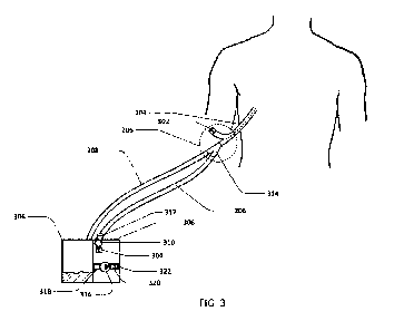
100861 Fig. 3 shows an embodiment of the chest drainage system with an active
drainage
tube relief valve and a passive chest tube relief valve. Chest tube 104 is
connected to drainage
tube 208. Drainage tube relief lumen 206 is in fluid communication with both
chest tube 104
8
CA 03016065 2018-08-28
WO 2017/152125
PCT/US2017/020791
and drainage tube 208. The connection among the 3 lumens chest tube, drainage
tube and
drainage tube relief, mem at tube-tube junction 205, which is at or near the
chest
tube/drainage tube junction. hi some embodiments, the relief lumen may connect
to the
drainage tube or chest tube at a different location. The chest tube, drainage
tube and drainage
.. tube relief lumen may be connected with connection barb 31.4. Chest tube
relief valve 302
may be incorporated into the chest tube, or a separate adapter designed to
connect to the chest
tube, for example, into connection. barb 314. In this embodiment, the chest
tube has at least
two lumens, as shown in F. 4. Pressure sensor 310, drainat:te tube relief
lumen valve 304,
and filterivent 312 are in fluid communication with drainage tube relief lumen
206.
Controller 308 includes pump 316, pressure sensor 310, drainage tube relief
valve 304,
filter/Vent 31.2, and fluid reservoir (or suction canister) 306, which is in
fluid communication
with drainage tube 208,
[00871 Controller 308 may alSO include pressure sensor 318 on the canister
side of the pump,
in-line flow sensor 320 on either side of the pump, and/or one-way valve 322
on either side of
the pump.
[00881 Pressure sensor 310 senses the pressure M tube-tube interface area 205
(via drainage
tube relief lumen 206). When the drainage tube is blocked or restricted, the
pressure in the
tube-tithe interface area increases, 'When this pressure increases to a set
pressure (generally, a
negative pressure), controller 308 opens drainage tube valve relief 304 (wind)
is normally
dosed) to allow filtered atmospheric pressure air to enter drainage tube
relief lumen 206.
This influx of air, in combination with the negative pressure in the drainage
tube caused by
pump 316, acts to clear the drainage tube of blockages/restrictions. Once the
pressure in the
tube-tubc interface area returns to normal, and/or after a set time, the
controller closes
drainage tube relief valve 304. Alternatively, the drainage tube valve may be
a passive valve
set to open and dose at set pressures.
(008)1 The mouitorlcontroller may monitor pressure inifbc drainaw *be
relieflnrnon.pnti
may pull additional suction in the fluid reservoiriStietion canister:asneeded
to maintain the
suction pressure in the proper range at the tube-tube interface area. For
example, when the
desired pressure is set to -20 cm1120, the monitor may activate the suction
pump to keep the
pressure at the tube-tube interface area between -15 0111.1120 and -25
01111120 or between -18
cm1120 and -22 crti1120. .1n another embodiment, the monitor may activate the
pump and
drainage tube relief valve 304 at regular temporal intervals as a preventative
measure to dear
arty pooled liquid from the drainage line, This is done by the controller
activating suction
9
CA 03016065 2018-08-28
WO 2017/152125
PCT/US2017/020791
pump 316 while simultaneously opening drainage tube relief valve 304 to allow
air to sweep
accumulated liquid .into the suction canister via the drainage tube.
100901 The chest tube may become blocked or restricted. To clear restrictions,
the suction
magnitude applied by the controller to the drainage tube and experienced by
the tube-tube
interface ma.y be increased. When the pressure in the tube-tube interface
reaches a set low
evc.chcst tube relief valve 302 opens and. allows filtered. atmospheric air to
enter the relief
lumen of the chest tube (see Fig. 4 for detail). This influx of air, in
combination with the
negative pressure in the drainage tube and tube-tube interface area caused by
pump 316, acts
to dear the chest tube of bloekagesirestrictions. A passive valve is shown
here; although an
.10 active valve, controlled by the controller, may be used. Alternatively,
a valve which is
operated manually, may be used. Any of the operations disclosed herein which
may be
controlled by the controller, may alternatively be controlled passively, or
manually. For
example, valve functions, suction functions, etc.
100911 The chest tube relief valve may have a different opening pressure and
closing
pressure, For example, the chest tube relief valve may open at a higher
pressure differential
(i,e, a more nenative pressure in the tube-tube interlace area), and close at.
a lower pressure
differential. This allows the valve to stay closed until a clear chest tube
blockage is present
and to minimize the flow resistance of the valve. Once the valve is open, this
allows the valve
to stay open to completely dear the tthest kibe blockage, even if the tube-
tube interface area.
pressure increases so that the pressure differential across the chest tube
valve drops below the
valve opening pressure. in other words, the pressure within the tube-tube
interface area may
be more negative when a chest tube blockage is created, but less negative, as
the chest tube
blockage is being cleared,
10092.1 Fig. 3 shows one chest tube in use with the thot drainage system, but
in some
embodiments, more than one chest tube .may be used with the system. Each chest
tube may
have its own drainage lumen and relief lumen and valve.
100931 Fig. 4 shows an embodiment of the chest tube Shown .in Fig. 3_ Chest
tube 104
includes drainage lumen 408 and chest tube relief lumen 406 incorporated into
the chest tube.
Chest tube relief valve 402 and filter/vent 404 ate also shown in fluid
communication with
chest tubc relief lumen 406, which is in fluid communication with chest tube
drainage lumen
408 via opening 412, Drainage openings 410 allow fluid from the chest cavity
to enter the
chest tube and drain through chest tube drainage lumen 408.
CA 03016065 2018-08-28
WO 2017/152125
PCT/US2017/020791
100941 During successful chest drainage, chest tube relief valve 402 is in the
closed position.
in this position, fluid draining from the chest generally does not enter chest
tube relief lumen
406 because of the fluid column in the chest tube relief lumen. A smaller
diameter Chest tube
relief lumen may help prevent fluid from entering the chest tube relief lumen.
The pressure in
chest tube relief lumen 406 is slightly negative during chest tube drainage
due to the negative
pressure exerted by the pump on the drainage line, the chest tube drainage
lumen, and to
some extent, the chest tube relief lumen, The Chest tube may become blocked or
restricted,
because of blood clots etc. To clear them, the monitor may apply additional
suction to
decrease the pressure in thee chest tube drainage lumen, and ultimately, the
chest tube relief
lumen, to a more negative pressure. AS this negative pressure drops below a
set valve
opening pressure, chest tube relief valve 402 opens, allowing atmospheric
(i.e., more positive
pressure) to enter the system. This, in combination with the negative pressure
exerted on the
drainage lumen, clears the chest tube drainage lumen. Once the pressure in the
chest tube
relief lumen increases back to 3 set valve closing pressure, chest tube relief
valve 402 closes
and normal drainage continues. The chest tube relief valve opening pressure
may be different
than the Chest tube relief valve closing pressure to allow drainage of the
chest tube. For
example, the chest tube relief valve openin9, pressure may be at a higher
pressure than the
Chest tube relief valve closing pressure.
100951 For example, the chest tube relief valve may open When the pressure
differential'
across the valve is about -10 cmI120, about ,20 ernkl24: about -30 critH20,:
about -40
cmf-1.20, about -50 cinH.20 or as even high as about -100 cinT1.20, Or for
example, the chest
tube relief valve may open when the pressure differential across the valve is
within a range of
about -10 etull20 to about -20 cmf120, or within a range of about -20 etrill20
to about -30
cmH20, or within a range of about -30 cmf120 to about -30 em}f20, or within a
range of
about -40 cmH20 to about -40 cmH20, or within a range of about -50 cmH20 to
about -100
cmi120.
100961 The chest tube relief valve may close at the same range, or at. a lower
differential than
the opening pressure. For example, the chest tube relief valve may close at a
pressure
differential of about to 0 cm112.0, about -5 cmH20, about -10 eml120, about -
1.5 cull-1.20, or
about -20 emH20, Or for example, the chest tube relief valve may close at a
pressure
differential range of about to 0 cad7120 to about -5 enaH20, or a range of
about -5 etnli.20 to
about -10 em1120, or a range of about -10 em1420 to about -15 cm}120, or a
range of about -
15 cm1420 to about -20 emi-120.
11
CA 03016065 2018-08-28
WO 2017/152125
PCT/US2017/020791
1.00971 The chest tube relief valve may take a variety of known forms,
including but not
limited to a check valve, umbrella valve, ball valve. Belleville valve, X-
fratm valve, cross
slit valve, or dome valve. The valve system preferably has a filter in place
to prevent the
entrance of bacteria or viruses from the atmosphere into the patient.
100981 In another embodiment of the chest tube, chest tube relief valve is
active, not passive,
and is controlled by the controller.
100991 In some embodiments of the chest tube,ehesttube relief valve is
incorporated. into the:
chest tube. in some embodiments, the chest tube relief valve is incorporated
into a connecter
which is connected to the chest tube. In some embodiments of the chest tube,
both the chest
tube relief lumen and the chest tube relief valve are incorporated into a
connecter which may
be connected to a chest tube.
(Ø1.001 In some embodiments, chest tube .relief valve 402 takes the form of
a magnetic check
valve that has a substantial difference in the pressure differential required
to open the valve,
and the pressure differential required to keep the valve open or close the
valve), thereby
amplifying the toggling effect of the valve, This is preferable to increase
the effectiveness of
the clog clearance cycle, because it allows for a greater pressure
differential when the air is
sweeping the drainage lumen via the relief lumen than if the valve opened and
closed at the
same pressure. The valve is normally closed in order to maximize drainage of
liquid as it
enters the chest tube and to reduce the need for continuous pumping.
Fig. 5 shows a magnetic embodiment of the chest tube valve. The magnetic chest
tube valve
includes housing 502, filter 504, ferrous plate 506, gasket 508, magnet 510,
seal plate 512,
and positioning lip 514. When the pressure differential across the valve
increases above a
desired threshold, for example -50 eME120, the force caused by the pressure
differential is
enough to overcome the magnetic force between the magnet and the ferrous
plate, thereby
moving the two away from each other. Once the magnet and the ferrous plate
move away
from each other, the magnetic force rapidly diminishes, as the magnetic three
is proportional
to (1 r3). As a result, the amount of pressure necessary to keep the valve
open is less than the
'pressure that was required to open it. This second pressure value, for
example -10 cinft20, is
determined by the maximum distance the magnet and seal plate can travel away
from the
ferrous .plate, which is in the exemplary embodiment shown in Figure 5
determined by
positioning lip 514 in the housing, that sets this distance.
r.01.01...1 Fig. 6A shows the chest drainage system's ability to detect and
clear pooled liquid in
the drainage tube. In section 'A', a -10 einfi.20 vacuum is properly
transmitted to the chest.
12
CA 03016065 2018-08-28
WO 2017/152125
PCT/US2017/020791
In section 'B', liquid begins to pooi in the bottom of the tubc, resulting in
a decreased
negative pressure (or an increased pressure). if unresolved clinically,
drainage would be
impeded. However, in section the drainage tube relief valve is opened and.
the liquid is
flushed into the drainage container, resulting is restoration of proper
suction in .Section
as well as proper negative pressure as measured. The valve is closed after
noirnal
drainage/pressures have been restored. In this example, the pressure is
measured at the tube-
tube interface area, however pressure may be measured in other and/or
additional locations in
the system, For example, pressure may be measured at or near the chest or
chest tube and also
at or near the suction device, and the .differential pressure IlleaSlifernellt
may be used to detect
I 0 flow .impediments or pooling or clotting of blood!fluid,
101021 The controller can identitY impedime.nts to .fluid drainage via a
measured absolute
pressure, change in pressure, pressure differential between or among 2 or more
locations, or
at one location, When an impediment to fluid drainage is identified, an alarm
may sound
and/or the controller may initiate .eicaring procedures, including opening,
and/or closing
valve(s) in the chest drainage system, as described else:where herein The
negative pressure in
the drainage tube may be increased, or changed in other ways, such as pulsed,
reversed etc.
1.01031 For example, if pressure measured at the tube-tube interface area is
reading around -
10 cm1120 to around -20 ern1120 and the reading changes to zero to -5 cmi120,
the controller
may open the drainage tube valve to filtered atmospheric air, 'The controller
may leave the
valve in this position for a set period of time, say 5-10 seconds or 10-30
seconds and then
may return the valve to its regular position. Alternatively, the controller
may close the valve
.when a set pressure i.s measured at the tube-tube interface area or
elsewhere. The controller
.may then cheek the pressure readings and if they have returned to normal., do
nothing more. If
they have not returned to normal, .indicating a blockage or slowing condition
is still present,
the controller may repeat the clearing procedure. This may be done repeatedly
until the
tubing is cleared. Alternatively or addition-ally, the procedure may change if
repeat clearings
are necessary. For example, the magnitude of negative pressure used by the
suction device to
clear the tubing may be increased, andfor the negative pressure may be pulsed.
The clearing
procedure may be performed in response to the pressure readings and/or it may
be done
automatically on a periodic basis.
101041 Figs, 6B-6F shows the chest drainage systerds 'ability to detect And
Clear pooled
liquid in the chest be. Fig. 6B shows the pressure irt the chest drainage
system over
I 3
CA 03016065 2018-08-28
WO 2017/152125
PCT/US2017/020791
This pressure may be measured by the controller, preferably via -the dn-tinage
tube .relief
lumen, but can alternatively be measured elsewhere.
j01051 Section A of F. 6B Shows normal dminage at a negative pressure, created
by the
suction pump of the chest drainage system. Section B shows additional suction
being pulled
by the controllerfmonitor. This additiOrkal SUCtiOn may be pulled
periodically., Or may be
.pulic.c1 based on pressure readings in the system. For example, additional
suction may be
pulled when the presence of tidal oscillations is no longer detected in the
drainage system by
the controller. The additional suction transfers negative pressure to the
drainage tube
drainage lumen, the chest tube drainage lumen, and ultimately the chest tube
relief lumen and
.10 chest tube relief IUMen valve. When the pressure differential across
the chest tube relief
lumen valve reaches the valve opening pressure, the chest tube .relief lumen
valve opens. The
valve may open automatically if the valve is passive, or by the controller, if
the valve is
active, Section C Show S the pressure .when the valve is open. The valve may
remain open for
a set period of time. Alternatively, the valve may remain open until the
controller senses that
the clog has been cleared. The negative pressure, or suction, within the
system, may remain
steady during this phase, as Shown in Fin, 613, or the nep,ativc.s. pressure
may become more
negative, as shown in Fig. 6C, or the pressure may become less negative, as
shown in Fig.
60.
101061 Section D shows the magnitude of the tegatiVe pressure docrtasiag as
..a resalt.of.a
reduction in suction being pulled by the controtlerlintantot When the pressure
in sygon
reaches the valve's set closing pressure, the valve closes (or is closed) and
fluid drainage
continues in a normal manner. The valve dosing pressure may be at a lower
magnitude
.negative pressure than that of the opening pressure, as shown here. The valve
closing
.pressure may be at or near normal drainage negative pressure.
1.0071 Figs. 63-6D show different slopes of .negative pressures in different
situations. In Fig.
6B the rate at which air is entering the system via the chest tube relief
lumen valve is the
same as the rate at which the suction pump is draining' the system during the
open valve
section C. in Fig, 6C, the rate of drainage is higher than the rate of air
entering the system. in
Fig_ 61), the rate of drainage is lower than the rate of air .entering the
system. The slope of the
pressure curve in section C may be controlled by the controller and the amount
of suction that
it is pulling,
101081 Fig. 6E shows an embodiment where the controller "overshoots" the
normal draining
suction pressure to dose the chest tube relief lumen valve. The valve closing
pressure in this
14
CA 03016065 2018-08-28
WO 2017/152125
PCT/US2017/020791
embodiment may be around .the normal draining pressure, or h may be at a less
negative
pressure (lower differential pressure).
101091 61' shows an embodiment where there is more than one chest tube.
In this
embodiment, the first chest tube .retief valve opens when the pressure in the
system reaches
valve opening pressure. It Inay be necessary to increase the magnitude of the
negative
.pressure in the system further to open the second chest tube relief lumen
valve. This is shown
as valve 2 opening pressure on the graph. There may be 1, 2, or more valve
opening pressures
depending on how many chest tubes are used on a single patient. The closing
pressures of the
multiple chest tube relief valves may be the same, or they may be different.
The ability to
detect the opening of the valves may be useful to determine whether one or
more of the chest
tubes is clogged, in which case an alarm or notification may be provided.
101101 in some embodiments, the chest drainage system may include a pH sensor.
Post,
surgery inketion and empyema are of particular concern to clinicians. The pH
of =fluid
drained front the body can be useful M diagnosing these, and other,
conditions. To aid in the
diagnosis, the chest drainage system may include a pH monitor in the
controllers with a
sensor in the reservoir, in the tubing, the pump, the valve device, or
anywhere M the system.
The results may be display-ed on the display device. The system may also
include a sampling
port to sample the fluid drained from the chest. The system may also include
iin infusion port
to infuse an additive into the drainage fluid. These ports may be, in the
reservoir, tubing,
controller, valve device, or elsewhere in the system, lbr example at the chest
tube / drainage
tube interface.
101111 in an embodiment of the device shown in... Fig: 3 (or =
othet.embodiments diSelosed
'herein), the system is capable a measuring the flow rate Of 'air evacuated
from the
canisterlreservoir, in addition to pressure in the canister and pressure in
the drainage tube
.relief lumen. :Evacuation .flow rate may be used to determine the presence
and rate of an air
leak from the Chest cavity. The, evacuation flow rate necessary to maintain
the systein at the
prescribed suction levet is equivalent to =the flow rate of air entering the
system (air leak), as
the .flows of air into and out of the system must be equal in the presence of
steady pressure.
Evacuation flow rate may be determined by the lbw rate of the air being
evacuated from the
canister via the integrated suction pump and the voltmie of liquid in the
canister. These
parameters may be tracked over time by the controller to .determine chest air
leak presence
and other parameters, such as air leak rate and changes to the air leak rate
over time. Flow
rate measurements are preferably made with any number of off-the-shelf
sensitive air flow
CA 03016065 2018-08-28
WO 2017/152125
PCT/US2017/020791
sensors that are known in the art. Flow at may alternatively or additionally
be measured by
-measuring the revolutions of the pump motor necessary to keep the suction at
a prescribed
level via a tachometer. Collected fluid volume measurements are preferably
made with a non
contact capacitive sensor, but may alternatively be made with optical sensors,
pressure
sensors, acoustic (such as ultrasonic) sensors, or any other liquid level
sensing methods
known in the art. In some embodiments, a capacitive sensor is mounted on the
inside of the
suction monitor and may use out-of-phase techniques to reduce interference
front within the
proximity, such as a human hand near or in contact with the container. Such a
technique uses
a level electrode, reference electrode, environment electrode, ground
electrode, and two
shield electrodes. In another embodiment, a compliant layer of material is
present on either
the suction monitor or the suction canister M the area of the capacitive
electrode in order to
minimize or eliminate any air gaps between the suction monitor and the
suction. canister.
f01121 Drainage fluid volume may be measured. and tracked in the presence or
absence of air
leak determination.
101131 Example of Data Processing System
101141 Ha 7 :is a block diagram of a data processing syStetitt, which :May be
used with Any:
embodiment of the invention. For example, the Systein 700 may be Used as part
of a:
controller/monitor. Note that while FIG. 7 illustrates various components of a
computer
system, it is not intended to represent any particular architecture or manner
of interconnecting,
the components; as such details are not germane to the present invention. It
will also be
appreciated that network computers, handheld computers, mobile devices,
tablets, cell phones
and other data processing systems which have fewer components or perhaps more
components may also be used with the present invention.
101151 As shown in FIG. 7, the computer system 700, which is a form of a data
processing
system, includes a bus or interconnect 702 which is coupled to one or more
microprocessors
703 and a ROM 707, a volatile RAM 705, and a non-volatiie memory 706, The
microprocessor 703 is coupled to cache memory 704. The bus 702 interconnects
these
various components together and also interconnects these components 703, 707,
705, and 706
to a display controller and display device 708, as well as to inputioutput WO)
devices 710,
which may be mice, keyboards, MOCIMS, network interfaces, printers, and other
devices
which are well-known in the art.
101161 Typically, the input/output devices 710 are coupled to the system
through inputioutput
controllers 709. The volatile. RAM 705 is typically implemented as dynamic RAM
(DRAM)
I 6
CA 03016065 2018-08-28
WO 2017/152125
PCT/US2017/020791
which requires power continuously in order to refresh or maintain the data in
the memory.
The non-volatile memory 706 is typically a magnetic 'hard drive, a magnetic
optical drive, an
optical drive, or a .DVD RAM or other type of memory system -which maintains
data even
after power is removed from the system. Typically, the non-volatile memory
will also be a
random access memory, although this is not required,
101171 While HG. 7 shows that the non-volatile memory is a locai de ce coupled
directly to
the rest of the components in the data processing system, the present
invention may utilize a
non-volatile memory which is remote from the system; such as, a network
storage device
which is coupled to the data processing system through a network interface
such as a modem
.10 or Ethernet interface_ The bus 702 may ilielude one or more buses
connected to each other
through various bridges, controllers, and/or adapters, as is well-known in the
an, in one
embodiment, the 1/0 controller 709 includes a USB (Universal Serial Bus)
adapter thr
controlling USB peripherals. Alternatively, 1/0 controller 709 may include
1EEE-1394
adapter, also known as FireiWire adapter, lb-17 controlling FireWirc devices,
SPI (serial
peripheral interface), 12C (inter-integrated circuit) or UART (universal
asynchronous
receiverltransmitter), or any other suitable technology,
r.0 MI Some portions of the preceding detailed descriptions have been
presented in terms of
algorithms and symbolic .representations of operations on data bits within a
computer
memory_ These algorithmic descriptions and representations are the ways used
by those
skilled in the data processing arts to most effectively convey the substance
of their work to
others skilled in the an An algorithm is here, and generally, conceived to .be
a sell-consistent
sequence of operations leading to a desired result. The operations are those
requiring. physical
.manipulations of physical quantities.
10119.1 It should be borne in mind, however, tharall. of-these and siniilar
'terms are to. be
itssociated with the appropriate physical quantities and are merely convenient
labels applied
to these quantities. Unless specifically stated otherwise as apparent froin
the above
discussion, it is appreciated that throughout the description, discussions
utilizing terms such
as those set forth in the claims below, refer to the action and processes of a
computer system,
or similar electronic computing device, that manipulates and transforms data
represented as
physical (electronic) quantities within the computer system registeis and
memories into
other data similarly represented as physical quantities within the computer
system memories
or registers or other such information storage, transmission or display
devices,
17
CA 03016065 2018-08-28
WO 2017/152125
PCT/US2017/020791
101201 The techniques shown in the figures can be implemented using code and
data stored
and executed on one or more electronic devices. Such electronic devices store
and
communicate (internally and/or witi other electronic devices over a network)
code and data
using computer-readable media, such as non-transitory computer-readable
storage media
(e.g., magnetic disks; optical disks; random access memory; read only memory;
flash
memory devices; phase-change memory) and transitory computer-readable
transmission
.media (e.g., electrical, optical, acoustical or other form of propagated
signals¨such as carrier
waves, infrared signals, digital signals).
101211 The processes or methods depicted in the preceding figures may be
performed by
.10 processing logic that comprises hardware (e.g. circuitry, dedicated.
oic. etc.), firmware,
software (e.g., embodied on a non-transitory computer readable medium), or a
combination
of both. Although the processes or methods are described above in terms of
some sequential
operations, 'tt Should be appreciated that some of the operations described
may be pertbrmed
in a different order. Moreover, some operations may be performed in parallel
rather than
sequentially.
r.01221 Various embodiments
O1 23j Tn one embodiment of the chest drainage.Systeitiea balloon.aballoons
may be used to
cleat- the chest tube of clogs. In the normal drainage configuration, the
balloons are deflated
to minimize the space they occupy within the chest tube lumen and maximize
drainage. For
some examples, see PCT application PCTILIS15/52960 which is incorporated
herein by
reference in its entirety. Clogs may be detected by sensing pressure and/or
pressure changes
within the system. Clogs may he cleared when they are sensed, or on a. timed
interval bases.
To clear clogs, the balloon(s) are inflated to urge clogs through the chest
tube and toward the
suction canister. The balloons may be compliant or non-compliant, or a hybrid
of the two.
Compliant balloons may bc used to conform to the shape of the inner chest tube
lumen,
which may be .used to provide a seating of the chest tube if the drainage
tubing is
subsequently flushed with fluid (gas or liquid) toward the suction canister.
This seat prevents
the flushing fluid from entering the chest: cavity. Alternatively, non-
compliant balloons may
be used to generate significant forces in order to compress and clear clogs.
This is especially
useful with robust, or firmer, clogs.
101241 A combination of these 'balloons may be used 0 :achieve both
objectives. For
example, a non-compliant balloon may be coupled wth a compliant layer :as
illustrated:*
'Fig. 8. 'Fig. 8 shows a balloon with compliant layer 802 and non-compliant
layer 804. in
18
CA 03016065 2018-08-28
WO 2017/152125
PCT/US2017/020791
another embodiment, the balloons may inflate directionally via valves botween
the balloons,
or may each have separate inflation lumens to inflate them each sequentially.
Alternatively,
the balloons may be tapered in shape and setni-eompliant such that they
&eetionally inflate
against the inner chest tube lumen wall as they are inflated. This is shown M
Fig. 9 which
shows balloon 902 as it is. inflated against the inside of the drainage lumen
of chest tube 904.
101251 The balloon may also inflate directionally hi an accordion-like
fashion, with the shape
of the balloon and/or pleats to control the direction of inflation, as
illustrated in Fig.10, which
shows balloon 100.2 and the inner wall of the drainage lumen of the chest tube
1004.
101261 In another embodiment, the balloon may be built into the chest tube
itself, such as a
1.0 coextruded inner wall that compresses inward as it inflates or expands
to fill the chest tube
drainage lumen, as illustrated in Figs. 11A and B. The figures show deflated
balloon 1102,
inflated balloon 1102, and chest tube drainage lumen .1104, Fig. 11 A shows a
concentric
configuration where Fig. I TB shows an offset configuration.
101271. In another embodiment, a balloon may be used to deliver energy to the
chest tube and
to any clogs within the drainage lumen of the chest tube to break up or
dissolve the clogs.
This may include thermal .energy, light energy, acoustic energy, or microwave
energy. In
some embodiments, the balloon may have a reinforcing structure., such as a
Nitinol coil, to:
increase the compression force against clogs, act as a chopping/breaking
.meehanism, and/or
act as a spring to control inflation direction/shape as discussed above.
Balloon inflation fluid
may be a gas or a liquid. The inflation fluid may be sterile. If sterile, for
example by
delivering the fluid across a sterile membrane (for example one with pore size
of 0.2 um) or
storing the fluid in a sterile reservoir for inflation and deflation cycles,
illustrated as 1202 in
Fig. 12.
10128! Another embodiment of the drainage system makes use
ituagnetic guidewirelO
clear the chest tube of dogs. The pick:wire is activated by enabling an
external
electromagnet such that the guidowire intermittently moves in and out of the
chest tube, in
similar fashion to a solenoid. This embodiment is illustrated in Fig. 13,
which shows
.inatmefic guidewirc .1302 and electromagnet 1304, as welt as clog .moving
feature 1306 at the
end of the
10129.1 Other erithodiments of the chest drainage system prevent adherence of
clogs to the
chest tube wall. In one embodiment, vibration energy, such as ultrasonic
energy, is used. In
another embodiment, the chest tube is made from, or coated with, a material to
prevent
19
CA 03016065 2018-08-28
WO 2017/152125
PCT/US2017/020791
adherence, such as, PTFE. In another embodiment, adherence prevention is
accomplished by
reducing the viscosity of the clots using coatings or drugs such as heparin or
a =throttibolytie.
101.301 In another embodiment, a .flushing mechanism is inemporated into a
balloon at the
patient end of the chest tube, such that once the balloon is fully inflated, a
flush port: is
exposed to allow fluid to flush pooled liquid through the drainage tubing and.
into the suction
canister, as illustrated. in Figs. 14A-14C. Balloon 1412 with flush port 1414
is shown in chest
tube 1410. In one case, the flush port comprises micmholes in the wall of the
balloon. Fig.
14A shows the balloon deflated. Fig. 1.4B shows the balloon partially
inflated. Fig. 1.4C
shows the balloon fidly inflated and shows the flush fluid direction .within
chest tube 1410,
101311 in another embodiment, multiple valves, such as balloon 'valves, are
used to seal, or
essentially seal, the chest tube so that suction can be applied to the
drainage tubing and/or the
chest tube To clear clots/blockages. The balloon(s) may also provide positive
pressure to the
chest tube and/or drainage tubing to flush pooled liquid into the collection
reservoir while
scaling the chest cavity from the positive pressure, as illustrated in Figs.
15.A-D, Fig. 15A
shows valve device 1500 with inner lumen 1502. The valve device may be part of
the chest
tube, or the drainage tube, or may be a separate device, preferably between
the chest tube and
the drainage tube. Balloon valves 1504 and. 1506 are shown, in addition to
opening/port 1508,
LUITICTI .1502 of the valve device is shown open in Fie. 15.A. The valve
device .may operate
with the following steps:
2.0 .. 101321 Step 1: Lumen .1502 is closed to the drainage tube by inflating
(or closing) balloon
valve 1506. The lumen remains open to the chest tube. A vacuum is exerted on
the chest tube
lumen by applying a negative pressure to lumen 1502 via opening 1508. The
negative
pressure applied to the Chest tube lumen is used to clear any blockage within
the chest tube.
This step is shown in Fig. 1.5B.
101331, Step 2: Balloon valve 1506 is deflated (or opened) and balloon valve
1504 is inflated
(or closed). Positive pressure is applied to lumen 1502 via opening 1508. This
serves to force
the blockage down through the drainage tube, without exerting any positive
pressure within
the chest cavity. This step is shown in Fig. 15C.
101341 Step 3: 'Balloon valve 1504 is deflated (or opened) allowing chest
drainage to proceed
normally. A valve and/or filter may be used in fluid conununication with
opening 1508. This
step is shown in Fig. 15D,
CA 03016065 2018-08-28
WO 2017/152125
PCT/US2017/020791
1.0135.1 These steps may be repeated multiple times to clear the chest tube.
The repetitions
may be based on a pre-set schedule, or they may be set. based on whether the
existence of a
Chest tube 'blockage is sensed.
t01361 in some .embodiments, the patient end .of tbe..chest..tube is -vented
to atmosphere,....ifor,
example, via a chest tube relief lumen, to allow.:sterile.airu purge dogs
fronfthe:thesttubt
during step 1.
101371 Another embodiment of the drainage sySteM inakes...use..of a flush port
for manual
intermittent flushing of the chest tube and drainage,:line, as thwn in Fig:
.16A.. :Chest tube
1602 includes flush port 1604 and flush opening. 1606. in one embodiment, the
flush port is
1.0 swabbable to ensure sterility prior to f1ushini4. In one embodiment,
the drainage tubing is
clamped and the flushing fluid is infused into the chest tube and into the
pleural cavity, but
subsequently drained once the drainage tubing is undamped arid the chest tube
pat:they is
restored. In one embodiment, the flush port cormects to a lumen, such as a
chest tube relief
linnet), that terminates at the patient (proximal) end of the chest tube such
that the flushing
fluid is infused through the chest tube toward the drainage tubing and suction
canister with
the drainage tubing -undamped by, sealing the chest tube prior to fluShing.
This may be
accomplished by balloon 1608, or other -valve, located within the chest tube
lumen, as shown
in Fig. 16B, The flushing fluid may be air, water, saline, heparin, a
thrombolytic agent such
as tissue plasminogen activator, or any other suitable fluid.
f 01381 Another embodiment of the chest drainage system monitors physiologic
parameters of
inicrest. In one embodiment, pressure is .monitored. For example, internal
chest pressure may
be sensed and monitored to ensure the applied negative andlor positive
pressure is being
properly transmitted to the chest cavity. Or, tbr example, pleural and/or
pericardial pressures
may be monitored to track healing. Or, for example, differential pressure
between the distal
and proximal Olds of the chest tube may be monitored to ensure thest tube
matey. Or, for
example, the ,pressure at the distal (non-patient) end of the chest tube, for
example at the
proximal and/or distal end of the drainage tubing or collection canister, inay
.be monitored for
tidal oscillations, which are indicative of tube potency,
101.391 In some embodiments, the volume and/or flow rate of the drained chest
fluid (either
gas, liquid, or both) may be measured and monitored over time, in another
embodiment, the
volume and/or flow rate of an air leak (from the patient's lung) is measured
to monitor wound
healing. In another embodiment, pH of the drained .fluid is measured to
monitor for
infections. Additional parameters, such as conductance, spectroscopic
signatures, protein
21
CA 03016065 2018-08-28
WO 2017/152125
PCT/US2017/020791
content, and specific gravity of the drained fluid may also be measured to
monitor patient
recovely. Any of these measurements may be one time measurements or
measurements made
over =time. For measurements made and collected over time, the controller may
analyze these
data for trends. These data may be integrated with the hospital's eleeirOnie
medical record
system (either communicated to, or data may be obtained from) and/or displayed
on a screen
on the device or on a connected monitor, which may be connected either by wire
or
wirelessly, In some embodiments, alarms or notifications may be activated by
the controller
when the parameters surpass certain thresholds, which may be preset or set by
the user. These
.may be visual andlor audible alarms or notifications.. These data may also
provide input to the
1.0 line-purging and clog-clearing functions of the device, such that, for
example, line purging is
activated When the suction at the chest drops below a certain level, or clog
clearing is
activated when tidal oscillations are diminished.
f01401 Another embodiment of the drainage system makes use of safety features
to prevent
dangerous pressures from occurring when inflating the balloons or flushing the
chest tube
and/or drainage tubing as described herein, in one embodiment, the pumps used
to inflate or
flush are connected to safety valves with crack pressures that are in the
range considered to
be physiologically safe, for example pre-venting suction below about -.20
cm1120, -40
cm1120õ or -70 cm1120, The pumps may be connected to pressure sensors with
control
systems to turn off the pumps if pressures are outside of the safe range.
101411 Another embodiment of the valve device includes a suction reservoir to
provide
additional suction to clear potential clogs from the chest tube, as shown in
Figs, 17A-17D,
Shown in these figures is valve device 1700, which may be placed between the
chest tube and
the drainage tube, or may be integrated into the drainage tube, or the chest
tube. Valve device
1700 includes inner lumen 171.6 with lumen port 1706, chest,side valve 1702
with chest-side
valve port 1704, .drainage-side valve 1712 with drainage-side valve port 1714,
chamber 1710
with chamber port 1718 and -within the chamber is evandable valve 1708.
rtlit<121 In this embodiment, expandable valve 1708 is expanded by pulling
suction (or
applying negative pressure) within chamber 1710 via chamber port 1718. In this
way,
expandable valve 1708 can generate additional suction within the chest tube.
This suction is
directed by additional valves 1702 and 1712, illustrated as
inflatableideflatable balloon valves
to temporarily seal lumen 1716. The sequence of events is:
101431 Step 11 Lumen 1716 is sealed via valve 17.12 to seal off chest tube
from the drainage
tube. This is done by applying pressure to valve 171.2 via port 1714,
inflating -valve 1712 to
22
CA 03016065 2018-08-28
WO 2017/152125
PCT/US2017/020791
dose off lumen 1716 on the drainage tube side of valve device 1700. This is
shown in Fig.
17B.
101441 Step 2: Additional suction is applied to the chest tube by expanding
valve 1708, This
is done by applying suction to chamber 1710 via port 1718. This is also shown
in Fig. 17B.
This applies additional suction to the chest tube, as indicated by the solid
arrow in lumen
1716 in Fig,. 17B.
101451 Step 3: The chest tube side of the valve device is then sealed via
valve 1702: by
applying pressure to the valve via port 1704. This is, shownin 17C
101461 Step 4: The drainage tube side of the valve device is then opened by
releasing the
pressure applied (or applying a vacuum) to valve 1712. Expandable valve 1708
is also
returned to its neutral state by releasing the vacuum applied (or applying
pressure) to
chamber 17W via pod 1718. Port 1706 (also described herein as drainage tube
relief lumen
port) allows filtered atmospheric air to enter lumen 1716. The release, or
pressurization, of
expandable valve 1708 may be enough to flush the drainage lumen of any
blockage, so that
fluid again may drain nonnally into the collection reservoir. If necessary,
additional pressure
may bc applied to lumen 1716 via port 1706 to flush the drainage tube.
Ahernatively or in
addition, the negative pressure applied to the drainage tube may be increased
(made more
negative). This step is also shown in rig. 17C.
101471 Step 5: Valve 1702 is opened by releasing the prow applied (Or applying
vacuum)
via port 1704. Lumen 1716 is now fully open and drainage may resume as normal.
This is
shown in Fig. 17D.
101481 These steps may be repeated as necessary to clear the drainage tube.
They may repeat
at a set time interval. They may repeat continuously until the drainage line
is cleared. They
may repeat only as necessary, when the drainage tube is blocked.
101491 Port 1706 may be always open or may be controlled, for example by a
solenoid, by
the controller to opew'close as needed. The lumen to port 1706 is also
described herein as the
drainage tube relief lumen.
101501 All the ports shown in Figs, 17A- I 7D may entineet :to Eta which are
controlled by
the controller. The ports and/or lines may :include filtertimembranct
toptveritebatatninateg
from entering the system.
101511 In some embodiments, chamber 1710 and valve 1708 are not used and the
components of the steps associated with the chamber and chamber valve are not
taken.
23
CA 03016065 2018-08-28
WO 2017/152125
PCT/US2017/020791
1.01524 Figs. I8A and 18B show a method of measuring air leak using the chest
drainage
system. If the chest tube and drainage tube are clear of blockages, pressure
within the chest
can be measured and monitored by the controller to calculate the rate of air
leak, As Shown in
Fig_ 18A, the chest may be sealed off from the drainage canister, for example,
using
drainage-side valve 1.7/2, and pressure may be measured using a lumen in fluid
communication .with the drainage lumen, such as via port 1706, or any other
lumen in fluid
conununication with the. chest tube lumen, for example a chest tube or
drainage tube relief
lumen, When the chest tube is sealed off form the vacuum source, the negative
pressure in the
chest tube lumen can be measured by .the controller, and will attenuate if the
patient has ui air
1.0 leak. The attenuation can be measured by the controller over time and
converted to a .rare of
air leak (mLitain). A graph of the pressure within the chest tube over time in
the presence of
ail air leak is shown in Fig. 1813.
j01.531 An air leak may alternatively be measured by keeping both valves (1702
and 1712)
open as shown in Fig. 19, and monitoring the pressure for spikes that result
from bubbles in
the water seal chamber in the suction canister. Pressure may be measured Via
opening 1706 or
any other area in fluid communication with lumen 1716, in a preferred
embodiment, this
method makes use of a sensitive pressure sensor that is connected in Line witi
a high-pass
filter in order to make the spikes from bubbling easier to detect. The various
methods of air
leak .detcetion described herein may be used independently or M combination.
101541 In another embodiment of the device, clog detection is performed by
comparing the
pressure measured within the chest (via the chest Mk relief lumen) and the
pressure just
distal to the chest tube (via the drainage tube relief lumen), i.e. in the
tube-tube interface area.
These pressures may be compared to one another, and when they differ by
certain amount,
for example 5 emB20, this is indicative of a clog in the chest tube.
Alternatively, the
pressure within the chest alone may be monitored, and when it increases by a
certain amount,
tbr example to above about 0 cmH20, this may also indicate that a dog in the
chest tube
exists. When a clog is detected, for example by either of these methods, the
controller may
automatically activate any of the clog-clearing mechanisms described herein.
Additionally,
similar automation may be applied to the drainage line purging mechanism, by
monitoring
the pressure distal to the chest tube (via the drainage tube relief lumen),
and activating a
purge of the drainage line when the pressure increases above a certain
threshold, for example
-35, -30, -25, -20.45. -10, -5 or 0 cm1120.
24
CA 03016065 2018-08-28
WO 2017/152125
PCT/US2017/020791
1.0155.1 'Mien a clog is detected, the device may also warn the clinician of
impending cardiac
tamponade.
101.561 Fig, 20 illustrates an embodiment of the chest tube cross-seetion ii
inore detail,
showing, chest tube relief lumen 2002 and chest tube drainage lumen 2004. in
sonic
embodiment's, relief lumen 2002 may be significantly smaller in cross
sectional area than
drainage lamen 2004. The same may be true for the drainal,N tube relief hnnen
and drainage
lumen. For example, the cross sectional area of the drainage lumen of either
the chest tube or
the drainage tube may be about 5 to about 10 times larger than the cross
section of the
associated relief lumen. Or for example, the cross sectional area of the
drainage lumen of
.10 either the chest tube or the drainage tube may be about 10 to about 20
times larger than the
cross section of the associated relief lumen. Or for example, the cross
sectional area of the
drainage lumen of either the chest tube or the drainage tube may be about 20
to 30 times
larger than the cross section of the associated relief lumen. Or for example,
the cross
sectional area of the drainage lumen of either the chest tube or the drainage
tube may be
about 30 to 40 times larger than the cross section of the associated relief
lumen.
r.01.57.1 Fig 21 Shows an .embodiment of the valve device.. Within the
cylindrical housing are
the balloon valves (includinn expandable valve) as illustrated in Figs. I 7A-
D. Fig. 21 shows
the valve device COTIllectilli4 points to .the chest aThe and drainage tube,
as well as the relief
Lumens. Barb 2102 connects to the drainage lumen of the chest tube, barb 2106
connects to
the chest tube relief lutnen, the 5 pneumatic connecters 2108, 2110, 2112,
2114, and. 2116
connect to chest tube relief lumen barb 2106, chest-side valve port 17(4,
lumen port 1706
(also described as the drainage tube relief port herein), chamber port 1718,
arid drainage-side
valve port 1714 of Fig. 17A respectively. The pneumatic connecters .may be in
any order.
Barb 2118 connects to the drainage tube drainage lumen.. The other end. of the
pneumatic
connecters connect to the monitor/controller and the pneumatics are controlled
by the
controller.
rtill.581 fig. 22 further illustrates these relationships by showing chest
tube 2202, relief lumen
barb 2106, drainage tubing 2204 and pneumatic connecters 2106-2116.
101.59.1 Fig. 23 shows an embodiment of the chest drainage system in use,
including chest
tube 2302, drainage tube 2310, valve device 2304 and. monitoricontmlier 2306.
in this
embodiment, the system is connected to standard suction device/canister 2308.
In this
embodiment, the valve device is connected in line between the chest tube and
drainage tube,
with leads to the suction canister, and the monitor may be placed wherever is
most
CA 03016065 2018-08-28
WO 2017/152125
PCT/US2017/020791
convenient, .incinding but not limited to the patient's bedside, IV pole, or
mounted to the
suction canister directly.
j01601 The connection between the pneumatic connecters coming from the valve
device and
the monitor is illustrated in Fig. 24, which shows the monitor-side pneumatic
connecter 2402,
which connects to pneumatic connecters 2106-211.6 in Fig. 21. Pneumatic
connecter 2402
connects the relief lumens and balloon valves of the valve device to the pumps
and. solenoids
housed within monitor 2404. The connecter preferably snaps into place, and
seals against: the
monitor with gaskets 2502 as shown. in Fig. 25. Preferably, the connections
include
membranes that act as sterile and/or liquid 'barriers to separate the lumens
on the patient side
.10 of the system from those on the monitor side, thereby preventing
contamination. or liquid
intrusion.
101611 in yet another embodiment, the chest tube clog clearance and drainage
lino purging
activities may be accomplished using a system shown in Fig. 26, in which a
full length relief
lumen 2604 runs the length of the system, from suction canisterfreservoir
2606, through
drainage tube 2(10, through valve device 2602, through chest tube 2608 to the
proximal tip
(patient side) of the chest tube., in this embodiment, clot-is are cleared
from the chest tube and
pooled liquid. is cleared from the drainage Line hi the same step, by applying
suctioi at the
suction canister and opening the full length relief lumen to allow air to dear
the entire
system. Clearance of the line may be performed at set intervals, or When the
pressure in the
chest (measured via the relief lumen) increases above a. certain threshold,
fOr example -35, -
30, -25, -.20, -15, -10, -5 or 0 em1120, or a combination of these approaches.
Also in this
embodiment, balloon valves may or may not be present in valve device 2602. if
not present,
air leak measurement can be accomplished by measuring the pressure in the
entire system and
watching for .attenuation as described herein. Alternatively, air leak can be
measured by
monitoring fo.r bubbles as illustrated .M Fig. 19. Alternatively, or ii
iddition, air leak nuty be
measured by measuring the air directly as it flows through the system with
either an in-line
flow sensor or by measurinnidetermininn the -volume of air moved by the
suction pump to
maintain adequate suction in the system. For example, a tachometer may be used
to measure
the number of revolutions of the motor driving the suction pump.
101621 In some embodiments, the controller is connected to a .iik...twork,
either wired. or
wireless, in order to transmit data for example to and/or from the patient's
electronic medical
record (EMR). The controller may also provide notifications of patient status
on the
controllertmouitor itself and/or by transmitting notifications and/or safety-
alarms to the EMR
26
CA 03016065 2018-08-28
WO 2017/152125
PCT/US2017/020791
or the clinician's phone, tablet, watch., etc. Additionally, the system may
interface with other
systems via wireless or wired technolegy, and there may be wireless
communication between
the components, for example between the monitor shown in Fig. 24, the
pneumatic connecter
shown in Fig. 24, and/or the valve device shown in Fig. 21..
(Ø1.63.1 In another embodiment., the number of balloon valves may be reduced
-.1riellor the
relief lumen port may be integrated with the valve device at: a different
location, N shown in
Figs. 27A-27D. The steps associated with this embodiment are:
1.01641 Step 1. Inner lumen 2716 is sealed to seal the chest tube from the
drainage tube. This
is done by applying pos.itive pressure to chamber 2710 via Chamber port 2718.
This closes
1.0 expandable 'MINT 2708 sealing off lumen 2716, as shown in Fig. 27B.
[0165...1 Step 2: The drainage tube is flushed by opening flush lumen port
2706 to atmospheric
'pressure or applying .positive pressure to lumen port 2706. This is also
shown in Fig, 2713.
t01661 Step 3: drainage-side balloon valve .2712 is then dosed by applying
pressure to the
balloon via drainage-side valve port 2714. 'This is Shown in Fig .27C.
.15 101671 Step 4: The chest tube is exposed to additional /locative
pressure by applying negative
pressure to expandable valve 2708 via chamber port 2718 of chamber 2710. Lumen
port 2706
will be dosed during this step. This is shown in Fig. 7C. The chest tube (not
shown) in this
step preferably has a relief lumen that. is controllably opened to allow
sterile air to enter the
Chest tube drainage lumen from the patient end as additional negative pressure
is applied,
20 which increases the effectiveness of cleAtrance, Lumen 2716 is now fully
open and drainage
may resume as normal_ This is shOW11 in Fig. 27D.
r.01681 in another embodiment, balloon valve 2712 may be removed such that the
steps are:
r,C41 691 Step 1: Inner lumen 2716 is sealed to seal the chest tube from the
drainage. tube. This
is done by applying positive pressure to chamber 2710 via chamber port 2718.
This closes
25 expandable valve 2708 sealing off lumen 2716, as shown in Fig. 2713.
t0170.1 Step 2: The drainage tube is flushed by opening flush lumen port 2706
to .1t.mospherie
pressure or applying positive pressure to lumen port 2706. This is also shown
in Fig. 27B.
101711 Step 3: Clear the chest tube by returning the expandable valve 2708 to
its neutral
position and opening the chest tube relief ILIUM.), (not Shown) (via a valve,
either passive or
30 active, not Shown) to allow air to sweep the drainage lumen of the chest
tube.
101721 This approach eliminates the. need for balloon 2712. and valve. port
2714, which are
only used when additional negative suction is pulled. Both of these steps may
be performed
27
CA 03016065 2018-08-28
WO 2017/152125
PCT/US2017/020791
by the, controller at regular intervals. Alternatively, the chest tube relief
lumen and/or the
drainage tube relief lumen may be 'kept open at all times, or optionally
closed when manually
capped by the user as desired or by the controller.
101731 In other embodiments, the action of seahrig.the balloon valvex.and
gerierining.suctlop,.
its disclosed in any of the embodiments herein, May beverformed .manually by
tbe userJar:
example, similar to the system shown in 'Fig. 12. In some embodiments, the
manual valve
connecter allows for attachment of one or more syringes, which can be used to
seal the
balloon valves and generate additional suction andSor positive pressure for
purging the
drainage line. In some embodiments, the manual valve device employs a valve
that can be
.10 configured to be 1) closed, 2) in communication with a chest,side or
drainage-side balloon
ValVe., or 3) in communication with the larger expandable -valve. In this
manner, the user ma
first connect to a balloon valve and apply positive pressure to create a seal,
then connect to
the larger expandable valve to generate ;Additional suction, The relief lumen
described herein
(either the chest tube relief lumen of the drainage tube relief lumen) can
also be activated
automatically or manually, or alternatively can be activated passively by
means of a cheek
valve that cracks once the pressure rises above a certain threshold, as
described in detail
herein..
101741 hi an alternate embodiment, the means of generating positive pressure
and suction is
within the valve device itself, such as a :fluid reservoir, so that a separate
syringe is not
required for activation,
1411751 In Figs. 28A-28C, a .method of measuring chest air leak is presented
in more detail. In
this embodiment, the chest tube and drainage tube aro kept dear thmugh the
.methods
described herein. An an leak may be monitored and quantified at both
relatively high and low
air leak flow rates. The applied standard suction causes the pressure in the
chest to be
negative,
101761 In the presence of relatively low flow rates, positive pressure is
applied w. chamber
2810 via chamber port 2818 to dose expandable valve 2808 to seal turnen 2816
as <Sliowniti
Fig. 2.813 The pressure in the chest is then monitored via lumen port 2814, if
there is an air
leak, the measured negative .pressure will become less negative as the
incoming air enters the
pleural space. Fig. 29 depicts an example pressure over time for two different
low flow air
leak rate measurements, The upper graph represents pressure attenuation over
time in the
presence of a small air leak, while the lower graph represents pressure
attenuation over time
in the presence of a 'larger air leak (still relatively 'low flow). The
magnitude of the change in
28
CA 03016065 2018-08-28
WO 2017/152125
PCT/US2017/020791
pressure is indicated by the slope of the line intersecting points 1 and 2.
The rate at which the
pressure increases (APIAt) may be converted to volumetric air leak rate
.(AVizit). Pons 1
and. 2 may be determined based solely on time, for example taking measurements
0,5 and 1
sec after creating =the seal as shown in Fig. 2813, Alternatively, they may be
based on pressure
values, cither absolute (e.g, once the pressure goes above -10 and -5 cm1-120)
or relative (e.g.
once the pressure increases by 2 and 4 emlI20 relative to baseline). Sample
benclatop data
illustrating this technique is presented in Fig. 30, which utilizes a second
degree polynomial
fit to convert rate of pressure change to air leak,
10177) In the presence of relatively high flow rates, positive pressure is
applied via balloon
valve port 2804 to close balloon valve 2802 to seal lumen 2816, sealing off
the drainage
lumen of the chest tube from the drainage lumen of the drainage tube. This is
shown in Fig.
28C. The. pressure is then monitored via lumen port 2814 to determine a
baseline pressure
-value due to the applied suction only (via negative pressure applied to the
drainage nibe).
Then, balloon valve 2.802 is opened and a new pressure value is measured, once
again via
1.5 lumen port 2814. lf the air leak rate is high., the will be a larger
detectable difference
between the two aforementioned pressure measurements due to the rapid influx
of air. This is
shown in Fig. 31. The upper graph represents a pressure differential in the
presence of a
relatively small air leak (though still high flow), while the lower graph
represents a pressure
differential in the presence of a larger air leak. The magnitude of the
difference between
these two measurements (AP) may be converted to volumetric air leak rate
(AV/iSt). Sample
.benchtop data illustrating this technique is presented in Fie.. 32, which
uses a linear fit to
convert rate of pressure change to air leak.
101781 The same methods a determining air leaks shown for low and 'high flow
scenarios
may also be perfomied by swapping the positions of the balloon valves in Fig.
28, relative to
lumen port 2814. in this ease, the balloon valve seal is inflatedfelosed when
.measuring low
flow and the larger expandable valve is inflatediclosed -When measuring high
flow.
[0179.1 One embodiment of the device may include a method for detecting the
presence, as
well as pmper placement, 1..)f the pneumatic cassette module (2402 in Fig. 24)
in the .monitor
(2404 in Fig. 24). This detection information may be used to start or stop
.functionality of the
device, activate or deactivate certain features of the device, sound alarms
and/or display alert
messages on the device, etc.
1.01801 One method for cassette .dctection may use an Infrared (110 emitter
and receiver to
detect the presence and proper placement of the cassette. This IR sensor may
measure the
29
CA 03016065 2018-08-28
WO 2017/152125
PCT/US2017/020791
.reticctivity of the cassette surface; the information may be used to inform
device function.
The reflectivity of the cassette may be modified to allow for a wide variety
of reflectance
values. The reflectivity of the cassette may be controlled by changing the
color, geometry, or
trty combination of color and geometry to adjust reflectance. In Fig. 33, the
relationship
.. between color and reflectance readings is shown, where the possible range
of values may be
0-100%, In Fig., 34, the relation between surface angle and reflectance
readings is shown for
a white surface where again, the possible range of values may be 0-400%.
101811 Another method for cassette .detection may use a color sensor with
attached LED to
illuminate the surface to be measured. This color sensor may be capable of
detecting various
.10 levels of a specific color, such as red and/or cyan, light intensity,
light temperature, as well as
any combination of these metrics. tn one embodiment, the color of the cassette
may be used
to differentiate between two or or desired function settings of the device. In
another
embodiment, the cassette my have features whieh allow a certain amount of
light to reach
the sensor, thus, variable light intensity may be used to control fin:tenon
settings of the device.
.. In another embodiment, the cassette may have features which modify the
temperature of the
light reaching the sensor, a Light filter for example: thus, variable light
temperature may be
used to control function settings of the device.
101821 Another .method for cassette detection may use a Hall Effect sensor to
detect the
presence of a magnet located th the cassette. This .magnotic sensor may be
used to ensure
complete and proper placement of cassette to locked-position in monitor by
defining
placetnent of the magnet and Hall Effect sensor, such that detection only
occurs when the
cassette is fully seated and locked in place.
101831 Another method for cassette detection may use an IR gate sensor to
detect both the
presence of the cassette in the monitor, as well as ensuring proper placement
in the monitor.
The cassette may include a feature or .feaiures which may interact with the IR
gate sensor,
providing information regarding the placement of the cassette in the monitor.
10184.1 Another method for cassette detection may utilize a IN light sensor to
detect the
'presence and proper placement of the cassette in the monitor. TO allow for
variability
between cassettes, features may be added to the cassette which modify the
amount of UV
light permitted to reach the sensor. This may be accomplished using, for
example, .13N/ filters,
passageways within the cassette, openings, minors, or some combination of
these features.
1.0185.1 Another method for cassette detcedon may involve capacitive sensing,
whereby the
cassette has varying IONTIS of dielectric strength depending on, for example,
the size of a strip
CA 03016065 2018-08-28
WO 2017/152125
PCT/US2017/020791
of nwtai embeddcd in or mourned to the cassette. A capacitive sensor within
the monitor may
then be able to distinguish between the various cassettes depending on this
vaiying level. of
capacitance.
t01861 One embodiment of the device may include a. method for measuring and
.quantifying
air leak .metrics which are then displayed via :a .seneen. on the monitor or
elsewhere., This
information may be used to start or stop functionality of the device,
activatedeactivate
certain features of the device, sound alarms and/or display alert messages on
the device,
provide information to attending physicians, as well as other actions related
to proper
removal of fluids from the chest. These actions may be performed manually or
automatically
.10 be the controller,
101871 On method for displaying air leak data May:
inehtde:.:deScribing:theOeturtence oft
particular air leak qualifier by showing the pereentage:of theaStirenients.
ever :the preViOns :X
hours for which that qualifier occurred. The an leak qualifiers may include/he
equivalent to:
"Persistent" for high flow, "Intermittene for moderate to low flow, and "None
for no flow.
As shown in Fig. 35, this infotmation may be presented:, for example,
graphically as a bar
graph. 3502 or circular chart 3504, or in a display using text and numbers
(3504
101881 Another method for presenting air leak data may include graphing
trending flow rate
data over a period of time, for example I 48 hours. The air leak flow rate
information may
be collected, for example, using an in-line .flow sensor, pressure sensors, or
pump tachometer.
.As shown in Fig. 36, the air leak flow rate trending data may be expressed as
(m/min) with
respect to time in hours past, for example. The pressure shown here is
measured at the barb
Or tube-tube interface.) area.
rt:11.891 Another .method tbr presenting air leak data may include graphing
trending air leak
qualifiers over a period of time, for example ¨ 48 'hours. The air leak
qualifier information
may be calculated, for example, using algorithms to relate changes in chest or
chest tube
pressure to -volumetric flow rate, or defining a pressure threshold, for
example -5 cm1120,
and relating the time to reach that value with a qualifier. As Shown in Fig.
37, the air leak
qualifier trending data may be expressed as, for example, "Persistent",
"Intermittent", and
"None' with respect to fink: in past, tbr example, for .the past x hours.
10190.1 Another method for presenting air leak data may include an
instantaneous
measurement initiated by, for example, pushing a button. As shown in Fig. 38,
the prompt for
instantaneous measurement may be a highlighted arrow, a physical button, a
virtual button,
31
CA 03016065 2018-08-28
WO 2017/152125
PCT/US2017/020791
graphic etc. 'The information provided may be in the form of, for example,
flow rate (mliinin)
or an air leak qualifier, such as "Persisted', "Intermittent", "None", or an
equivalent term.
101911 One embodiment of the de-vice may include a method for measuring and
quantifying
dm; clearance metrics to he displayed via a display or screen. This
information may he used
.to start or stop functionality of the device, activate/deactivate certain
features of the device,
sound alarms and/or display alert messages on the device, provide information
to .attending
physicians, as well as other actions related to proper removal of fluids from
the chest.
10192.1 One method for presenting clog clearance. data .may include displaying
the number of
dogs cleared over a certain amount of time, for example 1 ¨ 48 hours ¨ the
.range may be set
1.0 by the user or may be pre-set. As shown. in Fig, 39, the information
displayed may iriclude
description of the displayed metric, for example "Clogs Cleared", a nuinber or
quantifier
associated with the .metric, fbr example, the number of clogs cleared, and the
time interval
over which the metric occurred.
101931 Another method for presenting dog Clearance data May 'for.
tpmpie,
15 displaying the time for when the last clog occurred, the date on which
the lag clog Oceurred,
whether the clog was cleared, the tiime. passed since the last clog occurred,
or some
combination of these or similar metrics. As shown in 'Fig. 40, the information
displayed may
indicate what metric is being shown, for example "Last Clog".
101941 Another method for presenting clog clearance data may include a graph
displaying
20 trending pressure data from the tube-tube interface area, or elsewhere
in the system, over a
certain amount of time, for example 1 --- 48 hours, for a certain range of
'pressures, fbr
example -500 500 cmf120. As shown in Fig. 41, one or more pressure values may
be
plotted simultaneously with respect to ti.me, for example, pressures measured
at different
locations within the system. For example, the graph my display the applied
suction level ,set
25 by the user, shown by the solid line, and/or the graph may display the
pressure measured in
the tube-tube interface area, shown by the dashed line,
101951 Another method for presenting clog clearance data may include a graph
displaying
trending clog clearance data for a specific time interval (for example, 1
hour), over ;a =Min
amount of time, for example I ¨ 48 hours. As shown in Figõ 42, a bar graph may
be. utilized
30 to convey information regarding the number of clogs cle,4,1red each hour
over the past 10
hours, for example. The total number of dogs cleared over the total duration
of device use fbr
a particular patient may also be shown.
32
CA 03016065 2018-08-28
WO 2017/152125
PCT/US2017/020791
101961 Some embodiments disclosed herein include a balloon, or pneumatic,
valve, which
uses pressure variance to occlude or open the passageway by inflating or
deflating a flexible
membrane, or balloon. Figs, 43A-43E show an embodiment of these types of
valves. The
valve includes rigid housing 4302 and thin-walled silicone extrusion 4304, as
Shown in Figs
43A-E. Silicone extrusion 4304 may be either an extrusion or a. mold out of
any suitable
material, including silicone, latex, polymer, etc. The durometer of the
silicone may be, for
example 20A-70A, and the wall thickness, for example 0.005" ¨ 0.050". The wall
thickness
may be adjusted to achieve the desired sealing pressure. The wall thickness
may be consistent
along the length or vary along the length. The length of .the silicone
extrusion may be 0.5" ¨
1.0 1.0" and
the inner diameter of the silicone extrusion may be 0.1" ¨ 0.50", In one
embodiment
example of the balloon .valve, assembly begins by inserting the silicone
extrusion through the
center of the balloon housing. Next, the extrusion is folded up and over the
edge of the
'housing on both ends as shown in Figs. 43B and 43C. This process may be done
several
ways, for example, manually folding the extrusion. Another process may Maude
inserting a.
.15 balloon
through the center of the extrusion; upon placement, this balloon could be
inflated,
effectively expanding the silicone membrane until it folds over the edge of
.the housing.
Another process may incorporate a fixture which has two or more grasping
features that
expand radially in and out; the grasping features are used to grab and expand
the silicone
extrusion; once the extrusion is stretched, the housing may be moved forward
to accomplish
20 the
folding process. As shown in Fig 43C and 431), once the extrusion is folded
over the
housing, two 0-rings are placed over the silicone extrusion to ensure that the
silicone
membrane remains in place and is sealed with respect to the housing. Figs. 43
D and 43E
show the finished balloon valve.
101971 One embodiment of the chest drainage system may include vent/filter
membranes in
25 line with
the pneumatic tubing for the purpose of controlling quality of the air or
adding
dampening to the system. These membranes may have variable thickness, for
example 0.001"
0.010", have variable pore sizes, for example 0.2 100
micron, have variable cross
sectional areas, for example 0.01 ¨ 0.1 in2, and have different cross
sectional shapes, for
example circular or rectangular. The membranes may be placed within the
cassette, in line
30 with the
pneumatic tubing via barb connection, within the monitor, or within the valve
device. These vent membranes may be attached in many ways, for example
ultrasonic
welding, via a separate housing, or UV cure adhesive.
33
CA 03016065 2018-08-28
WO 2017/152125
PCT/US2017/020791
1.0198.1 One embodiment of the chest drainage system .may include a -feature
which allows the
monitor to be mounted in a convenient and effective location during use in -
various hospital
settings. This connection feature may be modular: the monitor may have a
feature that can
connect to many different: attachments that offer different mounting
solutions. One iteration
of the modular design may include a snap-and-lock .featurc, where the
attachment locks into
place and can be released, for example, by pushing a button to disengage the
lock. Another
iteration may include a threaded hole in the monitor that interfaces with a
threaded rod on the
attachment Yet another :iteration may include a sliding mechanism where the
attachment
grabs onto .rails or -hacks to connect with the monitor. Another iteration may
include a push-
and-lock feature, where the attachment can be locked and unlocked by pushing
it further into
the connection, similar to an SD card. Another iteration may include a single
or series of
magnet(s) which properly orient and hold the attachment firmly to the monitor.
I01.991 One method for creating a mountirig 17eature may include the use of a
rigid bracket on
which a pair of hook features are connected. As shown in Fig. 44, bracket
assembly 4402
may connect: to the monitor via one of the modular mounting options 4404.
Hooks 4406 may
be attached hi such a way that they are able to pivot to provide increased
adaptability.
[02001 Another method for creating a mounting feature may include the use of
flexible straps
in combination with a hook feature. As shown in Fig. 45, straps 4502 may be
attached to the
base of monitor 4504 and cover the sides of the monitor, with hook features
4506 integrated
with the strap. These straps may be fixed or mo-veable, for example, they may
be able to
retract inside of the monitor When not in use. In this case, the hook features
may have a place
to be stored as welt for example external wells into which the hooks click in
place.
102011 Another method for creating a inounting feature may :include the use of
flexible
tubing to provide a highly adaptive solution with many potential uses. As
shown in Fig. 46,
tubing 4602 may be able to take many shapes by simply beading the tube to mate
a desired
shape or feature, such as hook shape 4604. These tubes may be attached to the
monitor on the
back or sides, using one of the modular mounting options described herein. The
force
required to bend the tubing is low enough to not require a special tool, whilc
simultaneously
being stiff enough to hold its shape under the loads placed on it by the
monitor and additional
external forces.
102021 Figs. 47, 48, 49 and 50 show an embodiment of a dual-lumen chest tube.
'010 tube
4702 may be made using silicone, PVC, or odwr,:$4.,iiabie matpriO1
wit.h...a.:0able dUrometer,
for example 20A ¨ 80A. The effective outer diameter of the chest tube may vary
between Ur
34
CA 03016065 2018-08-28
WO 2017/152125
PCT/US2017/020791
-- 40Fr. The chest tube shown in Fig. 47 may include three sections: a chest
tube region, as
shown in Fig. 48, a transition region, as shown in Ha. 49, and a pull-through -
region, as
shown. M Fig. 50. The chest rube region comprises a dual-lumen extrusion with
holes 4802
near the patient side for drainage of fluid .from the body. The chest tube
region is preferably
capped with rounded tip 4804, but may also have an open patient end without a
cap. The
transition region separates the two chest tube lumens, for example chest tube
drainage lumen
and the chest tube relief lumen, into separate tube sections that are more
easily attached to
barbed connectors. Fig. 49 shows chest tube drainage lumen tube section 4902
and chest tube
relief lumen tube section 4904. Specifically, at the non-patient end of the
transition region,
1.0 both ltnen preferably become circular to allow for proper attachment to
standard barbs. The
pull-through region shown in Fig. 50 includes chest tube drainage lumen tube
section 4902
and chest tube relief lumen tube section 4904. The two tube sections may also
be joined, for
example with .webbing or adhesive. The ends of the two tubes may be tapered to
allow for
easier insertion into the chest and also easier pulling of the chest tube
through, from the
inside to the outside, the chest wall Alternatively, the tubes may not be
tapered or only one
of the tubes may be tapered. In some embodiments, the relief kilMen tube may
"dive" into the
larger tube so the outer profile on the non-patient end is just that of the
drainage tube. This is
shown in Fig. 51. The relief tube is also .preferably sealed near the non-
patient end, for
example with a plug of silicone, in order to prevent fluid ingress into the
relief lumen as the
tube is 'pulled through the patient wall.
102031 in some embodiments, the device is configured such that when the
pneumatic
connecter is disconnected from the monitor, the lumens enter a safe state.
This safe state may
include closing off either or both of the chest tube .relief lumen and the
drainage tube relief
lumen to prevent air from continuously entering the system, 'The safe state
may also .inelude
opening the balloon valves and expandable valve in the valve device so that
the inner lumen
remains open allowing fluid drainage to continue without obstruction. To
ensure the balloon
valves enter and remain in a safe state, their ports at fluidly lied to the
chest tube raid
lumen and/or the drainage tube relief lumen when disconnected from the
monitor. .rn this
way, the pressures on both sides of the balloon valves are equal and the
balloon valves
therefore remain in an open state, This is illustrawd in Fig. 52A and 5213,
where Fin. 52A
shows the connections in the. connected state and. Fig. 5213 shows the.
connections in the
disconnected state.
'15
CA 03016065 2018-08-28
WO 2017/152125
PCT/US2017/020791
1.02041 hi one embodiment: the .mechanism illustrated in Figs. 52A and 5213 is
accomplished
by means of a manifold design shown in Fig. 53. in this design, manifold 5302
may be used
to either; A) independently connect the lumens to their respective gasket when
connected to
the monitor, or B) tie the balloon seal lumens to the flush and/or relief
lumens when
disconnected to the monitor by means of a sliding mechanism that is activated
when
disconnected from the monitor. This mechanism is shown in Fig. 54 and may
include springs
within the connecter that .kcep the lumens in a safe state unless connected to
the monitor, The
safe connection/disconnection may also be achieved by other means.
1020.5) Fig. 55 Shows an alternative configuration to the system depicted in
Fig, 24, Fig. 55
.10 shows a pneumatic connecter which connects to the monitor by being placed
into its
receptacle and then slid into place, thereby making the independent lumen
connections. The
connecter is held in this position by means of a latch that can be disengaged
by the user when
mmoving the connecter. This allows the various lumens to enter the sale state
when
disconnected.
102061 In another embodiment, the same effect of providing a safe state for
the lumens is
achieved by spring-activated valves that either provide independent paths for
the lumens to
conaeet to their respective gaskets, or seal or tie the lumens together. An
example of an
individual such valve is shown in Fig, 56, and it-id-Wes 0-ring 5602 and
spring 5604 in both
the .connected and disconnected (safe) state. Fluid path $606 is closed in the
connected state
and open in the disconnected (safe) state.
102071 In another embodiment, the .m011401- is capable of keeping the
pneumatic comccter
comccted until the device is in a safe. state (i.e. balloon -valves are open).
Another way of
saying this is that the pneumatic connecter cannot be disconnected until the
monitor has
determined that the system is in safe mode. This may be achieved by mechanical
means, such
ts a latch that the .monitor itself engages with the pneumatic connecter when
sealing the
balloon -valves. This may be accomplished using a solenoid valve, motor, or
any other
suitable means.
102081 In another embodiment, the pneumatic: Coinicetets...-thay be:
diSeonneeted
-valve device shown in Fig. 21 to achieve a safe....dikoanceted
StaWasidescribedabove This is:
preferably achieved by the .mechanisrn shown in Fig. 57. To remove pneumatic
connecters
5702 from valve device 5704, for example when transferring the patient from
the ICU to the
step-down unit when active clog clearance and line purging is no longer
required, pneumatic
connecters 5702 may be disconnected from the valve device 5704. in this
.embodiment, slider
36
CA 03016065 2018-08-28
WO 2017/152125
PCT/US2017/020791
5706 is moved to tie the lumens to their respective safe states as shown in
Fig. 52. The same
slider also disengages a latch that. keeps the pneumatic connecters in place,
thereby allowing
the connecter to be removed from the valve device while allowing the lumens in
the valve
device to remain in a safe sate. In some embodiments, once the pneumatic
connecters have
been removed, the valve device and/or the pneumatic connecters are locked to
prevent re-
attachment of the pneumatic, connecter after it has been disconnected.. This
may be
accomplished, for example, by a latching mechanism that only engages when the
slider is in
its fully open position.
102091 in some embodiments, the chest drainage system includes the
monitor/controller
.10 shown in Fig, 58, In one etribctdiment, the Monitor includes screen
5802, integrated pump
(not shown) and mating ports between suction canister/reservoir 5804 and
monitor 5806,
including ports to provide suction to the reservoir, open the drainage tube
relief lumen valve
via integrated, solenoid or other means, and. capturctsceure the drainage
tubing and suction
canisteL in some embodiments, the Pneumatic lines are protected by :nen
integrated into the
canister itself to prevent egress of liquid from the canister.
[02101 In some embodiments, the suction canister/reservoir is protected from
liquid egress by
means of a tortuous path created by the internal geometry of the SUCti011
canisterireservoir as
shown in Fig. 59. The tortuous path may include a series of ribs 5904 and
channels 5902 to
separate the fluid collection chamber of the reservoir from the vacuum/suction
port which
connects .to the monitor. The tortuous path geometry makes it more difficult
for liquid to
reach the suction port regardless of monitor orientation
10:21.1.1 in some embodiments, an acederatileta is 'used to monitor
orientation of the monitor
and the controller provides an alert when the monitor is in a position that
may compromise
the suction port. In this example embodiment, the drainage tubing is first
connected to the
drainage canister and the drainatic canister is then connected to the monitor.
Alternatively,
the drainage tubing drainage lumen andfor drainage tube relief lumen may be
connected to
the monitor itself, and/or the two tubes (drainage tube drainage lumen and
drainage tube
relief lumen) may be connected separately. Jn the exemplary embodiment shown,
the
canister/reservoir is connected to the front of the monitor, bat in other
embodiments may be
connected to the back or either side of the monitor, or be separate. .1n. one
embodiment, the
suction canister/reservoir has a latchirto hinge that mates with a latch on
the suction monitor
-
as shown in Fig_ 60, such that once the canister is connected to monitor 6008,
hinge 6004
CA 03016065 2018-08-28
WO 2017/152125
PCT/US2017/020791
.must be manually depressed in order to disengage latch 6002 and remove
canister 6006 from
monitor 6008.
l021.21 In another embodiment of the device Shown in Fig. 61, the monitor has
modular
attachment receptacle 6102 for accepting any number of accessories for
mounting or handling
the device, including, but not limited to bed mounts, IV pole mounts, carrying
straps, or
handle 6104, as shown in Fig. 61 . In another embodiment, the device may have
multiple such
attachment receptacles to allow for multiple accessories to be connected at
once, for example
but not limited to a bed mount and a handle or a handle and carrying straps.
102131 In some embodiments, the chest drainage system .may be used with a
standard .ehest
1.0 tube without a chest tube relief lumen. In this case, the drainage tube
relief lumen and
drainage tube ltilfilen join tc.igether at a connection barb between the
drainage tube and the
chest tube. An example of this type of counection barb is shown in Fig. 62.
The connecter
includes chest tube connecter 6202, drainage lumen connecter 6204 and
drainat.>::e lumen relief
lumen connecter 6206. This connecter arrangement may be particularly
appropriate in
thoracic surgery where there is less concern of clogging within the chest
tube, and clearance
of the drainage line to maintain suction pressure is the primary concern. In
another
embodiment, the same type of .connection barb may be used with a chest tube
with a chest
tube relief lumen .that includes any of the passive valves described above and
in Figs. 4 and 5.
hi this configuration, the passive VaiVCS are normally closed, but the pump hi
the monitor
may generate additional suction. at temporal intervals (or when a blockage is
sensed.) in order
to surpass the crack pressure of the valve such that it opens and air can
sweep the chest tube
drainage lumen clear via air from the chest tube relief lumen This activation
may
alternatively or .additionally occur when the monitor detects that: the
magnitude of tidal
oscillations has diminished, indicating that a blockage is forming within the
chest tube. The
suction .monitor may also temporarily reduce the suction magnitude after such
an activation is
performed in order to ensure that the passive valve closes again.
102.141 In one em.bodiment of =the chest drainage system, controllerlmonitor
screen 6302 has
touch capability for user input. Alternatively the monitor may employ an array
of buttons. An
example touchscreen image is shown in Fig. 63. Touching this screen will
result in an
appropriate response by the triOnitOr, for example touching "suction" takes
the user to a
screen to adjust the suction value, touching "mode" allows the user to switch
between
drainage and air leak modes, and touching "drainage" or "air leak" takes the
user to a plot of
that metric over time, as shown in Fig. 64. When the monitor is showing a.
graph, touching
38
CA 03016065 2018-08-28
WO 2017/152125
PCT/US2017/020791
.the x-axis may change the time scale and touching the y-axis may change the
flow rate or
volume scale .(depending on which plot is shown).
1021.9 hi another em.bodiment of the pneumatic connecter/monitor interface,
the device may
check for proper seating of the pneumatic connecter with the monitor by
performing a self
-
pressurization cheek and monitoring for a pressure response that is indicative
of a properly-
seated connecter. This technique may also be used to detect various sizes of
canisters..
1.0216.1 In some embodiments of the chest drainage system, the monitor
provides pulsatile
suction (whether via the valve device or via the pump in the monitor to
maintain chest tube
p.ateney. This suction may be in the farm of a sine wave, square wave, or any
other suitable
1.0 oscillatory wavetbrmõ and may oscillate between, for example but not
limited to 0 to -40
cmli20, 0 to -60 cm1-120, 0 to -80 cm/1.20, 0 to -100 cmkI20,. -10 to -40
ctuff20, -20 to -60
cm1-120, and so on. These embodiments may or .may not include a chest tube
relief lumen,
102171 Any of the embodiments disclosed herein may be adapted to .funetion
with more than
one chest tube, fbr example, by connecting more than one chest tube to the
valve device or
the connection barb,