Note: Descriptions are shown in the official language in which they were submitted.
CA 09016495 2018-08-31
WO 2017/161378
PCT/US2017/023246
COLLECTION DEVICE FOR DIAGNOSTICS OF VAGINAL DISCHARGE
BACKGROUND
Field of the Invention:
100011 The present invention relates to menstrual blood and vaginal
discharge
collection for the purpose of menstrual blood and vaginal discharge
diagnostics.
Description of the Related Art:
100021 Currently, the only way to access systemic blood for diagnostic
analysis is
through invasive procedures such as a blood draw using a syringe, or a finger
prick
stick. For endometrial tissue analysis, one has to get a biopsy or a scrape
from the
cervix, which is both an invasive procedure and an uncomfortable experience.
Both
endometrial tissue and systemic blood contain important biomarkers used for
diagnostics in women health, yet both collection methodologies are
inconvenient,
costly and time consuming.
100031 Menstrual cups have previously been used as feminine hygiene
products
for the purpose of collecting the menstrual fluid during menses. The menstrual
cup is
usually made of medical grade silicone, shaped like a bell and is flexible. It
is worn
inside the vagina during menstruation to catch menstrual fluid (blood). The
menstruating woman removes the menstrual cup from the vagina, and disposes the
collected menstrual blood, for example, into a toilet or sink.
SUMMARY
[00041 In one aspect, a fluid collection device for the collection and
analysis of
vaginal discharge fluids is provided. The device includes a novel fluid
collection
receptacle having a fluid tight lid for menstrual blood and/or vaginal fluid
collection.
The lid serves to seal the menstrual blood so it can be transported to a
remote location
for analysis, or preserved or otherwise handled.
100051 In one aspect, a vaginal fluid collection system includes (i) a
menstrual
cup, the cup having a receptacle extending from an open top end to a closed
bottom
end and a stem attached to the receptacle at the bottom end thereof, the
receptacle
having an inner wall and an outer wall; and (ii) a lid dimensioned to fit on
the open
top end of the receptacle, the lid having an upper surface and a lower
surface, wherein
1
CA 03016495 2018-08-31
WO 2017/161378
PCT/US2017/023246
the cap and the open top end of the receptacle are dimensioned and arranged to
engage to form a fluid tight seal.
100061 In one or more embodiments, the collection system includes
complementary threaded grooves on the lid and the top open end of the cup.
[0007] In one or more embodiments, the collection system includes
depressions or
slots located on the top open end of the cup and protrusions located on a
circumference of the lid, wherein the protrusions are capable of engagement
with the
depressions.
[0008] In one or more embodiments, the lid and the open top end of the
receptacle
form a ball and socket mechanism.
[0009] In one or more embodiments, the collection system includes the
sealing
mechanism forming a snap-fit mechanism.
[0010] In one or more embodiments, the collection system includes the lid
having
an adhesive-backed sheet positionable to form an adhesive seal with the
menstrual
cup.
100111 In any preceding embodiments, the collection system further
includes an
additive.
[0012] In one or more embodiments, the additive is an anti-coagulant,
preservative or antibiotic or other chemicals which may be used for the
diagnostic
asmay or to lyse cells.
[0013] In one or more embodiments, the additive coats the inner wall of
the cup
and/or the lower surface of the lid.
[0014] In one or more embodiments, the additive is a fluid or solid housed
within
the cup.
100151 In one or more embodiments, the collection system further includes
a
container housing the additive separate from the menstrual cup.
100161 In any preceding embodiment, the collection system further includes
a
collection tube for storage of a vaginal fluid.
100171 In one or more embodiments, the collection tube houses an additive.
2
84433331
[0018] In one or more embodiments, the additive is an anti-coagulant,
preservative and/or
antibiotic or other chemicals for preservation of vaginal fluid or useful in
the diagnostic chemical
processes.
[0019] In one or more embodiments the additive coats the inner wall of the
collection tube.
[0020] In one or more embodiments, the additive is a fluid or solid housed
within the
collection tube.
[0021] In any preceding embodiment, the menstrual cup and/or the lid
includes a computer
readable identifier, RFID or any other kind of ID.
[0022] In any preceding embodiment, the collection system further includes
packaging for
use in shipping the sealed menstrual cup or the collection tube.
[0023] In another aspect, a vaginal fluid collection system includes a
fluid pervious top face
sheet; a fluid impervious backing sheet; an absorbent pad disposed between the
face sheet and
backing sheet; and a fluid collection test strip having a grippable portion
extending from an edge
of the strip, the fluid collection test strip disposed in fluidic contact with
the absorbent pad;
wherein at least one of the backing sheet or the top face sheet comprises an
opening sized to
allow the removal and/or insertion of the fluids collection test strip from
the fluid collection
system.
[0024] In one or more embodiments, the grippable portion is disposed in the
top face
opening or the grippable portion is disposed in the backing sheet opening.
[0025] In any preceding embodiment, the fluid collection test strip is
disposed between the
top face sheet and the absorbent pad.
[0026] In any preceding embodiment, the fluid collection test strip is
disposed in a recess
defined in the absorbent pad.
[0027] In any preceding embodiment, the fluid collection test strip is
disposed in a pocket
located on the absorbent pad.
[0028] In any preceding embodiment, the pocket is made up of the top face
sheet selectively
adhered and non-adhered to the absorbent pad to define the pocket.
[0029] In any preceding embodiment, the pocket opening is sized to allow
the removal
and/or insertion of the fluid collection test strip.
3
Date Recue/Date Received 2022-10-27
84433331
[0030] In any preceding embodiment, the fluid collection test strip
includes a fluid absorbing
layer disposed between upper and lower protective layers, the upper and lower
protective layers
having at least one opening, the at least one opening positioned to provide
fluidic contact with
the absorbent pad.
[0031] In any preceding embodiment, the fluids collection system further
includes a fluid
impervious layer disposed between the upper protective layer and the fluid
absorbing layer, the
fluid impervious layer having at least one opening to allow fluid flow to the
fluid absorbing
layer.
[0032] In any preceding embodiment, the upper and lower protective layers
and/or the fluid
impervious layer, when present, includes a plurality of openings.
100331 In any preceding embodiment, the fluid absorbing layer includes a
plurality of fluid
absorbing zones.
[0034] In any preceding embodiment, the plurality of fluid absorbing zones
are fluidically
isolated from one another and in fluidic communication with different openings
in the upper and
lower protective layers.
[0035] In any preceding embodiment, the fluid absorbing layer includes at
least one whole
blood test strip.
[0036] In any preceding embodiment, the fluid adsorbing layer includes at
least one plasma-
separating test strip.
[0037] In any preceding embodiment, the fluid collection test strip is
coated and/or selected
to have a pore size suitable to filter blood cells.
[0038] In any preceding embodiment, the fluid adsorbing layer includes at
least one plasma-
separating test zone and at least one whole blood test zone.
[0039] In any preceding embodiment, the plurality of test strips are in the
same layer.
[0040] In any preceding embodiment, the fluid collection test strip
includes a non-absorbent
sheet having at least one fluid adsorbent region in fluidic communication with
the absorbent pad.
[0041] In any preceding embodiment, the fluid collection test strip
includes a color indicator
selected to provide a visual indication of the presence of a biomarker in a
vaginal fluid.
[0042] In any preceding embodiment, the color indicator is readable using a
mobile device or
other electronic reader.
4
Date Recue/Date Received 2022-10-27
84433331
[0043] In any preceding embodiment, the fluid collection test strip
includes a computer
readable identifier, RFID or other kind of ID.
[0044] In any preceding embodiment, the collection system further includes
packaging for
use in shipping the fluid collection test strip or component thereof.
[0045] In another aspect, a vaginal fluid collection test strip includes a
fluid absorbing layer
disposed between upper and lower protective layers, the upper and lower
protective layers
comprising at least one opening, said at least one opening positioned to
provide fluidic
communication to the fluid absorbing layer and a fluid adsorbing layer
comprising a plasma-
separating test strip; a fluid impervious layer disposed between the upper
protective layer and the
fluid absorbing layer, the fluid impervious layer comprising a first opening
to allow fluid flow to
the fluid adsorbing layer and a second opening defining a window for viewing
separated plasma.
[0046] In any preceding embodiment, the upper and lower protective layers
include a
plurality of openings.
[0047] In any preceding embodiment, the fluid adsorbing layer includes a
plurality of fluid
adsorbing zones.
[0048] In any preceding embodiment, the plurality of fluid adsorbing zones
are fluidically
isolated from one another and in fluidic communication with different openings
in the upper and
lower protective layers.
100491 In any preceding embodiment, the fluid adsorbing layer includes at
least one plasma-
separating test zone and at least one whole blood test zone.
100501 In any preceding embodiment, the fluid adsorbing layer includes two
plasma-
separating test zones.
[0051] In any preceding embodiment, the fluid collection test strip
includes a color indicator
selected to provide a visual indication of the presence of a biomarker in a
vaginal fluid.
[0052] In any preceding embodiment, the fluid collection test strip
includes a computer
readable identifier, RFID or other kind of ID.
[0053] In another aspect, a vaginal fluid collection system is provided
having an absorbent
layer having a separable absorbent portion, the separable absorbent portion in
fluidic contact
with the absorbent layer, wherein the absorbent layer is integrated into a
tampon, panty liner or
menstrual pad.
Date Recue/Date Received 2022-10-27
84433331
[0054] In any preceding embodiment, the absorbent layer includes an
opening, wherein the
opening provides access to the separable absorbent portion and wherein the
opening is sized to
pet unit passage of the separable absorbent portion.
[0055] In any preceding embodiment, the separable absorbent portion is
attached to a string
accessible external to the tampon, panty liner or menstrual pad.
100561 In any preceding embodiment, the vaginal fluid collection system
further comprising
packaging for use in shipping the separable absorbent portion.
[0057] In another aspect, a website, an app or another digital service and
display is provided,
which stores vaginal fluid analysis information obtained in the method
according to any
preceding embodiment and displays the information to the user or a medical
professional.
[0058] In still another aspect, a method of analyzing vaginal fluid is
provided and includes
collecting vaginal fluid in a vaginal fluid collecting system according to any
of the embodiments
described herein; and analyzing the collected vaginal fluid.
100591 In any preceding embodiment, the method further includes
transporting the collected
vaginal fluid to a location for analysis.
[0060] In any preceding embodiment, the method further includes receiving
analytical data
relating to the analysis of the vaginal fluid sample.
[0061] In any preceding embodiment, the collection device is a fluid
collection test strip and
the analysis includes screening for presence of biomarkers in human fluids.
[0062] In any preceding embodiment, the collection device is a fluid
collection test strip and
the analysis includes detection or screening of any other health related
biomarker including but
not limited other viruses, bacteria and fungi.
[0063] In another aspect, a urine collection system is provided and
includes an absorbent pad
having a top face sheet; a fluid impervious backing sheet; an absorbent pad
disposed between the
face sheet and backing sheet; and a dried urine spot test sheet having a
grippable tab extending
from an edge of the sheet, the dried urine spot test sheet disposed between
and in fluidic contact
with the absorbent pad and the backing sheet;
[0064] wherein the backing sheet comprises an opening sized to allow the
passage of the
dried urine spot test strip and wherein the grippable member is disposed in
the backing sheet
opening.
6
Date Recue/Date Received 2022-10-27
84433331
[0065] In one or more embodiments, the absorbent pad is integrated into a
diaper.
[0066] In one or more embodiments, the absorbent pad is integrated into a
feminine hygiene
product.
[0066a] According to one aspect of the present invention, there is provided a
menstrual fluid
or vaginal fluid collection system comprising: a fluid pervious top face
sheet; a fluid impervious
backing sheet; an absorbent pad disposed between the top face sheet and
backing sheet; and a
fluid collection test strip having a grippable portion extending from an edge
of the fluid
collection test strip, wherein at least one of the backing sheet or the top
face sheet comprises an
opening, sized to allow the removal and/or insertion of the fluid collection
test strip from the
fluid collection system, wherein the fluid collection test strip is selected
to filter one or more
components of the menstrual fluid or vaginal fluid, wherein the fluid
collection test strip
comprises: a fluid entrance opening in fluidic contact with the absorbent pad;
a fluid absorbing
layer disposed between upper and lower protective layers, the upper and lower
protective layers
comprising at least one opening, said at least one opening positioned to
provide fluidic
communication to the fluid absorbing layer comprising a whole blood test zone
and a fluid
adsorbing layer comprising a plasma-separating test zone; and a fluid
impervious layer disposed
between the upper protective layer and the fluid absorbing layer, the fluid
impervious layer
comprising at least one opening to allow fluid flow to the fluid absorbing
layer and the fluid
adsorbing layer.
[0067] The menstrual blood diagnostic described herein provides a novel
device (menstrual
cup + lid) that allows the home collection of a blood sample; the device
allows the sample to stay
sterile inside the cup, without the need of pouring it into another collection
tube. The process of
taking a used menstrual pad and tampon and putting it into a small bag, before
sending the
material to a remote location for analysis, is also not described in prior
art.
BRIEF DESCRIPTION OF THE DRAWINGS
[0068] Fig. 1 is an exploded version of a menstrual cup with a lid
according to one or more
embodiments.
[0069] Figs. 2A-D illustrate a menstrual cup but with different lid
embodiments.
[0070] Fig. 2E illustrates only the menstrual cup lid, which is designed to
fit any menstrual
cup including the ones already on the market.
7
Date Recue/Date Received 2022-10-27
84433331
100711 Fig. 3 illustrates a concept of menstrual blood collection using a
menstrual cup and
blood collection tubes according to one or more embodiments.
[0072] Fig. 4A and 4B illustrate two embodiments of a dried blood spot
testing menstrual
pad according to one or more embodiments.
[0073] Fig. 4C illustrates the embodiment of a novel dried urine spot
testing urine collection
device, here illustrated as a diaper according to one or more embodiments.
[0074] Figs. 5A and 5B illustrates the dried blood testing card and its
cover according to one
or more embodiments.
[0075] Fig. 5C illustrates a DBS/DUS-card, which detects and/or measures
health markers
using colorimetric detection methods according to one or more embodiments.
[0076] Fig. 5D illustrates the same card but where the results are given
and interpreted using
a mobile device according to one or more embodiments.
[0077] Fig. 6 illustrates a menstrual pad or panty liner with a pull string
which takes a cube
of the pad out according to one or more embodiments.
[0078] Figs. 7A-7D is another embodiment of a dried blood spot testing
menstrual pad and
illustrates a menstrual pad with a fluid collection test strip which can
easily be removed from the
pad, according to one or more embodiments; this strip can allow for whole
blood separation or
plasma separation.
[0079] Figs. 8A-8E illustrates the blood collection strip according to one
or more
embodiments, which could allow for the plasma separation.
[0080] Figs. 9A-9D is another embodiment of the blood collection strip
according to one or
more embodiments.
[0081] Figs. 10A-10D is another embodiment of the blood collection strip
according to one
or more embodiments.
[0082] Fig. 11 is a menstrual pad, which has a strip on top of the top
layer of the pad which
can be peeled off and used for analysis.
[0083] Figs. 12A-12H illustrates different embodiments for a mail-in
concept of various
menstrual blood collection devices.
[0084] Fig. 13 illustrates a novel multi-barrier envelope a customized to
fit mail-in
requirements for dried blood spot samples.
8
Date Recue/Date Received 2022-10-27
CA 03016495 2018-08-31
WO 2017/161378
PCT/US2017/023246
100851 Fig. 14 is a customized collection kit for menstrual blood
collection and
mail-in a box according to one or more embodiments.
[0086] Fig. 15 is a home testing device which uses menstrual blood for
analysis in
at the point of care according to one or more embodiments.
[0087] Fig. 16A-16B illustrates the operation of the fluid collection
tcst strip in
Fig. 10 according to one or more embodiments.
[0088] Fig. 17A-17C illustrates one embodiment of a laboratory process
for
sample extraction from the fluid collection strip, and analysis hereof.
10089] Figs. 18A-18C exemplifies how data from an analysis can be
displayed to
a user on different embodiments of electronic devices according to one or more
embodiments.
100901 Fig. 19 is a process flow diagram illustrating the full
information flow of
the development of a remote blood analysis service according to one or more
embodiments.
DETAILED DESCRIPTION
100911 A fluid collection system is described for the collection,
storage, transport
and analysis of vaginal fluids. As used herein, 'vaginal fluid' refers to any
fluid that
can be collected from the vaginal cavity. Exemplary fluids include biological
fluid
secreted from the vagina throughout the various stages of the menstrual cycle,
including menstrual blood. It can also include fluids that can be collected
from the
vagina. A fluid collection device is also described for the collection and
analysis of
urine. In other embodiments, urine may be collected with vaginal fluids, such
as for
example, when traces of urine are collected from around the vagina.
[0092] Reference is first made to Fig. 1 to describe an embodiment of a
menstrual
fluid collection system in accordance with the invention indicated generally
by the
numeral 100. The menstrual fluid collection system includes a fluid receptacle
200
and sealing cap 300. The receptacle 200 is adapted to be flexible and
resilient. The
general structure of the fluid receptacle 200 can be adapted from menstrual
cups used
for the collection of menstrual fluid during menstruation. The receptacle
extend from
an open top end 201 to a closed bottom end 202 and an optional stem 203
affached to
the receptacle at the bottom end thereof, for use in insertion and removal of
the cup or
9
CA 03016495 2019-09-31
WO 2017/161378
PCT/US2017/023246
fluid receptacle. The receptacle includes a wall having an inner wall stuface
203
defining a cavity adapted for collecting fluid and an opposed outer wall
surface 204.
The open top end has a predetermined diameter, and includes a rim 210 adapted
to
provide resilient outward holding force sufficient for holding the receptacle
in
position within a woman's vaginal canal during use. Upper rim 210 acts as a
stabilizer once the cup is in use. The system also includes a lid 300 that
engages with
upper rim 210 of the cup to create a fluid-tight seal to store collected fluid
and permit
transport. The upper rim also includes a sealing feature that can be used as
part of a
closing mechanism with a lid 300. In one or more embodiments, the upper rim
210 is
fimctionalized with grooves 220, e.g., threaded groove, to facilitate fluid-
tight sealing.
Lid 300 contains complementary threaded grooves (not shown) that engage with
those
of the fluid receptacle to farm a fluid-tight seal.
[0093] The inner surface 203 of the fluid receptacle can be pre-coated
with a
substance that preserves the fluid sample or assists in its preparation for
analysis. For
example, the inner surface can be coated with, e.g., anticoagulants,
preservatives, an
antibiotic or other agent to prevent the growth of bacteria or other
microorganisms or
other chemicals which may be used for the diagnostic assay or to lyse cells,
selected
for the purpose of lengthening the durability and preserving the menstrual
blood for
transportation to a remote location for analysis. Exemplary additives include
EDTA,
sodium citrate, clot activators, such as heparin, lithium heparin, sodium
heparin,
ThinPrep, thrombin-based clot activators, K2EDTA, fluoride, oxalate or sodium
polyanethol sulfonate, collagenase, PBS or other chemical preservatives or
stabilizers.
The substance can be a coating, liquid, powder or a gel coating all or a
portion of the
inner surface of the wall 203 or an inner surface of lid 300 that is capable
of contact
with a fluid contained within the receptacle. In other embodiments, an
additive may
also be a powder or liquid that is added to the cup, e.g., located at the
bottom of the
cup 202, for or after fluid collection. The outer wall surface 204 may also
include a
coating such as a lubricant or similar materials, which eases the insertion of
the fluid
receptacle. The fluid receptacle or the lid (or both) can have a barcode and
an ID
code to uniquely identify the sample.
[0094] Fig. 1 further illustrates the mechanism of sealing a menstrual
blood
collection device 100, which consists of screwing threaded lid 300 onto rim
210 of a
menstrual cup 200 having complementary threads 220. On the upper part of the
CA 03016495 2018-08-31
WO 2017/161378
PCT/US2017/023246
menstrual-cup 201, the upper rim 210 is shaped with external threads 220. The
lid 300
has internal threads (not shown) so that it can be screwed on to the upper rim
210 of
the cup. In other embodiments, the thread can be reversed, so that the inner
surface of
the upper rim 210 is threaded on the outer circumference of the lid is
threaded. The
engaged threads seal in the menstrual blood so it can be transported to a
remote
location for analysis.
100951 The menstrual fluid collection system in accordance with the
invention can
include any lid and fluid sealing mechanism that provides a fluid-tight and
optionally
gas-tight fit. Exemplary sealing methods are illustrated in Figs. 2A-2D. The
cap can
include an optional gasket (not shown) to increase water and gas
impermeability.
[0096] In Fig. 2A, a fluid receptacle 400 is illustrated with a push lid
405. Small
depressions or slots 404 are located on an upper portion of the fluid
receptacle below
and in proximity to upper rim 410. The depressions 404 are shaped to engage
with
protrusions, hooks or clips 402 located on a lower circumference of lid 405,
and serve
as a closing mechanism for the menstrual fluid collection system. The
depressions can
be in the shape of holes or a slot. When the lid is pushed on to the cup, the
hooks
slide over upper rim 410 and engage with slots 404 to seal the menstrual
sample
collected inside the cup. The small holes will also prevent vacuum when the
menstrual cup is inside the vagina. The hooks can be evenly spaced around the
perimeter of the menstrual cup opening for better stability. The cap can
include an
optional gasket (not shown) that engages with the upper surface 201 of the
fluid
receptacle to create a fluid-tight seal.
[00971 In another embodiment, the seal is forrned between the cap and an
enlarged rim of the menstrual fluid receptacle. In one embodiment, Fig. 2B
illustrates
a menstrual fluid receptacle 420 and a lid 430 with a round enlarged rim 440.
Fluid
receptacle 420 includes an upper rim 425 that is slightly thicker, e.g., of
larger
diameter, than the rest of the cup. When the lid 430 is pressed on top of the
cup, the
rim 440 of the lid engages with upper rim 425 to seal the lid to the cup. Lid
430 can
be pushed below the upper rim 425 of the menstrual cup 420 to complete the
seal. Lid
rim 440 is pliant so that is can be pushed on to cup 420 and over upper rim
425 to
engaged with the lower edge 428 of upper rim 425. Once the lid is pushed down
on
top of the cup, it will effectively seal the sample collected inside, e.g., a
snap-fit
11
CA 03016495 2018-08-31
WO 2017/161378
PCT/US2017/023246
mechanism. Fig. 2C includes a similar sealing mechanism, except the closing
mechanism in Fig. 2C does not include a rim 440. In Fig. 2C, the lid 430
slides over
the upper top of riin 425 of the fluid receptacle. The cup can be coated with
an
adhesive on the inside on the cup which will then seal the lid to the cup.
[0098] In another embodiment, the seal between the cap and menstrual cup
is a
"ball and socket" design, in which a ball-shaped or convex curved surface of
one of
the elements fits into a cup-like depression or concave curved surface of the
other. In
one or more embodiments, the upper rim of the receptacle has the curved
surface and
an inner surface of the lid provides the cup-like depression. In Fig. 2D, a
fluid
receptacle 460 is illustrated with a lid 470, in which the upper rim 480 of
the cup has
a rounded and curved form, so when the lid 470 is pushed on top of the rim
480, an
inner surface of the lid rim 474 seals around the inside and outside of the
surface area,
making a tight and enclosed space for the collected menstrual sample. The
closing
mechanism between the menstrual cup 460 and the lid 470 functions like a ball
and
socket mechanism.
[0099] In other embodiments, the cap can be adapted to engage with
commercially available menstrual cups, which can be used as fluid receptacles
according to one or more embodiments. Fig. 2E illustrates a universal lid
designed to
work with any cup found in prior art of menstrual cups. e.g. Chambers
US20080077097 Al. For example, the lid can include an adhesive on the inside
which will form a liquid-tight seal with any cup. In other embodiments, the
lid can
include an adhesive-backed sheet. The sheet can be peeled off to expose the
adhesive
that is then used to seal the lid to the cup.
[0100] In one or more embodiments, the flexible fluid collection system
is made
of elastomeric material. In one or more embodiments, the cup is molded and can
be,
for example, formed in an injection mold. In other embodiments, the
elastomeric
material is a latex rubber or an organosilicon oxide polymer, i.e., a silicone
rubber.
Silicone rubber is used preferred because it rarely (if ever) causes skin
irritation, and
it has the necessary resiliency and durability. The silicone rubber is
preferably a
medical grade which is already FDA approved.
[0101] In use, the woman folds the cup lengthwise and inserts the cup
into the
vagina, top end first. Once inserted, the top end returns to its usual size
and is nested
12
Ca 03016495 2018-08-31
WO 2017/161378
PCT/US2017/023246
on the cervix. The cup is preferably positioned relatively low in the vagina
so that it
may be easier to remove, and also to prevent leakage. When the woman wants to
remove the cup, she grasps the stem and pulls the cup out. Once the cup is
taken out
of the vagina, the lid is secured to the cup, to seal and close the menstrual
fluid
sample inside the cup. In some embodiments, the menstrual cup is coated with
or
contains all the additives needed for the preservation and/or stabilization of
the
sample before and during transport. In other embodiments, stabilizing and/or
preserving components are provided separately and are added to the collected
menstrual blood after collection and before storage and transport. In one or
more
embodiments, the collected fluid is transported to a lab for diagnostic
testing. The
fluid receptacle or the lid can have a barcode and an ID code to uniquely
identify the
sample. Both the user and the analytical laboratory can scan the ID code,
e.g., with a
smartphone or other scanner, or manually enter to register the sample and/or
associate
the sample with a user profile. Results from the laboratory can be sent to the
user
with the same barcode, for example, by mail, phone or in a mobile application
or
website.
101021 In other embodiments, the menstrual blood is transferred to a
collection
tube before transporting to a remote location, as is illustrated in Fig. 3. In
one or more
embodiments, a fluid collection receptacle 510 is used as a collection device
of
vaginal fluid 520. After collection, the vaginal fluid content 520 is
transferred to a
blood collection tube 500. In one or more embodiments, the collection tube is
sterile.
In one or more embodiments, the collection tubes contain an antibiotic or
other agent
to prevent the growth of bacteria or other microorganisms. The inside of the
collection tube may pre-coated with a substance of e.g. anticoagulants, EDTA,
sodium citrate, heparin, lithium heparin, sodium heparin, potassium salt,
K2EDTA,
ThinPrep fluoride, oxalate or sodium polyanethol sulfonate. Once the menstrual
fluid
is in the tube, the coating is used to lengthen the durability and lastingness
of the
collected menstrual blood, before it is mailed to a remote location for fluid
analysis as
shown in Fig. 8A. The menstrual collection tubes can have a barcode and an ID
code
which the user and lab can scan with a smartphone or manually enter to
register the
sample and/or associate the sample with a user profile. Results from the lab
can be
send to the user with the same barcode, for example, by mail, phone or in a
mobile
application or website.
13
84433331
[0103] To keep the liquid blood sample viable for testing, a process for
cold chain goods can
be implemented. Depending on the specific testing, transit containers, packing
materials and
procedures are validated, to ensure the component surface temperature can be
maintained
between 2-10 Celsius during transportation. As far as practicable, transit
containers should be
equilibrated to their storage temperature prior to filling with components. If
melting ice is used
to keep the blood specimen cold, it should not come into direct contact with
the components.
Dead air space in packaging containers should be minimized, and transport time
normally should
not exceed 12 hours. In one or more embodiments, the sample is transported
using a blood
shipment kit. The blood shipment kit can include a cooling box (e.g. foam box)
or other
thermally insulating outer container, a secondary receptacle with absorbent
(e.g., towel) and gel
packs for cooling.
[0104] In one or more embodiments, a vaginal fluid collection kit includes
a vaginal fluid
collect receptacle with fluid tight lid. The kit can optionally also include
one or more of the
following: (i) packets of additive (with instructions to add the additive into
the vaginal fluid
collect receptacle), (ii) collection tubes (with instructions to transfer the
collected vaginal fluid
into the tubes before transport), and (iii) a blood shipment kit (with
instructions for the
preparation and shipping of the collected vaginal fluid sample). In one or
more embodiments, the
kit includes a return package that would allow the sample to be packed into
ice or other cold
storage shipping process.
[0105] In another aspect, the menstrual blood can be collected and
transported as a dried
sample on a stabilizing substrate. The dried sample may be more stable, weigh
less and provide
a ready format for testing on receipt at a remote testing site. In one or more
embodiments,
menstrual fluid collection is accomplished using a dried blood spot (DBS)
fluid collection pad,
alternately referred to herein as a fluid collection test strip (FCTS). The
fluid collection test strip
uses an absorbent layer, such as paper or cellulose, as the stabilizing
substrate. In certain
embodiments, the stabilizing substrate is used to collect and store blood. In
some embodiments,
as is described in greater detail herein, the pore size and chemical treatment
of the layer does not
distinguish or filter the various blood components and the dried blood spot
will contain "whole
blood," herein referred to as a "whole blood test strip." In other
embodiments, as is described in
greater detail below, the pore size and chemical treatment of the layer is
selected to filter the
various blood components. For example, pore size can be selected to allow flow
of the blood
plasma, while retaining the larger red and white blood cells. The fluid
collection test strip will
contain a region of red and white cells and a region containing blood plasma,
herein referred to
14
Date Recue/Date Received 2022-10-27
84433331
as a "plasma-separating test strip." Where not specified, the fluid collection
test strip can contain
either whole blood test strip or plasma-separating test strip or both.
Furthermore the terms
"dried blood spot" sheet, DBS-sheet, fluid collection test strip, and FCTS are
used
interchangeable, unless otherwise specified.
[0106] In one or more embodiments, the fluid collection system can be
adapted using the
sorbent materials and attachment features of conventional menstrual pads. Fig.
4A and 4B
illustrate two embodiments of a dried blood spot menstrual pad (DBS-pad) 600.
The DBS-pad
600 consists of a fluid collection pad and a dried blood spot card 630. The
absorbent pad is
made of cellulosic or synthetic absorbent material and can be prepared without
scents,
antimicrobial agents or other drugs/chemicals. The indication of use is for
absorption and
analysis of menstrual or other vaginal discharge. The device is designed to
acquire and hold
vaginal fluids, menstrual fluids or light urine.
[0107] Fig. 4A is a DBS-pad 600, which has an upper permeable top-sheet
layer 601 which
allows fluid to pass through to the core layer 610. The DBS-pad 600 may or may
not have wings
602 to increase the stability of the pad in the user's underwear. The layer
below the upper
permeable layer is an absorbent core 610 which acquires and stores fluid. The
absorbent core
610 is disposed over an optional impermeable cover layer 620 which however has
one or more
inlets 625 to allow fluid to travel through to a dried blood spot (DBS) card
630. The DBS-card
absorbs a certain amount of menstrual or vaginal discharge. The absorbent pad
and the dried
blood spot sheet are in fluidic contact with each other through inlets 625 of
impermeable cover
layer 620. Fluidic contact or fluidic communication as used herein means that
fluid flow through
the layers is possible when a fluid is present. The impermeable back sheet 640
prevents fluid
transfer. The DBS cardboard 630 is secured to the back sheet 640 using tab 631
which can be
positioned to be insertable through a slit or opening 641 in the impeuneable
back sheet 640 of
the pad. On the external backside of the pad is an attachment adhesive 642.
The tab 631 will be
visible on the external backside of the pad once the attachment adhesive 642,
which holds the
pad in place, is removed.
Date Recue/Date Received 2022-10-27
CA 03016495 2018-08-31
WO 2017/161378
PCT/US2017/023246
[0108] Another embodiment of a DBS testing menstrual pad is shown in Fig.
4B.
The cover layer 620 shown in Fig. 4A is absent in Fig. 4B and instead a cover
sheet
650 around the DBS-card 630 is provided. The cover sheet can be of a flexible
but
impermeable material including but not limited to plastics, e.g., ABS
(acrylonitrile
butadiene styrene) Acrylic (also known as Plexiglas, Lucite, PMMA), thin
metals:
Stainless steel (up to 0.060") Spring steel (up to 0.060"), foam: Depron foam-
often
used for RC planes, EPM, Cloths (impregnated leather, suede, felt, hemp,
cotton) or
magnetic sheets. Cover sheet 650 includes one or more inlets 652 to allow
fluid to
travel through to a dried blood spot (DBS) card 630. The absorbent pad and the
dried
blood spot sheet are in fluidic contact with each other through inlets 652 of
cover
sheet 650. The cover 650 can be secured to back sheet 640, for example, using
an
adhesive. For a better illustration of the cover with DBS testing cardboard
see Fig.
5B. In the one end of the cover there is an opening 651. The tab 631 sticks
out of the
opening 651 and passes through slip or opening 641 in the impermeable back
sheet
640. The tab 631 will be apparent on the external backside of the pad once the
attachment adhesive 642, which holds the pad in place, is removed.
[0109] In another embodiment, the opening 651 in the back of the cover is
closed
and instead the slip 631 comes out of an opening beneath the cover where
opening in
the back sheet of the menstrual pad 641 will also be located. In this case
vaginal fluid
does not leak.
101101 In use, a protective sheet that covers the adhesive 642 is peeled
away and
the pad 600 is secured to the undergarment of the user in much the same way as
a
menstrual pad. The user places the DBS-pad in her underwear. The blood runs
through the layers of the pad, and is aggregated in the bottom of the pad
where a small
cardboard or paper sheet absorbs the blood through one or more inlets. After
the
DBS-pad has been in place for a time sufficient for vaginal fluid to be
absorbed into
the dried blood spot plate, the pad is removed. After usage, the pad is
removed and
tab 631 on the backside of the pad is pulled and the DBS testing card is
pulled out
from the opening in the bottom layer 641 of the pad. The cover 650 remains
inside the
pad. The pad can be disposed of, while the DBS cardboard can be used for
health
analysis.
16
84433331
101111 Fig. 5A is a front and back view of an exemplary DBS testing card
630. The
cardboard has absorbent areas 635, which absorb and filters menstrual and
vaginal fluid, that are
defined by non-absorbent regions 637. Absorbent areas 635 are the areas which
will absorb
blood, while the rest of the sheet does not. The sheet may be of different
format and sizes as is
described herein below. The absorbent regions may be circular as shown in this
illustration or
any other format and may consist of multiple layers of membranes and filters.
There may be
multiple areas as illustrated here or one larger area of absorption. The DBS
testing cardboard has
a barcode 638 and an ID code 639 which the user and laboratory can scan with a
smartphone or
other scanner or manually enter to register the DBS testing cardboard 630 with
the user's profile.
Results from the laboratory will be sent to the user with the same barcode and
shown in a mobile
application or website. On the backside of the DBS testing cardboard 630 are
the user
instructions 632. Fig. 5B shows the same DBS-card inside a protective
cover650, which has
openings 652 that ensures blood can flow into the pad and only contacts the
areas of absorption
635. The DBS-card is removable from the protective cover 650 by pulling on tab
631.
[0112] Fig. 7 is another embodiment of a dried blood spot testing menstrual
pad and
illustrates a menstrual pad with a blood collection device strip which can
easily be removed from
the pad, according to one or more embodiments This strip allows for plasma
separation.
[0113] In one or more embodiments, the fluid collection test strip is
reversibly insertable into
an absorbent fluid collection pad. The fluid collection test strip can be
inserted into the pad
shortly prior to use or can be obtained in an assembled format. The strip may
also be inserted
during manufacturing. In one or more embodiments, the fluid collection test
strip can easily be
removed from the absorbent pad. The absorbent pad 700 has an impermeable back
sheet 740, an
absorbent core 710, which is illustrated in Fig. 7A. The fluid collection test
strip 800 can be
secured on the absorbent core 710 using a liquid permeable top layer 701 that
is adhered to the
absorbent core 710, for example using adhesive 702, and includes a recess or
pocket 705 for the
test strip. A slot 704 allows for easy insertion and/or removal of the fluid
collection test strip. In
Fig. 7B, adhesive or glue 702 is dispersed on top of the absorbent core
illustrated as the dark
grey areas. A glue-free area 703 serves as the base for pocket 705 in which
the fluid collection
test strip can placed. Fig. 7C shows the placement of permeable top sheet 701
on top of the
absorbent core 710, where the glue 702 keeps it in place. Additionally, the
top sheet 701 has an
open slot 704 which allows the insertion and removal of the fluid collection
strip 800, as is
shown in Fig. 7D.
17
Date Recue/Date Received 2022-10-27
84433331
[0114] An exemplary fluid collection strip 800 is shown in Figs. 8A-8D. The
strip has an
upper protective cover 801 illustrated in Fig. SA ,which is secured, e.g., by
adhesives applied to
the top frame 810. The protective cover 801 can be removed after the strip 800
is removed from
the used pad 700. The protective cover 801 can be made of any material
permeable for air, but
impermeable to liquid. The upper protective cover has an inlet 802 which
allows vaginal fluid
and menstrual blood to flow into the strip and be absorbed by an absorbent
material 820. In
exemplary embodiments, the absorbent material is paper. The absorbent paper
material 820 can
be of any kind of dried blood spot paper and can be treated or untreated to
stabilize certain
pathogens, proteins, DNA, RNA or other biomarkers of interest. The paper can
be coated and/or
selected to have a pore size suitable to filter blood cells, allowing red
blood cells and plasma to
be separated. Beneath the protective layer 801 is a top frame 810 illustrated
in Fig. 8B, which
can be made of any material impermeable to air and liquid. This layer also has
an inlet 812
which allows vaginal fluid and menstrual blood to flow into the strip and be
absorbed by
absorbent material 820. Further, the top frame also has an opening 811 which
functions as a
plasma collection window. Beneath the top frame of the strip is the absorbent
paper material
illustrated in Fig. 8C, which absorbs and separates menstrual blood into whole
blood and plasma.
Whole blood is collected in the inlet area of the strip, while clear plasma
fluid will appear in the
plasma window area of the strip. The inlets in the protective cover 802 and
the top frame of the
strip 812 allows menstrual blood and vaginal fluid to be absorbed in the
confined area of the
absorbent paper material below inlets 802 and 812, but not in the plasma
window area 811. Fluid
absorbed in this area flows laterally from the inlet area of the absorbent
paper material to the
plasma window area. In this process the material separates the red and white
blood cells from the
plasma. Consequently, the inlet area will contain whole blood while the plasma
window will
contain clear menstrual blood plasma. Fig. 8D shows the bottom frame of the
strip 830, which is
made of a similar material to the upper frame 810. The bottom frame 830 also
has an outlet 831
which allows excessive fluid to pass through, which prevents it from
travelling from the inlet to
the plasma window. Excessive fluid is absorbed by the absorbent core 710 of
the menstrual pad.
More layers, for example, additional frame and absorbent material layers are
contemplated.
[0115] Fig. 8E shows an exploded view of the blood absorbing strip 800. The
protective
cover 801 has adhesive on the back side 804 which sticks to the top frame of
the strip 810 but
due to the inlets and the plasma window inlet it will not adhere to the
absorbent paper material
820. On the backside of the top frame of the strip is also adhesive 814 which
adheres to the
bottom frame 830, but again it does not stick to the absorbent paper material
820.
18
Date Recue/Date Received 2022-10-27
84433331
101161 In other embodiments, the fluids collection test strip can include a
plurality of inlets.
In one or more embodiments, the one or more inlets are in fluid communication
with a plurality
of adsorbent material zones on the absorbent material layer. In one or more
embodiments, the
adsorbent material zones are fluidically isolated from one another, that is,
the two fluid flows do
not comingle. Figs. 9A-9D illustrate the four layers of an embodiment of the
fluid collection
strip 900 which has two inlets 902 in the protective cover and the top frame
of the strip 912.
More layers, for example, additional frame and absorbent material layers are
contemplated. In
this embodiment, however the absorbent paper material is split into two zones
920 and 921. This
makes it possible to treat the different areas of the paper absorbent material
with different
reagents to allow for more analysis from one strip. In this embodiment 921
does not separate the
menstrual blood into whole blood and plasma. This separation only happens in
920, where the
absorbent paper material 920 can be of any kind of dried blood spot paper and
can be treated or
untreated to stabilize certain pathogens, proteins, DNA, RNA or other
biomarkers of interest.
The paper can be coated and/or selected to have a pore size suitable to filter
blood cells, allowing
red blood cells and plasma to be separated. The bottom frame consequentially
also has two inlets
931.
101171 In other embodiments, the fluid collection test strip provides for
separation of
menstrual blood into whole blood and plasma in a plurality of zones. In Figs.
10A-10D, another
embodiment of the strip 1000 is illustrated, in which the absorbent paper
material layer includes
a plurality of absorbent material zones. See, Fig. 10C. The zones are
positioned with respect to
the inlets 1002 and 1212 to provide plasma separation from both inlets. In
this case 1020 and
1021 both does plasma separation which will be seen in the two plasma windows
1011 shown in
Fig. 10B. The paper materials 1020 and 1021can be of any kind of dried blood
spot paper and
can be treated or untreated to stabilize certain pathogens, proteins, DNA, RNA
or other
biomarkers of interest. The paper can be coated and/or selected to have a pore
size suitable to
filter blood cells, allowing red blood cells and plasma to be separated. In
Fig. 10D the bottom
frame 1030 of the strip 1000 is illustrated, again with two outlets 1031.
[0118] The same concept can also be used in tampons and panty liners or for
urine analysis
using dried urine spot cards (DUS-cards) in e.g. diapers as shown in Fig. 4C.
[Fig. 4C illustrates
a DUS-device 660, here as a diaper 661 but could be any urine collection
device for both
children and adults. The urine collection device has a DUS-card 663 with a tab
664 which can be
pulled out of an opening 662 in the urine collection device and sent in for
analysis in a remote
location.
19
Date Recue/Date Received 2022-10-27
84433331
[0119] In one or more embodiments, the fluid collection test strip may be
coated or be of
different pore sizes to filtrate blood cells and may also be of multiple
layers. A DUS-card can
include the same features of fluid impermeable and fluid-sorbent regions as
described for the
DBS-card in Fig. 5A or the multilayer fluid collection test strip as shown in
Figs. 7-10.
[0120] In one or more embodiments, the fluid collection test strip can
include an additive
that is capable of diagnosing various health markers using colorimetric
detection methods. In the
embodiment, a color represents the presence or absence of a biomarker. The
results could be
interpreted by a mobile device or similar especially if the biomarker is
quantifiable. The use of a
colorimetric detection provides the additional flexibility on on-location
diagnosis, and transport
of the fluid collection test strip is not required for diagnosis. Biomarkers
which could be
analyzed includes pathogens such as bacteria or viruses such as the human
papilloma virus, but
also biomarkers such as Hemoglobin Al c, Lipids, Hormones, cancer markers and
others.
[0121] The materials 820, 920, 921, 1020, 1021, 1100 can be made of
materials which filters
and separates whole blood into its various components. Any kind of cellulose
material can be
used. The materials may allow a high flow rate and high plasma yields, often
used for both
lateral and vertical flow amino assays. Media used in prior art can
efficiently separate samples at
a broad range of whole blood sample volumes. The plasma separating material
can be made of
e.g. glass borosilicate glass microfiber filter media containing unique
acrylic binder systems but
also many other variants are available and in development. Some materials may
be treated with a
coating technology which can improve the plasma separation, while at the same
time lowering
red cell lysing from the sample area. The type of paper or plasma separation
material can vary in
thickness and density, which influences the rate of adsorption and dispersion.
One of the
advantages of glass fiber material is that it does not soak up reagents, which
leaves less non-
specific analyte adsorption on the membrane. The specific glass fiber material
chosen can be
optimized for efficient separation of plasma from whole blood. On both cotton
based and fiber
glass materials, treatment can be added for DNA/RNA stabilization. The
treatment can be added
directly to the glass microfiber collection area. Commercially available
methods can lyse cells
exposing DNA/RNA, denature proteins and enzymes, and prevent microbial growth
enhancing
preservation for storage and analysis of nucleic acids. The cotton and
fiberglass materials 820,
920, 921, 1020, 1021, 1100 can be produced by e.g. GE, 1W Tremont or Perkin
Elmer Ahklstrom
and other manufacturers.
Date Recue/Date Received 2022-10-27
84433331
101221 The DBS cardboard can be composed of non-cellulose or cellulose
(filter paper)
matrix of specific pore size and thickness. Various commercial DBS cards are
available, namely
Whatman 903 cards FTA DMPK type-A, B, C cards and FTA Elute cards (GE
Healthcare,
Piscataway, NJ, USA), as per the type of analytical requirements. Routinely,
Whatman 903 cards
are basically used in newborns screening, FTA DMPK type A, B, C cards are used
in PK/TK
studies and FTA Elute cards are intended mainly for collection and
purification of DNA for
downstream analysis. All types of DMPK cards are available in two forms:
regular and
indicating. Indicating cards are useful for colourless samples like urine,
plasma, synovial fluid,
and cerebrospinal fluid and will most likely not be applicable in this use
case. DMPK type A and
B cards are chemically treated with proprietary reagents that, on contact
cause lysis of cells,
denature proteins, inactivate enzymes, and prevent the growth of bacteria.
These coated cards are
prepared to cause lyses of both cellular and nuclear membranes to expose
nucleic acids with
good stability for storage and analysis. These DMPK cards also inhibit the
enzymatic
degradation of several analytes namely procaine and acetyl salicylic acid from
esterases which
are present in the blood. These
21
Date Recue/Date Received 2022-10-27
CA 03016495 2018-08-31
WO 2017/161378
PCT/US2017/023246
enzymes are denatured and inactivated when blood is spotted on the card
leading to
enhanced analyte stability. DMPK-C and Ahlstrom 226 cards (ID Biological
Systems,
Greenville, SC) are not treated with any chemical; therefore, there are no
impregnated
chemicals to interfere with the analysis. Moreover, proteins will not be
denatured thus
DMPK-C and Ahlstrom 226 cards may be better choice for protein based
biomolectiles analysis.
[0123] US Food and Drug Administration (FDA) has approved three DBS
cards,
namely Ahlstrom 226-K062932, Whatman 903 and PerkinElmer 226 under 21 CFR
862.1675 as medical device for blood specimen collection, which can be used in
accordance with the current invention. Non-cellulose DBS cards (Bond Elm DMS
Card, .Agilent Technologies, Santa Clara, CA, USA) are also commercially
available
for DMPK research, which can be used in accordance with the present invention.
They are claimed to be superior in form of improved mass spectrometry (MS)
signal,
less effort in punching and hematocrit independent spot homogeneity.
[01241 In contrast to conventional biological matrices, a fluid
collection test strip
provides a huge simplification in the arena of storage and transportation.
Barring the
humidity factor, which has significant influence on specimen stability and
elevates the
chances of bacterial growth, fluid collection test strip cards can be shipped
and stored
at ambient temperature. For protection from environmental humidity, fluid
collection
test strips can be wrapped and packed in sealed plastic bags with adequate
desiccant
and a humidity indicator to find out at what time the desiccant has to be
replaced.
[0125] DBS cards are considered as non-regulated and exempt material as
per US
Department of Transportation (DOT) and the US postal service. Properly
labelled
DBS cards packets, which clearly convey the biohazardous nature of the content
inside package to transportation personnel and other employees, can be shipped
to
analytical laboratories through mail, courier, or express mail delivery
services. For
establishing sample integrity and safety from occupational exposure of
hazardous
blood samples, basic triple packaging technology is used for DBS card
shipment.
Triple package comprises of primary container, secondary container, and a
third
covering of high quality paper envelope with an affixed or printed version of
the
international biohazard symbol. DBS packages can be stored at cool and dry
place as
such or can also be kept in polystyrene foam boxes until transportation to
laboratories
22
CA 03016495 2018-08-31
WO 2017/161378
PCT/US2017/023246
If long-term stability of certain analytes at room temperature is not
established on
DBS cards, the packed DBS cards with desiccant can be stored in laboratory
freezers
until analysis to minimize analyte degradation.
10126] A fluid collection test strip can be placed in a zip-lock bag or
multi barrier
pouch, put into a pre-stamped envelope and mailed to a remote location for
analysis.
Optionally, the fluid collection test strip can be air dried, e.g., for 15-30
minutes. This
can he included in instructions of the back of the card if necessary. Results
from the
lab can he send to the user with the same barcode, for example, by mail, phone
or in a
mobile application or website. Dried fluid collection test strip can be
punched out
with various available diameter punching tools (manual, semi-automated, and
automated). Punched dried cards can be used directly (by microfluiclics) or by
extraction of analytes with suitable extraction solvent. See, e.g., Figs. 16-
17.
Extraction solvent should be optimized as per the solubility profile of the
analyte(s)
with consideration of minimizing extraction of interfering endogenous
impurities.
Extraction efficiency from fixed DBS can be improved by addition of liquid
anunonium. After extraction, samples are subjected to analysis. Liquid
chromatography, tandem mass spectrometry (LC-MS/MS), desorption electrospray
ionization mass spectrometry (DESI-MS), gas chromatography¨ mass spectrometry
(GC-MS), matrix assisted laser desorption mass spectrometry (MALDI-MS), MALD1
time-of-flight mass spectrometry (MALDI-TOF-MS), high perfonnance liquid
chromatography (HPLC), isoelectric focusing (IEF)-HPLC, direct laser
desorption
(LD) TOF-MS, inductively coupled plasma mass spectrometry (ICP-MS), laser
ablation (LA) ICP TOF-MS, polymerase chain reaction (PCR), enzyme linked
immunosorbent assay (ELISA) and microfluidic chip have successfully been
coupled
with the DBS method for qualitative and quantitative analyses of blood
samples.
Commercial instruments are available for fully automated online DBS sampling
and
analysis. Online automated tools (ABS2; Instech Solomon, Plymouth Meeting, PA,
USA and Culex; BASi, West Lafayette, IN, USA) are capable of collecting blood
from freely moving laboratory animals and can be coupled for serial sampling
(in
microlitre of blood volume) on DBS cards with high throughput and accuracy.
Automated Sample Card and Prep (SCAP) system (Prolab, Reinach, Switzerland)
can
be coupled with LC-MS/MS for online drug analysis.
23
CA 03016495 2018-09-31
WO 2017/161378
PCT/US2017/023246
[0127] In one or more embodiments, a vaginal fluid collection kit
includes a
vagianl fluid collection system, e.g., the menstrual pad including the fluid
collection
strip as described herein above. The kit can optionally also return packaging
(with
instructions for the preparation and shipping of the DBS test card sample).
101281 In one exemplary embodiment, the fluid collection test strip
sample can be
used for the detection of human papillomavirus (HPV). For the detection of HPV
the
DBS-pad is used and the DBS-card is sent for analysis at the lab. At the lab a
small
lcmxlcinxlmin or 1.5cmx1.5cmx1.5mm piece of the DBS-card is punched out using
sterile scissors or automated punching machines. Genomic DNA is extracted
using
commercial e.g. Q1Aamp DNA mini kit (catalog no. 51306; Qiagen, Hilden,
Germany) according to the dried spot protocol. HPV DNA detection can be
performed using two rounds of 50 cycles of PCR using the same set of My! 1 and
My09 degenerate primers. Those primers are targeted at the conserved LI region
of
the HPV genome, which allows detection of a broad range of HPV types. First-
round
PCR is performed using a reaction volume of 20 I while 100 ng of DNA is used
for
each reaction. For the second-round PCR, 1 I of the first-round PCR product
is used
in a reaction volume of 20 pl. p-Globin DNA detection should be performed for
all
samples as a housekeeping control using another pair of established primers.
Reactions are performed in duplicate, and specific HPV types are confirmed by
direct
sequencing using the Myll primer. The sequencing products are analyzed using
an
ABI 3730x1 genetic analyzer (Applied Biosystems, Foster City, CA), and
sequence
homology can be examined by the use of the NCBI BLAST search program. Another
method of detecting Human papilloma virus from the vaginal fluid collection
test strip
is to punch an area from the inlet with whole blood and put it into a solution
of
fixative such as but not limited to ThinPrep and put it onto a vortex machine.
Finally
an amount of the diluted vagnial fluid in the fixative can be analysed using
GeneExpert. The sample could also be analysed using other detection machines
such
as roche and may require to be spun down as part of the protocol. Other
protocols of
HPV detection may also be used on the DBS-card.
[0129] For fluid collection test strips, other types of analysis can also
be
performed. This includes detection of regular health biomarkers such as
Hemoglobin
Ale, Lipo profile, Vitamins, Minerals, Hormones and other kinds of blood
bioniarkers. In other embodiments, the DBS test sample can be used to detect
viruses
24
84433331
and bacteria as well as other cancer types such as endometrial cancer and
other cancer types that
can be detected in blood. For the liquid menstrual blood sample in the
menstrual cup with lid,
the same biomarkers should be present for analysis however it will also be
possible to look at
cells and perhaps collect these for later use, e.g., stem cells have been
shown as specifically
interesting.
[0130] In one or more embodiments, the fluid collection system includes a
fluid collection
test strip that can be removably integrated into a fluid absorbent pad. In one
or more
embodiments, the fluid absorbent pad can have the features of conventional
feminine menstrual
pads.
[0131] In other aspects, menstrual blood is collected using absorbent pads
or other foi in
factors that can be readily separated from the menstrual pad for shipping and
remote analysis. In
other embodiments, the DBS testing menstrual pad (the menstrual pad with a
fluid collection
strip) can also be incorporated into a panty liner, a tampon or a menstrual
cup, as will be readily
apparent to one of skill in the art.
[0132] Fig. 5C illustrates a DBS/DUS-detection card which detects and/or
measures health
markers using colorimetric detection methods 690. The detection card can be
incorporated in
feminine hygiene products or urine collection devices. The DBS/DUS-detection
card can for this
embodiment be made of paper which is coated with specific chemicals such as
antibodies which
through methods such as ELISA tests changes color once the analyte of interest
is detected. It
may employ lateral flow as illustrated in Fig. 5C but could alternatively be
using vertical flow.
The card may or may not have a cover as shown for the DBS-card in Fig. 5B. The
DBS/DUS-
detection card has inlets 691 which collects either the urine or vaginal fluid
and leads it using
capillary action (if lateral) or gravity (if vertical) through a challenging
system 693. In the
channeling system 693 a specific location may be coated with a specific
molecule (e.g. an
antibody for a target antigen in the vaginal fluid). Once the vaginal fluid
flows through the
channels (if paper fluidics this will be due to capillary force) 693 and
reaches and soaks the first
reaction zone 692, antigens in the vaginal fluid binds to the antibodies,
which have been put on
this location. Because these are not immobilized, antibody-antigen molecules
as well as
antibodies, which have not reacted with antigens, will flow by capillary force
to reaction zone 2,
694, where anti-antibodies are. These are not able to move and will bind to
all the antibodies
which has not reacted with an antigen. Only the antibody-antigen molecules
will continue its
flow to a third reaction zone, 695 where a colorimetric detection can take
place. The color
Date Recue/Date Received 2022-10-27
84433331
change will be shown on the card either in zone 3, 695. A control area to
ensure enough fluid
has run through the detection card may also be included. In this case much
like a pregnancy test
the user would have to see to lines of color change for the test to be
positive. These details in the
instructions will be clearly described on the back of the detection card.
[0133] Fig. 5D illustrates the same card as 5C but where the results are
given immediately
and interpreted using a mobile device. The camera of a device such as a mobile
phone 698 takes
a picture of the area where the colorimetric change has happened 695 and will
using image
analysis using pixel density 699 convert the color to a specific quantity of
the analyte.
[0134] Other methods and devices for collection menstrual and/or vaginal
fluids for analysis
are contemplated. In one or more embodiments, an absorbent pad is a removable
portion of the
absorbent pad used to collect menstrual fluid during menses. The pad portion
is readily
separable from the feminine pad and can be equipped with a tab or string for
easy removal. Fig.
6 illustrates a menstrual pad or panty liner where a pull string 681 can be
pulled after usage and a
cube of the pad 680 from the highly absorbent layer is pulled out and can be
used for menstrual
and vaginal fluid analysis.
[0135] In one or more embodiments, the fluid collection test strip is an
absorbent removable
strip that is secured or securable to a fluid absorbent pad or menstrual pad.
The fluid collection
test strip can be peeled off after it has been soaked with menstrual or
vaginal fluid. Fig. 11 is a
menstrual pad which has a strip 1100 on top of the top layer 1101 of a
menstrual pad. The strip
1100 can be peeled off after it has been soaked with menstrual or vaginal
fluid. The strip 1100
may be a separate strip the user buys and puts on any pad she is already
using, or it may already
be located on a specific pad. The strip design could e.g. be any of the
designs described in Fig. 8-
10.
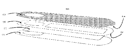
[0136] Figs. 12A-12H illustrates how the menstrual cup with lid 100,
collection tubes with
menstrual blood 500, used menstrual-pads/panty liners 700, DBS-cards 630,
square from highly
absorbent pad pulled out with string 680, sample collection devices 800, 900,
1000 and 1100, or
used tampons 1250 is put into a multi barrier
26
Date Recue/Date Received 2022-10-27
CA 03016495 2018-08-31
WO 2017/161378
PCT/US2017/023246
pouch 1220. The multi barrier pouch functions as an air-secured container to
preserve
the menstrual blood sample from air and moisture so it may be used for blood
analysis
after transportation to a remote location. The multi barrier pouch 1220 could
contain
an oxidizer and a desiccant and can also be marked with a barcode or ID number
1230, which the can be used to track samples from users of the products,
laboratory,
biobanks or similar. The multi barrier pouch 1220, with 100, 500, 700, 710,
630, 680
or 800, 900, 1000, 1100, 1250 is closed and put into a pre-stamped envelope
1240,
which is mailed to a specific remote location for analysis.
101371 Fig. 13 illustrates a 1300 multi barrier pouch married with an
envelope.
Once a specimen, such as 100, 500, 700, 710, 630, 680 or 800, 900, 1000, 1100,
1250
has been enclosed in the pouch, the opening 1301 of the pouch is sealed by
sliding the
rim 1302 together. The barriers of the pouch 1303 is made of three layers an
inner,
middle and outer material. The three layers could be a made of thermoplastic
materials such as e.g. rigid PVC, semi-rigid PVC, polycarbonate, acrylic,
impact-
modified acrylic, polystyrene, impact-modified polystyrene, ABS, polyethylene,
polypropylene, and combinations thereof. As the pouch 1300 is sealed by
closing
opening 1303, a second closing method 1304 is activated by removing the
release
liner 1305 from the barrier, exposing a semi-strong adhesive 1306. The
mechanism is
closed by folding the barrier 1304 down on top of 1302, and sealed by the
adhesive
1306 attaching to the outer part of the barrier 1307. As the multi barrier
pouch 1300 is
also an envelope, a pre-filled address 1308 is printed on the envelope and
filed with
pre-paid postage 1309 to be mailed to a laboratory for analysis.
101381 Fig. 14 illustrates a menstrual blood collection mail-in kit 1400.
The
different embodiments of menstrual blood collection shown in Fig 1, 2A-E, 3,
4A-B,
5A-B, 6,7, 8A-E, 9A-D, 10A-D, 11 can be delivered to the women with a full
collection kit as illustrated here with vaginal fluid collection device 100 as
the
example. The kit can further consist of instruction manuals of e.g. how to use
menstrual cups 200, how to seal the lid 300 on a system 100, guides to collect
the
samples in tubes if necesarry, as well as instructions on how to use the fluid
collection
strip illustrated in Fig. 7 to 11. The kit can also include regular menstrual
pads,
tampons and other menstrual blood collections devices, intended to be used in
a mail-
in procedure.
27
CA 03016495 2018-08-31
WO 2017/161378
PCT/US2017/023246
[0139] The kit can also consist of a multi barrier pouch 1402 such as 1220
or
1300 used for collection of 100, 500, 700, 710, 630, 680, 800, 900, 1000,
1100, 1250.
If there is only a multi barrier pouch, an envelope with pre-postage stamps
1240 is
also enclosed. The samples can then be sent in for remote analysis in a
laboratory.
The kit also has a unique ID 1403 that can be used to identify the kit, and be
used to
register online on a website, in an app or through a similar service.
[0140] Once a sample has been send via mail to a remote storage facility,
the
sample can be processed using both existing sample analysis methods already in
use
in commercial and clinical labs as well as new methods looking for unique
biomarkers found in vaginal fluid.
[0141] The biomarkers which can be analyzed are the ones contained in
menstrual
blood and vaginal fluid. Specifically, for the fluid collection strips HR-HPV
or any
other strain of HPV are optimal, just as endometrial cancer, HIV viral loads,
freefloating RNA or DNA is of interest. Other virus, bacteria or biomarkers
such as
vitamins, minerals, lipid profiles, hormone levels etc, in the blood can be
analyzed
and detected.
[0142] Fig. 15 illustrates a device 1500 for home testing and analysis of
menstrual
blood or vaginal fluid. Instead of sending the samples 100, 500, 630, 680,
690, 800,
900, 1000, 1100 or 1250 to a remote location for analysis, a device for home
usage as
presented here may be ideal. The device has an opening 1510 in which a sample
1520
is inserted to using an intennediary device that can hold the sample, do
sample
preparation or control the volume inserted into the device. The device is
specific for
certain types of menstrual blood collections such as menstrual blood (as a
fluid) from
a menstrual cup such as 510, from fluid collection strips such as 800, 900,
1000, 1100,
used tampon 1250, or electrochemical or optical biosensors as illustrated in
1520. The
device may be able to process all different formats as illustrated in this
embodiment.
The device is specifically calibrated for menstrual blood and vaginal
discharge. In the
case of fluent sample use the device may also have a centrifuge function.
Further the
device employs a number of different blood analysis techniques including
electrochemical testing, optical testing, polymerase chain reaction, macs
spectrometry,
28
CA 03016495 2018-08-31
WO 2017/161378
PCT/US2017/023246
chemical sequencing, chain-termination method, de novo sequencing for cutting
or
shearing larger DNA fragments, in vitro cloning to amplify individual DNA
molecules, in vitro cloning for amplifying individual DNA molecules. Methods
such
as next-generation sequencing can also be applied for genome sequencing,
genome
resequencing, transcriptome profiling (RNA-Seq), DNA-protein interactions
(ChIP-
sequencing), and epigenome characterization. These methods could be single-
molecule real-time sequencing, ion semiconductor, pyrosequencing, sequencing
by
synthesis, sequencing by ligation or chain termination. Methods can also
include
massively parallel signature sequencing (MPSS), Polony sequencing, 454
pyrosequencing, SOLiD sequencing, DNA nanoball sequencing, heliscope single
molecule sequencing, Nanopore DNA sequencing or RNAP sequencing. The data
results from the analysis can be transmitted to a computer or phone or other
similar
devises via. USB cable or wirelessly through Bluetooth, GSM, Wi-Fi, RFID and
other
transmission techniques not illustrated in this figure. A power outlet 1501
which can
be plugged into the wall can power the device. The device can also use a jack
or mini
jack which can be plugged into the phone to power the system and function as
the
transmission of data results.
[0143] Fig. 16A illustrates what happens once a vaginal fluid sample such
as
menstrual blood is introduced to a sample collection strip such as 1000. The
illustration shows what a sample collection strip looks like after the
protective cover
1001 has been removed. As the blood comes through the inlet 1012, it migrates
down
the paper 1020 and 1021. As both paper types in this example is filtration
paper that
can isolate plasma, the inlet 1012 will be soaked with larger cells such as
red-blood
cells. As the fluid travels laterally down the filtration paper 1020 and 1021,
it will in
plasma windows 1011 be visible as a more clear sample. The sample could here
be
e.g. blood plasma or other smaller cell types. As the sample arrives to the
blood
laboratory, either an automated system or a laboratory technician can punch or
cut out
a small sample from the sample collection strip 1000. Fig. 16B illustrates how
samples 1601, 1602, 1603 and 1604 is punched out. In this example, 1602 is
punched
out from inlet 1012. If the sample paper 1020 has not been stabilized with
e.g. a DNA
preservative, the sample will have the same characteristics as a whole blood
sample.
In this example, 1601 is a pure plasma sample. Beneath the second inlet 1012
of the
sample collection strip 1000, filtration paper 1021 is incorporated. The
filtration paper
29
CA 03016495 2018-08-31
WO 2017/161378
PCT/US2017/023246
may be coated with a DNA/RNA stabilizing agent, which will when sample 1603
and
1604 is punched out from the strip, be different from sample 1601 and 1602.
[0144] in Fig. 17A, 17B and Fig. 17C a laboratory process is illustrated.
Fig. 17A
illustrates how sample materials 1601 is punched out from the sample
collection strip
1000. With a pincet 1710, a laboratory technician will in Fig. 17B introduce
the
sample 1601 into a volumetric flask 1720 with a pre-defined buffer material
1725.
The buffer material can be a fixative or other required for processing the
biomarker of
interest. Once the sample 1601 has diluted into the buffer, it can be
introduced to a
chemical analyzer such as 1730.
[0145] Fig. 18 exemplifies how data from an analysis can be displayed to
a user
on an electronic device 1800 such as a website or an app on a computer 1810, a
handheld device such as a smartphone 1820 or on a smart-watch 1830. The data
can
also be displayed in other embodiments such as in glasses connected to the
Internet,
as audio through headphones, on a smart-mirror by the sink in a bathroom, or
similar
product carPgories. The data may be displayed with a number or a graph or
similar.
The data may be presented after interpretation from a health care professional
or
simply just as raw data. The cloth may also be displayed after data handling
which is
required for some biomarkers to translate biomarker level in menstrual blood
to the
equivalent in systemic blood.
[01461 Following are examples of data that the user can submit in the
app/website: Name, Age, Location (through GPS or IP address), Home address, E-
mail address, Password (to save their profile online), Pin code to access the
app (if
biometric functionality is used on a device, this can also be used), Credit
card
information to order testing or other services. The users can also input
information
about their menstrual cycle, and view their cycle in a calendar function. The
calendar
page can also display when a period is expected, or indicate when in the
menstruation
cycle the women is most likely to be fertile. This data can also be viewed as
historical
data, offering the user an opportunity to track her periods. Further a
biomarker menu
list from which biomarker analysis can be ordered and results viewed once
laboratory
has finished the analysis is available.
101471 As illustrated in Fig. 19, the data collected in the app/website
1930 is
encrypted and stored in a cloud service 1900. When a user receives a menstrual
blood
CA 03016495 2018-08-31
WO 2017/161378
PCT/US2017/023246
collection device such as 100, 200, 600, 680, 690, 800, 900, 1000 or 1100, the
user
registers the SN number 639 or a barcode 638 which may be labeled on the
menstrual
blood collection device, the multi barrier pouch, the mail-in kit 1400 or
similar. Once
registered the information is encrypted 1935 and stored in the cloud service
1900.
After the user has used the medical device 100, 200, 600, 680, 690, 800. 900,
1000 or
1100 it is sent in an envelope 1240 or 1300 with mail 1937 to a blood
laboratory 1940
for blood analysis. Once the blood sample is received at the blood laboratory
1940,
the laboratory registers 1945 the sample in the cloud service 1900. After the
blood
laboratory 1940 has inputted the serial number into the cloud service, the
details of
which biomarkers should be analyzed will be available 1946 for the staff so
blood
analysis can be performed. Only the biomarker chosen by the user is available
for the
staff, which means any other information stored about the user specific to the
SN
number cannot be viewed. Once the laboratory has performed the analysis,
results are
encrypted and stored 1945 in the cloud 1900. In the cloud 1900, the encrypted
results
can now be sent 1936 back to the user. The cloud service 1900 also has an
application
program interface (API) 1955, through which other organizations can get
specific
access to information in the cloud 1930 and the results of a blood analysis.
The API
1955 can have various pennission levels, often controlled by the user, where
e.g.
health providers 1950 such as medical doctors, can get access to specific user
information and blood results. It is also possible to give limited access to
e.g. medical
researchers 1960 or other providers 1970 such as insurance companies. In some
embodiments, a digital service can aggregate the cumulative, collected results
of a
menstrual fluid analysis. The aggregated data can provide an opportunity to
find new
information from the total amount of fluid analysis.
[0148] It will be appreciated by those skilled in the art that the
invention can take
many forms, and that such forms are within the scope of the invention as
claimed.
Therefore, the spirit and scope of the appended claims should not be limited
to the
descriptions of the preferred versions contained herein.
31