Note: Descriptions are shown in the official language in which they were submitted.
CA 03017199 2018-09-07
WO 2017/156246 PCT/US2017/021539
SYSTEM AND METHOD FOR MONITORING CONDITIONS OF A SUBJECT BASED
ON WIRELESS SENSOR DATA
CROSS-REFERENCE TO RELATED APPLICATION
[0001] This application claims priority to U.S. Provisional Application Serial
No.
62/305,854, filed on March 9, 2016, the entire disclosure of which is
incorporated herein by
reference.
FIELD OF THE INVENTION
[0002] Various embodiments of the present invention relate to a wireless
sensor that
offers convenient use for a subject, such as a patient, in the monitoring, in
real time (or quasi-real
time), of a medical signal, such as a hemodynamic parameter. In addition,
various embodiments
of the present invention also relate to methods for aligning data from
different wireless sensors
with each other, with another device, or both.
BACKGROUND
[0003] Monitoring various vital signs of a patient has been an important
aspect of
hospital patient care, especially for patients with diseases at advanced
stages, suffering from
severe trauma, or in other emergency settings. Additionally, outpatient
monitoring of various
physiological conditions are being increasingly used for evaluation of patient
health conditions
as well as early detection and treatment of heart diseases, diabetes, and
other diseases. For
example, an electrocardiogram (ECG or EKG) can be used to evaluate the heart
condition of a
patient, where electrodes are placed at certain locations on the chest, arms,
and/or legs. These
electrodes can be connected to an ECG machine by lead wires, and the electric
signals received
by the ECG machine can be analyzed and displayed for the physician's
information and further
interpretation.
-1-
CA 03017199 2018-09-07
WO 2017/156246 PCT/US2017/021539
[0004] Attempts have also been made to develop systems to improve a patient's
comfort,
freedom and privacy by decreasing the number and volume of devices directly or
indirectly
attached to the patient. For example, U.S. Patent No. 7,979,111 discloses a
wireless electrode
arrangement and method for patient monitoring, where a plurality of wireless
electrodes suitable
for attachment to the surface of the body of a patient are capable of
continuously monitoring of a
subject wirelessly. U.S. No. 9,101,264 and co-pending U.S. Patent Application
No. 14/216,174
(published as U.S. Patent Application Publication No. 20140275928) further
describe a network
of wireless sensors for monitoring hemodynamic parameters of a subject. The
disclosures of all
of these documents are incorporated herein by reference in their entireties.
[0005] Implantable devices such as implantable cardioverter defibrillators
(ICDs) or
pacemakers are often indicated for patients who have or are at increased risk
for various heart
conditions related to the heart's electrical system, such as ventricular and
atrial arrhythmias
including but not limited to ventricular fibrillation, ventricular
tachycardia, atrial fibrillation, and
bradycardia, etc. These implantable devices can monitor and/or manage certain
heart conditions
of the patients and prevent or control heart episodes that would otherwise
interfere with daily life
or be life threatening, and can therefore allow patients with certain heart
conditions to carry on
their normal lives with relatively few restrictions and generally low level of
discomfort.
However, these invasive devices cater primarily to patients who are at an
advanced stage of
disease.
[0006] Additionally, there can be limiting factors for these implantable
devices such as
inaccuracy in detecting the relevant heart condition episodes and
administering appropriate
therapies. For example, the positioning and contact of the leads of the ICDs
with the heart
muscle can be affected by the patient's movement, and the problem is more
acute for young and
more active patients. ICDs can also have lead failures after being worn by a
patient for an
extended period of time, e.g., a number of years. Lead positioning errors and
failures can cause
inaccurate or distorted electrograms, and may thereby lead to insufficient,
overly aggressive, or
otherwise inappropriate cardiac intervention, such as excessive number of
unwarranted shocks or
-2-
CA 03017199 2018-09-07
WO 2017/156246
PCT/US2017/021539
shocks with unnecessarily large magnitude, which can cause discomfort, pain,
and other
undesirable effects on the quality of life of the patients.
[0007] There is a need for a system that integrates the real time monitoring
capability of
wireless sensors worn by a patient that is accurate and convenient for the
patient to use and
replace. Ideally, such devices should be suitable not only for patients who
are at an advanced
stage of a disease condition, but also for relatively healthier subjects that
nonetheless desire
monitoring of a physiological condition. Further, there is a need to ensure
accurate
synchronization between such devices to facilitate the collection of medically-
relevant sensor
information data.
SUMMARY OF THE INVENTION
[0008] In one aspect, an electrode patch is disclosed that includes a first
electrode
configured to contact a subject, a first part of a first releasable electrical
connector coupled to the
first electrode and configured to releasably connect to a second part of the
first releasable
electrical connector, a first adhesive layer having an opening, with the first
electrode disposed
within the opening, and a first protective layer disposed over and covering
the first adhesive
layer. The first protective layer includes an opening corresponding to the
first releasable
electrical connector. In a preferred embodiment, the first part of the first
releasable electrical
connector is adhered to the first electrode, the first electrode is made from
hydrogel and the first
adhesive layer is made from hydrocolloid. The first part of the first
releasable electrical
connector may extend through the opening in the first protective layer. A
bottom surface of the
first protective layer is preferably adhesive for adhering to a subject. The
first protective layer
may be made from, for example, polyurethane with a moisture vapor transmission
rate of 300 to
1400 gm/m2/day.
[0009] In certain embodiments of the electrode patch, a first backer is
disposed over the
opening of the first adhesive layer and over at least a portion of the first
adhesive layer to provide
structural strength. The backer includes an opening corresponding to the first
part of the first
-3-
,
CA 03017199 2018-09-07
WO 2017/156246 PCT/US2017/021539
releasable electrical connector. In a specific embodiment, the first part of
the first releasable
electrical connector is formed from a top portion coupled to a bottom portion,
and the backer is
sandwiched between the top portion and the bottom portion. In specific
embodiments the backer
is formed from perforated polyethylene terephthalate or an ethylene-vinyl
acetate/polyethylene
blend.
[0010] In a specific embodiment, the electrode patch can further include a
second
electrode configured to contact the subject, a first part of a second
releasable electrical connector
physically and electrically coupled to the second electrode, a second adhesive
layer with an
opening, the second electrode disposed within the opening, and a second
protective layer
disposed over and covering the second adhesive layer, in which the second
protective layer has
an opening corresponding to the second releasable electrical connector. In one
variation, the first
protective layer and the second protective layer are contiguous and are
frangibly connected to
each other via a perforation. In another variation, the first protective layer
and the second
protective layer are not contiguous, and the electrode patch further includes
a release liner
disposed over respective top surfaces of the first protective layer and the
second protective layer
to hold them in alignment with each other. In yet another variation, an
isolating barrier, such as
closed-cell foam, is disposed between the first adhesive layer and the second
adhesive layer. A
bottom surface of the isolating barrier may be configured to adhere to the
subject.
[0011] In another aspect, a method is disclosed for obtaining physiological
data from a
subject. A sensor patch is first disposed on the subject. The sensor patch
adheres to the subject
and includes a first part of a first releasable electrical connector, which is
electrically coupled to
a sensor of the sensor patch, and which is configured to releasably connect to
a second part of the
first releasable electrical connector. Then, an electronics package is
electrically and physically
connected to the sensor patch. The electronics package includes the second
part of the first
releasable electrical connector for such electrical and physical connection.
This second part is
electrically coupled to electronics of the electronics package, which are
configured to monitor
-4-
CA 03017199 2018-09-07
WO 2017/156246 PCT/US2017/021539
the sensor to generate corresponding physiological data and to wirelessly
transmit the
corresponding physiological data to another device.
[0012] In one embodiment, the sensor patch includes a plurality of sensors,
such as
electrodes, held in a predetermined geometrical arrangement by a release
liner. In such
embodiments, the method further includes removing the release liner after
disposing the sensor
patch on the subject and prior to coupling the electronics package to the
sensor patch.
[0013] In yet another aspect, an electronics package for a wireless
physiological sensor
system is disclosed. The electronics package includes a substrate. A first
part of a first
releasable electrical connector is connected to the substrate and configured
to releasably connect
to a second part of the first releasable electrical connector disposed on a
sensor patch. A first
shell is disposed on the substrate, such as over the first part of the
releasable electrical connector.
A second shell is also disposed on the substrate. Finally, the electronics
package includes
electronics configured to monitor at least one sensor of the sensor patch to
generate
corresponding physiological data and to wirelessly transmit the corresponding
physiological data
to another device. The electronics include a first electronics sub-system
disposed in the first
shell and electrically connected to the first part of the first releasable
electrical connector, a
second electronics sub-system disposed in the second shell, and a first
flexible circuit, such as a
flexible circuit board, electrically connecting the first electronics sub-
system to the second
electronics sub-system. The electronics also preferably includes at least one
rechargeable battery.
[0014] To accommodate, for example, movement of the subject, in preferred
embodiments the substrate is preferably flexible, the first electronics sub-
system is flexibly
connected to the first part of the first releasable electrical connector, and
a length of the first
flexible circuit between the first shell and the second shell is substantially
greater than a
corresponding distance between the first shell and the second shell.
[0015] In some embodiments, at least a portion of the first shell and at least
a portion of
the first part of the first releasable electrical connector are disposed in
the substrate.
-5-
CA 03017199 2018-09-07
WO 2017/156246 PCT/US2017/021539
[0016] In preferred embodiments, to avoid electrical interference, the first
electronics
sub-system comprises analog front end circuitry to obtain signals from the at
least one sensor and
the second electronics sub-system comprises a wireless transceiver to
wirelessly transmit the
corresponding physiological data.
[0017] In a specific embodiment, at least three sensors are arranged in a
substantially L-
shaped configuration on the sensor patch, and the electronics package further
includes a third
shell disposed on the substrate, a third electronics sub-system disposed in
the third shell. A
second flexible circuit electrically connects the third electronics sub-system
to the second
electronics sub-system. Also, a first part of a second releasable electrical
connector is connected
to the substrate, with the second electronics sub-system electrically
connected to the first part of
the second releasable electrical connector. Similarly, a first part of a third
releasable electrical
connector is connected to the substrate and electrically connected to the
third electronics sub-
system. In such embodiments, the first shell, second shell and third shell may
be arranged in a
substantially L-shaped configuration on the substrate corresponding to the at
least three sensors,
and in particular corresponding to second parts of the first, second and third
releasable electrical
connectors on the sensor patch to mechanically and electrically couple the
electronics package to
the sensor patch.
[0018] In a particular refinement, such as when the sensors are electrodes,
and to provide
for clean signal collection from the sensors, the first flexible circuit can
include a first signal line
extending between the first shell and the second shell. Similarly, the second
flexible circuit
includes a second signal line extending between the second shell and the third
shell. The first
flexible circuit also includes a first open electrical line electrically
connected to the second signal
line and extending along the first signal line, while the second flexible
circuit further includes a
second open electrical line electrically connected to the first signal line
and extending along the
second signal line.
[0019] In various embodiments the first flexible circuit is at least partially
disposed
within the substrate, and the first flexible circuit includes a contact region
exposed from the
-6-
CA 03017199 2018-09-07
WO 2017/156246 PCT/US2017/021539
substrate, which can be used, for example, as a port for recharging,
programming or data
collection purposes.
BRIEF DESCRIPTION OF THE DRAWINGS
[0020] The various aspects and embodiments disclosed herein will be better
understood
when read in conjunction with the appended drawings, wherein like reference
numerals refer to
like components. For the purposes of illustrating aspects of the present
application, there are
shown in the drawings certain preferred embodiments. It should be understood,
however, that the
application is not limited to the precise arrangement, structures, features,
embodiments, aspects,
and devices shown, and the arrangements, structures, features, embodiments,
aspects and devices
shown may be used singularly or in combination with other arrangements,
structures, features,
embodiments, aspects and devices. The drawings are not necessarily drawn to
scale and are not
in any way intended to limit the scope of this invention, but are merely
presented to clarify
illustrated embodiments of the invention. In these drawings:
[0021] Fig. 1 illustrates a network formed by a plurality of sensors according
to an
embodiment of the invention;
[0022] Figs. 2A and 2B are logical block diagrams of a sensor according to an
embodiment of the invention;
[0023] Fig. 3 depicts the placement of multiple sensors on a subject according
to an
embodiment of the invention;
[0024] Fig. 4 is a top view of a first embodiment ECG sensor package;
[0025] Fig. 5 is a bottom view of the sensor package shown in Fig. 4; ,
[0026] Fig. 6 is a cross-sectional view of the sensor package shown in Fig. 4
along a line
6-6;
[0027] Fig. 7 is an exploded perspective view of an adhesive electrode patch
depicted in
Fig. 6;
[0028] Fig. 8 depicts another embodiment of a protective layer shown in Fig.
7; and
-7-
CA 03017199 2018-09-07
WO 2017/156246 PCT/U52017/021539
[0029] Figs. 9-12 depict steps that may be employed to attach the adhesive
electrode
patch depicted in Fig. 6 to a subject;
[0030] Fig. 13 is a bottom view of another embodiment sensor package;
[0031] Fig. 14 is a cross-sectional view of the sensor package of Fig. 13
along line 14-14;
[0032] Fig. 15 illustrates usage of the sensor package of Fig. 4 in
combination with the
sensor package of Fig. 13;
[0033] Fig. 16 illustrates timing of data collection performed by a node shown
in Fig. 1
and the creation of a related data packet;
[0034] Fig. 17 illustrates possible data synchronization issues when receiving
data
streams from multiple data sources having different sampling clocks;
[0035] Fig. 18 illustrates determination of a phase offset of a sample value
from a desired
sampling time;
[0036] Fig. 19 illustrates timing of the width of a reporting period using
synchronization
packets transmitted by a master node depicted in Fig. 1 and a timer present in
a data collection
node receiving the synchronization packets;
[0037] Fig. 20 illustrates using the synchronization packets transmitted by a
master node
to schedule data packet transmissions; and
[0038] Figs. 21-24 depict an embodiment garment system.
DETAILED DESCRIPTION
[0039] Certain embodiments of the present invention will now be discussed with
reference to the aforementioned figures. In one embodiment, the present
invention provides a
wireless sensor suitable for attachment to the skin of a subject. The sensor
can form a network
with similar sensors, and the data collected from these sensors can be
synchronized or aligned in
the time domain. The type of network may utilize a routing topology include:
star, mesh,
pseudo-mesh network, or any other routing topology. Each of the sensors can
include a sensing
component configured to detect a signal corresponding to at least one
physiological condition of
-8-
CA 03017199 2018-09-07
WO 2017/156246 PCT/US2017/021539
the subject, and a communication component configured to wireles sly transmit
the detected
signal to either another wireless sensor or an external monitoring device. The
external
monitoring device may be local to the patient, such as a cellular telephone,
tablet computer or
other type of computing device, or may be remote, such as an Internet server,
and may serve as a
master node for the network. The communication component of selected sensors
can also be
configured to receive and/or relay signals transmitted from other wireless
sensors.
[0040] As described herein, a wireless sensor includes a sensing component
configured
to detect a signal corresponding to a physiological condition, such as vital
signs including (but
certainly not limited to) hemodynamic parameters of a subject, such as, but
not limited to, a
hospital patient. Hemodynamics, as known in the art, relates to the study of
blood flow. The
circulatory system, including the heart, the arteries, the microcirculation,
and the vein, functions
to transport the blood to deliver 02, nutrients and chemicals to the cells of
the body, and to
remove the cellular waste products. The heart is the driver of the circulatory
system generating
cardiac output (CO) by rhythmically contracting and relaxing. This creates
changes in regional
pressures, and, combined with a complex valvular system in the heart and the
veins, ensures that
the blood moves around the circulatory system in one direction. Hemodynamic
parameters (or
properties), as described herein, include the physiological conditions
associated with the blood
flow, which includes not only the physical characteristics of the blood flow
itself, e.g., blood
flow rate, blood flow pressure, temperature, and pulse rate, but also those
parameters relating to
the blood components such as cells, proteins, chemicals, etc.
[0041] The vital signs to be monitored as contemplated in the disclosed
embodiments can
include, but are not limited to, ECG (electrocardiogram), EEG
(electroencephalogram), EMG
(el ectromyogram), EOG (electrooculogram), ERG (electroretinogram),
temperature, pulse
oximetry, oxygen saturation, oxyhemoglobin saturation, blood component
concentration (e.g.,
glucose level, lipid level, cholesterol level, triglyceride level, levels of
different salts,
concentration of different types of cells, concentration of blood proteins
such as thrombin, cancer
markers, heart failure markers), renal function test components (e.g.,
concentration of albumin,
-9-
CA 03017199 2018-09-07
WO 2017/156246 PCT/US2017/021539
urea, and creatinine in the urine), liver function test components, organ
functions, blood pressure
(such as atrial pressure, ventricular pressure, pulmonary artery pressure,
systolic pressure,
diastolic pressure, etc.), blood velocity, respiration rate, pulse rate, (end
tidal) CO2 level, blood
drug concentration, organic or inorganic substance concentration in the blood
(e.g. uric acid,
vitamins, heavy metals, carbon monoxide, bacterial toxin), cardiac output,
heart rate, heart
rhythm, heart rate variability, pH, pathogens, motion, weight, etc.
Additionally, the system can
be used to monitor migraines, a subject's galvanic skin response, and
responses to electrical
nerve and muscle stimulation, etc. Depending on the types of underlying
physiological
conditions to be monitored, the sensing component can include, but is not
limited to, an
electrochemical detector (such as an needle electrode galvanic electrode or a
band electrode for
detecting a surface potential or current), an electromagnetic detector (e.g.,
an optical detector
such as an infrared detector and visible light detector, as well as an x-ray
detector, gamma-ray
detector, etc.), a thermal detector, a pressure detector, an ultrasonic
detector, a chemical detector,
a magnetic detector, an x-ray detector, an accelerometer, a gyrometer, a
motion detector, etc.
Other detectors in emerging sensor technology, such as laser Doppler, paper
sensors, sensor
tattoos, etc., can also be used.
[0042] Further, each wireless sensor includes a communication component
configured
for wireless communication with other sensors, an external monitoring device
(e.g., master node)
or both. For example, the wireless electrodes described in U.S. Patent No.
7,979,111, which is
incorporated herein by reference, including the transmitting circuit, such as
the remote telemeter,
can be such a wireless sensor. A wireless sensor can include a mote as
described in the above
patent, or can include a fully integrated and functional communication circuit
that includes an
amplifier, a processor, a memory, a battery, and an RF module. Each or
selected ones of the
wireless sensors can further include a memory of suitable size (for example, 4
GB or 8 GB, to
store a large volume or size of relevant medical records of a subject), a data
processor, power
supply, etc.
-10-
CA 03017199 2018-09-07
WO 2017/156246 PCT/US2017/021539
[0043] In some embodiments, the wireless sensors form a mesh network, where
each
sensor (also referred to as a "node", "sensor node" or "regular node"
hereinafter) not only
captures and disseminates its own data, but may also serve as a relay for
other nodes, that is, the
nodes in the mesh network collaborate with each other to propagate the data in
the network. In
certain embodiments, the mesh network further includes one or more control
nodes (or master
nodes), which communicate with selected or all of the regular nodes. The
master nodes can
serve as a data acquisition, processing, and command center. In other
embodiments, the wireless
sensors communicate only with each other, e.g., for purpose of synchronizing
signal acquisition.
In further embodiments, the wireless sensors communicate only with an external
control node,
but do not communicate with each other or form a mesh network.
[0044] The wireless sensors or the network of the wireless sensors can
continuously
monitor selected physiological data of the subject, and communicates the
signals acquired from
the sensing components via the communicating components of the sensors to a
control or master
node. Each of the wireless sensors can be programmed such that signals
detected by the sensor
falling into a predetermined (e.g., an acceptable or normal) range are not
transmitted, or
transmitted at a lower frequency. The acceptable range for signals for
different subjects and for
each wireless sensor can be set individually, for example, based on the type
of the sensor, the
subject's condition, the therapy being used by the subject, etc. A control or
master node can
include a communication component configured to wirelessly receive signals
from each of the
plurality of wireless sensors, and send data and/or commands to each of the
plurality of wireless
sensors. The control or master node can further include a monitoring unit
coupled with the
communication component. For example, the monitoring unit can include a
readable medium
and a processor coupled to the computer readable medium. The computer readable
medium can
store coded instructions for execution by the computer processor, which, upon
the execution of
the instructions, carries out pre-designed tasks.
[0045] In some embodiments, the master node of a mesh network can be a PC or
workstation computer equipped with a communication component, such as a
dongle, for
-11-
CA 03017199 2018-09-07
WO 2017/156246 PCT/US2017/021539
communicating with the wireless sensors. The master node can also include a
portable device
having a processor, a memory, a display and/or other audiovisual output
capabilities to present
information to a user, and capabilities of wirelessly communicating with the
wireless sensors. In
other examples, the master node can include a commercial portable computing
device, such as a
smart phone (e.g., an iPhone, an Android-based phone, a Windows Mobile-based
phone, etc.), a
tablet (such as an iPad, a Samsung Galaxy Tab, Google Nexus 7 or 10, etc.), or
other similar
devices. In further examples, the control and communication capabilities of a
master node can
also be implemented on one or more regular nodes to "upgrade" such regular
nodes into "super
nodes" that include both sensing capabilities and the functionalities of a
master node. For
example, in some embodiments one or more of the nodes may include cellular
and/or satellite
telecommunications capabilities to establish communications with a remote
server.
[0046] In the following, a wireless sensor including ECG electrodes suitable
for
acquiring electrophysiological signals related to cardiac function is used for
illustrating the
operating principles of the sensors and the network formed therefrom. In these
sensors, each of
the sensors include one or more electrodes which can acquire data related to
the quality of the
ECG signal, such as the amplitude of a detected voltage, a detected current,
and/or electrical skin
resistance, and transmit such data to other sensors or the master nodes.
[0047] For ECG applications, multiple wireless sensors may be employed, which
are
placed on the subject's body in predetermined locations. Preferably, these
wireless sensors can
self-configure into a set or group which wirelessly sends diagnostic quality
ECG signals in a
synchronous fashion to a master node, which can derive or synthesize ECG
spectrum for display
or other forms usable by a physician (or other users) based on the transmitted
ECG signals.
These sensors can also be configured to send and/or receive signals to/from
the master node
when a proximity criterion is satisfied, e.g., when the master node is within
a predetermined
distance from the wireless sensor, e.g., within 3 feet.
[0048] For illustration purposes and not limitation, a mesh or pseudo-mesh
network
formed by a plurality of sensors can be represented by a schematic block
diagram as shown in
-12-
CA 03017199 2018-09-07
WO 2017/156246
PCT/US2017/021539
Fig. 1. The illustrated network includes six sensor nodes and a single master
node 110. The
sensor nodes can be divided, for example, into three clusters: cluster 120
(including node 1 and
node 6), cluster 130 (node 2 and node 5), and cluster 140 (node 4 and node 9).
The arrows in Fig.
1 represent communication paths between the nodes. More generally, a cluster
can be thought of
as having one, two or more nodes. As depicted in this example, the network
supports at least
two modes of communication: (1) communication between the master node and each
of the
nodes, and (2) communication between nodes within a cluster. Such a
configuration allows for
the sensor nodes to make their own decisions and reconfigure the network
independently of the
master node 110. The wireless communication within the mesh network can be
based on
proprietary communication stacks utilizing the principles of time domain
multiple access
(TDMA), with frequencies selected from various MICS bands (Medical Implant
Communications Service frequencies) or from the ISM (Industrial, Scientific,
and Medical
frequency bands (900MHz, 2.4 GHz, or 5.8GHz)) as would be appreciated by one
of ordinary
skill in the art.
[0049] For wireless sensors that are configured to detect ECG signals,
examples of which
are described herein, the sensors can be attached to the skin of a subject for
ECG signals
recordation in a manner that is similar to the configuration of traditional 3-
lead, 5-lead, or 12-
lead ECG leads. Signal acquisition between the nodes can be synchronized for
processing of the
ECG signals, as described later.
[0050] An example block diagram of the logical structure of an embodiment ECG
sensor
200 is illustrated in Fig. 2. Four electrodes 210 are provided, including
three signal electrodes
212 and an electronic ground electrode 214. These electrodes 212, 214 are
connected to
instrumentation amplifiers 230 via input protection circuit 220 that protect
against electric shock
and radio frequency interference. The instrumentation amplifiers 230 measure
the difference
between its two inputs and amplify that with a gain, e.g., of approximately
3.5. The gain of each
amplifier 230 can be adjusted by way of the resistors, as known in the art,
and are connected to
the ground electrode 214 via a respective resistor or resistors, as known in
the art. The amplified
-13-
,
CA 03017199 2018-09-07
WO 2017/156246 PCT/US2017/021539
signals are optionally filtered by bandpass filters 240 (typically to the
frequency response of 0.05
Hz to 60 Hz or alternatively 100Hz or 150 Hz). Additional gain can optionally
be provided in
the bandpass filter stage to reach a total system gain of, for example,
approximately 300. This
results in, for example, an input range of approximately 10 mV between any
pair of signal
electrodes 212. However, it will be appreciated that the input range may also
be adjustable, such
as through hardware/firmware or software changes. The individual channel
signals can then be
digitized by A/D converters 250. The converters' resolution may be, for
example, 12 bits or 16
bits. Or, the A/D converters 250 may have a higher native resolution, such as
24 bits, which is
then down-converted to a lower resolution, such as 16 bits. Collectively, the
amplifiers 230,
band pass filters 240 and A/D converters 250, inter alia, are referred to as
the analog front end
299 of the sensor 200, and may be provided by a discrete component, such as an
ADS1293 from
Texas Instruments, and the characteristics of each (gain, filtering, sampling
rate, etc.) may be
programmable, as known in the art. The digitized ECG signals from the analog
front end 299 are
passed through to a micro-processing unit (MPU) 260 for processing. The
processed signals
may be stored on board in a memory 270 coupled to the MPU 260, e.g., a flash
memory, which
can also store program code executable by the MPU 260 to control overall
operations of the
sensor 200. Additionally or alternatively, the processed signals can be sent
to an RF transmitter
280 and transmitted via an antenna 281, or via a wired connection, such as
USB, to, directly or
indirectly, an external device (not shown), e.g., a smartphone, a tablet, a
computer, another node,
etc.
[0051] Because the sensor 200 may work in a diverse array of environments,
many of
which may be electronically noisy, it is desirable in various embodiments that
noise cancellation
techniques be employed in the sensor 200 in the analog front end 299. As shown
in Fig. 2B, and
discussed in more detail later, the analog front end 299 and electrodes 210
are mounted on a
substantially L-shaped substrate 298, having a first arm 291 and a second arm
292 that is
substantially perpendicular to the first arm 291, such as from 70 to 120
with respect to the first
arm 291. Each signal electrode 212 is electrically connected to the analog
front end 299 by way
-14-
CA 03017199 2018-09-07
WO 2017/156246 PCT/US2017/021539
of a respective trace 216, while the electronic ground electrode 214 is
electrically connected to
the analog front end 299 by its own trace 214A. The three signal electrodes
212 are respectively
located at the ends and intersection of the arms 291, 292. The ground
electrode 214 may be
located anywhere on the substrate 298, such as next to one of the signal
electrodes 212 at the
ends of the arms 291, 292.
[0052] The active traces 216 that electrically connect the signal electrodes
212 with the
analog front end 299 pickup ECG signals from the body and also act as antennas
and as such can
pick up unwanted noise from the surrounding environment. To the extent that
this noise is
common to all of the signal electrodes 212, conventional common mode noise
rejection
techniques making use of the ground electrode 214 can be employed by the
analog front end 299
to reduce this noise. It is therefore desirable that the noise captured in
each channel through the
signal electrodes 212 and corresponding traces 216 be as identical as possible
with the noise on
the other signal electrodes 212 and corresponding traces 216. Each trace 216
will optionally
include at least one open lead 216A extending in a direction along trace 216,
forming an overall
trace that is substantially L-shaped to match the shape of the substance and
the orientation of the
electrodes. For example, other shapes may be advisable for different examples
and active traces
216 and/or open leads 216A need not be in a straight line. Additionally there
is minimal distance
between the traces 216 and open leads 216A extending from each of the
respective electrodes
212. In a circuit board configuration this distance is preferably between .4
and 4.4 mil. In other
embodiment, this distance is preferably at least less than 1 cm.
[0053] For example, the trace 216 extending from a first signal electrode 212
"1" at the
end of the first arm 291 includes and is electrically connected to a
substantially perpendicular
open lead 216A extending along the second arm 292 having a length that is
preferably similar in
length to the trace 216 for the third signal electrode 212 "3" at the end of
the second arm 292.
For example, that trace 216 is preferably between 2200 and 2600 mil, with a
more preferred
length of 2480 mil. That open lead 216A is preferably between 2500 and 3000
mil, with a more
preferred length of 2875 mil. Similarly, the trace 216 for the third signal
electrode 212 "3" at the
-15-
CA 03017199 2018-09-07
WO 2017/156246 PCT/US2017/021539
end of second arm 292 includes and is electrically connected to an open lead
216A extending
along the first arm 291 with a length that is preferably similar to that of
the trace 216 of the first
signal electrode 212 "1". For example, that trace 216 is preferably between
3000 and 3600 mil,
with a more preferred length of 3310 mil. That open lead 216A is preferably
between 2200 and
2800 mil, with a more preferred length of 2488 mil. The second signal
electrode 212 "2" at the
junction of the arms 291, 292 includes and is electrically connected to two
such open leads 216A,
substantially perpendicular to each other, running respectively along the
first arm 291 with a
length substantially equal to the trace 216 of the first signal electrode 212
"1" and along the
second arm 292 with a length substantially equal to the trace 216 of the third
signal electrode 212
"3". An additional trace 216 extending from the second signal electrode 212
"2" which like the
other trace 216s can be L shaped and connect to analog front end 299 may also
contain multiple
open leads 216A extend in perpendicular directions. That trace 216 is
preferably between 400
and 700 mil, with a more preferred length of 574 mil. One of such open leads
216A that are
perpendicular to each other are preferably between 2500 and 3200 mil and more
preferably 2962
mil while the other open lead 216A is preferably between 2000 and 2600 mil and
most
preferably 2305. Hence, the use of open leads 216A together with the active
traces 216 make
the noise picked up in each channel as common as possible, thus facilitating
its rejection in the
analog front end.
[0054] In some embodiments, multiple surface nodes can be placed on the skin
of the
subject. As shown in Fig. 3, a first surface node 201 can be placed high on
the sternum just
below the clavicle. This can be advantageous for detection of atrial rhythm,
as it is nearest the
heart's atria, affording the best opportunity to monitor atrial fibrillation.
There is less muscle in
this location to contaminate the ECG with any electromyogram (EMG) artifact,
and it can be on
a tissue that is less likely to move and contaminate the ECG with motion
artifact. An optional
second surface node 202 may be added nearest to the ventricles. Two electrodes
of this group
can be at locations V4 and V5 of a standard 12-lead ECG, and the third a proxy
for the left leg
location. The signals from the two surface nodes may be combined in various
ways to provide a
-16-
CA 03017199 2018-09-07
WO 2017/156246
PCT/US2017/021539
faithful representation of a standard 3, 5, or 12 lead ECG. The second surface
node 202 can also
be able to measure ventricular ischemia due to blockage of the major vessels.
An optional third
tripole surface node 203 may be added to further facilitate the derivation of
a full 12-lead ECG.
Alternatively, a calibration step may be employed to derive the 12 lead ECG.
This can be an
internal calibration by temporarily connecting the two (or more) sensors
electrically to calibrate,
and then disconnected the sensors for the remaining of the operation.
Alternatively, calibration
can be done with an external device (e.g. a wired 12-lead ECG machine) to
establish the baseline
correlation between the wired and wireless data to facilitate downstream
signal processing.
[0055] In a system where there are more than one wireless sensor, some or all
of the
wireless sensors can each individually transmit the collected physiological
data to an external
device (e.g., a monitoring device). Alternatively, one of the wireless sensors
can include
hardware and software necessary to serve as a master node or gateway that
receives detected
physiological data from other wireless sensors, and forward such signals via a
radio or WiFi link
to the external monitoring device at an appropriate rate (e.g., to save
battery power of the
sensors). The transmission can also be optionally compressed with little or no
information loss.
The transmitted physiological data can be processed by the monitoring device
with appropriate
program, or can be further uploaded to a server for processing and/or
analysis, which are
described further below. Signal acquisition of the various wireless sensors
can also be
synchronized with each other, as discussed later, to facilitate subsequent
processing of the
collected signal data.
[0056] Further, the wireless sensors according to one embodiment of the
present
invention can include different sensing components for monitoring a plurality
of different vital
signs. For example, one sensor can include a pressure detector for monitoring
the pulse rate, and
another sensor can include an electrochemical detector for blood glucose level
measurement or
the like. Thus, wireless sensors of different types for monitoring different
vital signs can be
conveniently worn by the subject depending on the needs of care for the
subject.
-17-
,
CA 03017199 2018-09-07
WO 2017/156246 PCT/US2017/021539
[0057] The use of hybrid sensors can provide a caregiver with more
comprehensive
information regarding the subject's condition in a more efficient and/or more
reliable manner.
For example, monitoring different vital signs simultaneously using different
types of wireless
sensors can provide redundancy and improved robustness of monitoring quality
as well as
facilitate reconciliation of inconsistencies among the data gathered from
different types of
sensors (for different vital signs), reduce false alarm rates, etc. Certain
vital signs can also be
considered as having higher priorities (e.g., because the sensors for
monitoring these vital signs
have higher reliability or accuracy), and as such, the data gathered for these
vital signs can be
given more weight when data gathered for other vital signs may suggest a
different condition the
subject is in. In addition, when implanted wireless sensors are used,
especially those implanted
relatively deep within the subject's body (e.g., in the subject's heart), one
or more surface-
attached sensors, e.g., those located near the implanted sensors, can be used
to relay the signals
acquired from the implanted sensors, e.g., to a master node, thereby providing
potentially better
quality signals for further processing and analysis while allowing for reduced
power
consumption of the implanted sensors. The wireless sensors can be further used
in conjunction
with certain medical devices worn by the subject (e.g., rehabilitating
devices, robotics,
prostheses, etc.), for collecting and transmitting sensed signals as a
feedback or input for these
devices so as to further enhance their functionalities.
[0058] The data collected from different types of sensors can be weighted,
ranked,
processed, validated, transmitted (via the master node, for example) to an
Electronic Health
Record (EHR) server, and utilized with other data in the EliR of a subject.
The ECG and other
vitals can be prioritized by the subject disease conditions and health status.
For example, an
otherwise healthy patient having atrial fibrillation (AF) surgery has a
limited set of parameters,
whereas a patient just discharged with Congestive Heart Failure (CHF) with co-
morbidities of
diabetes, and obesity, and multiple medications can be monitored for those
vital sign signals
relevant to disease specific algorithms based on ECG, blood glucose levels and
weight.
-18-
CA 03017199 2018-09-07
WO 2017/156246 PCT/US2017/021539
[0059] For example, the system can store "diagnostic templates" containing
threshold
levels of specific vital signs, which can trigger a diagnosis when the
threshold levels for the vital
signs are reached by a subject undergoing monitoring. In response to subject-
specific
information, the system can adjust the "diagnostic templates" based on disease-
specific risk
factors (e.g. heart rate variability in subjects having atrial fibrillation)
as well as subject-specific
risk factors (e.g. fluctuation in blood pressure in subjects with
hypertension). The system can
also differentially weigh different vital signs according to the indication
and subject's existing
conditions, measure the subject's vital sign variability, trends over time,
and deviations from
previous states using predetermined statistical models, for example,
statistical models that use
measurements such as average, standard deviation, and covariance. The data
processing and
analysis can be performed on the sensor nodes, or by a monitoring device that
is configured to
receive the sensor data from the various sensors or from a master node. The
monitoring device
may be a device local to the subject, such as a portable electronic device
(such as a cell phone,
PDA, tablet, etc.), or may be remote from the subject, such as an Internet
server or the like.
Communications with such a remote device may be made through an intermediate
device, such
as a cell phone or other wireless device, that is local to the user and
capable of forwarding
information received from the sensor nodes to the remote server. The
monitoring device may be
configured, e.g., through a suitable program, to communicate with one or nodes
to collect related
sensor information, process this sensor information and then present, such as
on a screen or by
way of any other suitable user interface, information related to the collected
sensor data, or to
forward this sensor data, in raw or processed form, to a remote device, such
as a server of a
healthcare provider.
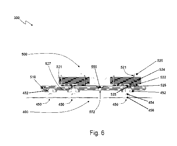
[0060] A first embodiment first sensor package 300 is shown in Figs. 4-6,
which is used,
for example, as an ECG sensor, such as the ECG sensor 200 above ¨ although
other sensing
capabilities are certainly possible. The ECG sensor package 300 includes an
adhesive electrode
patch 400 that is removably connected to electronics package 500. In preferred
embodiments,
snaps 450 are used as releasable electrical connectors to both physically and
electrically
-19-
CA 03017199 2018-09-07
WO 2017/156246 PCT/US2017/021539
removably connect the adhesive electrode patch 400 to the electronics package
500. Each snap
450 includes a first part 452 on the electronic package 500, such as a female
part, and a
corresponding second part 454 on the adhesive electrode patch 400, such as a
male part. Hence,
in use, the adhesive electrode patch 400 is first preferably placed at the
desired location on the
subject, where it adheres to, and makes electrical contact with, the subject's
skin. Then, the
electronics package 500 is snapped onto the adhesive electrode patch 400 via
the snaps 450, to
mechanically and electrically connect the electronics package to the adhesive
electrode patch 400.
It will be appreciated that other types of releasable electrical connectors
could be used, such as a
plug-and-socket arrangement, a magnetic-connector arrangement, or the like, as
known in the art,
each formed by a first part that can releasably connect to a second part to
establish an electrical
connection.
[0061] As illustrated in Fig. 5, the adhesive electrode patch 400 includes
three ECG
electrodes 402, 404, 406 arranged in an L-shaped configuration with respect to
each other, and a
single ground electrode 408 adjacent to one of the ECG electrodes 406 along
one of the arms of
the L-shaped configuration. It will be appreciated that the electronic ground
electrode 408 may
be disposed anywhere on the device 300 so long as it is electrically connected
to both the subject
and the electronics package 500. In preferred embodiments, the distance
between electrodes 402,
404, 406 is approximately two inches in both the horizontal and vertical
directions. In other
embodiments, the horizontal and vertical distances between electrodes 402,
404, 406 is less than
two inches, such as 1.5 inches or one inch, or even one inch or less,
depending upon the
capabilities of the analog front end 299. It will be appreciated that in other
embodiments the
distances between the electrodes 402, 404, 406 can be greater than two inches,
with the distance
limited only by the physical extents of the user. Adhesive electrode patch 400
maintains the
orientation and spacing between the electrodes 402, 404, 406 substantially
fixed, and knowledge
of this predetermined spacing and geometrical arrangement of the electrodes
402, 404, 406 can
be used in subsequent signal processing to obtain or compute additional
channels of ECG data.
-20-
CA 03017199 2018-09-07
WO 2017/156246
PCT/US2017/021539
[0062] The electrodes 402-408 are preferably formed from an electrically
conductive
hydrogel material, such as KM3OB from Katecho, Inc., of Des Moines, IA. A foam
barrier 409,
preferably a closed-cell foam such as Katecho SP 275, is used to help
electrically isolate the
ground electrode 408 from its neighboring ECG electrode 406. Each electrode
402-408 is
surrounded by a respective hydrocolloid layer 412-418, which also adheres to
the skin of the
subject. A suitable hydrocolloid material includes, for example, Hi-Tack
Hydrocolloid from
Amparo, Inc., of Placentia, CA. Finally, a protective layer 430 surrounds the
hydrocolloid layers
412-418 and also adheres to the skin of the subject. Each electrode 402-408 is
electrically
connected to a corresponding and respective second snap part 454; in preferred
embodiments, the
top surface of each electrode 402-408 directly contacts a bottom portion 456
of the
corresponding second snap part 454.
[0063] As illustrated in Figs. 4 and 6, the electronics package 500 includes a
flexible
substrate 510 to which are bonded three, separate compartments 501, 502, 503.
Pairs of the
compartments 501-503 are electrically connected to each other by way of
respective flexible
circuits 550. In preferred embodiments, the flexible circuits are flexible
circuits boards. It will
be appreciated, however, that flexible wires may also be used for the flexible
circuits 550,
without the need for a flexible circuit board. The flexible substrate 510 is
preferably made from
a resilient, electrically insulating material, such as silicone rubber or an
elastic textile. By way of
example, the flexible substrate 510 may be molded from PolyOne thermoplastic
elastomer (TPE),
of Avon Lake, OH. The flexible circuit boards 550 are U-shaped between their
respective pairs
of compartments 501-503 and are preferably disposed within the substrate 510.
For example, the
flexible circuit boards 550 may be molded into the substrate 510, and during
this molding
process a tool of the mold may form a respective depression in each of the
flexible circuit boards
550 which forms the U-shaped depression or bulge around which the substrate
510 is molded.
The U-shape of each flexible circuit board 550 provides for greater resilience
and stretching of
the flexible circuit boards 550 between the compartments 501-503.
Collectively, the
compartments 501-503 provide the electronics corresponding to, for example,
the logic indicated
-21-
,
CA 03017199 2018-09-07
WO 2017/156246 PCT/US2017/021539
in Fig. 2. It will be appreciated that other strain-relief features may be
used for the circuit boards
550, such as a zig-zagging pattern across the surface of the substrate 510, or
the like.
Fundamentally, the length of each flexible circuit board 550 is preferably
substantially longer
than the distance between the compartments 501, 502, 503 between which it is
connected, so as
to allow for some latitude of stretching and thus strain relief.
[0064] Additionally, in certain embodiments, the substrate 510 may be formed
so that a
portion of one or more of the flexible circuit boards 550 is exposed from
substrate 510, forming a
contact region 552 for the flexible circuit board 550. This contact region 552
can include
exposed electrical contacts on the flexible circuit board 550. These exposed
electrical contacts
can be used to electrically connect with the electronics of the sensor package
300, for example to
provide for charging of the battery or batteries 524 and for use as data
input/output (I/O) with an
external device, such as to obtain data stored in the sensor 300, to provide
data to the sensor 300,
to program the sensor 300, etc.
[0065] Each compartment 501-503 is disposed over a respective snap 450 and is
defined
by a rigid shell 520, and thus an overall L-shaped structure is formed by the
electronics package
500 corresponding to the L-shaped layout of the ECG electrodes 402-406. Each
shell 520 may
be made, for example, from plastic or any other suitable material, and is
preferably water -
resistant. In particular, each shell 501-503 is preferably over-molded with
the substrate 510 so
that any seams between the bottom surface of the shell 501-503 and its top
cover are covered by
and sealed with the substrate 510. Any suitable material may be used for the
shells 501-503,
such as plastic, polycarbonate or the like. By way of example, SABIC Lexan HP1
may be used,
of Pittsfield, MA. Each shell 520 is used to house and protect corresponding
sub-system
electronics 522 (and related PCB, if required), batteries 524 or both.
Collectively, the sub-
system electronics 522 in the shells 520 form the electronics of the package
500, which monitor
sensor signals arriving from the electrode patch 400 and transmit
corresponding physiological
data to another device, such as a master node. In preferred embodiments, the
batteries 524 are
free-floating within their respective shells 520 to accommodate any swelling
of the battery 524,
-22-
CA 03017199 2018-09-07
WO 2017/156246 PCT/US2017/021539
as well as mechanical tolerances. The flexible circuit boards 550 are used to
exchange power,
signals or both between the compartments 501-503, and can include, for
example, the open leads
discussed above in reference to ECG sensor system 200 to ensure superior
signal acquisition and
noise rejection. The flexible circuit boards 550 are preferably sealed to each
shell 520 that the
circuit board 550 enters so that stress on the flexible circuit board 550 is
not transferred to the
electronics or PCB 522 within the shell 520. For example, an over-molding
process may be used
to form the compartments 501-503 while simultaneously sealing the flexible
circuit boards 550
with the compartments 501-503; or, the top cover of each compartment 501-503
may be bonded
(by gluing, ultrasonic welding, over-molding, etc.) to the bottom surface of
the compartment
501-503 while simultaneously sandwiching the flexible circuit board 550
therebetween. The
resultant structure formed by the interconnected compartments 501-503 and
flexible circuit
boards 550 may then be used in another or same over-molding process that is
used to form the
substrate 510.
[0066] Each shell 520 also includes an opening 526 through which is disposed a
conductor 528 to establish an electrical connection between the first snap
part 452 and the sub-
systems electronics 522 within the shell 520. The conductor 528 may be
embedded in its
respective shell 520 in the over-molding process that creates, for example,
the floor of the
compartment 501-503, while the first snap part 452 may be embedded in the
substrate 510 in the
over-molding process that is used to form the substrate 510. The conductor 528
preferably seals
the opening 526 to ensure that the shell 520 remains water-resistant. Further,
because the bottom
surface of the shell 520 may bend and thus suffer vertical displacements with
respect to the PCB
522, in preferred embodiments the PCB 522 is not rigidly connected to the
conductor 528 but is
instead flexibly electrically connected to conductor 528, such as by way of a
metallic spring 527
or the like; the PCB 522 may mechanically engage with pins 521 within its
respective shell 520
to, for example, avoid lateral displacements and/or to push the PCB 522
towards the spring 527.
Hence, the conduction paths of ECG and ground signals from the subject may
flow as follows: (1)
skin of the subject, (2) hydrogel electrode 402-408, (3) second snap part 454,
(4) first snap part
-23-
CA 03017199 2018-09-07
WO 2017/156246 PCT/US2017/021539
452, (5) conductor 528, spring finger 527 and finally (6) PCB and related sub-
system electronics
522 within the shell 520.
[0067] Dividing the electronics of the sensor 300 into the multiple
compartments 501-
503 has various advantages. For example, because of the flexible nature of the
substrate 510, as
well as the U-shaped interconnecting circuit boards 550, a great deal of
elasticity and flexibility
is provided between the compartments 501-503. The elasticity and flexibility
allow sensor 300
to exhibit limited deformation in multiple dimensions. The limited deformation
provides strain
relieve and lessens the tug on any adhesive discussed below, which in turn
will improve the
longevity of the adhesion on the body. Moreover, the electronics 522 can be
separated and
modularized based upon function so as to reduce crosstalk, electrical
interference or both within
the sensor package 300. In particular, it is desirable that the wireless
transceiver electronics be
spaced from the analog front end 299 of the signal collection circuitry, and
in particular from the
analog-to-digital (A/D) circuits. Hence, in preferred embodiments, the
wireless transceiver is
disposed within one compartment 501 at the end of one leg of the L-shaped
structure, while the
AID circuits and related analog front end circuitry 299 are placed in the
compartment 503 at the
end of the other leg of the L-shaped structure. The central compartment 502 at
the juncture of
the legs of the L-shaped structure could contain, for example, the digital
processing equipment,
including a microprocessing unit, memory (volatile, non-volatile or both) and
the program code
stored in the memory and executable by the microprocessing unit to control
operations of the
sensor package 300. Suitable traces are provided on the flexible circuit
boards 550 to deliver
ECG and ground signals from the respective electrodes 402-408 to the analog
front end 299 in
compartment 503, to support noise rejection, and to also carry power and
digital signals between
the compartments 501-503.
[0068] Fig. 7 shows an exploded view of an embodiment of the adhesive
electrode patch
400. As indicated in Fig. 7, the adhesive electrode patch 400 is a layered
structure formed from
multiple subcomponents. A single protective layer 430 forms the top-most layer
of the structure
400, covering and extending beyond all other layers, and serves to protect the
layers below it
-24-
CA 03017199 2018-09-07
WO 2017/156246 PCT/US2017/021539
from water, oils, soap and other materials. Any suitable material may be used
for the protective
layer 430. For example, the protective layer 430 may be made from polyurethane
that is about
2.5 mils thick. The protective layer is preferably breathable, however, with a
moisture vapor
transmission rate (MVTR) of, for example, between 300 to 1400 gm/m2/day. The
bottom
surface of protective layer 430 preferably includes an adhesive, such as an
acrylic adhesive,
which is used to bind to both the layers immediately underneath it and to the
skin of the subject,
thus forming a water-resistant seal around the adhesive electrode patch 400.
The protective layer
430 includes openings 434 that each correspond to a respective electrode 402-
408.
[0069] The second part 454 of each snap 450 is formed from two subcomponents,
including a top component 458 and a bottom component 456. Each top component
458 provides
the male part of snap 450 extending from a respective flange, and may be
coated, for example,
with silver and silver chloride, and which is disposed through a respective
one of the openings
434 in the protective layer 430. The top surface of the flange on the top
component 458
preferably adheres to the adhesive on the bottom surface of protective layer
430. The bottom
component 456 of each second snap part 454 includes a stud extending from a
respective flange,
with the stud mating with the corresponding top component 458.
[0070] Below the top component 458 of each snap 450 is a separate, respective
backer
442, 444, 446, 448. Each backer 442-448 is used to provide mechanical strength
to the
respective electrode 402-408, and in particular to prevent the respective
second snap part 454
from pulling out of the adhesive electrode patch 400 when under tension. Any
suitable material
may be used for the backer 442-448, such as polyethylene terephthalate (PET).
The backer 442-
448 is preferably breathable; perforated PET may be used, for example, for
this purpose. Each
backer 442-448 includes an opening 449 that is sized to accept the stud of
bottom component
456 of the respective second snap part 454 but not the corresponding flange.
Each backer 442-
448 is thus sandwiched between the flanges of the top component 458 and bottom
component
456 of each second snap part 454. The remainder of the top surface of each
backer 442-448
adheres to the bottom surface of the protective layer 430.
-25-
CA 03017199 2018-09-07
WO 2017/156246 PCT/US2017/021539
[0071] The hydrocolloid layers 412-418 are individually disposed underneath
the
respective backers 442-448, with the foam barrier 409 being disposed between
hydrocolloid
layer 418 and hydrocolloid layer 416, as previously described, so as to better
electrically isolate
ground electrode 408 from ECG electrode 406. The natural adhesive properties
of the
hydrocolloid layers 412-418 causes their top surfaces to adhere to the
corresponding backer 442-
448 and their bottom surfaces to adhere to the skin of the subject. However,
additional adhesives
can be used if desired. The top and bottom surfaces of the foam barrier 409
are preferably
coated with an adhesive, such as an acrylic adhesive, to respectively adhere
to the bottom surface
of protective layer 430 and the skin of the subject. Each hydrocolloid layer
412-418 includes an
opening 419 sized to accept the flange on of the bottom component 456 of the
respective second
snap part 454 as well as the respective electrode 402-408, which lies under
its respective bottom
component 456 of the second snap part 454. Hence, the bottom component 456 is
sandwiched
between its respective backer 442-448 and its respective electrode 402-408,
with the bottom of
the bottom component 456 contacting, and thus electrically coupling to, its
respective electrode
402-408. Additionally, each electrode 402-408 thus lies within the respective
opening 419 in its
respective hydrocolloid layer 412-418. Like the hydrocolloid layers 412-418,
the natural
adhesive properties of the hydrogel electrodes 402-408 causes their top
surfaces to adhere to both
the corresponding backer 442, 448 and the flange of bottom portion 456 of the
corresponding
second snap part 454, while the bottom surface of each electrode 402-408
adheres to the skin of
the subject.
[0072] In preferred embodiments, the protective layer 430 includes
perforations 432.
The perforations 432 define areas respectively corresponding to each
compartment 501-503, and
are designed to tear when placed under excessive stress. Hence, due both to
the frangible nature
of protective layer 430, as well as the flexibility and stretching
capabilities of the substrate 510
and circuit boards 550, the sensor package 300 is capable of accommodating a
wide variety of
motions of the subject without pulling away from the skin, and thus ensures
solid and reliable
electrical connections between the electrodes 402-408 and the skin of the
subject.
-26-
CA 03017199 2018-09-07
WO 2017/156246 PCT/US2017/021539
[0073] In other embodiments, as shown in Fig. 8, rather than providing a
single
protective layer 430 with built-in stress relief via perforations 432, a
protective layer 430' may
instead be formed as three separate layers 431'-433' adjacent to each other,
each corresponding
to a region of a respective compartment 501-503. In such embodiments, it may
be desirable to
include a single release liner 439' disposed over the top surfaces of the
three, separate protective
layers 431'-433' so as to keep them in proper geometrical alignment with each
other; once the
electrode patch is attached to the skin of the subject, this top release liner
439' can then be peeled
away, leaving the three separate protective layers 431'-433' exposed.
[0074] Finally, referring back to Fig. 7, a bottom release liner 460 is
provided, which is
used to protect the bottom surface of adhesive electrode patch 400, such as
the bottom surfaces
of the electrodes 402-408, the hydrocolloid layers 412-418, and the protective
layer 430 or layers
431'-433'. The bottom release liner 460 is peeled away from the bottom surface
of adhesive
electrode patch 400 prior to application of adhesive electrode patch 400 to
the skin of the subject.
As indicated above, a release liner may also be provided for the top surface
of the adhesive
electrode patch 400, and which is removed prior to attaching the electronics
package 500 to the
adhesive electrode patch 400.
[0075] Figs. 9-12 illustrate embodiment steps that may be employed to use the
sensor
package 300. Prior to applying the adhesive electrode patch 400, the user or
medical practitioner
may first turn on the electronics package 500 to verify that it establishes
wireless
communications with the master node. For example, one of the compartments 501-
503 of the
electronics package 500 may include a button which, when pressed, turns on the
electronics
package 500. Hence, in certain embodiments, the top surface of the compartment
503 may be
flexible so that it returnably deforms under suitable pressure from the user
to, in turn, press on a
switch disposed within the compartment 501-503. Preferably, the switch, when
activated,
provides both tactile and audible feedback of being depressed. Upon activation
by this switch,
the electronics package 500 begins looking for a master node to synchronize
with, and this initial
-27-
CA 03017199 2018-09-07
WO 2017/156246 PCT/US2017/021539
synchronization step may be indicated by the flashing of an LED interface 509.
Success or
failure of this synchronization may be indicated by way of this LED interface
509.
[0076] Once the electronics package 500 is verified as working properly and
capable of
synchronizing with the master node, the adhesive electrode patch 400 may then
be applied to the
subject. The top surface of the electrode patch 400, such as the protective
layer 430 or a release
liner, may have markings or indicia used to indicate a centerline 401 that is
to be aligned with the
centerline of the chest of the subject. The top edge of the adhesive electrode
patch 400 is then
further aligned about 1 inch below the heads of the collar bone. The location
of the adhesive
electrode patch 400 on the subject is then noted for subsequent preparation of
this region for
application of the adhesive electrode patch 400, in which the region is shaved
(if needed),
abraded according to skin condition and then cleaned with alcohol wipes.
[0077] Then, as shown in Figs. 11 and 12, the release liner 460 is removed
from the back
of adhesive electrode patch 400 and the adhesive electrode patch 400 is
applied to the prepared
region of the skin at the location previously determined in Fig. 10 and
secured into position by
pressing firmly around the perimeter of the electrode patch 400. Thereafter,
the electronics
package 500 may be coupled, via the snaps 450, to the adhesive electrode patch
400.
[0078] Figs. 13 and 14 illustrate a second embodiment sensor package 600. The
sensor
package 600 includes, for example, three sensors 601-603, which may be any
type of sensor,
including sensors based upon electrical characteristics, optical
characteristics, thermal
characteristics, chemical characteristics, or the like. By way of example, the
first sensor 601
may be a skin temperature sensor, the second sensor 602 may be a sweat and/or
hydration sensor
and the third sensor 603 may be a blood oxygen sensor. The sensor package 600
includes a shell
610 within which are disposed the sensors 601-603, as well as electronics 620
(and related PCB)
coupled to both the sensors 601-603 and to a battery 630. The shell 610 is
preferably made from
a rigid material to protect the electronics 620 and battery 630, and may be
made from any
suitable material, such as plastic. The shell 610 includes one or more
openings through which
the sensors 601-603 extend to contact the skin of the subject. The sensors 601-
603 are
-28-
CA 03017199 2018-09-07
WO 2017/156246 PCT/US2017/021539
preferably sealed with the shell 610, each other, or both to prevent the
ingress of water or other
contaminants into the interior cavity of shell 610. Alternatively, or
additionally, the sensors may
be configured to be replaceably disposed within the shell 610 so that
different types of sensors
may be swapped in and out depending upon, for example, the desired
physiological condition to
be measured, exhaustion of the sensor, etc. An adhesive substrate 640 is
coupled to the shell 610
and is used to secure the sensor package 600 to the skin of the subject. As in
the previous
embodiment sensor 300, the adhesive substrate may be removably connected to
the shell 610.
[0079] Preferably, each sensor 601-603 is moveably disposed with respect to
the shell
610 and includes a respective biasing element 651-653 that is used to push or
bias the sensor
601-603 towards the skin of the subject. The biasing element 651-653 may be,
for example, a
spring, a layer of foam, or the like. In specific embodiments, based upon the
type of sensor 601-
603 used, the biasing element 651-653 may be a spring contact that is also
used to establish an
electrical connection between the sensor 601-603 and the electronics 620.
Additionally, foam
660, such as a closed-cell foam, may be used to electrically and optically
isolate the sensors 601-
603 from each other. The bottom surface of the foam 660, which contacts the
skin of the subject,
may be provided with an adhesive layer to adhere to the skin surface.
[0080] As illustrated in Fig. 15, by way of example, the sensor packages 300,
600 may be
deployed together to monitor various aspects of the subject, including the
gathering of ECG data
via first sensor package 300 and the collection of subject temperature, blood
oxygen levels and
hydration levels or ionic balance via second sensor package 600. By way of
further example,
one of the sensor packages, such as the first sensor package 300, can be
designated as a master
node. The second sensor package 600 can establish a wireless connection with
the first sensor
package 300 as the master node to relay subject temperature, blood oxygen and
hydration
information to the first sensor package 300, as well as to synchronize with
the first sensor
package 300. The first sensor package 300, as the master node, can then
forward this received
information to a local monitoring device, such as a subject's cell phone,
tablet, laptop computer,
desktop computer, or any other suitable device, including a remote device via
cellular or satellite
-29-
CA 03017199 2018-09-07
WO 2017/156246 PCT/US2017/021539
communications. The local monitoring device may process the collective sensor
data received
from the first sensor package 300 and provide corresponding medical
information to the subject,
a healthcare provider, a remote server or the like. Alternatively, each sensor
package 300, 600
may directly communicate and synchronize with the local monitoring device to
send sensor data
directly to the local monitoring device.
[0081] Devices for the continuous monitoring of subjects require the ability
to secure the
related sensor components onto the desirable site for a specific duration.
However, the human
body surface is a dynamic environment constantly exposed to various physical
and biological
variables, such as movement, sweat, etc. Heterogeneity in body shape poses
additional
challenges when designing on-body sensors. The various embodiments of sensor
packages
according to the present invention, such as the sensor packages 300, 600
discussed above,
provide a combination of features that allow on-body devices to stay on the
skin, even on
difficult topographies, for extended periods of time.
[0082] By way of example, one challenge to convention sensor systems is
accommodating different body contours. For sensors that have to be placed in a
specific location,
such as the middle of the chest, the device should conform to the various
topographies arising
from differences, such as gender and body shape, or modifications of the body
surface such as
from disease state, previous medical/non-medical procedures, etc. For sensors
that can be
applied to multiple locations on the body surface, the design of the device
should be able to
conform to the different topographies of the various locations. By disposing
the electronic
components into multiple compartments 501-503, embodiments reduce the
footprint of each
rigid compartment 501-503 and allow the compartments 501-503 to be connected
with flexible
material in ways and shapes that can be tailored to the location of the device
300 placement.
These features increase the range of body contours that the embodiments could
be applied. Also,
since the compartments 501-503 are connected to each other via flexible
materials (including the
flexible circuit boards 550 and the substrate 510), an overall increase in the
flexibility of the
device 300 is obtained, while allowing the flexibility of the device 300 to be
adjusted by
-30-
CA 03017199 2018-09-07
WO 2017/156246 PCT/US2017/021539
choosing materials with different physical properties. Further, built-in
stress relief, such as is
provided by the snaps 450 and the perforations 432, allows the device 300 to
flex while
maintaining electrical/sensor contact and increasing the range of topographies
the device 300 can
accommodate.
[0083] Another challenge for sensors is the size of the footprint of the
device. The
footprint of a device is determined by a number of factors, including the size
of the sensor(s), the
footprint of accessory components (e.g. battery, memory, supporting
electronics) and industrial
design. However, real estate on the body surface of the subject is not
unlimited, such as the
middle of the chest where the contour is relatively flat. The constrains of
the body surface
restrict the size and placement of such devices. Usability and user comfort
add additional
limitations to the footprint of sensor devices. The embodiment devices 300,
600 address this
challenge by providing a short distance (e.g., 2 inches or less) between
certain types of sensors,
such as between vertical and horizontal pairs of the ECG electrodes 402-406,
which reduces the
footprint of the devices 300, 600 while still being able to provide clinical-
grade information.
Additionally, by providing separate sensor packages, such as the ECG package
300 and the
supplementary sensor package 600, physical connectors between these packages
300, 600 are
eliminated, thus reducing the footprint of the sensor system, which would
otherwise experience
difficulties on smaller bodies if these sensor systems were integrated
together as a single sensor
device. In support of this feature, each device 300, 600 supports wireless
communications for
data transfer, thus eliminating the need for wires and other physical
connections. The devices
300, 600 also support wireless synchronization between themselves and/or
another device to
ensure data integrity when collecting sensor information across multiple
nodes, especially for
measurements that require high fidelity, such as ECG measurements.
Additionally, the
flexibility of sensor placement is increased because each sensor package 300,
600 is smaller than
would otherwise be the case in a single, integrated system.
[0084] A third challenge for on-body monitoring is to accommodate various
motions that
the sensors will experience. These motions are generated by the movement of
the entire body or
-31-
CA 03017199 2018-09-07
WO 2017/156246
PCT/US2017/021539
specific parts of the body (such as breasts in women and body tissue in larger
subjects), as well
as during various activities (such as walking and climbing stairs). The
ability to handle motions
has a direct impact on the quality of sensors signals that are acquired as
well as the longevity of
the device on the body. Embodiments of the invention, such as sensor package
300, address this
challenge by disposing the electronic components into multiple, separate
compartments, which
allow allocation and distribution of weight to specific parts of the device on
the body, and which
increase the flexibility of the device 300 to enhance comfort during motion.
For example, for the
tripole ECG sensor package 300, the heavier components, such as batteries, can
be allocated to
the upper two compartments 501, 502 in the middle of the chest, while the
lighter components
can be allocated to the bottom compartment 503 that will experience more
motion because it is
located near the breast area in women. Additionally, as the compartments 501-
503 are connected
via flexible material (including the flexible circuits 550 and substrate 510),
tension and tugging
during motion can be dampened within the device 300 such that the sensors 402-
408 remain in
good contact with the skin. Built-in stress relief, such as the use of snaps
450, perforations 432,
and the separation of the sensors 402-408 into mechanically frangible or
unconnected regions,
allows the device 300 to flex while maintaining electrical/sensor contact, and
increases the range
of motion the device 300 can accommodate. Also, the use of separate
compartments 501-503
allows the overall shape of the device 300 to be tailored for the strategic
placement of adhesives.
In particular, it is noted that adhesives come with various properties (e.g.
tensile strength, and
peel strength), and stronger materials are preferably shaped and placed along
the angle of
anticipated motions. In the case of the ECG sensor package 300, the more
adhesive hydrocolloid
layers 412-418 make up the majority of the adhesion surface in a round/oval
shape to
accommodate motion from all directions.
[0085] Another challenge faced by sensor systems arises from the inherent
properties of
skin. In order to acquire data form a body surface for an extended period, the
sensor system and
related adhesive will be exposed to the intrinsic properties of the skin, such
as oil secretions,
perspiration, and hair. While skin preparation prior to application may
alleviate some of the
-32-
,
CA 03017199 2018-09-07
WO 2017/156246
PCT/US2017/021539
problems (e.g. hair), other properties of the skin should ideally be taken
accounted for by the
sensor system. By way of example, the sensor package 300 employs hydrocolloid
layers 412-
418 to absorb excessive oil and perspiration while maintaining the area moist
for comfort.
Materials with properties similar to hydrocolloid, such as hydrogel, could
also be used. The
protective layer 430, which can be made of polyurethane, provides water-
resistance while
remaining breathable, thus allowing the release of excessive moisture from the
absorbent
hydrocolloid layers 412-418 for the comfort of long-term wearing. Materials
with similar
properties, such as GoreTexe could also be used. A structural layer provided
by the backers
442-448, which may be perforated PET, allows for maximum breathability through
the
protective layer 430. Further, in cases where the longevity of the adhesive on
the body is less
than the ideal wearing period, the adhesive electrode patch 400 is designed to
be replaceable (e.g.
via the implementation of snaps 450) and thus allows for the easy extension of
the monitoring
period, such as in the case of athletes, where sweating and movement can
significantly shorten
the life of the adhesive.
[008.5]A fifth challenge faced by sensor systems is exposure to the physical
environment.
Placing sensors on a body can in turn create a set of challenges specific to
the placement location.
For example, temperature, humidity, the type of clothing/bedding and the
amount of UV light the
device will be exposed to, are all factors that can impact the usability of
the sensor for the subject.
Exposure to the physical environment can determine the longevity of the device
on the body.
Sensor packages in various embodiment address this issue by, for example:
1) Allocation of electronic components into multiple compartments 501-503.
This
relieves constraints on the device footprint and therefore allows the height
of each compartment
501-503 to be minimized, which reduces the chances of that compartment 501-503
from getting
caught on clothing, bedding or the like, or from otherwise being physically
disturbed.
2) Use of smooth surfaces and corners. By designing the compartments to be
smoothly
rounded or to have rounded corners, the chances of the compartments 501-503
getting caught on
clothing, bedding or the like is reduced.
-33-
,
CA 03017199 2018-09-07
WO 2017/156246
PCT/US2017/021539
3) Built-in stress relief. In cases where the compartments 501-503 are caught
on clothing,
bedding or the like, the built-in stress relief (snaps 450, perforations 432,
etc.) can decrease the
chances for the device 300 to fail. The metal snaps 450 allow the electronics
package 500 to
detach from the adhesive electrode patch 400, and thus the subject's body, in
situations such as
under a strong tug from beddings, instead of breaking the device or adhesive.
The metal snaps
450 also provide an easy and familiar means for subjects to reconnect the
electronics package
500 to the adhesive electrode patch 400 and thus continue the monitoring
regime.
4) Connectors to the adhesive electrode patch 400 (e.g. snaps 450) raise the
electronics
package 500 away from the body. This separation distance between the body of
the subject and
the electronics package 500 allows the electronics package 500 to cool, allows
ventilation across
the adhesive electrode patch 400, and prevents moisture accumulation between
the adhesive
electrode patch 400 and the electronics package 500.
5) Water resistance. The protective layer 430, which overlaps all the other
layers below
it and extends beyond them, forms a barrier layer that protects the adhesive
electrode patch 400
from water damage.
6) Choice of materials. Rigid materials, such as for the shells 520, and
flexible materials,
such as for the substrate 510, can be tailored to their respective needs. For
example,
polycarbonate may be used for the shells 520, while TPE is used for the
substrate 510, since both
are low in UV-sensitivity, are non-reactive to water and oil, and are
insensitive to temperature
fluctuations within physiological ranges; these properties can help ensure the
longevity of the
device 300 on the body under normal usage. Of course, materials of similar or
other desirable
properties (such as silicone as a flexible material for substrate 510) can
also be used.
[0087] Yet another challenge faced by sensor systems is to create and maintain
specific
environments for the sensors. After placement of a sensor system on the body,
the device may
not generate any meaningful data until specific environments for each sensor
are created and
maintained throughout the monitoring period. For example, electrical sensors
require good
-34-
,
CA 03017199 2018-09-07
WO 2017/156246 PCT/US2017/021539
conduction to the body, and optical sensors require close proximity to the
skin. Sensor Packages
in various embodiments address this issue by, for example:
1) The use of hydrogel, hydrocolloid, and polyurethane for the ECG electrodes
402-408.
The hydrogel electrodes 402-408 provide a conductive path for ECG sensing. The
hydrocolloid
layers 412-418 absorb excessive moisture on the skin to help maintain the
chemical composition
of the hydrogel electrodes 402-408. The polyurethane protective layer 430
keeps the sealed
compartment under the protective layer 430 moist so that the hydrogel
electrodes 402-408 do not
dry out and become non-conductive. The polyurethane protective layer 430 also
allows the
release of excessive moisture from the hydrocolloid layers 412-418 and
hydrocolloid layers 412-
418 that may otherwise affect the performance of the hydrocolloid layers 412-
418. The
polyurethane protective layer 430 is water-resistant such that external
moisture (e.g. from
showering) will not affect the electrical sensing environment maintained under
the protective
layer 430.
2) Maintaining skin contact. The use of biasing element 651-653 that push or
bias the
sensor 601-603 towards the skin of the subject ensure optimal sensing.
3) Insulation. In cases where sensors are closely spaced, sensors are
preferably
sufficiently insulated to prevent interference with each other. For example,
in regions of the
adhesive electrode patch 400 where two electrical sensors are closely placed,
sweat accumulation
can cause shorting of the two electrodes 406, 408. The hydrocolloid material
416, 418 between
the two sensors 406, 408 helps to channel moisture away from the site; in
addition, the closed-
cell foam strip 409 between the two electrodes 406, 408 acts as a moisture
barrier. An optically-
absorbent foam material 660 can also be disposed between optical sensors to
absorb any
spillover light that may otherwise cause interfere between the sensors.
4) Signal monitoring. The sensing environment may degrade over time even when
specific design elements implemented. The sensor packages 300, 600 can
incorporate
monitoring of the sensing environments such and can communicate wirelessly to
the
-35-
CA 03017199 2018-09-07
WO 2017/156246
PCT/US2017/021539
user/operator that the device 300, 600 is no longer working as intended.
Examples of such
monitoring systems include impedance and signal-to-noise ratio monitoring.
[0088] As previously indicated, another aspect of various embodiments of the
invention
is to provide for the synchronization or time-domain alignment between nodes
for data collection
purposes. For example, two, three or more ECG sensor packages 300 may be used
by a subject.
The data collected from these packages can, if properly synchronized or
aligned, be used to
generate data that is similar to the configuration of traditional 3-lead, 5-
lead or 12-lead ECG
leads. Of course, synchronization is not limited merely to ECG signals, and
can be applied to
any situation in which the data collected from one node is to be synchronized
in time with data
collected from another node. More specifically, in many applications it is
desirable that
synchronization of the timing of the samples across nodes be at a higher
resolution than the
sampling rate. For example, the sampling rate may be only 100 Hz, but it may
be desirable that
the samples be synchronized to within 1 mSec of each other, or even less. In
such examples,
exactly when a sample is taken may be of less importance than that the sample
is taken within a
certain time tolerance (e.g., 1 mSec, 1 p,Sec, etc.) of the other samples from
the other nodes. To
facilitate such synchronization or timing alignment, a master clock may be
used to synchronize
or align sample acquisition within a specified tolerance. In particular, one
node in the network,
such as the master node or a local device, may be used to generate a master
clock signal that is
used to synchronize or otherwise align sample acquisition times across all of
the nodes.
[0089] By way of example, and with reference back to Fig. 1 (and considering
the case in
which each cluster 120, 130, 140 has only a single node), in one embodiment it
may be desirable
to wirelessly capture ECG signals from three sensor nodes 120-140, such as
from three ECG
sensor packages 300, on a subject and provide a composite waveform which is a
combination of
the signals from the three nodes 120-140. To do so, the signals from the nodes
120-140 are
preferably aligned in time in a known manner to within +/- 1 to 2 msec or even
better, depending
upon the processing that is subsequently performed on the collected data.
Hence, it will be
appreciated that other synchronization tolerances are also possible.
Continuity of the data is also
-36-
,
CA 03017199 2018-09-07
WO 2017/156246
PCT/US2017/021539
very desirable, as it is the combination of sensor information from all of the
nodes 120-140 (i.e.,
from each of the three sensor packages 300) that is subsequently processed to
generate
corresponding ECG information.
[0090] The data collection circuitry in each sensor node 120-140 (i.e., within
each sensor
package 300) includes a clock that is used to determine the acquisition time
and frequency of the
samples. Typically, this clock is used to drive a programmable interrupt
controller (PIC) to
generate interrupts to a processor after predetermined durations, which causes
the processor to
collect and process another sample from the sensors 402-408. In order to
achieve the above
desirable features, the system should ideally correct for the differences in
the PIC clocks and
align in time the samples of the three sensor nodes 120-140, so that each of
the samples in the
three sensor nodes 120-140 occurs within 1 to 2 msec (or better) of the other
samples. The PIC
clocks can run at a frequency of, for example, 8 Mhz +1- 20ppm, although it
will be appreciated
that other frequencies and tolerances are also possible. This 20 ppm
difference, although
seemingly small, will cause the corresponding samples of the three sensor
nodes 120-140 to drift
apart unpredictably if not accounted for and corrected, especially in
applications such as longer
term continuous monitoring.
[0091] To ensure synchronization (i.e., an understanding of the time alignment
of the
collected data) of the nodes 120-140, in preferred embodiments network
systems, the system
establishes a periodic synchronization signal which is sent to the three
sensor nodes 120-140
from, for example, the master node 110. It will be appreciated that another
node 120-140, or
another external device local to the nodes 120-140, could also generate the
synchronization
signal discussed herein, and that use of the master node 110 to generate the
synchronization
signal is simply one possible embodiment. Since only the relative time
alignment of the samples
is required, the accuracy of the synchronization signal is not necessarily as
important as the
repeatability and reliability of the signal. The three sensor nodes 120-140
can use the
synchronization signal generated by the master 110 to accomplish three goals:
(1) correct for
-37-
,
CA 03017199 2018-09-07
WO 2017/156246
PCT/US2017/021539
differences in the PIC clocks, (2) chronologically align the samples in each
data packet from the
nodes 120-140 with each other, and (3) minimize radio congestion.
[0092] Fig. 16 illustrates the sample acquisition process respectively
performed by each
node 120-140. By way of example, each sensor node 120-140 may take samples
from a subject
at a desired sampling frequency f8, such as 200 Hz. The desired sampling
frequency fs may be
preset for the system as a whole or may be programmatically set, such as by
the master node 110,
using any suitable signaling between the master node 110 and the other nodes
120-140. Each
node 120-140 thus generates a stream of sample data points, each sample data
point separated in
time from the other by a sampling separation time of t88 = 1/f8 seconds; in
this example tss = 5
msec, so that each sample data point would be separated from the next in time
by 5 msec. Each
sample data point thus has a sample value and a corresponding sample time ts.
A plurality of
these sample values may then be arranged in a packet 1010, and at periodic
reporting intervals
one or more of these packets 1010 can be sent out to the master 110. For
example, if the
reporting interval is 250 msec, then every 250 msec each sensor node 120-140
may send one or
more packets 1010 to the master node 110. Hence, to ensure that all sample
values are sent to
the master node 110, each packet 1010 should contain, in this embodiment, at
least 50 sample
values.
[0093] In addition to carrying sample values, each packet 1010 may also carry
timing
data indicating when the respective sample values were taken. For example, the
packet 1010
may contain 50 sample values and 50 respective sample times ts. Or, if it is
assumed that the
sample values are arranged in the packet 1010 in a predetermined manner, such
as from earliest
in time to latest in time, then the packet 1010 may simply carry the sample
time of one of these
sample values, such as the earliest in time, and the others can then later be
determined based
upon their respective positions in the packet 1010 in relation to the sample
value having the
given time. Or, the packet 1010 may simply indicate the reporting period in
which the sample
values were generated, and it is then assumed that the packets are arranged in
a predetermined
-38-
,
CA 03017199 2018-09-07
WO 2017/156246 PCT/US2017/021539
order with respect to a time defined within the reporting period, such as the
start of the reporting
period.
[0094] By way of example, and as shown in Fig. 16, each sensor node 120-140
may have
a sample clock 1002 which is based off of a higher-frequency base clock 1004,
such as the PIC
discussed above. Hence, it will be appreciated that a "clock" does not
necessarily require a
respective oscillator or the like, but could be a device which triggers based
upon events received
from another device having such an oscillator. For example, the base clock
1004 may include,
amongst other circuits, an 8 MHz oscillator, and the sample clock 1002 may be
provided by
dividing the base clock 1004 by 40,000 to get a sample separation time tõ of 5
msec. Any
suitable circuit may be used to do this as known in the art, such as using a
counter or the like.
For example, every 80 clock ticks of the 8 MHz oscillator may cause the base
clock 1004 to
trigger an interrupt for the CPU in the node 120-140, which then increments a
counter or value in
memory. Once this counter or value achieves a certain sample count value, in
this example a
value of 500 to achieve the desired 5 msec sample separation value tõ, the CPU
may then cause a
reading to be obtained to generate a sample data point and then resets the
counter or value to zero.
Any other suitable arrangements are possible, however, as known in the art.
For example,
synchronization based upon a clock in the ADC in the node 120-140 is also
possible and
applicable to the following.
[0095] Logically, as illustrated in Fig. 16, each sensor node 120-140 can be
viewed as
taking a sample on, for example, the rising edge of the sample clock 1002 to
generate a sample
value 1006 on each rising edge of the sample clock 1002. Each sample value
1006 has a
corresponding sample time ts 1008, which corresponds to its respective rising
edge of the sample
clock 1002 and which may be based off of a base time tB. It will be
appreciated that time, as
measured from the base time tB, may be measured as actual time values in units
of seconds, or in
logical time values (e.g., "clock ticks") based off of any suitable reference,
such as sample clock
1002 or, more preferably, base clock 1004 (thus providing for finer-grained
resolution of each
sample time ts). It will be further appreciated that the sample clock train
pulse 1002 may, in
-39-
CA 03017199 2018-09-07
WO 2017/156246 PCT/US2017/021539
actual implementation, be instead implemented by the use of sample count
values as discussed
above, which is simply indicated logically in the figures as a square pulse
train 1002.
[0096] After a sufficient number of samples 1006 have been taken for the
reporting
period tR, the sensor node 120-140 constructs a data packet 1010 containing a
data field 1012
holding the sample values 1006 (or information indicative thereof) in the
reporting period tR and
a time value 1014 indicative of the sample time ts 1008 of the first sample
1006 in the reporting
period tR, or even of each respective sample value 1006 in the data packet
1010. This time
value(s) 1014 may be the actual sample time(s), such as the base time tB
depicted in Fig. 16, or
may be a value indicative of another time, such as a sequence number that the
master node 110
can use to determine the reporting period tR that the data packet 1010
corresponds to. The data
packet 1010 may also include a header 1016 containing other information, such
routing
information, an indicator of the packet type, status information of the node
120-140 (e.g., battery
health), and the like, as known in the art. The data packet 1010 is then sent
to the master node
110 where the sample data 1012 can be processed with reference to their
respective sample times
ts, as computed from the time value(s) 1014 carried in, or otherwise indicated
by, the data packet
1010. Simply by way of example, for sample data 1006 located at a position "n"
within data
field 1012 of data packet 1010, the corresponding sample time ts for that
sample data 1006 may
be computed as ts = tB + n*tss, in which tss is the sample separation time,
and with the first
sample value 1006 having a sample time ts = tB, as provided by time value
1014, and understood
to have a position n = 0 in the data packet 1010. Of course, other logical
arrangements are
possible.
[0097] In various embodiments, it is desirable that the master node 110, or
any
computing device to which the master node 110 is connected and providing data,
be able to
understand how the sample times ts of the sample values 1006 in the data
packets 1010
respectively received from the nodes 120-140 correspond to each other in time
so that processing
of the sample data 1006 can be performed to obtain medically useful
information. By way of
example, and with further reference to Fig. 17 that illustrates
synchronization problems between
-40-
CA 03017199 2018-09-07
WO 2017/156246
PCT/US2017/021539
samples 1006 across different nodes 120-140, suppose the master node 110
wishes to collect data
values 1006 at specific desired times 1020 within a reporting period tR, these
desired times 1020
regularly separated from each other by a sample separation interval tss, and
which can be
respectively given as tRB + n*tõ, in which "n" is an interval ranging from 0
to N-1, with "N'
being the total number of samples 1006 in the reporting period tR, and tRB is
the base time at
which the reporting period tR begins. Sensor nodes 120-140 are disposed on the
subject, and
ideally send respective data streams 1022-1024 in the form of corresponding
data packets 1010,
in which each data value 1006 in the data packet 1010 is aligned with a
corresponding desired
time 1020. However, due to drifting of their respective clocks, as shown in
Fig. 17, this ideal is
typically not met and the data values 1006 of the data streams 1022-1024 are
not aligned with the
desired times 1020 of the master node 110, or even with each other. For
example, each sample
value 1006 of data stream 1022 from node 120 lags slightly behind the desired
times 1020. The
first data value 1006 of data stream 1024 from node 140 lags behind that of
data stream 1022 and,
moreover, each successive data value 1006 in data stream 1024 is successively
closer to the
corresponding desired times 1020, indicating that the sample separation time
of data stream 1024
does not equal the sampling separation time tõ expected by the master node
110. Data stream
1023 from node 130, on the other hand, is early, with each data value 1006
having been sampled
slightly before the corresponding desired time 1020. The phase and frequency
of the data
streams 1022-1024 can thus be out of alignment with the phase and frequency of
the data
collection times 1020 desired by the master node 110. Yet, to develop
medically relevant
information, it is often desirable to understand how the sample times ts of
the respective data
values 1006 correspond to the desired times 1020, to the sample times ts of
the corresponding
data values 1006 in the other data streams 1022-1024, or both.
[0098] To facilitate such an understanding, in preferred embodiments a
synchronization
device, such as the master node 110, broadcasts synchronization packets to the
sensor nodes 120-
140 at periodic intervals. Preferably, a multicast protocol is used so that
each sensor node 120-
140 receives the same synchronization packet at substantially the same time as
the other sensor
-41-
,
CA 03017199 2018-09-07
WO 2017/156246 PCT/US2017/021539
nodes 120-140. Each synchronization packet may include, for example, a
sequence number, a
time value or both. The sequence number may be used to identify, for example,
a reporting
period tR associated with that synchronization packet. Any suitable time
interval may be used to
successively transmit the synchronization packets, but it is preferred that
this time interval is
constant. In a specific embodiment, the time interval is set equal to the
reporting interval tR. The
synchronization packet may be transmitted, for example, a predetermined time
before the next
reporting period tR, such as 25 msec (before), 0 msec (start of), -25 msec
(after), etc., and the
sensor nodes 120-140 can make use of this predetermined time when calculating
phase shifts as
discussed in the following. Using these synchronization packets, the sensor
nodes 120-140 are
able to provide the time value 1014 in each data packet 1010 that they
transmit to the master
node 110, which allows the master node 110 to thereafter determine the
respective sample times
ts at which each sample value 1006 was collected.
[0099] By way of a first specific example, and with reference to Fig. 18, the
master node
110 may transmit a synchronization packet 1001 at the very beginning point tRs
of each reporting
period tR, in which each reporting period tR is a fixed length, such as 250
msec. Hence, every
250 msec, the master node 110 transmits a synchronization packet 1001 marking
the beginning
time tRs of that reporting period tR. The synchronization packet 1001 may
include a sequence
number or the like identifying the specific reporting period tR being marked.
Upon receipt of the
synchronization packet 1001, each sensor node 120-140 immediately references
its high-
frequency base clock 1004 to determine the time tsync of reception of the
synchronization packet
1001, and records in its memory this time tviic, together with the
corresponding sequence number
or like within the synchronization packet 1001 identifying the specific
reporting period tR.
Because the sample clock 1002 can be determined by the base clock 1004, each
sensor node 120-
140 can determine a time t -sample, in terms of the base clock 1004, that the
first sample value 1006
(indicated in the figure as "Value0") will be obtained in that reporting
period tR. The phase
difference 43, then, between when the first sample 1006 is actually taken by
the node 120-140 as
compared to the time tRs the master node 110 actually wanted this sample 1006
taken is thus
-42-
CA 03017199 2018-09-07
WO 2017/156246
PCT/US2017/021539
simply tB = tsample ¨ tsync, and can be negative or positive depending on
whether the first sample
1006 in the reporting period tR lagged or preceded the desired sampling time
at tRs. This value ts
can then be provided as the time value 1014 in the data packet 1010 for the
reporting period tit.
Each subsequent data value 1006 in the data field 1012 can then be assumed to
have a
corresponding sampling time of ts = tB + n*t as previously discussed.
[0100] Using interpolation of sample values 1006 around the respective desired
sampling
times 1020, the master node 110 can, for example, determine or extrapolate
what the sample
values 1006 received from each sensor node 120-140 should be at these desired
times 1020, or at
other nearby times, and use this extrapolated information to thereafter
generate medically useful
information. Any suitable interpolation methods may be used to derive a
computed sample value
at each of the desired sample times 1020 (or other times) using the collected
sample values 1006
and knowledge of their respective chronological times ts in relation to the
desired sample times
1020, such as linear interpolation, polynomial interpolation and the like.
[0101] As previously noted in relation to the hypothetical data stream 1024 of
Fig. 17,
the sampling clock 1002 of a sensor node 120-140 can be out of frequency with
the desired
sampling frequency f8. Hence, in addition to computing a numerical value tB
indicating how far
out of phase each data stream 1022-1024 is from the desired sampling times
1020, in preferred
embodiments each sensor node 120-140 also uses the synchronization packets
1001 to adjust the
timing of its respective sampling clock 1002 to provide improved frequency
locking between the
sampling clock 1002 and the desired sampling frequency fs. To facilitate such
frequency locking
between the master node 110 and the sensor nodes 120-140, in preferred
embodiments the sensor
nodes 120-140 monitor the amount of time elapsed between successive receipts
of
synchronization packets 1001 and use this time to determine a proper devisor
or count value of
the base clock 1004 to use to trigger a sampling time for the sampling clock
1002.
[0102] For example, and with additional reference to Fig. 19, suppose the base
clock
1004 has a frequency of fB, which is significantly greater than the desired
sampling frequency fs,
such as five or more times greater, more preferably 40,000; 80,000; 100,000 or
more times
-43-
,
CA 03017199 2018-09-07
WO 2017/156246 PCT/US2017/021539
greater than f8. Further suppose that there are N samples per reporting period
tR, and that
synchronization packets 1001 are sent at the beginning of each reporting
period tR. Upon receipt
of a first synchronization packet 1001, the sensor node 120-140 obtains the
time of receipt tRsi of
this first synchronization packet 1001 as measured by its base clock 1004.
Upon receipt of an
immediately subsequent second synchronization packet 1001, the sensor node 120-
140 obtains
the time of receipt tRS2 of this second synchronization packet 1001. The
duration of the reporting
period tR, as measured by the base timer 1004 of the sensor node 120-140 is
thus tRS2 ¨ tRsi. The
sample separation time tõ between subsequent data points, as measured by the
base clock 1004
of the sensor node 120-140, is then (tRS2 ¨ tRs1)/N. The sensor node 120-140
then uses its base
clock 1004 to generate the corresponding sampling clock 1002 having a sample
separation
period tõ = (tRS2 ¨ tRsi)/N. This can be done by any suitable method. For
example, the sensor
node 120-140 can monitor the count value of the base clock 1004, such as by
suitable
programming of the PIC, to trigger a sampling event for sample value "n"
(i.e., corresponding to
the rising edge of the sampling clock 1002 depicted in the figures) when the
count value of the
base clock 1004 equals tc + n*t in which tc is a constant integer value (and
could be zero), and
n is an integer value 0 < n < (N-1).
[0103] In preferred embodiments, when first synchronizing with the master node
110, the
sample separation period tõ is computed over a plurality of reporting periods
tR and then
averaged, such as over four or more reporting periods tR. Additionally, the
value tc is then
preferably adjusted so that the time interval tB (i.e., phase shift) between
when a sample value
1006 is taken and the desired time 1020 for that sample 1006 is zero or at
least minimized. By
way of example, when powering up, each sensor node 120-140 may first wait to
receive a
predetermined number of contiguous synchronization packets 1001 (which can be
determined by,
for example, the sequence numbers in the synchronization packets 1001 or by a
priori
knowledge of the expected separation time tR between the synchronization
packets 1001). These
received synchronization packets 1001 are used to generate an average value
for the sample
separation period tõ as measured from the base clock 1004 of that sensor node
120-140, as
-44-
CA 03017199 2018-09-07
WO 2017/156246 PCT/US2017/021539
discussed above. Then, each sensor node 120-M0 can assume that tc is zero and
measure the
resultant respective phase shift tB for that node 120-140, which can be
averaged over one or more
additional reporting periods tR. Once this average phase shift tB is known,
the respective count
values for the sample times 1002 of the sensor node 120-140 can be measured
off of the base
clock 1004 of the sensor node 120-140 as (n*tss) ¨ t13.
[0104] Preferably, each sensor node 120-140 continuously monitors the incoming
synchronization packets 1001 and adjusts its count value for the sample
separation period ts, and,
optionally, the phase offset value tB, thus providing a phase lock loop
("PLL") of the sample
times ts based upon the received synchronization packets 1001. If all of the
sensor nodes 120-
140 implement such PLL logic (either in hardware, software or combinations
thereof), the master
node 110 can assume that the sample data values 1006 carried in the data field
1012 of a data
packet 1010 are properly synchronized to the desired sample times 1020 for a
reporting period tR
as determined by the master node 110 via the synchronization packets 1001.
Hence, time
synchronization of the data values 1006 across all data packets 1010 from all
sensor nodes 120-
140 may then simply involve nothing more that correlating, for example, data
packet 1010
sequence numbers with each other.
[0105] For example, in response to a synchronization command broadcast from
the
master node 110, all of the sensor nodes 120-140 may reset their data packet
1010 sequence
numbers to a predetermined value, such as zero. Thereafter, the master node
110 may assume
that data packets 1010 received from the sensor nodes 120-140 having the same
sequence
numbers contain sample data 1006 for the same reporting period tR, all of
which is aligned on the
correct desired sample times 1020. Hence, in preferred embodiments, the time
value 1014 in
each data packet 1010 is simply a sequence number that can be correlated
against corresponding
sequence numbers in data packets 1010 from other sensor nodes 120-140 to
determine the
reporting period tR to which each data packet 1010 corresponds. Alternatively,
each sensor node
120-140 may explicitly include the sequence number received from a
synchronization packet
-45-
CA 03017199 2018-09-07
WO 2017/156246 PCT/US2017/021539
1001 marking, for example, the start of a reporting period tR to indicate this
reporting period tR to
which the data packet 1010 corresponds.
[0106] As noted above, in various embodiments, each sensor node 120-140
implements a
PLL, such as in software, to ensure synchronization in frequency and phase of
their respective
sample clocks with the synchronization packets 1001 transmitted by the master
node 110. It will
be appreciated that phase and frequency locking with the synchronization
packets 1001 can be
maintained by continuously adjusting the frequency of the sampling clock,
which is determined,
with reference to the above embodiments, by tss, in which each sample is
separated from its
immediate neighboring samples by tõ ticks of the base clock 1004. When it is
determined that
the sampling speed is too slow as compared to the synchronization packets
1001, the value of t88
can be reduced, thus increasing the sampling frequency. Similarly, when it is
determined that the
sampling speed is too fast as compared to the synchronization packets 1001,
the value of tõ can
be increased, thus reducing the sampling frequency. The PLL logic continuously
monitors the
sampling times against the synchronization packets 1001 and adjusts the value
of tõ so as to
maintain both frequency and phase locking with the synchronization packets
1001.
[0107] In preferred embodiments, when a node 120-140 is first establishing
communications with the master node 110, the value of tõ (i.e., the sampling
frequency) can be
changed relatively abruptly by using computed values, as discussed earlier
with reference to Fig.
19. However, once communications and synchronization has been established with
the master
node 110, it is preferred that the value of tõ is thereafter changed only by
predetermined
increments (or decrements) rather than abruptly by using computed values. For
example, the
predetermined increments may be 3, 2 and +1. Depending on how far out of
phase the
sampling clock is with the synchronization packets 1001, tõ may be incremented
(or
decremented) by one of these predetermined increments. These increments are
preferably no
greater than one part in 1,000 of the base clock 1004, more preferably no more
than one part in
5,000 of the base clock 1004, more preferably still no more than one part in
10,000 of the base
clock 1004. Hence, when an established communication link is present between
the node 120-
-46-
CA 03017199 2018-09-07
WO 2017/156246 PCT/US2017/021539
140 and the master 110, the sampling frequency tõ does not change abruptly,
but instead slews
slowly up or down - or not at all, when properly locked. This gentle slewing
of the sampling
frequency tõ ensures that the sensors 120-140 remain substantially locked,
over long time
periods, with the synchronization packets 1001, which avoids potential timing
jittering that might
otherwise come about from jittering in the base clocks of both the sensors 120-
140 and the
master node 110 itself.
[0108] Each sensor 120-140 can detect loss of communication with the master
node 110
by noting that a synchronization packet 1001 has not arrived within an
expected time window.
The loss of one, two or some predetermined number of synchronization packets
1001 can then be
interpreted by the sensor 120-140 as a communications failure with the master
node 110. When
communications is reestablished with the master node 110, the sensor 120-140
may reacquire
sampling lock by, for example, directly computing a new value for tõ as
described above in
relation to Figure 19, or may instead use more rapid slewing of the sampling
frequency tõ, such
as slewing which is fives time or more greater than the slewing that occurs
once communications
have been established (e.g., increments of 15, 10 and 5). This rapid
slewing allows for more
rapid frequency and phase locking with the synchronization packets 1001. Once
an initial
locking has occurred, the slew rate may then be adjusted downward to the
nominal values
discussed above to avoid jittering in the sample times.
[0109] In addition to using the synchronization packets 1001 to implement
phase and
frequency synchronization between the sensor nodes 120-140 and the master node
110, the
synchronization packets 1001 can also be used to prevent data packet 1010
collisions, and thus
facilitate more rapid communications between the sensor nodes 120-140 and the
master node
110.
[0110] In a preferred embodiment, each sensor node is allocated to a
respective cluster,
as indicated in Figure 1. For example, a first sensor "Node 1" may be
allocated to Cluster A 120,
a second sensor "Node 2" may be allocated to Cluster B 130 while third sensor
"Node 3" is
allocated to Cluster C 140. Allocation of the sensor nodes to their respective
cluster assignments
-47-
CA 03017199 2018-09-07
WO 2017/156246
PCT/US2017/021539
120-140 can be performed, for example, by the master node 110 during an
initialization step, in
which the master node 110 instructs each sensor what its respective cluster
120-140 value is.
Alternatively, the sensors may be preprogrammed with a respective cluster
value. Internally
within a cluster 120-140, a single sensor node may be elected, assigned or
preprogrammed to
communicate data collected in that cluster 120-140 to the master node 110.
[0111] To provide for data packet 1010 collision avoidance, as illustrated in
Fig. 20, each
cluster 120-140 delays the sending of its respective data packets 1010 based
upon its cluster
assignment. The first cluster 120, assigned to Cluster A, delays by a time
delay value tuA, as
measured from the point tits of the reporting period tR as indicated by the
synchronization packet
1001, such as by 60 msec. The second cluster 130 (Cluster B) delays by a time
delay value tuB,
such as 120 msec, while the third cluster 140 (Cluster C) delays by tuc, such
as by 180 msec.
Hence, in this specific embodiment, four 60 msec zones Z1-Z4 can be defined
within, for
example, the 250 msec reporting period tR defined between immediately adjacent
synchronization packets 1001. These zones Z1-Z4 can respectively be used by
the sensor nodes
for synchronization Z1, transmission of data packets for nodes in Cluster A
Z2, transmission of
data packets for nodes in Cluster B Z3 and transmission of data packets for
nodes in Cluster C
Z4.
[0112] With data synchronization between the various nodes, the master node
110, or a
computing device to which the master node 110 is connected and providing the
sample values
1006 and related sample times ts, can generate medically useful information
that might not
otherwise be possible if the data packets 1010 were not synchronized in time
with each other in a
known way. The following provides two non-limiting examples.
[0113] In the first example, two of the ECG sensor devices 300 are used, with
one being
placed centrally in the chest region, and the other laterally along the
ribcage of the subject. With
this arrangement, and the synchronization of data received by the two sensor
devices 300, a 12-
lead ECG system is made possible, also known as a Frank Lead System or an
Orthogonal
Electrocardiogram, as disclosed, for example, in
-48-
,
CA 03017199 2018-09-07
WO 2017/156246 PCT/US2017/021539
http://circ.ahajourna1s.org/content/30/6/853.full.pdf. In various embodiments,
the 3D electrical
activity of the heart is reconstructed using three axes of information: x, y
and z. The L-shape of
the ECG sensor 300 disposed on the central area of the chest of the subject
provides ECG
information for the x and y planes. The additional ECG sensor 300 disposed
along the ribcage of
the subject provides the ECG information for the z direction, as known with
the Frank Lead
system. In particular, the predetermined direction of vectors represented by
the potential
differences measured by the ECG sensors 300 can be used to generate
vectorcardiographic
information, including standard ECG data currently used in medicine. Knowledge
of the 3D
electrical activity recorded in a synchronous manner by the sensors 300,
together with the
predetermined geometry and spacing of the electrodes in the sensors 300 as
fixed by their
respective substrates, is then used to mathematically convert that data back
to standard 12-lead
information that doctors are familiar with using any suitable method as known
in the art, such as
by way of Body Surface Potential Mapping (e.g.
http://bio.felk.cvut.cz/biocmsms/index.php?page=bspm).
[0114] By way of another example, continuous blood pressure measurements can
be
developed using a single ECG sensor 300 in combination with the second
embodiment sensor
package 600, and more specifically, with the data developed by a
plethysmographic sensor 603
which for this embodiment is placed on the subject's fingertip although as can
be appreciated by
one of ordinary skill in the art can be placed on other locations on the body.
Hence, for this
embodiment, it will be appreciated that the other sensors 601, 602 need not be
used or even
provided in the second device 600. The ECG sensor 300 and the secondary
plethysmographic
sensor 600 may be disposed on the subject as shown, for example, in Fig, 15.
Initially, blood
pressure data is obtained using a traditional arm cuff or other suitable
method known in the art,
and these readings are used for subsequent calibration purposes. Thereafter,
synchronized ECG
data and plethysmographic data are respectively obtained by the ECG sensor 300
and the
secondary sensor 600. The plethysmographic data is used to provide information
regarding the
-49-
CA 03017199 2018-09-07
WO 2017/156246 PCT/US2017/021539
time when the pressure wave front of each pulse arrives at a specific
location. The blood
pressure ("BP") can then be calculated as follows.
[0115] Pulse transit time ("PTT") is first calculated, which can be defined as
the time
delay between the R-wave of the ECG signal obtained from the ECG sensor 300
and the arrival
of the pulse wave in the periphery, as measured by the oxygen sensor 603 in
the secondary
sensor 600. The R-wave can be detected by any suitable method known in the
art, such as by
using amplitude and slope criteria or local maxima detection. The arrival of
the pulse wave can
be determined, for example, by the peak value of the differentiated
plethysmographic signal,
which corresponds to the steepest part of the ascent of the plethysmography
signal. The pulse
wave velocity ("PWV") can then be calculated according to the following
formula:
PWV (cm/msec) = BDCxheight (cm)/PTT (msec),
in which BDC is a body correlation factor and height is the body length of the
subject. The BDC
can be determined experimentally, and is determined by the distance from
sternal notch to the
location of the secondary sensor 603. By adjusting the BDC, the secondary
sensor 603 can be
placed on different body parts depending on the need of the subject. For
example, if the subject
has a hand injury or other discomfort, the secondary sensor can be placed on
the chest or
shoulder.
[0116] The blood pressure BPcd is measured at a known calibration time "cal"
using a
conventional sphygmomanometer. A blood pressure BPpTuAL, at this calibration
time "cal" is
then calculated as a function of PWV at the calibration time (i.e., using PTT
as measured at the
this calibration time "cal"), and can be given (when the secondary sensor 603
is placed on the
fingertip) as:
BPpTT,CAL = (P1 xPWV(cal)x exp(P3 xPWV(cal))) + P2 xPWV(cal)P4,
in which PWV(cal) is calculated using the PTT at the calibration time "cal,"
"exp(x)" is the
exponent function ex, and P1-P4 are constants that can be experimentally
determined, and can be,
for example, P1 = 700, P2 = 766,000, P3 = -1 and P4 = 9.
-50-
CA 03017199 2018-09-07
WO 2017/156246
PCT/US2017/021539
[0117] Thereafter, the blood pressure of the subject can be measured as a
function of
time at the desired time intervals 1020 by using PWV calculated at these
desired time intervals
1020, according to the following formula:
BPFIT = (P1 xPWVx exp(P3 xPWV)) + P2 xPWVP4 ¨ (BPpyr,cAL ¨ BP),
in which PWV is calculated at a time "t" to determined BPpyr at that time "t"
by using "t" at a
master synchronization time 1020 and corresponding ECG and blood oxygenation
values 1006 at
the synchronization time 1020 and synchronization times 1020 around it. By
ensuring that the
data collection nodes are properly synchronized with each other, it is thus
possible to employ the
above method. If the secondary sensor 603 is placed on other parts of the
body, the
mathematical relationship between the PMV and blood pressure can be modified
per the initial
measurement using a conventional sphygmomanometer as can be appreciated by one
of ordinary
skill in the art.
[0118] While the adhesive electrode patch discussed above can allow, for
example, ECG
signal collection on the body for extended periods of time, there are cases
where the use of such
implementations is prohibitive or undesirable. For example, some users may be
allergic to the
adhesive material used in the adhesive electrode patch, or certain athletes
may find that their
range of movement exceeds the tolerances of the adhesive electrode patch, or
that excessive
sweating may be detrimental to the adhesive patch. To address such issues,
various
embodiments allow for the integration of sensors embedded within a garment, to
which an
electronics package can then be coupled to monitor a physiological condition
or conditions of the
user. Figs. 21-24 depict an embodiment utilizing garment system 700.
[0119] The garment system 700 includes a garment 710 as a basic substrate.
Garment
710 can be made from any suitable materials, including natural fibers, such as
cotton, synthetic
fibers, such as nylon, or combinations thereof. Coupled to garment 710 are
sensors 720, circuit
traces 730 and snap parts 740. Snap parts 740 correspond to snap parts on an
electronics
package 750, and thus facilitate removable connection, both physically and
electrically, of
electronics package 750 to garment 710. Simply by way of example, electronics
package 750
-51-
,
CA 03017199 2018-09-07
WO 2017/156246 PCT/US2017/021539
may be the same as, or similar to, the electronics package 500 discussed
earlier, and snap parts
740 would then snap into snap parts 452 of electronics package 500.
[0120] Circuit traces 730 are coupled to garment 710 and each electrically
connects a
sensor 720 with a corresponding snap part 740. The circuit traces 730 can be
formed from metal,
such as copper, silver or the like, or any other suitable conductive materials
including, but not
limited to, conductive plastic, conductive ink and conductive fibers. Circuit
traces 730 can be
embedded into the basic substrate of garment 710, such as by weaving or the
like, or can be
embedded in or bonded to a secondary substrate, such as polyurethane or
silicone, which is then
bonded to the basic substrate of garment 710.
[0121] As noted above, on the output end, each circuit trace 730 terminates as
a snap part
740 with the correct spacing so that it can directly connect to a
corresponding snap part on
electronics package 750. Thus, electronics package 750 can be used with an
adhesive electrode,
such as adhesive electrode patch 400, or with garment 710. On the input side,
each circuit trace
730 electrically terminates at a respective sensor 720. It will be appreciated
that circuit trace 730
may use a removable electrical connection to electrically connect to its
respective sensor 720,
such as the use of a snap connection analogous to that used for the
electronics package 750; or,
circuit trace 730 may be directly connected to sensor 720.
[0122] Each sensor 720 may be any type of sensor, including sensors based upon
electrical characteristics, optical characteristics, thermal characteristics,
chemical characteristics,
or the like. Sensors 720 may be embedded within the substrate of garment 710,
such as woven
or sewn into garment 710, or may be bonded, for example, to the internal
surface of garment 710.
By way of a specific example, one or more of sensors 720 may be electrodes.
Such electrodes
can be formed as a stack of materials that includes an aqueous medium, such as
hydrogel, similar
to the adhesive electrode patch 400 described above, and can be coupled,
preferably removably
coupled, to the internal surface of garment 710 and respective circuit trace
730. Any suitable
coupling mechanism(s) may be employed, such as glue, snaps, loop-and-hook
fasteners, etc.
Alternatively, such electrodes can be provided by a material or a combination
of materials that
-52-
CA 03017199 2018-09-07
WO 2017/156246 PCT/US2017/021539
are able to collect electrical signals from the skin without an aqueous
interface ("dry electrodes").
Examples of such materials include silver, stainless steel, and conductive
plastic. This material
or materials may be, for example, woven or sewn into the fabric of garment
710; alternatively,
such dry electrodes may also be removably coupled to garment 710 and the
respective circuit
trace 730.
[0123] By way of the specific example shown in Figs. 21-24, the garment 710
may
include four electrodes 720, including an electronic ground electrode 721 and
three active
electrodes 722, all of which are disposed on the internal surfaces of garment
710, or which are
woven into the fabric of garment 710, in a manner suitable to establish
electrical connection with
the skin of the user. The electronic ground electrode 721 can be placed at any
suitable location,
including on the right shoulder or on the side of the torso. The three active
electrodes 722 can be
placed in specific places to obtain clinically relevant ECG signals. By way of
example, one
active electrode 722 can be placed on the left shoulder, another can be placed
at or between the
fourth or fifth intercostal space to the right of the sternum, and a third can
be placed at the fifth
intercostal space at the midaxillary line. Together, these locations of the
three active electrodes
722 can provide ECG signals of Modified Chest Leads (mCL) 1 and 6, as well as
Lead I. It will
be appreciated that the active electrodes 722 can be placed at other specific
locations to obtain
other information corresponding to, for example, established ECG leads.
[0124] A potential challenge for acquiring sensor data using a garment, such
as the
garment 710, is ensuring the continuity and quality of the data received from
the sensor 720. By
way of example with electrodes, unlike an adhesive electrode, in which the
contact with the skin
is secured by adhesive, dry electrodes in a garment are more prone to movement
and result in
movement artifacts as well as discontinuation of the signal. To address this
issue, various
embodiments of the garment 710 include a tensioning system 760 that is
designed to apply
pressure to one or more of the sensors 720 to ensure that these sensors 720
remain firmly in
contact with the skin of the user and keep movement or shifting to a minimum.
-53-
CA 03017199 2018-09-07
WO 2017/156246 PCT/US2017/021539
[0125] In certain embodiments, the garment 710 includes or is formed from an
elastic
material that creates compression to the body and thus actively pushes the
sensors 720 towards
the body; that is, the garment 710 may be made as elastic, skin-tight apparel.
However, this may
not be sufficient for some sensors 720. For example, the two active electrodes
722 on the chest
may not be adequately pressed against the skin due to the elasticity of
garment 710 alone. To
ensure continuity and quality of the signals, tensioning system 760 may be
used, which wraps
around the chest to provide an additional force for securing the sensors 720
against the body. In
one embodiment, tensioning system 760 includes tension wires 762, a pulley
system that
provides anchor points for tension wires 762, and a dial 764 for adjusting
tightness. Tension
wires 762 are preferably located over the sensors 720 to secure them to the
body and extend
around the pulleys. Dial 764 may be rotated to increase or decrease tension on
tension wires 762,
by, for example, spooling or unspooling tension wire 762 via the pulley
system. However, any
suitable tensioning system 760 may be employed to apply pressure onto one or
more of sensors
720. For example, an elastic strap or drawstring in a casing may be used that
can be pulled from
one end to tighten garment 710; the elastic strap or drawstring can then be
locked in its new
position via snaps, buttons, spring locks, hooks, hook-and-loop fastener, etc.
Alternatively, an
inner compression layer can be used, which can be localized over the sensors
720 to be secured,
in which the stretch of the fabric of garment 710 provides additional pressure
against the sensors
720. By way of another example, and for the specific case of a sports bra,
multiple adjustment
locations for sizing the overall bra, including an adjustable chest strap and
shoulder straps, may
be used. The adjustments can be done, for example, by pulling a strap through
a D-ring or other
hardware and affixing it in place with loop-and-hook fastener or snaps;
alternatively, such
adjustment can be done with conventional lingerie hook-and-eye connections or
hooks into
pockets sewn into the fabric, analogous to those used on swimsuit tops.
[0126] Although tensioning system 760 is discussed above in relation to the
chest under
the sternum, it will be appreciated that it may be employed in other areas as
well, such as over
-54-
CA 03017199 2018-09-07
WO 2017/156246 PCT/U52017/021539
the shoulders. Such locations may be independently or collectively controlled,
depending upon
the embodiment of the design.
[0127] In various embodiments, by using the data transmission capability of
the
electronics package 750, the signal quality of the connection between each
active sensor 722 and
the user's skin can be assessed in real-time by software on an external
device, such as a
smartphone, master device 110, or the like, by any suitable measurements, such
as by analyzing
the signal-to-noise ratio ("SNR"), the impedance of the skin connection, noise
analysis in the
frequency domain, by visual display of a waveform for empirical judgment by
the user (e.g.
whether the waveform is smooth or not), etc. The external device can thus
provide real-time
feedback to the user, who can then adjust tensioning system 760 accordingly.
For example, if
the SNR is too low, the user may adjust tensioning system 760 to increase the
tension on wires
762 and thus increase the pushing force of the active sensors 722 against the
skin. On the other
hand, if the SNR is sufficiently high, the user can adjust tensioning system
760 to relieve tension
on wires 762 and thus relieve the user of any unnecessary discomfort.
Together, this allows the
user to achieve a balance between comfort and signal quality. In addition,
tensioning system 760
can allow garment 710 to accommodate different body types and different
movement
requirements for different activities (e.g., running versus martial arts).
Adjustable tensioning
system 760 can be used multiple times at various locations where movement of
sensors 720 is of
concern. In some cases, where vigorous movement is expected, tensioning system
760 can be
used together with adhesive ¨based sensors, such as adhesive electrodes, to
ensure maximum
signal quality and continuation.
[0128] Garment 710 is preferably washable. By way of example, for embodiments
in
which sensors 720 are removably coupled to garment 710, such as by way of
snaps or the like,
sensors 720 may be removed from garment 710 prior to washing. Garment 710 may
also include
a pouch into which electronics package 750 may be inserted, wholly or partly,
which may protect
electronics package 750 from physical disruption during exercise. Hence, some
or all of snaps
740 may terminate within such a pouch. It will be appreciated that circuit
traces 730 may be
-55-
CA 03017199 2018-09-07
WO 2017/156246 PC1'4152017/021539
routed, and snaps 740 may be positioned, to support any desired location of
electronics package
750 on garment 710. It will also be appreciated that garment 710 is not
limited to the shirts and
sports bras discussed above, but may also be used in, for example, hospital
gowns, shorts, pants,
socks, shoes and other types of clothing or apparel, and can thus facilitate
collection of sensor
data from any location on the body.
[0129] A benefit of various embodiments is that the same electronics package,
such as
electronics package 500 discussed above, may be used for both clinical use and
consumer use.
For example, in the clinical setting, a medical-grade wet electrode patch
(e.g., an electrode patch
conformal to ANSI/AAMI/EC12), such as adhesive electrode patch 400 discussed
above, can be
used to collect regulatory-approved (e.g., FDA-approved) ECG data, in
combination with a
corresponding electronics package, such as electronics package 500, which is
itself ideally
conformal to 1SO-60601-2-47 standards to generate such clinical-grade ECG
data. However, the
same electronics package can then be repurposed for use in a garment, such as
garment 710
discussed above, and use dry electrodes to collect ECG data for consumer use,
which may not
necessarily conform to the ANSI/AAMI/EC12 standards, but which can nonetheless
generate
data useful to the end-user, such as athletes interested in more accurate
heart rate and rhythm
data. The removable electronics package thus seamlessly and readily supports
both consumer
and clinical uses.
[0130] Moreover, as the electronics packages can support wireless
communications with
a master node, between themselves or both, the use of two or more electronics
packages can be
supported to generate additional sensor information. For example, two
electronics packages 500
may be coupled to a garment, having a correspondingly suitable arrangement of
sensors 720 and
snap parts 740, to generate multiple channels of ECG data, which can be
processed as discussed
previously to generate 5- or 12-lead ECG data, such as via the Frank Lead
System or an
Orthogonal Electrocardiogram. By way of another example, one garment, such as
garment 710,
could be used to generate ECG data in conjunction with a first electronics
package, while another
garment, such as a sock, could by fitted with another type of sensor, such as
a blood oxygen
-56-
CA 03017199 2018-09-07
WO 2017/156246 PCT/US2017/021539
sensor, which, by way of a suitable snap or snaps, couples to another,
smaller, second electronics
package. The two electronics packages could wirelessly communicate with each
other, or with a
master node, to exchange their respective sensor information, as previously
discussed, so that
both ECG data and blood oxygenation data, can be generated in a seamless and
convenient
manner. Because sensors 720 can be removably connected to garment 710,
different types of
sensors, optionally, if necessary, in conjunction with different types of
supporting electronics
packages, can thus be used in a replaceable manner to readily generate
multiple types of sensor
information about a user.
[0131] The present invention is not to be limited in scope by the specific
embodiments
described herein. Various modifications of the invention in addition to those
described herein
will become apparent to those skilled in the art from the foregoing
description and the
accompanying figures.
[0132] One having ordinary skill in the art will recognize that the various
mechanisms
described for the preferred embodiments of the device may be adapted and
interchanged between
the preferred embodiments, without significantly impacting the structure and
operation of the
device. Use of the words "preferred embodiment" or "preferably" is not
intended to imply that
any other embodiment is less preferred or is not encompassed in the scope of
the invention.
Those skilled in the art will recognize that the present invention has many
applications, may be
implemented in many manners and, as such is not to be limited by the foregoing
embodiments
and examples.
[0133] Any number of the features of the different embodiments described
herein may be
combined into one single embodiment, the locations of particular elements can
be altered and
alternate embodiments having fewer than or more than all of the features
herein described are
possible. Functionality may also be, in whole or in part, distributed among
multiple components,
in manners now known or to become known.
[0134] It will be appreciated by those skilled in the art that changes could
be made to the
embodiments described above without departing from the broad inventive concept
thereof. It is
-57-
CA 03017199 2018-09-07
WO 2017/156246 PCT/US2017/021539
understood, therefore, that this invention is not limited to the particular
embodiments disclosed,
but it is intended to cover modifications within the spirit and scope of the
present invention.
While there had been shown and described fundamental features of the invention
as applied to
being exemplary embodiments thereof, it will be understood that omissions and
substitutions and
changes in the form and details of the disclosed invention may be made by
those skilled in the art
without departing from the spirit of the invention. Therefore, the appended
claims are intended
to cover conventionally known, future developed variations and modifications
to the components
described herein as would be understood by those skilled in the art.
-58-