Note: Descriptions are shown in the official language in which they were submitted.
CA 03017916 2018-08-28
WO 2017/153799 PCT/IB2016/000431
EXTERNAL ULTRASOUND GENERATING TREATING DEVICE FOR
SPINAL CORD AND SPINAL NERVES TREATMENT, APPARATUS
COMPRISING SUCH DEVICE AND METHOD IMPLEMENTING SUCH
DEVICE
Technical field
The present invention relates to a device, an apparatus and a method
for the treatment of spinal cord and/or spinal nerve disorders, especially for
the transient disruption of the blood-spinal cord barrier and/or blood-spinal
nerves barrier of a human.
Background Art
The spinal cord and / or the spinal nerve(s) may to subject to various
physiological disorders which induce different forms of pathologies. There is
a clear need for improving therapies in this domain. Also, there is a need to
improve the repair and/or rehabilitation treatments of the spinal cord and/or
spinal nerve(s), for example for hemiplegia and paraplegia, including with
cell
transplant and/or stem cell regeneration.
Some available treatments include action of drugs on the spinal cord
and/or spinal nerve tissues. However, the blood-spinal cord barrier
(hereinafter BSCB) limits or prevents the penetration of therapeutic drugs in
the spinal cord or nerve tissues. Similarly, the blood-spinal nerve barrier
(hereinafter BSNB) prevents the penetration of therapeutic drugs in the spinal
cord or nerve tissues.
It is known to use spinal drug delivery catheters inserted in the spinal
canal, but this only allows injection of a fluid which only penetrates to a
limited and insufficient extent into spinal cord or spinal nerve tissues.
Some documents suggest the use of spinal cord electrical stimulation,
sometimes in association with drug delivery. US-6.319.241 describes
techniques for positioning therapy delivery elements within a spinal cord or a
CA 03017916 2018-08-28
WO 2017/153799 PCT/IB2016/000431
2
brain to provide electrical stimulation and/or drug infusion to a precise
target. US-6862479 describes implantable system control units (SCU) to
apply one or more stimulating drugs and/or electrical pulses to a spinal
section responsible for innervating the male reproductive organs. Such
methods do not cause any significant opening of the blood spinal cord
barrier.
WO-96/39079 describes a method and an apparatus for performing
ultrasonic imaging of a region of a patient while simultaneously applying
therapeutic ultrasonic waves to the region for rupturing vesicles administered
to that region, for purposes such as enhanced cavitation or the targeted
release of a bioactive agent contained in the vesicles into the region.
Many systems and methods have been disclosed which rely on high
energy ultrasounds for causing an intended damage to the targeted tissue.
US-2005/0240170 describes methods and systems for producing
hemostasis, tissue closure, or vessel closure by inserting a thermal delivery
probe into a passageway and emitting thermal energy from the probe to
produce the hemostasis or tissue closure. The thermal delivery probe may
have one or more ultrasound transducers positioned in an elongated shaft.
GR20070100349 discloses an ultrasound diathermy system that can be
applied to the spinal cord. It causes a cut and hemostasis in the tissues, it
seals vessels of relatively small transection without causing their rupture.
US-2008/0287837 discloses an interstitial end effector which is
interstitially insertable into patient tissue, which includes at least one
medical-treatment ultrasound transducer, and which includes at least one
end-effector-tissue-track ablation device. US-2007/073135, describes an
integrated ultrasound imaging and ablation probe. EP-1774989 discloses an
ultrasound probe which comprises one or more transducers positionable on,
in proximity to or within a cancerous mass of tissue. The one or more
transducers are capable of delivering sufficient levels of acoustic energy to
(a) induce coagulative necrosis of a region of the tissue surrounding the
transducer, and (b) induce sonoporation of a chemotherapy agent into
cancer cells in the tumor and in the margins of tissue adjacent the necrosis
CA 03017916 2018-08-28
WO 2017/153799 PCT/IB2016/000431
3
region of tissue. EP-0643982 describes an ultrasound thermotherapy probe
and method for treatment of prostate tissues. WO-2007/124458 describes a
method of thermal treatment for myolysis and destruction of benign uterine
tumors. JP-2007-289715 describes an ultrasonic diagnostic and therapeutic
system in which high density ultrasonic energy can be concentrated and
accurately irradiated on a desired position of a location to be treated.
WO-03/059437 describes a system and method for providing
directional ultrasound therapy to skeletal joints, such as spinal joints. WO-
03061756 describes a long-term implantable ultrasound therapy system and
method is provided that provides directional, focused ultrasound to localized
regions of tissue within body joints, such as spinal joints. US-2016/0016012
discloses an external stimulation apparatus using low intensity focused
ultrasound, which has a low intensity ultrasound focusing array having a
plurality of transducers for outputting low intensity ultrasound beams, and a
fixing device to which the low intensity ultrasound focusing array is
attached,
the fixing device being configured to fix the low intensity ultrasound
focusing
array to an upper body of a user.
US-2015/0224345 discloses a method of treating a patient having a
nerve injury or spinal cord injury or spinal cord lesions, comprising the
steps
of: activating an acoustic shock wave generator or source to emit acoustic
shock waves from a shock wave head; and administering an effective
exposure of acoustic shock waves in a pulse or wave pattern having a low
energy density less than 1.0 mJ/mm2 per shock wave directly onto a
treatment zone in a region extending from the medulla oblongata in the
lower brain stem to the lower end of the spinal cord.
US-2005/0020945 discloses an apparatus including an emitter means
to deliver acoustic, ultrasonic or vibratory energy in, into or from within a
region of the patient's brain or spine which contains or is transportably-
coupled to cerebrospinal fluid (CSF) or blood capable of bearing or bearing a
chemical or biological species, reactant, fragment or byproduct of the
disease.
CA 03017916 2018-08-28
WO 2017/153799
PCT/IB2016/000431
4
US-8942781 describes a percutaneous probe, made in MRI-compatible
materials, having : a body percutaneously inserted into the tissue of a
patient's body organ having a region to be analyzed, treated and monitored
during a single medical procedure; at least one information collection sensing
device, treatment application transducers organized in a 3600 fashion to emit
focused or defocused therapeutic ultra-sound waves.
US-8977361 describes an apparatus for the treatment, of a brain
affection, which comprises at least one implantable generator made of non-
ferromagnetic material comprising a casing, and an ultrasound generating
treating device positioned into said casing to induce brain affection
treatment
by emission of ultrasound waves.
US-2015/0231417 discloses a method for treating a spine comprising
the steps of: providing a magnetic resonance imaging (MRI) device;
identifying a surgical site for treatment of a spinal disorder with the MRI
device, the surgical site including a portion of a spine; providing a high
intensity focused ultrasound (HIFU) device including a transducer for
emitting ultrasound energy; determining parameters of treatment for the
surgical site; and applying a dosage of ultrasound energy to the surgical site
with the HIFU device for treating the disorder.
US-2013/0178765, US-2013/0281890 and US-2016/0001096 describe
methods and systems for non-invasive neuromodulation of the spinal cord
utilizing a transducer to deliver pulsed ultrasound energy to up regulate or
down regulate neural targets for the treatment of pain and other disease
conditions.
There remains the need for a system and a method capable of causing
the transient disruption of the blood-spinal cord barrier and/or of the blood-
spinal nerve barrier of a vertebrate subject. The specificity of these tissues
and their location within the spine vertebrae, especially in the spinal canal,
and the need to cause only a transient disruption of the blood-spinal cord
barrier and/or of the blood-spinal nerve barrier in the targeted tissues,
without damaging the targeted tissues, require a specific system and a
specific method not yet available from the prior art.
CA 03017916 2018-08-28
WO 2017/153799
PCT/1B2016/000431
Summary
The invention relates to an external ultrasound generating treating
device to induce spinal cord and/or spinal nerve treatment by emission of
5
ultrasound waves, wherein the ultrasound generating treating device is
suitable for external positioning against the back of a patient, said device
comprising an array of several ultrasound generating treatment transducers
distributed along a longitudinal direction and a lateral direction, wherein
the
external ultrasound generating device comprises at least two sub-arrays of
ultrasound generating treatment transducers, a left sub-array being located
on a left lateral side of a central longitudinal axis and a right sub-array
being
located on a right lateral side of the central longitudinal axis, laterally
opposite to the left side.
The external device is characterized in that it comprises a support
structure having at least one module comprising a left lateral section holding
at least a first left treatment transducer or set of treatment transducers of
the left sub-array, and a right lateral section holding at least a first right
treatment transducer or set of treatment transducers of the right sub-array,
and in that the support structure maintains, in use of the device, a constant
distance and a constant relative angular orientation around a longitudinal
axis between the first left and first right treatment transducers or set of
treatment transducers.
According to other optional features of such implantable device, taken
alone or in combination:
- The support structure may comprise an adjusting mechanism for
adjusting, around a longitudinal axis, a relative angular orientation
between the left and right lateral sections of the support structure,
so as to adjust the relative angular orientation around the
longitudinal axis between the first left and first right treatment
transducers or set of treatment transducers.
- The adjusting mechanism may comprise an articulation.
CA 03017916 2018-08-28
WO 2017/153799
PCT/IB2016/000431
6
- The support structure may comprise an adjusting mechanism for
adjusting a distance between the left and right lateral sections of the
support structure, so as to adjust the distance between the first left
and first right treatment transducers or set of treatment transducers.
- The adjusting mechanism may comprise a lock for maintaining, in
use of the device, a constant distance and a constant relative
angular orientation around the central longitudinal axis between the
first left and first right treatment transducers or set of treatment
transducers.
- The left and right lateral sections may have ultrasonic imaging
transducers for forming respectively a left and a right image of an
emission zone of the treatment transducer or set of treatment
transducers held on the same section.
- The external ultrasound generating treating device comprises
ultrasonic monitoring transducers.
- The support structure may comprise several modules arranged
successively along the longitudinal direction, each module comprising
a left lateral section, holding at least a left treatment transducer or
set of treatment transducers of the left sub-array, and a right lateral
section, holding at least a right treatment transducer or set of
treatment transducers of the right sub-array, and the support
structure maintains, in use of the device, a constant distance and a
constant relative angular orientation around a longitudinal axis
between the respective left and right treatment transducers or set of
treatment transducers.
- Several modules have each an adjusting mechanism for adjusting,
around a longitudinal axis, a relative angular orientation between the
respective left and right lateral sections of the support structure, so
as to adjust the angular orientation around a longitudinal axis
between the first left and first right treatment transducers or set of
treatment transducers.
CA 03017916 2018-08-28
WO 2017/153799
PCT/IB2016/000431
7
- The adjusting mechanisms of several modules may be mechanically
connected for simultaneous adjustment.
- At least two modules of the support structure may articulated to
allow a relative angular movement between the two modules around
an axis extending along the lateral direction.
- At least two modules of the support structure are articulated
through a flexible module connector.
The invention also relates to an apparatus for inducing spinal cord
and/or spinal nerve treatment by emission of ultrasound waves,
characterized in that it comprises:
- an external ultrasound generating treating device having any
of the above features;
- a generator to supply electricity to the external ultrasound
generating treating device;
- a controller.
In such apparatus, the ultrasound generating treating external device
may comprise left and right lateral sections of the external device having
respective ultrasonic imaging transducers for forming respectively a left and
a right image of an emission zone of the treatment transducer or set of
treatment transducers held on the same section, and the controller may
comprise an imaging module connected to the imaging transducers.
The invention also relates to a method for transiently opening the
blood-spinal cord barrier and/or the blood spinal nerves barrier in at least
one treatment zone of the spinal cord and/or spinal nerves of a patient, said
method comprising :
- positioning externally against the back of the patient:
at least one left ultrasound generating treatment transducer or
set of treatment transducers, having a left emission zone, on a left
lateral side of the back of the patient with respect to the spine of
the patient, and
CA 03017916 2018-08-28
WO 2017/153799 PCT/1B2016/000431
8
= at least one right ultrasound generating treatment transducer
or set of treatment transducers, having a right emission zone, on
a right lateral side of the back of the patient with respect to the
spine of the patient,
- forming at least one left image along a left imaging axis having a
set orientation with respect to the left emission zone and one right image
along right imaging axis having a set orientation with respect to the right
emission zone;
- orienting the left and right emission zones according to the left and
right images so that the left and right ultrasound emission zones are at least
partially superposed on the treatment zone of the spinal cord or on the spinal
nerves.
According to other optional features of such method, taken alone or in
combination:
- Orienting the left and right emission may comprise orienting the
treatment transducers or set of treatment transducers according to
the left and right images so that the left and right ultrasound
emission zones are at least partially superposed on the treatment
zone of the spinal cord or on the spinal nerves.
- Orienting the left and right emission may comprise controlling the
left and right treatment transducers or set of treatment transducers
so as to electronically steer the left and right emission zones.
- The treatment zone may extend throughout the extension of
several vertebrae of the patient.
- The method may involve the intravenous injection of an ultrasound
contrast agent in the patient's blood circulation system, prior to
and/or during the generation of the least one ultrasound treatment
beam.
- The treatment ultrasound beam has a resonant frequency ranging
from 0.5 to 4 MHz, preferably ranging from 0.75 to 2 MHz.
9
- The pressure level of the treatment beam may be determined to
obtain a pressure level within the spinal cord and/or spinal nerve
tissues between 0.8 MPa and 3.0 MPa.
- The applied treatment beam may have a mechanical index (MI)
within the spinal cord and/or spinal nerve tissues of from 0.3 to 3Ø
The invention also relates to an external ultrasound generating
treating device to induce spinal cord treatment by emission of ultrasound
waves, wherein the ultrasound generating treating device is suitable for
external positioning against the back of a patient, said device comprising an
array of several ultrasound generating treatment transducers distributed
along a longitudinal direction and a lateral direction, wherein the external
ultrasound generating device comprises at least two sub-arrays of ultrasound
generating treatment transducers, a left sub-array being located on a left
lateral side of a central longitudinal axis and a right sub-array being
located
on a right lateral side of the central longitudinal axis, laterally opposite
to the
left side;
wherein the device comprises a support structure having at least one
module comprising a left lateral section holding at least a first left set
of treatment transducers of the left sub-array, and a right lateral
section holding at least a first right set of treatment transducers of
the right sub-array, and in that the support structure maintains, in
use of the device, a constant distance and a constant relative
angular orientation around the central longitudinal axis between the
first left and first right set of treatment;
wherein the support structure comprises an adjusting mechanism for
adjusting, around the central longitudinal axis, a relative angular
orientation between the left and right lateral sections of the support
structure, so as to adjust the relative angular orientation around the
Date Recue/Date Received 2022-07-18
9a
central longitudinal axis between the first left and first right set of
treatment transducers;
wherein the adjusting mechanism comprises an articulation;
wherein the support structure comprises an adjusting mechanism for
adjusting a distance between the left and right lateral sections of the
support structure, so as to adjust the distance between the first left
and first right treatment transducers or set of treatment transducers;
and
wherein the adjusting mechanism comprises a lock for maintaining,
in use of the device, a constant distance and a constant relative
angular orientation around the central longitudinal axis between the
first left and first right treatment transducers or set of treatment
transducers, wherein the left and right lateral sections have
ultrasonic imaging transducers for forming respectively a left and a
right image of an emission zone of the treatment transducer or set of
treatment transducers held on the same section.
The invention also relates to an apparatus for inducing spinal cord
treatment by emission of ultrasound waves, comprising:
- an external ultrasound generating treating device as described herein;
- a generator to supply electricity to the external ultrasound generating
treating device; and
- a controller.
Brief descriotion of the drawinas
Date Recue/Date Received 2022-07-18
9b
The device, apparatus and method of the present invention will be
further described in detail below with reference to the accompanying
drawings showing preferred embodiments of the apparatus of the invention.
In the figures:
- Figure 1 represents schematically an example of the
positioning of
a device according to the invention against the back of a patient, in
cross-section though a transversal plane of the patient, viewed
from the top;
- Figure 2 represents schematically an embodiment of a module of
device according to the invention;
- Figure 3 and 4 represent schematically an example of the
positioning of a device according to the invention, comprising
several modules, against the back of a patient, respectively in back
view and in lateral view;
- Figure 5 represent schematically an enlarged view of a
portion of
the device of Figure 3.
Detailed description
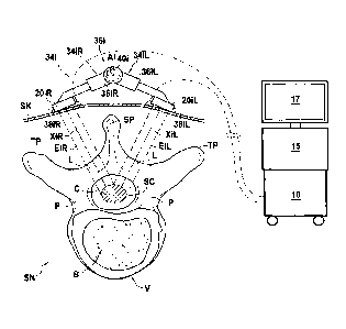
On FIG. 1 are shown the main components of an apparatus to induce
spinal cord or spinal nerves treatment by emission of ultrasound waves,
Date Recue/Date Received 2022-07-18
CA 03017916 2018-08-28
WO 2017/153799 PC17162016/000431
comprising an exemplary embodiment of an external ultrasound generating
treating device 12 according to the invention.
The apparatus comprises:
- an external ultrasound generating treating device 12;
5 - an
electrical generator 10 which generates electric signals to be
delivered to the transducers of the external ultrasound generating treating
device, where the generator may remain external to the body of the patient
in use of the apparatus;
- a controller 15, also external to the body, for example under the form
10 of a computer, to set and control the working parameters of the
generator.
According to an aspect of the invention, the external ultrasound
generating treating device 12 is suitable for external positioning against the
back of a patient who is awaiting the receipt of, or is receiving medical care
or was/is/will be the object of a medical procedure, or is monitored for the
diagnosis or the development of a disease. The patient can be any vertebrate
subject, especially a mammal and in particular a human i.e., a person of the
species Homo sapiens.
Fig. 3 to 5 illustrate schematically such a positioning in the case of a
human patient. On those figures, one can see the spine SN of the patient,
on the internal side of the skin SK of the back of the patient. The spine SN
comprises vertebrae V. In a typical human vertebra, as shown on Fig. 1 in a
transverse cross-section perpendicular to the extension of the spine, a
vertebra comprises a spinal canal SC portion which is delimited:
- towards the front by the vertebra body B,
- towards the sides by the two pedicles P which join the body B to
the two transverse process TP, and
- towards the
rear by the spinous process SP and the two laminas L
which join each the spinal process SP to one of the two transverse
processes TP.
The spinal cord C is located in the spinal canal and the spinal nerves
(not represented) emerge from the spinal cord and extend laterally out of
the spinal canal between two vertebrae.
CA 03017916 2018-08-28
WO 2017/153799
PCT/IB2016/000431
11
More particularly, the external ultrasound generating treating device 12
is suitable for positioning, preferably directly on the skin, against the
back,
along the extension of at least a portion of the spine. A coupling agent, such
as a gel, may be needed.
In operation, the generator 10 and the external ultrasound generating
treating device 12 are to be connected electrically. Such electrical
connection
could be permanent. However, electrical connection is preferably a cable
connection achieved through a connector device of the generator 10 and a
connection receiver of the external treating device 12 which can be
connected and disconnected, for example in the form of a plug-and-socket
connection.
The external ultrasound generating treating device 12 comprises an
array of several ultrasound generating treatment transducers distributed
along a longitudinal direction and a lateral direction.
The treatment transducers generate focused or unfocused ultrasounds.
The ultrasound generating treatment transducers 20 are preferably
chosen into the group formed by piezo-composite elements, piezo-ceramic
elements, CMUT elements (Capacitive micro-machined ultrasonic
transducers), or PVDF elements (Poly(vinylidene fluoride)). Piezo-composite
elements or piezo-ceramic elements usually have a size in the range of 1 to
50 mm in diameter. CMUT elements usually have a size in the range of 10 to
50 pm in diameter. Piezoelectric components are commonly used in the
medical field as ultrasound transducers. A given transducer can comprise one
or several discrete elements which are activated simultaneously.
The ultrasound treatment transducers have an ultrasound generating
resonant frequency which is preferably comprised between 0.5 and 4 Mhz,
more preferably between 0.75 and 2Mhz for achieving transient disruption of
the blood-spinal cord barrier and/or of the blood-spinal nerve barrier of the
targeted portion of the spinal cord and/spinal nerve(s).
In most commonly used ultrasound generating transducers, the
ultrasound energy is generated by virtue of the vibration created in the core
of the transducer by_an alternating voltage by virtue of a piezoelectric
effect
CA 03017916 2018-08-28
WO 2017/153799
PCT/IB2016/000431
12
or capacitive variation. The transducer is fed with an electric voltage which
may have a given frequency or which may have a frequency spectrum which
may be decomposed into preferably a limited number of main frequencies.
The core of the transducer may thus be designed such that it exhibits at
least one inherent resonant frequency.
A resonant frequency of the transducer can be defined as the frequency
of the drive signal for which the ratio of the acoustic power output divided
by
consumed electrical power reaches a maximum (at least within neighbouring
frequencies). For a typical piezoceramic transducer, this ratio is typically
between 50% and 90% at a resonant frequency. If the electric current fed
to the transducer exhibits such frequency, it will induce in the transducer a
resonant vibration which will generate ultrasound. If the electric current fed
to the transducer exhibits only a frequency or frequencies which lie outside
of a resonant range around the resonant frequency, then the acoustic power
output will be less than 25% of the power delivered when driven with a
given voltage at its resonant frequency.
It must be noted that the term resonant frequency, as used in this text,
covers an individual peak resonant frequency, at which the transducer 20
delivers a peak ultrasound field power/intensity for a given electric drive
signal power, or a resonant frequency range, around such peak resonant
frequency, for which the transducer 20 delivers a ultrasound field
power/intensity higher than a minimum field power/intensity, which may be
expressed as a percentage of the peak ultrasound field power/intensity.
A transducer may have a given operating frequency by choosing for
example its resonant thickness along a given direction along which the
ultrasound waves are to be emitted. For example thickness for a 1 MHz
transducer for PZ26 material should be at 2 mm along the desired direction
of emission.
The frequency content of the electric drive signal can be obtained
directly, in case of a simple alternating voltage having one frequency, such
as a pure sinusoidal signal. It can also be obtained through Fast Fourier
Transform (FFT), as known to the man skilled in the art of signal processing.
CA 03017916 2018-08-28
WO 2017/153799
PCT/IB2016/000431
13
It can be noted that, the intensity/power of the ultrasound field
generated by a given transducer will depend on the amplitude of the electric
drive signal delivered by the generator 10 at the operating frequency.
In use, the external ultrasound generating treating device 12 is
intended to be positioned against the back of the patient with its
longitudinal
direction parallel to the elongation line of the spine, i.e. in the sagittal
plane
of the patient, and its lateral direction extending perpendicularly to the
longitudinal direction, parallel to the axial and corona' planes of the
patient.
More precisely, the array of several ultrasound generating treatment
transducers comprises at least two sub-arrays of ultrasound generating
treatment transducers, a left sub-array being located on a left lateral side
of
a central longitudinal axis and a right sub-array being located on a right
lateral side of the central longitudinal, laterally opposite to the left side.
The external ultrasound generating treating device 12 comprises a
support structure 32 having at least one module 341, one of which can
arbitrarily be named a first module, each module comprising a left lateral
section 34iL, holding at least a first left treatment transducer 20iL or set
of
treatment transducers of the left sub-array, and a right lateral section 341R
holding at least a first right treatment transducer 201R or set of treatment
transducers of the right sub-array. It will be seen that the support structure
32 preferably comprises several modules, preferably several modules 341
having the same features.
The left and right lateral sections 341L, 341R of a module 341 of
the support structure 32 preferably comprise each a support member which
holds respectively the first left treatment transducer 2011 or set of
treatment
transducers and the first right treatment transducer 201R or set of treatment
transducers. The support member of a given module section 34iL, 34IR and
the arrangement of the treatment transducers on that support member are
preferably rigid enough so that, in use of the device, i.e. when exposed to
the normal forces involved in normal use, there is no movement of the
transducers relative to the support member and, if applicable, no relative
CA 03017916 2018-08-28
WO 2017/153799
PCT/IB2016/000431
=
14
movement between the set of transducers of a given module section 3411,
34iR.
The support structure 32 maintains, in use of the device, a
constant distance and a constant relative angular orientation around a
longitudinal axis, e.g. the central longitudinal axis Al of the module 341,
between the first left and first right treatment transducers or set of
treatment transducers 2011, 20iR. In other words, the support structure
holds the left and right treatment transducers or set of treatment
transducers 2011, 20iR rigidly enough to maintain, during use, a constant
distance and relative angular orientation between the first left and first
right
treatment transducers or set of treatment transducers 201L, 20iR.
As will be seen, a given module 341 of the support structure 32
may be arranged in the external ultrasound generating treating device 12 so
that its central longitudinal axis Al extends along or parallel to the
longitudinal direction of the external ultrasound generating treating device
12.
In use, i.e. at least during the duration of application of an
ultrasound treatment beam to the patient as will be described below, the first
left and first right treatment transducers or set of treatment transducers
2011_, 20iR have no relative movement and keep a same relative spatial
configuration. This same spatial relative configuration is maintained even in
spite of the patient having small movements during the application of the
ultrasound treatment beam, including movements due to the patient
breathing.
The constant distance is preferably maintained between any two
points of the first left and first right treatment transducers or set of
treatment transducers 2011, 20R.
An ultrasound generating treatment transducer 20iL, 20iR can be
considered to have a given ultrasound emission zone, typically in the form
approximately of a cylinder or a cone in which the intensity of the ultrasound
field is significant. For a set of treatment transducers of a given module
section 34iL, 341R, the combined treatment transducers thereby generate a
CA 03017916 2018-08-28
WO 2017/153799 PCT/1B2016/000431
combined section emission zone, which can be assimilated, for the purpose
of the invention, to an emission zone of a combined transducer. For
example, in Figure 1 is shown the case of said field of an external
ultrasound generating device 12 having left and right treatment transducers
5 or set of treatment transducers 2011, 20iR. Each left and right treatment
transducers or set of treatment transducers 20iL, 20iR, when properly
activated at its operating frequency, delivers an ultrasound field which can
be characterized by a border emission envelope Eli, EiR which is shown
here as a cylinder or a cone having a central axis XiL, XiR. The border
10 emission envelope of the emission zone EiL, EiR can be defined as the
envelope containing all locations where the acoustic pressure of the
ultrasound field generated by the corresponding left and right treatment
transducers or set of treatment transducers 20iL, 201R is equal to at least a
certain percentage, for example 25%, of the ultrasound field, at the same
15 distance from the transducer, along a direction of maximum acoustic
pressure. In real-world examples, the border envelope is not exactly a
cylinder or a cone but, for the type of transducers used in the field of
medical treatment ultrasound, can be considered as fairly close to a cone, or
at least may be comprised in such a cone. Thus, the treatment transducers
may have an ultrasound emission zone comprised in a cone having a central
emission axis XiL, XiR as its axis of symmetry. Such cone has preferably an
opening angle less than 30 degrees.
As can be seen on Fig. 1, the treatment transducers are
respectively arranged on their respective module sections so that the
emissions zones of the left and right treatment transducers or set of
treatment transducers 20iL, 20iR are targeted towards the spinal canal
when the external device 12 is positioned along the spine of a patient,
against the back the patient. Therefore, the central emission axis XiL, XiR of
the corresponding left and right emission zones is preferably perpendicular to
the longitudinal direction. In a plane perpendicular to the longitudinal
direction, the left and right emission zones are preferably directed so as
CA 03017916 2018-08-28
WO 2017/153799 PCT/IB2016/000431
16
converge on the spinal canal when the external device 12 is positioned along
the spine of a patient, against the back the patient.
The constant distance and relative angular orientation between
the first left and first right treatment transducers or set of treatment
transducers 2011, 201R induces that the emission zones, including the
combined emission zone if applicable, of the first left and first right
treatment
transducers or set of treatment transducers 2011, 20iR keep a same relative
spatial configuration, when the external device 12 is positioned along the
spine of a patient, against the back the patient. In other words, in use of
the
device, the support structure is rigid enough between the left and right
sections of a given module in order that the support structure does not
deform when subject to the normal forces endured during normal use of the
device.
For example, as in the shown embodiment, each section of the
module may comprise a support member having a rigid arm 3611, 36111
extending laterally, the two arms 3611, 36iR being connected at a
respective proximal end, and a respective transducer bracket 3811, 38iR
rigidly connected at their respective distal ends. The brackets may be in the
form of rigid plate like elements. Such brackets preferably extend along a
limited width according to the lateral direction, for example less than 5 cm,
preferably less than 3 cm. Such brackets preferably extend along a length
according to the longitudinal direction which is preferably comprise between
1 cm and 15 cm, preferably between 3 cm and 10 cm. The arms 3611, 36iR
may form an arch extending laterally between the transducer brackets 3811,
381R. The brackets are preferably spaced apart with their facing edges
laterally distant by at least lcm, preferably at least 3 cm.
In some embodiments, for a given module 34i of the support
structure 32, the relative sections 3411, 34iR have a non-adjustable relative
spatial configuration, including a constant distance and relative angular
orientation. Such non-adjustable relative spatial configuration 3411, 34iR of
the relative sections 3411, 34iR may be set once and for all, for example at
the moment of manufacture of the external device 12. In such a case, the
CA 03017916 2018-08-28
WO 2017/153799
PCT/182016/000431
17
the two arms 361L, 36iR of pertaining to the respective sections of a module
341 may be joined at their proximal end so as to form a single rigid and non-
adjustable part, for example in form of arigid arch.
Such a non-adjustable module is thus then designed in view of
predefined geometry of an expected patient's anatomy, so that the left and
right emission zones are preferably directed so as converge on the spinal
canal when the external device 12 is positioned along the spine of a patient,
against the back the patient.
However, in some embodiment of the invention, the support
structure of a given module 341 may comprise an adjusting mechanism 361
for adjusting the relative spatial configuration of the left and right
sections
34iL, 341R of the module 341. This allows for a more precise targeting of
the ultrasound treatment beam on the spinal cord and/or a spinal nerve.
Such adjustment may include the adjustment, around a
longitudinal axis, e.g. the central longitudinal axis Ai of the module 34i, of
a
relative angular orientation between the left and right lateral sections 341L,
341R of the support structure 32, so as to adjust the angular orientation
around the central longitudinal axis Al between the first left and first right
treatment transducers or set of treatment transducers 2011, 201R.
The adjusting mechanism may comprise an adjustment
articulation 36i.
In some embodiments, a single adjustment articulation 36i may
be provided between the two sections 3411, 34iR of a given module 341. As
in the example, such single articulation 361 may be located centrally, at the
proximal end of the arms 3611, 36iR.
In some embodiments, an adjustment articulation 36i may be
provided in each of the two sections 3411, 341R of a given module 341, for
example between the bracket 381L, 38iR and the distal end of the
corresponding arm 361L, 361R.
An adjustment articulation 361 may comprise a mechanical
articulation comprising two rigid parts having a relative motion along
respective sliding surfaces, such as a pivot or ball joint connection.
CA 03017916 2018-08-28
WO 2017/153799
PCT/IB2016/000431
18
An adjustment articulation 361 may be of the type having two or
three rotational degrees of freedom, for example around two or three
perpendicular articulation axes, including the central longitudinal axis Al of
the module 341 or another longitudinal axis parallel thereto.
However, as shown in the depicted embodiments, an adjustment
articulation may be of the type having only one degree of freedom, for
example around only a longitudinal axis, e.g. the central longitudinal axis
Al,
with no other possible rotational movement between the two sections 3411,
34iR of the module 341.
Similarly, the support structure 32 may comprise an adjusting
mechanism for adjusting a distance, for example along a lateral direction of
the module, between the left and right lateral sections 3411, 34iR of the
support structure of the module 341, so as to adjust the distance between
the first left and first right treatment transducers or set of treatment
transducers of that module 2011, 20iR. For example, in the example shown,
the arms of the support member in each section may be telescopic and
adjustable in length. Alternately, the brackets could be attached to the arms
in an adjustable manner along the extension of the arm.
Preferably the adjusting mechanism 36i comprises a lock 401 for
maintaining, in use of the device, a constant distance and a constant relative
angular orientation around the central longitudinal axis between the first
left
and first right treatment transducers or set of treatment transducers. The
lock may comprise a tightening screw tightening the adjustment mechanism
in a desired position. The lock may thus allow locking of the adjustment
mechanism in any position in a range of positions, to allow continuous
adjustment of the relative spatial configure of the two sections of the module
341 with a range of relative spatial configurations. The lock may comprise
indents allowing locking only in predefined spatial configurations.
The optimum relative spatial configuration of the left and first
right treatment transducers or set of treatment transducers 201L, 20iR of a
given module is dependent on the expected anatomy of a patient.
CA 03017916 2018-08-28
WO 2017/153799
PCT/IB2016/000431
19
For an external ultrasound generating treating device 12 intended
for use on an adult human, a range of adjustment of the angular orientation,
around the central longitudinal axis Al, between the first left and first
right
treatment transducers or set of treatment transducers 20iL, 20iR, is
preferably of at least 30 degrees, preferably of at least 60 degrees.
For an external ultrasound generating treating device 12 intended
for use on an adult human, a range of adjustment of the distance, along a
lateral direction of the module, between the first left and first right
treatment
transducers or set of treatment transducers 20iL, 20iR, is preferably of at
least 50 millimeters, preferably of at least 100 mm.
Having an optimal spatial configuration of the left and right
treatment transducers or set of treatment transducers, and maintining this
optimal spatial orientation is an important aspect. An optimal spatial
configuration is for exemple achieved when the left and right emissions
zones of the left and right treatment transducers or set of treatment are at
least partially superposed on the treatment zone of the spinal cord or on the
spinal nerves of the patient. Even more optimal is to have the left and right
emissions zones transducers intersecting a portion of minimum thickness of
the lamina of the vertrebrae before hitting the the spinal cord or on the
spinal nerves of the patient.
In non-adjustable modules, a proper design allows an adapatation
of the device to an average patient anatomy, already allowing in most cases
that a good portion of the left and right emission zones avoid at least the
spinous process and the transverse processes of the vertebrae.
Having transducers coming from both the left and the right side
and targeted at the same treatment zone of the spinal cord or on the spinal
nerves of the patient allows a better handling of the diffraction effects.
However, modules having an adjustment mechanism allow a
perfect adaptation of the external device to the patient's real anatomy, and
thus allows the most optimal ultrasound treatment conditions. Once an
optimal adjustment is determined and set, it is maintained during the used of
the device, for example by locking the adjusting mechanism with a lock.
CA 03017916 2018-08-28
WO 2017/153799 PCT/IB2016/000431
In some embodiments, the left and right lateral sections 3411,
341R of a given module 341 of the external ultrasound generating treating
device 12 may have ultrasonic imaging transducers 4211, 421R for forming
respectively a left and a right image of an emission zone of the treatment
5 transducer or set of treatment transducers 2011, 20iR held on the same
section. Such images are typically digital images obtained from the
ultrasound information collected by the ultrasonic imaging transducers 4211,
42iR. The imaging transducers may be of any suitable conventional type
known to the skilled man in the art. They may have an operating frequency
10 comprised between 200KHzz and 20 GHz, preferably from above 2GHz to 20
GHz. Each ultrasonic imaging transducer 4211, 421R may be formed of one
or several individual transducers. They may be held by the same support
member as the treatment transducers, for example the brackets 3811, 38iR.
The relative configuration of the ultrasonic imaging transducers 4211, 42iR
15 with respect of to the treatment transducer or set of treatment
transducers
2011, 20iR held on the same section is preferably fixed, but may be
different to the schematic shown on Fig. 2.
The external ultrasound generating treating device 12 may comprise
ultrasonic monitoring transducers 44i1, 44iR, for example wideband
20 ultrasonic transducers. Monitoring transducers may be flexible membrane
transducers. Monitoring transducers are preferably able to pick-up an
ultrasound signal over a wide frequency range, ideally between 50 kHz and
50 Mhz. Such monitoring transducers may be tailored and used for
monitoring cavitation due to the ultrasonic treatment.
A module 341 could be of limited extension the longitudinal
direction, for example corresponding to the length of a single vertebra of a
patient and adapted to treat a treatment zone of comparable extension. It
could be longer along that direction, for example corresponding to the length
of several vertebrae of a patient and adapted to treat a treatment zone of
comparable extension.
CA 03017916 2018-08-28
WO 2017/153799
PCT/1B2016/000431
21
An external ultrasound generating treating device 12 may
comprise a single module as described above.
Preferably, as shown on Figs. 2 to 5, the support structure 32
comprises several modules 341 arranged successively along the longitudinal
direction, each module 34i having one or several of the above features. Each
module 34i comprises a left lateral section holding at least a left treatment
transducer or set of treatment transducers of the left sub-array, and a right
lateral section holding at least a right treatment transducer or set of
treatment transducers of the right sub-array. As described above, the
support structure maintains, in each module 34i, in use of the device, a
constant distance and a constant relative angular orientation around the
central longitudinal axis of the respective module between the respective left
and first treatment transducers or set of treatment transducers of said
module 341.
All the modules 341 could have the same size. However, it can be
provided that different modules 341 could be of different sizes depending on
their location along the longitudinal direction of the external device 12.
All of the modules 34i could have the same features. However, it
can be provided that different modules 34i could have a different set of
features amidst the above described features.
Preferably several modules 341 have each an adjusting
mechanism 341 for adjusting, around their respective central longitudinal
axis, a relative angular orientation between the respective left and right
lateral sections of the support structure, so as to adjust the angular
orientation around the central longitudinal axis between the first left and
first
right treatment transducers or set of treatment transducers of that module
341.
In such a case, the adjusting mechanisms 361 of several modules
34i may be advantageously mechanically connected for simultaneous
adjustment. The simultaneous adjustment of the several modules 341 can
follow a predefined relative variation.
CA 03017916 2018-08-28
WO 2017/153799 PCT/IB2016/000431
22
In an external ultrasound generating treating device 12 having a
support structure 32 comprising several modules 341 arranged successively
along the longitudinal direction, at least two modules 341 of the support
structure 32 may be articulated to allow a relative angular movement
between the two modules around an axis extending along the lateral
direction. Such an example is represented on Figs 3 to 5. Upon positioning
of the external device against the back of a patient, such device can thus
adapt and conform to the shape of the spine, as particularly visible on Fig.
4.
Two successive modules 341 may be articulated though a single or
several inter-module articulation(s) 46, arranged in parallel or in series.
Two successive modules 34i may be articulated with only one
degree of freedom, for example around only one laterally extending axis Bi,
with no other possible rotational movement between the two modules 341.
However, two successive modules 34i may preferably be articulated with
several degrees of freedom. Preferably the modules are articulated so that
some degree of twisting around the longitudinal axis is also possible, in
addition to an articulation around the lateral axis. Furthermore, the
connection between two modules preferably additionally allows a relative
displacement, along a direction perpendicular to the lateral and longitudinal
directions, of two facing laterally extending edges of two consecutives
modules.
An articulation may comprise a mechanical articulation comprising
two rigid parts having a relative motion along respective sliding surfaces,
such as a pivot or ball joint connection.
However, as shown in the depicted embodiments, at least two
modules 341 of the support structure 32 are articulated through one or
several flexible module connector 46. A flexible module connector 46 may
be a sheet of flexible material, extending preferably in a longitudinally and
laterally extending plane, or a cable, extending preferably along the
longitudinal direction. A flexible module connector 46 may be elastic along
the longitudinal direction, or to the contrary it may be inelastic so as to
CA 03017916 2018-08-28
WO 2017/153799
PCT/IB2016/000431
23
define a set maximum distance between two consecutive modules 341 along
the longitudinal direction.
In the shown embodiment, the support structure of the external
device is symmetrical with respect to a plane of symmetry which extends
longitudinally and perpendicularly to the lateral direction. In use, this
plane
of symmetry is preferably aligned with the spine of the patient.
The left and right sections of a module are each connected
respectively to the left and right sections of consecutive modules along the
longitudinal direction by a respective flexible module connector 46, here
under the form of a sheet of flexible material, extending preferably in a
longitudinally and laterally extending plane. The flexible module connectors
46 between two successive modules are here arranged in parallel, spaced
apart along the lateral direction on each side of longitudinal axis.
Each flexible module connector 46 has a length along the
longitudinal direction which is preferably of at least 10 mm, more preferably
at least 20 mm, to allow sufficient flexibility and relative movement between
two consecutive modules.
The external ultrasound generating treating device 12 also comprises
an electrical connection network for connecting the ultrasound generating
transducers 20 to the generator 10 delivering electric drive signals. The
electric connection network may comprise one or several electrically
independent electric connection circuits, where it will be understood that a
given electric connection circuit is a circuit where a common electric drive
signal is circulating. An independent electric connection circuit may be used
to drive a single treatment transducer or may be used to drive a group of
treatment transducers. Each independent electric connection circuit will have
its own independent electric connection to the generator 10 and the
generator may deliver separate and different electric drive signals to each
independent electric connection circuit. For example it can be provided that
each module has its own independent electric connection circuit, which may
be shared between its left and right sections. Independent electric
CA 03017916 2018-08-28
WO 2017/153799
PCT/1B2016/000431
24
connection circuit may be useful for addressing possible impedance variation
between transducers.
In any case, imaging transducers and/or monitoring transducers, if
present, would preferably have their own separate electric connection circuit.
Preferably, the external ultrasound generating treating device 12 is
made of non-ferromagnetic materials, preferably MRI compatible materials.
The generator 10 is adapted for delivering electric drive signals to be
delivered to the ultrasound generating treatment transducers 20 of an
associated ultrasound generating treating device 12. The generator typically
comprises an alternating voltage generator able to generate an electric
signal, for example a sinusoidal electric voltage signal. One example of a
generator system that can be used with the inventive device may include a
system that integrates signal generation, amplification, and control into a
single unit. However, a generator system can also comprise one or several
individual components performing one or more of these functions. For
example, the generator can include an HP/Agilent 33120 function generator.
If needed, it can also include for example one or more of an ENI 2401_
Broadband RF amplifier, of a Rhode and Schwarz RF power meter, and /or
external computer controlling equipment over GPIB/Serial/USB interfaces.
Therefore, the controller 15 may comprise a computer. A computer
human/machine interface 17, for example a keyboard, and/or mouse and/or
a display and/or a touchscreen interface, can be provided to control the
system and give the user feedback. A radiofrequency board that generates
the RF signal and amplifies it may be provided, as well as a coupler to
measure the delivered RF power, and matching components to tune the
generator output to the impedance of the ultrasound elements. Preferably,
the generator 10 may be of a type capable to deliver 25-100 W peak RF
power, capable of sending burst lengths with durations of 1 microsecond to
continuous mode, and capable of sending bursts within the frequency range
of 200 kHz to 2 MHz. Such a system can be controlled to send pulses with
variable frequency and duty cycles for durations of approximately 2-5
minutes. The generator may be a class A/B RF system, which means that it is
CA 03017916 2018-08-28
WO 2017/153799 PCT/IB2016/000431
capable of generating nearly pure sinusoidal signals, but this may make the
system rather large. In some embodiments, the generator could be a class D
system, which tends to generate signals that are square wave on the output.
As seen on Fig. 2, the controller 15 may comprise a treatment control
5 module 15A for controlling the generator in view of providing the
adequate
electric drive signals to the treatment transducer or set of treatment
transducers 20iL, 20iR of the external ultrasound treating device 12.
The controller 15 may also comprise an imaging module 158
connected to the imaging transducers 4211, 42iR of the external ultrasound
10 treating device 12, if provided with such imaging transducers. The imaging
module 15B may be configured to display one or several images on a display
17, and/or to provide data extracted from a controller performed analysis of
the images.
The controller 15 may also comprise a monitoring module 15C
15 connected to the monitoring transducers 4411, 44iR of the external
ultrasound treating device 12, if provided with such monitoring transducers.
The monitoring module 15C may be configured to display one or several
images on a display 17, and/or to provide data extracted from a controller
performed analysis of the ultrasound signal collected by the imaging
20 transducers.
According to another aspect of the invention, it is provided a method
for transiently opening the blood-spinal cord barrier and/or the blood spinal
nerves barrier in at least one treatment zone of the spinal cord and/or spinal
25 nerves of a patient.
In the context of the invention, the terms "disrupting", "opening" or
"increasing the permeability" of the BSCB or BSNB are used interchangeably
to refer to an increased susceptibility of the BSCB or BSNB to the passage of
molecules therethrough that occurs without detectable damaging of the
spinal cord or spinal nerve tissue.
In the context of the invention, a "transient" opening refers to a
reversible opening occurring preferably for more than 1 hour, the BSCB or
CA 03017916 2018-08-28
WO 2017/153799 PCT/1132016/000431
26
BSNB returning after that to its initial state (i.e., the BSCB or BSNB state
before the application of the first ultrasound treatment beam).
In some embodiments, the BSCB or BSNB opening occurs for a period
of time from 1 to 48 hours, preferably from 5 to 24 hours, more preferably
from 6 to 10 hours. In some embodiments, the BSCB or BSNB opening
occurs for approximately 8 hours.
In some embodiments, the BSCB or BSNB disruption is delimited, i.e.,
occurs solely in a target region of the BSCB or BSNB. For instance, only a
region of the BSCB or BSNB surrounding damaged spinal cord or spinal nerve
tissue, such as a tumor, is targeted. In other embodiments, the BSCB or
BSNB disruption is generalized.
The disruption may be easily confirmed and/or evaluated by magnetic
resonance imaging (MRI). For example, a gadolinium-based magnetic
resonance (MR) contrast agent such as Dotarem0 (gadoterate meglumine,
Guerbet USA), which does not normally cross the BSCB or BSNB, can be used
to visualize the region of BSCB or BSNB disruption. When the agent is
injected in a patient, a Tlw MR sequence can be used to visualize regions of
hypersignal and therefore visualize the effect of BSCB or BSNB disruption by
ultrasound. BSCB or BSNB disruption typically leads to a change of 5-10% or
more in MR signal enhancement after contrast agent administration. With the
invention, a change of more than 25%, preferably more than 50% in MR
signal enhancement after contrast agent administration is contemplated. In
addition, dynamic contrast enhanced (DCE) MR imaging techniques can be
used to calculate the permeability of the BSCB or BSNB and to quantify the
magnitude of the permeability enhancement after ultrasound treatment.
The method can be used for delivering substances into targeted spinal
cord or spinal nerve tissue of the subject and/or for treating a the spinal
cord
or spinal nerve disease.
The method can be used to treat various physiological disorders which
induce different forms of pathologies including;
- spinal degenerative pathologies, such as amyotrophic lateral
sclerosis (ALS)
CA 03017916 2018-08-28
WO 2017/153799
PCT/IB2016/000431
27
- spinal cord tumor diseases, such as spinal astrocytomas
- spinal inflammatory pathologies, such as multiple sclerosis, etc...
It can also be used to improve the repair and/or rehabilitation
treatments of the spinal cord and/or spinal nerve(s), for example for
hemiplegia and paraplegia, including with cell transplant and/or stem cell
regeneration.
The method preferably comprises positioning externally against the
skin of the back of the patient:
- at least one left ultrasound generating treatment transducer or
set of treatment transducers, having a left emission, on a left lateral side
of
the back of the patient with respect to the spine of the patient, and
- at least one right ultrasound generating treatment transducer,
having a right emission zone, on a right lateral side of the back of the
patient
with respect to the spine of the patient.
Such method can thus be implemented with an external
ultrasound generating treating device 12 as described above.
As explained above, it can be considered that each treatment
transducer or set of treatment transducers has an ultrasound emission zone
comprised in a cone having a central emission axis as its axis of symmetry.
The method further comprises forming at least one left image along a
left imaging axis having a set orientation with respect to the left emission
zone and one right image along a right imaging axis having a set orientation
with respect to the right emission zone of the left and right treatment
transducers or set of treatment transducers. The imaging axis can be the
axis joining the center of the imaging transducer or set of imaging
transducers to the center of the object which is imaged by the imaging
transducer or set of transducers. Such set orientation may be obtained by
having a left and right imaging axis corresponding respectively to the left
and
right central emission axis of the left and right emission zones.
Advantageously, the method provides for orienting left and right
emission zones according to the left and right images so that the left and
CA 03017916 2018-08-28
WO 2017/153799 PCTAB2016/000431
28
right ultrasound emission zones are at least partially superposed on the
treatment zone of the spinal cord or on the spinal nerves. Of course, such
method is most conveniently implemented with an external device as
described above wherein the support structure of a given module 341
comprise an adjusting mechanism 361 for adjusting the relative spatial
configuration of the left and right sections 3411, 341R of the module 341.
Indeed, adjustment of the left and right sections 3411, 34iR of the module
341 is made simply and precisely thanks to the adjustment mechanism,
before or at the beginning of the treatment, and the relative configuration is
reliably maintained during the treatment.
However, even in the case of an external device having one or several
modules not having such adjusting mechanism, but having a left and a right
set of treatment transducers, it is also possible to orient the left and right
emission zones. In such a case, it is possible to orient the left and right
emission zones by controlling the left and a right set of transducers so as to
electronically steer the left and right emission zones. Such technique is
conventionally called "electronic beam steering". Such technique can involve
introducing time delays between the electric drive signals sent to the
individual treatment transducers within respectively the left and right sets
of
treatment transducers. The time delays may be computed to steer the beam
in one direction or the other. In any case, electronic beam steering can also
be implemented with external devices having an adjusting mechanism
according to the invention, thus in addition to the adjustment of the relative
angular orientation between the left and right lateral sections of the support
structure.
Even more preferably, such method is most conveniently implemented
with an external device as described above wherein the support structure of
a given module 341 comprise an adjusting mechanism 36i for adjusting the
relative spatial configuration of the left and right sections 34iL, 34iR of
the
module 34i, and wherein the left and right lateral sections 3411, 34iR of a
given module 341 of the external ultrasound generating treating device 12
have ultrasonic imaging transducers 42iL, 42iR for forming respectively a
CA 03017916 2018-08-28
WO 2017/153799
PCT/IB2016/000431
29
left and a right image of an emission zone of the treatment transducer or set
of treatment transducers 20iL, 20iR held on the same section. With such a
device, there is a direct and fixed correlation between the orientation of the
emission and zone and the orientation of the imaging axis held by a same
section. Therefore, superposition of the left and right emission zones can be
achieved simply by adjusting the relative spatial configuration of the left
and
right sections 34iL, 34iR of the module 34i, and by comparing the left and
right images until a predefined correlation between the two images is
obtained, which, by construction of the device, will be known to correspond
to a superposition of the emission zones on a desired target location, e.g. on
the spinal canal.
For example it is possible to construct the apparatus so that, when the
spinal canal appears in the center of each left and right image, then the
practitioner knows that the left and right emission zones are at least
partially
superposed on the treatment zone of the spinal cord or of the spinal nerves.
In a method as above, the treatment zone may extend throughout the
extension of several vertebrae of the patient. This is most conveniently
implemented with an external device as described above wherein the support
structure 32 comprises several modules 34i arranged successively along the
longitudinal direction, the external device being positioned so that its
longitudinal direction is parallel to the extension of the spine of the
patient.
The method comprises the application to the treatment zone of the
spinal cord and/or spinal nerves of the patient of at least one ultrasound
treatment beam. This can be achieved by proper activation of the treatment
transducer or set of treatment transducers 20i1, 20iR of an external device
12 as described above. The use of such a device allows for a very precise
control of the ultrasound energy and power delivered to the targeted spinal
cord and spinal nerve tissues. It also allows a precise targeting of the
treatment zone, with the possibility to precisely control the extension of
such
treatment zone where the ultrasound treatment beam is effectively applied.
CA 03017916 2018-08-28
WO 2017/153799
PCT/IB2016/000431
The terms "ultrasound beam", "ultrasound wave" and "ultrasound" are
used indifferently for designating sound waves with frequencies higher than
20 kHz. However the ultrasound treatment beam has preferably an
ultrasound frequency ranging from 0.5 to 4 MHz, more preferably ranging
5 0.75 to 2 MHz.
The method preferably involves the injection of an ultrasound contrast
agent in the patient's blood circulation system, prior to and/or during the
generation of the least one ultrasound treatment beam.
10 The term "ultrasound contrast agent" is used herein to refer to a
substance (solid, liquid or gas) that is able to enhance the contrast between
the region containing the agent and the surrounding tissue in an ultrasound
image. Advantageously, the ultrasound contrast agent corresponds to small
bubbles of a gas, termed "microbubbles," with an average diameter between
15 1 pm and lOpm. Said microbubbles oscillate and vibrate when a treatment
ultrasound beam is applied and may reflect ultrasound waves. The
ultrasound contrast agent is generally injected intravenously into the blood
stream in the patient's blood circulation system, wherein it remains for a
limited period of time.
20 The ultrasound contrast agent may be administered by injection,
preferably by systemic injection. Examples of systemic injections include
intravenous, subcutaneous, intramuscular, intradermal, intra vitreal and
intraperitoneal injection, or perfusion.
Preferably, the ultrasound contrast agent is administered as a bolus just
25 before the ultrasound treatment beam application. More preferably, the
ultrasound contrast agent is administered between 0 and 60 minutes before,
and/or during the ultrasound treatment beam application. When successive
ultrasound treatment beams are applied, the ultrasound contrast agent is
preferably delivered only once, just before the first ultrasound treatment
30 beam application of the cycle, though it may be delivered at activation of
each US beam, or by a continuous infusion through the activation of
successive ultrasound treatment beams.
CA 03017916 2018-08-28
WO 2017/153799 PCTAB2016/000431
31
According to the invention, the ultrasound contrast agent may contain
gaseous bubbles, a high concentration of gas, solid particles configured to
vaporize in response to ultrasound, liquid configured to vaporize in response
to ultrasound, micro particles configured to act as cavitation sites, solid
particles having higher acoustic impedance than tissue in the desired region,
and/or liquid with a high acoustic absorption coefficient.
In some embodiments, the ultrasound contrast agent is a microbubble
contrast agent, preferably selected from the group consisting of sulphur
hexafiuoride microbubbles (SonoVue0), microbubbles made of an albumin
shell and octafluoropropane gas core (OptisonC)), perflexane microbubbles
encapsulated in an outer lipid shell (ImagentC)), microbubbles made of
octafluoropropane gas core encapsulated in an outer lipid shell (DefinityC)),
or perfluorobutaine and nitrogen gas encapsulated in a lipid shell (BR38 ¨
Schneider et al., 2011). Preferably, the ultrasound contrast agent consists of
sulphur hexafluoride microbubbles. Microbubbles may contain a drug and/or
a nanoparticle which may be delivered in situ when the microbubbles are
exposed to the ultrasound treatment beam.
The microbubbles may have a mean diameter in a range from 1 pm to
lOpm. In some embodiments, the microbubbles have a mean diameter in a
range from 4 pm to 5 pm. In some other embodiments, the microbubbles
have a mean diameter in a range from 2 to 6 pm. In some embodiments, the
microbubbles have a mean diameter of approximately 7 pm, 6 pm, 5pm,
4pm, 3pm or 2pm. In a particular embodiment, the microbubbles have a
mean diameter of approximately 2.5 pm.
In some embodiments, the dose of ultrasound contrast agent ranges
between 0.05 and 0.15 ml/kg based on the total weight of the subject.
Preferably, the dose of ultrasound contrast agent is approximately 0.1 ml/kg.
In a particular embodiment, the maximum dose of ultrasound contrast agent
is up to 10 ml.
= 30
Preferably, the pressure level of the ultrasound treatment beam applied
to the spinal cord or spinal nerve tissues is comprised between 0.8 MPa and
CA 03017916 2018-08-28
WO 2017/153799
PCT/IB2016/000431
32
3.0 MPa. Advantageously, the ultrasound treatment beams are applied within
a pressure range of 0.8 MPa to 2.5 MPa, more preferably within a pressure
range of 0.8 MPa to 2.00, even more preferably within a pressure range of
0.8 MPa to 1.9, such as within a pressure range of 0.8 MPa to 1.5 MPa,
within a pressure range of 1.1 MPa to 1.5 MPa. In a particular embodiment,
the ultrasound treatment beams are applied with a pressure level of 1.25
MPa. In another embodiment, the ultrasound treatment beams are applied
with a pressure level of 1.5 MPa. In a further embodiment, the ultrasound
treatment beams are applied with a pressure level of 1.9 MPa. In the context
of the invention, the "pressure level" refers to the maximum acoustic
pressure measured in the acoustic field of the device in water. It is believed
that such pressure levels may be applied in a safe manner to human's spinal
cord and/or spinal nerve, i.e., no detected damages of spinal cord and/or
spinal nerve tissue should be observed.
In the context of the invention, the value of the pressure level
corresponds to the value onto the spinal cord and/or spinal nerve tissue. The
pressure emitted by the device may differ, to take into account potential
attenuation of intervening tissues and/or vertebra bone reverberation. One
skilled in the art will be able to adapt the value of the pressure level
coming
out of the emitter to obtain the required pressure level onto the spinal cord
and/or spinal nerve. Monitoring of the treatment zone with ultrasonic
monitoring transducers can be used for checking the effective value of the
pressure level in situ during the treatment.
Preferably, the applied ultrasound treatment beam to the spinal cord or
spinal nerve tissues has a mechanical index (MI) of approximately from 1 to
3.00, and preferably in the range of 1.05 to 1.8 in the case of a 1MHz
ultrasound treatment beam. In the context of the invention, the MI refers to
the peak negative pressure in situ (MPa) divided by the square root of the
frequency (MHz).
CA 03017916 2018-08-28
WO 2017/153799 PCT/IB2016/000431
33
Preferably, the ultrasound treatment beam is a pulsed beam. In the
context of the invention, a "pulse" refers to a continuous burst, without
interruption, of sinusoidal waves that may comprises several cycles.
In some embodiments, the method comprises the application one or
more pulses, or bursts, comprising from 100 to 100,000 successive cycles,
preferably from 1,000 to 75,000, more preferably from 10,000 to 50,000,
even more preferably from 20,000 to 30,000. In a particular embodiment,
the method comprises the application of pulses of 25,000 successive cycles.
In some embodiments, the mean burst duration of an ultrasound treatment
emission (i.e., the mean time from the start of a pulse to the end of that
pulse) is between 10 msec. and 100 msec., preferably between 15 msec. and
50 msec., more preferably between 20 msec. and 30 msec., even more
preferably approximately 25 msec.
The delay between two successive pulses is preferably from 30 msec.
to 1000 msec. In a particular embodiment, the delay between two successive
pulses is approximately 975 msec.
Advantageously, the successive pulses are applied within a total
duration from 1 to 20 minutes. In a particular embodiment, the successive
pulses are applied within a total duration that does not exceed 10 minutes,
preferably 5 minutes. In a particular embodiment, the successive pulses are
applied within a total duration of 150 seconds.
In a particular embodiment, pulses of 25,000 cycles are applied to the
subject, at a pulse repetition frequency (PRF) of 1 Hz, every 1000 msec. with
a pressure level of 1.1 MPa and a burst duration of about 23 msec. for a total
duration of 150 seconds.