Note: Descriptions are shown in the official language in which they were submitted.
OPTICAL FIBER SENSOR TO DETECT
THE POSITION OF A NEEDLE
BACKGROUND OF THE INVENTION
[0001] The present disclosure relates generally to optical fiber sensors.
In particular, the
present disclosure relates to devices, systems, and associated methods to
detect a position of a
cannula and/or a catheter tube using an optical fiber sensor. Detecting the
position of the cannula
and/or the catheter tube using the optical fiber sensor may be useful when
inserting the cannula
and/or the catheter tube into a blood vessel, such as a vein, of a patient.
Detecting the position of
the cannula and/or the catheter tube using the optical fiber sensor may also
be useful for
continuous monitoring of the position of the cannula and/or the catheter tube
within the blood
vessel.
[0002] Placing a cannula in a desired destination of a body of a patient
may be a difficult
task for a clinician in the medical field. For example, it may be difficult to
place the cannula in
proper position in the vein for infusion and/or blood collection. The
clinician may want to
advance the cannula far enough to access the blood or cerebrospinal fluid but
does not want to
advance the cannula too far so as to injure a wall of the vein, the spinal
cord, or the spinal cord
dura. Thus, it is important for the clinician to know when the cannula is
located within the vein.
[0003] In some instances, in order to verify proper placement of the
cannula of a catheter
device in the vein, for example, the clinician generally confirms that there
is "flashback" of
blood into, for example, a flashback chamber associated with the catheter
device. Flashback
generally entails an appearance of a small amount of blood within the catheter
device, which
may be to the clinician.
[0004] However, the time it takes for flashback to appear in the
flashback chamber is
highly dependent on the gauges size of the cannula and patient venous blood
pressure. For a 24G
cannula at low venous pressure typical of pediatric patient, it may take more
than five seconds
for flashback to appear, which may not be a very useful indicator.
[0005] Accordingly, there is a need in the art for devices, systems, and
methods that
provide an instantaneous indication of a location of the distal tip of the
cannula regardless of
gauge size and patient condition. In addition, it would also be desirable for
such a device to
provide continuous indication of cannula location. Such devices, systems, and
methods are
disclosed herein.
-Page 1-
Date Recue/Date Received 2020-12-07
CA 03018775 2018-09-21
WO 2017/172388 PCT/US2017/022937
BRIEF SUMMARY OF THE INVENTION
[0006] The present disclosure relates generally to optical fiber sensors.
In particular, the
present disclosure relates to devices, systems, and associated methods to
detect a position of a
cannula and/or a catheter tube using an optical fiber sensor. Detecting the
position of the cannula
and/or the catheter tube using the optical fiber sensor may be useful when
inserting the cannula
and/or the catheter tube into a blood vessel of a patient. Detecting the
position of the cannula
and/or the catheter tube using the optical fiber sensor may also be useful for
continuous
monitoring of the position of the cannula and/or the catheter tube within the
blood vessel.
[0007] In some embodiments, a system to detect the position of the cannula
may include
the cannula, which may include a distal tip, an elongated tubular shaft, and
an inner lumen
formed by the elongated tubular shaft. In some embodiments, the system may
include an optical
fiber sensor (which may be referred to in the present disclosure as "the
optical fiber") disposed
within the inner lumen of the cannula. In some embodiments, the optical fiber
may include a first
end, a second end, and a U-shaped portion disposed between the first and the
second end. In
some embodiments, the U-shaped portion may be disposed at least proximate the
distal tip. In
some embodiments, the system may include a light emitter coupled with the
first end of the
optical fiber. In some embodiments, the system may include a light receiver
coupled with the
second end of the optical fiber. In some embodiments, the light receiver may
be configured to
convert light into an electrical signal.
[0008] In some embodiments, the system may also include an electronic
processor,
which may be coupled with the light receiver and may receive the electrical
signal. In some
embodiments, the electronic processor may be configured to detect a change in
an intensity of
light received at the light receiver based on the electrical signal. In
particular, in some
embodiments, the electronic processor may be configured to detect a decrease
in the intensity of
light received at the light receiver based on the electrical signal.
[0009] In some embodiments, the decrease in the intensity of light may
occur in response
to the optical fiber contacting blood when the cannula is inserted into the
blood vessel. For
example, the light emitter may emit light, which may be transmitted from the
first end to the
second end of the optical fiber. The intensity of the light received at the
light receiver may be
monitored by the electronic processor. When the cannula enters the blood
vessel, blood within
the blood vessel may travel into the distal tip of the cannula and contact the
optical fiber, which
-Page 2-
CA 03018775 2018-09-21
WO 2017/172388 PCT/US2017/022937
may result in an increase in light lost from the optical fiber due to
refraction. In further detail,
before the optical fiber contacts blood, the optical fiber may contact air or
another medium,
which may have a lower refractive index than blood. Before the optical fiber
contacts blood, total
internal reflection or increased internal reflection may occur as light is
transmitted through the
optical fiber. Because a refractive index of blood may be higher than a
refractive index of air and
closer than the refractive index of air to a refractive index of the optical
fiber, when blood
contacts the optical fiber, more light may be lost from the optical fiber due
to refraction and the
optical fiber may become less efficient. Thus, less light may return to the
light detector than in an
absence of blood.
[0010] In some embodiments, the optical fiber may be configured to contact
blood when
the cannula is inserted in the blood vessel. In particular, in some
embodiments, the U-shaped
portion of the optical fiber may be configured to contact blood first, before
a remaining portion
of the optical fiber, when the cannula is inserted in the blood vessel. In
some embodiments, prior
to insertion of the cannula in the blood vessel, the optical fiber may contact
air. In some
embodiments, when the cannula is inserted into the blood vessel, blood may
surround or
immerse the optical fiber.
[0011] In some embodiments, the U-shaped portion may be disposed at least
proximate
the distal tip of the cannula. In some embodiments, a distal most portion of
the U-shaped portion
may be disposed proximal to a beveled edge of the distal tip, which may
increase a likelihood
that the beveled edge is fully inserted in the blood vessel when the decrease
in the intensity of
light is detected and/or protect the optical fiber as the cannula is inserted
through the skin, fat,
tissue, etc. of the patient.
[0012] In some embodiments, the optical fiber may be a homogeneous optical
fiber. In
these and other embodiments, the optical fiber may be without cladding so as
to let light escape
axially from the optical fiber, which may facilitate measurement of the
decrease in the intensity
of light when the optical fiber is exposed to blood.
[0013] In some embodiments, the optical fiber may be planar. In further
detail, in some
embodiments, the U-shaped portion, the first end, and the second end may be
disposed within a
same plane. In some embodiments, the first end and the second end may be
parallel to each
other.
-Page 3-
CA 03018775 2018-09-21
WO 2017/172388 PCT/US2017/022937
[0014] In some embodiments, the U-shaped portion of the optical fiber may
be the most
sensitive section of the optical fiber for detecting contact with blood. In
some embodiments, the
first end and the second end may be twisted with respect to each other. For
example, the first end
and the second end may be twisted with respect to each other one, two, three,
or more times to
form a helical shape. By forming a helical shape, a sensitive section of the
optical fiber may be
increased in length beyond the U-shaped portion. The U-shaped portion and/or
the helical shape
may result in increased or continuous loss of light as blood contacts the U-
shaped portion and/or
the helical shape, thus providing an improved or continuous indication of the
cannula position
within the vein. If the distal tip of the cannula exits the vein, the blood
flow within the cannula
may stop and light may no longer be lost. Thus, the U-shaped portion and/or
the helical shape of
the optical fiber may provide improved or continuous indication the location
or position of the
cannula tip. The first end and/or the second end may be bent and/or twisted in
any number of
ways which may serve to increase a surface area of the optical fiber that
contacts blood.
[0015] In some embodiments, the system may include an alarm. In some
embodiments,
the electronic processor may be configured to initiate the alarm, which may
include an audible
sound, a flashing light, etc. In some embodiments, the electronic processor
may initiate the alarm
in response to the electronic processor detecting the decrease in the
intensity of the light received
at the light receiver. In some embodiments, the alarm may be reusable. In some
embodiments,
the optical fiber may be disposable and configured to be separated at an
optical connector from
the alarm and/or a monitoring unit that includes the alarm. The optical
connector may be
disposed in any number of locations. In some embodiments, the optical
connector may be
disposed at a proximal end of a catheter device that includes the cannula.
[0016] The cannula may include any cannula that may be used with the
optical fiber
described in the present disclosure, including, but not limited to, a
hypodermic needle, such as an
intravenous catheter (IV) introducer needle, a peripheral intravenous catheter
(PIVC) introducer
needle, an epidural introducer needle, a spinal tap needle, etc. One of
ordinary skill in the art will
understand that the optical fiber may detect a decrease in light received at
the light receiver in
response to the optical fiber contacting a medium other than blood, such as,
for example,
cerebrospinal fluid. The medium may have a refractive index similar to blood
and/or between the
refractive index of air and the refractive index of the optical fiber.
-Page 4-
CA 03018775 2018-09-21
WO 2017/172388 PCT/US2017/022937
[0017] In some embodiments, the system may include a catheter system, which
may
include a catheter device. The catheter system may include any catheter
device. Exemplary
catheter devices may include both straight and ported intravenous catheter
devices, such as the
AUTOGUARDTm shielded catheter device commercially available from Becton,
Dickinson, and
Company, integrated peripheral intravenous catheter devices, winged catheter
devices, catheter
devices with safety mechanisms, catheter devices with needle shields, blood
collection sets, IV
access sets such as the BD NEXIVAlm Closed Intravenous (IV) Catheter system
available from
Becton, Dickinson, and Company, etc.
[0018] In some embodiments, the catheter device may include a catheter
adapter having a
catheter tube. In some embodiments, the cannula of the catheter device may
include an
introducer needle. In some embodiments, the optical fiber may be disposed
within the introducer
needle. In some embodiments, the catheter device may include a needle hub,
which may be
coupled with a proximal end of the introducer needle. In some embodiments, the
needle huh may
be coupled with the catheter adapter. In some embodiments, the first end and
the second end of
the optical fiber may extend through at least a portion of the catheter
device.
[0019] In some embodiments, the optical fiber may be disposed within the
catheter tube
such that the optical fiber contacts blood when the catheter tube is inserted
in the blood vessel of
the patient. In some instances, because a distal tip of the catheter tube may
be proximal to the
distal tip of the introducer needle when the introducer needle is inserted in
the blood vessel of the
patient, the catheter tube may still be outside the blood vessel. In some
embodiments, the
catheter tubing may be extruded over the optical fiber.
BRIEF DESCRIPTION OF THE SEVERAL VIEWS OF THE FIGURES
[0020] In order that the manner in which the above-recited and other
features and
advantages of the invention will be readily understood, a more particular
description of the
cannula capture mechanism briefly described above will be rendered by
reference to specific
embodiments thereof, which are illustrated in the appended Figures.
Understanding that these
Figures depict only typical embodiments and are not, therefore, to be
considered to be limiting of
its scope, the invention will be described and explained with additional
specificity and detail
through the use of the accompanying Figures in which:
[0021] Figure 1A is a cross-sectional view of an example portion of an
optical fiber
disposed within an example cannula, according to some embodiments;
-Page 5-
CA 03018775 2018-09-21
WO 2017/172388 PCT/US2017/022937
[0022] Figure 1B is a cross-sectional view of the portion of the optical
fiber of Figure lA
in contact with blood, according to some embodiments;
[0023] Figure 2A is a cross-sectional view of a portion of another optical
fiber disposed
within the cannula of Figure 1A, according to some embodiments;
[0024] Figure 2B is a cross-sectional view of a portion of another optical
fiber disposed
within the cannula of Figure 1A, according to some embodiments;
f00251 Figure 3 is a schematic block diagram of an example system to detect
a position
of a cannula, according to some embodiments;
[0026] Figure 4 is a perspective view of an example catheter device that
may be included
in the system of Figure 3;
[0027] Figure 5 is a side view of the optical fiber of Figure 1 disposed
within a wall of
catheter tubing, according to some embodiments; and
[0028] Figure 6 is a block diagram of an example method to detect the
position of the
cannula, according to some embodiments.
DETAILED DESCRIPTION OF THE INVENTION
[0029] The presently preferred embodiments of the described invention will
be best
understood by reference to the Figures, wherein like parts are designated by
like numerals
throughout. It will be readily understood that the components of the present
invention, as
generally described and illustrated in the Figures herein, could be arranged
and designed in a
wide variety of different configurations. Thus, the following more detailed
description of the
embodiments of the cannula locator device, cannula locator system, and
associated methods, as
represented in Figures 1 through 8, is not intended to limit the scope of the
invention, as claimed,
but is merely representative of some embodiments of the invention.
[0030] Generally, this application relates to optical fiber sensors. In
particular, the
present disclosure relates to devices, systems, and associated methods to
detect a position of a
cannula and/or a catheter tube using an optical fiber sensor. Detecting the
position of a distal tip
of the cannula and/or the catheter tube may allow a clinician to appropriately
position the distal
tip of the cannula during a medical procedure, such as, for example, an
intravenous infusion,
blood draw, spinal tap, or epidural.
[0031] Figures 1A-1B are cross-sectional views of a portion of an optical
fiber 10
disposed within a cannula 12, according to some embodiments. In some
embodiments, the
-Page 6-
CA 03018775 2018-09-21
WO 2017/172388 PCT/US2017/022937
cannula may include a distal tip 14, an elongated tubular shaft 16, and an
inner lumen 18 formed
by the elongated tubular shaft 16. In some embodiments, the optical fiber 10
may be disposed
within the inner lumen 18 of the cannula 12. In some embodiments, the optical
fiber 10 may
include a first end 20, a second end 22, and a U-shaped portion 24 disposed
between the first end
20 and the second end 22. In some embodiments, the U-shaped portion 24 may be
disposed at
least proximate the distal tip 14. In some embodiments, the first end 20 of
the optical fiber 10
may be coupled with a light emitter (not illustrated in Figures 1A-1B). In
some embodiments, the
second end 22 of the optical fiber 10 may be coupled with a light receiver
(not illustrated in
Figures 1A-1B). One of ordinary skill in the art will appreciate that the
first end 20 may be
coupled with the light receiver, and the second end 22 may be coupled with the
light emitter, in
some embodiments.
[0032] In some embodiments, a decrease in an intensity of light received at
the light
receiver may occur in response to the optical fiber 10 contacting blood or
another medium when
distal tip 14 of the cannula 12 is inserted into the blood vessel. For
example, the light emitter
may emit light, which may be transmitted from the first end 20 to the second
end 22 of the
optical fiber. The intensity of light received at the light receiver may be
monitored by an
electronic processor (not illustrated in Figures 1A-1B). In some embodiments,
when the distal tip
14 of the cannula 12 enters the blood vessel, blood within the blood vessel
may travel into the
distal tip 14 and contact the optical fiber 10, which may result in an
increase in light lost from the
optical fiber 10 due to refraction. In further detail, as illustrated in
Figure 1A, before the optical
fiber 10 contacts blood, the optical fiber 10 may contact air or another
medium inside the
cannula 12, which may have a lower refractive index than blood. Before the
optical fiber 10
contacts blood, total internal reflection or increased internal reflection may
occur as light is
transmitted through the optical fiber. Because a refractive index of blood may
be higher than a
refractive index of air and closer than the refractive index of air to a
refractive index of the
optical fiber 10, when blood contacts the optical fiber 10, as illustrated in
Figure 1B, more light
may be lost from the optical fiber 10 due to refraction and the optical fiber
10 may become less
efficient. Thus, less light may return to the light detector than in an
absence of blood.
[0033] In some embodiments, the optical fiber 10 may be configured to
contact blood
when the cannula 12 is inserted in the blood vessel. In particular, in some
embodiments, the U-
shaped portion 24 of the optical fiber may be configured to contact blood
first when the cannula
-Page 7-
CA 03018775 2018-09-21
WO 2017/172388 PCT/US2017/022937
12 is inserted in the blood vessel. In some embodiments, prior to insertion of
the cannula 12 in
the blood vessel, all or a portion of the optical fiber 10 may contact air. In
some embodiments,
when the cannula 12 is inserted into the blood vessel, blood may surround or
immerse the optical
fiber 10. In some embodiments, the U-shaped portion 24 may be disposed at
least proximate the
distal tip 14 of the cannula 12. In some embodiments, a distal most portion of
the U-shaped
portion 24 may be disposed proximal to a beveled edge 26 of the distal tip 14,
which may
increase a likelihood that the beveled edge 26 is fully inserted in the blood
vessel when the
decrease in the intensity of light is detected and/or protect the optical
fiber as the cannula 12 is
inserted through the skin, fat, tissue, etc. of the patient.
[0034] In some embodiments, the optical fiber 10 may be a homogeneous
optical fiber.
In these and other embodiments, the optical fiber 10 may be without cladding
so as to let light
escape axially from the optical fiber 10, which may facilitate measurement of
the decrease in the
intensity of light when the optical fiber 10 is exposed to blood.
[0035] In some embodiments, the optical fiber 10 may be planar, as
illustrated in Figures
IA-1B. In further detail, in some embodiments, the U-shaped portion 24, the
first end 20, and the
second end 22 may be disposed within a same plane. In some embodiments, the
first end 20 and
the second end 22 may be parallel to each other, as illustrated in Figures 1A-
1B.
[0036] Referring now to Figures 2A-2B, in some embodiments, the first end
20 and the
second end 22 may be twisted with respect to each other. For example, the
first end 20 and the
second end 22 may be twisted with respect to each other one, two, three, or
more times to form a
helical shape. Figure 2A illustrates the first end 20 and the second end 22
twisted with respect to
each other one time to form the helical shape. Figure 2B illustrates the first
end 20 and the
second end 22 twisted with respect to each other two times to form the helical
shape. The helical
shape of the optical fiber 10 may increase a length and surface area of the
sensitive section of the
optical fiber 10 that contacts blood, which may result in increased or
continuous loss of light as
light is transmitted through the optical fiber 10 in contact with blood as
blood flows in the
proximal direction within the cannula 12. Thus, the helical shape may provide
an improved or
continuous indication of the location or position of the cannula 12.
[0037] For example, when a medium contacting the optical fiber 10 changes
from a first
medium, for example air, to a second medium, for example blood, a loss in the
intensity of light
received by the light receiver may occur. A contact area between the optical
fiber 10 and the
-Page 8-
CA 09018775 2018-09-21
WO 2017/172388 PCT/US2017/022937
second medium may increase due to the helical shape, and the loss in the
intensity of light may
occur along the contact area. If the first medium and the second medium have
similar refractive
indexes, a movement from the first medium to the second medium may be more
easily detected
when the optical fiber includes the helical shape. The first end 20 and/or the
second end 22 may
be bent and/or twisted in any number of ways that may serve to increase the
surface area of the
optical fiber 10 that contacts blood.
[0038] The cannula 12 may include any cannula that may be used with the
optical fiber
described in the present disclosure, including, but not limited to, a
hypodermic needle, such as
an intravenous catheter (IV) introducer needle, a peripheral intravenous
catheter (PIVC)
introducer needle, an epidural introducer needle, a spinal tap needle, etc.
[0039] Referring now to Figure 3, in some embodiments, a system 28 may
include an
optical fiber sensor device 29. In some embodiments, the optical fiber sensor
device 29 may
include the cannula 12 and/or the optical fiber 10. The optical fiber sensor
device 29 may
correspond to any number of devices that include the optical fiber 10. In some
embodiments, the
optical fiber sensor device 29 may include a catheter device, as will be
discussed further with
respect to Figure 4. In some embodiments, the system 28 may include a light
emitter 30, which
may be coupled with the first end 20 of the optical fiber 10. In some
embodiments, the system 28
may include a light receiver 32, which may be coupled with the second end 22
of the optical
fiber 10. In some embodiments, the light receiver 32 may be configured to
convert light into an
electrical signal.
[0040] In some embodiments, the system 28 may also include an electronic
processor 34,
which may be coupled with the light receiver 32. In some embodiments, the
electronic processor
34 may be configured to receive the electrical signal from the light receiver
32 and monitor an
intensity of light received at the light receiver 32. In some embodiments, the
electronic processor
34 may be configured to detect a change in the intensity of light received at
the light receiver 32
based on the electrical signal. In particular, in some embodiments, the
electronic processor 34
may be configured to detect the decrease in the intensity of light received at
the light receiver 32,
which may occur in response to the optical fiber 10 contacting blood, for
example.
[0041] In some embodiments, the system 28 may include an alarm 36. In some
embodiments, the electronic processor 34 may be configured to initiate the
alarm 36, which may
produce an audible sound, a flashing light, etc. In some embodiments, the
electronic processor 34
-Page 9-
CA 03018775 2018-09-21
WO 2017/172388 PCT/US2017/022937
may initiate the alarm in response to the electronic processor 34 detecting
the decrease in the
intensity of the light received at the light receiver 32. In some embodiments,
the alarm 36 may be
configured to sound in response to the detection of the decrease in the
intensity of the light
received at the light receiver 32.
[0042] In some embodiments, the alarm 36 may be reusable. In some
embodiments, a
monitoring unit 38 may include one or more the following: the light emitter
30, the light receiver
32, the electronic processor 34, and the alarm 36. In some embodiments, one or
more of the
following elements may be selectively coupled to the optical fiber 10: the
light emitter 30, the
light receiver 32, the electronic processor 34, the alarm 36, and the
monitoring unit 38. In some
embodiments, an optical connector 40 may selectively couple the optical fiber
10 to one or more
of the elements. Thus, one or more of the elements may be uncoupled from the
optical fiber 10
via the optical connector 4, and the optical fiber 10, the cannula 12, and/or
the optical fiber
sensor device 29 may be disposed of while the one or more elements may be
reusable. In some
embodiments, the first end 20 and the second end 22 may be coupled with a
proximal end of the
optical connector 40, as illustrated in Figure 3. In some embodiments, the
light emitter 30 may
be coupled with the first end 20 and/or the light receiver 32 may be coupled
with the second end
22 via the optical connector 40.
[0043] The optical connector 40 may be disposed in any number of locations.
In some
embodiments, the optical connector 40 may be disposed at a proximal end of the
optical fiber
sensor device 29 that includes the cannula 12. In these and other embodiments,
the optical
connector 40 may be disposed within the optical fiber sensor device 29. In
some embodiments,
the optical connector 40 may be eliminated and the optical fiber sensor device
29 may be directly
coupled to the monitoring unit.
[0044] Referring now to Figure 4, in some embodiments, the optical fiber
sensor device
29 may include or correspond to a catheter device 42, an example of which is
illustrated in
Figure 4. The catheter device 42 may include any number of catheter devices.
Exemplary
catheter devices may include both straight and ported intravenous catheter
devices, such as the
AUTOGUARDTm shielded catheter device commercially available from Becton,
Dickinson, and
Company, integrated peripheral intravenous catheter devices, winged catheter
devices, catheter
devices with safety mechanisms, catheter devices with needle shields, blood
collection sets, IV
-Page 10-
CA 03018775 2018-09-21
WO 2017/172388 PCT/US2017/022937
access sets such as the BD NEXIVATh Closed Intravenous (IV) Catheter system
available from
Becton, Dickinson, and Company, etc.
[0045] In some embodiments, the catheter device 42 may include a catheter
adapter 44
having a catheter tube 46. In some embodiments, the cannula 12 of the catheter
device 42 may
include an introducer needle. In some embodiments, the catheter device 42 may
include a needle
hub 48, which may be coupled with a proximal end 50 of the cannula 12. In some
embodiments,
the needle hub 48 may be coupled with the catheter adapter 44. In some
embodiments, the first
end 20 and the second end 22 of the optical fiber 10 may extend through all or
a portion of the
catheter device 42, as illustrated in Figure 4.
[0046] In some instances, because the catheter tube 46 may be proximal to
the distal tip
14 of the cannula 12 when the cannula 12 is inserted in the blood vessel of
the patient, the
catheter tube 46 may still be outside the blood vessel when the cannula 12 is
within the blood
vessel. Referring now to Figure 5, in some embodiments, the optical fiber 10
may be disposed
within an outer wall of the catheter tube 46 such that the optical fiber 10
contacts blood when the
catheter tube 46 is inserted in the blood vessel of the patient. Thus, the
optical fiber 10 may
improve detection of a position of the catheter tube 46.
[0047] The optical fiber 10 may be disposed within the outer wall of the
catheter tube 46
in any number of ways. For example, the optical fiber 10 may be inserted,
molded, or co-
extruded into the catheter tube 46. In some embodiments, the optical fiber 10
may extend to a
distal tapered portion of the catheter tube 46 and/or may be planar, as
illustrated in Figure 5. In
some embodiments, the optical fiber 10 may extend to a distal edge of the
catheter tube 46.
[0048] In some embodiments, the optical fiber 10 may be disposed within an
inner wall
of the catheter tube 46 or another portion of the catheter device 42 (not
illustrated in Figure 5).
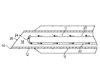
The optical fiber 10 may be disposed within the inner wall in any number of
ways. For example,
the optical fiber 10 may be inserted, molded, or co-extruded into the inner
wall. In some
embodiments, the cannula 12 may include a notch feature, which may define an
opening in the
elongated shaft 16 (not illustrated in Figure 5). In some embodiments, the
notch feature may be
configured to allow blood to flow between the distal tip 14 and the notch
feature. In some
embodiments. the blood may then flow out of the notch feature and into a
portion of the catheter
device, such as the catheter tube 46 and/or a body of the catheter adapter 44,
where the blood
may contact the optical fiber 10 disposed within the inner wall.
-Page 11-
CA 03018775 2018-09-21
WO 2017/172388 PCT/US2017/022937
[0049] The optical fiber 10 illustrated in Figure 5 may include or
correspond to the
optical fiber 10 described in the previous figures. For example, in some
embodiments, the first
end 20 and the second end 22 of the optical fiber 10 may be twisted with
respect to each other.
[0050] Figure 6 illustrates a block diagram of an example method 100 to
detect the
position of the cannula 12 and/or a catheter tube 46, according to some
embodiments. The
method 100 may begin at block 102 in which the cannula 12 and/or the catheter
tube 46 is
provided. In some embodiments, the cannula 12 may include the distal tip 14,
the elongated
tubular shaft 16, and the inner lumen 18 formed by the elongated tubular shaft
16. Block 102
may be followed by block 104.
[0051] At block 104, the optical fiber 10 may be provided. The optical
fiber 10 may be
disposed within the inner lumen 18 of the cannula 12, the outer wall of the
catheter tube 46,
and/or the inner wall of the catheter tube 46. In some embodiments, the
optical fiber may include
the first end 20, the second end 22, and the U-shaped portion 24 disposed
between the first end
20 and the second end 22. In some embodiments, the optical fiber 10 may be
disposed at least
proximate the distal tip 14. Block 104 may be followed by block 106.
[0052] At block 106, the light emitter 30 may be provided. Block 106 may be
followed
by block 108.
[0053] At block 108, the light receiver 32 may be provided. In some
embodiments, the
light receiver 32 may be coupled with the second end 22 of the optical fiber
10. In some
embodiments, the light receiver 32 may be configured to convert light into an
electrical signal.
Block 108 may be followed by block 110. Block 108 may be followed by block
110.
[0054] At block 110, the electronic processor 34 may be provided. In some
embodiments, the electronic processor 34 may be coupled with the light
receiver 32. Block 110
may be followed by block 112.
[0055] At block 112, an intensity of the light received at the light
receiver may be
detected.
[0056] Although illustrated as discrete blocks, various blocks may be
divided into
additional blocks, combined into fewer blocks, or eliminated, depending on the
desired
implementation. In some embodiments, the method 100 may include additional
blocks. For
example, in some embodiments, the method 100 may include inserting the cannula
12 into a
blood vessel and detecting the decrease in the intensity of light received at
the light receiver 32,
-Page 12-
CA 03018775 2018-09-21
WO 2017/172388 PCT/US2017/022937
which may occur in response to the optical fiber 10 contacting blood in the
blood vessel. As
another example, in some embodiments, the method 100 may include sounding the
alarm 36 in
response to detecting the decrease in the intensity of the light received at
the light receiver 32.
[0057] In addition to the previously described embodiments of the optical
fiber 10, the
optical fiber 10 may be modified in any suitable manner that allows it to
fulfill its intended
purpose. Further, the optical fiber 10 may be used in any suitable manner.
Also, in addition to the
previously described embodiments of the system 28, the system 28 may be
modified in any
suitable manner that allows it to fulfill its intended purpose. By way of non-
limiting illustration,
the system 28 may not include the alann 36. Further, in addition to the
previously described
embodiments of the catheter device 42, the catheter device 42 may be modified
in any suitable
manner that allows it to fulfill its intended purpose.
[0058] The present invention may be embodied in other specific forms
without departing
from its structures, methods, or other essential characteristics as broadly
described herein and
claimed hereinafter. The described embodiments and examples are to be
considered in all
respects only as illustrative, and not restrictive. The scope of the invention
is, therefore, indicated
by the appended claims, rather than by the foregoing description. All changes
that come within
the meaning and range of equivalency of the claims are to be embraced within
their scope.
-Page 13-