Note: Descriptions are shown in the official language in which they were submitted.
CA 03018789 2018-09-21
WO 2017/165346
PCT/US2017/023305
1
Surgical instrumentation assembly, set and surgical shoulder repair method
The invention concerns a surgical instrumentation assembly for positioning a
shoulder prosthesis. The invention also concerns a set comprising a shoulder
implant and
such a surgical instrumentation assembly. The invention also concerns a
surgical
shoulder repair method.
Surgical shoulder repair methods using conventional base plates require much
instrumentation to conform the bone of the patient to the implant. The
preparation of the
bone requires reaming, which is difficult to implement due to the exposition
of the bone,
and several types of implants require extensive reaming resulting in bone
loss, or require
special bone augments and corresponding instrumentations to provide enough
support for
the base plates of the implant. These methods need much instrumentation and
time.
It is known to use patient-specific implants and instrumentation which
directly fit the
shape of the bone of the patient. The instrumentation and the implants are
designed using
medical imaging technology such as CT scans, X-rays, MRI or the like.
However when using patient specific implants, which are directly fixed on the
cortical
bone of the glenoid cavity of the patient, the secondary anchoring, which is
provided by
bone growth, is much less efficient because the cortical bone does not
facilitate bone
growth. The anchoring of the implant therefore only relies on a mechanical
anchoring
provided by posts and screws.
The aim of the invention is to provide a new surgical instrumentation
assembly, set
and surgical shoulder repair method which provides a better anchoring for
patient specific
implants.
To this end, the invention concerns a surgical instrumentation assembly for
positioning a shoulder prosthesis, the shoulder prosthesis comprising a
patient-specific
shoulder implant adapted to fit onto a glenoid cavity of the scapula of a
patient, wherein
the assembly comprises a patient-specific impacting device having an underside
surface
congruent with the glenoid cavity of the scapula of the patient, said
underside surface
being provided with protrusions adapted to perforate the cortical bone of the
scapula upon
impact of the impacting device against the scapula by a one-sided translation
movement.
Thanks to the invention, the perforations made in the cortical bone facilitate
bone
growth induced by the cancellous bone, which is allowed to expand towards the
surface of
the implant. The secondary anchoring of the implant is therefore improved.
According to further aspects of the invention which are advantageous but not
compulsory, such a surgical instrumentation assembly may include one or
several of the
following features:
CA 03018789 2018-09-21
WO 2017/165346
PCT/US2017/023305
2
- The protrusions of the impacting device are adapted to create channels in
the
cortical bone of the scapula, the channels extending towards the cancellous
bone of the
scapula.
- The protrusions have a distribution and a shape arranged according to
different
densities, thicknesses and lengths determined on the basis of the density and
thickness of
the cortical bone of the glenoid cavity of the scapula of the patient.
- The impacting device comprises a post adapted to be inserted into a
positioning
hole drilled in the scapula.
- The assembly comprises a patient specific drilling guide for drilling holes
for
inserting a post of a shoulder implant and a screw for attaching the shoulder
implant.
- The drilling guide comprises a notch for positioning a reference marker on
the
scapula.
- The impacting device comprises a notch for alignment with a reference
marker.
- The protrusions are spikes.
The invention also concerns a set comprising a shoulder implant and a surgical
instrumentation assembly as mentioned here-above, wherein the shoulder implant
comprises a porous underside portion which bears a surface congruent with the
surface of
the glenoid cavity.
The invention also concerns a surgical instrumentation assembly for
positioning a
shoulder prosthesis, the shoulder prosthesis comprising a patient-specific
shoulder
implant adapted to fit onto a glenoid cavity of the scapula of a patient,
wherein the
assembly comprises a patient-specific impacting device having an underside
surface
which is a negative surface of the glenoid cavity of the scapula of the
patient, said
underside surface being provided with protrusions adapted to perforate the
cortical bone
of the scapula.
According to an advantageous embodiment, the impacting device provides a one-
sided translation movement with respect to the scapula.
The invention also concerns a surgical shoulder repair method comprising the
steps
of:
a) providing a patient specific impacting device having an underside surface
congruent with the surface of the glenoid cavity of the scapula of the
patient, said
underside surface being provided with protrusions adapted to perforate the
cortical bone
of the scapula upon impact of the impacting device by a one-sided translation
movement;
b) impacting the glenoid cavity to create channels through the cortical bone
of the
scapula;
CA 03018789 2018-09-21
WO 2017/165346
PCT/US2017/023305
3
c) providing a patient specific shoulder implant having an underside portion
made
of a porous material adapted to facilitate bone growth induced by the channels
created
through the cortical bone.
The combination of the perforations of the cortical bone and the porous
properties of
the underside portion of the implant allows bone growth in the porosities of
the implant,
thus improving the secondary anchoring of the implant in the scapula of the
patient.
According to further aspects of the invention which are advantageous but not
compulsory, such a surgical shoulder repair method may incorporate one or
several of the
following features:
- The method comprises prior to step a), further steps consisting in:
d) providing a patient-specific drilling guide having an underside surface
congruent
with the surface of the glenoid cavity of the scapula of a patient;
e) drilling holes for a post of the shoulder implant and a screw for
attachment of
the shoulder implant.
- The lengths of the post and screw are pre-determined.
- The method comprises a further step consisting in providing a notch in the
drilling
guide for placing a reference marker on the scapula.
- The distribution and shape of the protrusions are determined by imaging
technology on the basis of the bone characteristics of the glenoid cavity of
the scapula of
the patient.
- The density, the thickness and the length of the protrusions are determined
depending on the density and thickness of the cortical bone of the glenoid
cavity
measured by CT scans.
- Thinner and longer protrusions are used where the cortical bone is thicker.
- The method comprises a step consisting in aligning the impacting device with
a
reference marker provided on the scapula.
- The method comprises a step consisting in providing a notch in the impacting
device for alignment with the reference marker.
The invention will now be explained in reference to the annexed drawings, as
an
illustrative example. In the annexed drawings:
- figure 1 is a perspective view of a drilling guide and drilling tools
belonging to a
surgical instrumentation assembly according to the invention;
- figure 2 is a perspective view of a scapula of a patient in which drillings
have been
performed using the surgical instrumentation assembly of figure 1;
CA 03018789 2018-09-21
WO 2017/165346
PCT/US2017/023305
4
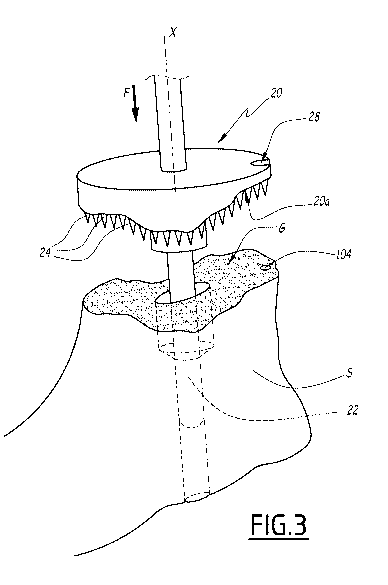
- figure 3 is a perspective view of the scapula of figure 2, in which is
inserted an
impacting device belonging to the surgical instrumentation assembly according
to the
invention;
- figure 4 is a sectional view of the scapula and impacting device of figure 3
during
impact;
- figure 5 is a sectional view of the scapula after impact and withdrawal of
the
impacting device;
- figure 6 is a sectional view of the scapula, and of a shoulder implant fixed
to the
scapula;
- figure 7 is a perspective view of the scapula and shoulder implant of figure
6.
Figure 1 shows a scapula S of a patient, the scapula S comprising a glenoid
cavity
G. The glenoid cavity G shows a damaged irregular surface which motivates the
attachment of an implant. The scapula S comprises a cortical bone area C,
which is the
outer and hard bone portion of the scapula S. The scapula S also comprises a
cancellous
bone area Ca, which is the inner and soft bone portion of the scapula S. The
cortical bone
C and the cancellous bone Ca are represented on figures 4 to 6.
Figure 1 also represents a drilling guide 1. The drilling guide 1 is patient
specific and
comprises a base plate 10 having an underside surface 10a which is congruent
to the
glenoid cavity G. The drilling guide comprises a tube 12 for inserting
drilling tools 3 and 4,
which are used for drilling holes in the scapula S. The tube 12 is centered on
a central
axis X. The tube 12 comprises a first section 12a, whose diameter is adapted
to receive a
bit 30 of the drilling tool 3 and a stop element 41 of the drilling tool 4.
The tube comprises
a second section 12b which has a reduced diameter adapted for insertion of the
bit 30 and
prevents further insertion of the stop element 41. The tube 12 comprises an
axial edge
12c which prevents insertion of a stop element 31 of the drilling tool 3.
The axial dimensions of the sections 12a and 12b along the axis X are
predetermined on the basis of the depth of a post and a screw used to attach a
shoulder
implant to the scapula S, which are patient-specific and determined using
medical imaging
technologies.
On figure 2, the scapula S is represented after drillings have been performed.
The
scapula S shows a first hole 100, drilled by the drilling tool 4, and a second
hole 102,
which is coaxial and adjacent to the hole 100, and which is drilled by the
drilling tool 3.
The hole 100 is adapted for insertion of a screw, while the hole 102 is
adapted for
insertion of the post of the shoulder implant.
CA 03018789 2018-09-21
WO 2017/165346
PCT/US2017/023305
According to an optional feature, the drilling guide 1 comprises a positioning
notch
13 provided on the base plate 10, and adapted to permit positioning of a
reference
marker. In the represented example, the reference marker is a hole 104 drilled
in the
scapula S. As a non-shown variant, the reference marker can be a pin, or the
like.
5 The
surgical instrumentation assembly also comprises an impacting device 20
having an underside surface 20a which is congruent to the surface of the
glenoid cavity G.
The underside surface 20a of the impacting device 20 is a negative surface of
the glenoid
cavity G.
The impacting device 20 comprises a post 22 made of two sections whose
diameters fit the diameters of the holes 100 and 102.
The impacting device 20 comprises protrusions 24, formed by spikes in this
example, which are provided on the underside surface and oriented along axis X
towards
the glenoid cavity G. The protrusions 24 form elongated elements protruding
from the
underside surface 20a along axis X. The protrusions 24 are adapted to
perforate the
cortical bone C upon impact of the impacting device 20 against the scapula S
by a one
sided translation movement, along axis X, as shown by arrow F.
Thanks to the orientation of the post 22 along axis X and to a rod 26 of the
impacting
device 20, which extends along axis X, and adapted to be handled by a
physician, the
impacting device 20 provides a one sided translation movement with respect to
the
scapula S.
As represented on figure 5, the protrusions 24 are adapted to create channels
106 in
the cortical bone C. The channels 106 extend towards the cancellous bone Ca.
The shape and spatial distribution of the protrusions 24 is arranged according
to
different densities, thicknesses and lengths determined on the basis of the
density and the
thickness of the cortical bone C. Depending on the properties of the cortical
bone C, which
are determined using imaging, such as CT scans, the shape and distribution of
protrusions 24 is determined so that the cortical bone C is properly
perforated during
impaction of the impacting device 20.
The length of the protrusions 24 can be comprised between 1 and 5 millimeters
depending on the thickness of the cortical bone C.
The thickness of the protrusions 24 can be comprised between 0.5 and 3
millimeters
depending on the hardness or density of the cortical bone C.
The density of the protrusions 24 can be comprised between 1 and 10
protrusions
per square centimeter depending on the hardness or density of the cortical
bone C.
The protrusions 24 are arranged and shaped so that thinner and longer
protrusions
24 are used where the cortical bone is thicker.
CA 03018789 2018-09-21
WO 2017/165346
PCT/US2017/023305
6
The impacting device 20 comprises a notch 28 adapted to be aligned with the
reference hole 104 provided on the glenoid cavity G.
Figures 6 and 7 represent a glenoid shoulder implant 5 comprising a base plate
52
and an underside portion 54 which is made of a porous material. Said porous
material
may, for instance, a metallic material, a ceramic material or a polymer
material, such as
plasma spray, titanium trabecular structure, or acid or laser etched surface
treatment. The
shoulder prosthesis implant is patient specific and the underside portion 54
comprises an
underside surface 54a which is congruent with the surface of the glenoid
cavity G. The
implant 5 comprises a post 56 extending along axis X and is adapted to receive
a screw
58, which is inserted in the hole 100 to attach the implant 5 to the scapula
S. The implant
5 comprises a hole 62 which runs through the base plate 52 and the underside
portion 54,
and which receives the screw 58.
The porous material of the underside portion 54 facilitates bone growth
induced by
the channels 106 in the cortical bone C. As represented on figure 6,
cancellous bone Ca
grows in the channels 106 inducing bone growth in the porosities of the
underside portion
54. Such bone growth improves the anchoring of the implant 5 in the scapula S.
Once the implant 5 is attached to the scapula S, a non-shown articulation
surface
can be fixed to the base plate 52, using non-shown screws which are inserted
in holes 60
provided in the base plate 52 and the underside portion 54. The holes 60
provide a guide
for drilling the scapula S to create holes 108 for inserting the screws in the
holes 60 and in
the scapula S.
The surgical shoulder repair method is implemented in the following manner.
The
characteristics of the scapula S of the patient are first determined using
imaging
technology. The shape of the glenoid cavity, the density, thickness and
hardness of the
cortical bone C, are used to design the drilling guide 1, the underside
surface 20a of the
impacting device 20 and the distribution and shape of the protrusions 24, and
the implant
5, including the shape of the underside surface 54a and the length of the post
56.
The glenoid cavity G is then prepared by removing, if necessary, remaining
cartilage
on the glenoid cavity G. The scapula S is then drilled using the drilling
guide 1. The
position of the drilling guide 1 is referenced using the notch 13. The glenoid
cavity G is
then impacted upon a one sided translational movement, using the impacting
device 20
positioned using the notch 28 and guided during the translational movement by
the post
22, to perforate of the cortical bone C and create channels 106 towards the
cancellous
bone Ca.
The patient specific implant 5 with its underside porous portion 54 and its
patient
specific underside surface 54a, is then attached to the scapula S using the
screw 58.
CA 03018789 2018-09-21
WO 2017/165346
PCT/US2017/023305
7
Bone growth in the porosities of the underside portion 54 may be accelerated
using bone
growth factors. The non-shown articulation surface may then be attached to the
implant 5.
The drilling guide 1 and the impacting device 20 may be disposed or recycled
after
completion of the surgical repair process.
