Note: Descriptions are shown in the official language in which they were submitted.
CA 03019241 2018-09-27
WO 2017/174558
PCT/EP2017/057946
1
A device for ultrasonic-accelerated hematoma lysis or thrombolysis of
intracerebral or
intraventricular hemorrhages or hematomas
Description:
Technical Field:
The present invention concerns a device for ultrasonic-accelerated hematoma
lysis or
thrombolysis of intracerebral or intraventricular hemorrhages or hematomas and
the use of such
a device for the treatment of intraventricular hemorrhages or hematomas.
The medical device according to the present invention is suitable for the
treatment of brain
hemorrhages.
Background Art:
Intracerebral hemorrhage (ICH) counts for 8 to 13% of all strokes world-wide
and results for a
wide spectrum of disorders. Intracerebral hemorrhage is more likely to result
in death or major
disability than ischemic stroke or subarachnoid hemorrhage. Intracerebral
hemorrhage usually
results from bleeding associated with amyloid angiopathy, tumors, hemorrhagic
conversion of
ischemic stroke, dural venous sinus thrombosis, vasculitis and vascular
malformations such as
cavernous angiomas, arteriovenous fistulae, arteriovenous malformations,
venous angiomas,
and aneurysms (Qureshi et al., Spontaneous intracerebral hemorrhage. N Engl J
Med. 2001b;
344:1450-60; Ruiz-Sanoval et al., Intracerebral hemorrhage in young people:
analysis of risk
factors, location, causes, and prognosis. Stroke: a journal of cerebral
circulation. 1999; 30:537-
41). ICH also results in hematomas that rupture or distort brain connections
and also affect
cerebral blood flow. These physical effects are generally termed the "mass
effect" (Keep et al.,
Intracerebral haemorrhage: mechanisms of injury and therapeutic targets.
Lancet Neurol. 2012;
11:720-31). For several decades there have been clinical trials of surgical
clot evacuation aimed
at reducing the mass effect. In a pig model of ICH, rtPA (recombinant tissue
plasminogen
activator) liquefied clots for aspiration, but there was also evidence of
delayed edema formation
(Rohde et al., Fibrinolysis therapy achieved with tissue plasminogen activator
and aspiration of
the liquefied clot after experimental intracerebral hemorrhage: rapid
reduction in hematoma
volume but intensification of delayed edema formation. J Neurosurg. 2002;
97:954-62) and an
increased inflammatory response (Thiex et al., The long-term effect of
recombinant tissue-
plasminogen-activator (rt-PA) on edema formation in a large-animal model of
intracerebral
hemorrhage. Neurol Res. 2003; 25:254-62). In this model, the hematoma could be
surgically
CA 03019241 2018-09-27
WO 2017/174558
PCT/EP2017/057946
2
removed without the use of rtPA, thereby reducing inflammation without having
an effect on
ICH-induced edema (Thiex et al., Minor inflammation after surgical evacuation
compared with
fibrinolytic therapy of experimental intracerebral hemorrhages. Neurol Res.
2005; 27:493-8).
Non-traumatic, spontaneous ICH is also associated with intraventricular
hemorrhages (IVH) in
40 to 50 % of all cases. IVH is associated with mortality rates of 30 to 80 %,
compared to 5 to
29 % for ICH without IVH (Hanley DF. lntraventricular hemorrhage: severity
factor and treatment
target in spontaneous intracerebral hemorrhage. Stroke. 2009; 40:1533-8). IVH
can result in
acute hydrocephalus by obstruction of the ventricular system or
extraventricular compression
from ICH (Lodhia et al. Hydrocephalus in a rat model of intraventricular
hemorrhage. Acta
neurochirurgica Supplement. 2006; 96:207-11; Zazulia AR. Hydrocephalus in ICH:
what do we
really know? Neurocritical care. 2008; 8:233-4). lntraventricular and
intracerebral clots have
been treated with rtPA (recombinant tissue plasminogen activator) by catheter-
directed
thrombolysis using ultrasound treatment (Newell et al., Minimally invasive
evacuation of
spontaneous intracerebral hemorrhage using sonothrombolysis. Journal of
neurosurgery. 2001;
115:592-601).
Spontaneous intracerebral hemorrhage thus occurs in a high number of patients
each year
without proven effective treatment. It is estimated that the annual incidence
of ICH is 10 to 30
cases per 100.000 persons per year, accounting for about 2 million strokes
annually worldwide
(Qureshi et al., Intracerebral haemorrahge. Lancet. 2009; 373:1632:-1644).
This condition is
fatal in 30 to 50 % of all occurrences, and the majority of survivors have
significant motor and
cognitive disabilities.
There are several different methods in order to conduct hematoma lysis or
thrombolysis of
intracerebral or intraventricular hemorrhages or hematomas, whereas ultrasound-
induced
hematoma lysis or ultrasound-accelerated thrombolysis appear to be promising
approaches for
treatment of such disorders. The catheter-based evacuation has been suggested
as a novel
surgical approach for the treatment of ICH. The safety and efficiency of
ultrasound was
evaluated in combination with recombinant tissue plasminogen activator (rtPA)
delivered for
micro catheter directly in spontaneous intraventricular (IVH) or intracerebral
(ICH) hemorrhage
in humans (Newell et al., Minimally invasive evacuation of spontaneous
intracerebral
hemorrhage using sonothrombolysis. Journal of neurosurgery. 2001; 115:592-
601). All patients
showed significant volume reduction in the treated hemorrhage, suggesting that
lysis and
drainage of spontaneous ICH and IVH with a reduction in mass effect can be
accomplished
rapidly and safely through sonothrombolysis using stereotactically delivered
drainage and
ultrasound catheters via a bur hole in the patient's skull. A ventricular
drainage catheter and an
CA 03019241 2018-09-27
WO 2017/174558
PCT/EP2017/057946
3
ultrasound infusion micro catheter were depicted prior to placement in an ICH.
A guidewire was
used for insertion.
Ultrasound-accelerated thrombolysis involves the breaking down or "melting" of
blood clots that
can form in arteries or veins. The methodology applies ultrasound energy along
with
thrombolytics (e.g. tPA) in order to accelerate the process of thrombolysis.
Acoustic energy
creates a pressure wave that results in a disruption of fibrin and other
components, and
ultrasound energy also helps to create a way for the thrombolytic compound to
get into the clot
by loosening up the fibrin cross-links by 'acoustic streaming'. Ultrasound
energy therefore alters
the shape of the fibrin network, thereby dissociating the clot directly.
A combined lysis of thrombus in brain ischemia using transcranial ultrasound
and systemic rtPA
was used by monitoring with a 2 MHz transcranial Doppler (TCD) (Alexandrov et
al.,
CLOTBUST: Design of a Randomized Trial of Ultrasound-Enhanced Thrombolysis for
Acute
lschemic Stroke, Journal of Neuroimaging, Volume 14, Issue 2, pages 108-112,
April 2004)
CLOTBUST: Design of a Randomized Trial of Ultrasound-Enhanced Thrombolysis for
Acute
lschemic Stroke. Journal of Neuroimaging, 14: 108-112). Image-guidance was
used in order to
improve catheter accuracy compared with a standard technique (Levitt et al.,
2012; Image-
guided cerebrospinal fluid shunting in children: catheter accuracy and shunt
survival, J
Neurosurg Pediatrics 10:112-117, 2012).
A common problem of catheter-based systems stems from draining hematomas which
block the
catheter system. Therefore, the catheter system must be frequently flushed
with a syringe,
which is only little efficient, and its application may also bear the danger
of infection and
cerebral abscess (Hoefnagel et al., Risk factors infections related to
external ventricular
drainage. Acta Neurochir; 150(3):209-214, 2008).
Therefore, common catheter-based systems that apply ultrasonic treatment are
not optimized
for efficient and protective treatment of ICH or IVH due to the fact that the
ultrasonic energy also
adversely affects surrounding tissue and also placement and guidance of the
catheters to the
ICH- or IVH-side still remains a problem.
WO 2016/007553 A describes an instrument that is specifically configured for
image guided or
stereotactic evacuation of intracerebral hemorrhage or other lesions. The
apparatus comprises
a cannula having a central channel terminating at an opening at a distal end
of the cannula. The
cannula is configured to be delivered through an aperture in a patient's skull
for delivery to a
treatment region within the cranium of the patient. A suction port is located
at a proximal
CA 03019241 2018-09-27
WO 2017/174558
PCT/EP2017/057946
4
location of the cannula and is in fluid communication with the central channel
for evacuating
fluid and/or debris from the target treatment region. An irrigation port is
disposed at a proximal
location of the cannula, the irrigation port is in fluid communication with
the distal end of the
cannula for delivering fluid to the target treatment region.
US 2012/0265123 Al describes an apparatus and a method to deliver ultrasound
energy
through the drain to dissolve hemorrhages and debris occluding the drain lumen
and ports.
Alternative devices are described in CN103083088 A which suggests a portable
positioning
device for intracerebral cathetering for treatment of cerebral hemorrhages.
Positioning of the
device is accomplished by computed tomography (CT) such that the device can
accurately
reach the puncture site.
CN201379823 Y describes a device for draining a hematoma of hypertensive
cerebral
hemorrhage (HCH) which uses two rows of drainage holes that are in fluid
communication with
an intracerebral hematoma drainage silicone tube. A metal lead pin is used to
be inserted into
the intracerebral hematoma drainage silicone tube.
JP2006043200 A describes a support system that is equipped with an image
acquiring device
for acquiring a brain image of a patient, a cerebral region detection part for
determining a
cerebral region from the brain image and a wrinkle range detection part for
detecting a region of
the brain wrinkle and an uneven region of the brain by determining the
difference between a
convex closure in the cerebral region and the brain region for the treatment
of ICH. A blood
vessel removing part is used for removing a blood vessel region from the
wrinkle region.
US 2008/319376 Al describes an ultrasound catheter with fluid delivery lumens,
fluid
evacuation lumens and a light source used for the treatment of intracerebral
hemorrhages. The
lumens can be used to deliver a fluid to a treatment site and/or to evacuate a
fluid from around
the treatment site.
US 5,318,518 describes an irrigating catheter for eliminating solids from body
organs and hollow
body organs or cavities of man and animals. The catheter consists of a
catheter body having at
least a first lumen and a second lumen, wherein the first lumen transports
fluid to the organs
and the second lumen discharges irrigating fluid from the organs.
The known catheter-based devices and methods are laborious and expensive and
often result
in a blockade of the catheter, which affects a rapid pressure release of the
brain and drainage of
CA 03019241 2018-09-27
WO 2017/174558
PCT/EP2017/057946
toxic metabolites from the hematoma. Thrombolytic substances such as rtPA will
become less
efficient in mature hematomas. Therefore, a rapid drainage of fluid and debris
is crucial for the
success for hematoma lysis or thrombolysis of ICH or IVH. On the other side,
self-flushing
systems suffer from the problem that they cannot be positioned or navigated to
the puncture site
5 .. efficiently. In addition, although ultrasonic treatment has been proven
to increase efficiency of a
lysis therapy, transcranial ultrasonic has also been associated with increased
bleedings. None
of the known devices or methods is optimized to remain intracerebrally and
intraventricularly for
a prolonged period of time to mediate drainage or as monitoring system.
In studies that use ultrasonic-accelerated hematoma lysis or thrombolysis of
artery occlusion,
treatment of strokes occurs unselectively and/or affects healthy areas within
the tissue, resulting
in an increased risk of bleedings, tissue damages, blood vessel damages, in
particular in areas
of the cerebral infarct (Nedelmann et al., Therapeutic ultrasound of acute
cerebral artery
occlusion. Der Nervenarzt. 2008;79(12):1399-400, 402-6; Alexandrov AV et al.,
Design of a
randomized trial of ultrasound-enhanced thrombolysis for acute ischemic
stroke. J
Neuroimaging 2004; 14(2): 108-112).
Disclosure of Invention:
It is therefore the object of the present invention to provide an improved
medical device for
treatment of intracerebral or intraventricular hemorrhages or hematomas that
minimizes
negative side effects that are associated with the application of ultrasonic
on tissues, blood
vessels or hematomas.
.. This object is solved by a medical device according to the present
invention.
The device of the present invention combines an ultrasonic-mediated imaging
system, pressure
sensoring means in the application of ultrasonic in the treatment of
intracerebral or
intraventricular hemorrhages or hematomas (ICH or IVH). The device comprises a
body that
accommodates a number of separated compartments or lumens consisting of a
flushing
catheter for flushing fluid and/or pharmaceutically active substances into the
intracerebral or
intraventricular hemorrhages or hematomas, a drainage catheter for draining
fluid from the
intracerebral or intraventricular hemorrhages or hematomas, an ultrasonic
probe duct and a
pressure sensor duct. The ultrasonic probe duct can contain an ultrasonic
probe or a stiletto (or
stylet), and the pressure sensor duct integrates a pressure sensor to promote
treatment of ICH
and IVH. The stylet enables quick freehand navigated placements of the
ventricular
catheter.
CA 03019241 2018-09-27
WO 2017/174558
PCT/EP2017/057946
6
The device according to the present invention allows efficient drainage of
fluid and debris from
the ICH or IVH site and a site-directed application of thrombolytics such as
rtPA over the lumen
of the flushing catheter. At the same time, the pressure sensor arranged in
the present sensor
duct allows monitoring of the intracranial pressure during flushing and lysis.
In a further lumen of
the body which forms the ultrasonic probe duct, an endosonographic probe,
preferably a 1OF
endosonographic probe, is integrated for B-scan imaging, Doppler and duplex
measurements.
The endosonographic probe provides high-definition imaging of intracranial
structures and
allows guiding of the device to the ICH- or IVH-sites. Ultrasonic-guided
imaging allows analysis
of blood flow and the lysis of hematomas in intracerebral and intracranial
vessels. The
endosonographic probe thus allows imaging to monitor evolution of ICH and
eventually a
resulting brain edema in real time. In the context of the present invention,
the endosonographic
probe is thus used both for imaging and treatment of ICH and IVH and allows
monitoring of the
progress of hematoma lysis.
Endosonography using the ultrasonic probe of the invention further allows the
identification of
bleedings, the presence of edemas and eventual side effects on surrounding
brain components
including, but not limited to compression of cerebral ventricles or a
displacement of the cerebral
midline. Ultrasonic imaging also replaces parts of COT (Cranial Computed
Tomography)
diagnostics and can be applied to all sites of intracerebral or
intraventricular hemorrhages or
hematomas over a prolonged period of time (e.g. up to 2 - 3 weeks). This
significantly reduces
CCT-dependent radiation impact on the patient.
In a preferred embodiment, the outer wall of the ultrasonic probe duct in the
lower distal part
portion of the body further comprises a membrane, which is permeable for
ultrasound. The
membrane can be integrated fully or partially in the catheter wall to emit
ultrasonic energy.
Furthermore, it is preferred that the endosonographic probe is rotatable
within the ultrasonic
probe duct. If the ultrasonic-permeable membrane is integrated only partially
in the ultrasonic
duct, it may be preferable to rotate the whole catheter in order to achieve an
unidirectional
emission of ultrasonic energy.
The lumen of the ultrasonic probe duct not only allows integration of the
endosonographic
imaging and treatment probe, but also of a stiletto that can be
interchangeably guided within the
ultrasonic probe duct. The stiletto or stylet is preferably inserted at the
time of implantation of
the device of the invention. The stiletto provides a higher stiffness for
puncture through the
cerebral tissue and the hematoma and therefore helps in guiding the device for
neuronavigation
for an image-guided placement of the catheter system to the center of ICH or
IVH, at the same
CA 03019241 2018-09-27
WO 2017/174558
PCT/EP2017/057946
7
time conserving surrounding cerebral tissue. Upon placement of the catheter
system at the
target site, the stiletto is replaced by an ultrasonic probe.
Neuro-navigated placement of the device is mediated through a puncture site or
hole in the skull
to the center of the hematoma. In a preferred embodiment, the outside part of
the body of the
device of the present invention comprises a connector for cranial fixation,
which is adapted to
allow angular adjustment of the body both in longitudinal and/or transverse
direction. The
connector is preferably provided as a connector block that integrates the body
with
compartments or lumens of the device of the present invention. The connector
allows a free
movement at different angles, an adjustment of the catheter trajectory within
the port system
and an entanglement of the body relative to the port. After adjustment of the
catheter, the
connector can be locked into position.
The endosonographic probe further allows variation of the ultrasonic frequency
in order to limit
depth of penetration and fast tissue exposition for ultrasonic energy. A
minimization of
penetration depth to the target sites of ICH and IVH reduces complications and
side effects
such as bleedings. At the same time, the knowledge about the exact position
and the provision
of coordinates provide valuable information on the form and volume of
bleedings. Ultrasonic ICH
or IVH can be adjusted in order to influence sonographic peak pressure and
penetration depth
of sonic energy. All these measures contribute to a minimization of focus
energy specifically to
the location of the intracerebral or intraventricular hemorrhages or hematomas
and a protection
of surrounding healthy tissues.
Preferably, hematoma lysis is carried out at ultrasonic frequencies between
5,5 to 10 MHz. A
preferred frequency of 10 MHz showed the safest modalities (MI (mechanical
index) of 0.55)
and the best imaging. Ultrasonic treatment and the additional provision of
pharmaceutically
active substances such as rtPA via the flushing catheter into the
intracerebral or intraventricular
hemorrhages or hematomas are preferred in order to dissociate associated
clots. The device of
the present invention allows high-detailed multimodular intracranial
endosonographic imaging in
real time and simultaneous flushing of pharmaceutically active substances into
the ICH or IVH
over prolonged periods of time, wherein at the same intracerebral or
intraventricular fluid from
the hemorrhages or hematomas is drained by the lumen of a separate drainage
catheter which
is part of the body. Intracerebral or intraventricular pressure is measured
during the procedure
by a pressure sensor that is integrated within a pressure sensor duct, and
which is also part of
the body of the device of the present invention.
The flushing catheter, the drainage catheter, the ultrasonic probe duct and
the pressure sensor
CA 03019241 2018-09-27
WO 2017/174558
PCT/EP2017/057946
8
duct of the device of the present invention are essential parts of the basic
body and thus, these
elements are integrated in a single unit that allows better handling of the
device during surgery
and treatment. As part of the unit, the flushing catheter, the drainage
catheter, the ultrasonic
probe duct and/or the pressure sensor duct are arranged in proximity to each
other and all
compartments or lumens are at least sectionally isolated by fluid-tight
catheter walls. The
ultrasonic probe duct is configured to integrate the endosonographic probe or,
if required, the
stiletto for implantation.
In order to improve the clinical application of the device and the
supply/drainage of substances
to/from the catheters, the upper proximal part of the flushing catheter and/or
drainage catheter
preferably laterally projects from the body in bended form. The lower distal
part of the flushing
catheter preferably disembogues into the drainage catheter in that the foot
end of the catheter is
permeably connected by means of one or more penetrations. For draining fluid
from the ICH or
IVH, the drainage catheter at the lower distal part of the catheter comprises
apertures at the
outer wall that allow uptake of the fluid and debris from the hematoma into
the catheter lumen
for subsequent transport to a collecting container. In the upper proximal part
of the body of the
device, the surrounding catheter wall of the drainage is fully closed and does
not contain
apertures to allow drainage of fluid or debris to the outlet of the drainage
catheter. For accurate
measurements, the pressure sensor is preferably arranged opposite to the
flushing catheter
within the body of the device. Preferably, the pressure sensor duct is
arranged in the outer
diameter of the body. The ultrasonic probe duct is arranged oppositely to the
drainage catheter,
wherein at least a section of the flushing catheter is arranged between the
drainage catheter
and the ultrasonic probe duct.
The optional stiletto (stylet) that is guided during implantation of the
device within the ultrasonic
probe duct is preferably equipped with a marker of neuro navigation. Preferred
markers of neuro
navigations are disposable marker spheres, preferably coated with IR-light,
retro-reflective foil,
passive markers, or multi-modality fiducial markers. In a preferred
embodiment, the neuro
navigation markers are passive marker plates incorporated in the stylet. The
marker geometry is
automatically recognized by a navigation camera for quick calibration.
Preferred reflective
markers are plastic spheres with a glass-grain coating to reflect infrared
light emitted by one or
more cameras. The reflection is detected by detectors arranged around the
cameras.
In a preferred embodiment, the pressure sensor is part of a pole with a
diameter which allows
guidance of the pole within the pressure probe duct. Preferably, the pressure
sensor is arranged
at the cone end of the pole, i.e. at the lower part of the body.
CA 03019241 2018-09-27
WO 2017/174558
PCT/EP2017/057946
9
The present invention also concerns a method for treatment of intracerebral or
intraventricular
hemorrhages or hematomas (ICH or IVH) via ultrasound-accelerated hematoma
lysis
thrombolysis by applying a device (catheter system) of the invention that
comprises a body,
which accommodates a number of separate compartments or lumens. The invention
also
concerns the use of such a device for treatment of intracerebral or
intraventricular hemorrhages
or hematomas, wherein the application of a device comprises the following
steps:
- flushing fluid and/or pharmaceutically active substances by means of a
flushing catheter
which forms an integral part of the body of the catheter system into the
intracerebral or
intraventricular hemorrhages or hematomas;
- draining fluid from the intracerebral or intraventricular hemorrhages or
hematomas by
means of a drainage catheter which forms an integral part of the body of the
catheter
system;
- guiding a stiletto within the ultrasonic probe duct which is arranged next
to the flushing
catheter and the drainage catheter in the body of the catheter system for
intracranial
placement of the catheter system;
- changing the stiletto against an endosonographic probe and guiding the
endosonographic
probe within the ultrasonic probe duct, and
- inserting a pressure probe into a pressure sensor duct of the body of the
catheter system for
monitoring intracranial pressure during flushing and hematoma lysis.
In a preferred embodiment, the pharmaceutically active substances are
thrombolytics such as
recombinant tissue plasminogen activator (rtPA), streptokinase, p-anisoylated
lys-plasminogen-
streptokinase activator complex, urokinase, and prourokinase. The tissue
plasminogen activator
compounds include Alteplase (tPA), Reteplase (sometimes called rPA), and
Tenecteplase. The
endosonographic probe is used both for ultrasonic lysis and real time imaging
of the ICH or IVH.
The device and the methods of the present invention allow a more efficient
ultrasonic-mediated
ICH or IVH lysis, thereby protecting surrounding tissue. Hematoma drainage,
catheter flushing
and rtPA application are carried out via different separate catheter lumens
within the body of the
device, wherein a separate pressure sensor duct bears a pressure sensor in the
lower part of
the body in order to monitor intracranial pressure during flushing and lysis.
One advantage of the device of the present invention is that it can be left in
the cerebrum
following lysis therapy to monitor intracranial pressure and for drainage
fluid from the ICH or IVH
site over the drainage catheter as integral part of the device of the
invention. Ultrasonic-
mediated imaging and ultrasonic-accelerated hematoma lysis or thrombolysis
allow a real-time
imaging and monitoring of the hematoma lysis process. The combination of
navigation-mediated
CA 03019241 2018-09-27
WO 2017/174558
PCT/EP2017/057946
and angle-controlled integration of the device into the ICH or IVH and the
implementation of a
removable stiletto are required for accurate placing of the catheter system in
the center of the
ICH or IVH. An additional connector allows an angular adjustment of the body
in longitudinal
and/or transverse direction; the device thereby can be placed in optimal
orientation relative to
5 the hematoma. This allows an efficient draining of the trajectory and
simultaneous protection of
important tissues and brain areas.
Best mode for carrying out the invention:
10 The present invention is illustrated in more detail in the accompanying
figures.
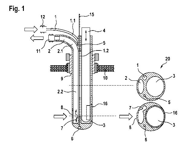
Fig. 1 shows an embodiment of the sonothrombolytic catheter system according
to the present
invention. The device is comprised of a body 20 that accommodates different
compartments or
lumens that are arranged in proximity to each other and are separated from
each other by
respective walls. Basically, the device of the invention contains several
catheters forming a
catheter system that includes different functions. The first compartment or
lumen is part of a
flushing catheter 1 for flushing fluid and/or pharmaceutically active
substances into the
intracerebral or intraventricular hemorrhages or hematomas. Preferably, rtPA
or other
thrombolytic substances are applied to the site of ICH or IVH by means of the
flushing catheter
1. The body 20 further accommodates a compartment or lumen of a drainage
catheter 2 for
draining fluid from the ICH or IVH. The body 20 also comprises a compartment
or lumen of an
ultrasonic probe duct 3 and a separate pressure sensor duct 5. The lumen of
the pressure
sensor duct 5 integrates a pressure sensor 6, preferably at the cone end of a
pole 15 for
measurement and analysis of intracranial pressures during implantation and
treatment of ICH or
IVH. The ultrasonic probe duct 3 both comprises either an endosonographic
probe 4 (Fig. 1) or
a stiletto 13 (Fig. 2) that can be interchangeably guided within the
ultrasonic probe duct 3. The
upper proximal portion 1.1 of the flushing catheter 1 and/or the upper
proximal portion 2.1 of the
drainage catheter 2 laterally project from the body 20 in bended form. The
lower distal portion
1.2 of the flushing catheter 1 and/or the lower distal portion 2.2 of the
drainage catheter 2 are
provided in straight form.
In the lower distal part of the body 20, the outer wall of the drainage
catheter 2 comprises one or
more apertures 8. Fluid or debris is flushed from the site of ICH or IVH
through the catheter
lumen to the upper end of the drainage catheter 2. The upper proximal portion
1.1 of the
flushing catheter 1 and/or the upper proximal portion 2.1 of the drainage
catheter 2 is equipped
with adapters 11 or ports 12, respectively.
CA 03019241 2018-09-27
WO 2017/174558
PCT/EP2017/057946
11
In the lower part of the body 20, the lumen of the flushing catheter 1 is
penetrated such that
flushing fluid from the flushing catheter 1 flows into the lower distal part
portion 2.2 of the
drainage catheter 2 through one or more penetrations 7.
The body 20 also comprises a connector 9 for cranial fixation, which is
adapted to allow an
angular adjustment of the body 20 in longitudinal and/or transverse direction.
The connector 9 is
integrated into a hole of a skull 10.
In a preferred embodiment, the circular lateral wall of the ultrasonic probe
duct 3 further
comprises a membrane 16, which is permeable for ultrasound. Furthermore, it is
preferred that
the endosonographic probe 4 is rotatable within the ultrasonic probe duct 3.
Fig. 2 shows a replacement of the endosonographic probe 4 by a stiletto 13
which is required
for implantation into the ICH or IVH. The stiletto 13 provides stiffness for
the implantation of the
catheter system and also may comprise additional markers of neuronavigation 14
at the upper
end, such as reflective marker spheres.