Note: Descriptions are shown in the official language in which they were submitted.
CA 03019347 2018-09-27
WO 2017/213789 PCT/US2017/032065
FILTER AND OCCLUDER SYSTEMS AND ASSOCIATED METHODS AND DEVICES
SUMMARY
[0001] Various systems, methods, and devices according to the present
disclosure are usable as flow devices, also described as filters or occluders,
such terms
being used interchangeably herein unless otherwise indicated by device
application.
[0002] Some aspects of the disclosure relate to filters that remain
patent for an
extended period of time in comparison to traditional filters. Such filters may
be
applicable for protecting against embolic release during complex endovascular
procedures or other filtration applications.
[0003] Some aspects of the disclosure relate to flow devices that remain
patent
for a desired period of and eventually become less patent and, if desired,
fully or nearly
fully occlusive over time. Such occluders may find use in a variety of
applications,
including techniques for reducing the patency of one or more blood vessels,
apertures
of grafts/stent-grafts, or branches of grafts/stent-grafts over time, as well
as others.
[0004] Some aspects of the disclosure relate to flow devices that are
capable of
being collapsed and removed from the vasculature from either a distal or a
proximal
approach direction (e.g., antegrade or retrograde directions) to facilitate,
for example,
intravascular removal of the devices from different access locations.
[0005] Some aspects of the disclosure relate to flow devices that are bi-
directionally deployable, where such devices can be deployed in a distal-to-
proximal
end or a proximal-to-distal end orientation to facilitate, for example,
intravascular
deployment of the devices from different access locations.
[0006] Some aspects of the disclosure relate to flow systems including a
plurality
of flow devices deployed and left in place to provide such advantages as
enhanced
protection against post-operative complications, including embolisms, for
example.
[0007] Some aspects of the disclosure relate to methods of making and
methods
of treatment using the flow devices and systems described herein, including
applications in which flow devices are implanted in the body for an extended
period of
time (e.g., including after conclusion of a primary treatment procedure, such
as EVAR)
and later retrieved from the body after a desired time period.
[0008] While multiple examples are disclosed, still other examples will
become
apparent to those skilled in the art from the following detailed description,
which shows
and describes illustrative examples. Accordingly, the drawings and detailed
description
are to be regarded as illustrative in nature and not restrictive.
CA 03019347 2018-09-27
WO 2017/213789 PCT/US2017/032065
BRIEF DESCRIPTION OF THE DRAWINGS
[0009] FIG. 1 illustrates a retrieval system and associated flow device,
according
to some examples.
[0010] FIG. 1A illustrates a portion of the flow device of FIG. 1,
according to
some examples.
[0011] FIGS. 2A-2F illustrate flow media, according to some examples.
[0012] FIGS. 3 and 4 illustrate another flow device, according to some
examples.
[0013] FIG. 5 illustrates another flow device, according to some
examples.
[0014] FIG. 5A illustrates a portion of the flow device of FIG. 5,
according to
some examples.
[0015] FIGS. 6-8 illustrates additional flow devices, according to some
examples.
[0016] FIGS. 9-12 illustrate a flow device with a flow reversion device
inserted
and deployed in the flow device, according to some examples.
[0017] FIG. 13 illustrates a system of flow devices deployed in the
aortic arch,
according to some examples.
[0018] FIG. 14 illustrates a retrieval system and associated flow device,
according to some examples.
DETAILED DESCRIPTION
[0019] Various aspects of the instant disclosure relate to flow devices,
also
described as filters or occluders, for modifying flow (e.g., filtering,
reducing, and/or
occluding flow) in body conduits, such as blood vessels. In some examples,
such
devices achieve relatively low porosity while maintaining patency for desired
periods of
time, including extended periods of time following implantation and associated
procedure cessation. Some examples of device applications include use for the
prevention of stroke, ischemic bowel, reduced renal function, distal
peripheral artery
occlusion, internal iliac occlusion, inferior mesenteric occlusion, selective
filtering and/or
occluding of implant branches (e.g., stent graft branches) and others that
this disclosure
will make apparent, such as partial or total occlusion of the gastric arteries
for the
treatment of obesity.
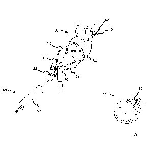
[0020] FIG. 1 shows a flow device 10 and retrieval system according to
some
examples. As shown, the flow device 10 includes a support frame 12 and a flow
media
14. The device 10 is configured for implantation in one or more body lumens
and can
have an outer diameter between 3 mm and 20 mm, although a variety of
dimensions
are contemplated.
2
CA 03019347 2018-09-27
WO 2017/213789 PCT/US2017/032065
[0021] The support frame 12, also described as a support structure, is
optionally
formed of a shape memory material, such as a nickel-titanium alloy, although a
variety
of materials, such as stainless steel or suitable polymeric materials are
contemplated. If
desired, the support frame 12 is formed as a cut tube (e.g., a laser cut tube)
that is
collapsible to an elongated, smaller diameter profile (not shown) for
intraluminal
deployment using a delivery system (e.g., a delivery catheter). If desired,
the support
frame 12 is optionally formed of discrete wires, for example using one or more
mandrel
wire forming operations. Although some examples are provided, a variety of
frame
shapes, materials, and manufacturing methods are contemplated, including those
disclosed in U.S. 8,668,714 (Cully et al.), issued March 11, 2014. In some
examples,
the support frame 12 is configured to self-expand or to be expanded (e.g., via
balloon)
to engage the wall of the body lumen into which it is deployed (e.g., against
a blood
vessel wall, such as the aortic arch) to anchor the device 10 in place.
[0022] As shown in FIG. 1, the support frame 12 includes a proximal
portion 20, a
distal portion 22 and an intermediate portion 24 between the proximal and
distal
portions 20, 22. As shown, the support frame 12 is generally formed of a
plurality of
frame members 30, also described as struts 30. The frame members 30 are
optionally
portions of a cut tube, discrete wires wound or coupled together, or of
another design as
desired. The support frame 12 is optionally self-expanding or expandable
(e.g., balloon
expandable) as desired.
[0023] The proximal portion 20 includes a first capture feature 32, and
tapers
conically away from the intermediate portion 24. An enlarged view of the
capture
feature 32 is shown in FIG. 1A.
[0024] As shown in FIG. 1A, the capture feature 32, also described as a
coupling
means 32, extends from the plurality of struts 30 and forms a generally
spherical shape
(e.g., round spherical), although a variety of shapes (e.g., oblong spherical)
are also
contemplated. In some examples, for example when formed by laser cutting, the
capture feature (or capture element) includes one or more relief cuts to
facilitate forming
the capture feature 32 into a desired shape (e.g., similar to a bell, or
"jingle bell").
Though largely obscured in FIG. 1A, a first radiopaque marker 34 (e.g., a
discrete piece
of radiopaque material) is optionally received and retained in the first
capture feature 32.
The first radiopaque marker 34 is optionally used to assist with placement of
the device
during a deployment operation and/or to recover the device 10 during a
recovery, or
retrieval operation, for example.
3
CA 03019347 2018-09-27
WO 2017/213789 PCT/US2017/032065
[0025] As shown in FIG. 1, and though partially obscured by the flow
media 14,
the intermediate portion 24 of the support frame 12 is generally cylindrical
in shape,
although a variety of shapes (e.g., tapered, hourglass, dog bone, and others)
are
contemplated.
[0026] The distal portion 22 of the support frame 12 is shown in FIG. 1
largely
covered by the flow media 14. In some examples, the distal portion 22 of the
support
frame 12 tapers conically to a second capture feature 40. As shown in FIG. 1,
the
tapered distal portion 22 supports the flow media 14 in a corresponding
conical shape
shown in FIG. 1. The second capture feature 40, also described as a coupling
means
40, is optionally substantially similar to the first capture feature 32. For
example, the
second capture feature 40 can be similarly shaped and formed to the first
capture
feature 32 and also includes a second radiopaque marker 42, although a variety
of
configurations are contemplated. If desired, the first and second capture
features 32,
40 and/or the first and second radiopaque markers 34, 42 are distinct from one
another,
for example having different radiopacities, shapes, materials, coatings, or
otherwise
being distinguishable from one another.
[0027] As shown in FIG. 1, the flow media 14 includes a first portion 50
received
over an outside surface of the intermediate portion 24 of the support frame 12
and a
second portion 52 received over the distal portion 22 of the support frame 12.
The flow
media 14 is optionally described as a porous fabric, where the term "porous
fabric" is
generally meant to indicate a layer of material configured to permit at least
some level of
fluid passage (having a desired fluid permeability) through one or more flow
pathways
or "pores" in the material.
[0028] As shown in FIG. 1, the first portion 50 is received outside of
the
intermediate portion 24. Though shown outside the intermediate portion 24, a
variety of
configurations are contemplated, including the first portion 50 being received
on an
inner surface of the intermediate portion 24, embedded with the intermediate
portion 24,
comprising multiple layers or parts sandwiching the intermediate portion 24,
and others.
In some examples, the first portion 50 of the flow media 14 is substantially
continuous,
where the first portion 50 may be substantially impermeable or permeable, or
have any
desired permeability to gases or water, blood, bile, or other bodily fluids as
desired. In
some examples, the first portion 50 is formed of one or more layers of
expanded PTFE
film adhered (e.g., by FEP applied to the film and/or support frame 12) or
otherwise
secured to the support frame 12 (e.g., by suturing, friction fit, or other
means for
securing).
4
CA 03019347 2018-09-27
WO 2017/213789 PCT/US2017/032065
[0029] According to some examples, the second portion 52 of the flow
media 14,
also described as the filtration portion 52 or flow control portion 52,
includes a plurality
of openings such that the second, or flow control portion 52 is permeable to
fluid flow,
for at least an initial desired time period. The flow control portion 52 is
optionally
configured to capture particulate or other substances in a fluid passing
through the flow
control portion 52. For example, with blood, it may be desirable to capture
plaque
debris, blood clot debris, or other content. As described in greater detail
below, in some
examples, one or more portions of the device 10 (e.g., the flow control
portion 52)
includes drug coatings, surface treatments (e.g., such as the surface
treatment
marketed under the tradename "CBAS" by W. L. Gore & Associates), or other
modification(s) to facilitate a breakdown of material caught in the flow
control portion 52.
[0030] In some examples, the device 10 is configured to be delivered "off-
the-
wire," without riding on a guidewire captured within a lumen of the device.
However, as
discussed further below, in some examples, one or more guidewires may be
utilized
during delivery of the device 10. In some examples, the device 10 can be
deployed
using well known intravascular catheter techniques from a compacted delivery
profile to
an expanded deployed profile. In at least this manner, the device 10 can be
left in the
body following a procedure or a portion of a procedure without the need of
removing a
guidewire from the device 10 and/or removing the device 10 with an associated
treatment device, such as an associated balloon catheter or stent-graft
deployment
system. Moreover, multiple devices can be deployed from a single delivery
system at
different delivery sites using such an "off-the-wire" approach. Generally,
push/pull
delivery catheters, constraining sheaths, and other delivery systems are
contemplated
for deploying the devices as desired.
[0031] FIG. 1 also shows a capture system 60 including a guide catheter
62 and
a snare catheter with a retractable loop 64. As indicated in FIG. 1, the
capture system
60 is optionally used to capture the first capture feature 32 at which point
the device can
be withdrawn and collapsed into the guide catheter 62 or another collapsing
feature for
withdrawal or position adjustment of the device. Additionally, the capture
system 60 or
a similar capture system is optionally utilized to capture and retrieve the
device 10 using
the second capture feature 40. In other words, the device 10 configuration
facilitates
retrieval and removal and/or repositioning of the device 10 from either distal
or proximal
approaches, also described as ante- or retrograde approaches in terms of flow.
Thus, a
user of the device 10 and capture system 60 is able to approach the device
from
different vascular entry points, or directions within a body lumen, as
desired.
CA 03019347 2018-09-27
WO 2017/213789 PCT/US2017/032065
[0032] FIGS. 2A ¨ 2F show a variety of potential configurations for the
openings
in the flow media 14, such as the flow control portion 52. Openings may be
formed by
removal processes (e.g., cutting or etching) films, sheets, membranes, or
other
materials. Openings may also be formed by weaving, knitting, or other
techniques
using individual or multi-fiber strands, or using other materials and/or
methods as
desired.
[0033] FIG. 2A shows a square lattice structure, such as those described
in U.S.
Pub. US 2013/0204347 ("Armstrong et al"), published August 8, 2013 usable for
the
flow media 14. FIG. 2B shows a modified lattice structure in which the
openings are
offset and rectangular, according to some examples of one or more portions of
the flow
media 14 (e.g., flow control portion 52). FIG. 2C shows a series of slits with
a desired
length, depth, and separation; FIG. 2D shows a series of ovular, or oval-
shaped
openings of a desired length, width, number and separation; FIG. 2E shows
generally
round openings with a desired diameter and separation; and FIG. 2F shows a
series of
random, irregular openings formed by an irregular fibrous structure; each of
the
foregoing provide just a few examples of configurations of one or more
portions of the
flow media 14 (e.g., configurations of the flow control portion 52).
[0034] The openings generally define a porosity level of the flow media
14. For
example, in some examples, the porosity level is defined as an average or
maximum
diameter or dimension of the openings being 500 microns, 400 microns, 300
microns,
200 microns, 100 microns, or other dimension. In some implementations (e.g.,
where
occlusion is desirable), the porosity level is defined as an average or a
maximum
diameter or dimension of the openings being less than 100 microns, such as 50
microns, 10 microns, or 5 microns, for example. The porosity level can also be
defined
as the openings being configured to filter down to 500 micron, 400 micron, 300
micron,
200 micron, or 100 micron or other maximum or average particle size. In some
implementations, (e.g., where occlusion is desirable), the porosity level of
the flow
media 14 is defined as the openings being configured to filter down to a
maximum or
average particle size of less than 100 micron particles sizes, such as 50
micron, 10
micron, or 5 micron particle sizes, for example.
[0035] In some examples, the flow control portion 52 is configured to
remain at a
desired patency level for a desired time period (e.g., minutes, hours, days,
weeks, or
months). In some examples, this facilitates use of the device 10 to remain
implanted
following completion of the primary procedure (e.g., EVAR) and to reduce the
incidence
of postoperative complications (e.g., stroke from embolic debris) by remaining
in the
6
CA 03019347 2018-09-27
WO 2017/213789 PCT/US2017/032065
body following completion of the procedure for a desired period (e.g.,
maintaining a
desired patency for a period of between 12 hours and a 7 days). In some
examples,
this extended patency helps allow controlled occlusion of vessel and/or
portion of an
endovascular device (e.g., stent graft) to reduce issues (e.g., system
circulatory issues)
associated with immediate or near immediate occlusion of such pathways. For
example, a more gradual occlusion or "low flow" occlusion may permit the body
to
accommodate such partially or reduced flow in the body vessel and thereby
reduce
negative physiologic impact.
[0036] The flow media 14 or a portion thereof (e.g., the flow control
portion 52 of
the flow media 14) is optionally provided with one or more treatments (e.g.,
the heparin-
based treatment provided by W.L. Gore and Associates under the trade name
"CBAS")
to maintain device patency for a desired period of time. In some examples, the
flow
control portion 52 is formed of expanded PTFE material or other fluoropolymer,
although any of a variety of biocompatible biomaterials are contemplated.
Various
adjustments can be made to the material as desired, including the number and
type of
material layers (e.g., expanded PTFE microstructure, density, layer-to-layer
variations)
and opening configurations (size, spacing, shape, and others) in order to
achieve a
desired patency, or flow vs. time profile for the device 10. In some examples,
the
desired patency is defined in terms of a minimum volumetric flow rate through
the
device 10 over the desired time period. The desired patency can also be
described in
terms of a minimum percentage of the initial volumetric flow rate exhibited by
the device
at the time of implantation over the desired time period (e.g., at around
100%, 90%,
80%, etc.).
[0037] FIGS. 3 and 4 show another flow device 110, according to some
examples. FIG. 3 is an isometric representation and FIG. 4 is generally a
side, partial
sectional representation of the device 110. As shown, the device 110 includes
various
features similar to those of the device 10. For example, the device 110
includes a
support frame 112 and a flow media 114. The support frame 112 also includes a
proximal portion 120, a distal portion 122 and an intermediate portion 124
between the
proximal and distal portions 120, 122. As shown, the support frame 112 is
generally
formed of a plurality of frame members 130, also described as struts 130. The
frame
members 130 are optionally portions of a cut tube, discrete wires wound or
coupled
together, or of another design as desired. The support frame 112 is optionally
self-
expanding or expandable (e.g., balloon expandable) as desired.
7
CA 03019347 2018-09-27
WO 2017/213789 PCT/US2017/032065
[0038] The proximal portion 120 is optionally conically tapered and
extends to a
first capture feature 132, also described as a coupling means.
[0039] As shown in FIGS. 3 and 4, the capture feature 132 is optionally
substantially similar to the capture features (or capture elements) previously
described
in association with device 10, although a variety of designs are contemplated.
[0040] As shown in FIGS. 3 and 4, the intermediate portion 124 of the
support
frame 112 is generally cylindrical in shape, although a variety of shapes
(e.g., tapered,
hourglass, dog bone, and others) are contemplated.
[0041] As shown in FIGS. 3 and 4, the distal portion 122 of the support
frame 112
tapers conically to a second capture feature 140. As described below, the
proximal and
distal portions 120, 122 receive the flow media 114 depending upon a position
of the
flow media 114 as dictated by flow direction.
[0042] The second capture feature 140 is optionally substantially similar
to the
second capture feature 40, although a variety of configurations are
contemplated.
[0043] As shown in FIGS. 3 and 4, the flow media 114 includes a first
portion 150
received on an inside surface of the intermediate portion 124 of the support
frame 112.
The first portion 150 is optionally substantially cylindrical, or tubular in
shape. Though
shown outside the intermediate portion 124, a variety of configurations are
contemplated, including the first portion 150 being received on an outer
surface of the
intermediate portion 124, embedded with the intermediate portion 124,
comprising
multiple layers sandwiching the intermediate portion 124, and others. In some
examples, the first portion 150 of the flow media 114 is substantially
continuous, with
the first portion 150 being substantially impermeable or permeable, or having
any
desired permeability to gases or water, blood, bile, or other bodily fluids as
desired. In
some examples, the first portion 150 is formed of one or more layers of
expanded PTFE
film adhered (e.g., by FEP applied to the film and/or support frame 112) or
otherwise
secured to the support frame 112 (e.g., by suturing, friction fit, or other
securing means).
As shown, the first portion 150 is generally cylindrical, or tubular in shape,
although a
variety of shapes are contemplated.
[0044] According to some examples, the second portion 152 of the flow
media
114 is substantially conical in shape and is attached to the first portion 150
of the flow
media (e.g., generally at the middle of the first portion, extending inwardly
from the first
portion 150). Similar to the flow control portion 52, the second portion 152,
also referred
to as the filtration portion 152 or flow control portion 152, includes a
plurality of openings
such that the flow control portion 152 is permeable to fluid flow for at least
desired time
8
CA 03019347 2018-09-27
WO 2017/213789 PCT/US2017/032065
period, according to some examples. The flow control portion 152 is modifiable
similarly
to the flow control portion 52 to achieve a desired patency, or flow vs. time
profile for the
device 110.
[0045] With the flow control portion 152 so configured, predominant flow
is able
to flip the flow control portion 152, or otherwise cause its configuration to
mirror, in vivo.
This feature, though not always necessary for such a bidirectional advantages,
can
provide the benefit of being able to implant the device 110 in either
direction, without
regard to whether the distal end or proximal end is pointing in the direction
of flow. This,
coupled with the ability to retrieve the device from either direction,
provides even further
benefits in the ability to deliver and/or retrieve the device 110 in antegrade
or retrograde
directions, for example. In particular, where the device 110 is pre-loaded
with a delivery
system (not shown) the ability to deliver the device 110 from either direction
can be
particularly advantageous as a user is not required to select a retro- or
antegrade
approach based upon the device orientation as assembled with the delivery
system (not
shown).
[0046] FIGS. 5 and 5A show still another flow device 210 according to
some
examples. As shown in FIG. 5, the device includes a support frame 212 and a
flow
media 214. The support frame 212 includes a proximal portion 220, a distal
portion 222
and an intermediate portion 224 between the proximal and distal portions 220,
222. As
shown, the support frame 212 is generally formed of a plurality of frame
members 230,
also described as struts 230. The frame members 230 are optionally portions of
a cut
tube, discrete wires wound or coupled together, or of another design as
desired. The
proximal portion 220 includes a first capture feature 232, also described as a
coupling
means 232, and the struts 230 at the proximal portion 220 curve inwardly to
define a
recurved, or inverted framework. An enlarged view of the first capture feature
232 is
shown in FIG. 5A. The support frame 212 is optionally self-expanding or
expandable
(e.g., balloon expandable) as desired.
[0047] As shown in FIG. 5A, the first capture feature 232 includes the
plurality of
struts 230 forming a generally spherical shape (e.g., round spherical),
although a variety
of shapes (e.g., oblong spherical) are also contemplated. Though largely
obscured in
FIG. 5A, a first radiopaque marker 234, (e.g., a discrete piece of radiopaque
material) is
optionally received and retained in the first capture feature 232. The first
radiopaque
marker 234 is optionally used to assist with placement of the device 210
during a
deployment operation and/or to recover the device 10 during a recovery, or
retrieval
operation.
9
CA 03019347 2018-09-27
WO 2017/213789 PCT/US2017/032065
[0048] As shown in FIG. 5, and though largely obscured by the flow media
214,
the intermediate portion 224 of the support frame 212 is generally cylindrical
in shape,
although a variety of shapes (e.g., tapered, hourglass, dog bone, and others)
are
contemplated.
[0049] The distal portion 222 of the support frame 212 is shown in FIG. 5
largely
covered by the flow media 214. In some examples, the distal portion 222 of the
support
frame 212 tapers conically to a second capture feature 240. As shown in FIG.
5, the
tapered distal portion 222 supports the flow media 214 in a corresponding
conical shape
shown in FIG. 5. The second capture feature 240 is optionally substantially
similar to
the first capture feature 232, the second capture feature 240 being similarly
shaped and
formed and also including a second radiopaque marker 242, although a variety
of
configurations are contemplated. If desired, the first and second capture
features 232,
240 and/or the first and second radiopaque markers 234, 242 are distinct from
one
another, for example having different radiopacities, shapes, materials,
coatings, or
otherwise being distinguishable from one another.
[0050] As shown in FIG. 5, the flow media 214 includes a first portion
250
received over an outside surface of the intermediate portion 224 of the
support frame
212 and a second portion 252. Though shown outside the intermediate portion
224, a
variety of configurations are contemplated, including the first portion 250
being received
on an inner surface of the intermediate portion 224, embedded with the
intermediate
portion 224, comprising multiple layers sandwiching the intermediate portion
224, and
others. As shown in FIG. 5, the first portion 250 of the flow media 214 is
substantially
continuous and includes a scalloped edge. The first portion 250 may be
substantially
impermeable or permeable, or have any desired permeability to gases or water,
blood,
bile, or other bodily fluids as desired. In some examples, the first portion
250 is formed
of one or more layers of expanded PTFE film adhered (e.g., by FEP applied to
the film
and/or support frame 212) or otherwise secured to the support frame 212 (e.g.,
by
suturing, friction fit, or using other securing means).
[0051] Similarly to the devices 10 and 110, according to some examples,
the
second portion 252 of the flow media 214, also described as the filtration
portion 252 or
flow control portion 252, includes a plurality of openings such that the
second, or flow
control portion 252 is permeable to fluid flow for at least a desired time
period. The flow
control portion 252 is modifiable similarly to the flow control portions 52,
152 to achieve
a desired patency, or flow vs. time profile for the device 210.
CA 03019347 2018-09-27
WO 2017/213789 PCT/US2017/032065
[0052] In the device 210, the capture portions 232, 240 are optionally
used
similarly to the capture portions 32, 132, 40, 140 for bi-directional
retrievability of the
device 210 following deployment in body lumen (e.g., blood vessel).
[0053] FIGS. 6, 7, and 8 show additional flow devices 310, 410, and 510
respectively. As shown in FIG. 6, the flow device 310 includes a support frame
312 and
a flow media 314. The support frame 312 is optionally an expandable or self-
expanding
stent structure and the flow media 314 is generally similar to the flow media
14, 114,
214 previously described, although as shown the flow media 314 is generally
disc-
shaped and extends across the inner-lumen of the support frame 312. The flow
media
314 includes a plurality of flow control portions 352, each positioned at a
different
longitudinal location along the support structure 312.
[0054] FIG. 7 shows the flow device 410 including a support frame 412 and
a
flow media 414. The support frame 412 is optionally an expandable or self-
expanding
stent structure and the flow media 414 is generally similar to the flow media
14, 114,
214, 314 previously described, although as shown the flow media 414 is
generally
conical, or dome-shaped and extends across the inner-lumen of the support
frame 414.
In some examples, the flow media 414 is capable of reversing or "flipping" in
direction
with flow, as described in association with other examples. As shown, the flow
media
314 includes a single flow control portion 452 positioned at a single,
intermediate
position, although a variety of positions are contemplated.
[0055] FIG. 8 shows the flow device 510 including a support frame 512 and
a
flow media 514. The support frame 512 is optionally an expandable or self-
expanding
stent structure and the flow media 514 is generally similar to the flow media
14, 114,
214, 314, 414 previously described, although as shown the flow media 514 is a
fibrous
material (e.g., a fibrous mat or matrix) that extends across the inner-lumen
of the
support frame 512. As shown, the flow media 514 includes a single flow control
portion
552 positioned at a single, proximal position, although a variety of positions
are
contemplated.
[0056] A variety of device designs and features have been disclosed. It
should
be understood that any combinations of any of the features from devices 10,
110, 210,
310, 410, 510 are contemplated.
[0057] FIGS. 9-10 illustrate a method of flow reversion, according to
some
examples, with reference to the device 310 although similar concepts may be
applicable
to one or more of the other flow devices described herein. FIG. 9 is a
schematic view of
the device 310 from a side view showing the support frame 312 and flow media
314.
11
CA 03019347 2018-09-27
WO 2017/213789 PCT/US2017/032065
FIG. 10 shows a balloon catheter device 600 (e.g., a balloon catheter 610 with
a
deployable stent 612 received over the balloon 614 of the balloon catheter
610) pushed
through the flow media 314 with the balloon 614 inflated and the secondary
stent 612
pressing the flow media 314 against the inner wall of the support frame 312.
FIG. 11
shows the balloon catheter removed and FIG. 12 is an end view of the device
310 with
the secondary stent 612 reverted generally to the flow available prior to
insertion and
deployment of the device 310.
[0058] Some examples relate to a flow system comprised of multiple,
independent flow devices that can be deployed on any of the branches of the
aorta
during endovascular aneurysm repair, including abdominal aortic aneurysm and
thoracic aneurysms (EVAR and TEVAR), transcatheter aortic valve replacement
(TAVR), patent foramen ovale (PFO) treatment, left atrial appendage occlusion
(LAAO),
structural heart treatments, atrial fibrillation treatments, and others. The
flow devices of
the systems are able to be left in the patient for extended periods and
retrieved post
procedure.
[0059] Some methods of treatment involve the use of multiple,
independent,
retrievable flow devices acting as embolic protection devices deployed in the
arch
vessels (e.g., for TEVAR and TAVR) and/or the visceral vessels (e.g., carotid
artery,
superior mesenteric artery, left and right renal arteries, and inferior
mesenteric artery) in
conditions where the risk of embolic debris is significant, for example.
Retrieval post
endovascular and/or surgical procedure is optionally accomplished utilizing a
retrieval
system (e.g., a snare retrieval system) such as those previously described.
[0060] FIG. 13 shows a flow device system 900, deployed in a systemic
treatment approach, according to some examples. FIG. 13 shows the aortic arch
1000
and its junctions with the brachiocephalic artery 1002, the left common
carotid artery
1004, and the left subclavian artery 1006. As shown, a plurality of flow
devices 910
similar to the flow device 10 are implanted in the arteries 1002, 1004, 1006
for systemic
protection in association with a procedure, such as those previously described
for
treating the heart or aorta, for example. In some examples, the flow control
portions of
the devices 910 are placed near the ostia of the arteries 1002, 1004, 1006 to
filter
emboli out of the flow in the aortic arch 1000 and deflect emboli downstream,
for
example.
[0061] Any of the devices 10, 110, 210, 310, 410, 510, and combinations
thereof,
are contemplated for such applications. For example, although in the example
of FIG.
13 the devices 910 are similar to device 10, in some examples, one more
devices
12
CA 03019347 2018-09-27
WO 2017/213789 PCT/US2017/032065
similar to device 510 are placed in one or more of the arteries 1002, 1004,
1006 with the
flow control portion 552 oriented toward the vessel ostia. For example, in
some
examples, the flow control portion 552 includes a fibrous material that
extends across
the inner-lumen of the support frame 512, having the fibers of the flow
control portion
552 oriented as desired relative to blood flow (e.g., generally perpendicular
or oblique to
the direction of flow). In other examples, the flow control portion 552
includes one or
more portions similar to other designs previously described.
[0062] As discussed above, in some examples, the flow devices may be
configured to be delivered "off-the-wire." That is, in some examples, the flow
devices
are configured to be delivered to a treatment site within a patient's
vasculature without
riding on a guidewire captured within a lumen of the device. However, as
mentioned
above, in some examples, one or more guidewires may be utilized during
delivery of the
flow devices disclosed herein.
[0063] Turning now to FIG. 14, a flow device 10 (similar to flow device
10
discussed above) is configured to be deliverable along a guidewire 66. In some
examples, one or more apertures are formed in the device 10 such that the
device 10
can be translated along a guidewire during delivery to the target site. The
exemplary
flow device 10 illustrated in FIG. 14 includes a first aperture 54 formed in
the capture
feature 40 and a second aperture 56 formed in the capture feature 32.
[0064] Those of skill in the art will appreciate that, similar to the off-
the-wire
examples discussed herein, such devices may alternatively be delivered to a
treatment
site along a guidewire and deployed using well known intravascular catheter
techniques
from a compacted delivery profile to an expanded deployed profile. In some
examples,
upon delivery and deployment, the guidewire can be subsequently removed from
the
device and such devices can be left in the body following a procedure or a
portion of a
procedure. That is, in some examples, the guidewire may be removed such that
the
device may remain implanted for a desired period (e.g., maintaining a desired
patency
for a period of between 0.5 hours and 7 days) following completion of a
procedure (e.g.,
TEVAR). As explained above, such an approach may reduce the incidence of
postoperative complications (e.g., stroke from embolic debris).
[0065] In examples where radiopaque markers are situated or received and
retained by the capture features (or capture elements), one or more lumens may
be
formed through such radiopaque markers such that the device 10 can be
delivered
along the guidewire 66. Those of skill in the art will appreciate that such
lumens can be
formed in radiopaque markers without significantly diminishing the radiopacity
of the
13
CA 03019347 2018-09-27
WO 2017/213789 PCT/US2017/032065
radiopaque marker. In some examples, a single lumen may be formed through a
radiopaque marker. In some other examples, a number of lumens may be formed
through a radiopaque marker. In some examples, forming a plurality of lumens
through
a radiopaque marker may assist with the ease of loading the device on the
guidewire.
Thus, in some examples where a single lumen is formed in a radiopaque marker,
it may
be beneficial to fix a relative orientation of the radiopaque marker and the
capture
feature (or capture element) within which it is received. In some such
examples, the
radiopaque marker may be prevented from rotating or rolling within the capture
feature
(or capture element).
[0066] Although the device 10 illustrated in FIG. 14 is shown with the
guidewire
66 extending thorough each of capture features 32 and 40, in some examples,
the
device may be loaded on the guidewire such that the guidewire extends through
a
subset or less than all of the capture features (or capture elements) of the
device. For
example and with reference to the device 10 illustrated in FIG. 14, in some
instances,
the device 10 may be loaded onto the guidewire 66 such that the guidewire 66
extends
through the first capture feature 32 or second capture feature 40, but not
both. In some
such examples, a flow device may be loaded on the guidewire such that the flow
device
extends through only a distally located capture feature (or capture element),
or
alternatively only a proximally located capture feature (or capture element).
Similarly, it
should be appreciated that the flow devices disclosed herein may be loaded on
the
guidewire in either of a distal-to-proximal orientation or a proximal-to-
distal orientation.
That is, in some examples, the flow devices may be reversibly loaded on the
guidewire.
[0067] Those of skill should appreciate that such a configuration
provides
versatility in that the devices may be deliverable from either an antegrade or
retrograde
direction. In various examples, the flow devices disclosed herein may be
loaded on any
commercial over the shelf guidewire.
[0068] As discussed above, in some examples where the device is delivered
over
a guidewire, the guidewire may be removed from the device after the device is
delivered
and deployed. In some other examples, upon deployment of the device, the
device
becomes secured at its position along the guidewire. Specifically, in some
examples,
upon deployment the capture features (or capture elements) through which the
guidewire extends secure the guidewire therein. In some examples, the capture
features (or capture elements) include one or more guidewire engagement
elements
that are configured to interface with the guidewire upon deployment of the
device. For
example, as shown in FIG. 14 second capture feature 40 includes a plurality of
14
CA 03019347 2018-09-27
WO 2017/213789 PCT/US2017/032065
guidewire engagement elements 58. In some examples, prior to deployment of the
device, the guidewire engagement elements are disengaged from the guidewire
such
that the device can be translated along the guidewire. That is, prior to
deployment of
the device, the guidewire engagement elements do not operate to secure the
device
against axial translation the guidewire. However, in these examples, upon
deployment
of the device, the engagement features engage the guidewire and operate to
obstruct or
otherwise prevent the device from being further axially translated along the
guidewire.
In some examples, upon retrieval of the device, the device is collapsed to its
pre-
deployment configuration wherein the guidewire engagement elements are
disengaged
from the guidewire such that the device can be translated along the guidewire.
In some
other examples, the guidewire engagement elements remain engaged with the
guidewire even after the device is collapsed to its pre-deployment
configuration. In
some such examples, the guidewire can be utilized to draw the device into a
retrieval
sheath or allow for a snare to be advanced over the existing guidewire to
capture the
device by snaring a capture feature (or capture element) and subsequently
drawing the
device into a retrieval sheath (such as a guide catheter as discussed herein)
as will be
appreciated by those of skill in the art.
[0069] In some examples, the device is configured such that it is
operable to be
delivered in either an off-the-wire configuration or an over-the-wire
configuration.
Specifically, the device may be delivered off-the-wire despite being adapted
or
otherwise configured to be loaded onto and delivered via a guidewire. Indeed,
in some
examples, a device may be configured for delivery over a guidewire yet be
delivered off-
the-wire. In some examples where the device is configured to be loaded on and
delivered via a guidewire, the lumens extending through the capture features
(or
capture elements) are generally configured such that debris captured by the
flow media
is not free to escape therethrough. In some examples, one or more one-way
valves
(e.g., such as one-way hemostatic valves) are integrated into the device such
that
captured debris is obstructed from escaping from the flow media through the
lumens. In
some examples, the filter media includes a guidewire lumen that is configured
to
accommodate the guidewire passing therethrough. In some examples, the filter
media
extends into a guidewire lumen extending through one or more components or
portions
of the device (such as the aperture or lumen extending through the capture
feature, as
explained below), wherein the guidewire lumen is collapsible or blockable (as
discussed
below).
CA 03019347 2018-09-27
WO 2017/213789 PCT/US2017/032065
[0070] In some examples, the one-way valve operates to allow a guidewire
to
pass through the device (such as through one or more of the lumens of the
capture
features or other lumens of the device). In some examples, one or more one-way
valves are positioned adjacent the filter media. In some such examples, the
one or
more one-way valves are positioned in or proximate to the lumens of the
capture
features (or capture elements). In some examples, a one-way valve is
incorporated into
the capture feature or the lumen thereof. In some examples, the capture
feature itself
operates as a one-way valve. In some such examples, the one or more guidewire
engagement elements of the capture feature (or capture element) may be
multipurposed in that they operate to secure the capture feature (and thus the
device) to
the guidewire (as explained above) and additionally operate together to
obstruct debris
from escaping through the aperture formed therein when the guidewire is not
otherwise
extending therethrough.
[0071] In some examples, in addition to blocking debris from escaping
from the
filter media, one or more of the one or more one-way valves engage the
guidewire such
that the device is obstructed from translating along the wire (as discussed
above).
Thus, in some examples, a one-way valve may be multipurposed to block the
escape of
debris (such as embolic debris) as well as secure the device to the guidewire.
[0072] In some examples, one-way valves may be incorporated distally,
proximally, or both distally and proximally of the filter media (also
described as ante- or
retrograde in terms of flow). Thus, it will be appreciated that the device may
include a
single one-way valve, or multiple one-way valves. In some examples where a
single
one-way valve is incorporated into the device, the single one-way valve may be
positioned relative to the filter media such that the one-way valve is further
antegrade
(or downstream relative to the heart).
[0073] In some examples, one or more tension springs or other resilient
members
operate to secure the device to the guidewire. In some examples, the capture
feature
(or capture element) includes one or more tension springs that operate to
cause the
capture feature to engage the wire such that the device is obstructed from
translating
along the wire (as discussed above). In some examples, the one or more tension
springs additionally or alternatively operate to constrict, collapse, or
otherwise block the
lumen or aperture extending through the capture features (or capture elements)
when
the guidewire is removed therefrom. In some examples, as mentioned above, the
filter
media extends into such lumens, and when the resilient member(s) cause the
lumen to
collapse, the debris remains captured by the filter media.
16
CA 03019347 2018-09-27
WO 2017/213789 PCT/US2017/032065
[0074] Additionally, in some examples, one or more elastic membranes,
silicone
grommets, and/or flapper valves may be utilized to prevent debris from
escaping from
the filter media through a guidewire lumen therein (as mentioned above). In
some
examples, such components operate to close with impinging flow
[0075] Although various examples of applications of the devices described
herein
and associated systems have been described it should be apparent that any of
applications are contemplated. Various modifications and additions can be made
to the
exemplary examples discussed without departing from the scope of the present
disclosure. For example, while the examples described above refer to
particular
features, the inventive scope of this disclosure also includes examples having
different
combinations of features and examples that do not include all of the above
described
features.
17