Note: Descriptions are shown in the official language in which they were submitted.
CA 03020049 2018-10-04
WO 2017/162645 PCT/EP2017/056655
1
Vascular valved prosthesis and manufacturing method
Field of the invention
The present invention relates to a fully biodegradable vascular valved
prosthesis, allowing tissue
regeneration and growth potential, a mandrel for electrospinning said vascular
valved prosthesis,
and a method for electrospinning said vascular valved prosthesis.
Background to the invention
Vascular, valvar and cardiac diseases are characterized by an abnormal
condition of the blood
vessels or valves. These diseases often result in malformations such as
lesions found in the heart's
valvar system or to the vascular system connected thereto. Advanced stages may
cause severe
disability to the patient and even create life-threatening conditions through
restriction or
reduction of the blood flow to important organs. Treatment of the
malformations usually involves
a surgical correction to repair the right ventricular outflow track (RVOT),
i.e. a major cardiac
output channel which is involved in over half of congenital heart disease
pathology. Examples of
malformations include pulmonary atresia, persistent truncus arteriosus,
vascular dispositioning
with a pulmonary stenosis, tetralogy of Fallot, etc.; and also some
procedures, such as the Ross
procedure or pulmonary autograft, involve similar surgical corrections.
When one of the body's natural valves malfunctions, there exist prosthetic
solutions designed to
improve the cardiac output and recover a stable medical condition. For
example, a mechanical or
natural valve prosthesis connected to ducts serving as vascular tubes may be
implanted to
partially bypass, or completely replace a malformation obstructing the blood
flow.
Current solutions mostly allow for a suboptimal, superficial correction of
observed malformations
resulting in several disadvantages. Particularly, their implantation procedure
carries increased
risks of infection, stenosis and/or calcification of the tube giving rise to
fibrosis, aneurysms,
regurgitation and/or stenosis of the valve.
Moreover, these current solutions, especially mechanical prostheses, are
crafted from materials
foreign to the human body (e.g., GoreTex , Dacron , etc.), which are likely to
degenerate more or
less rapidly depending on the patient's response and require the patient to
take anticoagulation
drugs for the remainder of his life. Further degeneration of the prosthetic
solutions is then
accompanied by a deterioration of the surgical corrections performed on the
right ventricular
CA 03020049 2018-10-04
WO 2017/162645 PCT/EP2017/056655
2
outflow track, forcing the patient to redo the surgical procedure. Therefore,
because of their size
and implications, these prostheses are considered very unsuitable for
children.
Although artificial materials may comprise a material strength above that of a
natural artery, they
may inadvertently prevent cell adhesion and tissue regeneration, which could
otherwise prove
beneficial for the safety and recovery rate of a patient. Since the current
solutions use these
artificial materials, the affected patients are forced to undergo numerous
surgical re-interventions
during their recovery process to maintain their stable medical condition by
proactively preventing
any degeneration. Each of these numerous re-interventions then further
increases the risk for
diseases, such as haemorrhagic syndrome, stroke, coronary artery disease,
arrhythmias,
conduction issues, or hospital-acquired infections; thereby significantly
contributing to increased
mortality rates, particularly for children.
Another solution may be found by extracting biological valves from animals;
however, these
valves may be undesirable because of their degeneration with time, their
limited supply and for
ethical reasons. Ideally, a vascular valved prosthesis manufactured from
materials that may
promote cell adhesion and tissue regeneration would provide an optimal
solution. Additionally, it
would also show complete biodegradability to be regenerated by surrounding
tissue, thus
evolving into a living, autologous valved vessel. However, achieving the
inclusion of biocompatible
materials within a product while also maintaining a high material and
structural quality has
proven difficult, in particular for complex tissue architectures.
Accordingly, there is a need to develop a more reliable and durable prosthesis
product. There is a
need for this product to retain good mechanical properties which may withstand
the frequent
patient movement, heart beating, and high fluidic pressures to which it will
be subjected. There is
also a need to develop a product which may be compatible with (young)
children, especially
children suffering from congenital heart disease affecting the right
ventricular outflow track.
There is also a need for a completely biodegradable product which may improve
the tissue
regeneration rate of the host.
Complicated arterial or vascular sections, for example those surrounding a
pulmonary or aortic
valve, may show a branching closely associated with further structural
features, such as a valve.
For those sections a standard tubular vascular prosthesis becomes less
reliable as it may be
difficult, or even impossible, to connect all arterial branches with a single
tube. The medical
choice may then be reconsidered to alternative shapes, such as bended
prosthesis; however,
these may result in a less reliable structural and material integrity over
extended periods of time.
For these reasons, incorporating a sufficiently sturdy, yet still elastic
enough splitting region into a
vascular prosthesis and in particular a vascular valved prosthesis has proven
difficult.
CA 03020049 2018-10-04
WO 2017/162645 PCT/EP2017/056655
3
Accordingly, there is a need for a product that may better mimic the shape of
a natural artery,
including branches, while also maintaining a high material and structural
reliability. There is also a
need for a product that may reliably combine all the above-described needs,
such as compatibility
with children and biodegradability, while also mimicking the shape of a
natural artery.
Additionally, there is a need for a method to make this product reliably.
A viable production method may be found in electrospinning, i.e., an
electrostatic fiber fabrication
technique method compatible with biomaterials. As its core concept, it
involves the spinning of
fibres around a mandrel or scaffold into the shape of choice. Using this
technique a vascular
valved prosthesis may be engineered with a nanoscale resolution and porosity
similar to the
natural extracellular matrix. However, one difficulty faced by this technique
is found in the
removal of the product, i.e., the electrospun prosthesis, from the mandrel.
Generally after
electrospinning the prosthesis sticks to the mandrel. Hence, to remove it
mechanical forces need
to be applied which can damage the prosthesis and/or its components. During
forced removal the
prosthesis will fumble up or crease and form folds along its surface. These
deformations may
degrade the structural and material integrity of the prosthesis prior to
implant, thereby greatly
diminishing the reliability and reproducibility of any prosthesis manufactured
using
electrospinning. Thus, a product that may be removed reliably from a mandrel
without any
deformations could have a much improved quality in comparison.
There exist several suboptimal solutions to avoid applying excessive force or
friction on the
prosthesis which add additional, time-consuming manufacturing steps. For
example, a degradable
sacrificial layer may be used between the mandrel and the prosthesis. However,
to remove this
sacrificial layer, the prosthesis has to be placed in a solution that may
negatively affect biological
materials or even prematurely initiate biodegradation. Alternatively, a
sacrificial layer may be
deposited consisting of materials with a lower electrical conductivity than
the mandrel material,
although, this in turn makes the electrospinning of a thick, i.e., above 100
um, prosthesis very
challenging and thus inaccurate. Another strategy is to wrap a mandrel with a
metallic wire or foil,
such as aluminium, that can be removed after the electrospinning process.
However, this
inadvertently leads to size variations of the prosthesis due to an unwanted
excess space between
the prosthesis and the mandrel, causing both the repeatability and reliability
of the method to
suffer. Reliably removing the prosthesis has so far proven difficult, yet by
addressing this problem
not only a faster, but also a more reliable production method would be
obtained that may directly
result in an improved product quality.
CA 03020049 2018-10-04
WO 2017/162645 PCT/EP2017/056655
4
As such, there is a need in the art for a product comprising an artificial
valve within a customizable
vascular valved prosthesis that may combine a high structural and a material
reliability, i.e., strong
enough to withstand a high workload, with a shape better suited for mimicking
natural arteries
for improved functionality, yet that may potentially also incorporate
biologically active materials.
Additionally, there is a need for a vascular valved prosthesis to show
complete biodegradability
that may be regenerated by surrounding tissue and thus may evolve into living,
autologous valved
vessel. This is particularly needed for (young) children for whom the vascular
valved prosthesis
should feature a growth potential matching that of the child allowing for
additional growth; thus
preventing the need for multiple, subsequent operations to replace the
previously implanted
valve by one with a larger diameter. Additionally, there is a need for a
valved vascular prosthesis
comprising a T-shaped bifurcation that may achieve the reconstruction of
additional arterial
branches, such as the pulmonary artery branches. Additionally, there is a need
for a method that
may produce such a product in a quick, reliable manner. There is also a need
to allow the product
to be detached without detrimental structural damage; thereby possibly
obtaining a product with
an improved quality in comparison. The present invention serves to answer one
or more of the
above discussed problems in one or more aspects of the invention.
Summary of the invention
A first aspect relates to a biodegradable electrospun vascular valved
prosthesis (100) comprising
an electrospun inner conduit (200) having a proximal end (P) and a distal end
(D), and which is
disposed within an electrospun outer conduit (300) having a proximal end and a
distal end,
wherein the inner conduit (200) is attached to the outer conduit (300) such as
to function as a
valve allowing unidirectional flow of a fluid through said outer conduit (300)
from the outer
conduit's proximal to distal end. The outer conduit may further be attached to
a T-shaped conduit
(400) to form a biodegradable electrospun vascular valved prosthesis (100)
comprising a
bifurcation (450), such as advantageously suitable for reconstruction of the
pulmonary artery
branches.
The proximal end of the inner conduit (200) may be disposed within the outer
conduit (300) and
subsequently attached along the inside of the outer conduit (300) towards the
proximal end of
the outer conduit (300) to form a circumferential commissure. The attachment
may be performed
using a variety of methods, such as sutures, staples, glue, welds (laser,
vibration, ultrasonic,
induction, high frequency) or a combination thereof. Next, the distal end of
the inner conduit may
be attached to the inside of the outer conduit towards the distal end of said
outer conduit to form
CA 03020049 2018-10-04
WO 2017/162645 PCT/EP2017/056655
focal commissures (250) at one or more discrete positions, preferably two or
more discrete
positions, such as three discrete positions, wherein the focal commissures
(250) may extend
longitudinally from the distal end of the inner conduit towards the proximal
end of the inner
conduit, such as for instance depicted in Figure 22. Preferably, the focal
commissures are
5 equidistantly spaced. Preferably, the focal commissures are coplanar.
It will be understood that the prosthesis according to the invention is
composed of or comprises a
separate inner conduit and outer conduit, wherein the inside surface of the
outer conduit is fixed
to the outside surface of the inner conduit only at discrete locations (i.e.
circumferentially along
the proximal end of the inner conduit and at the focal commissures (which may
be elongated
along the proximal-distal axis) at the distal end of the inner conduit). Such
attachment allows the
non-attached portions of the inner conduit to function as cusps. The inner
conduit is thus not
attached to the outer conduit over the entire surface of the conduit.
Attachment of the inner tube at its distal end at the (longitudinally
extended) focal/discrete
commissures results in a more robust valve structure having improved
functionality and allows for
greater design flexibility. This is of particular importance for electrospun
materials. On the other
hand, connection of the distal end of the inner conduit by means of focal
commissures creates
cusps without the need to provide pre-formed cusps, such as for instance by
means of specifically
designed molds to obtain pre-formed cusps. In such way, mold design is greatly
simplified.
According to the invention, the inner conduit and the outer conduit are
separately electrospun.
.. Accordingly, the outer conduit is not directly electrospun on the inner
conduit. In case of a T-
shaped prosthesis, also the distal T-shaped portion of the prosthesis is
separately electrospun. All
parts of the prosthesis are connected to each other as described herein after
electrospinning. This
has the advantage that the inner and outer conduit can have different
properties, such as
degradation rate, polymer types, etc, which may differentially impact the
mechanical or other
properties of the inner and outer conduit. In case for instance the outer
conduit would be
electrospun directly upon the inner conduit, compatible polymers would need to
be used in order
to achieve good cohesion between outer and inner conduit such that adequate
functionality is
ensured. The provision of separate outer and inner conduits according to the
present invention
bypasses the need for compatible polymers and thus allows more flexibility.
The manufacturing of an T-shaped outer conduit in one time by electrospinning
with one mandrel
appears extremely challenging for several reasons: (i) rotation problems can
occur due to the
asymmetric shape of the conduit, (ii) it is challenging to remove the mandrel
without damaging
the T-shaped tube. Consequently, in order to manufacture by electrospinning
with a simple
system, according to certain embodiments of the invention, a T-shaped valved
prosthesis is
CA 03020049 2018-10-04
WO 2017/162645 PCT/EP2017/056655
6
manufactured in different parts and then assembled together. Advantageously,
such prosthesis
can be manufactured with the mandrel according to the invention as described
herein.
Along the section where the inner conduit is disposed, the outer conduit may
further comprise
protrusions (350) that for instance resemble sinus-like structures.
Thus, upon disposing the inner conduit (200) within the outer conduit (300)
and subsequently
attaching said conduits to each other, a valve-like functionality may be
obtained resembling for
instance that of the sinus of Valsalva, i.e., sinuses of the aorta or the
pulmonary artery. During
systole the inner conduit (200) may allow the circulation of fluid in one
direction, namely from the
proximal (P) end towards the distal (D) end; however, during diastole the
inner conduit (200) may
prevent the circulation of fluid in the opposite direction. Optionally, a T-
shaped conduit (400)
element may be fixed to the outer (300) conduit in the direction of the
circulating fluid, similar to
pulmonary artery branches.
The vascular valved prosthesis (100) may be, at least partially, formed from
or coated or
impregnated with a biologically active compound, preferably selected from
peptides, growth
factors, biological hydrogels (such as gelatin) and/or stem cells (such as
adipose-derived stem
cells). The vascular valved prosthesis (100) may be, at least partially, multi-
layered wherein the
layers comprise a fibrous network of microfibers and/or nanofibers.
A second aspect relates to a mandrel (600, 700) for electrospinning for
instance the biodegradable
electrospun vascular valved prosthesis (100) as described herein. The mandrel
(600, 700) may
comprise multiple components, wherein at least a part of the mandrel (600,
700) is configured to
collapse with relation to the vascular valved prosthesis (100) once formed on
the mandrel, i.e., at
least a part of the mandrel may be folded or broken down inward upon removal
of at least one
mandrel fixation.
The exact components may vary depending on the conduit being electruspun,
e.g., the inner
conduit (200), the outer conduit (300) or the T-shaped conduit (400).
Typically, the mandrel
comprises at least one cylindrical-shaped central mandrel core (620, 720) and
at least two shell
pieces (640, 740) which when assembled form a cylindrical shaft or tube
surrounding at least part
of the mandrel core (620, 720).The shell pieces (640, 740) may be affixed to
the core using one or
more fixation means (660, 760). Additionally, at least one outer end of the
mandrel core
comprises a motor fixation means (670) to connect the mandrel (600, 700) to an
electrospinning
setup. Once everything is in place a conduit (200, 300, 400) may be
electrospun onto and
surrounding the shell pieces (640, 740).
After electrospinning, the mandrel fixation (660, 760) means securing the
shell pieces (640, 740)
to the mandrel core (620, 720) may be loosened or removed, such that the shell
pieces (640, 740)
CA 03020049 2018-10-04
WO 2017/162645 PCT/EP2017/056655
7
become detached from the mandrel core (620, 720). This allows removal of the
mandrel core
(620, 720) and/or fixation means, for instance by sliding out from within the
surrounding shell
pieces (640, 740), which during removal may remain in place. Once the core
(620, 720) is removed
the structural support for the shell pieces (640, 740) is lost so that they
may collapse inwardly.
When collapsing, the shell pieces (640, 740) do not form a firm contact with
the surrounding
electrospun conduit (200, 300, 400) and may therefore easily be removed out
from the prosthesis
without causing structural or frictional damage, and/or without the need for
using solvents or the
like.
In a third aspect of the invention, the invention comprises the use of the
mandrel for
.. electrospinning, such as for instance electrospinning a biodegradable
vascular valved prosthesis.
In a fourth aspect of the invention, the invention comprises the method for
manufacturing a
prosthesis, such as for instance a biodegradable vascular valved prosthesis as
defined herein, with
the mandrel according to the invention.
The present invention is in particular captured by numbered statements 1 to 49
below.
1. A (fully) biodegradable vascular valved prosthesis, comprising an
electrospun inner
conduit having a proximal end and a distal end and which is disposed within an
electrospun outer conduit having a proximal end and a distal end, wherein the
inner
conduit is attached to the outer conduit such as to function as a valve
allowing
unidirectional flow of a fluid through said outer conduit from the outer
conduit's proximal
to distal end.
2. The prosthesis according to statement 1, which is bifurcated, preferably
T-shaped.
3. The prosthesis according to statement 1 or 2, wherein said outer conduit
comprises at
least one radially outward protrusion; in case of a bifurcated or T-shaped
prosthesis the
protrusion(s) are located on the trunk of the prosthesis.
4. The prosthesis according to any of statements 1 to 3, wherein the
proximal end of said
inner conduit is attached circumferentially along the inside of the outer
conduit towards
the proximal end of the outer conduit to form a circumferential commissure;
wherein the distal end of said inner conduit is attached to the inside of the
outer conduit
towards the distal end of said outer conduit to form focal commissures at one
or more
discrete positions, preferably two or more discrete positions, such as three
discrete
positions, wherein the focal commissures may extend longitudinally from the
distal end of
the inner conduit towards the proximal end of the inner conduit; optionally
the focal
commissures are equidistantly spaced; optionally the focal commissures are
coplanar; in
CA 03020049 2018-10-04
WO 2017/162645 PCT/EP2017/056655
8
case of a bifurcated or T-shaped prosthesis the inner conduit is provided on
the trunk of
the prosthesis.
5. The prosthesis according to any of statements 1 to 4 for pulmonary
valve replacement or
aortic valve replacement.
6. The prosthesis according to any of statements 3 to 5, wherein said outer
conduit
comprises three coplanar radially equidistally spaced outward protrusions, and
wherein
the distal end of said inner conduit is attached at three equidistally spaced
discrete
positions to the inside of the outer conduit along longitudinal lines
separating the
protrusions.
7. The prosthesis according to any of statements 2 to 6, wherein a T-shaped
conduit is
attached along the distal end of said outer conduit.
8. The prosthesis according to any of statements 1 to 7, further comprising
a biologically
active compound, preferably selected from peptides, growth factors, biological
hydrogels
(such as gelatin) and/or stem cells (such as, but not exclusive of, adipose-
derived stem
cells).
9. The prosthesis according to any of statements 4 to 8, wherein
biodegradability, i.e. rate of
biodegratation, of the distal end of the commissures is faster than
biodegradability of the
proximal end of the commissures.
10. The prosthesis according to any of statements 1 to 9, wherein
biodegradability, i.e. rate of
biodegratation, of the inner conduit is slower than biodegradability of the
outer conduit.
11. The prosthesis according to any of statements 1 to 10, wherein the
inner and/or outer
conduit(s) is/are multilayered.
12. The prosthesis according to any of statements 1 to 11, characterized in
that the inner
conduit has:
- an elastic regimen of at least 40%, preferably at least 60%;
- a Young's modulus in the circumferential direction ranging from 5 to 100
MPa and
a Young's modulus in the radial direction ranging from 0.5 to 15 MPa; and
- a Lagrangian strain in the circumferential direction ranging from 0.1 to
0.4 MPa
and a Lagrangian strain in the radial direction ranging from 0.6 to 0.9 MPa.
13. The prosthesis according to any of statements 1 to 12, characterized in
that the outer
conduit has:
- a swelling ratio ranging from 60 to 97%; and
- a Young's modulus ranging 0.01 to 1 MPa.
CA 03020049 2018-10-04
WO 2017/162645 PCT/EP2017/056655
9
14. The prosthesis according to any of statements 1 to 13, wherein the
outer conduit and the
inner conduit comprises a fibrous network comprising microfibers and/or
nanofibers,
preferably wherein the diameter of the fibers ranges between at least 0.2 to
at most 3
um, more preferably between 0.5 and 1.5 um.
15. The prosthesis according to any of statements 1 to 14, wherein the
inner tube has at least
one region having a higher wall thickness at the distal end compared to the
wall thickness
at the proximal end.
16. The prosthesis according to any of statements 1 to 15, wherein the
inner conduit is
circumferentially or longitudinally reinforced, preferably by biodegradable
structures.
17. The prosthesis according to any of statements 1 to 16, wherein said
inner conduit and said
outer conduit are made by electrospinning of biodegradable aliphatic
polyesters and/or
biodegradable polyurethanes.
18. The prosthesis according to any of statements 1 to 17, wherein said
inner conduit is
attached to said outer conduit by sutures, staples, glue, welds (laser,
vibration, ultrasonic,
induction, high frequency) or a combination thereof.
19. The prosthesis according to any of statements 1 to 18, wherein said
inner conduit is
attached to said outer conduit by sutures, glue or staples made from a
biodegradable
material.
20. The prosthesis according to any of statements 1 to 19, wherein
- the diameter of the inner and outer conduits range between 15 and 25 mm;
- the length of the inner conduit ranges between 15 and 30mm;
- the length of the outer conduit ranges between 50 and 70 mm; and
- the external circumferential diameter of the protrusions ranges between
25 and 35
mm.
21. The prosthesis according to any of statements 4 to 20, wherein the
circumferential
commissure is coplanar with the most proximal end of the protrusions.
22. The prosthesis according to any of statements 1 to 21, wherein the
inner conduit when
implanted in a vascular structure degrades in a period ranging from 6 to 30
months, such
as 18 to 30 months and/or wherein the outer conduit when implanted in a
vascular
structure degrades in a period ranging from 6 to 18 months; preferably the
degradation
rate of the inner conduit does not exceed 12 months.
23. The prosthesis according to any of statements 1 to 22, wherein the
inner conduit has a
wall thickness ranging from 0.05 to 0.3 mm, such as 0.08 to 0.12 mm;
preferably 0.150 to
0.250 mm, preferably (about) 0.200 mm.
CA 03020049 2018-10-04
WO 2017/162645 PCT/EP2017/056655
24. The prosthesis according to any of statements 1 to 23, wherein the
outer conduit has a
wall thickness ranging from 0.1 to 1.2 mm, such as 0.5 mm to 1.2 mm, such as
0.3 to 0.7
mm.
25. The prosthesis according to any of statements 1 to 24, wherein the
inner and/or outer
5
conduit display anisotropic fiber orientation, wherein the anisotropic ratio
is preferable at
least 2:1, more preferably at least 30:1.
26. The prosthesis according to any of statements 1 to 25, wherein the
inner conduit and/or
outer conduit have a porosity ranging from 40 to 90%.
27. The prosthesis according to any of statements 1 to 26, wherein the
inner conduit has a
10 pore
size (i.e. mean diameter) ranging from 0.3 to 5 um and/or wherein the outer
conduit
has a pore size ranging from 20 to 40 um
28. A mandrel for electrospinning, comprising a cylindrical mandrel core,
one or more fixation
means, and two or more shell pieces; wherein
- said fixation means are configured for attaching said or more shell
pieces on or to said
mandrel core such as to form a cylindrical mandrel shell circumferentially
encapsulating at least part of said mandrel core or a cylindrical mandrel
shell affixed to
said mandrel core;
- said mandrel core and/or said fixation means are configured for being
slidably
removable from said mandrel, in particular without friction; and
- said two or more shell pieces are configured for collapsing radially inward
upon
mandrel core and/or fixation means removal from said mandrel.
29. The mandrel according to statement 28, wherein said mandrel is for
electrospinning a
vascular prosthesis, preferably a biodegradable vascular prosthesis.
30. The mandrel according to statement 28 or 29, further comprising one or
more radially
extending protrusions, wherein said protrusions are configured for collapsing
radially
inward upon mandrel core and/or fixation means removal from said mandrel.
31. The mandrel according to any of statements 28 to 30, wherein the
diameter of said
mandrel shell is at least 5% larger than the diameter of said mandrel core,
preferably at
least 10% larger.
32. The mandrel according to any of statements 28 to 31, wherein said
mandrel is bifurcated,
preferably T-shaped.
33. The mandrel according to statement 32, wherein said mandrel
comprises a mandrel trunk
which is slidably affixed around said mandrel core such as to obtain a
bifurcation,
preferably a T-shape.
CA 03020049 2018-10-04
WO 2017/162645 PCT/EP2017/056655
11
34. Use of the mandrel according to any of statements 28 to 33 for
electrospinning.
35. The use according to statement 34 for electrospinning a vascular
prosthesis, preferably a
biodegradable vascular prosthesis.
36. The use according to statement 34 or 35 for electrospinning the
prosthesis according to
any of statements 1 to 27.
37. Method for manufacturing a vascular prosthesis, preferably a prosthesis
according to any
of statements 1 to 27, comprising the step of electrospinning a vascular
prosthesis,
preferably a biodegradable vascular prosthesis, using the mandrel according to
any of
statements 28 to 33.
38. The method according to statement 37, wherein said vascular prosthesis
is suitable for
pulmonary valve replacement or aortic valve replacement, comprising the steps
of:
- electrospinning an inner conduit having a distal end and a proximal end;
electrospinning an outer conduit having a distal end and a proximal end, and
comprising three coplanar radially equidistally spaced outward protrusions;
- attaching the proximal end of said inner conduit circumferentially along the
inside of
the outer conduit towards the proximal end of the outer conduit to form a
circumferential commissure;
- attaching the distal end of said inner conduit at three equidistally
spaced discrete
positions to the inside of the outer conduit along longitudinal lines
separating the
protrusions;
- electrospinning a T-shaped conduit having a trunk and lateral arms
extending
therefrom;
- attaching the distal end of said outer conduit to the trunk of said T-
shaped conduit;
wherein said inner conduit is attached to the outer conduit such as to
function as a valve
allowing unidirectional flow of a fluid through said outer conduit from the
outer conduit's
proximal to distal end.
39. The prosthesis according to any of statements 1 to 26, which is
biofunctionalized.
40. The prosthesis according to any of statements 1 to 26 or 39, made of a
polymer or a blend
of polymers compatible with exogenous cell seeding and/or featuring a
biologically active
signaling mechanism to drive endogenous cells inside and around the scaffold
to achieve a
fully autologous conduit.
41. The prosthesis according to any of statements 1 to 26 or 39 to 40,
wherein the outer
conduit comprises a proximal circumferential reinforcement, such as a
reinforcement ring,
CA 03020049 2018-10-04
WO 2017/162645 PCT/EP2017/056655
12
preferably at or near the proximal end of the inner conduit, preferably
wherein the
reinforcement is biodegradable.
42. The prosthesis according to claim 41, wherein the reinforcement is
disposed on the inside
of the outer tube.
43. The prosthesis according to claim 41, wherein the reinforcement is
disposed on the
outside of the outer tube.
44. The prosthesis according to any of statements 1 to 26 or 39 to 43,
wherein the distal end
of the inner conduit has an increased wall thickness compared to the proximal
end of the
inner conduit.
45. The prosthesis according to any of statements 1 to 26 and 39 to 44,
wherein the inner
conduit comprises one or more longitudinal or circumferential reinforcements,
preferably
wherein the reinforcements are biodegradable.
46. The prosthesis according to statement 45, wherein the reinforcements
are at least at the
proximal end of the inner conduit.
47. The prosthesis according to statement 45 or 46, wherein the
reinforcements are at least
at or near the commissures.
48. The prosthesis according to statements 45 to 47, wherein the
reinforcements are
bioresorbable rods, such as depicted in Figure 8.
49. The prosthesis according to any of statements 1 to 26 and 39 to 48,
wherein the outer
conduit and if present the T-shaped conduit biodegrades faster than the inner
conduit.
The appended claims are hereby also explicitly incorporated by reference.
Figure legends
The following numbering refers to: (100) vascular valved prosthesis, (200)
inner conduit, (250)
focal commissure ¨ folded inner conduit, (300) outer conduit, (330) attachment
means ¨ between
the inner and outer conduits, (350) protrusions ¨ situated on the outer
conduit, (360) focal
connection points ¨ between the inner and outer conduits, (400) T-shaped
conduit, (430)
attachment means ¨ between the outer and T-shaped conduits, (450) bifurcation
¨ situated on T-
shaped conduit, (500) distal increased thickness of the inner conduit, (510)
inner reinforcement
ring, (520) outer reinforcement ring, (600) mandrel, (620) (cylindrical)
mandrel core, (640) shell
piece(s), (660) shell pieces fixation means, (650) protrusion ¨ situated on
mandrel, (670)
electrospinning set-up connector means, (700) bifurcated mandrel, (730)
mandrel trunk ¨ forming
CA 03020049 2018-10-04
WO 2017/162645 PCT/EP2017/056655
13
the connection to the outer conduit, (720) bifurcated mandrel core, (740)
bifurcated shell pieces,
(750) bifurcation ¨ situated on bifurcated mandrel, (760) shell pieces
fixation means, (770)
electrospinning set-up connector means.
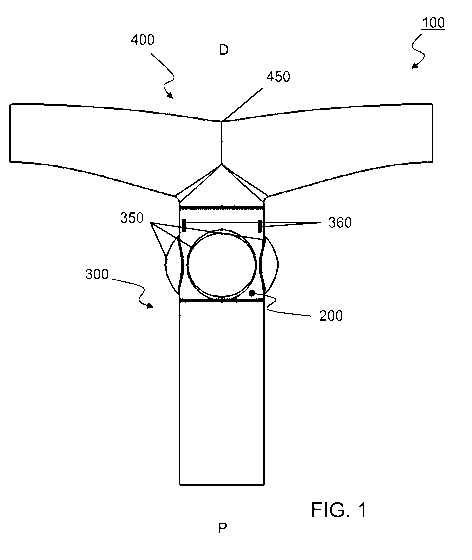
FIG. 1: Schematic cross-section of a vascular valved prosthesis (100)
according to an embodiment
of the present invention; the vascular valved prosthesis (100) comprises an
inner conduit (200)
disposed within an outer conduit (300) that is connected to a T-shaped conduit
(400). In this
embodiment, the inner conduit (200) is configured to function as a valve in a
way that fluidic flow
may be enabled from a proximal (P) end to a distal (D) end, but may be
restricted from D to P;
thus allowing unidirectional flow of a fluid through said vascular valved
prosthesis (100). The T-
shaped conduit (400) and the protrusions (350) are optional.
FIG. 2: Schematic illustration according to an embodiment of an electrospun
vascular valved
prosthesis (100) according to the present invention. The T-shaped conduit
(400) and the
protrusions (350) are optional.
FIG. 3: Schematic opened view according to an embodiment of an electrospun
vascular valved
prosthesis (100) with an open inner conduit (200), according to the present
invention. The T-
shaped conduit (400) and the protrusions (350) are optional.
FIG. 4: Schematic opened view according to an embodiment of an electrospun
vascular valved
prosthesis (100) with a closed inner conduit (200), according to the present
invention. The T-
shaped conduit (400) and the protrusions (350) are optional.
FIG. 5: Schematic illustration according to an embodiment of a mandrel (600)
for electrospinning
the inner conduit (200), according to the present invention.
FIG. 6: Schematic illustration shows an embodiment of a mandrel (600) for
electrospinning the
outer conduit (300), according to the present invention.
FIG. 7: Schematic illustration shows an embodiment of a bifurcated mandrel
(700) for
electrospinning the T-shaped conduit (400), according to the present
invention.
FIG. 8: Schematic illustration of an electrospun inner conduit (200) according
to an embodiment
of the present invention.
FIG. 9: Depicts in an embodiment an electrospun conduit manufactured with a
collapsible
mandrel according to an embodiment of the invention (b) compared to an
electrospun prosthesis
manufactured with a non-collapsible, monolithic mandrel according to a prior
art method (a).
FIG. 10: Depicts an embodiment of electrospun outer conduit (200) manufactured
with a
collapsible mandrel (600) according to the present invention.
CA 03020049 2018-10-04
WO 2017/162645 PCT/EP2017/056655
14
FIG. 11: Demonstrates an embodiment of a front view of a tubular, vascular
valved prosthesis
(100) according to the present invention.
FIG. 12: Demonstrates an embodiment of a top view of a tubular, vascular
valved prosthesis (100)
according to the present invention.
FIG. 13: Demonstrates an embodiment of a front view of a T-shaped, vascular
valved prosthesis
(100) according to the present invention.
FIG. 14: Demonstrates an embodiment of a side view of a T-shaped, vascular
valved prosthesis
(100) according to the present invention.
FIG. 15: Demonstrates an embodiment of a bottom view of a T-shaped, vascular
valved prosthesis
(100) according to the present invention.
FIG. 16: Demonstrates an embodiment of an assembly of a mandrel (600) for
electrospinning the
inner conduit (200).
FIG. 17: Demonstrates an embodiment of an assembly of a mandrel (600) for
electrospinning the
outer conduit (300).
FIG. 18: Demonstrates an embodiment of an assembly of a bifurcated mandrel
(700) for
electrospinning the T-shaped conduit (400).
FIG. 19: Schematic illustration according to an embodiment of an electrospun
vascular valved
prosthesis (100) according to the present invention having a distal over-
thickness (500) of the
inner conduit. The T-shaped conduit (400) and the protrusions (350) are
optional.
.. FIG. 20: A. Schematic illustration according to an embodiment of an
electrospun vascular valved
prosthesis (100) according to the present invention having an inner
reinforcement ring (510). B.
Demonstrates an embodiment of a bottom view of a T-shaped, vascular valved
prosthesis (100)
according to the present invention having an inner reinforcement ring sutured
inside the outer
conduit below the sinuses. The T-shaped conduit (400) and the protrusions
(350) are optional.
FIG. 21: Schematic illustration according to an embodiment of an electrospun
vascular valved
prosthesis (100) according to the present invention having an outer
reinforcement ring (520). The
T-shaped conduit (400) and the protrusions (350) are optional.
FIG. 22: Suture design of the inner conduit to the outer conduit according to
various
embodiments according to the invention. A. Curved vertical suture. B. Hill
shaped suture. C. Hill
shaped + vertical suture. D. short vertical suture (5 mm).
CA 03020049 2018-10-04
WO 2017/162645 PCT/EP2017/056655
Detailed description
Before the present system and method of the invention are described, it is to
be understood that
this invention is not limited to particular systems and methods or
combinations described, since
5 such systems and methods and combinations may, of course, vary. It is
also to be understood that
the terminology used herein is not intended to be limiting, since the scope of
the present
invention will be limited only by the appended claims.
As used herein, the singular forms "a", "an", and "the" include both singular
and plural referents
unless the context clearly dictates otherwise.
10 The terms "comprising", "comprises" and "comprised of" as used herein
are synonymous with
"including", "includes" or "containing", "contains", and are inclusive or open-
ended and do not
exclude additional, non-recited members, elements or method steps. It will be
appreciated that
the terms "comprising", "comprises" and "comprised of" as used herein comprise
the terms
"consisting of, "consists" and "consists of.
15 The recitation of numerical ranges by endpoints includes all numbers and
fractions subsumed
within the respective ranges, as well as the recited endpoints.
The term "about" or "approximately" as used herein when referring to a
measurable value such as
a parameter, an amount, a temporal duration, and the like, is meant to
encompass variations of
+/-10% or less, preferably +/-5% or less, more preferably +/-1% or less, and
still more preferably
+/-0.1% or less of and from the specified value, insofar such variations are
appropriate to perform
in the disclosed invention. It is to be understood that the value to which the
modifier "about" or
"approximately" refers is itself also specifically, and preferably, disclosed.
Whereas the terms "one or more" or "at least one", such as one or more or at
least one
member(s) of a group of members, is clear per se, by means of further
exemplification, the term
encompasses inter alio a reference to any one of said members, or to any two
or more of said
members, such as, e.g., any 3, Ll., 5, 6 or 7 etc. of said members, and up to
all said members.
All references cited in the present specification are hereby incorporated by
reference in their
entirety. In particular, the teachings of all references herein specifically
referred to are
incorporated by reference.
Unless otherwise defined, all terms used in disclosing the invention,
including technical and
scientific terms, have the meaning as commonly understood by one of ordinary
skill in the art to
which this invention belongs. By means of further guidance, term definitions
are included to
better appreciate the teaching of the present invention.
CA 03020049 2018-10-04
WO 2017/162645 PCT/EP2017/056655
16
In the following passages, different aspects of the invention are defined in
more detail. Each
aspect so defined may be combined with any other aspect or aspects unless
clearly indicated to
the contrary. In particular, any feature indicated as being preferred or
advantageous may be
combined with any other feature or features indicated as being preferred or
advantageous.
Reference throughout this specification to "one embodiment" or "an embodiment"
means that a
particular feature, structure or characteristic described in connection with
the embodiment is
included in at least one embodiment of the present invention. Thus,
appearances of the phrases
"in one embodiment" or "in an embodiment" in various places throughout this
specification are
not necessarily all referring to the same embodiment, but may. Furthermore,
the particular
features, structures or characteristics may be combined in any suitable
manner, as would be
apparent to a person skilled in the art from this disclosure, in one or more
embodiments.
Furthermore, while some embodiments described herein include some but not
other features
included in other embodiments, combinations of features of different
embodiments are meant to
be within the scope of the invention, and form different embodiments, as would
be understood
by those in the art. For example, in the appended claims, any of the claimed
embodiments can be
used in any combination.
In the present description of the invention, reference is made to the
accompanying drawings that
form a part hereof, and in which are shown by way of illustration only of
specific embodiments in
which the invention may be practiced. Parenthesized or emboldened reference
numerals affixed
to respective elements merely exemplify the elements by way of example, with
which it is not
intended to limit the respective elements. It is to be understood that other
embodiments may be
utilised and structural or logical changes may be made without departing from
the scope of the
present invention. The following detailed description, therefore, is not to be
taken in a limiting
sense, and the scope of the present invention is defined by the appended
claims.
The term "vascular valved prosthesis" as used herein refers to a structure
suitable for bypassing
or replacing a blood vessel section containing a weakened or malfunctioning
valve. In the current
invention it comprises an inner conduit and an outer conduit, in which the
inner conduit is
configured as a valve to allow unidirectional flow of a fluid through the
outer conduit. From here
on forward the terms "vascular valved prosthesis" and "prosthesis" may be used
interchangeably
and should be regarded as synonymous.
In some embodiments the prosthesis may further comprise additional structures
or components,
such as a bifurcation, reinforcements, commissures, protrusions, or other
forms adding
functionality to said prosthesis.
CA 03020049 2018-10-04
WO 2017/162645 PCT/EP2017/056655
17
The term "mandrel" as used herein refers to a structure comprising multiple
components, such as
a mandrel core, fixation mean and shell pieces. Preferably the mandrel is used
as a scaffold during
electrospinning for the production of prosthesis.
In some embodiments a mandrel may further comprise additional structures or
components, such
as shell pieces, trunks, commissures, protrusions, or other forms which add
functionality to said
mandrel and consequentially also to a prosthesis manufactured using said
mandrel.
The term "electrospinning" as used herein refers to an electrostatic fiber
fabrication technique
method which uses electric force to stack charged fibers, fiber solutions or
fiber melts, around or
within a shape of choice. The fiber diameter is typically in the order of
micrometers, but
diameters even thinner or thicker are also possible, depending on the
resolution and limitations
of the setup.
The standard laboratory setup for a person skilled in the art to practise
electrospinning typically
comprises a (1) spinneret, i.e., a device used to extrude a solution or melt,
for example a
hypodermic syringe needle, which is connected to a (2) power supply, typically
providing five to
fifty kV of direct current, in addition to (3) a syringe pump, configured to
pump the solution or
melt to the spinneret, and (4) a grounded collector, which gathers the
extruded solution or melt,
for example a spinning wheel or a mandrel.
In some embodiments the electrospinning setup is provided with fibers,
preferably in the micro-
or nanometer range, or with gels or melts. In further embodiments biologically
active compounds
may be comprised within the provided materials, preferably selected from
peptides, growth
factors, biological hydrogels or stem cells. Said biologically active
compounds may further
comprise functional tissues showing a level of biodegradability.
The term "diameter" as used herein refers to the maximal distance between two
antipodal points
of an object, i.e., diametrically opposite points lying on the edge of for
instance the prosthesis
inner wall, running as a straight line segment while passing through the
center of the prosthesis.
For the present invention the diameter is expressed in units of meter,
preferably millimeter (mm).
The term "length" as used herein refers to the most extended dimension of an
object, defined as
the maximal distance between two most extended points of said object. For the
present invention
the length is expressed in units of meter, preferably millimeter (mm).
The term "wall thickness" as used herein refers to the thickness of the wall
that partly seals the
inside of said object from the outside, defined as half the difference between
the outer diameter
and the inner diameter of said object. For the present invention the length is
expressed in units of
meter, preferably millimeter (mm).
CA 03020049 2018-10-04
WO 2017/162645 PCT/EP2017/056655
18
The term "elastic regime" as used herein refers to the ability of the
prosthesis to resist a distorting
influence or stress, i.e., an external force per unit area, and to return to
its original size and shape
when the stress is removed. For the present invention the elastic regimen is
expressed in terms of
percentage (%), wherein zero percent is defined as the prosthesis permanently
distorting as a
result of any force high enough to cause a distortion, and a hundred percent
is defined as the
prosthesis resisting any deformation as a result of any force which does not
exceed the material
strength in magnitude. The elastic regimen is measured with a tensile testing
machine.
The term "Young's modulus" as used herein refers the ratio of the stress,
i.e., force per unit area,
along an axis to the strain, i.e., proportional ratio of deformation over
initial length, along that
axis in the range of stress in which Hooke's law holds. The young's modulus in
the field of the
present invention may be determined in the circumferential direction, i.e.,
deformation of the
diameter of the prosthesis 'around' the symmetry axis; and in the radial
direction, i.e.,
deformation of the length of the prosthesis 'out' of the symmetry axis. For
the present invention
the Young's modulus is expressed in units of Pascal, preferably MegaPascal
(MPa), and is
measured with a tensile testing machine.
The term "Lagrangian strain" as used herein refers the extensibility of a
prosthesis, defined as the
finite, relative change in dimension, i.e., deformation, relative to the
original, reference dimension
resulting from a distorting influence or strain. The Lagrangian strain in the
field of the present
invention may be determined in the circumferential direction, i.e.,
deformation of the diameter of
the prosthesis 'around' the symmetry axis; and in the radial direction, i.e.,
deformation of the
length of the prosthesis 'out' of the symmetry axis. For the present invention
the Lagrangian
strain is expressed in units of Pascal, preferably MegaPascal (M Pa).
The term "swelling ratio" as used herein refers to the extent of swelling of
the prosthesis, defined
as the fractional increase in the dimension or volume of the prosthesis due to
fluidic absorption.
For the present invention the swelling ratio is expressed in percentage (%).
The term "pore size" as used herein refers to the mean diameter of an opening
in a surface of an
object, through which gases, liquids, or microscopic particles may pass. More
specifically, it is an
estimate or index of the ratio of the void within a material to the total
volume occupied by the
material including the voids (cfr. ANSI 7198). For the present invention the
pore size may be
expressed by a "pore size range" in units of meter, preferably millimeter
(mm).
The term "porosity" as used herein refers to the fraction of void (empty)
spaces in terms of
volume in an object over the total volume of said object. For the present
invention the porosity is
expressed in percentage (%).
CA 03020049 2018-10-04
WO 2017/162645 PCT/EP2017/056655
19
The term "fiber orientation" as used herein refers to the alignment of
material in an object
relative to the object; concretely, for the present invention the fiber
orientation refers to the
direction of fiber deposition during electrospinning. When the fiber
orientation of an object is
found to display a level of anisotropy, i.e., directional dependency, the
fiber orientation may be
expressed by using an "anisotropic ratio", which is defined as the ratio of
anisotropic fiber
orientation to isotropic fiber orientation.
In a first aspect of the invention, the invention comprises a biodegradable
electrospun vascular
valved prosthesis.
The terms "degradability" or "biodegradability" as used herein refer to the
complete dissolution
of the prosthetic material. Consequently, the "biodegradation rate"
corresponds to a period of
time; namely, the time for which it takes for the prosthetic material to
dissolve and lose at least a
part of its mechanical properties. In particular, the biodegradable vascular
valved prosthesis
according to the invention comprises an electrospun inner conduit having a
proximal end and a
.. distal end, and which is disposed within an electrospun outer conduit
having a proximal end and a
distal end, wherein the inner conduit is attached to the outer conduit such as
to function as a
valve allowing unidirectional flow of a fluid through said outer conduit from
the outer conduit's
proximal to distal end.
According to some embodiments an electrospun inner conduit disposed within an
electrospun
outer conduit has a proximal (P) end and a distal (D) end, wherein the inner
conduit is attached to
the outer conduit and is configured to enable unidirectional flow of a fluid
through said outer
conduit from the outer conduit's P to D end, such as to function as a valve.
Preferably an inner
conduit is configured to allow unidirectional flow of a fluid once it is
comprised within the
structure of an outer conduit. A demonstration of an inner conduit disposed
within an outer
conduit may be observed in figures 11 and 12; specifically, figure 11 provides
a frontal view of the
outside, while figure 12 a top view from the inside.
The entry point through which the fluid is supplied to the prosthesis is
hereby referred to as the
"entry", and the exit point through which the fluid leaves the prosthesis is
hereby referred to as
the "exit". Concretely, the P end corresponds to the direction through which a
fluid preferably
may be provided into an outer conduit, but once passed through the inner
conduit would be
unable to return through the same entry; whereas the D end corresponds to the
direction through
which said fluid that preferably may have been provided would be able to exit
the outer conduit;
therefore a flow of fluid is enabled from a P to D, but is restricted from D
to P, such as to function
as a valve. Accordingly, figure 12 thus shows a demonstration of the exit from
the D end.
CA 03020049 2018-10-04
WO 2017/162645 PCT/EP2017/056655
A schematic cross-section according to an embodiment may be found in figure 1
which shows a
vascular valved prosthesis (100) with an inner conduit (200) disposed within
an outer conduit
(300). The proximal (P) and distal (D) ends show the flow direction through
this embodiment of
the prosthesis. Figure 2 provides a graphical model of the same embodiment.
Figures 13 to 15
5 .. further demonstrate an exemplary assembly of the same embodiment; figure
13 provides a view
from the front, figure 14 from the side and figure 15 from the bottom.
In certain embodiments, the outer conduit has different properties than the
inner conduit. In
certain embodiments, the outer conduit has different mechanical properties
than the inner
conduit. In certain embodiments, the outer conduit has a different
biodegradation rate than the
10 .. inner conduit.
It will be understood that when referring to the outer conduit, in particular
certain properties of
the outer conduit, the same may apply to the T-shaped conduit, if and when
present in the
prosthesis according to the invention. For instance, when referring to
biodegradability of the
outer conduit, in certain preferred embodiments, the biodegradability of the T-
shaped conduit is
15 .. the same as the biodegradability of the outer conduit.
By using a combination of two conduits there are benefits for production and
structural
functionality of a prosthesis. First, by allowing separate production times
for both conduits the
total production time for the prosthesis may be decreased for both through
simultaneous
production. Second, it may allow a higher degree of electrospinning control
during production,
20 .. especially when using biologically active compounds. Third, a post-
production inspection for each
conduit may be easier to perform. Fourth, a level of production flexibility
may be obtained by
allowing the shapes and materials of the inner conduit, i.e., valves and the
outer conduit, i.e., the
tubular prosthesis, to be designed separately. Therefore, if a patient
pathology would require
specific, on-demand adjustments to either one of the conduits, these could be
produced without
adjusted the entire production mechanism. These adjustments may, for example,
include
different shapes, lengths, diameters, or even different biologically active
compounds; in case a
patient has a certain profile which could incite nefarious effects, such as
unwanted biological
deposits, allergic reactions, etc. Fifth, the use of a tube-like conduit to
create a valve inside
another tubular conduit is highly efficient in term of functionality (i.e.,
competence and
.. hemodynamic) of the valve.
In some embodiments an inner conduit is smaller in diameter than an outer
conduit. In some
embodiments, an inner conduit has the same or substantially the same diameter
than an outer
conduit, e.g., the outer diameter of the inner conduit (i.e. the lumen
diameter plus twice the wall
thickness) is the same or substantially the same as the inner diameter (i.e.
the lumen diameter,
CA 03020049 2018-10-04
WO 2017/162645 PCT/EP2017/056655
21
preferably excluding any eventually present protrusions) of the outer conduit.
Such embodiments
allow the inner conduit to be comprised within the structure of an outer
conduit; more
preferably, the diameter may be sufficiently large to prevent any leakage
which would impair the
valve functionality of the inner conduit. In some embodiments an inner conduit
is larger in
diameter than an outer conduit; such embodiments may be beneficial to improve
the closing of
the valve.
In some embodiments the prosthesis may be bifurcated; preferably the
bifurcation is T-shaped.
By incorporating a T-shaped bifurcation directly into the structure of the
prosthesis, the prosthesis
may better mimic the natural shape of a branching artery, and will allow
better functionality as
well as better structural stability and integrity.
In preferred embodiments, the bifurcation is located towards the distal end of
the outer conduit,
so as to split the volume fluid after passing through the inner conduit. An
exemplary bifurcation
may be found in figure 1, which shows a vascular valved prosthesis (100)
comprising an outer
conduit (300) that has been attached to a T-shaped conduit (400), i.e. to the
trunk of the T-shaped
conduit, which comprises said bifurcation (450). As can be seen from the
figure, the bifurcation is
located at the distal (D) end of the prosthesis (100). Figure 2 provides a
graphical model of the
same embodiment. Figures 13 to 15 further demonstrate an exemplary assembly of
the same
embodiment; specifically, figure 13 provides a view from the front, in figure
14 from the side and
in figure 15 from the bottom.
In some embodiments the prosthesis may replace a vessel section comprising a
pulmonary valve
or an aortic valve.
The advantage of a bifurcation, which is preferably T-shaped, is that it may
allow a better
compatibility with arterial systems showing a branching situated very close to
a faulty valve.
Examples of such arterial systems are those surrounding a pulmonary or aortic
valve, but also
other vascular regions or structures may benefit from the bifurcation.
Prosthesis replacement of
these arterial systems using a single tubular prosthesis would not provide
enough fluidic flow to
all branches; and would thus be an inadequate solution for certain patient
pathologies. Moreover,
these bifurcation branches are also biodegradable and may lead to a complete
regeneration by
autologous tissue. Thus, such autologous branches will have a growth
potential, which is
particularly important in children. Additionally, it may decrease the
difficulty of surgically
connecting one prosthesis to several branches, or different prostheses
together.
In some embodiments the outer conduit comprises at least one radially outward
protrusion.
In some embodiments an outer conduit, and by extension the prosthesis,
comprises at least one
radially outward protrusion. In certain embodiments, such protrusion(s) may
advantageously
CA 03020049 2018-10-04
WO 2017/162645 PCT/EP2017/056655
22
mimic (and hence replace) naturally occurring sinuses. These protrusions
advantageously aid in
the adequate mechanical functioning of the valve. Their presence may create a
"Venturi effect",
i.e., a reduction in fluid pressure when a fluid flows through a constricted
section of pipe, which
may help achieve a complete opening and closing of the valve with the lowest
possible amount of
shear stress and fatigue. Additionally, they may also reduce the time required
for the opening and
closing of the valve.
In some embodiments the prosthesis may be shaped to resemble natural arterial
protrusion, such
as to include a sinus, cavity, sacks, or other similarly shaped features. By
implementing
protrusions in an outer conduit different fluid rates may be obtained to
achieve different valve
actions. Resulting from the implementation of one or more protrusions the
prosthesis may better
resemble the functionality of a natural artery, and may improve fluid pressure
and flow control
which may prove beneficial for patient response and recovery rate.
In some embodiments the proximal end of said inner conduit is attached
circumferentially along
the inside of the outer conduit towards the proximal end of the outer conduit
to form a
circumferential commissure; wherein the distal end of said inner conduit is
attached to the inside
of the outer conduit towards the distal end of said outer conduit to form
focal commissures at
one or more discrete positions, preferably two or more discrete positions,
such as three discrete
positions, wherein the focal commissures may or may not extend longitudinally
from the distal
end of the inner conduit towards the proximal end of the inner conduit. The
vertical commissures
may help achieve the "valve-like" functionality of the inner conduit, and thus
of the prosthesis.
Preferably, the focal commissures are equidistantly spaced. Preferably, the
focal commissures are
coplanar.
In some embodiments the inner conduit may exhibit a vertical commissure
situated along the wall
of the inner conduit; preferably, two or more symmetrical commissures may be
present.
The inclusion of commissures may allow the inner conduit to expand or contract
in diameter
outside of the range determined by the material elasticity. This may enable
easier assembly of the
inner conduit within the outer conduit.
Additionally, the vertical commissures may improve valve dynamics and fatigue
life during
operation of the inner conduit.
To better clarify this technical feature, figures 3 and 4 demonstrate a
prosthesis (100) with an
open inner conduit (200) and a closed inner conduit (200), respectively. In
figure 3 the liquid may
flow freely through the prosthesis (100); however, in figure 4 the focal
commissures (250) will
allow flow of a liquid from the proximal (P) end to the distal (D) end
prevent, but prevent any flow
from the D to the P. As such, a valve-like functionality is obtained for the
prosthesis resembling
CA 03020049 2018-10-04
WO 2017/162645 PCT/EP2017/056655
23
that of a sinus of Valsalva, i.e., an aortic sinus. During heartbeat the
prosthesis can thus be
envisioned as an aorta or a pulmonary artery wherein the inner conduit
functions as sinus of
Valsalva; so that during systole, cfr. figure 3, the inner conduit may allow
the circulation of a fluid
through and during diastole, cfr. figure 4, the inner conduit may prevent the
backflow of the
passed fluid. Additionally, it may be noted that in both figures the T-shaped
conduit is fixed to the
outer conduit in the direction of the circulating fluid, similar to pulmonary
artery or aortic
branches.
In some embodiments the inner conduit may be set up to the outer conduit with
the use of
circumferential cohesion lines; said cohesion lines are configured in a way to
allow a proper
positioning of the inner conduit with regard to the outer conduit. The
advantage of using cohesion
lines is that it may allow a better positioning and placement of an inner
conduit within an outer
conduit; thus forming a vascular valved prosthesis. A proper placement may
allow a high degree
of competence and functionality, and may in turn reduce the risks of
prosthesis malfunctioning,
such as leakage or obstruction.
In some embodiments the distal end of the inner conduit can exhibit a
thickness characterized by
a larger wall width, situated in the direction of the center of the outer
conduit. The addition of a
thickness at said location may improve the closing of the inner conduit after
fluid is pumped
through said inner conduit, for example, during diastole.
In some embodiments the outer conduit comprises three coplanar radially
equidistally spaced
outward protrusions (e.g. sinus-like structures), and wherein the distal end
of said inner conduit is
attached at three equidistally spaced discrete positions to the inside of the
outer conduit along
longitudinal lines separating the protrusions.
In some embodiments the inner and/or outer conduit may further exhibit a
longitudinal
commissures situated along the wall of the inner conduit; preferably, at least
two symmetrical
commissures may be present; most preferably, three equidistally spaced
commissures may be
present. Such additional commissures function to further strengthen the
prosthesis (cfr. figure 8).
In some embodiments the distal end of the inner conduit is attached to the
inside of the outer
conduit towards the distal end of said outer conduit to form focal commissures
at one or more
discrete positions, preferably two or more discrete positions, such as three
discrete positions,
wherein the focal commissures may extend longitudinally from the distal end of
the inner conduit
towards the proximal end of the inner conduit. Preferably, the focal
commissures are
equidistantly spaced. Preferably, the focal commissures are coplanar.
In some embodiments a T-shaped conduit is attached along the distal end of
said outer conduit.
CA 03020049 2018-10-04
WO 2017/162645 PCT/EP2017/056655
24
In some embodiments the diameter of the proximal end (i.e. the trunk) of the T-
shaped conduit
could be slightly reduced vis-a-vis the diameter of the outer conduit (or vice
versa), in order to fit
inside the outer conduit (or vice versa) to facilitate its fixation with the
outer conduit with
protrusions.
In some embodiments the prosthesis comprises biologically active compounds.
Preferably, the biologically active compound is selected from peptides, growth
factors, biological
hydrogels such as gelatin, and/or may comprise stem cells such as adipose-
derived stem cells,
and/or combinations thereof.
Biologically active compounds may promote an improved level of (exogenous)
cell seeding and
tissue regeneration beneficial for the recovery rate of a patient. Seeding of
stem cells may
increase tissue regeneration speed ultimately replacing the prosthesis with
newly grown tissue for
which the prosthesis may serve as a scaffold. Alternatively, tissue
regeneration may entirely or at
least partially rely on cells already present in the patient's body by using
for instance the
biomaterial of the prosthesis as a signalling mechanism for driving endogenous
cells towards the
scaffold; possibly after appropriate stimulation of cell expansion and/or
differentiation.
In some embodiments the inner conduit is circumferentially or longitudinally
reinforced by
biodegradable structures. In some embodiments the outer conduit is
circumferentially or
longitudinally reinforced by biodegradable structures. In some embodiments the
prosthesis is
circumferentially and longitudinally reinforced by biodegradable structures.
In some embodiments, the outer or inner conduit is circumferentially or
longitudinally reinforced
by additional biodegradable structures, optionally wherein the additional
biodegradable
structures form a ring which is disposed on the inside or on the outside of a
part of the conduit.
The reinforcements may be made from the same or different material as the
inner or outer tube.
In certain embodiments, the reinforcements are electrospun.
In certain embodiments, longitudinal reinforcements extend along the entire
length or
substantially the entire length of the outer or inner conduit. In certain
embodiments, longitudinal
reinforcements extend along part of the length of the outer or inner conduit.
In certain
embodiments, reinforcements, such as longitudinal or circumferential
reinforcements, are
provided at least at or near the commissures. In certain embodiments,
reinforcements, such as
longitudinal or circumferential reinforcements, are provided on the inner
conduit at least at the
proximal end.
Such reinforcements, and in particular the reinforcement ring, can minimize
the flexure when the
inner conduit is actively opening and closing. Accordingly, the functionality
of the inner conduit
CA 03020049 2018-10-04
WO 2017/162645 PCT/EP2017/056655
over time is improved. If reinforcements are provided at or near commissures,
external forces
applied on the commissures can be decreased.
It will be understood that the reinforcements may be attached to the conduits
by means
described herein elsewhere, such as including by suture, stapling, gluing,
welding (laser, vibration,
5 ultrasonic, induction, high frequency) or a combination of the processes
described. Alternatively,
the reinforcements may be added during manufacturing of the conduit, for
instance by
electrospinning.
In certain embodiments, the reinforcements are local areas of the conduit
having a thicker
conduit wall. Accordingly, in certain embodiments, the outer or inner conduit
comprises
10 circumferential or longitudinal sections or areas, such as a ring, which
sections or areas have an
increased wall thickness relative to the remainder of the conduit wall.
In certain embodiments, the outer conduit comprises one or more
circumferential
reinforcements, preferably a ring along the inside or outside of the conduit,
wherein the
reinforcement is located at or near the proximal end of the inner conduit, as
for instance
15 illustrated in Figures 20 and 21. In certain embodiments, the outer
conduit comprises one or more
circumferential reinforcements, preferably a ring along the inside or outside
of the conduit,
wherein the reinforcement is located at or near the proximal end of the
protrusions.
In certain embodiments, the reinforcements increase the wall thickness by at
least 10%, such as at
least 20%, at least 30%, at least 40%, at least 50%, at least 60%, at least
70%, at least 90%, at least
20 .. 90%, at least 100%, or more, such as at least 150%, at least 200%, or
more. In certain
embodiments, the reinforcements do not increase the wall thickness by more
than 400%,
preferably by no more than 300%.
In certain embodiments, the circumferential reinforcement has a length of
between 1% and 50%
of the length of the conduit (i.e. the distance between the distal and
proximal end of the conduit),
25 such as between 5% and 25%. In certain embodiments, the circumferential
reinforcement has a
length of between 1 mm and 50 mm, such as between 3 mm and 20 mm, or between 5
mm and
10 mm.
Reinforcements of the prosthesis manufactured using biodegradable structures
may over-time
naturally decompose; thus allowing the implantation of temporary
reinforcements. This could
prove beneficial for reinforcements which might otherwise require a secondary
invasive
procedure for their removal, which might be detrimental to patient recovery
rate.
In certain embodiments, the inner conduit at its distal end has a wall
thickness which is larger
than the wall thickness at the proximal end. Such increased wall thickness at
the distal end
improves closure of the valve. In certain embodiments, at least the most
distal 10% has a wall
CA 03020049 2018-10-04
WO 2017/162645 PCT/EP2017/056655
26
thickness which is larger than the wall thickness at the proximal end. In
certain embodiments, at
least the most distal 20% has a wall thickness which is larger than the wall
thickness at the
proximal end. In certain embodiments, at least the most distal 30% has a wall
thickness which is
larger than the wall thickness at the proximal end. In certain embodiments, at
least the most
distal 40% has a wall thickness which is larger than the wall thickness at the
proximal end. In
certain embodiments, at least the most distal 50% has a wall thickness which
is larger than the
wall thickness at the proximal end. In certain embodiments, at most the most
distal 10% has a
wall thickness which is larger than the wall thickness at the proximal end. In
certain embodiments,
at most the most distal 20% has a wall thickness which is larger than the wall
thickness at the
proximal end. In certain embodiments, at most the most distal 30% has a wall
thickness which is
larger than the wall thickness at the proximal end. In certain embodiments, at
most the most
distal 40% has a wall thickness which is larger than the wall thickness at the
proximal end. In
certain embodiments, at most the most distal 50% has a wall thickness which is
larger than the
wall thickness at the proximal end. In certain embodiments, the wall thickness
of between 1 mm
and 20 mm of the most distal portion of the conduit is larger than the wall
thickness of the
proximal end, such as between 2 mm and 10 mm, or between 2 mm and 5 mm. In
certain
embodiments, the the wall thickness is increased by at least 10%, such as at
least 20%, at least
30%, at least 40%, at least 50%, at least 60%, at least 70%, at least 90%, at
least 90%, at least
100%, or more, such as at least 150%, at least 200%, or more. In certain
embodiments, the wall
thickness is not increased by more than 400%, preferably by no more than 300%,
more preferably
by no more than 200%, most preferably by no more than 100%. It will be
understood that the
section having increased wall thickness may be continuous (i.e. completely
circumferential) or
may be segmented (e.g. interrupted at the focal commissures).
According to the invention the prosthesis is manufactured using biodegradable
compounds. A
.. prosthesis manufactured using biodegradable structures may over-time
naturally decompose;
thus allowing the implantation of temporary prosthesis. For this purpose the
biodegradation rate
is determined by the persistence of the mechanical properties, and a structure
is fully degraded
once the mechanical properties of the structure are gone. As used herein, the
terms
"biodegradation" and "degradation" refer to the same biochemical process.
Biodegradation rate
can depend on the materials used (e.g. type of polymer), but can also depend
on particular
physical properties, such as pore density, thickness of the conduit, etc. In
certain embodiments,
the degradation rate of the inner conduit can be slower as the degradation
rate of the outer
conduit. In certain embodiments, the inner and outer conduit are made of the
same materials.
Hence, due to the thickness of the inner and outer conduit, the biodegradation
rate will be
CA 03020049 2018-10-04
WO 2017/162645 PCT/EP2017/056655
27
different. Biodegradation could prove beneficial for regenerative strategies
where the fault valve
may be regenerated using the prosthesis as a temporary mold for cell
attachment or deposition;
preferably the prosthesis will further promote cell seeding and attachment
which may be
beneficial for patient recovery rates and long-term health. In certain
embodiments, the entire
prosthesis is biodegradable, i.e. the prosthesis is fully biodegradable. This
biodegradability is at
the very basis of the regeneration of the prosthesis, potential for growth.
The main advantages of
this approach is to avoid re-interventions to surgically replace the outgrown
prosthesis, reduce
risk of surgical complications (e.g., bleeding, infection, heart block,
stroke, renal failure, death)
and minimise the human, social and financial costs related to these re-
interventions and
complications. This feature is especially important for prosthesis implanted
in children, which may
outgrow the prosthesis.
In some embodiments the inner conduit when implanted in a vascular structure
degrades (i.e.,
biodegradation) in a period ranging from at least 6 months to at most 48
months; preferably
between 12 to 36 months; most preferably between 18 to 30 months; most
preferably in about
24 months. In some embodiments the outer conduit when implanted in a vascular
structure
degrades in a period ranging from at least 1 month to at most 24 months;
preferably between 3 to
18 months; most preferably between 6 to 12 months. In some embodiments the T-
shaped conduit
when implanted in a vascular structure degrades in a period ranging from at
least 1 month to at
most 24 months; preferably between 3 to 18 months; most preferably between 6
to 12 months.
In preferred embodiments the outer and T-shaped conduits have essentially the
same
degradation rate. In some embodiments the biodegradability of the inner
conduit is different
from the biodegradability of the outer conduit. In some embodiments the
biodegradability of the
inner conduit is essentially the same as the biodegradability of the outer
conduit.
In preferred embodiments the biodegradability of the inner conduit is slower
than
biodegradability of the outer conduit; preferably, the biodegradability is two
times as slow; more
preferably the biodegradability is three times as slow. This slower
biodegradability of the inner
conduit guarantees the persistence of the competence of the valved part of the
device until the
newly regenerated tissue displays good mechanical properties.
By implementing different biodegradability rates along the diameter of the
prosthesis, the rate of
tissue regeneration may be promoted from the outside conduit towards the
inside conduit.
In a preferred embodiment the rate of biodegradability may be adjusted to a
patient pathology
and regeneration rates, for example, young patients may benefit from faster
prosthesis
degradation in comparison to older patients.
CA 03020049 2018-10-04
WO 2017/162645 PCT/EP2017/056655
28
In some embodiments the biodegradability of the distal end of the commissures
is different from
the biodegradability of the proximal end of the commissures. In preferred
embodiments the
biodegradability of the distal end of the commissures is faster than the
biodegradability of the
proximal end of the commissures; preferably, the biodegradability is twice as
fast; more
preferably, biodegradability is thrice as fast. The faster biodegradability of
the distal end of the
commissures can enhance with time the length of the coaptation surface of the
different parts of
the inner conduit during the diastole. By implementing different
biodegradability rates along the
length of the prosthesis, the rate of tissue regeneration may be promoted from
the distal end to
the proximal end.
In some embodiments the inner conduit is multilayered. In some embodiments the
outer conduit
is multilayered. In some embodiments both the inner and outer conduits are
multilayered. By
applying multiple layers over the inner or the outer conduit, material and
structural properties of
the prosthesis may be enhanced, and each layer may have its own structural
and/or functional
characteristics (e.g. biodegradability, directional structural reinforcements,
fibre orientation,
porosity, etc.). Multiple layers may be made from the same or from different
materials. For
electrospun conduits, the "core" refers the fibers located on the internal
part, situated towards
the mandrel; while the "shell" refers to the fibers located on the external
part, surrounding the
core.
The properties of the prosthesis may be enhanced by using layers with
different material and
structural properties to complement each other; for example, by combining a
layer which shows
an enhanced biodegradation rate or porosity, yet suffers from a weak material
strength, with a
layer that has a high material strength, yet has a reduced biodegradation rate
or porosity. Thus,
by properly designing and combining layers the prosthesis properties may be
enhanced in
comparison to the properties of singular layers. In some embodiments the layer
facing the inside
of the prosthesis may have a faster biodegradability rate than the layer
facing the outside of the
prosthesis. In some embodiments the layer facing the inside of the prosthesis
may have a slower
biodegradability rate than the layer facing the outside of the prosthesis.
In some embodiments the inner conduit is made by electrospinning of
(biodegradable) polymers
based on supramolecular chemistry or polymers comprising stereocomplexes. In
some
embodiments the outer conduit is made by electrospinning of (biodegradable)
polymers based on
supramolecular chemistry or polymers comprising stereocomplexes. In some
embodiments both
the inner conduit and said outer conduit are made by electrospinning of
(biodegradable) polymers
based on supramolecular chemistry or polymers comprising stereocomplexes. In
some
embodiments the inner conduit is made by electrospinning of (biodegradable)
polymers based on
CA 03020049 2018-10-04
WO 2017/162645 PCT/EP2017/056655
29
supramolecular chemistry or polymers comprising stereocomplexes, wherein the
polymers are
biodegradable aliphatic polyesters or biodegradable polyurethanes. In some
embodiments the
outer conduit is made by electrospinning of (biodegradable) polymers based on
supramolecular
chemistry or polymers comprising stereocomplexes, wherein the polymers are
biodegradable
aliphatic polyesters or biodegradable polyurethanes. In some embodiments both
the inner
conduit and said outer conduit are made by electrospinning of (biodegradable)
polymers based on
supramolecular chemistry or polymers comprising stereocomplexes, wherein the
polymers are
biodegradable aliphatic polyesters or biodegradable polyurethanes. In some
embodiments the
inner conduit is made by electrospinning of biodegradable aliphatic polyesters
or biodegradable
polyurethanes. In some embodiments the outer conduit is made by
electrospinning of
biodegradable aliphatic polyesters or biodegradable polyurethanes. In some
embodiments both
the inner conduit and said outer conduit are made by electrospinning of
biodegradable aliphatic
polyesters or biodegradable polyurethanes. In some embodiments the conduits
are made by
electrospinning a mixture of (co)polymers, such as biodegradable aliphatic
polyesters and
biodegradable polyurethanes. In some embodiments the outer conduit is made by
electrospinning
a combination of biodegradable aliphatic polyesters and biodegradable
polyurethanes. In some
embodiments both the inner conduit and outer conduit are made by co-
electrospinning of
(biodegradable) polymers into core-shell fibres, wherein the polymers are
biodegradable aliphatic
polyesters or biodegradable polyurethanes. In some embodiments both the inner
conduit and
said outer conduit are made by electrospinning a combination of biodegradable
aliphatic
polyesters and biodegradable polyurethanes.
The advantages of polyester and polyurethanes are that they may be readily
available and can be
easily processed through electrospinning. Applied polyester may be extremely
versatile in its
structural properties, ranging from very soft to very firm and durable. As
prosthesis material
polyester absorbs less than one percent of moisture by weight, offers good
dimensional stability
under a variety of conditions, and has unique filling properties. Once
implanted, polyester doesn't
promote the growth of bacteria making it very suitable for biological
applications. Alternatively,
applied polyurethane may be extremely versatile in its structural properties,
having high tensile
and load baring capacities. As prosthesis material polyester will remain
stable under fluidic
conditions with minimal swelling. Currently, polyurethane (PU) is the polymer
of choice in cardiac
applications due to its high biocompatibility, strong in vivo performance and
simplicity of
manufacturing. Artificial heart valves made from polyurethane have high
biocompatibilities
because of the extensive degrees of freedom that arise from having three
component monomer
parts (as compared to other polymers, where fewer components are usually
employed). This
CA 03020049 2018-10-04
WO 2017/162645 PCT/EP2017/056655
allows for unlimited variation in mechanical and physicochemical
characteristics. The advantage
of polylactic acid is that it may be readily available from renewable
resources, is relatively
inexpensive, and easily processed through electrospinning. Additionally,
applied polyester may be
extremely versatile in its structural properties, ranging from very soft to
very firm and durable. As
5 .. a biocompatible material polylactic acid is neither toxic nor
carcinogenic to the human body,
hence making it an excellent material for biomedical applications. As a
biodegradable prosthesis
material polylactic acid may break down inside the body within 6 months to 2
years. This gradual
degradation may be desirable for a support structure, because it can gradually
transfer the load to
the body as that area heals; thus functioning as a scaffold for regenerating
tissue.
10 In preferred embodiments the inner conduit is made by electrospinning a
mixture of (co)polymers
from the following list: Poly(ethylene glycol) (PEG), Poly(glycolic acid)
(PGA), Poly(lactic acid)
(PLA), Poly-4-hyrdoxybutyrate (P4HB), Polycaprolactone (PCL), Poly(glycerol
sebacate) (PGS),
Poly(ester urea urethane) (PEUU), Polydioxaneone (PDO), Polycarbonate (PCU),
poly(carbonate
urethane urea) (PCUU), Polyhedral oligomeric silsesquioxanes (POSS), and/or
combinations
15 thereof. All these polymers offer several advantages towards the
manufactory of a vascular valved
prosthesis, namely, they are mechanically compatible with the manufacturing
process of
electrospinning a trifoliate valve with the targeted mechanical functionality,
i.e., a degradation
rate and long-term efficient hemodynamic opening and closing of the sinuses;
they are
biocompatible; they can be processed by electrospinning; they are approved by
regulatory
20 .. commissions; they can be functionalized by multiple peptides depending
on the chosen technique
of peptide/polymer grafting, for example, peptides RGD and SDF1 may be
currently selected; they
can be seeded by cells; they can be sterilized under industrial conditions
without any structural
alteration of or exterior damage to the prosthesis; and last, they are readily
available for
manufactory. In more preferred embodiments aliphatic diisocyanates, such as
lysine
25 diisocyanantes (LDI) and 1,4-diisocyanatobutane (BDI), are used as
material of choice above
aromatic diisocyanates; since the latter was found to form toxic products,
e.g., diisocyanates such
as 4,4'methylenediphenyl diisocyanate (MDI) and toluene diisocyanate (TDI)
upon degradation
and was thus found unsuitable as biodegradable biomaterials. Most aliphatic
diisocyanates-based
polyurethanes have a Young's modulus and tensile strength of several tens of
MPa, and a large
30 breaking strain in the range of 100 - 1000%. As such, the selection of a
soft and ideally completely
elastic material, i.e., 100% recovery from deformation, based on aliphatic
diisocyanates is
preferred. The susceptibility of polymers to biodegradation is governed by
"soft" segment
components which are generally glycols, such as polyethylene glycol (PEG) or
polycaprolactone
diols (PCL). However, higher proportions of soft segments tend to be
correlated with an increased
CA 03020049 2018-10-04
WO 2017/162645 PCT/EP2017/056655
31
degradation rate. Given that the hard segments are known to be highly
thrombogenic due to their
high crystallinity and strong hydrogen bonding, alternative compositions were
sought after and
experimented with, such as synthetic materials with hydrophilic soft segments,
the addition of
functional groups as well as the coating, impregnation and grafting of
techniques to modify
surfaces. In particular, polyethylene oxide (PEO) was observed to provide a
successful surface
coating due to its neutral charge and flexibility; it may be utilised as a
possible permanent coating
to prevent protein surface adsorption to the prosthesis. After conducting a
comprehensive survey
of potentially suitable materials with regards to the mechanical properties,
biodegradability,
safety and compatibility with the electrospinning production process,
poly(ester urethane)-urea
(PEUU) was found to be preferred over poly(carbonate urethane urea) (PCUU).
In some embodiments the inner conduit is attached to the outer conduit by
sutures, staples, glue,
welds (laser, vibration, ultrasonic, induction, high frequency) or a
combination thereof. In
preferred embodiments where the prosthesis has a high rate of
biodegradability, the choice of
attachment will involve a level of biodegradability too; for example, sutures,
glue or staples made
from a biodegradable material. By properly attaching the inner conduit to the
outer conduit the
chance for unwanted effects, such as valve leakage or breakage, may be
significantly lower.
The attachment is demonstrated in figures 11 to 15 by means of sutures. The
former shows the
attachment formed between an inner conduit disposed within an outer conduit;
thus forming a
tubular vascular valved prosthesis. Specifically, figure 11 shows the
attachment from the outside
in a frontal view; while figure 12 provides a top view to observe the same
attachment from the
inside. Next, the combined inner and outer conduit may be affixed in a similar
manner to the T-
shaped conduit, as is demonstrated in figure 13 from the front, in figure 14
from the side and in
figure 15 from the bottom; thus forming a T-shaped vascular valved prosthesis.
Though it may be designed to resemble a natural artery, preferably the
prosthesis' structure will
contain inbuilt reserves of strength and stability above those of a natural
artery to better resist
the high workload performed by a pumping heart. In certain cases the
prosthesis may remain as a
(semi-) permanent part of a vascular system, so its structural integrity and
reliability can be
affected by long-term effects, such as aging or biological deposits.
In some embodiments the prosthesis is characterized in that the inner conduit
has a linear elastic
regimen of at least 20%; preferably at least 40%; more preferably at least
50%; most preferably at
least 60%. In some embodiments the prosthesis is characterized in that the
outer conduit has a
linear elastic regimen of at least 5%; most preferably at least 10%. In some
embodiments the
CA 03020049 2018-10-04
WO 2017/162645 PCT/EP2017/056655
32
prosthesis is characterized in that the T-shaped conduit has a linear elastic
regimen of at least 5%;
most preferably at least 10%.
In some embodiments the prosthesis is characterized in that the inner conduit
has a Young's
modulus in the circumferential direction ranging from at least 0.01 to at most
200 MPa;
preferably ranging from 0.1 to 150 MPa; more preferably ranging from 1 to 125
MPa; most
preferably ranging from 5 to 100 MPa. In some embodiments the prosthesis is
characterized in
that the inner conduit has a Young's modulus in the radial direction ranging
from at least 0.01 to
at most 200 MPa; preferably ranging from 0.05 to 100 MPa; more preferably
ranging from 0.1 to
30 MPa; most preferably ranging from 0.5 to 15 MPa. In some embodiments the
prosthesis is
characterized in that the outer conduit in the radial direction and/or the
circumferential direction
has a Young's modulus ranging from at least 0.001 to at most 100 MPa;
preferably ranging from
0.005 to 10 MPa; more preferably ranging from 0.01 to 1.5 MPa; most preferably
ranging from
0.01 to 0.5 MPa. In some embodiments the prosthesis is characterized in that
the T-shaped
conduit in the radial direction and/or the circumferential direction has a
Young's modulus ranging
from at least 0.001 to at most 100 MPa; preferably ranging from 0.005 to 10
MPa; more
preferably ranging from 0.01 to 1.5 MPa; most preferably ranging from 0.01 to
0.5 MPa. In
preferred embodiments the outer conduit and the T-shaped conduit have the same
Young's
modulus in the circumferential and radial direction.
In some embodiments the prosthesis is characterized in that the inner conduit
has a Lagrangian
strain in the circumferential direction ranging from at least 0.01 to at most
50 MPa; preferably
ranging from 0.04 to 10.0 MPa; more preferably ranging from 0.08 to 1.0 MPa;
most preferably
ranging from 0.1 to 0.4 MPa. In some embodiments the prosthesis is
characterized in that the
inner conduit has a Lagrangian strain in the radial direction ranging from at
least 0.01 to at most
50 MPa; preferably ranging from 0.05 to 10.0 MPa; more preferably ranging from
0.1 to 1.0 MPa;
most preferably ranging from 0.6 to 0.9 MPa. In some embodiments the
prosthesis is
characterized in that the outer conduit has a Lagrangian strain in the
circumferential direction
ranging from at least 0.01 to at most 50 MPa; preferably ranging from 0.02 to
10.0 MPa; more
preferably ranging from 0.04 to 1.0 MPa; most preferably ranging from 0.05 to
0.3 MPa. In some
embodiments the prosthesis is characterized in that the outer conduit has a
Lagrangian strain in
the radial direction ranging from at least 0.01 to at most 50 MPa; preferably
ranging from 0.05 to
10.0 MPa; more preferably ranging from 0.1 to 1.0 MPa; most preferably of
about 0.4 MPa. In
some embodiments the prosthesis is characterized in that the T-shaped conduit
has a Lagrangian
strain in the circumferential direction ranging from at least 0.01 to at most
50 MPa; preferably
ranging from 0.02 to 10.0 MPa; more preferably ranging from 0.04 to 1.0 MPa;
most preferably
CA 03020049 2018-10-04
WO 2017/162645 PCT/EP2017/056655
33
ranging from 0.05 to 0.3 MPa. In some embodiments the prosthesis is
characterized in that the T-
shaped conduit has a Lagrangian strain in the radial direction ranging from at
least 0.01 to at most
50 MPa; preferably ranging from 0.05 to 10.0 MPa; more preferably ranging from
0.1 to 1.0 MPa;
most preferably of about 0.7 MPa.
In some embodiments the prosthesis is characterized in that the inner conduit
has a swelling ratio
ranging from at least 0 to at most 99 %. In some embodiments the prosthesis is
characterized in
that the outer conduit has a swelling ratio ranging from at least 10 to at
most 99 %; preferably
ranging from 20 to 98%; more preferably ranging from 50 to 97 %; most
preferably ranging from
60 to 97%. In some embodiments the prosthesis is characterized in that the T-
shaped conduit has
a swelling ratio ranging from at least 10 to at most 99 %; preferably ranging
from 20 to 98%; more
preferably ranging from 50 to 97 %; most preferably ranging from 60 to 97%.
In some embodiments the inner conduit comprises a fibrous network comprising
microfibers
and/or nanofibers, wherein the diameter of the fibers ranging from at least
0.01 to at most 5.0
um; preferably ranging between 0.05 and 3.5 um; more preferably ranging
between 0.1 and 2.0
um; most preferably ranging between 0.5 and 1.5 um. In some embodiments the
outer conduit
comprises a fibrous network comprising microfibers and/or nanofibers, wherein
the diameter of
the fibers ranging from at least 0.01 to at most 5.0 um; preferably ranging
between 0.05 and 3.5
um; more preferably ranging between 0.1 and 2.0 um; most preferably ranging
between 0.5 and
1.5 um. In some embodiments the T-shaped conduit comprises a fibrous network
comprising
microfibers and/or nanofibers, wherein the diameter of the fibers ranging from
at least 0.01 to at
most 5.0 um; preferably ranging between 0.05 and 3.5 um; more preferably
ranging between 0.1
and 2.0 um; most preferably ranging between 0.5 and 1.5 um.
In some embodiments the internal diameter of the inner conduit ranges between
at least 1 and at
most 40 mm; preferably between 5 and at most 35 mm; more preferably between 10
and at most
25 mm; most preferably is about 18 mm. In some embodiments the internal
diameter of the outer
conduit ranges between at least 1 and at most 40 mm; preferably between 5 and
at most 35 mm;
more preferably between 10 and at most 25 mm; most preferably is about 18 mm.
In some
embodiments the internal diameter of the T-shaped conduit ranges between at
least 1 and at
most 40 mm; preferably between 5 and at most 35 mm; more preferably between 10
and at most
.. 25 mm; most preferably is about 18 mm. In some embodiments the difference
between the
internal diameter of the inner and outer conduits is at most 5 mm; preferably
at most 1 mm;
more preferably at most 0.1; most preferably less than 0.1 so that the
diameter of the inner and
outer diameter is essentially the same. In preferred embodiments the outer
conduit and the T-
shaped conduit have essentially the same internal diameter.
CA 03020049 2018-10-04
WO 2017/162645 PCT/EP2017/056655
34
In some embodiments the length of the inner conduits ranges between at least 5
and at most 40
mm; preferably between 8 and at most 35 mm; preferably between 12 and at most
30 mm; most
preferably between 15 and at most 25 mm. In some embodiments the length of the
outer
conduits ranges between at least 20 and at most 125 mm; preferably between 30
and at most 100
mm; preferably between 40 and at most 85 mm; most preferably between 50 and at
most 75 mm.
In preferred embodiments the length of the inner is shorter than the length of
the outer conduit.
In preferred embodiments the difference between the length of the inner
conduit and the length
of the outer conduit is at least 25 mm and at most 55 mm; preferably between
35 to 45 mm; most
preferably around 40 mm.
The circumferential diameter refers to a circular diameter wherein the
protrusions will fit. In some
embodiments the external circumferential diameter of the protrusions ranges
between at least 5
and at most 40 mm; preferably between 8 and at most 35 mm; preferably between
12 and at
most 30 mm; most preferably between 25 and at most 35 mm. In some embodiments
the
circumferential commissure is coplanar with the most proximal end of the
protrusions.
In some embodiments the inner conduit has a wall thickness ranging between at
least 0.01 and at
most 1.00 mm; preferably between 0.03 and at most 0.50 mm; more preferably
between 0.05 and
at most 0.3 mm; most preferably is about 0.1 mm or about 0.2 mm. In some
embodiments the
outer conduit has a wall thickness ranging between at least 0.01 and at most
2.00 mm; preferably
between 0.05 and at most 1.50 mm; more preferably between 0.3 and at most 1.0
mm or at most
1.2 mm. In certain embodiments, the thickness of the outer conduit is 0.8 mm
above the sinuses
(i.e. towards the distal end) and 1.1 mm below the sinuses (i.e. towards the
proximal end) to
minimize the flexure of the outer conduit due to the movement of the inner
conduit. In some
embodiments the T-shaped conduit has a wall thickness ranging between at least
0.01 and at
most 2.00 mm; preferably between 0.05 and at most 1.50 mm; more preferably
between 0.3 and
at most 1.0 mm; most preferably is about 0.3 mm. In preferred embodiments the
outer conduit
and the T-shaped conduit have essentially the wall thickness. In some
embodiments the inner
conduit has at least one region having a different wall thickness at the
distal end compared to the
wall thickness at the proximal end. In preferred embodiments the inner conduit
has at least one
region having a higher wall thickness at the distal end compared to the wall
thickness at the
proximal end; preferably the wall thickness is at least 0.01 mm thicker; more
preferably the wall
thickness is about 0.03 mm thicker; most preferably the wall thickness is
about 0.05 mm thicker.
This wall thickness difference corresponds to a percent difference of
preferably at least 10%;
more preferably about 25%; most preferably about 50%.A different wall
thickness may prove
beneficial for better valve functionality, decreasing the chance of fluid
leakage towards the distal
CA 03020049 2018-10-04
WO 2017/162645 PCT/EP2017/056655
end. Additionally, a different wall thickness may allow an easier disposition
of the inner conduit
within the outer conduit. In some embodiments the inner conduit has at least
one region having a
higher wall thickness at the proximal end compared to the wall thickness at
the distal end.
In some embodiments the inner conduit displays anisotropic fiber orientation,
wherein the
5 anisotropic ratio is at least 2:1, preferably at least 5:1; more
preferably at least 10:1; most
preferably about 20:1.1n some embodiments the outer conduit displays
anisotropic fiber
orientation, wherein the anisotropic ratio is at least 2:1, preferably at
least 5:1; most preferably
about 10:1. In some embodiments the T-shaped conduit displays anisotropic
fiber orientation,
wherein the anisotropic ratio is at least 2:1, preferably at least 5:1; most
preferably about 10:1.
10 .. Anisotropy effects on tensile properties of fibers may cause variation
in tensile toughness
depending on fiber orientation and strain rate, wherein a higher rate of
anisotropy may increase
the tensile strength and elastic modulus of an object produced using said
fibers.
In some embodiments the inner conduit has a porosity ranging from at least 20
to at most 99 %;
preferably ranging from 40 to 96%; more preferably ranging from 50 to 93 %;
most preferably
15 .. ranging from 60 to 90%. In some embodiments the outer conduit has a
porosity ranging from at
least 20 to at most 99 %; preferably ranging from 40 to 96%; more preferably
ranging from 50 to
93 %; most preferably ranging from 60 to 90%. In some embodiments the T-shaped
conduit has a
porosity ranging from at least 20 to at most 99 %; preferably ranging from 40
to 96%; more
preferably ranging from 50 to 93 %; most preferably ranging from 60 to 90%. In
preferred
20 .. embodiments the outer conduit and the T-shaped conduit have essentially
the same porosity.
In some embodiments the inner conduit has a pore size ranging between at least
0.05 and at most
15.0 um; preferably between 0.1 and at most 10.0 um; preferably between 0.2
and at most 8.0
um; most preferably between 0.3 and at most 5.0 um. In some embodiments the
outer conduit
has a pore size ranging between at least 1.0 and at most 90.0 um; preferably
between 10.0 and at
25 most 75.0 um; preferably between 15.0 and at most 50.0 um; most
preferably between 20.0 and
at most 40.0 um. In some embodiments the T-shaped conduit has a pore size
ranging between at
least 1.0 and at most 90.0 um; preferably between 10.0 and at most 75.0 um;
preferably between
15.0 and at most 50.0 um; most preferably between 20.0 and at most 40.0 um. In
preferred
embodiments the outer conduit and the T-shaped conduit have essentially the
same pore size.
30 In some embodiments the inner conduit has a permeability ranging between
at least 0 and at
most 20 mL cm-2 min'. In some embodiments the outer conduit has a permeability
ranging
between at least 0.1 and at most 20 mL cm-2 min-1; preferably between 1 and at
most 10.0 mL cm-2
min-1; more preferably between 2.5 and at most 7.5 mL cm-2 min-1; most
preferably is about 5 mL
-2
cm min'. In some embodiments the T-shaped conduit has a permeability ranging
between at
CA 03020049 2018-10-04
WO 2017/162645 PCT/EP2017/056655
36
least 0.1 and at most 20 mL cm-2 min'; preferably between 1 and at most 10.0
mL cm-2 min';
more preferably between 2.5 and at most 7.5 mL cm-2 min'; most preferably is
about 5 mL cm-2
min'. In preferred embodiments the outer conduit and the T-shaped conduit have
essentially the
same permeability.
In a second aspect of the invention, the invention comprises a mandrel for
electrospinning a
vascular valved prosthesis as described herein according to the present
invention.
The mandrel (600) may comprise multiple components, wherein at least a part of
the mandrel
(600) is configured to collapse with relation to the electrospun vascular
valved prosthesis (100).
The terms "collapse", "collapsible" or "collapsing" as used herein refer to
the technical feature of
the mandrel wherein at least a part of the mandrel may be folded or broken
down inward upon
removal of at least one means of fixation. The mandrel according to the
invention in essence is
composed of or at least comprises a central core, which typically is
cylindrically shaped, and
several (i.e. at least two) shell pieces which together may form a cylindrical
shaft or tube which
surrounds at least part of the mandrel core.
Also envisaged is the use of two half mandrel core pieces (i.e. a split
mandrel core), to which the
left and right ends of shell pieces may be respectively fixed. In such
embodiment, the mandrel
core does not extend through the entire interior of the shell pieces, but
rather serves to affix the
left and right ends of the shell pieces. In these embodiments, the mandrel
core needs not
necessarily be smaller in diameter than the shell pieces (when assembled), but
may merely serve
as a means to attach the shell pieces. In any case, detaching the shell pieces
from the mandrel
core allows the shell pieced to collapse.
The shell pieces may be affixed to the core with fixation means (one or more
fixation means may
secure each shell piece onto the mandrel core). At least due to the thickness
of the shell pieces,
the composed mandrel has a certain diameter, which is larger than the diameter
of the mandrel
core. A prosthesis may be electrospun onto and surrounding the shell pieces.
The mandrel is
configured such that the fixation means which secure the shell pieces to the
mandrel core may be
loosened or removed, such that the shell pieces become detached from the
mandrel core. To
remove the mandrel from the electrospun prosthetic, the mandrel core is
mechanically removed
from the shell pieces. This allows to remove the mandrel core for instance by
sliding out of the
surrounding shell pieces, which during sliding may remain in place and hence
do not exert any
friction onto a prosthesis which has been formed around the shell. Once the
core is removed, the
remaining support for the shell pieces is lost, such that they may collapse
inwardly. Then, by their
design, the shell pieces collapse and can be removed from the electrospun part
without friction
with the prosthetic. The shell pieces and protrusions are fixed to the mandrel
core by connectors
CA 03020049 2018-10-04
WO 2017/162645 PCT/EP2017/056655
37
that are slid or placed into a groove. The groove is configured to allow
removal of the mandrel
core after electrospinning of the prosthetic. When the mandrel core is
removed, the shell pieces
and the connectors collapsed according to their design. By collapsing, the
shell pieces do not form
or at least do not form a firm contact with the surrounding electrospun
prosthesis, such that they
can easily be removed from the prosthesis without any friction or causing
friction-based damage.
The assembled mandrel may typically have a generally cylindrical appearance
(i.e., the assembled
shell pieces), or be composed of subsections having a generally cylindrical
appearance (e.g., a
mandrel for creating bifurcated prostheses). It is to be understood however,
that "cylindrical"
encompasses geometries which are not mathematically exact cylinders, but
roughly correspond to
a cylinder (see e.g., figure 7).
Exemplary mandrels may be found in figures 5, 6 and 7 which show a mandrel
(600, 700) for
electrospinning an inner conduit, an outer conduit and a T-shaped conduit,
respectively.
Specifically, the mandrel (600) used for electrospinning the inner conduit,
cfr. figure 5, comprises
one mandrel core (620) encapsulated by at least three, preferably four,
radially inward collapsible
shell pieces (640) which are attached using two fixation means (660) of which
the top one further
comprises a electrospinning set-up connector means (670) to connect the
mandrel to an
electrospinning setup. The mandrel (600) used for electrospinning the outer
conduit, cfr. figure 6,
comprises one mandrel core (620) encapsulated by at least two radially,
preferably four, inward
collapsible shell pieces (640) which further comprise at least two protrusions
(650) and are
attached using two fixation means (660). The mandrel (700) used for
electrospinning the T-shaped
conduit, cfr. figure 7, comprises a mandrel core arranged to resemble a
bifurcation (750) and is
encapsulated by at least four radially inward collapsible shell pieces (740)
that are affixed using
two fixation means (760); additionally, opposite the bifurcation (750) a
mandrel trunk (730) is
foreseen that may serve as a future attachment point with an electrospun outer
conduit.
Hereto, according to the invention the mandrel for electrospinning comprises a
cylindrical
mandrel core, one or more fixation means, and two or more shell pieces;
wherein said fixation
means are configured for attaching said or more shell pieces on said mandrel
core such as to form
a cylindrical mandrel shell circumferentially encapsulating said mandrel core;
and said mandrel
core and/or said fixation means are configured for being removable by sliding
out of or over said
mandrel, preferably upon loosening of the fixation means; and said two or more
shell pieces are
configured for collapsing radially inward upon mandrel core and/or fixation
means removal from
said mandrel.
One major advantage of having a mandrel encapsulated by loose shell pieces
attached using
fixation means is that the shell pieces may be detached with the prosthesis
surrounding the
CA 03020049 2018-10-04
WO 2017/162645 PCT/EP2017/056655
38
mandrel. The detachment of shell pieces makes the detachment of the prosthesis
easier, reliable,
and faster leading with increased production times. By having a mandrel
encapsulated by
collapsing shell pieces attached using a fixation means, the shell pieces may
collapse inward
within the prosthesis surrounding the mandrel. This in turn prevents at least
partially or avoids
.. completely structural damage to the prosthesis that may occur during
detachment from the
mandrel by excessive mechanical force, strain from expansion, and/or human
error. Since these
potential deformations upon detachment may degrade the structural and material
integrity of the
prosthesis, the reliability and reproducibility of a prosthesis manufactured
using said mandrel with
collapsible shell pieces may be greatly improved in comparison. Thus, a
mandrel encapsulated by
.. collapsing shell pieces may make detachment of the prosthesis even easier
and more reliable,
resulting in a prosthesis of improved quality. An example of a conduit
electrospun using a mandrel
with collapsible pieces may be found in figure 9 (b); this is in sharp
contrast with a conduit
electrospun using a non-collapsible mandrel according to a prior art method
found in figure 9 (a)
which did not allow easy removal of the prosthesis, resulting in a forced
removal causing the
prosthesis to fumble up and form folds along its surface.
To better clarify this technical feature, figures 16 and 17 depict a mandrel
(600) with collapsible
shell pieces (640) and protrusions (650). As can be inferred, the mandrel core
(620) is
encapsulated by at least two shell pieces (640) that are stacked against each
other; said shell
pieces (640) are affixed to the core (620) by two fixation means (660) that
stabilize and lock the
mandrel components together. Optionally, on figure 17 the three shell pieces
(640) further
comprise three (650) protrusions attached to each shell pieces (640) using a
protrusion connector
(685). After electrospinning, the fixation (660) may be loosened or removed so
that the mandrel
core (620) can slide out from within the shell pieces (640); as a consequence,
the remaining
mandrel components, i.e., shell pieces (640) and optional protrusions (650),
will collapse inward
allowing the electrospun prosthesis (100) to be removed without causing
damage. An example of
a conduit electrospun using the above described mandrel may be found in figure
9 (b); this in
sharp contrast with a conduit electrospun using a non-collapsible mandrel
according to a prior art
method found in figure 9 (a) which did not allow easy removal of the
prosthesis, resulting in a
forced removal causing the prosthesis to fumble up and form folds along its
surface.
In some embodiments the shell pieces are fixed mechanically by connectors;
preferably the
connectors provide no friction on the mandrel core when fixed or removed. In
some
embodiments the shell pieces can be interchanged with shell pieces of
different dimensions, i.e.,
size and width. In some embodiments the shell pieces can be interchanged with
shell pieces of
greater dimensions. In some embodiments the shell pieces can be interchanged
with shell pieces
CA 03020049 2018-10-04
WO 2017/162645 PCT/EP2017/056655
39
of smaller dimensions. In some embodiments, one or more of the shell pieces
may comprise shell
piece sections. Such may be advantageous for instance if protrusions on the
mandrel are to be
provided, as depicted in figure 6, or for a bifurcated mandrel, as depicted in
figure 7.
An advantage of having a mandrel encapsulated by multiple, interchangeable
shell pieces, instead
of a singular one-piece mandrel, is the level of customization towards the
desired prosthesis
dimensions and components. By interchanging shell pieces with wider or
narrower variants, the
diameter of the prosthesis can be easily adjusted, without the need to produce
a completely new
mandrel. This translates into increased reliability, customization, and ease
of production.
In some embodiments the mandrel comprises one or more radially extending
protrusions,
wherein said protrusions are configured for collapsing radially inward upon
mandrel core and/or
fixation means removal from said mandrel.
In some embodiments the mandrel comprises an electrospinning set-up connector
means said
connector means is configured for attachment to an electrospinning system by a
person skilled in
the art. In alternative embodiments, the electrospinning set-up connector
means may be
detached from the mandrel core, thereby obtaining a higher degree of
compatibility with
electrospinning systems. Should an electrospinning system be modified for
different means of
rotation, or attachment, the fixation means comprising the electrospinning set-
up connector
means may be replaced without having to replace the mandrel. This in turn
makes the mandrel
easier to use and more compatible with advances in the art of electrospinning.
In some embodiments the mandrel is for electrospinning the inner conduit. In
some embodiments
the mandrel is for electrospinning the outer conduit. In some embodiments the
bifurcated
mandrel is for electrospinning the T-shaped conduit. In some embodiments the
mandrel is for
electrospinning the outer conduit comprising protrusions. In some embodiments
the mandrel is
for electrospinning a prosthesis. In some embodiments the mandrel is for
electrospinning a
vascular prosthesis. In some embodiments the mandrel is for electrospinning a
vascular valved
prosthesis. In some embodiments the mandrel is for electrospinning a
biocompatible vascular
valved prosthesis. In preferred embodiments the mandrel is for electrospinning
a vascular valved
prosthesis suitable for pulmonary valve replacement or aortic valve
replacement. In preferred
embodiments the mandrel is for electrospinning a biodegradable vascular valved
prosthesis.
A mandrel with collapsible protrusions may have advantages similar to the
above described
advantages of a mandrel with collapsible shell pieces. Mainly, any structural
damage to the
prosthesis during detachment from the mandrel may be avoided at least
partially to completely.
Since these potential deformations upon detachment may degrade the structural
and material
integrity of the prosthesis, the reliability and reproducibility of prosthesis
manufactured using said
CA 03020049 2018-10-04
WO 2017/162645 PCT/EP2017/056655
mandrel with collapsible shell pieces may be greatly improved in comparison.
Thus, a mandrel
encapsulated by collapsing shell pieces and collapsing protrusion may make
detachment of the
prosthesis even easier and more reliable, resulting in a prosthesis with
protrusion of improved
quality. An exemplary mandrel assembled according to this embodiment is shown
in figure 17.
5 Protrusions may be mounted on the mandrel core through connectors. The
protrusions and/or
connectors may for instance be inserted (e.g. slid) into the mandrel core
through longitudinal slits
or slots; whereas the shell pieces may be subsequently mounted and connected
to the mandrel
core and/or protrusions via fixation means (e.g. screws or connector pins). In
case of such
protrusions, advantageously each shell piece may be composed of a "left" and
"right" shell piece
10 section. The skilled person will understand that alternative
arrangements may also be employed.
For instance, the protrusions may be connected to the shell pieces prior to
mounting on the
mandrel core, as long as ultimately, removal or loosening of the fixation
means allows the
mandrel core and/or fixation means to be removed, after which the shell pieces
collapse and can
be removed from the prosthesis.
15 In some embodiments the diameter of the mandrel shell is at least 5%
larger than the diameter of
said mandrel core. In preferred embodiments the diameter of the mandrel shell
is at least 10%
larger than the diameter of said mandrel core. In some embodiments the
bifurcated mandrel
comprises at least one bifurcation. In preferred embodiments the bifurcated
mandrel is T-shaped.
In some embodiments the bifurcated mandrel comprises a mandrel trunk which is
affixed through
20 a sliding motion around or into said mandrel core such as to obtain a
bifurcation, preferably a T-
shape. The advantage of having an affixed mandrel trunk is that the mandrel
can be easily
customized for electrospinning a tubular prosthesis or a prosthesis comprising
a bifurcation. This
gives the mandrel a higher level of compatibility, and increases the ease of
use of said mandrel.
Preferably, the (central) axis of the mandrel trunk is orthogonal or
substantially orthogonal to the
25 (central) axis of the mandrel core. The mandrel trunk (or mandrel trunk
core) may have an
opening through which the mandrel core may be inserted, such as to create a
bifurcation. As with
the embodiments of the mandrel discussed herein elsewhere, a bifurcated
mandrel comprises
shell pieces encapsulating the mandrel (trunk) core, and which shell pieces
can be affixed to the
mandrel core as described herein elsewhere. According to certain embodiments,
the mandrel
30 trunk is also composed of a mandrel trunk core, encapsulated in mandrel
trunk shell pieces, which
may be affixed as described herein elsewhere.
It is preferred that the mandrel be at least partially manufactured from or
comprise electro-
conductive material, such as for instance a metal or metal alloy. Examples of
preferential metallic
materials include aluminum or stainless steel (cfr. AISI 303, AISI 316L). It
is to be understood that
CA 03020049 2018-10-04
WO 2017/162645 PCT/EP2017/056655
41
the mandrel should at least be suitable for electrospinning, such that
mandrels comprised of at
least partially comprising materials not suitable for electrospinning are not
envisaged according to
the present invention. In certain embodiments, at least the shell pieces
comprise or are composed
of electro conductive material. In a further aspect, the invention relates to
a kit of parts
comprising the individual components constituting the mandrel according to the
invention as
defined herein. Hereto, such kit may comprise a mandrel core, two or more
shell pieces, and one
or more fixation means, whereby each of the components are configured for
assembly of the
mandrel according to the invention as defined herein.
In a third aspect of the invention, the invention comprises the use of the
mandrel for
electrospinning, such as electrospinning a (biodegradable) prosthesis, such as
a vascular valved
prosthesis according to certain embodiments of the present invention.
In some embodiments the use of the mandrel may be purposed for electrospinning
a prosthesis.
In some embodiments the use of the mandrel may be purposed for electrospinning
a vascular
prosthesis. In some embodiments the use of the mandrel may be purposed for
electrospinning a
(biocompatible) vascular valved prosthesis.
In some embodiments the use of the mandrel may be purposed for electrospinning
a
biocompatible prosthesis. In some embodiments the use of the mandrel may be
purposed for
electrospinning a biocompatible vascular prosthesis. In preferred embodiments
the use of the
mandrel may be purposed for electrospinning a biocompatible vascular valved
prosthesis.
In some embodiments the use of the mandrel may be purposed for electrospinning
a
biodegradable prosthesis. In some embodiments the use of the mandrel may be
purposed for
electrospinning a biodegradable vascular prosthesis. In preferred embodiments
the use of the
mandrel may be purposed for electrospinning a biodegradable vascular valved
prosthesis.
In some embodiments the use of the mandrel may be purposed for electrospinning
the
.. prosthesis, according to an embodiment of the present invention.
In a fourth aspect of the invention, the invention comprises the method for
manufacturing a
vascular valved prosthesis according to an embodiment of the present
invention.
In some embodiments the method for manufacturing a vascular prosthesis
comprises the step of
electrospinning a vascular valved prosthesis, preferably a prosthesis
according to the invention as
described herein.
In some embodiments the method for manufacturing a vascular prosthesis
comprises the step of
electrospinning a prosthesis. In some embodiments the method for manufacturing
a vascular
prosthesis comprises the step of electrospinning a vascular prosthesis. In
preferred embodiments
CA 03020049 2018-10-04
WO 2017/162645 PCT/EP2017/056655
42
the method for manufacturing a vascular prosthesis comprises the step of
electrospinning a
vascular valved prosthesis.
In some embodiments the method for manufacturing a biocompatible vascular
prosthesis
comprises the step of electrospinning a biocompatible prosthesis. In some
embodiments the
method for manufacturing a biocompatible vascular prosthesis comprises the
step of
electrospinning a biocompatible vascular prosthesis. In preferred embodiments
the method for
manufacturing a biocompatible vascular prosthesis comprises the step of
electrospinning a
biocompatible vascular valved prosthesis.
In some embodiments the method for manufacturing a biodegradable vascular
prosthesis
comprises the step of electrospinning a biodegradable prosthesis. In some
embodiments the
method for manufacturing a biodegradable vascular prosthesis comprises the
step of
electrospinning a biodegradable vascular prosthesis. In preferred embodiments
the method for
manufacturing a biodegradable vascular prosthesis comprises the step of
electrospinning a
biodegradable vascular valved prosthesis.
In preferred embodiments the method for manufacturing a vascular valved
prosthesis comprises
the step of electrospinning a vascular valved prosthesis suitable for
pulmonary valve replacement
or aortic valve replacement. In preferred embodiments the method for
manufacturing a
biocompatible vascular valved prosthesis comprises the step of electrospinning
a biocompatible
vascular valved prosthesis suitable for pulmonary valve replacement or aortic
valve replacement.
In preferred embodiments the method for manufacturing a biodegradable vascular
valved
prosthesis comprises the step of electrospinning a biodegradable vascular
valved prosthesis
suitable for pulmonary valve replacement or aortic valve replacement.
In some embodiments the method for manufacturing a vascular valved prosthesis,
preferably
wherein said vascular valved prosthesis is suitable for pulmonary valve
replacement or aortic
valve replacement, comprises the steps of (1) electrospinning an inner conduit
having a distal end
and a proximal end; (2) electrospinning an outer conduit having a distal end
and a proximal end,
and optionally comprising three coplanar radially equidistally spaced outward
protrusions; (3)
attaching the proximal end of said inner conduit circumferentially along the
inside of the outer
conduit towards the proximal end of the outer conduit to form a
circumferential commissure; (4)
attaching the distal end of said inner conduit at two or more, preferably
three equidistally spaced
discrete, optionally longitudinal, positions to the inside of the outer
conduit, optionally along
longitudinal lines separating the protrusions; (5) electrospinning a T-shaped
conduit having a
trunk and lateral arms extending therefrom; (6) attaching the distal end of
said outer conduit to
the trunk of said T-shaped conduit; (7) wherein said inner conduit is attached
to the outer conduit
CA 03020049 2018-10-04
WO 2017/162645 PCT/EP2017/056655
43
such as to function as a valve allowing unidirectional flow of a fluid through
said outer conduit
from the outer conduit's proximal to distal end.
It is to be understood that the order of the steps in the method above may be
changed, e.g., the
T-shaped conduit may be attached to the outer conduit prior to attaching the
inner conduit to the
outer conduit. In certain embodiments, steps (2) and (5) of the method may be
combined, such as
to create in one step a T-shaped outer conduit. In this way there is no need
for separately
attaching the bifurcation to the outer conduit.
The method for manufacturing a biocompatible vascular valved prosthesis
requires steps (1), (2)
and (5) to use a biocompatible material, preferably from the list provided
herein. Optionally, this
.. method may introduce an additional step (8) to improve the
biocompatibility, such as providing a
biocompatible coating or functionalization with biologically active compounds.
The method for manufacturing a biodegradable vascular valved prosthesis
requires steps (1), (2)
and (5) to use a biocompatible material, preferably from the list provided
herein. Optionally, this
method may introduce an additional step (8) to improve the biodegradability,
such as providing a
coating to promote an improved level of exogenous cell seeding and tissue
regeneration.
In a further aspect, the invention relates to a prosthesis as described
herein, obtained or
obtainable with the mandrel and/or the methods according to the invention as
described herein.
Examples
Example 1: Properties and dimensions of an electrospun vascular valved
prosthesis.
A vascular valved prosthesis as described in the present invention may be
manufactured with
different parameters and may thus be obtained in different dimensions. In an
example, a T-
shaped vascular valved prosthesis with a diameter of about 18 mm comprises an
inner, outer and
T-shaped conduit affixed together through an attachment. To better illustrate
the example, a
reference is made to figures 11 and 12, which demonstrate a tubular vascular
valved prosthesis
from several perspectives; and figures 13, 14 and 15, which similarly
demonstrate a T-shaped
vascular valved prosthesis.
The inner conduit of the T-shaped vascular valved prosthesis with a diameter
of about 18 mm
displays the following properties:
- The internal diameter of the inner conduit is 18 mm.
- The length of the inner conduit is 20 mm.
- The thickness of the inner conduit is between 0.05 and 0.3 mm, preferably
around 0.1 mm.
CA 03020049 2018-10-04
WO 2017/162645 PCT/EP2017/056655
44
- The linear elastic regimen between 40% to 60%.
- The Young modulus in the circumferential direction is between 5 to 100
Mpa.
- The Young modulus in the radial direction is between 0.5 to 15 Mpa.
- The Lagrangian strain in the circumferential direction is between 0.1 to
0.4 Mpa.
- The Lagrangian strain in the radial direction is between 0.6 to 0.9 Mpa.
- The size of the pores ranges between 0.3 to 5 um.
- The porosity is at least 60% to at most 90%.
- The anisotropy ratio is about 20:1.
The outer conduit with protrusions of the T-shaped vascular valved prosthesis
with a diameter of
about 18 mm displays the following properties:
- The internal diameter of the outer conduit is 18 mm.
- The length of the outer conduit is 63 mm.
- The thickness of the outer conduit is between 0.3 and 1 mm, preferably
around 0.6 mm.
- The circumferential diameter of the outer conduit with protrusions is 28
mm.
- The Young modulus for both circumferential and radial direction is between
0.01 to 0.5 MPa.
- The Lagrangian strain in the circumferential direction is between 0.05 to
0.3 Mpa.
- The Lagrangian strain in the radial direction is about 0.4 Mpa.
- The swelling ratio is between 60 and 97%.
- The size of the pores ranges between 20 to 40 um.
- The porosity is at least 60% to at most 90%.
- The permeability is about 5 mL cm-2min-'
=
- The anisotropy ratio is about 10:1.
The T-shaped conduit of the T-shaped vascular valved prosthesis with a
diameter of about 18 mm
displays the following properties:
- The internal diameter of the proximal end of the T-shaped conduit is 18 mm.
- The internal diameter of the distal end of the T-shaped conduit is 12 mm.
- The vertical length of the T-shaped conduit is 23 mm.
- The horizontal length of the T-shaped conduit is 96 mm.
- The thickness of the T-shaped conduit is between 0.3 and 1 mm, preferably
around 0.6 mm.
- The Young modulus for both circumferential and radial direction is 0.01 to
0.5 MPa.
- The Lagrangian strain in the circumferential direction is between 0.05 to
0.3 Mpa.
- The Lagrangian strain in the radial direction is about 0.7 Mpa.
- The swelling ratio is between 60 and 97%.
- The size of the pores ranges between 20 to 40 um.
CA 03020049 2018-10-04
WO 2017/162645 PCT/EP2017/056655
- The porosity is at least 60% to at most 90%.
- The permeability is about 5 mL cm-2 min'
=
- The anisotropy ratio is about 10:1.
5 .. The outer and T-shaped conduits internal diameter and thickness should be
essentially the same.
The conduits of the prosthesis are made from an electrospun fibrous network
which comprises
nanofibers and/or micro fibers with a fiber diameter ranging from 0.5 to 1.5
um; preferably the
diameter of the fibers is about 1 um. By changing the orientation of the
fibers the porosity of the
inner and outer conduit can be controlled. This way, the inner conduit may be
processed by
10 multilayer electrospinning, wherein 2 to 3 distinct layers are
deposited, each layer ranging
between 20 to 50 um.
As a result of this variation in material ratio and processing parameters, the
conduits differ in their
material properties and biodegradability rate. Concretely, the inner conduit
supports the vascular
structure with a persistence of mechanical properties for 24 months; while the
outer and T-
15 shaped conduits support the vascular structure with a persistence of
mechanical properties for 6
to 12 months.
The figures further demonstrate how the inner conduit and the outer conduit
may be affixed; thus
forming a tubular, vascular valved prosthesis. Specifically, figure 11 shows
the attachments from
20 the outside in a frontal view; while figure 12 provides a top view to
observe the attachments from
the inside. Said attachment may be performed using a variety of methods, such
as sutures,
staples, glue, welds (laser, vibration, ultrasonic, induction, high frequency)
or a combination
thereof; however, for this example sutures were used. Next, the combined inner
and outer
conduit may be affixed in a similar manner to the T-shaped conduit, as is
demonstrated in figure
25 13 from the front, in figure 14 from the side and in figure 15 from the
bottom; thus forming a T-
shaped, vascular valved prosthesis.
Example 2. Properties and dimensions of a mandrel for electrospinning a
vascular prosthesis.
30 Figures 6 and 17 demonstrate a mandrel (600) for electrospinning an
outer conduit comprising
the following dimensions:
- The cylindrical mandrel core (620) has a diameter of 14 mm.
- The cylindrical mandrel core (620) encapsulated by shell pieces has a
diameter of 18 mm.
CA 03020049 2018-10-04
WO 2017/162645 PCT/EP2017/056655
46
- The upper and lower fixation means (660), which are affixed to the
cylindrical mandrel core
(620) encapsulated by shell pieces (640), each have a diameter of 22 mm.
- The upper and lower fixation means (660) each have a length of 40 mm.
- The shell pieces (640) each have a length of 150 mm.
- The operable electrospinning distance on the shell pieces situated between
the upper and lower
fixation means (660) is 120 mm.
- The cylindrical mandrel core has a length of 295.5 mm.
- The upper and lower fixation means (660) have a length of 40 mm.
- The upper and lower fixation means (660) have a length of 40 mm.
These dimensions are suitable for mandrels intended for electrospinning the
inner conduit (cfr.
figures 5 and 16) or T-shaped conduit (cfr. figures 7 and 18); yet, some
parameters such as the
length of the shell pieces and the fixation means may vary. Using the
dimensions as described
above, the mandrel is suitable for electrospinning the outer conduit with a
diameter of 18 mm as
described in example 1.
For reference, figure 9(b) shows a conduit which was electrospun with the
mandrel of this
example, compared to a conduit electrospun with a prior art mandrel, cfr.
9(a), i.e. a monolithic
mandrel, not comprising collapsible shell pieces. It is clear that the mandrel
according to the
invention produces better quality prosthesis.
Example 3. Method for the manufactory of a vascular valved prosthesis
comprising a T-shaped
bifurcation using a mandrel for electrospinning.
As an example of a method for manufactory of a T-shaped vascular valved
prosthesis (100) using a
mandrel for electrospinning we refer to figures 5-7 and 16-18. Respectively,
figures 5 and 16
demonstrate a schematic and an assembly of mandrel (600) for electrospinning
the inner conduit
(200); figures 6 and 17 similarly demonstrate a mandrel (600) for
electrospinning the outer
conduit (300); and figures 7 and 18 similarly demonstrate a mandrel (700) for
electrospinning the
T-shaped conduit (400).
The vascular valved prosthesis comprising a T-shaped bifurcation is
manufactured by comprising
the following steps:
(1) Solubilizing a polymer in an appropriate solvent for electrospinning,
preferably 1,1,1,3,3,3-
Hexafluoro-2-propanol preferably with a weight/volume concentration between 5-
15 % w/v.
(2) Electrospinning an inner conduit, an outer conduit and a T-shaped conduit,
wherein (i) said
inner conduit having a distal end and a proximal end is electrospun using the
mandrel shown in
CA 03020049 2018-10-04
WO 2017/162645 PCT/EP2017/056655
47
figures 5 and 16; (ii) said outer conduit having a distal end and a proximal
end is electrospun using
the mandrel shown in figures 6 and 17; and (iii) said T-shaped conduit having
a distal end and a
proximal end is electrospun using the mandrel shown in figures 7 and 18; steps
(i) to (iii) may be
performed simultaneously or in series, wherein the order of said steps has no
discernible impact
.. on the method.
(3) Attaching the proximal end of said inner conduit circumferentially along
the inside of the outer
conduit towards the proximal end of the outer conduit to form a
circumferential commissure; the
inner conduit can be fixed to the outer conduit by suture, stapling, gluing,
welding (laser,
vibration, ultrasonic, induction, high frequency) or a combination of the
processes described.
(4) Attaching the distal end of said inner conduit at three equidistally
spaced discrete positions to
the inside of the outer conduit along longitudinal lines separating the
protrusions.
(5) Attaching the distal end of said outer conduit to the trunk of said T-
shaped conduit, wherein
said inner conduit is attached to the outer conduit such as to function as a
valve allowing
unidirectional flow of a fluid through said outer conduit from the outer
conduit's proximal to
distal end; the outer conduit element can be fixed to the T-shaped conduit
element by suture,
stapling, gluing, welding (laser, vibration, ultrasonic, induction, high
frequency) or a combination
of the processes described.
Optionally, based on the functionality of said vascular valved prosthesis
comprising a T-shaped
bifurcation the method of manufactory may further comprise the following
steps:
(7) Functionalization of the device with biologically active compounds.
(8) Sterilization of the device.
The following electrospinning parameters were used to manufacture the inner
conduit (200) using
a mandrel (600) assembled for electrospinning an inner conduit:
- Flowrate: 10 mL/h.
- Electro-spinning distance: 120 mm.
- Voltage: + 12 kV.
- Mandrel velocity: 1000 rpm.
- Raster max speed: 30 mm/s.
The following electro-spinning parameters were used to manufacture the outer
conduit using a
mandrel (600) assembled for electrospinning an outer conduit:
- Flowrate: 10 mL/h.
- Electro-spinning distance: 120 mm.
- Voltage: + 12 kV.
- Mandrel velocity: 1000 rpm.
CA 03020049 2018-10-04
WO 2017/162645 PCT/EP2017/056655
48
- Raster max speed: 30 mm/s.
The following electro-spinning parameters were used to manufacture the T-
shaped conduit using
a bifurcated mandrel (700) assembled for electrospinning a T-shaped conduit:
- Flowrate: 10 mL/h.
- Electro-spinning distance: 100-150 mm.
- Voltage: + 12 kV.
- Mandrel velocity: 1000 rpm.
- Raster max speed: 30 mm/s.
Figures 13 to 15 demonstrate a vascular valved prosthesis manufactured as
described in this
example from several views in perspective; the conduits were affixed using the
methods
described in example 1.
Figure 20 and 21 demonstrate the application of a reinforcement ring on
prosthesis according to
the invention. The Table below shows that the functionality of the valve over
time is improved
when a reinforcement is present.
Comparative analysis of the opening area of the valve with and without a ring
after one hour of
test
Valved tube Effective opening area (%)
without a reinforcement ring 69
with a bioresorbable reinforcement ring 93