Note: Descriptions are shown in the official language in which they were submitted.
CA 03020261 2018-10-04
WO 2017/176921
PCT/US2017/026206
META-LENSES FOR SUB-WAVELENGTH RESOLUTION IMAGING
CROSS-REFERENCE TO RELATED APPLICATIONS
[0001] This application claims the benefit of and priority to U.S. Provisional
Patent
Application 62/318,649, filed April 5, 2016, and U.S. Provisional Patent
Application
62/397,854, filed September 21, 2016, all of which are incorporated herein by
reference in
their entireties.
STATEMENT OF FEDERALLY SPONSORED RESEARCH OR DEVELOPMENT
[0002] This invention is made with Government support under FA9550-14-1-0389
and
FA9550-16-1-0156, awarded by the Air Force Office of Scientific Research. The
Government has certain rights in the invention.
BACKGROUND
[0003] Sub-wavelength resolution imaging techniques allow images to be taken
with a
resolution that transcends the light wavelength limitation. The optical
systems using the sub-
wavelength resolution imaging techniques typically specifies optical lenses
having high
numeral apertures (NAs), which are conventionally bulky and expensive.
SUMMARY
[0004] In this disclosure, highly efficient, planar lenses having metasurfaces
(hereinafter
"meta-lenses") at the visible spectrum (e.g., red, green and blue wavelengths
(wavelength
X of 660 nanometers (nm), 532 nm and 405 nm, respectively)) are disclosed. The
metasurfaces allow the miniaturization of the planar lenses. The planar meta-
lenses may be
polarization insensitive or polarization sensitive.
[0005] According to some embodiments of the present disclosure, the meta-
lenses include
high-aspect-ratio titanium dioxide metasurfaces, which solution simultaneously
satisfies the
demands for high NA and high focusing efficiency. For example, meta-lenses
with NA of
about 0.8 and diffraction-limited focusing are disclosed to focus light at
wavelengths of about
1
CA 03020261 2018-10-04
WO 2017/176921
PCT/US2017/026206
405 nm, about 532 nm, and about 660 nm, with respective efficiencies of about
86%, about
73%, and about 66%.
[0006] According to some embodiments of the present disclosure, meta-lenses
with
numerical apertures of about 0.85 and about 0.6 and corresponding efficiencies
as high as
about 60% and about 90% may be achieved. These meta-lenses may be less than
about 600
nm thick and can focus incident light down to diffraction-limited spots as
small as about
0.64X, and provide high-resolution imaging.
[0007] These meta-lenses resolve nanoscale features separated by sub-
wavelength distances
and provide large magnifications up to magnification of about 170x with image
qualities
suitable for commercial and industrial applications. Accordingly, the meta-
lenses described
with respect to embodiments in the present disclosure can have widespread
applications in
laser-based microscopy, imaging, and spectroscopy, among other uses. In
particular, the
meta-lenses can achieve highly symmetric focal spots, with high Strehl ratios.
Such meta-
lenses allow a single-step lithography process and are compatible with large-
scale fabrication
processes.
[0008] It should be understood that the techniques of the present disclosure
may be used to
achieve meta-lenses with higher NA (e.g., higher than about 0.8). It should be
understood that
meta-lenses with a lower NA (e.g., lower than about 0.8) are further within
the scope of the
present disclosure, and the techniques described in the present disclosure are
applicable to
such meta-lenses. It should be further understood that, although certain types
of meta-lenses
(e.g., spherical or infinity-corrected, or point-to-point or bi-convex) are
described below, a
meta-lens with any desired phase profile may be implemented and is within the
scope of the
present disclosure. Further, combinations of phase profiles may be implemented
in a meta-
lens.
[0009] As used herein, the term "visible spectrum" refers to wavelengths
visible to humans.
The term encompasses an entire range of wavelengths visible across the human
population. It
is to be understood, however, that this range will vary between specific
humans. For example,
the visible spectrum may encompass wavelengths between about 400 nm to about
700 nm.
Additionally, the meta-lenses described herein may be optimized for certain
subranges of the
2
CA 03020261 2018-10-04
WO 2017/176921
PCT/US2017/026206
visible spectrum, or for certain ranges out of the visible spectrum (e.g.,
infrared (IR) or near-
infrared (NIR) spectrums).
[0010] In an aspect according to some embodiments, a meta-lens having a phase
profile
includes a substrate and a plurality of nanostructures disposed on the
substrate. Each
individual nanostructure of the nanostructures imparts a light phase shift
that varies
depending on a location of the individual nanostructure on the substrate. The
light phase
shifts of the nanostructures define the phase profile of the meta-lens.
[0011] In some embodiments, the light phase shift of each individual
nanostructure of the
nanostructures depends on the location of the individual nanostructure on the
substrate and a
size or an orientation of the individual nanostructure (or depends on other
design
parameter(s) of the nanostructure).
[0012] In some embodiments, the nanostructures are high-aspect-ratio
nanostructures.
[0013] In some embodiments, the nanostructures include nanofins, and the light
phase shift
of each individual nanofin of the nanofins depends on the location of the
individual nanofin
on the substrate and an orientation of the individual nanofin.
[0014] In some embodiments, an individual nanofin is located at an (x, y)
coordinate in an x-y
plane of the substrate, the individual nanofin is rotated by a defined angle
with respect to an
axis in the x-y plane, and the defined angle 8nf (x, y) of the individual
nanofin is determined
7r
by Onf(x,y) = ¨(f ¨ ), Vx2 +
y2 + f2\ where is a designed wavelength of the meta-lens
Ad
and f is a designed focal point of the meta-lens.
[0015] In some embodiments, the phase profile of the meta-lens is an infinity-
corrected lens
phase profile.
[0016] In some embodiments, each nanofin has a rectangular cross-section, with
an aspect
ratio of at least about 2:1.
[0017] In some embodiments, each nanofin is formed of a high index dielectric,
the index
being greater than approximately 2.
[0018] In some embodiments, the dielectric is titanium dioxide.
[0019] In some embodiments, a numerical aperture of the meta-lens is less than
or equal to
0.8.
3
CA 03020261 2018-10-04
WO 2017/176921
PCT/US2017/026206
[0020] In some embodiments, the meta-lens has a focusing efficiency of greater
than about
50% for visible spectrum wavelengths.
[0021] In some embodiments, the meta-lens is configured to resolve features
with sub-
wavelength gaps of about (2xANA)' where 2\, is a designed wavelength of the
meta-lens and NA
is a numeral aperture of the meta-lens.
[0022] In some embodiments, the nanostructures include nanopillars, and the
light phase shift
of each individual nanopillar of the nanopillars depends on the location of
the individual
nanopillar on the substrate and a size of the individual nanopillar.
[0023] In some embodiments, the nanostructures include nanopillars, and the
light phase shift
of each individual nanopillar of the nanopillars is (pt(x, y) = 2rr ¨ ¨21T
(A/X2 y2 + f2 ¨ f),
Ad
where Ad is a designed wavelength of the meta-lens and f is a designed focal
length of the
meta-lens.
[0024] In some embodiments, the light phase shift of the individual nanopillar
is realized by
adjusting a diameter of the individual nanopillar.
[0025] In some embodiments, a height of the individual nanopillar is greater
than a designed
wavelength of the meta-lens.
[0026] In some embodiments, a unit cell size U of the nanopillar on the
substrate meets a
A
criterion of (U <¨), where 2 is a designed wavelength of the meta-lens and NA
is a
2NA
designed numerical aperture of the meta-lens.
[0027] In another aspect according to some embodiments, a meta-lens includes a
substrate
and a plurality of nanostructures disposed on the substrate. Each individual
nanostructure of
the nanostructures imparts a phase profile that varies depending on a location
of the
individual nanostructure on the substrate and at least one property of the
individual
nanostructure.
[0028] In some embodiments, the phase profiles of the nanostructures that vary
are realized
by varying orientations of the nanostructures, dimensions of the
nanostructures, sizes of the
nanostructures, aspect ratios of the nanostructures, materials of the
nanostructures, spatial
arrangement of the nanostructures, shapes of the nanostructures, or a
combination of two or
4
CA 03020261 2018-10-04
WO 2017/176921
PCT/US2017/026206
more thereof
[0029] In some embodiments, the meta-lens has a phase profile of a spherical
lens, an
infinity-corrected lens, a point-to-point lens, or a bi-convex lens. In some
embodiments, the
meta-lens can be configured to focus collimated light to a point, focus
collimated light to a
line, focus uncollimated light to a point, focus uncollimated light to a line,
focus light from a
point to a point, focus light from a spot to a spot, or focus light from a
line to a line.
[0030] In some embodiments, the nanostructures include nanofins, each
individual nanofin of
the nanofins imparts a phase profile that varies depending on a location of
the individual
nanofin on the substrate and an orientation of the individual nanofin, and the
phase profiles of
the nanofins define a polarization sensitive phase profile of the meta-lens.
[0031] In some embodiments, the nanostructures include nanopillars, each
individual
nanopillar of the nanopillars imparts a phase profile that varies depending on
a location of the
individual nanopillar on the substrate and a diameter of the individual
nanopillar, and the
phase profiles of the nanopillars define a polarization insensitive phase
profile of the meta-
lens.
[0032] In yet another aspect according to some embodiments, an optical system
includes an
optical component and a first planar meta-lens optically coupled to the
optical component.
The first planar meta-lens includes a first substrate and a plurality of first
nanostructures
disposed on the substrate. Each individual first nanostructure of the first
nanostructures
imparts a phase profile that varies depending on a location of the individual
first
nanostructure on the substrate and at least one property of the individual
first nanostructure.
[0033] In some embodiments, the optical component is a second planar meta-
lens. The
second planar meta-lens includes a second substrate and a plurality of second
nanostructures
disposed on the second substrate. Each individual second nanostructure of the
second
nanostructures imparts a phase profile that varies depending on a location of
the individual
second nanostructure on the second substrate and at least one property of the
individual
second nanostructure.
[0034] In some embodiments, the second substrate with the second
nanostructures is stacked
on the first substrate with the first nanostructures, and a phase profile of
the first planar meta-
lens is different from a phase profile of the second planar meta-lens.
CA 03020261 2018-10-04
WO 2017/176921
PCT/US2017/026206
[0035] In some embodiments, the second planar meta-lens is configured to
correct for an
aberration of the first planar meta-lens.
[0036] Other aspects and embodiments of this disclosure are also contemplated.
The
foregoing summary and the following detailed description are not meant to
restrict this
disclosure to any particular embodiment but are merely meant to describe some
embodiments
of this disclosure.
BRIEF DESCRIPTION OF THE DRAWINGS
[0037] For a better understanding of the nature and objects of some
embodiments of this
disclosure, reference should be made to the following detailed description
taken in
conjunction with the accompanying drawings.
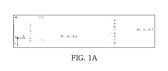
[0038] FIG. 1A illustrates cross-sectional views of two examples of meta-
lenses.
[0039] FIG. 1B illustrates a perspective view of a nanofin.
[0040] FIG. 1C illustrates a side view of a nanofin.
[0041] FIG. 1D illustrates a top view of a nanofin.
[0042] FIG. 1E illustrates a top view of another nanofin with a different
rotation angle.
[0043] FIG. 1F is a diagram illustrating meta-lens simulated efficiency versus
wavelength.
[0044] FIG. 1G is an optical image of a meta-lens.
[0045] FIG. 1H is a scanning electron micrograph image of a portion of a meta-
lens.
[0046] FIG. 11 is a scanning electron micrograph image of a portion of a meta-
lens.
[0047] FIG. 1J is a scanning electron micrograph image of a portion of a meta-
lens.
[0048] FIG. 1K is a scanning electron micrograph image of a portion of a meta-
lens.
[0049] FIG. 2 illustrates an experimental setup used to measure focal spot
sizes and
conversion efficiencies of the meta-lenses.
6
CA 03020261 2018-10-04
WO 2017/176921
PCT/US2017/026206
[0050] FIG. 3A illustrates a focal spot achieved by a fabricated meta-lens.
[0051] FIG. 3B illustrates a focal spot achieved by a fabricated meta-lens.
[0052] FIG. 3C illustrates a focal spot achieved by a fabricated meta-lens.
[0053] FIG. 3D illustrates a focal spot achieved by a commercially available
conventional
objective lens.
[0054] FIG. 3E illustrates a focal spot achieved by a commercially available
conventional
objective lens.
[0055] FIG. 3F illustrates a focal spot achieved by a commercially available
conventional
objective lens.
[0056] FIG. 3G is a diagram illustrating focal spot intensities for the
fabricated meta-lens of
FIG. 3A.
[0057] FIG. 3H is a diagram illustrating focal spot intensities for the
fabricated meta-lens of
FIG. 3B.
[0058] FIG. 31 is a diagram illustrating focal spot intensities for the
fabricated meta-lens of
FIG. 3C.
[0059] FIG. 3J is a diagram illustrating focal spot intensities for the
commercially available
conventional objective lens of FIG. 3D.
[0060] FIG. 3K is a diagram illustrating focal spot intensities for the
commercially available
conventional objective lens of FIG. 3E.
[0061] FIG. 3L is a diagram illustrating focal spot intensities for the
commercially available
conventional objective lens of FIG. 3F.
[0062] FIG. 4A is a diagram illustrating measured focal length versus
wavelength for a meta-
lens.
[0063] FIG. 4B is a diagram illustrating measured magnification versus
wavelength for a
7
CA 03020261 2018-10-04
WO 2017/176921
PCT/US2017/026206
meta-lens.
[0064] FIG. 5A is a diagram illustrating measured efficiency versus wavelength
for two
meta-lenses.
[0065] FIG. 5B is a diagram illustrating measured beam intensity for a meta-
lens.
[0066] FIG. 6 illustrates an experimental setup used to measure performance of
a meta-lens.
[0067] FIG. 7A illustrates a sample image formed by a meta-lens.
[0068] FIG. 7B illustrates a sample image formed by a meta-lens.
[0069] FIG. 7C illustrates a sample image formed by a meta-lens.
[0070] FIG. 7D illustrates a sample image formed by a meta-lens.
[0071] FIG. 7E illustrates a sample image formed by a meta-lens.
[0072] FIG. 7F illustrates a sample image formed by a meta-lens.
[0073] FIG. 7G illustrates a sample image formed by a meta-lens.
[0074] FIG. 7H illustrates a sample image formed by a meta-lens.
[0075] FIG. 71 illustrates a sample image formed by a meta-lens.
[0076] FIG. 7J illustrates a sample of a target object.
[0077] FIG. 7K illustrates a sample image of the target object of FIG. 7J
formed by a meta-
lens.
[0078] FIG. 7L illustrates an image of the target object of FIG. 7J taken by a
commercially
available conventional objective lens.
[0079] FIG. 7M illustrates a sample image formed by a meta-lens.
[0080] FIG. 8A illustrates a perspective view of an example of a nanopillar.
8
CA 03020261 2018-10-04
WO 2017/176921
PCT/US2017/026206
[0081] FIG. 8B illustrates phase shifts versus nanopillar radius.
[0082] FIG. 8C illustrates a result of simulation of a polarization
insensitive nanopillar-based
lens.
[0083] FIG. 9A illustrates a meta-lens including nanopillars.
[0084] FIG. 9B illustrates a perspective view of a nanopillar.
[0085] FIG. 9C illustrates a top view of a nanopillar.
[0086] FIG. 9D illustrates a phase map (p(D) (left) and a transmission map
T(D) (right) of
nanopillars, as functions of diameter across the visible spectrum.
[0087] FIG. 9E illustrates a comparison of phase calculated by finite
difference time domain
(FDTD) simulation of a nanopillar on a glass substrate as a function of
diameter D, and the
phase due to propagation in an isolated cylindrical waveguide.
[0088] FIG. 9F illustrates complex transmission coefficients (T(D)ei`P(D)) of
nanopillars at
three design wavelengths for a range of diameters to provide 27( phase
coverage.
[0089] FIG. 10A illustrates a scanning electron microscope images of a
fabricated meta-lens.
[0090] FIG. 10B illustrates a scanning electron microscope images of a
fabricated meta-lens.
[0091] FIG. 10C illustrates a scanning electron microscope images of a
fabricated meta-lens.
[0092] FIG. 10D illustrates a measured focal spot of a meta-lens.
[0093] FIG. 10E illustrates a measured focal spot of a meta-lens.
[0094] FIG. 1OF illustrates a measured focal spot of a meta-lens.
[0095] FIG. 10G illustrates horizontal cuts according to the focal spot
illustrated in FIG. 10D.
[0096] FIG. 10H illustrates horizontal cuts according to the focal spot
illustrated in FIG. 10E.
[0097] FIG. 101 illustrates horizontal cuts according to the focal spot
illustrated in FIG. 10F.
9
CA 03020261 2018-10-04
WO 2017/176921
PCT/US2017/026206
[0098] FIG. 10J illustrates focusing efficiencies for two fabricated meta-
lenses.
[0099] FIG. 10K illustrates an experimental setup used to measure performance
of meta-
lenses.
[00100] FIG. 10L illustrates an experimental setup used to measure
performance of
meta-lenses.
[00101] FIG. 11A illustrates a measured focal spot of a meta-lens.
[00102] FIG. 11B illustrates a measured focal spot of a meta-lens.
[00103] FIG. 11C illustrates a measured focal spot of a meta-lens.
[00104] FIG. 11D illustrates horizontal cuts corresponding to the focal
spot illustrated
in FIG. 11A.
[00105] FIG. 11E illustrates horizontal cuts corresponding to the focal
spot illustrated
in FIG. 11B.
[00106] FIG. 11F illustrates horizontal cuts corresponding to the focal
spot illustrated
in FIG. 11C.
[00107] FIG. 11G illustrates focusing efficiencies for two fabricated meta-
lenses.
[00108] FIG. 12A illustrates a simulated phase profile of a meta-lens
having an NA of
about 0.85 at a design wavelength of about 660 nm.
[00109] FIG. 12B illustrates a simulated phase profile of a meta-lens
having an NA of
about 0.85 at a design wavelength of about 532 nm.
[00110] FIG. 12C illustrates a simulated phase profile of a meta-lens
having an NA of
about 0.85 at a design wavelength of about 405 nm.
[00111] FIG. 12D illustrates a simulated intensity distribution of the meta-
lens
illustrated in FIG. 12A in the focal region at an x-z plane.
[00112] FIG. 12E illustrates a simulated intensity distribution of the meta-
lens
CA 03020261 2018-10-04
WO 2017/176921
PCT/US2017/026206
illustrated in FIG. 12B in the focal region at an x-z plane.
[00113] FIG. 12F illustrates a simulated intensity distribution of the meta-
lens
illustrated in FIG. 12C in the focal region at an x-z plane.
[00114] FIG. 13A illustrates an image formed by a meta-lens.
[00115] FIG. 13B illustrates an image formed by a meta-lens.
[00116] FIG. 13C illustrates an image formed by a meta-lens.
[00117] FIG. 13D illustrates an image formed by a meta-lens.
[00118] FIG. 14A illustrates an example of an optical system including one
or more
meta-lenses.
[00119] FIG. 14B illustrates an example of an optical system including one
or more
meta-lenses.
[00120] FIG. 14C illustrates an example of an optical system including one
or more
meta-lenses.
[00121] FIG. 14D illustrates an example of an optical system including one
or more
meta-lenses.
[00122] FIG. 14E illustrates an example of an optical system including one
or more
meta-lenses.
[00123] FIG. 14F illustrates an example of an optical system including one
or more
meta-lenses.
[00124] FIG. 14G illustrates an example of an optical system including one
or more
meta-lenses.
[00125] FIG. 14H illustrates an example of an optical system including one
or more
meta-lenses.
[00126] FIG. 141 illustrates an example of an optical system including one
or more
11
CA 03020261 2018-10-04
WO 2017/176921
PCT/US2017/026206
meta-lenses.
[00127] FIG. 15
illustrates a sample fabrication process for forming a visible spectrum
dielectric metasurface based on a conformal chemical vapor deposition approach
such as
atomic layer deposition (ALD).
[00128] FIG. 16A
illustrates a scanning electron microscope (SEM) image of a top
view of dielectric units formed using the disclosed fabrication process.
[00129] FIG. 16B
illustrates a scanning electron microscope (SEM) image of a
perspective view of the dielectric units formed using the disclosed
fabrication process.
[00130] FIG. 17
illustrates a sample fabrication process for forming dielectric
metasurfaces.
DETAILED DESCRIPTION
[00131]
Conventional high NA devices include precision-engineered compound lenses
that are bulky and expensive (e.g., costing upwards of thousands of U.S.
dollars). The bulky
and expensive compound lenses limit the type of applications that can
implement using such
conventional high NA devices and hinders their integration into compact and
cost-effective
systems. In addition, high NA and efficiency are not attainable by way of
visible planar
diffractive lenses because the cross-sections of constituent structures are in
wavelength scale
which precludes an accurate phase profile.
[00132]
Metasurfaces have emerged as one of the leading platforms for development
of miniaturized optical components. Meta-lenses include metasurfaces having
sub-
wavelength-spaced phase shifters with advanced control over the properties of
light, and
allow for versatile functionalities in planar structures of the meta-lenses.
There has been
considerable effort in the development of meta-lenses. Various optical
components ranging
from lenses, holograms and gratings to polarization-selective devices have
been demonstrated
using silicon-based and plasmonic metasurfaces. However, efficient operation
ranges of
meta-lenses to date have been in near-infrared (NIR) spectrums. Achieving
highly efficient
meta-lenses in the visible spectrum poses challenges. The high intrinsic
optical losses of
silicon and plasmonic materials of the metasurfaces in the visible spectral
range
(approximately 400 nm to approximately 700 nm) have prevented the realization
of high
12
CA 03020261 2018-10-04
WO 2017/176921
PCT/US2017/026206
efficiency metasurfaces in this region.
[00133]
Achieving highly efficient meta-lenses demands full control over the phase of
light through precisely fabricated, high-aspect-ratio nanostructures. However,
the
development of such precisely fabricated, high-aspect-ratio nanostructures are
subject to
availability of nanofabrication techniques. For instance, dielectrics with a
transparency
window in the visible spectrum may be used as alternative materials of the
metasurfaces.
However, achieving high-aspect-ratio sub-wavelength structures with vertical
sidewalls is
challenging for these dielectric materials using conventional top-down
fabrication processes
(e.g., lithography followed by dry etching). Non-vertical walls can result in
performance
degradation. Titanium dioxide (TiO2) diffractive elements are also used to
fabricate blazed
gratings at visible wavelengths (e.g., about 633 nm) through a dry etching
process. A TiO2-
based lens (NA of about 0.25) can be built at NIR wavelengths (e.g., about 860
nm) but the
lens experiences similar difficulties with tapered wall profiles as well as
surface roughness,
which are commonly associated with this technique. The surface roughness
contributes to
scattering losses and the tapered wall profile results in errors in the
realized phase.
[00134] In at
least some embodiments of the present disclosure, transmissive meta-
lenses with high-aspect-ratio nanostructures can achieve high NA and optical
efficiency in
the visible range. The metasurfaces including high-aspect-ratio nanostructures
with relatively
smooth surfaces are achieved by a fabrication approach based on titanium
dioxide (TiO2)
prepared by atomic layer deposition (ALD). The TiO2 may be, e.g., amorphous
TiO2. The
use of ALD avoids the aforementioned difficulties with dry etching and allows
for using high
quality amorphous TiO2 with negligible material and scattering loss. The high-
aspect-ratio
metasurfaces fabricated using this approach are substantially lossless in the
visible spectrum.
Based on this fabrication approach, transmissive planar lenses (meta-lenses)
can be achieved.
The meta-lenses can be polarization-sensitive or polarization-insensitive.
[00135] In some
embodiment, the meta-lenses can operate at, e.g., red (about 660 nm),
green (about 532 nm) and blue (about 405 nm) wavelengths with respective
efficiencies of
about 66%, about 73%, and about 86%. In some embodiments, meta-lenses with NA
of about
0.6 and about 0.85 can achieve focusing efficiencies up to, e.g., about 90%
and about 60%,
respectively. These lenses are capable of focusing light into diffraction-
limited spots. At their
13
CA 03020261 2018-10-04
WO 2017/176921
PCT/US2017/026206
respective design wavelengths, these focal spots may, e.g., approximately 1.5
times smaller
than those from a commercially available, conventional high NA device (e.g.,
Nikon model
CFI60 with magnification of 100x and NA of 0.8). The meta-lenses can yield sub-
wavelength
resolution, with image qualities comparable to or superior to that obtained by
the
conventional commercial device. It is to be understood that the term "design"
or "designed"
(e.g., as used in "design wavelength," "design focal length" or other similar
phrases below)
refers to parameters set during a design phase; which parameters after
fabrication may have
an associated tolerance.
[00136] It
should be understood that the techniques of the present disclosure to provide
high NA and efficiency is inclusive of providing lower NA and efficiency. In
other words, if
the meta-lens design technique is capable of achieving NA=0.8, then the meta-
lens design
technique is capable of achieving NA<0.8, such as NA=0.5 or NA=0.1, or other
NA suitable
for the design.
[00137] In some
embodiments, in addition to TiO2, other suitable dielectric materials
include those having a light transmittance over the visible spectrum of at
least about 40%, at
least about 50%, at least about 60%, at least about 70%, at least about 80%,
at least about
85%, at least about 90%, or at least about 95%. For example, other suitable
dielectric
materials can be selected from oxides (such as an oxide of aluminum (e.g.,
A1203), silicon
(e.g., SiO2), hafnium (e.g., Hf02), zinc (e.g., Zn0), magnesium (e.g., MgO),
or titanium (e.g.,
TiO2)), nitrides (such as nitrides of silicon (e.g., Si3N4), boron (e.g., BN),
or tungsten (e.g.,
WIN)), sulfides and pure elements. Aspect ratios of metasurfaces (e.g., a
ratio of height to
width of a nanofin or a ratio of height to diameter of a nanopillar) can be
greater than one, at
least about 1.5:1, at least about 2:1, at least about 3:1, at least about 4:1,
at least about 5:1, at
least about 6:1, or at least about 10:1.
[00138] In some
embodiments, a method of fabricating a visible spectrum meta-lens
involves ALD and etching. For example, the method can include providing a
substrate. The
method further includes applying a resist on a surface of the substrate and
patterning the
resist by, e.g., optical lithography, electron beam lithography, nano-
imprinting, or etching the
resist. The pattern defines openings in the resist, which expose portions of
the surface of the
substrate. The method includes forming a conformal coating, such as by atomic
layer
14
CA 03020261 2018-10-04
WO 2017/176921
PCT/US2017/026206
deposition (ALD), on the resist and the portions of the surface of the
substrate exposed in the
openings. The conformal coating forms the metasurfaces including
nanostructures. The top
surface of the conformal coating is above a top surface of the resist. The
method includes
removing a top portion of the conformal coating by, e.g., etching the
conformal coating to
expose the resist. The method further includes removing the resist to expose
the
metasurfaces including the nanostructures (e.g., nanofins or nanopillars).
[00139] In some embodiments, meta-lenses can include nanofins or
nanopillars (or
other nanostructures) that are formed of, or include, TiO2 (or other
materials). The meta-lens
can achieve a high NA (of, e.g., 0.8 or higher) and a high conversion
efficiency (of, e.g., 86%
or higher) at a visible spectrum (or other spectrums). Such meta-lenses can
provide
diffraction-limited focal spots at arbitrary design wavelengths, and can be
used in various
applications such as optical lithography, laser-based microscopy, and
spectroscopy. The
meta-lens can provide, e.g., a magnification of 170x or higher and can
optically resolve
structures as small as features with sub-wavelength spacing. In some
embodiments, the
single-layer lithographic fabric100ation of the meta-lenses can use
technologies such as deep
UV steppers, and accordingly can facilitate high manufacturing throughput.
[00140] In some embodiments, the ultra-thin and compact features of these
planar
meta-lenses together with their straightforward fabrication can be used in
achieving
miniaturized and lightweight optical systems. The technology will allow a host
of cost-
effective solutions for versatile applications ranging from imaging,
spectroscopy and laser
fabrication to wearable optics. For instance, the compact configuration of the
meta-lens can
be suitable for portable or handheld instruments for many applications. For
example, the
meta-lens may be used in cellphones, cameras, portable computers, microscopes,
virtual
reality devices, augmented reality devices, and other devices.
[00141] Structures of Meta-Lenses Including Nanofins
[00142] FIG. 1A diagrammatically illustrates cross sections in an x-z plane
of two
examples of transmissive dielectric meta-lenses. Each meta-lens includes a
substrate 100 and
multiple nanostructures 102 disposed on the substrate 100. On the left is a
meta-lens
designed for an infinity-corrected lens phase profile, and on the right is a
meta-lens designed
for a bi-convex lens phase profile. It should be further understood that,
although certain types
CA 03020261 2018-10-04
WO 2017/176921
PCT/US2017/026206
of meta-lenses are described here, a meta-lens with any desired phase profile
may be
implemented and is within the scope of the present disclosure. The building
blocks of the
meta-lenses of FIG. 1A include high-aspect-ratio TiO2 nanofins. Here nanofins
are illustrated
by way of example. Other geometries may alternatively or additionally be used
that satisfy
the conditions of being anisotropic with high-aspect-ratio. Although the
nanofins are shown
with a rectangular cross sectional shape, other shapes are encompassed by
embodiments of
this disclosure, such as square-shaped, triangular, and other polygonal or non-
polygonal
shapes that can impart a desired phase profile.
[00143] FIGs.
1B, 1C and 1D are, respectively, perspective, side, and top views of
examples of nanofins. In the embodiment illustrated in FIG. 1C, the nanofins
are formed on a
silicon dioxide (SiO2) substrate. In other embodiments, other substrates can
be used. A unit
cell area for each nanofin is defined as an S x S area encompassing the
nanofin in the x, y
plane, as shown in FIG. 1D.
[00144] In some
embodiments, such as the meta-lens illustrated on the left side of FIG.
1A, the meta-lens functions like a spherical lens. The meta-lens according to
this embodiment
has a phase profile ya(x, y) that meets the constraints of equation (1)
representing an infinity-
corrected lens phase profile, where is the design wavelength, x and y are the
coordinates of
each nanofin within the meta-lens and fis the focal length. The coordinates in
the x,y plane is
illustrated in FIG. 1B.
27r
(x , y) = ¨ (f ¨ x2 +y2 + f ) (1)
Ad
[00145] In some
other embodiments, meta-lenses can function as other types of lenses
rather than, or in addition to, a spherical lens. For example, in an
embodiment as illustrated
on the right side of the FIG. 1A, the meta-lens functions as a bi-convex lens
with a phase
profile va(x,y,z) that meets the constraints of equation (2).
16
CA 03020261 2018-10-04
WO 2017/176921
PCT/US2017/026206
27r
(Pd (X) Y, Z) = )(f ¨ (Apo b AD))
ADob = AI (X ¨ X ob)2 (.Y ¨ Y ob)2 (z ¨ Z ob)2 1
= Ai(x ¨ xini)2 + (y ¨ yini)2 + (z ¨ zini)2
(2)
f = fi+ f2
fi = Al x0b2 + yob2 + zob2
f2 = .. Alxim.2 + yi2 + zim,2
[00146] Implementation of the phase profile in the meta-lens can be
explained by way
of example with respect to equation (1). The phase profile of equation (1) is
imparted via
rotation of each individual nanofin by an angle 0 from a selected axis (e.g.,
either rotation
with respect to the x axis or rotation with respect to the y axis, and 0 for
all nanofins is with
respect to the same axis). As shown in the top view of a nanofin in FIG. 1E, a
rotation 0 of a
nanofin at a given coordinate (x, y) is indicated as Onf(x, y). In the case of
right-handed
circularly polarized incident light, the rotations yield a phase shift of (pnf
(x, y) = 2 9nf(x, y)
accompanied by polarization conversion to left-handed circularly polarized
light. Thus, each
nanofin at (x, y) is rotated by an angle (pnf (x, y) as shown in equation (3).
7r
0 n f (X, y) = ¨Ad (f ¨ A/X2 + y2 + f 2) (3)
[00147] For the bi-convex lens phase profile of equation (2), the rotation
of the
nanofins is by an angle as shown in equation (4) with respect to the
illustration on the right
side of FIG. 1A.
71-
0 n f (X, y) = T (f ¨ (ADob ADO) (4)
-d
[00148] In some embodiments, to improve or maximize polarization conversion
efficiency, the nanofins may operate as half-waveplates, which can be achieved
by
birefringence arising from the asymmetric cross section of nanofins with
appropriately
designed height, width, and length (e.g., as defined in FIGs. 1C and 1D). In
some
embodiments, a cross section of nanofins can have a 2-fold rotational
symmetry, or more
generally, an n-fold rotational symmetry where n is an integer that is 2 or
greater than 2. In
some embodiments, a first nanofin can be substantially aligned with a selected
axis (e.g., a
rotation 0 of the first nanofin is zero), and a second nanofin can be rotated
with respect to the
selected axis and with respect to the first nanofin by an angle 0 that is at
least about 5 , at
17
CA 03020261 2018-10-04
WO 2017/176921
PCT/US2017/026206
least about 10 , at least about 15 , or at least about 20 .
[00149] FIG. 1F
illustrates results of simulations for optimizing nanofin parameters at
three design wavelengths. The simulations can use, e.g., a finite difference
time domain
(FDTD) solver. The three simulated meta-lenses are designed for wavelengths =
660 nm,
= 532 nm, and = 405 nm, where .1d indicates design wavelength. For the
simulations,
periodic boundary conditions are applied at the x- and y-boundaries, and
perfectly matched
layers (PMLs) at the z-boundaries. For the simulated meta-lens designed for
.1d= 660 nm, the
nanofins have, e.g., dimensions of approximately W = 85 nm, L = 410 nm, and H
= 600 nm,
with center-to-center spacing of approximately S = 430 nm. For the simulated
meta-lens
designed for =532 nm, the nanofins have, e.g., dimensions of approximately W =
95 nm, L
= 250 nm, and H = 600 nm, with center-to-center spacing of approximately S =
325 nm. For
the simulated meta-lens designed at = 405
nm, the nanofins have, e.g., dimensions of
approximately W = 40 nm, L = 150 nm, and H = 600 nm, with center-to-center
spacing of
approximately S = 200 nm.
[00150] As shown
in FIG. 1F, conversion efficiencies as high as 95% are achieved for
the different designs, illustrating that a meta-lens can be designed for a
desired wavelength by
tuning of nanofin parameters. The term conversion efficiency as used in this
example is
defined as a fraction of the incident circularly polarized optical power that
is converted to
transmitted optical power with opposite helicity.
[00151] In some
embodiments, three meta-lenses are fabricated. The three fabricated
meta-lenses are designed for wavelengths = 660 nm, .1d = 532 nm, and .1d = 405
nm. Each
meta-lens has a diameter of, e.g., approximately 240 micrometers (pm) and a
focal length of,
e.g., approximately 90 pm, yielding an NA of about 0.8. In some embodiments,
electron
beam lithography techniques can be used to create a lens pattern in the
resist, where a
thickness of the resist can be substantially equal to the designed nanofin
height, H. ALD can
be then used to deposit amorphous TiO2 onto the developed resist. Amorphous
TiO2 can be
chosen because it has low surface roughness, minimal or no absorption at
visible
wavelengths, and a sufficiently high refractive index (e.g., approximately
2.4). The ALD
technique is conformal; and therefore a deposition thickness of at least W/2
(where W is the
nanofin width) can be used to produce void-free nanofins. The deposition can
leave a TiO2
18
CA 03020261 2018-10-04
WO 2017/176921
PCT/US2017/026206
film on top of the resist, which can be subsequently removed by controlled
blanket reactive
ion etching. The remaining electron beam resist can be stripped, leaving high
aspect-ratio
nanofins.
[00152] FIG. 1G is an optical image of one of the fabricated meta-lenses.
FIG. 1H is a
scanning electron microscope (SEM) image of the same fabricated meta-lens.
FIG. 11 is an
SEM image of another of the fabricated meta-lenses from a perspective view at
an edge of the
meta-lens. FIG. 1J is an SEM image of a high-magnification top view of a
portion of the
meta-lens of FIG. 11 near an edge of the meta-lens. FIG. 1K is an SEM image of
a top view
of a portion of the meta-lens near a center of the meta-lens of FIG. 11.
[00153] As discussed above, in some embodiments, the geometrical parameters
of the
nanofins can be defined by the resist rather than top-down etching. Therefore,
high-aspect-
ratio nanofins with approximately 90 vertical sidewalls can be obtained. It
is notable that
achieving these atomically smooth sidewalls is very challenging with a
conventional top-
down approach (e.g., lithography followed by dry etching) because inevitable
lateral etching
results in surface roughness and tapered or conical nanostructures.
[00154] Fabrication and Characterization of Meta-Lenses Including Nanofins
[00155] FIG. 2 illustrates an experimental setup used to measure focal spot
sizes and
conversion efficiencies of the meta-lenses. The experimental setup can
include, e.g., a laser, a
fiber coupled collimator, a long-pass (LP) filter, a quarter waveplate (I/ 4)
, one or more meta-
lenses, a magnification device (with, e.g., magnification of 100x), a tube
lens and a camera.
[00156] In some embodiments, the focal spots of the meta-lenses may be
characterized
using a custom-built microscope as illustrated in FIG. 2. The sources used in
the focal spot
characterizations may be one or more lasers with linewidths of, e.g., less
than 100 megahertz
(MHz). The laser beam may be collimated by a fiber-coupled collimator with a
beam size
diameter of, e.g., 4 millimeters (mm). The collimated beam may be passed
through a Glan-
Thompson polarizer and a quarter waveplate to generate circularly polarized
light. A
magnification device (with, e.g., magnification of 100x, and an NA of 0.9) may
be used to
magnify the image of light focused by the meta-lens. A tube lens with focal
length of, e.g., f =
180 mm may be used to form an image recorded on a camera (e.g., a charge-
coupled device
19
CA 03020261 2018-10-04
WO 2017/176921
PCT/US2017/026206
(CCD) or complementary metal¨oxide¨semiconductor (CMOS) camera).
[00157] FIG. 3A,
FIG. 3B and FIG. 3C illustrate focal spots that are achieved by some
of the fabricated meta-lenses and are recorded by the setup of FIG. 2.
Accordingly, FIG. 3G,
FIG. 3H and FIG. 31 are plots of focal spot intensities for the respective
fabricated meta-
lenses.
[00158] FIG. 3A
shows a highly symmetric focal spot that is obtained at 660 nm for
the meta-lens with design wavelength Ad = 660 nm. The vertical cut of the
focal spot is also
shown in Fig. 3G with a diffraction-limited
(2xANA) full-width at half-maximum (FWHM) of
approximately 450 nm. FIG. 3B shows a focal spot for the meta-lens designed at
the
wavelength of Ad = 532 nm; and FIG. 3H shows its corresponding vertical cut.
This meta-lens
design can be extended to the shorter wavelength region of the visible
spectrum, which is of
great interest in many areas of optics such as lithography and photo-
luminescence
spectroscopy. FIG. 3C shows a focal point for the meta-lens designed at the
wavelength Ad =
405 nm; and FIG. 31 shows its corresponding vertical cut with a FWHM of
approximately
280 nm. Although this wavelength is very close to the band gap of TiO2, namely
about Ag =
360 nm, the absorption loss is still negligible.
[00159] To
compare the performance of the meta-lenses with a commercially available
conventional objective lens, focal spots of such a conventional objective lens
are recorded.
The conventional objective lens has the same NA as the meta-lenses of FIGs 3A-
3C (0.8) and
is designed for visible light. The conventional object lens is bulkier and
more expensive to
build than the meta-lenses. Focal spot intensity profiles of the conventional
objective lens at
wavelengths of 660 nm, 532 nm, and 405 nm are measured using the same setup as
illustrated
in FIG. 2 and described above. Focal point results for the objective are shown
in FIGs. 3D-
3F, with corresponding vertical cuts of the focal spots in FIGs. 3J-3L. The
comparison
between FIGs. 3J-3L for the objective and FIGs. 3G-3I for the meta-lenses
reveals that the
meta-lenses provide smaller (e.g., approximately 1.5 times) and more symmetric
focal spots.
[00160] This
improvement provided by the meta-lenses is at least partially because
conventional high NA objective lenses are designed to image under broadband
illumination.
That is, the conventional objective lenses are designed to correct wavefront
aberrations for
CA 03020261 2018-10-04
WO 2017/176921
PCT/US2017/026206
multiple wavelengths for a range of angles of incidence to meet industry
standards for a
specified field of view, such as by cascading a series of precisely aligned
compound lenses.
However, fabrication imperfections in each individual optical lens and
residual aberration
errors, particularly spherical aberration, result in a focal spot size larger
than theoretical
predictions.
[00161] In
contrast, the meta-lens can be designed to have a phase profile free of
spherical aberration for normally incident light, which can result in a
diffraction-limited spot
at a specific design wavelength. For example, in some embodiments, a root mean
square of
the wave aberration function (WAF1A4s) for the meta-lens designed for 405 nm,
532 nm, and
660 nm may be respectively 0.006X, 0.012X and 0.017X. These values are close
to the
condition for a perfect spherical wavefront. In addition, due to the use of
the geometric phase,
the phase profile of the meta-lens can be dependent on the rotation of the
nanofins, which can
be controlled with very high precision, which is characteristic of electron
beam lithography.
Note that the present disclosure is not limited to electron beam lithography,
and other high
throughput lithography techniques (such as deep-ultraviolet (UV) lithography)
can provide
similar fabrication accuracy within the present disclosure.
[00162] Note
that although each meta-lens can be designed at a specific wavelength,
wavelength-scale focal spots can be observed at wavelengths away from the
designed
wavelength. For example, in some embodiments, for the meta-lens designed at Ad
= 532 nm,
focal spot sizes of approximately 745 nm and approximately 600 nm can be
measured at
incident wavelengths of A = 660 nm and A, = 405 nm, respectively. The
broadening of the
focal spot with respect to the theoretical diffraction-limited values may be
due to chromatic
aberration, because metasurfaces can be dispersive by nature.
[00163] In some
embodiments chromatic aberrations in the fabricated meta-lens can be
more pronounced than lenses based on conventional refractive optics, resulting
in a
wavelength-dependent focal length. FIG. 4A illustrates measured focal length
of the meta-
lens with Ad = 532 nm (D = approximately 2 mm,f = approximately 0.725 mm), and
FIG. 4B
illustrates magnification corresponding to the focal lengths of FIG. 4A, shown
after taking
into consideration the tube lens with a focal length of 100 mm. The wavelength-
dependent
focal length of the meta-lens is generally not an issue for laser-related
imaging, microscopy,
21
CA 03020261 2018-10-04
WO 2017/176921
PCT/US2017/026206
and spectroscopy because monochromatic sources with narrow linewidths are
used. For
example, in Raman microscopes or spectrometers, a 532 nm laser with a
linewidth of a few
picometers is common. In this case, the linewidth-induced broadening of the
focal spot size
and change in focal length is negligible in context.
[00164] Focusing
efficiency of the fabricated meta-lenses are also measured. The
source used for efficiency measurements is a supercontinuum laser with a
linewidth of, e.g.,
nm, where efficiency here refers to a ratio of an optical power of the focused
beam to an
optical power of the incident beam, as captured by a photodetector located at
a same position
as the camera. Incident optical power is also measured, by the light passing
through an
aperture (aluminum on glass) with the same size as the meta-lens.
[00165] FIG. 5A
illustrates measurement results of focusing efficiency for the meta-
lens designed for Ad= 660 nm, where the focusing efficiency remains above 50%
over most
of the visible spectrum. FIG. 5A also illustrates measurement results of
focusing efficiency
for the meta-lens designed for Ad= 532 nm, where there is a focusing
efficiency of 73% at the
design wavelength.
[00166] FIG. 5B
illustrates measurement results of beam intensity for the meta-lens
within a 40 lam span around its focal point. The negligible background signal
demonstrates
not only an excellent phase realization, where the beam converges to a
diffraction-limited
spot, but also a high conversion efficiency of each nanofin. For the meta-lens
designed for Ad
= 405 nm, a measured focusing efficiency of 86% is achieved. The latter
measurement can be
conducted using, e.g., a diode laser instead, because the shortest wavelength
that that tunable
laser can provide is approximately 470 nm. All of the efficiency measurements
are performed
using, e.g., right circularly polarized incident light. However, the
polarization sensitivity of
the design can be overcome by, e.g., implementing the phase profile using
circular cross
section nanopillars in which the phase is controlled via changing their
diameters.
[00167] To
demonstrate the use of the meta-lens in practical imaging, in some
embodiments, another meta-lens can be fabricated with /1d = 532 nm, diameter
of
approximately D = 2 mm and focal length of approximately f = 0.725 mm, giving
an NA of
about 0.8. The imaging resolution can be characterized using, e.g., the 1951
United States Air
22
CA 03020261 2018-10-04
WO 2017/176921
PCT/US2017/026206
Force (USAF) resolution test chart as the target object. FIG. 6 illustrates
the measurement
setup used to measure performance of the meta-lens. The light source can be a
tunable laser
set at, e.g., 550 nm with a bandwidth of, e.g., 5 nm. Because the resulting
image can be larger
than a sensing surface of the camera, the image can be projected onto a white
screen. Its
photo can be taken with, e.g., a digital single-lens reflex (DSLR) camera. The
smallest
features of the target object are lines with widths of, e.g., 2.2 lam and
center-to-center
distances of, e.g., 4.4 lam.
[00168] FIGs. 7A-
7I, 7K and 7M are images formed by the fabricated = 532 nm (D
= 2 mm,f = 0.725 mm) meta-lens. FIG. 7A shows an image of the target object
formed by the
meta-lens, where a dotted-line box indicates a set of four smallest target
object features, and
the smallest two features are the two at the bottom of the dotted-line box.
The scale bar in
FIG. 7A indicates 30 p.m. FIGs. 7B-7E are images of the dotted-line box
portion (as
illustrated in FIG. 7A) of the target object at source wavelengths of 480 nm
(FIG. 7B), 530
nm (FIG. 7C), 590 nm (FIG. 7D) and 620 nm (FIG. 7E). The scale bar in each of
FIGs. 7B-
7E indicates 5 p.m.
[00169] FIGs. 7F-
7I are images of the dotted-line box portion (as illustrated in FIG.
7A) of the target object. To characterize the effects of chromatic aberration,
the target object
is imaged at 530 nm without changing a distance between the meta-lens and the
target object,
while varying the bandwidth of the source: bandwidth of 10 nm (FIG. 7F), 30 nm
(FIG. 7G),
50 nm (FIG. 7H) and 100 nm (which can be the limit of the tunable laser, FIG.
71). Although
the quality of the image may slightly degrade from increasing the bandwidth,
the smallest
features can still be resolvable even at the maximum bandwidth of the laser
at, e.g., 100 nm.
[00170] FIG. 7J
shows an SEM micrograph of a nanoscale H-shaped target prepared
by focused ion beam, where a gap between neighboring holes is approximately
800 nm. The
target is used to compare imaging quality of the meta-lens to that of a
commercially available
conventional objective lens. FIG. 7K is an image of the target object of FIG.
7J formed by the
meta-lens. FIG. 7L is an image of target object of FIG. 7J formed by a
commercially
available conventional objective lens. The image formed by the meta-lens (as
illustrated in
FIG. 7K) has comparable or superior quality to the one formed by the
commercially available
conventional objective lens (as illustrated in FIG. 7L) with the same NA =
0.8. The change in
23
CA 03020261 2018-10-04
WO 2017/176921
PCT/US2017/026206
the image sizes can be due to the difference in the magnification of the
imaging systems. The
scale bar in each of FIGs. 7J-7L indicates 10 p.m.
[00171] FIG. 7M is an image formed by the meta-lens, showing that holes
with sub-
wavelength gaps of 480 nm can be resolved. The scale bar in FIG. 7M indicates
500 nm.
[00172] As can be seen from FIGs. 7A-7I, 7K and 7M, the fabricated = 532 nm
(D =
2 mm, f = 0.725 mm) meta-lens can resolve micrometer-sized lines well. In some
embodiments, the focal length of the meta-lens may vary as the wavelength
changes,
resulting in different levels of magnification (as illustrated in, e.g., FIG.
4B). In the
experimental setup, the meta-lens can be used together with a tube lens
(having, e.g., f = 100
mm) giving a magnification of, e.g., 138x (100/0.725) at, e.g., 530 nm. In
some
embodiments, for incident wavelengths of 480 nm, 590 nm, and 620 nm,
magnifications of
124x, 152x, and 167x can be obtained, respectively, as comparing the ratio of
the image sizes
formed on the camera to the known physical size of the USAF test object.
[00173] The meta-lenses described in some embodiments of the present
disclosure can
include nanofins with rectangular cross-section, where the nanofins can be
rotated to achieve
different target phases. The nanofins can be polarization sensitive. In some
embodiments,
polarization sensitivity of such nanofins can be overcome by implementing the
phase profile
using, e.g., circular cross-section nanopillars, in which the phase is
controlled by nanopillar
diameter.
[00174] Structures of Meta-Lenses Including Nanopillars
[00175] FIG. 8A diagrammatically illustrates an example of a nanopillar
according to
some embodiments of the present disclosure. The nanopillar has a height H in
the z-direction
and a cross-sectional radius R in the x-y plane. The nanopillar occupies a
unit space area with
dimensions Ux U FIG. 8B illustrates that a change in the radius of the
nanopillar affects the
phase shift characteristics of the nanopillar. FIG. 8C is a simulation result
showing that a
meta-lens including nanopillars formed of TiO2 with NA = 0.88 are insensitive
to
polarization.
[00176] FIG. 9A illustrates a meta-lens including nanopillars, according to
some
24
CA 03020261 2018-10-04
WO 2017/176921
PCT/US2017/026206
embodiments of the present disclosure. As illustrated in FIG. 9A, TiO2
nanopillars are
fabricated on a front surface of a substrate (e.g., a glass substrate), such
as, e.g., using an
electron beam lithography technique. In transmission mode, the meta-lens may
focus
collimated light that is incident on a back surface of the substrate into a
spot, as illustrated in
FIG. 9A. To accomplish the focusing, a nanopillar at position (x, y) imparts a
phase given by
equation (5), where Ad is a design wavelength for the meta-lens (e.g., the
meta-lens is
optimized for incident light having a wavelength Ad) and f is a design focal
length of the
meta-lens. It is to be understood that the term "design" (e.g., as used in
"design wavelength,"
"design focal length" or other phrases below) refers to parameters set during
a design phase;
which parameters after fabrication may have an associated tolerance.
27r
yot(x, y) = 27r ¨ (Vx2 +y2 + f 2 ¨ f ) (5)
[00177] In some
embodiments, the phase profile vt(x, y) for each nanopillar may be
realized by adjusting a diameter of the nanopillar. Equation (1) may represent
an infinity-
corrected phase profile to which a meta-lens may be designed, included by way
of example.
It is to be understood that other phase profiles may be used as a basis for
the meta-lens design
instead.
[00178] To
achieve high efficiency, other parameters such as nanopillar height H (as
illustrated in FIG. 9B) and unit cell size U (as illustrated in FIG. 9C) can
be optimized at the
design wavelength Ad. In the example as shown in FIG. 9C, a unit cell can be,
e.g.,
approximately a square and the unit cell size can be a wall length of the
square. In some other
embodiments, the unit cell may be of a different shape, and the unit cell size
may be defined
accordingly. Further, although the nanopillars are illustrated as being
cylindrical, in other
embodiments, the nanopillars may have other shapes.
[00179] In
addition to TiO2, other suitable dielectric materials include those having a
light transmittance over the visible spectrum of at least about 40%, at least
about 50%, at
least about 60%, at least about 70%, at least about 80%, at least about 85%,
at least about
90%, or at least about 95%. For example, other suitable dielectric materials
can be selected
from oxides, nitrides, sulfides and pure elements. Aspect ratios of
nanopillars (e.g., a ratio of
CA 03020261 2018-10-04
WO 2017/176921
PCT/US2017/026206
height to diameter of a nanopillar) can be greater than one, at least about
1.5:1, at least about
2:1, at least about 3:1, at least about 4:1, at least about 5:1, at least
about 6:1, or at least about
10:1.
[00180] Based on
the nanopillars as illustrated in FIGs. 9A-9C, a phase accumulation
may be realized by a waveguiding effect. Thus, the height H of the nanopillars
may be
designed to provide at least a 27c phase coverage through a range of diameters
determined for
the design of the nanopillars. A smallest diameter may be determined primarily
by
attainability due to fabrication constraints; and a largest diameter can be
equal to the unit cell
size U. The unit cell size U may be designed to meet the Nyquist sampling
criterion (U <
A
¨) for a high efficiency. It is to be understood, however, that in some other
embodiments,
2NA
the unit cell size U may be designed such that it does not meet the Nyquist
criterion (e.g.,
A
U
- 2NA
[00181] In some
embodiments, for a design wavelength Ad = 405 nm, a design unit cell
dimension is U=180 nm, a design nanopillar height is H=400 nm and the design
nanopillar
diameters D may vary between 80 nm to 155 nm. In some embodiments, for a
design
wavelength Ad = 532 nm, a design unit cell dimension is U=250 nm, a design
nanopillar
height is H=600 nm and the design nanopillar diameters D may vary between 100
nm to 220
nm. In some embodiments, a design wavelength Ad = 660 nm, a design unit cell
dimension is
U=350 nm, a design nanopillar height is H=600 nm and the design nanopillar
diameters D
may vary between 100 nm to 320 nm. In some embodiments, a first nanopillar can
have a
first diameter, and a second nanopillar can have a second diameter, where the
second
diameter is at least about 1.1 times greater than the first diameter, such as
at least about 1.2
times greater, at least about 1.3 times greater, at least about 1.4 times
greater, or at least about
1.5 times greater.
[00182] FIG. 9D
shows a phase map p(D) (left) and a transmission map T(D) (right),
respectively, as functions of diameter across the visible spectrum, for a meta-
lens designed
for incident light at a design wavelength Ad = 532 nm with nanopillars having
a design height
H = 600 nm and a design unit cell size U = 250 nm. As illustrated in FIG. 9D,
each point on
the phase map shows a relative phase difference between a nanopillar with
diameter D and a
26
CA 03020261 2018-10-04
WO 2017/176921
PCT/US2017/026206
reference point where there is no nanopillar (propagation through the air).
[00183] As a
comparison, the phase imparted solely by the waveguiding effect can be
calculated according to equation (6), where neff is an effective index of the
fundamental mode
(HEll) and H (nanopillar height) is the propagation length. The neff can be
determined using,
e.g., a single step-index circular waveguide model.
2 7r
wG = ffH (6)
Ad
[00184] FIG. 9E
shows that phase determined according to equation (6) (phase due to
propagation in an isolated cylindrical waveguide, considering its fundamental
mode HEll at
Ad= 532 nm) is similar to phase calculated via FDTD analysis of the nanopillar
on the glass
substrate (with design wavelength 2d = 532 nm, and nanopillars having a design
height H =
600 nm and a design unit cell size U = 250 nm). As shown in FIG. 9E, even
better agreement
in phases can be achieved for larger diameters, where the confinement of the
fundamental
mode increases. While the confinement along the propagation direction
(standing wave due
to reflections at both facets of the nanopillars) and near-field coupling
between nanopillars
can be neglected, an average absolute difference between phases calculated
using the
waveguiding effect and the full-wave analysis can be less than 16. This may
indicate that the
waveguiding effect may be the dominant mechanism accounting for the phase
realization. In
some embodiments, full phase coverage (27c) with high transmission (of, e.g.,
greater than
about 87%) can be achieved.
[00185] In some
embodiments, by varying diameters of nanopillars as a function of
position (xi, yi), the effective index of the propagating mode can be changed
to achieve the
desired phase profile of equation (5). To construct a corresponding meta-lens,
the phase mask
yot(xi, yi) may be discretized, assuming square lattice unit cells of
dimensions U x U. At
i((x1ig each position (xi,yi) an appropriate diameter minimizing 1Tnie0t31/) ¨
T(D)e") is
selected, where Tni is the transmission averaged over all the diameters.
[00186] FIG. 9F
shows the complex transmission coefficients (T(D)euP())) at three
design wavelengths for a range of diameters to provide 2n phase coverage. Each
point in the
27
CA 03020261 2018-10-04
WO 2017/176921
PCT/US2017/026206
complex plane represents an amplitude and phase of transmission of a
nanopillar with
diameter D, for a given unit cell size and nanopillar height at the
corresponding design
wavelength. High transmission (with small modulation over the range of used
diameters) and
close to 27c phase coverage is evident for all three design wavelengths.
[00187] Fabrication and Characterization of Meta-Lenses Including
Nanopillars
[00188] In some embodiments, three separate meta-lenses can be fabricated,
each with
a design NA of about 0.6, and are optimized for design wavelengths of about
405 nm, about
532 nm and about 660 nm. FIGs. 10A-10C are scanning electron microscope (SEM)
images
of one of the fabricated meta-lenses. FIGs 10D-10F illustrate measured focal
spots of the
fabricated meta-lenses. FIGs. 10G-10I are horizontal cuts corresponding
respectively to the
focal spots illustrated in FIGs. 10D-10F. FIG. 10J illustrates focusing
efficiencies for two of
the fabricated meta-lenses. FIGs. 10K and 10L illustrate experimental setups
used to
characterize the three fabricated meta-lenses.
[00189] In some embodiments, to calculate the Strehl ratio, the measured
intensities of
the horizontal cuts may be normalized to those of ideal airy functions with a
same area under
the curve. Airy functions with a maximum intensity of unity and diffraction-
limited full-
widthd at half-maximum (FWHM) of FWHM = 0.514 ¨NA are plotted over the
horizontal cuts
illustrated in FIGs 10G-10I. In some embodiments, Strehl ratios of about 0.80,
about 0.82 and
about 0.83 may be achieved at wavelengths of about 405 nm, about 532 nm and
about 660
nm, respectively. Corresponding Strehl ratios for vertical cuts (not shown
here) may be about
0.81, about 0.84 and about 0.81, which are close to those for horizontal cuts,
revealing
symmetry of the focal spots.
[00190] FIG. 10J illustrates measured focusing efficiencies for the meta-
lenses of
design wavelengths of 532 nm and 660 nm. Measured focusing efficiencies as
high as about
70% and about 90% can be obtained for meta-lenses designed at wavelengths 532
nm and
660 nm, respectively. In some embodiments, measured focusing efficiencies as
high as about
30% are obtained for meta-lenses designed at a wavelength of 405 nm (not shown
in FIG.
10J). Focusing efficiency can be defined as a ratio of optical power of the
measured focused
beam to optical power of the incident beam. The incident beam may be measured
as the
28
CA 03020261 2018-10-04
WO 2017/176921
PCT/US2017/026206
optical power passing through a circular aperture (e.g., aluminum on glass)
with a same
diameter (300 lam) as the meta-lenses.
[00191] In some
embodiments, three separate meta-lenses may be fabricated, each with
a design NA of about 0.85, optimized for design wavelengths of about 405 nm,
about 532 nm
and about 660 nm. FIGs. 11A-11C illustrate measured focal spots of the meta-
lenses. FIGs.
11D-11F illustrate horizontal cuts corresponding respectively to the focal
spots illustrated in
FIGs. 11A-11C. FIG. 3G illustrates focusing efficiencies for two of the
fabricated meta-
lenses.
[00192] As shown
in FIGs. 11A-11C, symmetric focal spots with diffraction-limited
FWHMs can be achieved. As shown in FIGs. 11D-11F, FWHMs of the horizontal cuts
of
these focal spots may be about 259 nm, about 327 nm, and about 424 nm for meta-
lenses at
respective design wavelengths of about 405 nm, about 532 nm and about 660 nm.
The
FWHMs of the vertical cuts of these focal spots may be about 256 nm, about 344
nm and
about 428 nm for the meta-lenses at respective design wavelengths of about 405
nm, about
532 nm and about 660 nm. The Strehl ratios from the measured horizontal cuts
may be about
0.76, about 0.82 and about 0.85 corresponding to meta-lenses designed at
respective
wavelengths of 405 nm, 532 nm and 660 nm. The Strehl ratios from the measured
vertical
cuts may be about 0.78, about 0.84 and about 0.85 corresponding to the meta-
lenses designed
at respective wavelengths of 405 nm, 532 nm and 660 nm. The diffraction-
limited focusing
and high Strehl ratios confirm the quality of fabrication and capability of
this TiO2-based
(other based on other suitable materials) platform to realize high performance
optics in the
visible spectrum.
[00193] FIG. 11G
illustrates efficiency of two of the meta-lenses with NA of about
0.85. In some embodiments, efficiencies as high as about 60% may be achieved
for both
meta-lenses with design wavelengths of about 532 and about 660 nm. In some
embodiments,
efficiency as high as about 33% may be achieved for the meta-lens designed at
about 405 nm.
This can be due to the stricter fabrication tolerance of this design. In other
words, nanopillars
designed for shorter wavelengths may have 27( phase coverage over a smaller
range of
diameters and smaller unit cells. In some embodiments, fabrication errors may
be more
pronounced, resulting in reduced efficiency. For example, a mean value of the
nanopillar
29
CA 03020261 2018-10-04
WO 2017/176921
PCT/US2017/026206
diameters used to build the meta-lens designed at 405 nm may be 120 nm,
whereas a mean
value of the nanopillar diameters used to build the meta-lens designed at 660
nm may be 215
nm.
[00194] FIGs. 12A-12C illustrate phase profiles of meta-lenses of NA of
about 0.85 at
the three design wavelengths (ild= 660 nm, 532 nm and 405 nm). The discretized
FDTD
simulated phases that are theoretically available with suitable nanopillar
diameters are
overlaid on FIGs. 12A-12C. Good curve fits may be obtained for all three
designs. This can
be further confirmed by performing FDTD analysis on meta-lenses with similar
NA of about
0.85 but smaller lens diameters (e.g., about 24 pim) than the fabricated ones
due to finite
computational resources.
[00195] FIGs. 12D-12F illustrate simulated intensity distributions of
respectively the
meta-lenses illustrated in FIGs. 12A-12C in the focal region at the x-z plane.
In some
embodiments, efficiency may be a ratio of the optical power in the focal spot
area (circle of
radius 2xFWHM spanning the center of the focal spot) to the incident optical
power. In some
embodiments, efficiencies as high as about 79%, about 83% and about 84% may be
achieved
for meta-lenses designed at wavelengths of about 405 nm, about 532 nm and
about 660 nm,
respectively.
[00196] In some embodiment, a meta-lens may be fabricated for imaging
purposes,
with a diameter of about 2 mm designed at about = 532 nm. The 1951 United
States Air
Force (USAF) resolution test chart may be used to discern a resolving power of
the meta-
lens. FIGs. 13A-13D illustrate images of the smallest three-bar groups on the
chart (bar width
of about 2.2 p.m and center-to-center distance of about 4.4 p.m), formed by
the meta-lens at
different illumination wavelengths. As shown in FIGs. 13A-13D, the meta-lens
can resolve
micron-sized features across the visible spectrum. Differences in image size
at various
wavelengths may be due to the focal length of the meta-lens being a function
of wavelength,
resulting in a wavelength-dependent magnification. For example, magnification
can change
from about 260x to about 337x by varying a wavelength of incidence from about
490 nm to
about 650 nm.
[00197] Configurations of Optical Systems Incorporating One or More Meta-
Lenses
CA 03020261 2018-10-04
WO 2017/176921
PCT/US2017/026206
[00198] FIGs.
14A-14I illustrate various examples of portions of optical systems
incorporating one or more meta-lenses according to various embodiments of the
present
disclosure.
[00199] As
illustrated in FIG. 14A, an optical system 610 may include a meta-lens 612
and a lens 613. The meta-lens 612 may be designed to provide one or more
functionalities
such as described in the present disclosure. The meta-lens 612 may be at a
distance from the
lens 613, or may be positioned against the lens 613 to contact the lens 613.
Also in this
embodiment, the meta-lens 612 and the lens 623 may have similar dimensions
(e.g.,
diameter).
[00200] As
illustrated in FIG. 14B, an optical system 620 may include a meta-lens 622
and a lens 623. The meta-lens 622 may be designed to provide one or more
functionalities
such as described in the present disclosure. The meta-lens 622 may be at a
distance from the
lens 623, or may be positioned against the lens 623 to contact the lens 623.
Also in this
embodiment, the meta-lens 622 may have a different (lesser or higher)
dimension (e.g.,
diameter) than the lens 623.
[00201] As
illustrated in FIG. 14C, an optical system 630 may include a meta-lens 632
and a lens 633. The meta-lens 632 may be designed to provide one or more
functionalities
such as described in the present disclosure. The meta-lens 632 may be at a
distance from the
lens 633, or may be positioned against the lens 633 to contact the lens 633.
Also in this
embodiment, the meta-lens 632 may have a much smaller dimension (e.g.,
diameter) than the
lens 633, such as to correct functionality of a portion of the lens 633.
[00202] As
illustrated in FIG. 14D, an optical system 640 may include three meta-
lenses 642a, 642b, and 642c. The meta-lenses 642 may be each designed to
provide one or
more functionalities such as described in the present disclosure. In this
example, the meta-
lenses 642a, 642b may be stacked (either as two meta-lens units in a single
fabricated meta-
lens, or as two separately fabricated meta-lenses). The meta-lens 642c may be
at a distance
from the meta-lens 642b, or may be positioned against (or stacked on) the meta-
lens 642b.
Also in this embodiment, the meta-lenses 642a, 642b, 642c may have similar
dimensions
(e.g., diameter), although in some other embodiments the relative dimensions
may be
different instead.
31
CA 03020261 2018-10-04
WO 2017/176921
PCT/US2017/026206
[00203] As
illustrated in FIG. 14E, an optical system 650 may include meta-lenses
652a, 652b and a lens 653. Each meta-lens 652a, 652b may be designed to
provide one or
more functionalities such as described in the present disclosure. In this
example, the lens 653
may be positioned between the meta-lenses 652a, 652b. The lens 652 may be at
distance from
one or both meta-lenses 652a, 652b or may be positioned against one or both
meta-lenses
652a, 652b. Also in this embodiment, the meta-lenses 652a, 652b and the lens
643 may have
similar dimensions (e.g., diameter), although in some other embodiments the
relative
dimensions may be different instead.
[00204] As
illustrated in FIG. 14F, an optical system 660 may include a meta-lens 662
and lenses 663a, 663b. The meta-lens 662 may be designed to provide one or
more
functionalities such as described in the present disclosure. In this example,
the meta-lens 662
may be positioned between the lenses 663a, 663b. The meta-lens 662 may be at
distance from
one or both lenses 663a, 663b or may be positioned against one or both lenses
663a, 663b.
Also in this embodiment, the meta-lens 662 and the lenses 663a, 663b may have
similar
dimensions (e.g., diameter), although in some other embodiments the relative
dimensions
may be different instead.
[00205] As
illustrated in FIG. 14G, an optical system 670 may include a meta-lens 672
and a reflector 675. The meta-lens 672 may be designed to provide one or more
functionalities such as described in the present disclosure. In this example,
the meta-lens 672
may be positioned in front of a reflector 675 (with respect to a direction of
incident light).
The meta-lens 672 may be at a distance from the reflector 675, or may be
positioned against
the reflector 675 to contact the reflector 675. Also in this embodiment, the
meta-lens 672 and
the reflector 675 may have similar dimensions (e.g., diameter), although in
some
embodiments the relative dimensions may be different instead.
[00206] As
illustrated in FIG. 14H, an optical system 680 may include a meta-lens 682
and a reflector 685. The meta-lens 682 may be designed to provide one or more
functionalities such as described in the present disclosure. In this example,
the meta-lens 682
may be positioned behind the reflector 685 (with respect to the direction of
incident light),
such as in a case in which the reflector 685 is partial reflective, or a case
in which the
reflector 685 is reflective on both sides. The meta-lens 682 may be at a
distance from the
32
CA 03020261 2018-10-04
WO 2017/176921
PCT/US2017/026206
reflector 685, or may be positioned against the reflector 685 to contact the
reflector 685. Also
in this embodiment, the meta-lens 682 and the reflector 685 may have similar
dimensions
(e.g., diameter), although in some other embodiments the relative dimensions
may be
different instead.
[00207] As
illustrated in FIG. 141, an optical system 690 may include a meta-lens 692,
a lens 693, and a reflector 695, illustrating that multiple components may be
combined with
one or more meta-lenses such as meta-lens 692. The meta-lens 692 may be
designed to
provide one or more functionalities such as described in the present
disclosure.
[00208] In some
embodiments, a meta-lens may have a stacked configuration, where a
first substrate with first nanopillars or first nanofins (or other
nanostructures) is stacked on a
second substrate with second nanopillars or second nanofins (or other
nanostructures).
Further, in some embodiments, more than two substrates with associated
nanostructures may
be stacked. Such a stacked configuration may be in a form of two or more meta-
lens units in
a single fabricated meta-lens, or may be two or more separately fabricated
meta-lenses
stacked together. In some embodiments, each meta-lens or meta-lens unit has a
different
phase profile. For example, a first meta-lens or meta-lens unit may have a
phase profile
designed for a particular focal length, and a second meta-lens or meta-lens
unit may have a
phase profile designed to correct for aberrations.
[00209] Although
various examples described and illustrated herein may refer to
specific design wavelengths, such as red, green or blue, it is to be
understood that a meta-lens
may be designed and fabricated for any wavelength in the visible spectrum or
outside of the
visible spectrum.
[00210] Although
described herein with respect to transmission of visible light through
the meta-lens, in some embodiments, the substrate may be reflective, and the
meta-lens is a
reflective lens. For example, the nanopillars or the nanofins may be
fabricated over a mirror.
In some such embodiments, the substrate includes a reflective layer. A
material of the
substrate may be transmissive (e.g., glass or other transmissive material)
with a reflective
layer on either side, or the material of the substrate may be non-
transmissive, with the
reflective layer between the material of the substrate and the nanopillars or
the nanofins. A
dielectric layer (e.g., a dielectric thin film) may optionally be disposed
between the reflective
33
CA 03020261 2018-10-04
WO 2017/176921
PCT/US2017/026206
layer and the nanopillars. Examples of a reflective layer may include a
metallic layer such as
a layer of aluminum, silver, gold, or other metal, or a layer of any other
reflective material.
[00211] Fabrication of Metasurfaces of Meta-Lenses
[00212] In some embodiments, the nanostructures (e.g., nanofins or
nanopillars) of the
meta-lenses can be fabricated through a fabrication process using atomic layer
deposition
(ALD) that provides for the formation of high efficiency, visible spectrum
dielectric
metasurfaces (DMs) having the desirable characteristics disclosed herein in
various
embodiments of the present disclosure. The fabrication process may implement
cleanroom
process operations such as lithography and reactive ion etching, reproducibly
providing deep
subwavelength dielectric units with desired geometries and low loss in the
visible spectrum.
[00213] In some embodiments, because the fabrication process uses ALD,
different
dielectric materials can be used to create a DM. For example, metal and non-
metal oxides
(such as an oxide of aluminum (e.g., A1203), silicon (e.g., SiO2), hafnium
(e.g., Hf02), zinc
(e.g., Zn0), magnesium (e.g., MgO), or titanium (e.g., TiO2)), metal and non-
metal nitrides
(such as nitrides of silicon (e.g., Si3N4), boron (e.g., BN), or tungsten
(e.g., WIN)), metal and
non-metal sulfides, and pure elements can be deposited via ALD. In some
embodiments,
titanium dioxide (TiO2) may be selected due to its high refractive index and
low loss in the
visible spectrum, but the fabrication process can use other dielectric
materials, such as other
dielectric materials selected depending on a desired end application.
[00214] In accordance with ALD, one or more deposition cycles may be
performed by
sequentially exposing a substrate, or a portion of the substrate, to
deposition gases of
chemical precursors or reactants. The precursors react and form at least a
partial layer of a
dielectric material on the substrate during a deposition cycle, and further
deposition cycles
can be performed until a desired thickness is attained. In some embodiments,
composite
coatings of different dielectric materials also can be formed using ALD.
[00215] FIG. 15 illustrates a sample fabrication process for forming a
visible spectrum
dielectric metasurface based on a conformal chemical vapor deposition approach
such as
atomic layer deposition (ALD). The process begins with providing a substrate
as shown in
step a of FIG. 15. The substrate may be transparent in the visible spectrum,
such as shown,
34
CA 03020261 2018-10-04
WO 2017/176921
PCT/US2017/026206
by way of example, as one including fused silica. Suitable substrates that are
transparent in
the visible spectrum can have a light transmittance of at least about 40%, at
least about 50%,
at least about 60%, at least about 70%, at least about 80%, at least about
85%, at least about
90%, or at least about 95%, over the visible spectrum or a design or working
wavelength in
the visible spectrum.
[00216] A resist
(e.g., an electron beam resist or a photoresist) is applied (shown in
step b of Fig. 15) to the substrate by spinning at a particular rate to set a
thickness of a
resulting resist layer. The thickness of the resist layer may be a parameter
which sets a height
of resulting dielectric units in the fabricated DM, and may contribute to a
phase of an exiting
light, and affects an efficiency of the DM. In general, the spin rate and a
viscosity of the resist
can be controlled to adjust the thickness of the applied resist layer. As an
example, a spin rate
of about 5,000 rpm (revolutions per minute) while applying an electron beam
resist of a
particular viscosity may result in a thickness of a resist layer of about 400
nm.
[00217] As shown
in step c of FIG. 15, the resist layer is exposed (e.g., using electron
beam lithography or photolithography) and developed, forming an inverse
pattern for forming
the dielectric units. A resulting patterned resist layer is formed with, or
defines, gaps,
openings, or recesses exposing portions of a surface of the substrate. The
openings can have
an aspect ratio greater than about one, such as at least about 1.5:1, at least
about 2:1, at least
about 3:1, at least about 4:1, or at least about 5:1, and up to about 10:1 or
greater, or up to
about 20:1 or greater.
[00218] As shown
in step d of FIG. 15, ALD is performed to deposit a film of a
dielectric material over the patterned resist layer and the exposed portions
of the surface of
the substrate. A feature of ALD exploited in the fabrication process to
achieve high aspect
ratio dielectric units with smooth surfaces may be the conformal nature of the
film as it is
deposited. Top and sidewall surfaces of the patterned resist layer and the
exposed portions of
the surface of the substrate can be conformally coated by the dielectric
material using ALD.
Thus, the film can take on a shape of the openings in the patterned resist
layer.
[00219] For
example, an opening having substantially vertical sidewalls (e.g.,
substantially perpendicular to a respective exposed portion of the surface of
the substrate
within the opening) results in a dielectric unit having substantially vertical
sidewalls, which
CA 03020261 2018-10-04
WO 2017/176921
PCT/US2017/026206
allow precise control over a phase of an exiting wavefront with reduced phase
errors. By way
of comparison, top-down etching techniques can introduce sloped sidewalls,
which can lead
to phase errors. Further, the conformal coating provides for dielectric units
having
exceptionally smooth surfaces so as to reduce losses from scattering, such as
having a root
mean square (RMS) surface roughness of no greater than about 20 nm, no greater
than about
15 nm, no greater than about 10 nm, no greater than about 5 nm, no greater
than about 4 nm,
no greater than about 3 nm, no greater than about 2 nm, or no greater than
about 1 nm. By
way of comparison, top-down etching techniques can result in undesired
sidewall roughness,
which can lead to scattering losses.
[00220] In
accordance with ALD, a first ALD cycle is performed to deposit the
dielectric material over the substrate held or housed within a deposition
chamber, followed by
performing a second ALD cycle to deposit the dielectric material over the
substrate, followed
by performing a third ALD cycle to deposit the dielectric material over the
substrate, and so
on until a desired amount of the dielectric material is deposited. In some
embodiments,
performing each ALD cycle includes exposing the substrate to deposition gases
of chemical
precursors or reactants. In the case of TiO2 as the dielectric material, a
first precursor is a
titanium-containing precursor, such as an organometallic compound of titanium,
and the
second precursor is an oxygen-containing precursor. The first precursor and
the second
precursor react and form at least a partial layer of the dielectric material
over the patterned
resist layer and the exposed portions of the surface of the substrate.
Accordingly, ALD is
used to successively deposit additional dielectric material of the film in a
layer-by-layer
fashion until the film fills the openings in the patterned resist layer (as
shown in step e of
FIG. 15). Each deposited layer conformally coats a previously deposited layer.
[00221] In the
fabrication process of some embodiments, a temperature of the ALD
cycles, TALD, may be controlled to be lower than that used for ALD in other
contexts. For
example, in other contexts, ALD can be used to deposit films at about 150 C
or higher;
however, for certain resists, temperatures above about 115 C can cause a
resist to reflow so
that a resist pattern deteriorates. Thus, in some embodiments, for the ALD
cycles as shown in
steps d and e of FIG. 15, the temperature may be controlled or maintained
below about
115 C, such as about 110 C or below, about 105 C or below, about 100 C or
below, or
about 90 C.
36
CA 03020261 2018-10-04
WO 2017/176921
PCT/US2017/026206
[00222] In some
embodiments, an amount of the dielectric material deposited in the
ALD cycles (as shown in parts d and e of FIG. 15) may be a function of a
maximum desired
feature size (e.g., a feature width or diameter) of a dielectric unit (e.g.,
wmax in step d of FIG.
15). Depending on the pattern of the resist layer, the dielectric film may be
deposited to a
thickness, tfilm, over the patterned resist layer to achieve the maximum
desired feature size.
For example, as illustrated in part e of FIG. 15, to obtain a feature width
wmax in an opening
of the patterned resist layer, tfilm? wmax/2, and a top surface of the
dielectric film is above the
top surface of the patterned resist layer. In some embodiments, tfilm is at
least about 50 nm
greater than wmax/2. It is noted that the thickness tfilm corresponds to an
amount of the
deposited dielectric material that would cover a planar surface with an
uniformly thick film of
the dielectric material having the thickness tfilm, and corresponds a
thickness of a top portion
of the dielectric film deposited over the top surface of the patterned resist
layer and extending
over the openings of the patterned resist layer.
[00223] As shown
in step f of FIG. 15, blanket etching may be performed to remove
the top portion of the dielectric film and expose the top surface of the
patterned resist layer
and top surfaces of resulting dielectric units disposed in the openings of the
patterned resist
layer. For example, reactive ion etching may be used, such as using a mixture
of C12 gas and
BC13 gas, to remove the top portion of the dielectric film to an etch depth
substantially equal
to the thickness tfilm, although an etch depth greater than the thickness
tfilm is also
contemplated, such as including removing a top portion of the patterned resist
layer via an
etch depth of tfilm + about 10 nm. In such fashion, excess dielectric film is
removed, and the
patterned resist layer (or a residual thereof) is exposed.
[00224] As shown
in step g of FIG. 15, the patterned resist layer is removed by
exposing to a resist removal solvent, leaving high aspect ratio dielectric
units of the DM over
the substrate, and having an aspect ratio greater than about one, such as at
least about 1.5:1, at
least about 2:1, at least about 3:1, at least about 4:1, or at least about
5:1, and up to about 10:1
or greater, or up to about 20:1 or greater.
[00225] FIG. 16A
illustrates a scanning electron microscope (SEM) image of a top
view of dielectric units formed using the disclosed fabrication process. The
fabrication
process can produce arbitrary nanostructures such as nanopillars or nanofins
of varying
37
CA 03020261 2018-10-04
WO 2017/176921
PCT/US2017/026206
widths, and can attain such widths on a visible spectrum subwavelength scale.
[00226] FIG. 16B
illustrates a scanning electron microscope (SEM) image of a
perspective view of the dielectric units formed using the disclosed
fabrication process. The
resulting nanostructures may be anisotropic with sidewalls substantially
perpendicular to a
surface of a substrate. A feature width down to about 25 nm on an about 75 nm
pitch (center-
to-center spacing between adjacent dielectric units), with a height of the
dielectric units from
about 100 nm to about 500 nm, can be attained by way of example. Further
feature width
reduction can be attained with the fabrication process of some embodiments of
this
disclosure. For example, dielectric units with widths of about 10 nm or less
are encompassed
by this disclosure.
[00227] In some
embodiments, dielectric units with high aspect ratios can be attained
with the fabrication process. For example, dielectric units with heights up to
about 500 nm or
greater or up to about 1,000 nm or greater can be attained. In some
embodiments, widths of
dielectric units are less than a design wavelength of incident light in the
visible spectrum,
such as no greater than about 400 nm, no greater than about 300 nm, no greater
than about
200 nm, no greater than about 150 nm, no greater than about 100 nm, no greater
than about
50 nm, about 50 nm to about 200 nm, or about 25 nm to about 200 nm.
[00228] In some
embodiments, dielectric units can have generally rectangular or other
elongated cross-sections (along or projected onto a plane parallel to a
surface of a substrate
over which the dielectric units are disposed) having a width and a length, and
where the
length is greater than the width, and the length and the width are less than a
design
wavelength of incident light in the visible spectrum, such as no greater than
about 400 nm, no
greater than about 300 nm, no greater than about 200 nm, no greater than about
150 nm, no
greater than about 100 nm, no greater than about 50 nm, about 50 nm to about
200 nm, or
about 25 nm to about 200 nm.
[00229] In some
embodiments, dielectric units can have generally circular cross-
sections or other even rotationally symmetric cross-section (along or
projected onto a plane
parallel to a surface of a substrate over which the dielectric units are
disposed) having a
diameter, and the diameter is less than a design wavelength of incident light
in the visible
spectrum, such as no greater than about 400 nm, no greater than about 300 nm,
no greater
38
CA 03020261 2018-10-04
WO 2017/176921
PCT/US2017/026206
than about 200 nm, no greater than about 150 nm, no greater than about 100 nm,
no greater
than about 50 nm, about 50 nm to about 200 nm, or about 25 nm to about 200 nm.
In some
embodiments, a pitch of adjacent dielectric units is less than a design
wavelength of incident
light in the visible spectrum, such as no greater than about 400 nm, no
greater than about 300
nm, no greater than about 200 nm, no greater than about 150 nm, no greater
than about 100
nm, no greater than about 50 nm, about 50 nm to about 200 nm, or about 25 nm
to about 200
nm.
[00230] In some
embodiments, dielectric units can have substantially consistent widths
or diameters along their long axes, such that a width or diameter at a top end
of a dielectric
unit is substantially the same as a width or diameter at a bottom end of the
dielectric unit. It is
noted that the above-mentioned dimensions are also applicable for openings of
a patterned
resist layer within which dielectric units are formed during the fabrication
process of some
embodiments.
[00231] In some
embodiments, an approach for creating transmissive dielectric
metasurfaces (DMs) may use a bottom-up nanofabrication process via atomic
layer
deposition, to provide high-aspect-ratio, anisotropic dielectric
nanostructures with reduced
surface roughness. In some embodiments, TiO2 may be selected as a base
material because it
has a transparency window (k 0) for wavelengths longer than about 360 nm and
its inter-
band transition lies just outside of the visible spectrum, resulting in a
sufficiently high index
of refraction for strong light-matter interactions. TiO2 may be deposited by
atomic layer
deposition (ALD) at about 90 C with tetrakis(dimethylamido)titanium (TDMAT)
as a
precursor due to its high deposition rate and absence of defect-driven
absorption that may
occur with TiC14-based precursors. Additionally, use of an ALD process offers
several
advantages. ALD is a self-limiting process providing conformal coverage and
monolayer
control of a film thickness. The conformal nature of the technique is
desirable to produce
high aspect ratio nanostructures. The uniform and monolayer coverage yields
smooth films
with reduced defects compared to those prepared via sputtering or evaporation.
Furthermore,
the ALD process allows precise control of a material phase of the deposited
TiO2, producing
amorphous, rutile, or anatase films, depending on the deposition temperature.
[00232] The TiO2
films may have surface roughness that is much smaller than the
39
CA 03020261 2018-10-04
WO 2017/176921
PCT/US2017/026206
incident wavelength. In some embodiments, a TiO2 film may be deposited on a
fused silica
substrate. The root mean square (RMS) roughness may be about 0.738 nm, which
is on the
order of the surface roughness of the underlying substrate. The measured
roughness of the
amorphous TiO2 is an order of magnitude less than other phases such as
polycrystalline rutile
or anatase. The latter two phases generally have grain boundaries and RMS
roughness as high
as about 5-10 nm, both of which can contribute to light-scattering loss.
[00233] To
achieve highly efficient metasurface devices while preserving the optical
properties of ALD-prepared TiO2, the fabrication process shown in FIG. 17 may
be used.
[00234] As
illustrated in step A of FIG. 17, an electron beam resist (EBR) is spun onto
a fused silica substrate to produce a layer with thickness, tresjst. Control
of t , is desired
_res.st
because it sets a height of final nanostructures. The resist is patterned
using electron beam
lithography and is subsequently developed in solution to remove the exposed
EBR. This
pattern may be the inverse of a final metasurface.
[00235] As
illustrated in step B of FIG. 17, the inverse of a final metasurface pattern
is
imprinted into the EBR by electron beam lithography and subsequent development
of the
pattern. The boxed area is an expanded cross-section of a maximum feature
width, w.
[00236] The
exposed sample is transferred to an ALD chamber set to about 90 C.
The purpose of this temperature is twofold: It produces the desired amorphous
phase and
keeps the EBR below its glass transition temperature (e.g., prevents
deterioration of
nanoscale patterns).
[00237] As
illustrated in step C of FIG. 17, initial TiO2 deposition via ALD
conformally coats sidewalls and top of the EBR and exposed substrate. During
the
deposition, the gaseous TiO2 precursor (Tetrakis(dimethylamido)titanium or
TDMAT) coats
substantially all exposed surfaces, producing a conformal film on top and
sidewalls of the
EBR as well as on the exposed fused silica substrate. The ALD process is
allowed to reach a
specific thickness such that all features are completely filled with TiO2.
[00238] As
illustrated in step D of FIG. 17, completed deposition of the TiO2 yields a
film thickness greater than half the width of the maximum feature size, tfiim
> w/2. Because
CA 03020261 2018-10-04
WO 2017/176921
PCT/US2017/026206
the conformal ALD process fills exposed features from both sides, the total
ALD film
thickness is tfilm > w/2, where w is a maximum width of all gaps or recesses.
In some
embodiments, the deposition may be allowed to proceed well beyond the lower
threshold of
half the feature width to ensure that TiO2 had sufficiently diffused into all
pores and that there
are no voids in the final nanostructures.
[00239] As
illustrated in step E of FIG. 17, the residual TiO2 film that coats a top
surface of the resist is removed by reactive ion etching the sample in a
mixture of BC13 and
C12 gas (about 8:2), similar to a planarization technique. The etch depth may
be substantially
equal to tfilm so that the etching process exposes the underlying resist and
the top of the
nanostructures. Step E of FIG. 17 shows the exposed tops of the TiO2
metasurface and
residual EBR after reactive ion etching with a mixture of C12 and BC13 ions
(top and side
views).
[00240] As
illustrated in step F of FIG. 17, the remaining resist is removed to retain
the
nanostructures that form the metasurface. In this way, nanostructures of
heights tresist may
be obtained while depositing a film of thickness tfilm w/2,
which is time-saving and
efficient. Step F of FIG. 17 shows the final dielectric metasurface after
removal of remaining
EBR (top and side views).
[00241] In some
embodiments, the approach illustrated in FIG. 17 may be different
from liftoff techniques, which generally cannot be used due to a conformal
coating of ALD
films. In addition, creating high-aspect-ratio nanostructures via dry etching
of TiO2, similar to
other dielectric materials, may be difficult and can lead to increased
sidewall roughness. This
ALD process may be also different from techniques where patterns created are
generally
fixed by a set template (e.g., anodic alumina or inverse opal). Here the ALD
process may
allow flexibility to form more complex nanostructures because ALD is used
directly on an
exposed EBR.
[00242] As used
herein, the singular terms "a," "an," and "the" may include plural
referents unless the context clearly dictates otherwise.
[00243] Spatial
descriptions, such as "above," "below," "up," "left," "right," "down,"
"top," "bottom," "vertical," "horizontal," "side," "higher," "lower," "upper,"
"over," "under,"
41
CA 03020261 2018-10-04
WO 2017/176921
PCT/US2017/026206
and so forth, are indicated with respect to the orientation shown in the
figures unless
otherwise specified. It should be understood that the spatial descriptions
used herein are for
purposes of illustration only, and that practical implementations of the
structures described
herein can be spatially arranged in any orientation or manner, provided that
the merits of
embodiments of this disclosure are not deviated by such arrangement.
[00244] As used
herein, the terms "approximately," "substantially," "substantial" and
"about" are used to describe and account for small variations. When used in
conjunction with
an event or circumstance, the terms can refer to instances in which the event
or circumstance
occurs precisely as well as instances in which the event or circumstance
occurs to a close
approximation. For example, when used in conjunction with a numerical value,
the terms can
refer to a range of variation less than or equal to 10% of that numerical
value, such as less
than or equal to 5%, less than or equal to 4%, less than or equal to 3%,
less than or equal
to 2%, less than or equal to 1%, less than or equal to 0.5%, less than or
equal to 0.1%,
or less than or equal to 0.05%. For example, two numerical values can be
deemed to be
"substantially" the same if a difference between the values is less than or
equal to 10% of an
average of the values, such as less than or equal to 5%, less than or equal
to 4%, less than
or equal to 3%, less than or equal to 2%, less than or equal to 1%, less
than or equal to
0.5%, less than or equal to 0.1%, or less than or equal to 0.05%.
[00245]
Additionally, amounts, ratios, and other numerical values are sometimes
presented herein in a range format. It is to be understood that such range
format is used for
convenience and brevity and should be understood flexibly to include numerical
values
explicitly specified as limits of a range, but also to include all individual
numerical values or
sub-ranges encompassed within that range as if each numerical value and sub-
range is
explicitly specified.
[00246] While
the present disclosure has been described and illustrated with reference
to specific embodiments thereof, these descriptions and illustrations do not
limit the present
disclosure. It should be understood by those skilled in the art that various
changes may be
made and equivalents may be substituted without departing from the true spirit
and scope of
the present disclosure as defined by the appended claims. The illustrations
may not be
necessarily drawn to scale. There may be distinctions between the artistic
renditions in the
42
CA 03020261 2018-10-04
WO 2017/176921
PCT/US2017/026206
present disclosure and the actual apparatus due to manufacturing processes and
tolerances.
There may be other embodiments of the present disclosure which are not
specifically
illustrated. The specification and drawings are to be regarded as illustrative
rather than
restrictive. Modifications may be made to adapt a particular situation,
material, composition
of matter, method, or process to the objective, spirit and scope of the
present disclosure. All
such modifications are intended to be within the scope of the claims appended
hereto. While
the methods disclosed herein have been described with reference to particular
operations
performed in a particular order, it will be understood that these operations
may be combined,
sub-divided, or re-ordered to form an equivalent method without departing from
the teachings
of the present disclosure. Accordingly, unless specifically indicated herein,
the order and
grouping of the operations are not limitations of the present disclosure.
43