Note: Descriptions are shown in the official language in which they were submitted.
CA 03020364 2018-10-05
WO 2017/177200 PCT/US2017/026714
SMALL MOLECULE DUAL-INHIBITORS OF TRPV4 AND TRPA1
FOR SANITIZING AND ANESTHETIZING
CROSS-REFERENCE TO RELATED APPLICATIONS
[0001]
This application claims priority to U.S. Provisional Patent Application No.
62/319,684,
filed April 7, 2016, U.S. Provisional Patent Application No. 62/331,951, filed
May 4, 2016, and
U.S. Provisional Patent Application No. 62/337,701, filed May 17, 2016, each
of which is
incorporated herein by reference in its entirety.
STATEMENT REGARDING FEDERALLY SPONSORED RESEARCH
[0002]
This invention was made with government support under grant numbers DE018549
and DE01852951 awarded by the National Institutes of Health/National Institute
of Dental and
Craniofacial Research (NIH/NIDCR), and grant numbers AR059402, AR31737, and
AR050452
awarded by the National Institutes of Health/National Institute of Arthritis
and Musculoskeletal
and Skin Diseases (N IH/NIAMS). The government has certain rights in the
invention.
FIELD
[0003]
This disclosure relates to methods and compositions for sanitizing a surface,
anesthetizing a subject, and treating inflammation, pain, itch, cancer,
autoimmune diseases,
fibrotic diseases, skin pigmentation, and/or other dermatological disorders.
INTRODUCTION
[0004]
The skin is the largest organ in many vertebrates, including humans. It
provides
barrier protection against the potentially harmful external environment. The
skin also represents
the site of first interaction of the ambient environment to immunologically
competent and
sentient structures of the organism. Cells endowed with sensory transduction
capacity for
warmth, cold, mechanical cues, pain, and itch are sensory neurons in the
dorsal root and
trigeminal ganglia with their peripheral axons directly interfacing with skin.
However,
successfully targeting the skin for treatment of inflammation, pain, itch,
cancer, autoimmune
diseases, fibrotic diseases, skin pigmentation, and other dermatological
disorders has remained
elusive.
1
CA 03020364 2018-10-05
WO 2017/177200 PCT/US2017/026714
[0005]
Biochemical pathways in the skin include those relating to the transient
receptor
potential (TRP) superfamily of ion channels. One ion channel in this family is
TRPV4. TRPV4
is a multimodally-activated non-selective cation channel permeable to calcium
(i.e., Ca++). The
TRPV4 ion channel is expressed robustly in epidermal keratinocytes of
mammalian skin.
However, TRPV4 is also expressed in skin-innervating sensory neurons. In Trpv4-
/- mice, an
epidermal phenotype of impaired barrier function between epidermis and dermis
has been
shown. In regards to pain signaling, TRPV4 has been found critical for
physiological withdrawal
responses to noxious osmotic and mechanical, but not thermal, cues and has
also been found
relevant for inflammation or nerve-damage-induced sensitization of
nociception. While it is
understood that TRPV4 is expressed in epidermal keratinocytes and skin-
innervating sensory
neurons, an in vivo role of TRPV4 in pathological pain evoked by UVB exposure
has not been
demonstrated.
Moreover, a direct role of TRPV4 in itch transmission has not been
demonstrated as of yet.
[0006]
TRPA1 is another TRP ion channel located on the plasma membrane. TRPA1 acts
as sensor for environmental irritants, pain, cold, and stretch. Although TRPV4
and TRPA1
function in the skin, it is not known whether targeting TRPV4 and/or TRPA1
would be useful in
the treatment of inflammation, pain, itch, cancer, autoimmune diseases,
fibrotic diseases, skin
pigmentation, and other dermatological disorders. Furthermore, specific TRPV4
and TRPA1
inhibitors are not presently known. New and successful treatments for
dermatological disorders,
as well as and methods for sanitizing and anesthetizing are needed.
SUMMARY
[0007]
In an aspect, the disclosure relates to a composition comprising an inhibitor
and
iodine, wherein the inhibitor inhibits TRPV4, TRPA1, or a combination thereof.
[0008]
In an aspect, the disclosure relates to composition comprising an inhibitor
and an
anesthetic, wherein the inhibitor inhibits TRPV4, TRPA1, or a combination
thereof.
[0009]
In an aspect, the disclosure relates to an inhibitor that inhibits TRPV4,
TRPA1, or a
combination thereof.
[0010]
In some embodiments, the inhibitor does not inhibit TRPV1, TRPV2, or TRPV3. In
some embodiments, the inhibitor comprises a compound according to Formula I:
2
CA 03020364 2018-10-05
WO 2017/177200 PCT/US2017/026714
A
D
(I)
wherein A, B, and C are independently selected from the group consisting of
aromatic,
heteroaromatic, cycloalkenyl, and heterocycloalkenyl groups; D is 01-03
alkylene; E is a bond,
or 01-02 alkylene; and R is selected from the group consisting of hydrogen,
hydroxyl, amino,
alkyl, alkenyl, heteroalkyl, aromatic ring, or heteroaromatic ring. In some
embodiments, the
inhibitor comprises a compound selected from the following:
(
16-18,
N
4110 16-8,
3
CA 03020364 2018-10-05
WO 2017/177200
PCT/US2017/026714
---____
N
\ /
1 16-12c,
N
/ 1
S N
H
N
\ /
1
N
/ N 16-13,
S N
0
H
\
N /
1
N
/ N 16-14,
S N
0
H
4
CA 03020364 2018-10-05
WO 2017/177200 PCT/US2017/026714
--,...õ
N /
\
1
N N 16-16,
/ 1IW
S
H
0
r
N
N 16-19 (16-8/18hy),
/
S N
0
H
1
N
N
XX
I./ \ / 15-43,
S N
H
N
\N
\ /
N
I 16-12, and
N_N>____.
\ N
H
S
CA 03020364 2018-10-05
WO 2017/177200 PCT/US2017/026714
N
110 GSK-205.
N
[0011]
In another aspect, the disclosure relates to methods of sanitizing a subject.
In some
embodiments, the method includes contacting the subject with the composition
as detailed
herein. In some embodiments, the subject is contacted with the composition for
a period of time
sufficient to cause a reduction in the population of microorganisms on the
subject. In some
embodiments, the composition is administered to a surface of the subject,
wherein the surface
is selected from the group consisting of a skin area, a wound, and an ulcer.
In some
embodiments, the composition disinfects the subject. In some embodiments, the
composition
sterilizes the subject. In some embodiments, the composition has antibacterial
activity.
[0012]
In another aspect, the disclosure relates to methods of anesthetizing a
subject. In
some embodiments, the method includes administering to the subject the
composition as
detailed herein. In some embodiments, the method includes co-administering an
anesthetic and
an inhibitor to the subject, wherein the inhibitor inhibits TRPV4, TRPA1, or a
combination
thereof. In some embodiments, the composition sanitizes and reduces pain.
[0013]
In another aspect, the disclosure relates to methods of treating and/or
preventing a
dermatological disorder in a subject in need thereof, the method comprising
administering to the
subject an effective amount of a TRPV4 and/or TRPA1 inhibitor. In some
embodiments, the
dermatological disorder is selected from the group consisting of pancreatitis,
epilepsy, arthritis,
osteoarthritis, multiple sclerosis, stroke, CNS autoimmune condition,
traumatic brain injury,
spinal cord injury, brain edema, CNS infection, neuro-psychiatric disorder,
skeletal
degenerative-inflammatory disorder, trigeminal pain, colitis, and sclerosis.
In some
embodiments, the trigeminal pain comprises headache.
[0014]
In some embodiments, the TRPV4 and/or TRPV4 inhibitor comprises a compound
according to Formula I:
6
CA 03020364 2018-10-05
WO 2017/177200 PCT/US2017/026714
A
D
(I)
wherein A, B, and C are independently selected from the group consisting of
aromatic,
heteroaromatic, cycloalkenyl, and heterocycloalkenyl groups; D is 01-03
alkylene; E is a bond,
or 01-02 alkylene; and R is selected from the group consisting of hydrogen,
hydroxyl, amino,
alkyl, alkenyl, heteroalkyl, aromatic ring, or heteroaromatic ring. In some
embodiments, TRPV4
and/or TRPA1 inhibitor comprises a compound selected from the following:
(
16-18,
N
4110 16-8,
7
CA 03020364 2018-10-05
WO 2017/177200
PCT/US2017/026714
---____
N
\ /
1 16-12c,
N
/ 1
S N
H
N
\ /
1
N
/ N 16-13,
S N
0
H
\
N /
1
N
/ N 16-14,
S N
0
H
8
CA 03020364 2018-10-05
WO 2017/177200 PCT/US2017/026714
--,...õ
NJ
\ /
1
N 16-16,
/
N
S N
H
0
r
N
/ N 16-19,
S N
0
H
1
N
N
XX
I./ \ / 15-43,
S N
H
N
\N \ N/
I 16-12, and
N
\ 7-----N1
H
S
9
CA 03020364 2018-10-05
WO 2017/177200 PCT/US2017/026714
\N
110 GSK-205.
N
In some embodiments, the compound inhibits TRPV4 and TRPA1. In some
embodiments, the
compound does not inhibit TRPV1, TRPV2, or TRPV3.
[0015] The disclosure provides for other aspects and embodiments that will
be apparent in
light of the following detailed description and accompanying Figures.
BRIEF DESCRIPTION OF THE DRAWINGS
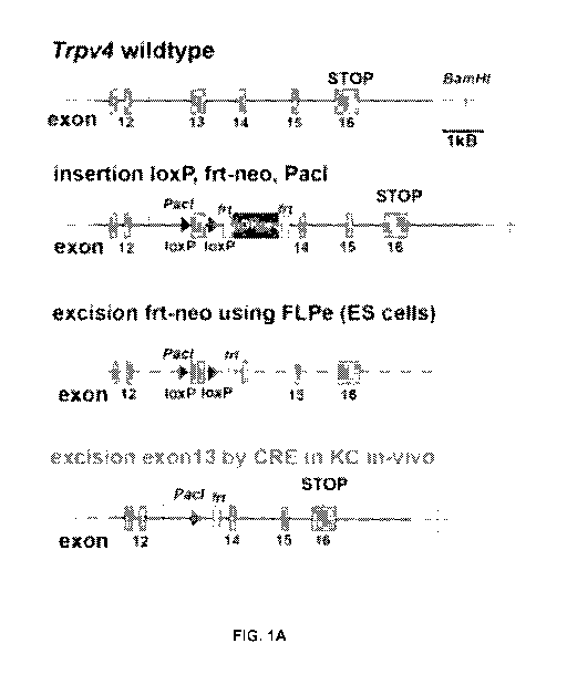
[0016] FIGS. 1A-1G: Keratinocyte-specific and inducible Trpv4 null mouse
and its
UVB response. (FIG. 1A) Gene-targeting of Trpv4 and genetic manipulation
underlying
generation of keratinocyte-specific and inducible Trpv4 knockout mice. Shown
are sequential
steps of mouse Trpv4 targeting, starting with flanking Trvp4 exon13 with loxP
elements and
insertion of a selection cassette, flanked by frt sites, in mouse embryonic
stem cells. After
generation of chimeric mice and stable transmission of the engineered
mutation, the selection
cassette was removed by breeding to FLPe mice. Resulting mice were homozygosed
and
crossed with K14-CRE-ERtam mice, which then permitted keratinocyte-specific
and inducible
Trpv4 knockout/knockdown. (FIG. 1B) DNA genotyping. Shown are PCR products of
VVT,
heterozygote and homozygous Trpv4I091" mice. Note that the PCR products needed
to be
digested with Pad, and that all mice were pre-screened to be CRE+ by another
genotyping
PCR. (FIG. 1C) Co-labeling of mouse skin for keratin-1 and keratin-14 indicate
the established
pattern for vehicle-induced control mice (upper panel), and a similar pattern
for specific TRPV4
knockdown in keratinocytes (lower panel). However, in these animals note a
slightly increased
expression of K14 in the stratum spinosum, reflecting attenuated TRPV4
expression and thus
reduced Ca ++ influx. K14 is normally down-regulated at the basal-to-
suprabasal transition,
concomitant with the rise in Ca-signaling and induction of terminal
differentiation. (FIG. 1D)
TRPV4 protein expression in L5 DRG neurons not different between genotypes.
Densitometry
CA 03020364 2018-10-05
WO 2017/177200 PCT/US2017/026714
of TRPV4 immunohistochemistry in L5 (= foot-pad innervating) DRG neurons
(upper panel
micrographs), the bar-diagram illustrates the lack of a difference in terms of
TRPV4 protein
abundance in oil- vs. tam-treated mice, for both base-line and 48 hours after
UV exposure,
confirming the specificity of TRPV4 knockdown in skin when using K14 as ORE
driver. Note the
characteristic morphology of decorated cells identifying them as DRG sensory
neurons. Note
also the different levels of TRPV4 expression in these neurons, as noted
previously; n=3
mice/group, 50 neurons/mouse. (FIG. 1E) Lack of TRPV4 expression in Merkel
cells in foot-
pad epidermis. A confocal triple-fluorescent micrograph panel is shown,
depicting representative
images of immuno-labeled paw-pads from iK0 control vs. tamoxifen-induced mice.
Note
complete knockdown of TRPV4 in this example (red channel). For Merkel cells
(green channel),
an anti-cytokeratin 8 antibody was used. Note lack of TRPV4 co-labeling in
Merkel cells. Blue
channel = DAPI. (FIG. 1F) Lack of effect of tamoxifen application in K14-CRE-
ERtam mice on
UVB behavioral sensitization. Note very similar withdrawal thresholds in (K14-
CRE-ERtam X
Trpv410/+) mice (= Trpv4 heterozygotes in keratinocytes when induced with
tamoxifen) for
noxious mechanical (upper diagram) and thermal (lower diagram) stimulation;
n=7 mice per
group. Also note the time-course with peak sensitivity at time-point 48 hours.
(FIG. 1G) Size
distribution of pERK-expressing L5 DRG neurons in oil-treated iK0 mice,
exposed to UVB. The
bar diagram illustrates size prevalence of small and medium-size sensory
neurons that express
pERK 48 hours after UVB exposure, note absence of larger neurons (>1200 pm2),
n=22
neurons.
[0017] FIGS. 2A-2G: UVB stimulation device and UVB keratinocyte control
experiments. (FIG. 2A) UV spectrum emitted by the LEDs, overlapped with the
spectrum of
quartz (red trace), which is almost fully permeable to UVB, and glass (blue
trace), which has a
very low UVB permeability. (FIG. 2B) Focusing properties of the ball lens.
(FIG. 2C) Focal
geometry of the combination of UV-LED and ball lens. (FIG. 20) Absence of
thermal effects of
the UV-LEDs; measurement of temperature in the focal point over time. (FIG.
2E) TRPV3
activation experiment. Induction of a Ca++ transient by camphor, which can be
blocked
effectively by 10 pM IPP, suggesting TRPV3-mediated signaling. (FIG. 2F) TRPV4
selective
activator GSK101-related findings. Ca++ transient in 1 MK in response to 5 nM
GSK101, which
can be completely blocked by 20 pM G5K205, suggesting it is specifically
mediated by TRPV4.
The GSK101-response can also be eliminated by absence of external Ca++, in
keeping with
TRPV4 signaling. (FIG. 2G) TRPV4 is sufficient for the UVB-Ca++ response ¨
HEK293T cell
heterologous transfection. Directed expression of TRPV4 in HEK293T cells leads
to a Ca++-
11
CA 03020364 2018-10-05
WO 2017/177200 PCT/US2017/026714
transient in response to UVB radiation, which is greatly reduced in control-
transfected cells.
Preexposure to 20 pM GSK205 virtually eliminates the Ca++-signal in TRPV4-
transfected cells,
and eliminates the moderate signal of control-transfected cells.
[0018] FIGS. 3A-3E: Keratinocyte-specific ablation of Trpv4 leads to
alterations in
nocifensive behavior in response to UVB. (FIG. 3A) Epidermal TRPV4 expression
and its
loss upon keratinocyte-specific ablation of Trpv4 in tam-induced iK0 mice. (i)
TRPV4
immunofluorescence. Note TRPV4 in epidermis of vehicle (oil) treated control,
but not tam-
induced iK0 mice. Bar = 10 pm. (ii) Western blot of epidermal lysates from paw-
pad skin.
Note knockdown and more complete loss of TRPV4 following induced Trpv4-
ablation ([3-actin
used for normalization). (iii) qRT-PCR for Trpv4 mRNA from paw-pad skin is
shown, indicating
significant Trpv4 knockdown in response to tam-treatment vs. carrier (oil).
P<0.0001, t-test. (iv)
lmmunofluorescence for epidermal lineage markers. In VVT skin, basal epidermal
marker
keratin-14 is downregulated and suprabasal marker keratin-1 is induced upon
commitment to
terminal differentiation. Upon knockdown of TRPV4, this balance appears
perturbed, with some
spinous layer cells showing co-labeling. Bar = 10 pm. (FIG. 3B) Nocifensive
behavior in
response to UVB exposure. Time-course (in hours) for nocifensive behavior
elicited by either a
noxious mechanical stimulus (automatic von Frey hair assay, left) or thermally-
evoked
nocifensive behavior (Hargreaves' assay, right). Note significantly less
sensitization in Trpv4
and in tam-treated iK0 mice, relative to oil-treated (vehicle) iK0 and VVT
mice. 1-110 animals
per group; ** p<0.01 ANOVA. (FIG. 3C) Correlation between nocifensive behavior
and level of
Trpv4 knockdown. n=12 animals are shown for which both parameters were
available and
Trpv4 mRNA levels <0.45. Note the four vehicle-induced animals (green symbols)
vs. their
tamoxifen-induced counterparts (red symbols). (FIG. 3D) Loss of epidermal
TRPV4 shows no
significant effect on nocifensive behaviors caused by formalin injection. Bars
depict average
cumulative nocifensive behavior within the first 10 minutes (phase l), and 10-
45 minutes (phase
II) post-injection. n=4 per group. (FIG. 3E) Phosphorylated ERK in L5 DRG
neurons. pERK
immunofluorescence of L5 DRG sections are shown for oil- and tam-treated iK0
animals
exposure to UVB. Note that only UVB-exposed control mice show pERK expression
in the paw-
pad-innervating L5 DRG. Quantifications are shown at right. n=3 animals per
group, 6 sections
per DRG per animal, ** p<0.01 ANOVA.
[0019] FIGS. 4A-4E: Structural and ultrastructural analyses showing that
UVB-
mediated skin tissue injury depends upon keratinocyte TRPV4, and Immuno-
histochemical analysis demonstrates that UVB-mediated activation of
keratinocytes and
12
CA 03020364 2018-10-05
WO 2017/177200 PCT/US2017/026714
recruitment of macrophages and neutrophils depends upon keratinocyte TRPV4.
(FIG.
4A) 1 pm toluidine-blue semi-thin sections. Micrographs show representative
findings of skin in
response to UVB, sampled 48 hours after UVB exposure. Note that upon UVB
stimulation, oil-
(TRPV+) but not tam-treated (TRPV-) iK0 mice exhibit separations at the
epidermal-dermal
boundary and robust signs of tissue injury; note granulocytes (Gr,
neutrophil). Note also that just
beneath the stratum corneum (SC), the upper epidermis shows extensive
structural damage
which could also be seen in skin of tam-treated iK0 mice where Trpv4 knockdown
was
incomplete, but not in those animals where it was more complete (see FIG.
S2A). Bars= 20 pm.
Der = dermis; Epi = epidermis. (FIG. 4B) Ultrastructural findings by EM.
Selected areas from 1
pm semithin sections of paw skin were examined by transmission electron
microscopy. (A-A')
and (C-C') show normal epidermal (Epi) structure for both, oil- and tam-
treated iK0 mice, in the
absence of UVB stimulation. (A) and (C) show and intact epidermis. Basal (BL)
and spinous
(Sp) layers are magnified A' and C' displaying a normal organization with no
evidence of
epidermal damage. (B,B'B"), (D-D') and (E-E') show representative findings of
skin in response
to UVB, sampled 48h after UVB exposure. (B) Disrupted epidermis in oil treated
iK0 mice. An
area equivalent to the boxed area is magnified in (B'), where granulocyte
infiltration of epidermis
is evident (Gr) and blistering with detachment of the epidermis from the
dermis (double arrows).
(B") Upper part of epidermis in contact with stratum corneum (SC), showing
extensive
vacuolization and deposits of fibrin inside the vacuoles (asterisks). (D)
Tamoxifen treated iK0
mice with incomplete knockdown of trvp4 show similar skin phenotype to oil
treated iK0 mice,
with robust signs of tissue damage to basal and spinous layer, fibrin deposits
(asterisks) and
intercellular spaces (arrowheads in D'). (E) Intact epidermis in iK0 with
complete knockdown of
trvp4, with normal basal and spinous layers in (E'). Der, dermis. Dotted lines
indicate the dermo-
epidermal boundary. Bars= 20 pm for A, B, C and D; 10 pm for B' and E' and 2
pm for the other
micrographs. (FIG. 4C) IL-6 upregulation in keratinocytes as marker of
epidermal activation. IL-
6 immunofluorescence reveals a reduced ability of TRPV4-deficient mice to
elevate keratinocyte
IL-6 expression in response to UVB exposure. Quantifications for protein is
shown next to
micrograph. Densitometries are for i-13 mice per group, showing significant
upregulation for oil-
treated iK0 mice, lack thereof for tam-treated. Right-hand bar diagram shows
11-6 mRNA
quantification and time-course. 11-6 mRNA was determined by qPCR after
isolation of total RNA
from paw-pad epidermis. Note the early and robust increase, albeit with
variation, at the 2 hour
time-point, in VVT control epidermis, in contrast the very moderate increase
in Trpv4-/- epidermis.
Note also the sustained robust upregulation at 24 hours, again moderately
upregulated in Trpv4
/-
epidermis. Quantifications are for n=8-12 mice/group. * denotes statistically
significant (p =
13
CA 03020364 2018-10-05
WO 2017/177200 PCT/US2017/026714
0.011, t-test); scale-bar = 20 pm. (FIG. 40) Recruitment of macrophages in UVB-
exposed skin.
Note that the numbers of dermal 0D68+ macrophages induced by UVB-exposure in
control
mice is significantly reduced when Trpv4 is ablated in the epidermis.
Quantifications are shown
at right (n=3 mice/group; * p<0.05 t-test); scale-bar = 20 pm. (FIG. 4E)
Recruitment of elastase-
expressing neutrophils to UVB-exposed skin. Shown are representative
immunofluorescence
micrographs and respective quantifications. Note a strong increase in
abundance of elastase-
expressing neutrophils in control mice, and a lack thereof in tam-treated iK0
mice. (n=4
mice/group, * p<0.05 t-test); scale-bar = 40 pm.
[0020] FIGS. 5A-5E: Histopathology in Trpv4-1- and control mice in response
to UVB.
(FIG. 5A) Trpv4 knockdown level of samples shown in FIGS. 4A-4E. This bar
diagram shows
relative level of knockdown of Trpv4 in comparison with VVT, of UVB-exposed
skin samples
shown in FIG. 4. An adjacent sample of hindpaw skin was RNA-extracted at 48h
post-exposure
and subjected to Trpv4 qRT-PCR; pooled VVT mRNA values from 10 mice were set
as 100%.
(FIG. 5B) Light microscopic analyses of 1 pm semithin sections findings from
Trpv4-/- and VVT
control mice. Normal skin is shown in the upper row for both genotypes in the
unstimulated
state, presence of epidermal and dermal inflammation in VVT control vs.
absence thereof in
Trpv4-/- when exposing the skin to UVB, sampling conducted at 48 hours. Note
inflammatory
changes similar to those of oil-treated iK0 mice, as shown in FIG. 4. (FIG.
5C) Ultrastructural
analyses of Trpv4-/- and VVT control mice. (A-A') and (B-B') VVT and Trpv4-/-
mice show normal
skin morphology with intact epidermis (Epi) in the absence of UVB stimulation.
A' and B' show
higher magnification of basal layer (BL) cells. (C-C') Damaged epidermis with
vacuolization
(inset in C) and granulocyte (neutrophil) (granulocyte - Gr) infiltrate (C').
(D-D') Normal
epidermal and dermal ultrastructure in Trpv4-/- mice exposed to UVB. Der -
dermis; NT, nerve
terminals. Dotted lines indicate the dermo-epidermal boundary. Bars=10 pm for
A, B, C, C' and
D and 2 pm for the other micrographs. (FIG. 50) IL-6 upregulation in epidermal
keratinocytes in
response to UVB depends on Trpv4; findings from Trpv4-/- and VVT control mice.
Fluorescent
micrographs from Trpv4-/- and VVT control skin, unexposed and exposed to UVB
are shown.
Note strong IL-6 signal in VVT, exposed to UVB, and low signal in Trpv4-/- for
both non-exposed
and UVB-exposed states. Also note IL-6-expressing innervating peripheral nerve
endings in the
dermis. (FIG. 5E) No difference in mast cell abundance in UVB-photodermatitis
in iK0 mice.
Left-hand micrograph shows mast-cells within sub-epidermal inflammatory
tissue, stained with
toluidine-blue, in an iK0 mouse induced with tamoxifen, right micrograph its
counterpart in an
oil-treated iK0 mouse. Mast-cells are indicated by white arrow-heads. Bar = 20
pm. Right-hand
14
CA 03020364 2018-10-05
WO 2017/177200 PCT/US2017/026714
bar diagram indicates quantification of mast-cell count per 63x visual field
(5 fields per mouse, 3
mice per group).
[0021] FIGS. 6A-6H: Ca++ influx into keratinocytes in response to UVB
depends on
TRPV4. (FIG. 6A) Custom-built UVB cell illumination apparatus. See also FIG.
2. (FIG. 6B)
Fluo-4 Ca++ imaging in 1 MKs. Fluorescent micrographs of 1 MKs after loading
with Ca++-
sensitive dye, fluo-4, before (upper) and at the end of UVB exposure (lower).
Bar = 10 pm. C-H
UVB-evoked Ca++ signaling profiles. Fluo-4 imaging was used to detect Ca++
transients in
1 MKs following UVB exposure. y-axis indicates the increase in fluorescence,
AF, normalized
for prestimulation signal, FO (AF/F0). The signal shown is that averaged from
50 cells. (FIG.
6C) Ca++ signaling is dependent upon UVB, and is strikingly reduced when
quartz coverslips
are replaced by glass ones, which prevent UVB permeation (see FIG. 2A). Note
that this
particular Ca++ signal in WT 1 MKs persisted after UVB, as is sometimes
observed. (FIG. 60)
UVB-evoked Ca++ signaling is dependent on external [Ca++]. (FIG. 6E) UVB-
evoked Ca++
signaling is not seen in Trpv4-/- 1 MKs, revealing the importance of the TRPV4
ion channel.
(FIG. 6F) UVB-evoked Ca++ signaling is strongly down-regulated in the presence
of TRPV4-
selective inhibitor, GSK205 (20 pM). (FIG. 6G) The UVB-evoked Ca++ signal is
not inhibited by
the TRPV3-selective inhibitor, IPP. For validation of IPP's activity, see FIG.
2E. (FIG. 6H) The
UVB-evoked Ca++ signal can be strongly reduced with specific PLC inhibitor,
U73122.
[0022] FIGS. 7A-70: Central role for keratinocyte TRPV4 in UVB-evoked Ca++
signaling and nocifensive behavior ¨ effects of ET1. (FIG. 7A) Effects of ET1
on UVB-
evoked Ca++ signaling in 1 MKs. Panel (i) shows averaged Ca++ transients in 1
MK in
response to UVB, their augmentation by co-exposure to ET1 peptide, and their
significant
attenuation by either G5K205, which inhibits TRPV4, or ET-convertase inhibitor
CG535066,
which blocks ET1 proteolytic processing. Panel (ii) shows ET-augmented, UVB-
induced Ca++
transients as in (i), but in this case, where they are attenuated by selective
antagonism of
ET(R)-A (BQ123) and ET(R)-B (BQ788). Panel (iii) illustrates the complete
elimination of the
ET1-augmented Ca++ transients when both subtypes of ET(R) are blocked. (FIG.
7B) 4a-PDD-
evoked Ca++ signaling in 1 MKs¨ ET1-related findings. Left-hand panel shows
Ca++ transients
(as per fura-2 ratiometric imaging) in response to the selective TRPV4
activator 4a-PDD. A
significant increase in the response can be observed by co-application of ET1,
and this is
partially dependent on ET(R)-A and completely dependent on ET(R)-B. (FIG. 7C)
Upregulation
of ET1 in mouse paw in response to UVB. lmmunohistochemistry reveals a
significantly
stronger ET1 signal in UVB-exposed skin of oil-vehicle-treated (TRPV4+) rather
than tamtreated
CA 03020364 2018-10-05
WO 2017/177200 PCT/US2017/026714
iK0 mice. Quantifications are for n=3 mice/group. *** denotes statistically
significant (p<0.001, t-
test). (FIG. 70) Nocifensive behavior in response to ET1 footpad injection
depends on
epidermal TRPV4. Bar diagram summarizes behavioral findings for Trpv4-/- vs.
VVT and for oil-
treated vs. tam-treated iK0 mice. Note that in VVT and oil-treated iK0 mice,
footpad injection of
ET1 leads to significant levels of mechanical allodynia. Trpv4-/- and tam-
treated iK0 mice fail to
respond; 1-17 mice/group, ** p<0.01, ANOVA.
[0023] FIGS. 8A-80: Central role for KC TRPV4 in UVB-evoked Ca signaling
and
nocifensive behavior ¨ ET1-related supplementary findings. (FIG. 8A)
Augmentation of
GSK101-evoked Ca ++ signaling by ET1. Shown are averaged Ca ++ measurements
(fura-2) in
response to 5 nM GSK101. Note the increase in signal in response to co-
exposure to ET1.
(FIG. 8B) ET1 secretion by non-stimulated 1 MK depends on TRPV4 and PLC. Shown
are
relative ET1 concentrations (determined by ELISA, pg/mL; vehicle-treated and
VVT control
normalized to 100) in supernatant of non-stimulated 1 MK. Note the clear
dependence on
TRPV4, as indicated by a 50% reduction in Trpv4-/- 1 MK. Moreover, there is a
significant down-
regulation by specific inhibition of TRPV4, which is dose-dependent (two doses
of G5K205) and
can be mediated by two different compounds (G5K205, RN1734). There is also
down-
regulation of ET1 secretion by a specific inhibitor of PLC (U73122), and by an
ET-convertase
inhibitor, 0G535066, which served as a control compound. In addition, PLC-
inhibitor robustly
affects ET1 secretion in VVT and Trpv4-/- 1 MK. (FIG. 8C) ET1 expression by
UVB-exposed
1 MK depends on TRPV4 and PLC - immunocytochemistry. Shown is specific ET1
immunolabeling in 1 MK, exposed to UVB using the UVB-LEDs, as for Ca ++
imaging. Use of the
UVB-LED device precluded application of a ET1 ELISA, only irradiated cells
could be examined.
Note the significant down-regulation of ET1 immunoreactivity by specific
inhibition of TRPV4
(two different compounds, G5K205, RN1734), by PLC inhibition (U73122), also by
inhibition of
ET-convertase (CG535066). (FIG. 80) ET1 expression by UVB-exposed 1 MK depends
on
TRPV4 and PLC ¨ quantification of immunocytochemistry. Densitometric
measurements of
1-125 cells per condition, background subtracted, are shown, indicating a
significant upregulation
of ET1 in response to UVB (* p<0.05 ANOVA), and significant down-regulation
vs. control-
treated and UVB-exposed cells for treatments with selective TRPV4 antagonists
(G5K205,
RN1734), PLC-inhibitor U73122 and ET-convertase inhibitor CG535066; = p<0.05,
t-test; #
p<0.05 ANOVA.
[0024] FIGS. 9A-9H: UVB-evoked inflammasome activation in keratinocytes
depends
on TRPV4. (FIG. 9A) Caspase-1 immunolabeling in footpad skin in response to
UVB.
16
CA 03020364 2018-10-05
WO 2017/177200 PCT/US2017/026714
Representative images are from sections of skins before stimulation (control)
or 48 hours post-
UVB exposure. Bars= 20 pm. (FIG. 9B) Quantifications of caspase-1
immunolabeling. Bar
diagrams show densitometry, 1-13 animals/group. Comparisons: UVB exposed VVT
vs. Trpv4-/-
and iK0 + oil vs. iK0 + tam. **p<0.01 ANOVA. (FIG. 9C) Western blotting for
caspase-1 from
1 MK UVB-exposure. Note that caspase-1 levels, in particular cleaved caspase-
1 (lower
band), are elevated in UVB-exposed VVT cells, but there is a complete absence
of both
procaspase-1 and cleaved caspase-1 in 1 MK from Trpv4-/- mice. (FIG. 90) IL-
111 is induced
upon UVB-exposure and is dependent on TRPV4. Anti-IL-113 immunofluorescence,
otherwise as
in panel A.
(FIG. 9E) Quantifications of IL-111 immunolabeling, 1-13 animals/group.
Comparisons: UVB exposed VVT vs. Trpv4-/- and iK0 + oil vs. iK0 + tam, **
p<0.01 ANOVA.
(FIG. 9F) IL-111 concentrations in interstitial fluid of UVB-exposed footpad.
IL-111 levels (ELISA)
are shown in lavaged interstitial fluid. Note strong up-regulation in WT and
oil-treated iK0 mice
after UVB, in contrast significant attenuation in Trpv4-/- and tam-treated iK0
mice. 1-15
mice/group, ** p<0.01 ANOVA. (FIG. 9G) CXCL5 is induced upon UVB-exposure and
is
dependent upon TRPV4. Anti-CXCL5 immunolabeling, otherwise as in panel A.
(FIG. 9H)
Quantifications of CXCL5 immunolabeling, 1-13 animals/group. Comparisons: UVB
exposed VVT
vs. Trpv4-/- and iK0 + oil vs. iK0 + tam, ** p<0.01 ANOVA.
[0025] FIGS. 10A-10B: Epidermal TRPV4, ET1 and IL-111 are elevated in
photodermatitis as compared to healthy human skin.
(FIG. 10A) Representative
micrographs of TRPV4, ET1 and IL-111 distribution in the epidermis of acute
photodermatitis, as
compared to healthy human skin. lmmunostaining for each antigen is increased
in acute
photodermatitis vs. healthy skin. Scale-bars = 50 pm (left), 100 pm (middle),
50 pm (right).
(FIG. 10B) Morphometric analysis for immunoreactive TRPV4, ET1 and IL-18.
Findings reveal
significantly increased immunolabeling for all three proteins in acute
photodermatitis as
compared to healthy human skin (n=3 subjects for normal, healthy skin, and 3
patients for acute
UV photodermatitis).
[0026]
FIG. 11: Exemplars of human chronic photodermatitis. Upper panel shows
normal healthy human skin, as displayed in FIG. 10A, for comparison. Lower
panel shows
examples of chronic photodermatitis with elevated expression of TRPV4, in
spinous and basal
layers, ET1 (throughout) and IL-111 (throughout). In comparison to acute
photodermatitis, note
reduced interstitial intraepidermal edema. Bar = 50 pm.
17
CA 03020364 2018-10-05
WO 2017/177200 PCT/US2017/026714
[0027] FIGS. 12A-120: External-topical application of a selective TRPV4
inhibitor
attenuates UVB-evoked nocifensive behavior and inflammation. (FIG. 12A) UVB-
induced
nocifensive behavior. Pain behavior is attenuated by topical application of
GSK205. The left-
hand diagram shows withdrawal thresholds after UVB-exposure in response to
noxious thermal
cues (Hargreaves' test), and their modulation by two doses of topically
applied GSK205 (1 mM
and 5 mM; applied 60' and 10' pre-exposure). The higher dose led to a
significant attenuation of
thermal allodynia at 48 hours post-UVB; n=6 mice/group; ** p<0.01 ANOVA. The
righthand
diagram shows development of moderate thermal allodynia in Trpv4-/- mice, and
similar
sensitization for vehicle-treated vs. 5mM GSK205-treated mice, indicating lack
of off-target
effects of the compound at 5 mM; n=5 mice/group. (FIG. 12B) GSK205-treatment
attenuates
keratinocyte expression of IL-111 in UVB-exposed footpad ¨ representative
micrographs.
Bars=20 pm. (FIG. 12C) GSK205-treatment attenuates keratinocyte expression of
IL-111 in
UVB-exposed footpad quantifications. Bar diagrams show densitometry results
from n=3
mice/group, ** p<0.01 ANOVA. (FIG. 120) GSK205-treatment attenuates secretion
of IL-111 by
UVB-exposed 1 MK. IL-111 concentrations in supernatant (ELISA), are shown in
response to
UVB. Cells were cultured +/- 5 pM GSK205. Note prevention of increase in IL-
111 secretion in
response to UVB upon treatment with GSK205. ** P<0.01 ANOVA.
[0028] FIGS. 13A-13B: Topical application of a selective TRPV4 inhibitor
attenuates
UVB-evoked nocifensive behavior by suppressing upregulation of pro-
algesicialgogenic
mediators in murine keratinocytes ¨ Findings for CXCL5 and IL6. (FIG. 13A)
GSK205-
treatment attenuates keratinocyte expression of CXCL5 in UVB-exposed footpad ¨
micrographs
and quantitation. As in FIG. 12B, specific immunolabeling for CXCL5, which is
selectively
upregulated in footpad keratinocytes in response to UVB, note attenuation with
GSK205
treatment. Bar diagrams show densitometry of CXCL5 immunolabeling, n=3 animals
per group,
sections analyzed per animal. Note significant differences for vehicle-treated
mice between
UVB-exposed and non-exposed, no such difference for mice treated topically
with 5 mM
GSK205. Comparison UVBexposed between vehicle and GSK205-treated, ** p<0.01
ANOVA.
Bar = 20 pm. (FIG. 13B) GSK205-treatment attenuates keratinocyte expression of
IL-6 in UVB-
exposed footpad ¨ micrographs and quantification. As in FIG. 12B and FIG. 13A,
specific
immunolabeling for IL-6, demonstrates similar regulation of IL-6 as for CXCL5
and IL-111.
Comparison UVB-exposed between vehicle and GSK205-treated, ** p<0.01 ANOVA.
Bar = 20
pm.
18
CA 03020364 2018-10-05
WO 2017/177200 PCT/US2017/026714
[0029] FIGS. 14A-140: External-topical application of a selective TRPV4
inhibitor
attenuates UVB-evoked nocifensive behavior and inflammation. (FIG. 14A) UVB-
photodermatitis is attenuated in mice treated with GSK205. Representative H&E
micrographs of
paw-pad skin are shown, bars = 20 pm. Treatment of UVB-exposed skin with
GSK205 improved
the skin architecture in mice as compared to vehicle-treated mice after 24
hours. (i) and (iii)
Representative skin sections of UVB-induced photodermatitis after GSK205
treatment showed
markedly reduced inflammatory infiltrate, less spongiosis and dermal-epidermal
blisters with
remaining epidermal thickening. (ii) and (iv) Vehicle-treated mice after UVB-
induced
photodermatitis were characterized by signs of severe acute photodermatitis
such as
spongiosis, epidermal hyperkeratosis, disrupted dermal-epidermal border
(blister), and a
marked inflammatory infiltrate with dilated blood vessels and dermal edema
(arrows). Also note
the erythrocyte accumulation in blood vessels indicative of dermatitis. (FIG.
14B) Topical
treatment with a TRPV4-specific inhibitor attenuates upregulation of algogenic
ET1/Edn1. Edn1
mRNA was determined by qPCR after extraction of total RNA from paw-pad
epidermis. In
vehicletreated skin, note increase of Edn1 expression with early up-regulation
at the 2 hour
time-point, and sustained elevation up to the 24 hour time-point. This time-
course resembles
that seen in VVT control mice, when comparing to Trpv4-/- (FIG. 15).
Importantly, topical
treatment with 5 mM GSK205 results in complete lack of this regulation; n=4
mice/group,
*p<0.05 ANOVA. (FIG. 14C) GSK205 does not function as sunscreen. Schematic
illustrates the
experimental set-up. (FIG. 140) G5K205 does not function as sunscreen. The bar
diagram
shows results from n=7-8 mice/group, note absence of a change in UVB
permeation with 5 mM
G5K205, topically applied as for (B), vs. vehiclecontrol, yet significantly
reduced with sunscreen
SPF100.
[0030] FIG. 15: Upregulation of ET1/End1 in mouse paw in response to UVB.
Edn1
mRNA was determined by qPCR after isolation of total RNA from paw-pad
epidermis. Note the
early increase, at the 2 hour time-point, in VVT control epidermis, and its
complete lack in Trpv4-
/-, and sustained upregulation at 24 hours, slightly reduced vs. 2 hour time-
point, again
complete lack of upregulation in Trpv4-/-. Quantifications are for n=4
mice/group. ** denotes
statistically significant (p<0.01, t-test).
[0031] FIGS. 16A-16B: Skin UVB permeability testing. (FIG. 16A)
Experimental set-up
for testing of skin permeability to UVB. (FIG. 16B) Results from FIG. 16A.
Note that intensity is
70% within 500pm radius to the center of the UV beam.
19
CA 03020364 2018-10-05
WO 2017/177200 PCT/US2017/026714
[0032] FIG. 17: Role of TRPV4 in itch transmission in mice in vivo.
Histamine (10%)
was injected intracutaneously into the cheek of C57b/6 control (VVT) or TRPV4
knockout mice
(Trpv4 pan-knockout; n=6 per group). Over 30 min, TRPV4 null mice showed a
significant
reduction ((p<0.01) t-test)) of itch-behavior as compared to VVT mice.
[0033] FIG. 18: The role of TRPV4 in itch. Shown is a graph of scratching
behavior after
administration of a pruritogen in mice with TRPV4 deletion in keratinocytes
after induction of the
TRPV4 knockout, as compared to mice without induction. VVithout induction, the
mice function
as wild-type control mice (Moore et al. Proc. Natl. Acad. Sci. U.S.A. 2013,
110, E3225-E3234).
Compared to control mice, scratch behavior was significantly reduced for mice
in which TRPV4
channels had been selectively deleted in skin keratinoctyes.
[0034] FIG. 19: Compound 16-19. Compound 16-8/18h was designed as a hybrid
of
compounds 16-8 and 16-18. Compound 16-19 may also be referred to herein as 16-
8/18hy.
[0035] FIG. 20: Inhibition of calcium ion flux through TRPV4. Compounds of
the
present invention demonstrated inhibitory activity against TRPV4 and were
stronger antagonists
that G5K205.
[0036] FIG. 21: Treatment of pain. Compounds as disclosed herein
attenuated
nocifensive behavior in mice.
[0037] FIG. 22: Treatment of pain after UVB exposure. Compounds as
disclosed herein
attenuated nocifensive behavior in a mouse model for sunburn.
[0038] FIG. 23: Effect on TRPA1. Compounds as disclosed herein inhibited
TRPA1, as
indicated by measuring calcium transience.
[0039] FIG. 24: Effect on TRPV1, TRPV2, and TRPV3. Compounds as disclosed
herein
did not inhibit TRPV1, TRPV2, or TRPV3, as indicated by measuring calcium
transience.
[0040] FIG. 25: Modifications of tool compound GSK205 for improved
targeting of
TRPV4. The synthesized compounds differed in the highlighted part of the
molecule, changed
residue indicated with arrow. Compound 16-19 compound was synthesized to
incorporate two
modifications from two compounds, 16-8 and 16-18, found most potent in anti-
TRPV4 screening
assays (see FIG. 26).
CA 03020364 2018-10-05
WO 2017/177200 PCT/US2017/026714
[0041]
FIGS. 26A-26B: Assessment of compounds in N2a cells with directed expression
of TRPV4. (FIG. 26A) Ca++ imaging screening of all compounds in N2A cells with
directed
expression of TRPV4 (rat). The cells were stimulated with TRPV4-selective
activator compound,
GSK101 (5 nM) in the presence of 5 pM of the respective inhibitor. The number
on each bar
corresponds to average peak ACa++ concentrations in P=z100 cells. Inset:
micrographs of pseudo-
colored cells before and after activation with 5nM GSK101, in addition note
the corresponding time
course of the averaged Ca++ signal (fura-2 Ca++ imaging). Except for compound
16-430, the
difference to vehicle control reach the level of statistical significance p<
0.01 (one-way ANOVA).
(FIG. 26B) Dose-response of the most potent, "winner" compounds in TRPV4-
expressing N2a cells.
The 1050 were; 0.45 0.05 pM (16-8), 0.59 0.12 pM (16- 18), 0.81 0.1 pM (16-
19), 4.19 0.71
pM (GSK205). Plot generated from averaged peak ACa++ concentration of 75 cells
per data-point.
[0042]
FIGS. 27A-27B: TRPV4 channel inhibition by compounds 16-8 and 16-19 -
patch-clamp e-phys. (FIG. 27A) Current- voltage relationship of TRPV4-mediated
currents
after activation with 5 nM GSK101. Recordings were performed in TRPV4-GFP+ N2a
cells. The
representative traces represent an average of P=z12 sweeps. In all
experiments, cells were pre-
incubated with the respective compound (5 pM) for 5 minutes. (FIG. 27B)
Average current
densities at -100mV/+100 mV were significantly diminished by inhibitors (*Ip<
0.05; one-way
ANOVA;r1 5 cells/group).
[0043]
FIGS. 28A-28B: Compound 16-8 inhibits TRPV4 in I cells more potently than
G5K205. (FIG. 28A) 1 (primary) articular chondrocytes (pig); dose-response
comparison
between the most potent compound, 16-8, and GSK205 in response to stimulation
with 5 nM
GSK101. Inset: Chondrocytes responding to activation with GSK101, fura-2 Ca++
imaging; right-
hand image taken at 5 sec after GSK101 application. 16-8 was significantly
more potent than
GSK205 (mean SEM, n= 6 independent expts,
25 cells/expt; *p< 0.05, t-test). Ordinate
shows average peak ACa++ concentrations. (FIG. 28B) 1 (primary) astrocytes
(rat); dose-
response comparison between 16-8 and GSK205 in response to 5 nM GSK101. Inset:
Astrocytes responding to activation with GSK101; right-hand image taken at 5
sec after GSK101
application (mean SEM, n= 5 independent expts,
200 cells/expt; *p< 0.05, t-test). Ordinate
shows average peak ACa++ concentrations.
[0044]
FIGS. 29A-29B: Compounds 16-8 and 16-19 also potently inhibit TRPA1, not
TRPV1-3. (FIG. 29A) Specificity vs TRPV1-3. Both 16-8 and 16-19 (5 pM each)
compounds did
not inhibit TRPV1, -2 or -3 channels (all mouse isoforms), directed over-
expression in N2a cells
21
CA 03020364 2018-10-05
WO 2017/177200 PCT/US2017/026714
and subsequent Ca++ imaging. Mean SEM is shown, 100 cells per condition. (FIG.
29B) Dose-
dependent inhibition of TRPA1 (mouse, directed expression in N2a cells) by GSK
205, 16-8 and
16-19, activation with 100 pM mustard oil, resulting in 1050 of 5.56 0.4 pM
(GSK205), 0.41 0.37
pM (16-19), 0.43 0.3 pM (16-8). Plot generated from averaged peak ACa++
concentration of 75
cells per data-point.
[0045] FIGS. 30A-30B: Cellular toxicity studies of compounds 16-8 and 16-
19. N2a
cells were subjected to increasing concentrations of compounds 16-8 and 16-19,
resulting cell
viability was analyzed for the next 48 h. (FIG. 30A) Time course of cell
viability in the presence of
various concentrations of 16-8. Note clear reduction at 40 and 80 pM. (FIG.
30B) As in (FIG.
30A), for compound 16-19, with similar outcome. Representative result of 2
independent
experiments.
[0046] FIGS. 31A-310: 16-8 and 16-19 effectively attenuate formalin-evoked
trigeminal
irritant pain. (FIG. 31A) Time-course of nocifensive behavior in VVT mice
following whisker-pad
injection of 4% formalin. The mice were pre-injected (i.p., 10 mg/kg; 15 min
before formalin) with
GSK205, 16-8 or 16-19. Note effective reduction of nocifensive behavior in the
late "neural"
phase by compounds 16-8, 16-19, not by GSK205. (FIG. 31B) Cumulative response
binned into
3 phases: acute phase (0-5 min), interphase (5-15 min), and late "neural"
phase (15-45 min).
Note significant reduction of nocifensive behavior in the late phase by 16-8,
16-19, not GSK205
(*P <0.01 vs vehicle and GSK205, one-way ANOVA). (FIG. 31C) As in (FIG. 31A),
but also
including Trpv4-/- mice. Compounds were applied i.p. 15 min before formalin
challenge, at 10
mg/kg except established TRPA1 blocker, A967079 (25 mg/kg). Previously-
established
attenuated nocifensive behavior in early and late phase in Trpv4-/- mice was
recapped, which
was reduced further by TRPA1 blocker, A967079. (FIG. 310) As in (FIG. 31B),
plus inclusion of
Trpv4-/- mice. Robust effects of TRPA1-blocker, A967079, were mimicked equi-
potently by 16-
8 and 16-19 for early phase, and by 16-8 for late phase, partially by 16-19
for late phase. (FIG.
31A, FIG. 31C) show averaged behavioral metrics per time-point, bars in (FIG.
31B, FIG. 310)
represent mean SEM; for (D) *Ip< 0.05; 41p< 0.005, one-way ANOVA; for all
panels n= 5-8
mice/group.
[0047] FIGS. 32A-32F: Compound 16-8 attenuates acute pancreatitis and
improves pain
behavior. (FIG. 32A) Caerulein-evoked acute pancreatitis causes pancreatic
edema, which is
eliminated by compound 16-8 (10 mg/kg, applied at 30 min before first exposure
to caerulein).
22
CA 03020364 2018-10-05
WO 2017/177200 PCT/US2017/026714
(FIG. 32B) Caerulein-evoked acute pancreatitis strongly elevates cellular
toxicity marker amylase
in serum. Amylase is reduced, but not significantly, in 16-8 treated animals.
(FIG. 32C) caerulein-
evoked acute pancreatitis causes elevated myelo-peroxidase (M PO) activity in
serum, a marker for
infiltration of inflammatory cells into the pancreas. MPO activity is
significantly reduced in 16-8
treated mice. (FIG. 320) caerulein- evoked acute pancreatitis can be readily
demonstrated
histologically, exemplified in the micrograph panels shown. Note increased
pancreas inflammation
in the middle-panel vs non-inflamed pancreas in vehicle-control challenged
mice, and its
attenuation by treatment with compound 16-8. (FIG. 32E) Bar diagram shows
quantitation of
inflammatory histologic parameters as shown in (FIG. 320). Note significant
increase of
inflammation-index in caerulein acute pancreatitis mice, and its significant
reduction upon
treatment with compound 16-8. (FIG. 32F) Caerulein-evoked acute pancreatitis
causes pain
behavior, significantly reduced by compound 16-8. Note greatly reduced
activity over the 6 h test
period in caerulein-induced acute pancreatitis. This nocifensive behavior is
greatly improved in
response to systemic application of compound 16-8. Results are expressed as
mean SEM; n=6
mice/group; *P <0.05 (one-way ANOVA).
[0048] FIGS. 33A-33E: Pharmaco-kinetics/pharmaco-tox of compounds 16-8, 16-
19
and G5K205 in-vivo. (FIG. 33A) Concentrations of compounds 16-8, 16-19 and
GSK205
(10mg/kg) in several murine tissues/organs 1h post-i.p. injection. (FIG. 33B)
Compound 16-19
time-course at 6h and 24h in several organs. 16-19 was selected because of its
elevated levels
at the 1h time-point, and based on the estimate that 16-19 is more lipophilic
than 16-8 and
GSK205. Note that metrics at 6h are invariably higher than at 24h. All values
are appreciably
higher than at 1h. (FIG. 33C) Concentrations of 16-8, 16-19 and GSK205 in fat
and pancreas
after one hour. Note lower concentration of 16-8 vs. 16-19 and GSK205, yet
above its IC50.
(FIG. 330) Concentrations of 16-8 in the pancreas at 1h and 24h time-points.
(FIG. 33E)
Structural stability of compound 16-19 in plasma as suggested by stable
concentration after
4h/37 C. Results are expressed as means SEM, n=6 mice/experimental group for
all
experiments.
[0049] FIGS. 34A-34C: Absence of cardiac, renal and hepatic toxicty of
compounds
16-8, 16-19. (FIG. 34A) Heart rate time-course after i.p. injection (10 mg/kg)
of compounds.
There was no significant difference in heart rates between vehicle and
compounds. (FIG. 34B)
Serum creatinin was not significantly elevated in animals treated with 16-19
and 16-8 vs vehicle
control. (FIG. 34C) Serum alanin-amino-transferase (ALT) levels were not
significantly elevated
in animals treated with 16-19 and 16-8 vs vehicle control. n=6 mice/group for
all experiments.
23
CA 03020364 2018-10-05
WO 2017/177200 PCT/US2017/026714
[0050] FIG. 35: General synthetic scheme for compounds 16-08 to 16-19.
Reagents
and conditions: (i) K2003, CH3CN. (ii) Zn, Me0H, 12M HCI. (iii) 1,1'-
Thiocarbonyldiimidazole.
(iv) 7M NH3 in Me0H. (v) Et0H, reflux.
DETAILED DESCRIPTION
[0051] The disclosure relates to treating a subject's skin or wounds in
order to sanitize them
and remove any contaminating and possibly harmful bacteria or other
microorganisms. The
disclosure also relates to diminishing pain, inflammation, and/or irritation
that may be caused by
iodine or a local anesthetic. Described herein are compositions and methods
for sanitizing a
subject, anesthetizing a subject, and treating and/or preventing a
dermatological disorder. The
skin functions as an essential barrier between the external environment and
the vertebrate
organism. Keratinocytes in the skin absorb UV-light, leading to skin
inflammation, pain, and itch
after over-exposure, which subsequently leads to skin pigmentation. The
inventors have
identified that the skin, in particular its epidermal epithelia, is
substantially involved in sensory
transduction. A mouse model of sunburn is described herein and used to induce
a state of
lowered sensory thresholds evoked by a limited, self-resolving inflammation in
response to UV
spectrum of light. UV-evoked lowering of sensory thresholds shares major
hallmarks of
pathological pain, which is a valuable feature of this model.
1. Definitions
[0052] Unless otherwise defined, all technical and scientific terms used
herein have the
same meaning as commonly understood by one of ordinary skill in the art. In
case of conflict,
the present document, including definitions, will control. Preferred methods
and materials are
described below, although methods and materials similar or equivalent to those
described
herein can be used in practice or testing of the present invention. All
publications, patent
applications, patents and other references mentioned herein are incorporated
by reference in
their entirety. The materials, methods, and examples disclosed herein are
illustrative only and
not intended to be limiting.
[0053] The terms "comprise(s)," "include(s)," "having," "has," "can,"
"contain(s)," and
variants thereof, as used herein, are intended to be open-ended transitional
phrases, terms, or
words that do not preclude the possibility of additional acts or structures.
The singular forms
"a," "and" and "the" include plural references unless the context clearly
dictates otherwise. The
present disclosure also contemplates other embodiments "comprising,"
"consisting of" and
24
CA 03020364 2018-10-05
WO 2017/177200 PCT/US2017/026714
"consisting essentially of," the embodiments or elements presented herein,
whether explicitly set
forth or not.
[0054] For the recitation of numeric ranges herein, each intervening number
there between
with the same degree of precision is explicitly contemplated. For example, for
the range of 6-9,
the numbers 7 and 8 are contemplated in addition to 6 and 9, and for the range
6.0-7.0, the
number 6.0, 6.1, 6.2, 6.3, 6.4, 6.5, 6.6, 6.7, 6.8, 6.9, and 7.0 are
explicitly contemplated.
[0055] Definitions of specific functional groups and chemical terms are
described in more
detail herein. The chemical elements are identified in accordance with the
Periodic Table of the
Elements, CAS version, Handbook of Chemistry and Physics, 75th Ed., inside
cover, and specific
functional groups are generally defined as described therein. Additionally,
general principles of
organic chemistry, as well as specific functional moieties and reactivity, are
described in
Organic Chemistry, Thomas Sorrell, University Science Books, Sausalito, 1999;
Smith and
March March's Advanced Organic Chemistry, 5th Edition, John VViley & Sons,
Inc., New York,
2001; Larock, Comprehensive Organic Transformations, VCH Publishers, Inc., New
York, 1989;
Carruthers, Some Modern Methods of Organic Synthesis, ad Edition, Cambridge
University
Press, Cambridge, 1987; the entire contents of each of which are incorporated
herein by
reference.
[0056] The term "acyl" or "carbonyl" refers to the group -C(0)R wherein R
is selected from
the group consisting of hydrogen, alkyl, alkenyl, alkynyl, aryl, cycloalkyl,
heterocyclyl, heteroaryl,
arylalkyl, cycloalkylalkyl, heteroarylalkyl and heterocyclylalkyl, any of
which may be optionally
substituted, e.g., with one or more substituents. For example, when R is
alkyl, such a group
may be referred to as an alkylcarbonyl group. The term "acyl" or "carbonyl"
includes, for
example, an alkylcarbonyl, cycloalkylcarbonyl, heterocyclylcarbonyl,
arylcarbonyl, or
heteroarylcarbonyl substituent, any of which may be further substituted (e.g.,
with one or more
substituents).
[0057] The term "alkyl" refers to a straight or branched hydrocarbon chain,
containing the
indicated number of carbon atoms. For example, C1-C12 alkyl indicates that the
alkyl group may
have from 1 to 12 (inclusive) carbon atoms, and C1-C4 alkyl indicates that the
alkyl group may
have from 1 to 4 (inclusive) carbon atoms. An alkyl group may be optionally
substituted.
Examples of C1-C4 alkyl groups include methyl, ethyl, n-propyl, isopropyl, n-
butyl, sec-butyl and
tert-butyl.
CA 03020364 2018-10-05
WO 2017/177200 PCT/US2017/026714
[0058] The term "alkylene" refers to divalent hydrocarbon groups having 1
to 6 carbon
atoms such as methylene, ethylene, propylene, and butylene. Alkylene groups
include 01-02
alkylene, or 01-03 alkylene, or 01-04 alkylene.
[0059] The term "alkenyl" refers to a straight or branched hydrocarbon
chain having one or
more double bonds. Examples of alkenyl groups include, but are not limited to,
allyl, propenyl, 2-
butenyl, 3-hexenyl and 3-octenyl groups. One of the double bond carbons may
optionally be
the point of attachment of the alkenyl substituent. An alkenyl group may be
optionally
substituted.
[0060] The term "alkynyl" refers to a straight or branched hydrocarbon
chain having one or
more triple bonds. Examples of alkynyl groups include, but are not limited to,
ethynyl, propargyl,
and 3-hexynyl. One of the triple bond carbons may optionally be the point of
attachment of the
alkynyl substituent. An alkynyl group may be optionally substituted.
[0061] The term "aryl" or "aromatic" refers to an aromatic monocyclic,
bicyclic, or tricyclic
hydrocarbon ring system, wherein any ring atom capable of substitution can be
substituted (e.g.,
with one or more substituents). Examples of aryl moieties include, but are not
limited to, phenyl,
naphthyl, and anthracenyl. An aromatic amine is an aryl group substituted with
one or more
amino groups. An aromatic alcohol is an aryl group substituted with one or
more hydroxyl
groups. Both aromatic amines and aromatic alcohols may be further substituted
with other
substitutents.
[0062] The term "arylalkyl" refers to an alkyl moiety in which an alkyl
hydrogen atom is
replaced with an aryl group. Arylalkyl includes groups in which more than one
hydrogen atom
has been replaced with an aryl group. Examples of arylalkyl groups include
benzyl, 2-
phenylethyl, 3-phenylpropyl, 9-fluorenyl, benzhydryl, and trityl groups.
[0063] The term "carboxyl" refers to the group ¨C(=0)0R, wherein R is
selected from the
group consisting of hydrogen, alkyl, alkenyl, alkynyl, aryl, cycloalkyl,
heterocyclyl, heteroaryl,
arylalkyl, cycloalkylalkyl, heteroarylalkyl, and heterocyclylalkyl any of
which may be optionally
substituted, e.g., with one or more substituents.
[0064] The term "carbonylamino" or "amido" refers to the group ¨C(0)NR'R"
wherein R' and
R" are independently selected from the group consisting of hydrogen, alkyl,
alkenyl, alkynyl,
aryl, cycloalkyl, heterocyclyl, heteroaryl, arylalkyl, cycloalkylalkyl,
heteroarylalkyl, and
26
CA 03020364 2018-10-05
WO 2017/177200 PCT/US2017/026714
heterocyclylalkyl, or R' and R", together with the nitrogen to which they are
attached, may form a
ring. The groups R' and R" may be optionally substituted, e.g., with one or
more substituents, or
when R' and R" together with the nitrogen to which they are attached form a
ring, the ring may
be optionally substituted, e.g., with one or more substituents.
[0065]
The term "cycloalkyl" as used herein refers to nonaromatic, saturated or
partially
unsaturated cyclic, bicyclic, tricyclic, or polycyclic hydrocarbon groups
having 3 to 12 carbons
(e.g., 3, 4, 5, 6, or 7 carbon atoms). Any ring atom can be substituted (e.g.,
with one or more
substituents). Cycloalkyl groups can contain fused rings. Fused rings are
rings that share one
or more common carbon atoms. Examples of cycloalkyl groups include, but are
not limited to,
cyclopropyl, cyclobutyl, cyclopentyl, cyclohexyl,
cyclohexenyl, cyclohexadienyl,
methylcyclohexyl, adamantyl, norbornyl, and norbornenyl. The term
"cycloalkenyl" refers to
cyclic alkenyl groups of from 4 to 8 carbon atoms having a single cyclic ring
and at least one
point of internal unsaturation. Any ring atom can be substituted (e.g., with
one or more
substituents).
[0066]
The term "halo" or "halogen" as used herein refers to any radical of fluorine,
chlorine,
bromine, or iodine.
[0067]
The term "haloalkyl" as used herein refers to an alkyl in which one or more
hydrogen
atoms are replaced with a halogen, and includes alkyl moieties in which all
hydrogens have
been replaced with halogens (e.g., perfluoroalkyl such as CF3).
[0068]
The term "heteroaryl" or "heteroaromatic" as used herein refers to an aromatic
5-8
membered monocyclic, 8-12 membered bicyclic, or 11-14 membered tricyclic ring
system
having 1-3 heteroatoms if monocyclic, 1-6 heteroatoms if bicyclic, or 1-9
heteroatoms if tricyclic,
said heteroatoms independently selected from 0, N, S, P, and Si (e.g., carbon
atoms and 1-3,
1-6, or 1-9 heteroatoms independently selected from 0, N, S, P, and Si if
monocyclic, bicyclic,
or tricyclic, respectively). Any ring atom can be substituted (e.g., with one
or more substituents).
Heteroaryl groups can contain fused rings, which are rings that share one or
more common
atoms. Examples of heteroaryl groups include, but are not limited to, radicals
of pyridine,
pyrimidine, pyrazine, pyridazine, pyrrole, imidazole, pyrazole, oxazole,
isoxazole, furan,
thiazole, isothiazole, thiophene, quinoline, isoquinoline, quinoxaline,
quinazoline, cinnoline,
indole, isoindole, indolizine, indazole, benzimidazole, phthalazine,
pteridine, carbazole,
carboline, phenanthridine, acridine, phenanthroline, phenazine,
naphthyridines, and purines.
27
CA 03020364 2018-10-05
WO 2017/177200 PCT/US2017/026714
[0069] The term "heterocyclyl" as used herein refers to a nonaromatic,
saturated or partially
unsaturated 3-10 membered monocyclic, 8-12 membered bicyclic, or 11-14
membered tricyclic
ring system having 1-3 heteroatoms if monocyclic, 1-6 heteroatoms if bicyclic,
or 1-9
heteroatoms if tricyclic, said heteroatoms selected from 0, N, S, Si, and P
(e.g., carbon atoms
and 1-3, 1-6, or 1-9 heteroatoms of 0, N, S, Si and P if monocyclic, bicyclic,
or tricyclic,
respectively). Any ring atom can be substituted (e.g., with one or more
substituents).
Heterocyclyl groups can contain fused rings, which are rings that share one or
more common
atoms. Heterocyclyl groups can include heterocycloalkenyl groups. Examples of
heterocyclyl
groups include, but are not limited to, radicals of tetrahydrofuran,
tetrahydrothiophene,
tetrahydropyran, piperidine, piperazine, morpholine, pyrroline, pyrimidine,
pyrrolidine, indoline,
tetrahydropyridine, dihydropyran, thianthrene, pyran, benzopyran, xanthene,
phenoxathiin,
phenothiazine, furazan, lactones, lactams such as azetidinones and
pyrrolidinones, sultams,
sultones, and the like.
[0070] The term heteroalkyl refers to a alkyl-, a alkenyl- or a alkynyl
group, wherein one or
more (preferably 1, 2, or 3) carbon atoms are replaced by one or more
heteroatoms, said
heteroatoms selected from 0, N, S, Si, and P. Heteralkyl groups include, for
example, an
alkyloxy group, as for example methoxy or ethoxy, or a methoxymethyl-, nitrile-
,
methylcarboxyalkylester- or 2,3-dioxyethyl-group. The term heteroalkyl refers
furthermore to a
carboxylic acid or a group derived from a carboxylic acid as for example acyl,
acyloxy,
carboxyalkyl, carboxyalkylester, such as for example methylcarboxyalkylester,
carboxyalkylamide, alkoxycarbonyl, or alkoxycarbonyloxy.
[0071] The term "hydroxy" or "hydroxyl" refers to an ¨OH radical. The term
"alkoxy" refers
to the group ¨0-R wherein R is alkyl, alkenyl, alkynyl, cycloalkyl or
heterocyclyl, any of which
may be optionally substituted, e.g., with one or more substituents. The term
"aryloxy" refers to
an ¨0-aryl radical. The term "haloalkoxy" refers to an ¨0-haloalkyl radical.
[0072] The term "substituent" refers to a group "substituted" on an alkyl,
alkenyl, alkynyl,
cycloalkyl, heterocyclyl, aryl, arylalkyl, or heteroaryl group at any atom of
that group. Suitable
substituents include, without limitation: acyl, acylamido, acyloxy, alkoxy,
alkyl, alkenyl, alkynyl,
amido, amino, carboxy, cyano, ester, halo, hydroxy, imino, nitro, oxo (e.g.,
0=0), phosphonate,
sulfinyl, sulfonyl, sulfonate, sulfonamino, sulfonamido, thioamido, thiol,
thioxo (e.g., C=S), and
ureido. In embodiments, substituents on a group are independently any one
single, or any
28
CA 03020364 2018-10-05
WO 2017/177200 PCT/US2017/026714
combination of the aforementioned substituents. In embodiments, a substituent
may itself be
substituted with any one of the above substituents.
[0073] The above substituents may be abbreviated herein, for example, the
abbreviations
Me, Et, and Ph represent methyl, ethyl and phenyl, respectively. A more
comprehensive list of
the abbreviations used by organic chemists appears in the first issue of each
volume of the
Journal of Organic Chemistry; this list is typically presented in a table
entitled Standard List of
Abbreviations. The abbreviations contained in said list, and all abbreviations
used by organic
chemists of ordinary skill in the art, are hereby incorporated by reference.
[0074] For compounds, groups and substituents thereof may be selected in
accordance with
permitted valence of the atoms and the substituents, such that the selections
and substitutions
result in a stable compound, e.g., which does not spontaneously undergo
transformation such
as by rearrangement, cyclization, elimination, etc.
[0075] Where substituent groups are specified by their conventional
chemical formulae,
written from left to right, they optionally encompass substituents resulting
from writing the
structure from right to left, e.g., -CH20- optionally also recites -OCH2-.
[0076] In accordance with a convention used in the art, the group:
is used in structural formulas herein to depict the bond that is the point of
attachment of the
moiety or substituent to the core or backbone structure.
[0077] The term "about" as used herein as applied to one or more values of
interest, refers
to a value that is similar to a stated reference value. In certain aspects,
the term "about" refers
to a range of values that fall within 20%, 19%, 18%, 17%, 16%, 15%, 14%, 13%,
12%, 11%,
10%, 9%, 8%, 7%, 6%, 5%, 4%, 3%, 2%, 1%, or less in either direction (greater
than or less
than) of the stated reference value unless otherwise stated or otherwise
evident from the
context (except where such number would exceed 100% of a possible value).
[0078] "Administration" or "administering" refers to delivery of a compound
or composition
by any appropriate route to achieve the desired effect. Administration may
include any
convenient route of administration, whether systemically/peripherally or at
the site of desired
action, including but not limited to, oral (e.g. by ingestion); topical
(including e.g. transdermal,
29
CA 03020364 2018-10-05
WO 2017/177200 PCT/US2017/026714
intranasal, ocular, buccal, and sublingual); pulmonary; respiratory (e.g. by
inhalation or
insufflation therapy using, e.g. an aerosol, e.g. through mouth or nose);
rectal; vaginal;
parenteral, for example, by injection, including subcutaneous, intradermal,
intramuscular,
intravenous, intraarterial, intracardiac, intrathecal, intraspinal,
intracapsular, subcapsular,
intraorbital, intraperitoneal, intratracheal, subcuticular, intraarticular,
subarachnoid, and
intrasternal; by implant of a depot, for example, subcutaneously or
intramuscularly. In certain
embodiments, administration may be topical. "Co-administered" refers to
simultaneous or
sequential administration. A compound or composition may be administered
before,
concurrently with, or after administration of another compound or composition.
One skilled in
the art can select an appropriate dosage and route of administration depending
on the patient,
the particular disease, disorder, or condition being treated, the duration of
the treatment,
concurrent therapies, etc. In certain embodiments, a dosage is selected that
balances the
effectiveness with the potential side effects, considering the severity of the
disease, disorder, or
condition (e.g., skin inflammation, pain, or itch).
[0079] The terms "control," "reference level," and "reference" are used herein
interchangeably. The reference level may be a predetermined value or range,
which is
employed as a benchmark against which to assess the measured result. "Control
group" as
used herein refers to a group of control subjects or cells. The predetermined
level may be a
cutoff value from a control group. The predetermined level may be an average
from a control
group. Cutoff values (or predetermined cutoff values) may be determined by
Adaptive Index
Model (AIM) methodology. Cutoff values (or predetermined cutoff values) may be
determined
by a receiver operating curve (ROC) analysis from biological samples of the
patient group.
ROC analysis, as generally known in the biological arts, is a determination of
the ability of a test
to discriminate one condition from another, e.g., to determine the performance
of each marker in
identifying a patient having CRC. A description of ROC analysis is provided in
P.J. Heagerty et
al. (Biometrics 2000, 56, 337-44), the disclosure of which is hereby
incorporated by reference in
its entirety. Alternatively, cutoff values may be determined by a quartile
analysis of biological
samples of a patient group. For example, a cutoff value may be determined by
selecting a
value that corresponds to any value in the 25th-75th percentile range,
preferably a value that
corresponds to the 25th percentile, the 50th percentile or the 75th
percentile, and more
preferably the 75th percentile. Such statistical analyses may be performed
using any method
known in the art and can be implemented through any number of commercially
available
software packages (e.g., from Analyse-it Software Ltd., Leeds, UK; StataCorp
LP, College
CA 03020364 2018-10-05
WO 2017/177200 PCT/US2017/026714
Station, TX; SAS Institute Inc., Cary, NC.). The healthy or normal levels or
ranges for a target
or for a protein activity may be defined in accordance with standard practice.
A control may be
a subject, or a sample therefrom, whose disease state is known. A control may
include cells,
referred to as "control cells." The subject, or sample therefrom, may be
healthy, diseased,
diseased prior to treatment, diseased during treatment, or diseased after
treatment, or a
combination thereof.
[0080]
"Effective amount" refers to a dosage of a compound or composition effective
for
eliciting a desired effect, commensurate with a reasonable benefit/risk ratio.
This term as used
herein may also refer to an amount effective at bringing about a desired in
vivo effect in an
animal, preferably, a human, such as reduction in skin inflammation, reduction
in pain, or
reduction in itch.
[0081]
"Polynucleotide" as used herein can be single stranded or double stranded, or
can
contain portions of both double stranded and single stranded sequence. The
polynucleotide
can be nucleic acid, natural or synthetic, DNA, genomic DNA, cDNA, RNA, or a
hybrid, where
the polynucleotide can contain combinations of deoxyribo- and ribo-
nucleotides, and
combinations of bases including uracil, adenine, thymine, cytosine, guanine,
inosine, xanthine
hypoxanthine, isocytosine, and isoguanine. Polynucleotides can be obtained by
chemical
synthesis methods or by recombinant methods.
[0082]
A "peptide" or "polypeptide" is a linked sequence of two or more amino acids
linked
by peptide bonds. The polypeptide can be natural, synthetic, or a modification
or combination of
natural and synthetic. Peptides and polypeptides include proteins such as
binding proteins,
receptors, and antibodies.
The terms "polypeptide", "protein," and "peptide" are used
interchangeably herein. "Primary structure" refers to the amino acid sequence
of a particular
peptide. "Secondary structure" refers to locally ordered, three dimensional
structures within a
polypeptide. Secondary structure may include beta-sheet and alpha-helices.
These structures
are commonly known as domains, e.g., enzymatic domains, extracellular domains,
transmembrane domains, pore domains, and cytoplasmic tail domains. Domains are
portions of
a polypeptide that form a compact unit of the polypeptide and are typically 15
to 350 amino
acids long. Exemplary domains include domains with enzymatic activity or
ligand binding
activity. Typical domains are made up of sections of lesser organization such
as stretches of
beta-sheet and alpha-helices. "Tertiary structure" refers to the complete
three dimensional
structure of a polypeptide monomer. "Quaternary structure" refers to the three
dimensional
31
CA 03020364 2018-10-05
WO 2017/177200 PCT/US2017/026714
structure formed by the noncovalent association of independent tertiary units.
A "motif" is a
portion of a polypeptide sequence and includes at least two amino acids. A
motif may be 2 to
20, 2 to 15, or 2 to 10 amino acids in length. In some embodiments, a motif
includes 3, 4, 5, 6,
or 7 sequential amino acids.
[0083]
"Subject" as used herein can mean a mammal that wants or is in need of the
herein
described methods or compositions. The subject may be a human or a non-human
animal. The
subject may be a mammal. The mammal may be a primate or a non-primate. The
mammal can
be a primate such as a human; a non-primate such as, for example, dog, cat,
horse, cow, pig,
mouse, rat, camel, llama, goat, rabbit, sheep, hamster, and guinea pig; or non-
human primate
such as, for example, monkey, chimpanzee, gorilla, orangutan, and gibbon. The
subject may
be of any age or stage of development, such as, for example, an adult, an
adolescent, or an
infant. As used herein, a "subject in need of treatment" refers to a subject
in need of the
compositions and methods detailed herein. The subject may have a surface in
need of
sanitizing. The subject may be in need of anesthetization. The subject may
have been
diagnosed with a dermatological disease or disorder associated with skin
inflammation, pain,
itch, or a combination thereof. In embodiments a subject can include human and
non-human
animals. The term "non-human animals" includes all vertebrates, e.g., non-
mammals (such as
chickens, amphibians, reptiles) and mammals, such as non-human primates,
domesticated
and/or agriculturally useful animals (such as sheep, dogs, cats, cows, pigs,
etc.), and rodents
(such as mice, rats, hamsters, guinea pigs, etc.). Accordingly, embodiments of
the methods
described herein relate to treatment of a cell or tissue, a cell or tissue
from a subject, or a
subject that may be a eukaryote, an animal, a vertebrate animal, a mammal, a
rodent (e.g., a
guinea pig, a hamster, a rat, a mouse), murine (e.g., a mouse), canine (e.g.,
a dog), feline (e.g.,
a cat), equine (e.g., a horse), a primate, simian (e.g., a monkey or ape), a
monkey (e.g.,
marmoset, baboon), an ape (e.g., gorilla, chimpanzee, orangutan, gibbon), or a
human.
[0084]
"Pharmaceutically acceptable" means suitable for use in a human or other
mammal.
The terms "pharmaceutically acceptable carriers" and "pharmaceutically
acceptable excipients"
are used interchangeably and refer to substances that are useful for the
preparation of a
pharmaceutically acceptable composition.
In certain embodiments, pharmaceutically
acceptable carriers are generally compatible with the other ingredients of the
composition, not
deleterious to the recipient, and/or neither biologically nor otherwise
undesirable. The phrases
"pharmaceutically acceptable" or "pharmacologically acceptable" refer to
molecular entities and
compositions that do not produce an adverse, allergic, or other untoward
reaction when
32
CA 03020364 2018-10-05
WO 2017/177200 PCT/US2017/026714
administered to an animal, or human. As used herein, "pharmaceutically
acceptable carrier"
includes any and all solvents, dispersion media, coatings, antibacterial and
antifungal agents,
isotonic and absorption delaying agents, and the like. In some embodiments, a
carrier includes
a solution at neutral pH. In some
embodiments, a carrier includes a salt. In some
embodiments, a carrier includes a buffered solution.
[0085]
"Sample" or "test sample" as used herein can mean any sample in which the
presence and/or level of a target is to be detected or determined, or a
portion from a subject or
portion of a composition as detailed herein. Samples may include liquids,
solutions, emulsions,
or suspensions. Samples may include a medical sample. Samples may include any
biological
fluid or tissue, such as blood, whole blood, fractions of blood such as plasma
and serum,
muscle, interstitial fluid, sweat, saliva, urine, tears, synovial fluid, bone
marrow, cerebrospinal
fluid, nasal secretions, sputum, amniotic fluid, bronchoalveolar lavage fluid,
gastric lavage,
emesis, fecal matter, lung tissue, peripheral blood mononuclear cells, total
white blood cells,
lymph node cells, spleen cells, tonsil cells, cancer cells, tumor cells, bile,
digestive fluid, skin, or
combinations thereof. In some
embodiments, the sample comprises cells. In some
embodiments, the sample comprises an aliquot. In other embodiments, the sample
comprises a
biological fluid. Samples can be obtained by any means known in the art. The
sample can be
used directly as obtained from a patient or can be pre-treated, such as by
filtration, distillation,
extraction, concentration, centrifugation, inactivation of interfering
components, addition of
reagents, and the like, to modify the character of the sample in some manner
as discussed
herein or otherwise as is known in the art.
[0086]
"Treatment" or "treating," when referring to disease in a subject, means
suppressing,
repressing, reducing, ameliorating, or completely eliminating the disease.
Suppressing the
disease involves administering a composition to a subject after induction of
the disease but
before its clinical appearance. Repressing or reducing or ameliorating the
disease involves
administering a composition to a subject after clinical appearance of the
disease. "Preventing"
the disease involves administering a composition to a subject prior to onset
of the disease.
2. Inhibition of TRPV4 and/or TRPA1
[0087]
Provided herein are compositions and methods for inhibiting TRPV4 and/or
TRPA1.
Detailed herein are inhibitors of TRPV4 and/or TRPA1, as well as compositions
comprising the
33
CA 03020364 2018-10-05
WO 2017/177200 PCT/US2017/026714
inhibitors. The inhibitors may be used in a variety of methods including
sanitizing a surface of a
subject, anesthetizing a subject, and treating various disorders and diseases.
a. TRP Channels
[0088] The compositions and methods disclosed herein relate to the
discovery that
epidermal keratinocytes function prominently to orchestrate UVB-mediated
inflammation and
sensitization of peripheral nerve endings in the skin. In that respect,
epidermal keratinocytes
have a role similar to a co-sensory cell. Keratinocytes abundantly express
TRPV4. As
described herein, TRPV4, expressed in epidermal keratinocytes, plays a role in
UV-induced
inflammation and pain. The TRPV4 channel exerts its role as a master regulator
of UVB-
evoked skin inflammation and nociception through Ca++ influx into
keratinocytes. This UVB-
evoked, TRPV4-mediated Ca++ influx re-programs the keratinocyte to function in
a pro-
inflammatory and pro-algesic (pro-pain) manner, via TRPV4-dependent secretion
of endothelin-
1 (ET-1), which may lead to sensation of itch and skin pigmentation. TRPV4 is
activated
contemporaneously with UVB exposure, which leads to activation of pro-algesic
pathways via
secreted factors previously demonstrated to have relevance in human pain. As
described in
further detail herein, mice with inducible Trpv4 deletions targeted to
keratinocytes were induced
for TRPV4 deletion, subsequently UVB-exposed, and found to be less sensitive
to noxious
thermal and mechanical stimulation than control mice. Based on these studies,
epidermal
TRPV4 was identified as a protein involved in the orchestration of UVB-
mediated skin
inflammation. In mouse skin, UVB-evoked inflammasome activation and increased
expression
of pro-algesic/algogenic mediators, such as IL1-fl, CXCL5, ET-1, and IL-6,
were TRPV4-
dependent. ET-1 has been shown in humans to not only elicit painful
sensations, but to also
elicit itch, when injected into the skin. Also, ET-1 has been identified as a
melanogen, that is, to
increase skin pigmentation by signaling to melanocytes. In primary murine
keratinocytes, UVB
caused a direct, TRPV4-dependent Ca-response. Moreover, in mice, topical
application of a
TRPV4-selective inhibitor reduced UVB-evoked epidermal inflammation and pain
behavior.
Additionally, it was found that epidermal expression of TRPV4, ET1, and 11_1f1
were increased in
acute human UV-photodermatitis. The term photodermatitis is used in this
application referring
to skin inflammation in response to UV radiation/light. This tissue response
can include pain,
irritation, itch, influx of inflammatory and pain-enhancing cells and tissue
injury. The compounds
as detailed herein may inhibit TRPV4. The compounds as detailed herein may
inhibit TRPA1.
The compounds as detailed herein may inhibit TRPV4 and TRPA1. The compounds as
34
CA 03020364 2018-10-05
WO 2017/177200 PCT/US2017/026714
disclosed herein may not inhibit TRPV1, TRPV2, or TRPV3. The inhibitor may
specific for
TRPV4 and TRPA1.
b. TRPA1 and/or TRPV4 Inhibitor
[0089] As TRPV4 is a Ca2+-permeable, nonselective cation channel, some
embodiments
provide for a TRPA1 and/or TRPV4 inhibitor that can inhibit the biological
function of TRPA1
and/or TRPV4 (e.g., inhibit cation channel activity, inhibit Ca++ transport
and/or availability).
Other embodiments provide for a TRPA1 and/or TRPV4 inhibitor that may inhibit
the expression
of mRNA encoding TRPA1 or TRPV4. Some embodiments provide a TRPA1 and/or TRPV4
inhibitor that may inhibit the translation of mRNA encoding TRPA1 or TRPV4 to
protein. Thus, a
TRPA1 and/or TRPV4 inhibitor may indirectly or directly bind and inhibit the
activity of TRPA1
and/or TRPV4 (e.g., binding activity or enzymatic activity), reduce the
expression of TRPA1
and/or TRPV4, prevent expression of TRPA1 and/or TRPV4, or inhibit the
production of TRPA1
and/or TRPV4 in a cell. Inhibit or inhibiting relates to any measurable
reduction or attenuation
of amounts or activity, e.g., amounts or activity of TRPA1 and/or TRPV4, such
as those
disclosed herein. "Amounts" and "levels" of protein or expression may be used
herein
interchangeably.
[0090] The inhibitors as detailed herein may inhibit TRPA1. The inhibitors
as detailed
herein may inhibit TRPV4. The inhibitors as detailed herein may inhibit TRPV4
and TRPA1.
The inhibitors as disclosed herein may not inhibit TRPV1, TRPV2, or TRPV3. The
inhibitor may
specific for TRPV4 and TRPA1.
[0091] In some embodiments, a TRPA1 and/or TRPV4 inhibitor can increase the
amount of,
or the biological activity of, a protein that can reduce the activity of TRPA1
and/or TRPV4.
Inhibitors capable of increasing the level of such a protein may include any
inhibitor capable of
increasing protein or mRNA levels or increasing the expression of the protein
that inhibits
TRPV4 and/or TRPA1. In one embodiment, a TRPA1 and/or TRPV4 inhibitor may
comprise the
protein itself. For example, a TRPA1 and/or TRPV4 inhibitor may include
exogenously
expressed and isolated protein capable of being delivered to the cells. The
protein may be
delivered to cells by a variety of methods, including fusion to Tat or VP16 or
via a delivery
vehicle, such as a liposome, all of which allow delivery of protein-based
inhibitors across the
cellular membrane. Those of skill in the art will appreciate that other
delivery mechanisms for
proteins may be used. Alternatively, mRNA expression may be enhanced relative
to control
CA 03020364 2018-10-05
WO 2017/177200 PCT/US2017/026714
cells by contact with a TRPA1 and/or TRPV4 inhibitor. For example, an
inhibitor capable of
increasing the level of a natively expressed protein that inhibits TRPV4
and/or TRPA1 may
include a gene expression activator or de-repressor. As another example, a
TRPA1 and/or
TRPV4 inhibitor capable of decreasing the level of natively expressed TRPV4
and/or TRPA1
protein may include a gene expression repressor. An inhibitor capable of
increasing the level of
a protein that inhibits TRPV4 and/or TRPA1 may also include inhibitors that
bind to directly or
indirectly and increase the effective level of the protein, for example, by
enhancing the binding
or other activity of the protein. An inhibitor capable of decreasing the level
of TRPV4 and/or
TRPA1 protein may also include compounds or compositions that bind to directly
or indirectly
and decrease the effective level of TRPV4 and/or TRPA1 protein, for example,
by inhibiting or
reducing the binding or other activity of the TRPV4 and/or TRPA1 protein.
[0092]
The amount or level of expression of a biomolecule (e.g., mRNA or protein) in
a cell
may be evaluated by any variety of techniques that are known in the art. For
example, the
inhibition of the level of protein expression (e.g., TRPV4 and/or TRPA1) may
be evaluated at
the protein or mRNA level using techniques including, but not limited to,
Western blot, ELISA,
Northern blot, real time PCR, immunofluorescence, or FACS analysis. For
example, the
expression level of a protein may be evaluated by immunofluorescence by
visualizing cells
stained with a fluorescently-labeled protein-specific antibody, Western blot
analysis of protein
expression, and RT-PCR of protein transcripts. The expression level of TRPA1
and/or TRPV4
may be compared to a control. The comparison may be made to the level of
expression in a
control cell, such as a non-disease cell or other normal cell. Alternatively
the control may
include an average range of the level of expression from a population of
normal cells.
Alternatively, a standard value developed by analyzing the results of a
population of cells with
known responses to therapies or agents may be used. Those skilled in the art
will appreciate
that any of a variety of controls may be used.
[0093]
A TRPA1 and/or TRPV4 inhibitor may include one or more compounds and
compositions. In some embodiments, a TRPA1 and/or TRPV4 inhibitor comprises a
compound.
In some embodiments, a TRPA1 and/or TRPV4 inhibitor is a compound.
In some
embodiments, a TRPA1 and/or TRPV4 inhibitor comprises a small molecule. In
some
embodiments, a TRPA1 and/or TRPV4 inhibitor is a small molecule. A TRPA1
and/or TRPV4
inhibitor may comprise a biological molecule, including nucleic acid
molecules, such as a
polynucleotide having RNAi activity against TRPA1 and/or TRPV4 or a substrate
thereof. In
some embodiments, the nucleic acid molecules include RNAs, dsRNAs, miRNAs,
siRNAs,
36
CA 03020364 2018-10-05
WO 2017/177200 PCT/US2017/026714
nucleic acid aptamers, antisense nucleic acid molecules, and enzymatic nucleic
acid molecules
that comprise a sequence that is sufficient to allow for binding to an
encoding nucleic acid
sequence and inhibit activity thereof (i.e., are complementary to such
encoding nucleic acid
sequences). Suitably, an RNAi molecule comprises a sequence that is
complementary to at
least a portion of a target sequence such that the RNAi can hybridize to the
target sequence
under physiological or artificially defined (e.g., reaction) conditions. In
some embodiments an
RNAi molecule comprises a sequence that is complementary such that the
molecule can
hybridize to a target sequence under moderate or high stringency conditions,
which are well
known and can be determined by one of skill in the art. In some embodiments an
RNAi
molecule has complete (100%) complementarity over its entire length to a
target sequence. A
variety of RNAi molecules are known in the art, and can include chemical
modifications, such as
modifications to the sugar-phosphate backbone or nucleobase that are known in
the art. The
modifications may be selected by one of skill in the art to alter activity,
binding, immune
response, or other properties. In some embodiments, the RNAi can comprise an
siRNA having
a length from about 18 to about 24 nucleotides, about 5 to about 50
nucleotides, about 5 to
about 30 nucleotides, or about 10 to about 20 nucleotides.
[0094] In some embodiments, the inhibitory nucleic acid molecule can bind
to a target
nucleic acid sequence under stringent binding conditions. The terms "stringent
conditions" or
"stringent hybridization conditions" includes reference to conditions under
which a
polynucleotide will hybridize to its target sequence, to a detectably greater
degree than other
sequences (e.g., at least 2-fold over background). An example of stringent
conditions include
those in which hybridization in 50% formamide, 1 M NaCI, 1% SDS at 37 C, and a
wash in 0.1x
SSC at 60 to 65 C is performed. Amino acid and polynucleotide identity,
homology and/or
similarity can be determined using the ClustalW algorithm, MEGALIGNTM
(Lasergene, WI).
Given a target polynucleotide sequence, for example of TRPA1 and/or TRPV4 or
biological
substrate thereof, an inhibitory nucleic acid molecule can be designed using
motifs and targeted
to a region that is anticipated to be effective for inhibitory activity, such
as is known in the art.
[0095] In other embodiments, a TRPA1 and/or TRPV4 inhibitor comprises an
antibody that
can specifically bind to a protein such as TRPA1 and/or TRPV4 or a fragment
thereof.
Embodiments also provide for an antibody that inhibits TRPA1 and/or TRPV4
through specific
binding to a TRPA1 and/or TRPV4 substrate molecule. The antibodies can be
produced by any
method known in the art, such as by immunization with a full-length protein
such as TRPA1
and/or TRPV4, or fragments thereof. The antibodies can be polyclonal or
monoclonal, and/or
37
CA 03020364 2018-10-05
WO 2017/177200 PCT/US2017/026714
may be recombinant antibodies. In embodiments, antibodies that are human
antibodies can be
prepared, for example, by immunization of transgenic animals capable of
producing a human
antibody (see, for example, International Patent Application Publication No.
WO 93/12227).
Monoclonal antibodies (mAbs) can be produced by a variety of techniques,
including
conventional monoclonal antibody methodology, e.g., the standard somatic cell
hybridization
technique of Kohler and Milstein, and other techniques, e.g., viral or
oncogenic transformation of
B-lymphocytes.
Animal systems for preparing hybridomas include mouse. Hybridoma
production in the mouse is very well established, and immunization protocols
and techniques for
isolation of immunized splenocytes for fusion are well known in the art.
Fusion partners (e.g.,
murine myeloma cells) and fusion procedures are also known.
[0096]
Any suitable methods can be used to evaluate a candidate active compound or
composition for inhibitory activity toward TRPA1 and/or TRPV4. Such methods
can include, for
example, in vitro assays, in vitro cell-based assays, ex vivo assays, and in
vivo methods. The
methods can evaluate binding activity, or an activity downstream of the enzyme
of interest. Ex
vivo assays may involve treatment of cells with an inhibitor of the invention,
followed by
detection of changes in transcription levels of certain genes, such as TRPA1
and/or TRPV4
through collection of cellular RNA, conversion to cDNA, and quantification by
quantitative real
time polymerase chain reaction (RT-QPCR). Additionally, the cell viability or
inflammation may
be determined after treatment with an inhibitor.
i. Compounds of Formula I
[0097]
In certain embodiments, the TRPA1 and/or TRPV4 inhibitor is a compound
according
to Formula I:
A
)N NB DE
(I)
wherein A, B, and C are independently selected from the group consisting of
aromatic,
heteroaromatic, cycloalkenyl, and heterocycloalkenyl groups;
38
CA 03020364 2018-10-05
WO 2017/177200 PCT/US2017/026714
D is 01-03 alkylene;
E is a bond, or 01-02 alkylene; and
R is selected from the group consisting of hydrogen, hydroxyl, amino, alkyl,
alkenyl,
heteroalkyl, aromatic ring, or heteroaromatic ring. In some embodiments, B and
C are
independently a phenyl group. In some embodiments, A is phenyl or heteroaryl.
In some
embodiments, A is pyridnyl. In some embodiments, R is 01-04 alkyl. In some
embodiments, A
is heteroaryl, B and C are phenyl, D is ethylene, E is methylene, and R is
methyl. In some
embodiments, R is ethyl.
[0098] In certain embodiments, the TRPA1 and/or TRPV4 inhibitor is the
following
compound:
410 41104 GSK205
N N)N
[0099] In certain embodiments, the TRPA1 and/or TRPV4 inhibitor excludes
the following
compound:
1410 GSK205
N N)N
39
CA 03020364 2018-10-05
WO 2017/177200 PCT/US2017/026714
[00100] In certain embodiments, the TRPA1 and/or TRPV4 inhibitor is the
following
compound:
(
N
NN 16-18
i.
4110
\ )N
H
S
[00101] In certain embodiments, the TRPA1 and/or TRPV4 inhibitor is the
following
compound:
\
N
0 N
410
_168
\ )------N1
H
S
CA 03020364 2018-10-05
WO 2017/177200 PCT/US2017/026714
[00102] In certain embodiments, the TRPA1 and/or TRPV4 inhibitor is the
following
compound:
N
I 16-12
N N
\ )N
H
S
[00103] In certain embodiments, the TRPA1 and/or TRPV4 inhibitor is the
following
compound:
-...........
N
\ )
1 16-12c
N
/ 1
S N
H
41
CA 03020364 2018-10-05
WO 2017/177200 PCT/US2017/026714
[00104] In certain embodiments, the TRPA1 and/or TRPV4 inhibitor is the
following
compound:
N-......,
\ /
1
N
N 16-13
/
S N
H
[00105] In certain embodiments, the TRPA1 and/or TRPV4 inhibitor is the
following
compound:
/--____
\
N
1
N
N 16-14
/
S N
10
H
42
CA 03020364 2018-10-05
WO 2017/177200 PCT/US2017/026714
[00106] In certain embodiments, the TRPA1 and/or TRPV4 inhibitor is the
following
compound:
--,.......
NI
\ /
1
N 16-16
/ 1IW
S N
H
[00107] In certain embodiments, the TRPA1 and/or TRPV4 inhibitor is the
following
compound:
41104
r
N
N 16-19
/
S N
H
43
CA 03020364 2018-10-05
WO 2017/177200 PCT/US2017/026714
[00108] In certain embodiments, the TRPA1 and/or TRPV4 inhibitor comprises
the following
compound:
QDLXf 1543
ii. Synthesis of Compounds
[00109] Compounds of Formula I may be synthesized by a variety of means known
in the art.
An exemplary synthesis is detailed in the Examples and FIG. 35.
[00110] General procedure for the SN2 displacement of 4-nitrophenethyl
bromide: Powdered,
oven-dried K2CO3 (1.5 eq.) and the amine (1.5 eq.) may be sequentially to a
room temperature
solution of the bromide (0.33 M) in anhydrous CH3CN. The reaction mixture may
be heated to
80 C (oil bath temp) until analysis of the reaction mixture by LCMS indicated
complete
consumption of the bromide (-6-18h). The mixture may be cooled to room
temperature and
diluted with brine (two volume equivalents). The resulting emulsion may be
extracted with
Et0Ac (2 x one volume equivalent). The combined extracts may be added to
silica gel (mass of
silica gel = 2x mass of starting bromide) and the mixture may be concentrated
to dryness under
reduced pressure. Flash column chromatography (RediSepRf SiO2, 100% CH2C12¨>
5% Me0H
in CH2Cl2) may confirm the product as a brown to amber oil.
[00111] General Procedure for the nitro to aniline reduction: A solution of
the nitro compound
(0.5 M in Me0H) may be cooled in an ice-NaCI bath. Zinc dust (4.5 eq.) may be
added in one
portion followed by drop wise addition of 12M HCI (4.5 eq.) over 2-3 minutes.
After 1h, the
cooling bath may be removed and the reaction mixture may be allowed to stir
over night at room
temperature. The following morning, the mixture may be cooled in an ice-NaCI
bath once again
and 30% aqueous NaOH may be added drop wise until pH 14 (universal indicating
pH paper)
was reached. The mixture may be diluted with CH2Cl2 (five volume equivalents)
and stirred for
44
CA 03020364 2018-10-05
WO 2017/177200 PCT/US2017/026714
minutes. After this time, insolubles may be removed at the vacuum and the
filter cake may be
washed with 0H20I2 (2 x 25 mL). The organic phase of the filtrate may be
separated, washed
with brine (100 mL) and dried (MgSO4).
[00112] The drying agent may be removed by filtration. Silica gel (-5g) may be
added and
the filtrate may be concentrated to dryness under reduced pressure. Flash
column
chromatography (RediSepRf 5i02, 100% CH2C12¨> 5% Me0H in 0H2012) may confirm
the
product as a clear, amber oil.
[00113] General procedure for thiourea formation: A solution of the aniline
(0.22 M) in
anhydrous 0H20I2 may be s added drop wise over 2-5 minutes to an ice-NaCI bath
cooled
solution of 1,1 -thiocarbonyldiimidazole (2 eq., 0.15 M) in anhydrous 0H20I2.
After 15 minutes,
the cooling bath may be removed and the reaction mixture may be stirred at
room temperature
until analysis by TLC (5% Me0H in 0H2012) indicates complete consumption of
the starting
aniline. The mixture may be cooled once again in an ice bath and 7M NH3 in
Me0H (10.5 eq.)
may be added drop wise over 2-5 minutes. The bath may be removed and the
mixture may be
stirred over night at room temperature. Silica gel (mass of silica gel = 2x
mass of starting
aniline) may be added and the mixture was concentrated to dryness under reduce
pressure.
Flash column chromatography (RediSepRf 5i02, 100% 0H20I2 ¨> 10% Me0H in
0H20I2) may
confirm the pure thiourea.
[00114] General procedure for thiazole formation: A mixture of the thiourea
(0.1 M) in Et0H
and the a-bromoacetophenone derivative (1.1 eq.) may be heated to 75 C (oil
bath
temperature) until analysis by TLC (5% Me0H in 0H20I2) indicates complete
consumption of
the thiourea. Silica gel (mass of silica gel = 2x mass of starting thiourea)
may be added and the
mixture may be concentrated to dryness under reduce pressure. Flash column
chromatography
(RediSepRf 5i02, 100% 0H20I2 ¨> 10% Me0H in 0H20I2) may confirm the pure
thiazole
hydrobromide.
c. Compositions
[00115] In other aspects, the disclosure provides compositions comprising
one or more
TRPA1 and/or TRPV4 inhibitor. The compositions may be used to treat a
subject's skin or
wounds in order to sanitize them, or to remove or reduce contaminating and
possibly harmful
bacteria or other microorganisms. While doing so, the compositions may also
reduce pain,
CA 03020364 2018-10-05
WO 2017/177200 PCT/US2017/026714
inflammation, irritation, or a combination thereof, that may be caused by or
result from a local
anesthetic.
[00116] In some embodiments, the composition comprises an inhibitor and
iodine. Iodine
may be present in the composition as elemental iodine, or as an iodine salt,
or a combination
thereof. Iodine salts may include, for example, potassium iodide and sodium
iodide.
[00117] In some embodiments, the composition comprises an inhibitor and an
anesthetic. In
some embodiments, the anesthetic is a local anesthetic. In some embodiments,
the anesthetic
is not a general anesthetic. In some embodiments, the anesthetic numbs the
skin of the
subject. The composition comprising an inhibitor and an anesthetic may be used
to efficiently
clean a subject's skin. The composition comprising an inhibitor and an
anesthetic may be
applied to the subject prior to a medical procedure, surgery, or an incision.
The composition
comprising an inhibitor and an anesthetic may be applied to the subject's
wound to promote
healing.
[00118] Local anesthetics reduce or eliminate the tactile and pain
sensations blocking
transmission in pain fibers and allow a medical professional to manipulate
anesthetized tissue
without fear of causing the subject pain. Unlike general anesthesia, local
anesthesia allows the
subject to be awake and aware during the procedure, thus promoting subject
confidence and
well-being during the medical procedure. Additionally, local anesthesia avoids
the some of the
risks associated with general anesthesia, such as nausea, vomiting, and
malignant
hyperthermia. Many compounds are available for use as local anesthetics. Local
anesthetics
may be divided into two broad categories based on their chemical structure.
Amide-containing
anesthetics include, for example, articaine, bupivacaine, cinchocaine,
etidocaine,
levobupivacaine, lidocaine, mepivacaine, prilocaine, ropivacaine, and
trimecaine. Another class
of local anesthetics contains an ester chemical residue and includes, for
example, benzocaine,
chloroprocaine, cocaine, cyclomethycaine, dimethocaine, piperocaine,
propoxycaine, procaine
(novocaine), proparacaine, and tetracaine.
[00119] The composition may comprise the TRPA1 and/or TRPV4 inhibitor in
combination
with a carrier, vehicle, or diluent. Embodiments provide for pharmaceutically
acceptable carriers
including, but not limited to, substances useful for topical, intrathecal,
ocular, parenteral,
intravenous, intraperitoneal intramuscular, sublingual, nasal, and oral
administration.
Administration may be systemic. "Pharmaceutically acceptable carrier" also
includes agents for
46
CA 03020364 2018-10-05
WO 2017/177200 PCT/US2017/026714
preparation of aqueous dispersions and sterile powders for injection or
dispersions. Examples
of pharmaceutically acceptable carriers and excipients are discussed, e.g., in
Remington
Pharmaceutical Science, 16th Ed. Certain exemplary techniques and compositions
for making
dosage forms are described in the following references: Modern Pharmaceutics,
Chapters 9 and
10, Banker & Rhodes, eds. (1979); Lieberman et al., Pharmaceutical Dosage
Forms: Tablets
(1981); and Ansel, Introduction to Pharmaceutical Dosage Forms, 2nd Ed.,
(1976). The carrier,
vehicle, or diluent may be suitable for topical application. In some
embodiments, the carrier
comprises water. In some embodiments, the carrier comprises an alcohol such as
methanol or
ethanol.
[00120] In certain embodiments, compositions are formulated for injection.
In certain
embodiments, compositions are formulated for topical administration. For
compositions suitable
for topical administration, the composition may be combined with one or more
carriers and used
in the form of cosmetic formulations. Formulations may include a foam, cream,
gel, lotion,
ointment, or solution. For example, a TRPA1 and/or TRPV4 inhibitor may be
suitably dissolved
in the alcohol of skin disinfectant gel or in lotions, creams, or other
formulations. In certain
embodiments, a TRPA1 and/or TRPV4 inhibitor may be included in or added to a
cosmetic
formulation. In certain embodiments, a TRPA1 and/or TRPV4 inhibitor may be
included in or
added to sun protection topical formulations.
[00121] For oral therapeutic administration, the composition may be
combined with one or
more carriers and used in the form of ingestible tablets, buccal tablets,
troches, capsules, elixirs,
suspensions, syrups, wafers, chewing gums, foods, and the like. The percentage
of the
compositions and preparations may, of course, be varied and may conveniently
be between
about 0.1 to about 100% of the weight of a given unit dosage form. The
tablets, troches, pills,
capsules, and the like may also contain the following: binders such as gum
tragacanth, acacia,
corn starch or gelatin; excipients such as dicalcium phosphate; a
disintegrating agent such as
corn starch, potato starch, alginic acid and the like; a lubricant such as
magnesium stearate;
and a sweetening agent such as sucrose, fructose, lactose or aspartame or a
flavoring agent
such as peppermint, oil of wintergreen, or cherry flavoring. The above listing
is merely
representative and one skilled in the art could envision other binders,
excipients, sweetening
agents and the like. When the unit dosage form is a capsule, it may contain,
in addition to
materials of the above type, a liquid carrier, such as a vegetable oil or a
polyethylene glycol.
Various other materials may be present as coatings or to otherwise modify the
physical form of
47
CA 03020364 2018-10-05
WO 2017/177200 PCT/US2017/026714
the solid unit dosage form. For instance, tablets, pills, or capsules may be
coated with gelatin,
wax, shellac or sugar and the like.
[00122] The amount of a TRPA1 and/or TRPV4 inhibitor in such therapeutically
useful
compositions is such that an effective dosage level will be obtained. The
selected dosage level
will depend upon a variety of factors including the activity of the particular
compound employed,
the route of administration, the time of administration, the rate of excretion
or metabolism of the
particular compound being employed, the duration of the treatment, other
drugs, compounds
and/or materials used in combination with the particular compound employed,
the age, sex,
weight, condition, general health and prior medical history of the patient
being treated, and like
factors well known in the medical arts.
[00123] In general, the daily dose contains from about 0.1 mg to about 2000
mg of the active
ingredient, or about 0.5 to about 60 mg of the active ingredient. This dosage
form permits the
full daily dosage to be administered in one or two oral doses. More than once
daily or twice
daily administrations, e.g., 3, 4, 5, or 6 administrations per day, are also
contemplated herein.
[00124] In some embodiments, as noted above, administering relates to
providing an amount
effective at bringing about a desired in vivo effect such as inhibition of
TRPA1 and/or TRPV4 in
an animal, such as a human.
d. Mouse Model
[00125] In other aspects, the disclosure provides a transgenic mouse whose
genome
comprises deletions of the Trpv4 gene in keratinocytes of the epidermis. The
transgenic mouse
may be a knockout for the Trpv4 gene in keratinocytes of the epidermis
following keratinocyte-
specific activation and expression of a site-specific recombination enzyme.
Knockout of the
Trpv4 gene may be carried out by any suitable means known in the art. For
example, the
transgenic mouse may be generated by Keratin-14 promoter expression of a site-
specific
recombination enzyme. Site-specific recombination enzymes may include ORE
recombinase.
The site-specific recombination enzyme may be fused to a mutated estrogen
receptor. An anti-
estrogen may have increased affinity to the mutated estrogen receptor relative
to wild-type
estrogen. The anti-estrogen may comprise tamoxifen. In some embodiments,
addition of the
anti-estrogen to the transgenic mouse drives the site-specific recombination
enzyme to the
nucleus and results in knockdown of expression of the Trpv4 gene. As such, the
keratinocyte-
specific deletion of the Trpv4 gene may be induced by applying an anti-
estrogen. In some
48
CA 03020364 2018-10-05
WO 2017/177200 PCT/US2017/026714
embodiments, deletion of the Trpv4 gene can be specifically and conditionally
induced in
keratinocytes of the epidermis. In some embodiments, deletion of the Trpv4
gene may be
achieved by expression of a constitutively active or inducible recombination
enzyme in
keratinocytes of the epidermis. In some embodiments, the transgenic mouse may
exhibit
reduced expression relative to a control of at least one of IL6, ET1,
caspase1, 11_113, and CXCL5,
or a combination thereof, in response to UVB exposure.
e. Methods
i. Methods of Sanitizing a Subject
[00126] Provided herein are methods of sanitizing a subject, or a surface
or portion of the
subject. The methods may include contacting the subject with a composition
comprising an
inhibitor and iodine, wherein the inhibitor inhibits TRPV4, TRPA1, or a
combination thereof. In
some embodiments, the inhibitor does not inhibit TRPV1, TRPV2, or TRPV3. In
some
embodiments, the inhibitor comprises a compound according to Formula I as
detailed herein. In
some embodiments, the subject is contacted with the composition for a period
of time sufficient
to cause a reduction in the population of microorganisms on the surface. The
surface of the
subject, or a portion thereof, contacted with the composition may be selected
from the group
consisting of a skin area, a wound, sore, and ulcer. In some embodiments, the
composition is
applied to a medical dressing or bandage, which is then applied to the
subject.
[00127] In some embodiments, the composition is administered to the subject
to treat the
subject's skin or wounds in order to sanitize them and reduce or remove any
contaminating and
possibly harmful bacteria or other microorganisms. In some embodiments, the
composition
disinfects the subject or a surface thereof. In some embodiments, the
composition sterilizes the
subject or surface thereof. In some embodiments, the composition has
antibacterial activity. In
some embodiments, the composition promotes healing of the skin, wound, sore,
or ulcer. In
some embodiments, the composition also diminishes pain, inflammation,
irritation, or a
combination thereof, that may be caused by or result from the iodine. In some
embodiments,
the inhibitor reduces or attenuates pain induced by the iodine. In some
embodiments, the
composition provides local inhibition of pain. In some embodiments, the
composition is applied
to the subject prior to a medical procedure such as surgery or an incision. In
such
embodiments, the composition may sanitize an area of the subject, prevent a
microbial
49
CA 03020364 2018-10-05
WO 2017/177200 PCT/US2017/026714
infection, reduce a microbial infection, treat a microbial infection, or a
combination thereof. The
composition may be administered prior to, during, or after surgery or other
medical procedure.
ii. Methods of Anesthetizing a Subject
[00128] Provided herein is a method of anesthetizing a subject. The method
may include
administering to the subject a composition comprising an inhibitor and an
anesthetic, wherein
the inhibitor inhibits TRPV4, TRPA1, or a combination thereof. The methods may
include co-
administering an anesthetic and an inhibitor to the subject, wherein the
inhibitor inhibits TRPV4,
TRPA1, or a combination thereof. In some embodiments, the inhibitor does not
inhibit TRPV1,
TRPV2, or TRPV3. In some embodiments, the inhibitor comprises a compound
according to
Formula I as detailed herein. In some embodiments, the anesthetic is a local
anesthetic. In
some embodiments, the composition diminishes pain, inflammation, irritation,
or a combination
thereof, that may be caused by or result from the local anesthetic. In some
embodiments, the
composition provides local inhibition of pain. In some embodiments, the
inhibitor reduces or
attenuates pain and/or burning induced by the anesthetic. In some embodiments,
the subject
has acute pulpitis or an inflamed "hot" tooth. In some embodiments, the
subject has a purulent
infection of the skin or soft tissue. In some embodiments, the composition is
administered to the
acute pulpitis, inflamed "hot" tooth, or purulent infection of the skin or
soft tissue of the subject.
In some embodiments, the composition is applied to the subject prior to a
medical procedure
such as surgery or an incision. The composition may be administered prior to,
during, or after
surgery, oral surgery, or other dental or medical procedure.
iii. Methods of Treating a Disease or Disorder
[00129] The compositions and methods may be used to treat a variety of
diseases and
disorders, including dermatological disorders. Provided herein are methods of
treating a
disease or disorder. The methods may include administering to the subject an
effective amount
of a TRPV4 and/or TRPA1 inhibitor as detailed herein.
[00130] In some embodiments, the disease or disorder that may be treated by
the
compositions and methods disclosed herein may include pancreatitis (for
example, according to
a model of pancreatitis induced by caerulein), epilepsy, arthritis,
osteoarthritis, multiple
sclerosis, stroke, CNS autoimmune conditions, traumatic brain/spinal cord
injury, brain edema,
CNS infections, neuro-psychiatric disorders, skeletal degenerative-
inflammatory disorders,
trigeminal pain such as headaches, colitis, and sclerosis.
CA 03020364 2018-10-05
WO 2017/177200 PCT/US2017/026714
[00131]
In other aspects, the disclosure provides methods of preventing dermatological
diseases or disorders such as irritation, pain, itch, pruritus, autoimmune
diseases, skin cancer
(including melanoma, for example, with topical treatment of TRPA1 and/or TRPV4
inhibitor-
based UV protection), autoimmune diseases, fibrotic disorders, pigmentation
disorders, and
others as described above. In some embodiments, the disclosure provides
methods of
preventing the development and/or exacerbation of Xeroderma pigmentosum,
Cockayne
syndrome, Bloom syndrome, Rothmund-Thomson syndrome, and Hartnup syndrome.
[00132] The dermatological disorder may be associated with the TRPA1 or TRPV4
pathway.
Dermatological disorders include, but are not limited to, photo-induced
inflammation, pain in
diseases involving skin pain, itch, cancer, autoimmune diseases, fibrotic
diseases, other
acneiform or inflammatory skin diseases, and pigmentation disorders.
For example,
dermatological disorders may include, but are not limited to, sunburn;
photoallergic reaction;
phototoxic reaction; phytophotodermatitis (Berloque dermatitis); acute and
chronic actinic
dermatitis; atopic dermatitis exacerbation; all subtypes of rosacea including
trigeminal-pain
associated rosacea; all lupus erythematosus subtypes (systemic, discoid,
subacute); atopic
dermatitis; actinic prurigo; prurigo nodularis; prurigo subacuta; prurigo
pigmentosa; Lichen
simplex (also called neurodermatitis); diabetic pruritus; uremic pruritus;
pruritus induced by
metabolic (liver) diseases; pruritus induced by malignancies like lymphoma;
pruritus induced by
polycythemia vera; pruritus induced by scabies; pruritus induced by bullous
pemphigoid; pruritus
induced by urticaria (especially but not exclusively actinic urticaria);
pruritus induced by
insect/arachnoid vector bite; pruritus induced by parasitosis; melanoma; non-
melanoma skin
cancer (BCC, SCC); actinic keratosis and other premalignant skin cancers;
mycosis fungoides;
Sezary syndrome; Xeroderma pigmentosum; Cockayne syndrome; all lupus
erythematosus
subtypes (systemic, discoid, subacute); dermatomyositis; erythema multiforme;
lichen planus;
fibrotic diseases induced by UV-exposure (Rhinophyma, chronic actinic
dermatitis, actinic
reticuloid, photoaging, hyalinosis cutis et mucosae; polymorph light eruption;
Acne aestivalis; all
porphyria subforms with implications on photo-induced skin changes
(erythropoetic porphyria,
erythropoetic protoporphyria, Porphyria variegate); photo-induced Herpes
simplex infection
(Herpes labialis); morbus Darier; disseminated superficial actinic
porokeratosis; pityriasis rubra
pilaris; Bloom syndrome; Rothmund-Thomson syndrome; Hartnup syndrome
photoaging;
wrinkles; photo-induced inflammation; pigmentation; and pigmentation
disorders.
51
CA 03020364 2018-10-05
WO 2017/177200 PCT/US2017/026714
[00133]
In some embodiments, the disclosure provides a method of treating a subject
wherein the method comprises administering an inhibitor of TRPA1 and/or TRPV4
in a
pharmaceutically acceptable composition.
iv. Methods of Reducing Skin Inflammation
[00134]
In an aspect, the disclosure provides methods of reducing skin inflammation in
a
subject in need thereof. The methods may comprise administering to the subject
an effective
amount of a TRPA1 and/or TRPV4 inhibitor. The skin inflammation may be related
to UVB
exposure.
[00135]
Skin inflammation may be associated with conditions including, but not limited
to,
sunburn (acute photodermatitis), photoallergic
reaction, phototoxic reaction,
phytophotodermatitis (Berloque dermatitis), acute and chronic actinic
dermatitis, atopic
dermatitis exacerbation, and rosacea.
v. Pain Management
[00136]
In other aspects, the disclosure provides methods of pain management. The
methods may comprise administering to at least a portion of the skin of a
subject in need thereof
an effective amount of a TRPA1 and/or TRPV4 inhibitor. The pain may be related
to UVB
exposure.
[00137]
Pain may be chronic or acute. Pain may be associated with or result from
conditions
including, but not limited to, all subtypes of rosacea including trigeminal-
pain associated
rosacea, reflex sympathetic dystrophy (RSD), and all lupus erythematosus
subtypes (systemic,
discoid, subacute).
vi. Methods of Reducing Itch
[00138]
In other aspects, the disclosure provides methods of reducing itch in a
subject in
need thereof. ET-1 has been shown to elicit itch, and as shown in the
Examples, increased
expression of ET-1 was TRPV4-dependent. The methods may comprise administering
to the
subject an effective amount of a TRPA1 and/or TRPV4 inhibitor.
[00139]
Itch may be chronic or acute. Itch may be associated with or result from
conditions
including, but not limited to, rosacea, atopic dermatitis, actinic prurigo,
prurigo nodularis, prurigo
52
CA 03020364 2018-10-05
WO 2017/177200 PCT/US2017/026714
subacuta, prurigo pigmentosa, Lichen simplex (also called neurodermatitis),
diabetic pruritus,
and uremic pruritus. Itch or pruritus may be associated with or result from
conditions including
metabolic (liver) diseases, malignancies like lymphoma, polycythemia vera,
scabies, bullous
pemphigoid, urticaria (especially but not exclusively actinic urticaria),
insect/arachnoid vector
bite, and parasitosis.
vii. Method of Treating Cancer
[00140] In other aspects, the disclosure provides methods of treating
cancer in a subject in
need thereof. The methods may comprise administering to the subject an
effective amount of a
TRPA1 and/or TRPV4 inhibitor.
[00141] The cancer and related conditions may include, but are not limited
to, melanoma,
non-melanoma skin cancer (BCC, SCC), actinic keratosis and other premalignant
skin cancers,
mycosis fungoides, Sezary syndrome, and Xeroderma pigmentosum.
viii. Methods of Treating an Autoimmune Disease
[00142] In other aspects, the disclosure provides methods of treating an
autoimmune disease
in a subject in need thereof. The methods may comprise administering to the
subject an
effective amount of a TRPA1 and/or TRPV4 inhibitor.
[00143] Autoimmune diseases may include, but are not limited to, all lupus
erythematosus
subtypes (systemic, discoid, subacute), dermatomyositis, erythema multiforme,
and lichen
plan us.
ix. Methods of Treating a Fibrotic Disease
[00144] In other aspects, the disclosure provides methods of treating a
fibrotic disease in a
subject in need thereof. The methods may comprise administering to the subject
an effective
amount of a TRPA1 and/or TRPV4 inhibitor.
[00145] Fibrotic diseases may include conditions induced by UV-exposure,
such as, for
example, Rhinophyma, chronic actinic dermatitis, actinic reticuloid,
photoaging, and hyalinosis
cutis et mucosae.
53
CA 03020364 2018-10-05
WO 2017/177200 PCT/US2017/026714
x. Methods of Treating Other Acneiform or Inflammatory Skin Disease
[00146] In other aspects, the disclosure provides methods of treating other
acneiform or
inflammatory skin disease in a subject in need thereof. The methods may
comprise
administering to the subject an effective amount of a TRPA1 and/or TRPV4
inhibitor.
[00147] Acneiform or inflammatory skin diseases may include, but are not
limited to,
polymorph light eruption, Acne aestivalis, photo-induced Herpes simplex
infection (Herpes
labialis), morbus Darier, disseminated superficial actinic porokeratosis,
Pityriasis rubra pilaris,
and all porphyria subforms with implications on photo-induced skin changes
such as, for
example, erythropoetic porphyria, erythropoetic protoporphyria, and Porphyria
variegate.
xi. Methods of Reducing Skin Pigmentation
[00148] In other aspects, the disclosure provides methods of reducing skin
pigmentation in a
subject in need thereof. ET-1 has been shown to signal to skin melanocytes and
function as a
major melanogen (= enhancing skin pigmentation), and as shown in the Examples,
increased
expression of ET-1 was TRPV4-dependent. The methods may comprise administering
to the
subject an effective amount of a TRPA1 and/or TRPV4 inhibitor.
[00149] Skin inflammation, pain, itch, and/or pigmentation may also be
associated with
disorders including, but not limited to, Cockayne syndrome, non-UV skin burn
less than 3rd
degree, disturbed wound healing, exposure and pathological response to poison
ivy, and pain of
bone fractures directly adjacent to the skin such as fractures of the tibia,
digits, or skull. For
example, one or more of these disorders may be symptomatic of reflex
sympathetic dystrophy
(RSD).
xii. Methods of Treating or Preventing Cosmetic Conditions
In other aspects, the disclosure provides methods of treating or preventing
cosmetic conditions.
For example, the disclosure provides methods of treating or preventing
photoaging, wrinkles,
photo-induced inflammation, pigmentation, and pigmentation disorders. The
methods may
comprise administering to the subject an effective amount of a TRPA1 and/or
TRPV4 inhibitor.
54
CA 03020364 2018-10-05
WO 2017/177200
PCT/US2017/026714
xiii.
Methods For Identifying a Selective Inhibitor of TRPA1 and/or TRPV4
In a further aspect, the disclosure provides methods for identifying a
selective inhibitor of
TRPA1 and/or TRPV4. The methods may include (a) contacting a mouse with a test
compound; (b) determining a biological activity of TRPA1 and/or TRPV4 after
contacting with
the test compound; and (c) determining a control level of biological activity
of TRPA1 and/or
TRPV4 in the absence of the test compound; (d) comparing the biological
activity of TRPA1
and/or TRPV4 from step (b) with the biological activity of TRPA1 and/or TRPV4
from a model of
TRPV4 deletion, wherein the model of TRPV4 deletion includes the transgenic
mouse as
disclosed herein or a pan-null Trpv4-/- mouse; and (e) identifying the test
compound as a
selective inhibitor of TRPA1 and/or TRPV4 when at least one of (i) the TRPA1
and/or TRPV4
biological activity is lower in the presence of the test compound than the
TRPA1 and/or TRPV4
biological activity in the absence of the test compound; (ii) the TRPA1 and/or
TRPV4 biological
activity in the presence of the test compound is about the same as, or lower
than, the TRPA1
and/or TRPV4 biological activity in the model of TRPV4 deletion; or (iii) any
combination of (i)
and (ii).
3. Examples
Example 1: Materials and Methods
[00150] Animals. The Trpv4 genomic locus was engineered so that loxP sites
surrounded
exon13 which encodes TM5-6. This mutation was propagated in mice which were
crossed to
K/4-CRE-ERtam mice, so that ((Trpv4lox/lox)X(K/4-CRE-ERtam))-mice could be
induced by
tamoxifen administration via oral gavage for 5 consecutive days at 6 mg/day in
0.3 mL cornoil,
at age 2-4 months of age, plus a one-time booster two weeks after the last
application. Male
and female mice were induced equally. Efficiency of knockdown was verified by
qRT-PCR for
Trpv4 using primers sense 5'-CCTGCTGGTCACCTACATCA (SEQ ID NO: 1) and antisense
5'-
CTCAGGAACACAGGGAAGGA (SEQ ID NO: 2), with the former primer located in exon
13. All
animal experimentation described here was conducted in full compliance with
NIH and Duke
University internal guidelines, and under a valid IACUC protocol.
[00151] Using the same genomic clone that was used for generating the Trpv4-
/- pan-null
mouse, the Trpv4 targeting construct was electroporated into mouse ES cells
(057BL6
background), and orthotopic integration was verified by PCR and Southern blot.
The engineered
mutation was introduced into the germline by mating of chimeric mice with
057BL6 VVT mice.
CA 03020364 2018-10-05
WO 2017/177200 PCT/US2017/026714
The selection marker was deleted by FLPemediated excision of the frt-pGK-neo-
frt cassette in
FLPe deleter mice. Genotyping was accomplished by PCR and subsequent Pad l
digest (FIG.
1B).
[00152] 8-12 week old mice were used throughout the experiments. Trpvel-/-
mice3 have
been outcrossed to VVT (C57BL/6J) background and genotyped by PCR3,15. Animals
were
housed in climate-controlled rooms on a 12/12 h light/dark cycle with water
and standardized
rodent diet that was available ad libitum. All experiments were conducted in
compliance and
accordance with the guidelines of the NIH and the Institutional Animals' Care
and Use Committee
(IACUC) of Duke University, and under a valid IACUC protocol of the Duke
University IACUC.
All animal methods described in this publication were approved by the Duke
University IACUC.
[00153] Behavioral assessment of withdrawal thresholds. Behavioral tests were
performed
to evaluate the decrease in withdrawal thresholds in response to mechanical
von Frey hair or
thermal stimuli applied to hind paws. Tests were conducted. These withdrawal
thresholds were
ascertained before and after UV exposure. Mice were exposed using a Bio Rad
Gel Doc 2000
UV transilluminator (302 nm) for 5 minutes with an exposure of 600 mJ/cm2, 3-5
days after the
last application of tamoxifen/oil.
[00154] Paw interstitial fluid analysis. 48 hours after UV exposure, each
animal received an
intraplantar injection of 10 pL PBS directly posteuthanasia. The interstitial
fluid was immediately
collected and analyzed by ELISA (Biorad) for presence of IL-111.
[00155] In-vivo topical interventions. ET1 injections: After determining
base-line withdrawal
thresholds, each animal received an intraplantar injection of 10 pL 100 nM ET-
1 plus
contralateral vehicle. Thresholds were again evaluated 1 hour after injection.
[00156] GSK205 topical treatment: A viscous solution of 68%Et0H/5%glycerol
plus 1 mM or
mM GSK205 (none for control) was applied to hindpaws by rubbing in 20 pL,
applied at time-
points 1 hour and again 10 min before UV exposure.
[00157] Formalin-induced nocifensive behavior: 4% formalin was injected
into the right
hindpaw. Mice were then videotaped for 50 mins and behavior analyzed by
blinded observers.
[00158] Mouse tissue processing for 1 pm semithin sections and EM. Samples
were
processed and subjected to EM.
56
CA 03020364 2018-10-05
WO 2017/177200 PCT/US2017/026714
[00159] Mouse and human tissue processing for immunohistochemistry. Routine
procedures
were followed, and human tissue was processed under institutional review-board
approval
(UCSF).
[00160] Primary mouse keratinocyte cell culture. Primary mouse keratinocytes
were derived
from back skin of newborn mice.
[00161] Analysis of IL-113 secreted by cultured keratinocytes after UV
exposure. Before UV
irradiation, culture media was replaced with PBS. The cells were then
irradiated at a dose of 50
mJ/cm2 with UVB. 24 hours later, supernatants were assayed using IL-113 ELISA
(R&D
Systems, Minneapolis, MN).
[00162] Ca++ imaging of cultured keratinocytes. Ca++ imaging of 1 MK was
conducted
following routine procedures. For UVB stimulation, a customized device was
built. The system
comprised a printed circuit board for electrical interconnects and mechanical
support and an
ultraviolet light-emitting diode (UV LED). Customized provisions at the
cellular end included a
quartz coverslip as the bottom of the cell culture dish plus a thermal
equilibration stage (HW-101
Dagan Corporation), fitted to an Olympus BX61 upright microscope. The UV LED
was a III-
nitride-based type (UVTOP-295 BL; Sensor Electronic Technology).
[00163] The operating wavelength was 295 nm (FIG. 2A), with a full-width half-
max of 12 nm.
The optical power was 500 mW. The focal point was aimed at the plane of the
upper surface of
the quartz coverslip, which was used to minimize UV absorption along the
optical path towards
the cells (FIG. 2A-C). The optical intensity at the focal point was estimated
to be 150 mW/mm2.
[00164] Keratinocyte UV irradiation using 295 nm LED and immunocytochemistry.
1 MK
were exposed to UVB using the UV optical system (295 nm LED). 24 hours later
the cells were
fixed and fluorescently immunolabeled for ET1. Digital images were captured
and subjected to
morphometry.
[00165] Statistical Analysis. Data are expressed as mean SEM. Numeric
signals or values
were averaged for their respective groups, and the statistical mean +/-
standard error of the
mean were compared between groups by using a fixed-effect one-way ANOVA and
post-hoc
Scheffe test, or Student's t-test, or two-tail t-tests, or one-way ANOVA
followed by Tukey post-
hoc test at a significance level of p<0.05.
57
CA 03020364 2018-10-05
WO 2017/177200 PCT/US2017/026714
[00166] Chemicals/biological. The following biologicals and compounds were
used:
Endothelin1; BQ123 and BQ788 (ET(R) blockers for ET(R)-A and ET(R)-B; Sigma,
St. Louis,
MO), U73122 (PLC inhibitor; Tocris, Ellisville, MO), 4a-phorbol 12,13
didecanoate (4a-PDD;
TRPV4 activator; Tocris), G5K205 (TRPV4 inhibitor (Li et al., 2011; Phan et
al., 2009; Vincent
and Duncton, 2011)), RN-1734 (TRPV4 inhibitor; Tocris), 0G535066 (endothelin-
converting
enzyme inhibitor, Tocris), isopentenyl pyrophosphate, IPP (TRPV3 inhibitor;
Sigma); and
Camphor (TRPV3 activator; Whole Foods).
[00167] Behavioral assessment of withdrawal thresholds and nocifensive
behavior.
Behavioral tests were performed to evaluate the decrease in withdrawal
thresholds in response
to mechanical or thermal stimuli applied to hind paws. These withdrawal
thresholds were
ascertained before and after UVB exposure. Mice were confined by plexi-glass
enclosures on
top of 25 x 26 cm Bio Rad Gel Doc 2000 UV transilluminator (302 nm), and
otherwise allowed to
openly explore this environment. UV-exposure lasted for 5 minutes with an
exposure of 600
mJ/cm2. Careful observations upon initiation of this method demonstrated that
hindpaws were
exposed to UV throughout this period.
[00168] Automated von Frey hair testing. Hindpaw mechanical withdrawal
thresholds were
determined by the automated von Frey hair method, using a 0.5 mm diameter
stainless steel
filament, part of an automated plantar touch stimulator (Ugo Basile, Modena,
Italy). Relevant
detail included pre-test acclimatization in a quiet room for 30 min,
conducting the test at the
same time of day and blinded observers. The stimulus was delivered to the
hindpaw,
automatically discontinued upon withdrawal, and its intensity recorded
automatically. 6-8 trials
per animal were conducted, with equal exposure of both hindpaws, leading to an
average
withdrawal threshold. Results are reported as A-threshold, which was
calculated by subtracting
post-treatment from pre-treatment measurements, expressed as % or relative to
1Ø
[00169] Hargreaves' test. Hindpaw thermal (hot) withdrawal thresholds were
determined by
the well-established Hargreaves' method, using an infra-red thermal
stimulation device that
delivers the stimulus from underneath the hindpaw combined with automatic shut-
off upon
withdrawal (Ugo Basile). Stimulation and measurements were conducted as
described for von
Frey hair testing. A cutoff of 20 sec was set to prevent tissue damage.
[00170] Formalin-induced nocifensive behavior. Videos were read by blinded
observers for
the total amount of time each mouse spent flinching or licking the injected
hindpaw.
58
CA 03020364 2018-10-05
WO 2017/177200 PCT/US2017/026714
[00171] Mouse tissue processing for 1 pm semithin sections and electron
microscopy.
Samples were fixed in 2% glutaraldehyde, 4% PFA, and 2 mM CaCl2 in 0.05 M
sodium
cacodylate buffer, pH 7.2, at room temperature for >1 h, dehydrated, postfixed
in 1% osmium
tetroxide, and processed for Epon embedding. Semi-thin sections (1 pm) were
stained with
toluidine blue and photographed with an Axioplan 2 microscope (Zeiss). For EM
analysis,
ultrathin sections (60-70 nm) were counterstained with uranyl acetate and lead
citrate. EM
images were taken with a transmission electron microscope (Tecnai G2- 12; FEI)
equipped with
a digital camera (model XR60; Advanced Microscopy Techniques, Corp.).
[00172] Mouse tissue processing for immunohistochemistty. Routine procedures
were
followed as described previously (Chen et al., 2009). Mice were perfused
transcardially with 30
mL PBS, followed by 30 mL 4% paraformaldehyde. Tissues, including the L5 DRGs
(bilateral),
and footpad preparations, were dissected and post-fixed in 4%
paraformaldehyde. Tissue
blocks were further cryoprotected in 30% sucrose in PBS for 24-48 hours. For
mouse TRPV4,
keratin-specific antibodies, phospho-ERK, IL-6, IL-111, CXCL5 and caspase-1,
tissue was
prepared as frozen blocks and subsequently sectioned on a cryostat. For CD68,
CD15
(neutrophil elastase) and CD3, mouse skin was prepared by 2% PFA perfusion.
Footpad and
DRG sections (both at 6-10 pm) were thaw-mounted, blocked with 5% normal goat
serum
(NGS; Jackson), then incubated overnight at 4 C with the following primary
antibodies: rabbit
anti-TRPV4 (1:300; Abcam), mouse anti-keratin 14 (1:300; Abcam against C-
terminal peptide
beyond residue 850); rabbit anti-keratin 14 (1:1000; Fuchs-Lab), rabbit anti-
phosph-ERK1/2
(1:500; Cell signaling technologies), goat anti-IL-111 (1:800; Abcam), goat
anti-IL-6 (1:200; Santa
Cruz Biotechnology Inc); rabbit anti-caspase-1 (1:200; Biovision Research
Products, CA); goat
anti-mouse LIX/CXCL5 (1:200; R&D Systems Inc), anti-CD68, anti-CD25, and anti-
CD3
(AbDSerotec). Immunodetection was accomplished with appropriate fluorescently-
labeled
secondary antibodies (AlexaFluor595, AlexaFluor488-conjugated antibodies at
1:600;
lnvitrogen; for CD15 biotinylated secondary antibody from donkey, 1:400
followed by
rhodamine-streptavidine 1:250), or with peroxidase-linked detection reagents
(for CD68) for 2
hours at room-temperature. Sections were rinsed, mounted, and cover-slipped
with fluoromount
(Sigma). Digital micrographs were obtained using a BX61 Olympus upright
microscope, also
with a Zeiss LSM510 confocal, both equipped with high-res CCD camera, and
acquired with
constant exposure settings, using ISEE or Zeiss Zen software. Morphometric
analysis was
conducted using ImageJ freeware (v1.45) with tailored regions-of-interest that
spared the
nuclear compartment. ImageJ was also used for determination of DRG surface
area.
59
CA 03020364 2018-10-05
WO 2017/177200 PCT/US2017/026714
[00173] Human tissue specimens immunolabeling. Human tissue was deparaffinized
with
xylene and ethanol series, then washed in PBS, and incubated at 80 C for 20
min in Antigen
Retrieval buffer (Biogenex). Subsequently, specimens were washed in PBS.
Endogenous
peroxidase was blocked with 0.3% H202+ 0.01 % sodium azide in PBS for 10 min
at room
temperature, followed by washing steps in PBS. Blocking was performed in 5%
normal horse
serum + 0.3% Triton-X-100 in PBS for 1 hour at room temperature. Primary
antibodies (anti-
TRPV4, Abcam, same as for mouse, 1:8,000; anti-ET1; anti-IL-1b as for mouse
tissue) were
incubated overnight at 4 C in Ventana Antibody dilution buffer (Fisher). After
washing in PBS,
specimens were incubated with biotinylated donkey-anti-rabbit secondary
(Vector, BA-1000), in
diluted blocking buffer for 30 min. After washing with PBS, Avidin Biotin
block was applied
(Vector, PK4000) for 30 min at room temperature, and the positive
immunoreactivity was
visualized with DAB (Fisher, NC 9567138). After washing in water, hematoxylin
was used to
counterstain nuclei. Tissues were washed, dehydrated, and then mounted in
Permount
(Fisher). For morphometric quantificaton of TRPV4, IL-1f1, and ET1, five
sections from each
patient and healthy volunteers (n=3/group) were examined at a magnification of
x20 and
photographed. The entire section was digitalized using Leica software, and
analyzed using
ImageJ. For quantification, DAB and HE staining in 3 randomly selected
epidermal regions (3.5
x 1.25 inches) of each image were separated using the !soData thresholding
method in the
Color Threshold Plugin. Relative signal intensities were calculated from
background-corrected
measurements. Values are expressed as mean of averages determined from five
sections per
patient. Quantification of human skin tissue for TRPV4, ET1, and IL-1f1 was
performed from
acute photodermatitis as compared to healthy skin (n=3 per group).
Quantification of various
subforms of chronic photodermatitis as compared to acute photodermatitis and
healthy skin is
currently under active study. More final results await availability and proper
staining of at least 3
cases per subgroup of human chronic photodermatitis.
[00174] Primary mouse keratinocyte cell culture. The epidermis was separated
from the
dermis by a 1-hour dispase (BD Biosciences) treatment. Then the keratinocytes
were
dissociated from the epidermis using trypsin. Keratinocytes were plated on
collagen coated
dishes or glass or quartz coverslips and grown in keratinocyte serum free
media (Gibco)
supplemented with bovine pituitary extract and epidermal growth factor (EGF)
(R&D Systems),
100 pmol cholera toxin (Calbiochem, San Diego, CA, USA) and 1X
antibiotics/antimycotics
(Gibco), in an incubator at 5% CO2 and 37 C.
CA 03020364 2018-10-05
WO 2017/177200 PCT/US2017/026714
[00175] UVB-stimulation of cultured keratinocytes; calcium imaging. Ca++
imaging of mouse
1 MK in response to chemical activation of TRPV4 was conducted after loading
with 2 pM
fura2-AM, following a ratiometric Ca2+ imaging protocol with 340/380 nm blue
light for dual
excitation. Ratios of emissions were acquired at 0.5 Hz. AR/R0 was determined
as the fraction
of the increase of a given ratio over baseline ratio, divided by baseline
ratio. For stimulation of
cells with UVB, where fura-2 was not suitable because of the proximity of
stimulation with
340/380 nm and 295 nm, 2 pM f1uo4-AM was used instead. Ca++ imaging was
carried out at
488 nm excitation, acquisition of emissions at 0.5 Hz, expressed as AF/F0. In
the custom-built
UV optical system, UV LEDs were capped with a ball lens, a transparent optical
window in the
shape of a hemispherical lens (FIG. 2B). The LED output optical beam focused
at 15-20 mm
from the lens, with a spot diameter of approximately 1.5-2.0 mm (FIG. 2C). The
electrical
power supply for the UV LEDs was a surface mount component on the printed
circuit board,
which had a steady state 20 mA current output that was controlled by an
external switch. The
thermal equilibration stage was set for physiological temperature. We
confirmed the non-
thermal nature of UVB stimulation using the customized 295 nm LED device in a
dedicated
experiment (FIG. 20), thus confirming the specific modality of stimulation as
UVB.
[00176] Keratinocyte UV irradiation using 295 nm LED and immunocytochemistry.
Mouse
keratinocytes were cultured on collagen coated quartz coverslips and then
stimulated from the
bottom using the previously mentioned UV optical system using the 295 nm LED.
24 hours later
the cells were fixed in 4% formaldehyde in PBS for 20 minutes, permeabilized
with 0.1% Triton
X-100 in PBS for 10 minutes, washed, then blocked in 10% normal goat serum in
PBS for 45
minutes. Coverslips were incubated overnight with primary antibody mouse anti-
ET1 (1:200;
Abcam), washed three times in PBS and incubated with secondary antibody for 2
hours at 25 C.
Coverslips were washed three times in PBS, once with double-distilled H20.
Digital images were
captured using a 40X immersion lens on the BX61 Olympus upright microscope.
Morphometric
analysis was conducted using ImageJ freeware with tailored regions-of-
interest.
[00177] Determining UVB permeation of the skin. First, the spot size of the UV
input optical
beam from a LED (UVTOP-295 UV) was estimated, as shown in FIG. 16A, using the
razor-edge
optical spot occlusion method (results shown in FIG. 16B). The UV LED was
powered with 20
mA of current, resulting in 500 pW of optical power in a circular focal spot
1.5 mm in diameter,
with 70% of the total flux in a 0.5 mm beam radius. The UV optical power
transmitted through
the sample was detected by a Hamamatsu 5127-66BR UV detector, and the output
of the
photodetector was measured using a Keithley 236 source measure unit. Next, the
foot-pad
61
CA 03020364 2018-10-05
WO 2017/177200 PCT/US2017/026714
epidermis of a mouse was measured for UV transmission by placing it on a
quartz coverslip and
exposure to the UV beam. The GSK205 was administered to the foot-pad skin in
an alcohol and
glycerol solution as for the experiments shown in FIG. 12A. The vehicle-
control group was
treated with the alcohol and glycerol solution only. Another control group
consisted of a
commercially-available SPF100 preparation sunscreen in form of a cream, which
was applied
similar to the vehicle-control. The data for the GSK205 and sunscreen was
normalized to this
control data.
[00178] Western blotting. Samples were separated by SDS-PAGE, and transferred
to PVDF
membranes (Bio-Rad). Membranes were blocked with dry milk, then probed with
primary
antibodies (rabbit anti-TRPV4 (immunogen = final C-terminal epitope of TRPV4
as for
immunolabeling), Alomone; anti-caspase-1, Biovision; mouse anti-fl-actin,
(clone AC-5) Abcam;
mouse anti-fl-tubulin, Iowa Hybridoma bank), followed by horseradishperoxidase-
conjugated
secondary antibodies (Jackson lmmunoresearch). Secondary antibodies were
detected using
Supersignal West Dura Extended Duration substrate (Amersham).
[00179] Acute pancreatitis mouse model. C57BLJ6J male mice 8-10 weeks of age
were
subjected to acute pan- creatitis by intraperitoneal injections of
supramaximal doses of caerulein
(50 pg/kg) every hour for a total of 6 h, as previously described in61.
Control animals received
25% DMSO-saline solution by intraperitoneal injection every hour for 6 h.
Compound 16-8 was
dissolved in this vehicle (10 mg/kg) and injected i.p. 30 min prior to the
first injection of caerulein.
Animals were sacrificed 1 h after the last injection. Blood was collected and
pancreatic tissue was
promptly isolated, weighed for determination of pancreas wet weight/body
weight ratio. Samples of
tis- sues were fixed overnight in 10% neutral-buffered formalin, paraffin
embedded and H&E-
stained, or pancreatic tissue was quickly frozen and assessed for
myeloperoxidase (MPO)
activity. Serum amylase, MPO and histologic evaluation were conducted as
described
previously61.
[00180] Assessment of nocifensive behavior8: Mice were housed in individual
cages and
video-recorded during the entire experiment. Two mice at a time were observed.
Linear
movement was measured as one event when mice passed through the median plane
of the
cage. Analysis began immediately after the first caerulein/vehicle injec- tion
and continued until
the end of the experiment. Results were expressed as the sum of the movement
events
spanning the 6 h time-period following the first injection.
62
CA 03020364 2018-10-05
WO 2017/177200 PCT/US2017/026714
[00181] Cell cultures. N2a cells were used for directed expression of TRP
ion channels as
described previously13. TRPV4-eGFP from rat was used, previously found to
respond to
stimulation with GSK101 and hypotonicity in similar manner as native, non-
fused TRPV4. All
other channels were native channels from mouse, eGFP was co-transfected.
Stimulation of over-
expressed TRPV4 was conducted with GSK101 (5 nM), TRPV1 with capsaicin (10
pM), TRPV2
with hypotonicity (270 mosmol/L), TRPV3 with camphor (100 pM) and TRPA1 with
mustard oil
(100 pM). eGFP control-transfected N2a cells did not respond to these stimuli.
Ca++ imaging was
performed as described previously13,14,34.
[00182] To visualize dose-response relationships, Hill plots were conducted
using the Igor
Pro software program, which derived the plots based on the following equation:
Fx talf
rate
1.1 _____________________________ 1 x
y = Base + (Max = Base) /
[00183] Primary porcine chondrocytes derived from femoral condyles of
skeletally mature
pigs were cultured and subjected to Ca++ imaging as described
previously19,32,62,63.
[00184] Astrocyte cultures were conducted following established pr0t0c01564-
66.
Astrocytes were prepared from Sprague Dawley rat embryos (E18). Briefly, the
isolated cortices
were minced, and then incubated with trypsin and DNase. Dissociated cells were
suspended in
Dulbeccco's Modified Eagle's Medium (DMEM) supplemented with 10% fetal calf
serum and
penicillin/streptomycin (100 [Jim! and 100 pg/ml, respectively). Thereafter,
cell sus- pensions were
plated in 75 cm2 tissue culture flasks (10 x 106 cells/flask) which were pre-
coated with poly-L-lysine
(10 pg/ml). The cells were maintained in a 10%002 incubator at 37 C. After 10-
12 days, the
media was removed and adherent cells were trypsinized (0.25%) and plated out
onto coverslips
for subsequent Ca++ imaging34,67. >95% of the cells were found to express
astrocyte marker,
glial fibrillary acidic protein (GFAP)68.
[00185] Cell viability in culture. N2a cells were cultured in 96 well
plates for 24-48 h. Cell
viability studies relied on metabolic capability monitored with the indicator
dye resazurin. Its
reduction to resorufin (indicated by color change dark blue to pink) was
monitored over time.
63
CA 03020364 2018-10-05
WO 2017/177200 PCT/US2017/026714
Changes in absorbance at A = 570 nm were recorded using a microplate reader
(Molecular
Devices). Metabolically active and viable cells shared the ability to reduce
resazurin to resorufin
whereas dead cells did not. Eight replicate cultures per experimental point
were studied.
[00186] Assessment of hepatic, renal and cardiac function in mice treated with
16-...
compounds. Mice were treated i.p. with compounds 16-8 and 16-19 (10 mg/kg).
Hepatic and
renal integrity were analyzed by alanine amino-transferase- and creatinine
assays (Sigma), both
relying on measurement of absorbance at A = 570 nm in 96-well micro-
titerplates. 8 technical
replicates per animal were performed.
[00187] For heart rate assessment in mice treated with 16-8 and 16-19,
animals were fitted
with two electrodes, one to the ear, via clip, one to the rib-cage, using firm
adhesive. Heart rate
was monitored and analyzed using axoscope and clampfit 9.2 software (Molecular
Devices)
[00188] Liquid Chromatography - Tandem Mass Spectrometry (LC-MS/MS). Mice were
treated with 10 mg/ kg i.p. of the respective inhibitor. Post-euthanasia
harvested tissue was
frozen in liquid nitrogen and stored at-80 C for further analysis.
[00189] Frozen tissue samples were partially thawed and cut into 1 mm
slices, 5-15 mg
tissue, 2-fold excess water (mass/vol.), 6-fold excess acetonitrile (16-...
compounds) or methanol
(G5K205) containing appropriate amount of internal standard, and 2.5 mm
zirconia/silica bead
(Biospec Products Inc.) were added to 500- pL polypropyl- ene (PP) conical
tube, homogenized
in a Fast-Prep apparatus (Thermo-Savant) at speed "4" for 20 sec at room temp,
and
centrifuged at 13,600 g for 5 min at room temp. Depending on the expected
concentration range
of the measured compound, the supernatant was diluted 1/4-1/20 (in Mobile
phase A, see
below) and placed in autosampler for LC-MS/MS analysis.
[00190] The LC-MS/MS assay for 16-... compounds and GSK205 was developed on an
Agilent
1200 series LC system interfaced with Applied Biosystems API 5500QTrap, a
hybrid triple
quadruple-linear trap MS/MS spectrometer.
[00191] Analyst (version 1.6.1) software was used for mass parameters tuning,
data
acquisition, and quantification. LC column: 3 x 4 mm RP C18 (Phenomenex, AJO-
4287) was
operated at 35 C. Mobile phase A: 0.1% formic acid, 2% acetonitrile, in LC/MS-
grade water;
mobile phase B: acetonitrile; flow rate: 1 mlimin, 1:1 MS/MS:waste split. Run
time was 4 min.
Diverter valve was used to send flow to MS/MS only between 1.2 and 2.5 min.
The elution
64
CA 03020364 2018-10-05
WO 2017/177200 PCT/US2017/026714
gradi- ent was: 0-0.5 min, 1%B; 0.5-1.2 min, 1-95%B; 1.2-1.5 min, 95%B; 1.5-
1.6 min, 95-
1%B. Autosampler was oper- ated at 4 C; injection volume was kept at 10-50
pL. Electrospray
ionization (ESI) source parameters were: positive ionization mode, curtain gas
flow = 30,
ionization potential = 5500 V, temperature = 500 C, nebulizing gas 1 flow =
30, nebulizing gas
2 = 30, declustering potential = 20 V. 16-... compounds and GSK205 were
individually infused
as 100 nM solutions in 50%A/50%B at 10 plimin flow rate and parameters
optimized to provide
maximal ion count for "parent" and collision-produced ("daughter") MS/MS ions.
Parent/daughter
quantifier [qualifier] ions utilized: G5K205 (401.1/280[370]), 16-8
(400.1/279.1[91.1], 16-16
(387.1/280[105]), 16-18 (415.2/280[370], 16-19 (414.1/279.2[91.1]. Standard
(analyte of
interest)/internal standard pairs utilized: G5K205/16-16, 16-8/16- 16, 16-
18/G5K205, 16-19/16-
8.
[00192] Calibration samples (n = 6) were prepared by adding pure standard of
the measured
compound to tissue homogenate (tissue + 2-fold excess water, mass/vol) in the
appropriate
range needed for the particular dosing regime. Organs studied were analyzed
alongside the
study samples. The following are typical ranges used (the lower value
representing also the
LLOQ at 80% accuracy limit, all other calibrator levels at 85% accuracy
limit): 0.38-6 nM
(plasma), 6-100 nM (skin), 6-48 nM (heart), 7.5-120 nM (brain), 19-300 or 1500-
24000 nM
(liver), 56-900 or 1500-12000 nM (kidney), 500-8000 nM (fat). Peak
integration, calibration,
and quantification was performed within Analyst software. The response of the
peak area
standard/int. std. to nominal concentration was linear with r= 0.999 or
better.
[00193] Patch Clamp Recordings. Heterologously transfected N2a cells were
subjected to
patch clamp electrophys- iological recordings. Briefly, 24 h after
transfection cells were
prewashed with extracellular fluid (ECF) which contained (in mM) 1 MgCl2, 10
Glucose, 10
HEPES, 145 NaCI and 2 CaCl2 (pH 7.4, 310m0sM). Cells were then incubated with
or without
TRPV4 inhibitors in ECF for 5 min before whole cell recording. Cover slips
were transferred to a
recording chamber mounted on the stage of a Leica inverted microscope that was
equipped with
fluorescent filters. Transfected cells were identified before patching by
their green florescent
color. Cells were patched with a 2.5-3.0 MO glass electrode pulled from
borosilicate glass
capillaries using pipette puller (Sutter instruments). The intracellular
solution contained (mM) 140
CsCI, 10 HEPES, 1 EGTA, 0.3 Na-GTP, 2 Na2-ATP, and 2 MgCl2 (pH 7.4, 295 mOsM).
Whole
cell currents were recorded using pclamp 9.2 software and Axopatch 200B
amplifier (Molecular
Devices). The cells were first clamped at -65 mV before applying a 1 s voltage
ramp from -110
CA 03020364 2018-10-05
WO 2017/177200 PCT/US2017/026714
mV to +120 mV. The voltage ramp was applied every 2 seconds for 15 to 20
sweeps. Capacitance
was monitored throughout the experimental recordings. Reported data was within
3 pF.
Example 2: Generation of an Epidermal-Specific, Tamoxifen-Inducible Trpv4 Null
Mouse
[00194] To circumvent developmental issues that can arise in gene-targeted
mice with
ubiquitous deletions, we developed an inducible conditional system to assess
the roles of
TRPV4 in UVBmediated skin irritation, inflammation, and sensory sensitization.
Using mouse
ES cells, we first built Trpv4lox/lox mice so that the sizable exon coding for
transmembrane
domains 5, 6, and the interjacent pore loop was flanked by loxP elements.
After crossing to
FLPe mice to remove the selection marker, flanked by frt elements, these
animals were mated
with tamoxifen (tam)-inducible, Keratin-14 (K14)-CREER transgenic mice. The
constructs and
genotyping are summarized in FIG. 1A-B.
[00195] We focused on adult (2 month) glandular mouse paw-pad skin for our
analyses, as it
more closely resembles human skin. Tamoxifen-induction resulted in efficient
knockdown of
Trpv4 expression in skin epidermis, as judged by anti-TRPV4 immunolabeling,
qRT-PCR and
Western blotting (FIG. 3A).
However, the gross and microscopic appearance of the
skin/epidermis in tam-treated inducible Trpv4 knockout (iKO) mice was normal.
Given the
established dependence of skin renewal on keratinocyte Ca++ signaling, we
noted that,
interestingly, Trpv4-knockdown resulted in no gross alterations in the
skin/epidermis nor in the
induction of the terminal differentiation-specific marker keratin-1 (K1),
which is known to be
governed by elevated Ca++ influx suprabasally (FIG. 3A and FIG. 1C). Closer
analysis of
Trpv4-deficient skin revealed that expression of K14 was sustained
suprabasally. This keratin is
normally down-regulated at the basal-to-suprabasal transition, concomitant
with the rise in Ca++
signaling and induction of terminal differentiation.
[00196] Taken together, despite these more moderate abnormalities, inducing
Trpv4
knockdown in keratinocytes at age 8 weeks does not lead to gross interference
with cyto- and
layer architecture of the epidermis.
Example 3: Nocifensive Behavior Elicited by UVB Exposure Is Dependent on
Epidermally-Expressed TRPV4
[00197]
Underscoring the specificity of Trpv4 gene targeting, peripheral sensory
neurons
innervating the footpad still showed robust expression of TRPV4 (FIG. 1D).
This enabled us to
66
CA 03020364 2018-10-05
WO 2017/177200 PCT/US2017/026714
evaluate whether epidermal Trpv4-deficiency critically affects UVB-mediated
nocifensive
behaviors. For this purpose, we assayed two relevant submodalities -- thermal
and mechanical
stimulation -- and compared our iK0 mice to pan-null Trpv4-/- and their wild-
type (VVT) controls
(FIG. 3B). 48 hours after UV-exposure, both tamtreated iK0 and Trpv4-/- mice
displayed much
lower sensitivity to noxious radiant heat (Hargreaves' test), also towards
noxious mechanical
stimulation (using automated von Frey hair testing), than their respective
controls. We
concluded that epidermal-specific TRPV4 deficiency is equivalent to global
Trpv4 ablation in
reducing UVB-induced behavioral sensitization to radiant heat and mechanical
stimuli, that is, in
attenuating thermal and mechanical allodynia.
[00198] Further underscoring the importance of epidermal TRPV4 in
regulating nocifensive
behavior, a good correlation existed between UV-sensitivity to thermal stimuli
and the level of
Trpv4 gene knockdown, particularly at <0.45 the VVT levels of Trpv4 mRNA (FIG.
3C). This
indicated presence of a threshold for Trpv4 knockdown to influence nocifensive
behavior.
Although the dose-responsiveness was less obvious in our mechanical assay (not
shown), we
attributed this to the involvement of forced hind limb (foot) movement in the
assay, which will
confound the stimulus. By contrast, the Hargreaves' assay applies a purely
thermal cue which
becomes noxious without involving confounding stimuli.
[00199] In order to assess the specificity of the injurious stimulus, we
induced irritation with
foot-pad injections of formalin, eliciting the well-established bi-phasic
response. In this assay,
conditional epidermal knockdown of TRPV4 had no effect on direct peripheral
chemical irritation
(phase I) or the early maladaptive neural response (phase II) (FIG. 3D). Taken
together, these
results suggested that the level of epidermal Trpv4 knockdown is the
determining factor for the
degree of attenuation of nocifensive behavior caused by UVB-irradiation. This
is specific
because chemical irritant-induced nocifensive behavior is not affected by
epidermal Trpv4
knockdown.
[00200] In additional control experiments, Trpv4loxI+ heterozygous mice had
virtually
identical behavioral sensitization (similar to VVT) in response to UVB,
irrespective of ORE-
induction with tamoxifen or vehicle (FIG. 1E). These findings exclude a
functional role for CREER
on its own and reiterate the specificity of our approach in targeting Trpv4
ablation to
keratinocytes. Also, Trpv4-/- skin was equally permeable to UVB as its VVT
counterpart.
67
CA 03020364 2018-10-05
WO 2017/177200 PCT/US2017/026714
Example 4: Activation Markers of Skin-innervating Peripheral Neurons Support
Behavioral Findings
[00201] In VVT mice, the footpad is innervated by sensory neurons of the L5
DRG, which we
examined by immunolabeling. TRPV4 expression was unchanged with foot-pad
exposure to
UVB, an irritant cue known to sensitize innervating neurons (FIG. 1D).
Interestingly, while
sensitization could be verified in control mice, it appeared to be absent in
tam-treated iK0 mice,
as documented by labeling for phosphorylated ERK (pERK), a known marker of
sensory neuron
activation in response to inflammation and irritation (FIG. 3E). Furthermore,
size-measurements
of pERK-expressing L5 DRG neurons revealed them to be small-to-medium size,
suggesting
their possible involvement in relay of noxious stimuli (FIG. 1F). These
results are in good
agreement with the nocifensive behavior defects seen in our mice, and further
underscore a role
for epithelial-expressed TRPV4 in governing UVB-induced activation in skin-
innervating DRG
sensory neurons.
Example 5: UVB-Induced Skin Inflammation Depends Upon Epidermal Expression of
TRPV4
[00202] To understand how loss of TRPV4 affects UVB-induced skin, we performed
light
microscopy and ultrastructural analyses (FIG. 4A and FIG. 5A-C). In response
to UVB, robust
signs of inflammation appeared within control skin, as evidenced by intra-
epidermal infiltrates of
granulocytes. VVith the epidermis, focal blistering occurred, accompanied by
extensive
vacuolization. In skin of tam-treated iK0 mice with incomplete targeting,
inflammatory changes
were still observed and were perhaps moderately less severe. In striking
contrast, however, in
skin areas where conditional epidermal ablation of Trpv4 was complete, no
signs of
inflammation or blistering were seen. These data demonstrated convincingly
that epidermal
TRPV4 is necessary for skin to mount a pro-inflammatory response to UVB
exposure.
Moreover, since the inflammation involved immune cells and the conditional
knockout was
specific to epidermis, the data further highlight the importance of epidermal-
inflammatory cell
crosstalk in the response. Specifically, these data imply that in normal skin,
the epidermal
keratinocyte triggers inflammatory cell recruitment as part of a UVB response,
and that this
circuitry is interrupted when epidermal TRPV4 is knocked down.
[00203] We next sought to identify the specific TRPV4-dependent epidermal
signals that
occur in VVT mice exposed to UVB, and the immune cell populations that
respond. IL-6 was a
68
CA 03020364 2018-10-05
WO 2017/177200 PCT/US2017/026714
suitable candidate for the epidermal signal since it is an established marker
of skin epidermal
activation during UV dermatitis, and in addition, IL-6 is robustly algogenic.
Indeed not only was
IL-6 immunoreactivity observed in the UVB-exposed epidermis of control mice,
but in addition,
this robust IL-6 upregulation was virtually eliminated in conditionally
targeted as well as pan-null
Trpv4 knockout skin (FIG. 4B and FIG. 50).
[00204] Both macrophages and neutrophils are known to contribute to the
reduction of pain
thresholds via their expression of a host of proalgesic/algogenic mediators
such as TNFa, IL-6,
IL-8, proteases, and chemokines. As judged by immunostaining for 0D68
(macrophages) and a
cell type-specific elastase (indicative of activated neutrophils, also known
to enhance
nociception), UVB-induced infiltration of both of these cell populations was
markedly reduced in
the skin of Trvp4-conditional knockout mice (FIG. 4C-D). By contrast, the mast
cell infiltrate was
unaffected (FIG. 5E), underscoring the specificity of macrophage and activated
neutrophil
findings; T-cell count was not changed between genotypes either. Taken
together, these
findings showed that TRPV4 expression by keratinocytes is critical for their
ability to generate
IL-6 and attract macrophages and activated neutrophils in response to UVB
radiation.
Example 6: The UVB-Induced Ca++ Response in Primary Mouse Keratinocytes to UVB
is
Critically Dependent on Extracellular Ca++ Influx through TRPV4
[00205] To further dissect the underlying mechanisms involved, we built a
customized device
for specific and narrow-band UVB stimulation of primary mouse epidermal
keratinocytes (1 MK)
cultured in vitro (FIG. 6A-B and FIG. 2A-D). This allowed use of the Ca++
sensitive dye, fluo-4,
and assessment of 1 MK's Ca++ dynamics following UVB exposure (FIG. 6B). The
elicited
Ca++ signal was obliterated by a UV-refracting glass coverslip, underscoring
the strict
dependence of the calcium response on UVB (FIG. 6C and FIG. 2A).
[00206] Next, we asked whether the UVB-mediated Ca++ response is dependent on
extracellular Ca++, and recorded affirmative findings by sequential exposure
to first UVB, then
Ca++ (FIG. 60). This finding prompted us to directly query the role of TRPV4
in the UVB-
mediated Ca++ response. Indeed, 1 MK from Trpv4-/- mice exhibited a greatly
diminished
response relative to their VVT counterparts (FIG. 6E). Moreover, when a
selective small
molecule-compound, GSK205 (Vincent and Duncton. Current Topics in Medicinal
Chemistry
2011, 11, 2216-2226), was used to block TRPV4 channel function, WT 1 MK showed
a very
similar response to that of Trpv4-/- 1 MK (FIG. 6F).
69
CA 03020364 2018-10-05
WO 2017/177200 PCT/US2017/026714
[00207] In view of the known robust expression of TRPV3 in keratinocytes
(Moqrich et al.,
2005; Peier et al., 2002), we also addressed TRPV3's role in UVB-mediated Ca++
increase, but
observed no effect with the TRPV3-selective inhibitor, IPP (FIG. 6G). The same
dose of IPP
was effective in inhibiting camphor-evoked Ca++ transients (FIG. 2E),
validating the negative
result.
[00208] Together, our experiments indicated that UVB exposure to the epidermis
elicits the
influx of extracellular Ca++ through TRPV4 and not TRPV3 channels. Since both
channels
were present, the data further suggested that TRPV4 channels are selectively
activated by UVB
light. We obtained corroborating findings by chemically activating TRPV4 with
GSK101, which
can directly stimulate TRPV4 in WT 1 MK. The GSK101-mediated response was
dependent
upon external Ca++ and was eliminated by the TRPV4 inhibitor GSK205 (FIG. 2F).
These
findings showed that direct chemical channel activation of TRPV4 shares
critical properties of
UVB-evoked Ca++ dynamics.
[00209] To assess whether TRPV4 is sufficient for the UVB-evoked Ca++ influx,
we
introduced high levels of TRPV4 into HEK293 epithelial cells. TRPV4 expression
endowed
these cells with the ability to generate robust Ca++ signaling in response to
UVB (FIG. 2G).
Moreover, if they were pre-exposed to G5K205, the response was blocked. Thus,
heterologous
TRPV4 expression is sufficient for UVB radiation to cause a cellular Ca++
transient.
Example 7: Elevated Endothelin-1 is a Critical Epidermal Effector of the UVB-
TRPV4-Ca++
Response
[00210] The UVB-TRPV4-Ca++ response depended on upon phospholipase-C (PLC), as
it
was virtually eliminated by the specific PLC inhibitor, U73122 (FIG. 6H). The
reliance of TRPV4
activation upon PLC signaling suggested that PLC's respective lipid products,
such as IP3,
might be involved. It also hinted at possible involvement of G protein-coupled
receptor signaling.
[00211] Using a candidate approach, we focused on endothelin receptors
[ET(R)], which are
known to be expressed in skin keratinocytes. ET(R)s were particularly good
candidates since
they function pro-algesic-/algogenically, and their cognate peptide ligand,
endothelin-1 (ET1), is
elevated when keratinocytes are exposed to UVB. When our 1 MK were exposed to
ET1, they
exhibited a significant increase in their UVB-induced Ca++ signaling (FIG.
7A(i)). This response
was dependent upon TRPV4, as it was greatly diminished by G5K205. Consistent
with this
result, ET1 augmented GSK101-evoked Ca++ signaling (FIG. 8A).
CA 03020364 2018-10-05
WO 2017/177200 PCT/US2017/026714
[00212]
We next blocked ET1 secretion by applying the proendothelin convertase-
inhibitor,
0GS35066. This inhibitor significantly diminished UVB-induced Ca++ signaling
in 1 MK
(FIG.7A(i)). Since ET1's enhancing effect on Ca++ signaling was dependent upon
TRPV4, and
the UVB-Ca++ response was significantly sustained by ET1 secretion in vitro,
we posited that
autocrine/paracrine signaling involving ET1 may function to activate its
cognate receptors, ET-
R(A) and ET-R(B). In good agreement with this notion, ET-R inhibitors markedly
attenuated the
Ca++ signal in response to UVB and ET1 co-exposure. Interestingly, antagonism
of ET-R(A)
eliminated later phases of the Ca++ response, while leaving the initial rise
unaffected; by
contrast, antagonism of ET-R(B) converted the UVB-Ca++ response into a more
protracted one
(FIG. 7A(ii)). Co-application of both inhibitors completely eliminated the UVB-
induced Ca++
response by 1 MKs (FIG. 7A(iii)).
[00213]
Taken together, these findings indicate that UVB-mediated ET1 secretion is a
significant contributor to the UVB-TRPV4-Ca++ response. The data further
suggest that
independently from UVB's other effects, ET1-ET(R) co-signaling can amplify a
TRPV4-
dependent Ca++ response. In support of this notion, the UVB-Ca++ response
could be
recapitulated by omitting UVB-exposure and instead co-treating 1 MKs with ET1
and the
selective TRPV4 activator 4a-PDD. Moreover, this response was significantly
attenuated by
ET(R)-A inhibition, and greatly diminished by ET(R)-B inhibition (FIG. 7B).
Selective
antagonism of ET(R)s led to attenuation for both compounds, yet showed a
slight difference to
the pattern observed with UVB, namely co-dependency for both ET(R)s for UVB,
and
recruitment of ET(R)-B more than -A for 4a-PDD. Given the pleiotropic effects
of UVB, these
minor differences were not surprising, and overall, the results provided
compelling support for
the interdependence of TRPV4 and ET(R) signaling in the response.
[00214]
Interestingly, un-stimulated 1 MKs produced appreciable levels of ET1,
secretory
behavior which was dependent upon TRPV4 and PLC (FIG. 8B). We tested whether
UVB
causes ET1 upregulation in a TRPV4-dependent manner. Given the exquisite wave-
length
dependence of ET1 expression in 1 MKs, we resorted to the UVB-LED device as
for Ca++
imaging (FIG. 6A and FIG. 2A-D).
UVB-exposed 1 MKs showed increased ET1
immunolabeling, which was diminished when cells were preincubated with TRPV4-
inhibitors,
G5K205 and RN1734 (FIG. 8C-D). Consistent with its effects on UVB-evoked Ca++
transients,
PLC-inhibitor U73122 also dampened ET1 expression. Together, these findings
suggested a
limited feed-forward mechanism that involves TRPV4-dependent increase of ET1
expression in
response to UVB, and autocrine/paracrine signaling via ET(R)s. This leads to
TRPV4-
71
CA 03020364 2018-10-05
WO 2017/177200 PCT/US2017/026714
dependent Ca++ signaling, which in turn amplifies ET1 signaling in a
paracrine/autocrine
fashion.
[00215] To assess the physiological relevance of our findings in vivo, we
exposed paw-pads
to UVB. An interesting time course of Edn1 mRNA expression was apparent in paw-
pad skin of
VVT mice where it peaked after 120 min and relented at 24 hours, but remained
significantly
elevated. In contrast, there was no regulated expression of Edn1 in paw-pad
skin of Trpv4-/-
mice. These findings suggest a more direct regulation of Edn1 gene-expression
by TRPV4 in
response to UVB. ET1 was readily detected in control epidermis but reduced in
TRPV4-
deficient epidermis (FIG. 7C). TRPV4 was also critical for the facilitatory
effect of ET1 on
nocifensive behavior, as judged by the finding that the withdrawal thresholds
in response to von
Frey hair stimulation were significantly lowered in control but not Trpv4-/-
mice, both pan- and
conditional null, in response to subepidermal ET1 injections (FIG. 70). This
result suggested
that ET1's pro-algesic/algogenic effect is fully dependent on TRPV4 in
keratinocytes. Although
previous studies had shown that ET1 is sufficient to elicit nocifensive
behavior, the elimination of
ET1's pro-algesic/algogenic effect in our Trpv4-conditional null mice was
unexpected given that
both ET(R)s and TRPV4 are expressed by sensory afferents, which were
unaffected in our tam-
treated iK0 mice.
Example 8: UVB-Induced Activation of Inflammasomes by Keratinocytes Depends
Upon
TRPV4
[00216] Another signaling mechanism linking UVB exposure to inflammation and
nociception
in keratinocytes is the inflammasome, a large multiprotein complex that
assembles in response
to infection and other cellular injury, and triggers an inflammatory cascade
culminating in
caspase-activation and production of cytokines IL-1 and IL-18. Previously, its
formation was
shown to depend upon Ca++ signaling, prompting us to query the dependence of
inflammasome activation on TRPV4. Indeed, although caspase-1 was upregulated
in UVB-
treated control skin, this was largely eliminated in Trpv4-1- and greatly
attenuated in tam-treated
iK0 skin (FIG. 9A-B). Moreover, even though caspase-1 cleavage (activation)
was readily
detected by Western blotting of lysates from VVT 1 MK exposed to UVB, caspase-
1 expression
was lacking in Trpv4-null counterparts, even without UVB (FIG. 9C).
[00217] Similarly, upregulation of the pro-algesic inflammasome product IL-
18 was readily
detected in response to UVB treatment of control skin epidermis but not TRPV4-
deficient
72
CA 03020364 2018-10-05
WO 2017/177200 PCT/US2017/026714
epidermis. This was demonstrated not only by immunolabeling but also by
measuring IL-113
levels in paw-pad edema interstitial fluid (FIG. 90-F). Together, these
results establish a
fundamental importance of TRPV4 in keratinocyte-mediated activation of
inflammasomes, which
enhance caspase-1-mediated proteolytic cleavage of pro-IL-13 to form active IL-
113.
[00218] Related to IL-113 secretion by skin in response to UVB, we queried
dependence of
CXCL5 on Trpv4. CXCL5, whose expression is dependent upon IL113/IL1R1
signaling, has
recently been reported to function in a proalgesic/algogenic manner in
keratinocytes in response
to UVB in rodents and humans. Consistent with the reliance of inflammasome
function and IL-
113 expression on TRPV4, we found that similarly, UVB-induced
proalgesic/algogenic CXCL5
upregulation is also dependent upon keratinocyte-derived TRPV4 (FIG. 9G-H).
Example 9: Clinical Relevance of Epidermally-derived TRPV4 in Transmitting
Nociceptive
Responses to UVB Exposure
[00219] Interestingly, TRPV4 was significantly increased in the epidermis
of human patients
with UV photodermatitis (FIG. 10A and FIG. 11). As compared to healthy skin
controls, a robust
increase was also seen for ET1 and IL-113 immunostaining in acute
photodermatitis (FIG. 10A-
B). These findings suggest that TRPV4 is also likely to be involved in UVB-
induced
photodermatitis, one of the various responses of human skin to damaging UV
radiation.
[00220] In view of the observed impact of epidermal-specific TRPV4-deficiency
on mouse
nociception in response to UVB, and because of the unambiguous effects of
selective TRPV4
blockers on 1 MK in vitro, we tested the possible clinical relevance of our
findings. For this
purpose, we topically applied TRPV4 inhibitor GSK205 to VVT mouse skin and
subsequently
exposed animals to UVB (FIG. 12A). While solvent-treated control mice
displayed a normal
thermal hypersensitivity response, mice treated with 1 mM GSK205 showed a -24
hour delay in
sensitivity, and increasing the dose to 5 mM GSK205 resulted in a sustained
attenuation of
thermally-evoked nocifensive behavior. Of note, this treatment also resulted
in a significantly
reduced sensitivity of mice to von Frey hair mechanical stimulation.
[00221] To assess specificity of the external-topical treatment, we applied 5
mM GSK205 to
Trpv4-1- mice vs. vehicle control (FIG. 12A). Nocifensive behavior did not
show any inter-group
differences, yet was significantly different from pre-stimulation thresholds.
Based upon these
results, topical treatment with 5 mM GSK205 in vivo did not elicit off-target
effects on other
channels or signaling pathways that might measurably influence withdrawal
behavior. Although
73
CA 03020364 2018-10-05
WO 2017/177200 PCT/US2017/026714
GSK205-mediated antagonism of macrophage and neural TRPV4 cannot be excluded,
the
epidermis is the initial target of topically applied drugs, and the effects we
measured with 5 mM
GSK205 were consistent with those we observed in Trpv4 tam-induced iK0 skin.
[00222] Histopathology of GSK205-topically-treated skin showed hallmarks of
UVB-
photodermatitis in vehicle-treated paws, strikingly contrasting to GSK205-
treated animals (5
mM), whose paws showed virtual elimination of inflammation (FIG. 14A). In view
of TRPV4-
dependence of ETIEdn1 expression in skin, we measured Edn1 mRNA abundance in
GSK205
vs. vehicle treated paw-pad skin. We detected an early upregulation in vehicle-
treated mice at
the 2 hour time-point which was still significantly elevated over preexposure
animals at 24
hours, resembling Ednl regulation and time-course in untreated VVT mice (FIG.
14B, compare
to FIG. 15). In striking contrast, and in keeping with histopathology, Ednl
mRNA-expression in
GSK205 topically-treated skin was found unchanged.
[00223] To validate these finding, we tested UVB absorption in GSK205-treated
paw-pad
skin, and whether GSK205 thus functions as sunscreen. Results were negative,
as
demonstrated by equal UVB permeation of GSK205- vs. vehicle-treated paw-pad
skin, yet valid,
as demonstrated by significant decrease of UVB permeation with SPF100
sunscreen (FIG.
14CD and FIG. 16AB). These results corroborate that effects of topically
applied GSK205 are
caused by TRPV4 antagonism, likely by affecting TRPV4 in epidermal
keratinocytes.
[00224] We also tested the response of down-stream UVB effector mechanisms to
GSK205
treatment in vivo. In mouse skin, IL-113 was upregulated in response to UVB
with vehicle
treatment, yet failed to upregulate with 5 mM GSK205 (FIG. 12B-C). This in
vivo finding was
recapitulated in 1 MK whose UVB-induced increase in IL-113 secretion was
completely
eliminated in the presence of 5 pM GSK205 (FIG. 120). Comparably significant
blocks were
observed on CXCL5 and IL-6 upregulation (FIG. 13A-B).
[00225] Thus, the UVB-evoked signaling in the epidermis was reduced both when
TRPV4
was antagonized by topical application of specific small molecule inhibitors,
and when Trpv4
was targeted genetically in our keratinocyte-specific and inducible Trpv4
conditional null mice.
This suggests ion channel function of TRPV4 to be the critical factor common
to both
experimental approaches. Taken together, these findings render selective TRPV4
blockers,
such as GSK205, excellent candidates for therapeutic approaches to reduce
damaging
inflammatory responses caused by UVB exposure in humans.
74
CA 03020364 2018-10-05
WO 2017/177200 PCT/US2017/026714
Example 10: Importance of TRPV4 for the Itch-Response
[00226] Behavioral studies show reduced scratching behavior in Trpv4 null
mice compared to
VVT mice. Histamine (10%) was injected intracutaneously into the cheek of
C57b/6 control (VVT)
or Trpv4 null mice (n=6 per group). Over 30 min, Trpv4 null mice showed a
significant reduction
((p<0.01) t-test) as compared to VVT mice (FIG. 17). The results demonstrated
that similar to
TRPV1 and TRPA1, TRPV4 is involved in itch, and that blockers of TRPV4
activation may be
beneficial for the treatment of itch.
Example 11: Evidence for the role of TRPV4 in itch
[00227] It was further examined whether TRPV4 has a role in itch using mice
with a selective
TRPV4 deletion and Compound 48/80. Compound 48/80 (available from Sigma-
Aldrich, St.
Louis, MO) is a well-established pruritogen and elicits histamine-dependent
itch by
degranulating mast-cells. Mice were used in which TRPV4 channels had been
selectively
deleted in skin keratinoctyes by gene targeting, and the targeted allele was
induced by feeding
of tamoxifen, as detailed in Example 2. VVild-type mice were used as a
control.
[00228] Compound 48/80 (100 micrograms in 50 pL) was injected retro-
auricularly into the
mice, and mouse scratch behavior in response thereto was monitored. Results
are shown in
FIG. 18. Compared to control mice, scratch behavior over 30 min was
significantly reduced for
mice in which TRPV4 channels had been selectively deleted in skin
keratinoctyes.
[00229] The results provided additional evidence for the role of TRPV4 in
itch, and in
particular, dependence of itch on the TRPV4 ion channel expressed in
keratinocytes of the skin.
The results clearly indicated that TRPV4 in skin epithelial cells
(keratinocytes), and not sensory
neurons or immune-related or allergy-related cells, is the critical site of
TRPV4 expression and
function in histamine-dependent itch. These findings also suggested that
topical targeting of
TRPV4 channels may be successful in combating itch.
Example 12: Overall Preparation of Compounds
[00230] The general scheme for preparation of compounds 16-8, 16-12c, 16-
13, 16-14, 16-
16, 16-18, and 16-19 is below, with the following reagents and conditions for
each step: (i)
K2CO3, CH3CN; (ii) Zn, Me0H, 12 M HCI; (iii) 1,1'-Thiocarbonyldiimidazole (iv)
7 M NH3 in
Me0H; (v) Et0H, reflux:
CA 03020364 2018-10-05
WO 2017/177200 PCT/US2017/026714
40 Br
02N R,1\1 = 02N 13a-d H2N 14a-d
1-1
7 Aryl 7
1 so
HBr n
0
H2N N
Aryl)Br 15a-d 16-XX
[00231] Step (i) General procedure for the 5N2 displacement of 4-
nitrophenethyl bromide.
Powdered, oven-dried K2003 (1.5 eq.) and the amine (1.5 eq.) were added
sequentially to a
room temperature solution of the bromide (0.33 M) in anhydrous CH3ON. The
reaction mixture
was heated to 80 C (oil bath temp) until analysis of the reaction mixture by
LCMS indicated
complete consumption of the bromide (-6-18 hours). The mixture was cooled to
room
temperature and diluted with brine (two volume equivalents). The resulting
emulsion was
extracted with Et0Ac (2 x one volume equivalent). The combined extracts were
added to silica
gel (mass of silica gel = 2x mass of starting bromide) and the mixture was
concentrated to
dryness under reduced pressure. Flash column chromatography (RediSepRf SiO2,
100%
CH2C12¨> 5% Me0H in CH2C12) gave the product as a brown to amber oil. The
yield of the
intermediates 13a-d (the tertiary amines formed in step (i)) are presented in
Table 1.
Table 1. Yield of tertiary amines 13a-d formed in step (i)
Intermediate No. R n yield
13a Me 0 17%
13b Me 1 49%
13c Me 2 42%
13d Et 1 15%
[00232] Step (ii) General Procedure for the nitro to aniline reduction. A
solution of the nitro
compound (0.5 M in MeOH) was cooled in an ice-NaCI bath. Zinc dust (4.5 eq.)
was added in
one portion followed by drop wise addition of 12 M HCI (4.5 eq.) over 2-3
minutes. After 1 hour,
the cooling bath was removed, and the reaction mixture was allowed to stir
over night at room
temperature. The following morning, the mixture was cooled in an ice-NaCI bath
once again
76
CA 03020364 2018-10-05
WO 2017/177200 PCT/US2017/026714
and 30% aqueous NaOH was added drop wise until pH 14 (universal indicating pH
paper) was
reached. The mixture was diluted with 0H2012 (five volume equivalents) and
stirred for 5
minutes. After this time, insolubles were removed at the vacuum, and the
filter cake was
washed with 0H2012 (2 x 25 mL). The organic phase of the filtrate was
separated, washed with
brine (100 mL), and dried (MgSO4). The drying agent was removed by filtration.
Silica gel (-5
g) was added, and the filtrate was concentrated to dryness under reduced
pressure. Flash
column chromatography (RediSepRf 5i02, 100% CH2C12¨> 5% Me0H in 0H2012) gave
the
product as a clear, amber oil. The yield of the intermediates 14a-d (the
anilines formed in step
(ii)) are presented in Table 2.
Table 2. Yield of anilines 14a-d formed in step (ii)
Intermediate No. R n yield
14a Me 0 75%
14b Me 1 84%
14c Me 2 97%
14d Et 1 85%
[00233] Steps (iii) and (iv) General procedure for thiourea formation. A
solution of the aniline
(0.22 M) in anhydrous 0H2012 was added drop wise over 2-5 minutes to an ice-
NaCI bath
cooled solution of 1,1'-thiocarbonyldiimidazole (2 eq., 0.15 M) in anhydrous
0H2012. After 15
minutes, the cooling bath was removed and the reaction mixture was stirred at
room
temperature until analysis by TLC (5% Me0H in 0H2012) indicated complete
consumption of the
starting aniline. The mixture was cooled once again in an ice bath and 7 M NH3
in Me0H (10.5
eq.) was added drop wise over 2-5 minutes. The bath was removed and the
mixture was stirred
over night at room temperature. Silica gel (mass of silica gel = 2x mass of
starting aniline) was
added and the mixture was concentrated to dryness under reduce pressure. Flash
column
chromatography (RediSepRf 5i02, 100% 0H2012 ¨> 10% Me0H in 0H2012) gave the
pure
thiourea. The yield of the intermediates 15a-d (the thioureas formed in steps
(iii)-(iv)) are
presented in Table 3.
77
CA 03020364 2018-10-05
WO 2017/177200 PCT/US2017/026714
Table 3. Yield of thioureas 15a-d formed in steps (iii)-(iv)
Intermediate No. R n yield
15a Me 0 99%
15b Me 1 96%
15c Me 2 88%
15d Et 1 67%
Step (v) General procedure for thiazole formation. A mixture of the thiourea
(0.1 M) in Et0H
and the a-bromoacetophenone derivative ( 1.1 eq.) was heated to 75 C (oil bath
temperature)
until analysis by TLC (5% Me0H in 0H2012) indicated complete consumption of
the thiourea.
Silica gel (mass of silica gel = 2x mass of starting thiourea) was added, and
the mixture was
concentrated to dryness under reduced pressure. Flash column chromatography
(RediSepRf
5i02, 100% CH2Cl2 ¨> 10% Me0H in 0H2012) gave the pure thiazole hydrobromide.
The yield of
the final products 16-8 to 16-19 (the thiazole hydrobromides formed in step
(v)) are presented in
Table 4.
Table 4. Yield of thiazole hydrobromides 16-8 to 16-19 formed in step (v)
Compound No. R n aryl yield
16-8 Me 1 phenyl 56%
16-12c Me 2 3-pyridyl 82%
16-13 Me 1 4-pyridyl 83%
16-14 Me 1 2-pyridyl 94%
16-16 Me 0 3-pyridyl 98%
16-18 Et 1 3-pyridyl 31%
16-19 Et 1 phenyl 93%
16-43C Me 1 3-pyridyl 52%
78
CA 03020364 2018-10-05
WO 2017/177200 PCT/US2017/026714
Example 13: Preparation of Compounds 16-8 and 16-19
[00234] Compound 16-8. Compound 16-8 was prepared as detailed in Example 11.
Briefly,
a suspension of the bromide (5.01 g, 21.8 mmol), N-benzyl methylamine (4.2 mL,
33 mmol, 1.5
eq.) and K2CO3 (4.6 g, 33 mmol, 1.5 eq.) in anhydrous CH3CN (65 mL) was heated
to 80 C (oil
bath temp) for 18 hours, after which time the starting material was nearly
complete. The mixture
was cooled to room temperature and diluted with brine (120 mL). The resulting
emulsion was
extracted with Et0Ac (2 x 60 mL). The combined extracts were added to silica
gel (-10 g) and
the mixture was concentrated to dryness under reduced pressure.
Flash column
chromatography (RediSepRf SiO2 (120 g), 100% CH2C12¨> 5% Me0H in CH2Cl2) gave
the
product as a clear, dark orange oil (2.91 g, 49%). 1H NMR (CDCI3, 400 MHz):
8.13 (d, J = 8.4
Hz, 2H), 7.32 (d, J = 8.4 Hz, 2H), 7.30-7.22 (m, 5H), 3.55 (s, 2H), 2.91 (t, J
= 6.8 Hz, 2H), 2.68
(t, J = 6.8 Hz, 2H), 2.29 (s, 3H). ESIMS: m/z 271 [(M+H)+].
= Br
N
02N K2003, CH3CN
02N
270.33 g/mol
[00235] A solution of the nitro compound (2.8 g, 10.4 mmol) in Me0H (20 mL)
was cooled in
an ice-NaCI bath. Zinc dust (325 mesh, 3 g, 4.5 eq.) was added followed by
drop wise addition
of 12 M HCI (3.8 mL, 4.5 eq.) over 2-3 minutes. After 1 hour, the cooling bath
was removed and
the reaction mixture was allowed to stir over night at room temperature. The
following morning,
the mixture was cooled in an ice-NaCI bath once again and 30% aqueous NaOH was
added
drop wise until pH 14 (universal indicating pH paper) was reached. The mixture
was diluted
with CH2Cl2 (100 mL) and stirred for 5 minutes. After this time, insolubles
were removed at the
vacuum and the filter cake was washed with CH2Cl2 (2 x 25 mL). The organic
phase of the
filtrate was separated, washed with brine (100 mL) and dried (MgSO4). The
drying agent was
removed by filtration. Silica gel (-5 g) was added and the filtrate was
concentrated to dryness
under reduced pressure. Flash column chromatography (RediSepRf 5i02 (120 g),
100%
CH2C12¨> 5% Me0H in CH2Cl2) gave the product as a clear, amber oil (2.1 g,
84%). ESIMS:
m/z 241 [(M+H)+]. This material was used in the next step without further
analysis or
purification.
79
CA 03020364 2018-10-05
WO 2017/177200 PCT/US2017/026714
N Zn, HCI
Me0H N
02N H2N
[00236] A solution of the amine (2.1 g, 8.7 mmol) in anhydrous 0H2012 (40 mL)
was added
dropwise over 2-5 minutes to an ice-NaCI bath cooled solution of 1,1'-
thiocarbonyldiimidazole
(95%, 3.1 g, 17.4 mmol, 2 eq.) in anhydrous 0H20I2 (120 mL). After 15 minutes,
the cooling
bath was removed and the reaction mixture was stirred at room temperature for
1.5 hours after
which time analysis by TLC (5% Me0H in 0H2012) indicated complete consumption
of the
starting aniline. The mixture was cooled once again in an ice bath and 7 M NH3
in Me0H (13
mL, 91 mmol, 10.5 eq.) was added dropwise over 2-5 minutes. The bath was
removed and the
mixture was stirred over night at room temperature. Silica gel (-5 g) was
added and the mixture
was concentrated to dryness under reduce pressure. Flash column chromatography
(RediSepRf
5i02 (120 g), 100% 0H2012 ¨> 10% Me0H in 0H2012) gave the thiourea as an amber
oil that
solidified to a tacky residue upon standing (2.5 g, 96%).
A
N N \µN
1401
N CH2C12
then NH3/Me0H 1
H2N H2N N
299.44 g/mol
[00237] A mixture of the thiourea (2.5 g, 8.3 mmol) and 2-bromoacetophenone
(1.8 g, 9.1
mmol, 1.1 eq.) in Et0H (80 mL) was heated to 75 C (oil bath temperature) for
20 minutes after
which time analysis by TLC (5% Me0H in 0H2012) indicated complete consumption
of the
thiourea. Silica gel (-5 g) was added and the mixture was concentrated to
dryness under
reduce pressure. Flash column chromatography (RediSepRf 5i02 (120 g), 100%
0H2012 ¨> 10%
Me0H in 0H2012) gave the thiazole hydrobromide as a straw colored glass (3.73
g, 93%).
0
N Br II
1 s N
H2N N i HBr Et0H S N
CA 03020364 2018-10-05
WO 2017/177200 PCT/US2017/026714
Compound 16-8
[00238] Compound 16-19. Upon examination of the activity of GSK205 relative
to
compounds 16-8 and 16-18, it seemed that removal of a nitrogen from the
pyridyl group
increased the potency of the TRPV4 antagonist, and addition of an extra carbon
to the nitrogen
carbon side chain increased the potency of the TRPV4 antagonist. Compound 16-
19 was
formed and based on the structures of 16-8 and 16-18. See FIG. 19.
[00239] Compound 16-19 was prepared as detailed in Example 11. Briefly, a
suspension of
the bromide (5.01 g, 21.8 mmol), N-benzyl ethylamine (4.9 mL, 33 mmol, 1.5
eq.) and K2CO3
(4.6 g, 33 mmol, 1.5 eq.) in anhydrous CH3CN (65 mL) was heated to 80 C (oil
bath temp) for
18 hours, after which time the starting material was nearly complete. The
mixture was cooled to
room temperature and diluted with brine (120 mL). The resulting emulsion was
extracted with
Et0Ac (2 x 60 mL). The combined extracts were added to silica gel (-10 g) and
the mixture
was concentrated to dryness under reduced pressure. Flash column
chromatography
(RediSepRf 5i02 (120g), 100% CH2C12-> 5% Me0H in CH2C12) gave the product as
an orange
oil that solidified upon standing at room temperature (3.3 g, 53%). 1H NMR
(CDCI3, 400 MHz):
8.13 (d, J = 8.4 Hz, 2H), 7.32 (d, J = 8.4 Hz, 2H), 7.30-7.22 (m, 5H), 3.55
(s, 2H), 2.91 (t, J = 6.8
Hz, 2H), 2.68 (t, J = 6.8 Hz, 2H), 2.87 (q, J = 6.8 Hz, 2H), 1.20 (t, J = 6.8
Hz, 3H). ESIMS: m/z
285 [(M+H)+].
N
N Br
O K2CO3, CH3CN el
02N
[00240] A solution of the nitro compound (3.0 g, 10.4 mmol) in Me0H (20 mL)
was cooled in
an ice-NaCI bath. Zinc dust (325 mesh, 3 g, 4.5 eq.) was added followed by
drop wise addition
of 12 M HCI (3.8 mL, 4.5 eq.) over 2-3 minutes. After 1 hour, the cooling bath
was removed and
the reaction mixture was allowed to stir over night at room temperature. The
following morning,
the mixture was cooled in an ice-NaCI bath once again and 30% aqueous NaOH was
added
drop wise until pH 14 (universal indicating pH paper) was reached. The mixture
was diluted
with CH2C12 (100 mL) and stirred for 5 minutes. After this time, insolubles
were removed at the
vacuum and the filter cake was washed with CH2C12 (2 x 25 mL). The organic
phase of the
filtrate was separated, washed with brine (100 mL) and dried (MgSO4). The
drying agent was
removed by filtration. Silica gel (-5 g) was added and the filtrate was
concentrated to dryness
81
CA 03020364 2018-10-05
WO 2017/177200 PCT/US2017/026714
under reduced pressure. Flash column chromatography (RediSepRf SiO2 (120 g),
100%
CH2C12¨> 5% Me0H in 0H2012) gave the product as a clear, amber oil (2.3 g,
87%). ESIMS:
m/z 255 [(M+H)+]. This material was used in the next step without further
analysis or
purification.
Zn, HCI
Me0H
N
02N H2N
[00241] A solution of the amine (0.110 g, 0.43 mmol) in anhydrous 0H2012 (2
mL) was added
dropwise over 2-5 minutes to an ice-salt bath cooled solution of 1,1'-
thiocarbonyldiimidazole
(95%, 0.162 g, 0.87 mmol, 2 eq.) in anhydrous 0H2012 (6 mL). After 15 minutes,
the cooling
bath was removed and the reaction mixture was stirred at room temperature for
1.5 hours after
which time analysis by TLC (10% Me0H in 0H2012) indicated complete consumption
of the
starting aniline. The mixture was cooled once again in an ice bath and 7 M NH3
in Me0H (620
pL, 4.3 mmol, 10 eq.) was added dropwise over 2-5 minutes. The bath was
removed and the
mixture was stirred over night at room temperature. Silica gel (-1 g) was
added and the mixture
was concentrated to dryness under reduce pressure. Flash column chromatography
(RediSepRf
5i02 (40 g), 100% 0H2012 ¨> 10% Me0H in 0H2012) gave the thiourea as an amber
oil that
solidified upon standing (0.130 g, 97%).
A
N N
N J CH2Cl2 N
ei
then NH3/Me0H
H
H2N 2 N N
[00242] A mixture of the thiourea (159 mg, 0.51 mmol) and 2-bromoacetophenone
(0.113 g,
0.56 mmol, 1.1 eq.) in Et0H (5 mL) was heated to 75 C (oil bath temperature)
for 1 hour, after
which time analysis by TLC (10% Me0H in 0H2012) indicated complete consumption
of the
thiourea. Silica gel (-1 g) was added and the mixture was concentrated to
dryness under
reduce pressure. Flash column chromatography (RediSepRf 5i02 (40 g), 100%
0H2012 ¨> 10%
Me0H in CH2Cl2) gave the thiazole hydrobromide as a straw colored glass (0.165
g, 78%).
82
CA 03020364 2018-10-05
WO 2017/177200 PCT/US2017/026714
0
N Br
1 40
1 N
/
H2N N Et0H S N HBr
Compound 16-19
Example 14: Effect on TRPV4-mediated Calcium Transport
[00243] Compounds (GSK205, 16-12, 16-13, 16-14, 16-18, 16-8, and 16-19)
were tested for
their effect on TRPV4-mediated calcium influx in N2a cultured cells with
targeted expression of
human TRPV4. Ca2+ imaging was performed according to Li et al. (Environ.
Health Perspect
2011, 119, 784-93) and Moore et al. (Proc. Natl. Acad. Sci. U.S.A. 2013, 110,
E3225-E3234).
Briefly, Ca2+ imaging of primary mouse epidermal keratinocytes (1 MK) in
response to
chemical activation of TRPV4 was conducted after loading with 2 pM fura2-AM,
following a
ratiometric Ca2+- imaging protocol with 340/380 nm blue light for dual
excitation. Ratios of
emissions were acquired at 0.5 Hz. AR/R0 was determined as the fraction of the
increase of a
given ratio over baseline ratio, divided by baseline ratio. For stimulation of
cells with UVB, where
fura-2 was not suitable because of the proximity of stimulation with 340/380
nm vs. 295 nm, 2
pM f1uo4-AM was used instead. Ca2+ imaging was carried out at 488 nm
excitation, acquisition
of emissions at 0.5 Hz, expressed as AF/F0. TRPV4 was activated with 10 nM
GSK101, a
specific activator, which had no effect on RFP-transfected cells. Each of the
six compounds
were added to a concentration of 2.5 pM, and its effect was observed.
[00244] Results are shown in FIG. 20. The bar diagram to the left shows the
effects of 2.5
pM of the respective inhibitor (pre-incubation for 10 min). The ordinate is
the peak calcium
concentration in the cells (average n>75 cells) in nM. All six compounds were
effective in
inhibiting TRPV4-mediated calcium influx, with 16-18 and 16-8 being the most
potent. 16-19 did
not show enhanced inhibition over 16-8 (data not shown). It was noted that
GSK205 had a very
low potency at this concentration in cultured cells with targeted over-
expression.
Example 15: In Vivo Pain Model in Mice
[00245] Compounds were tested for their effect in reducing pain using an in
vivo pain model
in mice. For mouse formalin-evoked irritant behavior measurements, mice were
well-fed, well-
rested, and tested at the same time of day, at the same time-point of their
circadian rhythm.
83
CA 03020364 2018-10-05
WO 2017/177200 PCT/US2017/026714
They were allowed to acclimate to a plexiglas chamber for at least 30 min
before testing, and
received 10 pL subcutaneous injection of 4% of formalin (diluted from an
aqueous solution of
commercial 37% formaldehyde with normal saline (NS)) through a 30-gauge needle
into the
right whiskerpad, as further detailed in Luccarini et al. (J. Pain, 2006, 7,
908-914). Normal saline
was used as control injection. After injection, mice were immediately placed
back into the
chamber and the rubbing behavior was recorded by a private consumer-type video-
camera for a
45 min observation period. The recording time was divided into 9 blocks of 5
min, and a
nociceptive score was determined per block by measuring the time that the
animals spent
rubbing the injected area predominantly with the ipsilateral fore-paw and
rarely with hind-paw.
This rubbing behavior with fore-paw is evoked by pain, which is distinct from
itch behavior.
Behavioral analysis was conducted by observers blinded to treatment.
[00246] To investigate the effects of the specific compounds GSK205, 16-8, and
16-19 on
formalin-induced nociceptive behavior, mice received a single subcutaneous
injection of the
compounds into the whiskerpad (10 pL, dissolved in 4% DMSO) 15 min before
formalin
injection. Control animals received the same volume of NS, 4% DMSO.
[00247] Results are shown in FIG. 21. FIG. 21A shows the time-course of
nocifensive
behavior in response to whisker-pad injection of formalin to BL6 mice. Note
the biphasic
response and "clean" controls. FIG. 21B shows quantitation of FIG. 21A, with
n=10 animals per
group. Note the significant increase in response to formalin injection. FIG.
21C shows similar
quantitation when compounds where pre-injected topically at 0.5 mM/10 pL. This
concentration
had no effect on residual nocifensive behavior in Trpv4-/- mice. Note the lack
of effect of
GSK205 at this concentration, yet a significant attenuation of nocifensive
behavior, in particular
of the centrally caused second phase, in response to 16-8 and 16-19. FIG. 210
shows the time
course for the three compounds. GSK205, at this concentration, was not
different from control,
whereas 16-19 and 16-8 significantly attenuated nocifensive behavior. Note the
reduction to
control levels with the less lipophilic 16-8 at the 45-min time-point, and the
reversal to control
levels with the more lipophilic 16-19 at this time-point. Also note that none
of the compounds
influenced the first phase, which was caused by the direct irritation that
formalin causes on
peripheral whisker-pad nerve endings.
84
CA 03020364 2018-10-05
WO 2017/177200 PCT/US2017/026714
Example 16: UVB-Exposure Evoked Nocifensive Behavior
[00248] Compounds were tested for their effect on nocifensive behavior
(response to pain)
following UVB overexposure. Behavioral tests were performed to evaluate the
decrease in
withdrawal thresholds in response to mechanical von Frey hair or thermal
stimuli applied to hind
paws. The Von-Frey apparatus (Ugo Basile) applied a mechanical stimulus with a
flexible steel
wire from underneath the hind paw. The force leading to withdrawal was
determined. For the
thermal stimuli test, paws were stimulated with heat from underneath applied
by an infrared
beam (Hargreave's test apparatus; Ugo Basile), and withdrawal latencies were
recorded. The
withdrawal thresholds were ascertained before and after UV exposure. Mice were
exposed to
UVB 3-5 days after the last application of tam/oil, using a Bio-Rad Gel Doc
2000 UV
transilluminator (302 nm) for 5 min with an exposure of 600 mJ/cm2. This
represents 5-10 times
the minimal erythema-inducing dose, in keeping with the rationale of inducing
sunburn and
studying sunburn-evoked pain.
[00249] Results are shown in FIG. 22, demonstrating the effective topical
treatment of UVB-
overexposure evoked nocifensive behavior by compound 16-8. Topically applied
16-8 was
especially effective in reducing the response to pain following UVB exposure,
whereas GSK205
at this concentration (0.5 mM in 40 pL), was as effective as vehicle
(n=4/group).
Example 17: Effect on TRPA1
[00250] Compounds were tested for their effect on TRPA1. N2a permanent cells
were
transfected with human TRPA1 cDNA, using a pcDNA3.1 expression plasmid. They
were co-
transfected with eGFP-expressing plasmid, or, for control, with eGFP plasmid
only.
[00251] Vehicle-treated TRPA1-expressing cells showed a robust Ca2+ transience
in
response to 60 pM AITC and also to 1 mM mustard oil, which are both known
electrophilic
TRPA1-activators. However, eGFP-expressing control-transfected cells (no
TRPA1) did not
respond to the TRPA1-activator AITC. Cells were then pre-exposed to 5 pM of
compounds 16-
8 and 16-19 for 10 min.
[00252] Results are shown in FIG. 23. Averaged Ca2+ signal is shown from >75
cells. The
resulting Ca2+ transient evoked by 60 pM AITC was reduced by >80% upon
administration of
compound 16-8 or 16-19 at 5 pM, indicating appreciable TRPA1-inhibitory
effects of both
CA 03020364 2018-10-05
WO 2017/177200 PCT/US2017/026714
compounds. Therefore, compounds 16-8 and 16/8-18hy showed inhibitory activity
against
human TRPA1. For doses below or equaling 25 pM, GSK205 did not inhibit TRPA1
activity.
Example 18: Effect on TRPV1, TRPV2, and TRPV3
[00253] Compounds were tested for their effect on TRPV1, TRPV2, and TRPV3.
Methods
were similar to those described in Example 15. Briefly, N2a cells were
transfected with human
TRPV1, TRPV2, or TRPV3. eGFP-transfection was used as control. For specific
stimulation of
TRPV1,5 pM capsaicin was used. For stimulation of TRPV2, hypotonicity (260
mosmol/L) was
used, based on previous reports of TRPV2 being osmotically responsive. N2a
cells were not
responsive to hypotonicity, and neither were control-transfected cells. For
TRPV3-expressing
cells, camphor (20% of a commercially available stock solution) was used.
Camphor by itself did
not stimulate eGFP-expressing control N2a cells or native N2a cells.
Stimulation and control
protocols were applied as for TRPA1-expressing N2a cells as detailed above in
Example 15.
[00254] Results are shown in FIG. 24, which shows the Ca2+ signals for
specific stimulation
of TRPV1, TRPV2, and TRPV3. Control-transfected N2a cells did not respond to
capsaicin
(TRPV1 activation) or camphor (TRPV3 activation). For TRPV1, TRPV2, and TRPV3,
Ca2+
transience was not affected by administration of 16-8 or 16-19. Therefore,
compounds 16-8 and
16-19 did not affect TRPV1, TRPV2, and TRPV3.
Example 19: Chemical synthesis of GSK205 derivatives and assessment of their
TRPV4-
inhibitory potency in cell-based assays.
[00255] We modified compound GSK205 by generating 7 primary modifications, as
shown in
FIG. 25. One additional compound (16-19) that had the combined respective
modifications of the
two most potent compounds, as defined in primary screens, was also
synthesized. We assessed
TRPV4-inhibitory potency of these synthetic compounds in a Ca++ imaging assay
in neuronal 2a
(N2a) permanent tissue culture cells with directed expression of mammalian
(rat) TRPV4. TRPV4
channels were stimulated with a selective activator compound, G5K1016790A
(GSK101), used
at 5 nM. For first round assessment, all TRPV4-inhibitory compounds were used
at 5 pM (FIG.
26A). Compound 16-43C did not inhibit Ca++ influx, and its effect was similar
to vehicle control.
All other compounds inhibited TRPV4-mediated Ca++ influx, with compounds 16-8
and 16-18
emerging as the two most potent. Compound 16-19 which incorporated the
modifications of
both 16-8 and 16-18, was also effective in inhibiting TRPV4-mediated currents.
However, we did
86
CA 03020364 2018-10-05
WO 2017/177200 PCT/US2017/026714
not find a significant difference between corn- pound 16-19 and 16-8, both of
which virtually
eliminated Ca++ influx.
[00256] We then conducted more detailed dose-response assessments for
compounds 16-8,
16-18 and 16-19, which showed an 1050 of 0.45, 0.81 and 0.59 pM, respectively,
vs. an 1050 of
4.19 pM for parental compound GSK205. These findings represent an increased
potency of the
GSK205-derivative compounds by approximately 10-fold for 16-8, 8-fold for 16-
19 and 5-fold for
16-18. Surprisingly, compound 16-19 was not significantly more potent than 16-
8, whereas 16-8
was more potent than 16-18. Based on these results, we tested 16-8 and 16-19
vs GSK205 (as a
control) in patch-clamp studies (FIG. 27). Our results indicate significantly
increased potency of
compounds 16-8 and 16-19 as compared to the parental molecule, GSK205 (all
applied at 5 pM)
in attenuating TRPV4-mediated currents.
[00257] We next decided to assess potency of the most potent compound 16-8 vs.
parental
compound GKS205 in two types of primary cells that are known to express TRPV4
and in which
TRPV4 function has been demon- strated in a relevant biological context. We
examined articular
chondrocytes, which have prominent TRPV4 expression, where TRPV4 serves as the
mechanotransducer of physiologic mechanical loads to regulate the cells'
anabolic response,
and thus tissue homeostasis, in cartilage19. In addition, we studied brain
astrocytes, where
TRPV4 expression and relevant function has been previously demonstrated in
regulating
astrocyte cellular edema, in the coupling of neuronal activity to cerebral
blood flow, and in
mediating CNS traumatic injury35-37. Fulfilling our main objective, in both
cell types we
observed significantly increased potency of corn- pound 16-8 as compared to
the parental
molecule, GSK205 (FIG. 28). Evaluation of the inhibitory potency of GSK205
derivatives in
these primary cells, which express functional and biologically-relevant TRPV4
without directed
over-expression of the channel (FIG. 28), directly corroborate the findings of
more basic studies
using heterologously TRPV4-overexpressing immortalized cell lines (FIG. 26),
strongly
supporting our conclusions on the increased potency of the newly derived
compounds. Taken
together, we identified compound 16-8 as a TRPV4-inhibitory compound with sub-
micromolar
potency in heterologous systems, with approximately a 10x increase in potency
as compared to
its parental molecule, G5K205. Moreover, 16-8 proved more effective in TRPV4-
expressing
primary skeletal and CNS-derived cells. However, the rational modification to
compound 16-19,
intended to further enhance potency, did not yield the intended effect.
87
CA 03020364 2018-10-05
WO 2017/177200 PCT/US2017/026714
[00258] Before testing these compounds in relevant in-vivo animal models,
we next tested the
cellular toxicity as well as the specificity of these compounds against other
selected TRP ion
channels.
Example 20: Novel TRPV4 inhibitors are selective, with benign toxicity
profile, yet display
potent inhibi- tion of TRPA1.
[00259] In heterologously transfected permanent N2a cells, we did not
observe inhibitory
potency of compounds 16-8 or 16-19 toward TRPV1, TRPV2 and TRPV3 (FIG. 29A).
However,
we made the unexpected discovery of sub-micromolar inhibitory potency vs TRPA1
for
compounds 16-8 and 16-19, micromolar potency for GSK205, and, remarkably, no
significant
activity for compound 16-18 (FIG. 29B). We recorded 1050 of 0.41, 0.43,5.56 pM
and >25 pM for
compounds 16-8, 16-19, GSK205 and 16-18, respectively.
[00260] In terms of cellular toxicity, we found first evidence of toxicity
using a sensitive cell
viability assay over a time-course of 2 days, at 20 pM, and more pronounced
effects at 40 pM
(FIG. 30).
[00261] Taken together, our results indicate that compounds 16-8 and 16-19
also inhibit
TRPA1 at sub-micromolar potency, and their cellular toxicity vs their
inhibitory potency against
TRPV4/TRPA1 ranges at a factor of 50-100.
[00262] Assessment of specificity of 16-8, 16-18 and 16-19 against a wider
spectrum of
receptors and ion channels will be the subject of dedicated future studies
directed toward
translation of these compounds to the clinic.
[00263] We next evaluated these compounds to an in-vivo model of irritant pain
known to rely
on both, TRPV4 and TRPA1.
Example 21: TRPV4/TRPA1 dual-inhibitors are effective in containing trigeminal
irritant
pain.
[00264] We have previously described TRPV4 as a cellular receptor for
f0rma1in13, and
demonstrated its involvement in forma- lin irritant-evoked pain behavior, with
a focus on
trigeminally-mediated irritant pain behavior. In this recent study, we also
demonstrate the co-
contribution of TRPV4 together with TRPA1 in trigeminal formalin-evoked pain.
We also showed
the effective attenuation of trigeminal formalin-evoked pain behavior using
GSK205 in a dose-
88
CA 03020364 2018-10-05
WO 2017/177200 PCT/US2017/026714
dependent manner. Moreover, we found irritant pain behavior in response to
selective activation
of TRPV4 in the trigeminal territory, which was blocked by GSK205, and the
absence of such an
effect in Trpv4-/- pan-null mice.
[00265] VVith this pertinent background as a rationale, we applied TRPV4/TRPA1
dual-
inhibitory compounds 16-8 and 16-19 systemically, using GSK205 as control, at
10 mg/kg
dosage. We prioritized the dual-inhibitors over testing of compound 16-18
(TRPV4-only inhibitor)
because (i) the trigeminal formalin pain model relies on both TRPV4 and TRPA1,
(ii) we intended
to develop TRPV4/TRPA1 dual-inhibitory molecules toward translational use in
the first place.
None of the compounds were effective at significantly diminishing pain
behavior in the early phase
after formalin whisker-pad injection, which represents an acute chemical
tissue injury pain. In the
delayed phase, which is understood as neurally-mediated pain indicative of
early maladaptive
neural plasticity, there was a significant attenuation of formalin-evoked pain
behavior in
response to compound 16-8 and 16-19, with compound 16-8 diminishing pain
behavior at a
remarkable >50%13, and compound 16-19 also showing a robust effect (FIG.
31A,B). Of note, at
mg/kg systemic application, there was no significant effect of GSK205, which
was effective
previously in a dose-dependent manner when applied by intradermal injection13.
Thus,
compounds 16-8 and 16-19, upon systemic application, effectively attenuate the
late, neurally-
mediated phase of trigeminal formalin pain, and these compounds are more
potent in-vivo than
their parental compound, GSK205.
[00266] In view of these in-vivo findings, taken together with the results
from heterologous
cellular expression sys- tems that indicate an additional TRPA1-inhibitory
effect of compounds
16-8 and 16-19, we decided to assess effectiveness of these compounds in a
setting of
genetically-encoded absence of Trpv4 (Trpv4-/- mouse), in order to better
define their TRPA1-
inhibitory potency in-vivo. We observed significant residual irritant-pain
behavior in all phases of
the formalin model in Trpv4-/- mice, consistent with our previous report13
(FIG. 31C,D).
Immediate-phase pain behavior was virtually eliminated with compounds 16-8 and
16-19, both
applied again at 10 mg/kg body-weight. Late-phase pain behavior was strikingly
reduced when
applying compound 16-8, and still significantly reduced vs vehicle-treated
Trpv4-/- when
applying compound 16-19, although not as potently as 16-8. A reference TRPA1-
inhibitory
compound, A967079, was used at 25 mg/kg body weight as a positive control to
inhibit TRPA1,
based on a previous report38. Reduction of pain behavior in Trpv4-/- mice was
striking, more
than 50% in the late neural phase. We noted equal potency of compound 16-8 at
10 mg/kg body
89
CA 03020364 2018-10-05
WO 2017/177200 PCT/US2017/026714
weight vs. refer- ence TRPA1-inhibitory compound A967079 at 25 mg/kg body
weight, both
reducing formalin-evoked trigeminal pain behavior to similarly robust degree
(Fig. 31C,D). We
conclude that compound 16-8 also functions as a potent TRPA1-inhibitor in an
in-vivo irritant pain
model specifically designed to assess the contribution of TRPA1 to trigeminal
irritant pain, and at
least as potent as an established reference TRPA1-antagonistic compound.
Example 22: Potent TRPV4/TRPA1 dual-inhibitor, 16-8, is effective at
controlling
inflammation and pain in acute pancreatitis.
[00267] These findings define compound 16-8 as a potent TRPV4/TRPA1 dual-
inhibitor mol-
ecule, based on cell-based and live-animal results. We therefore decided to
test it in a more
specific preclinical pain model that relies on both, TRPV4 and TRPA1, in order
to establish proof-
of-principle that a dual-inhibitor can effectively treat pain and inflammation
in pancreatitis. We
used a pancreatitis model because it provides high translation potential due
to unmet clinical
need for new effective treatments in this condition39, as well as the known
role of both TRPV4
and TRPA1 in pancreatitis pain and inflammation8,25,40.
[00268] Pancreatitis was induced with caerulein, a well-characterized model
for acute
pancreatitis41. Animals were treated with 10 mg/kg bw 16-8 by intraperitoneal
injection, 30 min
before induction of inflammation. We found significant attenuation of
inflammatory parameters,
namely edema, which was virtually eliminated in 16-8 treated animals.
Furthermore, serum
amylase, a marker of inflammatory injury of the pancreas, was significantly
reduced by 16-8
treatment, as was myelo-peroxidase content of the pancreas, as a marker of
inflammatory cell
infiltration of the pancreas. The histopathological score for pancreas
inflammation was also
significantly reduced (FIG. 32A¨E). Of note, pain behavior, similar to the
effect of 16-8 on pancreas
edema, was virtually eliminated upon treatment with compound 16-8. Thus,
compound 16-8 was
found to be highly effective in attenuating pain and inflammation of acute
chemically-evoked
pancreatitis.
Example 23: Benign preliminary toxicity and pharmacokinetics of novel
TPRV4/TRPA1
inhibitors.
[00269] Promising properties of potent 16-... compounds prompted us to
attempt to define
their initial preliminary features in terms of in-vivo toxicity and
pharmacokinetics, which will be
followed by more extensive and in-depth investigations in future studies. For
this, we chose
CA 03020364 2018-10-05
WO 2017/177200 PCT/US2017/026714
compound 16-8 as the all-around most potent dual TRPV4/TRPA1 inhibitor, and
also compound
16-19 with its potentially increased lipophilicity (TABLE 5). We measured
compound
concentration in several organs and plasma, and detected
micromolar/submicromolar
concentrations in liver and kidney, less than 100 nM concentrations in heart,
brain, brainstem,
trigeminal ganglion and skin. Of note, compounds were virtually undetectable
in plasma (FIG.
33A). We detected higher concentrations of 16-19 in non-nervous tissue,
especially liver and
kidney, a pattern perhaps related to compound 16-19's increased lipo-
philicity. Based on this
finding, we next examined 6 and 24 h time-points and detected 10-20 fold
higher concen- trations
of 16-19 at the 6 h time-point, compared to the 1 h time-point, indicative of
compound
sequestration into solid organs (FIG. 33B¨D). Values at the 24 h time-point
were lower than at
the 6 h but higher than at the 1 h time-point, indicating ongoing protracted
compound clearance.
We next confirmed that low/non-detectable concentrations in plasma were not
caused by
compound denaturation/degradation in plasma, as indicated in FIG. 33E, which
shows no
loss of detectable compound after 4 h incubation in plasma at 37 C, conducted
using
compound 16-19.
TABLE 5. Properties of GSK205, 16-8, and 16-19.
Compound MW PSA c-logP(oct/H20)
GSK205 400 37.53 5.35
16-8 399.6 24.4 6.33
16-19 413.6 24.2 6.58
MW = Molecular weight. PSA = Polar surface area. C-logP(oct/H20) indicates
calculated
logP(octanol solubility/H20 solubility).
[00270] We next performed basic preliminary in-vivo toxicity studies for
compounds 16-8 and
16-19, both at 10 mg/ kg bw, which was the effective concentration in both in-
vivo pain models.
We did not detect first evidence of cardiac, hepatic and renal toxicity, when
comparing
compounds 16-8 and 16-19 with vehicle (FIG. 34). For cardiac assessment, we
did not detect
differences and changes in heart rate over 1 h, conducted by EKG at the 6 h
time-point. Serum
creatinin as marker of renal function and alanine-amino-transferase (ALT) as
marker of hepato-
cellular integrity were not significantly elevated in animals treated with
compounds 16-8 or 16-19.
Thus, initial evidence for potent 16-... compounds highlights their acceptable
preliminary
pharmacokinetics proper- ties as well as lack of gross systemic toxicity.
Future studies will be
91
CA 03020364 2018-10-05
WO 2017/177200 PCT/US2017/026714
necessary for more detailed assessment of the pharmacokinetics and toxicity of
these
compounds.
***
[00271] The foregoing description of the specific aspects will so fully
reveal the general
nature of the invention that others can, by applying knowledge within the
skill of the art, readily
modify and/or adapt for various applications such specific aspects, without
undue
experimentation, without departing from the general concept of the present
disclosure.
Therefore, such adaptations and modifications are intended to be within the
meaning and range
of equivalents of the disclosed aspects, based on the teaching and guidance
presented herein.
It is to be understood that the phraseology or terminology herein is for the
purpose of
description and not of limitation, such that the terminology or phraseology of
the present
specification is to be interpreted by the skilled artisan in light of the
teachings and guidance.
[00272] The breadth and scope of the present disclosure should not be limited
by any of the
above-described exemplary aspects, but should be defined only in accordance
with the
following claims and their equivalents.
[00273] All publications, patents, patent applications, and/or other
documents cited in this
application are incorporated by reference in their entirety for all purposes
to the same extent as
if each individual publication, patent, patent application, and/or other
document were individually
indicated to be incorporated by reference for all purposes.
[00274] For reasons of completeness, various aspects of the invention are
set out in the
following numbered clauses:
[00275] Clause 1. A composition comprising an inhibitor and iodine, wherein
the inhibitor
inhibits TRPV4, TRPA1, or a combination thereof.
[00276] Clause 2. A composition comprising an inhibitor and an anesthetic,
wherein the
inhibitor inhibits TRPV4, TRPA1, or a combination thereof.
[00277] Clause 3. The composition of clause 1 or 2 wherein the inhibitor
does not inhibit
TRPV1, TRPV2, or TRPV3.
92
CA 03020364 2018-10-05
WO 2017/177200 PCT/US2017/026714
[00278] Clause 4. The composition of any one of the above clauses, wherein the
inhibitor
comprises a compound according to Formula I:
A
Ni
(I)
wherein A, B, and C are independently selected from the group consisting of
aromatic,
heteroaromatic, cycloalkenyl, and heterocycloalkenyl groups; D is C1-C3
alkylene; E is a
bond, or C1-C2 alkylene; and R is selected from the group consisting of
hydrogen, hydroxyl,
amino, alkyl, alkenyl, heteroalkyl, aromatic ring, or heteroaromatic ring.
[00279] Clause 5. The composition of clause 4, wherein the inhibitor comprises
a compound
selected from the following:
(
16-18,
N
16-8,
)N
93
CA 03020364 2018-10-05
WO 2017/177200
PCT/US2017/026714
---____
N
\ /
1 16-12c,
N
/ 1
S N
H
N
\ /
1
N
/ N 16-13,
S N
0
H
\
N /
1
N
/ N 16-14,
S N
0
H
94
CA 03020364 2018-10-05
WO 2017/177200 PCT/US2017/026714
--,...õ
N /
\
1
N N 16-16,
/ 1IW
S
H
0
r
N
N 16-19 (16-8/18hy),
/
S N
0
H
1
N
N
XX
I./ \ / 15-43,
S N
H
N
\N
\ /
N
I 16-12, and
N_N>____.
\ N
H
S
CA 03020364 2018-10-05
WO 2017/177200 PCT/US2017/026714
N
110 GSK-205.
N
[00280]
Clause 6. A method of sanitizing a subject, the method comprising contacting
the
subject with the composition of any one of clauses 1 and 3-5.
[00281] Clause 7. The method of clause 6, wherein the subject is contacted
with the
composition for a period of time sufficient to cause a reduction in the
population of
microorganisms on the subject.
[00282] Clause 8. The method of clause 6 or 7, wherein the composition is
administered to a
surface of the subject, wherein the surface is selected from the group
consisting of a skin area,
a wound, and an ulcer.
[00283] Clause 9. The method of any one of clauses 6-8, wherein the
composition disinfects
the subject.
[00284] Clause 10. The method of any one of clauses 6-8, wherein the
composition sterilizes
the subject.
[00285] Clause 11. The method of any one of clauses 6-8, wherein the
composition has
antibacterial activity.
[00286] Clause 12.
A method of anesthetizing a subject, the method comprising
administering to the subject the composition of any one of clauses 2-5.
[00287] Clause 13. A method of anesthetizing a subject, the method comprising
co-
administering an anesthetic and an inhibitor to the subject, wherein the
inhibitor inhibits TRPV4,
TRPA1, or a combination thereof.
96
CA 03020364 2018-10-05
WO 2017/177200 PCT/US2017/026714
[00288] Clause 14. The method of clause 13, wherein the composition sanitizes
and reduces
pain.
[00289] Clause 15. A method of treating and/or preventing a dermatological
disorder in a
subject in need thereof, the method comprising administering to the subject an
effective amount
of a TRPV4 and/or TRPA1 inhibitor.
[00290] Clause 16. The method of clause 15, wherein the dermatological
disorder is
selected from the group consisting of pancreatitis, epilepsy, arthritis,
osteoarthritis, multiple
sclerosis, stroke, CNS autoimmune condition, traumatic brain injury, spinal
cord injury, brain
edema, CNS infection, neuro-psychiatric disorder, skeletal degenerative-
inflammatory disorder,
trigeminal pain, colitis, and sclerosis.
[00291] Clause 17. The method of clause 16 wherein the trigeminal pain
comprises
headache.
[00292] Clause 18. The method of any one of clauses 15-17, wherein the TRPV4
and/or
TRPV4 inhibitor comprises a compound according to Formula I:
A
N
(I)
wherein A, B, and C are independently selected from the group consisting of
aromatic,
heteroaromatic, cycloalkenyl, and heterocycloalkenyl groups; D is C1-C3
alkylene; E is a bond,
or C1-C2 alkylene; and R is selected from the group consisting of hydrogen,
hydroxyl, amino,
alkyl, alkenyl, heteroalkyl, aromatic ring, or heteroaromatic ring.
[00293] Clause 19. The method of clause 18, wherein the TRPV4 and/or TRPA1
inhibitor
comprises a compound selected from the following:
97
CA 03020364 2018-10-05
WO 2017/177200
PCT/US2017/026714
(
N
16-18,
1
N iN\
N
H
S
\
N
0 N
. _168,
\ )N
H
S
N
\ /
1 16-12c,
N
/ 1
S N
H
98
CA 03020364 2018-10-05
WO 2017/177200
PCT/US2017/026714
N
\ /
1
N
N / 16-13,
S N
0
H
\
N /
1
N
N / 16-14,
S N
0
H
-.....,
N
\ /
1
N 16-16,
/ IIW
S N
H
99
CA 03020364 2018-10-05
WO 2017/177200 PCT/US2017/026714
16-19,
15-43,
\NN
16-12, and
)N
\N
4110 GSK-205.
[00294] Clause 20. The method of clause 18 or 19, wherein the compound
inhibits TRPV4
and TRPA1.
100
CA 03020364 2018-10-05
WO 2017/177200 PCT/US2017/026714
[00295] Clause 21. The method of clause 18 or 19, wherein the compound does
not inhibit
TRPV1, TRPV2, or TRPV3.
SEQUENCES
(SEQ ID NO: 1) 5'-CCTGCTGGTCACCTACATCA
(SEQ ID NO: 2) 5'-CTCAGGAACACAGGGAAGGA
101