Note: Descriptions are shown in the official language in which they were submitted.
CA 03020903 2018-10-12
WO 2017/180326
PCT/US2017/024673
WAVE-BASED PATIENT LINE BLOCKAGE DETECTION
TECHNICAL FIELD
This disclosure relates to detecting a blockage in a patient line.
BACKGROUND
Dialysis is a treatment used to support a patient with insufficient renal
function.
The two principal dialysis methods are hemodialysis and peritoneal dialysis.
During
hemodialysis ("HD"), the patient's blood is passed through a dialyzer of a
dialysis
machine while also passing a dialysis solution or dialysate through the
dialyzer. A semi-
permeable membrane in the dialyzer separates the blood from the dialysate
within the
dialyzer and allows diffusion and osmosis exchanges to take place between the
dialysate
and the blood stream. These exchanges across the membrane result in the
removal of
waste products, including solutes like urea and creatinine, from the blood.
These
exchanges also regulate the levels of other substances, such as sodium and
water, in the
blood. In this way, the dialysis machine acts as an artificial kidney for
cleansing the
blood.
During peritoneal dialysis ("PD"), the patient's peritoneal cavity is
periodically
infused with dialysate. The membranous lining of the patient's peritoneum acts
as a
natural semi-permeable membrane that allows diffusion and osmosis exchanges to
take
place between the solution and the blood stream. These exchanges across the
patient's
peritoneum result in the removal of waste products, including solutes like
urea and
creatinine, from the blood, and regulate the levels of other substances, such
as sodium
and water, in the blood.
Automated PD machines called PD cyclers are designed to control the entire PD
process so that it can be performed at home usually overnight without clinical
staff in
attendance. This process is termed continuous cycler-assisted PD (CCPD). Many
PD
cyclers are designed to automatically infuse, dwell, and drain dialysate to
and from the
patient's peritoneal cavity. The treatment typically lasts for several hours,
often beginning
with an initial drain cycle to empty the peritoneal cavity of used or spent
dialysate. The
1
CA 03020903 2018-10-12
WO 2017/180326
PCT/US2017/024673
sequence then proceeds through the succession of fill, dwell, and drain phases
that follow
one after the other. Each phase is called a cycle.
SUMMARY
In one aspect, a method includes measuring a first pressure at a proximal end
of a
medical tube connected to a medical device. The method also includes measuring
a
second pressure at the proximal end of the medical tube. The method also
includes
determining an elapsed time between the first pressure measurement and the
second
pressure measurement. The method also includes determining a location of an
occlusion
in the medical tube based on the elapsed time.
Implementations can include one or more of the following features.
In some implementations, the medical device includes a dialysis machine.
In some implementations, the dialysis machine includes a peritoneal dialysis
(PD)
machine.
In some implementations, at least one of the first pressure and the second
pressure
includes a local extremum of pressure measurements at the proximal end of the
medical
tube.
In some implementations, the local extremum includes at least one of a local
maximum and a local minimum.
In some implementations, the first pressure and the second pressure are
measured
by a pressure sensor mounted at the proximal end of the medical tube.
In some implementations, the elapsed time represents a period of oscillations
of
an elastic wave.
In some implementations, the elastic wave originates from the proximal end of
the
medical tube.
In some implementations, the elastic wave is generated in response to at least
one
of an increase and a decrease in pressure in the medical tube.
In some implementations, a fluid flowing through the medical tube is at least
partially blocked by the occlusion.
2
CA 03020903 2018-10-12
WO 2017/180326
PCT/US2017/024673
In some implementations, the fluid being at least partially blocked by the
occlusion causes an increase or a decrease in pressure in the medical tube.
In some implementations, the at least one of an increase and a decrease in
pressure is in response to a motion of a pump of the medical device.
In some implementations, the oscillations of the elastic wave are caused at
least in
part by the elastic wave being reflected back from the location of the
occlusion.
In some implementations, the medical tube includes a catheter at a distal end
of
the medical tube.
In some implementations, the method also includes inferring a type of the
occlusion based at least in part on the determined location of the occlusion.
In some implementations, the type of the occlusion includes one or more of a
pinch of the medical tube, a kink in the medical tube, a deposit in the
medical tube, and a
deposit blocking a hole of a catheter at a distal end of the medical tube.
In some implementations, the deposit includes omental fat.
In some implementations, the method also includes determining the location of
the occlusion in the medical tube based on the elapsed time and a wave speed
of the
elastic wave.
In some implementations, the wave speed of the elastic wave is based on one or
more of dimensions of the medical tube, a material composition of the medical
tube, and
a density of a fluid flowing through the medical tube.
In some implementations, the wave speed of the elastic wave is empirically
determined.
In some implementations, the method also includes performing a calibration
prior
to determining the location of the occlusion. The calibration is for
determining a wave
speed of an elastic wave propagating through the medical tube.
In some implementations, the calibration is for determining the wave speed of
the
elastic wave propagating through the medical tube for a particular medical
tube and
cassette configuration used in the medical device.
In another aspect, a method includes measuring a plurality of pressures at a
proximal end of a medical tube connected to a medical device. The method also
includes
3
CA 03020903 2018-10-12
WO 2017/180326
PCT/US2017/024673
determining one or more elapsed times between local extrema of the measured
pressures.
The method also includes determining a location of an occlusion in the medical
tube
based on the one or more elapsed times.
Implementations can include one or more of the following features.
In some implementations, the local extrema include at least one of a local
maximum and a local minimum.
In some implementations, the method also includes removing noise components
from the measured pressures before determining the local extrema of the
measured
pressures.
In some implementations, the magnitudes of the pressure measurements decay
over time when the occlusion is a partial occlusion.
In some implementations, the method also includes subtracting, from the
measured pressures, values that approximate the decay of the pressure
measurements as a
result of the occlusion being a partial occlusion before determining the local
extrema.
In some implementations, at least one of the local extrema of the measured
pressures corresponds to an end of a pump motion that causes fluid to flow
through the
medical tube.
In some implementations, the method also includes determining an elapsed time
between i) the end of the pump motion, and ii) an occurrence of a local
extrema that
occurs after the end of the pump motion. The method also includes determining
the
location of the occlusion based on the elapsed time.
In some implementations, the elapsed time represents a first half-wave period
of
oscillations of an elastic wave generated in response to at least one of an
increase and a
decrease in pressure in the medical tube.
In some implementations, the method also includes performing one or more
signal
processing techniques on the measured pressures.
In another aspect, a method includes measuring a first pressure at a proximal
end
of a medical tube connected to a medical device. The medical tube includes a
plurality of
zones. The method also includes measuring a second pressure at the proximal
end of the
medical tube. The method also includes determining an elapsed time between the
first
4
CA 03020903 2018-10-12
WO 2017/180326
PCT/US2017/024673
pressure measurement and the second pressure measurement. The method also
includes
determining in which of the plurality of zones an occlusion is located based
on the
elapsed time.
Implementations can include one or more of the following features.
In some implementations, the medical tube includes five zones.
In some implementations, the medical tube includes a catheter at a distal end
of
the medical tube. At least one of the zones includes the catheter.
In some implementations, the medical tube includes a port connecting the
catheter
to the medical tube. At least one of the zones includes the port.
In another aspect, a medical device includes a medical tube having a proximal
end
connected to an outlet of the medical device. The medical device also includes
a pressure
sensor mounted at the proximal end of the medical tube. The pressure sensor is
configured for measuring a first and second pressure at the proximal end of
the medical
tube. The medical device also includes a processor configured for determining
an elapsed
time between the first pressure measurement and the second pressure
measurement. The
processor is also configured for determining a location of an occlusion in the
medical
tube based on the elapsed time.
Implementations can include one or more of the following features.
In some implementations, the medical device includes a dialysis machine.
In some implementations, the medical device includes a peritoneal dialysis
machine.
Implementations can include one or more of the following advantages.
In some implementations, the systems and techniques described herein can be
used to determine a location of an occlusion in the medical tube (e.g., in a
patient line or
in the catheter). In some examples, the type of occlusion can be inferred
based on the
determined location. The dialysis machine can determine an appropriate
response for
addressing the particular type of occlusion, including emitting an alert
indicating the
presence of the occlusion and/or adjusting one or more operating parameters of
the
dialysis machine in an attempt to clear the occlusion and/or to modulate the
flow in the
medical tube to avoid an overpressure condition.
5
CA 03020903 2018-10-12
WO 2017/180326
PCT/US2017/024673
In some implementations, the use of elastic waves for determining the location
of
the occlusion allows the methods described herein to be insensitive to
hydrostatic effects
(e.g., which would have a greater effect on methods that are based on pressure-
flow
relationships in the fluid).
In some implementations, the dialysis machine is configured to determine the
location of the occlusion using the pressure sensor built into the dialysis
machine without
requiring a separate pressure sensor.
Other aspects, features, and advantages of the invention will be apparent from
the
description and drawings, and from the claims.
DESCRIPTION OF DRAWINGS
Fig. 1 shows an example of a peritoneal dialysis (PD) system.
Fig. 2 is a perspective view of a PD cycler and a PD cassette of the PD system
of
Fig. 1, with a door of the PD cycler in the open position to show the inner
surfaces of the
PD cycler that interface with the PD cassette during use.
Fig. 3 is a perspective view of an open cassette compartment of the PD cycler
of
Fig. 1.
Fig. 4 is an exploded, perspective view of the PD cassette of Fig. 2, which
includes dome-shaped fastening members that can be mechanically connected to
piston
heads of the PD cycler of Fig. 1.
Fig. 5 is a perspective, cross-sectional view of the fully assembled PD
cassette of
Fig. 4.
Fig. 6 is a perspective view of the fully assembled PD cassette of Fig. 4,
from a
flexible membrane and dome-shaped fastening member side of the PD cassette.
Fig. 7 is a perspective view of the fully assembled PD cassette of Fig. 4,
from a
rigid base side of the PD cassette.
Fig. 8 is a perspective view of the PD cassette in the cassette compartment of
the
PD cycler of the PD system of Fig. 1.
6
CA 03020903 2018-10-12
WO 2017/180326
PCT/US2017/024673
Figs. 9A-9G are diagrammatic cross-sectional views of the PD system of Fig. 1
with the PD cassette disposed in the cassette compartment of the PD cycler,
during
different phases of a PD treatment and setup.
Fig. 10 shows a schematic diagram of the PD cycler of Fig. 1 connected to a
patient.
Fig. 11 shows an example experimental system for determining a propagation
speed of elastic waves.
Figs. 12A-G show representative graphs of pressures over time as measured by a
pressure sensor of the system of Fig. 11.
Fig. 13 shows a representative graph of oscillation periods versus various
clamping distances measured using the experimental system of Fig. 11.
Fig. 14 shows a schematic of a dialysis system that includes a PD machine.
Fig. 15 shows a cross-sectional view of an example partial internal occlusion.
Figs. 16A-B show a cutaway view and a photograph, respectively, of an example
partial external occlusion.
Fig. 17A shows a pressure waveform that includes pressure measurements over
time made by a pressure sensor of the PD machine of Fig. 14.
Fig. 17B shows a pressure waveform that includes a processed version of the
data
of Fig. 17A.
Fig. 18 shows a representative graph of first half-wave periods of elastic
wave
oscillations.
Fig. 19 shows a representative graph of second half-wave periods of elastic
wave
oscillations.
Fig. 20 shows a representative graph of third half-wave periods of elastic
wave
oscillations.
Fig. 21 shows a pressure waveform that includes pressure measurements over
time while performing multiple short-stroke tests.
Fig. 22 shows a computer system and related components.
Like reference symbols in the various drawings indicate like elements.
7
CA 03020903 2018-10-12
WO 2017/180326
PCT/US2017/024673
DETAILED DESCRIPTION
A dialysis machine (e.g., a peritoneal dialysis (PD) machine) can include a
pressure sensor mounted at a proximal end of a patient line that provides PD
solution to a
patient through a catheter. During treatment, an occlusion (e.g., a partial
occlusion or a
complete occlusion) can occur at different locations in the patient line
and/or the catheter.
Elastic waves may be generated at a pump that introduces (e.g., for fill
cycles) or
withdraws (e.g., for drain cycles) the solution into/out of the patient line.
For example,
when the solution is introduced or withdrawn suddenly, elastic waves travel
distally down
the patient line until they encounter the occlusion, and are then reflected
back (e.g.,
toward the pressure sensor). Utilizing principles of elastic wave theory, the
location of the
occlusion relative to the pressure sensor can be determined. For example, if
the speed and
the transit time of the wave are known, the distance that the wave traveled
can be
determined.
For a patient line of uniform properties, outgoing and reflected waves will
travel
at a common speed. This speed can be analytically predicted if the elastic
properties and
cross-sectional dimensions of the tubing are known, as well as determined
based on
empirical data. The transit time of the wave can be determined based on
elapsed times
between local extrema (e.g., local maxima or minima) of pressure measurements
made by
the pressure sensor. For example, oscillations in the measured pressure values
as a result
of the waves being reflected can be determined, and a period of such
oscillations can be
measured. The period (e.g., the transit time of the wave) can be multiplied by
the speed of
the wave to determine the distance traveled (e.g., from the pressure sensor,
to the
occlusion, and back to the pressure sensor), and the distance can be divided
by two to
determine the location of the occlusion relative to the location of the
pressure sensor.
Because some types of occlusions typically occur in certain parts of the
patient line, the
occlusion type can often be inferred based on the determined location.
The use of elastic waves for determining the location of the occlusion allows
the
methods described herein to be insensitive to hydrostatic effects, which would
have a
greater effect on methods that are based on pressure-flow relationships in the
fluid.
Further, the methods described herein operate in the frequency domain. Thus,
provided
8
CA 03020903 2018-10-12
WO 2017/180326
PCT/US2017/024673
that waves have sufficient amplitude for accurate detection, the results are
relatively
insensitive to amplitude-attenuating effects that may vary from case to case.
Fig. 1 shows a PD system 100 that includes a PD machine (also generally
referred
to as a PD cycler) 102 seated on a cart 104. Referring also to Fig. 2, the PD
machine 102
includes a housing 106, a door 108, and a cassette interface 110 that contacts
a disposable
PD cassette 112 when the cassette 112 is disposed within a cassette
compartment 114
formed between the cassette interface 110 and the closed door 108. A heater
tray 116 is
positioned on top of the housing 106. The heater tray 116 is sized and shaped
to
accommodate a bag of PD solution such as dialysate (e.g., a 5 liter bag of
dialysate). The
PD machine 102 also includes a user interface such as a touch screen display
118 and
additional control buttons 120 that can be operated by a user (e.g., a
caregiver or a
patient) to allow, for example, set up, initiation, and/or termination of a PD
treatment.
Dialysate bags 122 are suspended from fingers on the sides of the cart 104,
and a
heater bag 124 is positioned in the heater tray 116. The dialysate bags 122
and the heater
bag 124 are connected to the cassette 112 via dialysate bag lines 126 and a
heater bag line
128, respectively. The dialysate bag lines 126 can be used to pass dialysate
from dialysate
bags 122 to the cassette 112 during use, and the heater bag line 128 can be
used to pass
dialysate back and forth between the cassette 112 and the heater bag 124
during use. In
addition, a patient line 130 and a drain line 132 are connected to the
cassette 112. The
patient line 130 can be connected to a patient's abdomen via a catheter (e.g.,
the catheter
1002 of Fig. 10) and can be used to pass dialysate back and forth between the
cassette
112 and the patient's peritoneal cavity during use. The catheter 1002 may be
connected to
the patient line 130 via a port (1004 of Fig. 10) such as a fitting. The drain
line 132 can
be connected to a drain or drain receptacle and can be used to pass dialysate
from the
cassette 112 to the drain or drain receptacle during use.
The PD machine 102 also includes a control unit 139 (e.g., a processor). The
control unit 139 can receive signals from and transmit signals to the touch
screen display
118, the control panel 120, and the various other components of the PD system
100. The
control unit 139 can control the operating parameters of the PD machine 102.
In some
9
CA 03020903 2018-10-12
WO 2017/180326
PCT/US2017/024673
implementations, the control unit 139 is an MPC823 PowerPC device manufactured
by
Motorola, Inc.
Fig. 3 shows a more detailed view of the cassette interface 110 and the door
108
of the PD machine 102. As shown, the PD machine 102 includes pistons 133A,
133B
with piston heads 134A, 134B attached to piston shafts 135A, 135B (piston
shaft 135A
shown in Figs. 9A-G) that can be axially moved within piston access ports
136A, 136B
formed in the cassette interface 110. The pistons 133A, 133B, piston heads
134A, 134B,
and piston shafts 135A, 135B are sometimes referred to herein as pumps. The
piston
shafts 135A, 135B are connected to stepper motors that can be operated to move
the
pistons 133A, 133B axially inward and outward such that the piston heads 134A,
134B
move axially inward and outward within the piston access ports 136A, 136B. The
stepper
motors drive lead screws, which move nuts inward and outward along the lead
screws.
The stepper motors may be controlled by driver modules (e.g., the driver
modules 1438a,
1438b of Fig. 14). The nuts, in turn, are connected to the pistons 133A, 133B
and thus
cause the pistons 133A, 133B to move inward and outward as the stepper motors
rotate
the lead screws. Stepper motor controllers (e.g., in communication with the
microcontroller 1436 of Fig. 14) provide the necessary current to be driven
through the
windings of the stepper motors to move the pistons 133A, 133B. The polarity
and
sequencing of the current determines whether the pistons 133A, 133B are
advanced or
retracted. In some implementations, the stepper motors require 200 steps to
make a full
rotation, and this corresponds to 0.048 inch of linear travel (e.g., for a
leadscrew with a
given thread pitch).
The PD system 100 also includes encoders (e.g., optical encoders) that measure
the rotational movement of the lead screws. The axial positions of the pistons
133A,
133B can be determined based on the rotational movement of the lead screws, as
determined by the encoders. Thus, the measurements of the encoders can be used
to
accurately position the piston heads 134A, 134B of the pistons 133A, 133B.
As discussed below, when the cassette 112 (shown in Figs. 2 and 4-7) is
positioned within the cassette compartment 114 of the PD machine 102 with the
door 108
closed, the piston heads 134A, 134B of the PD machine 102 align with pump
chambers
CA 03020903 2018-10-12
WO 2017/180326
PCT/US2017/024673
138A, 138B of the cassette 112 such that the piston heads 134A, 134B can be
mechanically connected to dome-shaped fastening members 161A, 161B of the
cassette
112 overlying the pump chambers 138A, 138B. As a result of this arrangement,
movement of the piston heads 134A, 134B toward the cassette 112 during
treatment can
decrease the volume of the pump chambers 138A, 138B and force dialysate out of
the
pump chambers 138A, 138B, while retraction of the piston heads 134A, 134B away
from
the cassette 112 can increase the volume of the pump chambers 138A, 138B and
cause
dialysate to be drawn into the pump chambers 138A, 138B.
As shown in Fig. 3, the cassette interface 110 includes two pressure sensors
151A,
151B that align with pressure sensing chambers 163A, 163B (shown in Figs. 2,
4, 6, and
7) of the cassette 112 when the cassette 112 is positioned within the cassette
compartment
114. Portions of a membrane 140 of the cassette 112 that overlie the pressure
sensing
chambers 163A, 163B adhere to the pressure sensors 151A, 151B using vacuum
pressure.
Specifically, clearance around the pressure sensors 151A, 151B communicates
vacuum to
the portions of the cassette membrane 140 overlying the pressure sensing
chambers
163A, 163B to hold those portions of the cassette membrane 140 tightly against
the
pressure sensors 151A, 151B. The pressure of fluid within the pressure sensing
chambers
163A, 163B causes the portions of the cassette membrane 140 overlying the
pressure
sensing chambers 163A, 163B to contact and apply pressure to the pressure
sensors
151A, 151B.
The pressure sensors 151A, 151B can be any sensors that are capable of
measuring the fluid pressure in the sensing chambers 163A, 163B. In some
implementations, the pressure sensors are solid state silicon diaphragm
infusion pump
force/pressure transducers. One example of such a sensor is the Model 1865
force/pressure transducer manufactured by Sensym Foxboro ICT. In some
implementations, the force/pressure transducer is modified to provide
increased voltage
output. The force/pressure transducer can, for example, be modified to produce
an output
signal of 0 to 5 volts.
Still referring to Fig. 3, the PD machine 102 also includes multiple
inflatable
members 142 positioned within inflatable member ports 144 in the cassette
interface 110.
11
CA 03020903 2018-10-12
WO 2017/180326
PCT/US2017/024673
The inflatable members 142 align with depressible dome regions 146 of the
cassette 112
(shown in Figs. 4-6) when the cassette 112 is positioned within the cassette
compartment
114 of the PD machine 102. While only a couple of the inflatable members 142
are
labeled in Fig. 3, it should be understood that the PD machine 102 includes an
inflatable
member 142 associated with each of the depressible dome regions 146 of the
cassette
112. The inflatable members 142 act as valves to direct dialysate through the
cassette 112
in a desired manner during use. In particular, the inflatable members 142
bulge outward
beyond the surface of the cassette interface 110 and into contact with the
depressible
dome regions 146 of the cassette 112 when inflated, and retract into the
inflatable
member ports 144 and out of contact with the cassette 112 when deflated. By
inflating
certain inflatable members 142 to depress their associated dome regions 146 on
the
cassette 112, certain fluid flow paths within the cassette 112 can be
occluded. Thus,
dialysate can be pumped through the cassette 112 by actuating the piston heads
134A,
134B, and can be guided along desired flow paths within the cassette 112 by
selectively
inflating and deflating the various inflatable members 142.
Still referring to Fig. 3, locating pins 148 extend from the cassette
interface 110 of
the PD machine 102. When the door 108 is in the open position, the cassette
112 can be
loaded onto the cassette interface 110 by positioning the top portion of the
cassette 112
under the locating pins 148 and pushing the bottom portion of the cassette 112
toward the
cassette interface 110. The cassette 112 is dimensioned to remain securely
positioned
between the locating pins 148 and a spring loaded latch 150 extending from the
cassette
interface 110 to allow the door 108 to be closed over the cassette 112. The
locating pins
148 help to ensure that proper alignment of the cassette 112 within the
cassette
compartment 114 is maintained during use.
The door 108 of the PD machine 102, as shown in Fig. 3, defines cylindrical
recesses 152A, 152B that substantially align with the pistons 133A, 133B when
the door
108 is in the closed position. When the cassette 112 (shown in Figs. 4-7) is
positioned
within the cassette compartment 114, hollow projections 154A, 154B of the
cassette 112,
inner surfaces of which partially define the pump chambers 138A, 138B, fit
within the
recesses 152A, 152B. The door 108 further includes a pad that is inflated
during use to
12
CA 03020903 2018-10-12
WO 2017/180326
PCT/US2017/024673
compress the cassette 112 between the door 108 and the cassette interface 110.
With the
pad inflated, the portions of the door 108 forming the recesses 152A, 152B
support the
projections 154A, 154B of the cassette 112 and the planar surface of the door
108
supports the other regions of the cassette 112. The door 108 can counteract
the forces
applied by the inflatable members 142 and thus allows the inflatable members
142 to
actuate the depressible dome regions 146 on the cassette 112. The engagement
between
the door 108 and the hollow projections 154A, 154B of the cassette 112 can
also help to
hold the cassette 112 in a desired fixed position within the cassette
compartment 114 to
further ensure that the pistons 133A, 133B align with the fluid pump chambers
138A,
138B of the cassette 112.
The control unit (139 of Fig. 1) is connected to the pressure sensors 151A,
151B,
to the stepper motors (e.g., the drivers of the stepper motors) that drive the
pistons 133A,
133B, and to the encoders that monitor rotation of the lead screws of the
stepper motors
such that the control unit 139 can receive signals from and transmit signals
to those
components of the system. The control unit 139 monitors the components to
which it is
connected to determine whether any complications exist within the PD system
100, such
as the presence of an occlusion.
Fig. 4 is an exploded, perspective view of the cassette 112, Fig. 5 is a
perspective,
cross-sectional view of the fully assembled cassette 112, and Figs. 6 and 7
are perspective
views of the assembled cassette 112, from the membrane side and from the rigid
base
side, respectively. Referring to Figs. 4-6, the flexible membrane 140 of the
cassette 112 is
attached to a periphery of the tray-like rigid base 156. Rigid dome-shaped
fastening
members 161A, 161B are positioned within recessed regions 162A, 162B of the
base
156. The dome-shaped fastening members 161A, 161B are sized and shaped to
receive
the piston heads 134A, 134B of the PD machine 102 of Fig. 3. In some
implementations,
the dome-shaped fastening members 161A, 161B have a diameter, measured from
the
outer edges of flanges 164A, 164B, of about 1.5 inches to about 2.5 inches
(e.g., about
2.0 inches) and take up about two-thirds to about three-fourths of the area of
the recessed
regions 162A, 162B. The annular flanges 164A, 164B of the rigid dome-shaped
fastening
members 161A, 161B are attached in a liquid-tight manner to portions of the
inner
13
CA 03020903 2018-10-12
WO 2017/180326
PCT/US2017/024673
surface of the membrane 140 surrounding substantially circular apertures 166A,
166B
formed in the membrane 140. The annular flanges 164A, 164B of the rigid dome-
shaped
fastening members 161A, 161B can, for example, be thermally bonded or
adhesively
bonded to the membrane 140. The apertures 166A, 166B of the membrane 140
expose
the rigid dome-shaped fastening members 161A, 161B such that the piston heads
134A,
134B are able to directly contact and mechanically connect to the dome-shaped
fastening
members 161A, 161B during use.
The annular flanges 164A, 164B of the dome-shaped fastening members 161A,
161B, as shown in Fig. 5, form annular projections 168A, 168B that extend
radially
inward and annular projections 176A, 176B that extend radially outward from
the side
walls of the dome-shaped fastening members 161A, 161B. When the piston heads
134A,
134B (shown in Fig. 3) are mechanically connected to the dome-shaped fastening
members 161A, 161B, the radially inward projections 168A, 168B engage the rear
angled
surfaces of the sliding latches 145A, 147A of the piston heads 134A, 134B to
firmly
secure the dome-shaped fastening members 161A, 161B to the piston heads 134A,
134B.
Because the membrane 140 is attached to the dome-shaped fastening members
161A,
161B, movement of the dome-shaped fastening members 161A, 161B into and out of
the
recessed regions 162A, 162B of the base 156 (e.g., due to reciprocating motion
of the
pistons 133A, 133B of Fig. 3) causes the flexible membrane 140 to similarly be
moved
into and out of the recessed regions 162A, 162B of the base 156. This movement
allows
fluid to be forced out of and drawn into the fluid pump chambers 138A, 138B,
which are
formed between the recessed regions 162A, 162B of the base 156 and the
portions of the
dome-shaped fastening members 161A, 161B and membrane 140 that overlie those
recessed regions 162A, 162B.
Referring to Figs. 4 and 6, raised ridges 167 extend from the substantially
planar
surface of the base 156 towards and into contact with the inner surface of the
flexible
membrane 140 when the cassette 112 is compressed between the door 108 and the
cassette interface 110 of the PD machine 102 to form a series of fluid
passageways 158
and to form the multiple, depressible dome regions 146, which are widened
portions (e.g.,
substantially circular widened portions) of the fluid pathways 158, as shown
in Fig. 6.
14
CA 03020903 2018-10-12
WO 2017/180326
PCT/US2017/024673
The fluid passageways 158 fluidly connect the fluid line connectors 160 of the
cassette
112, which act as inlet/outlet ports of the cassette 112, to the fluid pump
chambers 138A,
138B. As noted above, the various inflatable valve members 142 of the PD
machine 102
act on the cassette 112 during use. During use, the dialysate flows to and
from the pump
chambers 138A, 138B through the fluid pathways 158 and dome regions 146. At
each
depressible dome region 146, the membrane 140 can be deflected to contact the
planar
surface of the base 156 from which the raised ridges 167 extend. Such contact
can
substantially impede (e.g., prevent) the flow of dialysate along the region of
the pathway
158 associated with that dome region 146. Thus, the flow of dialysate through
the
cassette 112 can be controlled through the selective depression of the
depressible dome
regions 146 by selectively inflating the inflatable members 142 of the PD
machine 102.
Still referring to Figs. 4 and 6, the fluid line connectors 160 are positioned
along
the bottom edge of the cassette 112. As noted above, the fluid pathways 158 in
the
cassette 112 lead from the pumping chambers 138A, 138B to the various
connectors 160.
The connectors 160 are positioned asymmetrically along the width of the
cassette 112.
The asymmetrical positioning of the connectors 160 helps to ensure that the
cassette 112
will be properly positioned in the cassette compartment 114 with the membrane
140 of
the cassette 112 facing the cassette interface 110. The connectors 160 are
configured to
receive fittings on the ends of the dialysate bag lines 126, the heater bag
line 128, the
patient line 130, and the drain line 132. In some examples, the connectors 160
are bonded
to tubing that is integral cassette 112. One end of the fitting can be
inserted into and
bonded to its respective line and the other end can be inserted into and
bonded to its
associated connector 160. By permitting the dialysate bag lines 126, the
heater bag line
128, the patient line 130, and the drain line 132 to be connected to the
cassette, as shown
in Figs. 1 and 2, the connectors 160 allow dialysate to flow into and out of
the cassette
112 during use. As the pistons 133A, 133B are reciprocated, the inflatable
members 142
can be selectively inflated to allow fluid to flow from any of the lines 126,
128, 130, and
132 to any of ports 185A, 185B, 187A, and 187B of the pump chambers 138A,
138B,
and vice versa.
CA 03020903 2018-10-12
WO 2017/180326
PCT/US2017/024673
The rigidity of the base 156 helps to hold the cassette 112 in place within
the
cassette compartment 114 of the PD machine 102 and to prevent the base 156
from
flexing and deforming in response to forces applied to the projections 154A,
154B by the
dome-shaped fastening members 161A, 161B and in response to forces applied to
the
planar surface of the base 156 by the inflatable members 142. The dome-shaped
fastening
members 161A, 161B are also sufficiently rigid that they do not deform as a
result of
usual pressures that occur in the pump chambers 138A, 138B during the fluid
pumping
process. Thus, the deformation or bulging of the annular portions 149A, 149B
of the
membrane 140 can be assumed to be the only factor other than the movement of
the
pistons 133A, 133B that affects the volume of the pump chambers 138A, 138B
during the
pumping process.
The base 156 and the dome-shaped fastening members 161A, 161B of the cassette
112 can be formed of any of various relatively rigid materials. In some
implementations,
these components of the cassette 112 are formed of one or more polymers, such
as
polypropylene, polyvinyl chloride, polycarbonate, polysulfone, and other
medical grade
plastic materials. In some implementations, these components can be formed of
one or
more metals or alloys, such as stainless steel. These components of can
alternatively be
formed of various different combinations of the above-noted polymers and
metals. These
components of the cassette 112 can be formed using any of various different
techniques,
including machining, molding, and casting techniques.
As noted above, the membrane 140 is attached to the periphery of the base 156
and to the annular flanges 164A, 164B of the dome-shaped fastening members
161A,
161B. The portions of the membrane 140 overlying the remaining portions of the
base
156 are typically not attached to the base 156. Rather, these portions of the
membrane
140 sit loosely atop the raised ridges 165A, 165B, and 167 extending from the
planar
surface of the base 156. Any of various attachment techniques, such as
adhesive bonding
and thermal bonding, can be used to attach the membrane 140 to the periphery
of the base
156 and to the dome-shaped fastening members 161A, 161B. The thickness and
material(s) of the membrane 140 are selected so that the membrane 140 has
sufficient
flexibility to flex toward the base 156 in response to the force applied to
the membrane
16
CA 03020903 2018-10-12
WO 2017/180326
PCT/US2017/024673
140 by the inflatable members 142. In some implementations, the membrane 140
is about
100 micron to about 150 micron in thickness. However, various other
thicknesses may be
sufficient depending on the type of material used to form the membrane 140.
As shown in Fig. 8, before treatment, the door 108 of the PD machine 102 is
opened to expose the cassette interface 110, and the cassette 112 is
positioned with its
dome-shaped fastening members 161A, 161B aligned with the pistons 133A, 133B
of the
PD machine 102, its pressure sensing chambers 163A, 163B aligned with the
pressure
sensors 151A, 151B of the PD machine 102, its depressible dome regions 146
aligned
with the inflatable members 142 of the PD machine 102, and its membrane 140
adjacent
to the cassette interface 110. In order to ensure that the cassette 112 is
properly positioned
on the cassette interface 110, the cassette 112 is positioned between the
locating pins 148
and the spring loaded latch 150 extending from the cassette interface 110. The
asymmetrically positioned connectors 160 of the cassette act as a keying
feature that
reduces the likelihood that the cassette 112 will be installed with the
membrane 140 and
dome-shaped fastening members 161A, 161B facing in the wrong direction (e.g.,
facing
outward toward the door 108). Additionally or alternatively, the locating pins
148 can be
dimensioned to be less than the maximum protrusion of the projections 154A,
154B such
that the cassette 112 cannot contact the locating pins 148 if the membrane 140
is facing
outward toward the door 108. The pistons 133A, 133B are typically retracted
into the
piston access ports 136A, 136B during installation of the cassette 112 to
avoid
interference between pistons 133A, 133B and the dome-shaped fastening members
161A,
161B and thus increase the ease with which the cassette 112 can be positioned
within the
cassette compartment 114.
After positioning the cassette 112 as desired on the cassette interface 110,
the door
108 is closed and the inflatable pad within the door 108 is inflated to
compress the
cassette 112 between the inflatable pad and the cassette interface 110. This
compression
of the cassette 112 holds the projections 154A, 154B of the cassette 112 in
the recesses
152A, 152B of the door 108 and presses the membrane 140 tightly against the
raised
ridges 167 extending from the planar surface of the rigid base 156 to form the
enclosed
fluid pathways 158 and dome regions 146 (shown in Fig. 6). Referring briefly
also to
17
CA 03020903 2018-10-12
WO 2017/180326
PCT/US2017/024673
Figs. 1 and 2, the patient line 130 is then connected to a patient's abdomen
via a catheter,
and the drain line 132 is connected to a drain or drain receptacle. In
addition, the heater
bag line 128 is connected to the heater bag 124, and the dialysate bag lines
126 are
connected to the dialysate bags 122. At this point, the pistons 133A, 133B can
be coupled
to dome-shaped fastening members 161A, 161B of the cassette 112 to permit
priming of
the cassette 112 and the lines 126, 128, 130, 132. Once these components have
been
primed, treatment can be initiated.
Figs. 9A-9G, which will be discussed below, are cross-sectional views of the
system during different stages of the setup, priming, and treatment. These
figures focus
on the interaction between the piston 133A of the PD machine 102 and the pump
chamber 138A of the cassette 112 during the setup, priming, and treatment. The
interaction between the other piston 133B and pump chamber 138B is identical
and thus
will not be separately described in detail.
Fig. 9A shows the piston 133A fully retracted into the piston access port 136A
of
the cassette interface 110. The cassette 112 is positioned in the cassette
compartment 114
of the PD machine 102 and the inflatable pad in the door 108 of the PD machine
102 is
inflated such that the cassette 112 is pressed tightly against the cassette
interface 110 of
the PD machine 102, as explained above.
Referring to Fig. 9B, with the cassette 112 properly installed within the
cassette
compartment 114 of the PD machine 102 and the appropriate line connections
made, the
piston 133A is advanced to initiate the process of mechanically connecting the
piston
head 134A of the PD machine 102 to the dome-shaped fastening member 161A of
the
cassette 112. As the piston 133A is advanced, a front angled surface 188A of a
sliding
latch 145A and a front angled surface 191A of a sliding latch 147A contact a
rear surface
of the annular projection 168A, which extends radially inward from the dome-
shaped
fastening member 161A. The rear surface of the annular projection 168A is
approximately perpendicular to the longitudinal axis of the piston 133A.
As the piston 133A continues to advance, the dome-shaped fastening member
161A contacts the inner surface of the portion of the rigid base 156 that
forms the
recessed region 162A, as shown in Fig. 9B. The rigid base 156 prevents further
forward
18
CA 03020903 2018-10-12
WO 2017/180326
PCT/US2017/024673
movement of the dome-shaped fastening member 161A. The membrane 140, which is
attached to the peripheral flange 164A of the dome-shaped fastening member
161A, also
stretches and moves into the recessed region 162A due to the advancing piston
133A.
Due to the angled geometries of the front angled surfaces 188A, 191A of the
sliding
latches 145A, 147A and the resistance provided by the rigid base 156 to the
forward
motion of the dome-shaped fastening member 161A, the sliding latches 145A,
147A are
caused to move radially inward (i.e., toward the longitudinal axis of the
piston 133A) as
the piston head 134A continues to be advanced relative to the dome-shaped
fastening
member 161A. More specifically, the forward motion of the sliding latches
145A, 147A
is converted into a combined forward and radially inward motion due to the
sliding
motion of the front angled surfaces 188A, 191A of the sliding latches 145A,
147A against
the rear surface of the annular projection 168A of the dome-shaped fastening
member
161A. The radial inward movement of each of the sliding latches 145A, 147A in
turn
causes a forward movement of a latch lock 141A of the piston head 134A due to
the
mated geometries of the outer surfaces of legs 155A, 157A of the latch lock
141A and the
surfaces of the sliding latches 145A, 147A that are positioned adjacent to and
brought
into contact with those outer surfaces of the legs 155A, 157A. This forward
movement of
the latch lock 141A is resisted by a spring 143A in the piston head.
Fig. 9C shows the piston head 134A at a point during the connection process at
which the sliding latches 145A, 147A have been deflected radially inward a
sufficient
distance to allow the sliding latches 145A, 147A to pass beyond the annular
projection
168A that extends radially inward from the dome-shaped fastening member 161A.
In this
position, outer peripheral surfaces of the sliding latches 145A, 147A, which
are
substantially parallel to the longitudinal axis of the piston 133A, contact
and slide along
an inner surface of the annular projection 168A of the dome-shaped fastening
member
161A, which is also substantially parallel to the longitudinal axis of the
piston 133A. The
spring 143A is further compressed due to the radially inwardly deflected
positions of the
sliding latches 145A, 147A.
Referring to Fig. 9D, as the sliding latches 145A, 147A pass beyond the
annular
projection 168A, the spring 143A is allowed to expand. The expansion of the
spring 143A
19
CA 03020903 2018-10-12
WO 2017/180326
PCT/US2017/024673
causes the latch lock 141A to move rearward. As a result, the outer surfaces
of the legs
155A, 157A of the latch lock 141A contact the correspondingly angled adjacent
surfaces
of the sliding latches 145A, 147A, causing the sliding latches 145A, 147A to
move
radially outward underneath the projection 168A of the dome-shaped fastening
member
161A. Rear angled surfaces 190A, 193A of the sliding latches 145A, 147A ride
along the
front surface of the projection 168A of the dome-shaped fastening member 161A,
which
is slightly angled toward the rear of the dome-shaped fastening member 161A,
as the
sliding latches 145A, 147A move radially outward. The sliding latches 145A,
147A
become wedged beneath the projection 168A as the sliding latches 145A, 147A
move
radially outward.
Fig. 9E illustrates the completed mechanical connection between the piston
head
134A and the dome-shaped fastening member 161A in which the sliding latches
145A,
147A have moved to maximum outwardly displaced positions within the dome-
shaped
fastening member 161A. In this configuration, the projection 168A of the dome-
shaped
fastening member 161A is effectively pinched between a rear member 137A of the
piston
head 134A and the sliding latches 145A, 147A, resulting in a secure engagement
between
the piston head 134A and the dome-shaped fastening member 161A. As a result of
the
secure engagement of the piston head 134A to the dome-shaped fastening member
161A,
the amount of slippage of the piston head 134A relative to the dome-shaped
fastening
member 161A can be reduced (e.g., minimized) and thus precise pumping can be
achieved.
After mechanically coupling the piston head 134A of the PD machine 102 to the
dome-shaped fastening member 161A of the cassette 112, a priming technique is
carried
out to remove air from the cassette 112 and from the various lines 126, 128,
130, 132
connected to the cassette 112. To prime the cassette 112 and the lines 126,
128, 130, 132,
the piston 133A and inflatable members 142 are typically operated to pump
dialysate
from the heater bag 124 to the drain and from each of the dialysate bags 122
to the drain.
Dialysate is also passed (e.g., by gravity) from the heater bag 124 to the
patient line 130
to force any air trapped in the patient line out of a hydrophobic filter
positioned at the
distal end of the patient line 130.
CA 03020903 2018-10-12
WO 2017/180326
PCT/US2017/024673
After priming is complete, the patient line 130 is connected to the patient
and the
PD machine 102 is operated to drain any spent dialysate that was left in the
patient's
peritoneal cavity from a previous treatment. To drain the spent dialysate from
the
patient's peritoneal cavity, the inflatable members 142 of the PD machine 102
are
configured to create an open fluid flow path between the patient line 130 and
the port
187A (shown in Fig. 4) of the pump chamber 138A, and the piston 133A is
retracted to
draw spent dialysate from the peritoneal cavity of the patient into the pump
chamber
138A via the patient line 130, as shown in Fig. 9F. Because the piston head
134A is
mechanically connected to the dome-shaped fastening member 161A and the dome-
shaped fastening member 161A is attached to the membrane 140 of the cassette
112, the
retraction of the piston 133A causes the dome-shaped fastening member 161A and
the
portion of the membrane 140 attached to the dome-shaped fastening member 161A
to
move rearwardly. As a result, the volume of the pump chamber 138A is increased
and
spent dialysate is drawn into the pump chamber 138A from the peritoneal cavity
of the
patient. The spent dialysate travels from the patient line 130 through the
pressure sensing
chamber 163A and then enters the pump chamber 138A via the port 187A. The
pressure
sensor 151A is able to monitor the pressure in the pressure sensing chamber
163A, which
is approximately equal to the pressure in the pump chamber 138A, during this
process.
Referring to Fig. 9G, after drawing the dialysate into the pump chamber 138A
from the peritoneal cavity of the patient, the inflatable members 142 are
configured to
create an open fluid flow path between the port 185A (shown in Fig. 4) of the
pump
chamber 138A and the drain line 132, and the dialysate is forced out of the
pump
chamber 138A to the drain by advancing the piston 133A and decreasing the
volume of
the pump chamber 138A. The piston 133A is typically advanced until the dome-
shaped
fastening member 161A contacts or nearly contacts the inner surface of the
recessed
region of the base 156 so that substantially all of the dialysate is forced
out of the fluid
pump chamber 138A via the port 185A.
During the patient drain phase of the treatment, the pistons 133A, 133B are
typically alternately operated such that the piston 133A is retracted to draw
spent
dialysate solution into the pump chamber 138A from the patient while the
piston 133B is
21
CA 03020903 2018-10-12
WO 2017/180326
PCT/US2017/024673
advanced to pump spent dialysate solution from the pump chamber 138B to the
drain and
vice versa.
To begin the patient fill phase, the inflatable members 142 are configured to
create a fluid flow path between the pump chamber 138A and the heater bag line
128, and
then the piston 133A is retracted, as shown in Fig. 9F, to draw warm dialysate
from the
heater bag 124 to the pump chamber 138A. The warm dialysate travels from the
heater
bag 124 through the heater bag line 128 and into the pump chamber via the port
185A.
The warm dialysate is then delivered to the peritoneal cavity of the patient
via the
patient line 130 by configuring the inflatable members 142 to create a clear
fluid flow
path between the pump chamber 138A and the patient line 130 and advancing the
piston
133A, as shown in Fig. 9G. The warm dialysate exits the pump chamber 138A via
the
port 187A and travels through the pressure sensing chamber 163A to the patient
line 130
before reaching the peritoneal cavity of the patient. The pressure sensor 151A
is able to
monitor the pressure in the pressure sensing chamber 163A, which is
approximately
equal to the pressure in the pump chamber 138A, during this process.
During the patient fill phase of the treatment, the pistons 133A, 133B are
typically
alternately operated such that the piston 133A is retracted to draw warm
dialysate into the
pump chamber 138A from the heater bag 124 while the piston 133B is advanced to
pump
warm dialysate from the pump chamber 138B to the patient and vice versa. When
the
desired volume of dialysate has been pumped to the patient, the machine 102
transitions
from the patient fill phase to a dwell phase during which the dialysate is
allowed to sit
within the peritoneal cavity of the patient for a long period of time.
During the dwell period, toxins cross the peritoneum of the patient into the
dialysate from the patient's blood. As the dialysate dwells within the
patient, the PD
machine 102 prepares fresh dialysate for delivery to the patient in a
subsequent cycle. In
particular, the PD machine 102 pumps fresh dialysate from one of the four full
dialysate
bags 122 into the heater bag 124 for heating. To do this, the pump of the PD
machine 102
is activated to cause the pistons 133A, 133B to reciprocate and certain
inflatable
members 142 of the PD machine 102 are inflated to cause the dialysate to be
drawn into
the fluid pump chambers 138A, 138B of the cassette 112 from the selected
dialysate bag
22
CA 03020903 2018-10-12
WO 2017/180326
PCT/US2017/024673
122 via its associated line 126. The dialysate is then pumped from the fluid
pump
chambers 138A, 138B to the heater bag 124 via the heater bag line 128.
After the dialysate has dwelled within the patient for the desired period of
time,
the spent dialysate is pumped from the patient to the drain in the manner
described above.
The heated dialysate is then pumped from the heater bag 124 to the patient
where it
dwells for a desired period of time. These steps are repeated with the
dialysate from two
of the three remaining dialysate bags 122. The dialysate from the last
dialysate bag 122 is
typically delivered to the patient and left in the patient until the
subsequent PD treatment.
After completion of the PD treatment, the pistons 133A, 133B are retracted in
a
manner to disconnect the piston heads 134A, 134B from the dome-shaped
fastening
members 161A, 161B of the cassette. The door 108 of the PD machine 102 is then
opened and the cassette 112 is removed from the cassette compartment 114 and
discarded.
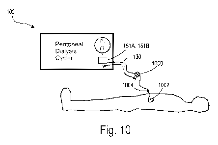
Fig. 10 shows a schematic diagram of the PD machine 102 connected to a
patient.
The patient line 130 is connected to the patient's abdomen via the catheter
1002, and the
catheter is connected to the patient line via the port 1004. The patient line
130 may be a
tube made of a flexible material (e.g., a polymer) that is at least partially
distended by
operating pressures in the PD machine 102. For example, the patient line 130
may be an
elastic polymer tube that develops a swell in response to positive operating
pressures in
the PD machine 102. The patient line 130, the port 1004, and the catheter 1002
are
sometimes referred to herein as the patient line-catheter conduit, or simply
the conduit. At
least one of the pressure sensors 151A, 151B is located at a proximal end of
the patient
line 130 (e.g., at the end of the patient line 130 that is nearest to the PD
machine 102). At
least one of the pressure sensors 151A, 151B is selectably configured to
measure the
pressure in the patient line 130. In some implementations, the pressure
sensors 151A,
151B include a transducer that generates a signal as a function of the
pressure imposed.
The signal is indicative of the magnitude and sign of the measured pressure.
During a PD treatment cycle, an occlusion can occur at different locations in
the
conduit. For example, the patient line 130 may become kinked or pinched, holes
in the
catheter 1002 may become occluded (e.g., with omental fat), or the patient
line 130 may
23
CA 03020903 2018-10-12
WO 2017/180326
PCT/US2017/024673
develop an internal blockage at some location (e.g., from a deposit of omental
fat). The
PD machine 102 is configured to adjust its operation in response to an
occlusion being
detected. For example, the control unit 139 may be configured to adjust one or
more
operating parameters of the PD machine 102 in an attempt to clear the
occlusion and/or to
modulate the flow in the patient line to avoid an overpressure condition. In
some
implementations, the control unit 139 may be configured to provide an alert
indicating
that an occlusion has been detected. For example, a visual, tactile, and/or
audible alert
may be directed to the patient (e.g., to wake the patient).
In order to determine an appropriate response, the PD machine 102 is
configured
to ascertain the type of occlusion that is present. In some implementations,
the type of
occlusion can be inferred based on the location of the occlusion in the
conduit. For
example, if an occlusion is detected in the catheter 1002, the PD machine 102
can infer
that holes in the catheter 1002 may be occluded. Similarly, if the occlusion
is detected
somewhere along the patient line 130, the PD machine 102 can infer that the
patient line
130 is kinked or pinched. The PD machine 102 is configured to determine a
location of
the occlusion relative to the position of the pressure sensor 151A, 151B. The
particular
location of the occlusion can be considered by the PD machine 102 to determine
the
appropriate response. In the example shown in Fig. 10, an occlusion 1008 is
present in
the patient line 130 at a distance x from the pressure sensor 151A (e.g., at
or near the
patient line inlet), which may be indicative of a kink or a pinch in the
patient line 130.
Motion (e.g., rapid motion) of the pump mechanism creates an impulse (e.g., a
step input and/or a near-instantaneous pulse) in local pressure. The onset or
stoppage of
flow of the PD solution (e.g., the dialysate) can present a wavefront. In
response, the
patient line 130 may develop a deformity. For example, the elastic material of
the patient
line 130 may locally expand (in the case of positive pressure) or locally
contract (in the
case of negative pressure) in response to the step input. The local (e.g.,
positive or
negative) distension in cross-sectional area travels axially along the wall of
the patient
line 130 itself (e.g., as opposed to traveling in the PD solution) as an
elastic wave. The
wave carries with it local pressure variations, which may be detected by the
pressure
sensor 151A, 151B that is sampling fast enough to resolve the pulse as it
travels.
24
CA 03020903 2018-10-12
WO 2017/180326
PCT/US2017/024673
When an elastic wave encounter a discontinuity in the dispersion relation of
the
elastic wave, at least a portion of the wave is reflected back toward the
source. An
occlusion in the conduit, or a kink or pinch in the line, are examples of such
a
discontinuity. Thus, when the elastic wave encounters the occlusion 1008, at
least a
portion is reflected back toward the pressure sensor 151A, 151B. The speed at
which the
elastic wave travels (e.g., the propagation speed) is the same in both
directions, and is a
function of the material properties and the geometry (e.g., cross-sectional
geometry) of
the materials comprising the conduit. The pressure sensor 151A, 151B is used
to
determine the timing of the wave's motion. For example, a single pulse can be
detected
as a difference in timing, and a period of an oscillatory wave can be
measured.
If the propagation speed co of the elastic wave is known, and the time
required for
the elastic wave to travel from the pressure sensor 151A, to the occlusion
1008, and back
to the pressure sensor 151A T is known, the distance traveled by the elastic
waves (e.g.,
from the pressure sensor 151A, to the occlusion 1008, and back to the pressure
sensor
151A) can be determined. The distance traveled can be divided by two to
determine the
location of the occlusion 1008 in the conduit relative to the location of the
pressure
sensor 151A. That is, the distance x along the conduit from the location of
the pressure
sensor 151A to the location of the occlusion 1008 can be determined according
to
Equation 1:
T* c0
X = - 2 (1)
where T is the transit time of the elastic waves, co is the propagation speed
of the elastic
waves, and x is the distance along the conduit from the location of the
pressure sensor
151A to the location of the occlusion 1008 for the first reflection of the
wave. The wave
reflections continue; the reflected wave is again reflected by the proximal
end of the tube,
the reflection travels back toward the occlusion, and is in turn reflected
back. At each
step, energy is lost, thereby resulting in an oscillation with a decaying
amplitude.
CA 03020903 2018-10-12
WO 2017/180326
PCT/US2017/024673
The propagation speed co of the elastic wave in distensible tubing carrying an
incompressible fluid can be determined according to Equation 2:
A F3
co =_ _ (2)
p 0A
where A is the cross-sectional area of the lumen of the tubing, p is the
density of the fluid,
and P is the local transmural pressure. The value of the term ¨aa: comes from
the stress-
strain relationship of the tubing. Thus, this term is a function of the
elastic modulus of the
tubing material and of the tube's cross-sectional dimensions. Accordingly,
Equation 2
confirms that the propagation speed co is a function of the material
properties of the tube,
the dimensions of the tube, and the density of the fluid traveling through the
tube.
As mentioned above, elastic waves can be reflected (or, e.g., scattered) when
they
reach a discontinuity in the carrying medium. In the case of the 1-dimensional
waves of
interest in this example, such a discontinuity may be represented by a change
in the
characteristic impedance Zo of the tubing. The characteristic impedance Zo for
a harmonic
forcing of pressure waves (e.g., at frequency co) in such a tube, accounting
for the effect
of viscous damping, can be determined according to Equation 3:
2
pc0
Z0 = A (3)
twAo
where A, is the luminal area at zero P, i represents the imaginary number V1,
and is
given by Equation 4:
1 87rico /2
2 it l i
A = r (_p w2 + (4)
pc0 A0
26
CA 03020903 2018-10-12
WO 2017/180326
PCT/US2017/024673
where ,u is the dynamic viscosity of the fluid. If a traveling wave reaches a
boundary at
distance x in the conduit with a terminal impedance ZT, defined by Equation 5:
P (x,t)
Z T = - (5)
Q (x,t)
where P (x, t) and Q (x, t) are the local instantaneous transmural pressure
and volumetric
flow rate, respectively, a fraction of the wave will be reflected if ZT # Zo.
The fraction of
the wave reflected may be embodied by the reflection coefficient F given by
Equation 6:
Zo ¨ZT
r = (6)
Zo +ZT
In short, for the systems and techniques described herein, Equations 1-6
establish
that: i) a local deviation in either the available area for flow, or the
effective distensibility
of the tubing, causes at least a partial reflection of elastic waves
propagated through the
tubing; and ii) for tubing of uniform properties and cross-section, the
outgoing and
reflected elastic waves will transit the unaffected length of tubing at a
common speed.
Thus, if the transit time T of an elastic wave from the pressure sensor 151A,
to the
affected location (e.g., the location of the occlusion 1008), and back to the
pressure
sensor 151A is measured, and if the wave speed co is known, the distance x
along the
conduit from the location of the pressure sensor 151A to the location of the
occlusion
1008 can be determined according to Equation 1.
Because the outgoing and reflected elastic waves will transit the length of
the tube
at a common speed in a given system (e.g., because the propagation speed co is
a function
of the material properties of the tube, the dimensions of the tube, and the
density of the
fluid traveling through the tube), the propagation speed co may be initially
determined for
a given system (e.g., the dialysis system 100). Once the propagation speed co
is known,
the transit time T of elastic waves can be measured. The distance x along the
conduit from
the location of the pressure sensor 151A to the location of the occlusion 1008
(e.g., the
location of the occlusion) can then be determined.
27
CA 03020903 2018-10-12
WO 2017/180326
PCT/US2017/024673
In some implementations (e.g., implementations in which the conduit includes
segments connected in series, such as a patient line and a catheter connected
in series),
the various segments of the conduit may have different elastic properties
and/or cross-
sectional dimensions. Further, the segments may be connected by fittings with
yet other
values of elastic properties and dimensions. While such complexities in the
physical
conduit carrying elastic waves may cause complexities in the characteristic
relationship
of transit time T versus distance x to the occlusion, this relationship may
still be
repeatable and monotonic, thus preserving the effectiveness of the method
described
herein.
Experiment 1
Fig. 11 shows an example experimental system 1100 in which the propagation
speed c, of elastic waves can be determined. The system 1100 includes a
syringe pump
1110 that is configured to produce flow into a conduit that includes a tube
1130 (e.g.,
which mimics a patient line) and a catheter 1102 connected to the tube 1130
via a port
1104. In this example, the syringe pump 1110 was driven by a programmable
stepper
motor. The catheter 1102 is submerged in a reservoir of fluid 1112 (e.g., in
place of a
patient). An occlusion 1108 is present in the tube 1130 at various distances x
from a
pressure sensor 306 that is positioned at a proximal end of the tube 1130. In
this example,
the occlusion was created by hemostat clamping the tube 1130 at various
distances x. The
clamping of the tube 1130 represents a complete occlusion.
A small volume (e.g., approximately 0.32 cubic centimeters) of water was
injected by the syringe pump 1110 at a fixed rate (e.g., a relatively high
rate of flow of
6.4 cubic centimeters per second). For example, the fixed rate of flow may
create an
impulse (e.g., a step input and/or a near-instantaneous pulse) in local
pressure. At the end
of the dispensing stroke, the flow of water was abruptly stopped. The tube
1130 develops
a local distension in cross-sectional area due to the sudden injection of
water that travels
axially along the wall of the tube 1130 as an elastic wave. The elastic wave
carries with it
local pressure variations. As the elastic wave travels distally along the tube
1130, it
28
CA 03020903 2018-10-12
WO 2017/180326
PCT/US2017/024673
reaches the occlusion 1108, and at least a portion is reflected back
proximally toward the
pump 1110.
The pressure sensor 1106 is configured to measure the pressure in the tube
1130 at
the proximal end of the tube 1130 over time. The pressure measurements can be
used to
detect reflections of the elastic waves, in particular, times at which such
reflections arrive
at the proximal end of the tube 1130. In some implementations, the pressure
measurements occur at a frequency in the order of ones of hertz, tens of hertz
(e.g., 1-99
Hz), hundreds of hertz, or thousands of hertz (e.g., 1 kHz ¨2 kHz). The
experiment is
repeated at various distances x of the occlusion 1108.
Figs. 12A-G show representative graphs of pressure P (in mbar) measured by the
pressure sensor 1106 versus time (in seconds). The occlusions 1108 (e.g., the
clamping of
the tube 1130) occur at distances x of 80 cm, 100 cm, 140 cm, 180 cm, 220 cm,
260 cm,
and 295 cm, respectively.
Referring to Fig. 12C, which shows pressures P measured when the tube 1130
was clamped at a distance x of 140 cm, the measured pressure is initially
slightly above
ambient and rises substantially uniformly during the pumping stroke. After the
substantially uniform rise, oscillations occur. The period T of the
oscillations (e.g., the
transit time T of the elastic wave from the pressure sensor 1106, to the
location of the
occlusion 1108, and back to the pressure sensor 1106) is approximately 78
milliseconds.
Using Equation 1, the propagation speed c, of the elastic waves is determined
to be
approximately 36 meters per second.
The calculation of the propagation speed c, with reference to Fig. 12C is made
under the assumption that the oscillations are attributable to successive
arrivals of a
reflected elastic wave. Because the propagation speed c, should be uniform
across
various locations of the occlusion 1108 (e.g., in the case of uniform tubing),
additional
tests were performed at various distances to corroborate the validity of
Equation 1 and
confirm that the oscillations were attributable to successive arrivals of a
reflected elastic
wave. While Figs. 12A-G show representative graphs of pressure versus time for
clampings that were located at distances of 80 cm to 295 cm, pressures may be
measured
29
CA 03020903 2018-10-12
WO 2017/180326
PCT/US2017/024673
using other clamping locations. In some implementations, additional signal
processing
can be performed to extend limits of occlusion detection to any location of
occlusions.
Fig. 13 shows a representative graph of the periods T of the oscillations (in
milliseconds) versus the various distances x of the clamping locations (in
centimeters).
The measured periods Tare based on the data shown in Figs. 12A-G. The data
shown in
Fig. 13 indicates that the measured periods T (e.g., the transit time T of the
elastic wave
from the pressure sensor 1106, to the location of the occlusion 1108, and back
to the
pressure sensor 1106) are commensurate with the corresponding clamping
distances x.
That is, the data verify that the propagation speed c, of the elastic waves is
substantially
uniform (e.g., approximately 36 2 m/s) for all of the distances x measured,
thereby
corroborating the validity of Equation 1 and confirming that the oscillations
are
attributable to successive arrivals of reflected elastic waves. Now that the
propagation
speed c, is known, the transit time T of elastic waves can be measured to
determine
unknown distances x of other occlusions 1108 which may occur.
In some examples, the empirical determination of oscillation period T versus
clamping distances x can be performed to characterize or "calibrate" the
relationship
between period T and distance x while accounting for non-uniform segments of
the
conduit. For example, the slope of the period T versus distance x curve of
Fig. 13 may
change at certain junctions in the conduit assembly, which in some examples
can have the
effects of enhancing the sensitivity of the detection method. In some
examples, prior to
treatment, an empirical determination can be made in which an occlusion is
intentionally
applied at known distances x, thereby providing a specific calibration of the
current
conduit assembly.
Experiment 2
While Experiment 1 corroborated the validity of Equation 1 in the experimental
system 1100 of Fig. 11 testing for complete occlusions, Experiment 2 studies a
similar
technique implemented in an actual dialysis machine (e.g., the PD machine 102
of Figs.
1-10) using the built-in pressure sensor 151A to test for partial occlusions.
The advanced
CA 03020903 2018-10-12
WO 2017/180326
PCT/US2017/024673
testing described below was performed to achieve results that are more
relevant to real
PD treatment.
The experiment primarily focused on flow in the drain direction. The choice to
focus on flow in the drain direction was made for the following reasons: i) a
majority of
problematic blockages typically occur in the drain direction; ii) a greater
potential for
difficulty was predicted in the drain direction due to possible pull-off of
cassette film
from the pump; and iii) initial tests in the fill direction suggested that the
same patterns of
pressure versus flow should be obtainable ¨ albeit with the potential for
different
calibration curves that may need to be empirically determined.
Fig. 14 shows a schematic of a dialysis system 1400 in which the propagation
speed c, of elastic waves can be determined. The dialysis system 1400 includes
the PD
machine 102, the PD cassette 112 housed in the PD machine 102, a patient line
1430, and
the pressure sensor 151A located at a proximal end of the patient line 1430.
The patient
line 1430 may be substantially similar to the patient line 130 described above
with
respect to Figs. 1 and 10. In some implementations, the patient line 1430 may
be a 10-
foot patient line with dual patient connectors. In this example, the PD
machine 102 is
controlled by a computing device 1434 and a microcontroller 1436 such as an
ATmega
2650 microcontroller manufactured by Atmel Corporation. In some
implementations, the
PD machine 102 may be controlled by a control unit (e.g., a processor) of the
PD
machine 102, such as the control unit 139 shown in Fig. 1. The microcontroller
1436 is
operatively coupled to driver modules 1438a, 1438b. The driver modules 1438a,
1438b
may be DRV8825 stepper motor driver modules manufactured by Pololu
Corporation.
The dialysis system 1400 includes various experimental components that can
perform the
functions of: i) introducing a controlled level of occlusion to the patient
line 1430 at a
known location; ii) enabling external programmable control of the pump heads
to execute
flow actuation according to the methods described herein; and iii) performing
data
acquisition from the onboard pressure sensor (e.g., the pressure sensor 151A)
and in some
examples a separate inline pressure sensor for validation purposes.
The microcontroller 1436, at the direction of commands issued by the computing
device 1434, is configured to control the driver modules 1438a, 1438b to cause
the driver
31
CA 03020903 2018-10-12
WO 2017/180326
PCT/US2017/024673
modules 1438a, 1438b to operate pumps (e.g., the piston heads 134A, 134B) of
the PD
machine 102 in order to impose specified flow patterns. The microcontroller
1436 and the
driver modules 1438a, 1438b provided pulse streams to the stepper motors
driving the
pumps to accomplish the following types of motion: i) return to the "home"
position as
defined by an onboard limit switch; ii) move forward at a specified step rate
(e.g., to
achieve a particular flow rate), by a specified number of steps, in a user-
defined stepping
mode from full stepping to various increments of microstepping; and iii) move
backward
at a specified step rate, by a specified number of steps in a user-defined
stepping mode.
Some flow patterns (e.g., characterized by combinations of step rates, number
of steps,
stepping mode, etc.) were determined to be more desirable than others for the
purpose of
occlusion detection. Such desirable flow patterns were programmed in a
sequence that is
described below.
The pumps are configured to cause fluid to be pumped through a patient line-
catheter conduit that includes the patient line 1430, a catheter 1402, and a
port 1404 that
connects the patient line 1430 to the catheter 1402. The catheter 1402 may be
a Flex
Neck Classic catheter. The catheter 1402, the port 1404, and a portion of the
patient line
1430 is submerged in a basin of water 1412 (e.g., in place of a patient). The
water was
held at room temperature (e.g., 20-25 C). The free surface of the water was
kept at the
same height (e.g., 1 centimeters) with respect to the direction of gravity
as that of the
pressure sensor 151A of the PD cycler 102. An occlusion 1408 was provided in
the
patient line 1430 at various distances x from the pressure sensor 151A. In
this example,
the occlusion 1408 was created using various methods and at various distances
x, as
described in more detail below. The occlusions 1408 represented both full and
partial
occlusions.
The experiment included the following general steps:
i. create an impulsive change in a pressure condition at the
proximal end of
the patient line 1430 (e.g., at the location of the pressure sensor 151A) by
providing a short burst of water flow in either the fill or the drain
direction
that is abruptly ceased, thereby creating elastic waves in the patient line
1430;
32
CA 03020903 2018-10-12
WO 2017/180326
PCT/US2017/024673
ii. detect and measure the transit time T for elastic waves to travel from
the
pressure sensor 151A, to the location of the occlusion 1408, and back to
the pressure sensor 151A; and
iii. empirically determine a calibration curve between the transit time T
and
the distance to the occlusion x, thereby determining an effective value of
the propagation speed c, of the elastic waves.
The experiment was performed across a large number of cassettes, with
different
types, degrees, and locations of flow restriction (e.g., occlusions), in order
to investigate
the potential sensitivity (e.g., true positive rate) and specificity (e.g.,
true negative rate) of
the detection method, as described in more detail below.
A small volume (e.g., approximately 0.33 cubic centimeters) of water was moved
through the patient line 1430 in the drain direction by a first pump of the PD
machine 102
(e.g., a pump controlled by a first one of the driver modules 1438a) at a
fixed rate (e.g.,
4.4 cubic centimeters per second). At the end of the stroke, the first pump
was abruptly
stopped. The patient line 1430 develops a local deformity due to the injected
water. Such
a deformity causes elastic waves to be generated in the patient line 1430. The
pressure
sensor 151A, which is built into the PD machine 102 and located at the
proximal end of
the patient line 1430, was used to detect the reflected elastic waves in a
manner
substantially similar to that described above with respect to Fig. 11.
The partial occlusions 1408 used in the experiment were characterized for
their
relative flow restrictions. The characterization was done quantitatively via
the fluidic
resistance Rf values of the partial occlusions 1408 as given by Equation 7:
I AP I
-R=
(7)
where
AP= pressure difference from upstream to downstream of occlusion (8)
and
Q = volumetric flow rate (9)
33
CA 03020903 2018-10-12
WO 2017/180326
PCT/US2017/024673
The pressures were initially measured using both the pressure sensor 151A of
the
PD machine 102 and a reference pressure transducer 1440 positioned downstream
from
the pressure sensor 151A. The separate pressure measurements were taken to
ensure that
the pressure sensor 151A built into the PD machine 102 was capable of
achieving the
sensitivity required to detect the elastic waves. For example, the pressure
sensor 151A is
configured to detect the pressure in the patient line 1430 through a membrane
of the
cassette 112, and various fluidic elements are positioned between the pressure
sensor
151A and the proximal end of the patient line 1430. It was considered that
these elements
may have the potential to diminish and/or distort the elastic waves. Thus,
measurements
made by the reference pressure transducer 1440 were used to verify the
fidelity of the
measurements made by the pressure sensor 151A. A high degree of fidelity was
observed,
and the reference pressure transducer 1440 was removed to avoid possible
artifacts.
Utilizing only measurements from the pressure sensor 151A of the PD machine
102, AP due to the applied occlusion 1408 was inferred by first obtaining a
baseline
pressure measurement with no occlusion 1408. The baseline pressure measurement
was
then subtracted from the pressure measurement with the occlusion 1408
according to
Equation 10:
AP= Pwith occlusion ¨ Pwithout occlusion (10)
Due to the likelihood of turbulent flow and other sources of viscous pressure
losses that are not linearly related to Q, the fluidic resistance Rf for a
given flow
restriction is in general a function of Q. In order to isolate the effect of
flow resistance
from capacitive or inertial effects, AP is measured at steady state. For these
reasons,
measurements related to the fluidic resistance Rf were performed under
prolonged flow at
a fixed flow value (e.g., a fixed flow value of Q = 30 milliliters per
minute). Such a flow
value was chosen because it represents the critical value for the Drain
Complication
condition, described in more detail below, and is representative of the order
of magnitude
of mean flow rate occurring throughout a treatment.
34
CA 03020903 2018-10-12
WO 2017/180326
PCT/US2017/024673
The ability to detect a partial occlusion (e.g., as compared to detecting a
complete
occlusion) presents challenges that do not manifest when detecting a complete
occlusion.
Typically, the less restrictive an occlusion is, the greater is the challenge
for sensitivity
and specificity of a method for determining its location. A relevant standard
for
quantifying partial occlusions in the PD machine 102 comes from the Drain
Complication
and Fill Complication conditions. Drain Complication and Fill Complication
conditions
occur when there is a flow restriction sufficient to depress the flow below a
threshold
value for a particular period of time. In a model case of a steady-state flow
restriction, the
threshold value of restriction that would generate a Drain Complication is one
that would
require a pressure of approximately -200 mbar (as measured at the pressure
sensor 151A)
to drive a flow of approximately 30 milliliters per minute. Thus, the
measurements
related to the fluidic resistance Rf were performed under prolonged flow at
the fixed flow
rate of Q = 30 milliliters per minute. An occlusion that requires -200 mbar to
produce a
steady-state flow rate of 30 ml/min is referred to herein as a "drain-critical
occlusion."
Applying Equation 7 to the conditions defined by the "drain-critical
occlusion,"
the total fluidic resistance of the system 1400 can be determined according to
Equation
11:
D drain¨ critical,total = 200 mbar = 6.7 mbar
Itf 30 ml/min ml/min (11)
In Equation 11, the superscript "total" refers to the fact that the pressure
sensor
151A shows the effect of all fluidic resistances occurring in, and inherent
to, the cassette
112, the patient line 1430, and the catheter 1402. Thus, some components of
the total
fluidic resistance are due to normally occurring elements in the flowpath
(e.g., the
conduit), Ribaselme Because such normally occurring elements are arranged in
series with
the additional resistance created by the occlusion 608, and due to the
additive property of
resistances in series, a drain-critical value of occlusion-specific resistance
Ril"in-
critical.occluszon can be determined according to Equation 12:
CA 03020903 2018-10-12
WO 2017/180326
PCT/US2017/024673
Rdrain¨critical,occlusion = Rdrain¨critical,total Dnormal
'f (12)
Ribaseh" for the patient line 1430, the port 1404 with two patient connectors,
and
the catheter 1402 was measured to be approximately 0.095 mbar/(ml/min). Thus,
the
drain-critical value of the fluidic resistance of a partial occlusion itself
is approximately
6.7 mbar/(ml/min). Over the course of Experiment 2, partial occlusions 1408
were tested
with occlusion-specific resistances in the range of approximately 1-10
mbar/(ml/min),
thus representing values in the range of approximately 0.15-1.5 times the
drain-critical
value of occlusion-specific resistance RI
ram-criticaLocclusion
The partial occlusions 1408 were designed to model two basic types of real
occlusions: i) internal occlusions (e.g., in which an obstruction lodges
itself within the
lumen of the patient line 1430; and ii) external occlusions, in which the
patient line 1430
is pinched from the outside. In designing the physical means of applying the
partial
occlusions 1408 to the patient line 1430 and/or the catheter 1402, the goal
was to
determine whether the detection method can provide a measurement of the
distance x of
the occlusion 1408 that is sensitive and specific for the distance x but
insensitive to the
type of restriction or the value of the fluidic resistance Rf of the occlusion
1408 (e.g., for
fluidic resistance Rf values within the range of interest of approximately 1-
10
mbar/(ml/min)).
Partial occlusions 608 of both types (e.g., internal and external) having
repeatable
fluidic resistance Rf values were applied at various locations x over a
relatively large
number of cases to test for repeatability.
Fig. 15 shows a cross-sectional view of an example partial internal occlusion
1502 installed in the patient line 1430. The partial internal occlusion 1502
was fabricated
to serve as a model of an internal occlusion. For example, the partial
internal occlusion
1502 is meant to represent a partially blocked patient line, with a well-
controlled orifice
of known flow characteristics. The partial occlusion 1408 of Fig. 14 may
represent the
partial internal occlusion 1502. In this example, the internal occlusion 1502
is a
cylindrical insert made of stainless steel, although other shapes and/or
materials may be
used. The internal occlusion 1502 is configured to be positioned at a chosen
distance x
36
CA 03020903 2018-10-12
WO 2017/180326
PCT/US2017/024673
such that the internal occlusion 1502 is sufficiently gripped by the patient
line 1430 in
order to remain in position throughout the tests. The internal occlusion 1502
includes a
circular orifice 1504 for allowing the fluid tested (e.g., water) to flow
through the internal
occlusion 1502. The orifice 1504 has a diameter that results in the internal
occlusion 1502
having a fluidic resistance Rf value in the range of 1-10 mbar/(ml/min). The
diameter of
the orifice 1504 may result in particular fluidic resistance Rf values for the
occlusion
1502 according to Table 1:
Diameter of orifice Rf
(mm) mbar/(ml/min)
0.30 8.7-9.1
0.34 5.5-6.4
0.38 3.1-3.2
0.51 0.8-1.0
The fluidic resistance Rf values of the occlusion 1502 as shown in Table 1
depend
on the particular working fluid used in the system 1400 (e.g., in this
example, water).
Thus, if a different fluid were used, such as dialysate, the fluidic
resistance Rf values
would be different. The diameter or the orifice 1504 may be configured to have
a
diameter that results in appropriate fluidic resistance Rf values based on the
working fluid
that is used. In this example, the diameters of the orifice 1504 were chosen
to achieve the
desired fluidic similarity with known conditions of interest for dialysate
flow, using the
drain-critical value Rf as a benchmark as discussed above. Thus, the results
presented
herein are largely sufficient to validate the method for its applicability to
the condition of
dialysate as the working fluid. However, at least two characteristics would be
expected to
vary to some extent if dialysate were substituted for water as used in these
tests. For
example, the exact value of the propagation speed c, of the elastic waves is
affected by
the density of the fluid according to Equation 2. Further, the diameter of
occlusion
required to achieve a particular value of fluidic resistance is a function of
fluid viscosity.
Figs. 16A and 16B show a cutaway view and a photograph, respectively, of an
example partial external occlusion applied to the patient line 1430. The
partial external
occlusion was fabricated to serve as a model of an external "pinching" style
of occlusion.
37
CA 03020903 2018-10-12
WO 2017/180326
PCT/US2017/024673
For example, the partial external occlusion is meant to represent the style of
occlusion
occurring when the patient line 1430 is pinched or kinked, with the applied
value of
restriction being precisely controlled during the test. The partial occlusion
1408 of Fig.
14 may represent the partial external occlusion 1502. In this example, the
external
occlusion is in the form of a clamping mechanism 1602 that is configured to
apply a
partial occlusion of the pinching type. The clamping mechanism 1602 includes
rods 1604
that are configured to apply uniform stresses to substantially opposite
surfaces of the
patient line 1430 that cause the patient line 1430 to deform. In this example,
the rods
1604 are made of stainless steel and have a diameter of 3.2 millimeters,
although other
dimensions and/or materials may be used. The stress applied to the patient
line 1430 may
be referred to as a Hertzian line-contact stress. The clamping mechanism 1602
also
includes washers (e.g., Belleville washers) that are configured to cause the
rods 1604 to
press together as the clamping mechanism 1602 is tightened. For example, an
operator
may tighten the clamping mechanism 1602 during a "long-stroke" (e.g., having a
flow
value of approximately 30 milliliters per minute) to achieve a target pressure
reading by
the pressure sensor 151A, thereby actively setting the fluidic resistance Rf
value of the
external occlusion desired for the particular test.
Referring again to Fig. 14, prior to any sequence of tests concerning a
particular
cassette 112, locations on the patient line 1430 were measured with a
precision of
approximately 3 millimeters. The patient line 1430 and the catheter 1402
were then
primed with water to substantially eliminate the presence of air bubbles in
the conduit.
The partial occlusion 1408 was then placed such that the occlusion 1408 was
centered at
the desired distance x. The testing was repeated for partial occlusions 1408
of both the
internal and external type and having various fluidic resistance Rf values.
For partial occlusions 1408 of the internal type (e.g., such as the partial
internal
occlusion 1502 of Fig. 15), the occlusion 1408 was first positioned near a
distal end of
the patient line 1430 (e.g., at a distance of approximately x = 295
centimeters). The
occlusion 1408 was then repositioned to various distances x for subsequent
tests. Similar
tests were also performed with the occlusion 1408 positioned in the catheter
1402. The
patient line 1430 was primed after each repositioning of the occlusion 1408 to
minimize
38
CA 03020903 2018-10-12
WO 2017/180326
PCT/US2017/024673
the occurrence of air bubbles. For partial occlusions 1408 of the external
type (e.g., such
as the partial external occlusion in the form of a clamping mechanism 1602 of
Figs. 16A
and 16B), the occlusion 1408 was positioned to the various distances x for
testing. The
patient line 1430 was primed after each repositioning of the occlusion 1408 to
minimize
the occurrence of air bubbles.
With the occlusion 1408 in place, both a "long-stroke" test for measuring the
fluidic resistance Rf of the occlusion 1408 and a "short-stroke" test (e.g., a
sudden
injection of approximately 0.32 cubic centimeters of fluid at a fixed flow
rate of
approximately 6.4 cubic centimeters per second) for determining the location
of the
blockage (e.g., the distance x) were performed.
The long-stroke test included a single, prolonged motion of the pump at a
constant
speed corresponding to a flow rate of Q = 30 milliliters per minute. As
described above,
the pump is operated by the microcontroller 1436 and the driver modules 1438a,
1438b.
The pressure sensor 151A was monitored during the test. The pressures measured
by the
pressure sensor 151A typically approached a steady-state value from the mid-
to end-
point of the stroke. The steady-state value was recorded for the purpose of
calculating the
fluidic resistance Rf
The short-stroke test included one or more single rapid motions of the pump
that
were designed to impart a pressure impulse on the patient line 1430, thereby
causing an
elastic wave to be generated in the patient line 1430 as described above. The
short-stroke
test was performed by moving water having a volume of approximately 0.33 cubic
centimeters through the patient line 1430, although other volumes could be
used to
optimize signal-to-noise ratio or the operational limitations of the dialysis
system 1400.
For a particular value of dispensed volume, the speed of the pump was
maximized under
appropriate constraints in order to maximize the amplitude of the pressure
waveforms
associated with the transit of the elastic waves. The constraints included
avoidance of
missed motor steps (e.g., momentary stalling of the motor by requiring power
beyond its
capability), avoidance of pressures outside the range of the pressure sensor
151A, and
avoidance of damage to components of the dialysis system 1400.
39
CA 03020903 2018-10-12
WO 2017/180326
PCT/US2017/024673
Regarding avoidance of missed motor steps, preliminary tests were conducted
with full-stepping of the pump stepper motor with pulse delays of 2.00, 2.50,
and 3.00
milliseconds. Steps were occasionally missed for the 2.00 and 2.50 millisecond
pulse
delays, but were not missed for the 3.00 millisecond pulse delays. Thus, full-
stepping of
the pump motor with a total pulse delay of 3.00 milliseconds for 25 steps was
employed,
which resulted in a dispensed volume of 0.33 cubic centimeters. Pressures
outside the
range of the pressure sensor 151A and damage to components of the dialysis
system 1400
were not observed to occur when operating at any of the pulse delays.
Fig. 17A shows a pressure waveform 1702 that includes pressure measurements
over time made by the pressure sensor 151A during the short-stroke test. The
pressure
measurements were sampled at a frequency of 1 kHz. In this example, the
occlusion 1408
was positioned at a distance x = 220 centimeters along the patient line 1430.
The pump
stroke had a duration of approximately 75 milliseconds. The measured pressure,
steady in
the absence of pump motion, is seen to drop rapidly during a pump stroke
having a
duration of approximately 75 milliseconds that commences at approximately t =
1
second. After abrupt cessation of pump motion, oscillations occur due to the
elastic
effects described above. The period T of the oscillations (e.g., which
corresponds to the
transit time T of the elastic waves from the pressure sensor 151A, to the
location of the
occlusion 1408, and back to the pressure sensor 151A) can be evaluated to
determine the
propagation speed c, of the elastic waves according to Equation 1. Once the
propagation
speed c, of the elastic waves is known, locations x of occlusions (e.g., at
unknown
positions of the conduit) can subsequently be determined by evaluating the
period T of
oscillations.
Superimposed with the oscillations is high-frequency noise and a gradual decay
from the peak excursion of pressure (e.g., at approximately t = 1.075 seconds)
toward
zero. The decay occurs due to the occlusion 1408 being a partial occlusion.
Because the
high-frequency noise and the decay are not relevant for purposes of
determining the
period T of the oscillations, they can be removed from the waveform 1702 using
one or
more signal processing techniques. For example, the waveform 1702 can be
smoothed to
reduce the effect of the high-frequency noise using a moving average taken as
the mean
CA 03020903 2018-10-12
WO 2017/180326
PCT/US2017/024673
of the measured pressures spanning 15 milliseconds on either side of a given
data point
(e.g., sometimes referred to as a 15 millisecond half-width moving average).
Further, a
background curve approximating the overall decay onto which the oscillations
are
superimposed can be subtracted from the waveform 1702. The background curve to
be
subtracted from the waveform 1702 may be obtained, for example, using a 50
millisecond half-width moving average. Prior to applying the moving averages,
the data
were truncated to the time domain which begins at the cessation of the pump
motion at
approximately t = 1.075 seconds.
Fig. 17B shows a pressure waveform 1704 that includes the data of Fig. 17A
after
being smoothed and after having the background curve subtracted. The waveform
904
has a relatively more symmetrical pattern as compared to the waveform 1702 of
Fig.
17A, thereby enabling a more accurate evaluation of the oscillation period T.
The data shown in Figs. 17A and 17B correspond to the short-stroke test
performed with a dispensed water volume of 0.33 cubic centimeters at a fixed
rate of 4.4
milliliters per second for an occlusion 1408 positioned at a distance x = 220
centimeters
along the patient line 1430. Data was also obtained for various other cassette
112/occlusion 1408 configurations at various different distances x for the
occlusion 1408.
For example, 15 different cassette-occlusion combinations were used, for both
internal
and external partial occlusions, and each combination was tested at 5-8
different distances
x for the occlusion 1408. For each test, the period T of the resulting
oscillations was
evaluated using at least three different methods: i) first half-wave period;
ii) other half-
and full-wave periods; and iii) Fast Fourier Transform. It was determined that
the first
half-wave period method achieved the greatest sensitivity and specificity for
determining
the distance x of the occlusion 1408.
Sensitivity and specificity are statistical measures of the performance of the
detection method. The sensitivity, also referred to as the true positive rate,
measures the
proportion of positives that are correctly identified as such. In this
context, the sensitivity
may correspond to the ability of the system to correctly identify occlusions
(e.g., for
distances x within a particular range). The specificity, also referred to as
the true negative
rate, measures the proportion of negatives that are correctly identified as
such. In this
41
CA 03020903 2018-10-12
WO 2017/180326
PCT/US2017/024673
context, the specificity may correspond to the accuracy of the detection
method (e.g., the
margin of error of determined distances x).
The first half-wave period is the time measurement from the end of the pump
motion to a first local extremum of the pressure measurements, represented by
T1 in Fig.
17B. For drain-direction flow, the first local extremum is a local maximum. As
compared
to latter half-waves (e.g., T2 and T3), sensitivity and specificity benefit
from certain
features of the first half-wave. For example, onset is relatively precise
because it is well
defined by the cessation of the pump motion (e.g., during short-stroke
testing), the timing
of which can be measurable with high precision. Further, completion of the
first half-
wave can be measured with a relatively high signal-to-noise ratio due to the
signal
maximization and the noise minimization associated with the first half-wave
period. For
example, the first half-wave presents the elastic wave with its maximum
amplitude,
which maximizes the precision of measuring the extremum that defines its
endpoint by
minimizing the effect of various sources of noise on the measurement. After
the first half-
wave, a rapid decay of amplitude in the subsequent oscillations occurs due to
viscoelastic
effects and the effects of partial wave reflection (e.g., as described above
with reference
to Equation 6). Further, subsequent wave reflections may generate additional
sources of
noise due to constructive and destructive interference of partially
transmitted waves. The
effects of such noise do not manifest in the first half-wave period.
The latter half-wave periods (e.g., the second half-wave period T2 and the
third
half-wave period T3) appear to have substantially equal durations (e.g., as
might be
expected of a naturally resonating wave), while the first half-wave period T1
appears to
be relatively shorter (e.g., because the first half-wave period T1 is the
incipient period
upon impulsively starting the elastic wave). Thus, it was not obvious a priori
that the
half-wave period T1 would correlate well with the distance x of the occlusion
1408.
However, use of the first half-wave produced the best sensitivity and
specificity in the
analyses performed.
As the name implies, because the first half-wave period Ti only represents
half of
the period T of the oscillations, the first half-wave period Ti corresponds to
the transit
time of the elastic wave from the pressure sensor 151A to the location of the
occlusion
42
CA 03020903 2018-10-12
WO 2017/180326
PCT/US2017/024673
1408 (e.g., not the full round-trip transit time 7). Thus, when using the
first half-wave
period T1 to determine the distance x to the occlusion 1408, Equation 1 can be
simplified
as Equation 13:
X = * Co (13)
where T1 is the first half-wave period, co is the propagation speed of the
elastic waves,
and x is the distance along the conduit from the location of the pressure
sensor 151A to
the location of the occlusion 1408.
Fig. 18 shows a representative graph of the first half-wave periods of the
oscillations (in seconds) versus the various distances x of the occlusions
1408 (in
centimeters) for the 15 different cassette-occlusion combinations. The
occlusions 1408,
of both the internal and external types, were located at various distances x
that correspond
to positions along the patient line 1430 (e.g., x = 60, 100, 140, 150, 180,
200, 220, 250
centimeters), distances x that correspond to positions between the patient
connectors of
the port 1404 (e.g., x = 304-307 centimeters), and distances that correspond
to positions
along the catheter 1402 (e.g., x = 310-365 centimeters). The occlusions 1408
had various
fluidic resistance Rf values (e.g., Rf = 5.9-6.4, 8.7-9.1, 5.5-5.9, 3.1-3.2,
0.8-1.0, 6.4-10.2,
7.0-9.6, 5.2-7.0, 1.4-2.2, 8.0-8.5, 6.3-6.9, 1.3-1.8, 8.2-9.6, 6.6-7.8, and
1.5-2.1
mbar/(ml/min)).
Among the distances x tested, evaluation of the first half-wave period
resulted in
sensitivity (e.g., the ability to correctly identify occlusions) for distances
x greater than or
equal to approximately 100 centimeters. In some examples, for distances x of
less than
100 centimeters, the local maxima of the pressure measurements may be
undetectable.
The range of sensitivity may be extended to lower distance values x by
increasing the
strength of the pressure impulse and/or by implementing additional or
alternate signal
processing of the pressure waveforms (e.g., 1702, 1704 of Figs. 17A and 17B).
Recalling that the goal of this experiment was to determine whether the
detection
method can provide a measurement of the distance x of the occlusion 1408 that
is
sensitive and specific for the distance x but insensitive to the type of
restriction or the
43
CA 03020903 2018-10-12
WO 2017/180326
PCT/US2017/024673
value of the fluidic resistance Rf of the occlusion 1408, the first half-wave
periods
corresponding to each distance x would ideally be identical. However, the
vertical scatter
seen in the data of Fig. 18 implies a specificity (e.g., an accuracy) of
approximately 40
centimeters. In some implementations, the detection method may be employed to
determine in which of five sections/zones the occlusion 1408 is located. For
example, the
detection method can be used to determine whether the occlusion 1408 is
located in a
first zone of the patient line 1430 (e.g., approximately x = 0-100
centimeters), in a second
zone of the patient line 1430 (e.g., approximately x = 100-200 centimeters),
in a third
zone of the patient line 1430 (e.g., approximately x = 200-295 centimeters),
between the
patient connectors of the port 1404 (e.g., approximately x = 304-307
centimeters), or in
the catheter 1402 (e.g., approximately x = 310-365 centimeters).
While the detection method described above largely focuses on using the first
half-wave period for evaluating the period T of the oscillations, other
methods can be
employed. For example, the second half-wave period or the third half-wave
period (e.g.,
T2 and T3, respectively, as shown in Fig. 17B) can be evaluated.
Alternatively, frequency-
based signal analyses (e.g., Fast Fourier Transform) may be used to determine
the
distance to the occlusion x.
Fig. 19 shows a representative graph of the second half-wave periods of the
oscillation (in seconds) versus the various distances x of the occlusions 1408
(in
centimeters) for the 15 different cassette-occlusion combinations, and Fig. 20
shows a
representative graph of the third half-wave periods of the oscillation (in
seconds) versus
the various distances x of the occlusions 1408 (in centimeters) for the 15
different
cassette-occlusion combinations. Both graphs show a larger degree of vertical
scatter as
compared to the vertical scatter present in the data of Fig. 18, and thus
indicate reduced
specificity, for the reasons discussed above with respect to Fig. 17B.
In some implementations, the Fast Fourier Transform (FFT) of the pressure
waveform can be used to evaluate the period T of the oscillations. For
example, the
pressure waveform (e.g., 1702, 1704 of Figs. 17A and 17B) can be transformed
into the
frequency domain, and the transform can be evaluated to determine the period T
of the
oscillations. However, a limited number of wave periods transpiring prior to
the
44
CA 03020903 2018-10-12
WO 2017/180326
PCT/US2017/024673
substantially full decay of the wave amplitude may result in imprecision in
the frequency
space (e.g., due to the relatively short window in the time domain), thereby
resulting in
diminished sensitivity and/or specificity.
In some implementations, the specificity is improved (e.g., the vertical
scatter of
the data is reduced) by employing additional signal processing to enhance the
accuracy of
the wave period measurement. In some implementations, the specificity is
improved by
performing a pre-test calibration routine to account for any cassette- or
medical
tube/patient line-specific variations in the wave period versus the distance
x.
In some implementations, the specificity of the detection method is improved
by
performing multiple short-stroke tests and averaging the results. For example,
referring to
Fig. 21, a long-stroke test may be initially performed (e.g., in the drain
direction) during a
first phase 2102 in which the pump moves at a constant speed corresponding to
a flow
rate of Q = 30 milliliters per minute. During the first phase 2102, the pump
is withdrawn
and fluid is pulled from the patient line 1430 into the pump cylinder. The
long-stroke test
may be initially performed to adjust the configuration of a partial external
occlusion (e.g.,
as shown in Fig. 16) in order to achieve the desired fluidic resistance before
performing
the series of short-stroke tests. The initial mean pressure value may be
subtracted from
the pressure measurements. The steady-state pressure is used for determining
the fluidic
resistance Rf of the total flowpath. If the fluidic resistance Rf exceeds a
threshold value
(e.g., a predetermined threshold value), multiple short-stroke tests are
performed to
determine the location of the occlusion. During a second phase 2104, the pump
may be
returned to position for the start of the short-stroke tests; however, in some
implementations, the pump is not returned to position (e.g., to avoid reversal
of flow
during detection). The second phase 2104 begins with a pause to allow
transients to
complete, followed by a long pump-stroke in the fill direction (e.g., fluid is
pumped from
the pump cylinder into the patient line 1430). During a third phase 2106, the
multiple
short-stroke tests are then performed. The resulting pressure measurements, as
well as
any analyses performed to determine the location of the occlusion x, can be
averaged to
reduce the uncertainty, thereby improving the specificity of the detection
method.
45
CA 03020903 2018-10-12
WO 2017/180326
PCT/US2017/024673
While the detection method has been largely described as being implemented in
a
testing environment, similar techniques can be employed for detecting
occlusions in the
conduit when the patient line is attached to a patient receiving a dialysis
treatment (e.g.,
as shown in Fig. 10). For example, the detection method can be employed for
determining the distance x of the occlusion 1008 in the conduit by measuring
the period T
of elastic wave oscillations generated in the patient line 130 itself In
particular, the
propagation speed c, of the elastic waves generated in a particular system
configuration
can be determined according to Equation 1 in advance of a treatment by
positioning a test
occlusion at a known distance x and measuring the period T of the oscillations
¨ that is,
each specific cassette-patient line-port-catheter combination may be
"calibrated" prior to
use. Once the propagation speed c, for the system is known, the period T of
oscillations
can be measured during an actual dialysis treatment, and Equation 1 can be
used to
determine the distance x of the occlusion 1008. Alternatively, an
experimentally
determined correlation between the period T and the distance x of the
occlusion 1008 may
be used. The type of the occlusion 1008 can then be inferred based on the
determined
location of the occlusion 1008, as described above.
While the dialysis system has been largely described as being a peritoneal
dialysis
(PD) system, other medical treatment systems can employ the techniques
described
herein. Examples of other medical treatment systems include hemodialysis
systems,
hemofiltration systems, hemodiafiltration systems, apheresis systems, and
cardiopulmonary bypass systems.
Fig. 22 is a block diagram of an example computer system 2200. For example,
the
control unit (139 of Fig. 1), the computing device (1434 of Fig. 14), and/or
the
microcontroller (1436 of Fig. 14) could be examples of the system 2200
described here.
The system 2200 includes a processor 2210, a memory 2220, a storage device
2230, and
an input/output device 2240. Each of the components 2210, 2220, 2230, and 2240
can be
interconnected, for example, using a system bus 2250. The processor 2210 is
capable of
processing instructions for execution within the system 2200. The processor
2210 can be
a single-threaded processor, a multi-threaded processor, or a quantum
computer. The
processor 2210 is capable of processing instructions stored in the memory 2220
or on the
46
CA 03020903 2018-10-12
WO 2017/180326
PCT/US2017/024673
storage device 2230. The processor 2210 may execute operations such as causing
the
dialysis system to carry out dialysis functions.
The memory 2220 stores information within the system 2200. In some
implementations, the memory 2220 is a computer-readable medium. The memory
2220
can, for example, be a volatile memory unit or a non-volatile memory unit. In
some
implementations, the memory 2220 stores information (e.g., executable code)
for causing
the pumps of the dialysis system to operate as described herein.
The storage device 2230 is capable of providing mass storage for the system
2200. In some implementations, the storage device 2230 is a non-transitory
computer-
readable medium. The storage device 2230 can include, for example, a hard disk
device,
an optical disk device, a solid-date drive, a flash drive, magnetic tape, or
some other large
capacity storage device. The storage device 2230 may alternatively be a cloud
storage
device, e.g., a logical storage device including multiple physical storage
devices
distributed on a network and accessed using a network.
The input/output device 2240 provides input/output operations for the system
2200. In some implementations, the input/output device 2240 includes one or
more of
network interface devices (e.g., an Ethernet card), a serial communication
device (e.g., an
RS-232 port), and/or a wireless interface device (e.g., an 802.11 card, a 3G
wireless
modem, or a 4G wireless modem). In some implementations, the input/output
device
2240 may include short-range wireless transmission and receiving components,
such as
Wi-Fi, Bluetooth, and/or near field communication (NFC) components, among
others. In
some implementations, the input/output device includes driver devices
configured to
receive input data and send output data to other input/output devices, e.g.,
keyboard,
printer and display devices (such as the touch screen display 118). In some
implementations, mobile computing devices, mobile communication devices, and
other
devices are used.
In some implementations, the system 2200 is a microcontroller (e.g., the
microcontroller 1436 of Fig. 14). A microcontroller is a device that contains
multiple
elements of a computer system in a single electronics package. For example,
the single
47
CA 03020903 2018-10-12
WO 2017/180326
PCT/US2017/024673
electronics package could contain the processor 2210, the memory 2220, the
storage
device 2230, and input/output devices 2240.
Although an example processing system has been described in Fig. 22,
implementations of the subject matter and the functional operations described
above can
be implemented in other types of digital electronic circuitry, or in computer
software,
firmware, or hardware, including the structures disclosed in this
specification and their
structural equivalents, or in combinations of one or more of them.
Implementations of the
subject matter described in this specification can be implemented as one or
more
computer program products, i.e., one or more modules of computer program
instructions
encoded on a tangible program carrier, for example a computer-readable medium,
for
execution by, or to control the operation of, a processing system. The
computer readable
medium can be a machine readable storage device, a machine readable storage
substrate,
a memory device, a composition of matter effecting a machine readable
propagated
signal, or a combination of one or more of them.
The term "computer system" may encompass all apparatus, devices, and machines
for processing data, including by way of example a programmable processor, a
computer,
or multiple processors or computers. A processing system can include, in
addition to
hardware, code that creates an execution environment for the computer program
in
question, e.g., code that constitutes processor firmware, a protocol stack, a
database
management system, an operating system, or a combination of one or more of
them.
A computer program (also known as a program, software, software application,
script, executable logic, or code) can be written in any form of programming
language,
including compiled or interpreted languages, or declarative or procedural
languages, and
it can be deployed in any form, including as a standalone program or as a
module,
component, subroutine, or other unit suitable for use in a computing
environment. A
computer program does not necessarily correspond to a file in a file system. A
program
can be stored in a portion of a file that holds other programs or data (e.g.,
one or more
scripts stored in a markup language document), in a single file dedicated to
the program
in question, or in multiple coordinated files (e.g., files that store one or
more modules,
sub programs, or portions of code). A computer program can be deployed to be
executed
48
CA 03020903 2018-10-12
WO 2017/180326
PCT/US2017/024673
on one computer or on multiple computers that are located at one site or
distributed
across multiple sites and interconnected by a communication network.
Computer readable media suitable for storing computer program instructions and
data include all forms of non-volatile or volatile memory, media and memory
devices,
including by way of example semiconductor memory devices, e.g., EPROM, EEPROM,
and flash memory devices; magnetic disks, e.g., internal hard disks or
removable disks or
magnetic tapes; magneto optical disks; and CD-ROM and DVD-ROM disks. The
processor and the memory can be supplemented by, or incorporated in, special
purpose
logic circuitry. The components of the system can be interconnected by any
form or
medium of digital data communication, e.g., a communication network. Examples
of
communication networks include a local area network ("LAN") and a wide area
network
("WAN"), e.g., the Internet.
A number of implementations of the invention have been described.
Nevertheless,
it will be understood that various modifications may be made without departing
from the
spirit and scope of the invention. Accordingly, other implementations are
within the scope
of the following claims.
49