Note: Descriptions are shown in the official language in which they were submitted.
CA 03020911 2018-10-11
WO 2017/192855
PCT/US2017/031066
Intraocular Lens Designs for Improved Stability
Cross-Reference to Related Applications
01 This application claims the benefits under 35 U.S.C. 119(e) of priority
to
U.S. Provisional Patent Application No. 62/332,163, filed May 5,2016, entitled
"INTRAOCULAR LENS DESIGNS FOR IMPROVED STABILITY," which is
incorporated herein by reference in its entirety.
02 This application is related to U.S. Patent Application No. 15/342,806,
filed
November 3, 2016, entitled "MODULAR INTROCULAR LENS DESIGNS,
TOOLS AND METHODS," U.S. Patent Application No. 15/218,658, filed
July 25, 2016, entitled "MODULAR INTRAOCULAR LENS DESIGNS, TOOLS
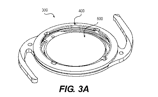
AND METHODS," U.S. Patent Application No. 15/176,582, filed June 8, 2016,
entitled "MODULAR INTRAOCULAR LENS DESIGNS AND METHODS," U.S.
Patent Application No. 15/150,360, filed May 9,2016, entitled "MODULAR
INTRAOCULAR LENS DESIGNS, TOOLS AND METHODS," now U.S. Patent
No. 9,421,088, U.S. Provisional Patent Application No. 62/332,163, filed
May 5, 2016, entitled "INTRAOCULAR LENS DESIGNS FOR IMPROVED
STABILITY," U.S. Provisional Patent Application No. 62/318,272, filed
April 5, 2016, entitled "MODULAR INTRAOCULAR LENS DESIGNS, TOOLS
AND METHODS," U.S. Patent Application No. 15/054,915, filed
February 26, 2016, entitled "MODULAR INTRAOCULAR LENS DESIGNS
AND METHODS," U.S. Provisional Patent Application No. 62/256,579, filed
November 17, 2015, entitled "MODULAR INTRAOCULAR LENS DESIGNS,
TOOLS AND METHODS," U.S. Provisional Patent Application No.
62/250,780, filed November 4, 2015, entitled "MODULAR INTRAOCULAR
LENS DESIGNS, TOOLS AND METHODS," U.S. Patent Application No.
CA 03020911 2018-10-11
WO 2017/192855
PCT/US2017/031066
14/828,083, filed August 17, 2015, entitled "MODULAR INTRAOCULAR
LENS DESIGNS, TOOLS AND METHODS," now U.S. Patent No. 9,364,316,
U.S. Patent Application No. 14/808,022, filed July 24, 2015, entitled
"MODULAR INTRAOCULAR LENS DESIGNS AND METHODS," now U.S.
Patent No. 9,387,069, U.S. Provisional Patent Application No. 62/110,241,
filed January 30, 2015, entitled "MODULAR INTRAOCULAR LENS
DESIGNS, TOOLS AND METHODS," U.S. Patent Application No.
14/610,360, filed January 30, 2015, entitled "MODULAR INTRAOCULAR
LENS DESIGNS, TOOLS AND METHODS," U.S. Provisional Patent
Application No. 61/941,167, filed February 18, 2014, entitled "MODULAR
INTRAOCULAR LENS DESIGNS, TOOLS AND METHODS," U.S. Patent
Application No. 13/969,115, filed August 16, 2013, entitled "MODULAR
INTRAOCULAR LENS DESIGNS & METHODS," now U.S. Patent
No. 9,289,287, U.S. Patent Application No. 13/937,761, filed July 9,2013,
entitled "MODULAR INTRAOCULAR LENS DESIGNS AND METHODS," now
U.S. Patent No. 9,125,736, U.S. Provisional Patent Application No.
61/830,491, filed June 3, 2013, entitled "MODULAR INTRAOCULAR LENS
DESIGNS AND METHODS," U.S. Patent Application No. 13/748,207, filed
January 23, 2013, entitled "MODULAR INTRAOCULAR LENS DESIGNS &
METHODS," now U.S. Patent No. 9,095,424, U.S. Provisional Patent
Application No. 61/589,981, filed on January 24, 2012, entitled "LASER
ETCHING OF IN SITU INTRAOCULAR LENS AND SUCCESSIVE
SECONDARY LENS IMPLANTATION," and U.S. Provisional Patent
Application No. 61/677,213, filed on July 30, 2012, entitled "MODULAR
2
CA 03020911 2018-10-11
WO 2017/192855
PCT/US2017/031066
INTRAOCULAR LENS DESIGNS & METHODS," each of which is
incorporated herein by reference in its entirety.
Technical Field
03 The present disclosure generally relates to intraocular lenses (10Ls). More
specifically, the present disclosure relates to embodiments of IOL designs for
improved stability in the capsular bag.
Background
04 The human eye functions to provide vision by transmitting light through a
clear
outer portion called the cornea, and focusing the image by way of a
crystalline
lens onto a retina. The quality of the focused image depends on many factors
including the size and shape of the eye, and the transparency of the cornea
and
the lens.
05 When age or disease causes the lens to become less transparent (e.g.,
cloudy),
vision deteriorates because of the diminished light, which can be transmitted
to
the retina. This deficiency in the lens of the eye is medically known as a
cataract.
An accepted treatment for this condition is surgical removal of the lens from
the
capsular bag and placement of an artificial intraocular lens (I0L) in the
capsular
bag. In the United States, the majority of cataractous lenses are removed by a
surgical technique called phacoemulsification. During this procedure, an
opening
(capsulorhexis) is made in the anterior side of the capsular bag and a thin
phacoemulsification-cutting tip is inserted into the diseased lens and
vibrated
ultrasonically. The vibrating cutting tip liquefies or emulsifies the lens so
that the
3
CA 03020911 2018-10-11
WO 2017/192855
PCT/US2017/031066
lens may be aspirated out of the capsular bag. The diseased lens, once
removed,
is replaced by an 10L.
06 After cataract surgery to implant an 10L, the optical result may be
suboptimal.
For example, shortly after the procedure, it may be determined that the
refractive
correction is erroneous leading to what is sometimes called "refractive
surprise."
This can be caused, in part, by post-operative movement of the IOL in the
capsular bag. Effective lens position (ELF), often measured using Scheimpflug
photography (e.g., Pentacam, Oculus, Germany), is a measure of the anterior-
posterior distance from the anterior surface of the cornea to the anterior
surface
of the lens (a.k.a., anterior chamber depth or ACD). ELF can change
significantly
post-operatively, where a 1.0mm shift in ELF corresponds to a 3.0 Diopter
change in visual power. Thus, there is a need for an IOL that is more stable
post-
operatively to mitigate changes in ELF and reduce refractive surprise.
Summary of the Disclosure
07 Embodiments of the present disclosure provide 10Ls that improve ELF
stability
by, for example, increasing anterior-posterior stiffness of the 10L,
increasing
anterior-posterior dimensions of the IOL and/or increasing contact area with
the
equator of the bag to resist movement of the IOL as the bag collapses over
time.
These 10Ls may be non-modular, unitary, or monolithic (i.e., single component)
or modular (multiple component). In modular embodiments, the IOL system may
include intraocular base and optic components, which, when combined, form a
modular IOL.
08 In one embodiment, a modular IOL includes an annular base having two
radially
outward extending haptics. The base may define a center hole and an inside
4
CA 03020911 2018-10-11
WO 2017/192855
PCT/US2017/031066
perimeter, with a radially inward open recess around the inside perimeter. The
modular IOL system also includes a lens having an optical body with first and
second tabs extending radially outward from the optical body. The base and
lens
may be assembled with the first and second tabs of the lens disposed in the
recess of the base. The base may have an anterior-posterior dimension greater
than the lens to increase the anterior-posterior stiffness of the assembly.
The
base may also have an anterior-posterior dimension approximating the anterior-
posterior dimension inside the capsular bag (i.e., between leaflets of the
capsular
bag) for mitigating anterior-posterior shift in the bag.
09 In another embodiment, a modular IOL includes a base configured to receive
a
conventional lens. The base may be annular with a center hole, two radially
outward extending haptics, and an inside ledge to receive a conventional lens
with haptics. The base and lens may be assembled with the perimeter of the
lens
resting on the ledge of the base and the haptics of the lens extending through
a
slot in the base. Similar to other embodiments described herein, the base may
have an anterior-posterior dimension greater than the lens to increase the
anterior-posterior stiffness of the assembly. In addition, the base may also
have
an anterior-posterior dimension approximating the anterior-posterior dimension
inside the capsular bag (i.e., between leaflets of the capsular bag) for
mitigating
anterior-posterior shift in the bag.
In yet another embodiment, a non-modular IOL includes an enlarged annular rim
around an optic for increasing anterior-posterior rigidity. The enlarged
annular rim
may have an anterior-posterior dimension approximating the anterior-posterior
dimension inside the capsular bag (i.e., between leaflets of the capsular
bag). A
gap in the rim maybe provided to enable folding for delivery via an injector.
The
5
CA 03020911 2018-10-11
WO 2017/192855
PCT/US2017/031066
rim may extend radially outward to form buttresses between the optic and
haptics
extending therefrom.
11 The 10Ls according to embodiments of the present disclosure may be applied
to
a variety of IOL types, including fixed monofocal, multifocal, toric,
accommodative, and combinations thereof. In addition, the 10Ls according to
embodiments of the present disclosure may be used to treat, for example:
cataracts, large optical errors in myopic (near-sighted), hyperopic (far-
sighted),
and astigmatic eyes, ectopia lentis, aphakia, pseudophakia, and nuclear
sclerosis.
12 Various other aspects and advantages of embodiments of the present
disclosure
are described in the following detailed description and drawings.
Brief Description of the Drawings
13 The drawings illustrate example embodiments of the present disclosure. The
drawings are not necessarily to scale, may include similar elements that are
numbered the same, and may include dimensions (in millimeters) and angles (in
degrees) by way of example, not necessarily limitation. In the drawings:
14 Figure 1 is a schematic diagram of the human eye shown in cross-section;
15 Figure 2 is a schematic diagram of the lens of the human eye shown in
sagittal
cross-section;
16 Figure 3A is a perspective view of a modular IOL according to the present
disclosure;
17 Figure 3B is a chart of the results of a bench test comparing the
performance of
the modular IOL shown in Figure 3A to a commercially available 101_,
6
CA 03020911 2018-10-11
WO 2017/192855
PCT/US2017/031066
18 Figures 4A-4D are perspective, top, cross-sectional and detailed views,
respectively, of the base of the modular IOL shown in Figure 3A,
19 Figures 5A-5E are perspective, top, cross-sectional and detailed views,
respectively, of the lens of the modular IOL shown in Figure 3A,
20 Figures 6A and 6B are perspective and cross-sectional views, respectively,
of an
alternative modular IOL according to the present disclosure;
21 Figures 7A-7B are perspective views of an alternative base for use with a
conventional IOL according to the present disclosure;
22 Figures 8A-80 are perspective, cross-sectional and top views, respectively,
of a
non-modular IOL according to the present disclosure;
23 Figures 9A and 9B are perspective views of alternative non-modularIOLs
according to the present disclosure;
24 Figures 10A and 10B are top and cross-sectional views, respectively, of
another
alternative non-modular IOL according to the present disclosure;
25 Figures 11A and 11B are top and cross-sectional views, respectively, of yet
another alternative non-modular IOL according to the present disclosure;
26 Figures 12A and 12B are top and cross-sectional views, respectively, of a
further
alternative non-modular IOL according to the present disclosure; and
27 Figures 13A-130 are perspective views of various alternative non-
modularIOLs
according to the present disclosure.
Detailed Description
28 Reference will now be made in detail to examples of the present disclosure,
which are illustrated in the accompanying drawings. Wherever possible, the
same reference numbers will be used throughout the drawings to refer to the
7
CA 03020911 2018-10-11
WO 2017/192855
PCT/US2017/031066
same or like parts. In the discussion that follows, relative terms such as
"about,"
"substantially," "approximately," etc. are used to indicate a possible
variation of
10% in a stated value, numeric or otherwise, unless other variations are
indicated.
29 With reference to Figure 1, the human eye 10 is shown in cross section. The
eye
has been described as an organ that reacts to light for several purposes. As a
conscious sense organ, the eye allows vision. Rod and cone cells in the retina
24 allow conscious light perception and vision including color differentiation
and
the perception of depth. In addition, the human eye's non-image-forming
photosensitive ganglion cells in the retina 24 receive light signals which
affect
adjustment of the size of the pupil, regulation and suppression of the hormone
melatonin, and entrainment of the body clock.
30 The eye 10 is not properly a sphere; rather it is a fused two-piece unit.
The
smaller frontal unit, more curved, called the cornea 12 is linked to the
larger unit
called the sclera 14. The corneal segment 12 is typically about 8 mm (0.3 in)
in
radius. The sclera 14 constitutes the remaining five-sixths; its radius is
typically
about 12 mm. The cornea 12 and sclera 14 are connected by a ring called the
limbus. The iris 16, the color of the eye, and its black center, the pupil,
are seen
instead of the cornea 12 due to the cornea's 12 transparency. To see inside
the
eye 10, an ophthalmoscope is needed, since light is not reflected out. The
fundus
(area opposite the pupil), which includes the macula 28, shows the
characteristic
pale optic disk (papilla), where vessels entering the eye pass across and
optic
nerve fibers 18 depart the globe.
31 Thus, the eye 10 is made up of three coats, enclosing three transparent
structures. The outermost layer is composed of the cornea 12 and sclera 14.
The
8
CA 03020911 2018-10-11
WO 2017/192855
PCT/US2017/031066
middle layer consists of the choroid 20, ciliary body 22, and iris 16. The
innermost layer is the retina 24, which gets its circulation from the vessels
of the
choroid 20 as well as the retinal vessels, which can be seen within an
ophthalmoscope. Within these coats are the aqueous humor, the vitreous body
26, and the flexible lens 30. The aqueous humor is a clear fluid that is
contained
in two areas: the anterior chamber between the cornea 12 and the iris 16 and
the
exposed area of the lens 30; and the posterior chamber, between the iris 16
and
the lens 30. The lens 30 is suspended to the ciliary body 22 by the suspensory
ciliary ligament 32 (Zonule of Zinn), made up of fine transparent fibers. The
vitreous body 26 is a clear jelly that is much larger than the aqueous humor.
32 The crystalline lens 30 is a transparent, biconvex structure in the eye
that, along
with the cornea 12, helps to refract light to be focused on the retina 24. The
lens
30, by changing its shape, functions to change the focal distance of the eye
so
that it can focus on objects at various distances, thus allowing a sharp real
image
of the object of interest to be formed on the retina 24. This adjustment of
the lens
30 is known as accommodation, and is similar to the focusing of a photographic
camera via movement of its lenses.
33 The lens has three main parts: the lens capsule, the lens epithelium, and
the lens
fibers. The lens capsule forms the outermost layer of the lens and the lens
fibers
form the bulk of the interior of the lens. The cells of the lens epithelium,
located
between the lens capsule and the outermost layer of lens fibers, are found
predominantly on the anterior side of the lens but extend posteriorly just
beyond
the equator.
34 The lens capsule is a smooth, transparent basement membrane that completely
surrounds the lens. The capsule is elastic and is composed of collagen. It is
9
CA 03020911 2018-10-11
WO 2017/192855
PCT/US2017/031066
synthesized by the lens epithelium and its main components are Type IV
collagen
and sulfated glycosaminoglycans (GAGs). The capsule is very elastic and so
causes the lens to assume a more globular shape when not under the tension of
the zonular fibers, which connect the lens capsule to the ciliary body 22. The
capsule varies between approximately 2-28 micrometers in thickness, being
thickest near the equator and thinnest near the posterior pole. The lens
capsule
may be involved with the higher anterior curvature than posterior of the lens.
35 Various diseases and disorders of the lens 30 may be treated with an 10L.
By
way of example, not necessarily limitation, an IOL according to embodiments of
the present disclosure may be used to treat cataracts, large optical errors in
myopic (near-sighted), hyperopic (far-sighted), and astigmatic eyes, ectopia
lentis, aphakia, pseudophakia, and nuclear sclerosis. However, for purposes of
description, the IOL embodiments of the present disclosure are described with
reference to cataracts, which often occurs in the elderly population.
36 As seen in Figure 2, the shape of the lens 30 is generally symmetric about
the
visual axis 37. However, the lens 30 is not symmetric about the sag ittal
plane 39.
Rather, the anterior side 33 of the lens 30 has a radius of curvature (RA)
that is
greater than the radius of curvature (Re) of the posterior side 35. The
equatorial
diameter (D) resides more anteriorly, with the posterior lens thickness (Tp)
being
greater than the anterior lens thickness (TA).
37 Rosen et al. (2006) published data suggesting the equatorial diameter D,
the
posterior lens thickness Tp, the anterior lens thickness TA, and the anterior
radius
of curvature RA change with age, whereas the posterior radius of curvature Rp
and the ratio TA/Tp remain constant. Using best-fit linear equations, Rosen et
al.
CA 03020911 2018-10-11
WO 2017/192855
PCT/US2017/031066
described the following age-dependent equations for these parameters (all in
mm):
38 D = 0.0138( 0.002) *Age + 8.7 ( 0.14) (R2= 0.57; p <0.0001);
39 TA = 0.0049( 0.001)* Age + 1.65 ( 0.075) (R2= 0.45; p < 0.0001);
40 Tp = 0.0074 ( 0.002)* Age + 2.33 ( 0.11) (R2= 0.44; p <0.0001);
41 RA = 0.046 ( 0.017)* Age + 7.5 ( 1.13) (R2= 0.27; p = 0.016);
42 Rp = -5.5 ( 0.9), and
43 TA/Tp = 0.70 ( 0.13).
44 These data or other empirically measured data may be used to describe the
shape and size of the lens for a particular age group, such as cataracts in
elderly
patients at a mean age of 70, by way of example, not limitation. Such data may
be useful to determine the space available for an intraocular implant to be
placed
in the capsular bag. For example, assume an ocular implant (such as an 10L) is
to be centered in the equatorial plane, with an anterior-posterior height "H"
at
radial distance "X" from its center point. Also assume it is desired to have
the
anterior and posterior sides of the implant at radial distance X come into
contact
with the walls of the capsular bag to mitigate migration of the implant.
Mathematical modeling may be used to determine the height (H) of the lens
capsule at any given radial distance (X) from the visual axis 37 along the
equatorial plane.
45 The total height H is equal to the sum of the anterior height (HA) and the
posterior
height (Hp). The anterior height (HA) may be given by the equation HA = Y -
(RA -
TA). While RA and TA are empirically known, the distance (Y) from the
equatorial
plane may be given by the equation Y = (RA2-X2)^0.5. Combining these
equations, the anterior height may be given by HA = (RA2-X2)^0.5 - (RA - TA),
and
11
CA 03020911 2018-10-11
WO 2017/192855
PCT/US2017/031066
solved using empirical data. The posterior height (Hp) may be similarly
calculated
using the posterior radius (Re) and posterior thickness (Tp) solved using
empirical
data. Adding the posterior height (Hp) to the anterior height (HA) provides
the total
height (H) at a distance (X) from the visual axis. Thus, the desired height
(H) of
the intraocular implant at radial distance X may be estimated such that the
implant is in contact with the anterior and posterior walls of the capsular
bag.
Alternative mathematical models as described in the literature may be used as
well.
46 The following detailed description describes various embodiments of modular
and
non-modular IOL systems. Features described with reference to any one
embodiment may be applied to and incorporated into other embodiments.
47 With reference to Figure 3A, a base 400 and a lens 500 form an embodiment
of a
modular IOL 300 when assembled. A general description of the modular IOL 300
follows, with further detailed provided in U.S. Provisional Patent Application
No. 62/318,272, which is hereby fully incorporated by reference.
48 With reference to Figures 4A-4D, the base 400 is shown in more detail.
Figure 4A
is a perspective view, Figure 4B is a top view, Figure 40 is sectional view
taken
along line A-A in Figure 4B, and Figure 4D is a detailed sectional view of
circle C
in Figure 40. Dimensions (mm) are given by way of example, not necessarily
limitation.
49 The base 400 includes an annular ring 402 defining a center hole 404. A
pair of
haptics 406 extend radially outward from the annular ring 402. The annular
ring
402 includes a lower rim 408, an upper rim 410 and an inward-facing recess
412,
into which the lens 500 may be inserted to form modular IOL 300.
12
CA 03020911 2018-10-11
WO 2017/192855
PCT/US2017/031066
50 The upper rim 410 of annular ring 402 may include one or more notches 416
to
provide access for a probe (e.g., Sinskey hook) intra-operatively, which
allows
the base 400 to be more easily manipulated. The haptics 406 may include holes
415 adjacent the annular ring 402 for the same purpose as notches 416. A pair
of square edges 417 may extend around the posterior periphery of the annular
ring 402 to help reduce cellular proliferation (posterior capsular
pacification or
PCO) onto the lens 500.
51 With specific reference to Figure 4D, the deep portion of the recess 412
may
have a squared profile defined by horizontal posterior surface 418, a
horizontal
anterior surface 420 and a vertical lateral or outer surface 422. The recess
may
also include a flared anterior surface 426 extending radially inward and
anteriorly
outward from the horizontal anterior surface 420, and a flared posterior
surface
428 extending radially inward and posteriorly outward from the horizontal
posterior surface 418. The inside diameter of the posterior rim 408 may be
smaller than the inside diameter of the anterior rim 410. With this
arrangement,
the lens 500 may be placed through the circular opening defined by the
anterior
rim 410 to land or rest upon the posterior rim, and the flared anterior wall
426
together with the flared posterior wall 428 may act as a funnel to guide the
tabs
504 and 506 of the lens 500 into the deep portion of the recess 412. When
fully
seated in the recess 412, the horizontal posterior wall 418, the horizontal
anterior
wall 420 and the vertical lateral wall 422 form a keyed geometry with the
corresponding horizontal and vertical sides of the tabs 504 and 506 to limit
movement of the lens 500 relative to the base 400 in anterior, posterior and
radial
directions.
13
CA 03020911 2018-10-11
WO 2017/192855
PCT/US2017/031066
52 As best seen in Figure 4D, the base 400 may have an anterior-posterior
height
of H = HA + Hp, where H is approximately 1mm, HA is approximately 0.5mm at a
radial distance of approximately 3.2mm from the center point OF, and Hp is
approximately 0.5mm at a radial distance of 2.65mm from the center point CP.
However, as described previously, the posterior thickness Tp of the native
lens 30
is greater than the anterior thickness TA of the native lens 30. Therefore,
these
relative dimensions may be adjusted. For example, Hp may be made greater than
HA such that the sagittal mid-plane MP of the base 400 is aligned (+/- 0.5mm)
with the equatorial plane of the lens 30 when the modular IOL 300 is implanted
in
the capsular bag. The ratio HA/Hp may be constant at approximately 0.7 ( 0.3),
for example. In addition, H may be selected such that the anterior-most
portion
of the anterior rim 410 is in close proximity (within 0.5mm) to the anterior
side 33
of the lens 30 and the posterior-most portion of the posterior rim 408 is in
close
proximity (within 0.5mm) to the posterior side 35 of the lens 30 when
implanted in
the capsular bag. Thus, by way of example, not limitation, HA may be
approximately 0.5mm to 1.0mm at a radial distance of approximately 2.75mm to
3.25mm from the center point OF, and Hp may be approximately 0.75mm to
1.5mm at a radial distance of 2.25mm to 2.50mm from the center point OF,
maintaining a constant ratio HA/Hp of approximately 0.7 ( 0.3), for example.
53 With reference to Figures 5A-5E, the lens 500 is shown in more detail.
Figure 5A
is a perspective view, Figure 5B is a top view, Figure 5C is sectional view
taken
along line A-A in Figure 5B, Figure 5D is a detailed sectional view of circle
B in
Figure 5C, and Figure 5E is a detailed top view of circle C in Figure 5B.
Dimensions (mm) are given by way of example, not necessarily limitation.
14
CA 03020911 2018-10-11
WO 2017/192855
PCT/US2017/031066
54 The lens 500 may include an optic portion 502 and one or more tabs 504 and
506. As shown, tab 504 is fixed, whereas tab 506 may be actuated. Fixed tab
504
may include a thru hole 208 so that a probe (e.g., Sinskey hook) or similar
device
may be used to engage the hole 208 and manipulate the tab 504. Actuatable tab
506 may be actuated between a compressed position for delivery into the hole
404 of the base 400, and an uncompressed extended position (shown) for
deployment into the recess 412 of the base 400, thus forming an interlocking
connection between the base 400 and the lens 500. It also is contemplated that
actuatable tab 506 may be inserted into recess 412, and may be actuated
between the compressed position to facilitate entry of fixed tab 504 into
recess
412, and the uncompressed extended position to insert fixed tab 504 further
into
recess 412 to form the interlocking connection between base 400 and lens 500.
55 Actuatable tab 506 may include two members 510 and 512, each with one end
connected to the edge of the optic 502, and the other end free, thus forming
two
cantilever springs. A rim 514 may extend around the perimeter of the optic
502,
terminating shy of the springs 510 and 512, thus allowing the springs 510 and
512 to fully compress against the edge of the optic 502. The rim 514 of the
lens
500 may have an outside diameter that is greater than the inside diameter of
the
posterior rim 408 of the base 400 such that the lens 500 doesn't fall through
the
opening 404 of the base 400 and such that the lens 500 is circumferentially
supported around its perimeter by the posterior rim 408 of the base 400. A
gusset with a guide hole 516 may be disposed between the two members 510
and 512 to facilitate manipulation by a probe. Similarly, a guide hole 508 may
be
provided in the fixed tab 504 to provide access for a probe (e.g., Sinskey
hook) or
similar device to manipulate the fixed tab 504 into the recess 412 in the base
CA 03020911 2018-10-11
WO 2017/192855
PCT/US2017/031066
400. A notch 518 may be provided in the fixed tab 504 to provide asymmetry as
a visual indicator that the anterior side is up (rather than down) when the
notch is
counter-clockwise of the hole 508.
56 As seen in Figure 50, the anterior and posterior sides of the optic 502 may
have
convex radii corresponding to the desired power (Diopter) of the optic. The
fixed
tab 504 and the spring tabs 510 and 512 may have a flared cross-section as
shown. More specifically, and as better seen in the detailed view shown in
Figure
5D, the fixed tab 504 extends radially outward from the optic 502 from a
thinner
inner portion 504B to a flared thicker outer portion 504A. Hole 508 may extend
through thinner inner portion 504B. The outermost profile of the thicker
portion
504A has a squared profile with an anterior horizontal side, a posterior
horizontal
side, and a lateral or outer vertical side that are keyed to the recess 412 as
described previously to minimized anterior-posterior and radial/lateral
movement
of the lens 500 relative to the base 400. The thicker portion 504A also
provides
for improved engagement with the plunger of an injector to mitigate jamming of
the lens 500 in the injector. The thinner portion 504B also provides an
anterior
and a posterior offset from the surfaces defining the recess 412 of the base
400,
thereby mitigating adhesion between the lens 500 and the base 400. The same
flared configuration and associated advantages also applies to each of the
spring
tabs 510 and 512 as shown.
57 Commercially available 10Ls typically have an equatorial diameter
(excluding
haptics) of about 6mm, an anterior-posterior thickness of about 0.2 mm at 6mm
diameter and 0.7 mm at the center, providing an overall volume of about 12
mm3.
Lens 500 is similarly dimensioned, but the base 400 adds substantially more
volume. The base 400 may have an equatorial diameter (excluding haptics) of
16
CA 03020911 2018-10-11
WO 2017/192855
PCT/US2017/031066
about 7.8 mm, an anterior-posterior thickness of about 1 mm, providing an
overall
volume of about 26 cubic millimeters [13.4 mm3 base, 12.5 mm3 optic] when the
lens is disposed into the base. Thus, the size of the combined base 400 and
lens
500 is volumetrically much larger than conventional 10Ls available on the
market.
This relatively larger volume is intended to fill the capsular bag more like a
natural
lens, thus increasing the stability of the modular IOL 300 and reducing post-
operative migration due to the bag collapsing around the base 400. By way of
comparison, a typical natural lens has an equatorial diameter of about 10.4
mm,
an anterior-posterior dimension of about 4.0 mm for a corresponding volume of
about 180 mm3. Due to anatomic variability, a natural lens may have a volume
ranging from 130 mm3 to 250 mm3. Thus, the modular IOL 300 (base 400 plus
lens 500) consumes greater than 10% (about 20% to 10.4%) of the volume of the
bag after the natural lens has been extricated, whereas a conventional IOL
consumes less than or equal to 10% (about 10% to 5%) of the volume of the bag.
In other words, the modular IOL 300 consumes about twice the volume of the bag
compared to a conventional 10L.
58 Also by comparison to conventional 10Ls, modular IOL 300, by virtue of the
annular ring 402 of the base 400, provides a relatively large diameter and
rigid
platform that resists deflection (i.e., increased stiffness in the sagittal
plane,
thereby improving anterior-posterior stability). Coupled with the relatively
long
sweeping haptics 406 which offer a significant relative increase in surface
contact
with the capsular bag, the modular IOL 300 provides superior centering and
stability within the capsular bag.
59 The ability to resist deflection was demonstrated in a bench test comparing
the
performance of modular IOL 300 to a commercially available IOL (Alcon model
17
CA 03020911 2018-10-11
WO 2017/192855
PCT/US2017/031066
SA60), the results of which are shown in Figure 3B. In the test set-up, the
test
IOL was placed in a lOmm inside diameter simulated capsular bag and the
assembly was submerged in a warm bath. Various loads were applied to the
middle of test IOL while in a horizontal orientation, and the resulting
downward
displacement was measured. As can be seen from the results shown in Figure
3B, the commercially available IOL was displaced roughly 5 times the amount
that the modular IOL 300 was displaced, and the commercially available IOL
failed to support a load of 0.058 grams as the haptics were displaced out of
the
simulated capsular bag. This demonstrates the significant relative increase in
stiffness of modular IOL 300 compared to a common commercially available 10L.
60 This test set-up may be compared to a mechanical model of a center load on
beam with two simple supports described by F=kegAx, where F = applied force,
keg = equivalent stiffness and Ax = displacement. Equivalent stiffness takes
into
account the cross-sectional moment of inertia of the beam as well as the
material
properties of the beam (Young's elastic modulus). However, since 10Ls are made
of plastic (rather than an elastic material such as metal), the equivalent
stiffness
will vary over a range of applied forces. In the described bench test, the
modular
IOL 300 had an equivalent stiffness of approximately 0.5 to 2.0 g/mm over a
range of applied loads of 0.032 to 0.100 g, whereas the commercially available
IOL had an equivalent stiffness of approximately 0.15 to 0.20 g/mm over a
range
of applied loads of 0.032 to 0.044 g.
61 In general, when the base 400 and lens 500 are assembled to form modular
IOL
300, the features may be configured such that the mid-plane of the optic 502
is
parallel with the mid-plane of the base 400, and the central (anterior-
posterior)
axis of the optic 502 is coincident and collinear with the central (anterior-
18
CA 03020911 2018-10-11
WO 2017/192855
PCT/US2017/031066
posterior) axis of the base 400. Assuming anatomic symmetry of the native lens
capsule and centration of the base 400 in lens capsule, this configuration
essentially aligns the central axis of the optic 502 with the central
(anterior-
posterior) axis of the capsular bag, thus providing centration of the optic
502.
However, there may be instances where the visual (fovea!) axis is not aligned
with the anatomic (pupillary axis), wherein the difference is called angle of
kappa.
In such instances, it may be desirable to offset the central axis of the optic
500
relative to the base 400, thus providing de-centration. This may be
accomplished,
for example, by configuring the tabs 504 and 506, the recess 412 and/or the
haptics 406 such that the central (anterior-posterior) axis of the optic 502
is
laterally (nasally or temporally) offset relative to the central (anterior-
posterior)
axis of the base 400.
62 By way of example, not limitation, the lateral walls defining the recess
412 in the
base 400 may be offset relative to the haptics 406 so that the central axis of
the
optic 502 is offset. Different offsets could be provided, for example, 0.5 mm
through 2.0 mm at .5 mm increments. Angular orientation marks on the base 400
and lens 500 may be provided to indicate the direction of the offset (nasally
or
temporally). Similarly, the mid-plane of the assembled base 400 and optic 500
may be tilted relative to the equatorial plane of the native capsular bag. To
compensate for this tilt, for example, the tabs 504 and 506, the recess 412
and/or
the haptics 406 may be configured such that the mid-plane of the optic 502 is
counter-tilted.
63 The base 400 and lens 500, including the alternative embodiments described
herein, may be formed by cryogenically machining and polishing hydrophobic
acrylic material. Optionally, the base 400 may be manufactured by forming two
19
CA 03020911 2018-10-11
WO 2017/192855
PCT/US2017/031066
(anterior and posterior) components and adhesively connecting them together.
For example, the two components may be cryogenically machined hydrophilic
acrylic connected together by a U.V. curable adhesive. Alternatively, the two
components may be formed of different materials adhesively connected together.
For example, the anterior component may be formed of hydrophilic acrylic which
does not adhere to ocular tissue, and the posterior component may be formed of
hydrophobic acrylic which does adhere to ocular tissue.
64 As a further alternative, the base 400 may be manufactured by cryogenic
machining the first component and over-molding the second component. The first
component may include geometric features that become interlocked when over-
molded, thus mitigating the need for adhesive to connect the components. For
example, the base 400 may be manufactured by cryogenic machining of
hydrophilic acrylic to form the posterior component, and over-molding the
anterior
component of a moldable material such as silicone.
65 While hydrophobic acrylic renders the base 400 and lens 500 visible using
optical
coherence tomography (OCT), it may be desirable to incorporate a material that
enhances OCT visualization. Example "OCT-friendly" materials include but are
not limited to polyvinyl chloride, glycol modified poly (ethylene
terephthalate)
(PET-G), poly (methyl methacrylate) (PMMA), and a polyphenylsulfone, such as
that sold under the brand name RADELTM, as described in U.S. Patent
Application Publication No. 2013/0296694 to Ehlers et al., which is
incorporated
herein by reference. Such OCT-friendly materials may be applied to or
incorporated into a portion of the base 400 or lens 500.
66 By way of example, a concentric ring of OCT-friendly material may be
applied to
each of the lower and upper rims 408/410. The rings may have different
CA 03020911 2018-10-11
WO 2017/192855
PCT/US2017/031066
diameters to aid in detecting tilt of the base. Also by way of example, OCT-
friendly material may be applied to the tabs 504/506 of the lens 500. This may
aid
in determining if the base 400 and lens 500 are correctly assembled in the
eye.
Points of OCT-friendly material may be applied to portions of the base 400
that
line up to corresponding OCT-friendly points on the optic 500 to indicate
proper
assembly in the eye.
67 As an alternative to solid material, the base 400 and lens 500 may be made
of
hollow material that can be subsequently inflated in the eye. In this
arrangement,
the base 400 and lens 500 may be made from molded silicone, for example, and
inflated with a liquid such as saline, silicone gel or the like using a
syringe and
needle. The needle may pierce the wall of the base 400 and lens 500 after
implantation in the eye to inflate the components. The material may self-seal
after
removal of the needle. As an alternative to a hollow material, the base 400
and
lens 500 may be formed of a sponge-like material such as silicone hydrogel
that
swells upon hydration. Both approaches allow the size of the corneal incision
to
be smaller, as the base 400 and lens 500 are delivered in an uninflated or
unswelled state and subsequently inflated or swelled once inside the eye.
68 In general, the modular IOL 300, comprising the assembled base 400 and lens
500, including the alternative embodiments described herein, allows for the
lens
500 to be adjusted or exchanged while leaving the base 400 in place, either
intra-
operatively or post-operatively. Examples of instances where this may be
desirable include, without limitation: exchanging the lens 500 to correct a
suboptimal refractive result detected intra-operatively, exchanging the lens
500 to
correct a suboptimal refractive result detected post-operatively (residual
refractive
error); rotationally adjusting the lens 500 relative to the base 400 to fine
tune toric
21
CA 03020911 2018-10-11
WO 2017/192855
PCT/US2017/031066
correction; laterally adjusting the lens 500 relative to the base 400 for
alignment
of the optic with the true optical axis (which may not be the center of the
capsular
bag); and exchanging the lens 500 to address the changing optical needs or
desires of the patient over longer periods of time. Examples of the latter
instance
include, but are not limited to: an adult or pediatric IOL patient whose
original
optical correction needs to be changed as s/he matures; a patient who wants to
upgrade from a monofocal IOL to a premium IOL (toric, multifocal,
accommodating or other future lens technology); a patient who is not satisfied
with their premium IOL and wants to downgrade to monofocal 101_, and a patient
who develops a medical condition where an IOL or a particular type of IOL is
contra-indicated.
69 With reference to Figures 6A and 6B, an alternative modular IOL 330 is
shown in
perspective and cross-sectional views, respectively. Alternative modular IOL
330
may include an alternative base 600 and the lens 500 as described above. As
will be appreciated by the following description, alternative base 600 may be
similar to base 400 except for anterior rim 610 and posterior rim 608, the
description of the similar aspects and advantages being incorporated herein by
reference. Alternative base 600 includes an annular ring defining a center
hole.
A pair of haptics 606 extend radially outward from the annular ring. The
annular
ring includes a lower rim 608, an upper rim 610 and an inward-facing recess
612,
into which the lens 500 may be inserted to form modular IOL 330.
70 With specific reference to Figure 6B, the lower rim 608 and upper rim 610
may
have a relatively exaggerated height and may be angled radially inward to form
a
funnel leading to the recess 612. With this arrangement, the actuatable tabs
506
of the lens may be compressed and the lens 500 may be placed through the
22
CA 03020911 2018-10-11
WO 2017/192855
PCT/US2017/031066
circular opening defined by the anterior rim 610, with the funnel shape of the
anterior rim 610 guiding the tabs 504 and 506 into the recess 612 of the base
600
to form a keyed geometry to limit movement of the lens 500 relative to the
base
600 in anterior, posterior and radial directions. The funneled shape of the
posterior rim 608 prevents the lens 500 from falling posteriorly during
insertion of
the lens 500 into the base 600.
71 The base 600 may have the dimensions as shown by way of example, not
necessary limitation. As best seen in Figure 6B, the rims 608 and 610 of the
base 400 may have a combined anterior-posterior height that is 2.0 to 3.0 (or
more) times the maximum thickness of the optic portion 502 of the lens 500.
For
example, the combined height of the rims 608 and 610 may be approximately
3mm at a radial distance of approximately 2.9mm from the center point. As
described previously, the height of posterior rim 608 may be made greater than
the height of anterior rim 610 such that the sagittal mid-plane of the base
600 is
aligned (+1- 0.5mm) with the equatorial plane of the lens 30 when the modular
IOL 330 is implanted in the capsular bag. The height ratio of the anterior rim
610
to the posterior rim 608 may be constant at a value less than 1.0 such as
approximately 0.7 ( 0.3), for example. As shown, the combined height of the
anterior rim 610 and the posterior rim 608 are selected such that the anterior-
most portion of the anterior rim 610 is in close proximity (within 0.5mm) to
or
pushing against the anterior side 33 of the lens 30 and the posterior-most
portion
of the posterior rim 608 is in close proximity (within 0.5mm) to or pushing
against
the posterior side 35 of the lens 30 when implanted in the capsular bag.
72 With reference to Figures 7A and 7B, an alternative base 700 for use with a
conventional IOL 100 is shown in perspective views, where Figure 7A shows the
23
CA 03020911 2018-10-11
WO 2017/192855
PCT/US2017/031066
base 700 standing alone and Figure 7B shows the combined base 700 and
conventional IOL 100 assembled to form modular IOL 360. Alternative base 700
is similar to base 400 described previously, with the exception of inverted T-
slots
730, the description of the similar aspects and advantages being incorporated
herein by reference.
73 The base 700 includes an annular ring 702 defining a center hole 704. A
pair of
haptics 706 extend radially outward from the annular ring 702. The annular
ring
702 includes a lower rim 708, an upper rim 710 and an inward-facing recess
712,
into which the conventional IOL 100 may be inserted to form modular IOL 360.
The upper rim 710 of annular ring 702 may include one or more notches 716 to
provide access for a probe (e.g., Sinskey hook) intra-operatively, which
allows
the base 700 to be more easily manipulated. The haptics 706 may include holes
715 adjacent the annular ring 702 for the same purpose as notches 716.
74 The annular ring 702 may include a pair of inverted-T-shaped slots 730 to
accommodate the diametrically opposed haptics 106 of the conventional IOL 100.
When the haptics 106 of the conventional IOL 100 are placed in the slots 730,
the
posterior side of the optic portion 102 of the conventional IOL 100 may rest
upon
the anterior surface of the posterior rim 708. The posterior portion of the
slots 730
may have a greater width than the anterior portion thereof to accommodate the
angle of the haptics 106 and to lock the IOL 100 to the base 700 when rotated
relative thereto. The addition of the base 700 adds to the anterior-posterior
rigidity and height of a conventional IOL 100, thereby improving its
stability.
75 With reference to Figures 8A-80, perspective, cross-sectional and top
views,
respectively, of a non-modular IOL 800 is shown schematically. Non-modular IOL
800 incorporates several of the stability advantages described previously, but
in a
24
CA 03020911 2018-10-11
WO 2017/192855
PCT/US2017/031066
non-modular configuration. IOL 800 includes an optic portion 802 that may be
monofocal (fixed focal length), accommodating (variable focal length), toric,
multifocal, or extended depth-of-focus pattern, for example. IOL 800 also
includes
two or more haptics 806 extending radially outward from the periphery of the
optic portion 802. Each haptic includes a posterior flange 808 and an anterior
flange 810 extending radially inward and flared in an outward posterior and an
outward anterior direction, respectfully, from an outer rim 809. Each haptic
806
includes a connecting arm 812 that connects the outer rim 809 to the periphery
of
the optic 802. Each connecting arm 812 may include a window 814 for added
flexibility. The posterior flange 808 and the anterior flange 810 are
configured to
compress relative to each other in an anterior-posterior direction, acting
like
cantilever leaf springs about outer rim 809.
76 With specific reference to Figure 8B, which is a cross-sectional view taken
along
line B-B in Figure 8A, it can be appreciated that the posterior flange 808 is
sized
and configured differently than anterior flange 810 in order to conform to the
shape of the capsular bag. As described previously, the posterior thickness of
the native lens is greater than the anterior thickness of the native lens. In
order
for the anterior flange 810 to conform to the anterior side 33 of the lens
capsule
and the posterior flange 808 to conform to the posterior side 35 of the lens
capsule, the anterior flange 810 may have an anterior height HA and arc length
that is less than the posterior height Hp and arc length of the posterior
flange 808.
For example, Hp may be made greater than HA such that the sagittal mid-plane
MP of the base 800 is aligned (+/- 0.5mm) with the equatorial plane of the
lens
capsule when the IOL 800 is implanted in the capsular bag. The ratio HA/Hp may
be constant at approximately 0.7 ( 0.3), for example.
CA 03020911 2018-10-11
WO 2017/192855
PCT/US2017/031066
77 With specific reference to Figures 8B and 80, the radial length (in the sag
ittal
plane) of the posterior flange 808 and anterior flange 810 may be selected
such
that the inner-most edge does not interfere with the field of vision through
the
optic 802. In other words, the posterior flange 808 and the anterior flange
810
may extend radially inward from the outer rim 809 up to the outer diameter of
the
optic portion 802, where the inner edge of the posterior flange 808 and the
anterior flange 810 forms an arc conforming to the outside diameter of the
optic
802. The outer rim 809 may also form an arc, wherein the haptics 806 conform
the circular shape of the equator of the natural lens capsule. By way of
example,
not necessarily limitation, the arc shape of the haptics 806 may extend 60 -90
,
90 -120 , or 120 -150 around the circumference of the optic 802. The larger
the
arc length of the haptics, the greater the contact area with the equator of
the
natural lens capsule, the greater the stability of the IOL 800 in the capsular
bag,
but this must be balanced against the deliverability of the IOL 800 through a
small
incision using an injector.
78 With reference to Figures 9A and 9B, alternative non-modularIOLs 900 and
950,
respectively, are shown in perspective view. 10Ls 900 and 950 are similar to
IOL
800 described above in that the haptics include flared flanges for improved
stability; the description of the similar aspects and advantages being
incorporated
herein by reference.
79 With specific reference to Figure 9A, IOL 900 includes an optic portion 902
that
may be monofocal (fixed focal length), accommodating (variable focal length),
toric, multifocal, or extended depth-of-focus pattern, for example. IOL 900
also
includes two or more haptics 906 extending radially outward from the periphery
of
the optic portion 902. Each haptic 906 includes a posterior flange 908 and an
26
CA 03020911 2018-10-11
WO 2017/192855
PCT/US2017/031066
anterior flange 910 extending radially inward and flared in an outward
posterior
and an outward anterior direction, respectfully, from an outer rim 909. Each
haptic 906 includes a pair of connecting arms 912 that connect the outer rim
909
to the periphery of the optic 902. Each pair of connecting arms 912 may
include a
window 914 for added flexibility. The posterior flange 908 and the anterior
flange
910 are configured to compress relative to each other in an anterior-posterior
direction, acting like cantilever leaf springs about outer rim 909. Compared
to IOL
800, the flanges 908 and 910 of IOL 900 have a smaller radial length (in the
sagittal plane) extending from the outer rim 909 toward the optic 902. In
addition,
a gap 911 is provided between the connecting arms 912 and the flanges 908 and
910 along the inside connection to the outer rim 909 to provide space for the
flanges 908 and 910 to compress and fold down toward the optic 902. The gap
911 allows the connection between the outer rim 909 and the flanges 908 and
910 to function as a resilient hinge and allows the flanges 908 and 910 to
better
conform to the inside of the capsular walls that may vary in size and
dimension.
80 With reference to Figure 9B, IOL 950 is similar to IOL 900, the description
of
similar aspects and advantages being incorporated herein by reference. IOL 950
includes one or more haptics 906 including curvilinear arms 916 (rather than
connecting arms 912) extending from the periphery of optic 902 to form the
outer
rim 909 from which the flanges 908 and 910 extend. As in the prior embodiment,
a gap 911 is provided to enhance the flexibility of the flanges 908 and 910
relative to the curvilinear arms 916 along outer rim 909 such that the
connection
therebetween functions as a resilient hinge.
81 With reference to Figures 10A and 10B, an alternative non-modular IOL 1000
is
shown schematically. Figure 10A is a top view of the IOL 1000 and Figure 10B
is
27
CA 03020911 2018-10-11
WO 2017/192855
PCT/US2017/031066
a cross-sectional view taken along line B-B in Figure 10A. IOL 1000 includes
an
optic portion 1002 that may be monofocal (fixed focal length), accommodating
(variable focal length), toric, multifocal, or extended depth-of-focus
pattern, for
example. IOL 1000 also includes a pair of haptics 1006 extending outwardly
from
the optic portion 1002. A pair of gusset plates 1004 connects the haptics 1006
to
the optic portion 1002. Whereas a conventional IOL provides haptics extending
from the optic portion, IOL 1000 utilizes the gusset plates 1004 to push the
attachment location of the haptics 1006 radially outward, thereby relatively
increasing the anterior-posterior stiffness of the IOL in the sagittal plane.
IOL
1000 also includes a posteriorly extending ridge 1008 around the periphery of
the
optic 1002 and the periphery of the gusset plates 1004, excluding the haptics
1006 and the junction of the haptics 1006 to the gusset plates 1004. The ridge
1008 increases the cross-sectional moment of inertia of the IOL 1000 in the
sagittal plane, thereby increasing its stiffness and stability, without
affecting the
flexibility of the haptics 1006. As seen in cross-section, the ridge 1008 may
have
an inside fillet and an outside square edge as shown, to inhibit cellular
proliferation onto the optic portion 1002. By way of example, not necessarily
limitation, the haptics may have an outside extent of 13mm (haptic tip to
haptic
tip), the optic may have a diameter of 5mm to 6mm, and the gusset plates 1004
may have a mean sagittal width of 1mm to 2mm. Thus, with a 5.0mm diameter
optic 1002, the haptics 1006 may be attached to the gusset plates 1004 at a
diameter of 7.0mm to 9.0mm.
82 With reference to Figures 11A and 11B, another alternative non-modular IOL
1100 is shown schematically. Figure 11A is a top view of the IOL 1100 and
Figure 11B is a cross-sectional perspective view taken along line B-B in
Figure
28
CA 03020911 2018-10-11
WO 2017/192855
PCT/US2017/031066
11A. As will be appreciated by the following description, IOL 1100 may be
similar
to IOL 1000 except with regard to ridge 1108, the description of the similar
aspects and advantages being incorporated herein by reference. IOL 1100
includes an optic portion 1102 that may be monofocal (fixed focal length),
accommodating (variable focal length), toric, multifocal, or extended depth-of-
focus pattern, for example. IOL 1102 also includes a pair of haptics 1106
extending outwardly from the optic portion 1102. A pair of gusset plates 1104
connects the haptics 1006 to the optic portion 1102. Whereas a conventional
IOL
provides haptics extending from the optic portion, IOL 1100 utilizes the
gusset
plates 1104 to push the attachment location of the haptics 1106 radially
outward,
thereby relatively increasing the anterior-posterior stiffness of the IOL in
the
sagittal plane. IOL 1100 also includes a ridge 1108 that extends around the
periphery of the optic 1102 and extends in both an anterior and a posterior
direction. The ridge 1108 increases the cross-sectional moment of inertia of
the
IOL 1100 in the sagittal plane, thereby increasing its stiffness and
stability,
without affecting the flexibility of the gusset plates 1104 or the haptics
1106. As
seen in cross-section, the ridge 1108 may be rounded in an oval shape.
83 With reference to Figures 12A and 12B, yet another alternative non-modular
IOL
1200 is shown schematically. Figure 12A is a top view of the IOL 1200 and
Figure 12B is a cross-sectional view taken along line B-B in Figure 12A. As
will
be appreciated by the following description, IOL 1200 may be similar to IOL
1000
except with regard to a gusset or support portion 1204 and one or more ridges
1208, the description of the similar aspects and advantages being incorporated
herein by reference.
29
CA 03020911 2018-10-11
WO 2017/192855
PCT/US2017/031066
84 IOL 1200 includes an optic portion 1202 that may be monofocal (fixed focal
length), accommodating (variable focal length), toric, multifocal, or extended
depth-of-focus pattern, for example. IOL 1200 also includes a pair of haptics
1206 extending outwardly from the optic portion 1202. The or support portion
1204 extends around the periphery of the optic portion 1220 and connects the
haptics 1206 to the optic portion 1202. Whereas a conventional IOL provides
haptics extending from the optic portion, IOL 1200 utilizes the support
portion
1204 to push the attachment location of the haptics 1206 radially outward,
thereby relatively increasing the anterior-posterior stiffness of the IOL 1200
in the
sagittal plane.
85 The support portion 1204 may surround the optic 1202. For example, the
support
portion 1204 may extend concentrically, a full 360 , around a radially-outer
periphery of the optic 1202. In one example, the support portion 1204 may
include an annular plate that forms a band around the optic 1202. The plate
may
have a substantially constant width between its inner and outer
circumferences.
86 The support portion 1204 may include an anterior-facing surface 1204a and a
posterior-facing surface 1204b. At least one of the anterior-facing and
posterior-
facing surfaces 1204a and 1204b of the support portion 1204 may extend
substantially perpendicular to an optical axis 1202a of the optic 1202. Optic
1202
may have a curved anterior-facing surface 1202b and/or a curved posterior-
facing surface 1202c. An annular concave region 1203 may be formed on the
anterior and/or posterior sides of IOL 1200, where the support portion 1204
meets optic 1202, due to the angle formed between the anterior-facing surfaces
1204a and 1202b of the support portion 1204 and the optic 1202, respectively,
CA 03020911 2018-10-11
WO 2017/192855
PCT/US2017/031066
and/or the angle formed between the posterior-facing surfaces 1204b and 1202c
of the support portion 1204 and the optic 1202, respectively.
87 A thickness of the support portion 1204, measured between the anterior-
facing
and posterior-facing surfaces 1204a and 1204b of the support portion 1204, may
be substantially equal to a thickness of the radially-outer periphery of the
optic
1202 (measured between the peripheries of the anterior-facing and posterior-
facing surfaces 1202b and 1202c of the optic 1202). Additionally or
alternatively,
the thickness of the support portion 1204 may be substantially equal to a
thickness of the haptics 1206 (measured between anterior-facing and posterior-
facing surfaces 1206a and 1206b of the haptics 1206).
88 IOL 1200 also may include one or more ledges or ridges 1208. The one or
more
ridges 1208 may extend around, along, and/or about one or more portions of the
radially-outer peripheries of the support portion 1204 and haptics 1206. In
one
example, the one or more ridges 1208 may include one or more ridges that
extend in an anterior direction from the anterior-facing surface 1204a of the
support portion 1204. For example, the one or more anteriorly extending ridges
my include a ridge 1208a and/or a ridge 1208b. Additionally or alternatively,
the
one or more ridges 1208 may include one or more ridges that extend in a
posterior direction from the posterior-facing surface 1204b of the support
portion
1204. For example, the one or more posteriorly extending ridges may include a
ridge 1208c and/or a ridge 1208d. The one or more ridges 1208 may increase
the cross-sectional moment of inertia of the entire IOL 1200 in the sagittal
plane,
including the optic 1202, support portion 1204 and haptics 1206, thereby
increasing its stiffness and stability. While Figures 12A and 12B show a pair
of
anteriorly extending ridges 1208a and 1208b and a pair of posteriorly
extending
31
CA 03020911 2018-10-11
WO 2017/192855
PCT/US2017/031066
ridges 1208c and 1208d, it is contemplated that fewer ridges may be employed.
For example, IOL 1200 may include only the anteriorly extending ridges 1208a
and 1208b, or only the posteriorly extending ridges 1208c and 1208d.
89 As seen in cross-section in Figure 12B, the one or more ridges 1208 may
have a
squared profile to mitigate cellular proliferation onto the optic 1202. For
example,
one or more of ridges 1208a, 1208b, 1208c, and 1208d may include opposing
surfaces 1208e and 1208f that extend substantially perpendicular to the
anterior-
facing and/or posterior-facing surfaces 1204a and 1204b of the support portion
1204. Additionally or alternatively, opposing surfaces 1208e and 1208f may
extend substantially parallel to one another. Additionally or alternatively,
one or
more of ridges 1208a, 1208b, 1208c, and 1208d may include an end surface
1208g that extends substantially parallel to the anterior-facing and/or
posterior
facing surfaces 1204a and 1204b of the support portion 1204. The surfaces
1208f may be flush with radially-outer peripheral surfaces of the support
portion
1204 and/or the haptics 1206.
90 The ridge 1208a may extend on, along, or around the outside curvature of
one of
haptics 1206, and may be tapered (e.g., may taper down in height) at the tip
of
that haptic 1206 or proximate the tip of that haptic 1206. The tapered portion
may define a first end of the ridge 1208a. The ridge 1208a may have a second
end opposite its first end. The second end may be tapered (e.g., may taper
down
in height). The tapering at the second end of the ridge 1208a may have a
greater
slope than the tapering at the first end. Ridges 1208b, 1208c, and 1208d may
be
similarly shaped.
91 In between their tapered ends, ridges 1208a, 1208b, 1208c, and 1208d may
have
heights (measured in the anterior-posterior direction relative to surfaces of
the
32
CA 03020911 2018-10-11
WO 2017/192855
PCT/US2017/031066
support portion 1204) such that the anterior-facing surface 1202b of the optic
1202 may extend anterior to ridge 1208a and/or ridge 1208b, and/or the
posterior-facing surface 1202c of the optic 1202 may extend posterior to ridge
1208c and/or ridge 1208d. It also is contemplated that one or more of ridges
1208a, 1208b, 1208c, 1208d may have a constant height in between its tapered
ends.
92 As best seen in Figure 12A, the ridges 1208a and 1208b may be discrete
ridges,
separated by a gap. Additionally or alternatively, the ridges 1208c and 1208d
may be discrete ridges, separated by a gap. For example, an inside curvature
of
the haptics 1206 may exclude ridges to allow for radial compression of the
haptics 1206 toward the optic portion 1202.
93 Ridge 1208a may include a first curved portion 1208h and a second curved
portion 1208i. First and second curved portions 1208h and 1208i may be
substantially concave, viewed from the perspective of optic 1202. Where first
and second curved portions 1208h and 1208i meet they may form a convex
portion 1208j of ridge 1208a. Ridges 1208b, 1208c, and/or 1208d may be
similarly shaped.
94 The one or more ridges 1208 may be arranged in pairs. For example, ridges
1208a, 1208b may form a first, anterior pair or ridges, and/or ridges 1208c,
1208d
may form a second, posterior pair of ridges. With respect to the pair of
ridges
1208a and 1208b, an end portion of one of the ridges may extend past the
opposing end portions of the other ridge and toward an intermediate portion of
the other ridge. A similar arrangement may exist for pair of ridges 1208c and
1208d.
33
CA 03020911 2018-10-11
WO 2017/192855
PCT/US2017/031066
95 With reference to Figures 13A-130, a variety of alternative non-modularIOLs
1300A, 1300B and 13000 are shown in perspective view. Each IOL 1300
includes an optic portion 1302 that may be monofocal (fixed focal length),
accommodating (variable focal length), toric, multifocal, or extended depth-of-
focus pattern, for example. Each IOL 1300 also includes two or more haptics
1306 connected to the optic portion 1302 via connecting arms 1312. By
comparison to a conventional IOL where the haptics are curvilinear to provide
radial spring force in addition to contact with inside equator of the lens
capsule,
connecting arms 1312 provide radial spring force independent of the haptics
1306, and haptics 1306 may be circular to maintain the same amount of contact
area with the inside equator of the lens capsule independent of radial
compression of the connecting arms 1312. This configuration provides more
consistent stability of the IOL 1300 in the capsular bag, regardless of the
size of
the capsular bag. The haptics 1306 may extend 60 ¨ 90 , 90 ¨ 120 , or 120 ¨
1500 around the circumference of the optic 1302, and may have a constant
radius
of about 4.0 to 5.0 mm, for example. The connecting arms 1312 may be in the
form of a multi-bar cantilever (zig-zag) spring 1312A, a single bar cantilever
(curvilinear) spring 1312B, or a multi-leaf spring 13120, for example.
96 The foregoing discussion of the disclosure has been presented for purposes
of
illustration and description. The foregoing is not intended to limit the
disclosure to
the form or forms disclosed herein. Although the disclosure has included
description of one or more embodiments and certain variations and
modifications,
other variations and modifications are within the scope of the disclosure,
e.g., as
may be within the skill and knowledge of those in the art, after understanding
the
present disclosure. It is intended to obtain rights which include alternative
34
CA 03020911 2018-10-11
WO 2017/192855
PCT/US2017/031066
embodiments to the extent permitted, including alternate, interchangeable
and/or
equivalent structures, functions, ranges or steps to those claimed, whether or
not
such alternate, interchangeable and/or equivalent structures, functions,
ranges or
steps are disclosed herein, and without intending to publicly dedicate any
patentable subject matter.