Note: Descriptions are shown in the official language in which they were submitted.
CA 03020999 2018-10-12
WO 2017/181015 PCT/US2017/027636
- 1 -
ADENO-ASSOCIATED VIRUS VECTOR DELIVERY OF MICRO-
DYSTROPHIN TO TREAT MUSCULAR DYSTROPHY
[0001] This application claims priority benefit of United States Provisional
Application No. 62/323,163, filed April 15, 2016 and United States Provisional
Application No. 62/473,253, filed March 17, 2017, both of which are
incorporated by
reference herein in their entirety.
FIELD OF INVENTION
[0002] The invention provides gene therapy vectors, such as adeno-associated
virus
(AAV) vectors, expressing a miniaturized human micro-dystrophin gene and
methods
of using these vectors to reduce and prevent fibrosis in subjects suffering
from
muscular dystrophy. The invention also provides for combination gene therapy
methods to protect muscle fibers from injury, increase muscle strength.
BACKGROUND
[0003] The importance of muscle mass and strength for daily activities, such
as
locomotion and breathing, and for whole body metabolism is unequivocal.
Deficits in
muscle function produce muscular dystrophies (MDs) that are characterized by
muscle weakness and wasting and have serious impacts on quality of life. The
most
well-characterized MDs result from mutations in genes encoding members of the
dystrophin-associated protein complex (DAPC). These MDs result from membrane
fragility associated with the loss of sarcolemmal-cytoskeleton tethering by
the DAPC.
Duchenne Muscular Dystrophy (DMD) is one of the most devastating muscle
diseases
affecting 1 in 5000 newborn males.
[0004] This application includes two translational approaches to develop
treatment
for DMD. Fibrotic infiltration is profound in DMD and is a significant
impediment to
any potential therapy. It is also important to consider that gene replacement
alone is
hampered by the severity of fibrosis, already present in very young children
with
DMD. In fact, muscle biopsies at the usual age of diagnosis, between 4-5 years
old,
show very significant levels of fibrosis.
[0005] DMD is caused by mutations in the DMD gene leading to reductions in
mRNA and the absence of dystrophin, a 427 kD sarcolemmal protein associated
with
the dystrophin-associated protein complex (DAPC) (Hoffman et al., Cell
51(6):919-
CA 03020999 2018-10-12
WO 2017/181015 PCT/US2017/027636
-2-
28, 1987). The DAPC is composed of multiple proteins at the muscle sarcolemma
that form a structural link between the extra-cellular matrix (ECM) and the
cytoskeleton via dystrophin, an actin binding protein, and alpha-dystroglycan,
a
laminin-binding protein. These structural links act to stabilize the muscle
cell
.. membrane during contraction and protect against contraction-induced damage.
With
dystrophin loss, membrane fragility results in sarcolemmal tears and an influx
of
calcium, triggering calcium-activated proteases and segmental fiber necrosis
(Straub
et al., Curr Opin. Neurol. 10(2): 168-75, 1997). This uncontrolled cycle of
muscle
degeneration and regeneration ultimately exhausts the muscle stem cell
population
.. (Sacco et al., Cell, 2010. 143(7): p. 1059-71; Wallace et al., Annu Rev
Physiol, 2009.
71: p. 37-57), resulting in progressive muscle weakness, endomysial
inflammation,
and fibrotic scarring.
[0006] Without membrane stabilization from dystrophin or a micro-dystrophin,
DMD will manifest uncontrolled cycles of tissue injury and repair and
ultimately
replace lost muscle fibers with fibrotic scar tissue through connective tissue
proliferation. Fibrosis is characterized by the excessive deposits of ECM
matrix
proteins, including collagen and elastin. ECM proteins are primarily produced
from
cytokines such as TGFP that is released by activated fibroblasts responding to
stress
and inflammation. Although the primary pathological feature of DMD is myofiber
.. degeneration and necrosis, fibrosis as a pathological consequence has equal
repercussions. The over-production of fibrotic tissue restricts muscle
regeneration and
contributes to progressive muscle weakness in the DMD patient. In one study,
the
presence of fibrosis on initial DMD muscle biopsies was highly correlated with
poor
motor outcome at a 10-year follow-up (Desguerre et al., J Neuropathol Exp
Neurol,
.. 2009. 68(7): p. 762-7). These results point to fibrosis as a major
contributor to DMD
muscle dysfunction and highlight the need to develop therapies that reduce
fibrotic
tissue. Most anti-fibrotic therapies that have been tested in mdx mice act to
block
fibrotic cytokine signaling through inhibition of the TGFP pathway. MicroRNAs
(miRNAs) are single-stranded RNAs of ¨22 nucleotides that mediate gene
silencing at
the post-transcriptional level by pairing with bases within the 3' UTR of
mRNA,
inhibiting translation or promoting mRNA degradation. A seed sequence of 7 bp
at
the 5' end of the miRNA targets the miRNA; additional recognition is provided
by the
remainder of the targeted sequence, as well as its secondary structure. MiRNAs
play
CA 03020999 2018-10-12
WO 2017/181015 PCT/US2017/027636
- 3 -
an important role in muscle disease pathology and exhibit expression profiles
that are
uniquely dependent on the type of muscular dystrophy in question (Eisenberg et
al.
Proc Natl Acad Sci US A, 2007. 104(43): p. 17016-21). A growing body of
evidence
suggests that miRNAs are involved in the fibrotic process in many organs
including
heart, liver, kidney, and lung (Jiang et al., Proc Natl Acad Sci U S A, 2007.
104(43):
p. 17016-21). Recently, the down-regulation of miR-29 was shown to contribute
to
cardiac fibrosis (Cacchiarelli et al., Cell Metab, 2010. 12(4): p. 341-51) and
reduced
expression of miR-29 was genetically linked with human DMD patient muscles
(Eisenberg et al. Proc Natl Acad Sci U S A, 2007. 104(43): p. 17016-2). The
miR-29
family consists of three family members expressed from two bicistronic miRNA
clusters. MiR-29a is coexpressed with miR-29b (miR-29b-1); miR-29c is co-
expressed with a second copy of miR-29b (miR-29b-2). The miR-29 family shares
a
conserved seed sequence and miR-29a and miR-29b each differ by only one base
from miR-29c. Furthermore, electroporation of miR-29 plasmid (a cluster of miR-
29a
and miR-29b-1) into mdx mouse muscle reduced the expression levels of ECM
components, collagen and elastin, and strongly decreased collagen deposition
in
muscle sections within 25 days post-treatment (Cacchiarelli et al., Cell
Metab, 2010.
12(4): p. 341-51).
[0007] Adeno-associated virus (AAV) is a replication-deficient parvovirus, the
single-stranded DNA genome of which is about 4.7 kb in length including 145
nucleotide inverted terminal repeat (ITRs). There are multiple serotypes of
AAV.
The nucleotide sequences of the genomes of the AAV serotypes are known. For
example, the nucleotide sequence of the AAV serotype 2 (AAV2) genome is
presented in Srivastava et al., J Virol, 45: 555-564 (1983) as corrected by
Ruffing et
al., J Gen Virol, 75: 3385-3392 (1994). As other examples, the complete genome
of
AAV-1 is provided in GenBank Accession No. NC 002077; the complete genome of
AAV-3 is provided in GenBank Accession No. NC 1829; the complete genome of
AAV-4 is provided in GenBank Accession No. NC 001829; the AAV-5 genome is
provided in GenBank Accession No. AF085716; the complete genome of AAV-6 is
provided in GenBank Accession No. NC 00 1862; at least portions of AAV-7 and
AAV-8 genomes are provided in GenBank Accession Nos. AX753246 and
AX753249, respectively (see also U.S. Patent Nos. 7,282,199 and 7,790,449
relating
to AAV-8); the AAV-9 genome is provided in Gao et al., J. Virol., 78: 6381-
6388
CA 03020999 2018-10-12
WO 2017/181015 PCT/US2017/027636
- 4 -
(2004); the AAV-10 genome is provided in Mol. Ther., 13(1): 67-76 (2006); and
the
AAV-11 genome is provided in Virology, 330(2): 375-383 (2004). The AAVrh74
serotype is described in Rodino-Klapac et al. J. Trans. Med. 5: 45 (2007). Cis-
acting
sequences directing viral DNA replication (rep), encapsidation/packaging and
host
cell chromosome integration are contained within the ITRs. Three AAV promoters
(named p5, p19, and p40 for their relative map locations) drive the expression
of the
two AAV internal open reading frames encoding rep and cap genes. The two rep
promoters (p5 and p19), coupled with the differential splicing of the single
AAV
intron (e.g., at AAV2 nucleotides 2107 and 2227), result in the production of
four rep
proteins (rep 78, rep 68, rep 52, and rep 40) from the rep gene. Rep proteins
possess
multiple enzymatic properties that are ultimately responsible for replicating
the viral
genome. The cap gene is expressed from the p40 promoter and it encodes the
three
capsid proteins VP1, VP2, and VP3. Alternative splicing and non-consensus
translational start sites are responsible for the production of the three
related capsid
proteins. A single consensus polyadenylation site is located at map position
95 of the
AAV genome. The life cycle and genetics of AAV are reviewed in Muzyczka,
Current Topics in Microbiology and Immunology, 158: 97-129 (1992).
[0008] AAV possesses unique features that make it attractive as a vector for
delivering foreign DNA to cells, for example, in gene therapy. AAV infection
of cells
in culture is noncytopathic, and natural infection of humans and other animals
is silent
and asymptomatic. Moreover, AAV infects many mammalian cells allowing the
possibility of targeting many different tissues in vivo. Moreover, AAV
transduces
slowly dividing and non-dividing cells, and can persist essentially for the
lifetime of
those cells as a transcriptionally active nuclear episome (extrachromosomal
element).
The AAV proviral genome is infectious as cloned DNA in plasmids which makes
construction of recombinant genomes feasible. Furthermore, because the signals
directing AAV replication, genome encapsidation and integration are contained
within
the ITRs of the AAV genome, some or all of the internal approximately 4.3 kb
of the
genome (encoding replication and structural capsid proteins, rep-cap) may be
replaced
with foreign DNA such as a gene cassette containing a promoter, a DNA of
interest
and a polyadenylation signal. The rep and cap proteins may be provided in
trans.
Another significant feature of AAV is that it is an extremely stable and
hearty virus.
It easily withstands the conditions used to inactivate adenovirus (56 to 65 C
for
CA 03020999 2018-10-12
WO 2017/181015 PCT/US2017/027636
- 5 -
several hours), making cold preservation of AAV less critical. AAV may even be
lyophilized. Finally, AAV-infected cells are not resistant to superinfection.
[0009] Multiple studies have demonstrated long-term (> 1.5 years) recombinant
AAV-mediated protein expression in muscle. See, Clark et al., Hum Gene Ther,
8:
659-669 (1997); Kessler et al., Proc Nat. Acad Sc. USA, 93: 14082-14087
(1996); and
Xiao et al., J Virol, 70: 8098-8108 (1996). See also, Chao et al., Mol Ther,
2:619-623
(2000) and Chao et al., Mol Ther, 4:217-222 (2001). Moreover, because muscle
is
highly vascularized, recombinant AAV transduction has resulted in the
appearance of
transgene products in the systemic circulation following intramuscular
injection as
described in Herzog et al., Proc Natl Acad Sci USA, 94: 5804-5809 (1997) and
Murphy et al., Proc Natl Acad Sci USA, 94: 13921-13926 (1997). Moreover, Lewis
et al., J Virol, 76: 8769-8775 (2002) demonstrated that skeletal myofibers
possess the
necessary cellular factors for correct antibody glycosylation, folding, and
secretion,
indicating that muscle is capable of stable expression of secreted protein
therapeutics.
[0010] Functional improvement in patients suffering from DMD and other
muscular dystrophies require both gene restoration and reduction of fibrosis.
There is
a need for methods of reducing fibrosis that may be paired with gene
restoration
methods for more effective treatments of DMD and other muscular dystrophies.
miR29 is a potential gene regulator and an ideal candidate for reducing muscle
fibrosis.
SUMMARY OF INVENTION
[0011] The present invention is directed to gene therapy methods that directly
reduce the three primary components of connective tissue (collagen 1, collagen
3 and
fibronectin) by delivering the microRNA miR29. In this system, the miR29 binds
to
the 3' UTR of the collagen and fibronectin gene to down regulate expression.
The
invention is directed to gene therapy vectors, e.g. AAV, expressing the guide
strand of
the microRNA miR29 and method of delivering miR29 to the muscle to reduce
and/or
prevent fibrosis.
[0012] In addition, the invention provides for combination therapies and
approaches for reducing and preventing fibrosis using gene therapy vectors
deliver
miR-29 to suppress fibrosis along with micro-dystrophin to address the gene
defect
CA 03020999 2018-10-12
WO 2017/181015
PCT/US2017/027636
- 6 -
observed in DMD. As shown in Examples 5-7, the combination treatment resulted
in
a greater reduction in fibrosis, increased muscle size and increased muscle
force.
[0013] In one embodiment, the invention provides for a rAAV vector expressing
miR-29. For example, the rAAV vector comprises a polynucleotide sequence
expressing miR29c such as a nucleotide sequence comprising the miR-29c target
guide strand of SEQ ID NO: 3, the miR-29c guide strand of SEQ ID NO: 4 and the
natural miR-30 back bone and stem loop (SEQ ID NO: 5). An exemplary
polynucleotide sequence comprising the miR-29c cDNA in a miR-30 backbone is
set
out as SEQ ID NO: 2 (Figure 1).
[0014] An exemplary rAAV of the invention is the pAAV.CMV.Mir29C which
comprises the nucleotide sequence of SEQ ID NO: 1; wherein the CMV promoter
spans nucleotides 120-526, an EF la intron spans nucleotides 927-1087 and
nucleotides 1380-1854, the guide stand of miR-29c spans nucleotide 1257-1284
and
the shRNA-miR29-c with primary seed sequence spans nucleotides 1088-1375, and
the poly A sequence spans nucleotides 1896-2091. In one aspect, the rAAV
vectors
of the invention are AAV1, AAV2, AAV4, AAV5, AAV6, AAV7, AAVrh.74,
AAV8, AAV9, AAV10, AAV11, AAV12 or AAV13.
[0015] Another exemplary rAAV of the invention is the pAAV.MHC.Mir29C
which comprises the nucleotide sequence of SEQ ID NO: 12; wherein the MCK
enhancer spans nucleotides 190-395, the MHC promoter spans nucleotides 396-
753,
an EFla intron spans nucleotides 1155-1315 and nucleotides 1609-2083, the
guide
stand of miR-29c spans nucleotide 1487-1512 and the shRNA-miR29-c with primary
seed sequence spans nucleotides 1316-1608, and the poly A sequence spans
nucleotides 2094-2146. In one aspect, the rAAV vectors of the invention are
AAV1,
AAV2, AAV4, AAV5, AAV6, AAV7, AAVrh.74, AAV8, AAV9, AAV10, AAV11,
AAV12 or AAV13.
[0016] In another aspect, the rAAV vectors of the invention may be operably
linked to a muscle-specific control element. For example the muscle-specific
control
element is human skeletal actin gene element, cardiac actin gene element,
myocyte-
specific enhancer binding factor MEF, muscle creatine kinase (MCK), tMCK
(truncated MCK), myosin heavy chain (MHC), C5-12 (synthetic promoter), murine
creatine kinase enhancer element, skeletal fast-twitch troponin C gene
element, slow-
CA 03020999 2018-10-12
WO 2017/181015 PCT/US2017/027636
- 7 -
twitch cardiac troponin C gene element, the slow-twitch troponin I gene
element,
hypozia-inducible nuclear factors, steroid-inducible element or glucocorticoid
response element (GRE).
[0017] For example, any of the rAAV vectors of the invention are operably
linked
to the muscle-specific control element comprising the MCK enhancer nucleotide
sequence of SEQ ID NO: 10 and/or the MCK promoter sequence of SEQ ID NO: 11.
[0018] The invention also provides for pharmaceutical compositions (or
sometimes
referred to herein as simply "compositions") comprising any of the rAAV
vectors of
the invention.
[0019] In another embodiment, the invention provides for methods of producing
a
rAAV vector particle comprising culturing a cell that has been transfected
with any
rAAV vector of the invention and recovering rAAV particles from the
supernatant of
the transfected cells. The invention also provides for viral particles
comprising any of
the recombinant AAV vectors of the invention.
[0020] In another embodiment, the invention provides for methods of reducing
fibrosis in a subject in need comprising administering a therapeutically
effective
amount of any rAAV vector of the invention expressing miR-29. For example, any
of
the rAAV of the invention are administered to subjects suffering from muscular
dystrophy to reduce fibrosis, and in particular reduces fibrosis in skeletal
muscle or in
cardiac muscle of the subject. These methods may further comprise the step of
administering a rAAV expressing micro-dystrophin.
[0021] "Fibrosis" refers to the excessive or unregulated deposition of
extracellular
matrix (ECM) components and abnormal repair processes in tissues upon injury
including skeletal muscle, cardiac muscle, liver, lung, kidney, and pancreas.
The
ECM components that are deposited include fibronectin and collagen, e.g.
collagen 1,
collagen 2 or collagen 3.
[0022] In another embodiment, the invention provides for methods of preventing
fibrosis in a subject in need comprising administering a therapeutically
effective
amount of the any recombinant AAV vector of the invention expressing miR-29.
For
example, any of the rAAV of the invention are administered to subjects
suffering
from muscular dystrophy to prevent fibrosis, e.g. the rAAV of the invention
expressing miR-29 are administered before fibrosis is observed in the subject.
In
CA 03020999 2018-10-12
WO 2017/181015 PCT/US2017/027636
- 8 -
addition, the rAAV of the invention expressing miR-29 are administered to a
subject
at risk of developing fibrosis, such as those suffering or diagnosed with
muscular
dystrophy, e.g. DMD. The rAAV of the invention are administered to the subject
suffering from muscular dystrophy in order to prevent new fibrosis in these
subjects.
These methods may further comprise the step of administering a rAAV expressing
micro-dystrophin.
[0023] The invention also provides for methods of increasing muscular force
and/or
muscle mass in a subject suffering from muscular dystrophy comprising
administering
a therapeutically effective amount of any of the rAAV vector of the invention
expressing miR-29. These methods may further comprise the step of
administering a
rAAV expressing micro-dystrophin.
The terms "combination therapy" and "combination treatment" refer to
administration
of a rAAV vector of the invention expressing miR-29 and a rAAV vector
expressing
micro-dystrophin.
[0024] In any of the methods of the invention, the subject may be suffering
from
muscular dystrophy such as DMD, Becker muscular dystrophy or any other
dystrophin-associated muscular dystrophy. In addition, in any of the methods
of the
invention, the subject may be suffering from dystrophinopathy.
[0025] In another embodiment, the invention provides for recombinant AAV
vectors comprising a nucleotide sequence encoding a micro-dystrophin protein.
The
invention provides for a rAAV comprising a) a nucleotide sequence having at
least
85% identity to the nucleotide sequence SEQ ID NO: 7 and encodes a functional
micro-dystrophin protein, b) the nucleotide sequence of SEQ ID NO: 7, or c)
the
nucleotide sequence of SEQ ID NO: 9.
[0026] An exemplary rAAV expressing micro-dystrophin of the invention is the
pAAV.mck.micro-dystrophin which comprises the nucleotide sequence of SEQ ID
NO: 9 and shown in Figure 10 and 11. This rAAV vector comprises the MCK
promoter, a chimeric intron sequence, the coding sequence for the micro-
dystrophin
gene, polyA, ampicillin resistance and the pGEX plasmid backbone with pBR322
origin or replication. In one aspect, the recombinant AAV vectors of the
invention are
AAV1, AAV2, AAV4, AAV5, AAV6, AAV7, AAVrh.74, AAV8, AAV9, AAV10,
AAV11, AAV12 or AAV13.
CA 03020999 2018-10-12
WO 2017/181015 PCT/US2017/027636
- 9 -
[0027] The invention provides for rAAV vectors encoding a micro-dystrophin
protein that is, e.g., at least at least 65%, at least 70%, at least 75%, at
least 80%,
81%, 82%, 83%, 84%, 85%, 86%, 87%, 88%, or 89%, more typically at least 90%,
91%, 92%, 93%, or 94% and even more typically at least 95%, 96%, 97%, 98% or
99% sequence identity to SEQ ID NO: 8, wherein the protein retains micro-
dystrophin activity. The micro-dystrophin protein provides stability to the
muscle
membrane during muscle contraction, e.g. micro-dystrophin acts as a shock
absorber
during muscle contraction.
[0028] The invention provides for rAAV vectors expressing micro-dystrophin
comprising a nucleotide sequence that has at least 65%, at least 70%, at least
75%, at
least 80%, 81%, 82%, 83%, 84%, 85%, 86%, 87%, 88%, or 89%, more typically at
least 90%, 91%, 92%, 93%, or 94% and even more typically at least 95%, 96%,
97%,
98% or 99% sequence identity to SEQ ID NO: 7, and encodes a functional micro-
dystrophin protein.
[0029] The invention provides for rAAV vectors expressing micro-dystrophin
comprising a nucleotide sequence that hybridizes under stringent conditions to
the
nucleic acid sequence of SEQ ID NOS: 7, or compliments thereof, and encodes a
functional micro-dystrophin protein.
[0030] The term "stringent" is used to refer to conditions that are commonly
understood in the art as stringent. Hybridization stringency is principally
determined
by temperature, ionic strength, and the concentration of denaturing agents
such as
formamide. Examples of stringent conditions for hybridization and washing are
0.015
M sodium chloride, 0.0015 M sodium citrate at 65-68 C or 0.015 M sodium
chloride,
0.0015M sodium citrate, and 50% formamide at 42 C. See Sambrook et al.,
Molecular Cloning: A Laboratory Manual, 2nd Ed., Cold Spring Harbor
Laboratory,
(Cold Spring Harbor, N.Y. 1989). More stringent conditions (such as higher
temperature, lower ionic strength, higher formamide, or other denaturing
agent) may
also be used, however, the rate of hybridization will be affected. In
instances wherein
hybridization of deoxyoligonucleotides is concerned, additional exemplary
stringent
hybridization conditions include washing in 6x SSC 0.05% sodium pyrophosphate
at
37 C (for 14-base oligos), 48 C (for 17-base oligos), 55 C (for 20-base
oligos), and
60 C (for 23-base oligos).
CA 03020999 2018-10-12
WO 2017/181015
PCT/US2017/027636
- 10 -
[0031] Other agents may be included in the hybridization and washing buffers
for
the purpose of reducing non-specific and/or background hybridization. Examples
are
0.1% bovine serum albumin, 0.1% polyvinyl-pyrrolidone, 0.1% sodium
pyrophosphate, 0.1% sodium dodecylsulfate, NaDodSO4, (SDS), ficoll, Denhardt's
solution, sonicated salmon sperm DNA (or other non-complementary DNA), and
dextran sulfate, although other suitable agents can also be used. The
concentration
and types of these additives can be changed without substantially affecting
the
stringency of the hybridization conditions. Hybridization experiments are
usually
carried out at pH 6.8-7.4, however, at typical ionic strength conditions, the
rate of
hybridization is nearly independent of pH. See Anderson et al., Nucleic Acid
Hybridisation: A Practical Approach, Ch. 4, IRL Press Limited (Oxford,
England).
Hybridization conditions can be adjusted by one skilled in the art in order to
accommodate these variables and allow DNAs of different sequence relatedness
to
form hybrids.
[0032] In another aspect, the rAAV vectors expressing micro-dystrophin
comprises
the coding sequence of the micro-dystrophin gene operably linked to a muscle-
specific control element. For example, the muscle-specific control element is
human
skeletal actin gene element, cardiac actin gene element, myocyte-specific
enhancer
binding factor MEF, muscle creatine kinase (MCK), tMCK (truncated MCK), myosin
heavy chain (MHC), C5-12 (synthetic promoter), murine creatine kinase enhancer
element, skeletal fast-twitch troponin C gene element, slow-twitch cardiac
troponin C
gene element, the slow-twitch troponin I gene element, hypozia-inducible
nuclear
factors, steroid-inducible element or glucocorticoid response element (GRE).
[0033] In addition, the invention provides for rAAV vectors expressing micro-
.. dystrophin comprising a muscle-specific control element comprising the
nucleotide
sequence of SEQ ID NO: 10 or SEQ ID NO: 11.
[0034] The invention also provides for pharmaceutical compositions (or
sometimes
referred to herein as simply "compositions") comprising any of the rAAV
vectors of
the invention.
[0035] In another embodiment, the invention provides for methods of producing
a
rAAV vector particle comprising culturing a cell that has been transfected
with any
rAAV vector of the invention and recovering rAAV particles from the
supernatant of
CA 03020999 2018-10-12
WO 2017/181015 PCT/US2017/027636
- 11 -
the transfected cells. The invention also provides for viral particles
comprising any of
the recombinant AAV vectors of the invention.
[0036] The invention also provides for methods of producing a functional micro-
dystrophin protein comprising infecting a host cell with a recombinant AAV
vector
expressing micro-dystrophin of the invention and expressing a functional micro-
dystrophin protein in the host cell.
[0037] In another embodiment, the invention provides for methods of reducing
fibrosis in a subject in need comprising administering a therapeutically
effective
amount of any rAAV vector of the invention expressing micro-dystrophin. For
example, any of the rAAV of the invention are administered to subjects
suffering
from muscular dystrophy or dystrophinopathy to reduce fibrosis, and in
particular
reduces fibrosis in skeletal muscle or in cardiac muscle of the subject.
[0038] In another embodiment, the invention provides for methods of preventing
fibrosis in a subject in need comprising administering a therapeutically
effective
.. amount of the any recombinant AAV vector of the invention expressing micro-
dystrophin. For example, any of the rAAV of the invention are administered to
subjects suffering from muscular dystrophy or dystrophinopathy to prevent
fibrosis,
e.g. the rAAV of the invention expressing micro-dystrophin are administered
before
fibrosis is observed in the subject. In addition, the rAAV of the invention
expressing
micro-dystrophin are administered to a subject at risk of developing fibrosis,
such as
those suffering or diagnosed with dystrophinopathy or muscular dystrophy, e.g.
DMD
or Becker muscular dystrophy. The rAAV of the invention are administered to
the
subject suffering from dystrophinopathy or dystrophinopathy muscular dystrophy
in
order to prevent new fibrosis in these subjects.
[0039] The invention also provides for methods of increasing muscular force
and/or
muscle mass in a subject suffering from muscular dystrophy or dystrophinopathy
comprising administering a therapeutically effective amount of any of the rAAV
vector of the invention expressing miR-29.
[0040] Any of the foregoing methods comprising the step of administering the
rAAV expressing miR-29c of the invention may comprise a further step of
administering any of the rAAV expressing the micro-dystrophin described
herein.
The terms "combination therapy" and "combination treatment" refer to
administration
CA 03020999 2018-10-12
WO 2017/181015 PCT/US2017/027636
- 12 -
of a rAAV vector of the invention expressing miR-29 and an rAAV vector
expressing
micro-dystrophin.
[0041] In the methods of administering an rAAV vector expressing miR-29 and an
rAAV vector expressing the micro-dystrophin protein, these rAAV vectors may be
administered concurrently, or administered consecutively with the rAAV vector
expressing miR29 administered immediately before the rAAV expressing the micro-
dystrophin protein, or administered consecutively with the rAAV vector
expressing
miR29 is administered immediately after the rAAV expressing the micro-
dystrophin
protein. Alternatively, the methods of the invention are carried out wherein
the AAV
vector expressing the micro-dystrophin protein is administered within about 1-
5 hours
or 5-12 hours or 12 to 15 hours or 15 to 24 hours after administering the rAAV
expressing miR-29 or the methods of the invention are carried out wherein the
AAV
vector expressing the micro-dystrophin protein is administered within about 1-
5 hours
or 5-12 hours or 12 to 15 hours or 15 to 24 hours before administering the
rAAV
expressing miR-29. Alternatively, the methods of the invention are carried out
wherein the AAV vector expressing the micro-dystrophin protein is administered
within about 1 or 6 or 12 or 24 hours after administering the rAAV expressing
miR-
29 or the methods of the invention are carried out wherein the AAV vector
expressing
the micro-dystrophin protein is administered within about 1 or 6 or 12 or 24
hours
before administering the rAAV expressing miR-29.
[0042] The invention contemplates administering any of the AAV vectors of the
invention to patients diagnosed with dystrophinopathy or muscular dystrophy,
such as
DMD or Becker Muscular dystrophy, before fibrosis is observed in the subject
or
before the muscle force has been reduced in the subject or before the muscle
mass has
been reduced in the subject.
[0043] The invention also contemplates administering any of the rAAV of the
invention to a subject suffering from dystrophinopathy or muscular dystrophy,
such as
DMD or Becker Muscular dystrophy, who already has developed fibrosis, in order
to
prevent new fibrosis in these subjects. The invention also provides for
administering
any of the rAAV of the invention to the patient suffering from muscular
dystrophy
who already has reduced muscle force or has reduced muscle mass in order to
protect
the muscle from further injury.
CA 03020999 2018-10-12
WO 2017/181015 PCT/US2017/027636
- 13 -
[0044] In any of the methods of the invention, the rAAV vector are
administered by
intramuscular injection or intravenous injection.
[0045] In addition, in any of the methods of the invention, the rAAV vector or
composition is administered systemically. For examples, the rAAV vector or
composition is parentally administration by injection, infusion or
implantation.
[0046] In another embodiment, the invention provides for composition
comprising
any of the rAAV vectors expressing miR29 or any of the rAAV vectors expressing
micro-dystrophin or comprising both a rAAV vector expressing miR-29 and a rAAV
vector expressing micro-dystrophin for reducing fibrosis in a subject in
needIn
addition, the invention provides for compositions comprising any of the
recombinant
AAV vectors expressing miR29 or any of the rAAV vectors expressing micro-
dystrophin or comprising both a rAAV vector expressing miR-29 and a rAAV
vector
expressing micro-dystrophin for preventing fibrosis in a patient suffering
from
dystrophinopathy or muscular dystrophy, such as DMD or Becker Muscular
dystrophy.
[0047] The invention also provides for compositions comprising any of the rAAV
vectors of the invention expressing miR29 or any of the rAAV vectors
expressing
micro-dystrophin protein or comprising both a rAAV vector expressing miR-29
and a
rAAV vector expressing micro-dystrophin protein for increasing muscular force
and/or muscle mass in a subject suffering from dystrophinopathy or muscular
dystrophy, such as DMD or Becker Muscular dystrophy.
[0048] In a further embodiment, the invention provides for compositions
comprising any of the rAAV vectors of the invention expressing miR29 or any of
the
rAAV vectors expressing micro-dystrophin protein or comprising both a rAAV
vector
expressing miR-29 and a rAAV vector expressing micro-dystrophin protein for
treatment of dystrophinopathy or muscular dystrophy, such as DMD or Becker
Muscular dystrophy.
[0049] The compositions of the invention are formulated for intramuscular
injection or intravenous injection. The composition of the invention is also
formulated for systemic administration, such as parentally administration by
injection,
infusion or implantation. In addition, any of the compositions are formulated
for
CA 03020999 2018-10-12
WO 2017/181015 PCT/US2017/027636
- 14 -
administration to a subject suffering from dystrophinopathy or muscular
dystrophy,such as DMD, Becker muscular dystrophy or any other dystrophin
associated muscular dystrophy.
[0050] In a further embodiment, the invention provides for use of any of the
rAAV
vectors of the invention expressing miR29 or any of the rAAV vectors
expressing
micro-dystrophin or comprising both a rAAV vector expressing miR-29 and a rAAV
vector expressing micro-dystrophin for preparation of a medicament for
reducing
fibrosis in a subject in need. For example, the subject is in need suffering
from
dystrophinopathy or muscular dystrophy, such as DMD, Becker muscular dystrophy
or any other dystrophin associated muscular dystrophy.
[0051] In another embodiment, the invention provides for provides for use of
any
of the rAAV vectors of the invention expressing miR29 or any of the rAAV
vectors
expressing micro-dystrophin or comprising both a rAAV vector expressing miR-29
and a rAAV vector expressing micro-dystrophin for the preparation of a
medicament
for preventing fibrosis in a subject suffering from muscular dystrophy. In
addition,
the invention provides for use of the recombinant AAV vectors of the invention
expressing miR29 or any of the rAAV vectors expressing micro-dystrophin or
comprising both a rAAV vector expressing miR-29 and a rAAV vector expressing
micro-dystrophin for the preparation of a medicament for the increasing
muscular
strength and/or muscle mass in a subject suffering from dystrophinopathy or
muscular
dystrophy, such as DMD or Becker Muscular dystrophy.
[0052] The invention contemplates use of the any of the AAV vectors of the
invention for the preparation of a medicament for administration to a patient
diagnosed with DMD before fibrosis is observed in the subject or before the
muscle
force has been reduced in the subject or before the muscle mass has been
reduced in
the subject.
[0053] The invention also contemplates use of any of the AAV vectors of the
invention for the preparation of a medicament for administration to
administering any
of the rAAV of the invention to a subject suffering from muscular dystrophy
who
already has developed fibrosis, in order to prevent new fibrosis in these
subjects. The
invention also provides for administering any of the rAAV of the invention to
the
CA 03020999 2018-10-12
WO 2017/181015
PCT/US2017/027636
- 15 -
patient suffering from muscular dystrophy who already has reduced muscle force
or
has reduced muscle mass in order to protect the muscle from further injury.
[0054] The invention also provides for use of the rAAV vectors of the
invention
expressing miR296 or any of the rAAV vectors expressing micro-dystrophin or
comprising both a rAAV vector expressing miR-29 and a rAAV vector expressing
micro-dystrophin for the preparation of a medicament for treatment of muscular
dystrophy.
[0055] In any of the uses of the invention, the medicament is formulated for
intramuscular injection. In addition, any of the medicaments may be prepared
for
administration to a subject suffering from muscular dystrophy such as DMD or
any
other dystrophin associated muscular dystrophy.
[0056] In addition, any of the medicaments of the invention may be a
combination
therapy in which the rAAV vectors expressing miR-29 and rAAV vectors
expressing
micro-dystrophin are administered concurrently, or administered consecutively
with
the rAAV vector expressing miR29 administered immediately before the rAAV
expressing micro-dystrophin, or administered consecutively with the rAAV
vector
expressing miR29 administered immediately after the rAAV expressing micro-
dystrophin. Alternatively, the medicament comprises administration of the AAV
vector expressing micro-dystrophin administered within about 1-5 hours after
administering the rAAV expressing miR-29 or the medicament comprises the AAV
vector expressing micro-dystrophin administered within about 1-5 hours before
administering the rAAV expressing miR-29.
BRIEF DESCRIPTION OF DRAWING
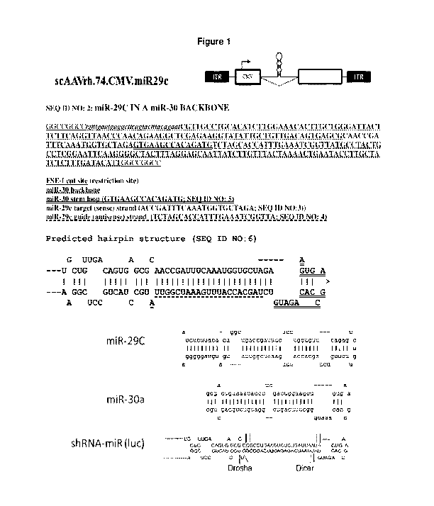
[0057] Figure 1 provide a schematic of rAAV vector scAAVCrh.74.CMV.miR29c
and the nucleotide sequence of the miR-29c in a natural miR-30 backbone and
the
nucleotide sequence of the predicted hairpin structure.
[0058] Figure 2A-CD illustrates that injection of miR-29c into muscle reduces
collagen throughout the muscle and restores miR-29c expression.
[0059] Figure 3A-3C demonstrates that injection of miR-29c improves absolute
muscle force (panel A) and specific muscle force (panel B) but does not
protect
against contraction-induced damage (panel C).
CA 03020999 2018-10-12
WO 2017/181015 PCT/US2017/027636
- 16 -
[0060] Figure 4A-4C displays the number of muscle fibers expression micro-
dystrophin to measure of efficacy of transgene delivery.
[0061] Figure 5A-5C demonstrates that co-delivery of miR-29c with micro-
dystrophin reduces collagen expression (panel A) and fibrosis-induced
dystrophin
expression.
[0062] Figure 6A-6D illustrates that intramuscular injection of miR-29c /micro-
dystrophin inhibits extracellular matrix (ECM) in mdx/utrn+/- mice as measured
by
collagen 1 alpha (panel A), collagen 3 alpha (panel B) , fibronectin (panel C)
and
TGF-f3 (panel D).
[0063] Figure 7A-7C demonstrates the intramuscular injection of miR-29c
increased absolute force (panel A), normalized specific force (panel B) and
added
protection from contraction-induce damage(panel C) in the muscle.
[0064] Figure 8 illustrates that the miR-29c4t-dys combination increases
muscle
size in mice treated at 3 months of age. Sections of treated and untreated
mdx/utrn+/-
gastrocnemius muscles stained with picrosirius Red to stain for collagen are
shown.
Fibrotic areas are pink and intact muscle is in green. On the macroscopic
level, miR-
29c4t-dys combination decreases fibrosis and increases total cross sectional
area.
[0065] Figure 9A-F demonstrates that treatment with miR-29c co-delivered with
micro-dystrophin increased muscle hypertrophy and hyperplasia as shown by an
increase in the overall weight of the injected gastroc compared to either one
injected
alone (panel A), an increase in the an increase in average fiber size (panel
B), an
increase in cross-sectional area of the muscle (panel D; uninjected: 24.6 vs.
miR-29c:
26.3 vs. micro-dys: 26.6 vs. micro-dys/miR-29c: 33.1) and an increase in the
number
of muscle fibers (panel E) but the number of muscle fibers per unit area was
not
affected (panel F). Panel C compares mdx/utrn+/- controls with miR-29c4t-dys
treated
mdx/utrn+/-, the average diameter increased from 25.96 to 30.97iim
[0066] Figure 10A-G demonstrates that early treatment of AAV.miR-29c/micro-
dystrophin combination therapy is more effective at reducing fibrosis and ECM
expression. Panel A shows picrosirius red staining of wild-type, uninjected,
AAV.miR-29c, AAV.micro-dystrophin, and AAV.miR-29c/AAV.micro-dystrophin of
mice injected at 4-5 wks of age taken out twelve weeks post-injection. Panel B
provides quantification of picrosirius red staining showing co-treated muscle
had a
CA 03020999 2018-10-12
WO 2017/181015 PCT/US2017/027636
- 17 -
51.1% reduction in collagen compared to uninjected GAS muscle. Panel C
demonstrates that qRT-PCR confirms an increase in miR-29c transcript levels in
the
treated cohorts. Semi-quantitative qRT-PCR shows a significant reduction in
collagen I and III (panels d, e), fbn (panel f) and TGF-01 (panel g) levels in
the
AAV.miR-29c/AAV.micro-dystrophin treated muscle compared to the contralateral
limb and each of the single therapies Error bars, SEM for n=5
(scAAVrh.74.CMV.miR-29c), n=5 (scAAVrh.74.CMV.miR-
29c/ssAAVrh.74.MCK.micro-dystrophin), n=6 (ssAAVrh.74.MCK.micro-
dystrophin), n=9(mdx/utrn+/- mice). 1-way ANOVA (*p<0.05, ** p<0.01, ***
p<0.001)
[0067] Figure 11 demonstrates early combination therapy restores force and
protects against contraction-induced damage. Measurement of absolute (panel A)
and
normalized specific force (panel b) following tetanic contraction in all three
treatment
injected GAS muscles were significantly increased compared to untreated
mdx/utrn+/-
muscle (panel C). Muscles were then assessed for loss of force following
repetitive
eccentric contractions. Only mice co-treated with miR-29c/micro-dystrophin and
micro-dystrophin alone showed a protection from loss of force compared with
untreated mdx/utrn \- muscles (blue). Two-way analysis of variance
demonstrates
significance in decay curves Error bars, SEM for n=5 (rAAVrh.74.CMV.miR-29c),
n=6 (rAAVrh.74.CMV.miR-29c/rAAVrh.74.MCK.micro-dystrophin), n=5
(rAAVrh.74.MCK.micro-dystrophin), n=15 (mdx/utrn+/- mice). 1-way ANOVA
(*p<0.05,**p<0.01, *** p<0.001, ****p<0.0001).
[0068] Figure 12 illustrates miR-29c/micro-dystrophin combination treatment
increases muscle size in mice treated at 1 month of age. Treated and untreated
mdx/utrn+/- GAS muscles were sectioned and staining with picrosirius Red to
stain for
collagen. Fibrotic areas are pink and intact muscle is in green. On the
macroscopic
level, miR-29c/micro-dystrophin combination decreases fibrosis and increases
total
cross sectional area.
[0069] Figure 13A ¨ 13G demonstrates that early treatment (at 4-5 weeks) of
AAV.MCK.miR-29c/micro-dystrophin combination therapy is more effective at
reducing fibrosis and ECM expression. Panel A provide picrosirius red staining
of
uninjected and AAV.MCK.miR-29c/AAV.MCK.micro-dystrophin of mice injected at
4-5wks of age taken out twelve weeks post-injection. Original magnification,
x20
CA 03020999 2018-10-12
WO 2017/181015 PCT/US2017/027636
- 18 -
Panel B provides quantification of picrosirius red staining demonstrating co-
treated
muscle had a 50.9% reduction in collagen compared to untreated GAS muscle
Panel
C provides qRT-PCR confirming an increase in miR-29c transcript levels in the
treated cohort. Semi-quantitative qRT-PCR shows a significant reduction in
Collagen
lA (Co11A; panel D) and Collagen 3A (Col3A; panel E), Fibronectin (Fbn; panel
F)
and Tgff31 (panel G) levels in the AAV.MCK.miR-29c/AAV.micro-dystrophin
treated
muscle compared to the contralateral limb therapies. (*p<0.05,****p<0.0001).
[0070] Figure 14A ¨ 14G demonstrates that late treatment (treatment at 12
weeks)
with AAV.MCK.miR-29c/micro-dystrophin combination therapy is effective at
reducing fibrosis and ECM expression. Panel A provides picrosirius red
staining of
untreated, AAV.MCK.miR-29c and AAV.MCK.miR-29c/AAV.micro-dystrophin
twelve weeks post-injection. Original magnification, x20. Panel B provides
quantification of picrosirius red staining which demonstrates that co-treated
muscle
had a 30.3 % reduction in collagen compared to untreated GAS muscle. Panel C
provides qRT-PCR confirming an increase in miR-29c transcript levels in the
treated
cohorts. Semi-quantitative qRT-PCR demonstrated a significant reduction in
Collagen lA (Co11A; panel D), Collagen 3A (Col3A; panel E), Fibronectin (Fbn;
Panel F) and Tgff31 (panel G) levels in the AAV.miR-29c/AAV.micro-dystrophin
treated muscle compared to the contralateral limb. One-way ANOVA. All data
represent mean SEM. (** p<0.01, ****p<0.0001).
[0071] Figure 15A-15C demonstrates that early combination therapy (treatment
at
4-5 weeks) restored force and protected against contraction-induced damage.
Measurement of absolute (panel A) and normalized specific force (panel B)
following
tetanic contraction MCK.miR-29c/micro-dystrophin injected GAS muscles were
significantly increased compared to untreated mdx/utrn+/- muscle. (C) Muscles
were
then assessed for loss of force following repetitive eccentric contractions.
Mice co-
treated with miR-29c/micro-dystrophin and micro-dystrophin alone showed
protection from loss of force compared with untreated mdx/utrn \- muscles
(red).
Two-way ANOVA. All data represent mean SEM (****p<0.0001).
[0072] Figure 16A ¨ 16C demonstrates that late combination therapy restored
force and protected against contraction-induced damage. Measurement of
absolute
(panel A) and normalized specific force (panel B) following tetanic
contraction
rAAV.MCK.miR-29c and rAAV expressing micro-dystrophin injected GAS muscles
CA 03020999 2018-10-12
WO 2017/181015
PCT/US2017/027636
- 19 -
were significantly increased compared to untreated mdx/utrn+/- muscle. In
Panel C,
muscles were then assessed for loss of force following repetitive eccentric
contractions. Mice co-treated with rAAV.MCK.miR-29c/rAAV expressing micro-
dystrophin showed a protection from loss of force compared with untreated
mdx/utrn+\- muscles (red). Two-way ANOVA. All data represent mean SEM
(**p<0.01, ****p<0.0001).
[0073] Figure 17A-17D demonstrates that combination treatment increases muscle
hypertrophy 3 months post injection. Panel A demonstrates that rAAV. MCK.miR-
29c co-delivered with rAAV expressing micro-dystrophin failed to increase the
overall weight of the injected GAS. Panel B demonstrates that rAAV.MCK.miR-
29c/rAAV expressing micro-dystrophin combination treatment induced an increase
in
average fiber size. Comparing mdx/utrn+/- controls with miR-29c/micro-
dystrophin
treated mdx/utrn+/-, the average diameter increased from 28.96 to 36.03m .
Panel C
shows that co-delivery produced a shift towards wild-type fiber size
distribution.
Panel D provided the number of muscle fibers per mm2 in the miR-29c/micro-
dystrophin combination treatment was significantly less than untreated mice
and
wild-type (***p<0.01, ****p<0.0001).
[0074] Figure 18A-18B provides the nucleic acid sequence (SEQ ID NO: 1
pAAV.CMV.Mir29C ) of an exemplary rAAV vector comprising the mature guide
strand of miR-29c (nucleotides 1257-1284) and the natural mi-30 backbone
(nucleotides 1088-1375). The construct also comprises the CMV promoter
(nucleotides 120-526), two EFla introns at nucleotides 927-1087 and 1380-1854
and
a polA at nucleotides 1896-2091.
[0075] Figure 19 provides a schematic of the rAAV vector pAAV.MCK.micro-
dystrophin.
[0076] Figure 20A-D provides the nucleic acid sequence (SEQ ID NO: 9;
pAAV.MCK.micro-dystrophin) of an exemplary rAAV vector expressing micro-
dystrophin.
[0077] Figure 21A-D provides the nucleotide sequence of the human micro-
dystrophin nucleotide sequence (SEQ ID NO: 7)
[0078] Figure 22 provides the nucleotide sequence (SEQ ID NO: 12
pAAV.MCK.Mir29C) of an exemplary rAAV vector comprising the mature guide
CA 03020999 2018-10-12
WO 2017/181015 PCT/US2017/027636
- 20 -
strand of miR-29c (nucleotides 1487-1512) and the natural mi-30 backbone
(nucleotides 1088-1375). The construct also comprises the MCK enhancer
(nucleotides 190-395), MCK promoter (nucleotides 396-753), two EFla introns at
nucleotides 1155-1315 and 1609-2083 and a polA at nucleotides 2094-2148.
DETAILED DESCRIPTION
[0079] The present invention provides for gene therapy vectors, e.g. rAAV
vectors,
overexpressing miR-29 microRNA and methods of reducing and preventing fibrosis
in muscular dystrophy patients. The present invention also provides for
combination
gene therapy methods which comprise administering a gene therapy vector
expressing
miR-29 in combination with a gene therapy vector expressing micro-dystrophin
that is
deleted in DMD patients.
[0080] Muscle biopsies taken at the earliest age of diagnosis of DMD reveal
prominent connective tissue proliferation. Muscle fibrosis is deleterious in
multiple
ways. It reduces normal transit of endomysial nutrients through connective
tissue
barriers, reduces the blood flow and deprives muscle of vascular-derived
nutritional
constituents, and functionally contributes to early loss of ambulation through
limb
contractures. Over time, treatment challenges multiply as a result of marked
fibrosis
in muscle. This can be observed in muscle biopsies comparing connective tissue
proliferation at successive time points. The process continues to exacerbate
leading to
loss of ambulation and accelerating out of control, especially in wheelchair-
dependent
patients.
[0081] Without a parallel approach to reduce fibrosis it is unlikely that the
benefits
of exon skipping, stop-codon read-through, or gene replacement therapies can
ever be
fully achieved. Even small molecules or protein replacement strategies are
likely to
fail without an approach to reduce muscle fibrosis. Previous work in aged mdx
mice
with existing fibrosis treated with AAV.micro-dystrophin demonstrated that we
could
not achieve full functional restoration (Human molecular genetics 22, 4929-
4937
(2013)). It is also known that progression of DMD cardiomyopathy is
accompanied
by scarring and fibrosis in the ventricular wall. Micro-RNA delivery is
particularly
innovative because of lack of immune barriers and relative ease of delivery.
Micro-
CA 03020999 2018-10-12
WO 2017/181015 PCT/US2017/027636
- 21 -
RNAs are small (-200bp) and can therefore be packaged in AAV along with a
therapeutic cassette to correct or bypass the genetic defect.
[0082] As used herein, the term "AAV" is a standard abbreviation for adeno-
associated virus. Adeno-associated virus is a single-stranded DNA parvovirus
that
.. grows only in cells in which certain functions are provided by a co-
infecting helper
virus. There are currently thirteen serotypes of AAV that have been
characterized.
General information and reviews of AAV can be found in, for example, Carter,
1989,
Handbook of Parvoviruses, Vol. 1, pp. 169-228, and Berns, 1990, Virology, pp.
1743-
1764, Raven Press, (New York). However, it is fully expected that these same
principles will be applicable to additional AAV serotypes since it is well
known that
the various serotypes are quite closely related, both structurally and
functionally, even
at the genetic level. (See, for example, Blacklowe, 1988, pp. 165-174 of
Parvoviruses
and Human Disease, J. R. Pattison, ed.; and Rose, Comprehensive Virology 3:1-
61
(1974)). For example, all AAV serotypes apparently exhibit very similar
replication
properties mediated by homologous rep genes; and all bear three related capsid
proteins such as those expressed in AAV2. The degree of relatedness is further
suggested by heteroduplex analysis which reveals extensive cross-hybridization
between serotypes along the length of the genome; and the presence of
analogous
self-annealing segments at the termini that correspond to "inverted terminal
repeat
sequences" (ITRs). The similar infectivity patterns also suggest that the
replication
functions in each serotype are under similar regulatory control.
[0083] An "AAV vector" as used herein refers to a vector comprising one or
more
polynucleotides of interest (or transgenes) that are flanked by AAV terminal
repeat
sequences (ITRs). Such AAV vectors can be replicated and packaged into
infectious
viral particles when present in a host cell that has been transfected with a
vector
encoding and expressing rep and cap gene products.
[0084] An "AAV virion" or "AAV viral particle" or "AAV vector particle" refers
to
a viral particle composed of at least one AAV capsid protein and an
encapsidated
polynucleotide AAV vector. If the particle comprises a heterologous
polynucleotide
(i.e. a polynucleotide other than a wild-type AAV genome such as a transgene
to be
delivered to a mammalian cell), it is typically referred to as an "AAV vector
particle"
or simply an "AAV vector". Thus, production of AAV vector particle necessarily
CA 03020999 2018-10-12
WO 2017/181015 PCT/US2017/027636
- 22 -
includes production of AAV vector, as such a vector is contained within an AAV
vector particle.
AAV
[0085] Recombinant AAV genomes of the invention comprise nucleic acid
molecule of the invention and one or more AAV ITRs flanking a nucleic acid
molecule. AAV DNA in the rAAV genomes may be from any AAV serotype for
which a recombinant virus can be derived including, but not limited to, AAV
serotypes AAV-1, AAV-2, AAV-3, AAV-4, AAV-5, AAV-6, AAV-7, AAV-8, AAV-
9, AAV-10, AAV-11, AAV-12 and AAV-13. Production of pseudotyped rAAV is
disclosed in, for example, WO 01/83692. Other types of rAAV variants, for
example
rAAV with capsid mutations, are also contemplated. See, for example, Marsic et
al.,
Molecular Therapy, 22(11): 1900-1909 (2014). As noted in the Background
section
above, the nucleotide sequences of the genomes of various AAV serotypes are
known
in the art. To promote skeletal muscle specific expression, AAV1, AAV6, AAV8
or
AAVrh.74 may be used.
[0086] DNA plasmids of the invention comprise rAAV genomes of the invention.
The DNA plasmids are transferred to cells permissible for infection with a
helper
virus of AAV (e.g., adenovirus, El-deleted adenovirus or herpes virus) for
assembly
of the rAAV genome into infectious viral particles. Techniques to produce rAAV
.. particles, in which an AAV genome to be packaged, rep and cap genes, and
helper
virus functions are provided to a cell, are standard in the art. Production of
rAAV
requires that the following components are present within a single cell
(denoted herein
as a packaging cell): a rAAV genome, AAV rep and cap genes separate from
(i.e., not
in) the rAAV genome, and helper virus functions. The AAV rep and cap genes may
be from any AAV serotype for which recombinant virus can be derived and may be
from a different AAV serotype than the rAAV genome ITRs, including, but not
limited to, AAV serotypes AAV-1, AAV-2, AAV-3, AAV-4, AAV-5, AAV-6, AAV-
7, AAVrh.74, AAV-8, AAV-9, AAV-10, AAV-11, AAV-12 and AAV-13.
Production of pseudotyped rAAV is disclosed in, for example, WO 01/83692 which
is
incorporated by reference herein in its entirety.
[0087] A method of generating a packaging cell is to create a cell line that
stably
expresses all the necessary components for AAV particle production. For
example, a
CA 03020999 2018-10-12
WO 2017/181015
PCT/US2017/027636
-23 -
plasmid (or multiple plasmids) comprising a rAAV genome lacking AAV rep and
cap
genes, AAV rep and cap genes separate from the rAAV genome, and a selectable
marker, such as a neomycin resistance gene, are integrated into the genome of
a cell.
AAV genomes have been introduced into bacterial plasmids by procedures such as
.. GC tailing (Samulski et al., 1982, Proc. Natl. Acad. S6. USA, 79:2077-
2081),
addition of synthetic linkers containing restriction endonuclease cleavage
sites
(Laughlin et al., 1983, Gene, 23:65-73) or by direct, blunt-end ligation
(Senapathy &
Carter, 1984, J. Biol. Chem., 259:4661-4666). The packaging cell line is then
infected with a helper virus such as adenovirus. The advantages of this method
are
.. that the cells are selectable and are suitable for large-scale production
of rAAV.
Other examples of suitable methods employ adenovirus or baculovirus rather
than
plasmids to introduce rAAV genomes and/or rep and cap genes into packaging
cells.
[0088] General principles of rAAV production are reviewed in, for example,
Carter, 1992, Current Opinions in Biotechnology, 1533-539; and Muzyczka, 1992,
.. Curr. Topics in Microbial. and Immunol., 158:97-129). Various approaches
are
described in Ratschin et al., Mol. Cell. Biol. 4:2072 (1984); Hermonat et al.,
Proc.
Natl. Acad. Sci. USA, 81:6466 (1984); Tratschin et al., Mol. Cell. Biol.
5:3251
(1985); McLaughlin et al., J. Virol., 62:1963 (1988); and Lebkowski et al.,
1988 Mol.
Cell. Biol., 7:349 (1988). Samulski et al. (1989, J. Virol., 63:3822-3828);
U.S. Patent
.. No. 5,173,414; WO 95/13365 and corresponding U.S. Patent No. 5,658.776 ; WO
95/13392; WO 96/17947; PCT/U598/18600; WO 97/09441 (PCT/U596/14423); WO
97/08298 (PCT/U596/13872); WO 97/21825 (PCT/U596/20777); WO 97/06243
(PCT/FR96/01064); WO 99/11764; Perrin et al. (1995) Vaccine 13:1244-1250; Paul
et al. (1993) Human Gene Therapy 4:609-615; Clark et al. (1996) Gene Therapy
.. 3:1124-1132; U.S. Patent. No. 5,786,211; U.S. Patent No. 5,871,982; and
U.S. Patent.
No. 6,258,595. The foregoing documents are hereby incorporated by reference in
their entirety herein, with particular emphasis on those sections of the
documents
relating to rAAV production.
[0089] The invention thus provides packaging cells that produce infectious
rAAV.
.. In one embodiment packaging cells may be stably transformed cancer cells
such as
HeLa cells, 293 cells and PerC.6 cells (a cognate 293 line). In another
embodiment,
packaging cells are cells that are not transformed cancer cells, such as low
passage
293 cells (human fetal kidney cells transformed with El of adenovirus), MRC-5
cells
CA 03020999 2018-10-12
WO 2017/181015 PCT/US2017/027636
- 24 -
(human fetal fibroblasts), WI-38 cells (human fetal fibroblasts), Vero cells
(monkey
kidney cells) and FRhL-2 cells (rhesus fetal lung cells).
[0090] Recombinant AAV (i.e., infectious encapsidated rAAV particles) of the
invention comprise a rAAV genome. In exemplary embodiments, the genomes of
both rAAV lack AAV rep and cap DNA, that is, there is no AAV rep or cap DNA
between the ITRs of the genomes. Examples of rAAV that may be constructed to
comprise the nucleic acid molecules of the invention are set out in
International Patent
Application No. PCT/US2012/047999 (WO 2013/016352) incorporated by reference
herein in its entirety.
[0091] The rAAV may be purified by methods standard in the art such as by
column chromatography or cesium chloride gradients. Methods for purifying rAAV
vectors from helper virus are known in the art and include methods disclosed
in, for
example, Clark et al., Hum. Gene Ther., 10(6): 1031-1039 (1999); Schenpp and
Clark, Methods Mol. Med., 69427-443 (2002); U.S. Patent No. 6,566,118 and WO
98/09657.
[0092] In another embodiment, the invention contemplates compositions
comprising rAAV of the present invention. Compositions of the invention
comprise
rAAV and a pharmaceutically acceptable carrier. The compositions may also
comprise other ingredients such as diluents and adjuvants. Acceptable
carriers,
diluents and adjuvants are nontoxic to recipients and are preferably inert at
the
dosages and concentrations employed, and include buffers such as phosphate,
citrate,
or other organic acids; antioxidants such as ascorbic acid; low molecular
weight
polypeptides; proteins, such as serum albumin, gelatin, or immunoglobulins;
hydrophilic polymers such as polyvinylpyrrolidone; amino acids such as
glycine,
glutamine, asparagine, arginine or lysine; monosaccharides, disaccharides, and
other
carbohydrates including glucose, mannose, or dextrins; chelating agents such
as
EDTA; sugar alcohols such as mannitol or sorbitol; salt-forming counter ions
such as
sodium; and/or nonionic surfactants such as Tween, pluronics or polyethylene
glycol
(PEG).
[0093] Titers of rAAV to be administered in methods of the invention will vary
depending, for example, on the particular rAAV, the mode of administration,
the
treatment goal, the individual, and the cell type(s) being targeted, and may
be
CA 03020999 2018-10-12
WO 2017/181015 PCT/US2017/027636
- 25 -
determined by methods standard in the art. Titers of rAAV may range from about
1x10⁶, about 1x10⁷, about 1x10⁸, about 1x10⁹, about lx101 , about lx1011,
about
lx1012, about lx1013to about lx1014 or more DNase resistant particles (DRP)
per ml.
Dosages may also be expressed in units of viral genomes (vg).
[0094] Methods of transducing a target cell with rAAV, in vivo or in vitro,
are
contemplated by the invention. The in vivo methods comprise the step of
administering an effective dose, or effective multiple doses, of a composition
comprising a rAAV of the invention to an animal (including a human being) in
need
thereof. If the dose is administered prior to development of a
disorder/disease, the
administration is prophylactic. If the dose is administered after the
development of a
disorder/disease, the administration is therapeutic. In embodiments of the
invention,
an effective dose is a dose that alleviates (eliminates or reduces) at least
one symptom
associated with the disorder/disease state being treated, that slows or
prevents
progression to a disorder/disease state, that slows or prevents progression of
a
disorder/disease state, that diminishes the extent of disease, that results in
remission
(partial or total) of disease, and/or that prolongs survival. An example of a
disease
contemplated for prevention or treatment with methods of the invention is
FSHD.
[0095] Combination therapies are also contemplated by the invention.
Combination as used herein includes both simultaneous treatment and sequential
treatments. Combinations of methods of the invention with standard medical
treatments (e.g., corticosteroids) are specifically contemplated, as are
combinations
with novel therapies.
[0096] Administration of an effective dose of the compositions may be by
routes
standard in the art including, but not limited to, intramuscular, parenteral,
intravenous,
oral, buccal, nasal, pulmonary, intracranial, intraosseous, intraocular,
rectal, or
vaginal. Route(s) of administration and serotype(s) of AAV components of the
rAAV
(in particular, the AAV ITRs and capsid protein) of the invention may be
chosen
and/or matched by those skilled in the art taking into account the infection
and/or
disease state being treated and the target cells/tissue(s) that are to express
the miR-29
.. miRNA and/or micro-dystrophin.
[0097] The invention provides for local administration and systemic
administration
of an effective dose of rAAV and compositions of the invention including
CA 03020999 2018-10-12
WO 2017/181015 PCT/US2017/027636
- 26 -
combination therapy of the invention. For example, systemic administration is
administration into the circulatory system so that the entire body is
affected. Systemic
administration includes enteral administration such as absorption through the
gastrointestinal tract and parental administration through injection, infusion
or
implantation.
[0098] In particular, actual administration of rAAV of the present invention
may be
accomplished by using any physical method that will transport the rAAV
recombinant
vector into the target tissue of an animal. Administration according to the
invention
includes, but is not limited to, injection into muscle, the bloodstream and/or
directly
into the liver. Simply resuspending a rAAV in phosphate buffered saline has
been
demonstrated to be sufficient to provide a vehicle useful for muscle tissue
expression,
and there are no known restrictions on the carriers or other components that
can be
co-administered with the rAAV (although compositions that degrade DNA should
be
avoided in the normal manner with rAAV). Capsid proteins of a rAAV may be
modified so that the rAAV is targeted to a particular target tissue of
interest such as
muscle. See, for example, WO 02/053703, the disclosure of which is
incorporated by
reference herein. Pharmaceutical compositions can be prepared as injectable
formulations or as topical formulations to be delivered to the muscles by
transdermal
transport. Numerous formulations for both intramuscular injection and
transdermal
transport have been previously developed and can be used in the practice of
the
invention. The rAAV can be used with any pharmaceutically acceptable carrier
for
ease of administration and handling.
[0099] The dose of rAAV to be administered in methods disclosed herein will
vary
depending, for example, on the particular rAAV, the mode of administration,
the
treatment goal, the individual, and the cell type(s) being targeted, and may
be
determined by methods standard in the art. Titers of each rAAV administered
may
range from about 1x10⁶, about 1x10⁷, about 1x10⁸, about 1x10⁹, about lx1010,
about lx1011, about 1x10¹², about 1x10¹³, about 1x10¹⁴, or to about 1x10¹⁵ or
more DNase resistant particles (DRP) per ml. Dosages may also be expressed in
units
of viral genomes (vg) (i.e., 1x10⁷ vg, 1x10⁸ vg, lx109 vg, 1x101 vg, 1x10¹¹
vg,
1x10¹² vg, 1x10¹³ vg, 1x10¹⁴ vg, 1x10¹⁵ respectively). Dosages may also be
expressed in units of viral genomes (vg) per kilogram (kg) of bodyweight
(i.e., 1x101
vg/kg, 1x10¹¹ vg/kg, 1x10¹² vg/kg, 1x10¹³ vg/kg, 1x10¹⁴ vg/kg, 1x10¹⁵ vg/kg
CA 03020999 2018-10-12
WO 2017/181015 PCT/US2017/027636
- 27 -
respectively). Methods for titering AAV are described in Clark et al., Hum.
Gene
Ther., 10: 1031-1039 (1999).
[00100] In particular, actual administration of rAAV of the present invention
may
be accomplished by using any physical method that will transport the rAAV
recombinant vector into the target tissue of an animal. Administration
according to
the invention includes, but is not limited to, injection into muscle, the
bloodstream
and/or directly into the liver. Simply resuspending a rAAV in phosphate
buffered
saline has been demonstrated to be sufficient to provide a vehicle useful for
muscle
tissue expression, and there are no known restrictions on the carriers or
other
components that can be co-administered with the rAAV (although compositions
that
degrade DNA should be avoided in the normal manner with rAAV). Capsid proteins
of a rAAV may be modified so that the rAAV is targeted to a particular target
tissue
of interest such as muscle. See, for example, WO 02/053703, the disclosure of
which
is incorporated by reference herein. Pharmaceutical compositions can be
prepared as
injectable formulations or as topical formulations to be delivered to the
muscles by
transdermal transport. Numerous formulations for both intramuscular injection
and
transdermal transport have been previously developed and can be used in the
practice
of the invention. The rAAV can be used with any pharmaceutically acceptable
carrier
for ease of administration and handling.
[00101] For purposes of intramuscular injection, solutions in an adjuvant such
as
sesame or peanut oil or in aqueous propylene glycol can be employed, as well
as
sterile aqueous solutions. Such aqueous solutions can be buffered, if desired,
and the
liquid diluent first rendered isotonic with saline or glucose. Solutions of
rAAV as a
free acid (DNA contains acidic phosphate groups) or a pharmacologically
acceptable
salt can be prepared in water suitably mixed with a surfactant such as
hydroxpropylcellulose. A dispersion of rAAV can also be prepared in glycerol,
liquid
polyethylene glycols and mixtures thereof and in oils. Under ordinary
conditions of
storage and use, these preparations contain a preservative to prevent the
growth of
microorganisms. In this connection, the sterile aqueous media employed are all
readily obtainable by standard techniques well-known to those skilled in the
art.
[00102] The pharmaceutical carriers, diluents or excipients suitable for
injectable
use include sterile aqueous solutions or dispersions and sterile powders for
the
extemporaneous preparation of sterile injectable solutions or dispersions. In
all cases
CA 03020999 2018-10-12
WO 2017/181015
PCT/US2017/027636
- 28 -
the form must be sterile and must be fluid to the extent that easy
syringability exists.
It must be stable under the conditions of manufacture and storage and must be
preserved against the contaminating actions of microorganisms such as bacteria
and
fungi. The carrier can be a solvent or dispersion medium containing, for
example,
water, ethanol, polyol (for example, glycerol, propylene glycol, liquid
polyethylene
glycol and the like), suitable mixtures thereof, and vegetable oils. The
proper fluidity
can be maintained, for example, by the use of a coating such as lecithin, by
the
maintenance of the required particle size in the case of a dispersion and by
the use of
surfactants. The prevention of the action of microorganisms can be brought
about by
various antibacterial and antifungal agents, for example, parabens,
chlorobutanol,
phenol, sorbic acid, thimerosal and the like. In many cases it will be
preferable to
include isotonic agents, for example, sugars or sodium chloride. Prolonged
absorption of the injectable compositions can be brought about by use of
agents
delaying absorption, for example, aluminum monostearate and gelatin.
[00103] Sterile injectable solutions are prepared by incorporating rAAV in the
required amount in the appropriate solvent with various other ingredients
enumerated
above, as required, followed by filter sterilization. Generally, dispersions
are
prepared by incorporating the sterilized active ingredient into a sterile
vehicle which
contains the basic dispersion medium and the required other ingredients from
those
enumerated above. In the case of sterile powders for the preparation of
sterile
injectable solutions, the preferred methods of preparation are vacuum drying
and the
freeze drying technique that yield a powder of the active ingredient plus any
additional desired ingredient from the previously sterile-filtered solution
thereof.
[00104] Transduction with rAAV may also be carried out in vitro. In one
embodiment, desired target muscle cells are removed from the subject,
transduced
with rAAV and reintroduced into the subject. Alternatively, syngeneic or
xenogeneic
muscle cells can be used where those cells will not generate an inappropriate
immune
response in the subject.
[00105] Suitable methods for the transduction and reintroduction of transduced
cells into a subject are known in the art. In one embodiment, cells can be
transduced
in vitro by combining rAAV with muscle cells, e.g., in appropriate media, and
screening for those cells harboring the DNA of interest using conventional
techniques
such as Southern blots and/or PCR, or by using selectable markers. Transduced
cells
CA 03020999 2018-10-12
WO 2017/181015
PCT/US2017/027636
- 29 -
can then be formulated into pharmaceutical compositions, and the composition
introduced into the subject by various techniques, such as by intramuscular,
intravenous, subcutaneous and intraperitoneal injection, or by injection into
smooth
and cardiac muscle, using e.g., a catheter.
[00106] Transduction of cells with rAAV of the invention results in sustained
expression of miR-29 or micro-dystrophin. The present invention thus provides
methods of administering/delivering rAAV which express of miR-29 and or micro-
dystrophin to an animal, preferably a human being. These methods include
transducing tissues (including, but not limited to, tissues such as muscle,
organs such
as liver and brain, and glands such as salivary glands) with one or more rAAV
of the
present invention. Transduction may be carried out with gene cassettes
comprising
tissue specific control elements. For example, one embodiment of the invention
provides methods of transducing muscle cells and muscle tissues directed by
muscle
specific control elements, including, but not limited to, those derived from
the actin
and myosin gene families, such as from the myoD gene family [See Weintraub et
al.,
Science, 25]: 761-766 (1990], the myocyte-specific enhancer binding factor MEF-
2
[Cserjesi and Olson, Mol Cell Biol 11: 4854-4862 (1990], control elements
derived
from the human skeletal actin gene [Muscat et al., Mol Cell Biol, 7: 4089-4099
(1987)], the cardiac actin gene, muscle creatine kinase sequence elements [See
Johnson et al., Mol Cell Biol, 9:3393-3399 (1989)] and the murine creatine
kinase
enhancer (mCK) element, control elements derived from the skeletal fast-twitch
troponin C gene, the slow-twitch cardiac troponin C gene and the slow-twitch
troponin I gene: hypoxia-inducible nuclear factors (Semenza et al., Proc Natl
Acad
Sci USA, 88: 5680-5684 (1991)), steroid-inducible elements and promoters
including
the glucocorticoid response element (GRE) (See Mader and White, Proc. Natl.
Acad.
Sci. USA 90: 5603-5607 (1993)), and other control elements.
[00107] Muscle tissue is an attractive target for in vivo DNA delivery,
because it is
not a vital organ and is easy to access. The invention contemplates sustained
expression of miRNAs from transduced myofibers.
[00108] By "muscle cell" or "muscle tissue" is meant a cell or group of cells
derived from muscle of any kind (for example, skeletal muscle and smooth
muscle,
e.g. from the digestive tract, urinary bladder, blood vessels or cardiac
tissue). Such
CA 03020999 2018-10-12
WO 2017/181015
PCT/US2017/027636
- 30 -
muscle cells may be differentiated or undifferentiated, such as myoblasts,
myocytes,
myotubes, cardiomyocytes and cardiomyoblasts.
[00109] The term "transduction" is used to refer to the
administration/delivery of
the miiR29 guide strand or the coding region of the micro-dystrophin to a
recipient
cell either in vivo or in vitro, via a replication-deficient rAAV of the
invention
resulting in expression of a miR29 or micro-dystrophin by the recipient cell.
[00110] Thus, the invention provides methods of administering an effective
dose
(or doses, administered essentially simultaneously or doses given at
intervals) of
rAAV that encode miR29 and/or micro-dystrophin to a patient in need thereof.
EXAMPLES
Example 1
Confirmation of Duchenne Muscular Dystrophy Models
[00111] The mdx mouse provides a convenient, yet incomplete, animal model to
study DMD pathogenesis. This model is a cross of the mdx mouse with a
heterozygous knockout of the utrophin gene (mdx:utm+1-), which presents with
increased fibrosis and more faithfully recapitulates the pathology of human
DMD.
Mdx mice have a nonsense mutation in exon 23 of DMD that results in a
relatively
mild phenotype and a near-normal life span. By 3 weeks of age, the diaphragm
and
limb muscle of mdx mice develop signs of endomysial inflammation. These
symptoms subside in the limb muscle after the mice reach adulthood while the
inflammation in the diaphragm muscle continues to progressively worsen. In mdx
mice lacking telomerase, muscular dystrophy progressively worsens with age;
mdx
mice lacking utrophin (DKO) have a phenotype more characteristic of human DMD
with early onset muscle weakness, severe fibrosis, and premature death.
Utrophin, an
.. autosomal paralog of the dystrophin, shares a high degree of sequence
homology that
may compensate for the lack of dystrophin in the mdx mouse in the double KO
(dystrophin plus utrophin); a severe phenotype with early death is observed.
The
premature death in the DKO mouse precludes progression of inflammation and
fibrosis, but the mdx:utrn+/- mouse presents a model with similarities to the
human
disease exhibiting a striking degree of fibrosis, and a longer survival than
the DKO,
providing a better model for our proposed translational studies. A recent
report
confirms the use of the mdx:utrn+/- mouse as an ideal model to study fibrosis
in the
CA 03020999 2018-10-12
WO 2017/181015 PCT/US2017/027636
-31 -
context of DMD. In the present study, increased fibrosis as measured by Sirius
red
staining was accompanied by increased collagen transcript levels and decreased
mir29c levels.
Example 2
Delivery of miR29 to DMD Mice Reduces Fibrosis
[00112] Preliminary studies have demonstrated that there is a significant
increase in
Sirius Red staining for collagen and a decrease in miR-29c levels in human DMD
patients and the mdx/utrn+/- mouse. Gene delivery of miR-29 using muscle
specific
AAV vectors is potentially safe and efficient. To generate the rAAV vector,
referred
to herein as rAAVrh.74.CMV.miR29c, the 22 nucleotide miR29c sequence (target
strand SEQ ID NO: 3 and guide strand SEQ ID NO: 4) was cloned into a miR-30
scaffold driven by a CMV promoter. The expression cassette (SEQ ID NO: 2) was
cloned into a self-complementary AAV plasmid and packaged using AAVrh.74, a
serotype known to express well in muscle. The miR-29c cDNA was synthesized
using a custom primer containing the miR-29c target (sense) strand, miR-30
stem
loop and miR-29c guide (antisense) strand in the miR-30 backbone. Three bases
of
the miR-29c sequence were modified. This sequence was then cloned into a self-
complementary AAV ITR containing plasmid driven by the CMV promoter and
polyA sequence.
[00113] As shown in Figure 1, the pAAV.CMV.miR29C plasmid contains the
mir29c cDNA in a miR-30 stem loop backbone flanked by AAV2 inverted terminal
repeat sequences (ITR). It is this sequence that was encapsidated into
AAVrh.74
virions. In addition, a few nucleotides with in the miR-29c target sequence
were
changed to mimic Watson-crick pairing at this site as in shRNA-miR(luc).
According
to ShRNA-luc design, the hairpin should be perfectly complementary throughout
its
length. Plus, the more changes to the passenger strand, the more likely the
elimination of any endogenous mechanism that regulates miR-29 processing that
could recognize the miRNA via the stem. The 19th base of the guide strand was
modified to a cytosine to mimic the nucleotide that precedes the cleavage site
in
natural mi-29c sequence and the corresponding base on the other strand was
changed
to preserve pairing.
[00114] The gene therapy vector scrAAVrh.74.CMV.miR29c (1x10¹¹ vgs) was
injected into the quadriceps muscle of 3 month old mdx/utrn+/- miceQuadriceps
CA 03020999 2018-10-12
WO 2017/181015 PCT/US2017/027636
- 32 -
muscle was analyzed 3 months post-injection by Sirius Red staining and
analyzed by
NIH ImageJ software as described in Nevo et al. (PloS One, 6: e18049 (2011).
MiR29c, collagen and elastin levels were quantified by RT-PCR. Delivery of miR-
29c to young mdx/utrn +7- mice significantly increases mir-29c levels and a
significant
reduction in Sirius red staining in the quadriceps muscle of 6 month old
mdx/utrn+/-
mice (3 months post injection). There was a reduction in collagen and elastin
levels in
the treated muscles when evaluated by RT-PCR.
[00115] Demonstration of increased fibrosis and decreased miR29 expression in
the mdx/utrn+/- mice and dystrophin-deficient patients validates the mouse
model as
being representative of the human disease. Initial results using AAV-delivered
miR29
as an anti-fibrotic therapy suggest that there is significant beneficial
effect with
reduction in Sirius Red staining and collagen and elastin levels, which are
key
contributors in fibrosis.
Example 3
Injection of MiR-29c Reduces Collagen and Restores miR-29c
[00116] To determine whether rAAVrh.74.CMV.MiR-29c could reduce fibrosis,
12-week-old mdx/utrn \- mice received an intramuscular injection of
rAAVrh.74.CMV.MiR-29c at 5x10¹¹ vgs to the left gastrocnemius (GAS) muscle.
The mice were analyzed at 12 weeks post injection. Picrosirius red staining
revealed
a significant decrease in collagen staining throughout the GAS muscles (Fig.
2a)
compared to the untreated contralateral mdx/utrn +/- GAS muscle.
Quantification of
the picrosirius red staining shows that treated muscle had a 18.3% reduction
in
collagen compared to the untreated muscle (treated- 23.3% 1.3 vs. untreated-
29.5%
0.7)(Fig2b). To confirm overexpression of miR-29c in treated muscle, total RNA
was extracted from the GAS muscle from 24 week old WT, miR-29c treated and
mdx/
-Ft-
utrn mice and subjected to quantitative reverse-transcription ¨PCR (qRT-PCR)
analysis for miR-29c expression. The results showed that miR-29c was
significantly
increased in the GAS muscle of the treated mice compared to untreated mice
(Fig.
2d).
Example 4
MiR-29c Improves Absolute and Specific Muscle Force but does not protect
against Contraction-Induced Damage
CA 03020999 2018-10-12
WO 2017/181015 PCT/US2017/027636
- 33 -
[00117] Knowing that fibrosis can impact muscle function, we wanted to test
whether reducing fibrosis by increasing expression of MiR-29c could protect
mdx/utrn+/- muscle from contraction-induced injury and increase overall force.
The
functional properties of the gastrocnemius muscle from mdx/utrn+/- mice
treated with
.. rAAVrh.74.CMV.MiR-29c were assessed. Twelve weeks post injection, the GAS
was isolated to perform in vivo force measurements.
[00118] The GAS procedure follows the protocol listed in Hakim et al.,
(Methods
Mol Biol. 709: 75-89, 2011) for analyzing transverse abdominal muscle
physiology
but adapted for the GAS. Briefly, mice were anesthetized using
ketamine/xylazine
__ mixture. The hind limb skin was removed to expose the GAS muscle and the
Achilles
tendon. The distal tendon was dissected out and a double square knot was tied
around
the tendon with 4-0 suture as close to the muscle as possible, another second
double
square knot is tied right next to the first knot and then tendon is cut. The
exposed
muscle was constantly dampened with saline. Mice were then transferred to a
thermal
controlled platform and maintained at 37 . The knee was secured to the
platform with
a needle through the patella tendon, the tendon suture to the level arm of the
force
transducer (Aurora Scientific, Aurora, ON, Canada), and the foot was secured
with
tape. The GAS muscle contractions were elicited by stimulating the sciatic
nerve via
bipolar platinum electrodes. Once the muscle was stabilized, the optimal
length was
determined by incremental stretching the muscle until the maximum twitch force
was
achieved. After a 3-minute rest period, the GAS was stimulated at 50,100,150,
and
200 Hz, allowing a 1-minute rest period between each stimulus to determine
maximum tetanic force. Muscle length was measured. Following a 5-minute rest,
the
susceptibility of the GAS muscle to contraction-induced damage was assessed.
After
500 ms of stimulation, the muscle was lengthened by 10% of the optimal length.
This
consisted of stimulating the muscle at 150Hz for 700ms. After the stimulation,
the
muscle was returned to the optimal length. The cycle was repeated every minute
for a
total of 5 cycles. Specific force was calculated by dividing the maximum
tetanic
force by the GAS muscle cross sectional area. After the eccentric
contractions, the
__ mice were then euthanized and the GAS muscle was dissected out, weighed and
frozen for analysis.
[00119] Each GAS was subjected to a series of repeated eccentric contraction.
By
comparing the force ratio of each contraction versus the first contraction
revealed that
CA 03020999 2018-10-12
WO 2017/181015 PCT/US2017/027636
- 34 -
after the fifth contraction untreated muscle decayed to 0.56 0.05 versus
treated 0.50
0.04 (p < 0.0001). The injected group showed a slight decrease in the degree
of
protection compared to WT controls, that decayed to 0.92 0.02 (Fig. 3c).
This data
shows that reducing fibrosis by increasing expression of miR-29c leads to
increase in
both absolute and specific force but does not significantly protect muscle
from
contraction-induced injury.
[00120] rAAVrh.74.MiR-29c treated GAS muscle showed significant improvement
in absolute force when compared to untreated mdx/utrn+/- GAS muscle (rAAV.miR-
29c- 2277 161.7 vs. mdx/utrn+/- untreated- 1722 145.7; Fig. 3a), and also
normalized specific force in rAAVrh.74.miR-29c treated GAS muscle specific
improvement when compared to untreated GAS muscle (rAAV.miR-29c- 204.7
11.7 vs. mdx/utrn+/- untreated- 151.6 14.5; Fig. 3b). Force was still
significantly
reduced when compared to wild-type controls (rAAV.miR-29c- 204.7 11.7 vs.
wild-type- 312.0 34.1).
Example 5
Co-delivery with Micro-Dystrophin Further Reduces fibrosis
[00121] To determine whether miR-29c/micro-dystrophin combined gene therapy
approach would be more beneficial at reducing fibrosis, 12-week-old mdx/utrn
\-
mice received an intramuscular injection of rAAVrh.74.CMV.MiR-29c at 5x10¹¹
vgs
to the left gastrocnemius muscle. The following gene therapy vectors were
administered by intramuscular injection (IM) into the left gastrocnemius (GAS)
muscle of 3 month old mdx/utrn+/- mice, a DMD mouse model:
scAAVrh.74.CMV.miR-29c alone, co-delivered with rAAVrh.74.MCK.micro-
dystrophin, and rAAVrh.74.MCK.micro-dystrophin alone.
[00122] The pAAV.MCK.micro-dystrophin plasmid contains the human micro-
dystrophin cDNA expression cassette flanked by AAV2 inverted terminal repeat
sequences (ITR) as shown in Fig. 10. It is this sequence that was encapsidated
into
AAV rh.74 virions. The pAAV.MCK.micro-dystrophin plasmid was constructed by
inserting the MCK expression cassette driving a codon optimized human micro-
dystrophin cDNA sequence into the AAV cloning vector as described in Rodino-
Klapac et al. (Mol Ther. 2010 Jan;18(1):109-17). A MCK promoter/enhancer
sequence was used to drive muscle-specific gene expression and is composed of
the
mouse MCK core enhancer (206 bp) fused to the 351 bp MCK core promoter
CA 03020999 2018-10-12
WO 2017/181015
PCT/US2017/027636
- 35 -
(proximal). After the core promoter, the 53 bp endogenous mouse MCK Exonl
(untranslated) is present for efficient transcription initiation, followed by
the SV40
late 16S/19S splice signals (97 bp) and a small 5'UTR (61 bp). The intron and
5'
UTR are derived from plasmid pCMVB (Clontech). The micro-dystrophin cassette
has a consensus Kozak immediately in front of the ATG start and a small 53 bp
synthetic polyA signal for mRNA termination. The human micro-dystrophin
cassette
contains the (R4¨R23/A71-78) domains. The complementary DNA was codon
optimized for human usage and synthesized by GenScript (Piscataway, NJ).
[00123] The mice were analyzed at 12 and 24 weeks post injection. First, the
number of muscle fibers expressing micro-dystrophin was used to assess the
efficacy
of transgene delivery and to make sure we had similar levels of micro-
dystrophin
expressed in each group. We found that micro-dystrophin was not different
between
cohorts treated with micro-dystrophin alone (71.85 2.25%) compared with miR-
29c/micro-dystrophin combination therapy (75.03 1.91%) (Fig.4).
[00124] GAS muscle was analyzed 12 months post-injection to assess collagen
accumulation by Sirius Red staining and subsequent quantification with ImageJ.
Additional outcomes included miR-29c and collagen transcript levels, force
measurements in the GAS muscle, fiber diameter measurements and western blot
analysis for proteins involved in muscle regeneration (MyoD, Myogenin). The
amount of fibrosis was analyzed by picrosirius red staining, which revealed a
significant decrease in collagen staining throughout the GAS muscles in all
treated
groups (Fig. 5a) compared to the untreated contralateral mdx/utrn+/- GAS
muscle or
micro-dystrophin alone. Quantification of the picrosirius red staining shows
that co-
treated muscle had a 40.8% reduction in collagen compared to the untreated
muscle
(treated- 17.47% 0.75 vs. untreated-29.5% 0.7) (Fig. 5b). To confirm
expression of
miR-29c, qRT-PCR was performed on the GAS muscle and all treatment groups had
an increase in miR-29c compared to untreated muscle (Fig. Sc).
[00125] Analogous to DMD tissue, a significant reduction in miR-29c levels in
mdx/utrn+/- muscle was observed which correlated with increased fibrosis
measured
by picrosirius red staining. Following 3 months of treatment with scAAV.miR-
29c
alone, there was a significant reduction in fibrosis (treated-23.5% 1.3 vs.
untreated-
27.8% 0.6) in the GAS muscle. When co-delivered with micro-dystrophin,
further
reduction in collagen (41%) was observed by picrosirius red staining
(combination
CA 03020999 2018-10-12
WO 2017/181015 PCT/US2017/027636
- 36 -
treatment: 17.47% 0.75 vs. untreated: 29.5% 0.7) (p<0.0001) (Fig.5b). To
confirm
expression of miR-29c, qRT-PCR was performed on the GAS muscle and all
treatment groups had an increase in miR-29c compared to untreated muscle (Fig.
5b).
[00126] At 24 weeks post-injection, the results were similar to those observed
12
weeks post injection. There was a 47% reduction in collagen by picrosirius red
staining compared to the untreated muscle (combination treatment: 16.5 1.23
vs.
untreated: 31.07 0.93; p<0.0001) and a coincident increase in miR-29c
transcript
level.
[00127] To further validate reduction of collagen observed by picrosirius red
staining, qRT-PCR was performed on the muscle to quantify transcript levels of
Co11A, Col3A and also another ECM component, fibronectin (Fbn). qRT-PCR
analysis detected a decrease in Co11A and Col3A following each treatment,
however
only the cohort treated with both micro-dystrophin and miR-29c showed
significant
reduction (Fig. 6a and 6b). The analysis revealed that Fbn was significantly
reduced
only in the co-treated cohort (Fig. 6c).
[00128] TGF-01 has been previously shown to be up regulated in dystrophic
muscle, likely playing a role in the initiation of the fibrotic cascade. TGF-
01 is a
known pro-fibrotic cytokine that down regulates miR-29c and is responsible for
conversion of myoblasts to myofibroblasts with an increase in collagen and
muscle
fibrogenesis. qRT-PCR analysis shows that co-treated muscle had significantly
lower
levels of TGF-01 compared to uninjected muscle and either treatment alone
(Fig. 6d).
At 6 months post injection, co-treated muscle continued to show reduced CollA,
Col3A, Fbn and TGF-01 levels, whereas only slight reductions in Co11A mRNA
levels in the miR-29 and the micro-dystrophin only groups were observed
[00129] An increase in specific and absolute force was observed in the muscle
treated with miR-29c alone compared to the untreated limb, which when combined
with micro-dystrophin led to absolute and specific force that were not
significantly
different than wild-type. We also observed a significant increase in gastroc
weight in
those muscles that were co-treated.
[00130] Initial results using rAAV.miR-29c as an anti-fibrotic therapy suggest
that
there is beneficial effect with reduction in collagen levels, a key
contributor in
CA 03020999 2018-10-12
WO 2017/181015
PCT/US2017/027636
- 37 -
fibrosis. Moreover, when combined with micro-dystrophin to improve membrane
stability, miR29 up regulation normalized muscle force.
Example 6
Further Increase in Absolute Force and Added Protection from Contraction-
induced Damage
[00131] Knowing that miR-29-treated muscle had a modest but significant
increase
in absolute and specific force, the combination therapy of miR-29c
overexpression
and micro-dystrophin gene replacement impact on muscle function was
investigated.
Twelve weeks post injection, we isolated the GAS for which we performed in
vivo
force measurements. The rAAVrh.74.MiR-29c vector described above in Example 2
and a rAAV
[00132] Co-treated rAAVrh.74.MiR-29c and rAAV expressing Micro-Dys treated
GAS muscle showed significant improvement in absolute force when compared to
untreated mdx/utrn+/- GAS muscle (co-treated- 3582.4 79.4 nM vs. mdx/utrn+/-
untreated- 1722 145.7 nM vs. wild-type- 3005 167.3 nM) (Fig.7), and also
normalized specific force in rAAVrh.74.miR-29c/micro-dys treated GAS muscle
specific improvement when compared to untreated GAS muscle (co-treated mice-
244.2 6.6 nM/mm2 vs. mdx/utrn+/- untreated- 151.6 14.5 nM/mm2 vs. 312.0
34.1 nM/mm2) (Fig 7). Both absolute and specific force was not significantly
different from wild-type controls.
[00133] Each GAS was subjected to a series of repeated eccentric contraction.
By
comparing the force ratio of each contraction versus the first contraction
revealed that
after the fifth contraction untreated muscle decayed to 0.54 0.06 versus co-
treated
0.66 0.04 (p < 0.0001), which can be contributed to the micro-dystrophin
since the
micro-dystrophin alone also decayed to 0.66 0.04. The treated group was still
significantly lower than wild-type that decayed to 0.92 0.02 (Fig. 7c).
Similar
findings were seen at 24 weeks post injection This data shows that reducing
fibrosis
and gene replacement leads to increase in both absolute and specific 5force
and
significantly protects muscle from contraction-induced injury.
Example 7
Combination treatment increases muscle hypertrophy and hyperplasia
[00134] MiR-29c co-delivered with micro-dystrophin increased the overall
weight
of the injected gastroc compared to either one injected alone at three months
of age
CA 03020999 2018-10-12
WO 2017/181015
PCT/US2017/027636
- 38 -
(Fig. 8, Fig. 9a). To investigate the source of increased muscle mass,
myofiber
diameters are measured. miR-29c4t-dys combination treatment demonstrated an
increase in average fiber size. Comparing mdx/utrn+/- controls with miR-29c4t-
dys
treated mdx/utrn+/-, the average diameter increased from 25.96 to 30.97iim
(Fig. 9b).
The co-delivery produced a shift towards wild-type fiber size distribution
(Fig. 9c).
Although the average fiber size was increased does not explain the ¨30%
increase in
gross muscle weight. Total cross-sectional area of the muscle was also
measured.
Gastroc muscles from all groups were full slide scanned and the total area was
measured. Muscles co-treated with micro-dys/miR-29c had a significant increase
in
cross sectional area compared to untreated and either treatment alone
(uninjected:
24.6 vs. miR-29c: 26.3 vs. micro-dys: 26.6 vs. micro-dys/miR-29c: 33.1) (Fig.
8, Fig.
9d).
[00135] miR-29c has been reported it to play a role in the myoD/Pax7/myogenin
pathway and it was hypothesized that miR-29c may be impacting regeneration and
activation of satellite cells (muscle stem cells) to differentiate in myogenic
lineage.
To test this, the total number of muscle fibers from the full slide scanned
images was
counted. An increased number of muscle fibers following miR-29c4t-dys
combination treatment (Fig. 9e). Finally, given that muscle fiber diameters in
mdx/utrn+/- mice are heterogeneous with many small fibers and some
hypertrophic
fibers, it was determined whether the number of fibers per unit area
(cells/mm2) was
affected with treatment. miR-29c4t-dys combination treatment was not different
than
wild-type (Fig. 9f).
Example 8
Early Treatment With Combination Prevents Fibrosis
[00136] In view of the potential importance of combinatorial miR-29c and micro-
dystrophin as a prophylactic therapy for DMD, a cohort of younger mdx/utrn+/-
mice
were treated at 4 weeks of age. Using the same paradigm as for other groups as
described herein, the following treatments were compared for efficacy for
prevention
of fibrosis by intramuscular injection of GAS: scAAVrh.74.CMV.miR-29c alone,
ssAAVrh74.MCK.micro-dystrophin + scAAVrh.74.CMV.miR-29c combination
therapy, or ssAAVrh74.MCK.micro-dystrophin alone at the same dose. The mice
were necropsied 12 weeks post injection. A significant decrease in collagen
staining
throughout the GAS muscles in all treated groups compared to the untreated
CA 03020999 2018-10-12
WO 2017/181015 PCT/US2017/027636
- 39 -
contralateral mdx/utrn +1- GAS muscle was observed (Fig. 10A). Quantification
of the
picrosirius red staining showed that muscle co-treated with micro-
dystrophin/miR-29c
had a 51% reduction in collagen compared to the untreated muscle (treated-
11.32%
1.18 vs. untreated-23.15% 0.90) (p<0.0001) (Fig. 10) and qRT-PCR confirmed
.. Co11A, Col3A, Fbn and TGF-01 reduction following combinatorial therapy
(Fig. 10D
and E).
Example 9
Early Combination Therapy Restores Force and Protects from Contraction-
Induced Damage Better than Late Treatment
[00137] In vivo force measurement was carried out on the GAS of the mice
treated
early with the combination therapy as described in Example 8. In 4-week-old
mdx/utrn+/- mice, co-treatment using miR-29c/micro-dystrophin showed
significant
improvement in absolute force when compared to untreated mdx/utrn+/- mice and
there was no difference from wild type (co-treated: 2908 129.5 mN vs.
untreated:
1639.4 116.9 mN vs. wild-type: 3369.73 154.1 mN). Specific force was also
normalized to wild type levels following combinatorial therapy (co-treated
338.9
22.34 mN/mm2 vs. untreated 184.3 13.42 mN/mm2 vs. WT 364.3 7.79 mN/mm2)
(Fig. 11A and B and 12).
[00138] Next, each GAS was subjected to a series of repeat eccentric
contractions.
By comparing the force ratio of each contraction by the fifth contraction,
untreated
muscle decayed to 0.53 0.04 versus co-treated 0.82 0.04 (p < 0.0001). The
combinatorial treatment group was slightly lower than wild type but not
significantly
different, which decayed to 0.93 0.01 (Fig. 11C). These data show that
reducing
fibrosis and gene replacement lead to increase in both absolute and specific
force and
significantly protects muscle from contraction-induced injury.
[00139] These experiments suggest that gene replacement should be started in
the
newborn period. Efforts are clearly moving in the direction of identifying DMD
and
other muscular dystrophies in the newborn period. The Ohio Newborn Screening
Study illustrates the potential for identification of DMD in newborns using CK
7
Neurol. as a biomarker (>2000 U/L) with DNA confirmation on the same dried
blood
spot (Mendell et al., Ann. Neurol. 71: 304-313, 2012). This methodology is now
being extended to other states in the USA (PPMD May 16, 2016: Next Steps with
CA 03020999 2018-10-12
WO 2017/181015 PCT/US2017/027636
- 40 -
Newborn Screening) and in other countries, particularly the UK (UK National
Screening Committee) and China (Perkin ElmerTM launches screening in China).
[00140] miR-29 has also shown promise as a treatment modality for
cardiac,
pulmonary, and liver fibrosis. Myocardial infarction in mice and humans is
associated with miR-29 down-regulation. Rooij et al. (Proc. Natl. Acad. Sci,
USA
105:13027-13032, 2008) demonstrated that exposing fibroblasts to a miR-29b
mimic
decreased collagen transcripts providing a path for clinical translation for
cardiac
fibrosis. Subsequent studies showed that in a bleomycin-induced pulmonary
fibrosis
mouse model, attenuation of fibrosis could be achieved using the Sleeping
Beauty
(SB) transposon system-based delivery of miR-29b.14. Currently, a miR-29b
mimic
is in a clinical Phase 1 Safety-Tolerability local intradermal trial in
healthy volunteers
(miRagen TherapeuticsTm MRG-201). Compared to miR-29 oligonucleotide delivery
that would require repeated administration related to the half-life of the
oligonucleotides, AAV gene therapy could potentially provide a path for single-
.. delivery gene transfer.
Example 10
Treatment with Muscle Specific Expression of miR-29 and Micro-dystrophin
Reduced Fibrosis and ECM Expression
[00141] AAV vectors comprising the miR29c sequence and a muscle specific
promoter MCK were also generated and tested as a combination therapy with AAV
vectors expressing micro-dystrophin. To generate the rAAV vector, referred to
herein
as rAAV.MCK.miR29c, the 22 nucleotide miR29c sequence (target strand SEQ ID
NO: 3 and guide strand SEQ ID NO: 4) was cloned into a miR-30 scaffold driven
by a
MCK promoter (SEQ ID NO: 11). The expression cassette (SEQ ID NO: 12) was
cloned into a single stranded AAV plasmid and packaged using AAVrh74, a
serotype
known to express well in muscle. The miR-29c cDNA was synthesized using a
custom primer containing the miR-29c target (sense) strand, miR-30 stem loop
and
miR-29c guide (antisense) strand in the miR-30 backbone. Three bases of the
miR-
29c sequence were modified. This sequence was then cloned into a single
stranded
AAV ITR containing plasmid driven by the MCK promoter and polyA sequence.
[00142] The pAAV.MCK.miR29C plasmid contains the mir29c cDNA in a miR-30
stem loop backbone flanked by AAV2 inverted terminal repeat sequences (ITR).
It is
this sequence that was encapsidated into AAVrh74 virions. In addition, a few
CA 03020999 2018-10-12
WO 2017/181015 PCT/US2017/027636
- 41 -
nucleotides with in the miR-29c target sequence were changed to mimic Watson-
crick
pairing at this site as in shRNA-miR(luc). According to ShRNA-luc design, the
hairpin should be perfectly complementary throughout its length. Plus, the
more
changes to the passenger strand, the more likely the elimination of any
endogenous
mechanism that regulates miR-29 processing that could recognize the miRNA via
the
stem. The 19th base of the guide strand was modified to a cytosine to mimic
the
nucleotide that precedes the cleavage site in natural mi-29c sequence and the
corresponding base on the other strand was changed to preserve pairing.
[00143] Early treatment of AAV.MCK.miR-29c/micro-dystrophin combination
therapy was more effective at reducing fibrosis and ECM expression. 4-5-week-
old
mdx/utrn+\- mice received an intramuscular injection of rAAVrh.74.MCK.MiR-29c
and rAAVrh74.MCK.micro-dystrophin at 5x10¹¹ vgs to the left gastrocnemius
muscle
as described in Example 5. The muscles were harvested twelve weeks post
injection.
Picrosirius red staining of muscle harvested from uninjected and mice injected
with
combination therapy of rAAV.MCK.miR-29c/rAAV.MCK.micro-dystrophin showed
co-treated muscle had a 50.9% reduction in collagen compared to untreated GAS
muscle (See Fig. 13a and 13b). qRT-PCR confirmed an increase in miR-29c
transcript levels in the treated cohort (Fig. 13c). Semi-quantitative qRT-PCR
showed
a significant reduction in Collagen Al and Collagen 3A (Fig. 13d, e),
Fibronectin
(Fig. 130 and Tgf(31 (Fig. 13g) levels in the AAV.MCK.miR-29c/AAV.micro-
dystrophin treated muscle compared to the contralateral limb therapies.
(*p<0.05,****p<0.0001).Late treatment of AAV.MCK.miR-29c/micro-dystrophin
combination therapy is effective at reducing fibrosis and ECM expression.
Three
month old mdx/utrn+\- mice received an intramuscular injection of
rAAVrh.74.MCK.MiR-29c and rAAVrh.74.MCK.micro-dystrophin at 5x10¹¹ vgs to
the left gastrocnemius muscle as described in Example 5. The muscles were
harvested twelve weeks post injection. Picrosirius red staining of untreated,
AAV.MCK.miR-29c and AAV.MCK.miR-29c/AAV.micro-dystrophin treated muscle
showed co-treated muscle had a 30.3 % reduction in collagen compared to
untreated
GAS muscle (See Fig. 14a and 14b) qRT-PCR confirmed an increase in miR-29c
transcript levels in the treated cohorts (Fig. 14c). Semi-quantitative qRT-PCR
shows
a significant reduction in Collagen lA and Collagen 3A (Fig. 14d, e),
Fibronectin
(Fig. 140 and Tgf(31 (Fig. 14G) levels in the AAV.miR-29c/AAV.micro-dystrophin
CA 03020999 2018-10-12
WO 2017/181015 PCT/US2017/027636
- 42 -
treated muscle compared to the contralateral limb. One-way ANOVA. All data
represent mean SEM. (** p<0.01, ****p<0.0001).
Example 11
Early Combination Therapy Restores Force and Protects from Contraction-
Induced Damage Better than Late Treatment
[00144] In vivo force measurement was carried out on the GAS of the mice
treated
early with the muscle-specific expression of miR-29 and micro-dystrophin. as
described in Examples 8 and 9. In 4-week-old mdx/utrn' - mice, co-treatment
using
rAAV.MCK.miR-29c/and rAAV expressing micro-dystrophin showed significant
improvement in absolute force when compared to untreated mdx/utrn' - mice and
there was no difference from wild type (Fig. 15a). Specific force was also
normalized
to wild type levels following combination therapy (Fig. 15b).
[00145] Muscles were then assessed for loss of force following repetitive
eccentric
contractions as described in Example 9. Mice co-treated with rAAV.MCK.miR-
29c/rAAV.MCK.micro-dystrophin and rAAV.MCK.micro-dystrophin alone showed a
protection from loss of force compared with untreated mdx/utrn \- muscles
(Fig.
15c).
[00146] In 12-week-old mdx/utrn' - mice, co-treatment using rAAV.MCK.miR-
29c/and rAAV expressing micro-dystrophin restored force and protected against
contraction-induced damage. Measurement of absolute (Fig. 16a) and normalized
specific force (Fig. 16b) following tetanic contraction rAAV.MCK.miR-29c and
rAAV expressing micro-dystrophin injected GAS muscles were significantly
increased compared to untreated mdx/utrn'- muscle. Subsequently, muscles were
assessed for loss of force following repetitive eccentric contractions as
described in
Example 9. Mice co-treated with MCK.miR-29c/micro-dystrophin showed a
protection from loss of force compared with untreated mdx/utrn \- muscles
(Fig. 16c).
These data show that reducing fibrosis and gene replacement lead to increase
in both
absolute and specific force and significantly protects muscle from contraction-
induced
injury.
Example 12
Early Combination treatment increases muscle hypertrophy and hyperplasia
[00147] Co-delivery of rAAV.MCK.miR-29 with rAAV expressing micro-
dystrophin did not increase overall weight of the injected gastroc compared to
either
CA 03020999 2018-10-12
WO 2017/181015 PCT/US2017/027636
-43 -
one injected alone at three months post-injection (Fig.17a). Myofiber
diameters were
also measured. miR-29c/micro-dystrophin combination treatment demonstrated an
increase in average fiber size. Comparing mdx/utrn+/- controls with miR-
29c/micro-
dystrophin treated mdx/utrn+/-, the average diameter increased from 28.96 to
36.03m
__ (Fig. 17b). The co-delivery produced a shift towards wild-type fiber size
distribution
(Fig. 17c). The number of muscle fibers per mm2 in the miR-29c/micro-
dystrophin
combination treatment was significantly less than untreated mice and wild-type
(Fig.
17d; ***p<0.01, ****p<0.0001).
REFERENCES
1. Hoffman, E.P., Brown, R.H., Jr. & Kunkel, L.M. Dystrophin: the protein
product of the Duchenne muscular dystrophy locus. Cell 51, 919-928 (1987).
2. Straub, V. & Campbell, K.P. Muscular dystrophies and the dystrophin-
glycoprotein complex. Curr Opin Neurol 10, 168-175 (1997).
3. Sacco, A., et al. Short telomeres and stem cell exhaustion model
Duchenne
muscular dystrophy in mdx/mTR mice. Cell 143, 1059-1071 (2010).
4. Wallace, G.Q. & McNally, E.M. Mechanisms of muscle degeneration,
regeneration, and repair in the muscular dystrophies. Annu Rev Physiol 71, 37-
57 (2009).
5. Zhou, L. & Lu, H. Targeting fibrosis in Duchenne muscular dystrophy. J
Neuropathol Exp Neurol 69, 771-776 (2010).
6. Desguerre, I., et al. Endomysial fibrosis in Duchenne muscular
dystrophy: a
marker of poor outcome associated with macrophage alternative activation. J
Neuropathol Exp Neurol 68, 762-773 (2009).
7. Kim, J., et al. microRNA-directed cleavage of ATHB15 mRNA regulates
vascular development in Arabidopsis inflorescence stems. Plant J 42, 84-94
(2005).
8. Ambros, V. MicroRNA pathways in flies and worms: growth, death, fat,
stress, and timing. Cell 113, 673-676 (2003).
9. Eisenberg, I., et al. Distinctive patterns of microRNA expression in
primary
muscular disorders. Proc Natl Acad Sci USA 104, 17016-17021 (2007).
10. Jiang, X., Tsitsiou, E., Herrick, S.E. & Lindsay, M.A. MicroRNAs and
the
regulation of fibrosis. FEBS J 277, 2015-2021 (2010).
CA 03020999 2018-10-12
WO 2017/181015
PCT/US2017/027636
- 44 -
H. van Rooij, E., et al. Dysregulation of microRNAs after myocardial
infarction
reveals a role of miR-29 in cardiac fibrosis. Proc Natl Acad Sci U S A 105,
13027-13032 (2008).
12. Cacchiarelli, D., et al. MicroRNAs involved in molecular circuitries
relevant
for the Duchenne muscular dystrophy pathogenesis are controlled by the
dystrophin/nNOS pathway. Cell Metab 12, 341-351 (2010).
13. DiPrimio, N., McPhee, S.W. & Samulski, R.J. Adeno-associated virus for
the
treatment of muscle diseases: toward clinical trials. Curr Opin Mol Ther 12,
553-560 (2010).
14. Mendell, J.R., et al. Sustained alpha-sarcoglycan gene expression after
gene
transfer in limb-girdle muscular dystrophy, type 2D. Ann Neurol 68, 629-638
(2010).
15. Mendell, J.R., et al. Limb-girdle muscular dystrophy type 2D gene
therapy
restores alpha-sarcoglycan and associated proteins. Ann Neurol 66, 290-297
(2009).
16. Mendell, J.R., et al. A phase 1/2a follistatin gene therapy trial for
becker
muscular dystrophy. Molecular therapy : the journal of the American Society
of Gene Therapy 23, 192-201 (2015).
17. Carnwath, J.W. & Shotton, D.M. Muscular dystrophy in the mdx mouse:
histopathology of the soleus and extensor digitorum longus muscles. J Neurol
Sci 80, 39-54 (1987).
18. Coulton, G.R., Morgan, J.E., Partridge, T.A. & Sloper, J.C. The mdx
mouse
skeletal muscle myopathy: I. A histological, morphometric and biochemical
investigation. Neuropathol Appl Neurobiol 14, 53-70 (1988).
19. Cullen, M.J. & Jaros, E. Ultrastructure of the skeletal muscle in the X
chromosome-linked dystrophic (mdx) mouse. Comparison with Duchenne
muscular dystrophy. Acta Neuropathol 77, 69-81 (1988).
20. Dupont-Versteegden, E.E. & McCarter, R.J. Differential expression of
muscular dystrophy in diaphragm versus hindlimb muscles of mdx mice.
Muscle Nerve 15, 1105-1110 (1992).
21. Stedman, H.H., et al. The mdx mouse diaphragm reproduces the
degenerative
changes of Duchenne muscular dystrophy. Nature 352, 536-539 (1991).
22. Deconinck, A.E., et al. Utrophin-dystrophin-deficient mice as a model
for
Duchenne muscular dystrophy. Cell 90, 717-727 (1997).
CA 03020999 2018-10-12
WO 2017/181015 PCT/US2017/027636
- 45 -
23. Grady, R.M., et al. Skeletal and cardiac myopathies in mice lacking
utrophin
and dystrophin: a model for Duchenne muscular dystrophy. Cell 90, 729-738
(1997).
24. Love, D.R., et al. An autosomal transcript in skeletal muscle with
homology to
dystrophin. Nature 339, 55-58 (1989).
25. Tinsley, J.M., et al. Primary structure of dystrophin-related protein.
Nature
360, 591-593 (1992).
26. Tinsley, J., et al. Expression of full-length utrophin prevents
muscular
dystrophy in mdx mice. Nat Med 4, 1441-1444 (1998).
27. Squire, S., et al. Prevention of pathology in mdx mice by expression of
utrophin: analysis using an inducible transgenic expression system. Hum Mol
Genet 11, 3333-3344 (2002).
28. Rafael, J.A., Tinsley, J.M., Potter, A.C., Deconinck, A.E. & Davies,
K.E.
Skeletal muscle-specific expression of a utrophin transgene rescues utrophin-
dystrophin deficient mice. Nat Genet 19, 79-82 (1998).
29. Zhou, L., et al. Haploinsufficiency of utrophin gene worsens skeletal
muscle
inflammation and fibrosis in mdx mice. J Neurol Sci 264, 106-111 (2008).
30. Gutpell, K.M., Hrinivich, W.T. & Hoffman, L.M. Skeletal Muscle Fibrosis
in
the mdx/utrn+/- Mouse Validates Its Suitability as a Murine Model of
Duchenne Muscular Dystrophy. PloS one 10, e0117306 (2015).
31. Rodino-Klapac, L.R., et al. Micro-dystrophin and follistatin co-
delivery
restores muscle function in aged DMD model. Human molecular genetics 22,
4929-4937 (2013).
32. Cushing, L., et al. M1R-29 is a Major Regulator of Genes Associated
with
Pulmonary Fibrosis. Am J Respir Cell Mob Biol (2010).
33. Roderburg, C., et al. Micro-RNA profiling reveals a role for miR-29 in
human
and murine liver fibrosis. Hepatology 53, 209-218 (2011).
34. Nevo, Y., et al. The Ras antagonist, farnesylthiosalicylic acid (FTS),
decreases
fibrosis and improves muscle strength in dy/dy mouse model of muscular
dystrophy. PloS one 6, e18049 (2011).
35. Rodino-Klapac, L.R., et al. A translational approach for limb vascular
delivery
of the micro-dystrophin gene without high volume or high pressure for
treatment of Duchenne muscular dystrophy. J Transl Med 5, 45 (2007).
CA 03020999 2018-10-12
WO 2017/181015
PCT/US2017/027636
- 46 -
36. Mulieri, L.A., Hasenfuss, G., Ittleman, F., Blanchard, E.M. & Alpert,
N.R.
Protection of human left ventricular myocardium from cutting injury with 2,3-
butanedione monoxime. Circ Res 65, 1441-1449 (1989).
37. Rodino-Klapac, L.R., et al. Persistent expression of FLAG-tagged micro
dystrophin in nonhuman primates following intramuscular and vascular
delivery. Molecular therapy: the journal of the American Society of Gene
Therapy 18, 109-117 (2010).
38. Grose, W.E., et al. Homologous recombination mediates functional
recovery
of dysferlin deficiency following AAV5 gene transfer. PloS one 7, e39233
(2012).
39. Liu, M., et al. Adeno-associated virus-mediated microdystrophin
expression
protects young mdx muscle from contraction-induced injury. Mob Ther 11, 245-
256
(2005).