Note: Descriptions are shown in the official language in which they were submitted.
CA 03022377 2018-10-26
WO 2017/189976
PCT/US2017/030077
DESCRIPTION
TARGETED MEASURE OF TRANSCRIPTIONAL ACTIVITY RELATED TO
HORMONE RECEPTORS
[0001] The present application claims the priority benefit of United States
Provisional
Application Serial No. 62/329,774, filed April 29, 2016, the entire contents
of which is hereby
incorporated by reference.
BACKGROUND OF THE INVENTION
[0002] The invention was made with government support under Grant No.
HHSN261200800001E awarded by the National Institutes of Health. The government
has
certain rights in the invention.
1. Field of the Invention
[0003] The present invention relates generally to the fields of molecular
biology and
medicine. More particularly, it concerns methods of treating cancer based on
transcriptional
profiling.
2. Description of Related Art
[0004] Endocrine therapy (also known as hormonal therapy) is the foundation
for
palliative treatment of metastatic hormone receptor-positive and HER2-negative
(HR+/HER2-
) breast cancer (Giordano et al., 2014; Cardoso et al., 2014). However, the
efficacy of palliative
therapy is variable according to the patient and treatment type. Furthermore,
there is consistent
evidence for molecular progression events which decouple cancer cells from
their reliance on
estrogen in advanced disease through a change in hormone receptor expression
(e.g., a loss of
progesterone receptor (PR, gene name PGR) in about 20% of metastatic breast
cancers or a
loss of estrogen receptor (ER, gene name ESR1) in about 10% of metastatic
breast cancers
(Lower et al., 2005; Hoefnagel et al., 2010; Amir et al., 2012; Thompson et
al., 2010), the
acquisition of functionally active gene mutations in ESR1 (Toy et al., 2013;
Robinson et al.,
2013), and changes in transcriptional profiles towards a decreased dependence
on estrogen.
[0005] While guidelines currently recommend endocrine therapy as the first-
line
treatment for patients with relapsed HR-VHER2- breast cancer, the selection of
later lines of
treatment can become increasingly challenging if there is concern that
secondary endocrine
- 1 -
CA 03022377 2018-10-26
WO 2017/189976
PCT/US2017/030077
resistance may have developed (Giordano et al., 2014; Cardoso et al., 2014).
Clinical variables
can be used to estimate the sensitivity to further endocrine-based therapy,
such as the patient's
history of prior endocrine sensitivity in the adjuvant setting, the number of
previous relapse
events, the development of visceral metastases, and the number of previous
endocrine
(hormonal) treatments administered. While a number of genomic multigene-assays
have
proven their value in individualizing treatment decisions in early hormone
receptor-positive
(HR+) breast cancer beyond the use of immunohistochemical evaluation of
standard markers
(van't Veer et al., 2002; Paik et al., 2004; Filipits et al., 2011), there is
need for a customized
assay with a strong analytical validity for predicting treatment response for
use in the setting
of advanced breast cancer, including Stage II, III, or IV as defined by
current criteria from the
American Joint Commission for Cancer Staging (AJCC Stage).
SUMMARY OF THE INVENTION
[0006] Embodiments of the present disclosure provide methods for the detection
and
treatment of breast tumors sensitive to hormonal therapy (also known as
endocrine therapy)
alone or in combination with other anti-cancer therapies. In a first
embodiment, there is
provided a method of treating breast cancer in a patient comprising: (a)
determining the
expression level of a set of genes in a patient sample that are related to
both estrogen receptor
(ER) and progesterone receptor (PR) expression; (b) calculating an index of
sensitivity to
endocrine therapy (SETERADR index) based on an index of ER- and PR-related
gene expression;
and (c) administering an effective amount of a endocrine therapy to the
patient based on the
predicted sensitivity of the patient's cancer to endocrine therapy (SETER/pR
index).
[0007] In another embodiment, the present disclosure provides a method of
treating
breast cancer in a subject comprising administering an effective amount of an
endocrine
therapy to said subject, wherein the subject has been determined to be
sensitive to endocrine
therapy based on a SETER/pR index. In some aspects, the subject has been
determined to be
sensitive to endocrine therapy based on a SETER/pR index by determining the
expression level
of a set of estrogen receptor (ER)- and progesterone receptor (PR)-related
genes in a sample
from the subject and calculating the SETER/pR index based on the ER- and PR-
related gene
expression.
[0008] A further embodiment provides a composition comprising an effective
amount
of an endocrine therapy for the treatment of breast cancer in a subject
identified to be sensitive
- 2 -
CA 03022377 2018-10-26
WO 2017/189976
PCT/US2017/030077
to endocrine therapy based on a SETERADR index. In some aspects, the subject
has been
determined to be sensitive to endocrine therapy based on a SETERADR index by
determining the
expression level of a set of estrogen receptor (ER)- and progesterone receptor
(PR)-related
genes in a sample from the subject and calculating the SETERADR index based on
the ER- and
PR-related gene expression.
[0009] In some aspects of the above embodiments, calculating the SETERiptz
index
comprises normalizing the expression of the set of ER- and PR-related genes to
a set of
reference genes. In certain aspects, calculating is further defined as the
difference between the
average expression of the set of ER- and PR-related genes and the average
expression of the
.. set of reference genes. In some aspects, the method further comprises the
addition of an
optimizing constant. In particular aspects, the optimizing constant has a
value of 2. In some
aspects, a SETERiptz index greater than 0 identifies a patient as sensitive to
endocrine therapy.
In certain aspects, a SETERADR index greater than 0.5 identifies a patient as
sensitive to endocrine
therapy. In some aspects, a SETERADR index greater than 1 identifies a patient
as sensitive to
endocrine therapy.
[0010] In certain aspects, the set of ER- and PR-related genes comprises at
least 10 of
the genes selected from the group consisting of SLC39A6, STC2, CA12, ESR1,
PDZKl, NPY1R,
CD2, MAPT, QDPR, AZGP1, ABAT, ADCY1, CD3D, NAT], MRPS30, DNAJC12, SCUBE2,
and KCNE4. In some aspects, the set of ER- and PR-related genes comprises at
least 11, 12,
13, 14, 15, 16 or 17 of the genes selected from the group consisting of
SLC39A6, STC2, CA12,
ESR1, PDZKl, NPY1R, CD2, MAPT, QDPR, AZGP1, ABAT, ADCY1, CD3D, NAT], MRPS30,
DNAJC12, SCUBE2, and KCNE4. In some aspects, the set of ER- and PR-related
genes
consists of SLC39A6, STC2, CA12, ESR1, PDZKl, NPY1R, CD2, MAPT, QDPR, AZGP1,
ABAT, ADCY1, CD3D, NAT], MRPS30, DNAJC12, SCUBE2, and KCNE4.
[0011] In some aspects, the set of reference genes comprises at least 5 of the
genes
selected from the group consisting of LDHA, ATP5J2, VDAC2, DARS, UCP2, UBE2Z
AK2,
WIPF2, APPBP2, and TRIM2. In certain aspects, the set of reference genes
comprises at least
6, 7, 8 or 9 of the genes selected from the group consisting of LDHA, ATP5J2,
VDAC2, DARS,
UCP2, UBE2Z AK2, WIPF2, APPBP2, and TRIM2. In some aspects, the set of
reference genes
consists of LDHA, ATP5J2, VDAC2, DARS, UCP2, UBE2Z AK2, WIPF2, APPBP2, and
TRIM2.
- 3 -
CA 03022377 2018-10-26
WO 2017/189976
PCT/US2017/030077
[0012] In certain aspects, the breast cancer is Stage II, Stage III or Stage
IV breast
cancer. In certain aspects, the breast cancer is hormone receptor positive. In
some aspects, the
hormone receptor is ER and/or PR. In certain aspects, the breast cancer has
essentially normal
expression of HER2 (i.e., HER2-negative), such as compared to non-cancer
level.
[0013] In some aspects, the endocrine therapy comprises selective estrogen
receptor
modulation (SERM), aromatase inhibition (Al), or selective estrogen receptor
degradation
(SERD) class of treatment. In some aspects, the SERM therapy comprises
tamoxifen or
toremifene. In certain aspects, the aromatase inhibitor therapy comprises
letrozole, anastrozole
or exemestane. In some aspects, the SERD therapy comprises fulvestrant. In
certain aspects, a
second treatment, such as a sequential or concurrently administered treatment,
may be
administered to increase the effectiveness of the endocrine therapy. In
certain aspects, the
second treatment comprises an additional endocrine therapy, such as the
suppression of ovarian
release of estrogen, to increase the effectiveness of the first endocrine
therapy. In some aspects,
the second treatment comprises a biotherapy to increase the effectiveness of
the endocrine
therapy.
[0014] In certain aspects, the patient sample is blood, saliva, urine,
cytology sample,
tissue biopsy, or surgically resected tissue. In some aspects, the patient
sample is blood. In
certain aspects, the tissue biopsy is further defined as formalin-fixed and
paraffin-embedded
(FFPE). In some aspects, the tissue biopsy is further defined as a tumor
biopsy. In certain
aspects, the cytology sample or tumor biopsy is preserved by flash freezing or
an RNA
stabilization agent.
[0015] In some aspects, step (a) comprises isolating RNA from the patient
sample. In
certain aspects, the sample may be digested with a lysis buffer and/or the RNA
may be enriched
by picodroplet enrichment. In certain aspects, determining the expression
level comprises
performing reverse transcription-quantitative real-time PCR (RT-qPCR),
microarray analysis,
or RNA sequencing. In some aspects, determining the expression level comprises
direct
hybridization to a template, such as Nanostring nCounter assay or Quantigene
assay,
picodroplet targeting and reverse transcription, or RNAse protection assay.
[0016] In some aspects, the patient has previously been administered an anti-
cancer
therapy. In certain aspects, the anti-cancer therapy is chemotherapy and/or
endocrine therapy.
In some aspects, the patient exhibited sensitivity to the chemotherapy and/or
endocrine therapy.
- 4 -
CA 03022377 2018-10-26
WO 2017/189976
PCT/US2017/030077
In certain aspects, the chemotherapy is taxane-anthracycline chemotherapy. In
some aspects,
the anti-cancer therapy was administered for at least 5 years.
[0017] In certain aspects, step (b) further comprises detecting the proportion
of
transcript which contains a mutation from the ESR1 gene. In some aspects, the
proportion is
calculated as the expression of mutated ESR1 over the expression of the wild-
type ESR1. In
certain aspects, the mutation in the ESR1 gene is S463P, V534E, P535H, L536Q,
L536R,
Y537C, Y537S, Y537N, or D538G. In some aspects, the proportion of mutated ESR1
transcripts in the sample is interpreted relative to the result of the
measurement of the SETER/pR
index.
[0018] In some aspects, the method further comprises administering at least a
second
anti-cancer therapy. In certain aspects, the anti-cancer therapy is
chemotherapy,
immunotherapy, surgery, radiotherapy, or biotherapy. In some aspects, the anti-
cancer therapy
is a second endocrine therapy. In certain aspects, the endocrine therapy
and/or at least a second
anti-cancer therapy are administered orally, intravenously, intraperitoneally,
intratracheally,
intratumorally, intramuscularly, endoscopically, intralesionally,
percutaneously,
subcutaneously, transcutaneously, regionally, or by direct injection or
perfusion. In some
aspects, the endocrine therapy and/or at least a second anti-cancer therapy
are administered
simultaneously. In certain aspects, the endocrine therapy is administered
prior to the at least a
second anti-cancer therapy. In some aspects, the patient is human.
[0019] In a further embodiment, there is provided a method for determining the
tumoral
sensitivity of a subject with breast cancer comprising: (a) determining the
expression level of
a set of estrogen receptor (ER)- and progesterone receptor (PR)-related genes
in a sample; and
(b) calculating an index of sensitivity to endocrine therapy (SETER/pR index)
based on the ER-
and PR-related gene expression.
[0020] In some aspects, calculating the SETEn/pR index comprises normalizing
the
expression of the set of ER- and PR-related genes to a set of reference genes.
In certain aspects,
calculating is further defined as the difference between the average
expression of the set of ER-
and PR-related genes and the average expression of the set of reference genes.
In some aspects,
the method further comprises the addition of an optimizing constant. In
particular aspects, the
optimizing constant has a value of 2. In some aspects, a SETER/pR index
greater than 0 identifies
a patient as sensitive to endocrine therapy. In certain aspects, a SETER/pR
index greater than 0.5
- 5 -
CA 03022377 2018-10-26
WO 2017/189976
PCT/US2017/030077
identifies a patient as sensitive to endocrine therapy. In some aspects, a
SETERADR index greater
than 1 identifies a patient as sensitive to endocrine therapy.
[0021] In certain aspects, the set of ER- and PR-related genes comprises at
least 10 of
the genes selected from the group consisting of SLC39A6, STC2, CA12, ESR1,
PDZKl, NPY1R,
CD2, MAPT, QDPR, AZGP1, ABAT, ADCY1, CD3D, NAT], MRPS30, DNAJC12, SCUBE2,
and KCNE4. In some aspects, the set of ER- and PR-related genes comprises at
least 11, 12,
13, 14, 15, 16 or 17 of the genes selected from the group consisting of
SLC39A6, STC2, CA12,
ESR1, PDZKl, NPY1R, CD2, MAPT, QDPR, AZGP1, ABAT, ADCY1, CD3D, NAT], MRPS30,
DNAJC12, SCUBE2, and KCNE4. In some aspects, the set of ER- and PR-related
genes
consists of SLC39A6, STC2, CA12, ESR1, PDZKl, NPY1R, CD2, MAPT, QDPR, AZGP1,
ABAT, ADCY1, CD3D, NAT], MRPS30, DNAJC12, SCUBE2, and KCNE4.
[0022] In some aspects, the set of reference genes comprises at least 5 of the
genes
selected from the group consisting of LDHA, ATP5J2, VDAC2, DARS, UCP2, UBE2Z
AK2,
WIPF2, APPBP2, and TRIM2. In certain aspects, the set of reference genes
comprises at least
6, 7, 8 or 9 of the genes selected from the group consisting of LDHA, ATP5J2,
VDAC2, DARS,
UCP2, UBE2Z AK2, WIPF2, APPBP2, and TRIM2.In some aspects, the set of
reference genes
consists of LDHA, ATP5J2, VDAC2, DARS, UCP2, UBE2Z AK2, WIPF2, APPBP2, and
TRIM2.
[0023] In certain aspects, the breast cancer is metastatic breast cancer. In
some aspects,
the breast cancer is Stage II, Stage III or Stage IV breast cancer. In certain
aspects, the breast
cancer is hormone receptor positive. In some aspects, the hormone receptor is
ER and/or PR.
In certain aspects, the breast cancer has essentially normal expression of
HER2.
[0024] In some aspects, the endocrine therapy comprises selective estrogen
receptor
modulation (SERM), aromatase inhibition (Al), or selective estrogen receptor
degradation
(SERD) class of treatment. In some aspects, the SERM therapy comprises
tamoxifen or
toremifene. In certain aspects, the aromatase inhibitor therapy comprises
letrozole, anastrozole
or exemestane. In some aspects, the SERD therapy comprises fulvestrant.
[0025] In certain aspects, the sample is blood, saliva, urine, cytology
sample, tissue
biopsy, or surgically resected tissue. In some aspects, the sample is blood.
In certain aspects,
the tissue biopsy is further defined as formalin-fixed and paraffin-embedded
(FFPE). In some
aspects, the tissue biopsy is further defined as a tumor biopsy. In certain
aspects, the cytology
- 6 -
CA 03022377 2018-10-26
WO 2017/189976
PCT/US2017/030077
sample or tumor biopsy is preserved by flash freezing or an RNA stabilization
agent. In some
aspects, the cytology sample is preserved in an alcohol-based fixative, either
as cells preserved
on a glass slide or in solution.
[0026] In some aspects, step (a) comprises isolating RNA from the sample. In
certain
aspects, determining the expression level comprises performing reverse
transcription-
quantitative real-time PCR (RT-qPCR), microarray analysis, or RNA sequencing.
In some
aspects, determining the expression level comprises direct hybridization to a
template, such as
Nanostring assay or Quantigene assay, or RNAse protection assay.
[0027] In some aspects, the subject has previously been administered an anti-
cancer
therapy. In certain aspects, the anti-cancer therapy is chemotherapy and/or
endocrine therapy.
In some aspects, the subject exhibited sensitivity to the chemotherapy and/or
endocrine
therapy. In certain aspects, the chemotherapy is taxane and/or anthracycline
chemotherapy. In
some aspects, the anti-cancer therapy was administered for at least 5 years.
[0028] In certain aspects, step (b) further comprises detecting the proportion
of
transcript that contains a mutation in the ESR1 gene. In some aspects, the
proportion is
calculated as the expression of mutated ESR1 over the expression of the wild-
type ESR1. In
certain aspects, the mutation in the ESR1 gene is S463P, V534E, P535H, L536Q,
L536R,
Y537C, Y537S, Y537N, or D538G.
[0029] Other objects, features and advantages of the present invention will
become
apparent from the following detailed description. It should be understood,
however, that the
detailed description and the specific examples, while indicating preferred
embodiments of the
invention, are given by way of illustration only, since various changes and
modifications within
the spirit and scope of the invention will become apparent to those skilled in
the art from this
detailed description.
BRIEF DESCRIPTION OF THE DRAWINGS
[0030] The following drawings form part of the present specification and are
included
to further demonstrate certain aspects of the present invention. The invention
may be better
understood by reference to one or more of these drawings in combination with
the detailed
description of specific embodiments presented herein.
- 7 -
CA 03022377 2018-10-26
WO 2017/189976
PCT/US2017/030077
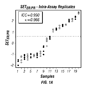
[0031] FIGS. 1A-D: Tests for reproducibility of the SETERNR index in primary
breast cancers: (A) Replicates of the assay procedure. (B) Intra-tumoral
heterogeneity across
three biopsies from each tumor. (C) Inter-sample type comparison between
matched samples
of tissue and scrape cytology samples from each tumor. (D) Inter-platform
comparison of
.. Affymetrix platform U133 A and U133Plus2Ø
[0032] FIGS. 2A-F: Tests for reproducibility of the SETER/pR index in primary
breast cancers: (A) Serial spike-in of RNA from normal liver samples. (B)
Contamination of
breast samples. (C) Duration of extended ex vivo cold ischemic time of samples
before
preservation. (D) Inter-platform comparison. (E) Inter-laboratory comparison.
(F) Intra-assay
validation.
[0033] FIGs. 3A-D: Kaplan-Meier plots of survival according to the SETERNR
index in relapsed metastatic (Stage IV) breast cancer after treatment with
hormonal
therapy: (A) Progression-free survival (PFS) in 79 patients whose next
treatment after tumor
biopsy was hormonal therapy, 7 months difference in median PFS. (B) Overall
survival (OS)
in the same 79 patients whose next treatment after tumor biopsy was hormonal
therapy, 31
months difference in median OS. (C) Progression-free survival (PFS) in the
subset of 46
patients with a clinical history of prior response to hormonal therapy, 11
months difference in
median PFS. (D) Overall survival (OS) in the same subset of 46 patients with a
clinical history
of prior response to hormonal therapy, 30 months difference in median OS.
[0034] FIGS. 4A-D: Tests for reproducibility of the SETERNR index in primary
breast cancers when comparing across assay type and sample type (Quantigene
Hybridization Assay): (A) Measurements from Affymetrix U133A microarray from
fresh
frozen tumor sample compared to measurements from Quantigene customized assay
(Luminex
bead-based hybridization) using matched formalin-fixed and paraffin-embedded
(FFPE)
sample, gray dashed line shows the linear regression line. (B) Comparison of
repeat testing
from FFPE tissue sections on slides, including 2 different technicians, each
performing each
batch of testing on different days, and including 3 batches each, with each
batch containing
different lots of reagents. (C) Validation study of the results from the study
shown in (A), using
different tumors and correcting the SETER/pR index measurements from the
Quantigene method
by applying the equation from the linear regression analysis shown in (A). (D)
Validation study
of the results from the study shown in (B), using different tumors and
correcting the SETER/pR
- 8 -
CA 03022377 2018-10-26
WO 2017/189976
PCT/US2017/030077
index measurements from the Quantigene method by applying the equation from
the linear
regression analysis shown in (A).
[0035] FIGS. 5A-B: Tests for reproducibility of the SETERNR index in primary
breast cancers when comparing across assay type and sample type (Translation
to
Nanostring nCounter Hybridization Assay): Translation of SETERADR from fresh-
frozen (FF)
RNA profiled on Affymetrix U133A microarray to Nanostring N-counter
hybridization
platform. (A) Calibration cohort of primary breast cancers to calibrate
SETER/piz index from
U133A in FF sample to Nanostring platform using FFPE sample; (B) Validation
cohort of
primary breast cancers to test the calibrated SETERADR index using the
Nanostring platform with
FFPE sample.
[0036] FIGS. 6A-D: Tests for reproducibility of the SETERNR index in primary
breast cancers when comparing across assay type and sample type (Translation
to Agilent
44K Microarray): Translation of SETERADR from fresh-frozen (FF) RNA profiled
on
Affymetrix U133A microarray to the Agilent 44K V2 microarray platform. (A,C)
Calibration
cohort of primary breast cancers to calibrate SETERADR index from U133A in FF
sample to
Agilent 44K V2 arrays using FF sample (A) or FFPE sample (C) using linear
regression. (B,D)
Validation cohort of primary breast cancers to test the calibrated SETERADR
index using the
Agilent 44K V2 arrays with FF sample (B) or FFPE sample (D).
[0037] FIGS. 7A-D: Tests for reproducibility of the SETERNR index in primary
breast cancers when comparing across assay type and sample type (Translation
to
RainDance picodroplet targeted RNA Sequencing Assay): Translation of SETERADR
from
fresh-frozen (FF) RNA profiled on Affymetrix U133A microarray to the custom
targeted RNA
sequencing method using RainDance picodroplet-based Illumina MiSeq RNA-Seq
assay (RD).
(A,C) Calibration cohort of primary breast cancers to calibrate SETERADR index
from U133A in
FF sample to RD assay using FP sample (A) or 1-1-PE sample (C) using linear
regression. (B,D)
Validation cohort of primary breast cancers to test the calibrated SETERADR
index using the RD
assay with FF sample (B) or FFPE sample (D).
[0038] FIG. 8: SETERNR index versus Frequency of ESR1 Mutations in Stage IV
(Metastatic) Breast Cancer: Targeted needle biopsies from a metastatic site
were
prospectively obtained from 82 patients with HR+/HER2- breast cancer at the
time of any
progression event. Purified RNA was subjected to targeted RNA sequencing for
the SETER/pR
- 9 -
CA 03022377 2018-10-26
WO 2017/189976
PCT/US2017/030077
index genes using the RainDance (RD) platform. ESR1 LBD mutations were
identified in 17%
(14/82) of metastases (range of mutated transcripts 1%-98%). High frequency
mutations
(>10% of transcripts) were only observed in metastases with higher SETERADR
values (above
the median).
[0039] FIG. 9A-F: Kaplan-Meier plots of survival according to the SETERNR
index
(RD targeted RNA sequencing assay) according to mutation status of ESRI gene
in
relapsed metastatic (Stage IV) breast cancer after treatment with hormonal
therapy: In
patients who next received endocrine therapy (n=58), ESR1 mutation frequency
alone did not
predict a difference in progression-free survival (A) or overall survival (B).
Higher SETERADR
alone, using the RainDance (RD) assay, predicted longer progression-free (C)
and overall
survival (D). The predictions were more pronounced in patients without LBD
mutation (E and
F).
[0040] FIG. 10: SETERNR index versus Frequency of ESRI Mutations in Stage II-
III (Primary) Breast Cancer: Targeted biopsies from the primary HR+/HER2-
breast cancer
were prospectively obtained from 95 patients at the time of initial diagnosis.
Purified RNA was
subjected to targeted RNA sequencing for the SETERADR index genes using the
RainDance (RD)
platform. Rare mutations of the LBD of ESR1 were observed in cancers with
higher SETERADR
index values: in 15% (14/95) of primary cancer samples (range of mutated
transcripts 1%-3%).
[0041] FIGS. 11A-B: Survival according to the SETER/pR index in Stage II and
III
breast cancer after treatment with surgery and neoadjuvant chemotherapy (NAC),
and
prior to treatment with hormonal therapy: The relationship between the
SETER/pa index
and the residual cancer burden (RCB) after completion of NAC in patients with
clinical Stage
II-III HR+/HER2- breast cancer at time of initial diagnosis. (A) The
prognostic model for the
continuous SETERADR index (y-axis) compared to the continuous RCB using
coefficients from
the multivariable model shown in Table 3. (B) Classes of SETERADR index using
pre-defined
cutpoint of 1.85 to distinguish high SETER/pa index (solid lines) versus low
SETER/pa index
(dashed lines) are shown for patients according to the RCB classes of moderate
residual disease
or extensive residual disease. SETERADR index classes were prognostic for
patients with RCB-II
and for patients with RCB-III. Excellent prognosis was observed for RCB-II
with high
SETER/pa index (solid line).
- 10 -
CA 03022377 2018-10-26
WO 2017/189976
PCT/US2017/030077
[0042] FIG. 12: Survival according to the SETERNR index in Stage II and III
breast
cancer after treatment with surgery, and prior to treatment with chemotherapy
and
hormonal therapy: Kaplan-Meier plot of the distant relapse-free survival for
patients with
lymph node-positive HR /HER2- breast cancer (i.e. Stage II or III) shown for
high SETEnipa
index (low-risk), compared to low SETER/pR index (high-risk). This result was
from a blinded
and independent external validation study.
[0043] FIGS. 13A-E: Pre-analytical and analytical datasets. (A) Inter-assay
reproducibility comparing Affymetrix U133A and Plus2.0 microarrays. (B) Inter-
sample type
reproducibility comparing cytology (scrape) and tissue samples. (C) Intra-
assay replicates and
intra-tumoral heterogeneity: tissue samples were taken from three different
macroscopic tumor
areas A, B and C of the same resection specimen to evaluate intra-tumoral
heterogeneity. In a
subset of cases, the laboratory procedure was repeated at 5 different levels.
(D) Influence of
cold ischemic delay and sample type: tissue samples of surgical specimens of
the same tumors
were stored in fixative of were snap frozen with increasing time delay after
surgical removal.
(E) Contamination with liver and normal tissue: tumor samples were mixed at
different ratios
with normal breast tissue or liver tissue to evaluate the effect of
contamination.
[0044] FIGS. 14A-B: (A) Schematic to illustrate the different levels of
overlap
between the datasets. Study A and B share the same case, tissue sample and
array data. Study
III shares the same case with study I and II, but an individual sample was
taken and processed
and profiled individually. (B) Overlap of the different analytical and pre-
analytic dataset with
samples and/or cases of the discovery dataset.
[0045] FIG. 15: Selection of the genes with expression levels correlated most
strongly
and reliably to the expression levels of ESR1 and PGR genes, through a series
of technical and
biological filtering steps.
[0046] FIGS. 16A-B: Distribution of the ESR1- and PGR-associated genes in the
hormone-receptor-positive discovery cohort (A) and the reference genes in the
hormone-
receptor-positive, HER2-negative subset of the discovery cohort (B).
[0047] FIGS. 17A-B: (A) Distribution of the target- and reference genes in the
discovery dataset. The sum of the target genes is plotted against the sum of
the reference genes
to illustrate the difference in variation. (B) Using 175 additional hormone
receptor-negative
- 11-
CA 03022377 2018-10-26
WO 2017/189976
PCT/US2017/030077
cases, the score was scaled linearly to assign negative values to hormone
receptor-negative
tumors.
[0048] FIG. 18: Reproducibility of SETERADR index measurements with FFPE and
Quantigene platform over time and different operators, using different lots of
reagents. FFPE
sections from five different samples were assayed once per week for 20 weeks.
Two different
operators (as indicated by blue and green data points) performed the assay
[0049] FIG. 19: Determination of a quality control threshold for SETER/FR
index
measurements with FFPE and Quantigene platform. Limiting dilutions of RNA
derived from
FFPE tissue (125ng ¨ 1.95ng) were assayed from five primary breast cancers.
The
measurements were compared with the SETERADR index value measured using
fresh/frozen
RNA profiled on U133A microarray. The absolute deviation of the SETERADR index
measurement from the U133A measurement is shown against the median reference
gene value
and a cut off of >4.0 for the median reference gene value was determined to be
optimal for
quality assessment as pass or fail.
[0050] FIGS. 20A-B: (A) SETER/pR according to stage at diagnosis and (B)
according
to the number of the biopsied relapse event in patients with metastatic breast
cancer.
[0051] FIGS. 21A-C: SETER/FR and clinical and pathological tumor
characteristics. (A)
Site of metastatic breast cancer for protocol biopsy, (B) PGR status by
immunohistochemistry
and (C) prior sensitivity to endocrine treatment.
[0052] FIG. 22: Expression levels of transcripts used in the SETER/pR index as
measured using our RD method of targeted RNA sequencing of RNA derived from
plasma
exosomes from peripheral blood sample and from FFPE tumor biopsy of a liver
metastasis
from the same patient.
DESCRIPTION OF ILLUSTRATIVE EMBODIMENTS
[0053] The course of breast cancer therapy usually relies on following a
sequence of
available endocrine treatments (Barrios et al., 2012; Dodwell et al., 2006),
unless a
symptomatic disease burden or more rapidly progressive disease favors a switch
to
chemotherapy (Giordano et al., 2014; Cardoso et al., 2014; Beslij a et al.,
2007). However, the
treatment strategy increasingly requires nuanced clinical judgment as the
selection of treatment
- 12 -
CA 03022377 2018-10-26
WO 2017/189976
PCT/US2017/030077
options continues to expand to include additional endocrine treatments,
chemotherapy
treatments, and other molecular targeted approaches. Accordingly, the present
disclosure
overcomes challenges associated with current technologies by providing an
index of tumoral
sensitivity to endocrine therapy, referred to herein as the SETER/pa index.
[0054] The SETER/pR index is calculated using the expression level of a
combination of
genes related to both the estrogen receptor (ER) gene (ESR1) and the
progesterone receptor
(PR) gene (PGR), such as disclosed in Table 5. In some embodiments, the
SETER/pR index is
used to predict the sensitivity of breast cancer, particularly metastatic
breast cancer, to
endocrine therapy alone or in combination with other therapies. Thus, further
embodiments
include methods of treating breast cancers identified to be sensitive to
endocrine therapy using
the SETER/pR index by administering a endocrine therapy to the patient.
[0055] The SETER/pa index was validated using a prospective cohort of needle
biopsy
samples from metastases of hormone receptor-positive breast cancer (stored in
RNA
preservative then profiled using Affymetrix U133A gene expression arrays) that
were
annotated with clinical, treatment, and survival information. Further
experiments were
performed to estimate the reproducibility of the gene expression measurements
under the
effects of intratumoral heterogeneity, technical repetition, different
microarray platforms, and
different types of tumor biopsies, in order to develop the technically robust
customized assay
provided herein.
I. Definitions
[0056] As used herein, "essentially free," in terms of a specified component,
is used
herein to mean that none of the specified component has been purposefully
formulated into a
composition and/or is present only as a contaminant or in trace amounts. The
total amount of
the specified component resulting from any unintended contamination of a
composition is
therefore well below 0.05%, preferably below 0.01%. Most preferred is a
composition in which
no amount of the specified component can be detected with standard analytical
methods.
[0057] As used herein the specification, "a" or "an" may mean one or more. As
used
herein in the claim(s), when used in conjunction with the word "comprising,"
the words "a" or
"an" may mean one or more than one.
- 13 -
CA 03022377 2018-10-26
WO 2017/189976
PCT/US2017/030077
[0058] The use of the term "or" in the claims is used to mean "and/or" unless
explicitly
indicated to refer to alternatives only or the alternatives are mutually
exclusive, although the
disclosure supports a definition that refers to only alternatives and
"and/or." As used herein
"another" may mean at least a second or more.
[0059] Throughout this application, the term "about" is used to indicate that
a value
includes the inherent variation of error for the device, the method being
employed to determine
the value, or the variation that exists among the study subjects.
[0060] "Treatment" and "treating" refer to administration or application of a
therapeutic agent to a subject or performance of a procedure or modality on a
subject for the
purpose of obtaining a therapeutic benefit of a disease or health-related
condition. For example,
a treatment may include administration of a hormonal therapy.
[0061] "Subject" and "patient" refer to either a human or non-human, such as
primates,
mammals, and vertebrates. In particular embodiments, the subject is a human.
[0062] The term "therapeutic benefit" or "therapeutically effective" as used
throughout
this application refers to anything that promotes or enhances the well-being
of the subject with
respect to the medical treatment of this condition. This includes, but is not
limited to, a
reduction in the frequency or severity of the signs or symptoms of a disease.
For example,
treatment of cancer may involve, for example, a reduction in the size of a
tumor, a reduction in
the invasiveness of a tumor, reduction in the growth rate of the cancer, or
prevention of
metastasis. Treatment of cancer may also refer to prolonging survival of a
subject with cancer.
[0063] "Prognosis" refers to as a prediction of how a patient will progress,
and whether
there is a chance of recovery. "Cancer prognosis" generally refers to a
forecast or prediction of
the probable course or outcome of the cancer. As used herein, cancer prognosis
includes the
forecast or prediction of any one or more of the following: duration of
survival of a patient
susceptible to or diagnosed with a cancer, duration of recurrence-free
survival, duration of
progression-free survival of a patient susceptible to or diagnosed with a
cancer, response rate
in a group of patients susceptible to or diagnosed with a cancer, duration of
response in a patient
or a group of patients susceptible to or diagnosed with a cancer, and/or
likelihood of metastasis
and/or cancer progression in a patient susceptible to or diagnosed with a
cancer. Prognosis also
includes prediction of favorable survival following cancer treatments, such as
a conventional
cancer therapy.
- 14 -
CA 03022377 2018-10-26
WO 2017/189976
PCT/US2017/030077
[0064] An "anti-cancer" agent is capable of negatively affecting a cancer
cell/tumor in
a subject, for example, by promoting killing of cancer cells, inducing
apoptosis in cancer cells,
reducing the growth rate of cancer cells, reducing the incidence or number of
metastases,
reducing tumor size, inhibiting tumor growth, reducing the blood supply to a
tumor or cancer
cells, promoting an immune response against cancer cells or a tumor,
preventing or inhibiting
the progression of cancer, or increasing the lifespan of a subject with
cancer.
[0065] The term "primer," as used herein, is meant to encompass any nucleic
acid that
is capable of priming the synthesis of a nascent nucleic acid in a template-
dependent process.
Typically, primers are oligonucleotides from ten to twenty and/or thirty base
pairs in length,
but longer sequences can be employed. Primers may be provided in double-
stranded and/or
single-stranded form, although the single-stranded form is preferred.
[0066] The term "antibody" herein is used in the broadest sense and
specifically covers
monoclonal antibodies (including full length monoclonal antibodies),
polyclonal antibodies,
multispecific antibodies (e.g., bispecific antibodies), and antibody fragments
so long as they
exhibit the desired biological activity.
[0067] The phrases "pharmaceutical or pharmacologically acceptable" refers to
molecular entities and compositions that do not produce an adverse, allergic,
or other untoward
reaction when administered to an animal, such as a human, as appropriate. The
preparation of
a pharmaceutical composition comprising an antibody or additional active
ingredient will be
known to those of skill in the art in light of the present disclosure.
Moreover, for animal (e.g.,
human) administration, it will be understood that preparations should meet
sterility,
pyrogenicity, general safety, and purity standards as required by FDA Office
of Biological
Standards.
[0068] As used herein, "pharmaceutically acceptable carrier" includes any and
all
aqueous solvents (e.g., water, alcoholic/aqueous solutions, saline solutions,
parenteral vehicles,
such as sodium chloride, Ringer's dextrose, etc.), non-aqueous solvents (e.g.,
propylene glycol,
polyethylene glycol, vegetable oil, and injectable organic esters, such as
ethyloleate),
dispersion media, coatings, surfactants, antioxidants, preservatives (e.g.,
antibacterial or
antifungal agents, anti-oxidants, chelating agents, and inert gases), isotonic
agents, absorption
delaying agents, salts, drugs, drug stabilizers, gels, binders, excipients,
disintegration agents,
lubricants, sweetening agents, flavoring agents, dyes, fluid and nutrient
replenishers, such like
- 15 -
CA 03022377 2018-10-26
WO 2017/189976
PCT/US2017/030077
materials and combinations thereof, as would be known to one of ordinary skill
in the art. The
pH and exact concentration of the various components in a pharmaceutical
composition are
adjusted according to well-known parameters.
[0069] The term "unit dose" or "dosage" refers to physically discrete units
suitable for
use in a subject, each unit containing a predetermined quantity of the
therapeutic composition
calculated to produce the desired responses discussed above in association
with its
administration, i.e., the appropriate route and treatment regimen. The
quantity to be
administered, both according to number of treatments and unit dose, depends on
the effect
desired. The actual dosage amount of a composition of the present embodiments
administered
to a patient or subject can be determined by physical and physiological
factors, such as body
weight, the age, health, and sex of the subject, the type of disease being
treated, the extent of
disease penetration, previous or concurrent therapeutic interventions,
idiopathy of the patient,
the route of administration, and the potency, stability, and toxicity of the
particular therapeutic
substance. For example, a dose may also comprise from about 1 jig/kg/body
weight to about
1000 mg/kg/body weight (this such range includes intervening doses) or more
per
administration, and any range derivable therein. In non-limiting examples of a
derivable range
from the numbers listed herein, a range of about 5 jig/kg/body weight to about
100 mg/kg/body
weight, about 5 jig/kg/body weight to about 500 mg/kg/body weight, etc., can
be administered.
The practitioner responsible for administration will, in any event, determine
the concentration
of active ingredient(s) in a composition and appropriate dose(s) for the
individual subject.
[0070] The term "immune checkpoint" refers to a molecule such as a protein in
the
immune system which provides inhibitory signals to its components in order to
balance
immune reactions. Known immune checkpoint proteins comprise CTLA-4, PD1 and
its ligands
PD-Ll and PD-L2 and in addition LAG-3, BTLA, B7H3, B7H4, TIM3, KIR. The
pathways
involving LAG3, BTLA, B7H3, B7H4, TIM3, and KIR are recognized in the art to
constitute
immune checkpoint pathways similar to the CTLA-4 and PD-1 dependent pathways
(see e.g.
Pardo11, 2012; Mellman et al., 2011).
[0071] An "immune checkpoint inhibitor" refers to any compound inhibiting the
function of an immune checkpoint protein. Inhibition includes reduction of
function and full
blockade. In particular the immune checkpoint protein is a human immune
checkpoint protein.
Thus the immune checkpoint protein inhibitor in particular is an inhibitor of
a human immune
checkpoint protein.
- 16-
CA 03022377 2018-10-26
WO 2017/189976
PCT/US2017/030077
[0072] The terms "hormonal" and "endocrine" therapy or treatment are used
interchangeably herein to refer to an agent which blocks the body's ability to
produce a specific
hormone (e.g., estrogen) or interferes with hormone action.
[0073] The term "determining an expression level" as used herein means the
application of a gene specific reagent such as a probe, primer or antibody
and/or a method to a
sample, for example a sample of the subject and/or a control sample, for
ascertaining or
measuring quantitatively, semi-quantitatively or qualitatively the amount of a
gene or genes,
for example the amount of mRNA. For example, a level of a gene can be
determined by a
number of methods including for example immunoassays including for example
immunohistochemistry, ELISA, Western blot, immunoprecipitation and the like,
where a
biomarker detection agent such as an antibody for example, a labeled antibody,
specifically
binds the biomarker and permits for example relative or absolute ascertaining
of the amount of
polypeptide biomarker, hybridization and PCR protocols where a probe or primer
or primer set
are used to ascertain the amount of nucleic acid biomarker, including for
example probe based
and amplification based methods including for example microarray analysis, RT-
PCR such as
quantitative RT-PCR, serial analysis of gene expression (SAGE), Northern Blot,
digital
molecular barcoding technology, for example Nanostring:nCounterTM Analysis,
and TaqMan
quantitative PCR assays. Other methods of mRNA detection and quantification
can be applied,
such as mRNA in situ hybridization in formalin-fixed, paraffin-embedded (FFPE)
tissue
samples or cells. This technology is currently offered by the QuantiGene
ViewRNA
(Affymetrix), which uses probe sets for each mRNA that bind specifically to an
amplification
system to amplify the hybridization signals; these amplified signals can be
visualized using a
standard fluorescence microscope or imaging system. This system for example
can detect and
measure transcript levels in heterogeneous samples; for example, if a sample
has normal and
tumor cells present in the same tissue section. As mentioned, TaqMan probe-
based gene
expression analysis (PCR-based) can also be used for measuring gene expression
levels in
tissue samples, and for example for measuring mRNA levels in FFPE samples. In
brief,
TaqMan probe-based assays utilize a probe that hybridizes specifically to the
mRNA target.
This probe contains a quencher dye and a reporter dye (fluorescent molecule)
attached to each
end, and fluorescence is emitted only when specific hybridization to the mRNA
target occurs.
During the amplification step, the exonuclease activity of the polymerase
enzyme causes the
quencher and the reporter dyes to be detached from the probe, and fluorescence
emission can
occur. This fluorescence emission is recorded and signals are measured by a
detection system;
- 17 -
CA 03022377 2018-10-26
WO 2017/189976
PCT/US2017/030077
these signal intensities are used to calculate the abundance of a given
transcript (gene
expression) in a sample.
[0074] The term "sample" as used herein includes any biological specimen
obtained
from a patient. Samples include, without limitation, whole blood, plasma,
serum, red blood
cells, white blood cells (e.g., peripheral blood mononuclear cells), ductal
lavage fluid, nipple
aspirate, lymph (e.g., disseminated tumor cells of the lymph node), bone
marrow aspirate,
saliva, urine, stool (i.e., feces), sputum, bronchial lavage fluid, tears,
fine needle aspirate (e.g.,
harvested by fine needle aspiration that is directed to a target, such as a
tumor, or is random
sampling of normal cells, such as periareolar), any other bodily fluid, a
tissue sample (e.g.,
tumor tissue) such as a biopsy of a tumor (e.g., needle biopsy) or a lymph
node (e.g., sentinel
lymph node biopsy), and cellular extracts thereof. In some embodiments, the
sample is whole
blood or a fractional component thereof such as plasma, serum, or a cell
pellet. In some
embodiments, the sample is a formalin fixed paraffin embedded (FFPE) tumor
tissue sample,
e.g., from a solid tumor of the breast.
[0075] A "biopsy" refers to the process of removing a tissue sample for
diagnostic or
prognostic evaluation, and to the tissue specimen itself. Any biopsy technique
known in the art
can be applied to the methods and compositions of the present invention. The
biopsy technique
applied will generally depend on the tissue type to be evaluated and the size
and type of the
tumor (i.e., solid or suspended (i.e., blood or ascites)), among other
factors. Representative
biopsy techniques include excisional biopsy, incisional biopsy, needle biopsy
(e.g., core needle
biopsy, fine-needle aspiration biopsy, etc.), surgical biopsy, and bone marrow
biopsy. Biopsy
techniques are discussed, for example, in Harrison's Principles of Internal
Medicine, Kasper,
et al., eds., 16th ed., 2005, Chapter 70, and throughout Part V. One skilled
in the art will
appreciate that biopsy techniques can be performed to identify cancerous
and/or precancerous
cells in a given tissue sample.
SETERNR Index
[0076] Embodiments of the present disclosure provide an index of tumoral
sensitivity
to endocrine therapy, referred to herein as the SETERADR index. The SETERADR
index is calculated
using the expression level of a combination of genes related to both estrogen
receptor (ER) and
progesterone receptor (PR), such as disclosed in Table 5 including SLC39A6,
STC2, CA12,
ESR1, PDZKl, NPY1R, CD2, MAPT, QDPR, AZGP1, ABAT, ADCY1, CD3D, NAT], MRPS30,
- 18 -
CA 03022377 2018-10-26
WO 2017/189976
PCT/US2017/030077
DNAJC12, SCUBE2, and KCNE4. In some aspects, 5, 6, 7, 8, 9, 10, 11, 12, 13,
14, 15, 16, 17,
or 18 of the genes in Table 5 are used to determine the SETERADR index. The ER-
and PR-related
genes can be normalized to reference genes, such as disclosed in Table 5
including LDHA,
ATP5J2, VDAC2, DARS, UCP2, UBE2Z AK2, WIPF2, APPBP2, and TRIM2. In some
aspects,
2, 3, 4, 5, 6, 7, 8, 9 or 10 of the reference genes disclosed in Table 5 are
used to normalize the
expression of the ER- and PR-related genes.
zi8i T
[0077] In some aspects, the SETER/pR index is calculated as: SETER/pR =
18 -
Z 21 Ri
2, where T, is the expression of the ith of the 18 target genes and R, the
expression of
the jth of the 10 reference genes. A constant is added to optimize the
separation into hormone
10 receptor-positive and negative cases by immunohistochemistry at a score
value of 0.
A. Isolation of RNA
[0078] Aspects of the present disclosure concern the isolation of RNA from a
patient
sample for use in determining the SETERADR index. The patient sample may
blood, saliva, urine,
or a tissue biopsy. The tissue biopsy may be a tumor biopsy that has been
flash-frozen (e.g. in
liquid nitrogen), formalin-fixed and paraffin-embedded (FFPE), and/or
preserved by a RNA
stabilization agent (e.g., RNAlater). In some aspects, isolation is not
necessary, and the assay
directly utilizes RNA from within a homogenate of the tissue sample. In
certain aspects the
homogenate of 1-1-PE tumor sample is enzymatically digested.
[0079] RNA may be isolated using techniques well known to those of skill in
the art.
Methods generally involve lysing the cells with a chaotropic (e.g.,
guanidinium isothiocyanate)
and/or detergent (e.g., N-lauroyl sarcosine) prior to implementing processes
for isolating
particular populations of RNA. Chromatography is a process often used to
separate or isolate
nucleic acids from protein or from other nucleic acids. Such methods can
involve
electrophoresis with a gel matrix, filter columns, coated magnetic beads,
alcohol precipitation,
and/or other chromatography.
B. Expression Assessment
[0080] In certain aspects, methods of the present disclosure concern measuring
expression of ER- and PR-related genes as well as one or more reference genes
in a sample
from a subject with breast cancer. The expression information may be obtained
by testing
- 19 -
CA 03022377 2018-10-26
WO 2017/189976
PCT/US2017/030077
cancer samples by a lab, a technician, a device, or a clinician. In a certain
embodiment, the
differential expression of one or more genes including those of Table 5 may be
measured.
[0081] Expression levels of the genes can be detected using any suitable means
known
in the art. For example, detection of gene expression can be accomplished by
detecting nucleic
acid molecules (such as RNA) using nucleic acid amplification methods (such as
RT-PCR,
droplet-based RT amplification, exon capture of RNA sequence library, next
generation RNA
sequencing), array analysis (such as microarray analysis), or hybridization
methods (such as
ribonuclease protection assay, bead-based assays, or Nanostring . Detection of
gene
expression can also be accomplished using assays that detect the proteins
encoded by the genes,
.. including immunoassays (such as ELISA, Western blot, RIA assay, or protein
arrays).
[0082] The pattern or signature of expression in each cancer sample may then
be used
to generate a cancer prognosis or classification, such as predicting cancer
survival or
recurrence, using the SETER/pR index. The expression of one or more of ER- and
PR-related
genes could be assessed to predict or report prognosis or prescribe treatment
options for cancer
patients, especially breast cancer patients.
[0083] The expression of one or more ER- and PR-related genes may be measured
by
a variety of techniques that are well known in the art. Quantifying the levels
of the messenger
RNA (mRNA) of a gene may be used to measure the expression of the gene.
Alternatively,
quantifying the levels of the protein product of ER- and PR-related genes may
be to measure
the expression of the genes. Additional information regarding the methods
discussed below
may be found in Ausubel et al., (2003) Current Protocols in Molecular Biology,
John Wiley
& Sons, New York, NY, or Sambrook et al. (1989) Molecular Cloning: A
Laboratory
Manual, Cold Spring Harbor Press, Cold Spring Harbor, NY. One skilled in the
art will know
which parameters may be manipulated to optimize detection of the mRNA or
protein of
interest.
[0084] A nucleic acid microarray may be used to quantify the differential
expression
of a plurality of ER- and PR-related genes. Microarray analysis may be
performed using
commercially available equipment, following manufacturer's protocols, such as
by using the
Affymetrix GeneChip technology (Santa Clara, CA) or the Microarray System
from lncyte
(Fremont, CA). Typically, single-stranded nucleic acids (e.g., cDNAs or
oligonucleotides) are
plated, or arrayed, on a microchip substrate. The arrayed sequences are then
hybridized with
- 20 -
CA 03022377 2018-10-26
WO 2017/189976
PCT/US2017/030077
specific nucleic acid probes from the cells of interest. Fluorescently labeled
cDNA probes may
be generated through incorporation of fluorescently labeled deoxynucleotides
by reverse
transcription of RNA extracted from the cells of interest. Alternatively, the
RNA may be
amplified by in vitro transcription and labeled with a marker, such as biotin.
The labeled probes
are then hybridized to the immobilized nucleic acids on the microchip under
highly stringent
conditions. After stringent washing to remove the non-specifically bound
probes, the chip is
scanned by confocal laser microscopy or by another detection method, such as a
CCD camera.
The raw fluorescence intensity data in the hybridization files are generally
preprocessed with
a robust statistical normalization algorithm to generate expression values.
[0085] Quantitative real-time PCR (qRT-PCR) may also be used to measure the
differential expression of a plurality of ER- and PR-related genes. In qRT-
PCR, the RNA
template is generally reverse transcribed into cDNA, which is then amplified
via a PCR
reaction. The amount of PCR product is followed cycle-by-cycle in real time,
which allows for
determination of the initial concentrations of mRNA. To measure the amount of
PCR product,
the reaction may be performed in the presence of a fluorescent dye, such as
SYBR Green,
which binds to double-stranded DNA. The reaction may also be performed with a
fluorescent
reporter probe that is specific for the DNA being amplified.
[0086] For example, extracted RNA can be reverse-transcribed using a GeneAmp
RNA PCR kit (Perkin Elmer, Calif., USA), following the manufacturer's
instructions. In some
embodiments, gene expression levels can be determined using a gene expression
analysis
technology that measure mRNA in solution. Methods of detecting gene expression
are
described for example in U.S. Patent Application Nos. U520140357660, and
U520130259858;
incorporated herein by reference. Examples of such gene expression analysis
technologies
include, but not limited to RNAscopeTM, RT-PCR, Nanostring , QuantiGene , gNPA
,
HTG , microarray, and sequencing. For example, methods of Nanostring use
labeled reporter
molecules, referred to as labeled "nanoreporters," that are capable of binding
individual target
molecules. Through the nanoreporters label codes, the binding of the
nanoreporters to target
molecules results in the identification of the target molecules. Methods of
Nanostring are
described in U.S. Pat. No. 7,473,767 (see also, Geiss et al., 2008). Methods
may include the
RainDance droplet amplification method such as described in U.S. Patent No.
8,535,889,
incorporated herein by reference. Sequencing may include exon capture, such as
Illumina
targeted sequencing after the generation of a tagged library for next
generation sequencing (e.g.
- 21 -
CA 03022377 2018-10-26
WO 2017/189976
PCT/US2017/030077
described in International Patent Application No. W02013131962, incorporated
herein by
reference).
[0087] A non-limiting example of a fluorescent reporter probe is a TaqMan
probe
(Applied Biosystems, Foster City, CA). The fluorescent reporter probe
fluoresces when the
quencher is removed during the PCR extension cycle. Multiplex qRT-PCR may be
performed
by using multiple gene-specific reporter probes, each of which contains a
different fluorophore.
Fluorescence values are recorded during each cycle and represent the amount of
product
amplified to that point in the amplification reaction. To minimize errors and
reduce any sample-
to-sample variation, qRT-PCR is typically performed using a reference
standard. The ideal
reference standard is expressed at a constant level among different tissues,
and is unaffected
by the experimental treatment. The system can include a thermocycler, laser,
charge-coupled
device (CCD) camera, and computer. The system amplifies samples in a 96-well
format on a
thermocycler. During amplification, laser-induced fluorescent signal is
collected in real-time
through fiber optics cables for all 96 wells, and detected at the CCD. The
system includes
software for running the instrument and for analyzing the data.
[0088] To minimize errors and the effect of sample-to-sample variation, RT-PCR
can
be performed using an internal standard. The ideal internal standard is
expressed at a constant
level among different tissues, and is unaffected by an experimental treatment.
RNAs commonly
used to normalize patterns of gene expression are mRNAs for the housekeeping
genes GAPDH,
(3-actin, and 18S ribosomal RNA.
[0089] A variation of RT-PCR is real time quantitative RT-PCR, which measures
PCR
product accumulation through a dual-labeled fluorogenic probe (e.g., TAQMAN
probe). Real
time PCR is compatible both with quantitative competitive PCR, where internal
competitor for
each target sequence is used for normalization, and with quantitative
comparative PCR using
a normalization gene contained within the sample, or a housekeeping gene for
RT-PCR (see
Heid et al., 1996). Quantitative PCR is also described in U.S. Pat. No.
5,538,848. Related
probes and quantitative amplification procedures are described in U.S. Pat.
No. 5,716,784 and
U.S. Pat. No. 5,723,591. Instruments for carrying out quantitative PCR in
microtiter plates are
available from PE Applied Biosystems (Foster City, CA).
[0090] The steps of a representative protocol for quantitating gene expression
level
using fixed, paraffin- embedded tissues as the RNA source, including mRNA
isolation,
- 22 -
CA 03022377 2018-10-26
WO 2017/189976
PCT/US2017/030077
purification, primer extension and amplification are given in various
published journal articles
(see Godfrey et al., 2000; Specht et al., 2001). Briefly, a representative
process starts with
cutting about 10 ptri thick sections of paraffin-embedded neoplasm tissue
samples or adjacent
non-cancerous tissue. The RNA is then extracted, and protein and DNA are
removed.
Alternatively, RNA is isolated directly from a neoplasm sample or other tissue
sample. After
analysis of the RNA concentration, RNA repair and/or amplification steps can
be included, if
necessary, and RNA is reverse transcribed using gene specific primers,
followed by preparation
of a tagged RNA sequencing library, and paired-end sequencing. In another
example, the RNA
is not reverse transcribed, but is directly hybridized to a specific template
and then labeled with
oligonucleotides and/or chemical or fluorescent color to be detected and
counted by a laser.
[0091] Immunohistochemical staining may also be used to measure the
differential
expression of a plurality of ER- and PR-related genes. This method enables the
localization of
a protein in the cells of a tissue section by interaction of the protein with
a specific antibody.
For this, the tissue may be fixed in formaldehyde or another suitable
fixative, embedded in wax
.. or plastic, and cut into thin sections (from about 0.1 mm to several mm
thick) using a
microtome. Alternatively, the tissue may be frozen and cut into thin sections
using a cryostat.
The sections of tissue may be arrayed onto and affixed to a solid surface
(i.e., a tissue
microarray). The sections of tissue are incubated with a primary antibody
against the antigen
of interest, followed by washes to remove the unbound antibodies. The primary
antibody may
be coupled to a detection system, or the primary antibody may be detected with
a secondary
antibody that is coupled to a detection system. The detection system may be a
fluorophore or
it may be an enzyme, such as horseradish peroxidase or alkaline phosphatase,
which can
convert a substrate into a colorimetric, fluorescent, or chemiluminescent
product. The stained
tissue sections are generally scanned under a microscope. Because a sample of
tissue from a
subject with cancer may be heterogeneous, i.e., some cells may be normal and
other cells may
be cancerous, the percentage of positively stained cells in the tissue may be
determined. This
measurement, along with a quantification of the intensity of staining, may be
used to generate
an expression value for the biomarker.
[0092] An enzyme-linked immunosorbent assay, or ELISA, may be used to measure
the differential expression of a plurality of ER- and PR-related genes. There
are many
variations of an ELISA assay. All are based on the immobilization of an
antigen or antibody
on a solid surface, generally a microtiter plate. The original ELISA method
comprises
- 23 -
CA 03022377 2018-10-26
WO 2017/189976
PCT/US2017/030077
preparing a sample containing the biomarker proteins of interest, coating the
wells of a
microtiter plate with the sample, incubating each well with a primary antibody
that recognizes
a specific antigen, washing away the unbound antibody, and then detecting the
antibody-
antigen complexes. The antibody-antibody complexes may be detected directly.
For this, the
primary antibodies are conjugated to a detection system, such as an enzyme
that produces a
detectable product. The antibody-antibody complexes may be detected
indirectly. For this, the
primary antibody is detected by a secondary antibody that is conjugated to a
detection system,
as described above. The microtiter plate is then scanned and the raw intensity
data may be
converted into expression values using means known in the art.
[0093] An antibody microarray may also be used to measure the differential
expression
of a plurality of ER- and PR-related genes. For this, a plurality of
antibodies is arrayed and
covalently attached to the surface of the microarray or biochip. A protein
extract containing the
biomarker proteins of interest is generally labeled with a fluorescent dye.
[0094] The labeled ER- and PR-related genes proteins may be incubated with the
antibody microarray. After washes to remove the unbound proteins, the
microarray is scanned.
The raw fluorescent intensity data may be converted into expression values
using means known
in the art.
[0095] Luminex multiplexing microspheres may also be used to measure the
differential expression of a plurality of biomarkers. These microscopic
polystyrene beads are
internally color-coded with fluorescent dyes, such that each bead has a unique
spectral
signature (of which there are up to 100). Beads with the same signature are
tagged with a
specific oligonucleotide or specific antibody that will bind the target of
interest (i.e., biomarker
mRNA or protein, respectively). The target, in turn, is also tagged with a
fluorescent reporter.
Hence, there are two sources of color, one from the bead and the other from
the reporter
molecule on the target. The beads are then incubated with the sample
containing the targets, of
which up 100 may be detected in one well. The small size/surface area of the
beads and the
three dimensional exposure of the beads to the targets allows for nearly
solution-phase kinetics
during the binding reaction. The captured targets are detected by high-tech
fluidics based upon
flow cytometry in which lasers excite the internal dyes that identify each
bead and also any
reporter dye captured during the assay. The data from the acquisition files
may be converted
into expression values using means known in the art.
- 24 -
CA 03022377 2018-10-26
WO 2017/189976
PCT/US2017/030077
[0096] In situ hybridization may also be used to measure the differential
expression of
a plurality of biomarkers. This method permits the localization of mRNAs of
interest in the
cells of a tissue section. For this method, the tissue may be frozen, or fixed
and embedded, and
then cut into thin sections, which are arrayed and affixed on a solid surface.
The tissue sections
are incubated with a labeled antisense probe that will hybridize with an mRNA
of interest. The
hybridization and washing steps are generally performed under highly stringent
conditions.
The probe may be labeled with a fluorophore or a small tag (such as biotin or
digoxigenin) that
may be detected by another protein or antibody, such that the labeled hybrid
may be detected
and visualized under a microscope. Multiple mRNAs may be detected
simultaneously,
provided each antisense probe has a distinguishable label. The hybridized
tissue array is
generally scanned under a microscope. Because a sample of tissue from a
subject with cancer
may be heterogeneous, i.e., some cells may be normal and other cells may be
cancerous, the
percentage of positively stained cells in the tissue may be determined. This
measurement, along
with a quantification of the intensity of staining, may be used to generate an
expression value
for each biomarker.
C. ESRI Mutations
[0097] Activating mutations in the estrogen receptor gene, ESR1, are a key
mechanism
in acquired endocrine resistance in breast cancer therapy. Accordingly, some
aspects of the
present invention further refine the SETER/pR index by including variables for
the expression of
mutated ESR1. The presence of transcript expressing a mutated form of ESR1 is
detected by
specific primers that amplify a specific part of the ligand-binding domain
sequence of ESR1
transcript that is known to be a region that is enriched for activating
mutations. The proportion
of the transcript expressing a mutated form of ESR1 is calculated as the
expression of mutated
ESR1 over the expression of ESR1 measured using different primers that detect
a region of the
ESR1 transcript that is reliably expressed in samples and is not prone to
mutation. In one
example, the mutation status is incorporated logistically with SET index
status (yes/no
combined with high/low). In another example, the mutation status of the
transcript, the
proportion of ESR1 transcript that is mutated, and the SET index value are
incorporated into a
multivariable index score, where the coefficients of the score are based on
multivariable Cox
regression model of prognosis following endocrine therapy.
[0098] Mutations of ESR1 are known in the art. For example, five ESR1
mutations
identified encoding p.Leu536G1n, p.Tyr537Ser, p.Tyr537Cys, p.Tyr537Asn and
p.Asp538Gly
- 25 -
CA 03022377 2018-10-26
WO 2017/189976
PCT/US2017/030077
were shown to result in constitutive activity and continued responsiveness to
anti-estrogen
therapies in vitro (Robinson et al., 2013). Other ESR1 mutations include
S463P, V534E,
P535H, L536Q, L536R, Y537C, Y537S, Y537N, and D538G.
III. Methods of Treatment
[0099] Provided herein are methods for treating or delaying progression of
breast
cancer in an individual determined to be sensitive to endocrine therapy using
the SETEnipa
index comprising administering to the individual an effective amount of a
hormonal therapy.
The breast cancer may be Stage II, Stage III, or Stage IV breast cancer and,
in particular aspects,
the Stage IV breast cancer is metastatic and relapsed after prior treatments.
In certain aspects,
the breast cancer is hormone receptor-positive (i.e., positive for the
receptors for the hormones
estrogen (ER-positive cancers) and/or progesterone (PR-positive cancers)
and/or HER2-
negative.
[00100]
Exemplary hormonal therapies for breast cancer include the SERM, Al,
and SERD classes of drugs that inhibit the activity of the estrogen and
estrogen-receptor
complex, such as tamoxifen, toremifene, and fulvestrant. Other hormonal
therapies include
treatments to lower estrogen levels including aromatase inhibitors such as
letrozole,
anastrozole, and exemestane. Permanent ovarian ablation can be done by
surgically removing
the ovaries. This operation is called an oophorectomy. More often, ovarian
ablation is done
with drugs called luteinizing hormone-releasing hormone (LHRH) analogs, such
as goserelin
(Zoladex ) or leuprolide (Lupron ). These drugs stop the signal that the body
sends to ovaries
to make estrogens. They can be used alone or with other hormone drugs
(tamoxifen, aromatase
inhibitors, fulvestrant) as hormone therapy in pre-menopausal women. The
effectiveness of
hormonal therapy may also be enhanced by the addition of an additional therapy
to
synergistically inhibit a different biological pathway, such as palbociclib
(Cdk4/6 inhibitor),
everolimus (mTOR/PI3K inhibitor), immune therapy, or other therapies.
[00101] In
some embodiments, the individual has cancer that is resistant (has
been demonstrated to be resistant) to one or more anti-cancer therapies. In
some embodiments,
resistance to anti-cancer therapy includes recurrence of cancer or refractory
cancer. Recurrence
may refer to the reappearance of cancer, in the original site or a new site,
after treatment. In
some embodiments, resistance to anti-cancer therapy includes progression of
the cancer during
- 26 -
CA 03022377 2018-10-26
WO 2017/189976
PCT/US2017/030077
treatment with the anti-cancer therapy. In some embodiments, the cancer is at
early stage or at
late stage.
[00102] In
some aspects, the patient has been previously administered a
hormonal therapy and/or additional anti-cancer therapy. For example, the
patient may have
been administered a hormonal therapy in combination with chemotherapy, such as
for five
years. In some aspects, the patients has shown previous sensitivity to a
hormonal therapy.
[00103] In
some aspects, the hormonal therapy is administered in combination
with at least one additional anti-cancer therapy. The hormonal therapy may be
administered
before, during, after, or in various combinations relative to the additional
anti-cancer agent.
The administrations may be in intervals ranging from concurrently to minutes
to days to weeks.
In embodiments where the hormonal therapy is provided to a patient separately
from an anti-
cancer agent, one would generally ensure that a significant period of time did
not expire
between the time of each delivery, such that the two compounds would still be
able to exert an
advantageously combined effect on the patient. In such instances, it is
contemplated that one
may provide a patient with the hormonal therapy and the anti-cancer therapy
within about 12
to 24 or 72 h of each other and, more particularly, within about 6-12 h of
each other. In some
situations it may be desirable to extend the time period for treatment
significantly where several
days (2, 3, 4, 5, 6, or 7) to several weeks (1, 2, 3, 4, 5, 6, 7, or 8) lapse
between respective
administrations.
[00104] The hormonal
therapy and, optionally the anti-cancer agent, may be
administered by the same route of administration or by different routes of
administration. In
some embodiments, the hormonal therapy and/or anti-cancer agent is
administered
intravenously, intramuscularly, subcutaneously, topically, orally,
transdermally,
intraperitoneally, intraorbitally, by implantation, by inhalation,
intrathecally,
intraventricularly, or intranasally. An effective amount of the hormonal
therapy and/or anti-
cancer agent may be administered for prevention or treatment of disease. The
appropriate
dosage of the hormonal therapy and anti-cancer agent be determined based on
the type of
disease to be treated, severity and course of the disease, the clinical
condition of the individual,
the individual's clinical history and response to the treatment, and the
discretion of the attending
physician.
- 27 -
CA 03022377 2018-10-26
WO 2017/189976
PCT/US2017/030077
[00105]
Intratumoral injection, or injection into the tumor vasculature is
specifically contemplated for discrete, solid, accessible tumors. Local,
regional or systemic
administration also may be appropriate. For tumors of >4 cm, the volume to be
administered
will be about 4-10 ml (in particular 10 ml), while for tumors of <4 cm, a
volume of about 1-3
ml will be used (in particular 3 m1). Multiple injections delivered as single
dose comprise about
0.1 to about 0.5 ml volumes.
A. Pharmaceutical Compositions
[00106]
Also provided herein are pharmaceutical compositions and formulations
comprising the hormonal therapy, optionally an anti-cancer agent and a
pharmaceutically
acceptable carrier.
[00107]
Pharmaceutical compositions and formulations as described herein can
be prepared by mixing the active ingredients (such as an antibody or a
polypeptide) having the
desired degree of purity with one or more optional pharmaceutically acceptable
carriers
(Remington's Pharmaceutical Sciences 22nd edition, 2012), in the form of
lyophilized
formulations or aqueous solutions. Pharmaceutically acceptable carriers are
generally nontoxic
to recipients at the dosages and concentrations employed, and include, but are
not limited to:
buffers such as phosphate, citrate, and other organic acids; antioxidants
including ascorbic acid
and methionine; preservatives (such as octadecyldimethylbenzyl ammonium
chloride;
hexamethonium chloride; benzalkonium chloride; benzethonium chloride; phenol,
butyl or
benzyl alcohol; alkyl parabens such as methyl or propyl paraben; catechol;
resorcinol;
cyclohexanol; 3-pentanol; and m-cresol); low molecular weight (less than about
10 residues)
polypeptides; proteins, such as serum albumin, gelatin, or immunoglobulins;
hydrophilic
polymers such as polyvinylpyrrolidone; amino acids such as glycine, glutamine,
asparagine,
histidine, arginine, or lysine; monosaccharides, disaccharides, and other
carbohydrates
including glucose, mannose, or dextrins; chelating agents such as EDTA; sugars
such as
sucrose, mannitol, trehalose or sorbitol; salt-forming counter-ions such as
sodium; metal
complexes (e.g. Zn- protein complexes); and/or non-ionic surfactants such as
polyethylene
glycol (PEG). Exemplary pharmaceutically acceptable carriers herein further
include
insterstitial drug dispersion agents such as soluble neutral-active
hyaluronidase glycoproteins
(sHASEGP), for example, human soluble PH-20 hyaluronidase glycoproteins, such
as
rHuPH20 (HYLENEX , Baxter International, Inc.). Certain exemplary sHASEGPs and
methods of use, including rHuPH20, are described in US Patent Publication Nos.
- 28 -
CA 03022377 2018-10-26
WO 2017/189976
PCT/US2017/030077
2005/0260186 and 2006/0104968. In one aspect, a sHASEGP is combined with one
or more
additional glycosaminoglycanases such as chondroitinases.
B. Anti-Cancer Therapy
[00108] In
certain embodiments, the compositions and methods of the present
embodiments involve hormonal therapy in sequence or combination with at least
additional
anti-cancer agent. The additional therapy may be radiation therapy, surgery
(e.g., lumpectomy
and a mastectomy), chemotherapy, targeted molecular inhibitor, gene therapy,
DNA therapy,
viral therapy, RNA therapy, immunotherapy, bone marrow transplantation,
nanotherapy,
monoclonal antibody therapy, or a combination of the foregoing. The additional
therapy may
be in the form of adjuvant, neoadjuvant, or palliative therapy.
[00109] In
some embodiments, the additional therapy is the administration of
small molecule enzymatic inhibitor or anti-metastatic agent. In some
embodiments, the
additional therapy is the administration of side- effect limiting agents
(e.g., agents intended to
lessen the occurrence and/or severity of side effects of treatment, such as
anti-nausea agents,
etc.). In some embodiments, the additional therapy is radiation therapy. In
some embodiments,
the additional therapy is surgery. In some embodiments, the additional therapy
is a combination
of radiation therapy and surgery. In some embodiments, the additional therapy
is gamma
irradiation. In some embodiments, the additional therapy is therapy targeting
receptor or
receptor kinase signaling molecules, cyclin-dependent kinases or the cell
cycle control,
mTOR/PI3K pathway, HSP90 inhibitor, tubulin inhibitor, apoptosis inhibitor,
and/or
chemopreventative agent. The additional therapy may be one or more of the
chemotherapeutic
agents known in the art.
[00110]
Various combinations may be employed. For the example below a
hormonal therapy is "A" and an anti-cancer therapy is "B":
A/B/A B/A/B B/B/A A/A/B A/B/B B/A/A A/B/B/B B/A/B/B
B/B/B/A B/B/A/B A/A/B/B A/B/A/B A/B/B/A B/B/A/A
B/A/B/A B/A/A/B A/A/A/B B/A/A/A A/B/A/A A/A/B/A
[00111]
Administration of any compound or therapy of the present embodiments
to a patient will follow general protocols for the administration of such
compounds, taking into
account the toxicity, if any, of the agents. Therefore, in some embodiments
there is a step of
monitoring toxicity that is attributable to combination therapy.
- 29 -
CA 03022377 2018-10-26
WO 2017/189976
PCT/US2017/030077
1. Chemotherapy
[00112] A
wide variety of chemotherapeutic agents may be used in accordance
with the present embodiments. The term "chemotherapy" refers to the use of
drugs to treat
cancer. A "chemotherapeutic agent" is used to connote a compound or
composition that is
administered in the treatment of cancer. These agents or drugs are categorized
by their mode
of activity within a cell, for example, whether and at what stage they affect
the cell cycle.
Alternatively, an agent may be characterized based on its ability to directly
cross-link DNA, to
intercalate into DNA, or to induce chromosomal and mitotic aberrations by
affecting nucleic
acid synthesis.
[00113] Examples of
chemotherapeutic agents include alkylating agents, such as
thiotepa and cyclosphosphamide; alkyl sulfonates, such as busulfan,
improsulfan, and
piposulfan; aziridines, such as benzodopa, carboquone, meturedopa, and
uredopa;
ethylenimines and methylamelamines, including altretamine,
triethylenemelamine,
trietylenephosphoramide, triethiylenethiophosphoramide, and
trimethylolomelamine;
acetogenins (especially bullatacin and bullatacinone); a camptothecin
(including the synthetic
analogue topotecan); bryostatin; callystatin; CC-1065 (including its
adozelesin, carzelesin and
bizelesin synthetic analogues); cryptophycins (particularly cryptophycin 1 and
cryptophycin
8); dolastatin; duocarmycin (including the synthetic analogues, KW-2189 and
CB1-TM1);
eleutherobin; pancratistatin; a sarcodictyin; spongistatin; nitrogen mustards,
such as
chlorambucil, chlomaphazine, cholophosphamide, e stramus tine,
ifosfamide,
mechlorethamine, mechlorethamine oxide hydrochloride, melphalan, novembichin,
phenesterine, prednimustine, trofosfamide, and uracil mustard; nitrosureas,
such as carmustine,
chlorozotocin, fotemustine, lomustine, nimustine, and ranimnustine;
antibiotics, such as the
enediyne antibiotics (e.g., calicheamicin, especially calicheamicin gammalI
and calicheamicin
omegaIl); dynemicin, including dynemicin A; bisphosphonates, such as
clodronate; an
esperamicin; as well as neocarzinostatin chromophore and related chromoprotein
enediyne
antiobiotic chromophores, aclacinomysins, actinomycin, authrarnycin,
azaserine, bleomycins,
cactinomycin, carabicin, carminomycin, carzinophilin, chromomycinis,
dactinomycin,
daunorubicin, detorubicin, 6-diazo-5-oxo-L-norleucine, doxorubicin (including
morpholino-
doxorubicin, cyanomorpholino-doxorubicin, 2-pyrrolino-doxorubicin and
deoxydoxorubicin),
epirubicin, esorubicin, idarubicin, marcellomycin, mitomycins, such as
mitomycin C,
mycophenolic acid, nogalarnycin, olivomycins, peplomycin, potfiromycin,
puromycin,
quelamycin, rodorubicin, streptonigrin, streptozocin, tubercidin, ubenimex,
zinostatin, and
- 30 -
CA 03022377 2018-10-26
WO 2017/189976
PCT/US2017/030077
zorubicin; anti-metabolites, such as methotrexate and 5-fluorouracil (5-FU);
folic acid
analogues, such as denopterin, pteropterin, and trimetrexate; purine analogs,
such as
fludarabine, 6-mercaptopurine, thiamiprine, and thioguanine; pyrimidine
analogs, such as
ancitabine, azacitidine, 6-azauridine, carmofur, cytarabine, dideoxyuridine,
doxifluridine,
enocitabine, and floxuridine; androgens, such as calusterone, dromostanolone
propionate,
epitiostanol, mepitiostane, and testolactone; anti-adrenals, such as mitotane
and trilostane; folic
acid replenisher, such as frolinic acid; aceglatone; aldophosphamide
glycoside; aminolevulinic
acid; eniluracil; amsacrine; bestrabucil; bisantrene; edatraxate; defofamine;
demecolcine;
diaziquone; elformithine; elliptinium acetate; an epothilone; etoglucid;
gallium nitrate;
hydroxyurea; lentinan; lonidainine; maytansinoids, such as maytansine and
ansamitocins;
mitoguazone; mitoxantrone; mopidanmol; nitraerine; pentostatin; phenamet;
pirarubicin;
losoxantrone; podophyllinic acid; 2-ethylhydrazide; procarbazine;
PSKpolysaccharide
complex; razoxane; rhizoxin; sizofiran; spirogermanium; tenuazonic acid;
triaziquone; 2,2',2"-
trichlorotriethylamine; trichothecenes (especially T-2 toxin, verracurin A,
roridin A and
anguidine); urethan; vindesine; dacarbazine; mannomustine; mitobronitol;
mitolactol;
pipobroman; gacytosine; arabinoside ("Ara-C"); cyclophosphamide; taxoids,
e.g., paclitaxel
and docetaxel gemcitabine; 6-thioguanine; mercaptopurine; platinum
coordination complexes,
such as cisplatin, oxaliplatin, and carboplatin; vinblastine; platinum;
etoposide (VP-16);
ifosfamide; mitoxantrone; vincristine; vinorelbine; novantrone; teniposide;
edatrexate;
daunomycin; aminopterin; xeloda; ibandronate; irinotecan (e.g., CPT-11);
topoisomerase
inhibitor RFS 2000; difluorometlhylomithine (DMF0); retinoids, such as
retinoic acid;
capecitabine; carboplatin, procarbazine,plicomycin, gemcitabien, navelbine,
farnesyl-protein
tansferase inhibitors, transplatinum, and pharmaceutically acceptable salts,
acids, or
derivatives of any of the above,
2. Radiotherapy
[00114]
Other factors that cause DNA damage and have been used extensively
include what are commonly known as y-rays, X-rays, and/or the directed
delivery of
radioisotopes to tumor cells. Other forms of DNA damaging factors are also
contemplated,
such as microwaves, proton beam irradiation (U.S. Patents 5,760,395 and
4,870,287), and UV-
irradiation. It is most likely that all of these factors affect a broad range
of damage on DNA,
on the precursors of DNA, on the replication and repair of DNA, and on the
assembly and
maintenance of chromosomes. Dosage ranges for X-rays range from daily doses of
50 to 200
roentgens for prolonged periods of time (3 to 4 wk), to single doses of 2000
to 6000 roentgens.
-31 -
CA 03022377 2018-10-26
WO 2017/189976
PCT/US2017/030077
Dosage ranges for radioisotopes vary widely, and depend on the half-life of
the isotope, the
strength and type of radiation emitted, and the uptake by the neoplastic
cells.
3. Immunotherapy
[00115] The
skilled artisan will understand that additional immunotherapies may
be used in combination or in conjunction with methods of the embodiments. In
the context of
cancer treatment, immunotherapeutics, generally, rely on the use of immune
effector cells and
molecules to target and destroy cancer cells. Rituximab (RITUXANCI) is such an
example.
The immune effector may be, for example, an antibody specific for some marker
on the surface
of a tumor cell. The antibody alone may serve as an effector of therapy or it
may recruit other
cells to actually affect cell killing. The antibody also may be conjugated to
a drug or toxin
(chemotherapeutic, radionuclide, ricin A chain, cholera toxin, pertussis
toxin, etc.) and serve
as a targeting agent. Alternatively, the effector may be a lymphocyte carrying
a surface
molecule that interacts, either directly or indirectly, with a tumor cell
target. Various effector
cells include cytotoxic T cells and NK cells
[00116] Antibody-drug
conjugates have emerged as a breakthrough approach to
the development of cancer therapeutics. Cancer is one of the leading causes of
deaths in the
world. Antibody¨drug conjugates (ADCs) comprise monoclonal antibodies (MAbs)
that are
covalently linked to cell-killing drugs. This approach combines the high
specificity of MAbs
against their antigen targets with highly potent cytotoxic drugs, resulting in
"armed" MAbs that
deliver the payload (drug) to tumor cells with enriched levels of the antigen
(Carter et al., 2008;
Teicher 2014; Leal et al., 2014). Targeted delivery of the drug also minimizes
its exposure in
normal tissues, resulting in decreased toxicity and improved therapeutic
index. The approval
of two ADC drugs, ADCETRIS (brentuximab vedotin) in 2011 and KADCYLA
(trastuzumab emtansine or T-DM1) in 2013 by FDA validated the approach. There
are
currently more than 30 ADC drug candidates in various stages of clinical
trials for cancer
treatment (Leal et al., 2014). As antibody engineering and linker-payload
optimization are
becoming more and more mature, the discovery and development of new ADCs are
increasingly dependent on the identification and validation of new targets
that are suitable to
this approach (Teicher 2009) and the generation of targeting MAbs. Two
criteria for ADC
targets are upregulated/high levels of expression in tumor cells and robust
internalization.
[00117] In
one aspect of immunotherapy, the tumor cell must bear some marker
that is amenable to targeting, i.e., is not present on the majority of other
cells. Many tumor
- 32 -
CA 03022377 2018-10-26
WO 2017/189976
PCT/US2017/030077
markers exist and any of these may be suitable for targeting in the context of
the present
embodiments. Common tumor markers include CD20, carcinoembryonic antigen,
tyrosinase
(p97), gp68, TAG-72, HMFG, Sialyl Lewis Antigen, MucA, MucB, PLAP, laminin
receptor,
erb B, and p155. An alternative aspect of immunotherapy is to combine
anticancer effects with
immune stimulatory effects. Immune stimulating molecules also exist including:
cytokines,
such as IL-2, IL-4, IL-12, GM-CSF, gamma-IFN, chemokines, such as MIP-1, MCP-
1, IL-8,
and growth factors, such as FLT3 ligand.
[00118]
Examples of immunotherapies currently under investigation or in use
are immune adjuvants, e.g., Mycobacterium bovis, Plasmodium falciparum,
dinitrochlorobenzene, and aromatic compounds (U.S. Patents 5,801,005 and
5,739,169; Hui
and Hashimoto, 1998; Christodoulides et al., 1998); cytokine therapy, e.g.,
interferons oc, 13,
and y, IL-1, GM-CSF, and TNF (Bukowski et al., 1998; Davidson et al., 1998;
Hellstrand et
al., 1998); gene therapy, e.g., TNF, IL-1, IL-2, and p53 (Qin et al., 1998;
Austin-Ward and
Villaseca, 1998; U.S. Patents 5,830,880 and 5,846,945); and monoclonal
antibodies, e.g., anti-
CD20, anti-ganglioside GM2, and anti-p185 (Hollander, 2012; Hanibuchi et al.,
1998; U.S.
Patent 5,824,311). It is contemplated that one or more anti-cancer therapies
may be employed
with the antibody therapies described herein.
[00119] In
some embodiments, the immunotherapy may be an immune
checkpoint inhibitor. Immune checkpoints are molecules in the immune system
that either turn
up a signal (e.g., co-stimulatory molecules) or turn down a signal. Inhibitory
checkpoint
molecules that may be targeted by immune checkpoint blockade include adenosine
A2A
receptor (A2AR), B7-H3 (also known as CD276), B and T lymphocyte attenuator
(BTLA),
cytotoxic T-lymphocyte-associated protein 4 (CTLA-4, also known as CD152),
indoleamine
2,3-dioxygenase (IDO), killer-cell immunoglobulin (KIR), lymphocyte activation
gene-3
(LAG3), programmed death 1 (PD-1), T-cell immunoglobulin domain and mucin
domain 3
(TIM-3) and V-domain Ig suppressor of T cell activation (VISTA). In
particular, the immune
checkpoint inhibitors target the PD-1 axis and/or CTLA-4.
[00120] The
immune checkpoint inhibitors may be drugs such as small
molecules, recombinant forms of ligand or receptors, or, in particular, are
antibodies, such as
human antibodies (e.g., International Patent Publication W02015016718; Pardo11
2012; both
incorporated herein by reference). Known inhibitors of the immune checkpoint
proteins or
analogs thereof may be used, in particular chimerized, humanized or human
forms of antibodies
- 33 -
CA 03022377 2018-10-26
WO 2017/189976
PCT/US2017/030077
may be used. As the skilled person will know, alternative and/or equivalent
names may be in
use for certain antibodies mentioned in the present disclosure. Such
alternative and/or
equivalent names are interchangeable in the context of the present invention.
For example it is
known that lambrolizumab is also known under the alternative and equivalent
names MK-3475
and pembrolizumab.
[00121] In
some embodiments, the PD-1 binding antagonist is a molecule that
inhibits the binding of PD-1 to its ligand binding partners. In a specific
aspect, the PD-1 ligand
binding partners are PDL1 and/or PDL2. In another embodiment, a PDL1 binding
antagonist
is a molecule that inhibits the binding of PDL1 to its binding partners. In a
specific aspect,
PDL1 binding partners are PD-1 and/or B7-1. In another embodiment, the PDL2
binding
antagonist is a molecule that inhibits the binding of PDL2 to its binding
partners. In a specific
aspect, a PDL2 binding partner is PD-1. The antagonist may be an antibody, an
antigen binding
fragment thereof, an immunoadhesin, a fusion protein, or oligopeptide.
Exemplary antibodies
are described in U.S. Patent Nos. 8,735,553, 8,354,509, and 8,008,449, all
incorporated herein
by reference. Other PD-1 axis antagonists for use in the methods provided
herein are known in
the art such as described in U.S. Patent Application No. 20140294898,
2014022021, and
20110008369, all incorporated herein by reference.
[00122] In
some embodiments, the PD-1 binding antagonist is an anti-PD-1
antibody (e.g., a human antibody, a humanized antibody, or a chimeric
antibody). In some
embodiments, the anti-PD-1 antibody is selected from the group consisting of
nivolumab,
pembrolizumab, and CT-011. In some embodiments, the PD-1 binding antagonist is
an
immunoadhesin (e.g., an immunoadhesin comprising an extracellular or PD-1
binding portion
of PDL1 or PDL2 fused to a constant region (e.g., an Fc region of an
immunoglobulin
sequence). In some embodiments, the PD-1 binding antagonist is AMP- 224.
Nivolumab, also
known as MDX-1106-04, MDX-1106, ONO-4538, BMS-936558, and OPDIVO , is an anti-
PD-1 antibody described in W02006/121168. Pembrolizumab, also known as MK-
3475,
Merck 3475, lambrolizumab, KEYTRUDA , and SCH-900475, is an anti-PD-1 antibody
described in W02009/114335. CT-011, also known as hBAT or hBAT-1, is an anti-
PD-1
antibody described in W02009/101611. AMP-224, also known as B7-DCIg, is a PDL2-
Fc
fusion soluble receptor described in W02010/027827 and W02011/066342.
[00123]
Another immune checkpoint that can be targeted in the methods
provided herein is the cytotoxic T-lymphocyte-associated protein 4 (CTLA-4),
also known as
- 34-
CA 03022377 2018-10-26
WO 2017/189976
PCT/US2017/030077
CD152. The complete cDNA sequence of human CTLA-4 has the Genbank accession
number
L15006. CTLA-4 is found on the surface of T cells and acts as an "off' switch
when bound to
CD80 or CD86 on the surface of antigen-presenting cells. CTLA4 is a member of
the
immunoglobulin superfamily that is expressed on the surface of Helper T cells
and transmits
an inhibitory signal to T cells. CTLA4 is similar to the T-cell co-stimulatory
protein, CD28,
and both molecules bind to CD80 and CD86, also called B7-1 and B7-2
respectively, on
antigen-presenting cells. CTLA4 transmits an inhibitory signal to T cells,
whereas CD28
transmits a stimulatory signal. Intracellular CTLA4 is also found in
regulatory T cells and may
be important to their function. T cell activation through the T cell receptor
and CD28 leads to
increased expression of CTLA-4, an inhibitory receptor for B7 molecules.
[00124] In
some embodiments, the immune checkpoint inhibitor is an anti-
CTLA-4 antibody (e.g., a human antibody, a humanized antibody, or a chimeric
antibody), an
antigen binding fragment thereof, an immunoadhesin, a fusion protein, or
oligopeptide.
[00125]
Anti-human-CTLA-4 antibodies (or VH and/or VL domains derived
therefrom) suitable for use in the present methods can be generated using
methods well known
in the art. Alternatively, art recognized anti-CTLA-4 antibodies can be used.
For example, the
anti-CTLA-4 antibodies disclosed in: US 8,119,129, WO 01/14424, WO 98/42752;
WO
00/37504 (CP675,206, also known as tremelimumab; formerly ticilimumab), U.S.
Patent No.
6,207,156; Hurwitz et al., 1998; Camacho et al., 2004; Mokyr et al., 1998 can
be used in the
methods disclosed herein. The teachings of each of the aforementioned
publications are hereby
incorporated by reference. Antibodies that compete with any of these art-
recognized antibodies
for binding to CTLA-4 also can be used. For example, a humanized CTLA-4
antibody is
described in International Patent Application No. W02001014424, W02000037504,
and U.S.
Patent No. 8,017,114; all incorporated herein by reference.
[00126] An exemplary
anti-CTLA-4 antibody is ipilimumab (also known as
10D1, MDX- 010, MDX- 101, and Yervoy ) or antigen binding fragments and
variants thereof
(see, e.g., WOO 1/14424). In other embodiments, the antibody comprises the
heavy and light
chain CDRs or VRs of ipilimumab. Accordingly, in one embodiment, the antibody
comprises
the CDR1, CDR2, and CDR3 domains of the VH region of ipilimumab, and the CDR1,
CDR2
and CDR3 domains of the VL region of ipilimumab. In another embodiment, the
antibody
competes for binding with and/or binds to the same epitope on CTLA-4 as the
above-
mentioned antibodies. In another embodiment, the antibody has at least about
90% variable
- 35 -
CA 03022377 2018-10-26
WO 2017/189976
PCT/US2017/030077
region amino acid sequence identity with the above-mentioned antibodies (e.g.,
at least about
90%, 95%, or 99% variable region identity with ipilimumab).
[00127]
Other molecules for modulating CTLA-4 include CTLA-4 ligands and
receptors such as described in U.S. Patent Nos. US5844905, US5885796 and
International
Patent Application Nos. W01995001994 and W01998042752; all incorporated herein
by
reference, and immunoadhesions such as described in U.S. Patent No. US8329867,
incorporated herein by reference.
4. Surgery
[00128]
Approximately 60% of persons with cancer will undergo surgery of
some type, which includes preventative, diagnostic or staging, curative, and
palliative surgery.
Curative surgery includes resection in which all or part of cancerous tissue
is physically
removed, excised, and/or destroyed and may be used in conjunction with other
therapies, such
as the treatment of the present embodiments, chemotherapy, radiotherapy,
hormonal therapy,
gene therapy, immunotherapy, and/or alternative therapies. Tumor resection
refers to physical
removal of at least part of a tumor. In addition to tumor resection, treatment
by surgery includes
laser surgery, cryosurgery, electrosurgery, and microscopically-controlled
surgery (Mohs'
surgery).
[00129]
Upon excision of part or all of cancerous cells, tissue, or tumor, a cavity
may be formed in the body. Treatment may be accomplished by perfusion, direct
injection, or
local application of the area with an additional anti-cancer therapy. Such
treatment may be
repeated, for example, every 1, 2, 3, 4, 5, 6, or 7 days, or every 1, 2, 3, 4,
and 5 weeks or every
1,2, 3, 4, 5, 6,7, 8,9, 10, 11, or 12 months. These treatments may be of
varying dosages as
well.
5. Other Agents
[00130] It is
contemplated that other agents may be used in combination with
certain aspects of the present embodiments to improve the therapeutic efficacy
of treatment.
Recently validated and approved clinical examples include the concurrent
administration of
hormonal therapy with a biotherapy that inhibits the cell cycle (e.g.,
palbociclib) or the
mTOR/PI3K pathway (e.g., everolimus). Further examples can therefore be
contemplated.
These additional agents include agents that affect the upregulation of cell
surface receptors and
GAP junctions, cytostatic and differentiation agents, inhibitors of cell
adhesion, agents that
- 36 -
CA 03022377 2018-10-26
WO 2017/189976
PCT/US2017/030077
increase the sensitivity of the hyperproliferative cells to apoptotic
inducers, or other biological
agents. Increases in intercellular signaling by elevating the number of GAP
junctions would
increase the anti-hyperproliferative effects on the neighboring
hyperproliferative cell
population. In other embodiments, cytostatic or differentiation agents can be
used in
combination with certain aspects of the present embodiments to improve the
anti-
hyperproliferative efficacy of the treatments. Inhibitors of cell adhesion are
contemplated to
improve the efficacy of the present embodiments. Examples of cell adhesion
inhibitors are
focal adhesion kinase (FAKs) inhibitors and Lovastatin. It is further
contemplated that other
agents that increase the sensitivity of a hyperproliferative cell to
apoptosis, such as the antibody
c225, could be used in combination with certain aspects of the present
embodiments to improve
the treatment efficacy.
IV. Articles of Manufacture or Kits
[00131]
Further embodiments of the invention include kits for the measurement,
analysis, and reporting of ER- and PR-related gene expression and
transcriptional output. A kit
may include, but is not limited to microarray, quantitative RT-PCR, or other
genomic platform
reagents and materials, as well as hardware and/or software for performing at
least a portion of
the methods described. For example, custom microarrays or analysis methods for
existing
microarrays are contemplated. Accordingly, an article of manufacture or a kit
is provided
comprising a customized assay for determining the SETER/pR index also provided
herein. The
article of manufacture or kit can further comprise a package insert comprising
instructions for
using the customized assay to determine the SETER/pR index and to then treat
or delay
progression of breast cancer in an individual. Probes for any of the ER- and
PR-related genes
described herein may be included in the article of manufacture or kits.
Suitable containers
include, for example, bottles, vials, bags and syringes. The container may be
formed from a
variety of materials such as glass, plastic (such as polyvinyl chloride or
polyolefin), or metal
alloy (such as stainless steel or hastelloy). In some embodiments, the
container holds the
formulation and the label on, or associated with, the container may indicate
directions for use.
The article of manufacture or kit may further include other materials
desirable from a
commercial and user standpoint, including other buffers, diluents, filters,
needles, syringes, and
package inserts with instructions for use. Suitable containers for the one or
more agent include,
for example, bottles, vials, bags and syringes.
- 37 -
CA 03022377 2018-10-26
WO 2017/189976
PCT/US2017/030077
V. Examples
[00132] The following examples are included to demonstrate preferred
embodiments
of the invention. It should be appreciated by those of skill in the art that
the techniques
disclosed in the examples which follow represent techniques discovered by the
inventor to
function well in the practice of the invention, and thus can be considered to
constitute preferred
modes for its practice. However, those of skill in the art should, in light of
the present
disclosure, appreciate that many changes can be made in the specific
embodiments which are
disclosed and still obtain a like or similar result without departing from the
spirit and scope of
the invention.
Example 1 ¨ Evaluation of SETERNR Index in Advanced Breast Cancer
[00133]
Performance in studies of pre-analytical and analytical conditions: FIG.
1 shows the performance of the SETER/pR in the different technical datasets
used for
development of the index. SETER/pR had an excellent reproducibility in
technical replicates
(ICC = 0.990), intra-tumoral replicates (ICC = 0.953) and across different
tissue samples
(cytology vs. tissue, pp = 0.952). Score values obtained from Plus2.0 arrays
had a slight bias
towards higher values when compared to U133A microarrays.
[00134]
Performance in independent studies of pre-analytical and analytical
conditions: FIG. 2 shows the performance of SETER/pR in pre-analytical and
analytical
validation studies that were not previously used in the feature selection
process. The cross-
.. platform reproducibility was validated in an independent dataset of 32
cases profiled on both
U133A and Plus2.0 microarrays with pp = 0.994 for the corrected score and pp =
0.995 for
inter-laboratory reproducibility. There was no significant bias of the
adjusted score values in
an interaction model. The technical reproducibility of the assay on U133A
microarrays was
validated in an independent dataset of 36 data pairs (pp = 0.993). SETER/pR
was considerably
.. stable over relevant ranges of contamination with liver or normal breast
tissue with negative
score values regressing more rapidly to the baseline of ESR/PGR associated
transcription levels
in liver or normal breast tissue. Over a range of 0 % to 90 % contamination,
risk categories
were highly consistent (ic = 0.865 and 0.842, respectively). There was no
statistically significant
effect of time delay and sample preservation method or extended cold ischemic
delay on
SETEnipa measurements.
- 38 -
CA 03022377 2018-10-26
WO 2017/189976
PCT/US2017/030077
[00135] Prognostic prediction of endocrine sensitivity in
metastatic breast
cancer: The characteristics of 140 patients with hormone-receptor positive,
HER2-negative
metastatic breast cancers are summarized in Table 1. SETERADR was positively
associated with
PR immunohistochemical status (p <0.0001) and prior clinical history of
endocrine sensitivity
(p = 0.0471), and negatively associated with the number of prior progression
events (p = 0.009).
The observed range of SETERADR was comparable in samples from different sites
of metastasis.
[00136] Table 1: Characteristics of 140 patients with metastatic
breast cancer.
Patient Characteristics
Stage IV 45 32
Stage I-III 95 68
Misce
Yes 80 57
No 60 43
Positive 80 57
Negative 60 43
iiiEZEZEMPOOt
Sensitive 70 50
Resistant 39 28
No Prior Endocrine Rx 31 22
IS000-fiaRkaiiN-00-000.
Initial Diagnosis 20 14
1st 42 30
2nd 26 19
3rd 14 10
4th or more 38 27
Prot
Endocrine-based 97 69
Chemotherapy-based 33 24
Other 8 6
Radiotherapy alone 2 1
Progression Events N %
Progression 130 93
Censored 10 7
Death 97 70
Censored 43 30
Age Median Range
Age 55 37-82
Progression-Free Survival (months) 5.53 0.16-74
Overall Survival (months) 24 0.16-126
- 39 -
CA 03022377 2018-10-26
WO 2017/189976
PCT/US2017/030077
[00137] The
continuous SETER/pR index was predictive for progression-free
survival in patients receiving endocrine-based therapy (hazard rate 0.609
(0.475 - 0.782), p =
0.0001, Table 2), but not in patients receiving chemotherapy (hazard rate
0.651(0.403 - 1.051),
p = 0.0792). Further analysis was performed on the survival of patients whose
biopsy was
obtained at a time of recurrence (after prior systemic therapy) and whose next
treatment
included endocrine therapy (Table 2). The continuous SETER/pR index was
independently
prognostic for PFS on hormonal therapy in a multivariate model including PR
status of the
metastasis, the number of prior relapse events, and the presence or absence of
any visceral
metastasis (Table 2), if patients had previously demonstrated prior clinical
evidence of
sensitivity to endocrine therapy.
- 40 -
[00138] Table 2: Evaluation of SETERADR index.
0
t..)
o
'.
1-
.........................'Metastatic Cancer: SETERfeR
IndeirFgggggMgMM6WtatieCthitkkSETW**tL65ZETFETEi
Chemotherapy .........................1Jni vari ate Cox Regression
..........................iiiiiiiiTiifatj.y:,uig.,to-
,,c.ovRpgr:ewpommmmmj oe
o
HR lower upper p HR lower upper p o
--.1
o
S ETER/PR 0.65 0.40 1.05 0.08 SETER/PR 1.03 0.49 2.20
0.93
Endocrine ........................................õ
Therapy HR lower upper p HR
lower upper p
S ETER/PR 0.61 0.48 0.78 <0.01 S ETER/PR 0.40 0.26
0.62 <0.01
.................... ..............
Endocrine Um variate Cox Regression
......................... Ennljai.yariatOVOVRPgr:05$4.-Wmaammmmum:mumnu:
........................................:.
Therapy for HR lower upper p HR
lower upper p
Relapsed Stage SETER/pR 0.66 0.50 0.87 <0.01 S
ETER/PR 0.37 0.23 0.59 <0.01
IV Cancer Multi variale Co \ Regression
..:.:.:.::..............................:3
Ciiiiiiiiii.E.i.i.iM.01-
404.4W.iipo*RogtoloRoCIEF.i.i.i.i.i.i.i.i.i.i.i.i.i.g.Eni.i.i.i.i.i.i.i.:!i!i!i
!:.i.i.i.i.i.i.i.E.i! p ....................
HR lower upper p HR lower upper p 2
S ETER/PR 0.82 0.59 1.14 0.24 SETER/PR 0.49 0.27
0.89 0.02 2
"
.6.
1- PR status 0.56 0.31 1.03 0.06 PR
status 0.66 0.36 1.22 0.18 --'
N,
Visceral 1.79 1.04 3.09 0.04
Visceral 1.59 0.91 2.77 0.11
,
Event >2 2.59 1.30 5.16 <0.01 Event >2 2.66 1.34
5.29 <0.01
Prior 0.44 0.23 0.84 0.01 Prior
0.47 0.25 0.89 0.02
Sens. Sens.
...............................................................................
...............................................................................
..............................
Endocrine Univ iii ite Co Regieioti
...........'....................................................CddeIZ6g6---
"""""""""------':
Jnwanate
.................
Therapy for HR lower upper P HR
lower upper p
Relapsed Stage SETERipR 0.46 0.29 0.75 <0.01
SETER/PR 0.24 0.12 0.48 <0.01
:.:.:.:.:.
.................................................................
IV Cancer &
Multivariate Cox
Regression......................HEELMLETRAT004#WCOKg00.0i0CgA
...................
Prior Clinical HR lower upper p HR
lower upper p
1-d
Sensitivity to SETER/PR 0.58 0.35 0.96 0.03
S ETER/PR 0.32 0.15 0.69 <0.01 n
1-i
Endocrine PR status 0.49 0.24 0.97 0.04
PR status 0.52 0.26 1.06 0.07
Therapy Visceral 1.45 0.73 2.88 0.29
Visceral 1.17 0.57 2.41 0.68 cp
t..)
o
Event >2 2.82 1.32 6.03 <0.01 Event >2 3.23 1.48
7.07 <0.01 1-
--.1
o
c.,.)
o
o
--.1
--.1
CA 03022377 2018-10-26
WO 2017/189976
PCT/US2017/030077
[00139] The
threshold was selected to dichotomize the SETER/pR that optimized
the classification into treatment sensitive and insensitive cases at 6 months
of PFS after the
start of palliative endocrine therapy. Dichotomized in this way (with
threshold of 0.65),
SETER/pa was independently prognostic for PFS on hormonal therapy in both
univariate and
multivariate analyses (Table 2). In particular, the theranostic effect was
more pronounced in
the subset of patients who had previously demonstrated clinical evidence of
sensitivity to
endocrine therapy. FIG. 3 shows Kaplan-Meier plots using this optimized
threshold for
SETER/pR index in the same cohort of patients.
[00140] Transfer to a
customized assay based on formalin-fixed paraffin-
embedded tissue: The transferability of the score to a customized assay based
on formalin-
fixed, paraffin-embedded (FFPE) tissue was evaluated using the Affymetrix
QuantiGene Plex
platform (FIG. 4). 36 breast cancer samples were profiled in duplicate on
U133A microarrays
and using the targeted assay. The measurements on the QuantiGene platform were
repeated in
duplicate using two different single sections of the same paraffin block and
two different lots
of reagents. Two different individuals performed the experiments on different
days. The
technical reproducibility of the FFPE-based assay under these conditions was
excellent (pp =
0.994 and 0.997, respectively). A linear model was then fit using the mean of
the two replicates
of the array- and QuantiGene measurements of the 36 samples to evaluate the
effect of the
technology transfer on score values. The estimates for the technology transfer
were then
validated in a set of 31 independent samples profiled on both platforms. The
inter-assay
reproducibility was excellent with pp = 0.980 and lc = 0.943 for risk
stratification. Similar
results were observed when the measurement of SETER/pR index in 1-1-PE samples
were
translated to other technical platforms, including Nanostring nCounter (FIG.
5), Agilent 44K
version 2 gene expression microarrays (FIG. 6), and picodroplet-based targeted
RNA
sequencing using the combined RainDance (droplet targeting and amplification)
and Illumina
(sequencing) platforms (FIG. 7). This demonstrates that the SETER/pR index of
gene expression
measurements robustly translates for use with different sample types and
technical platforms.
SETER/pR index is based on the concept of measuring ESR1 and PGR-associated
transcription,
and its calculation avoids modeling on outcome data ¨ a method prone to over-
fitting. It is a
reproducible gene expression assay that is based on 18 informative and 10
reference genes, and
pilot results demonstrate how it can be translated for use with fixed tumor
samples in routine
clinical use. Furthermore, the SETER/pR index was robust to critical pre-
analytical conditions
- 42 -
CA 03022377 2018-10-26
WO 2017/189976
PCT/US2017/030077
(tissue and cytologic samples, ex vivo ischemia, preservation or fixation of
tissue samples, and
intratumoral spatial heterogeneity) and analytical conditions (technical
reproducibility at all
levels of the assay procedure, different technical platforms for the assay).
[00141]
SETER/pR is the first multi-gene assay to be developed specifically for
metastatic breast cancer. Higher SETER/pR index was associated with longer
progression-free
and overall survival for patients with metastatic breast cancer treated by
endocrine therapy,
particularly for those who had previously demonstrated clinical sensitivity to
hormonal therapy
by prolonged progression-free interval in the adjuvant or palliative setting
(Table 2, FIG. 3).
Therefore, there is potential that SETER/pR is a promising candidate for a
further development
as a diagnostic tool in the setting of palliative endocrine treatment.
[00142]
Furthermore, it was demonstrated that the targeted next generation
sequencing approach can include additional targeted probes to detect and
measure the
proportion of expressed sequence of estrogen receptor alpha gene (ESR1)
transcript at the loci
of known mutations of the gene. ESR1 is one of the transcripts in the SETERADR
index and can
be mutated in Stage IV metastatic breast cancer, due to ligand-independent
activation of ER.
This can be observed from the targeted RNA sequencing assay for the SETER/pR
index, wherein
the fraction of mutated ESR1 transcripts can be measured, and this is
associated with higher
SETER/pR index values (FIG. 8). It has been reported by others that mutations
in the DNA for
ESR1 is a possible cause of resistance to endocrine therapy for patients with
metastatic breast
cancer, and may be acquired due to previous endocrine treatments. Thus, some
metastatic
cancers with high SETER/pR index have wild-type ESR1 (and might be expected to
respond well
to hormonal therapy) whereas others with high SETER/pR index but frequently
mutated ESR1
transcripts might demonstrate resistance to hormonal therapy (FIG. 8). The
preliminary results
demonstrate that measuring and analyzing the transcribed sequence at the known
loci of
relevant mutations in ESR1, when combined with the measurement of the
transcription that
should result from normal ESR1 activation (measured by SETER/pR index)
provides a highly
predictive diagnostic algorithm (FIG. 9). This addition includes within the
assay whether the
ESR1 gene was mutated at known loci and the proportion of the transcript that
contain the
mutation.
-43 -
CA 03022377 2018-10-26
WO 2017/189976
PCT/US2017/030077
Example 2¨ Clinical Validation of SETERNR Index in Stage II to III Disease
[00143] The
SETERADR index described in Example 1 relates to its use as a
prognostic diagnostic test to predict improved progression-free and overall
survival of patients
who receive endocrine therapy for their metastatic breast cancer, and is based
on testing of a
routine formalin-fixed and paraffin-embedded (1-1-PE) tissue section from
breast cancer that is
Stage IV (i.e. metastatic).
[00144] The
index was next tested for feasibility using different customized
genomic technologies including RNA hybridization methods, e.g. QuantiGene Plex
method
(FIG. 4), and Nanostring method (FIG. 5), other types of microarray platform
such as Agilent
44K version 2 arrays (FIG. 6), and targeted RNA sequencing methods that
involve targeted
reverse transcription of the source RNA (e.g. RainDance droplet amplification
method) (FIG.
7), or an exon capture method (e.g. Illumina Targeted sequencing) after the
preparation of a
tagged library next generation sequencing. It was also found that the SETERADR
index can be
measured in tissue samples or blood samples (when there is sufficient RNA in
the blood
sample).
[00145]
Furthermore, it was demonstrated that the targeted next generation
sequencing approach can include additional targeted probes to detect and
measure the
proportion of expressed sequence of estrogen receptor alpha gene (ESR1)
transcript at the loci
of known mutations of the gene. ESR1 is one of the transcripts in the SETERADR
index and can
be mutated in Stage IV metastatic breast cancer, but is rare in original
primary breast cancer.
It has been reported by others that mutations in the DNA for ESR1 might be
acquired due to
previous endocrine treatments. However, the present data shows that the
relevant mutations in
ESR1, when measured by SETERADR index using the RD assay for targeted RNA
sequencing
approach, with much higher depth of sequencing reads than typically obtained
from DNA
sequencing methods, demonstrated that a low proportion (1-3%) of ESR1
transcript were
mutated at known loci in 15% of previously untreated primary ER+ breast
cancers (FIG. 10).
Similar to the observation in metastatic cancers, the subset of primary
cancers that contained
rare mutated ESR1 transcripts were a subset of the cancers with higher values
for SETERADR
index (FIG. 10). Thus, the inter-relationship between SETERADR index value and
proportion of
mutant ESR1 transcripts was observed in both relapsed metastatic cancer
samples (FIG. 8) and
in untreated primary cancers (FIG. 10)
- 44 -
CA 03022377 2018-10-26
WO 2017/189976
PCT/US2017/030077
[00146] The
SETERADR index was also shown to independently predict relapse-
free survival outcome for patients with Stage II-III breast cancer that was
HR+/HER2- and who
received sequential taxane-based and anthracycline-based chemotherapy regimens
as
preoperative treatment, i.e. neoadjuvant chemotherapy (NAC), followed by
surgery for local
tumor treatment and to evaluate tumor response using the residual cancer
burden (RCB)
prognostic score, and then received any standard adjuvant hormonal therapy as
adjuvant
treatment. In that population, there were three variables that were each
independently
prognostic: the original burden of disease at time of diagnosis, i.e. clinical
Stage (c-Stage); the
burden of residual cancer after completion of the chemotherapy (RCB index);
and the SETERADR
index to predict sensitivity to the subsequent adjuvant hormonal therapy
(Table 3). This was
observed in two different cohort of patients with median follow up (f-up) of 8
years and 5 years,
respectively (Table 3).
[00147]
Table 3: Prognostic evaluation of the SETERADR index in the context of
neoadjuvant chemotherapy (NAC), followed by adjuvant hormonal therapy for
Stage II-III
HR+/HER2- breast cancer.
HR lower 95 upper 9 p =
HRiriiiiiineAtikOligiM11$1110461M606COPIEREMOVINIAMSWigiM6.11:Alii5Pliiiii
11111W0 9n 1111110iii0.119C
Univariate: All HR+/HER2- Univariate: All HR+/HER2-
SETE1/pR 0.72 0.57 0.90 0.004 0.78 0.59 1.03 0.08
Univariate: HR+/HER2- with RCB-II/III Univariate: HR+/HER2- with RCB-
II/III
SETER/pR 0.23 0.10 0.53 <0.01 0.32 0.07 1.35 0.13
Multivariate: All HR+/HER2- Multivariate: All HR+/HER2-
c-Stage III 2.07 1.13 3.78 <0.01 5.34 1.72 16.60 <0.01
=
RCB 3.12 2.13 4.70 <0.01 1.57 1.05 2.34 0.03
0.43 0.31 0.58 <0.01 0.64 0.42 0.96 0.03
[00148]
FIG. 11 shows the relationship between the SETERADR index and the
residual cancer burden (RCB) after completion of NAC in patients who had
clinical Stage II-
III HR+/HER2- breast cancer at time of initial diagnosis. The example
particularly identified
many patients with moderate residual cancer after NAC (RCB -II) and high
SETERADR index
(solid line) whose prognosis from subsequent adjuvant hormonal therapy was
excellent (FIG.
11B). Yet other patients had poor prognosis from the combination of extensive
residual disease
after NAC (RCB -III) and low SETERADR index (dashed line), as shown in FIG.
11B.
[00149] The
SETERADR index was also shown to extend to patients with Stage II-
III breast cancer as a prognostic diagnostic test to predict excellent disease-
free, relapse-free,
and overall survival of patients who receive endocrine therapy as adjuvant
treatment. Blinded
- 45 -
CA 03022377 2018-10-26
WO 2017/189976
PCT/US2017/030077
clinical validation study results are shown in FIG. 12. This was a blinded
validation analysis
of primary tumor samples from patients who received a standard chemotherapy
(FECo/Do)
followed by standard adjuvant hormonal therapy prescribed for at least 5
years. The SETER/pR
index was calculated from U133A gene expression microarrays using RNA from
frozen tumor
samples.
[00150]
Although this result in FIG. 12 is from a blinded independent analysis
of an external cohort who were uniformly treated with a taxane-anthracycline
chemotherapy
regimen, a larger sample size will be used to obtain more precise estimates of
the ten-year
distant relapse-free survival rate in the patients with high SETER/pR index.
Ten years is an
appropriate endpoint for survival analyses in the HR+/HER2- patient
population. A 10-year
DRFS of greater than 90% would be a clinically useful and actionable result
because it would
identify patients with outstanding survival probability despite nodal
metastases at the time of
diagnosis. In future studies, this might be an appropriate population and
survival benchmark
for comparisons of alternative treatment options that would avoid
chemotherapy, such as
hormonal therapy alone or hormonal therapy with a targeted molecular therapy.
Conversely,
the concerning prognostic risk for patients with low SETER/pR index might
itself be clinically
useful to encourage participation in the many adjuvant clinical trials that
are becoming
available to patients with stage II to III disease (such as palbociclib or
everolimus).
Example 3 ¨ Materials and Methods
Patients and Samples
[00151]
Discovery dataset (N= 389): The first part was a subset of a published
dataset (Symmans et al., 2010) of 437 Affymetrix U133A microarray profiles
from patients at
The University of Texas MD Anderson Cancer Center (MD Anderson) with newly
diagnosed
invasive breast cancer. 242 hormone receptor-positive cases with available
information on
estrogen receptor 1 (ESR1), progesterone receptor (PGR) and HER2- status were
used. 181
additional hormone receptor-negative cases of this dataset were used for
scaling of the score.
The second part consisted of 147 new samples of patients with hormone receptor-
positive
breast cancer of patients at MD Anderson who participated in a research
protocol to obtain
FNA of newly diagnosed breast cancer or tissue samples after surgery for
invasive breast
cancer. The patients did not receive systemic treatment prior to sample
collection. Samples
- 46 -
CA 03022377 2018-10-26
WO 2017/189976
PCT/US2017/030077
were stored in RNAlater. Table 4 shows receptor status, stage and type of
tissue samples of
patients in the discovery cohort.
[00152]
Table 4: Discovery cohort receptor status, AJCC stage and type of
sample.
Receptor Status AJCC Stage Sample
HR+/HER2- HR+/HER2+ I II III IV NA Tissue Cytology
Discovery I 204 38 4 127 110 1 0 0
242
Discovery 28 19 20 80 20 1 26 134 13
II
Total 332 57 24 207 130 2 26 134 272
[00153]
Clinical cohort for stage IV breast cancer (N = 140): The clinically
annotated dataset consisted of patients who participated in a research
protocol to obtain fine-
needle aspiration (FNA) of metastatic breast cancer at MD Anderson between
2004 and 2013.
Patients were treated according to the choice of the patient and physician.
329 cases were
available in the research database for the retrospective analysis. 234 were
profiled on
Affymetrix U133A microarrays. 212 microarrays passed quality control. 32 HER2-
positive and
26 hormone receptor-negative tumors were excluded. 14 additional cases were
excluded for
other reasons (no follow-up data after biopsy, diagnosis other than breast
cancer) resulting in
140 cases used in this study. Median follow-up times were 5.1 months for
progression-free
survival and 18.6 months for overall survival (Table 2).
[00154] ER-
and PR-positivity was defined by nuclear staining of? 10% of
tumor cells. HER2 positivity was defined as an immunohistochemistry score of
3+ and/or a
HER2/CEP17 ratio of > 2.2 as determined by fluorescence in-situ hybridization.
The
manuscript was written according to the REMARK guidelines (McShane et al.,
2006).
Molecular assays
[00155]
Affymetrix U133A microarrays: RNA was extracted, processed and
hybridized to Affymetrix human genome U133A microarrays (U133A GeneChip,
Affymetrix,
Santa Clara, CA, USA) as described previously. In brief, the raw intensity
files were processed
using the MASS .0 algorithm to generate probeset-level intensities, normalized
to a median
array intensity of 600, 10g2-transformed and scaled using the expression of
1,322 breast cancer
reference genes within each sample (Symmans et al., 2010; Hatzis et al.,
2011).
- 47 -
CA 03022377 2018-10-26
WO 2017/189976
PCT/US2017/030077
[00156]
Affymetrix QuantiGene Plex platform: The QuantiGene Plex assay
(Affymetrix, Sanata Clara, CA, USA) is hybridization-based multiplex platform
based on
fluorescent beads to capture specific RNA sequences using a tree-like signal-
amplification.
FFPE tissue homogenates were prepared using one single 10 um FFPE slide with
the
QuantiGene Sample Processing Kit for FFPE Tissue Homogenates (Affymetrix,
Sanata Clara,
CA, USA) according to the manufacturer's instructions. A customized QuantiGene
Plex
reagent System was used with a customized selection of pre-designed gene-
specific assays to
capture the 10 reference genes and 18 target genes. The raw values were
background corrected
by subtracting the background value and 10g2-transformed.
[00157] Inter-assay
reproducibility (U133A vs. Plus2): This dataset is a subset
of the MicroArray Quality Control study (MAQC, to be described elsewhere)
consisting of 88
hormone receptor-positive breast cancer samples from three different centers.
The tissue
samples were taken from surgical specimens of patients without any prior
systemic treatment.
The samples were then chopped in pieces, mixed, split up in two equal parts
and stored in
RNAlater for processing on both Affymetrix U133A and Plus2.0 microarrays at
MDACC. The
probeset-wise Pearson correlation coefficients were calculated for each of the
22,277 probesets
available on both platforms. Information from this dataset was used for
development of
SETER/PR.
[00158]
Inter-sample type reproducibility (cytology vs. tissue): This dataset
included 87 cases from the cross-platform dataset, one additional hormone
receptor-positive
case and 28 hormone receptor-negative cases. In addition to the tissue samples
taken after
surgery as described above, at the same time a scrape (=cytology) sample was
taken by scraping
with a scalpel over the cut-surface of the tumor. Samples were stored in RNA
later for
processing on U133A microarrays. Pearson correlation coefficients were
calculated for each
probesets to evaluate reproducibility using different tissue types.
Information from this dataset
was used for development of SETERADR.
[00159]
Intra-assay reproducibility and intratumoral heterogeneity: These
samples were collected using a subset of 49 surgical specimens of the inter-
sample type dataset
and 2 additional cases. Three tissue samples were taken from different
macroscopic tumor areas
of the same tumor. One sample (A) was used for the technical reproducibility
study. The sample
was chopped in pieces, mixed and 80% were used to repeat the laboratory
protocol at five
different levels: RNA extraction, cDNA synthesis, cRNA synthesis and rescan of
the same
- 48 -
CA 03022377 2018-10-26
WO 2017/189976
PCT/US2017/030077
chip. The remaining 20% of the sample mix were used to repeat all steps,
resulting in a total of
six technical replicates. The additional tumor samples (B) and (C) were
profiled to be used
together with original sample (A) for the spatial reproducibility study. Using
the 20 x 6 and 51
x 3 data points of the two nested datasets, the intraclass-correlation
coefficient (ICC) for each
probeset on the array was calculated in each dataset to obtain a measure of
technical and spatial
reproducibility, respectively. The ICC can be interpreted as the fraction of
the total variation
in the data that can be attributed to differences between different tumor
samples. For example,
an ICC of 0.95 means that 95% of the total variation can be explained by
differences between
tumors and 5% by differences within tumors. Information from this dataset was
used for
validation of the reproducibility of SETEnipa.
[00160]
Dataset for the effects of tissue handling: This was a published dataset
of 11 tumors of previously untreated patients with breast cancer that were
collected at the time
of intraoperative pathology assessment at MD Anderson. The design of the study
and the details
on the statistical analysis are described previously (Giordano et al., 2014).
In brief, the tissue
samples were minced, mixed and divided into eight equal portions. One portion
was placed
immediately in RNAlater. The remaining portions were placed RNAlater after
being held at
room temperature for 20, 40, 60, 120, or 180 minutes, or snap frozen in dry
ice immediately (0
minutes or baseline) and after 40 minutes at room temperature. A linear mixed-
effects model
(LME) with random within-group intercept was used to estimate the effect of
sample
preservation method (RNAlater vs. fresh frozen) and time delay (0 vs. 40 mm)
using the r
package 1me4 (Cardoso et al., 2014). The effect of sample stabilization delay
(cold ischemic
time) was assessed using a similar model with fixed slope (for the cold
ischemic time effect)
and a random intercept (for biological variation among tumors). The
statistical significance of
the coefficients was evaluated by using the likelihood ratio test to compare
the full model with
a reduced model that did not include the term of interest. Information from
this dataset was
used for validation of the reproducibility of SETEnipa.
[00161]
Contamination with liver and normal tissue: A dataset of 11 breast
cancer samples that were pooled with 11 individual liver samples at different
ratios: 0%, 5%,
10%, 15%, 20%, 25%, 50%, 75%, 90% and 100% liver. For the normal tissue
contamination
dataset, 11 breast cancer samples that were pooled with 11 individual normal
breast tissue
samples using the same ratios. Information from this dataset was used for
validation of the
reproducibility of SETEnipa.
- 49 -
CA 03022377 2018-10-26
WO 2017/189976
PCT/US2017/030077
[00162]
Validation of Inter-assay reproducibility; inter-lab reproducibility: A
published dataset of 16 cases, that were profiled on two different microarray
platforms (U133A
and Plus2) and in two different laboratories (MDACC and JBI) was used to
validate cross-
platform reproducibility and to evaluate the effect of processing in different
laboratories.
Information from this dataset was used for validation of the reproducibility
of SETER/pa.
[00163] A
dataset of 36 cases was used to evaluate the feasibility of the
customized assay. All samples were profiled in duplicate on U133A microarrays
and on the
Quantigene Plex platform. A linear model was fit to evaluate the platform
effect and to develop
conversion factors for the new platform. This dataset was also used to
validate the intra-assay
reproducibility of the SETERADR in both platforms. For validation of the
results, another set of
36 tumors was profiled on both platforms.
[00164]
Relationship of the datasets: FIG. 13 illustrates the study designs for the
different comparisons of pre-analytical and analytical conditions that were
used to identify the
most reliable genes to measure in the algorithm in order to calculate the
SETERiptz index, and
the corresponding results from those studies are presented in FIGS. 1&2. FIG.
14 shows the
partial overlap of the cases and/or samples of the different datasets. Of the
147 patients in part
II of the discovery cohort, 88 patients were also included in the cross-
platform dataset. Among
those were 77 with overlapping samples, i.e. for 77 of the 147 U133A arrays
used in discovery,
additional matched PLUS2.0 array data was available. In the remaining 11
cases, the tumor of
the same patient was used, but an individual sample was taken and profiled
separately on
U133A and PLUS2.0 arrays. 39 of the 51 patients in the spatial reproducibility
study were also
included in the discovery datasets. In 13 cases, one of the three samples from
different tumor
areas was also used for discovery, in 26 cases, 3 individual tumor samples and
U133A
microarray profiles were used. 50 of the 51 patients in the spatial
reproducibility study were
also included in the cross-tissue dataset, but different tumor samples and
microarray profiles
were used.
[00165]
Selection of ESR1- and PGR-related genes: The goal of the feature
selection process was to identify a small number of probesets that are both
associated with
ESR1- and PGR- expression but are also highly reproducible. A series of
unspecific and
specific filtering steps was applied using the training dataset as well as
information derived
from the different technical datasets (FIG. 15). The seven consecutive steps
were as follows:
- 50 -
CA 03022377 2018-10-26
WO 2017/189976
PCT/US2017/030077
1. To select probesets with a high reproducibility, unspecific filtering for
the selection
of
ESR1- and PGR-associated genes was mainly based on reproducibility: only
probesets with a intraclass correlation coefficient of at least 0.6 for both
technical and spatial reproducibility were retained.
2. Then, reference probesets were removed ("AFFX" in the probeset ID).
3. Probesets with low expression (expression of at least 5 in less than 40% of
samples)
and/or low variability in the discovery dataset (interquartile range smaller
than
0.5 and/or range between 5th and 95th quantile smaller than 1) were removed.
4. Probesets associated with Aurora kinase A expression as a surrogate for
proliferation
were removed (discovery dataset, Spearman ---> > 0.5).
5. Of the remaining 6826 probesets, only probesets with an absolute Spearman
correlation coefficient of at least 0.3 for both ESR1 and PGR were selected,
resulting in 158 probesets (discovery dataset).
6. To further reduce the number of candidates, only those with good
reproducibility across different microarray platforms (cross-platform
correlation > 0.9) and high variability (discovery dataset; inter-quartile
range >
1.5) were kept. This step left 24 probesets representing 18 genes.
7. One representative gene was selected for each probeset using a voting
scheme
including all expression and quality metrics described above.
[00166]
Selection of Reference genes: For selection of the reference genes, only
the 331 HER2-negative cases of the discovery cohort were used. For the
selection of reference
genes, the following filtering steps were applied to the 22,283 available
probesets:
1. ICC for technical reproducibility > 0.9, ICC for spatial reproducibility >
0.8.
2. No strong correlation with ER, PR or HER2 (abs. ---> <0.4).
3. Good cross-tissue (--->>0.8) and cross-platform reproducibility (--->>0.8).
4. Variance of < 0.2 and 95% of values within a range of 1.75. As probeset
variance is
typically a function of probeset intensity, the variance criterion was
loosened to
cover lower expression ranges, too (variance of < 0.75 for a median gene
expression range from 8-9 and to variance < 1.5 to cover a range of 7-8). From
the probesets passing all filtering criteria, 10 representative genes were
selected.
-51 -
CA 03022377 2018-10-26
WO 2017/189976
PCT/US2017/030077
[00167] Definition of SETERADR: FIG. 16 shows the distribution of
the selected
target and reference genes and Table 5 lists the annotations. In FIG. 17A, the
mean of the target
genes is plotted against the mean of the reference genes to illustrate the
much tighter
distribution of the reference genes. Using the 389 cases of the discovery
dataset and 175
additional hormone receptor-negative cases, the score was scaled linearly to
optimize the
discrimination of hormone receptor-positive vs. hormone receptor-negative
cases at a SETER/pR
value of 0 (FIG. 17B).
[00168] Table 5: ESR1- and PGR-associated genes and reference
genes.
Symbol Name Entrez ID Band
SLC39A6 Solute carrier family 39 (zinc transporter), member 6 25800
18(112.2
STC2 Stanniocalcin 2 8614 505.1
CA12 Carbonic anhydrase XII 771 15q22
ESR1 Estrogen receptor 1 2099 6q25.1
PDZK1 PDZ domain containing 1 5174 1(121
NPY1R Neuropeptide Y receptor Y1 4886 4q32.2
CD2 CD2 molecule 914 1p13.1
MAPT Microtubule-associated protein tau 4137 17(121.1
QDPR Quinoid dihydropteridine reductase 5860 4p15.31
AZGP1 Alpha-2-glycoprotein-1, zinc-binding 563 7(122.1
ABAT 4-aminobutyrate aminotransferase 18 16p13.2
ADCY1 Adenylate cyclase 1 107 7p12.3
CD3D CD3D molecule, delta (CD3-TCR complex) 915 11(123
NATI N-acetyltransferase 1 (arylamine N-aminotransferase) 9 8p22
MRPS30 Mitochondrial ribosomal protein S30 10884 501
DNAJC12 DNAJ (Hsp40) homolog, subfamily C, member 12 56521 10422.1
SCUBE2 Signal peptide, CUB domain, EGF-like 2 57758 11p15.3
KCNE4 Potassium channel, voltage-gated subfamily E regulatory subunit 4
23704 206.1
LDHA Lactate dehydrogenase A 3939 11p15.4
ATP5J2 ATP synthase, mitochondrial Fo complex, subunit F2 9551
7q22.1
VDAC2 Voltage dependent anion channel 2 7417 10q22
DARS Aspartylt tRNA synthetase 1615 2q21.3
UGP2 UDP-glycose phosphorylase 2 7360 2p14-p13
UBE2Z Ubiquitin-conjugating enzyme E2Z 65264 17(121.32
AK2 Adenylate kinase 2 204 1p34
WIPF2 WAS/VVASL interacting protein family, member 2 147179
17(121.2
APPBP2 Amyloid beta precursor protein (cytoplasmic tail) binding protein 2
10513 17(123.2
TRIM2 Tripartite motif containing 2 23321 4q31.3
[00169] Reproducibility of SETER/pR: Cross-platform reproducibility shown
in
Table 6 lists the results of the linear model for cross-platform
reproducibility using the first
dataset. The estimates were then validated with the second dataset after
conversion of the
signature using U133A 1 * Plus2 ¨ 0.2.
- 52 -
CA 03022377 2018-10-26
WO 2017/189976
PCT/US2017/030077
[00170] Table 6: Linear model to evaluate the influence of
platform bias.
Estimate Standard Error t-value p-value
Intercept -0.240 0.0258 -9.298 <0.001
Platform 0.966 0.0145 66.803 <0.001
[00171]
Consistent measurements of SETER/pR index across technicians, batches
of reagents, and over time: FIG. 18 shows the result from 20 consecutive
weekly tests of
SETERADR index in 5 different breast cancer samples. In each case, the
laboratory standard
operating protocol (SOP) was followed, beginning with direct lysis of
unstained FFPE tissue
sections (without prior RNA purification) and measurement of genes expression
using the
Quantigene method. Two laboratory technicians (shown as different colors)
alternated to
perform the weekly measurements and each technician utilized 3 different
batches of
Quantigene reagents during the course of the study. The SETERADR index was
consistently
reproducible over 20 consecutive measurements, each starting from the level of
an unstained
FFPE tissue section.
[00172]
Quality control for measurements of SETER/pR index: FIG. 19 shows that
a median reference gene value of greater than 4.0 (10g2 scale) provides an
appropriate cutpoint
for quality assessment of an acceptable SETERADR index result. This was
determined from a
limiting dilution study to determine a minimum threshold for reporting
SETERADR index
measurements with FFPE samples and Quantigene platform. Limiting dilutions of
RNA
derived from FFPE tissue (125ng ¨ 1.95ng) were assayed from five primary
breast cancers.
The measurements were compared with the SETERADR index value measured using
fresh/frozen
RNA profiled on U133A microarray. The absolute deviation of the SETERADR index
measurement from the U133A was apparent when the median expression of
reference genes
was <4.0 (10g2 scale). There was no deviation for SETERADR index values when
the median of
reference genes expression was >4Ø
[00173] Effect of
cold ischemia on SETER/pR: Table 7 lists the results of the linear-
mixed effects model with random within group intercept to estimate the effect
of sample
preservation method (RNAlater vs. fresh frozen) and time delay (0 vs. 40 min)
on SETERADR.
Table 8 lists the results of the model for the effect of sample stabilization
delay (cold ischemic
time) with fixed slope (for the cold ischemic time effect) and a random
intercept (for biological
variation among tumors). The statistical significance of the coefficients was
evaluated by using
- 53 -
CA 03022377 2018-10-26
WO 2017/189976
PCT/US2017/030077
the likelihood ratio test to compare the full model with a reduced model that
did not include
the term of interest.
[00174]
Table 7: Linear mixed-effects analysis of the 2x2 study for the effect of
sample preservation method and time delay on SETER/pR *(0 vs. 40 min),
**(RNAlater vs. fresh
frozen).
Fixed Effects Estimate 95% CI p-value
Intercept 0.99 0.13 ¨ 1.84 NA
Time Delay 0.02 -0.12-0.14 0.85
Stabilization -0.08 -0.21 ¨ 0.05 0.22
Random Effects Estimate 95% CI p-value
Between-tumor SD 1.31 NA NA
Within-tumor SD 0.20 NA NA
Intra-class correlation (ICC) 0.97 NA NA
[00175]
Table 8: Mixed-effects analysis of the effect of cold ischemic time on
SETER/PR.
Fixed Effects Estimate 95% CI p-value
Intercept 0.94 0.11 ¨ 2.08 NA
Cold Ischemic Time -0.0003 -0.0001 ¨ 0.0005 0.47
Random Effects Estimate 95% CI p-value
Between-tumor SD 1.28 NA NA
Within-tumor SD 0.21 NA NA
Intra-class correlation (ICC) 0.98 NA NA
[00176]
Stage-related changes of SETER/pR index: FIG. 20 shows the decrease in
SETER/pR according to stage at diagnosis and the number of the biopsied
relapse event in
patients with metastatic breast cancer. SETER/pR in metastatic breast cancer
Table 9 gives a
summary of the clinical and pathological characteristics of the subset of 79
metastatic breast
cancer patients treated with endocrine-based therapy, Table 10 lists the
protocol treatments. In
FIG. 21, SETER/pR is plotted according to clinical and pathological
characteristics.
[00177]
Measurement of SETER/pR index in blood samples: FIG. 22 shows the
comparison of expression measurements of SETER/pR index genes, using the RD
targeted RNA
sequencing method, in matched samples of RNA from the metastatic breast cancer
(derived
from FFPE biopsy of a liver metastasis) and the RNA from the patient's
peripheral blood
- 54 -
CA 03022377 2018-10-26
WO 2017/189976
PCT/US2017/030077
(derived from plasma exosomes), and demonstrates the feasibility of measuring
SETER/pR index
form blood samples.
[00178]
Table 9: Clinical and pathological characteristics of the subset of 79
patients with relapsed metastatic breast cancer and endocrine-based protocol
treatment.
PR Status N %
Positive 49 62
Negative 30 38
Prior Endocrine Sensitivity
Sensitive 46 58
Resistant 23 29
No prior endocrine therapy 10 13
Number of Relapse Event (biopsied)
1st _ 2nd 53 67
> 3rd 26 33
Visceral Metastases Present
Visceral 39 49
Only Soft Tissue / Bone 40 51
Progression-Free Survival
Progression Event 75 95
Censored 4 5
Overall Survival
Death 57 72
Censored 22 28
[00179]
Table 10: Protocol treatment of the subset of 79 patients with relapsed
metastatic breast cancer and endocrine-based therapy.
Protocol Treatment N
Tamoxifen 9
Tamoxifen & Goserelin 1
Anastrozole 13
Anastrozole & Goserelin 4
Anastrozole & Erlotinib 1
Anastrozole & Bevacizumab 1
Anastrozole & Gefitinib 1
Exemestane 10
Exemestane & Everolimus 8
Fulvestrant 7
Fulvestrant & Goserelin 1
Fulvestrant & Dasatinib 1
Letrozole 8
Letrozole & Goserilin 2
Letrozole & Imatinib 2
Letrozole & Leuprorelin 1
- 55 -
CA 03022377 2018-10-26
WO 2017/189976
PCT/US2017/030077
Megestrol acetate 5
Estradiol 2
Fluoxymesterone 1
TAS-108 1
Statistical methods
[00180]
Identification and selection of target and reference probesets from
U133A microarrays: The purpose was to identify a small number of highly
reproducible
probesets that are associated with ESR1- and PGR- expression (probesets
205225_at and
208305_at) based on Spearman' s rank correlation coefficient in the 389 cases
of the discovery
cohort. A series of unspecific and specific filtering steps was applied using
the discovery
dataset and analytical datasets to select probesets with good intra-tumoral
and technical
reproducibility, good reproducibility across different microarray platforms
and across different
types of tissue samples, and favorable expression metrics (by means of minimal
expression
levels and variability).
[00181]
Pearson's correlation was used for the evaluation of cross-platform and
cross-tissue reproducibility of each candidate probeset on the array. The
intraclass-correlation
coefficient (ICC) was used to evaluate intra-assay and intra-tumoral
reproducibility. Probesets
strongly associated with proliferation were removed. The final list included
18 probesets
representing 18 ESR- and PGR-associated genes. For selection of the reference
genes, 331
hormone receptor-positive, HER2-negative cases of the training dataset were
used to select
probesets with little variability and high reproducibility across samples. The
final list included
10 probesets.
z18Tt zj-21RJ
[00182] SETER/pR was defined as: SETER/pR =
18 -
10 2, where T, is
the expression of the ith of the 18 target genes and R, the expression of the
jth of the 10 reference
genes. A constant was added to optimize the separation into hormone receptor-
positive and
negative cases by immunohistochemistry at a score value of 0.
[00183] Analytical
and pre-analytical performance of SETER/pR: To examine the
performance of the summarized SETERADR in the technical datasets used in the
discovery
process, we used the same methods as for the evaluation of the individual
candidates. In
addition, a linear model was fit to evaluate the effect of different
microarray platforms. Pearson
correlation was used to evaluate intra-assay, inter-assay and inter-laboratory
reproducibility. A
linear mixed-effects model (LME) with random within-group intercept was used
to estimate
- 56 -
CA 03022377 2018-10-26
WO 2017/189976
PCT/US2017/030077
the effect of sample preservation method (RNAlater vs. fresh frozen) and time
delay (0 vs. 40
min) using the r package 1me4. The effect of sample stabilization delay (cold
ischemic time)
was assessed using a similar model with fixed slope (for the cold ischemic
time effect) and
random intercept (for biological variation among tumors). The statistical
significance of the
coefficients was evaluated by using the likelihood ratio test to compare the
full model with a
reduced model that did not include the term of interest. To examine the impact
of contamination
with normal breast tissue and liver tissue, SETERADR values were plotted
against the percentage
of contaminant. Fleiss' lc statistic for multiple raters was used to evaluate
the reproducibility of
risk class assignment.
[00184] SETER/pR in
metastatic breast cancer: For survival analyses, the R
package survival was used. Progression-free survival was the time from the
start of protocol
treatment to disease progression or death from any cause. The endpoint for
overall survival was
death from any cause. Prior endocrine sensitivity was defined as a history of
at least 6 months
of progression-free survival while on endocrine therapy for metastatic disease
or 5 years of
progression-free survival while on adjuvant endocrine therapy for primary
breast cancer.
Logistic regression was used to model relationship between the continuous
SETERADR and
endocrine sensitivity and Cox regression for the relationship with survival
outcomes. The
Kaplan-Meier method and log-rank test were used to evaluate survival outcomes
using the
dichotomized score. All statistical analyses and computations were performed
in R v. 3.1.2 (R
Core Team, 2015) and Bioconductor (Huber et al., 2015).
* * *
[00185] All of the methods disclosed and claimed herein can be made and
executed
without undue experimentation in light of the present disclosure. While the
compositions and
methods of this invention have been described in terms of preferred
embodiments, it will be
apparent to those of skill in the art that variations may be applied to the
methods and in the
steps or in the sequence of steps of the method described herein without
departing from the
concept, spirit and scope of the invention. More specifically, it will be
apparent that certain
agents which are both chemically and physiologically related may be
substituted for the agents
described herein while the same or similar results would be achieved. All such
similar
substitutes and modifications apparent to those skilled in the art are deemed
to be within the
spirit, scope and concept of the invention as defined by the appended claims.
- 57 -
CA 03022377 2018-10-26
WO 2017/189976
PCT/US2017/030077
REFERENCES
The following references, to the extent that they provide exemplary procedural
or other
details supplementary to those set forth herein, are specifically incorporated
herein by
reference.
Amir, E. et al. J. Clin. Oncol. 30, 587-592, 2012.
Austin-Ward and Villaseca, Revista Medica de Chile, 126(7):838-845, 1998.
Ausubel et al., Current Protocols in Molecular Biology, John Wiley & Sons,
New York,
NY, 2003.
Barrios, C. et al. Ann. Oncol. 23, 1378-1386, 2012.
Beslija, S. et al. Ann. Oncol. 18, 215-225, 2007.
Bukowski et al., Clinical Cancer Res., 4(10):2337-2347, 1998.
Camacho et al. J Clin Oncology, 22(145), 2004.
Cardoso, F. et al. Ann. Oncol. 25, 1871-1888, 2014.
Carter, P. J., and Senter, P. D. Cancer J 14, 154-169, 2008.
Christodoulides et al., Microbiology, 144(Pt 11):3027-3037, 1998.
Davidson et al., J. Immunother., 21(5):389-398, 1998.
Dodwell, D. et al., Breast 15, 584-594, 2006.
Filipits, M. et al. Clin. Cancer Res. 17, 6012-6020, 2011.
Geiss et al. Nature Biotechnology, 26, 317-325, 2008.
Giordano, S. H. et al. J. Clin. Oncol. 32, 1-23, 2014.
Godfrey et al., J. Mol. Diag. 2:84-91, 2000.
Hanibuchi et al., mt. J. Cancer, 78(4):480-485, 1998.
Heid et al, Genome Research 6:986-994, 1996.
Hellstrand et al., Acta Oncologica, 37(4):347-353, 1998.
Hoefnagel, L. D. et al. Breast Cancer Res. 12, R75, 2010.
Hollander, Front. Immun., 3:3, 2012.
Huber, W. et al. Nat. Publ. Gr. 12, 115-121, 2015.
Hui and Hashimoto, Infection Immun., 66(11):5329-5336, 1998.
Hurwitz et al. Proc Natl Acad Sci. 95(17): 10067-10071, 1998.
International Patent Application No. W01995001994
International Patent Application No. W01998042752
International Patent Application No. W02001014424
- 58 -
CA 03022377 2018-10-26
WO 2017/189976
PCT/US2017/030077
International Patent Application No. W02013131962
International Patent Publication No. W02000037504
International Patent Publication No. W02006121168
International Patent Publication No. W02009101611
International Patent Publication No. W02009114335
International Patent Publication No. W02010027827
International Patent Publication No. W02011066342
International Patent Publication No. W02015016718
Leal, M., Ann N Y Acad Sci 1321, 41-54, 2014.
Lower, E. E. et al. Breast Cancer Res. Treat. 90, 65-70, 2005.
McShane, L. M. et al. Breast Cancer Res. Treat. 100, 229-235, 2006.
Mellman et al., Nature, 480:480- 489, 2011.
Mokyr et al., Cancer Res., 58:5301-5304, 1998.
Paik, S. et al. N. Engl. J. Med. 351, 2817-2826, 2004.
Pardoll, Nature Rev Cancer, 12:252-264, 2012.
Qin et cll., Proc. Natl. Acad. Sci. USA, 95(24):14411-14416, 1998.
R Core Team. R: A language and environment for statistical computing. R
Foundation for
Statistical Computing, Vienna, Austria, 2015.
Remington's Pharmaceutical Sciences 22nd edition, 2012.
Robinson et al., Nature Genetics, 45:1446-151, 2013.
Robinson, D. R. et al. Nat. Genet. 45, 1446-51, 2013.
Sambrook et al. Molecular Cloning: A Laboratory Manual, Cold Spring Harbor
Press, Cold
Spring Harbor, NY, 1989.
Specht et al., Am. J. Pathol. 158:419-29, 2001.
Symmans, W. F. et al. J. Clin. Oncol. 28, 4111-4119, 2010.
Teicher, B. A. Current cancer drug targets 9, 982-1004, 2009.
Teicher, B. A. Current opinion in oncology 26, 476-483, 2014.
Thompson, A. M. et al. Breast Cancer Res. 12, R92, 2010.
Toy, W. et al. Nat. Genet. 45, 1439-45, 2013.
U.S. Patent Application No. 20110008369
U.S. Patent Application No. 2014022021
U.S. Patent Application No. 20140294898
U.S. Patent Application No. 20130259858
U.S. Patent Application No. 20140357660
- 59 -
CA 03022377 2018-10-26
WO 2017/189976
PCT/US2017/030077
U.S. Patent No. 4,870,287
U.S. Patent No. 5,538,848
U.S. Patent No. 5,716,784
U.S. Patent No. 5,723,591
U.S. Patent No. 5,739,169
U.S. Patent No. 5,760,395
U.S. Patent No. 5,801,005
U.S. Patent No. 5,824,311
U.S. Patent No. 5,830,880
U.S. Patent No. 5,844,905
U.S. Patent No. 5,846,945
U.S. Patent No. 5,885,796
U.S. Patent No. 6,207,156
U.S. Patent No. 7,473,767
U.S. Patent No. 8,008,449
U.S. Patent No. 8,017,114
U.S. Patent No. 8,329,867
U.S. Patent No. 8,354,509
U.S. Patent No. 8,535,889
.. U.S. Patent No. 8,735,553
US Patent Publication No. 2005/0260186
US Patent Publication No. 2006/0104968
Veer, L. J. Van et al. 415, 2002.
- 60 -