Note: Descriptions are shown in the official language in which they were submitted.
CA 03022773 2018-10-31
WO 2017/203517
PCT/IL2017/050566
HYBRID ACCOMMODATING INTRAOCULAR LENS ASSEMBLAGES
Field of the Invention
This invention relates to accommodating intraocular lens assemblages in
general and in-the-bag accommodating intraocular lens assemblages in
particular.
Background of the Invention
Referring to Figure 1 and Figure 2, the structure and operation of a human
eye are described as context for the present invention. Figure 1 and Figure 2
are
cross section views of an anterior part of a human eye 10 having a visual axis
VA
for near vision and distance vision, respectively, in an axial plane of the
human
body. The human eye 10 has an anterior transparent cap like structure known as
a
cornea 11 connected at its circumferential periphery to a spherical exterior
body
made of tough connective tissue known as sclera 12 at an annular corneal
limbus
13. An iris 14 inwardly extends into the human eye 10 from its root 16 at the
corneal limbus 13 to divide the human eye's anterior part into an anterior
chamber
17 and a posterior chamber 18. The iris 14 is a thin annular muscle structure
with
a central pupil. The iris 14 is activated by inter alia ambient light
conditions,
focusing for near vision, and other factors for a consequential change in
pupil
diameter. An annular ciliary body 19 is connected to zonular fibers 21 which
in
turn are peripherally connected to an equatorial edge of a capsular bag 22
having
an anterior capsule 23 and a posterior capsule 24 and containing a natural
crystalline lens 26. Contraction of the ciliary body 19 allows the lens 26 to
thicken to its natural thickness Ti along the visual axis VA for greater
positive
optical power for near vision (see Figure 1). Relaxation of the ciliary body
19
tensions the zonular fibers 21 which draws the capsular bag 22 radially
outward as
shown by arrows A for compressing the lens 26 to shorten its thickness along
the
visual axis VA to T2<T1 for lower positive optical power for distance vision
(see
Figure 2). Near vision is defined at a distance range of between about 33 cm
to 40
1
CA 03022773 2018-10-31
WO 2017/203517
PCT/IL2017/050566
cm and requires an additional positive optical power of between about 3
Diopter
to 2.5 Diopter over best corrected distance vision. Healthy human eyes undergo
pupillary miosis to about 2mm pupil diameter for near vision from an about 3mm
to 6 mm pupil diameter for distance vision corresponding to ambient
illumination
conditions.
Cataract surgery involves capsulorhexis in an anterior capsule 23 for
enabling removal of a natural crystalline lens 26. Capsulorhexis typically
involves preparing an about 4mm to about 5mm diameter circular aperture in an
anterior capsule 23 to leave an annular anterior capsule flange 27 and an
intact
posterior capsule 24. Figure 1 and Figure 2 denote the boundary of the
circular
aperture by arrows B. Separation between a capsular bag's annular anterior
capsule flange 27 and its intact posterior capsule 24 enables growth of
capsular
epithelial cells which naturally migrate over its internal capsule surfaces
inducing
pacification of a posterior capsule 24 abbreviated as PCO and/or capsular
fibrosis with capsular contraction. While secondary cataracts are ruptured by
YAG laser to clear a visual axis and restore vision, capsular contraction is
untreatable.
Accommodating Intraocular Lens (AIOL) assemblages designed to be
positioned within a vacated capsular bag 22 are known as in-the-bag AIOL
assemblages. Presently envisaged in-the-bag AIOL assemblages are large
monolithic dual optics structures of inherent bulkiness that require a large
corneal
incision for implantation in a human eye and proper positioning inside its
capsular
bag since a slight deviation of one optics of a dual optics structure from its
visual
axis results in optical distortion. Moreover, previously envisaged in-the-bag
AIOL assemblages do not lend to being formed with a toric lens component for
correcting astigmatism since dialing a bulky dual optics structure inside a
capsular
bag to a predetermined angle required to correct astigmatism poses a great
risk of
tearing a capsular bag.
There is a need for improved in-the-bag AIOL assemblages.
2
CA 03022773 2018-10-31
WO 2017/203517
PCT/IL2017/050566
Summary of the Invention
The present invention is directed towards hybrid Accommodating Intra
Ocular Lens (AIOL) assemblages including two discrete component parts in the
form of a discrete base member for initial implantation in a vacated capsular
bag
and a discrete lens unit for subsequent implantation in the vacated capsular
bag for
anchoring thereto. The discrete lens unit includes a lens optics having at
least two
lens haptics radially outwardly extending therefrom. The discrete base member
includes a flat circular base member centerpiece. The lens optics and the base
member centerpiece are both made of suitable implantable bio-compatible
transparent optical grade material and necessarily have the same refractive
index.
The lens optics and the base member centerpiece are preferably made from the
same material but can be made from different materials.
The lens optics has an anterior lens optics surface for distance vision
correction and a posterior lens optics surface having a central circle for
near vision
correction. The posterior lens optics surface preferably has an annular multi-
focal
segment surrounding its central circle calculated for affording good
intermediate
vision in an implanted healthy eye. Alternatively, for implantation in an
impaired
vision eye, a degenerate lens unit can have a posterior lens optics surface
constituted by a mono-focal lens optics surface.
The base member centerpiece has a penetration property enabling a
posterior lens optics surface to be intimately immerged in its anterior base
member centerpiece surface when compressed thereagainst to create a single
refractive index optical continuum. Full ciliary body relaxation causes a full
immersion of the posterior lens optics surface in the anterior base member
centerpiece surface thereby nullifying the optical powers of both the central
circle
and its surrounding annular multi-focal segment such that only the anterior
lens
optics surface is optically active for distance vision. Ciliary body
contraction
causes a full axial separation of the posterior lens optics surface from the
anterior
3
CA 03022773 2018-10-31
WO 2017/203517
PCT/IL2017/050566
base member centerpiece surface such that both the anterior lens optics
surface
and the posterior lens optics surface's central circle are optically active
for near
vision. In an intermediate ciliary body state between ciliary body contraction
and
full ciliary body relaxation, the posterior lens optics surface's central
circle only is
immersed in the anterior base member centerpiece surface, and its annular
multi-
focal segment is optically active together with the anterior lens optics
surface for
intermediate vision.
Brief Description of Drawings
In order to understand the invention and to see how it can be carried out in
practice, preferred embodiments will now be described, by way of non-limiting
examples only, with reference to the accompanying drawings in which similar
parts are likewise numbered, and in which:
Fig. 1 is a cross section of an anterior part of a human eye in its natural
near vision condition in an axial plane of the human body;
Fig. 2 is a cross section of an anterior part of a human eye in its natural
distance vision condition in an axial plane of the human body;
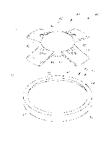
Fig. 3 is a perspective front view of a hybrid AIOL assemblage including a
discrete lens unit and a discrete base member for in situ assembly in a
capsular
bag during cataract surgery;
Fig. 4 is a top plan view of the discrete lens unit;
Fig. 5 is a transverse cross section of the discrete lens unit along line 5-5
in
Figure 4;
Fig. 6 is a top plan view of the discrete base member;
Fig. 7 is a transverse cross section of the discrete base member along line
7-7 in Figure 6;
Fig. 8 is a transverse cross section of an in-the-hand assembled hybrid
AIOL assemblage;
4
CA 03022773 2018-10-31
WO 2017/203517
PCT/IL2017/050566
Fig. 9 is a transverse cross section of an edge of another discrete base
member;
Fig. 10 is a transverse cross section of an edge of yet another discrete base
member;
Fig. 11 is a cross section of an implanted hybrid AIOL assemblage for near
vision corresponding to Figure 1;
Fig. 12 is a cross section of the implanted hybrid AIOL assemblage for
distance vision corresponding to Figure 2; and
Fig. 13 is a cross section of the implanted hybrid AIOL assemblage for
intermediate vision.
Detailed Description of Drawings
Figure 3 show a hybrid AIOL assemblage 30 including a discrete lens unit
40 and a discrete base member 60 for in situ assembly in a capsular bag during
cataract surgery. The discrete lens unit 40 includes a lens optics 41 and at
least
two equispaced lens haptics 42 radially outward extending from the lens optics
41. The discrete lens unit 40 preferably includes four equispaced lens haptics
42.
The lens unit 40 can be manufactured as a monolithic structure. Alternatively,
the
lens haptics 42 can be manufactured separately from the lens optics 41 and
attached thereto using industry known attachment technologies. The discrete
base
member 60 has a base member centerline 61 and includes a flat circular base
member centerpiece 62 and a base member surround 63. The base member 60 can
be manufactured as a monolithic structure. Alternatively, the base member
surround 63 can be manufactured separately from the base member centerpiece 62
and attached thereto using industry known attachment technologies. The hybrid
AIOL assemblage 30 is entirely made from implantable biocompatible material.
The lens optics 41 and the base member centerpiece 62 are made from optical
grade transparent materials and have the same refractive index. The lens
optics 41
5
CA 03022773 2018-10-31
WO 2017/203517
PCT/IL2017/050566
and the base member centerpiece 62 are preferably formed from the same
material.
Figure 4 and Figure 5 show the lens optics 41 has an optical axis 43 for co-
axial alignment with a visual axis VA. The lens optics 41 has an anterior lens
optics surface 44, a posterior lens optics surface 46 and a lens optics edge
47. The
lens optics 41 has a similar diameter and thickness as standard IOLs currently
being used for cataract surgery. The anterior lens optics surface 44 affords a
primary optical power calculated for optimal distance vision correction in an
implanted eye. Healthy eyes require good vision at both near distance and
intermediate distances and therefore the posterior optic lens surface 46
preferably
has a multifocal optical gradient from a maximal optical power at the lens
optics
axis 43 diminishing towards the lens optics edge 47. Accordingly, the
posterior
lens optics surface 46 includes a center circle 48 having an approximate 2.5
mm
diameter around the lens optics axis 43 corresponding to near vision pupil
size
under normal reading illumination conditions. The central circle 48 has the
required added power to the principle distance correction optical power of the
anterior lens optics surface 44 for near vision in an intended implanted eye.
The
central circle 48 typically has an optical power of around 3.0 Diopter. From
the
boundary of the central circle 48, the optical power is gradually decreased
towards
the lens optics edge 47 using manufacturing methods known to the art. In a
degenerate version of the discrete lens unit 40 for implantation in an
impaired
vision eye, the posterior lens optics surface 46 can be constituted by a
single
mono-focal lens optics surface for providing best correction for near vision
only
in an intended implanted eye.
Each lens haptics 42 has a lens haptics free end 51 with a lens haptics
curved edge 52 corresponding to a curvature of an anchoring interface of the
discrete base member 60. Each lens haptics 42 preferably has a manipulation
aperture 53 for enabling proper positioning of the lens unit 40 relative to
the base
member 60. Each lens haptics 42 preferably has an elongated anterior spacer
pair
6
CA 03022773 2018-10-31
WO 2017/203517
PCT/IL2017/050566
54 adjacent to the lens optics 41 for spacing an anterior capsule flange 27
therefrom to enable circulation of aqueous humor between an anterior capsule
flange 27 and the lens unit 40.
The anterior lens optics surface 44 but can also be designed for
simultaneous correction of astigmatism in an intended implanted eye.
Accordingly, the lens unit 40 is provided with an optical axis marker 56 for
assisting correct alignment of the lens unit 40 with respect to a human visual
axis
VA during implantation. The optical axis marker 56 is preferably placed on a
lens
haptics 42 not to impede vision. The manipulation apertures 53 are employed
for
dialing a properly positioned lens unit 40 around the lens optics axis 43 for
setting
at a required position for astigmatic correction.
Figure 6 and Figure 7 show the discrete base member 60 has a flat circular
base member centerpiece 62 having a flat circular anterior base member
centerpiece surface 64 and a flat circular posterior base member centerpiece
surface 66. The flat anterior and posterior base member centerpiece surfaces
64
and 66 have zero optical power. The base member surround 63 is formed with an
elevated circumferential retainer 67 for forming a circumferential groove 68
with
the anterior base member centerpiece surface 64 for receiving the lens haptics
free
ends 51 for anchoring the discrete lens unit 40 on the discrete base member
60.
The base member surround 63 preferably has a square cross section for
preventing
the migration of epithelial cells from a capsular periphery. Figure 8 shows
the
assembled hybrid AIOL assemblage 30 on mounting the lens unit 40 on the base
member 60 by means of the lens haptics free ends 51 being flexed into the
circumferential groove 68 such that the lens haptics 42 urge the posterior
lens
optics surface 46 away from the anterior base member centerpiece surface 64.
Capsular bag size can vary by several millimeters. The hybrid AIOL
assemblages 30 are designed such that the same discrete lens unit 40 can be
implanted in different sized human capsular bags. This is achieved by the
provision of discrete base members 60 having their circumferential groove 68
at
7
CA 03022773 2018-10-31
WO 2017/203517
PCT/IL2017/050566
the same radius R relative to the base member centerline 61 and compensating
for
capsular size differences by radial outward extending of the base member
surround 63 and the elevated circumferential retainer 67 as can be seen on
comparison of Figure 9 to Figure 7. Figure 10 shows an alternative elevated
circumferential retainer 67 in the form of a pliable rim 69 designed to be
flexed
towards the anterior base member centerpiece surface 64 by the anterior
capsule
flange 27 as denoted by arrow C to improve the mechanical interface between
the
anterior capsule flange 27 and the lens haptics 42. The pliable rim 69 is
deployed
at the same radius R from the base member centerline 61 and capsular size
differences are compensated by radial outward extending of the base member
surround 63 with respect to the base member centerline 61.
Figure 11 show an implanted hybrid AIOL assemblage 30 in an operative
near vision state corresponding to Figure 1. Full ciliary body contraction is
accompanied by iris contraction to about 2.5 mm diameter pupil size. Figure 11
shows the anterior capsule flange 27 contacting the anterior lens optics
surface 44
and/or the haptics spacers 54 but not urging the lens optics 41 towards the
base
member centerpiece 62 such that the posterior lens optics surface 46 is spaced
apart from the anterior base member centerpiece surface 64. Accordingly, the
hybrid AIOL assemblage 30 affords the combined optical power of the anterior
lens optics surface 44 and the central circle 48's optical power to enable
near
vision.
Figure 12 shows the implanted hybrid AIOL assemblage 30 in an operative
distance vision state corresponding to Figure 2. Figure 12 shows the anterior
capsule flange 27 pressing down on the anterior lens optics surface 44 and/or
the
haptics spacers 54 for urging the lens optics 41 towards the base member
centerpiece 62. The posterior lens optics surface 46 is entirely intimately
immerged in the anterior base member centerpiece surface 64 such that the
posterior lens optics surface 46 and the anterior base member centerpiece
surface
64 create a single refractive index optical continuum of zero optical power
8
CA 03022773 2018-10-31
WO 2017/203517
PCT/IL2017/050566
whereby the hybrid AIOL assemblage 30 affords optical power by virtue of the
anterior lens optics surface 44 suitably determined for best distance vision
of an
intended implanted eye.
Figure 13 shows the implanted hybrid AIOL assemblage 30 in an operative
intermediate vision state. The intermediate vision involves a ciliary body
contraction much smaller than for near vision leading to the posterior lens
optics
surface 46 being partially intimately immerged in the anterior base member
centerline surface 64. As shown, only the central circle 48 and the anterior
base
member centerpiece surface 64 create a single refractive index optical
continuum
of zero optical power. The annular multi-focal segment 49 is spaced apart from
the anterior base member centerpiece surface 64 thereby affording the required
additional optical power for intermediate vision.
While the invention has been described with respect to a limited number of
embodiments, it will be appreciated that many variations, modifications, and
other
applications of the invention can be made within the scope of the appended
claims.
9
