Note: Descriptions are shown in the official language in which they were submitted.
- 1 -
ELECTROSURGICAL SYSTEM
[0001] This
application is a divisional application of co-pending application
Serial No. 2,720,075, filed March 31, 2009.
Background
Field
[0002] The present
application relates generally to electrosurgical systems
and methods. More specifically, the present application relates to
determination of an
electrosurgery endpoint using phase shift monitoring.
Discussion of the Relevant Art
[0003] Surgical
procedures often involve cutting and connecting bodily
tissue including organic materials, musculature, connective tissue and
vascular conduits.
For centuries, sharpened blades and sutures have been mainstays of cutting
and
reconnecting procedures. As bodily tissue, especially relatively highly
vascularized issue
is cut during a surgical procedure, it tends to bleed. Thus, medical
practitioners such as
surgeons have long sought surgical tools and methods that slow or reduce
bleeding
during surgical procedures.
CA 3022982 2018-11-01
- 2 -
[0004] More recently, electrosurgical tools have become available
that use
electrical energy to perform certain surgical tasks. Typically,
electrosurgical tools are hand
tools such as graspers, scissors, tweezers, blades, needles, and other hand
tools that
include one or more electrodes that are configured to be supplied with
electrical energy
from an electrosurgical generator including a power supply. The electrical
energy can be
used to coagulate, fuse, or cut tissue to which it is applied. Advantageously,
unlike typical
mechanical blade procedures, application of electrical energy to tissue tends
to stop
bleeding of the tissue.
[0005] Electrosurgical tools typically fall within two
classifications: monopolar
and bipolar. In monopolar tools, electrical energy of a certain polarity is
supplied to one or
more electrodes on the tool. A separate return electrode is electrically
coupled to a patient.
Monopolar electrosurgical tools can be useful in certain procedures, but can
include a risk
of certain types of patient injuries such as electrical
CA 3022982 2018-11-01
- 3 -
burns often at least partially attributable to functioning of the return
electrode. In bipolar
electrosurgical tools, one or more electrodes is electrically coupled to a
source of
electrical energy of a first polarity and one or more other electrodes is
electrically
coupled to a source of electrical energy of a second polarity opposite the
first polarity.
Thus, bipolar electrosurgical tools, which operate without separate return
electrodes,
can deliver electrical signals to a focused tissue area with a reduced risk of
patient
injuries.
[0006] Even with the relatively focused surgical effects of bipolar
electrosurgical tools, however, surgical outcomes are often highly dependent
on
surgeon skill. For example, thermal tissue damage and necrosis can occur in
instances
where electrical energy is delivered for a relatively long duration or where a
relatively
high-powered electrical signal is delivered even for a short duration. The
rate at which a
tissue will achieve the desired coagulation or cutting effect upon the
application of
electrical energy varies based on the tissue type and can also vary based on
pressure
applied to the tissue by an electrosurgical tool. However, even for a highly
experienced
surgeon, it can be difficult for a surgeon to assess how quickly a mass of
combined
tissue types grasped in an electrosurgical instrument will be fused a
desirable amount.
[0007] Attempts have been made to reduce the risk of tissue damage
during electrosurgical procedures. For example, previous electrosurgical
systems have
included generators that monitor an ohmic resistance or tissue temperature
during the
electrosurgical procedure, and terminated electrical energy once a
predetermined point
was reached. However, these systems have had shortcomings in that they have
not
provided consistent results at determining tissue coagulation, fusion, or
cutting
endpoints for varied tissue types or combined tissue masses. These systems can
also
fail to provide consistent electrosurgical results among use of different
tools having
different tool and electrode geometries. Typically, even where the change is a
relatively
minor upgrade to tool geometry during a product's lifespan, the
electrosurgical
generator must be recalibrated for each tool type to be used, a costly, time
consuming
procedure which can undesirably remove an electrosurgical generator from
service.
CA 3022982 2018-11-01
- 4 -
Summary
[0008] In view
of at least the foregoing shortcomings of the previous
electrosurgical systems, there is a need in the art to improve control of
electrosurgical
procedures to enhance consistency of electrosurgical results among
electrosurgical
tools and tissue types.
Accordingly, there is a need for an improved electrosurgical
system that can accurately assess an electrical energy application endpoint
for a
desired electrosurgical procedure. There is also a need for an electrosurgical
system
that monitors tissue properties during the electrosurgical procedure to assess
the
energy application endpoint. There is also a need for an imp`roved
electrosurgical
system that can rapidly accommodate various electrosurgical tools with minimal
impact
on surgical outcome. To address some or all of these needs and provide various
additional advantages as discussed below in greater detail, various
embodiments,
methods, systems, and apparatuses for electrosurgical procedures are provided.
[0009] In
various embodiments, methods and apparatuses for bloodless
dissection of connective and vascular tissue are provided. The various methods
and
apparatuses described herein can be used in minimally invasive surgery,
particularly
laparoscopic surgery.
[0010] In
certain embodiments, an electrosurgical tool comprises a handle
assembly, an elongate shaft, a jaw assembly, and a force regulation mechanism.
The
handle assembly comprises a stationary handle and an actuation handle movably
coupled to the stationary handle. The elongate shaft extends distally from the
handle.
The elongate shaft has a proximal end and a distal end defining a central
longitudinal
axis therebetween. The jaw assembly is positioned on the distal end of the
elongate
shaft. The jaw assembly comprises a first jaw and a second jaw. The first jaw
has an
inner surface, an outer surface, and at least one electrode disposed on the
inner
surface. The second jaw has an inner surface, an outer surface, and at least
one
= electrode disposed on the inner surface. The jaw assembly is actuatable
by movement
of the if from an open configuration in which the inner surface of the first
jaw is spaced
apart from the inner surface of the second jaw to a closed configuration in
which the
inner surface of the first jaw is proximate the inner surface of the second
jaw. The force
CA 3022982 2018-11-01
- 5 -
regulation mechanism couples the handle assembly to the jaw assembly. The
force
regulation assembly is configured such that in the closed configuration, the
jaw
assembly delivers a gripping force between the first jaw and the second jaw
between a
predetermined minimum force and a predetermined maximum force.
[0011] In other embodiments, an electrosurgical tool is provided
comprising a
handle assembly, an elongate shaft, and a jaw assembly. The handle assembly
comprises a moveable actuation handle. The elongate shaft extends distally
from the
handle. The elongate shaft has a proximal end and a distal end defining a
central
longitudinal axis therebetween. The jaw assembly is positioned on the distal
end of the
elongate shaft. The jaw assembly comprises a first jaw, a second jaw, and a
blade.
The first jaw has an inner surface, an outer surface, a proximal end and a
distal end,
and at least one electrode disposed on the inner surface. The second jaw has
an inner
surface, an outer surface, a proximal end and a distal end and at least one
electrode
disposed on the inner surface. The blade is advanceable along the inner
surface of the
first jaw along a cutting path defined between a retracted position adjacent
the proximal
end and an advanced position between the proximal end and the distal end. The
jaw
assembly is actuatable from an open configuration to a closed configuration by
movement of the actuation handle. The at least one electrode on the first jaw
and the at
least one electrode on the second jaw define a sealing area enclosing the
cutting path
of the blade.
[0012] In other embodiments, an electrosurgical tool is provided
comprising a
handle assembly, an eleongate shaft, and a jaw assembly. The elongate shaft
extends
distally from the handle assembly. The shaft having a proximal end and a
distal end
defining a central longitudinal axis therebetween. The jaw assembly is
positioned on
the distal end of the elongate shaft. The jaw assembly comprises a first jaw
and a
second jaw. The first jaw has an inner surface, an outer surface, a proximal
end and a
distal end, and at least one fusion electrode disposed on the inner surface.
The second
jaw has an inner surface, an outer surface, a proximal end and a distal end
and at least
one fusion electrode disposed on the inner surface and a cutting electrode
disposed on
the outer surface.
CA 3022982 2018-11-01
- 6 -
[0013] In certain
embodiments, an electrosurgical system for performing
surgical procedures on body tissue of a patient comprises an electrosurgical
generator
and an electrosurgical tool. The electrosurgical tool comprises a memory
module
storing tool data. The electrosurgical generator is configured to receive the
tool data
from the memory module and apply an electrosurgical signal profile to the
electrosurgical tool based on the tool data.
[0014] In other
embodiments, an electrosurgical generator for performing
surgical procedures on body tissue of a patient comprises a power supply, a
signal
generation module, and a first tool port. The signal generation module is
electrically
coupled to the power supply. The signal generation module is configured to
generate a
radiofrequency signal. The first
tool port is configured to interface with an
electrosurgical tool having tool data stored therein. The first tool port is
adapted to
receive the tool data stored on the electrosurgical tool and to supply the
readiofrequency signal from the signal generation module to the tool.
[0015] In some
embodiments, a controller for electrosurgical tools comprises
a first actuator, a second actuator, and a tool selector. The first actuator
is movable
between an on position and an off position for actuating a first
electrosurgical action
when in the on position. The second actuator is movable between an on position
and
an off position for actuating a second electrosurgical action when in the on
position.
The tool selector has a first state wherein the controller is adapted to be
operatively
coupled to a first electrosurgical tool and a second state wherein the
controller is
adapted to be operatively coupled to a second electrosurgical tool.
[0016] In certain
embodiments, a surgical tool can comprise jaw elements
having a plurality of electrodes to be used for both electrosurgical
coagulation and
cutting. The electrodes can be powered in a first configuration to provide
coagulation ¨
leading to hemostasis of small vascular vessels and tissue ¨ and powered in a
second
configuration for electrosurgical cutting of the coagulated tissue. The two
powered
configurations can be generated by addressing different electrodes on the jaw
elements
and applying them with voltages appropriate for electrosurgical coagulation
and/or
cutting. In some embodiments, the surgical tool can initially be powered in
the first
CA 3022982 2018-11-01
- 7 -
configuration to provide coagulation, and then can be powered in the second
configuration for electrosurgical cutting. In other embodiments, the
electrosurgical tool
can be powered only in a coagulating configuration to achieve tissue
hemostasis, only
in a cutting configuration to dissect tissue, or in a cutting configuration
followed by a
coagulation configuration.
[0017] At the same time, various embodiments of the surgical tools
described
herein can include different electrode configurations. I.e., while in one
embodiment only
the lower jaw is utilized to provide both coagulation and cutting functions,
another
embodiment can also employ the upper jaw element to be used in the coagulation
and/or cutting process. In yet another embodiment, each jaw element can carry
multiple electrode elements, greatly increasing the functionality of the tool.
A specific
electrode arrangement can allow for tools that are more suitable for
particular surgical
procedures.
[0018] Another aspect of the surgical tools described herein relates
to
activation and deactivation of one or multiple electrodes, based on the
position of the
jaw elements. This position-based actuation allows, for example, activation of
the upper
jaw electrodes only in a near-closed position of the tool (or, in other
embodiments, in an
opened or near-opened position of the tool). In some embodiments, electrical
switches
in the jaw element driving mechanism can be positioned in a hand-piece of the
surgical
tool to selectively activate and deactivate one or multiple electrodes based
on a position
of the jaw elements. In other embodiments, the activation and deactivation can
be
performed by sliding contacts that are assembled in the hand-piece.
[0019] Yet another aspect of the surgical tools described herein is
the
automated switching from coagulation to cutting, enabled by use of a multi-
electrode
generator. Here, a tissue feedback mechanism triggers both switching from one
set of
coagulation electrodes (applied with voltages appropriate for coagulation) to
another set
of cutting electrodes (applied with voltages appropriate for cutting). As
such, each
individual tool electrode can be relayed through a bus-bar connection to any
polarity of
choice of the power supply. In addition, tool position switches in the hand
tool can
CA 3022982 2018-11-01
- 8 -
provide with logic switching for the population of different coagulation
and/or cutting
settings, depending on the specific tool position.
[0020] In certain embodiments, an electrosurgical tool is provided
comprising
a first jaw, a second jaw, a first electrode, a second electrode, and a third
electrode.
The second jaw is pivotable with respect to the first jaw. The first electrode
is
positioned on the first jaw. The second electrode is positioned on the first
jaw. The
third electrode is positioned on the first jaw. The electrosurgical tool can
be selectively
configurable in a coagulation configuration such that at least one of the
first, second,
and third electrodes is electrically coupled with a source of electrical
energy having a
first polarity and at least one other of the electrodes is electrically
coupled with a source
of electrical energy having a second polarity generally opposite the first
polarity and in a
cutting configuration such that one of the first, second, and third electrodes
is electrically
coupled with a source of electrical energy having a cutting voltage and at
least one
other of the electrodes is configured to be a return electrode.
[0021] In other embodiments, an electrosurgical tool having a proximal
end
and a distal end is provided comprising a distal end-piece, an elongate shaft,
a handle
assembly, and a switching mechanism. The distal end-piece is positioned at the
distal
end of the tool. The distal end-piece comprises a first jaw element, a second
jaw
element, and a plurality of electrodes. The first and second jaw elements are
movable
relative to one another between an open position and a closed position. The
plurality of
electrodes is disposed on at least one of the first jaw element and the second
jaw
element. The plurality of electrodes is selectively configurable in one of a
coagulation
configuration and a cutting configuration. The elongate shaft has a distal end
connected to the distal end-piece and a proximal end. The handle assembly is
positioned at the proximal end of the tool and connected to the proximal end
of the
elongate shaft. The handle assembly comprises a hand-piece and a trigger. The
trigger is pivotally coupled to the hand-piece and operably coupled to the
distal end-
piece such that movement of the trigger relative to the hand-piece moves the
first and
second jaw elements relative to one another. The switching mechanism is
electrically
CA 3022982 2018-11-01
- 9 -
coupled to the distal end-piece to selectively configure the plurality of
electrodes in one
of the coagulation configuration and the cutting configuration.
[0022] In other embodiments, a method for substantially bloodless
dissection
of biological tissue is provided. The method comprises positioning an
electrosurgical
tool adjacent tissue to be dissected, measuring tissue properties to determine
the
switching point from coagulation to cutting, applying electrical energy to the
electrosurgical tool, assessing the tissue coagulation (phase shift) through a
feedback
loop, switching a configuration of the electrosurgical tool, and applying
electrical energy
to the electrosurgical tool in a cutting configuration. The electrosurgical
tool comprises a
plurality of electrodes configurable in one of a coagulation configuration and
a cutting
configuration. Applying electrical energy to the electrosurgical tool
comprises applying
electrical energy to the electrosurgical tool in the coagulation configuration
to achieve
hemostasis in the tissue. Switching the electrosurgical tool comprises
switching the
electrosurgical tool to the cutting configuration.
[0023] In some embodiments, a method for controlling an output of an
electrosurgical generator operatively coupled to a bipolar electrosurgical
device is
provided. The method comprises measuring a phase angle, determining a target
phase
angle, measuring the phase angle of a second measurement signal, and ceasing
delivery of a treatment signal. Measuring the phase angle comprises measuring
a
phase angle of a first measurement signal applied to tissue of a patient via
at least one
electrode of the electrosurgical device. The first measurement signal is
applied to the
tissue prior to treatment of the tissue by the electrosurgical device.
Determining a target
phase angle comprises determining a target phase angle using the phase angle
of the
first measurement signal. Following delivery of a treatment signal comprises
following
delivery of a treatment signal to the tissue. Measuring the phase angle of a
second
measurement signal comprises measuring the phase angle of a second measurement
signal applied to the tissue. The treatment signal is capable of causing
modification of
the tissue. Ceasing delivery of the treatment signal comprises ceasing
delivery of the
treatment signal to the tissue when the phase angle of the second measurement
signal
reaches the target phase angle.
CA 3022982 2018-11-01
- 10 -
[0024] In other embodiments, a method for controlling an output of an
electrosurgical generator operatively coupled to a bipolar electrosurgical
device is
provided. The method comprises determining permittivity and conductivity of
tissue,
determining a threshold phase angle, measuring a phase angle, and ceasing the
delivery of the treatment signal. Determining permittivity and conductivity of
tissue
comprises determining permittivity and conductivity of tissue of a patient
using a
measurement signal. The measurement signal is applied to tissue of a patient
via at
least one electrode of the electrosurgical device. The measurement signal is
applied to
the tissue prior to modification of the tissue by the electrosurgical device.
Determining a
threshold phase angle comprises determining a threshold phase angle based on
the
permittivity and the conductivity of the tissue. Measuring a phase angle
comprises
measuring a phase angle of a signal applied to the tissue. Ceasing the
delivery of the
treatment signal comprises ceasing the delivery of the treatment signal to the
tissue
when the phase angle of the signal reaches the threshold phase angle.
[0025] In other embodiments, a method of characterizing tissue prior to
the
delivery of electrosurgical energy to the tissue via a bipolar electrosurgical
device is
provided. The method comprises measuring phase angle, determining the product
of
the relative permittivity and conductivity, and characterizing the tissue.
Measuring
phase angle comprises measuring phase angle of a measurement signal applied to
tissue of a patient via at least one electrode of the electrosurgical device.
The
measurement signal is applied to the tissue at a predetermined frequency prior
to
modification of the tissue by the electrosurgical device. Determining the
product of the
relative permittivity and conductivity comprises determining the product of
the relative
permittivity and conductivity of the tissue using the phase angle measurement
and the
predetermined frequency. Characterizing the tissue comprises characterizing
the tissue
based on the product of the relative permittivity and conductivity of the
tissue.
[0026] In other embodiments, a method of characterizing tissue prior to
the
delivery of electrosurgical energy to the tissue via a bipolar electrosurgical
device is
provided. The method comprises generating a measurement signal, determining a
treatment endpoint condition, and stopping delivery of a treatment signal.
Generating a
CA 3022982 2018-11-01
- 11 -
measurement signal comprises generating a measurement signal applied to tissue
of a
patient positioned between at least two jaw members of an electrosurgical
device. At
least one of the jaw members comprises an electrode. The measurement signal is
delivered to the tissue via the electrode and applied to modification of the
tissue by the
electrosurgical device.
Determining a treatment endpoint condition comprises
determining a treatment endpoint condition using the measurement signal. The
treatment endpoint condition is determined substantially independently of the
dimensions of the tissue positioned between the at least two jaw members.
Stopping
delivery of a treatment signal comprises stopping delivery of a treatment
signal to the
tissue when the treatment endpoint condition is reached. The treatment signal
is
capable of causing modification of the tissue.
[0027] In other
embodiments, an electrosurgical system for application of
treatment energy to a patient involved in bipolar electrosurgery is provided.
The system
comprises an electrosurgical generator, an electrosurgical control unit, and
an
electrosurgical tool. The electrosurgical generator is configured to generate
and output
a treatment energy along with a measurement signal. The electrosurgical
control unit is
configured to direct the output of treatment energy and a measurement signal.
The
electrosurgical tool is removably connected to one of the electrosurgical
generator and
the electrosurgical control unit and arranged to contact tissue and apply the
treatment
energy and the measurement signal to the tissue. The electrosurgical control
unit
measures permittivity and conductivity of the tissue through the application
of the
measurement signal.
Brief Description of the Drawings
[0028] The
present inventions may be understood by reference to the
following description, taken in connection with the accompanying drawings in
which the
reference numerals designate like parts throughout the figures thereof.
CA 3022982 2018-11-01
- 12 -
[0029] Figure 1A is a schematic block diagram of an embodiment of
electrosurgical system.
[0030] Figure 1B is a schematic block diagram of another embodiment of
electrosurgical system.
[0031] Figure 2A is a perspective view of components of one embodiment
of
an electrosurgical system.
[0032] Figure 2B is a perspective view of components of one embodiment
of
an electrosurgical system.
[0033] Figure 2C is a perspective view of components of one embodiment
of
electrosurgical system.
[0034] Figure 3A is a perspective view of an electrosurgical unit for
use in an
electrosurgical system.
[0035] Figure 3B is a front view of the electrosurgical unit of Figure
3A.
[0036] Figure 3C is a rearview of the electrosurgical unit of Figure
3A.
[0037] Figure 4A is an exemplary screenshot of a display of the
electrosurgical unit of Figure 3A.
[0038] Figure 4B is another exemplary screenshot of the display of the
electrosurgical unit of Figure 3A.
[0039] Figure 5 is a block diagram of various embodiments of an
electrosurgical unit.
[0040] Figure 6 is a front view of a user interface of an
electrosurgical unit.
[0041] Figure 7 is a front view of a user interface of an
electrosurgical unit.
[0042] Figure 8 is a front view of a user interface of an
electrosurgical unit.
[0043] Figure 9 is a block diagram of an electrosurgical unit.
CA 3022982 2018-11-01
- 13 -
[0044] Figure 10 is a block diagram of an electrosurgical unit.
[0045] Figure Ills a graphical representation of a high voltage
driving signal
at low frequency relative to a low voltage measurement voltage at a high
frequency.
[0046] Figure 12 is a graphical representation of filtered measurement
and
current signals for a time near the end of the fusion process.
[0047] Figure 13 is a block diagram of an electrosurgical unit.
[0048] Figure 14 is a block diagram of an electrosurgical unit.
[0049] Figure 15 is a schematic diagram of an external measurement
circuitry
of an electrosurgical unit.
[0050] Figure 16 is a schematic diagram of switch circuitry of an
electrosurgical unit.
[0051] Figure 17 is a schematic diagram of a phase comparator or
detection
circuitry of an electrosurgical unit.
[0052] Figure 18 is a schematic diagram of a battery power circuitry
of an
electrosurgical unit.
[0053] Figure 19 is a schematic diagram of an input interface of an
electrosurgical unit.
[0054] Figure 20 is a graphical representation of experimental data
for the
voltage applied to the tissue during a typical a vessel fusion process.
[0055] Figure 21 is a graphical representation of experimental data
for the
voltage applied to the tissue during the measurement cycle.
[0056] Figure 22 is a graphical representation of experimental data
for the
voltage applied to the tissue during the RF measurement cycle to determine the
phase
shift through the tissue.
CA 3022982 2018-11-01
- 14 -
[0057] Figure 23 is a graphical representation of a sample of
experimental
data for a typical vessel sealing process, showing a temporal showing a
temporal
snapshot of applied voltage, electrical current, and dissipated power at 1
second into
the fusion.
[0058] Figure 24 is a graphical representation of a sample of
experimental
data for a typical vessel sealing process, showing the peak voltage and peak
electrical
current as function of fusion time.
[0059] Figure 25 is a graphical representation of a sample of
experimental
data for a typical vessel sealing process, showing the vessel impedance as
function of
fusion time.
[0060] Figure 26 is a graphical representation of a vessel sealing and
tissue
welding process in accordance with various embodiments of the present
invention
showing the relative impedances of various tissues as a function of time.
[0061] Figure 27 is a graphical representation of a fusion/vessel
sealing
process in accordance with various embodiments of the present invention
showing a
temporal snapshot of applied voltage, electrical current, and dissipated power
at 4
seconds into the fusion process.
[0062] Figure 28 is a graphical representation for a fusion/vessel
sealing
process showing a temporal snapshot of applied voltage, electrical current,
and
dissipated power at 7 seconds into the fusion process.
[0063] Figure 29 is a graphical representation of bursting pressure as
a
function of phase shift used in end point determination.
[0064] Figure 30 is a table of dielectric constants or permittivity
and
conductivities for various types of biological tissue, arranged by increasing
values of the
product of dielectric constants and tissue conductivity.
[0065] Figure 31 is a graphical representation of empirically
determined phase
shifts to adequately fuse and/or weld various types of biological tissue.
CA 3022982 2018-11-01
- 15 -
[0066] Figure 32 is a graphical representation of endpoint phase
shifts relative
to initial phase shift measurements of various types of biological tissue.
[0067] Figure 33 is a graphical representation of a phase diagram of
two
electrosurgical tools and their associated capacitance and resistance.
[0068] Figure 34 is a graphical representation of a phase diagram of
an
electrosurgical tool in tissue contact and the associated capacitance and
resistance.
[0069] Figure 35 is a graphical representation of the ohmic resistance
of a
porcine renal artery during the electrosurgical fusion process.
[0070] Figure 36 is a graphical representation of phase shift during
the
electrosurgical fusion process.
[0071] Figure 37 is a graphical representation of the derivate of the
phase
shift during the electrosurgical fusion process.
[0072] Figure 38 is a graphical representation of phase shift during
the
electrosurgical fusion process.
[0073] Figure 39 is a graphical representation of the derivate of the
phase
shift during the electrosurgical fusion process.
[0074] Figure 40 is a block diagram of a fusion or welding process of
an
electrosurgical unit.
[0075] Figure 41A is a perspective view of an embodiment of
laparoscopic
sealer/divider.
[0076] Figure 41B is a disassembled view of a laparoscopic
sealer/divider of
Figure 1A.
[0077] Figures 42A-42C are views of an actuator of the laparoscopic
sealer/divider of Figure 41A.
[0078] Figure 43 is a top cross-sectional view of an actuator of a
laparoscopic
sealer/divider of Figure 41A.
CA 3022982 2018-11-01
- 16 -
[0079] Figures 44A-44D are views of a shaft assembly of a laparoscopic
sealer/divider of Figure 41A.
[0080] Figures 45A-45C are views of a jaw assembly of a laparoscopic
sealer/divider of Figure 41A.
[0081] Figures 46A-46G are cross-sectional side views of a
laparoscopic
sealer/divider of Figure 41A.
[0082] Figure 47 is a perspective view of a controller of a
laparoscopic
sealer/divider of Figure 41A.
[0083] Figure 48A is a side view of a jaw assembly of a laparoscopic
sealer/divider of Figure 41A.
[0084] Figures 48B-48C are graphical representations of exemplary
vessel
sealing pressures provided by a laparoscopic sealer/divider of Figure 41A.
[0085] Figure 49 is a top level view of an electrode configuration of
a
laparoscopic sealer/divider of Figure 41k
[0086] Figure 50 is a top level view of a jaw assembly of a
laparoscopic
sealer/divider of Figure 41A.
[0087] Figure 51 is a side view of a jaw assembly of a laparoscopic
sealer/divider of Figure 41A.
[0088] Figure 52 provides views of a jaw assembly of a laparoscopic
sealer/divider of Figure 41A.
[0089] Figure 53A is a perspective view of a jaw assembly of a
laparoscopic
sealer/divider of Figure 51A.
[0090] Figure 53B is a perspective view of an actuator of a
laparoscopic
sealer/divider of Figure 41A.
[0091] Figure 54 provides views of portions of a shaft assembly of a
laparoscopic sealer/divider of Figure 41A.
CA 3022982 2018-11-01
- 17 -
[0092] Figure 55 provides views of a jaw assembly of a laparoscopic
sealer/divider of Figure 41A.
[0093] Figure 56 is a perspective view of an embodiment of surgical
tool for
use in a laparoscopic surgical procedure.
[0094] Figure 57 is a perspective drawing of the distal end of an
exemplary
tissue fusion/cutting devices.
[0095] Figures 58A-D are schematic drawings of various embodiments of
distal end configurations for an electrosurgical bloodless tissue dissection
device.
[0096] Figures 59A-C are schematic drawings of active electrode
switching
circuitries in the hand tools.
[0097] Figure 60 is a schematic drawing of the inside of the hand-
piece,
illustrating the embodiment of active electrode switching mechanism based on
the
opening of the jaw elements.
[0098] Figure 61 depicts another embodiment of an active electrode
switching
mechanism, also based on the opening of the jaw elements.
[0099] Figure 62 depicts an embodiment of a passive switching
mechanism,
also based on the opening of the jaw elements.
[0100] Figure 63 depicts another embodiment of a passive switching
mechanism, based on both the opening and closing of the jaw elements.
[0101] Figure 64 depicts a schematic circuitry that connects five
electrodes
through relays to a bus bar which is relayed to a measurement circuit, or an
electrosurgical power plant.
[0102] Figure 65 schematically illustrates one embodiment of a method
for
substantially bloodless dissection of biological tissue.
[0103] Figure 66 is a perspective view of an electrosurgical
instrument in a
closed condition.
CA 3022982 2018-11-01
- 18 -
[0104] Figure 67 is a perspective view of an electrosurgical
instrument in an
open condition.
[0105] Figure 68 is a side view of an electrosurgical instrument in an
open
condition.
[0106] Figure 69 is an enlarged perspective view of a clamping portion
of an
electrosurgical instrument in an open condition.
[0107] Figure 70 is a side section view of an electrosurgical
instrument in an
open condition.
[0108] Figure 71 is an enlarged perspective view of a clamping jaw
portion
with the top clamping jaw removed.
[0109] Figure 72 is an enlarged perspective view of an actuator for
advancing
electrodes.
[0110] Figure 73 is an enlarged side view of clamping jaws in an open
condition with electrodes extended.
[0111] Figure 74 is an enlarged side section view of clamping jaws in
an open
condition and having electrodes extended.
[0112] Figure 75 is an enlarged perspective view of an actuator sled
and
associated electrical contacts.
[0113] Figure 76 is an enlarged perspective view of an electrode.
[0114] Figure 77 illustrates a relationship between clamping jaws and
tissue
to be fused in a first, grasping condition.
[0115] Figure 78 illustrates a relationship between clamping jaws and
tissue
to be fused in a second, compressing condition.
[0116] Figure 79 illustrates a relationship between clamping jaws and
tissue
to be fused in a third, electrode-extending condition.
CA 3022982 2018-11-01
- 19 -
[0117] Figure 80 illustrates a relationship between clamping jaws and
tissue
to be fused in a final, electrode-extending condition.
[0118] Figure 81 is a perspective cut-out view of a body conduit
showing an
electrosurgical instrument moving into position to occlude a lumen of a
conduit.
[0119] Figure 82 is a perspective view of a body conduit showing an
electrosurgical instrument in position to occlude a lumen of a conduit.
[0120] Figure 83 is a perspective view of a body conduit showing an
electrosurgical instrument occluding a lumen of a conduit.
[0121] Figure 84 is a schematic diagram illustrating current
concentration
through tissue in a first, non-contact condition.
[0122] Figure 85 is a schematic diagram illustrating current
concentration
through tissue in a full-contact condition
[0123] Figure 86 illustrates electrosurgical energy radiation
associated with
penetrating electrodes.
[0124] Figure 87 illustrates a thermal zone associated with penetrating
electrodes.
[0125] Figure 88 illustrates a thermal zone associated with penetrating
electrodes with the electrodes withdrawn.
[0126] Figure 89 illustrates electrosurgical energy radiation
associated with
penetrating electrodes within approximated tissue.
[0127] Figure 90 illustrates a thermal zone associated with penetrating
electrodes within approximated tissue.
[0128] Figure 91 illustrates a thermal zone associated with penetrating
electrodes with electrodes withdrawn.
[0129] Figure 92 is an end view of a conduit closed or occluded using a
suturing technique.
CA 3022982 2018-11-01
- 20 -
[0130] Figure 93 is an end view of a conduit closed or occluded using
a
stapling technique.
[0131] Figure 94 is an end view of a conduit closed or occluded using
a
compressive fusion technique.
[0132] Figure 95 is an end view of a conduit closed or occluded using
a
compressive fusion technique with inserted electrodes.
[0133] Figure 96 is a graphical representation of exemplary burst
pressure
data of an occlusion using a compressive fusion technique with inserted
electrodes
[0134] Figure 97 is an enlarged perspective view of a clamping jaw
showing
an associated cutting element.
[0135] Figure 98 is an enlarged perspective view of a clamping jaw
showing
an associated cutting element comprising an electrosurgical wire electrode.
[0136] Figure 99 is an enlarged perspective view of a clamping jaw
showing
an associated cutting element comprising an electrosurgical or mechanical
wedge
electrode-knife.
[0137] Figure 100 is an enlarged perspective view of a clamping jaw
showing
an associated cutting element comprising an electrosurgical or mechanical
double edge
knife.
[0138] Figure 101 is an enlarged perspective view of a clamping jaw
showing
a plurality of current intensifying elements comprising holes.
[0139] Figure 102 is an enlarged perspective view of a clamping jaw
showing
a plurality of current intensifying elements comprising extended posts.
[0140] Figure 103a is an enlarged perspective view of a clamping jaw
showing a plurality of current intensifying elements comprising extended arcs.
[0141] Figure 103b is an enlarged perspective view of a clamping jaw
showing a plurality of current intensifying elements comprising extended
squares.
CA 3022982 2018-11-01
- 21 -
[0142] Figure 103c is an enlarged perspective view of a clamping jaw
showing
a plurality of current intensifying elements comprising extended rods.
[0143] Figure 103d is an enlarged perspective view of a clamping jaw
showing a plurality of current intensifying elements comprising extended "ball-
and-
cups'.
[0144] Figure 104 is an enlarged perspective view of a clamping jaw
showing
a plurality of current intensifying elements comprising extended rectangles.
[0145] Figure 105a is an enlarged perspective view of a clamping jaw
showing a plurality of current intensifying elements comprising extended
ridges.
[0146] Figure 105b is an enlarged perspective view of a clamping jaw
showing a plurality of current intensifying elements comprising linear
"spicket-and-
sockets".
[0147] Figure 106 is an enlarged perspective view of a clamping jaw
showing
a plurality of current intensifying elements comprising extended pyramids or
cones.
[0148] Figure 107 shows a cross-section view of a clamping jaw with an
exemplary compressed artery with an application of electrical or thermal
energy.
[0149] Figures 108a and b are views of an exemplary portion of an
artery
sealed and cut (108a top plan view, 108b along 8-8).
[0150] Figures 109a and bare views of an exemplary portion of tissue
sealed
and cut (109a top plan view, 109b along 9-9).
[0151] Figure 110 shows a cross-sectional view of a clamping jaw with
an
exemplary compressed artery with an application of ultrasonic energy.
[0152] Figure 111 shows a cross-sectional view of a clamping jaw with
an
exemplary compressed artery with an application of UV or IR radiant energy.
CA 3022982 2018-11-01
- 22 -
Detailed Description
[0153] The
following description is provided to enable any person skilled in
the art to make and use the surgical tools and perform the methods described
herein
and sets forth the best modes contemplated by the inventors of carrying out
their
inventions. Various modifications, however, will remain apparent to those
skilled in the
art. It is contemplated that these modifications are within the scope of the
present
disclosure.
Electrosurgical System
[0154] Figure 1A
illustrates a schematic diagram of an electrosurgical system
2. The electrosurgical system 2 can comprise an electrosurgical unit (ESU) 10
and an
electrosurgical tool 40. The electrosurgical tool 40 can be electrically
coupled to the
electrosurgical unit 10. In some embodiments, an electronic coupler 30 such as
an
electrical wire, wire bundle, or cable can electrically couple the
electrosurgical tool 40 to
the ESU 10. In some embodiments, the electrosurgical system 2 can optionally
further
comprise an external tool controller 80.
[0155] With
continued reference to Figure 1A, the electrosurgical unit 10 can
comprise a generator 12 and a feedback circuit 20. The generator 12 can
include an
actuator 16 such as a power supply and a signal processor configured to
generate a
radiofrequency (RE) electrosurgical signal. The generator 12 can further
comprise a
display 14. The
display 14 can be configured to indicate the status of the
electrosurgical system 2, including, among other information, the status of
the actuator
16 and the status of the electrosurgical tool 40 electrically coupled to the
electrosurgical
unit 10.
[0156] With
continued reference to Figure 1A, the feedback circuit 20 of the
ESU 10 can comprise a phase discriminator 22, a tissue identifier 24, and an
encryption
CA 3022982 2018-11-01
- 23 -
module 26. In some embodiments, the phase discriminator 22 can be electrically
coupled to the tissue identifier 24. The phase discriminator 22 can be
configured to
receive information from the electrosurgical tool 40 electrically coupled to
the ESU 10.
In some embodiments, the information from the electrosurgical tool 40
comprises
information regarding an applied voltage and a supplied current to the
electrosurgical
tool, and the phase discriminator 22 can be configured to calculate a phase
difference
between the applied voltage and the supplied current. The encryption module 26
can
be configured to transmit and receive data formatted in an encrypted protocol.
The
encrypted protocol can be one of several commercially-available encryption
protocols,
or, in some embodiments can be a purpose developed encryption protocol.
[0157] With
continued reference to Figure 1A, In some embodiments, the
feedback circuit 20 can be one or more integrated circuits, printed circuit
boards, or
other processor collocated with the generator 12 within an integrated ESU 10.
As
Illustrated in Figure 1B, In other embodiments, the feedback circuit 20' can
be
electrically coupled to a stand-alone generator 12 to form an ESU 10'. The
tool 40 can
be electrically coupled to the feedback circuit 20'. Other aspects of
electrosurgical
systems having a stand-alone generator 12' and feedback circuit 20' can be
substantially similar to systems having an integrated ESU discussed with
respect to
Figure 1A.
[0158] With
continued reference to Figure 1A, the tool 40 can comprise an
indicator 42, a tissue selector 50, an actuator 60, and a memory 70. In some
embodiments, the indicator 40 can comprise an audio indicator 44 such as a
speaker, a
chime, a clicker device, or another audio generation device. In some
embodiments, the
indicator 40 can comprise a visual indicator 46 such as a lamp, an LED, a
display, a
counter, or another visual indication device. In some embodiments, the visual
indicator
46 comprises a multi-color LED. In some embodiments, the tool 40 comprises
both an
audio indicator 44 and a visual indicator 46.
[0159] The tissue
selector 50 can comprise an electrode assembly 52 and a
cutting tool 54. In various
embodiments, various electrode assemblies can be
configured to perform a desired electrosurgical procedure such as, for
example,
CA 3022982 2018-11-01
- 24 -
coagulation, cutting, or fusion, on a particular tissue. In some embodiments,
the
electrode assembly 52 can be configured for use as a vascular sealer. In other
embodiments, the electrode assembly 52 can be configured for use as a
bariatric
stapler. In still other embodiments, the electrode assembly 52 can be
configured for
use as a tissue cutting device. In some embodiments, the cutting tool 54 can
be a
mechanical element such as a stationary or moveable blade or sharpened edge.
In
other embodiments, the cutting tool 54 can be an electrosurgical element such
as an
energizable wire or filament.
[0160] With
continued reference to Figure 1A, the actuator 60 can be
operatively coupled to the tissue selector 50 to selectively select tissue.
For example, in
some embodiments, the tissue selector 50 can include a jaw-based grasper, and
the
actuator can comprise an actuation mechanism to selectively move the grasper
from an
open position to a closed position. In other embodiments, it is contemplated
that other
tissue selectors can be used in the electrosurgical system 2. In some
embodiments, the
actuator 60 can also be configured to selectively energize the electrodes. For
example,
the actuator 60 can comprise a switch or button on the tool.
[0161] With
continued reference to Figure 1A, the tool 40 can further comprise
a memory 70. In some embodiments, the memory 70 comprises an encryption module
72 and a configuration device module 74. The encryption module 72 can be
configured
to facilitate an encrypted information exchange with the encryption module 26
on ESU
10. The configuration device module 74 can store operational parameter
information
about the tool 40. For example, in some embodiments, the configuration device
module
74 can store information regarding the electrode assembly, the number of uses
and
total operational time of use of the tool, and other operational parameters.
[0162] With
continued reference to Figure 1A, the electrosurgical system 2
can further comprise an external tool controller 80 electrically coupling the
ESU 10 to
the tool 40. In some embodiments, the external tool controller 80 comprises a
tool
selector 82 such as a switch. The external tool controller 80 can allow for
multiple
devices to connect thereto. A tool selector 82 allows selection of one of the
multiple
devices to be energized. For example the tool selector 82 can comprise a dial,
switch,
CA 3022982 2018-11-01
- 25 -
or toggle. The tool actuator 84 can selectively electrically couple the
selected tool 40
with the ESU 10. 3
[0163] With reference to Figure 2A, an exemplary embodiment of
electrosurgical system 102 is illustrated including an ESU 110, and an
electrosurgical
fusion tool 120. The electrosurgical fusion tool 120 can be electrically
coupled to the
ESU 110 by an electrical coupler 130 such as with an cabled connection to a
tool port
112 on the ESU 110. In the illustrated embodiment, the electrosurgical fusion
tool 120
comprises a tissue sealer and divider, as discussed in further detail below
with respect
to Figures 41A-55. The electrosurgical fusion tool 120 comprises visual
indicators 122
such as multi-color LEDs positioned there on to apprise a user of the status
of the tool.
In other embodiments, the electrosurgical fusion tool 120 can be electrically
coupled to
a generator or a different electrosurgical unit. In some embodiments, a manual
controller such as a hand of foot switch can be electrically coupled to the
ESU 110 or
the electrosurgical fusion tool 122 to allow selective control of the tool.
[0164] With reference to Figure 2B, an exemplary embodiment of
electrosurgical system 202 is illustrated including an ESU 210, and an
electrosurgical
tool 220. The electrosurgical tool 220 can be electrically coupled to the ESU
210 such
as with a cabled connection to a tool port 212 on the ESU 210. In the
illustrated
embodiment, the electrosurgical tool 220 comprises an electric cutting and
coagulation
tool, as discussed in further detail below with respect to Figures 56-65. The
electrosurgical tool 220 comprises visual indicators 222 such as multi-color
LEDs
positioned there on to apprise a user of the status of the tool. In other
embodiments,
the electrosurgical tool 220 can be electrically coupled to a generator or a
different
electrosurgical unit. In some embodiments, a manual controller such as a hand
of foot
switch can be electrically coupled to the ESU 210 or the electrosurgical
fusion tool 222
to allow sElective control of the tool.
[01(6] With reference to Figure 2C, an exemplary embodiment of
electrosuq ical system 2302 is illustrated including an ESU 310, and an
electrosurgical
tool 320. [he electrosurgical tool 320 can be electrically coupled to the ESU
310 such
as with a cabled connection to a tool port 312 on the ESU 310. In the
illustrated
CA 3022982 2018-11-01
- 26 -
embodiment, the electrosurgical tool 320 comprises an electrosurgical stapling
tool, as
discussed in further detail below with respect to Figures 66-111. The
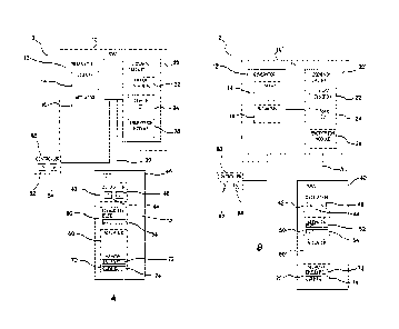
electrosurgical
tool 320 comprises visual indicators 322 such as multi-color LEDs positioned
thereon to
apprise a user of the status of the tool. In other embodiments, the
electrosurgical tool
320 can be electrically coupled to a generator or a different electrosurgical
unit. In
some embodiments, a manual controller such as a hand of foot switch can be
electrically coupled to the ESU 310 or the electrosurgical tool 322 to allow
selective
control of the tool.
Integrated Electrosurgical Unit
[0166] With
reference to Figures 3A-3C, of electrosurgical unit 410 is
illustrated in perspective, front, and rear views. The electrosurgical unit
410 can be an
integrated ESU as discussed above with respect to Figure 1A, and can comprise
a
generator and a feedback circuit. In some embodiments, the housing or console
of the
electrosurgical unit 410 can be sized and configured to fit on a standard
operating room
cart or storage rack. In some
embodiments, the housing or console of the
electrosurgical unit 410 can be configured to be stackable with other surgical
electrical
equipment.
[0167] With
reference to Figures 3A-3B, a perspective view of the
electrosurgical unit 410 is illustrated. In the illustrated embodiment, the
electrosurgical
unit 410 comprises two dedicated tool ports 412, one bipolar tool port 414,
and one
electrical power port 416. In other embodiments, electrosurgical units can
comprise
different numbers of ports. For example, in some embodiments, an
electrosurgical unit
can comprise more or fewer than two dedicated teleports 412, more or fewer
than one
bipolar tool port 414, and more or fewer than one power port 416.
[0168] With
continued reference to Figures 3A-3B, each dedicated tool port
412 is configured to be coupled to electrosurgical tool having a memory, as
described
above with respect to Figure 1A. Thus the dedicated tool ports 412 can be
electrically
coupled to the feedback circuit of the electrosurgical unit 410 as well as the
generator.
CA 3022982 2018-11-01
- 27 -
In some embodiments, the dedicated tool ports 412 con comprise multi-pin
connectors
comprising a plurality of electrical connection pins or pin receptacles. In
some
embodiments, the connectors can comprise more than 10, for example 20 pins or
pin
receptacles. As discussed above with respect to Figure 1A, and discussed in
further
detail below, the dedicated tool ports 412 can be configured for encrypted
transmission
and reception of data from an electrically coupled electrosurgical tool.
[0169] With
continued reference to Figures 3A-3B, the bipolar tool port 414
can include a plug configured to receive a conventional bipolar
electrosurgical tool. The
bipolar tool port 414 can be coupled to the generator of the electrosurgical
unit 410. In
some embodiments, the bipolar tool port 414 is not coupled to the feedback
circuit of
the electrosurgical unit 410. Thus, advantageously, the electrosurgical unit
410 can
energize both specialized electrosurgical tools, as described in further
detail here and,
conventional bipolar electrosurgical tools. Accordingly, the electrosurgical
unit 410 can
be used in place of a standalone bipolar electrosurgical generator without
requiring
additional rack or cart space in a surgical workspace.
[0170] With
continued reference to Figures 3A-3B, the electrical power port
416 can be coupled to the generator of the electrosurgical unit 410. The
electrical
power port 416 can be configured to supply direct current. For example, in
some
embodiments, the electoral power port 416 can provide approximately 12 Volts
DC.
The electrical power port 416 can be configured to power a surgical accessory,
such as
a respirator, pump, light, or another surgical accessory. Thus,
advantageously, in
addition to replacing electrosurgical generator for standard bipolar tools,
the
electrosurgical unit 410 can also replace a surgical accessory power supply.
In some
embodiments, replacing presently-existing generators and power supplies with
the
electrosurgical unit 410 can reduce the amount of storage space required on
storage
racks cards or shelves in the number of mains power cords required in a
surgical
workspace.
[0171] With
continued reference to Figures 3A-3B, the electrosurgical unit 410
can comprise a display 420. In some embodiments, the display can comprise a
multi-
line display capable of presenting text and graphical information such as for
example an
CA 3022982 2018-11-01
- 28 -
LCD panel display, which, in some embodiments can be illuminated via backlight
or
sidelight. In some embodiments, the display 420 can comprise a multi-color
display that
can be configured to display information about a particular tool electrically
coupled to
the electrosurgical unit 410 and a color that corresponds to a standard color
associated
with a surgical procedure (such as, for example cutting operations displayed
in yellow
text and graphics, fusion or welding operations displayed in purple, and
coagulation
displayed in blue, bloodless dissection operations can be displayed in yellow
and blue).
In some embodiments, as discussed in further detail below, the display can be
configured to simultaneously indicate status data for a plurality of tools
electrically
coupled to the electrosurgical unit 410. In some embodiments, a user can
toggle the
display 420 between presenting status of multiple electrically connected tools
and status
of a single electrically connected tool. Further exemplary aspects of the
display are
discussed generally with respect to Figures 4A and 4B, and more specifically
with
respect to operation of the system below.
[0172] With
continued reference to Figures 3A-36, the electrosurgical unit can
comprise a user interface such as, for example a plurality of buttons 422. The
buttons
422 can allow user interaction with the electrosurgical unit such as, for
example,
requesting an increase or decrease in the electrical energy supplied to one or
more
tools coupled to the electrosurgical unit 410. In other embodiments, the
display 420 can
be a touch screen display thus integrating data display and user interface
functionalities.
In some embodiments, the electrosurgical unit 410 can comprise an audible
indicator,
such as a speaker or chime to alert a user of a possible error, the
termination of
electrical energy supplied, or other conditions. In some
embodiments, the
electrosurgical unit 410 can be configured such that the audible indicator can
sound a
particular sound during cutting operations, a different sound during fusion or
welding
operations, and another distinct sound during coagulation operations to
provide audible
feedback to a user.
[0173] With
reference to Figure 3C, a rearview of the electrosurgical unit 410
is illustrated. In the illustrated embodiment, the rear of the electrosurgical
unit 410
includes a rear panel 430. The rear panel 430 can include various ports, such
as a
CA 3022982 2018-11-01
- 29 -
controller port 432 configured to be electrically coupled to an external
controller such as
a foot pedal controller, as described above with respect to Figure 1A. The
rear panel
430 can also include a grounding lug. In other embodiments, one or more
controller
ports and/or the grounding lug can be located on another face of the
electrosurgical unit
410, for example on the front face or a side face. The rear face of the
electrosurgical
unit 410 can include a power module 440 including a mains power port
configured to be
plugged into an AC power mains such as a wall socket and a master power switch
for
powering the electrosurgical unit 410 on and off. In other embodiments, the
master
power switch can be positioned on another face of the electrosurgical unit
410, for
example on the front face or a side face. The rear phase of the
electrosurgical unit 410
can also include a heat exchange feature, such as, for example slots, a grill,
or a
plurality of louvers 450. In other embodiments, the heat exchange feature can
be
positioned on another face of the electrosurgical unit 410, for example on the
front face
or a side face. The heat exchange feature can enhance air or other fluid
cooling of the
generator, the feedback circuit, and other electrical components housed within
the
electrosurgical unit 410 console.
[0174] With
reference to Figure 4A, an exemplary screen shot of the display is
illustrated. In the illustrated embodiment, the display 420 can be portioned
to display
status information for ADC tools 460, a bipolar tool 470, a first
radiofrequency
electrosurgical tool 480, and a second radiofrequency electrosurgical tool
490,
corresponding to the four ports on the front panel of the electrosurgical unit
410
discussed above with respect to Figures 3A, 3B in the illustrated screenshot,
a first
section 462 displays information regarding the DC tool 460. A second section
472
displays information regarding the bipolar electrosurgical tool 470. A visual
indicator
such as a status bar graph 474 can be used to illustrate a proportion of total
available
electrical energy to be applied to the bipolar electrosurgical tool 470 when
actuated. As
discussed above, the visual indicator can be color-coded to indicate a
surgical
procedure to be performed. A third section 482 can display information
regarding a first
radiofrequency electrosurgical tool 480 with a visible indicator such as a
status bar
graph 484. A fourth section 492 can display information regarding a second
radiofrequency electrosurgical tool 490 with separate visual indicators or bar
graphs
CA 3022982 2018-11-01
- 30 -
494, 496, 498 for each type of surgical operation that can be performed for
that tool.
For example an electrosurgical tool operable to cut, coagulate, or fuse tissue
could have
three color-coded bar graphs. The display 420 can also include a controller
icon, such
as a foot pedal icon 476 positions in a section corresponding to a tool to
which a foot
pedal is electrically coupled.
[0175] With reference to Figure 4B, another exemplary screen shot of
the
display 420 is illustrated. It is illustrated, the display has been configured
to maximize
information presentation of the section 492 corresponding to the second of
electrosurgical tool. As discussed above, in some embodiments electrosurgical
unit can
be configurable display status information regarding a single tool
electrically coupled
thereto. In some embodiments, the electrosurgical unit can allow user
manipulation of
energy levels applied to electrosurgical tool. In one configuration, energy
levels for an
electrosurgical tool can be adjusted proportionally for each type of
electrosurgical
procedure to be performed by the tool. For example, a user can increase or
decrease a
master energy level which correspondingly increases or decreases the energy
levels
supplied to you electrosurgical operation performed by the tool, which can be
reflected
in the bar graphs 494, 496, 498 on the display 420. In another configuration,
energy
levels for electrosurgical tool can be manipulated in a procedure-specific
manner. For
example, a user can increase or decrease in energy level corresponding to one
of the
electrosurgical procedures performed by specific electrosurgical tool while
leaving
energy levels for other electrosurgical procedures unchanged. This change can
be
reflected in one of the bar graphs on the display 420, for example, the cut
bar graph
494.
Electrosurgical System Phase Angle Operation
Electrosurgical Unit
[0176] Generally, an electrosurgical unit is provided that includes an
electrosurgical generator, an electrosurgical controller and one or more
electrosurgical
CA 3022982 2018-11-01
- 31 -
tool. The controller can be incorporated in or attached to the generator with
the tool
attached to the controller.
[0177] In one embodiment, a controller is attachable to various
electrosurgical
generators. The generator attached to the controller provides the supply of RF
energy
as directed by the controller. The controller provides feedback control and
preprogrammed settings for the various attachable generators. This is largely
enabled
by using an internal measurement signal that is independent from the attached
generator. In other words, regardless of the driving frequency of the drive
signal the
generator generates (which has an impact on the end point measurement, e.g.,
the
phase shift), the measurement signal and hence the final phase shift remains
the same.
[0178] Referring to Figure 5, in one embodiment, an electrosurgical
generator
includes an RF amplifier, pulse width modulator (PWM) and relays. The
electrosurgical
generator is coupled to a 120Hz Voltage main input. The main input is isolated
with a
low leakage isolation transformer of a power supply 631. The power supply
provides
operational voltages for the control processor 637 and the RF amplifier 633.
Additionally, the power supply includes two 50VDC output modules connected in
series
to provide a total output of 100VDC and 8 Amps. RF power is generated by the
RF
amplifier, e.g., a switched mode low impedance RF generator that produces the
RF
output voltage. In one embodiment, a 600 peak cut voltage for cutting and 10
Amp
current for coagulation/fusing is generated.
[0179] Fusing tissue in one embodiment involves applying RF current to
a
relatively large piece of tissue. Because of the potentially large tool
contact area tissue
impedance is very low. Accordingly, to deliver an effective amount of RF
power, the
current capability of the RF amplifier is large. As such, where a typical
generator might
be capable of 2 to 3 amps of current, the RF amplifier of the generator can
supply more
than 5 Amps RMS into low impedance loads. This results in rapid tissue fusion
with
minimal damage to adjacent tissue.
[0180] The RF amplifier circuitry has redundant voltage and current
monitoring. One set of voltage and current sensors are connected to the PWM
circuitry
CA 3022982 2018-11-01
- 32 -
and are used for servo control. The voltage and current can also be read by
the
processor 637 using an analog to digital converter (ADC) located on the PWM
circuitry.
The PWM circuitry also has an analog multiplier, which calculates power by
computing
the product of the voltage and current. The PWM circuitry uses the average
value of
voltage and current and does not include a phase angle and thus is actually
calculating
Volt Amps Reactive (VAR) rather than actual power. A second set of voltage and
current sensors are also connected to the Telemetry circuitry 642. The signals
are
connected to an ADC for redundant monitoring of the voltage and current. The
processor multiplies the voltage and current readings to verity that power
output does
not exceed 400 Watts. The Telemetry circuitry has monitoring circuits that are
completely independent of the PWM circuitry. This includes the ADC, which has
an
independent voltage reference.
[0181] The RF amplifier in one embodiment is a switching class D push
pull
circuitry. As such, the amplifier can generate large RF voltages into a high
tissue
impedance, as well as large RF currents into low tissue impedance. The output
level of
the RF amplifier is controlled by Pulse Width Modulation (PWM). This high
voltage
PWM output signal is turned into a sine wave by a low pass filter on the RF
amplifier.
The output of the filter is the coagulation output of the RF amplifier. The
output is also
stepped up in voltage by an output transformer resulting in the cut output of
the RF
amplifier. Only one output is connected to the control servo on the PWM
circuitry at a
time and only one output is selected for use at a time.
[0182] Coupled to the RF amplifier is the PWM circuitry 634. The PWM
634
in one embodiment receives voltage and current set points, which are input by
the user
through a user interface, to set the output level of the RF amplifier. The
user sets points
are translated into the operating levels by digital to analog converters of
the PWM. The
user sets points are translated into the operating levels by digital to analog
converters of
the PWM. The set points in one embodiment include a maximum voltage output,
maximum current output, maximum power output, and a phase stop. The servo
circuit
of the PWM circuitry controls the RF output based on the three set points. The
servo
circuit as such controls the output voltage of the RF amplifier so that the
voltage,
CA 3022982 2018-11-01
- 33 -
current, and power set points are not exceeded. For example, the output of the
ESG is
restricted to be less than 400 watts. The individual voltage and current set
point can be
set to exceed 400 watts depending on the tissue impedance. The power servo
however
limits the power output to less than 400 watts.
[0183] The RF output voltage and current are regulated by a feedback
control
system. The output voltage and current are compared to set point values and
the output
voltage is adjusted to maintain the commanded output. The RF output is limited
to 400
Watts. Two tool connections are supported by using relays 635 to multiplex the
RF
output and control signals. The EMI line filter 636 limits the RF leakage
current by the
use of an RF isolation transformer and coupling capacitors.
[0184] The cut and coagulation output voltages of the RF amplifier are
connected to the relay circuitry 635. The relay circuitry in one embodiment
contains a
relay matrix, which steers the RF amplifiers output to one of the three output
ports of the
electrosurgical unit. The relay matrix also selects the configuration of the
tool
electrodes. The RF output is always switched off before relays are switched to
prevent
damage to the relay contacts. To mitigate against stuck relays steering RF to
an idle
output port each output port has a leakage current sensor. The sensor looks
for
unbalanced RF currents, such as a current leaving one tool port and returning
through
another tool port. The current sensors on are located on the Relay PCB, and
the
detectors and ADC are on the Telemetry PCB. The CPU monitors the ADC for
leakage
currents. Any fault detected results in an alarm condition that turns off RF
power.
[0185] The relay circuitry also contains a low voltage network analyzer
circuit
used to measure tool impedance before RF power is turned on. The circuit
measures
impedance and tissue phase angle. The processor 637 uses the impedance
measurement to see if the tool is short-circuited. If a Tool A or B output is
shorted the
system warns the user and will not turn on RF power. The RF amplifier is fully
protected against short circuits. Depending on the servo settings the system
can
operate normally into a short circuit, and not cause a fault condition.
CA 3022982 2018-11-01
- 34 -
[0186] Voltage and current feedback is provided using isolation
transformers
to insure low leakage current. The control processor 637 computes the power
output of
the RF amplifier and compares it to the power set point, which in one
embodiment is
input by the user. The processor also monitors the phase lag or difference
between
current and voltage. Additionally, in one embodiment, the processor matches
the
different phase settings, which depend on tissue types to the monitored phase
difference. The processor as such measures a phase shift of tissue prior to
any
application of RF energy. As will be described in greater detail below, the
phase
measurement is proportional to tissue permeability and conductivity that
uniquely
identifies the tissue type. Once the tissue type is identified, the phase
angle associated
with an end point determination of that tissue type can be determined. The
generator in
one embodiment has three RF output ports (Tool A, Tool B and generic bipolar).
The
tool A and B ports 639 are used to connect smart tools, while the generic
bipolar port
640 supports standard electro surgical tools. Audible tones are produced when
the RF
output is active or an alarm condition exists.
[0187] The hand and foot controls are also isolated to limit leakage
current.
The control processor checks the inputs for valid selections before enabling
the RF
output. When two control inputs from the switches are simultaneously activated
the RF
output is turned off and an alarm is generated. Digital to analog converters
are used to
translate control outputs into signals useable by the Analog Servo Control.
The control
set points are output voltage and current. The analog to digital converter is
used to
process the analog phase angle measurement. Voltage RMS, current RMS, and
power
RMS information from the controller is also converted into a form usable for
presentation to the user. The digital I/O bus interface 638 provides digital
communication between the user, controller and hand/foot switches. Isolation
circuitry
is used to eliminate a possible leakage path from the electrosurgical
generator. It also
provides communication between the user and the generator though a data
channel
protocol.
[0188] In one embodiment, there are four tool Interface circuits in the
unit.
These circuits are used to electrically isolate the user input switches from
mains power
CA 3022982 2018-11-01
- 35 -
inside the system. The four tool interface circuits are identical and have an
on board
microprocessor to read the user switch inputs as well as the tool crypto
memory and
script memories. The switch closure resistance is measured with an ADC to
eliminate a
contaminated switch contact being read as a closure. Switch closures below 300
Ohms
are valid, while any reading above 1000 Ohms is open. Readings between 300 and
1000 Ohms are considered to be faulty inputs.
[0189] The four
tool interface circuits communicate with the processor using
an RS485 network. Each tool interface circuit has jumpers to select its
address and
location in the unit. The RS485 interface is isolated to eliminate any
potential leakage
current paths. One tool interface circuit is connected to each of the Tool A
and B ports.
A third tool interface circuit is connected to the DC output port, and the
fourth circuit is
connected to the rear panel foot switch inputs. The processor is the network
master and
each of the four circuits is a network slave. The processor polls each circuit
for input.
The tool interface circuitry can only reply to commands. This makes the
network
deterministic and prevents any kind of dead lock. Each Tool Interface circuit
is
connected to a System OK logic signal. If a system error is detected by a Tool
Interface
circuit, this signal is asserted. The processor monitors this signal and
indicates a fault.
This signal also has a hardware connection to the PWM circuit and will disable
the RF
amplifier when asserted. A system error could be two input switches activated
at the
same time, or a loss of communication with the processor. The Tool A & B ports
as well
as the DC port have a micro switch that detects when a tool is plugged into
the
receptacle. Until this switch is depressed the Tool Interface circuit front
panel
connections are configured off to prevent any leakage current flowing from
front panel
connections. Once the switch is depressed the Tool Interface allows the
processor to
initiate reads and writes to the tool crypto memory and script memory. Once a
tool is
detected a window opens in the user interface display showing the type of tool
connected and status. The generic bipolar port supports legacy tools, which do
not
have any configuration memory. The tissue measurement circuitry is used to
monitor
the bipolar connection contacts. When a bipolar tool is connected the tool
capacitance
is detected and the processor opens the bipolar tool window on the user
interface
display and shows status for the bipolar tool. The DC port is used to
interface with 12
CA 3022982 2018-11-01
- 36 -
Volt DC powered custom surgical tools. When a tool is plugged into this port a
window
opens in the user interface display showing the type of tool connected and
status. When
the DC tool script commands power on, the processor closes a relay on the
Power
Control and Isolation circuitry 643 turning on the isolated 12 Volt tool
power.
[0190] The power control and isolation circuitry 643 has two other
features. It
controls the 100 Volt power supply that drives the RF amplifier. This power
supply is
turned on by a relay controlled from the PWM circuitry. The processor commands
this
power supply on via the PWM circuitry. If the PWM circuitry is reset or
detects a fault
condition, the relay will not operate leaving the 100 Volt power supply off.
Also located
on the power control and isolation circuitry is a RS485 isolation circuit that
adds an extra
layer of isolation.
[0191] The front panel interface circuitry 641 is used to connect the
front
panel control switches and LCD display to the processor. The front panel
interface
circuitry also contains a microprocessor, which is powered by an isolated
standby power
supply, which is on whenever the main power switch is on. When the front panel
power
switch is pressed, the microprocessor uses a relay on the Power Control and
Isolation
circuitry to turn on the main logic power supply. When the button is pressed
to turn
power off, the microprocessor signals a power off request to the processor.
When the
processor is ready for power to be turned off it signals the microprocessor to
turn off
power. The power control relay is then opened, turning off the main power
supply.
[0192] In one embodiment, the generator accepts only single switch
input
commands. With no RF active, e.g., RF energy applied, multiple switch
closures, either
from a footswitch, tool, or a combination of footswitch and tool are ignored.
With RF
active, dual closures shall cause an alarm and RF shall be terminated. The
footswitch
in one embodiment includes momentary switches providing activation of the
application
of RF energy. The switches for example when manipulated initiates activation
of the RF
energy for coagulation, for cutting and/or sequenced coagulation or cutting. A
two-
position pushbutton on the foot pedal switch allows toggling between different
tools.
The active port is indicated on the display of the generator and an LED on the
hand tool.
CA 3022982 2018-11-01
- 37 -
[0193] In one embodiment, all RF activation results in a RF ON Tone.
Activation tone volume is adjustable, between 40dBA (minimum) and 65dB
(maximum)
with a rear panel mounted control knob. The volume control however does not
affect
audio volume for alarms. Also, in one embodiment, a universal input power
supply is
coupled to the generator and operates over the input voltage and frequency
range
without the use of switches or settings. A programming port in one embodiment
is used
to download code to the generator and is used to upload operational data.
[0194] The generator in one embodiment provides output power has a 12V
DC at 3 Amps. Examples of such tools that use DC power are, but are not
limited to, a
suction/irrigation pump, stapler, and a morcellator (tool for dividing into
small pieces and
removing, such as a tumor, etc.). The DC connector has intuitive one-way
connection.
Similar to the other tool receptacles, a non-sterile electronic chip module is
imparted
into the connector of the appropriate DC-powered hand tool by a one-time, one-
way
locking mechanism. Tool-specific engravings on both the connector and chip
module
ensure that the chip module fits only to the type of tool for which it has
been
programmed. The chip connector allows tool recognition and the storage of data
on tool
utilization. The DC connector is also configured to prevent improper
insertion. The
generator is also configured to recognize the attached DC-powered tool. The
generator
reads configuration data from the tool connector, allowing tool recognition
and the
storage of tool utilization data.
[0195] The controller in one embodiment recognizes the tool upon the
tool
being attached to the controller. Based on the recognized tool, the generator
accesses
and initiates specific operations and setting parameters utilized to configure
the
controller to properly apply RF energy as desired by the tool. For example,
parameters
set includes but not limited to an automatic preset of the output voltage,
activation of
specific output pins (connected to tool) or determination of the feedback
cycle.
[0196] In one embodiment, the controller supplies control signals
and/or
power to a connected tool to indicate when they are active via a LED and/or a
distinctive
audio tone. The controller is also arranged to display when and/or which
specific tool is
CA 3022982 2018-11-01
- 38 -
active. The controller also prevents the tool from being reused after certain
expiration of
the tool shelf life, or a specific time period after the first tool
activation.
[0197] In one embodiment, phase measurement is a relative measurement
between two sinusoidal signals. One signal is used as a reference, and the
phase shift
is measured relative to that reference. Since the signals are time varying,
the
measurement cannot be done instantaneously. The signals must be monitored long
enough so that difference between them can be determined. Typically the time
difference between two know points (sine wave cross through zero) are measured
to
determine the phase angle. In the case of the phase controller, the device
makes the
output sine wave with a precise crystal controlled clock. That exact same
clock is use
to read the input samples with the analog to digital converter, In this way
the output of
the phased controller is exactly in phase with the input of the phase
controller. The
phase controller in one embodiment compares the input sine wave signal to a
reference
sine wave to determine the amount of phase shift.
[0198] The phase controller does this comparison using a mathematical
process known as a Discreet Fourier Transform (DFT). In this particular case
1024
samples of the input signal are correlated point by point with both a sine
function, and a
cosine function. By convention the cosine part is called real, and the sine
part is called
imaginary. If the input signal has no phase shift the result of the DFT is
100% real. If
the input signal has a 90-degree phase shift the result of the DFT is 100%
imaginary. If
the result of the DFT has both a real and imaginary component, the phase angle
can be
calculated as the arctangent of ratio of the imaginary and real values.
[0199] It should be appreciated that the phase angle calculation is
independent of units of the real and imaginary numbers. Only the ratio
matters. The
phase results of the phase controller are also independent of gain and no
calculation of
impedance is made in the process of calculating the phase angle. By performing
a
DFT, the phase controller encodes the phase measurement as a pair of numbers.
[0200] A user interacts with the electrosurgical unit via a graphical
panel
display and associated switches 641. The front panel switches allow
interaction with
CA 3022982 2018-11-01
- 39 -
LCD display menus generated on the graphical panel display. The menus allow
language selection, and modification of tool set points. In one embodiment,
only when
a tool is plugged in and detected by the unit, parameters can be changed for
that tool.
[0201] The electrosurgical unit as described above includes one or more
receptacles in which electrosurgical tools connect to the unit. Through this
connection,
a tool and unit communicate with each other. Connecting the tool also causes
the
controller to update the display of the system to show tool information and
current
intensity.
[0202] An example of a display or user interface 641 is shown in Figure
6.
The user interface provides tool information such as tool status for each
connected tool
and allows a user to modify set points, e.g., the application or intensity of
the RF
energy. The user interface in one embodiment also shows the tool settings for
functions
for each connected tool. In the illustrated embodiment, three tools are
connected to the
generator. Accordingly, a suction/irrigation pump display 621, a Kii fusion
tool display
622 and a spatula tool display 623 are shown. Associated operations or actions
available for each tool are also provided in which the suction/irrigation pump
has an
on/off setting 624; the Kii fusion tool has relative power settings for cut
625, coagulation
626 and fuse 627; and the spatula tool has relative power settings for cut 628
and
coagulation 629.
[0203] In one embodiment, the user interface allows a simultaneous
change
to all settings for a selected tool (indicated by the highlighted rim 631) by
pushing single
button from the navigation buttons 632. For example, as shown in Figure 7,
pushing the
"up" button 633 will simultaneously change the cut, coagulation and fuse
relative power
settings for the connected Kii fusion tool. Additionally, the settings can be
changed
individually by navigating into a sub menu, as shown in Figure 8. In the
illustrated case,
the coagulation level of the Kii fusion tool is changed without changing the
cut and/or
fuse relative power setting. By selecting the default button 634, the settings
for all tool
functions of the selected tool are returned to the default setting. Also, as
warranted by
the context, an associated button operation and corresponding label can vary
as shown
in button 635 being a menu button in Figure 7 and a back button in Figure 8.
CA 3022982 2018-11-01
- 40 -
[0204] A block diagram illustrating a controller in accordance with
various
aspects of the invention is shown in Figure 9. As shown, the output of a
generator is
fed into circuitry that determines the frequency of the driving signal and
circuitry to
measure the phase shift between voltage and current applied to the tissue. The
voltage
applied by the generator is sent through a buffer/level shifter 541 that
reduces the
amplitude of the output voltage. The signal is processed to deliver the
frequency of the
generator output via frequency measurement 542 and fed into a microcontroller
543.
The frequency of the driving signal can directly impact the phase shift.
Similarly, the
generator output is sent through a signal conditioning circuitry 544 to reduce
high-
frequency noise, and then conditioned via voltage and current conditioning
545a-b and
filtered by multi-pole low pass filter 546a-b to deliver signals to represent
applied
voltage and current. Both signals representing voltage and current are
measured for
phase shift using a phase comparator 547. The output of the phase comparator
is fed
into the microcontroller 543. Depending on the frequency of the
electrosurgical unit
used, which can determine the final phase shift to be reached, the
microcontroller
compares the output of the phase comparator with the trigger level determined
by the
driving frequency of the generator. When such trigger level is achieved, i.e.,
the tissue
fusion or welding is completed, the microcontroller 543 causes the tissue to
be
disconnected from the generator and indicates that state by acoustical or
visual
indicators 548 (buzzer, display, lights, etc.). An over-voltage detector 549
is also
provided that is supplied the generator output to detect excessive voltage the
condition
of which is supplied to the microcontroller 543.
[0205] Figure 10 shows a block diagram of a controller in accordance
with
various embodiments of electrosurgical unit utilizing the phase shift between
voltage
and current to determine the end-point of the fusion process. A
microcontroller 553
delivers a low-voltage square-wave signal 551 at 5MHz, which is converted by a
4-pole
low pass filter 550 into a low-voltage sin-wave signal 552 at 5 MHz. The low-
voltage
5MHz signal is superimposed to the output of the generator, which is typically
in the 100
to 200V range at frequencies of 300 to 500 kHz. As an example, the
superimposed
voltage signal of a 200V driving voltage at 500kHz and a 5V measurement
voltage at 5
MHz is shown in Figure 11.
CA 3022982 2018-11-01
- 41 -
[0206] The combined voltages are then applied to the tissue and, just as
in
the previous example, also conditioned through a buffer/level shifter
circuitry for
processing. Similarly, the current through the tissue is measured and also
conditioned
for processing. The processed voltage (and current) signal containing the high
voltage
(and high current) signal at 300 to 500kHz from the ESU, as well as low
voltage (low
current) signal at 5MHz are sent through a multi-pole band pass filter
centering at
5MHz. The filter discriminates the signal from the ESU, leaving only the two
signals at
5MHz for measuring the phase shift in a phase comparator. The filtered signals
for both
the voltage and current at 5MHz are illustrated in Figure 12 at a time near
the end of the
fusion process.
[0207] The measured phase shift is fed into a microcontroller, which
compares the reading with a pre-determined level indicative to the completion
of the
fusion process at 5MHz frequency. Again, when such a trigger level is
achieved, i.e.,
the tissue fusion or welding is completed, the microcontroller 553 will cause
the tissue to
be discontinued from the generator and indicate that state by acoustical or
visual
indicator 548 (buzzer, display, lights, etc.).
[0205] Figure 13 shows a schematic block diagram of one aspect of a
controller. As shown, a microprocessor 561 times the switching of the tissue
between
the output of a generator and an internal measurement circuit. As a result,
the tissue is
periodically assessed for the status of the fusion process by measuring the
phase shift
of a low-voltage and low-current measurement signal. Depending on the value of
the
obtained phase shift, the tissue is either switched back to the high-voltage
output of the
generator for further fusion, or permanently disconnected from the generator.
As such,
the internal circuit comprises of a microprocessor 561 generating a low-
voltage square
wave signal 562 at 500kHz that is transferred into a low-voltage sinusoidal
wave 563 at
500kHz. This signal is applied to the tissue, and analyzed by a phase
comparator 564
only when it electrically disconnected from the generator during regular
measurement
intervals.
[0209] In one embodiment, the phase shift is derived directly from the
driving
signal, i.e., the voltage and current supplied by the electrosurgical
generator to the
CA 3022982 2018-11-01
- 42 -
tissue. In one embodiment, an electrical circuit modifies the driving voltage
having one
(sinusoidal) frequency by superimposing a measurement signal at a vastly
different
frequency. As a result, electrical energy for the fusion process is provided
at one
frequency, while simultaneously applying as second signal at a second
frequency for
measurement. Separation of the two different signals by using band pass
filters in the
measurement circuit allows continuous measurement of the phase shift during
the
electrosurgical fusion or welding process. In one embodiment, the controller
periodically
interrupts the supply of electrosurgical energy to assess the status of the
fusion or
welding process by applying a low-voltage measurement signal. Depending on the
phase shift obtained during the measurement cycle, the controller switches the
driving
signal from the generator back to the tissue or isolates the tissue. In one
embodiment,
the controller interrupts the tissue fusion or welding process at a pre-
determined level of
phase shift by terminating the supply of RF energy from the generator to the
tissue.
[0210] Figure 14 depicts a controller or control unit in accordance
with
aspects of the present invention for the controlled fusion or welding of
biological tissue.
As shown, the control unit is connecting the bipolar power outlet of a
generator 507 to
the tool 508 that is arranged to compress vessels or tissue. The tool also
houses a
switch 509 that activates the fusion process. If the generator is equipped
with an input
for hand activation (rather than using a foot pedal 511 or other intermediary
device), a
third connection 512 from the control unit to the generator allows activation
of the
generator with the same hand switch.
[0211] The controller in one embodiment includes a processor 513 that
controls the switching of the tissue between the direct output of the
generator and an
internal measurement circuit, e.g., switch 515. It is powered with an internal
battery
power module 514. The timed switching causes the tissue to be fused in
intervals while
periodically measuring the status of the tissue. As such, the measurement
signal is a
500kHz sinusoidal low voltage signal, generated by a signal generator 518 when
fed
with a 500kHz square wave from the microprocessor 513. When the low-voltage
sinusoidal measurement signal is applied to the tissue, a phase comparator 516
measures the phase shift between the applied measurement voltage and the
current
CA 3022982 2018-11-01
- 43 -
caused by application of the measurement voltage. Depending on the result
analyzed
or processed by the processor, the tissue will be either be switched back to
the
generator, or disconnected from the generator accompanied by an acoustical
and/or
visual indication via LEDs/buzzers 517.
[0212] Figure 15 shows in one embodiment of the external measurement
circuit that generates the low-voltage sinusoidal signal used to measure the
phase shift.
It is generated by passing a 500kHz square wave through a 4-pole low-pass
active filter
531. The 4-pole low pass filter removes higher harmonic components and passes
the
sinusoidal fundamental frequency. The 500KHz square wave is generated via the
PWM
peripheral 522 in the microcontroller 524.
[0213] Figure 16 illustrates switch 515 configured to switch between
the
application of the drive signal and the measurement signal, e.g., the 500 kHz,
5 Volt
peak-to-peak sine wave reference signal, from the generator. Although the use
of a
solid-state switch to implement the switching offers a long operational life
and inherent
current surge control, it can be difficult to block the relatively high
voltage (-200VAC)
and high frequency (-500KHz) signal generated by a typical generator in
bipolar
coagulation mode. As such, two double pole, double-throw mechanical relays
527,528
are used. The first relay 527 switches between the generator and the reference
signal.
The second relay 528 limits the current surge, which can damage the relay and
create
an electromagnetic interference (EMI) pulse that can disrupt the low-voltage
circuitry.
Additionally, this protects the tissue against complications or issues caused
by electrical
arcing. Since most generators are constant power devices, the highest voltages
occur
during conditions of no load. By first switching in the generator through a
series
resistor, the output voltage of the generator is shared across the resistor,
limiting the
voltage imparted to the tissue. Furthermore, the resistor serves as an energy
limiter,
enabling high conductive channels in the tissue to fuse before the full power
of the
generator is applied.
[0214] In one embodiment, switching takes place in the following
sequence.
When switching from the low voltage measurement or reference signal to the
generator,
the first relay 528 switches out both ends of the reference and switches in
one
CA 3022982 2018-11-01
- 44 -
generator lead directly and one through a 100 Ohm resistor. The 100 Ohm
resistor
limits the surge current to two amps for a 200 Volt source. If a shorted
output occurs,
400 watts are dissipated in the 3 Watt resistor, which would quickly burn up.
However,
approximately 50 milliseconds after the first relay 528 switches in the
generator, a
second relay 527 switches out the 100 Ohm resistor, keeping it from burning up
and
allowing the full power of the generator to be delivered to the tissue. When
the device
switches the other way (from the ESU to the reference signal), it first
switches in the 100
Ohm resistor, reducing the current, and then switches out the generator
entirely. This
sequence reduces inductive kickback and EMI generation.
[0215] The relays 527,528 in one embodiment are of a latching type.
Most
mechanical relays draw a fair amount of power in their non-default state (an
electrical
current is needed to fight the force of the returning spring). Since the
controller is
equipped with a battery of limited power capacity, two latching type relays
are used.
These relays only use current to transition between two stable states and can
operate at
a much lower power level.
[0216] The phase detection circuitry 530 is shown in Figure 17, which
measures the phase shift between the two above-mentioned sine waves. The first
part
of the circuit level-shifts the sine wave to the same DC value as a reference
voltage.
The level-shifted signal is then sent to the negative input of a comparator
531. The
positive input is connected directly to the DC reference voltage. A small
amount of
hysteresis is used to reduce switching noise. The output of the comparator is
a square
wave with the same phase as the input sine wave. These two signals are sent to
an
exclusive "OR" gate 532. The output of the gate is high when one of the two
inputs is
high, and low otherwise. The duty cycle of the output is therefore linearly
related to the
phase of the two input square waves. The duty cycle is converted to a DC
voltage
through a low pass filter, which is measured by the analog to digital
converter peripheral
of the microcontroller.
[0217] Figure 18 shows the battery power circuit that is powering the
control
circuit by two low-capacity coin cells. The battery provides a life of 500
fusing cycles
over a 5-hour time span. When a specific number of seals, or a specific time
limit have
CA 3022982 2018-11-01
- 45 -
been reached, the controller issues a warning and ceases operating. The
controller
manages its power demand around the power characteristics of the specific
batteries
used. The controller includes management controls that prevent specific
operations
from occurring simultaneously that may exceed the power capacity of the
batteries,
power down selected portions of the circuit between fusing cycles, and slow
the
microcontroller oscillator down from 4 MHz to 32kHz between fusing cycles.
[0218] Figure 19 shows an input port 534 adapted for connecting to a
tool.
With engagement of a switch on the tool, the controller takes initial
measurements on
the tissue (shorting, etc.) and based on the initial measurements activates
the generator
to supply electrosurgical power that is passed and controlled by the
controller.
[0219] As many generators can exclusively (but also alternatively,
with the
surgeons preference) be activated with a foot-pedal, the controller
accommodates such
a scenario. For example, if the generator is activated with a foot switch
while
subsequent activation of the hand switch on the tool occurs, the controller
allows
switching-in of the output of the generator.
[0220] The result of using the control circuit described above is
shown in
Figure 20, showing the effective voltage applied to the biological tissue as
function of
time. As shown in this specific example of porcine renal arteries, the tissue
is being
exposed to 6 high-power fusion intervals of about 850ms time duration,
interrupted by 5
measurement cycles of about 300ms.
[0221] In one embodiment, the fusion process starts with depressing a
switch
on the tool, which starts an initial measurement sequence. This point in time
is marked
start (switch on) 535. The tool in one embodiment checks the resistance
between the
two electrodes and if the phase shift is within an acceptable range. Verifying
the phase
shift prevents an attempt to re-fuse already fused tissue. Based on the
results of the
initial check, the controller switches-in the activated output of the
generator to the
tissue. This starts the application of RF energy to the compressed tissue.
After about
850ms, the controller disconnects the tissue from the generator and switches
back to
the first tissue assessment phase. Depending on the result, the tissue gets
heated
CA 3022982 2018-11-01
- 46 -
further, or remains disconnected from the generator to remain on the
measurement
circuit. The latter case is marked "power stop (switch on)" 536. In this case,
an
acoustical and/or visual signal is given off the unit, indicated that the
tissue is sealed (or
that shorting of the electrodes has occurred). The supply of the measurement
signal to
the tissue is ended when the switch on the tool is released, marked "manual
stop
(switch off)" 537. At this point, all supply of energy to the tissue is
terminated.
[0222] A more detailed analysis of the measurement cycle 538 is shown
in
Figures 21 and 22, showing that additional measurements (other than the phase
shift)
can be included in that measurement period. Such measurements, for example,
could
prevent attempting to fuse already fused tissue, or powering of electrically
shorted
electrodes.
[0223] In Figure 22, a more detailed analysis of the measurement
plateau 539
of 2V in Figure 21. As shown, a detailed view of the low-voltage measurement
signal
540 at 500kHz used to determine the phase shift through the tissue during the
RF
measurement cycle.
Electrosurgical Systems and Processes
[0224] Electrosurgical systems and processes in various embodiments
apply
monopolar or bipolar high-frequency electrical energy to a patient during
surgery. Such
systems and processes are particularly adapted for laparoscopic and endoscopic
surgeries, where spatially limited access and visibility call for simple
handling, and are
used to fuse blood vessels and weld other biological tissue and in one aspect
to cut,
dissect and separate tissue/vessels. In particular embodiments, the systems
and
processes include the application of RE energy to mechanically compressed
tissue to
(a) desiccate the tissue, and (b) to denature collagens (type I-III) and other
proteins,
which are abundant in most biological tissue. As heating of collagens to an
appropriate
temperature causes them to unfold, shrink or denature, the system enables the
sealing
of capillaries and blood vessels during surgery for permanent occlusion of the
vessels.
As described in greater detail below, as an example, arteries up to seven
millimeters
CA 3022982 2018-11-01
- 47 -
can be occluded and dissected by radio frequency (RE) energy and mechanical
pressure.
[0225] When concurrently applying controlled high-frequency electrical
energy
to the compressed tissue, the tissue is compressed with a relatively high
pressure
(about 10 - 20 kg/cm2), and the tissue is supplied with sufficient electrical
energy to
denature proteins and remove sufficient water in the tissue. During this
process, the
applied voltages are sufficiently reduced to avoid electrical arcing
(typically < 200V
RMS).
[0226] When applying electrical energy in the described manner stated
above,
the tissue quickly moves through the following fusion/welding process.
Starting at body
temperature the tissue (a) heats quickly, leading to (b) cell rupture,
expelling of juices
(mainly water and salt ions), (c) unraveling and "activation" of collagens and
elastin in
the blood vessels at about 60-650 C, and (d) desiccation of the vessel. Here,
the
desiccation process can be seen by the release of water in form of steam where
the
vessel temperature has reached about 1000 C. The reduction of water in
presence of
unraveled collagen and elastin strands leads to formation of bonds between
collagen
strands, leading to a strong and elastic seal of the tissue, As confirmed by
measurements, the strongest (highest burst pressure) vessel fusions are
obtained when
the vessels have been heated to at least 70 C, pressurized with about 10 - 20
kg/cm2,
and then desiccated by about 40-50% of their original water content.
[0227] Electrically, the tissue can be characterized during the fusion
process
by its impedance, which is typically starting at 10-100 Ohms purely ohmic
resistance.
During the fusion process, the purely ohmic resistance reduces by 20-50%
before it
increases by two orders of magnitude. As the resistance approaches a final
value, the
impedance of the tissue gradually increases in capacitive behavior with a
phase shift of
about 20 degrees. The tissue will exhibit a pronounced capacitive behavior at
the end
of the fusion process with a phase shift of about 40 degrees, even though the
ohmic
component will remain nearly unchanged during this phase.
CA 3022982 2018-11-01
- 48 -
[0228] Referring now to Figure 23, graphical representation
exemplifying
experimental data for the sealing of a four-millimeter diameter porcine renal
artery in
accordance with various embodiments of electrosurgical system is shown. The
fusion
process is performed by compressing the artery with 0.75 millimeter wide
electrodes
with a compression load of three pounds, and by energizing it with a voltage-
stabilized
electrosurgical power supply using 200V at 60W maximum power setting. Voltage
501,
current 502 and electrical power 503 in the beginning of the fusion process (1
second)
are shown. As can be seen, the sinusoidal voltage and current are
substantially in-
phase, e.g., the phase difference or angle equals zero. At this time, the
impedance of
the artery is purely ohmic with a value of about 100 Ohms.
[0229] The temporal progression of the applied peak voltage and peak
current
for the same-sized artery is provided in Figure 24. The applied voltage
quickly
stabilizes to a constant value, which is an artifact of the voltage-stabilized
power supply.
Regardless of the applied load, voltage-stabilized electrosurgical power
supplies
regulate the output voltage to a pre-set value since the voltage has a
dominant impact
on the electrosurgical effect. In contrast to the voltage, the current driven
through the
artery increases from an initial 1A to 1.5A at 0.5s, and then gradually
reduces over the
next three seconds to about 0.2A. For the remaining 4 seconds of the fusion
time the
peak value of the current remains nearly unchanged.
[0230] Another way to depict the information from Figure 24 is shown
in
Figure 25, showing the impedance 506 of the artery as function of fusion time.
The
initial impedance of the harvested artery is 75 Ohms. With application of high
frequency
electrical energy the artery heats quickly, leading to shrinkage of
collagen's, rupture of
cell membranes, and the ultimate expelling of trapped liquid (mainly water and
ions). As
a result, the impedance has reduced to about 54 Ohms. Further supply of
electrical
energy starts to desiccate the artery, resulting in an impedance increase. At
about 4
seconds into the fusion process the impedance of the artery starts to
stabilize, with a
slow increase of the impedance from about 800 Ohms to about 1,200 Ohms.
[0231] The fusion process could be terminated (a) at a fixed and
absolute
resistance (for example 2k Ohms), which would neglect both the size and type
of tissue,
CA 3022982 2018-11-01
-49 -
(b) at a specific multiple of the time where the ohmic resistance is minimal,
(c) at a
specific multiple of the time where the ohmic resistance is the same as the
initial one, or
(d) at a specific multiple of the time where the ohmic resistance is a certain
factor of the
minimal one. However, considering burst pressure of fused arteries and thermal
spread,
the termination of the fusion process is determined to be in the flattened
part of the
impedance curve. As can be seen in Figure 25, however, this region is also an
inexact
range for impedance measurements. Similarly, each succession of (a) to (d)
becomes
better in determining the end-point of the fusion time (resulting in the
highest desired
bursting pressure with the least desired thermal spread). Utilizing the ohmic
resistance
only as termination criterion can lead to incomplete results. This can be more
pronounced when fusing differently sized tissues (even of same nature), also
exemplified in Figure 26 showing the relative resistance (relative to the
initial resistance)
of various-sized arteries and other tissue as a function of fusion time.
[0232] Termination of the fusion process for same-material tissue
(i.e.,
arteries) cannot be controlled with desired precision by specifying one
relative ohmic
load (e.g., when the resistance reaches 3 times the initial resistance).
Instead, the
relative change in resistance depends on the size of the vessel, i.e., < 2 mm
arteries
seal in fractions of a second (where the resistance about doubles compared to
the initial
resistance), about 3 mm arteries seal in about 2 seconds (where the resistance
about
triples), and 15 mm arteries/veins seal in about 7 seconds (where the
resistance
increases by a factor of 5). At the same time, some arteries may not follow
that
characterization (e.g., a 3-4mm artery would not reach more than 2.5 times the
initial
resistance). Instead, the fusion process should end within the flat region in
Figure 25.
As previously described, precision is difficult in the flat region with the
function of time at
different fusion times.
Phase Based Monitoring
[0233] In one aspect, the determination of the end-point of the fusion
process
is given by monitoring the phase shift of voltage and current during the
fusion process.
CA 3022982 2018-11-01
- 50 -
Unlike impedance, the phase shift changes much more pronounced at times where
the
artery desiccates and the fusion completes, and hence offers a more sensitive
control
value than the impedance. This can be seen when monitoring the voltage and
current
as function of time at different fusion times, as is shown in Figure 23 for
the beginning of
the fusion process.
[0234] In Figure 23, the beginning of the fusion shows that the
applied voltage
and current are in phase (with a shift of about -3 degrees), revealing that
the artery
behaves dominantly like an ohmic load of about 75 ohms. Further supply of
energy
leads to heating of the artery, an initial reduction in impedance (caused by
shrinking of
collagens, cell membrane rupture and expelling of mainly water and dissolved
ions),
and a subsequent increase in impedance. During this period of vessel fusion,
the phase
difference between voltage and current remains small with minimal changes,
indicating
that the artery is purely ohmic.
[0235] The artery is not fully desiccated, and thus the seal is not
complete.
Referring to Figure 27, at 4 seconds into the fusion process, the phase
difference slowly
increases to -10 degrees (current leads). While further supply of electrical
energy does
not significantly change the value of the resistance (see Figure 24), it does
cause a
pronounced increase in phase difference between voltage and current. This can
be
seen in Figure 28 at 7 seconds into the fusion process, showing a phase
difference of
about 25 degrees. The vessel fusion process continues and yields the desired
burst
pressures at the least desired thermal spread when the phase difference or
angle
reaches about 35-40 degrees as shown in Figure 29. Also, as shown the phase
angle
reaches about 20 to 40 degrees. Similarly, the phase difference or angle
necessary to
result in welding of other tissue reaches about 45-50 degrees for lung tissue,
and 60 to
65 degrees for small intestine. However, for all types of tissue, reaching a
high end of
the phase range can lead to excessively long sealing times. Accordingly, as
will be
described in greater detail below, the application of RF energy, i.e., drive
signal, via an
electrosurgical generator in conjunction with the measuring or monitoring of
phase shift,
i.e., a measurement signal, via an electrosurgical controller are provided to
fuse or weld
vessels and tissue in accordance with various embodiments of electrosurgical
system.
CA 3022982 2018-11-01
- 51 -
Endpoint Determination based on Tissue Properties
[0236] Using the phase difference between voltage and current as a
control
value in the fusion or welding process, instead of the impedance, can be
further shown
when characterizing the tissue electrically. When considering vessels and
tissue to be
a time-dependant ohmic resistor R and capacitor C in parallel (both of which
depend on
the tissue size and type) the phase difference can be obtained with
Rp = d
A , where -R is the ohmic resistance, P the specific resistance, A the area,
and d the thickness of the fused tissue,
1
Xc. =
C , whereXc is the capacitive impedance, the frequency, and C the
capacity of the tissue, and
E = Eo = A
C =
d , where g and 60 are the relative and absolute permittivity.
The phase differenceg) can then be expressed as
Xc
= arctan7 = arctankw = e = co = pr]
R
[0237] As such, the difference between monitoring the phase difference
p as
opposed to the (ohmic) resistance R is that cp depends on the applied
frequency co and
material properties only (namely, the dielectric constant E and the
conductivity p), but
not on tissue dimensions (namely the compressed tissue area A and tissue
thickness
d). Furthermore, the relative change in phase difference is much larger at the
end of the
CA 3022982 2018-11-01
- 52 -
fusion process than the change in tissue resistance, allowing for easier and
more
precise measurement.
[0238] In
addition, with measurement of the initial dielectric properties of
the tissue (dielectric constant E and conductivity p) at a certain frequency,
the type of
tissue can be determined. The dielectric properties for various types of
biological tissue,
arranged by increasing values of the product of dielectric constant E and
conductivity p)
are given in Figure 30 at a frequency of 350kHz (which is in the frequency
range of a
typical electrosurgical generator). By measurement of the product of
dielectric constant
e and conductivity p of the tissue (which are material characteristics and
independent of
tissue dimensions) before the actual tissue fusion or welding process, the
phase shift
required to adequately fuse or seal the specific biological tissue can be
determined from
Figure 30. The phase shift required to reliably fuse or seal the respective
type of tissue
is measured as function of the product of dielectric constant E and
conductivity p of the
tissue (at 350kHz). Figures 31 and 32 further emphasize this function in which
in Figure
31, endpoint determination is shown as a function of an initial phase reading
and in
Figure 32, end point determination is shown as a function of tissue properties
(conductivity times relative permittivity). The function of tissue properties
can also be
expressed as end = 38 + 29 [1-exp(-0.0091 p s)].
[0239] As a
result, (a) measurement of the dielectric properties of the tissue
and (b) control and feedback of the phase difference allows for a precise
control and
feedback mechanism for various tissue types, regardless of the tissue size and
allows
employing standard electrosurgical power supplies (which individually run in a
very
close range of frequencies). It should be noted that however that specific
frequency of
the tissue properties measurement is performed can be the same or different
from the
specific frequency of the phase If the tissue measurement is based on the
driving
frequency of the generator, and various generators are used (all of which run
in a close
range of frequencies) though, the end points will be different. Hence, for
such a case, it
can be desirable to (1) use an external measurement signal (which is at the
same
frequency), or (b) utilize a stand-alone generator.
CA 3022982 2018-11-01
- 53 -
[0240] As such,
the controller is configured to determine the product of
dielectric constant and conductivity, as well as the phase difference between
the applied
voltage and current to monitor and control the tissue fusion or welding
process. In
particular, control and feedback circuitry of the controller determines when
the phase
difference reaches the phase shift value determined by the result of the
dielectric and
conductivity measurements. When this threshold is reached, the fusion or
welding
process is terminated. An indicator, e.g., visual or audible, is provided to
signal the
termination and in one aspect the controller restricts (completely, nearly
completely or to
a predetermined minimum) further delivery of electrical energy through the
electrodes.
As such, the tool generating the seal, weld or connection of the tissue
provides
atraumatic contact to the connecting tissue and provides enough burst
pressure, tensile
strength, or breaking strength within the tissue.
Capacitive Load Compensation of Connected Tools
[0241] In one
embodiment, measuring and accounting for the tool capacitance
and tool resistance is provided for consistent initial tissue assessment
(conductivity and
permittivity) which provides the tissue-specific endpoint of the process
(i.e., coag, fuse,
or weld). In another aspect of the invention, measuring and accounting for the
tool
capacitance and tool resistance is provided for consistent tissue feedback
measurements (phase shift) which ensures consistent tissue modification
results (i.e.,
coag, fuse or weld).
[0242] Figure 33
shows phase diagrams of two electrosurgical tools. As can
be seen, both tools are electrically represented as a resistive or ohmic load
(originating
mainly from the wire harness 1500 connecting the hand tool to the generator,
as well as
the connections within the hand tools), as well as a capacitive load
(originating mainly
from the tool jaws, as well as the wire harness 1500 connecting the hand tool
to the
generator). In a phase diagram, the tool can be characterized by a phase angle
D.
[0243] The values of the ohmic and capacitive impedances found in
typical
arrangements of tools are in the range of 1-10 Ohms for the ohmic load and 1 ¨
100
CA 3022982 2018-11-01
- 54 -
kOhms for capacitive resistances (several ten to several hundred pF
capacitance at
several 100kHz). Even for two equal tools variations in the tool
characteristics (such as
wire connections, harness length, etc.) can lead to different phase angles 0
and CI' for
the same tool. As will be shown in the following, these variations can lead to
different
tissue measurement results, used both before and during tissue assessment.
[0244] As shown in Figure 34, the phase diagram of an electrosurgical
tool
that is in contact with tissue composes of the resistive and capacitive
component of the
tool (dotted arrows) which add to the ohmic and capacitive component of the
tissue
(solid arrows) to present a total load to the electrosurgical generator
(dashed line). For
tissue measurement techniques that rely on the phase shift of voltage and
current, the
presence of the tool significantly alters the results of the intended tissue
measurement
by the apparent phase.
[0245] In this context, the presence of the tool (impedance) does not
pose an
actual problem if the tissue measurement before powering (to determine end
point of
fuse/weld), or during powering (to determine the end point of the fuse/weld)
has been
defined with the very same tool (i.e., tool impedance). Instead, variances of
the tool
impedances lead to different results in both the initial tissue assessment
(pointing to an
inaccurate endpoint) and tissue feedback measurement (determining the end
point of
the fuse/weld).
[0246] As such, the controller used to measure the phase shift during
the
tissue modification process can be used to initially determine the initial
tool impedance
(e.g., during plug-in of the tool connector to the electrosurgical generator),
where
tolerances/changes in the tool characteristics are then accounted for in the
tissue
measurement algorithm. This will allow for tissue measurement values which are
independent of the ohmic and capacitive values and/or tolerances of the
specific
electrosurgical tool.
[0247] Accordingly, generally speaking, when tool capacitance
increases, the
endpoint phase shift decreases. In particular, when the tool capacitance
increases, the
capacitive impedance decreases (X=1/coC). Decreased capacitive impedance leads
to
CA 3022982 2018-11-01
- 55 -
a smaller or decreased end point phase shift. Similarly, when tool resistance
increases,
the end point phase shift decreases.
[0248] Also, from an initial tissue determination perspective, generally
speaking, when tool capacitance increases, the apparent initial phase shift
decreases
compared to the "ideal" value. The "ideal" value being a tool having zero or
near zero
capacitance. Similarly, when tool resistance increases, the apparent initial
phase shift
decreases compared to the "ideal" value. As such, when the tool capacitance
(C= EcoA/d)
and/or the tool resistance (R = p d/A) increase, there is an increase in
permittivity and/or
conductivity which reflects a decrease in tan cp, i.e., a decrease in phase.
In one
example, an electrosurgical tool having a capacitance of 160 pF had an initial
phase
shift of 9 - 59 degrees versus a tool having a capacitance of 230 pF having an
initial
phase shift of 6-23 degrees. Additionally with tissue permittivity and
conductivity
product values being inversely proportional with the initial phase shift, when
tool
capacitance and/or resistance increases, the apparent tissue permittivity and
conductivity product value increases compared to the "ideal" value.
[0249] Figure 35 shows the ohmic resistance of a porcine renal artery
during
the electrosurgical fusion process. As was shown previously, the fusion
process of
blood vessels and/or welding of tissue can be better controlled when the phase
difference or angle between applied voltage and incurred current is measured
and used
to interrupt the fusion/sealing process. Depending on the type of tissue, the
end point
has been found to be ideal at about 40 degrees (blood vessels) or 60 degrees
(intestines), respectively.
[0250] Instead of the tissue quickly reaching a pre-determined phase
(ranging
from 40 to 60 degrees, depending on type of tissue), the measured phase shift
approaches the cut-off threshold asymptotically. This is shown in Figure 36
for the
same seal as given in Figure 35. As can be seen, the phase shift quickly
increases
during the initial fusion process, but then increases slowly for the remainder
of the seal.
The asymptotic approach can require a significant amount of time to reach the
final
phase threshold (e.g., 40 degrees). As such, instead of depending on the phase
value
to reach a definite value alone, additionally the derivate of the phase can be
used to
CA 3022982 2018-11-01
- 56 -
avoid asymptotic approaches to a finalized phase value. The derivative of the
phase
value of the same seal is shown in Figure 37. As shown, the phase changes
(increases) strongly during the first 0.5s into the seal and changes little
for the
remainder of the seal. After about 1.5s sealing time, the derivative of the
phase do/dt
reaches a pre-determined value of 0.1 degrees / second to terminate the seal
(independent of the actual phase reading).
[0251]
Additionally, the determined phase value can be overshot without
being detected, for example, when the phase trip level is reached during the
read out
time of the processor controlling the power supply. In such cases, the
processor may
not recognize that the final phase stop has been reached. This is shown in
Figure 38
for welding of porcine intestines. As can be seen, the phase shift overshoots
a pre-
determined phase threshold of 60 degrees, but instead reaches an asymptotic
steady-
state level of 50 degrees. Instead of relying on the phase value to reach a
definite value
alone, the derivate of the phase is also used to ensure the seal to end.
[0252] The
derivative of the phase value of the same seal is shown in Figure
39. As shown, the phase changes (increases) strongly during the first 0.25s
into the
weld and changes only little for the remainder of the seal. At about 1.5s into
the weld,
the derivative of the phase do/dt reaches a pre-determined value of 0.1
degrees /
second and terminates the weld (independent of the actual phase reading). The
derivate of the phase in one embodiment is set to 0.02 degrees per second. A
range of
phase derivate from 0.2 to 0.01 degrees per second has also been found to be
acceptable. In the latter case, the derivate of the phase angle reading
provides a safety
feature for terminating a seal/weld.
[0253] As
previously described and described throughout the application, the
electrosurgical generator ultimately supplies RF energy to a connected
electrosurgical
tool. The electrosurgical generator ensures that the supplied RF energy does
not
exceed specified parameters and detects faults or error conditions. In
various
embodiments, however, an electrosurgical tool provides the commands or logic
used to
appropriately apply RF energy for a surgical procedure. An electrosurgical
tool includes
memory having commands and parameters that dictate the operation of the tool
in
CA 3022982 2018-11-01
- 57 -
conjunction with the electrosurgical generator. For example, in a simple case,
the
generator can supply the RE energy but the connected tool decides how much
energy is
applied. The generator however does not allow the supply of RE energy to
exceed a
set threshold even if directed to by the connected tool thereby providing a
check or
assurance against a faulty tool command.
[0254] In one
embodiment, each tool comes with an integrated circuit that
provides tool authentication, configuration, expiration, and logging.
Connection of tools
into the receptacles or ports initiates a tool verification and identification
process. Tool
authentication in one embodiment is provided via a challenge-response scheme
and/or
a stored secret key also shared by the controller. Other parameters have hash
keys for
integrity checks. Usages are logged to the controller and/or to the tool
integrated circuit.
Errors in one embodiment can result in unlogged usage. In one embodiment, the
log
record is set in binary and interpreted with offline tools or via the
controller.
[0255] In one
embodiment, connection of a standard bipolar tool into the
standard bipolar outlet will not actively check the tool. However,
the controller
recognizes a connection so that the information on the bipolar outlet can be
displayed
on the monitor or user interface of the unit. The display reserves a field for
the bipolar
outlet before the outlet is activated. In one embodiment, the controller uses
time
measurement components to monitor a tool's expiration. Such components utilize
polling oscillators or timers, real-time calendar clocks and are configured at
boot time.
Timer interrupts are handled by the controller and can be used by scripts for
timeouts.
Logging also utilizes timers or counters to timestamp logged events.
[0256] The tool in
one embodiment has memory integrated with or removable
from the tool. A tool algorithm or script within the tool's memory is loaded
into a script
interpreter of the generator. The script provides commands and parameters
readying
the tool for use when connected to the generator. Upon activation of a switch
coupled
to the tool, the controller detects the switch closure, and authenticates the
tool, checks
the tool's expiration status, and initializes internal data structures
representing the
receptacle's tool. A subsequent activation of the tool switch initiates an
event that
causes the script to direct the generator to supply RF energy. The controller
logs the
CA 3022982 2018-11-01
- 58 -
usage to both the tool and the generator. When the tool is disconnected from
the
receptacle of the generator, the controller resets the information associated
with the
receptacle. The controller constantly monitors the generator for proper
operation.
Unrecoverable errors and faults are announced and further operation of the
system is
prevented. All faults are stored in the controller's memory and/or the tool's
memory.
[0257] Data from a specific procedure (e.g., from power-up to power-
down) is
stored on each tool. The tool additionally holds the data from a procedure,
i.e., the
number of tool uses, the power setting and faults. Each tool in one embodiment
holds
the information from all other tools as well. Tool memory includes but is not
limited to
the following parameters: serial number of generator, time stamp, tissue
assessment
and endpoint setting for each tool use, cut, coagulation, weld, power setting,
duration of
RF and endpoint (auto stop, fault, manual stop, etc.).
[0258] The generator logs usage details in an internal log that is down
loadable. The generator has memory for storage of code and machine
performance.
The generator has reprogrammable memory that contains instructions for
specific tool
performance. The memory for example retains a serial number and tool use
parameters. The generator stores information on the type of tools connected.
Such
information includes but is not limited to a tool identifier, e.g., a serial
number of a
connected tool, along with a time stamp, number of uses or duration of use of
the
connected tool, power setting of each and changes made to the default setting.
The
memory in one embodiment holds data for about two months or about 10,000 tool
uses
and is configured to overwrite itself as needed.
[0259] In one embodiment, the controller includes a state machine
interpreter
module that parses tool scripts. Tool scripts represent a tool process for a
specific or
given tool. The tool scripts are stored on memory connected to or integrated
with a tool,
the controller or a combination thereof. The state machine interpreter module
responds
to specific events, such as a switch activation/de-activation, tool positions
or exceeding
measurement thresholds. The module upon response controls the output of RE
energy
and/or electrode activation. In one embodiment, an interpreter module is
provided for
each tool input receptacle. The controller detects tool events and forwards
the detected
CA 3022982 2018-11-01
- 59 -
event to the appropriate interpreter module. The module in turn requests
actions of the
controller based on the detected event which provides output to the connected
tool
associated with the appropriate tool input receptacle and also the appropriate
interpreter
module.
[0260] In one
embodiment, the controller has a specific or predetermined
fixed tool script for a specific input receptacle. As such, only this tool
script is used for
the tool connected to the particular input receptacle. The interpreter module
includes an
event detector and a script parser. The event detector receives and identifies
tool
events, such as a switch activation/de-activation event or a measurement event
(e.g.,
phase threshold exceeded). The event detector formulates requests to the
controller to
control RF output, output selection and/or selection of outputs, changes to
the display
and audio tones. Other events detected include detecting hand and foot
switches, jaw
switches, phase over and phase under-after-over events, shorts and opens, tool
script
states. The script parser interprets the tool scripts. Keywords in the scripts
assist the
script parser to extract operational commands and data for tool operation
based on a
detected event identified by the event detector. In addition to the voltage,
current, etc.
set points, a tool script specifies the RF source as from the CUT or the COAG
source.
The script also specifies which electrodes get connected to RF+, RF-, or
allowed to
float. Because the script controls the electrode configuration, and can set
thresholds
that trigger events, a script can completely reconfigure tool during its use.
[0261] The script
controls the voltage and current output settings as well as
sequences of voltage and current settings. For example
the permittivity and
conductivity of blood vessels is the same independent of size. A small blood
vessel will
fuse very rapidly while a large vessel may take several seconds. Applying a
large
amount of current to a small vessel may cause excess tissue damage, while
using a
small amount of current will take an unacceptably long time to perform the
fusion
function. So to modify tool performance the script can initially command a
small amount
of RF current, and if fusion endpoint is not reached in less than one second,
a high
current is commanded to speed the fusion of a large vessel. Another script
usage to
modify tool performance to switch from one operation (coagulation) to another
operation
CA 3022982 2018-11-01
- 60 -
(cut) is to reconfigure the tool electrodes and ESG output to simplify a
multistep process
such as fuse and cut. When the clinician starts the process the script will
first setup the
unit for the fusion, measure the tissue phase angle that indicates the fusion
endpoint.
RF power is then turned on until the fusion endpoint is reached. The unit will
then turn
off RF power and beep to indicate that fusion is complete. The unit then
switches the
electrodes to the cut configuration, sets the RF output for cut, and restarts
the RF
output. The cut operation is stopped by the clinician when the cut is
completed.
[0262] Referring to Figure 40, an overview of tool operations is
provided. A
tool connected to the electrosurgical generator is verified 601. The endpoint
is
determined 602. The tool applies energy 603, e.g., RF energy, and continues
until an
endpoint is reached or an error condition is detected. Upon determination of
an
endpoint being reached or exceeded 604, the tool is deactivated (e.g.,
application of
energy is stopped) ending the process.
[0263] Based on the tool algorithm for the connected tool, the tool
verification
and determination of an end point can vary. In particular, a tool short is
determined by
measuring resistance at a tissue contacting surface of the tool. If the
resistance is less
than ten (10) Ohms, a tool short condition is recognized. In accordance with
various
embodiments, the product of measured tissue permittivity and conductivity or
an initial
phase shift is utilized to determine the end point for a connected tool.
[0264] In accordance with various embodiments, phase shift and/or a
phase
rate of change is measured throughout the process to determine if an endpoint
is
reached or exceeded. Also, timeout parameters, e.g., a timer or counter
reaching or
exceeding a set time limit, or a fault condition stops or interrupts the
process even if the
determined end point is not reached or exceeded.
Handheld Electrosurgical Tools
[0265] As described generally above and described in further detail
below,
various handheld electrosurgical tools can be used in the electrosurgical
systems
CA 3022982 2018-11-01
- 61 -
described herein. For example, electrosurgical graspers, scissors, tweezers,
probes,
needles, and other instruments incorporating one, some, or all of the aspects
discussed
herein can provide various advantages in an electrosurgical system. Various
embodiments electrosurgical tool are discussed below. It is contemplated that
one,
some, or all of the features discussed generally below can be included in any
of the
embodiment of tool discussed below. For example, it can be desirable that each
of the
tools described below include a memory for interaction with a feedback circuit
as
described above. However, in other embodiments, the tools described below can
be
configured to interact with a standard bipolar power source without
interaction of a tool
memory. Furthermore, although it is contemplated that certain aspects of these
embodiments can be combined with certain aspects of other electrosurgical
tools within
the scope of this application. Certain aspects of these electrosurgical tools
are
discussed generally herein, and in more detail with respect to various
embodiments
below.
[0266] As
discussed above with respect to Figures 1A and 1B, and
electrosurgical tool can desirably include a memory. The memory can include an
encryption module and a configuration device module. The configuration device
module
can store certain types of tool data. For example the configuration device
module can
store operational parameters for the tool, including software to be
transferred to an
electrosurgical unit upon successful electrical connection to the
electrosurgical unit.
These operational parameters can include data regarding various
electrosurgical
procedures to be performed by the tool and corresponding energy level ranges
and
durations for these operations, data regarding electrode configuration of a
tool, and data
regarding switching between electrodes to perform different electrosurgical
procedures
with the tool. Advantageously, unlike prior art electrosurgical systems,
changes to tool
profiles and periodic tool updates can be rapidly made without downtime to
electrosurgical generators, as the software for tool operation can reside in
electrosurgical tool itself, rather than the generator. Accordingly, updates
can be made
during tool production.
CA 3022982 2018-11-01
- 62 -
[0267] The configuration device module can further store a data log
comprising, for example, a record of information of each previous tool use.
For
example, in some embodiments, the data log can contain timestamp data
including an
electrosurgical unit identifier, a log of electrosurgical procedures perform
by the tool,
and a log of durations and energies applied to the tool. In some embodiments,
it can be
desirable that use of a particular tool is limited to a maximum usage period
or number of
procedures, especially where electrosurgical tool has not been configured for
sterilization and reuse. Accordingly, in some embodiments, the configuration
device
module can be configured to prevent operation of a tool after a predetermined
usage or
number of procedures. In some embodiments, a tool can comprise a mechanical
lockout in addition to or in place of the data log, such as a breakaway single-
use
connector to reduce the possibility of unintended reuse.
[0268] In some embodiments, it is desirable that the tool communicate
with
the electrosurgical unit through an encrypted protocol. Accordingly, the
memory can
further store an encryption module, or encryption key to facilitate this
encrypted
communication.
[0269] As discussed above with respect to figures 18 and one be, it can
be
desirable that an electrosurgical tool for use in the electrosurgical system
includes one
or more audio and/or visual indicators. In some embodiments, the
electrosurgical tool
can include an array of LEDs, or a multi-color LED assembly such as a three-
color LED
assembly capable of generating many combined colors. The visual indicator can
be
configured to illuminate with a color corresponding to the type of
electrosurgical
procedure performed by the tool. Were a tool is configured to perform multiple
different
types of electrosurgical procedures, desirably the visual indicator updates to
reflect the
currently-selected electrosurgical procedure. Thus, advantageously, a user can
tell,
while watching the surgical field, what type of electrosurgical procedure the
tool is
configured to perform.
Electrosurgical Fusion Tool
CA 3022982 2018-11-01
- 63 -
[0270] With
reference to Figures. 41A-41B, one embodiment of a hand held
laparoscopic sealer/divider or fusion tool 1100 is provided. In the
illustrated
embodiment, the sealer/divider comprises a handle assembly 1110, an elongate
shaft
1120 extending from the handle assembly 1110, and a jaw assembly 1130
positioned
on the elongate shaft 1120 opposite the handle assembly 1110. The elongate
shaft
1120 has a proximal end and a distal end defining a central longitudinal axis
therebetween. In the illustrated embodiment, the handle assembly 1110
comprises a
pistol-grip like handle. The elongate shaft 1120 and the jaw assembly 1130, in
one
embodiment, are sized and shaped to fit through a 5mm diameter trocar cannula
or
access port. In other embodiments, the elongate shaft and jaw assembly can be
sized
and configured to fit through trocar cannulae or access ports having other
standard, or
non-standard sizes. In Figure 41A, the handle assembly 1110 is shown in a
first or
initial position in which the jaws are open.
[0271] With
reference to Figures. 41A-42B, the handle assembly 1110
comprises a stationary handle 1112 and an actuation handle 1114 movably
coupled to
the stationary handle. In the illustrated embodiment, the stationary handle
1112
comprises a housing formed of right 1112R and left handle 1112L frames. In
other
embodiments, the stationary handle 1112 can be a single component, or can be a
housing formed of more than two pieces. In the illustrated embodiment, the
actuation
handle 1114 is slidably and pivotally coupled to the stationary housing, as
discussed in
further detail below. In operation, the actuation handle 1114 can be
manipulated by a
user, e.g., a surgeon to actuate the jaw assembly, for example, selectively
opening and
closing the jaws.
[0272] With
continued reference to Figures 42A-42B, in the illustrated
embodiment, the actuation handle 1114 is coupled to the stationary handle 1112
to form
a force regulation mechanism 1200 coupling the handle assembly 1110 to the jaw
assembly 1130. Desirably, the force regulation mechanism 1200 can be
configured
such that in a closed configuration, the jaw assembly 1130 delivers a gripping
force
between the first jaw 1132 and the second jaw 1134 between a predetermined
minimum
force and a predetermined maximum force.
CA 3022982 2018-11-01
- 64 -
[0273] With continued reference to Figures 42A-42B, in the illustrated
embodiment, the actuation handle 1114 is coupled to the stationary handle 1112
at two
sliding pivot locations 1202, 1204 to form the force regulation mechanism
1200. The
actuation handle 1114 has a first end 1116 including a gripping surface formed
thereon,
and a second end 1118 opposite the first end 1116. In the illustrated
embodiment, the
actuation handle 1114 is coupled to a pin 1206 adjacent the second end 1118.
In some
embodiments, the actuation handle 1114 can be integrally formed with a
protrusion
extending therefrom defining a pin surface, while in other embodiments, a pin
can be
press-fit into an aperture in the actuation handle. The 1206 pin can be
contained within
slots in the stationary handle 1112, such as corresponding slots formed in the
right and
left handle frames 1112R, 1112L of the stationary handle housing. These slots
can
allow the sliding pin 1206 to move over a predetermined range. In some
embodiments,
the slots can be configured to define a desired actuation handle path as the
actuation
handle is moved from the first position corresponding to open jaws to a second
position
corresponding to closed jaws. For example, the illustrated embodiment includes
generally linear slots formed in the stationary handle 1112 at an angle from
the central
longitudinal axis of the elongate shaft 1120. In other embodiments, the slots
can be
formed generally parallel to the central longitudinal axis. In some
embodiments, the
slots can be curvilinear.
[0274] In the illustrated embodiment, the force regulation mechanism
1200
includes a biasing member such as a trigger spring 1208 that biases the pin in
a
proximal direction towards the rear of the pin slots in the right and left
handle frames
(see, for example, Figure 42B). The trigger spring 1208 and the actuation
handle 1114
can pivot freely or unhindered at their attachment point 1202. The biasing
member
1208 can be preloaded to a predetermined force. In operation, as a
predetermined
force is exerted on the actuation handle 1114, a biasing force exerted by the
trigger
spring 1208 is overcome, and the second end 1118 of the actuation handle 1114
can
translate generally distally, guided by the pin in the slots.
CA 3022982 2018-11-01
- 65 -
[0275] While the illustrated embodiment includes a pin-in-slot
arrangement
coupling one pivot point of the actuation handle to the stationary handle, in
other
embodiments, it is contemplated that other connections can be formed. For
example, in
some embodiments, a slot can be formed in the actuation handle and a mating
projection can be formed in the stationary handle. Furthermore, while the
illustrated
embodiment includes a tension coil spring forming the biasing member, in other
embodiments, other biasing members are contemplated. For example, the biasing
member can comprise a compression spring, a torsion spring, an elastomeric
band, a
fluid-filled shock absorbing unit, or another suitable biasing device.
[0276] With continued reference to Figures 42A-42B, in the illustrated
embodiment, the actuation handle 1114 is slidably and pivotably coupled to the
stationary handle 1112 at a location between the first and second ends 1116,
1118 of
the actuation handle. An actuation member such as a pull block 1250 can be
coupled
to the actuation handle. In the illustrated embodiment, an actuation path of
the pull
block 1250 is defined by rails formed in the right and left handle frames
1112L, 1112R.
When the actuation handle 1114 is moved proximally, the pull block 1250 also
moves,
effectively closing the jaws thereby clamping any tissue between the jaws. In
the
illustrated embodiment, the rails guide the pull block 1250 to slide
proximally and distally
while limiting movement in other directions. In other embodiments, various
other guide
members such as a pin-in-slot arrangement can define the actuation path of the
actuation member.
[0277] As illustrated, the pull block 1250 comprises a generally
rectangular
prismatic structure having a generally open top and bottom faces and a
substantially
closed proximal end. The actuation handle 1114 can extend through the top and
bottom faces of the pull block 1250. An edge of the actuation handle 1114 can
bear on
the proximal end of the pull block 1250 such that movement of the actuation
handle
1114 relative to the stationary handle can move the pull block 1250 generally
longitudinally along the actuation path defined by the rails. A distal end of
the pull block
1250 can be coupled with an actuation shaft such as an actuation tube, bar, or
rod,
which can extend longitudinally along the elongate shaft of the
sealer/divider. Thus, in
CA 3022982 2018-11-01
- 66 -
operation, movement of the actuation handle 1114 from the first position to
the second
position translates the pull block 1250 longitudinally within the stationary
housing, which
correspondingly translates the actuation rod generally linearly along the
longitudinal axis
with respect to the elongate shaft. Movement of this actuation tube can
control relative
movement of the jaws in the jaw assembly.
[0278] With continued reference to Figures 42A and 42B, in some
embodiments, the sealer/divider can include a latch mechanism 1260 to maintain
the
actuation handle 1114 in the second position with respect to the stationary
handle. In
the illustrated embodiment, the actuation trigger comprises an extended latch
arm 1262
which can engage a matching latch 1264 contained within actuation handle 1112
for
holding the actuation trigger at a second or closed position. In other
embodiments, it is
contemplated that the one portion of the latch mechanism can be formed on a
portion of
the actuation handle 1114 adjacent the second end of the actuation handle
1114, and a
mating portion of the latch mechanism can be formed on the actuation handle
1112. In
still other embodiments, it is contemplated that the a portion of the latch
mechanism can
be formed on the pull block 1250 and a mating portion of the latch mechanism
can be
formed on the stationary housing.
[0279] In some
embodiments, the jaw assembly 1130 of the sealer/divider
comprises an advanceable cutting blade 1400 (Figure 44B) that can be coupled
to a
blade actuator such as a blade trigger 1402 positioned on the handle assembly
1110. A
blade actuation mechanism 1404 can operatively couple the blade trigger to the
cutting
blade. In the illustrated embodiment, the blade trigger 1402 is positioned on
a proximal
surface of the handle assembly such that it can be easily operated in a pistol-
grip
fashion. As illustrated, the blade actuation mechanism 1404 comprises a
pivoting blade
advancement link that transfers and reverses the proximal motion of the blade
trigger
1402 to a blade actuation shaft assembly coupled to the cutting blade. In
other
embodiments, the blade trigger 1402 can be positioned elsewhere on the
actuation
handle 1112 such as on a distal surface of the actuation handle 1112 such that
distal
movement of the blade trigger 1402 can advance the cutting blade distally
without
transfer of advancement directions via a linkage. In operation, a user can
move the
CA 3022982 2018-11-01
- 67 -
blade trigger 1402 proximally to advance the cutting blade 1400 from a
retracted
position to an extended position. The blade actuation mechanism 1404 can
include a
biasing member such as a blade return spring 1406 to biases the blade
advancement
lever distally within the actuator and thereby bias the cutting blade 1400
into the
retracted position.
[0280] With reference to Figure 42C, the handle assembly also comprises
a
wire harness 1500. The wire harness 1500, in certain embodiments, comprises
six
insulated individual electrical wires or leads contained within a single
sheath. As
illustrated, the wire harness 1500 can exit the housing of the actuation
handle 1112 at a
lower surface thereof and can run generally upwards along the interior of the
actuation
handle 1112. In other embodiments, other wire routings can be made. For
example, in
some embodiments, the wire harness 1500 can exit a lower portion of the
proximal
surface of the actuation handle 1112. The wires within the harness can provide
electrical communication between the sealer/divider and an electrosurgical
generator
and/or accessories thereof, as discussed above.
[0281] In certain embodiments of sealer/divider, inside the actuation
handle
1112, two of the leads are attached to rotational coupling clips 1502
configured to allow
infinite rotation of the jaw assembly 1130, as discussed in greater detail
below, two of
the other leads are attached to a visible indicator 1504, such as a multi-
colored LED,
and the remaining two leads are attached to a switch 1506. In some
embodiments, the
switch 1506 is connected to a user manipulated activation button and is
activated when
the activation button is depressed. In one aspect, once activated, the switch
1506
completes a circuit by electrically coupling the two leads together. As such,
an electrical
path is then established from an electrosurgical generator to the actuator to
supply radio
frequency power to one of the two leads attached to the rotational coupling
clips 1502,
[0282] Referring now to Figure 43, the handle assembly is coupled to a
rotational shaft assembly 1600. In certain embodiments, coupling of the handle
assembly to the rotational shaft assembly 1600 is configured to allow infinite
360 degree
roation of the jaw assembly 1130 with respect to the handle assembly. In the
illustrated
embodiment, the handle assembly 1110 connects to the shaft 1120 at five
locations or
CA 3022982 2018-11-01
- 68 -
connections providing a continuous 360 degree rotation of the entire shaft
while
simultaneously allowing complete actuation of the actuation handle 1114, e.g.,
sealing
and/or dividing of the vessel. As illustrated, the first two connections are
rotational
coupling clips 1502 which make contact with the rotational shaft assembly at
the
actuation tube and conductive sleeve. The next area of engagement or the third
connection is a rotational hub assembly 1602 which is located between the two
rotational coupling clips 1502.
[0283] With continued reference to Figure 43, the rotational shaft
assembly
1600 is desirably contained within the right and left handle frames such that
proximal
and distal movement of the jaw assembly 1130 with respect to the handle
assembly
1110 is prevented while allowing for rotational movement. For example,
inwardly-
extending flanges can be formed on the actuation handle 1112 that interfere
with
proximal and distal movement of the rotational hub assembly 1602, rotational
coupling
clips 1502, or other components of the rotational shaft assembly 1600. The
fourth
connection is at a plurality of threaded nuts 1604 and the pull block 1250.
The fifth
connection is between the blade lever 1608 and a rear blade shaft 1606. The
rotation
shaft assembly 1600 can also comprises a rotation knob 1610 which is fixed to
the outer
cover tube. The rotation knob 1610 allows the surgeon to rotate the shaft of
the device
while gripping the handle. While the rotational shaft assembly 1600 is
illustrated as
having five connection locations with the actuation handle 1112, in some
embodiments,
a rotational shaft assembly can have fewer connection locations, such as for
example,
1, 2, 3, or 4 connection locations. In still other embodiments, it can be
desirable that a
rotational shaft assembly has more than 5 connection locations, such as, for
example 6,
7, 8, or more than 8 connection locations.
[0284) Desirably, the rotational shaft assembly 1600 provides the
vessel
sealer/divider with continuous 360 degree rotation throughout operation of the
electrosurgical instrument. By using rotational coupling clips 1502 for the
electrical
connections to the shaft, the shaft can operate, e.g., deliver RF energy, at
any
orientation or rotation of the jaw assembly 1130 relative to the handle
assembly. Thus,
advantageously, the surgeon is provided more surgical options for the
placement and
CA 3022982 2018-11-01
- 69 -
activation of the sealer/divider. Advantageously, with a rotational shaft
assembly 1600,
the wires and electrical and mechanical connections, as such, do not interfere
with
continuous, infinite rotation of the shaft. To maintain a bipolar connection
through the
rotational shaft assembly 1600, one of the electrical connections is
electrically isolated
from other conductive portions of the shaft.
[0285] As discussed in further detail below, in some embodiments, the
sealer/divider can be configured to grasp with a gripping force within a
predetermined
range. In one embodiment, an overall tolerance stack-up over the length of the
shaft
can be controlled so that the force applied to the jaw assembly 1130 from the
handle
assembly can be maintained accurately within the predetermined range. The
overall
length of the shaft 1120 can be controlled by using threaded nuts 1604 and a
threaded
coupling. The threaded nuts 1604 can be adjusted to tightly control the length
of the
elongate shaft 1120. The length is controlled by maintaining the location of
the
threaded nuts 1604 in relation to the hub portions of the shaft. In the
illustrated
embodiment, attached to the distal end of the actuation tube is a threaded
coupling.
Attached to the threaded coupling are two threaded nuts, which are configured
to
engage with the pull block 1250. The pull block 1250 engages with the threaded
nuts
1604 which are attached to the rear of the actuation tube, causing the
actuation tube to
move proximally. The described interaction can also be reversed so that the
threaded
nuts 1604 and coupling are attached to an outer cover tube rather than the
actuation
tube. In other embodiments, other length adjustment mechanisms can be used to
control the overall tolerance stack-up such as a lock screw to selectively
secure the
position of the pull block 1250 at a desired location relative to the
actuation tube or
toothed ratchet interfaces defining set distance relationships between the
pull bock and
the actuation tube. In other embodiments, a length adjustment mechanism can be
positioned at the distal end of the elongate shaft, e.g., where the elongate
shaft
interfaces with the jaw assembly 1130.
[0286] Referring to Figures 44A-440, the elongate shaft 1120 can
comprise a
plurality of actuation members extending therethrough. In the illustrated
embodiment,
the elongate shaft comprises an actuation tube 1122 coupling the jaw assembly
1130
CA 3022982 2018-11-01
- 70 -
with the handle assembly 1110 and a blade actuation shaft assembly 1124
coupling the
blade trigger 1402 with the cutting blade. In some embodiments, the blade
actuation
shaft assembly 1124 comprises a two-piece shaft having a proximal portion and
a distal
portion. The proximal portion of the blade shaft assembly can terminate at a
proximal
end at an interface node 1126. In the illustrated embodiment, the interface
node 1126
comrpsies a generally spherical protrusion portion which is adapted to engage
the blade
advancing lever. In other embodiments, the interface node can comprise other
geometries such as cubic or rectangular prismatic protrusions. In the
illustrated
embodiment, the proximal portion of the blade shaft is operatively coupled to
the distal
portion of the blade shaft assembly 1124. The distal portion of the blade
shaft can
comprise a mount at its distal end for attachment of the cutting blade. In the
illustrated
embodiment, the mount comprises at least one heat stake post. In
certain
embodiments, both the proximal and distal portions of the blade shaft are at
least
partially positioned within a generally tubular section of the actuation tube
1122. (see,
e.g., Figure 44C).
[0287] As
discussed above with respect to length adjustment of the elongate
shaft 1120, in the illustrated embodiment attached to the distal end of the
actuation tube
1122 is a threaded coupling 1150 (Figure 44D). As illustrated, attached to the
threaded
coupling 1150 are two thread nuts 1604, which are configured to engage with
the pull
block 1250. In the illustrated embodiment, the actuation tube 1122 is housed
within an
outer cover tube. While the actuation tube 1122 is illustrated as a generally
tubular
member that can be nested within the outer cover tube 1126, and that can have
a blade
actuation shaft 1124 nested within it, in other embodiments, a non-tubular
actuation
member can be used, for example, a shaft, a rigid band, or a link, which, in
certain
embodiments can be positioned generally parallel to the blade actuation shaft
within the
outer cover tube.
[0288] With
continued reference to Figure 44A, in the illustrated embodiment,
attached to the distal end of the outer cover tube 1126 is the rotational
shaft assembly
1600. The rotational shaft assembly 1600 comprises two mating hubs 1602 and a
conductive sleeve 1610. In the illustrated embodiment, the hubs 1602 snap
together,
CA 3022982 2018-11-01
-71 -
engaging with the outer cover tube. In other embodiments, the hubs can be of a
monolithic construction and configured to interface with mating features on
the outer
cover tube. The conductive sleeve 1610 can be attached to the proximal portion
of the
assembled hubs after they are attached to the outer cover tube. When the
conductive
sleeve 1610 is attached to the rear of the assembled hubs 1602, the sleeve
1610 traps
the exposed end of an isolated wire 1612 (see Figure 44D). In the illustrated
embodiment, the isolated wire 1612 extends from its entrapment point under the
conductive sleeve through a slot in the actuation tube 1122 and then inside a
protective
sleeve 1614. The protective sleeve 1614 and isolated wire 1612 extend distally
inside
the actuation tube 1122, towards the jaw assembly 1130. In other embodiments,
the
isolated wire can be formed integrally with a protective sheath and no
separate
protective sleeve is present in the actuation tube.
[0289] With reference to Figures. 45A-45C, attached to the distal end
of the
elongate shaft 1120 is the jaw assembly 1130. In certain embodiments, the jaw
assembly 1130 comprises a lower jaw 1134, upper jaw 1132, upper conductive
assembly 1142, lower nonconductive spacer 1144, and jaw pivot pin 1146. In the
illustrated embodiments, the jaw pivot pin 1146 pivotally couples the upper
and lower
jaws 1132, 1134 and allows the upper jaw 1132 to pivot relative to the lower
jaw 1134.
In other embodiments, other pivotal couplings are contemplated. As
illustrated, the
proximal portion of the upper jaw 1132 extends through the lower jaw 1134 and
into a
hole in the actuation tube 1122.
[0290] In some embodiments, one jaw can be fixed with respect to the
elongate shaft 1120 such that the opposing jaw pivots with respect to the
fixed jaw
between an open and a closed position. For example, in the illustrated
embodiment,
the proximal portion of the lower jaw 1134 extends inside the cover tube 1126
and is
crimped in place, fixing the jaw assembly 1130 to the rotation shaft assembly
1600.
Thus, in the illustrated embodiment, the upper jaw 1132 is moveable with
respect to a
fixed lower jaw 1134. In other embodiments, both jaws can be pivotally coupled
to the
elongate shaft such that both jaws can pivot with respect to each other.
CA 3022982 2018-11-01
- 72 -
[0291] Attached to the upper jaw 1132 is the upper conductive assembly
1142, which comprises a nonconductive portion 1702 and a conductive pad 1704
(see
Figure 45B). The nonconductive portion 1702 isolates the conductive pad 1704
from
the upper jaw 1132, likewise isolating it from the rest of the shaft assembly
1120. The
isolated wire 1612 can be routed to electrically couple the conductive pad
1704 on the
upper jaw 1132 to the wiring harness 1500 in the handle assembly 1110. In the
illustrated embodiment, the isolated wire 1612 extends from the distal end of
the
protective sleeve which is housed at the proximal end of the lower jaw and
extends into
the upper jaw 1132. The upper jaw 1132 can have a slot positioned to receive
the
isolated wire. The isolated wire 1612 then extends through a hole in the upper
jaw 1132
and drops into a slot in the nonconductive portion. The isolated wire then
extends to the
distal end of the nonconductive portion and drops through to the conductive
pad (see
Figure 44D),
[0292] The jaw assembly 1130 can include one or more nonconductive
space
maintaining members such as spacers 1144 to reduce the risk that electrodes on
the
upper jaw 1132 and lower jaw 1134 can come into direct contact and create a
short. In
the illustrated embodiment, the lower nonconductive spacer 1144 is housed
inside the
u-groove portion of the lower jaw and contains space maintaining protrusions
which
prevent the conductive pad from contacting the lower jaw (see Figure 45C).
[0293] Turning now to some of the operational aspects of the
electrosurgical
instruments described herein, once a vessel 1030 or tissue bundle has been
identified
for sealing, the upper and lower jaws are placed around the tissue (see Figure
46A).
The actuation handle 1114 is squeezed moving the actuation handle 1114
proximally
with respect to the actuation handle 1112 (see Figure 46B). As the actuation
handle
1114 moves proximally it pushes the pull block 1250 along the rails in the
right and left
handle frames. The pull block 1250 engages with the threaded nuts 1604 which
are
attached to the rear of the actuation tube 1122, causing the actuation tube
1122 to
move proximally. Proximal movement of the actuation tube pivots the upper jaw
1132,
coupled to the pull tube, towards the lower jaw, effectively clamping the
tissue (see
Figure 460). The force applied to the tissue by the upper jaw is translated
through the
CA 3022982 2018-11-01
- 73 -
pull tube and pull block 1250 to the actuation handle 1114. Once the preloaded
force
has been overcome, the actuation handle 1114 will begin to move the sliding
pin 1206
distally (see Figure 46D). When the preload on the trigger spring has been
overcome,
the actuation handle 1114 pivot point shifts from the sliding pin 1206 to the
rear portion
of the pull block 1250 where it contacts the actuation trigger. The sliding
pin 1206 can
advance distally because the preloaded force on the trigger spring 1208 has
been
overcome.
[0294] The continued manipulation of the actuation handle 1114 pivots
the
actuation handle 1114 to a location where the actuation handle 1114 engages
with the
latch mechanism 1260 in the right and left handle frames that maintains the
trigger in
the engaged position and prevents the trigger from returning to an opened
position.
When the engaged position is reached and nothing is present between the upper
and
lower jaws 1132, 1134, the trigger spring is extended to a distance that
ensures that the
force applied to the electrodes of the jaw assembly 1130 is near the lower end
of the
force range required for optimal vessel sealing. When a large, e.g., maximum,
amount
of tissue is placed in the jaws, the actuation handle 1114 extends the trigger
spring
1208 a greater distance. However, the trigger spring 1208 ensures that the
maximum
amount of force applied does not exceed the maximum end of the force range
used for
optimal vessel sealing. From the engaged position, sealing radio frequency
energy is
applied to the tissue by depressing the power activation button. Once the
tissue has
been sealed, the actuation trigger can be reopened by continuing proximal
advancement to a position that allows the actuation trigger's finger portion
to disengage
from the latch portions of the left and right handle frames. (See Figures. 46A-
46F))
[0295] The floating dual pivoting mechanism including a sliding pin
1206 and
a pull block 1250 described above desirably provides a minimum force, optimal
for
sealing vessels and tissue, is maintained regardless of the amount of
substance
contained between the upper and lower jaws. This mechanism also reduces the
risk
that an extremely large amount of force is applied to the tissue. If too much
force is
applied to a vessel or tissue bundle, potential damage could occur. Thus, if a
very small
vessel or thin tissue bundle is clamped within the jaw, the instrument applies
the
CA 3022982 2018-11-01
- 74 -
minimum amount of force required to obtain a good tissue weld. The same is
true with
a very large vessel or tissue bundle. Since the travel of the jaw can vary
greatly
depending on tissue thickness, the force applied by the jaw is adjustable. It
is desired
that the instrument be self-adjusting and automatic (no action from the user).
The
floating dual pivot mechanism described below provides the self-adjustment,
applying a
specific range of force along the length of the electrode.
[0296] Once the actuation handle 1114 has been depressed to a
predetermined force range for optimal vessel sealing, it will engage the
matching latch
of the right and left handle frames, locking the actuation trigger from moving
further
distally (See Figure 46E). At this point the user can depress the activation
button,
applying the appropriate energy to the tissue for proper sealing.
[0297] Once the tissue has been sealed, the user can actuate the blade
trigger 1402. When the blade trigger 1402 is moved proximally, the blade lever
pivots,
forcing the front and rear blade shafts and cutting blade 1400 to move
distally. The
cutting blade advances forward and divides the sealed portion of the tissue
(see Figure
46F). When the user releases the blade trigger 1402, the blade spring resets
the
cutting blade to its original position. When the blade trigger 1402 has been
returned to
its original or initial position the user can continue to squeeze the
actuation handle 1114
to open the upper jaw. Continued proximal movement of the actuation handle
1114 will
disengage the actuation handle 1114 from the latch mechanism 1260 of the right
and
left handle frames by biasing the extended arm portion 1262 of the actuation
trigger
upwards, over the end of the latch, to a position where the trigger can be
released (see
Figure 46G).
[0298] The electrosurgical instrument is connectable to an
electrosurgical
generator specifically configured to apply the proper amount of energy to the
tissue
when the activation button is depressed, such as the electrosurgical generator
described above. With reference to Figure 47, the instrument is also
connectable to an
intermediate control unit 1800 in conjunction with an electrosurgical
generator. The
intermediate control unit 1800 can monitor the tissue sealing and ensure that
the proper
amount of sealing energy is applied to the tissue. The control unit 1800 in
one aspect
CA 3022982 2018-11-01
- 75 -
can have a set of cables configured to plug into most typical electrosurgical
generators.
The control unit also has a port for connecting the wiring harness 1500 plug
from the
instrument (see Figure 47).
[0299] With continued reference to Figure 47, in certain embodiments,
the
non-sterile power controller interfaces with the sterile vessel sealer/divider
through a
cord extending from the sealer/divider beyond the sterile field and plugged
into the
controller. In one aspect, the controller regulates and/or distributes power
from a non-
sterile reusable power supply to which the controller is attached or
integrated. In some
embodiments, the controller can be configured for a single use to maintain
sterility of the
surgical environment. In order to prevent reuse of the non-reusable
controller, the cord
of the electrosurgical tool, once plugged into the non-sterile controller
cannot be
removed. This connection permanently couples the sterile and non-sterile
portions,
preventing the user from being able to disconnect the controller for reuse in
unintended
surgical procedures or purposes. (see Figure 47)
[0300] In grasping jaw assemblies such as the jaw assembly 1130 of the
electrosurgical tool, the gripping force generated between the jaws can vary
along the
length of the jaws from a relative maximum Fmax near the proximal end to a
relative
minimum Fmin near the distal end. In some embodiments, the electrosurgical
tool can
be configured such that the forces are optimized along the length of the
active electrode
portions of the jaws, a predetermined force range for vessel sealing is
maintained. A
predetermined maximum amount of force utilized to obtain a proper vessel seal
is
desirably not exceeded at the proximal end of the active electrodes (closest
to the
pivot). In addition a gripping force at the distal most ends of the active
electrodes is
desirably greater than a predetermined minimum amount of force for optimal
vessel
sealing. Desirably, the gripping force generated at every point along the jaw
assembly
1130 is within the range defined by the predetermined maximum force and the
predetermined minimum force to achieve optimal sealing. (See Figure 48A).
[0301] In some embodiments, the electrode width to form vessel seals
is
between about 0.25 mm and about 1.5 mm. In other embodiments, the electrode
width
is desirably between about 0.4 mm and about 1mm. In other embodiments, the
CA 3022982 2018-11-01
- 76 -
electrode width is preferably between about 0.6 mm and 0.8mm. In some
embodiments, the electrode width is approximately 0.75mm. With an electrode of
0.75mm, and the sufficient pressure for this type of electrode to achieve a
vessel seal is
approximately 3 pounds (see Figures. 48B and 48C). However it can bee seen
from
Figure 48C that a force range of approximately 0.4 pound to 2.3 kg on a 0.75mm
electrode can maintain burst pressures greater than 15 psi. In some
embodiments, the
jaw and electrode arrangement desirably can maintain a pressure of between 3
and 39
kg/cm"2, more desirably 10-30 kg/cm"2, and preferably approximately 23
kg/cm"2.
Embodiments having different electrode widths can have different force ranges.
In
order to maximize sealing surface area while still maintaining the electrode
configuration
described above, in some embodiments, multiple rows of 0.75mm electrodes may
be
provided (see Figure 48D).
[0302] In some
embodiments, electrode geometry on the conductive pads of
the jaw assembly 1130 ensures that the sealing area completely encloses the
distal
portion of the blade cutting path. Single linear electrodes could cause vessel
leakage
when only a portion of a vessel is sealed. In one embodiment, the electrodes
positioned on the jaw assembly 1130 comprise a single u-shaped electrode 1902
surface on each of the upper and lower jaws. Each u-shaped electrode can
comprise
generally parallel linear legs 1910 extending from a proximal end of the
conductive pad
of the jaw towards the distal end and a curved connector 1912 at the distal
end
extending from one leg to the opposite leg. Desirably, the u-shaped electrodes
can
completely encompass the distal end of the blade cutting path. In other
emnbodiments,
to provide a greater sealing area, two or more spaced u-shaped electrode
surfaces on
both the upper and lower jaws can be provided (see Figure 49). In some
embodiments,
the electrodes 1904 can be connected at the distal ends to create a completely
enclosed seal (see Figure 49). In certain embodiments one or multiple bridge
members
1908between the u-shaped electrode 1906 surfaces can further ensure that the
sealing
area completely encloses the distal portion of the blade cutting path.
[0303] In some
embodiments, for some surgical procedures the outer shape
of the jaws 1130' can be curved such that the distal ends of the jaws are
offset with
CA 3022982 2018-11-01
- 77 -
respect to the longitudinal axis from the proximal ends of the jaws to improve
visibility
for a user such as a surgeon. In embodiments with curved jaws, the u-shaped
electrodes can also be provided in a curved fashion while still maintaining
proper
electrode width and spacing (see Figure 50).
[0304] With reference to Figure 51, in certain embodiments, the
electrosurgical device can include a tissue dissector formed on the jaw
assembly 1130".
Advantageously, this integrated tissue dissector can facilitate dissection of
non-vascular
tissue either bluntly or electro-surgically, without having to exchange the
vessel
sealer/divider with another instrument. Thus,
this multiple tool functionality can
advantageously facilitate quicker surgical procedures. The reduced number of
tool
exchanges can be especially advantageous in laparoscopic procedures or
procedures
with relatively limited access as tool exchanges can be time consuming in
these surgical
environments.
[0305] With
continued reference to Figure 51, in some embodiments, one of
the jaws of the jaw assembly 1130" can have an extended distal end distally
beyond the
distal end of the other jaw (see Figure 51). In the illustrated embodiment,
the lower jaw
1134" can have an extended distal end. Advantageously, in embodiments where
the
lower jaw 1134" is pivotally fixed to the elongate shaft, this extended
arrangement can
facilitate stability of the lower jaw during dissection. In other embodiments,
the upper
jaw 1132 can have an extended distal end, allowing the tissue dissector to be
pivoted
during the dissection operation by movement of the actuation handle 1114. In
some
embodiments, the extended distal end can be tapered in shape such that the
distal end
is relatively short and narrow compared to relatively more proximal portions
of the jaw.
Advantageously, this tapered shape allows the distal end to access tissue
positioned in
relatively confined environments while reducing the risk that adjacent tissue
is
contacted.
[0306] With
reference to Figures 52A, 52B, In some embdodiments, both jaws
of the jaw assembly 1130" are tapered laterally and/or in height along the
length of the
jaw's electrode portions, or at least part of the electrode portions. In
these
embodiments, the jaw assembly 1130" has a low-profile distal end which can be
used
CA 3022982 2018-11-01
- 78 -
1
for tissue dissection. Advantageously, the low-profile distal end can also
enhance
access of the jaw assembly 1130"' to relatively confined surgical
environments.
[0307] With reference to Figures 53A, 538, in certain embodiments, a
cutting/coagulating electrode can be disposed on an exterior surface of the
jaw
assembly 1130 to provide tissue dissection. In some embodiments, the
cut/coagulation
electrode is located on the jaw at, for example, the distal end on the outer
surface of
either the upper or lower jaw (see Figure 53A). Desirably, the electrode 1920
can be
electrically isolated or insulated from other components of the jaw assembly
1130,
providing an active electrode for the bi-polar instrument. As such, an
isolated wire can
extend from the cut/coagulation electrode 1920 to the proximal end of the
elongate shaft
1120 (similar to the isolated wire extending from the conductive pad on the
upper jaw)
to electrically couple the cut/coagulation electrode to the wiring harness
1500 of the
electrosurgical tool in the handle assembly. In some embodiments, the isolated
wire
can extend within a protective sleeve within the outer cover tube of the
elongate shaft.
In other embodiments, the isolated wire can be integrally formed with a
protective
sheath. The isolated wire also in one aspect is coupled to a rotational
connection, e.g.,
a rotational clip, similar to the isolated wire extending for the conductive
pad.
[0308] With reference to Figure 53B, the cut/coagulation electrode in
one
aspect can be selectively activated by at least one actuation button 1922,
1924 or
switch on the handle assembly 1110. In some embodiments, the handle assembly
can
comprise a cut button 1922 to actuate the electrode with a tissue cutting
electrosurgical
signal and a coagulation button 1924 to actuate the electrode with a tissue
coagulating
electrosurgical signal. For example, in Figure 53B, separate cut and
coagulation
buttons are illustrated on the actuator adjacent a tissue sealing button to
actuate the
electrodes on inner surfaces of the jaws. In other embodiments, a single,
multifunction
switch or button can actuate the cut/coagulation electrode in the desired
configuration.
In still other embodiments, the cut/coagulation electrode can be configured to
receive
only a cutting or only a coagulation electrosurgical signal, and a single
corresponding
actuation button or switch can be used to selectively actuate the electrode.
CA 3022982 2018-11-01
- 79 -
[0309] The vessel sealer/divider can use thin metallic tubes and small
diameter machined rods for the internal elongated components used to actuate
jaws
such as the actuation tube and the blade actuation shaft. However, such
components
can be costly and in some embodiments, manufacturing and materials costs can
be
desirably reduced through the use of elongate injection molded plastic
components. As
discussed above with respect to the blade actuation shaft 1124, in some
embodiments,
costs and manufacturing difficulties can be reduced further through the use of
an
elongated shaft formed of two mating polymer shaft sections 124a, 1124b such
as a
proximal or rear shaft portion and a distal or front shaft portion. In some
embodiments,
the two shaft portions 1124a, 1124b can be connected by interlocks 1960, e.g.,
projections on one shaft section or component mating with corresponding slots
on the
other shaft section, to maintain concentricity and prevent unnecessary
movement in
their axial direction (see Figure 54A-C). In other embodiments, other mating
structures
can be formed on the two mating shaft portions. For example, one of the shaft
portions
can be formed with one or more barbs thereon and the other shaft portion can
be
formed with a recess configured to receive and retain the barbs. In still
other
embodiments, the two mating shaft portions can be adhered with a chemical
adhesive
or epoxy, either in addition to, or in place of interlocks formed on the shaft
portions.
[0310] With reference to Figures 55A and 55B, in certain embodiments,
the
elongate shaft 1120 of the electrosurgical tool can be configured such that
the outer
surface thereof does not translate proximally and distally during actuation of
the jaw
assembly 1130 by the actuation handle 1114. In other embodiments, moving the
outer
shaft component can be used to open and close the jaws and provide a proper
clamping force without manipulating the handle assembly. However, moving the
outer
shaft component can also cause the vessel sealer/divider to move in relation
to a trocar
seal and thus potentially complicating a gas seal between the sealer/divider
and the
insufflated body cavity. As such, it can be desirable that the outer most
shaft
components remaining stationary throughout a surgical procedure. As such, in
certain
embodiments, the elongate shaft maintains the moving components (e.g., the
pull tube
and the blade actuation shaft) on the inside of a stationary outer cover tube
(which may
also con a dielectric coating or insulating sleeve). With continued reference
to Figures
CA 3022982 2018-11-01
- 80 -
55A and 55B, as illustrated, the stationary outer cover tube is connected to
the
stationary portion of the jaws, while the pull tube is connected to the moving
portion of
the jaws (e.g., the upper jaw). Thus, as the jaw assembly 1130 is actuated
from an
open position (Figure 55A) to a closed position (Figure 55B), the pull tube
translates
longitudinally proximally while the outer cover sleeve remains stationary.
[0311] As discussed above with respect to the electrosurgical system,
in
certain embodiments the electrosurgical tool can comprise a memory such as a
tool ID
chip mounted on a small PCB. In some embodiments, the PCB can be disposed on
or
in the actuation handle 1112. In other embodiments, the PCB and chip can be
integrated in the plug of the wiring harness. The PCB and chip can be molded
with a
tool-specific pattern. The tool ID chip and PCB can be electrically connected
into the
wiring harness and plug of the electrosurgical tool. A "spacer" between the
plug and the
tool ID chip, can allow the use of the same connector for all tools. In some
embodiments, the spacer can have the same shape for all tools on the plug
side, and a
tool-specific pattern on the chip side such that during assembly there is a
reduced risk
that a PCB for one type of electrosurgical tool can be assembled into a
different type of
electrosurgical tool.
[0312] As discussed above with respect to the electrosurgical system,
when
the plug is inserted into the generator, the encrypted tool information stored
in the
memory is verified. General information (serial number of tool and generator)
are
exchanged, and the tool-specific software is uploaded into the generator. With
completion of each tool use, tool-specific information (connections to
generator,
individual tool uses, errors, etc.) can be communicated, if needed, and stored
in
memory of the generator, the tool chip or both. In exemplary emvbodiment, the
generator's memory is sized to hold data for about two months while the tool
chip's
memory can hold data for one surgical procedure.
[0313] As discussed above with respect to the electrosurgical system,
in
some embodiments, the electrosurgical fusion tool can be used in a system
which
CA 3022982 2018-11-01
- 81 -
monitors various operational parameters and determines a radiofrequency
endpoint
based on phase angle.
[0314] Although the present invention has been described in certain
specific
aspects, many additional modifications and variations would be apparent to
those
skilled in the art. It is therefore to be understood that the present
invention may be
practiced otherwise than specifically described, including various changes in
the size,
shape and materials, without departing from the scope and spirit of the
present
invention. Thus, embodiments of the present invention should be considered in
all
respects as illustrative and not restrictive.
Electrosurgical Dissection Tool
[0315] Laparoscopic surgical procedures typically require the dissection
of
connective or vascular tissue. Depending on factors such as tissue type, size,
location
and condition of the specific tissue, different tools and techniques can be
used to
perform a specific procedure. The choice of an individual tool can be based on
functionality combined with a desire that the selected tool provide relatively
little
traumatic damage to the surrounding tissue. As an example, the dissection of
connective tissue is usually performed by mechanical or electrosurgical
cutting,
whereas the dissection of vascular tissue typically relies on ligating
techniques
employing clips or staplers followed by a mechanical cut. Consequently, a
typical
laparoscopic procedure including dissection of both connective tissue and
vascular
tissue calls for multiple tools being consecutively exchanged through trocar
access
ports to the surgical site. This tool exchange increases both the cost and
time of the
surgical procedure. It is hence desirable to provide multi-functional tools
that can
greatly reduce the number of tool exchanges during laparoscopic procedures.
[0316] Referring now to Figure 56, in the illustrated embodiment, a
bloodless
tissue-dissecting tool 2101 comprises a proximal hand-piece 2102 that connects
CA 3022982 2018-11-01
- 82 -
through a shaft 2103 to a distal end-piece 2104. Activation of the trigger
2105 on the
hand-piece 2102 allows closing and opening of the jaw elements 2106, 2107 on
the
distal end-piece 2104 so that tissue can be clamped between the upper 2106 and
lower
2107 jaw elements.
[0317] With continued reference to Figure 56, in some embodiments, the
tool
2101 can be configured to be electrically coupled to an electrosurgical
generator. For
example, in some embodiments, the tool 2101 can include an integrated power
cord, or
a socket or other connector adapted to receive a power cord. At least a
portion of the
tool can be selectively energized through actuation of a control or switch on
the
electrosurgical generator. For example, in some embodiments, the tool can be
energized with a handswitch or a footswitch on or coupled to the
electrosurgical
generator.
[0318] With reference to Figure 57, an exemplary prior art
electrosurgical
device is illustrated. Electrosurgical tissue sealing devices that include a
mechanical
cutter can be used to first electrosurgically coagulate and then mechanically
cut through
a variety of tissue types. Certain harmonic tissue dividers can also be used
to
coagulate and/or to dissect a variety of tissue, ranging from connective to
highly
vascular tissue, such as organs.
[0319] As schematically depicted in Figure 57, prior-art electrosurgical
tissue
dissectors include a lower jaw forming a first electrode 2201 and an upper jaw
forming a
second electrode 2202. In the prior art devices, the two jaw elements ¨ or
electrodes
2201, 2202 ¨ supply a relatively large amount of pressure to the tissue. High
pressure
with simultaneous application of electrical energy to the compressed tissue
can be used
to permanently occlude very large blood vessels by electrosurgical vessel
fusion. After
the electrical fusion process has been completed, the tissue can be separated
by
advancing a mechanical blade 2203.
[0320] In contrast to the prior art electrosurgical devices, with
reference to
Figure 58a, one embodiment of an electrosurgical tool that can be configured
in either
an electrosurgical coagulation state or an electrosurgical cutting state is
shown. In the
CA 3022982 2018-11-01
- 83 -
illustrated embodiment, a lower jaw element 2301 comprises a first coagulating
electrode 2302, a second coagulating electrode 2303, and an electrosurgical
cutting
electrode 2304. Each of the electrodes can be electrically isolated from each
other by
insulating members 2305. The upper jaw 2306 is not energized in this
embodiment,
but is merely used to press tissue against the lower jaw element 2301.
[0321] With the electrode arrangement illustrated in Figure 58a, tissue
that is
in contact with the lower jaw element 2301 can be coagulated by electrically
coupling
each of the two coagulation electrodes 2302, 2303 with the corresponding
outlet of a
bipolar electrosurgical unit. Here, the two coagulation electrodes 2302 and
2303 can be
supplied with electrical energy having opposite polarities. In some
embodiments, it can
be desirable that the supplied electrical energy has a potential difference of
no more
than 200V to reduce the risk of arcing and that electrode 2302 and 2303 have
the same
contact area with the tissue. The latter ensures the same electrosurgical
effect for both
electrodes.
[0322] With continued reference to Figure 58a, after the two
coagulation
electrodes 2302, 2303 have achieved substantial hemostasis within the
coagulated
tissue volume, the tissue can be electrosurgically cut by applying energy to
an
electrosurgical cutting electrode 2304. During the electrosurgical cutting
operation, the
two coagulation electrodes 2302, 2303 can be electrically coupled to a
corresponding
outlet or outlets of a bipolar electrosurgical unit to function as return
electrodes. Here,
the potential difference between the cutting electrode 2304 and the two return
electrodes 2302 and 2303 can desirably be between approximately 300-500V,
while the
two return electrodes can desirably be substantially equipotential.
[0323] With continued reference to Figure 58a, in some embodiments, it
can
be desirable that the relative contact area of the electrodes with the tissue
is much
smaller for the cutting electrode 2304 than for the return electrodes 2302,
2303. For
example, in some embodiments, desirably the cutting electrode can have a
contact area
that is between approximately 1% and 20% as large as a contact area of one of
the
return electrodes 2302, 2303. More desirably, the cutting electrode can have a
contact
area that is between about 5% and 10% as large as a contact area of one of the
return
CA 3022982 2018-11-01
- 84 -
electrodes 2302, 2303. In one embodiment, the cutting electrode can have a
contact
area that is approximately 10% as large as a contact area of one of the return
electrodes 2302, 2303. This relative proportion between cutting area sizes
lead to a
relatively high current density (and hence high power density) in tissue close
to the
cutting electrode, which facilitates localized vaporization, or
electrosurgical cutting of the
tissue.
[0324] With continued reference to Figure 58a, an additional aspect of
the
illustrated electrode arrangement is that the lower jaw 2301 can be used for
both
coagulation and cutting, regardless of whether the jaws are in an opened or
closed
position. This multiple functionality is advantageous when using the tool to
spot-
coagulate tissue, or to dissect tissue by configuring the tool in a cutting
state and
brushing the tool against the tissue,
[0325] Another embodiment of electrode arrangement for a surgical tool
is
illustrated in Figure 58b. In the illustrated embodiment, the upper jaw 2306
is not only
used to press tissue against the lower jaw element 2301, but it also includes
an upper
electrode 2307 disposed thereon which can be supplied with electrical energy.
Tissue
can be coagulated by supplying the two lower coagulation electrodes 2302, 2303
with a
first electrical polarity, and the upper electrode 2307 with a second,
opposing polarity
from a bipolar electrosurgical unit. Again, it is desirable that when
configured for
coagulation, the potential difference between the upper electrode 2307 and the
two
lower electrodes 2302, 2303 does not exceed 200V to reduce the risk of arcing
to the
tissue and that electrode 2307 has the same contact area with the tissue as
the
combined surface area of electrodes 2302 and 2303. The latter ensures the same
electrosurgical effect for both electrode sides.
[0326] With continued reference to Figure 58b, after hemostasis of the
tissue
between the upper electrode 2306' and the two lower electrodes 2302, 2303 has
been
substantially achieved, the tissue can be electrosurgically cut by supplying
the
electrosurgical cutting electrode 2304 with electrical energy. The upper
coagulation
electrode 2307 on the upper jaw 2306' can be configured as a return electrode
by
electrically coupling it with the corresponding outlet of a bipolar
electrosurgical unit.
CA 3022982 2018-11-01
- 85 -
[0327] With continued reference to Figure 58b, when the surgical tool
is
configured as a electrosurgical cutting device, desirably the potential
difference between
the cutting electrode 2304 and the return electrode 2307 is between
approximately 300-
500V. In some embodiments, it can be desirable that the contact area of the
electrodes
with the tissue is much smaller for the cutting electrode 2304 than with the
return
electrode 2307 on the upper jaw 2306'. For example, in some embodiments,
desirably
the cutting electrode can have a contact area that is between approximately 1%
and
20% as large as a contact area of the return electrode 2307. More desirably,
the cutting
electrode can have a contact area that is between about 5% and 10% as large as
a
contact area of the return electrode 2307. In one embodiment, the cutting
electrode can
have a contact area that is approximately 10% as large as a contact area of
the return
electrode 2307. This relative sizing can lead to relatively high current
density (and
hence high power density) in the tissue close to the cutting electrode 2304,
which
facilitates localized vaporization, or electrosurgical cutting of the tissue.
With the
surgical tool distal end of Figure 58b having electrodes 2302, 2303, 2304,
2307 as
described above, only tissue between the two jaw elements can be coagulated
and/or
cut. Thus, unlike the embodiment of Figure 58a, the tool illustrated in Figure
58b is not
configured to be used by employing the lower electrode only.
[0328] Another embodiment of electrode arrangement for a surgical tool
is
illustrated in Figure 58c. In the illustrated embodiment, the upper jaw 2306"
includes an
upper electrode 2307', but also shows two cutting electrodes 2304 and 2309
that are
sandwiched between two coagulation electrodes 2302 and 2303. In difference to
the
embodiment shown in Figure 58b, both coagulation and cutting is distinguished
for
cases where the hand tool (and hence the jaw members) are fully opened or not
fully
opened. With a fully opened tool, tissue can be coagulated by applying the two
lower
coagulation electrodes 2302 and 2303 with opposing polarities, and will be cut
by
applying cutting electrode 2304 with the first and both electrodes 2302 and
2303 to the
second polarity. In difference, a not fully opened tool will coagulate tissue
by applying
both lower coagulation electrodes 2302 and 2303 with one polarity and
electrode 2307'
to the opposing one, while cutting occurs between electrode 309 and return
electrode
2307'. Again, it is desirable that when configured for coagulation, the
potential
CA 3022982 2018-11-01
- 86 -
difference between the two lower electrodes 2302 and 2303 (tool fully open) or
the
upper electrode 2307 and the two lower electrodes 2302, 2303 (tool not fully
open)
does not exceed 200V to reduce the risk of arcing to the tissue.
[0329] The
separation of cutting electrodes 2304 and 309 facilitates cutting of
tissue that is positioned within the upper and lower jaw elements (not fully
opened), or
cutting of tissue in contact with the bottom side of the tool. The separation
prevents
inadvertent cutting of tissue.
[0330] Another
embodiment of electrode arrangement for a surgical tool is
illustrated in Figure 58d where the upper jaw 2306" includes two separate
electrodes
2307" and 2308. In this configuration, the upper jaw element 2306" can be used
to
press tissue against the lower jaw element 2301, but can also supply
electrical energy.
[0331] With
continued reference to Figure 58d, the electrodes 2302, 2303,
2307", 2308 can be selectively configured to a coagulation state. By supplying
the
coagulation electrodes 2302, 2303 on the lower jaw 2301 and the two
coagulation
electrodes 2307', 2308 on the upper jaw 2306" with alternating polarities,
tissue within
the jaws can be coagulated. For
example, in one possible coagulation state
configuration, one coagulation electrode 2302 on the lower jaw 2301, and one
coagulation electrode 2308 on the upper jaw 2306" can be electrically coupled
to a
source of electrical energy having a first polarity. The other coagulation
electrode 2303
on the lower jaw 2301, and the other coagulation electrode 2307" on the upper
jaw
2306¨ can be electrically coupled to a source of electrical energy having a
second
polarity generally opposite the first polarity. While this is an illustrative
example, it is
contemplated that other combinations of connections of the electrodes 2302,
2303,
2307", 2308 with electrical energy sources are possible to configure the tool
in a
coagulation state. It can be desirable that the contact area of the opposing
coagulation
electrode(s) are the same to provide the same electrosurgical effect for both
electrode
sides.
[0332] With
continued reference to Figure 58d, after homeostasis of the tissue
between the upper electrodes 2307", 2308 and the two lower electrodes 2302,
2303 by
CA 3022982 2018-11-01
- 87 -
application of electrical energy with the electrodes in the coagulation state,
the tissue
can be electrosurgically cut. The distal end of the surgical tool can be
configured into a
cutting state by supplying the electrosurgical cutting electrode 2304 with
electrical
energy. In various embodiments, one, some, or all of the other electrodes
2302, 2303,
2307", 2308 can be configured to function as return electrodes when the tool
is in a
cutting state by electrically coupling them with the corresponding outlet of a
bipolar
electrosurgical unit.
[0333] With
continued reference to Figure 58d, when the tool is configured in
a cutting state, the potential difference between the cutting electrode and
the return
electrode is desirably between approximately 300-500V. Further, it can be
desirable
that the relative contact area of the electrodes with the tissue is much
smaller for the
cutting electrode 2304 than for any of the return electrodes 2302, 2303,
2307", 2308 or
combinations thereof. For example, in some embodiments, desirably the cutting
electrode 2304 can have a contact area that is between approximately 1% and
20% as
large as a contact area of one of the return electrodes. More desirably, the
cutting
electrode can have a contact area that is between about 5% and 10% as large as
a
contact area of one of the return electrodes. In one embodiment, the cutting
electrode
can have a contact area that is approximately 10% as large as a contact area
of one of
the return electrodes. Just as
with the embodiment illustrated and described with
respect to Figure 58a, the electrode arrangement illustrated in the embodiment
of
Figure 58d can be used to spot-coagulate tissue, or to dissect the tissue when
"brushing" the tool against it in a cutting mode.
[0334] The
practicality of the tool configurations of Figures 58a through 58d
can be further enhanced by selective activation and/or deactivation of the
selected
electrodes. In some embodiments, this selective activation and deactivation
can be
performed by operator-depressed electrical switches such as wired or wireless
hand or
foot operated switches, or switches positioned on the hand-piece. The
electrosurgical
unit will then address specific electrodes, depending on how far the jaws are
opened
and closed.
CA 3022982 2018-11-01
- 88 -
[0335] Figure 59a
illustrates a schematic circuit diagram for an electrode
arrangement as given in Figure 58a. Here, activation of a single-pole
electrical switch
401 connects the outer coagulating electrodes 2302, 2303 to opposing
polarities, while
the center "cutting" electrode 2304 remains disengaged. This setting
configures the
electrodes in a coagulation state. Alternately, activation of a double-pole
electrical
switch 402 supplies the center "cutting" electrode 2304 with electrical energy
having a
first polarity, and the outer return electrodes 2302, 2303 with electrical
energy having a
second polarity generally opposing the first polarity. This setting configures
the
electrodes in a cutting state. As a result, the tool can be used for
electrosurgical
coagulation and/or cutting, and hence can perform the bloodless dissection of
tissue.
[0336] Figure 59b
illustrates a schematic power supply circuit that can be
used for the electrode arrangement shown in Figure 58b. In the illustrated
embodiment,
activation of a double-pole electrical switch 2403 connects the two outer
coagulating
electrodes 2302, 2303 on the lower jaw to a supply of electrical energy of a
first polarity,
and the coagulating electrode 2307 on the upper jaw to a supply of electrical
energy of
a second polarity substantially opposite the first polarity. With the switch
2403 in this
position, the cutting electrode 2304 remains disengaged. This setting
configures the
electrodes of the surgical tool in a coagulation state. Alternately,
activation of a single-
pole electrical switch 2404 allows the lower jaw electrodes 2302, 2303 to be
used for
coagulation. The electrode on the upper jaw 2307 and the cutting electrode
2304
remain disengaged in this alternate coagulation configuration. To dissect
tissue after it
has been coagulated, a separate electrode outlet 2405 on an electrosurgical
generator
is used to address the cutting electrode 2304. Desirably, the cutting
electrode is
supplied with voltages of 300-500V with respect to the two return electrodes
2302, 2303
on the lower jaw.
[0337] Figure 59c
illustrates a schematic power supply circuit that can be
used to address the electrode arrangement of Figure 58d. In the
illustrated
embodiment, activation of a double-pole electrical switch 2406 connects the
two
coagulating electrodes 2302, 2303, 2307", 2308 on both the lower and upper jaw
to
sources of electrical energy having opposing polarities. The cutting electrode
2304
CA 3022982 2018-11-01
- 89 -
remains disengaged. This setting can be used to configure the electrodes of a
surgical
tool in a coagulation state to coagulate tissue that is clamped between the
upper and
lower jaw element. Alternately, in other embodiments, a second coagulation
double-
pole switch 2407 can be implemented to separate the activation of the upper
and lower
jaws such that one or both jaws can be selectively actuated during a
coagulation state.
To utilize the lower jaw of the tool for electrosurgical cutting of coagulated
tissue,
activation of the cutting double-pole switch 2408 connects the cutting
electrode 2304 to
a source of electrical energy having a first polarity and the two return
electrodes 2302,
2303 to a source of electrical energy having a second polarity substantially
opposite the
first polarity. The voltage supplied by the generator for this setting is
desirably between
approximately 300-500V to facilitate electrosurgical cutting. In the
illustrated
embodiment, the electrodes 2307", 2308 on the upper jaw element remain
unaddressed
during electrosurgical cutting.
[0338] As discussed in more detail above, the activation (or
deactivation) of
specific electrodes can configure the tool in a coagulation state or a cutting
state. In
certain embodiments, the selective activation and deactivation of specific
electrodes can
be facilitated by push-buttons, switches, or other electrical switching
devices mounted
on the hand-piece of the laparoscopic tool, or wired or wireless switches. In
other
embodiments, the selective activation and deactivation of specific electrodes
can be
facilitated by switches or other electrical switching devices that are
incorporated into the
handle mechanism of the hand-piece to switch at various positions of the jaw
elements.
[0339] Regarding the circuit shown in Figure 59a, referring to the tool
shown
in Figure 58a, switching devices mounted on the hand-piece can be used to
allow a
user to selectively configure the electrodes on the tool. Switch 2401 can be a
hand-
activated switching device mounted on the hand-piece that can be selectively
activated
to configure the electrodes of the tool in a coagulation state. Switch 2402
can be a
hand-activated switching device mounted on the hand-piece that can be
selectively
activated to configure the electrodes of the tool in a cutting state. In
another
embodiment, switches 2401, 2402 can be incorporated into the handle mechanism
to
CA 3022982 2018-11-01
-90 -
such that the tool is automatically switched from a coagulation state to a
cutting state at
a predetermined position of the clamping members.
[0340] One benefit of switching the electrodes from a coagulation
state to a
cutting state at different positions of the jaw elements (e.g., open and
nearly closed
jaws) can be seen with respect to the embodiment of Figure 59b. In certain
embodiments, switches 2403 and 2404 can be incorporated within the handle of
the
surgical tool for self-switching based on the position of the trigger
mechanism, rather
than on the outside of the hand tool for hand-activation. In one embodiment,
switch
2403 can be disengaged and switch 2404 engaged in a fully open jaw element
position.
Thus, with the jaw elements fully opened, the switches 2403, 2404 can be
configured
such that only the lower jaw element can be used for spot coagulation. In this
embodiment when the trigger of the hand-piece is actuated to move the jaw
elements
closed from the fully opened jaw position, switch 2404 is disengaged and 2403
simultaneously engaged. Thus, with the jaw elements moved into a partially-
closed
configuration, the tool can be used to coagulate or cut tissue that is clamped
between
the upper and lower jaw element.
[0341] In the described embodiment, the electrode switches are
automatically
actuated as the jaw elements are closed. Although the described embodiment
includes
a switch point between a coagulation state and a cutting state upon
commencement of
closure from the jaws fully opened position, other embodiments can have
different
switching positions. For example, with this automatic switching, the switches
2403,
2404 can be configured such that the electrodes are activated and deactivated
at any
position in an opening or closing cycle. In other embodiments, a surgical tool
can
include the electrode configuration of Figure 58b and the switching circuit of
Figure 59b
with the switches 2403, 2404 configured for manual actuation, such as by
positioning on
the tool hand-piece.
[0342] Similarly, in certain embodiments, a surgical tool having the
electrode
configuration of Figure 58d with the switching circuit of Figure 59c can have
the
switches 2406, 2408 incorporated into the trigger mechanism for automatic
switching
between a coagulation state and a cutting state at certain jaw element
positions. In
CA 3022982 2018-11-01
- 91 -
certain embodiments, it can also be desirable to incorporate the second
coagulation
switch 2407 into the trigger mechanism of the hand-piece, disengaging the
electrodes
on the upper jaw element in predetermined jaw position such as a fully opened
jaw
position. This switching arrangement of the second coagulation switch 2407
allows for
example to spot-coagulate tissue using the lower jaw element without
inadvertently
touching tissue with the electrodes on the upper jaw element. In other
embodiments, it
can be desirable for the second coagulation switch 2407 to be positioned on
the hand-
piece to be manually actuated by a user, allowing a user to selectively engage
and
disengage the electrodes on the upper jaw element. In other embodiments, all
of the
switches 2406, 2407, 2408 of the switching circuit of Figure 59c can be
positioned on
the hand-piece of the surgical tool to be manually actuated by the user.
[0343] With reference to Figure 60, one configuration of tool switching
is
illustrated. In the illustrated embodiment, electrical contacts are
incorporated both into
the hand-piece 2501 and the trigger 2502. For example, as illustrated, the
hand-piece
2501 includes a first electrical contact 2504, a second electrical contact
2506, and a
third electrical contact 507 positioned therein. In the illustrated
embodiment, the trigger
2502 includes a first electrical contact 2503 and a second electrical contact
2505. All of
the electrical contacts 2503, 2504, 2505, 2506, 2507 are positioned to engage
and
disengage one another at predetermined relative positions of the trigger 2502
and the
hand-piece 2501.
[0344] With continued reference to Figure 60, as shown, the first
contact 2503
on the trigger 2502 engages the first contact 2504 on the hand-piece 2501 when
the
jaws are in a fully opened position, but the first contacts 2503, 2504 are
disconnected
when the trigger 2502 is moved from the open position to close the jaws. In
the
illustrated embodiment, with the jaws in the fully opened position, the second
contact
2505 on the trigger 2502 engages the second contact 2506 on the hand-piece
2501.
But, the second contacts 2505, 2506 are disconnected when the trigger 2502 is
moved
from the open position to close the jaws. As the jaws are closed further, the
second
contact 2505 on the trigger 2502 becomes engaged with the third contact 2507
on the
hand-piece 2501, and the first contact 2503 on the trigger engages the second
contact
CA 3022982 2018-11-01
- 92 -
2506 on the hand-piece 2501. This engagement allows switching of the polarity
of the
contacts 2507 as the hand-piece is closed further. As a result, and with
reference to
Figure 58b, the switching mechanism in Figure 60 allows for activation of the
upper
electrode 2307 and a lower coagulating electrode 2303 with opposing polarities
in a fully
open jaw position. With progressive tissue desiccation, the jaws start to
close, and the
upper electrode 2307 becomes electrically disengaged (by disconnecting contact
2503
and 2504 in Figure 60), whereas the lower electrode 2303 is switched to the
same
polarity as the second electrode 2302 (by connecting contact 2505 from 2506 to
contact
2507 in Figure 60). In a separate step, the desiccated tissue between the
upper and
lower jaw elements can now be electrosurgically dissected.
[0345] With reference to Figure 61, another embodiment of a switching
mechanism is illustrated with the jaw members in a fully opened position. In
the
illustrated embodiment, concentric contact strips are disposed on the hand-
piece and
opposing contact pins are mounted on the trigger. In other embodiments,
contact pins
can be mounted on the hand-piece and contact strips positioned on the trigger.
In the
illustrated embodiment, trigger movement allows the pin contacts (which are
connected
to specific electrodes) to be supplied with electrical energy at certain tool
positions. In
some embodiments, the polarity of a single pin (i.e., the same electrode)
might change
as the jaws are opened or closed.
[0346] One contact strip and pin arrangement is illustrated in Figure
61 for an
electrode configuration of Figure 3b. In the illustrated embodiment, pin 2601
is
electrically coupled to the electrode 2307 (Figure 58b) on the upper jaw
member and is
disengaged. As illustrated, pin 2602 is electrically coupled to one of the
coagulating
electrodes 2302, 2303 (Figure 58b) on the lower jaw. As illustrated, pin 2602
is
engaged as the trigger is moved from the 'fully-open" position to a partially
closed
position. With further advancement of trigger, pin 2602 changes to the same
polarity as
the second coagulating electrode so that both can be used as return electrodes
for
cutting.
[0347] While both Figure 60 and Figure 61 show active switching
mechanisms
in the hand tool (where active electrodes can be switched), which allows the
tools to be
CA 3022982 2018-11-01
- 93 -
used with "conventional" electrosurgical generators, Figure 62 shows a
configuration for
passive switching. Here, a momentary switch 2701 is mounted in the handle and
is
closed by a trigger 2702 when lever 2703 is brought into the "fully open"
position.
[0348] Similarly,
Figure 63 shows the incorporation of two momentary
switches 2801 and 2802 that are closed by trigger 2803 and 2804 in the "tool
fully open"
and "tool fully closed" position, respectively. The closing of the momentary
switches as
shown in Figures 62 and 63 is then used for logic switching of multi-electrode
generators, as described in the following.
[0349] Figure 64
shows a schematic of a multi-electrode switching power
supply for directly connecting individual tool electrodes (such as all
individual electrodes
in Figures 58a through 58d) to an internal RF power source. Instead of
switching two
polarities of an external electrosurgical unit to different electrodes with
active switches in
the hand tools, this arrangement facilitates population of individually-
connected
electrodes with different polarities by switching within the power supply.
Depending on
the tool position, as determined by tool position switches shown in Figures 62
and 63,
the electrodes can be populated differently as determined by pre-determined
logic. As
such, the five electrode connection points 2901 through 2905 are connected to
a relay
bank 2906 to a bus bar 2907. Through selected switching of all relays in the
relay bank
2906, each outlet point 2901 through 2905 can be independently and/or
concurrently
connected to the plant connection points 2908 and 2909, respectively. The
plant
connection points 2908 and 2909 themselves can be connected through the relay
bank
2910 to the two outlets of a tissue measurement circuit 2911, or a RF plant
2912.
[0350] With
reference to Figure 65, in certain embodiments, methods of using
an electrosurgical tool for substantially bloodless tissue dissection are
schematically
illustrated. The
illustrated method includes a positioning step 2952, a tissue
assessment step 2954, an applying-electrical-energy-to-coagulate step 2956, a
tissue
measurement step 2958, a switching step 2960, and an applying-electrical-
energy-to-
cut step 2962. In the positioning step 2952 an electrosurgical tool having a
plurality of
electrodes being configurable in one of a coagulation configuration and a
cutting
configuration is positioned adjacent to tissue to be dissected. In certain
embodiments,
CA 3022982 2018-11-01
- 94 -
the electrosurgical tool comprises aspects of the electrosurgical tools
discussed herein
and illustrated in Figures 56 and 58-63.
[0351] In the
tissue assessment step 2954, a measurement signal is applied
to the tissue by the coagulation electrodes to determine a future trigger
level to switch
from coagulation to cutting. This determination can be achieved by measuring
the
product of conductivity an& permittivity of the tissue, pointing to the
desired electrical
phase shift switching level for the respective tissue. For example, in some
embodiments, desirable cutting switching levels occur at 10 degrees to 40
degrees.
More desirably, the preferred switching level for blood vessels is between 10
to 30
degrees phase shift, while for highly vascular tissue (such as organs) it is
rather
between 20 to 40 degrees.
[0352] In the applying-electrical-energy-to-coagulate step 2956, electrical
energy is applied to the electrosurgical tool in a coagulation configuration
to achieve
hemostasis in the tissue. In various
embodiments discussed herein, electrode
configurations for coagulation are provided. For example, applying electrical
energy to
the electrosurgical tool in the coagulation configuration can comprise
supplying one of a
plurality of electrodes with electrical energy having a first polarity and
supplying another
of the plurality of electrodes with electrical energy having a second polarity
generally
opposite the first polarity. Desirably, a potential difference between the
electrode
having the first polarity and the electrode having the second polarity is no
more than
approximately 200 V.
[0353] During the
coagulation process of the tissue the phase shift between
applied voltage and incurred current is measured concurrently in step 2958 to
provide
feedback of the coagulation status. Once the pre-determined switching level is
reached, the process will proceed to the switching step 2960.
[0354] In the
switching step 2960, as discussed above, some embodiments of
electrosurgical tool can comprise a handle assembly including a switching
mechanism.
This switching mechanism can selectively configure the electrosurgical tool in
either the
coagulation configuration or the cutting configuration depending on a position
of a
CA 3022982 2018-11-01
- 95 -
trigger of the handle assembly. As discussed above, in some embodiments the
switching mechanism can be configured such that with the electrosurgical tool
in an
open position, the electrodes are configured in the coagulation configuration.
The
switching mechanism can further be configured such that when the
electrosurgical tool
is moved towards a closed position, the electrodes are configured in the
cutting
configuration. In other embodiments, switching of the configuration of
electrodes from
the coagulation configuration to the cutting configuration can occur at
different
predetermined positions of the trigger of the handle assembly. In yet
another
embodiment, the switching can occur within a multi-electrode power supply as
shown in
Figure 64.
[0355] In the
applying-electrical-energy-to-cut step 2962, electrical energy is
applied to the electrosurgical tool in a cutting configuration to dissect the
tissue. In
various embodiments discussed herein, electrode configurations for cutting are
provided. For example, applying electrical energy to the electrosurgical tool
in the
cutting configuration can comprise supplying one of a plurality of electrodes
with
electrical energy and configuring another of the plurality of electrodes as a
return
electrode. Desirably, a potential difference between the cutting electrode and
the return
electrode is between approximately 300 V and approximately 500 V.
Electrosurgical Tissue Stapler
[0356]
Historically, connecting or reconnecting living tissue has involved the
use of suture, clips or staples. More recently, the use. of electricity or
heat has come to
be used to complete the connection of living tissue or seal connected tissue
against
leakage or bleeding.
[0357] However,
there remains a need to secure or connect portions of living
tissue, especially conduits, without the use of staples, suture or clips.
[0358] An
apparatus and method for permanently attaching or connecting
living tissue comprising an electro-surgically generated electrical current
that is
CA 3022982 2018-11-01
- 96 -
delivered to tissue by a clamping jaw having features that increase current
density at
preferred locations are provided.
[0359] Referring to Figures 66-72 a surgical tissue fusing or welding
instrument 3200 having an elongate body 3210, a proximal end 3230 comprising
an
operable handle 3235, and a distal end 3220 comprising a jaw assembly is
provided. In
some embodiments the jaw assembly can include fixed jaw 3280 and an operable
jaw
3260 pivotable with respect to the fixed jaw 3280. In other embodiments, the
jaw
assembly can include two operable jaws. As discussed in further detail below,
the
tissue fusing or welding instrument 3200 can be configured to perform a
stapling-like
procedure, which can desirably be applied, for example in bariatric surgical
procedures,
or other procedures where staple-like closure of tissues is desirable.
[0360] With reference to Figures 66-68, in certain embodiments, the
elongate
body 3210 can be sized and configured to be used through a surgical access
port such
as a trocar cannula for use in a laparoscopic procedure. For example, the
elongate
body can have an outer diameter corresponding to one of several standard sizes
of
trocar cannulae, or the elongate body can be sized for a non-standard
application-
specific access port. In other embodiments, the elongate body 3210 can be
sized and
configured for use in a portless surgical incision.
[0361] With continued reference to Figures 66-68, the proximal handle
3235
can be sized and configured to be usable by one hand of a user. The proximal
handle
3235 can provide connecting features such as an electrical plug for connection
to an
electrosurgical surgical generator such as the electrosurgical generator
discussed
above with respect to the electrosurgical system. In some embodiments, the
proximal
handle 3235 can include an operating switch 3240. The operating switch 3240
can
allow the user to electrically energize an active portion of the device 3200
selectively.
The proximal handle can also include a movable lever 3236 operatively coupled
to the
jaw assembly to allow the user to grasp, hold and compress selected tissue
between
the distal jaw portions 3260, 3280. Figure 66 illustrates the electrosurgical
tool 3200 in
a closed state with surfaces 3261, 3281 of the distal jaw portions 3260, 3280
proximate
one another. Figures 67-68 illustrate the electrosurgical tool 3200 in an open
state with
CA 3022982 2018-11-01
- 97 -
with surfaces 3261, 3281 of the distal jaw portions 3260, 3280 spaced apart
from one
another such that tissue can be received in a gap 3295 formed therebetween.
[0362] With
reference to Figures 69-72, the jaw assembly 3250 of the
electrosurgical tool 3200 can include a plurality of electrodes positioned
thereon to
simulate stapling action during application. In the illustrated embodiment, a
plurality of
electrodes 3320 is arranged in pairs in spaced rows within correspondingly
spaced
recesses 3300 in the first, fixed jaw 3280. The electrodes extend in four
generally
parallel columns extending longitudinally from a proximal end of the jaw
assembly to a
distal end of the jaw assembly. In other embodiments, it is contemplated that
the
number and arrangement of electrodes can be different from the illustrated
embodiment. For example, in some embodiments, the first jaw 3280 can include
spaced single electrodes, in other embodiments, the first jaw 3280 can include
spaced
rows of 3, 4, 5, 6, 7, or more than 7 electrodes. In still other embodiments,
the first jaw
3280 can include geometric arrangements of electrodes such as, for example,
electrodes in angled, curvilinear, or shaped rows, or electrodes can be
randomly
distributed in corresponding randomly distributed recesses in the first jaw
3280. For use
in bipolar surgical procedures, it can be desirable that the electrodes are
configured to
be applied in pairs such that one pair member can be electrically coupled to
an
electrical energy source having a first polarity and the second member of each
pair can
be electrically coupled to an electrical energy source having a second
polarity opposite
the first polarity. In the illustrated embodiment, the electrodes 3320 are
sized and
configured to selectively extend and recede into the recesses 3300 to contact
tissue
positioned in the jaw assembly as further discussed below.
[0363] With
continued reference to Figures 69-72, In the illustrated
embodiment, the second jaw 3260 is pivotably coupled to the first jaw 3280. As
illustrated, the movable second jaw 3260 is hingedly coupled to the first jaw
3280 at a
proximal pivot point 3290. The second jaw 3260 can be operatively coupled to
the
movable lever 3236 such that the jaw assembly can be opened and closed by
force
supplied to the movable lever 3236.
CA 3022982 2018-11-01
- 98 -
[0364] With continued reference to Figures 69-72, the jaw assembly can
further comprise a cutting element 3371 such as a slidable or movable cutting
blade. In
the illustrated embodiment, the first jaw 3280 comprises a linear slot 3370
that is sized
and configured to hold the cutting element 3371. In operation, the cutting
element is
advanceable along the slot 3370 from a proximal position within the first jaw
3280 to a
distal position within the first jaw 3280. In other embodiments, other cutting
elements
3371 can be used in the electrosurgical tool. For example, some embodiments
can
have reciprocating mechanical cutting blades or radially advanceable cutting
elements.
Other embodiments of electrosurgical tool can include electrical cutting
elements such
as cutting electrodes.
[0365] With reference also to Figures 73-80 in certain embodiments,
the
electrodes 3320 can be urged upward or selectively extended by a distally
moving
actuation member such as a sled 3380 comprising a substantially flat elongate
body
3381 and at least one cam or peak 3385 arranged to contact the electrodes 3320
at
desired intervals. In some embodiments, the electrodes 3320 can be arranged in
a
staggered pattern. In other embodiments, the cams or peaks 3385 on the
actuation
member may can be arranged in a staggered pattern to accomplish a sequential
extension of the electrodes 3320. In still other embodiments, all of the
plurality of
electrodes 3320 can be selectively extended substantially concurrently, such
as by
movement of a plurality of cams or peaks on an actuation member.
[0366] With continued reference to Figures 73-80, in some embodiments,
the
electrosurgical tool is configured such that a sequential extension pattern
includes a
number of electrodes 3320 extended at any given moment or with any given force
to
desirably maximize the force supplied to the proximal lever 3236 and maximize
the
current density between the electrodes 3320 and the compressed tissue 3030.
Advantageously, sequential extension and energizing of the electrodes 3320 can
prevent excessive thermal damage to compressed tissue 3030 as would be the
case if
all electrodes 3320 were to be energized at the same time. In embodiments of
electrosurgical tool including concurrent extension of the plurality of
electrodes 3320,
CA 3022982 2018-11-01
- 99 -
the electrodes can be sequentially energized to reduce the risk of thermal
damage to
tissue.
[0367] With reference to Figure 75, in certain embodiments, the
electrodes
3320 can be electrically coupled to the electrosurgical tool through contacts
disposed on
the actuation member or sled 3380. In other embodiments, the electrodes can be
electrically coupled to the electrosurgical tool through one or more wires
extending
longitudinally within the jaw assembly, a contact strip disposed on or in one
of the jaws,
or another electrical coupling. In the illustrated embodiment, electrical
contact between
the actuation member peaks 3385 and electrosurgical tool, which can be coupled
to an
electrical power source such as a generator can be provided by contact strips
3390,
3391, 3392, 3393 associated with the elongate flat portion 3381 of the movable
actuator
sled 3380. The sled 3380 can be configured to move and energize the electrodes
in a
sequence or rhythm. In various embodiments, the sled 3380 can be automatically
or
manually controlled.
[0368] As discussed further below, in some embodiments, the contact
strips
3390, 3391, 3392, 3393 can be electrically energized such that the
electrosurgical tool
operates as a bipolar surgical tool. In the illustrated embodiment, which
includes four
longitudinally extending columns of electrodes 3320 (see, e.g., Figure 71),
one of the
contact strips 3390, 3391, 3392, 3393 can electrically couple with one or more
electrodes 3320 in a corresponding longitudinal column of electrodes. In other
embodiments, other electrical contact arrangements are contemplated including
more or
fewer than four contact strips on the actuation member. For example, two
contact strips
can be relatively wide to each couple with two columns of electrodes in a four
electrode
column electrosurgical tool such as that illustrated in Figure 71. In other
embodiments,
the electrosurgical tool can have more or fewer than four longitudinal columns
of
electrodes and can have a correspondingly more or fewer than four contact
strips.
[0369] With reference to Figure 76, the electrodes 3320 can be
configured to
be extended and retracted by the sliding actuation member peaks. In the
illustrated
embodiment, the electrodes 3320 comprise a flat body portion 3324 that is
sized and
configured to nest within recesses 3300 of the first jaw portion 3280 and
maintain the
CA 3022982 2018-11-01
- 100 -
electrode 3320 in a particular position depending on the relative position of
the actuation
member peak 3385. The flattened body 3324 can include a contacting surface 323
that
is configured to elevate the electrode 3320 in response to the motion of an
associated
cam or contactor peak 3385. The flattened structural portion 3321 of the
electrode 3320
transitions into a pair of pointed penetrating elements 3325, 3327 that extend
through
holes in the recesses 3300 of the first jaw 3280.
[0370] In operation, as the sled 3380 is advanced distally, the
contacting
surfaces 323, 322 of the electrodes 3320 and the cam surfaces 389 of the
contactor
peaks 3385 engage and extend the individual pairs of electrodes 3320 beyond
the
contacting face 3281 of the first jaw 3280. As the sled 3380 is advanced
distally past a
pair of electrodes 3320, the pair retracts into the first jaw 3280. Desirably,
the
electrodes 3320 are configured to be maintained within the jaw assembly after
extension of the electrodes rather than be deposited in tissue once the
electrosurgical
tool is removed from a tissue site. As illustrated, the electrode pairs 3320
do not extend
completely out of the first jaw 3280 as a contact surface 330 on the upper
surface of the
flattened structural portion 3321 interferes with the contacting face 3281 of
the first jaw.
While the illustrated embodiment illustrates paired electrodes 3320 with a
connecting
flattened structural portion 3321, in other embodiments, single electrodes
3320 can be
maintained within the first jaw by a flared lower portion or flanged
extensions tthat
interfere with the contacting face 3281 of the first jaw.
[0371] With reference to Figures 77-80, in certain embodiments, the
movable
lever 3236 is configured to actuate both the jaw assembly and moveable
electrodes in a
multi-step actuation process. In some embodiments, the movable lever 3236 can
be
operatively coupled to the jaw assembly such that a first action associated
with a user
grasping the movable proximal lever 3236 is that of the jaw assembly grasping
selected
tissue positioned therein, such as a body conduit or vessel 3030 (Figure 77).
Upon
further movement of the movable lever 3236 by the user, the jaw assembly
begins to
compress the selected, grasped tissue 3030 (Figure 78) as the movable jaw 3260
continues to pivot from the open state (Figure 67) towards the closed state
(Figure 66).
In the illustrated embodiments, the movable lever 3236 is operatively coupled
to the
CA 3022982 2018-11-01
- 101 -
plurality of electrodes 3320 in the jaw assembly such that upon advancement of
the
movable lever 3236, the plurality of paired-electrodes 3320 are sequentially
advanced
by the sled 3380 up from within the first jaw 3280 and toward the opposing
face 261 of
the movable, second jaw 3260 (Figures 79-80).
[0372] With reference to Figures 79 and 80, as the electrodes 3320 are
sequentially advanced through the tissue 3030 compressed between the first jaw
3280
and the second jaw 3260, the electrodes 3320 are energized sequentially as
they are
extended by electrical coupling to the contacts 3390, 3391, 3392, 3393 on the
sled 3380
(Figure 75). This sequential energizing can create an exaggerated current
density as
the electrodes 3320 extend into the compressed tissue 3030. Once the
electrodes 3320
have been extended and energized, they are sequentially disconnected from
electrical
contact with the corresponding electrical contacts on the sled 3380. The
disconnected
electrodes 3320 can then cool down in contact with the treated tissue 3030. In
the
illustrated embodiment, only the electrodes 3320 in direct contact with the
sliding peaks
3385 of the actuation sled 3380 are energized. Once the contactor peaks 3385
have
fully extended the electrodes 3320 and moved beyond any particular electrode
or
electrode pair, there is no longer a connection of the previous electrodes
3320 to a
power supply to which the electrosurgical tool 3200 is coupled. In other
embodiments,
substantially all of the electrodes 3320 can be energized substantially
concurrently by
arrangement of electrical coupling to selectively provide energy to the
electrodes 3320.
[0373] Referring now to Figures 81-83, exemplary illustrations of a
body
conduit 3030 that may be closed, occluded, or sealed and subsequently
separated are
shown in accordance with certain embodiments of a jaw assembly of an
electrosurgical
tool 3200. In Figure 81, the conduit 3030 is first selected and grasped. In
Figures 82-
83, the grasped tissue 3030 is fully compressed between distal jaws 3260,
3280. The
movable lever 3236 associated with the proximal handle 3235 can be further
actuated
and the electrodes 3320 are sequentially energized and elevated into the
compressed
tissue 3030 (see, e.g., Figures 77-80). When the tissue 3030 is fully fused or
welded in
response to the energy supplied by the electrodes, a cutting element 3371 may
be
CA 3022982 2018-11-01
- 102 -
selectively advanced, as further discussed below with respect to Figures 98-
100. The
cutting element 3371 is sized and configured to cut the conduit or tissue 3030
between
rows of electrode fusion leaving a plurality of fusion rows on each side of
the cut. The
electrodes 3320 are subsequently withdrawn from the selected tissue 3030 as
the jaws
3260, 3280 are separated (see, e.g., Figures 77-80).
[0374] With
reference to Figures 84-85, certain aspects of a bipolar
electrosurgical tissue fusion operation are illustrated. In previous bipolar
surgical tools,
electrical energy of a first polarity (+) can be provided to surface contact
electrode pins
3405 on a first paddle 3400, and electrical energy of a second polarity (-)
can lw
provided to electrode pins 3425 on a second paddle 3420. The paddles can be
compressed over tissue such as a vessel having two portions 3030, 3030' such
that the
first paddle 3400 compresses an outer wall 3036 of the first portion 3030, and
the
second paddle 3420 compresses an outer wall 3037 of the second portion 3030'.
In
order for the two portions of tissue to be welded or fused together, the
electrical energy
must travel a relatively long distance between the pins 3405, 3425 to the
interface
between inner walls 3033, 3034 of the tissue portions 3030, 3030'. As the
distance
between pins increases in a bipolar electrosurgical instrument, the current
density tends
to decrease. Therefore, using such a device, it can be necessary to apply
electrical
energy over a fairly long duration, which can undesirably damage tissue 3030,
3030'.
[0375] With reference to Figures 86-91, advantageously, with an
electrosurgical tool 3200, high current density of a short duration can
produce effective
seals/welds and with minimal or substantially no radiant thermal effects.
Unlike
conventional surface contact electrodes, an exemplary inserted electrode 3325
in the
electrosurgical tool 3200 can provide a dense current path resulting in
elevated thermal
activity within the compressed tissue 3030. The margin of thermal damage
concomitant
to electrosurgical surface radiation is potentially noteworthy and as such the
minimization or elimination of the margin of radiant thermal damage by
inserting the
electrodes 3325 such as, for example with sharpened or tapered tips 3326 to
allow the
electrodes 3325 to penetrate tissue to be fused. In other embodiments, the
electrode
CA 3022982 2018-11-01
- 103 -
3325 can be otherwise configured to direct the current path in a manner that
concentrates or focuses the energy at a particular location.
[0376] A section
view of the activity associated with the electrodes 3325 may
be seen in Figures 86-91 where a penetrating electrode element 3325 is
inserted
through or into a portion of compressed tissue 3030 through action of the
tapered tip
3326 to create an interface surface 3470 within the tissue 3030. Energy from
an energy
source is supplied to the electrode 3325 and subsequently radiated into the
adjacent
tissue radially from the interface surface 3470. As the tissue is energized,
it heats to a
particular temperature at which it loses fluid content. The tissue 3030 then
fuses at the
cellular level in a manner that resembles cross-linking. The cross-linked
collagen forms
a continuous structure 3465 of denatured cells. When the electrode 3325 is
removed,
the denatured structure 3465 remains. As illustrated in Figures 89-91, the
denatured
structure 3465 may serve as a connecting structure 3475 between two portions
of tissue
3030, 3030' such as two opposing walls 3033, 3034 of a compressed conduit 3030
that
have been compressed to form a closure or occlusion. When fused with an
electrosurgical tool 3200 described herein, the denatured structure 3475
generally
extends through all tissue that has been compressed between the jaws 3260,
3280 of
the electrosurgical tool 3200 and energized by the movable electrodes 3320.
The
denatured structure 3475 can resemble an "hourglass" shape where there is a
wide
first, insertion portion, a narrow mid portion and a wide exit portion.
[0377] Electro-
surgery involves managing the timing and temperature of the
procedure. Too little generated heat within the tissue prevents the tissue
from properly
fusing or welding and too much heat within the tissue may destroy it and
result in
complications. As such, the electrosurgical tool can be less sensitive to the
variables
within living tissue. The instrument may be coupled to feedback systems that
measure
or respond to conditions that develop within treated tissue. For
instance, the
electrosurgical tool may desiccate tissue during the heating phase so that
resistance to
electrical current develops. In some embodiments, that resistance may be
measured or
otherwise used to control the delivery of electrosurgical energy to the
electrodes. In
some embodiments, the phase changes between the initiation of the
electrosurgical
CA 3022982 2018-11-01
- 104 -
energy and any subsequent point during the delivery of the electrosurgical
energy may
be used to control the delivery. In other embodiments, a measurement of the
temperature of the treated tissue can also be used to control the delivery.
[0378] A comparison between various methods of conduit occlusion may be
appreciated in Figures 92-95, Figure 92 illustrates a sutured conduit 3030.
The
sutured conduit 3030 comprises a plurality of individual or running sutures
3480
terminating in at least one knot 3481. The suturing process can require
expertise, be
time consuming, and may not always result in optimum occlusion. As a result
the
conduit 3030 may leak or ooze.
[0379] Figure 93 illustrates a stapled conduit 3030 in which a plurality
of
staples 3490 have been driven into the conduit 3030. The staples 3490 have
folds
3491 to retain them in the conduit 3030 and apply occlusive forces to the
conduit.
Stapling using a surgical stapler, results in a more secure closure than
suturing in many
cases. However, even with stapling, suturing may be used to complete the
closure
since staples 3490 may not accommodate the wide variations in tissue thickness
or
texture. Several surgical procedures make use of stapling. In these cases,
most of the
staples 3490 remain within the surgical site. Generally, the staples 3490 are
made from
metal, such as titanium. It may be appreciated that a great deal of force is
applied to
the jaw portions of a stapling device to accomplish all the actions required
to occlude
the subject tissue 3030 and subsequently insert the staples 3490 and fold 3491
them
appropriately. It should also be noted that the cartridges holding the staples
3490 are
complex and expensive devices and hold only a single load of staples 3490.
Therefore,
there are generally several exchanges of stapling instruments during a typical
surgical
procedure. For example, during a surgical procedure involving the intestines,
it is not
uncommon to use, between three and ten cartridges of staples with each
cartridge
holding, up to thirty-six or more staples. The residual metal mass left behind
is therefore
significant. Moreover, if removal is desired, staples are not easily cut and,
in addition,
some of them may be dislodged during a cutting procedure. This may result in
residual
CA 3022982 2018-11-01
- 105 -
pieces of metal within a body cavity. In addition, electro-cautery is often
used to
completely seal a vessel or conduit 3030 that has been stapled.
[0380] With
reference to Figure 94, compressive, external electrosurgical
fusion such as applied by surface contact electrodes described above with
respect to
Figures 84 and 85 can be adequate for small vessels or conduits. However, as
discussed above, there may be excessive radiant thermal damage associated with
the
use of these modalities, especially in larger conduits 3030. Thermal damage
that
eliminates the regeneration of residual tissue or prevents vascular re-
perfusion or
regeneration is undesirable in most cases.
Accordingly, compressive, external
electrosurgical fusion can be undesirable in relatively larger vessels or
conduits where
thermal damage can occur. Additionally, in some instances, compressive
electrosurgical fusion can fail to provide sufficient compressive forces,
resulting in non-
occluded areas 3032 adjacent the conduit wall 3031. Both suturing and stapling
accommodate regeneration when done properly in most cases. However, surgical
stapling can often be responsible for necrosis of residual tissue since the
delivery
devices do not compensate well for variations in tissue thickness or texture.
[0381] As is
apparent from the above discussion and Figure 94, the
electrosurgical tool 3200 described herein can fuse or weld in a manner that
emulates
the placement of a plurality of staples. The portions of tissue that have been
treated
resemble a connection made by staples. Moreover, with the electrosurgical tool
3200
described herein, unlike a stapler, the second, closing jaw does not have to
be of
sufficient strength to provide an anvil for the folding or bending of staple
legs. Thus, the
electrosurgical tool 3200 can be particularly advantageous in applications
where the
device may have to be operated through a small tubular access port.
[0382] With
reference to Figure 95, compressing selected tissue and
subsequently creating a plurality of denatured connecting structures 3475 for
example
with an electrosurgical tool 3200 as described herein provides a combination
of
occlusive security and minimal thermal radiation damage. Adequate vascular
regeneration and minimization of necrosis of residual tissue are also
provided.
Accodingly, use of the electrosurgical tools 3200 described herein for conduit
occlusion
CA 3022982 2018-11-01
- 106 -
can desirably provide advantages of tissue suturing or stapling with reduction
of the
drawbacks of external contact electrosurgical fusion. Advantageously, sealing
a conduit
with an electrosurgical tool 3200 as described herein can also be accomplished
relatively quickly and easily by a surgeon.
[0383] With
reference to Figure 96, experimental data for sealing strength of
various embodiments of electrosurgical tools is presented graphically.
Various
experiments were performed on porcine small intestinal tissue to demonstrate
the
strength of sealing of an electrosurgical tool 3200 as described herein. Using
tools
having trenchwidth (that is, spacing between adjacent electrodes) of between 0
and
0.055 inches, porcine intestinal tissue was sealed using the electrosurgical
tool 3200
described herein and its burst pressure measured. As a control, it was
initially
established that a conventionally stapled section of intestinal tissue can
withstand a
burst pressure of 0.5+-0.1 pounds per square inch. Multiple tests were
conducted at
various trenchwidths, and a statistical range of the results was plotted in
Figure 96, with
mean data for each trenchwidth appearing at a point designated in the range.
As is
apparent from Figure 96, for relatively small trenchwidths, the
electrosurgical tool 3200
can create an intenstinal tissue seal burst strength that outperforms
conventional
stapling. For relatively large trenchwidths, the electrosurgical tool 3200 can
create an
intenstinal tissue seal burst strength that performs similarly to, or
marginally less than
conventional stapling. Accordingly, the electrosurgical tools 3200 described
herein offer
similar or increased burst strength performance while being faster and easier
to use and
having other advantages discussed above.
[0384] Referring to Figures 97-100, as discussed above, in some
embodiments, the jaw assembly of the electrosurgical tool 3200 can include a
cutting
element 3371 such as a selectively operable cutting component. The cutting
component can be selectively moved between a proximal location and a distal
location
to cut tissue compressed between the jaws of the jaw assembly. In various
embodiments, the cutting element 3371 can be a sharp blade, hook, knife, or
other
cutting element that is sized and configured to cut between denatured
structures 3475
in compressed tissue. As illustrated in Figure 99, in some embodiments, the
cutting
CA 3022982 2018-11-01
- 107 -
element 3371 includes a sharpened edge 3372 on one of the proximal edge or the
distal
edge to allow cutting of tissue when the cutting element 3371 is moved in one
direction
towards the sharpened edge 3372. As illustrated in Figure 100, in some
embodiments,
the cutting element 3371 includes a first sharpened edge 3372 and a second
sharpened
edge 373 on each of the proximal edge and the distal edge of the cutting
element 3371
to allow cutting of tissue when the cutting element 3371 is moved either
proximally or
distally along the slot 3370 in the fixed jaw 3280.
[0385] While in illustrated embodiments, the cutting element is
illustrated as a
mechanical element, in other embodiments, the cutting element 3371 can
comprise an
energizable element or wire that can be selectively energized by a generator
or power
source. An electrosurgical cutting element 3371 can easily separate the
compressed
and fused tissue portion and can additionally provide fluid stasis or
additional sealing of
the lumen 3032 associated with the treated tissue 3030.
[0386] Figures 101-106 illustrate various configurations of current
intensifying
elements 3500, 3510, 3520, 3522, 3524, 3526, 3530, 3540, 3545, 3550 for use in
an
electrosurgical tool such as the electrosurgical tool 3200 described herein.
The
elements can be configured to focus or direct energy on or into a position
within
compressed tissue 3030. Thus, in various embodiments an electrosurgical tool
can
include a plurality of current intensifying elements in place of or in
addition to a plurality
of extendable electrodes as discussed above. Each of the various current
intensifying
elements can be desirable for certain surgical environments depending, among
other
considerations, on the depth of tissue penetration desired and the degree of
energy
intensification desired. In some embodiments, an electrosurgical tool can
include a
plurality of extendable electrodes as described above on one jaw of a jaw
assembly and
a plurality of current intensifying elements on the other jaw of the jaw
assembly. In
other embodiments, an electrosurgical tool can include a first plurality of
current
intensifying elements on one jaw of the jaw assembly and a second plurality of
current
intensifying elements on the other jaw of the jaw assembly.
[0387] In some embodiments, the elements can comprise holes 3500 that
function as energy horns, as shown in Figure 101. In other embodiments, the
elements
CA 3022982 2018-11-01
- 108 -
can additionally comprise rods 3510 or spikes that are stationary or movable,
as
depicted in Figure 102. As an alternative, some applications may use a less
intrusive
configuration such as a plurality of subtle arcs or mounds 3520 (Figure 103a),
Some
applications may favor a slightly more aggressive configuration comprising a
plurality of
raised squares 3522 (Figure 103b), rods 3524 (Figure 103c), "ball-and-cup"-
like
configurations 3526 (Figure 103d), or rectangles 3530 (Figure 104) where
energy can
be focused or concentrated at edges and corners. In other embodiments, the
elements
can comprise a plurality of elongate rows 3540 (Figure 105a) or socket-and-
spickets
3545 (Figure 105b). In other embodiments, the elements can comprise a
plurality of
pyramids or cones 3550 (Figure 106) or the like that are sized and configured
to
penetrate into the surface of tissue.
[0388] Figure 107 illustrates a cross-sectional view of tissue that has
been
compressed and fused with an electrosurgical tool. As illustrated, the tissue
3030 is
compressed within a square-patterned embodiment of the upper 3260 and lower
jaw
3280 elements, and subjected to electrical RF current or thermal energy. This
energy
application can be accomplished by connecting both upper and lower jaw
elements to a
bipolar electrosurgical unit, or by encapsulating electrical (ohmic) heaters
within each
jaw element. Even though there can be some compression between "uncompressed"
tissue areas, as well as some energy overspill into the "uncompressed" tissue
area, the
directly compressed and energized tissue areas will be the first areas to fuse
together
and can be the only ones to seal.
[0389] With reference to Figure 108a, an example of the visual
appearance of
the obtained results on a fused and separated blood vessel 3030 is
illustrated. As can
be seen, each of the two sections of a sealed and cut vessel may include a
pattern
corresponding to the pattern of electrodes or current intensifying elements.
For
example, as shown in Figures 103a-b, the divided portions of the vessel are
each
sealed in a fluid tight manner by the respective double-rows of fused squares.
The
tissue between the fused squares, on the other hand, does not have to be
fused, or
even connected. For example, with reference to Figure 108b, a cross-section
along line
8-8 in Figure 108a illustrates the fused and non-fused areas in the cut
vessel. In this
CA 3022982 2018-11-01
- 109 -
example, the fused and denatured (square) tissue elements are separated by
tissue
areas that have not been connected to opposing tissue areas.
[0390] Figure
109a illustrates an exemplary sealed and cut tissue segment
3030, obtained by welding the tissue in two double-rows of round areas, and
cutting the
tissue between the two double rows. The divided portions of the tissue are
each sealed
in a fluid tight manner by the respective double-rows of fused circles. The
tissue
between the fused circles, on the other hand, does not have to be fused, or
even
connected. This is shown, for example, in Figure 109b, which depicts a cross-
section
along line 9-9 in Figure 109a. In this example, the fused and denatured
(circular) tissue
elements are separated by tissue areas that have not been connected (to
opposing
tissue areas).
[0391] With
reference to Figure 110, tissue 3030 within a jaw assembly 3250
of an electrosurgical tool having square patterned recesses is illustrated in
cross-
section. As illustrated, the tissue 3030 is compressed within the square
pattern of the
upper and lower jaw elements. In some embodiments of electrosurgical tool,
energy
can be supplied to the tissue by applying the upper electrode with ultrasonic
energy,
which can cause friction of the tissue with both upper and lower jaw element.
The
movement of the upper jaw element in Figure 110 is indicated for illustrative
purposes
as parallel to the drawing plane, although the movement can also be provided
in the
transversal direction. Even
though there will be some compression between
"uncompressed" tissue areas into the "uncompressed" tissue area, also through
heat
conduction by the tissue, the directly compressed and energized tissue areas
can be
initially fused and can be the only areas to seal.
[0392] Referring
to Figure 111, tissue 3030 within a jaw assembly 3250 of an
electrosurgical tool having square patterned recesses is illustrated in cross-
section.
Energy is then supplied to the tissue by irradiating it with UV and/or IR
radiation,
provided for example through fiber-optical cables within the square-patterned
areas.
Even though there will be some compression between "uncompressed" tissue
areas, as
well as some UV/IR energy overspill into the "uncompressed" tissue area, also
through
CA 3022982 2018-11-01
- 110 -
scattering, the directly compressed and energized tissue areas will be the
first ones and
can be the only ones to seal.
[0393]
It is believed that UV (200 to 400 nanometers) is absorbed by proteins
(and hemoglobin), leading to cleavage of chemical bonds within the proteins,
while IR
(>1 micrometer) is strongly absorbed by water, causing heating of the tissue.
It has
been demonstrated that the fusion of clamped arteries using incoherent UV
within the
spectral range of 300 to 500 nanometers, without substantial heating of the
artery can
be accomplished. The irradiation of the pressurized tissue with UV can cause
collagens
to bind each other through photochemical reactions, without desiccation or
thermally-
induced collagen degeneration.
[0394] In one aspect, the tissue is fused or welded in a manner that emulates
the
placement of a plurality of staples. The portions of tissue that have been
treated
resemble a connection made by staples. However, using the electrosurgical
tool, a
single grasping procedure can simulate the release of tens of staples, thus
resulting in
significant time savings over a similar procedure with a surgical stapler.
When
compared with a surgical stapler, advantageously, the second, closing jaw of
the
electrosurgical tool does not need to be of sufficient strength to provide an
anvil for the
folding or bending of staple legs. It may therefore favor laparoscopic
applications where
the device may have to be operated through a small tubular access port.
[0395] In one embodiment, the blade advancement shaft is comprised of a
polymeric material. In one embodiment, the electrosurgical generator comprises
an
analysis module configured to receive the tool data from the memory module. In
one
embodiment, the analysis module is configured to monitor operational data from
the
electrosurgical tool. In one embodiment, the operational data comprises a
complex
impedance value. In one embodiment, the electrosurgical generator further
comprises a
second tool port configured to interface with a second electrosurgical tool
having tool
data stored therein; the second tool port adapted to receive the tool data
stored on the
second electrosurgical tool and to supply the radiofrequency signal from the
signal
generation module to the second tool. In one embodiment, the electrosurgical
generator
further comprising an actuator port electrically coupled to the first tool
port and the
Date Recue/Date Received 2020-09-04
- 111 -
second tool port. In one embodiment, the second actuator comprises a cut
switch for
actuating a cutting operation. In one embodiment, the cut switch comprises a
cut foot
pedal. In one embodiment, the tool selector comprises a toggle switch.
[0396] Although this application discloses certain preferred embodiments and
examples, it will be understood by those skilled in the art that the present
inventions
extend beyond the specifically disclosed embodiments to other alternative
embodiments
and/or uses of the invention and obvious modifications and equivalents
thereof.
Further, the various features of these inventions can be used alone, or in
combination
with other features of these inventions other than as expressly described
above. Thus,
it is intended that the scope of the present inventions herein disclosed
should not be
limited by the particular disclosed embodiments described above, but should be
determined only by a fair reading of the following claims.
Date Recue/Date Received 2020-09-04