Note: Descriptions are shown in the official language in which they were submitted.
ANASTOMOSIS DEVICES
FIELD
[0001] This disclosure relates generally to implantable medical devices,
and more
specifically, to implantable devices for connecting tissue layers to create an
anastomosis. A method of implanting an anastomosis in a patient is also
provided.
BACKGROUND
[0002] An anastomosis is a surgical connection between two tissue
structures, such
as blood vessels or intestines. For example, in the context of coronary artery
bypass
graft surgery, a graft vessel is anastomosed to a native coronary artery so
that blood
can flow through the graft vessel.
[0003] Anastomoses can be created in various manners including, but not
limited to:
end-to-end, end-to-side, and side-to-side anastomoses. Often, suturing is used
to
create such anastomoses.
SUMMARY
[0004] One aspect of the invention relates to an implantable medical device
for
creating an anastomosis that includes a tubular structure that includes at
least one
elongate member forming a framework of interconnected struts. The tubular
structure
includes (1) a central portion defining a longitudinal axis, the central
portion including a
plurality of central portion cells defined by the elongate member, (2) a first
apposition
portion at a first end of the central portion, the first apposition portion
including a
plurality of first flange cells defined by the elongate member, and (3) a
second
apposition portion at a second end of the central portion, the second
apposition portion
including a plurality of second flange cells defined by the elongate member.
At least
some of the second flange cells are closed at a first end by an undulating
portion of the
elongate member and opened at a second end to the central portion, in at least
one
exemplary embodiment, the elongate member forms (1) a first pattern extending
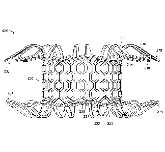
longitudinally along the central portion, (2) a first flange cell of the
plurality of first flange
cells, (3) a second pattern extending longitudinally along the central portion
and
opposing the first pattern, and (4) a second flange cell of the plurality of
second flange
CA 3023400 2018-11-07
cells. In some embodiments a single elongate member forms the central portion,
the
first apposition portion, and the second apposition portion. In other
embodiments, the
central portion cells are open to longitudinally-adjacent central portion
cells and are
closed to circumferentially-adjacent central portion cells. In further
embodiments, each
of the plurality of second flange cells is open to one or more of the central
portion cells
of the plurality of central portion cells.
[0005] A second aspect of the invention relates to an implantable medical
device for
creating an anastomosis. The device includes a tubular structure including at
least one
elongate member forming a framework of interconnected struts. The tubular
structure
includes (1) a central portion having a plurality of body cells defined by the
elongate
member, (2) a first apposition portion at a first end of the central portion
having a
plurality of first flange cells defined by the elongate member, and (3) a
second
apposition portion at a second end of the central portion having a plurality
of second
flange cells defined by the elongate member. The elongate member may be formed
such that (1) the elongate member forms a first pattern traversing the central
portion
along a longitudinal axis, (2) the elongate member defines a first flange cell
of the first
plurality of flange cells, (3) the elongate member traverses the central
portion along the
longitudinal axis in a second pattern opposing the first pattern, and (4) the
elongate
member defines a second flange cell of the second plurality of flange cells.
In at least
one embodiment, each successive flange cell of the first and second plurality
of flange
cells is out of phase with directly preceding flange cells of the first and
second plurality
of flange cells. Additionally, the body cells may be open to longitudinally-
adjacent body
cells and may be closed to circumferentially-adjacent body cells. In some
embodiments, each of the plurality of first flange cells may be open to the
body and
each of the plurality of second flange cells may be open to the body.
[0006] A third aspect of the invention relates to a method of implanting an
anastomosis device in a patient that includes (1) navigating a delivery sheath
containing
the anastomosis device to a target location within the patient and (2)
deploying the
anastomosis device from the delivery sheath such that at least one layer of
tissue is
between the first apposition portion and the second apposition portion. The
CA 3023400 2018-11-07
anastomosis device includes a tubular structure that includes at least one
elongate
member forming a framework of interconnected struts. The tubular structure
includes
(1) a central portion that includes a plurality of body cells defined by the
elongate
member, (2) a first apposition portion at a first end of the central portion
having a
plurality of first flange cells defined by the elongate member such that the
plurality of
first flange cells are open to the central portion, and (3) a second
apposition portion at a
second end of the central portion that includes a plurality of second flange
cells defined
by the elongate member such that the plurality of second flange cells are open
to the
central portion.
DESCRIPTION OF DRAWINGS
[0007] The accompanying drawings are included to provide a further
understanding
of the disclosure and are incorporated in and constitute a part of this
specification,
illustrate embodiments, and together with the description serve to explain the
principles
of the disclosure.
[0008] FIG. 1 is a cutaway perspective view of an exemplary anastomosis
device
that has been implanted within a patient to act as a shunt between the
patient's
gallbladder and intestine in accordance with some embodiments;
[0009] FIG. 2A is a side view of an exemplary anastomosis device in
accordance
with some embodiments;
[0010] FIG, 28 is a perspective view of the anastomosis device of FIG. 2A;
[0011] FIG. 2C is an end view of the anastomosis device of FIG. 2A;
[0012] FIG. 20 is a side view of the anastomosis device of FIG. 2A prior to
forming
flanges;
[0013] FIG. 2E is a perspective view of the anastomosis device of FIG. 2A
prior to
forming flanges;
3
CA 3023400 2018-11-07
[0014] FIG. 3A is a flat pattern of an anastomosis device in accordance
with some
embodiments;
[0015] FIG. 38 is an enlarged view of a flange cell of the anastomosis
device of FIG.
3A in a deployed configuration;
[0016] FIG. 3C is an enlarged view of the flange cell of the anastomosis
device of
FIG. 3A in a low-profile delivery configuration;
[0017] FIG, 3D is an enlarged view of a cell of the anastomosis device of
FIG. 3A in
a deployed configuration;
[0018] FIG. 3E is an enlarged view of a cell of the anastomosis device of
FIG. 3A in
a crushed configuration;
[0019] FIG. 4 is a fiat pattern of an anastomosis device in accordance with
some
embodiments;
[0020] FIG. 5 is a flat pattern of an anastomosis device in accordance with
some
embodiments;
[0021] FIG. 6A is a perspective view of a framework of another exemplary
anastomosis device in a low-profile delivery configuration in accordance with
some
embodiments;
[0022] FIG. 6B is a perspective view of the framework of FIG. 6A in
accordance with
some embodiments;
[0023] FIG. 7A is a perspective view of another exemplary anastomosis
device in
accordance with some embodiments;
[0024] FIG. 7B is an end view of the anastomosis device of FIG. 7A;
[0025] FIG. 7C is a side view of the anastomosis device of FIG. 7A;
4
CA 3023400 2018-11-07
[0026] FIG. 8A is a perspective view of another exemplary anastomosis
device in
accordance with some embodiments;
[0027] FIG. 8B is a different perspective view of the stent of FIG. 8A;
[0028] FIG. 9A is a perspective view of another exemplary anastomosis
device in
accordance with some embodiments;
[0029] FIG. 98 is an end view of the anastomosis device of FIG. 9A;
[0030] FIG. 9C is a side view of the anastomosis device of FIG. 9A;
[0031] FIG. 9D is a side view of the framework of the anastomosis device of
FIG. 9C
before the flange structures are formed;
[0032] FIG. 9E is a perspective view of a center portion of the anastomosis
device of
FIG. 9C in a low-profile delivery configuration;
[0033] FIG. 10 is a perspective view of an anastomosis device on a forming
mandrel
in accordance with some of embodiments;
[0034] FIG. 11A is a perspective view of another exemplary anastomosis
device in
accordance with some embodiments;
[0035] FIG. 118 is another perspective view of the anastomosis device of
FIG. 11A;
[0036] FIG. 11C is an end view of the anastomosis device of FIG. 11A;
[0037] FIG. 11D is a side view of a central portion of the anastomosis
device of FIG.
11A that includes an expansion member in accordance with some embodiments; and
[0038] FIG. 12 is a perspective view of another exemplary anastomosis
device in
accordance with some embodiments.
CA 3023400 2018-11-07
DETAILED DESCRIPTION
100391 Persons skilled in the art will readily appreciate that various
aspects of the
present disclosure can be realized by any number of methods and apparatus
configured
to perform the intended functions. It should also be noted that the
accompanying
drawing figures referred to herein are not necessarily drawn to scale, but may
be
exaggerated to illustrate various aspects of the present disclosure, and in
that regard,
the drawing figures should not be construed as limiting.
[0040] The present disclosure is directed to implantable devices for
connecting
tissue layers, for example, to circumvent a conduit or organ blockage, such as
by
creating a direct passage between tissue structures (e.g. connecting a
gallbladder and a
portion of a gastrointestinal tract) to create an anastomosis that facilitates
material flow
therebetween. The devices described herein are endoscopically deployable or
deliverable via a catheter and may include self-expanding apposition
mechanisms that
facilitate a secure connection between the tissue structures (such a
connection may
also be referred to herein as a "shunt," "passageway," "shunt passageway," or
"tunnel").
Such design features simplify implantation and reduce the likelihood of
complications.
In some embodiments, the devices provided herein are configured to be
removable
after implantation. As one example, the device is implanted and remains in
place until
the gallbladder and/or its associated ducts are cleared of blockages, after
which the
device is removed. In another example, the device remains implanted until the
body
grows a tissue-anastomosis around the device, and then the device is removed.
In
other embodiments, tissue ingrowth into and/or around the device permanently
implants
the device, and the device is not removed. The devices described herein can
provide
alternative treatments for patients who are not suitable candidates for other
types of
treatments (e.g., gallbladder removal surgery) and/or to avoid known
complications of
other types of treatments (e.g., external biliary drainage).
[00411 This disclosure refers to anastomosis devices in an exemplary
fashion. That
is, it should be understood that the inventive concepts disclosed in this
disclosure can
also be applied to other types of devices. For example, this disclosure also
provides
implantable devices that, in some embodiments, can be used for occluding
tissue
6
CA 3023400 2018-11-07
structures, organs, body conduits, blood vessels, the GI tract, and the like.
For
example, in some embodiments the devices provided herein can be used to
occlude
septa! defects. In other embodiments, the devices provided herein can be used
to
occlude a patient's vasculature or GI tract. In some such embodiments, the
device does
not include a tunnel or central aperture through the device. Rather, in some
embodiments a covering material seals the device to inhibit, modulate, or
substantially
prevent material from flowing through the device.
[0042] Referring to FIG. 1, an exemplary anastomosis device 40 in
accordance with
some embodiments provided herein that can be implanted in a patient to create
a fluidic
connection between two organs, spaces, tissue structures, conduits, and the
like, and
combinations thereof is shown. For example, in the depicted implementation the
anastomosis device 40 is connecting a gallbladder 10 (that defines an internal
gallbladder space 12) with an intestine 20 (that defines an internal
intestinal space 22).
Hence, the anastomosis device 40 is acting as a fluidic shunt device between
the
internal gallbladder space 12 and the internal intestinal space 22. Such an
implementation may provide a beneficial treatment to the patient when, for
example, a
flow blockage exists in the native anatomical conduits connecting the internal
gallbladder space 12 and the internal intestinal space 22. For example, in
some
instances the patient may have one or more gallstones that cause a blockage of
the
patient's cystic duct 14 and/or common bile duct 16. In such a case, the
anastomosis
device 40 can provide a fluidic passageway such that bile from the gallbladder
10 can
flow into the intestine 20. If not for the anastomosis device 40, when bile is
blocked
from flowing out of the gallbladder 10 cholecystitis (inflammation of the
gallbladder 10)
may result.
[0043] While the anastomosis devices provided herein can be used in some
implementations to relieve or prevent cholecystitis as described above, it
should be
understood that the anastomosis devices provided herein can also be used in
many
other types of implementations within a patient. For example, the anastomosis
devices
provided herein can be used in conjunction with various body tissue structures
and
7
CA 3023400 2018-11-07
organs such as, but not limited to, stomachs, colons, small intestines,
pancreases,
blood vessels, bladders, kidneys, conduits, and the like.
[0044] In general, some embodiments of the anastomosis devices provided
herein
(of which anastomosis device 40 is one type of example), include a first
tissue
apposition portion 42a, a second tissue apposition portion 42b, and a central
portion 44
therebetween. The central portion 44 defines a lumen 46 that extends
longitudinally
from a first end of the anastomosis device 40 to a second end of the device
40. The
lumen 46 acts as a connection (e.g., a shunt passageway) between the internal
gallbladder space 12 and the internal intestinal space 22, such that the
internal
gallbladder space 12 is in fluid communication with the internal intestinal
space 22 via
the anastomosis device 40.
[0045] Referring to FIGS. 2A-2E, an exemplary anastomosis device 200 that
includes a framework of elongate elements that define a first apposition
portion 202, a
second apposition portion 204, and a central portion 206 is depicted. In some
embodiments, the anastomosis device 200 can be a type of stent device, which
can
refer broadly to devices that include a framework of elongate elements and
include
devices such as, but not limited to, anastomosis devices. The central portion
206 is
disposed between and interconnects the first apposition portion 202 and the
second
apposition portion 204. A covering material (not shown in FIGS. 2A-2E) can be
disposed on at least some portions of the framework. Such covering materials
(e.g.,
covering material and others described below) may also be referred to herein
merely as
a covering.
[0046] In some embodiments, the central portion 206 can form a body that
defines a
lumen 207 that extends between the first apposition portion 202 and the second
apposition portion 204. The first and second apposition portions 202 and 204
can form
flanges extending substantially radially outward from opposite ends of the
central
portion 206. In some embodiments, the lumen 207 provides an anastomosis
passageway or tunnel through which biological materials or fluids can pass.
The device
200 is shown in an expanded configuration (also referred to herein as a
deployed
8
CA 3023400 2018-11-07
configuration). The expanded or deployed configuration is the configuration
that the
device 200 naturally exhibits in the absence of external forces acting upon
the device
200. It should be understood that when the anastomosis device 200 is implanted
in a
patient, the configuration of the device 200 may be somewhat different than
shown
because of the external forces from the patient's anatomy that are exerted on
the device
200.
[0047] In some embodiments, the first apposition portion 202, the second
apposition
portion 204, and the central portion 206 are formed of elongate elements such
as spring
wire (e.g., L605 steel or stainless steels), shape memory alloy wire (e.g.,
nitinol or nitinol
alloys), super-elastic alloy wire (e.g., nitinol or nitinol alloys), or other
suitable types of
elongate elements or wires, or combinations thereof. In some such embodiments,
the
first apposition portion 202, the second apposition portion 204, and the
central portion
206 can be formed from the same piece of precursor material that is cut to
create the
framework of elongate elements, In some such embodiments, the precursor
material is
a tubular material or a sheet material. In some embodiments, different types
of
elongate elements are used at different locations of the first apposition
portion 202, the
second apposition portion 204, and/or the central portion 206. In some
embodiments,
the elongate elements of the first apposition portion 202, the second
apposition portion
204, and the central portion 206 (or portions thereof) may be constructed of
polymeric
materials.
[0048] Suitable materials for the elongate elements of the devices provided
herein
include a variety of metallic materials including alloys exhibiting, shape
memory, elastic
and super-elastic characteristics. Shape memory refers to the ability of a
material to
revert to an originally memorized shape after plastic deformation by heating
above a
critical temperature. Elasticity is the ability of a material to deform under
load and return
or substantially return to its original shape when the load is released. Most
metals will
deform elastically up to a small amount of strain. Super-elasticity refers to
the ability of
a material to deform under strain to much larger degree than typical elastic
alloys,
without having this deformation become permanent. For example, the super-
elastic
materials included in the frames of some anastomosis device embodiments
provided
9
CA 3023400 2018-11-07
herein are able to withstand a significant amount of bending and flexing and
then return
or substantially return to the frame's original form without deformation. In
some
embodiments, suitable elastic materials include various stainless steels which
have
been physically, chemically, and otherwise treated to produce a high
springiness, metal
alloys such as cobalt chrome alloys (e.g., ELGILOYTM, MP35N, L605),
platinum/tungsten alloys. Embodiments of shape memory and super-elastic alloys
include the NiTi alloys, ternary shape memory alloys such as NiTiPt, NiTiCo,
NiTiCr, or
other shape memory alloys such as copper-based shape memory alloys. Additional
materials could combine both shape memory and elastic alloys such as a drawn
filled
tube where the outer layer is constructed of nitinol and the inner core is a
radiopaque
material such as platinum or tantalum. In such a construct, the outer layer
provides the
super-elastic properties and the inner core remains elastic due to lower
bending
stresses.
[0049] In some embodiments, the elongate elements used to construct the
devices
provided herein can be treated in various ways to increase the radiopacity of
the
devices for enhanced radiographic visualization. In some embodiments, the
devices
are at least partially a drawn-filled type of NiTi containing a different
material at the core,
such as a material with enhanced radiopacity. In some embodiments, the devices
include a radiopaque cladding or plating on at least portions of the first
apposition
portion, the second apposition portion, and the central portion. In some
embodiments,
one or more radiopaque markers are attached to the devices. In some
embodiments,
the elongate elements and/or other portions of the devices provided herein are
also
visible via ultrasound.
[0050] In some embodiments, the first apposition portion 202, the second
apposition
portion 204, and the central portion 206, comprise a framework of
interconnected
elongate elements that is constructed by cutting a tube. In one such
embodiment, a
tube of metallic material (e.g., nitinol, stainless steel, cobalt, etc.) is
laser cut, and then
the tube is expanded and shaped into the desired configuration. In some such
embodiments, the metallic material is shape-set in the desired configuration
so that the
material receives a shape-memory whereby the material will naturally strive to
attain the
CA 3023400 2018-11-07
desired configuration. In some embodiments, shape memory materials such as
nitinol
may strive to attain the desired configuration when exposed to body
temperature.
[0051] As described in more detail below, in some embodiments a covering
material
can be disposed on or around some portions, or on or around all of the first
apposition
portion 202, the second apposition portion 204, and/or the central portion
206. In some
embodiments, portions of the first apposition portion 202, the second
apposition portion
204, and/or the central portion 206 can remain free of the covering material.
In some
embodiments, no covering material is included on the anastomosis device 200.
[0052] The first apposition portion 202 and the second apposition portion
204 each
include a plurality of struts 208. In some embodiments, the struts 208 of each
of the
first and second apposition portions 202 and 204 are configured to form, in a
general
sense, flanges that contact tissue surfaces. More particularly, the first
apposition
portion 202 and the second apposition portion 204 are configured to engage one
or
more layers of tissue therebetvveen, and to provide apposition forces against
the tissue
surfaces. The apposition forces provided by the first and second apposition
portions
202 and 204 can facilitate fixation of the device 200 to the tissue and
provide migration
resistance such that the device 200 can reliably remain positioned at a target
site in a
patient as desired.
[0053] In some embodiments, the materials and configuration of the
anastomosis
device 200 (and the other anastomosis device embodiments provided herein)
allow the
devices to be elastically crushed, folded, and/or collapsed into a low-profile
delivery
configuration for containment within a lumen for transcatheter or
endoscopic/thorascopic delivery, and to self-expand to an operative size and
configuration once positioned at a desired target site within a body and
deployed from
the lumen. For example, the anastomosis device 200 can be configured in a
collapsed
delivery configuration in which the plurality of struts 208 are radially
compressed such
that they are forced to extend substantially parallel to the axis of the
central portion 206,
and in which the diameter of the central portion 206 is also crushed to become
smaller.
11
CA 3023400 2018-11-07
Due to the use of such materials and structure, the device 200 may also
exhibit, for
example, beneficial fatigue resistance and elastic properties.
[0054] After deployment, the plurality of struts 208 extend from the
central portion
206 at a radial orientation and geometry to exert a desired level of
apposition pressure
on the tissue. In some embodiments, the plurality of struts 208 extend from
the central
portion 206 such that the nominal measure of the angle between the struts 208
and the
longitudinal axis of the device 200 is about 100 , or about 90 , or about 80 ,
or about
700, or about 600, or about 50 , or about 40 , or about 300, or about 20 , or
about 10 ,
and the like.
[0055] Still referring to FIGS. 2A-2E, in some embodiments of the
anastomosis
device 200 (and in some embodiments of the other anastomosis devices provided
herein) the plurality of struts 208 are interconnected by connecting members
210. The
connecting members 210 are shown in deployed configurations in which the
connecting
members 210 are arranged in a series of undulations¨each having a vertex 214
extending towards the central portion 206 and a vertex 215 extending away from
the
central portion 206. In some embodiments, the struts 208 can connect to the
connecting members 210 at the vertex 214. In other embodiments, the struts 208
can
connect to the connecting members 210 at the vertex 215.
[0056] In some embodiments, the connecting members 210 serve to support and
stabilize the struts 208 to thereby cause the apposition portions 202 and 204
to have a
more rigid construct. In some such embodiments, the apposition portions 202
and 204
can exert a greater level of apposition pressure while maintain a compliancy
by which
the apposition portions 202 and 204 can conform to the anatomical topography
of the
tissue. In addition, the sealing capabilities of the apposition portions 202
and 204 may
be enhanced. In some embodiments, the stability and support provided by the
connecting member 210 serves to increase the apposition force provided against
the
gallbladder or provided against the portion of the gastrointestinal tract, for
example.
12
CA 3023400 2018-11-07
[0057] In some embodiments, the connecting members 210 combine to form
circumferential rings 216 and 218 extending circumferentially around a
radially-outer
circumference of each of the first and second apposition portions 202 and 204,
respectively. The circumferential rings 216 and 218 can have a shape that is
wavy or
that undulates circumferentially around edges of the first and second
apposition portions
202 and 204. In some embodiments, the circumferential rings 216 and 218 can
have a
shape that undulates in an axial direction, as can be seen in FIG. 2A. In some
embodiments, the circumferential rings 216 and 218 can have a shape that
undulates in
a radial direction, as can be seen in FIG, 2C. In some embodiments, the
circumferential
rings 216 and 218 can have a shape that undulates in both axial and radial
directions.
In some embodiments, the circumferential rings 216 and 218 can undulate
sinusoidally
around the edges of one or both of the first and second apposition portions
202 and
204. Forming one or both of the circumferential rings 216 and 218 with a
sinusoidal,
serpentine, or otherwise undulating shape can increase an amount of surface
area of
contact between the first and second apposition portions 202 and 204 and
tissue, thus
reducing force at a given location on that tissue.
[0058] The central portion 206 can include a series of body struts 220,
each
extending longitudinally and forming the central body 206 of the anastomosis
device
200. The body struts 220 define body cells 222 of the central portion 206 and
separate
the respective body cells 222 from circumferentially-adjacent body cells 222.
In some
embodiments, each of the body struts 220 can include a plurality of axially-
extending
portions 224 interconnected with a plurality of angled portions 226. This can
allow the
body struts 220 to create a relatively strong central portion 206 without
necessarily
interconnecting the body struts 220 across the body cells 222 at several
locations along
the length of the body struts 220.
[0059] The struts 208 of the apposition portions 202 and 204 can define
flange cells
228 between the struts 208. The flange cells 228 can be open cells, with no
strut
separating the flange cells 228 from the central portion 206. As shown in
FIGS, 2B, 2D.
and 2E, the flange cells 228 are closed at a distal-most end of the flange
cells 228 by
13
CA 3023400 2018-11-07
connecting members 210 and are open at a center-most end of the flange cells
228,
such that the flange cells 228 are open to the body cells 222.
[0060] FIGS. 2D and 2E show the anastomosis device 200 in a partially-
formed
configuration, prior to forming the anastomosis device 200 in the shape
illustrated in
FIGS. 2A, 2B, and 2C. As shown in FIGS. 2D and 2E, the anastomosis device 200
can
have a substantially cylindrical shape in the partially-formed configuration,
with the
struts 208 extending substantially parallel to the body struts 220. The
anastomosis
device 200 can be shaped in a manufacturing process, such as, for example,
that
described with respect to FIG. 10 (below), to transform the anastomosis device
200
from the pre-formed configuration shown in FIGS, 2D and 2E to the final
configuration
shown in FIGS. 2A, 2B, and 2C,
[0061] In some embodiments, the anastomosis device 200 can be formed in a
manner such that an elongate member forms a first pattern traversing the
central
portion 206 along a longitudinal axis, the elongate member defines a first
flange cell 228
of the first apposition portion 202, the elongate member traverses the central
portion
206 along the longitudinal axis in a second pattern opposing the first
pattern, the
elongate member defines a second opposing flange cell 228, and the elongate
member
then repeats those winding steps to form additional patterns of the central
portion 206
and flanges cells 228 of the anastomosis device 200.
[0062] In some embodiments, the anastomosis device 200 can be formed in a
manner such that the elongate member defines a flange cell 228 of the first
apposition
portion 202, the elongate member traverses the central portion 206, the
elongate
member defines a flange cell 228 of the second apposition portion 204, the
elongate
member traverses the central portion 206, and thereafter the elongate member
repeats
the pattern to form additional flange cells while traversing the central
portion 206 in
between. In some embodiments, each successive pattern and each successive
flange
cell 228 can be out of phase with those directly preceding.
14
CA 3023400 2018-11-07
[0063] The central portion 206 is shown in a deployed or expanded
configuration in
FIGS. 2A-2C, In some embodiments, the central portion 206, as described above,
can
include a variety of metallic shape memory materials and super-elastic alloys.
Thus, the
central portion 206 can be configured to self-expand to the deployed
configuration. In
some embodiments, the central portion 206 is balloon expandable into the
deployed
configuration. Alternatively, supplemental expansion forces can be applied to
a self-
expandable device by balloon dilation. The diameter of the central portion 206
can be
made in any size as desired in order to suit the intended use and/or delivery
system of
the anastomosis device 200. For example, in the low-profile delivery
configuration the
anastomosis device 200 can be disposed within a delivery sheath that has about
a 15
Fr. (5 mm) outer diameter. However, in some embodiments, sheaths that are
smaller or
larger than 15 Fr. can be used. For example, sheaths that have outer diameters
of 6
Fr., 7 Fr., 8 Fr., 9 Fr., 10 Fr., 11 Fr., 12 Fr., 13 Fr, 14 Fr., 16 Fr., 17
Fr., 18 Fr., 19 Fr., 20
Fr., and larger than 20 Fr., can be used in some embodiments. When the
anastomosis
device 200 is configured in its expanded deployed configuration as shown, the
diameter
of the central portion 206 increases to a deployed diameter. In some
implementations,
the deployed outer diameter of the central portion 206 is configured to at
least partially
anchor the device 200 via an interference fit with the tissue aperture in
which the central
portion 206 resides. Additionally, when the central portion 206 and the tissue
aperture
have an interference fit relationship, para-device leakage may be reduced or
minimized.
In such a case, leakage of the contents of the organs, conduits, and other
types of
tissue structures in which the anastomosis device 200 may be deployed can be
substantially prevented. For example, when the anastomosis device 200 is used
between a gallbladder and GI tract (e.g., refer to FIG. 1), leakage into the
abdominal
cavity can be substantially prevented.
[0064] In some implementations, the deployed outer diameter of the central
portion
206 is slightly less than the diameter of the tissue aperture in which the
central portion
206 resides, and the apposition portions 202 and 204 compress the tissue to
provide
the migration resistance. In some embodiments, the fully expanded diameter of
the
central portion 206 is about 30 mm, or about 25 mm, or about 20 mm, or about
15 mm,
CA 3023400 2018-11-07
or about 12 mm, or about 10 mm, or about 8 mm, or about 6 mm, or about 4 mm,
and
the like.
[0065] In some embodiments, one or more portions of the anastomosis device
200
includes a covering material. For enhanced visualization of the framework of
the
anastomosis device 200, the anastomosis device 200 is shown in FIGS, 2A-2E
without
a covering material. In some embodiments, a covering material is disposed on
at least
some portions (or on all) of the first apposition portion 202, the second
apposition
portion 204, and/or the central portion 206. In some embodiments, some
portions of the
first apposition portion 202, the second apposition portion 204, and/or the
central portion
206 are not covered by the covering material.
[0066] In some embodiments, the covering material is generally fluid
impermeable.
That is, in some embodiments the covering material is made of a material that
inhibits or
reduces passage of blood, bile and/or other bodily fluids and materials
through the
covering material itself. In some embodiments, the covering material has a
material
composition and configuration that inhibits or prevents tissue ingrowth and/or
endothelialization or epithelialization into the covering material. Some such
embodiments that are configured to inhibit or prevent tissue ingrowth and/or
endothelialization can be more readily removed from the patient at a future
date if so
desired. In some embodiments, the covering material, or portions thereof, has
a
microporous structure that provides a tissue ingrowth scaffold for durable
sealing and/or
supplemental anchoring strength of the anastomosis device 200.
[0067] In some embodiments, the covering material comprises a
fluoropolymer, such
as an expanded polytetrafluoroethylene (ePTFE) polymer, or polyvinylidene
fluoride
(PVDF). In some embodiments, the covering material comprises a polyester, a
silicone,
a urethane, another biocompatible polymer, polyethylene terephthalate (e.g.,
Dacron ),
bioabsorbable materials, copolymers, or combinations thereof. In some
embodiments,
the covering material comprises a bioabsorbable web. In some other
embodiments, the
bioabsorbable material may also provide an anti-migration feature by promoting
16
CA 3023400 2018-11-07
attachment between the device 200 and tissue until the bioabsorbable material
is
absorbed.
[00681 In some embodiments, the covering material (or portions thereof) is
modified
by one or more chemical or physical processes that enhance one or more
properties of
the material. For example, in some embodiments, a hydrophilic coating is
applied to the
covering material to improve the wettability and echo translucency of the
material. In
some embodiments, the covering material, or portions thereof, is modified with
chemical
moieties that facilitate one or more of endothelial cell attachment,
endothelial cell
migration, endothelial cell proliferation, and resistance to or promotion of
thrombosis. In
some embodiments, the covering material, or portions thereof, is modified to
resist
biofouling. In some embodiments, the covering material, or portions thereof,
is modified
with one or more covalently attached drug substances (e.g., heparin,
antibiotics, and the
like) or impregnated with the one or more drug substances. The drug substances
can
be released in situ to promote healing, reduce tissue inflammation, reduce or
inhibit
infections, and to promote various other therapeutic treatments and outcomes.
In some
embodiments, the drug substance is a corticosteroid, a human growth factor, an
anti-
mitotic agent, an antithrombotic agent, a stem cell material, or dexamethasone
sodium
phosphate, to name some embodiments. In some embodiments, a pharmacological
agent is delivered separately from the covering material to the target site to
promote
tissue healing or tissue growth.
[00691 Coatings and treatments may be applied to the covering material
before or
after the covering material is joined or disposed on the framework of the
anastomosis
device 200. Additionally, one or both sides of the covering material, or
portions thereof,
may be coated. In some embodiments, certain coatings and/or treatments are
applied
to the covering material(s) located on some portions of the anastomosis device
200,
and other coatings and/or treatments are applied to the material(s) located on
other
portions of the anastomosis device 200. In some embodiments, a combination of
multiple coatings and/or treatments are applied to the covering material, or
portions
thereof. In some embodiments, certain portions of the covering material are
left
uncoated and/or untreated In some embodiments, the device 200 is fully or
partially
17
CA 3023400 2018-11-07
coated to facilitate or frustrate a biological reaction, such as, but not
limited to,
endothelial cell attachment, endothelial cell migration, endothelial cell
proliferation, and
resistance to or promotion of thrombosis.
[0070] In some embodiments, a first portion of the covering material is
formed of a
first material and a second portion of the covering material is formed of a
second
material that is different than the first material. In some embodiments, the
covering
material includes multiple layers of materials, which may be the same or
different
materials. In some embodiments, portions of the covering material have one or
more
radiopaque markers attached thereto to enhance in vivo radiographic
visualization of
the anastomosis device 200, or one or more echogenic areas to enhance
ultrasonic
visibility.
[0071] In some embodiments, one or more portions of the covering material
are
attached to the framework of the device 200, such as the central portion 206
and/or the
apposition portions 202 and 204. The attachment can be accomplished by a
variety of
techniques such as, but not limited to, stitching the covering material to the
framework
of the device 200, adhering the covering material to the framework of the
device 200,
laminating multiple layers of the covering material to encompass portions of
the
elongate members of the device 200, using clips or barbs, or laminating
multiple layers
of the covering material together through openings in the framework of the
device 200.
In some embodiments, the covering material is attached to the framework of the
device
200 at a series of discrete locations thereby facilitating the flexibility of
the framework.
In some embodiments, the covering material is loosely attached to the
framework of the
device 200. It is to be appreciated that the covering material may be attached
to the
framework of the device 200 using other techniques or combinations of
techniques
described herein.
[0072] In some embodiments, the framework of the device 200 (or portions
thereof)
is coated with a bonding agent (e.g., fluorinated ethylene propylene or other
suitable
adhesive) to facilitate attachment of the covering material to the framework.
Such
18
CA 3023400 2018-11-07
adhesives may be applied to the framework using contact coating, powder
coating, dip
coating, spray coating, or any other appropriate means.
[0073] The covering material can adapt to changes in the length and/or
diameter of
the central portion 206 in a variety of manners. In a first example, the
covering material
can be elastic such that the covering material can stretch to accommodate
changes in
the length and/or diameter of the device 200. In a second example, the
covering
material can include slackened material in the low-profile delivery
configuration that
becomes less slackened or totally urtslackened when the device 200 is in the
expanded
configuration. In a third example, the covering material can include folded
portions
(e.g., pleats) that are folded in the low-profile configuration and less
folded or totally
unfolded when the device 200 is in the expanded configuration. In other
embodiments,
an axial adjustment member is free of covering material. In some embodiments,
combinations of such techniques, and/or other techniques can be used whereby
the
covering material can adapt to changes in the length and/or diameter of the
central
portion 206.
[0074] FIG. 3A is a flat pattern of an anastomosis device 300 in accordance
with
some exemplary embodiments. In some embodiments, the anastomosis device 300
can be similar to the anastomosis device 200 described above. For example, the
anastomosis device 300 includes a framework of elongate elements that defines
a first
apposition portion 302, a second apposition portion 304, and a central portion
306. The
central portion 306 is disposed between and interconnects the first apposition
portion
302 and the second apposition portion 304. In some embodiments, the
anastomosis
device 300 is formed from a tubular material that is cut (e.g., laser cut) and
shape-set to
a preferred form. Other materials and fabrication techniques are also
envisioned. A
covering material as described above (not shown in FIG. 3A) can be disposed on
at
least some portions (or all) of the framework of the anastomosis device 300.
[0075] The anastomosis device 300 is shown in FIG. 3A as a flat pattern for
clarity.
However, the anastomosis device 300 can be formed into a tubular shape, with
the
central portion 306 forming a substantially cylindrical structure, and with
the first and
19
CA 3023400 2018-11-07
second apposition portions 302 and 304 extending outward from opposing ends of
the
central portion 306, In some embodiments, the central portion 306 can form a
tubular
body that defines a lumen that extends between the first apposition portion
302 and the
second apposition portion 304. The first and second apposition portions 302
and 304
can form flanges extending substantially radially outward from opposite ends
of the
central portion 306. In some implementations, the lumen defined by the central
portion
306 provides an anastomosis passageway or tunnel through which biological
materials
and liquids can pass. It should be understood that when the anastomosis device
300 is
implanted in a patient, the configuration of the device 300 may be somewhat
different
than shown because of the external forces from the patient's anatomy that are
exerted
on the device 300.
[0076] In some embodiments, the connecting members 310 combine to form
circumferential rings 316 and 318 extending substantially circumferentially
around a
radially-outer circumference of each of the first and second apposition
portions 302 and
304, respectively. The circumferential rings 316 and 318 can have a shape that
is wavy
or that undulates circumferentially around the outer edges of the first and
second
apposition portions 302 and 304. In some embodiments, the circumferential
rings 316
and 318 can undulate sinusoidally around the edges of one or both of the first
and
second apposition portions 302 and 304. Forming one or both of the
circumferential
rings 316 and 318 with a sinusoidal, serpentine, or otherwise undulating shape
can
increase an amount of surface area of contact between the first and second
apposition
portions 302 and 304 and tissue, thus reducing force at a given location on
that tissue.
Forming one or both of the circumferential rings 316 and 318 with a
sinusoidal,
serpentine, or otherwise undulating shape can also help facilitate
crushability (for
deployment via a low-profile) of the first and second apposition portions 302
and 304
while maintaining other desirable properties.
[0077] The central portion 306 can include a series of body struts 320,
each
extending substantially axially and forming the central body of the
anastomosis device
300. The body struts 320 define body cells 322 of the central portion 306 and
separate
the respective body cells 322 from circumferentially-adjacent body cells 322.
In some
CA 3023400 2018-11-07
embodiments, each of the body struts 320 can include a plurality of axially-
extending
portions 324 interconnected with a plurality of angled portions 326. This can
allow the
body struts 320 to create a relatively strong central portion 306 without
necessarily
interconnecting the body struts 320 across the body cells 322 at several
locations along
the length of the body struts 320.
[00781 The struts 308 of the apposition portions 302 and 304 can define
flange cells
328 between the struts 308. In some embodiments, the flange cells 328 are open
cells
(with no strut separating the flange cells 328 from the central portion 306).
In some
embodiments, the flange cells 328 are closed at a distal-most end of the
flange cells
328 by connecting members 310 and are open at a center-most end of the flange
cells
328, such that the flange cells 328 are open to the body cells 322. The angled
portions
326 can partially separate axially-adjacent body cells 322 but leave gaps such
that each
body cells 322 is open to each axially-adjacent body cell 322.
[00791 FIG. 3B is an enlarged view of a single flange cell 328 of the
anastomosis
device 300 in a deployed configuration. FIG. 3C is an enlarged view of a
single flange
cell 328 of the anastomosis device 300 in a crushed configuration. The
anastomosis
device 300 can be elastically crushed, folded, and/or collapsed into a low-
profile
delivery configuration (with the flange cell 328 in the crushed configuration
illustrated in
FIG. 3C) for containment within a lumen for transcatheter or
endoscopicithorascopic
delivery lumen. In some embodiments, the anastomosis device self-expands (upon
deployment from the delivery lumen) to an operative size and configuration
once
positioned at a desired target site within a body (e.g., the flange cell 328
expands to the
deployed configuration as illustrated in FIG. 3B).
[0080] FIG. 3D is an enlarged view of the body cell 322 of the anastomosis
device
300 in a deployed configuration. FIG. 3E is an enlarged view of the body cell
322 of the
anastomosis device 300 in a crushed configuration,
[00811 The frame of the anastomosis device 300 can be formed using any of the
materials and techniques described herein. For example, in some embodiments
the
frame of the anastomosis device 300 is formed from a precursor material that
is cut to
I.
CA 3023400 2018-11-07
create the framework. In some such embodiments, the precursor material is a
single
piece of precursor material such as, but not limited to, a tubular material or
a sheet
material. In some embodiments, the frame of the anastomosis device 300 can be
formed as a wire-wound structure of a single wire or a plurality of wires that
form the
structures of the first apposition portion 302, the second apposition portion
304, and the
central portion 306, so as to create the open structures of the body cells 322
and the
flange cells 328 as well as the undulating shape of the circumferential rings
316 and
318. In some embodiments, a wire-wound structure may advantageously facilitate
functionality of the open structures of the body cells 322 and the flange
cells 328 as well
as the undulating shape of the circumferential rings 316 and 318.
[0082] In some embodiments, the anastomosis device 300 can be wire-wound
(or
laser cut) in a manner such that an elongate member forms (i) a first pattern
traversing
the central portion 306 along a longitudinal axis. (ii) a first flange cell
328 of the first
apposition portion 302, (iii) a second pattern traversing the central portion
306 along the
longitudinal axis opposing the first pattern, (iv) a second opposing flange
cell 328, and
so on. The elongate member can be formed to repeat those patterns of the
central
portion 306 and flanges cells 328 to construct a complete anastomosis device
300.
[0083] In some embodiments, the anastomosis device 300 can be wire-wound
(or
laser cut) in a manner such that the elongate member defines a flange cell 328
of the
first apposition portion 302, the elongate member traverses the central
portion 306, the
elongate member defines a flange cell 328 of the second apposition portion
304, the
elongate member traverses the central portion 306, and thereafter the elongate
member
repeats the pattern to form additional flange cells while traversing the
central portion
306 in between. In some such embodiments, each successive pattern and flange
cell
are symmetric to those proceeding.
[0084] FIG. 4 is a fiat pattern of an anastomosis device 400 in accordance
with other
exemplary embodiments. In some embodiments, the anastomosis device 400 can be
similar to the anastomosis devices 200 and 300 described above. For example,
in
some embodiments the anastomosis device 400 includes a framework of elongate
CA 3023400 2018-11-07
elements that defines a first apposition portion 402, a second apposition
portion 404,
and a central portion 406. The central portion 406 is disposed between and
interconnects the first apposition portion 402 and the second apposition
portion 404. A
covering material as described above (not shown in FIG. 4) can be disposed on
at least
some portions (or all) of the framework.
[0085] The anastomosis device 400 is shown in FIG. 4 as a flat pattern for
clarity.
However, the anastomosis device 400 can be formed into a tubular form, with
the
central portion 406 forming a substantially cylindrical structure, and with
the first and
second apposition portions 402 and 404 extending outward from opposing ends of
the
central portion 406. In some embodiments, the central portion 406 can form a
tubular
body that defines a lumen that extends between the first apposition portion
402 and the
second apposition portion 404. The first and second apposition portions 402
and 404
can form flanges extending substantially radially outward from opposite ends
of the
central portion 406. In some implementations, the lumen defined by the central
portion
406 provides an anastomosis passageway or tunnel through which biological
materials
can pass. It should be understood that when the anastomosis device 400 is
implanted
in a patient, the configuration of the device 400 may be somewhat different
than shown
because of the external forces from the patient's anatomy that are exerted on
the device
400.
[0086] In some embodiments, the connecting members 410 combine to form
circumferential rings 416 and 418 extending substantially circumferentially
around a
radially-outer circumference of each of the first and second apposition
portions 402 and
404, respectively. The circumferential rings 416 and 418 can have a shape that
is wavy
or that undulates circumferentially around edges of the first and second
apposition
portions 402 and 404. In some embodiments, the circumferential rings 416 and
418 can
have a shape that undulates, as can be seen in FIG. 4. In some embodiments,
the
circumferential rings 416 and 418 can undulate sinusoidally along the edges of
one or
both of the first and second apposition portions 402 and 404. Forming one or
both of
the circumferential rings 416 and 418 with a sinusoidal, serpentine, or
otherwise
undulating shape can, in some implementations, increase an amount of surface
area of
CA 3023400 2018-11-07
contact between the first and second apposition portions 402 and 404 and
tissue, thus
reducing the force at a given location on that tissue. Forming one or both of
the
circumferential rings 416 and 418 with a sinusoidal, serpentine, or otherwise
undulating
shape can also help facilitate crushability of the first and second apposition
portions 402
and 404 while maintaining other desirable properties.
[0087] The central portion 406 can include a series of body struts 420,
each body
strut extending substantially axially and forming the central body of the
anastomosis
device 400. The body struts 420 define body cells 422 of the central portion
406 and
separate the respective body cells 422 from circumferentially-adjacent body
cells 422.
In some embodiments, each of the body struts 420 may include a plurality of
axially-
extending portions 424 interconnected with a plurality of angled portions 426.
Such a
configuration can allow the body struts 420 to create a relatively strong
central portion
406 without necessarily interconnecting the body struts 420 at several
locations along
the length of the body struts 420.
[0088] In some embodiments, the struts 408 of the apposition portions 402
and 404
can define flange cells 428 between the struts 408. In some such embodiments,
the
flange cells 428 can be open cells, with no strut separating the flange cells
428 from the
central portion 406. The flange cells 428 can be closed at a distal-most end
of the
flange cells 428 by connecting members 410 and can be open at a center-most
end of
the flange cells 428, such that the flange cells 428 can be open to the body
cells 422.
As illustrated in FIG. 4, the flange cells 428 of the first apposition portion
402 are
aligned with the body cells 422 and open to the body cells 422, and the flange
cells 428
of the second apposition portion 404 are aligned with the body struts 420 but
are askew
of the body cells 422.
[0089] In some embodiments, the angled portions 426 can partially separate
longitudinally-adjacent body cells 422 but leave gaps such that each of the
body cells
422 is open to each longitudinally-adjacent body cell 422.
94
CA 3023400 2018-11-07
[0090] In some embodiments, the anastomosis device 400 can be wire-wound
(or
laser cut) in a manner such that an elongate member forms (i) a first pattern
traversing
the central portion 406 along a longitudinal axis, (ii) a first flange cell
428 of the first
apposition portion 402, (iii) a second pattern traversing the central portion
406 along the
longitudinal axis opposing the first pattern, (iv) a second opposing flange
cell 428, and
so on. The elongate member can repeat those patterns to form all of the
central portion
406 and flanges cells 428 of the anastomosis device 400.
[0091] In other embodiments, the anastomosis device 400 can be wire-wound
(or
laser cut) in a manner such that the elongate member defines a flange cell 428
of the
first apposition portion 402, the elongate member traverses the central
portion 406, the
elongate member defines a flange cell 428 of the second apposition portion
404, the
elongate member traverses the central portion 406, and thereafter the elongate
member
repeats the pattern to form additional flange cells while traversing the
central portion
406 in between. In some embodiments, each successive pattern and each
successive
flange cell 428 can be out of phase with those proceeding. In some
embodiments, each
successive pattern and each successive flange cell 428 can be in phase with
those
proceeding.
[0092] FIG. 5 is a flat pattern of an anastomosis device 500 in accordance
with some
exemplary embodiments. In some embodiments, the anastomosis device 500 can be
similar to the anastomosis devices 200, 300, and 400 described above. For
example,
the anastomosis device 500 includes a framework of elongate elements that
defines a
first apposition portion 502, a second apposition portion 504, and a central
portion 500.
The central portion 506 is disposed between and interconnects the first
apposition
portion 502 and the second apposition portion 504. A covering material as
described
above (not shown in FIG. 5) can be disposed on at least some portions (or on
all
portions) of the framework.
[0093] The anastomosis device 500 is shown in FIG. 6 as a flat pattern for
clarity.
However, the anastomosis device 500 can be formed into a tubular form, with
the
central portion 506 forming a substantially cylindrical structure, and with
the first and
CA 3023400 2018-11-07
second apposition portions 502 and 504 extending outward from opposing ends of
the
central portion 506. In some embodiments, the central portion 506 can form a
tubular
body that defines a lumen that extends between the first apposition portion
502 and the
second apposition portion 504. The first and second apposition portions 502
and 504
can form flanges extending substantially radially outward from opposite ends
of the
central portion 506. In some implementations, the lumen defined by the central
portion
506 provides an anastomosis passageway or tunnel through which biological
materials
can pass. It should be understood that when the anastomosis device 500 is
implanted
in a patient, the configuration of the device 500 may be somewhat different
than shown
because of the external forces from the patient's anatomy that are exerted on
the device
500.
10094] In some embodiments, the connecting members 510 combine to form
circumferential rings 516 and 518 extending substantially circumferentially
around a
radially-outer circumference of each of the first and second apposition
portions 502 and
504, respectively. The circumferential rings 516 and 518 can have a shape that
is wavy
or that undulates circumferentially around edges of the first and second
apposition
portions 502 and 504. In some embodiments, the circumferential rings 516 and
518 can
have a shape that undulates, as shown in FIG. 5. In some such embodiments, the
circumferential rings 516 and 518 can undulate sinusoidally along the edges of
one or
both of the first and second apposition portions 502 and 504. Forming one or
both of
the circumferential rings 516 and 518 with a sinusoidal, serpentine, or
otherwise
undulating shape may, in some implementations, increase an amount of surface
area of
contact between the first and second apposition portions 502 and 504 and
tissue, thus
reducing force at a given location on that tissue. Forming one or both of the
circumferential rings 516 and 518 with a sinusoidal, serpentine, or otherwise
undulating
shape can also help facilitate crushability to a low-profile delivery
configuration of the
first and second apposition portions 502 and 504 while maintaining other
desirable
properties.
26
CA 3023400 2018-11-07
[0095] The central portion 506 can include a series of body struts 520,
each body
strut extending substantially axially and forming the central body of the
anastomosis
device 500. In some embodiments, the body struts 520 define body cells 522 of
the
central portion 506 and separate the respective body cells 522 from
circumferentially-
adjacent body cells 522. In some embodiments, each of the body struts 520 can
include a plurality of axially-extending portions 524 interconnected with a
plurality of
angled portions 526. This can allow the body struts 520 to create a relatively
strong
central portion 506 without necessarily interconnecting the body struts 520 at
several
locations along the length of the body struts 520. The struts 508 of the
apposition
portions 502 and 504 can define flange cells 528 between the struts 508.
[0096] As illustrated in FIG. 5, in some embodiments each column of body
cells 522
is aligned with a flange cell 528 at one end, and is open at an opposite end.
In some
embodiments, the body cells 522 can be axially aligned with and open to a gap
530
extending between adjacent struts 508 of one or both of the first and second
apposition
portions 502 and 504. The angled portions 526 can partially separate axially-
adjacent
body cells 522 but can leave gaps such that each body cells 522 is open to
each axially
-
adjacent body cell 522.
[0097] In some embodiments, the anastomosis device 500 can be formed in a
manner such that an elongate member forms (i) a first pattern traversing the
central
portion 506 along a longitudinal axis, (ii) a first flange cell 528 of the
first apposition
portion 502, (iii) a second pattern traversing the central portion 506
opposing the first
pattern, (iv) a second opposing flange cell 528, and so on. In some
embodiments, the
elongate member repeats those patterns to form additional portions of the
central
portion 506 and flanges cells 528 to complete the anastomosis device 500.
[0098] In some embodiments, the anastomosis device 500 can be formed in a
manner such that the elongate member defines a flange cell 528 of the first
apposition
portion 502, the elongate member traverses the central portion 506, the
elongate
member defines a flange cell 528 of the second apposition portion 504, the
elongate
member traverses the central portion 506, and thereafter the elongate member
repeats
27
CA 3023400 2018-11-07
the pattern to form additional flange cells while traversing the central
portion 506 in
between. In some embodiments, each successive pattern and each successive
flange
cell can be out of phase with those directly preceding. In some embodiments,
each
successive pattern and each successive flange cell can be in phase with those
directly
preceding.
[0099] Referring to FIGS. 6A and 68, the framework 600 of another example
anastomosis device includes a first apposition portion 602, a second
apposition portion
604, and a central portion 606. For enhanced visualization of the framework
600, the
framework 600 is shown without a covering material, however covering
material(s) as
described elsewhere herein can be applied, In FIG. SA, the framework 600 is
shown in
a low-profile delivery configuration. In FIG. 68, the apposition portions 602
and 604 are
shown in their expanded (deployed) configurations, while the central portion
606 is still
shown in its low-profile configuration. When the framework 600 is fully
expanded, the
central portion 606 will become radially enlarged (e.g., refer to FIGS. 7A¨C).
[00100] The central portion 606 is disposed between the first apposition
portion 602
and the second apposition portion 604. The central portion 606 defines a lumen
607
that extends between the first apposition portion 602 and the second
apposition portion
604. In some embodiments, the lumen 607 provides an anastomosis passageway or
tunnel through which biological materials and liquids can pass.
[00101] The materials, configurations, and techniques for construction of the
framework 600 (and for the anastomosis devices that utilize framework 600) can
be the
same as those described above in reference to the anastomosis device 200The
first
apposition portion 602 and the second apposition portion 604 are configured to
engage
one or more layers of tissue therebetween, and to provide apposition forces
against the
tissue surfaces. The apposition forces provided by the first and second
apposition
portions 602 and 604 can facilitate attachment of the framework 600 to the
tissue and
provide displacement resistance such that the framework 600 can reliably
remain
positioned at a target site in a patient as desired.
28
CA 3023400 2018-11-07
[00102] The first and second apposition portions 602 and 604 are formed of
elongate
elements in the form of struts 608. In some embodiments, the struts 608 are
configured
to naturally form loops or semi-circles after deployment from a delivery
sheath. In some
such embodiments, the deployed apposition portions 602 and 604 are therefore
comprised of a plurality of struts that jointly form toraid-shaped portions
that are
configured to contact tissue surfaces. In some embodiments, the deployed
apposition
portions 602 and 604 form other shapes such as, but not limited to, flanges,
petals,
hemispherical, and the like.
[00103] In the low-profile delivery configuration, the plurality of struts 608
are
compressed such that they extend substantially parallel to the central portion
606. The
materials of device 600 allow the anastomosis devices to be elastically
crushed, folded,
and/or collapsed into a low-profile configuration for containment within a
lumen for
transcatheter or endoscopicithorascopic delivery, and to self-expand to an
operative
size and configuration once positioned at a desired target site within a body
and
deployed from the lumen.
[00104] The central portion 606 includes at least one stent ring 616. As
shown, the
stent rings 616 are aligned with each other along the longitudinal axis of the
central
portion 606. In some embodiments, the stent rings 616 exhibit a serpentine
pattern. It
is to be appreciated that suitable patterns for the devices described herein
include a
variety of shapes and/or patterns. In some embodiments, the stent rings 616
are
interconnected to each other by at least one strut 608 of the apposition
portions 602 and
604.
[00105] The central portion 606 is shown in a low-profile configuration. In
some
embodiments, the central portion 606, as discussed above, can include a
variety of
metallic shape memory materials and super-elastic alloys. Thus, the central
portion 606
can be configured to self-expand to a deployed configuration. In some
embodiments,
the central portion 606 is balloon expandable to a deployed configuration. The
diameter
of the central portion 606 can be made in any size as desired in order to suit
the
intended use and/or delivery system of the anastomosis device. For example,
the
29
CA 3023400 2018-11-07
undeployed or low-profile delivery of the central portion 606 can be disposed
within a
delivery sheath that has about a 15 Fr. (5 mm) outer diameter. However, in
some
embodiments, sheaths that are smaller or larger than 15 Fr. can be used. For
example,
sheaths that have outer diameters of 6 Fr., 7 Fr., 8 Fr_ 9 Fr., 10 Fr., 11
Fr., 12 Fr., 13 Fr.,
14 Fr., 16 Fr., 17 Fr., 18 Fr., 19 Fr., 20 Fr., and larger than 20 Fr., can be
used in some
embodiments. During deployment, the diameter of the central portion 606
adjusts to a
deployment diameter. In some embodiments, the deployed diameter of the central
portion 606 is configured to at least partially anchor the device 600 via an
interference fit
with a tissue aperture. In other embodiments. a distance between the
apposition
portions is configured at least partially to anchor the device 600. In some
embodiments,
the diameter of the central portion 606 increases, e.g., to about 30 mm, about
25 mm,
about 20 mm, about 15 mm, about 12 mm, about 10 mm, about 8 mm, about 6 mm,
about 4 mm, and the like,
[00106] Referring to FIGS. 7A-7C, another example anastomosis device 700
includes
a framework of elongate elements that defines a first apposition portion 702,
a second
apposition portion 704, and a central portion 706. The central portion 706 is
disposed
between and interconnects the first apposition portion 702 and the second
apposition
portion 704, A covering material 712 is disposed on at least some portions of
the
framework. In some embodiments, the central portion 706 defines a lumen 707
that
extends between the first apposition portion 702 and the second apposition
portion 704.
In some implementations, the lumen 707 provides an anastomosis passageway or
tunnel through which biological materials or liquids can pass. The device 700
is shown
in an expanded configuration. The expanded configuration is the configuration
that the
device 700 naturally exhibits in the absence of external forces acting upon
the device
700. It should be understood that when the anastomosis device 700 is implanted
in a
patient, the configuration of the device 700 may be somewhat different than
shown
because of the external forces from the patient's anatomy that are exerted on
the device
700,
CA 3023400 2018-11-07
[00107] The materials, configurations, and techniques for construction of the
anastomosis device 700 can be the same as those described above in reference
to the
anastomosis device 200.
[00108] The apposition portions 702 and 704 of the anastomosis device 700 are
analogous to the apposition portions 602 and 604 described above in reference
to
framework 600. The apposition portions 702 and 704 naturally configure
themselves
into the exemplary toriodal shapes shown.
[00109] In some embodiments, the central portion 706 is a cellular
construction made
up of multiple diamond-shaped cells 716 that are interconnected by joints 714.
In other
exemplary embodiments, such cells of the central portion 706 may have other
shapes.
In some embodiments, open spaces 710 are defined by the diamond-shaped cells
716.
It should be understood that the depicted configuration of the central portion
706 is just
one example, and many other types of configurations can be incorporated.
[00110] Referring to FIGS. 8A and 8B, an anastomosis device 800 includes a
first
apposition portion 802, a second apposition portion 804, and a central portion
806. The
device 800 is shown with a covering material 112 (as per any of the other
covering
materials described herein, and attached to the device 800 in any of the
manners
described above). In some embodiments, the covering material 112 is attached
to
device 800 to create a single conduit 807. In some embodiments, the central
portion
806 is covered independently from the apposition portions 802 and/or 804 such
that
cover on the apposition portions are distinct from the covering material 112
that creates
the central lumen 807. In other embodiments, the central portion 806 is
covered (or
partially covered), while the apposition portions 802 and 804 remain free of
covering
material 112.
[00111] The central portion 806 is disposed between and interconnects the
first
apposition portion 802 and the second apposition portion 804. In some
embodiments,
an additional central end portion 813 extends beyond one or both of the
apposition
portions 802 and 804. The central end portion 813 can extend from one or both
of the
apposition portions 802 and to any desired length. In some embodiments, no
central
31
CA 3023400 2018-11-07
end portions 813 are included. Having one or both of the central end portion
813 can
help to facilitate device removal in some cases. For example, an endoscopic
grasper
can be used to grasp the central end portion 813 and remove the device 800.
[00112] The central portion 806 defines a lumen 807 that extends between the
first
apposition portion 302 and the second apposition portion 804. In some
embodiments,
the lumen 807 provides an anastomosis passageway or tunnel through which
biological
materials or liquids can pass. The device 800 is shown in a deployed
(expanded)
configuration. The expanded or deployed configuration is the configuration
that the
device 800 or a portion thereof naturally exhibits in the absence of external
forces acting
upon the device 800.
[00113] In some embodiments, the first apposition portion 802, the second
apposition
portion 804, and the central portion 806 can comprise a spring wire (e.g.,
L605 steel or
stainless steels), shape memory alloy wire (e.g., nitinol or nitinol alloys),
super-elastic
alloy wire (e.g., nitinol or nitinol alloys), other suitable types of wires,
or combinations
thereof. In some such embodiments, the first apposition portion 802, the
second
apposition portion 804, and the central portion 806 can be formed from the
same piece
of precursor material that is cut to create the wire structure as desired. For
example, in
some such embodiments the precursor material is a tube (e.g., a nitinol tube)
that is
laser cut to form the desired wire structure. In some embodiments, different
types of
wires are used at different locations of the first apposition portion 802, the
second
apposition portion 804, and/or the central portion 806. In other embodiments,
the first
apposition portion 802, the second apposition portion 804, and the central
portion 806
or portions thereof may be constructed of polymeric materials.
[00114] The first apposition portion 802 and the second apposition portion 804
are
configured to engage one or more layers of tissue therebetween, and to provide
apposition forces against the tissue surfaces. The apposition forces provided
by the
first and second apposition portions 802 and 804 can facilitate fixation of
the device 800
to the tissue and provide displacement resistance such that the device 800 can
reliably
remain positioned at a target site in a patient as desired. In the depicted
embodiment,
32
CA 3023400 2018-11-07
each of the first and second apposition portions 802 and 804 comprise a series
of
overlapping petals 809 that are collectively configured to form, in a general
sense, discs
that contact tissue surfaces. Although the discs shown in the depicted
embodiment 800
are perpendicular to the central portion 806, the discs of first and second
apposition
portions 802 and 804 can be formed at non-orthogonal angles to facilitate
apposition of
varying tissue thicknesses and tissue topographies. The discs of first and
second
apposition portions 802 and 804 distribute the apposition pressure to a large
tissue
contact surface area, thereby facilitating apposition of diseased tissue (e.g.
gangrenous)
with minimal force.
[001151 In some embodiments, the first apposition portion 802 and the second
apposition portion 804 each include a plurality of struts 808 that generally
form a series
of petals 809 having an S-shaped bend. These bends can affect the available
apposition force and improve the ease of manufacturing. For example, during
some
manufacturing processes of device 800, the device pattern is cut from a
cylindrical tube
and the proximal end of the cut tube is compressed towards the distal end of
the cut
tube. Including an S-shaped bend in the device can be advantageous during this
process. In other embodiments, increasing the number of the petals 809, the
amount of
overlap, and/or the thickness of the struts 808 can increase the available
apposition
force. In some embodiments, the first apposition portion 802 and/or the second
apposition portion 804 can be formed in different manners (other than the
series of
petals 809 having an S-shaped bend). For example, in some embodiments the
first
apposition portion 802 and/or the second apposition portion 804 can be formed
as loops
that approximate radial spokes, and the like.
1001161 The number of petals 809 and the percentage of overlap of adjacent
petals
809 can be selected to tailor the apposition force and area as desired. In
some
embodiments, each strut 808 is connected to one rhombus shaped cell on either
end of
the struts 808. In some such embodiments, the diameter of the first and second
apposition portions 802 and 804 are determined by the length of the struts 808
that
connects the cells and the angle of twist during manufacturing process. The S-
shaped
struts 808 establish a preferential bending location that can affect the shape
of the petal
33
CA 3023400 2018-11-07
809 during shape setting process. In some embodiments. the S-shaped struts 808
can
provide flexibility in design by not having to attach an entire length of
frame to graft
material and/or not having to use an elastomer material for graft. In some
embodiments, the S-shaped struts 808 can permit attachment of relatively thin
and
flexible material for a relatively small device profile. In some embodiments,
the S-
shaped struts 808 can enable collapsibility of the first and second apposition
portions
802 and 804, and ultimately improve the ability of the device 800 to be loaded
within a
sheath, and deployed via an endoscope working channel.
[00117] When the anastomosis device is configured in its low-profile delivery
configuration, the plurality of struts 808 are compressed such that they
extend
substantially parallel to the longitudinal axis of the central portion 806. In
some
embodiments, the materials of device 800 allow the anastomosis devices to be
elastically crushed, folded, and/or collapsed into a low-profile configuration
for
containment within a lumen for transcatheter or endoscopicithorascopic
delivery, and to
self-expand to an operative size and configuration once positioned at a
desired target
site within a body and deployed from the lumen. In addition, the device 800
may
exhibit, for example, beneficial fatigue resistance and elastic properties.
[00118] The central portion 806 includes at least one stent ring 816. As
shown, the
stent ring 816 includes a series of interconnected cells 810. During radial
expansion,
the cell 810 expands in the circumferential direction and collapses in the
longitudinal
direction. The radial strength of the central portion 806 can be increased by
varying the
geometry of the stent ring, varying the tube thickness of the initial tubular
construct, or
selecting a stronger material. It should be clear that suitable patterns for
the devices
described herein include a variety of different shapes andior patterns. In
some
embodiments, the stent rings 816 are interconnected to each other by at least
one
bridge member 812.
[00119] The central portion 806 is shown in an expanded or deployed
configuration.
In some embodiments, the central portion 806, as discussed above, can include
a
variety of metallic shape memory materials and super-elastic alloys. Thus, the
central
34
CA 3023400 2018-11-07
portion 806 can be configured to self-expand to a deployed configuration. In
some
embodiments, the central portion 806 is balloon expandable to a deployed
configuration. The diameter of the central portion 806 can be made in any size
as
desired in order to suit the intended use and/or delivery system of the
anastomosis
device. For example, the undeployed or low-profile delivery of the central
portion 806
can be disposed within a delivery sheath that has about a 15 Fr, (5 mm) outer
diameter.
However, in some embodiments, sheaths that are smaller or larger than 15 Fr.
can be
used. For example, sheaths that have outer diameters of 6 Fr., 7 Fr., 8 Fr., 9
Fr., 10 Fr.,
11 Fr., 12 Fr., 13 Fr., 14 Fr., 16 Fr., 17 Fr., 18 Fr., 19 Fr., 20 Fr., and
larger than 20 Fr.,
can be used in some embodiments. The device 800 can be longitudinally
stretched to
reduce the first and second apposition portions 802 and 804 to a smaller
diameter. The
size of the first and second apposition portions 802 and 804 can be reduced to
at least
as small as central portion 806 of the device 800. This reduction in size of
the first and
second apposition portions 802 and 804 enables crushing/crimping of the device
800 on
to a catheter for endoscopic delivery, for example.
[00120] During deployment, the diameter of the central portion 806 expands to
a
larger diameter. In some embodiments, the deployed diameter of the central
portion
806 is configured to at least partially anchor the device 800 via an
interference fit with
the tissue aperture. In some embodiments, the diameter of the central portion
806
increases, e.g., to about 30 mm, about 25 mm, about 20 mm, about 15 mm, about
12
mm, about 10 mm, about 8 mm, about 6 mm, about 4 mm, and the like.
[00121] In other embodiments, a distance between the apposition portions is
configured at least partially to anchor the device 800. In some embodiments,
the
distance between the apposition portions is less than 5mm, e.g., less than 4
mm, less
than 3 mm, less than 2 mm. less than 1 mm, and so forth. In some embodiments,
the
distance between the flange member 809 and the flange member design can be
tailored
accommodate tissue conditions pre and post drainage. For example, the flanges
809
can be sufficiently flexible and the distance between the flanges sized so as
to avoid
pressure necrosis on the thicker tissue.
CA 3023400 2018-11-07
[00122] Referring to FIGS. 9A-9E, an anastomosis device 900 includes a first
apposition portion 902, a second apposition portion 904, and a central portion
906 is
illustrated. For simplicity, the device 900 is shown without a covering
material; however,
in some embodiments the covering material(s) described elsewhere herein can be
applied to portions of or all or the frame material. The central portion 906
is disposed
between the first apposition portion 902 and the second apposition portion
904. In
some embodiments, the central portion 906 defines a lumen 907 that extends
between
the first apposition portion 902 and the second apposition portion 904. In
some
embodiments, the lumen 907 provides an anastomosis passageway or tunnel
through
which biological materials or liquids can pass. While in the depicted
embodiment the
central portion 906 includes a single row of cells, in some embodiments two,
three, four,
five, or more than five rows of cells are included. The device 900 is show in
a deployed
configuration. In some embodiments, the expanded or deployed configuration is
the
configuration that the device 900 or a portion thereof naturally exhibits in
the absence of
external forces acting upon the device 900.
[00123] In some embodiments, the first apposition portion 902, the second
apposition
portion 904, and the central portion 906 can comprise a spring wire (e.g.,
L605 steel or
stainless steels), shape memory alloy wire (e.g., nitinol or nitinol alloys),
super-elastic
alloy wire (e.g., nitinol or nitinol alloys), other suitable types of wire, or
combinations
thereof. In some such embodiments, the first apposition portion 902, the
second
apposition portion 904, and the central portion 906 can be formed from the
same piece
of precursor material that is cut to create the wire structure as desired. For
example, in
some such embodiments the precursor material is a tube (e.g., a nitinol tube)
that is
laser cut to form the desired wire structure. In some embodiments, different
types of
wires are used at different locations of the first apposition portion 902, the
second
apposition portion 904, and/or the central portion 906. In some embodiments,
the first
apposition portion 902, the second apposition portion 904, and the central
portion 906
or portions thereof may be constructed of polymeric materials.
36
CA 3023400 2018-11-07
[00124] The first apposition portion 902 and the second apposition portion 904
are
configured to engage one or more layers of tissue therebetween, and to provide
apposition forces against the tissue surfaces. The apposition forces provided
by the
first and second apposition portions 902 and 904 can facilitate attachment of
the device
900 to the tissue and provide displacement resistance such that the device 900
can
reliably remain positioned at a target site in a patient as desired. In some
embodiments,
each of the first and second apposition portions 902 and 904 are configured to
form, in
a general sense, discs that contact tissue surfaces.
[00125] The first apposition portion 902 and the second apposition portion 904
each
include a plurality of struts 908. The anastomosis device 900 can be
configured in a
collapsed delivery configuration in which the plurality of struts 908 is
compressed such
that they extend substantially parallel to the central portion 906. The device
900 may
exhibit, for example, beneficial fatigue resistance and elastic properties. In
some
embodiments, the materials of the device 900 allow the anastomosis devices to
be
elastically crushed, folded, and/or collapsed into a low-profile configuration
for
containment within a lumen for transcatheter or endoscopic/thorascopic
delivery, and to
self-expand to an operative size and configuration once positioned at a
desired target
site within a body and deployed from the lumen.
[00126] During deployment, the plurality of struts 908 protrude from the
central portion
906 at an axial orientation and shape to achieve specific apposing pressures
on the
tissue. In some embodiments, the plurality of struts 908 protrude from the
central
portion 906 such the exposed face of the apposition portions 902 and 904 is
substantially perpendicular to the longitudinal axis of the device 900.
[00127] Still referring to FIGS. 9A--9E, in the depicted embodiment the
plurality of
struts 908 are interconnected by a connecting member 910. The connecting
member
910 is shown in a deployed configuration in which the connecting member 910 is
arranged in a series of undulations each having a vertex 918 extending away
from the
central portion 906. When the anastomosis device 900 is configured in its low-
profile
delivery configuration, the measure of the angle at the vertex 918 between
adjacent the
37
CA 3023400 2018-11-07
struts 908 is less than the measure of the measure of the angle at the vertex
918
between adjacent the struts 908 when the anastomosis device 900 is configured
in its
deployed expanded configuration as shown. In some embodiments, the measure of
the
angle at the vertex 918 between adjacent the struts 908 decrease when the
anastomosis device is configured in its low-profile delivery configuration.
For example,
the measure of the angle can be is less than 100 , e.g., less than 90 , less
than 80 ,
less than 70 , less than 60 , less than 50 , less than 40 , less than 30 ,
less than 20',
less than 10 , and so forth. In some embodiments, the measure of the angle at
the
vertex 918 between adjacent the struts 908 decrease when the anastomosis
device is
configured in its low-profile delivery configuration. For example, the angle
can be less
than 100 , e.g., less than 90', less than 80 , less than 70 , less than 60',
less than 50 ,
less than 40', less than 30 , less than 20 , less than 10 , and so forth. The
stability and
support provided by the connecting member 910 serves to increase the
apposition force
provided against the gallbladder or provided against the portion of the
gastrointestinal
tract.
[00128] When the anastomosis device is configured in its low-profile delivery
configuration, a cell 914 expands longitudinally (as shown in FIG. 9E) and, as
the struts
908 are compressed towards the longitudinal axis, the distance between the
adjacent
vertices 918 is reduced. During deployment, the cell 914 expands radially (as
shown in
Fig. 9D), and the distance between the struts 908 increases. In some
embodiments,
the vertex 918 extends away from the central portion 906 as the adjacent
vertex 918 is
compressed together.
[00129] The connecting member 910, as described above, can comprise a variety
of
metallic shape memory materials and super-elastic alloys. Thus, the connecting
member 910 can be configured to self-expand to an expanded deployed
configuration,
e.g., including a pre-determined angle of the vertex 918. The connecting
member 910
typically operate from closed (nearly aligned) to open positions that can be
around 90-
100 degrees between them, but can be made to open less or more than 90-100
degrees in certain configurations.
38
CA 3023400 2018-11-07
[00130] Referring to FIG. 10, an exemplary forming mandrel 1000 can be used to
create some embodiments of the apposition portions of the anastomosis devices
provided herein. For example, the forming mandrel 1000 can be used to create
the
frame as shown in FIGS. 9A, 9B, and 9C. The winding mandrel 1000 can be
configured
with the dimensional spacing, radii, and angles corresponding to the shape of
the
device 900 as desired. The forming mandrel 900 can also be readily modified to
create
other embodiments of devices having other configurations as desired.
[00131] In some embodiments, the mandrel 1000 includes two identical endplates
1002 and 1004, a shaft 1008, a central bore 1010, and a collar 1006. The
endplates
1002, 1004, are oriented with the shaft 1008 such that the endplates 1002,
1004
oppose each other. In some embodiments, the endplates 1002, 1004 includes a
locking
mechanism, such as a setscrew, by which the endplates 1002, 1004 are
releasably
lockable to the shaft 1008. When the individual locking mechanisms are
released, the
individual endplates 1002 and/or 1004 can be axially translated, removed from
the shaft
1008, and/or rotated in relation to the shaft 1008 and in relation to each
other.
[00132] In some embodiments, after the device framework is mounted onto the
mandrel 1000 as described above, the assembly is heated to shape-set the
device to its
configuration, e.g., a deployed or expanded configuration. In one such non-
limiting
example, the devise is laser cut from a NiTi tube, and the NiTi tube in an
expanded
state on the mounting mandrel 1000 is heated at about 470 C for about 8
minutes, In
other embodiments, higher or lower temperatures and shorter or longer times
are used.
The heating process will cause the laser cut NiTi tube to be heat-set into the
deployed
shape or the memory shape. Accordingly, the laser cut NiTi tube will tend to
naturally
self-expand to reconfigure itself to the memory shape when deployed from a
delivery
sheath to a target site within a body. In some embodiments, only a portion of
the device
is heated to a memory shape. For example, only the apposition portions 902
and/or
904, or the struts 908 are heated.
39
CA 3023400 2018-11-07
[00133] In some embodiments, a diameter of the shaft 1008 is the desired
deployed
diameter of the central portion 906. To mount the device framework, at least
one
endplate 1002 or 1004 is removed from the shaft 1008 and the shaft 1008 is
inserted
into the lumen of the framework. The removed endplate is re-attached to the
shaft 1008
such that distance between the two endplates 1002 and 1004 is approximately
equal to
the desired length of the central portion 906 of the device. This distance
causes end
regions of the device to press against the endplates 1002 and 1004 and causes
the
struts 908 to bend and causes the connecting member 910 to extend from the
longitudinal axis of the device at an angle of about 900. The collar 1004 can
be secured
around the mounted device framework (as shown) to constrain the framework in
the
desired configuration until forming is complete.
[00134] Referring to FIGS.11A-11C, an exemplary anastomosis device 100
includes a
framework of elongate elements that defines a first apposition portion 102, a
second
apposition portion 104, and a central portion 106. The central portion 106 is
disposed
between and interconnects the first apposition portion 102 and the second
apposition
portion 104. A covering material 112 is disposed on at least some portions of
the
framework. Such a covering material (e.g., covering material 112 and others
described
below) may also be referred to herein merely as a covering.
[00135] In some embodiments, the central portion 106 defines a lumen 107 that
extends between the first apposition portion 102 and the second apposition
portion 104.
In some implementations, the lumen 107 provides an anastomosis passageway
(i.e., a
tunnel) through which biological materials or liquids can pass. The device 100
is shown
in an expanded configuration (also referred to herein as a deployed
configuration). The
expanded or deployed configuration is the configuration that the device 100
naturally
exhibits in the absence of external forces acting upon the device 100. In
should be
understood that when the anastomosis device 100 is implanted in a patient, the
configuration of the device 100 may be somewhat different than shown because
of the
external forces from the patient's anatomy that are exerted on the device 100.
CA 3023400 2018-11-07
[00136] The framework of anastomosis device 100 can be made using any of the
materials and techniques as described above in reference to other anastomosis
devices. In some embodiments, the first apposition portion 102, the second
apposition
portion 104, and the central portion 106, comprise a framework of
interconnected
elongate elements that is constructed by cutting a tube or a sheet. In some
such
embodiments, a tube of metallic material (e.g., nitinol, stainless steel,
cobalt, etc.) is
laser cut, and then the tube is expanded and shaped into the desired
configuration. In
some such embodiments, the metallic material is shape-set in the desired
configuration
so that the material receives a shape-memory whereby the material will
naturally strive
to attain the desired configuration. In some embodiments, shape memory
materials
such as nitinol may strive to attain the desired configuration when exposed to
body
temperature.
[00137] In some embodiments, a covering material 112 can be disposed on some
portions or on all of the first apposition portion 102, the second apposition
portion 104,
and/or the central portion 106. In some embodiments, portions of the first
apposition
portion 102, the second apposition portion 104, and/or the central portion 106
can
remain free of the covering material 112.
[00138] The first apposition portion 102 and the second apposition portion 104
each
include a plurality of struts 108. In some embodiments, the struts 108 of each
of the
first and second apposition portions 102 and 104 are configured to form, in a
general
sense, discs that contact tissue surfaces. More particularly, the first
apposition portion
102 and the second apposition portion 104 are configured to engage one or more
layers
of tissue therebetween, and to provide apposition forces against the tissue
surfaces.
The apposition forces provided by the first and second apposition portions 102
and 104
can facilitate fixation of the device 100 to the tissue and provide migration
resistance
such that the device 100 can reliably remain positioned at a target site in a
patient as
desired.
41
CA 3023400 2018-11-07
[00139] In some embodiments, the materials and configuration of the
anastomosis
device 100 (and the other anastomosis device embodiments provided herein)
allow the
devices to be elastically crushed, folded, and/or collapsed into a low-profile
delivery
configuration for containment within a lumen for transcatheter or
endoscopic/thorascopic delivery, and to self-expand to an operative size and
configuration once positioned at a desired target site within a body and
deployed from
the lumen. For example, the anastomosis device 100 can be configured in a
collapsed
delivery configuration in which the plurality of struts 108 are radially
compressed such
that they are forced to extend substantially parallel to axis of the central
portion 106,
and in which the diameter of the central portion 106 is also crushed to become
smaller.
Due to the use of such materials and structure, the device 100 may also
exhibit, for
example, beneficial fatigue resistance and elastic properties.
[00140] After deployment, the plurality of struts 108 extend from the central
portion
106 at a radial orientation and geometry to exert a desired level of
apposition pressure
on the tissue. In some embodiments, the plurality of struts 108 extend from
the central
portion 106 such that the nominal measure of the angle between the struts 108
and the
longitudinal axis of the device 100 is about 100 , or about 90 , or about 80 ,
or about
70 , or about 60 , or about 50 , or about 40 , or about 30 , or about 20 , or
about 10 ,
and the like. In some embodiments, the plurality of struts 108 extend from the
central
portion 106 such that the nominal measure of the angle between the struts 108
and the
longitudinal axis of the device 200 is in a range from about 80 to about 100
, or about
70 to about 90 , or about 60' to about 80', or about 50 to about 70', or
about 40' to
about 60 , or about 30' to about 50 , or about 20' to about 40 , or about 10
to about
30
[00141] Still referring to FIGS, 11A-11C, in some embodiments of the
anastomosis
device 100 (and in some embodiments of the other anastomosis devices provided
herein) the plurality of struts 108 are interconnected by connecting members
110. The
connecting members 110 are shown in deployed configurations in which the
connecting
members 110 are arranged in a series of undulations¨each having a vertex 114
42
CA 3023400 2018-11-07
extending towards the central portion 106 and a vertex 115 extending away from
the
central portion 106. In some embodiments, the connecting members 110 serve to
support and stabilize the struts 108 to thereby cause the apposition portions
102 and
104 to have a more rigid construct. In some such embodiments, the apposition
portions
102 and 104 can exert a greater level of apposition pressure while maintain a
compliancy by which the apposition portions 102 and 104 can conform to the
anatomical topography of the tissue. In addition, the sealing capabilities of
the
apposition portions 102 and 104 may be enhanced. The stability and support
provided
by the connecting member 110 serves to increase the apposition force provided
against
the gallbladder or provided against the portion of the gastrointestinal tract.
[00142] While in the depicted embodiment the connecting members 110 are a
series
of generally linear segments that are joined to form a chevron between
adjacent struts
108, in some embodiments the connecting members 110 comprise a continuous wavy
or sinusoidal configuration (e.g., a sine wave). For example, in some
embodiments the
connecting members 110 may be linear between the struts 108 when the
anastomosis
device 100 is in its deployed configuration. While in the depicted embodiment,
the
connecting members 110 extend from the radial ends of the struts 108, in some
embodiments the connecting members 110 may be attached to or extend from the
struts 108 at other locations on the struts 108. In some embodiments, two or
more sets
of connecting members 110 can be included (extending from one or more of the
struts
108).
[00143] When the anastomosis device 100 is configured in its low-profile
delivery
configuration, the measure of the angle defined by the vertices 114 and 115 is
less than
the measure of the angle defined by the vertices 114 and 115 when the
anastomosis
device 100 is configured in its deployed expanded configuration as shown. Said
another way, as the struts 108 are compressed towards the device's
longitudinal axis,
the distance between the adjacent vertices 114 and 115 is reduced. In some
embodiments, each vertex 114 extends towards the central portion 106 and each
vertex
43
CA 3023400 2018-11-07
115 extends away from the central portion 106 when the anastomosis device 100
is in
the collapsed low-profile delivery configuration.
[00144] The connecting member 110, as described above, can comprise a variety
of
materials including, but not limited to, metallic shape memory materials and
super-
elastic alloys. Thus, the connecting members 110 can be configured to self-
expand to
an expanded deployed configuration, e.g., including to a pre-determined angle
of the
vertices 114 and 115.
[00145] Referring also to FIG. 11D, the central portion 106 includes one or
more
circumferential stent rings 116 and one or more axial adjustment members 118.
It
should be understood that for enhanced visibility, the central portion 106 is
shown in
FIG. 110 without a covering material. The axial adjustment members 118
interconnect
the stent rings 116. Using this construct, the central portion 106 is
configured to axially
expand or contract in response to tensile forces transferred to the central
portion 106
from the apposition portions 102 and 104. Such forces can be the result of the
apposition pressure applied to tissue(s) compressed between the apposition
portions
102 and 104. Said another way, the axial adjustment members 118 can act as
suspension springs so that the anastomosis device 100 can axially extend or
contract to
accommodate various thicknesses of tissue between the apposition portions 102
and
104. This feature can be advantageous, for example, because tissue may be
thicker
when it is inflamed, and may become thinner as it returns to normal (heals).
In such a
case, the anastomosis device 100 can automatically adjust in response to
varying tissue
thicknesses throughout the healing process.
[00146] In the depicted embodiment, two stent rings 116 are included. In some
embodiments, fewer or more than two stent rings 116 can be included. In the
depicted
embodiment, the stent rings 116 are aligned with each other. That is, the
peaks and/or
valleys of each individual stent ring 116 is positioned in axial alignment
with the peaks
and/or valleys of the other individual stent ring 116. However, such alignment
is not
required in all embodiments. In the depicted embodiment, the stent rings 116
exhibit a
44
CA 3023400 2018-11-07
pattern of peaks and valleys in a sinusoidal-like pattern. However, it should
be clear
that the stent rings 116 can be configured to have any other suitable
geometry. For
example, a serpentine pattern or a pattern of closed rhombus-shaped cells are
suitable
in some embodiments. The stent rings 116 are interconnected to each other by
at least
one axial adjustment member 118, and the stent rings 116 are connected to the
struts
108 of the apposition portions 102 or 104.
[00147] The central portion 106 is shown in a deployed or expanded
configuration. In
some embodiments, the central portion 106, as described above, can comprise a
variety of metallic shape memory materials and super-elastic alloys. Thus, the
central
portion 106 can be configured to self-expand to the deployed configuration. In
some
embodiments, the central portion 106 is balloon expandable to the deployed
configuration, or supplemental expansion forces can be applied to a self-
expandable
device by balloon dilation. The diameter of the central portion 106 can be
made in any
size as desired in order to suit the intended use and/or delivery system of
the
anastomosis device 100. For example, in the low-profile delivery configuration
the
anastomosis device 100 can be disposed within a delivery sheath that has about
a 15
Fr. (5 mm) outer diameter. However, in some embodiments, sheaths that are
smaller or
larger than 15 Fr. can be used. For example, sheaths that have outer diameters
of 6
Fr., 7 Fr, 8 Fr., 9 Fr., 10 Fr., 11 Fr., 12 Fr., 13 Fr., 14 Fr., 16 Fr., 17
Fr., 18 Fr., 19 Fr., 20
Fr., and larger than 20 Fr., can be used in some embodiments. When the
anastomosis
device 100 is configured in its expanded deployed configuration as shown, the
diameter
of the central portion 106 increases to a deployed diameter. In some
implementations,
the deployed outer diameter of the central portion 106 is configured to at
least partially
anchor the device 100 via an interference fit with the tissue aperture in
which the central
portion 106 resides. However, in some implementations the deployed outer
diameter of
the central portion 106 is slightly less than the diameter of the tissue
aperture in which
the central portion 106 resides, and the apposition portions 102 and 104
compress the
tissue to provide the migration resistance. In some embodiments, the fully
expanded
diameter of the central portion 106 is about 30 mm, or about 25 mm, or about
20 mm, or
about 15 mm, or about 12 mm, or about 10 mm, or about 8 mm, or about 6 mm, or
CA 3023400 2018-11-07
about 4 mm, and the like. In some embodiments, the fully expanded diameter of
the
central portion 106 is in a range between about 20 mm to about 30 mm, or about
15 mm
to about 25 mm, or about 10 mm to about 20 mm, or about 5 mm to about 15 mm,
or
about 4 mm to about 8 mm, and the like.
[00148] The one or more axial adjustment members 118 are disposed within the
central portion 106 so as to interconnect the stent rings 116. In some
embodiments, the
axial adjustment members 118 are configured in an undulating or horseshoe-like
shape
(not shown). The undulations of the axial adjustment member 118 extend in
directions
so that the central portion 106 can axially extend as a result of axially
extending the
axial adjustment members 118 (e.g., by causing the axial adjustment members
118 to
become more linear). The undulations of the axial adjustment members 118
provide a
store of excess material and/or mechanical energy to facilitate the expansion
or
contraction of the axial length of the device 100.
[00149] The length of the central portion 106 can be made in any dimension as
desired in order to suit the intended use and/or delivery system of the
anastomosis
device 100. The inclusion of the one or more axial adjustment members 118 can
allow
the anastomosis device 100 to be useable over a range of tissue thicknesses,
and can
advantageously improve contact between the tissues for enhancing anastomosis
performance. In some embodiments, the adjacent stent rings 116 can be
longitudinally
separated from each other until the device reaches an axial adjustment limit,
e.g., until
the axial adjustment members 118 appear as a substantially straight line.
[00150] In some implementations, the axial length of the device is at least
somewhat
adjusted before or during deployment, e.g., by a clinician to accommodate
particular
tissue thicknesses at a target implant site. In other example implementations,
the axial
adjustment members 118 automatically responds to mechanical forces exerted on
the
deployed device 100 in situ. For example, the axial adjustment members 118 may
permit the axial length of the device 'I 00 to dynamically adjust during
deployment and/or
during the tissue healing process. In one such example implementation in which
an
46
CA 3023400 2018-11-07
anastomosis is created between a gallbladder and a duodenum, the gallbladder
(if
inflamed) can have an initial wall thickness that later reduces when the
inflammation
subsides. The axial adjustment members 118 can permit the axial length of the
device
100 to dynamically adjust from the initial thickness to the later thickness as
the
inflammation of the gallbladder wall subsides.
[00151] The anastomosis device 100 also includes the covering material 112.
The
covering material 112 can be constructed of any of the materials and using any
of the
techniques described above in reference to the covering materials of the other
anastomosis devices provided herein. In some embodiments, the covering
material 112
is disposed on at least some portions (or on all) of the first apposition
portion 102, the
second apposition portion 104, and the central portion 106. In some
embodiments,
some portions of the first apposition Portion 102, the second apposition
portion 104,
and/or the central portion 106 are not covered by the covering material 112.
[00152] Referring to FIG. 12, another exemplary anastomosis device 160
includes a
framework of elongate elements that defines a first apposition portion 162, a
second
apposition portion 164, and a central portion 166. The central portion 166 is
disposed
between and interconnects the first apposition portion 162 and the second
apposition
portion 164. A covering material 172 is disposed on at least some portions of
the
framework. In some embodiments, the central portion 166 defines a lumen 167
that
extends between the first apposition portion 162 and the second apposition
portion 164.
In some implementations, the lumen 167 provides an anastomosis passageway or
tunnel through which biological materials or liquids can pass. The device 160
is shown
in an expanded configuration. The expanded configuration is the configuration
that the
device 160 naturally exhibits in the absence of external forces acting upon
the device
160.
[00153] The materials, configurations, and techniques for construction of the
anastomosis device 160 can be the same as those described above in reference
to the
other anastomosis devices provided herein. In some embodiments, the
anastomosis
47
CA 3023400 2018-11-07
device 160 does not include elongate elements that interconnect the struts 168
(in
contrast to the connecting members 110 that interconnect the struts 108 of the
anastomosis device 100).
[00154] In some embodiments, the anastomosis device 160 can be constructed to
have a tailored radial strength by varying design parameters such as the
number of
cells, tube thickness, cell geometry, covering material, and the like. For
example, in
anastomosis device applications the central portion 166 is designed to have a
radial
strength that is resistant to circumferential loading from the surrounding
tissue. The
radial strength of some such anastomosis devices facilitates the remodeling of
the
tissue external to the lumen, and can cause the tissue to have a lumen size
that
approximates the lumen size of the device.
[00155] In some embodiments, the free ends of one or more of the struts 168
include
a member 170. In some embodiments, the member 170 can include an anchor, barb,
protrusion, atraumatic member, and/or a support scaffold for the covering
material 172.
In some embodiments two or more struts 168 includes members 170 that have the
differing configurations. In some embodiments, each of the struts 168 have
members
170 with the same configuration.
[00156] It should be understood that one or more design features of the
anastomosis
devices provided herein can be combined with other features of other
anastomosis
devices provided herein. In effect, hybrid designs that combine various
features from
two or more of the anastomosis device designs provided herein can be created,
and are
within the scope of this disclosure.
[00157] In some embodiments the devices provided herein can be used for
sealing or
anchoring a heart valve implant. A heart valve implant enables one-way flow of
blood
from a heart chamber and usually has a first inflow end and a second outflow
end. The
contractions of the heart cause flow of blood through the valve from the
inflow end to
the outflow end. Between the inflow and outflow ends, a valve assembly within
the heart
48
CA 3023400 2018-11-07
valve implant provides for one way flow, opening to allow flow from the inflow
to the
outflow end when the pressure of the blood is higher on the inflow end, and
closing to
prevent flow when the pressure on the outflow end is higher than the inflow
end. In
some embodiments, the device includes a tunnel or central aperture through the
device
with apposition portions to anchor a valve assembly and seal against backward
flow. A
valve assembly can be attached in the tunnel or central aperture. The
apposition
portions of the device can be configured to be highly conformable to the
topography of
the heart chambers or blood vessels, and compliant with the beating movements
of the
heart. In some embodiments, a covering material is configured to allow flow
through a
valve assembly in the tunnel or aperture while preventing flow around the
apposition
portions.
[00158] The invention of this application has been described above both
generically
and with regard to specific embodiments. It will be apparent to those skilled
in the art
that various modifications and variations can be made in the embodiments
without
departing from the scope of the disclosure. Thus, it is intended that the
embodiments
cover the modifications and variations of this invention provided they come
within the
scope of the appended claims and their equivalents.
49
CA 3023400 2018-11-07