Note: Descriptions are shown in the official language in which they were submitted.
CA 03023787 2018-11-09
WO 2017/194265 1
PCT/EP2017/058956
Anti-CTLA-4 Antibodies
Field of the Invention
The present invention relates to antibodies that bind to cytotoxic T-
lymphocyte-associated
protein 4 (CTLA-4).
Background to the Invention
T-cell exhaustion is a state of T-cell dysfunction that arises during many
chronic infections
and cancer. It is defined by poor T-cell effector function, sustained
expression of inhibitory
receptors and a transcriptional state distinct from that of functional
effector or memory T-
cells. Exhaustion prevents optimal control of infection and tumors. (E John
Wherry, Nature
Immunology 12, 492-499 (2011)).
T-cell exhaustion is characterized by the stepwise and progressive loss of T-
cell functions.
Exhaustion is well-defined during chronic lymphocytic choriomeningitis virus
(LCMV)
infection and commonly develops under conditions of antigen-persistence, which
occur
following many chronic infections including hepatitis B virus, hepatitis C
virus and human
immunodeficiency virus infections, as well as during tumor metastasis.
Exhaustion is not a
uniformly disabled setting as a gradation of phenotypic and functional defects
can manifest,
and these cells are distinct from prototypic effector, memory and also anergic
T cells.
Exhausted T cells most commonly emerge during high-grade chronic infections,
and the
levels and duration of antigenic stimulation are critical determinants of the
process. (Yi et al.,
ImmunologyApr 2010; 129(4):474-481).
Circulating human tumor-specific CD8+ T cells may be cytotoxic and produce
cytokines in
vivo, indicating that self- and tumor-specific human CD8+ T cells can reach
functional
competence after potent immunotherapy such as vaccination with peptide,
incomplete
Freund's adjuvant (IFA), and CpG or after adoptive transfer. In contrast to
peripheral blood,
T-cells infiltrating tumor sites are often functionally deficient, with
abnormally low cytokine
production and upregulation of the inhibitory receptors PD-1, CTLA-4, TIM-3
and LAG-3.
Functional deficiency is reversible, since T-cells isolated from melanoma
tissue can restore
IFN-y production after short-term in vitro culture. However, it remains to be
determined
whether this functional impairment involves further molecular pathways,
possibly resembling
T-cell exhaustion or anergy as defined in animal models. (Baitsch et al., J
Clin Invest.
2011;121(6):2350-2360).
CA 03023787 2018-11-09
WO 2017/194265 2
PCT/EP2017/058956
Cytotoxic T-lymphocyte-associated protein 4 (CTLA-4), also called 0D152, is a
type I
transmembrane protein encoded in humans by the CTLA4 gene. The molecular
properties
and biological functions of CTLA-4 described herein are reviewed in McCoy and
Le Gros
Immunology and Cell Biology (1999) 77: 1-10 and Grosso and Kunkel, Cancer
Immunity
(2013) 13: 5.
Binding of the positive costimulatory receptor CD28 to its ligands CD80 and
CD86 on
antigen presenting cells (APCs) leads to activation of T cells, resulting in T
cell proliferation
and production of interleukin-2 (IL-2). CTLA-4 is expressed at the cell
surface of activated
CD4+ and CD8+ T cells, and is an important negative regulator of T cells
function. CTLA-4
has a structure similar to CD28, and also binds to both CD80 and CD86 on APCs,
but with
greater avidity and affinity (Collins et al., Immunity (2002) 17: 201-210).
CTLA-4 has been shown to negatively regulate immune activation through both
intrinsic and
extrinsic mechanisms, summarised in Table 1 of Grosso and Kunkel, Cancer
Immunity
(2013) 13: 5. Briefly, (i) reverse signalling through CD80 and CD86 on APCs
results in
suppression of T cell responses and/or promotes conversion of naïve T cells to
Tregs, (ii)
signaling through CTLA-3 stimulates production of regulatory cytokines such as
TGF-B,
resulting in inhibition of antigen presentation by APCs and inhibition of T
cell function, (iii)
binding of CTLA-4 to CD80/CD86 reduces availability of these ligands for
binding by CD28,
resulting in reduced activation of T cells by APCs, (iv) binding of CTLA-4 to
CD80/CD86
causes their transendocytosis, reducing the ability for APCs to activate T
cells, (v) CTLA-4
recruits inhibitory proteins such as PP2A and PTPN11 to the T cell synapse,
inhibiting
signalling through CD28 and TCR, (vi) CTLA-4 acts as a high affinity
competitor occupying
CD80/86 and thereby preventing binding by CD28, (vii) a soluble splice variant
of CTLA-4
may be capable of inhibiting T cell activation, and (viii) CTLA-4 inhibits the
T cell stop signal,
which is important for activation of T cells by APCs.
Inhibition of negative regulation by CTLA-4 has been shown to promote
stimulation of
adaptive immune response and T cell activation. CTLA-4-blocking antibodies
have been
shown to be efficacious in mouse models of cancer, and anti-CTLA-4 antibodies
such as
ipilimumab (Yervoy, MDX-010, 10D1; described in W02001014424 Al) and
tremelimumab
(ticilimumab; CP-675,206) are being investigated as strategies to promote anti-
tumor
immunity in cancer. Blockade of CTLA-4 is also a promising therapeutic
strategy for
disorders associated with T cell exhaustion such as chronic viral infection.
CA 03023787 2018-11-09
WO 2017/194265 3 PCT/EP2017/058956
1pilimumab has been demonstrated not to be capable of binding to murine CTLA-4
(WO
2001/1014424 Al, Table 5, page 81), and tremelimumab has likewise been shown
not to
bind to murine CTLA-4 (Hanson et al. Proc Amer Assoc Cancer Res (2004) 64:
877).
Hanson et al. also discloses that tremelimumab displays binding to human CD28.
Summary of the Invention
The present invention is concerned with antibodies, or antigen binding
fragments, that bind
to CTLA-4. Heavy and light chain polypeptides are also disclosed. The
antibodies, antigen
binding fragments and polypeptides may be provided in isolated and/or purified
form and
may be formulated into compositions suitable for use in research, therapy and
diagnosis.
In some embodiments the antibody, or antigen binding fragment, or polypeptide
may be
effective to restore T-cell function in T-cells, e.g. CD4+ or CD8+ T-cells. In
some
embodiments, the antibody, or antigen binding fragment, or polypeptide may be
effective to
restore T-cell function in T-cells exhibiting T-cell exhaustion or T-cell
anergy.
In one aspect of the present invention an antibody, or antigen binding
fragment, is provided,
which binds to CTLA-4, and which displays substantially no binding to CD28.
In another aspect of the present invention an antibody, or antigen binding
fragment, is
provided, which binds to CTLA-4, and which does not prevent or inhibit
interaction between
CD28 and CD80, and/or interaction between CD28 and CD86.
In one aspect of the present invention an antibody, or antigen binding
fragment, is provided,
the amino acid sequence of the antibody may comprise the amino acid sequences
i) to iii), or
the amino acid sequences iv) to vi), or preferably the amino acid sequences i)
to vi):
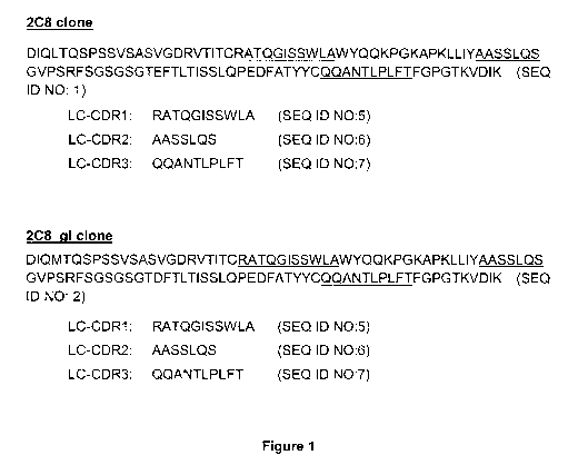
i) LC-CDR1: RATQGISSWLA (SEQ ID NO:5);
ii) LC-CDR2: AASSLQS (SEQ ID NO:6);
iii) LC-CDR3: QQANTLPLFT (SEQ
ID NO:7);
iv) HC-CDR1: SNTAAWN (SEQ ID
NO:8);
v) HC-CDR2: RTYYRSKWYSDYGLSVKS (SEQ ID NO:9);
vi) HC-CDR3: EGSGGTLIY (SEQ ID
NO:10);
or a variant thereof in which one or two or three amino acids in one or more
of the
sequences (i) to (vi) are replaced with another amino acid.
CA 03023787 2018-11-09
WO 2017/194265 4 PCT/EP2017/058956
In some embodiments LC-CDR1 is RATQGISSWLA (SEQ ID NO:5). In some embodiments
LC-CDR2 is AASSLQS (SEQ ID NO:6). In some embodiments LC-CDR3 is QQANTLPLFT
(SEQ ID NO:7). In some embodiments HC-CDR1 is SNTAAWN (SEQ ID NO:8). In some
embodiments HC-CDR2 is RTYYRSKWYSDYGLSVKS (SEQ ID NO:9). In some
embodiments HC-CDR3 is EGSGGTLIY (SEQ ID NO:10).
In some embodiments the antibody, or antigen binding fragment, may comprise at
least one
light chain variable region incorporating the following CDRs:
LC-CDR1: RATQGISSWLA (SEQ ID NO:5)
LC-CDR2: AASSLQS (SEQ ID NO:6)
LC-CDR3: QQANTLPLFT (SEQ ID NO:7).
In some embodiments the antibody, or antigen binding fragment, may comprise at
least one
heavy chain variable region incorporating the following CDRs:
HC-CDR1: SNTAAWN (SEQ ID NO:8)
HC-CDR2: RTYYRSKWYSDYGLSVKS (SEQ ID NO:9)
HC-CDR3: EGSGGTLIY (SEQ ID NO:10).
The antibody may comprise at least one light chain variable region
incorporating the CDRs
shown in Figure 1. The antibody may comprise at least one heavy chain variable
region
incorporating the CDRs shown in Figure 2.
The antibody may comprise at least one light chain variable region (VL)
comprising the
amino acid sequence of one of SEQ ID NOs 1, 5, 6, 7; or 2, 5, 6, 7 or one of
the amino acid
sequences shown in Figure 1 or an amino acid sequence having at least 70%,
more
preferably one of at least 75%, 80%, 85%, 86%, 87%, 88%, 89%, 90%, 91%, 92%,
93%,
94%, 95%, 96%, 97%, 98%, 99%, or 100%, sequence identity to one of SEQ ID NOs
1, 5, 6,
7; or 2, 5, 6, 7, or to the amino acid sequence of the VL chain amino acid
sequence shown in
Figure 1.
The antibody may comprise at least one heavy chain variable region (VH)
comprising the
amino acid sequence of one of SEQ ID NOs 3, 8, 9, 10; or 4, 8,9, 10 or one of
the amino
acid sequences shown in Figure 2 or an amino acid sequence having at least
70%, more
preferably one of at least 75%, 80%, 85%, 86%, 87%, 88%, 89%, 90%, 91%, 92%,
93%,
94%, 95%, 96%, 97%, 98%, 99%, or 100%, sequence identity to one of SEQ ID NOs
3, 8, 9,
10; or 4, 8, 9, 10, or to the amino acid sequence of the VH chain amino acid
sequence shown
in Figure 2.
CA 03023787 2018-11-09
WO 2017/194265 5 PCT/EP2017/058956
The antibody may comprise at least one light chain variable region comprising
the amino
acid sequence of one of SEQ ID NOs 1, 5, 6, 7; or 2, 5, 6, 7, or one of the
amino acid
sequences shown in Figure 1 (or an amino acid sequence having at least 70%,
more
preferably one of at least 75%, 80%, 85%, 90%, 95%, 98%, 97%, 980,to , 99% or
100%,
sequence identity to one of SEQ ID NOs 1, 5, 6, 7; or 2, 5, 6, 7, or to one of
the amino acid
sequences of the VL chain amino acid sequence shown in Figure 1) and at least
one heavy
chain variable region comprising the amino acid sequence of one of SEQ ID NOs
3, 8, 9, 10;
or 4, 8, 9, 10, or one of the amino acid sequence shown in Figure 2 (or an
amino acid
sequence having at least 70%, more preferably one of at least 75%, 80%, 85%,
86%, 87%,
88%, 89%, 90%, 91%, 92%, 93%, 94%, 95%, 98%, 97%, 98%, 99%, or 100%, sequence
identity to one of SEQ ID NOs 3, 8, 9, 10; or 4, 8,9, 10, or to one of the
amino acid
sequences of the VH chain amino acid sequence shown in Figure 2).
The antibody may optionally bind CTLA-4, optionally human or murine CTLA-4. In
some
embodiments, the antibody is capable of binding to both of human and murine
CTLA-4. The
antibody may optionally have amino acid sequence components as described
above. The
antibody may be an IgG. In one embodiment an in vitro complex, optionally
isolated,
comprising an antibody, or antigen binding fragment, as described herein,
bound to CTLA-4
is provided.
The antibody may optionally inhibit or prevent interaction or functional
association between
human CTLA-4 and human CD80 or 0D86, or between murine CTLA-4 and murine CD80
or
0D86. Such inhibition or prevention of interaction or functional association
between CTLA-4
and CD80 or 0D86 may inhibit or prevent CD80 or 0D86-mediated activation of
CTLA-4,
CD80/CTLA-4 signalling or 0D86/CTLA-4 signalling.
In one aspect of the present invention an isolated light chain variable region
polypeptide is
provided, the light chain variable region polypeptide comprising the following
CDRs:
LC-CDR1: RATQGISSWLA (SEQ ID NO:5)
LC-CDR2: AASSLQS (SEQ ID NO:6)
LC-CDR3: QQANTLPLFT (SEQ ID NO:7).
In one aspect of the present invention an isolated light chain variable region
polypeptide is
provided, comprising an amino acid sequence having at least 85% sequence
identity to the
light chain sequence: SEQ ID NO:1 or 2 (Figure 1). In some embodiments the
isolated light
chain variable region polypeptide is capable of binding to CTLA-4.
CA 03023787 2018-11-09
WO 2017/194265 6
PCT/EP2017/058956
In one aspect of the present invention an isolated heavy chain variable region
polypeptide is
provided, the heavy chain variable region polypeptide comprising the following
CDRs:
HC-CDR1: SNTAAWN (SEQ ID NO:8)
HC-CDR2: RTYYRSKWYSDYGLSVKS (SEQ ID NO:9)
HC-CDR3: EGSGGTLIY (SEQ ID NO:10).
In one aspect of the present invention an isolated heavy chain variable region
polypeptide is
provided, comprising an amino acid sequence having at least 85% sequence
identity to the
heavy chain sequence of SEQ ID NO:3 or 4 (Figure 2). In some embodiments the
isolated
heavy chain variable region polypeptide is capable of binding to CTLA-4.
In one aspect of the present invention an antibody, or antigen binding
fragment, is provided,
the antibody, or antigen binding fragment, comprising a heavy chain and a
light chain
variable region sequence, wherein:
the light chain comprises a LC-CDR1, LC-CDR2, LC-CDR3, having at least 85%
overall
sequence identity to LC-CDR1: RATQGISSWLA (SEQ ID NO:5), LC-CDR2: AASSLQS
(SEQ ID NO:6), LC-CDR3: QQANTLPLFT (SEQ ID NO:7), and;
the heavy chain comprises a HC-CDR1, HC-CDR2, HC-CDR3, having at least 85%
overall
sequence identity to HC-CDR1: SNTAAWN (SEQ ID NO:8), HC-CDR2:
RTYYRSKWYSDYGLSVKS (SEQ ID NO:9), HC-CDR3: EGSGGTLIY (SEQ ID NO:10).
In some embodiments the degree of sequence identity may be one of 86%, 87%,
88%, 89%,
90%, 91%, 92%, 93%, 94%, 95%, 96%, 97%, 98%, 99%, or 100%.
In another aspect of the present invention an antibody, or antigen binding
fragment,
optionally isolated, is provided comprising a heavy chain and a light chain
variable region
sequence, wherein:
the light chain sequence has at least 85% sequence identity to the light chain
sequence:
SEQ ID NO:1 or 2 (Figure 1), and;
the heavy chain sequence has at least 85% sequence identity to the heavy chain
sequence
of SEQ ID NO:3 or 4 (Figure 2).
In some embodiments the degree of sequence identity may be one of 86%, 87%,
88%, 89%,
90%, 91%, 92%, 93%, 94%, 95%, 96%, 97%, 98%, 99%, or 100%.
CA 03023787 2018-11-09
WO 2017/194265 7
PCT/EP2017/058956
In some embodiments the antibody, antigen binding fragment, or polypeptide
further
comprises variable region light chain framework sequences between the CDRs
according to
the arrangement LCFR1:LC-CDR1:LCFR2:LC-CDR2:LCFR3:LC-CDR3:LCFR4. The
framework sequences may be derived from human consensus framework sequences.
In one aspect of the present invention an isolated light chain variable region
polypeptide,
optionally in combination with a heavy chain variable region polypeptide as
described herein,
is provided, the light chain variable region polypeptide comprising the
following CDRs:
LC-CDR1: RATQGISSWLA (SEQ ID NO:5)
LC-CDR2: AASSLQS (SEQ ID NO:6)
LC-CDR3: QQANTLPLFT (SEQ ID NO:7).
In some embodiments the antibody, antigen binding fragment, or polypeptide
further
comprises variable region heavy chain framework sequences between the CDRs
according
to the arrangement HCFR1:HC-CDR1:HCFR2:HC-CDR2:HCFR3:HC-CDR3:HCFR4. The
framework sequences may be derived from human consensus framework sequences.
In one aspect of the present invention an isolated heavy chain variable region
polypeptide,
optionally in combination with a light chain variable region polypeptide as
described herein,
is provided, the heavy chain variable region polypeptide comprising the
following CDRs:
HC-CDR1: SNTAAWN (SEQ ID NO:8)
HC-CDR2: RTYYRSKWYSDYGLSVKS (SEQ ID NO:9)
HC-CDR3: EGSGGTLIY (SEQ ID NO:10).
In some embodiments, the antibody, or antibody binding fragment, may further
comprise a
human constant region. For example selected from one of IgG1, IgG2, IgG3 and
IgG4.
In some embodiments, the antibody, or antibody binding fragment, may further
comprise a
murine constant region. For example, selected from one of IgG1, IgG2A, IgG2B
and IgG3.
In another aspect of the present invention, an antibody or antigen binding
fragment,
optionally isolated, which is capable of binding to CTLA-4, which is a
bispecific antibody or a
bispecific antigen binding fragment is provided. The bispecific antibody or
antigen binding
fragment comprises (i) an antigen binding fragment or polypeptide capable of
binding to
CTLA-4 as described herein, and (ii) an antigen binding fragment or
polypeptide which is
capable of binding to a target protein other than CTLA-4.
CA 03023787 2018-11-09
WO 2017/194265 8
PCT/EP2017/058956
In some embodiments, the target protein other than CTLA-4 may be a cell
surface receptor,
e.g. a receptor expressed on the cell surface of T cells. In some embodiments
the cell
surface receptor may be an immune checkpoint receptor, e.g. a costimulatory
receptor or an
inhibitory receptor. In some embodiments, the costimulatory receptor may be
selected from
0D27, 0D28, ICOS, CD40, 0D122, 0X43, 4-1BB and GITR. In some embodiments, the
inhibitory receptor may be selected from B7-H3, B7-H4, BTLA, LAG-3, A2AR,
VISTA, TIM-3,
PD-1, and KIR.
In some embodiments, the target protein other than CTLA-4 may be a cancer
marker whose
expression is associated with a cancer. In some embodiments, the cancer marker
may be
expressed at the cell surface. In some embodiments, cancer marker may be
selected from
HER-2, HER-3, EGFR, EpCAM, CD30, 0D33, 0D38, CD20, 0D24, CD90, CD15, 0D52,
CA-125, 0D34, CA-15-3, CA-19-9, CEA, 0D99, CD117, CD31, 0D44, CD123, CD133,
ABCB5 and 0D45.
In another aspect of the present invention a chimeric antigen receptor (CAR)
is provided,
comprising an antigen binding fragment as described herein.
In another aspect the present invention provides a cell comprising a CAR as
described
herein.
In another aspect of the present invention an in vitro complex is provided,
comprising an
antibody, antigen binding fragment, polypeptide, CAR or cell as described
herein bound to
CTLA-4. The in vitro complex may optionally be isolated.
In another aspect of the present invention, a composition, e.g. a
pharmaceutical composition
or medicament, is provided. The composition may comprise an antibody, antigen
binding
fragment, polypeptide, CAR or cell as described herein and at least one
pharmaceutically-
acceptable carrier, excipient, adjuvant or diluent.
In another aspect of the present invention an isolated nucleic acid encoding
an antibody,
antigen binding fragment, polypeptide or CAR as described herein is provided.
The nucleic
acid may have a sequence of one of SEQ ID NOs 11, 12, 13 or 14 (Figure 3), or
a coding
sequence which is degenerate as a result of the genetic code, or may have a
nucleotide
sequence having at least 70% identity thereto, optionally one of 75%, 80%,
85%, 86%, 87%,
88%, 89%, 90%, 91%, 92%, 93%, 94%, 95%, 96%, 97%, 98%, 9-0,to,
or 100`)/0.
CA 03023787 2018-11-09
WO 2017/194265 9
PCT/EP2017/058956
In one aspect of the present invention there is provided a vector comprising a
nucleic acid
described herein. In another aspect of the present invention, there is
provided a host cell
comprising the vector. For example, the host cell may be eukaryotic, or
mammalian, e.g.
Chinese Hamster Ovary (CHO), or human or may be a prokaryotic cell, e.g. E.
coll.
In one aspect of the present invention a method for making an antibody, or
antigen binding
fragment, polypeptide or CAR as described herein is provided, the method
comprising
culturing a host cell as described herein under conditions suitable for the
expression of a
vector encoding the antibody, antigen binding fragment, polypeptide or CAR,
and recovering
the antibody, antigen binding fragment, polypeptide or CAR.
In another aspect of the present invention an antibody, antigen binding
fragment,
polypeptide, CAR, cell or composition is provided for use in therapy, or in a
method of
medical treatment. In another aspect of the present invention an antibody,
antigen binding
fragment, polypeptide, CAR, cell or composition as described herein is
provided for use in
the treatment of a T-cell dysfunctional disorder. In another aspect of the
present invention,
the use of an antibody, antigen binding fragment, polypeptide, CAR, cell or
composition as
described herein in the manufacture of a medicament or pharmaceutical
composition for use
in the treatment of a T-cell dysfunctional disorder is provided.
In another aspect of the present invention a method of enhancing T-cell
function comprising
administering an antibody, antigen binding fragment, polypeptide, CAR, cell or
composition
as described herein to a dysfunctional T-cell is provided. The method may be
performed in
vitro or in vivo.
In another aspect of the present invention a method of treating a T-cell
dysfunctional
disorder is provided, the method comprising administering an antibody, antigen
binding
fragment or polypeptide as described herein to a patient suffering from a T-
cell dysfunctional
disorder.
In another aspect of the present invention an antibody, antigen binding
fragment,
polypeptide, CAR, cell or composition is provided for use in the treatment of
a cancer. In
another aspect of the present invention, the use of an antibody, antigen
binding fragment,
polypeptide, CAR, cell or composition as described herein in the manufacture
of a
medicament or pharmaceutical composition for use in the treatment of a cancer
is provided.
In another aspect of the present invention a method of killing a tumour cell
is provided, the
method comprising administering an antibody, antigen binding fragment,
polypeptide, CAR,
CA 03023787 2018-11-09
WO 2017/194265 1 0
PCT/EP2017/058956
cell or composition as described herein to a tumour cell. The method may be
performed in
vitro or in vivo. Killing of a tumour cell may, for example, be as a result of
antibody
dependent cell-mediated cytotoxicity (ADCC), complement dependent cytotoxicity
(CDC), or
through the action of a drug conjugated to the antibody, antigen binding
fragment,
polypeptide, CAR, cell or composition.
In another aspect of the present invention a method of treating a cancer is
provided, the
method comprising administering an antibody, antigen binding fragment,
polypeptide, CAR,
cell or composition as described herein to a patient suffering from a cancer.
The cancer may be a cancer which expresses or overexpresses CTLA-4, or may
comprise
cells which express or overexpress CTLA-4.
In another aspect of the present invention a method of modulating an immune
response in a
subject is provided, the method comprising administering to the subject an
antibody, antigen
binding fragment, polypeptide, CAR, cell or composition as described herein
such that the
immune response in the subject is modulated.
In another aspect of the present invention a method of inhibiting growth of
tumor cells is
provided, comprising administering an antibody, antigen binding fragment,
polypeptide,
CAR, cell or composition as described herein. The method may be in vitro or in
vivo. In some
embodiments a method of inhibiting growth of tumor cells in a subject is
provided, the
method comprising administering to the subject a therapeutically effective
amount of an
antibody, antigen binding fragment, polypeptide, CAR, cell or composition as
described
herein.
In another aspect of the present invention a method is provided, the method
comprising
contacting a sample containing, or suspected to contain, CTLA-4 with an
antibody, antigen
binding fragment, CAR or cell as described herein, and detecting the formation
of a complex
of antibody, antigen binding fragment, CAR or cell and CTLA-4.
In another aspect of the present invention a method of diagnosing a disease or
condition in a
subject is provided, the method comprising contacting, in vitro, a sample from
the subject
with an antibody, antigen binding fragment, CAR or cell as described herein,
and detecting
the formation of a complex of antibody, antigen binding fragment, CAR or cell
and CTLA-4.
CA 03023787 2018-11-09
WO 2017/194265 11
PCT/EP2017/058956
In a further aspect of the present invention the use of an antibody, antigen
binding fragment,
CAR or cell as described herein, for the detection of CTLA-4 in vitro is
provided. In another
aspect of the present invention the use of an antibody, antigen binding
fragment, CAR or cell
as described herein, as an in vitro diagnostic agent is provided.
In methods of the present invention the antibody, antigen binding fragment,
polypeptide,
CAR or cell may be provided as a composition as described herein.
In another aspect the present invention provides a method of treating or
preventing a cancer
in a subject, comprising:
(a) isolating at least one cell from a subject;
(b) modifying the at least one cell to express or comprise the antibody,
antigen
binding fragment, polypeptide, CAR, nucleic acid or vector described herein,
and;
(c) administering the modified at least one cell to a subject.
In another aspect the present invention provides a method of treating or
preventing a cancer
in a subject, comprising:
(a) isolating at least one cell from a subject;
(b) introducing into the at least one cell the nucleic acid or vector
described herein,
thereby modifying the at least one cell, and;
(c) administering the modified at least one cell to a subject.
In another aspect the present invention provides a kit of parts comprising a
predetermined
quantity of the antibody, antigen binding fragment, polypeptide, CAR,
composition, nucleic
acid, vector or cell described herein.
In some embodiments the antibody may be clone 208 or 208_gl as described
herein.
Description
Antibodies
Antibodies according to the present invention preferably bind to CTLA-4 (the
antigen),
preferably human or murine CTLA-4, optionally with a KD in the range 2 to 20
nM.
Antibodies according to the present invention may be provided in isolated
form.
CA 03023787 2018-11-09
WO 2017/194265 12
PCT/EP2017/058956
Antibodies according to the present invention may exhibit least one of the
following
properties:
a) binds to human or mouse CTLA-4 with a KD of 1 pM or less, preferably one of
100 nM, 75 nM, 50 nM, 40 nM, 30 nM, 20 nM, 15 nM , 12.5 nM, 0 nM, 9
nM, nM, 7 nM, nM, nM, .4. nM 3 nM, nM, nM, 500 pM (e.g. as
determined by SPR);
b) binds to human or mouse CTLA-4 with an affinity of binding of EC50 = 1 pM
or
less, preferably one of 100 nM, 75 nM, 50 nM, 40 nM, 30 nM, 20 nM, 15 nM,
12.5 nM, 10 nM, 9 nM, nM, 7 nM, nM, nM, .4. nM 3 nM, nM,
nM, 500 pM (e.g. as determined by ELISA);
c) binds to human or mouse CTLA-4 with an avidity of binding of EC50 = 500 pM
or
less, preferably one of 400 pM, 300 pM, 200 pM, 150 pM, 100 pM, 90 pM,
E30 pM, 75 pM, 70, 65 pM, 60 pM, 55 pM, 50 pM (e.g. as determined by
ELISA);
d) displays substantially no binding to 0D28 (e.g. human 0D28 or mouse 0D28).
e) binds to human and mouse CTLA-4, and displays substantially no binding to
binds
to human 0D28;
f) inhibits or prevents interaction between CTLA-4 and CD80, optionally human
CTLA-4 and human CD80;
g) inhibits or prevents interaction between CTLA-4 and 0D86, optionally human
CTLA-4 and human 0D86;
h) inhibits or prevents interaction between CTLA-4 and CD80 and interaction
between CTLA-4 and 0D86, optionally human CTLA-4, human CD80 and human
CD86;
i) does not inhibit or prevent interaction between 0D28 and CD80, optionally
human
0D28 and human CD80;
j) does not inhibit or prevent interaction between 0D28 and 0D86, optionally
human
0D28 and human 0D86;
k) does not inhibit or prevent interaction between 0D28 and CD80 and
interaction
between 0D28 and 0D86, optionally human 0D28, human CD80 and human 0D86;
I) increases activation of T cells in vitro;
m) increases IL-2 production by T cells in a T cell reactivation assay;
n) increases one or more of T-cell proliferation, IL-2 production and IFNy
production
in response to infection;
o) inhibits tumour growth, optionally in vivo.
CA 03023787 2018-11-09
WO 2017/194265 13
PCT/EP2017/058956
In some embodiments, the antibody according to the present invention may be
useful in
methods for expanding a population of immune cells, e.g. T cells. The
antibodies according
to the invention are useful for expanding populations of immune cells with
desirable
properties.
In some embodiments, the antibody of the present invention is useful to expand
in methods
for expanding a population of immune cells with an effector phenotype (e.g.
CTLs) in
preference to immune cells with an immunoregulatory/immunosuppressive
phenotype (e.g.
Tregs)
By "antibody" we include a fragment or derivative thereof, or a synthetic
antibody or synthetic
antibody fragment.
In view of today's techniques in relation to monoclonal antibody technology,
antibodies can
be prepared to most antigens. The antigen-binding portion may be a part of an
antibody (for
example a Fab fragment) or a synthetic antibody fragment (for example a single
chain Fv
fragment [ScFv]). Suitable monoclonal antibodies to selected antigens may be
prepared by
known techniques, for example those disclosed in "Monoclonal Antibodies: A
manual of
techniques ", H Zola (CRC Press, 1988) and in "Monoclonal Hybridoma
Antibodies:
Techniques and Applications ", J G R Hurrell (CRC Press, 1982). Chimeric
antibodies are
discussed by Neuberger et al (1988, 8th International Biotechnology Symposium
Part 2,
792-799).
Monoclonal antibodies (mAbs) are useful in the methods of the invention and
are a
homogenous population of antibodies specifically targeting a single epitope on
an antigen.
Polyclonal antibodies are useful in the methods of the invention. Monospecific
polyclonal
antibodies are preferred. Suitable polyclonal antibodies can be prepared using
methods well
known in the art.
In some embodiments, the antibody/fragment is a fully human antibody/fragment.
A fully
human antibody/fragment is encoded by human nucleic acid sequence(s). Fully
human
antibodies/fragments are devoid of non-human amino acid sequences.
In some embodiments, the antibody/fragment may be a chimeric
antibody/fragment. A
chimeric antibody/fragment may comprise amino acid sequences derived from
different
antibodies/fragments. For example, a chimeric antibody/fragment may comprise
CDRs or
CA 03023787 2018-11-09
WO 2017/194265 14
PCT/EP2017/058956
variable domain sequence(s) from one antibody/antigen binding fragment, and
constant
region sequence(s) from another antibody/antigen binding fragment. In some
embodiments,
a chimeric antibody/fragment may comprise CDRs from one antibody/antigen
binding
fragment, and constant region sequence(s) and framework region sequence(s)
from another
.. antibody/antigen binding fragment.
In some embodiments, the chimeric antibody/fragment may comprise CDRs or
variable
domain sequence(s) of anti-CTLA-4 clone 208 or 208_gl described herein, and
constant
region sequence(s) from another antibody/antigen binding fragment. In some
embodiments,
the chimeric antibody/fragment may comprise CDRs of anti-CTLA-4 clone 208 or
208_gl
described herein, and constant region sequence(s) and framework region
sequence(s) from
another antibody/antigen binding fragment.
In some embodiments, a chimeric antibody/fragment may comprise CDRs or
variable
domain sequence(s) from an antibody from one species and constant region
sequence(s)
from an antibody from another species. In some embodiments, a chimeric
antibody/fragment
may comprise CDRs from an antibody from one species and constant region
sequence(s)
and framework region sequence(s) from an antibody from another species.
In some embodiments, a chimeric antibody/fragment according to the present
invention may
comprise CDRs or variable domain sequence(s) from anti-CTLA-4 clone 208 or
208_gl
described herein, and constant region sequence(s) from an antibody from a non-
human
species. In some embodiments, a chimeric antibody/fragment according to the
present
invention may comprise CDRs from anti-CTLA-4 clone 208 or 208_gl described
herein, and
constant region sequence(s) and framework region sequence(s) from an antibody
from a
non-human species. In some embodiments, the non-human species is e.g. a non-
human
mammal (e.g. rabbit, guinea pig, rat, mouse or other rodent (including any
animal in the
order Rodentia), cat, dog, pig, sheep, goat, cattle (including cows, e.g.
dairy cows, or any
animal in the order Bos), horse (including any animal in the order Equidae),
donkey, or non-
human primate).
In some embodiments, an antibody/fragment according to the invention may
comprise
modifications (e.g. one or more amino acid substitutions) to increase
similarity to antibodies
naturally produced in a species of interest.
Antigen binding fragments of antibodies, such as Fab and Fab2 fragments may
also be
used/provided as can genetically engineered antibodies and antibody fragments.
The
CA 03023787 2018-11-09
WO 2017/194265 15
PCT/EP2017/058956
variable heavy (VH) and variable light (VL) domains of the antibody are
involved in antigen
recognition, a fact first recognised by early protease digestion experiments.
Further
confirmation was found by "humanisation" of rodent antibodies. Variable
domains of rodent
origin may be fused to constant domains of human origin such that the
resultant antibody
retains the antigenic specificity of the rodent parent antibody (Morrison et
al (1984) Proc.
Natl. Acad. Sd. USA 81, 6851-6855).
That antigenic specificity is conferred by variable domains and is independent
of the
constant domains is known from experiments involving the bacterial expression
of antibody
fragments, all containing one or more variable domains. These molecules
include Fab-like
molecules (Better et al (1988) Science 240, 1041); Fv molecules (Skerra et al
(1988)
Science 240, 1038); single-chain Fv (ScFv) molecules where the VH and VL
partner domains
are linked via a flexible oligopeptide (Bird et al (1988) Science 242, 423;
Huston et al (1988)
Proc. Natl. Acad. Sd. USA 85, 5879) and single domain antibodies (dAbs)
comprising
isolated V domains (Ward et al (1989) Nature 341, 544). A general review of
the techniques
involved in the synthesis of antibody fragments which retain their specific
binding sites is to
be found in Winter & Milstein (1991) Nature 349, 293- 299.
By "ScFv molecules" we mean molecules wherein the VH and VL partner domains
are
covalently linked, e.g. by a flexible oligopeptide.
Fab, Fv, ScFv and dAb antibody fragments can all be expressed in and secreted
from E.
coli, thus allowing the facile production of large amounts of the said
fragments.
Whole antibodies, and F(ab1)2 fragments are "bivalent". By "bivalent" we mean
that the said
antibodies and F(ab1)2 fragments have two antigen combining sites. In
contrast, Fab, Fv,
ScFv and dAb fragments are monovalent, having only one antigen combining site.
Synthetic
antibodies which bind to CTLA-4 may also be made using phage display
technology as is
well known in the art.
Also provided are multispecific antibodies and multispecific antigen binding
fragments,
comprising an antigen binding fragment or a polypeptide according to the
present invention.
As used herein, 'multispecific' means having specificity for more than one
epitope. In some
embodiments, a multispecific antibody or multispecific antigen binding
fragment may be
specific for e.g. 2 (bispecific), 3 (trispecific), 4, 5, 6, 7, 8, 9 or 10
different epitopes.
CA 03023787 2018-11-09
WO 2017/194265 16
PCT/EP2017/058956
In some embodiments, multispecific antibodies/fragments according to the
invention may
have specificity for more than one target molecule. In some embodiments, a
multispecific
antibody/fragment may be specific for e.g. 2 (bispecific), 3 (trispecific), 4,
5, 6, 7, 8, 9 or 10
different target molecules.
In some embodiments, the multispecific antibodies and multispecific antigen
binding
fragments comprise an antigen binding fragment capable of binding to CTLA-4,
and an
antigen binding fragment capable of binding to another target protein. In some
embodiments
the multispecific antibodies/fragments comprise an antigen binding fragment
capable of
binding to CTLA-4, and e.g. 1, 2, 3 ,4 ,5 6, 7, 8, or 9 antigen binding
fragment(s) capable of
binding to another target protein, i.e. a protein other than CTLA-4.
The present application also provides an antibody or antigen binding fragment
which is
capable of binding to CTLA-4, and which is a bispecific antibody or a
bispecific antigen
binding fragment. In some embodiments, the bispecific antibody or bispecific
antigen binding
fragment may be isolated.
In some embodiments, the bispecific antibodies and bispecific antigen binding
fragments
comprise an antigen binding fragment or a polypeptide according to the present
invention. In
some embodiments, the bispecific antibodies and bispecific antigen binding
fragments
comprise an antigen binding fragment capable of binding to CTLA-4, wherein the
antigen
binding fragment which is capable of binding to CTLA-4 comprises or consists
of an antigen
binding fragment or a polypeptide according to the present invention.
In some embodiments the bispecific antibodies and bispecific antigen binding
fragments
comprise an antigen binding fragment capable of binding to CTLA-4, and an
antigen binding
fragment capable of binding to another target protein.
The antigen binding fragment capable of binding to another target protein may
be capable of
binding to another protein other than CTLA-4.
In some embodiments, the target protein may be a cell surface receptor. In
some
embodiments, the target protein may be a cell surface receptor expressed on
the cell
surface of an immune cell, e.g. T cell. In some embodiments the cell surface
receptor may
be an immune checkpoint receptor. In some embodiments, the immune checkpoint
receptor
may be a costimulatory receptor. In some embodiments, the costimulatory
receptor may be
selected from 0D27, 0D28, ICOS, CD40, 0D122, 0X43, 4-1 BB and GITR. In some
CA 03023787 2018-11-09
WO 2017/194265 17
PCT/EP2017/058956
embodiments, the immune checkpoint receptor may be an inhibitory receptor. In
some
embodiments, the inhibitory receptor may be selected from B7-H3, B7-H4, BTLA,
LAG-3,
A2AR, VISTA, TIM-3, PD-1, and KIR.
In some embodiments, the target protein may be a cancer marker. That is, the
target protein
may be a protein whose expression (e.g. upregulated expression) is associated
with a
cancer. In some embodiments, the cancer marker may be expressed at the cell
surface. In
some embodiments the cancer marker may be a receptor. In some embodiments, the
cancer
marker may be selected from HER-2, HER-3, EGFR, EpCAM, CD30, 0D33, 0D38, CD20,
0D24, CD90, CD15, 0D52, CA-125, 0D34, CA-15-3, CA-19-9, CEA, 0D99, CD117,
CD31,
0D44, CD123, CD133, ABCB5 and 0D45.
In some embodiments, the antigen binding fragment for 0D27 may comprise the
CDRs, light
and heavy chain variable domains or other 0D27 binding fragment of e.g. anti-
0D27
antibody clone 0323 (Millipore) or varlilumab (Celldex Therapeutics). In some
embodiments,
the antigen binding fragment for 0D28 may comprise the CDRs, light and heavy
chain
variable domains or other 0D28 binding fragment of e.g. anti-0D28 antibody
clone 0D28.6
(eBioscience), clone 0D28.2, clone JJ319 (Novus Biologicals), clone 204.12,
clone B-23,
clone 10F3 (Thermo Scientific Pierce Antibodies), clone 37407 (R&D Systems),
clone 204-
12 (Abnova Corporation), clone 15E8 (EMD Millipore), clone 204-12, clone
YTH913.12 (AbD
Serotec), clone B-T3 (Acris Antibodies), clone 9H6E2 (Sino Biological), clone
C28/77
(MyBioSource.com), clone KOLT-2 (ALPCO), clone 152-2E10 (Santa Cruz
Biotechnology),
or clone XPH-56 (Creative Diagnostics). In some embodiments, the antigen
binding
fragment for ICOS may comprise the CDRs, light and heavy chain variable
domains or other
ICOS binding fragment of e.g. anti-ICOS antibody clone ISA-3 (eBioscience),
clone 5P98
(Novus Biologicals), clone 1G1, clone 3G4 (Abnova Corporation), clone 669222
(R&D
Systems), clone TQ09 (Creative Diagnostics), or clone C398.4A (BioLegend). In
some
embodiments, the antigen binding fragment for CD40 may comprise the CDRs,
light and
heavy chain variable domains or other CD40 binding fragment of e.g. anti-CD40
antibody
clone 82111 (R&D Systems), or A5KP1240 (Okimura et al., AM J Transplant (2014)
14(6)
1290-1299). In some embodiments, the antigen binding fragment for CD122 may
comprise
the CDRs, light and heavy chain variable domains or other CD122 binding
fragment of anti-
CD122 antibody clone mik[32 (PharMingen). In some embodiments, the antigen
binding
fragment for 0X43 may comprise the CDRs, light and heavy chain variable
domains or other
0X43 binding fragment of e.g. anti-0X43 antibodies disclosed in US
20130280275, US
8283450 or W02013038191, e.g. clone 12H3 or clone 20E5. In some embodiments,
the
antigen binding fragment for 4-i BB may comprise the CDRs, light and heavy
chain variable
CA 03023787 2018-11-09
WO 2017/194265 18
PCT/EP2017/058956
domains or other 4-1BB binding fragment of e.g. anti-4-1BB antibody PF-
05082566 (Fisher
et al., Cancer Immunol lmmunother (2012) 61: 1721-1733), or urelumab (BMS-
665513;
Bristol-Myers Squibb; Li and Liu, Clin Pharmacol (2013); 5: 47-53). In some
embodiments,
the antigen binding fragment for GITR may comprise the CDRs, light and heavy
chain
variable domains or other GITR binding fragment of e.g. anti- GITR antibody
TRX-518
(Tolee; Schaer et al., (2010) 11(12): 1378-1386), or clone AIT 518D (LifeSpan
Biosciences). In some embodiments, the antigen binding fragment for B7-H3 may
comprise
the CDRs, light and heavy chain variable domains or other B7-H3 binding
fragment of e.g.
anti-B7-H3 antibody clones disclosed in US 20130078234, W02014160627 or
W02011109400. In some embodiments, the antigen binding fragment for B7-H4 may
comprise the CDRs, light and heavy chain variable domains or other B7-H4
binding fragment
of e.g. anti-B7-H4 antibody clones disclosed in W02013067492, W02009073533 or
EP2934575, for example clone 2H9. In some embodiments, the antigen binding
fragment for
BTLA may comprise the CDRs, light and heavy chain variable domains or other
BTLA
binding fragment of e.g. anti-BTLA antibody clone 167, clone 2G8, clone 4C5
(Abnova
Corporation), clone 4B8 (antibodies-online), clone MIH26 (Thermo Scientific
Pierce
Antibodies), clone UMAB61 (OriGene Technologies), clone 330104 (R&D Systems),
clone
1B4 (LifeSpan BioSciences), clone 440205, clone 5E7 (Creative Diagnostics). In
some
embodiments, the antigen binding fragment for LAG-3 may comprise the CDRs,
light and
heavy chain variable domains or other LAG-3 binding fragment of e.g. anti-LAG-
3 antibody
clone 17134 (Enzo Life Sciences), clone 333210 (R&D Systems), clone 14L676
(United
States Biological), BMS-986016, or an anti-LAG-3 antibody described in WO
2015042246
Al. In some embodiments, the antigen binding fragment for A2AR may comprise
the CDRs,
light and heavy chain variable domains or other A2AR binding fragment of e.g.
anti-A2AR
antibody clone 7F6 (Millipore; Koshiba et al. Molecular Pharmacology (1999);
55: 614-624.
In some embodiments, the antigen binding fragment for VISTA may comprise the
CDRs,
light and heavy chain variable domains or other VISTA binding fragment of e.g.
anti-VISTA
antibodies disclosed in W02015097536 or U520140105912, e.g. clone 13F3. In
some
embodiments, the antigen binding fragment for TIM-3 may comprise the CDRs,
light and
heavy chain variable domains or other TIM-3 binding fragment of e.g. anti-TIM-
3 antibody
clone F38-2E2 (BioLegend), clone 2E2 (Merck Millipore; Pires da Silva et al.,
Cancer
Immunol Res (2014) 2(5): 410-422), clone 6136E2, clone 024 (Sino Biological)
clone 344801
(R&D Systems), clone E-18, clone H-191 (Santa Cruz Biotechnology), or clone
13A224
(United States Biological). In some embodiments, the antigen binding fragment
for PD-1 may
comprise the CDRs, light and heavy chain variable domains or other PD-1
binding fragment
of e.g. anti-PD-1 antibody clone J116, clone MIH4 (eBioscience), clone 7A11B1
(Rockland
lmmunochemicals Inc.), clone 192106 (R&D Systems), clone J110, clone J105 (MBL
CA 03023787 2018-11-09
WO 2017/194265 19
PCT/EP2017/058956
International), clone 12A7D7, clone 7A11I31 (Abbiotec), clone #9X21
(MyBioSource.com),
clone 4H4D1 (Proteintech Group), clone D3W4U, clone D304S (Cell Signaling
Technology),
clone RMP1-30, clone RMP1-14 (Merck Millipore), clone EH12.2H7 (BioLegend),
clone
1061227 (United States Biological), clone UMAB198, clone UMAB197 (Origene
Technologies), nivolumab (BMS-936558), lambrolizumab, or anti-PD-1 antibodies
described
in WO 2010/077634 or WO 2006/121168. In some embodiments, the antigen binding
fragment for KIR may comprise the CDRs, light and heavy chain variable domains
or other
KIR binding fragment of e.g. anti-KIR antibody clone 1-7F9 (Romagne et al.,
Blood (2009)
114(13): 2667-2677), lirilumab (BMS-986015; Sole et al., J lmmunother Cancer
(2013);
1:P40) or anti-KIR antibodies described in US 2015/0344576 or WO 2014/066532.
In some
embodiments, the antigen binding fragment for HER-2 may comprise the CDRs,
light and
heavy chain variable domains or other HER-2 binding fragment of e.g. anti-HER-
2 antibody
trastuzumab (Herceptin), or anti-HER-2 antibodies described in WO 2003/006509
or WO
2008/019290. In some embodiments, the antigen binding fragment for HER-3 may
comprise
the CDRs, light and heavy chain variable domains or other HER-3 binding
fragment of e.g.
anti-HER-3 antibody clone MM-121 (Lyu et al., Int. J Clin Exp Pathol (2015)
8(6): 6143-
6156), MEHD7945A (Schaefer et al., Cancer Cell (2011) 20(4): 472-486), AMG 888
(U3-
1287; Aurisicchio et al., Oncotarget (2012) 3(8): 744-758) or anti-HER-3
antibodies
described in W02008/100624 or WO 2013048883. In some embodiments, the antigen
binding fragment for EGFR may comprise the CDRs, light and heavy chain
variable domains
or other EGFR binding fragment of e.g. anti-EGFR antibody panitumumab (ABX-
EGF;
Vectibix), cetuximab (Erbitux), nimotuzumab, matazumab (EMD 7200) or antibody
clone
048-006 (Sogawa et al., Nucl Med Comm (2012) 33(7): 719-725). In some
embodiments, the
antigen binding fragment for EpCAM may comprise the CDRs, light and heavy
chain variable
domains or other EpCAM binding fragment of e.g. anti-EpCAM antibody
edrecolomab, ING-
1, 3622W4, or adecatumumab (Munz et al., Cancer Cell Int (2010) 10:44). In
some
embodiments, the antigen binding fragment for CD30 may comprise the CDRs,
light and
heavy chain variable domains or other CD30 binding fragment of e.g. anti-CD30
antibody
brentuximab (cAC10), clone SGN-30 (Wahl et al., Cancer Res 2002 62(13):3736-
3742),
clone 5F11 (Borchmann et al., Blood (2003) 102(1): 3737-3742), or anti-CD30
antibodies
described in WO 1993024135 or WO 2003059282. In some embodiments, the antigen
binding fragment for CD33 may comprise the CDRs, light and heavy chain
variable domains
or other CD33 binding fragment of e.g. anti-CD33 antibody lintuzumab (SGN-33),
gemtuzumab (Mylotarg), or clone hP67.7 (Sievers et al., Blood (1999) 93(11):
3678-3684). In
some embodiments, the antigen binding fragment for CD38 may comprise the CDRs,
light
and heavy chain variable domains or other CD38 binding fragment of e.g. anti-
CD38
antibody daratumumab (Darzalex), 5AR650984 (Martin et al., J Clin Oncol (2014)
32:5s,
CA 03023787 2018-11-09
WO 2017/194265 20
PCT/EP2017/058956
(suppl; abstr 8532) or M0R202 (MorphoSys AG), or anti-0D38 antibodies
described in WO
2006099875 or US 20100285004. In some embodiments, the antigen binding
fragment for
CD20 may comprise the CDRs, light and heavy chain variable domains or other
CD20
binding fragment of e.g. anti-CD20 antibody rituximab, ocrelizumab,
ofatumumab,
obinutuzumab or BM-ca (Kobayashi et al., Cancer Med (2013) 2(2): 130-143). In
some
embodiments, the antigen binding fragment for CD24 may comprise the CDRs,
light and
heavy chain variable domains or other CD24 binding fragment of e.g. anti-CD24
antibody
clone eBio5N3 (eBioscience), clone ML5 (BD Biosciences), or anti-CD24
antibodies
described in WO 2008059491. In some embodiments, the antigen binding fragment
for
CD90 may comprise the CDRs, light and heavy chain variable domains or other
CD90
binding fragment of e.g. anti-CD90 antibody clone 5E10 (BD Biosciences). In
some
embodiments, the antigen binding fragment for CD15 may comprise the CDRs,
light and
heavy chain variable domains or other CD15 binding fragment of e.g. anti-CD15
antibody
clone C3D-1, Carb-3 (DAKO NS), MMA (Roche) or BY87 (Abcam). In some
embodiments,
the antigen binding fragment for CD52 may comprise the CDRs, light and heavy
chain
variable domains or other CD52 binding fragment of e.g. anti-CD52 antibody
alemtuzumab,
clone HI186, or clone YTH34.5 (AbD Serotec). In some embodiments, the antigen
binding
fragment for CA-125 may comprise the CDRs, light and heavy chain variable
domains or
other CA-125 binding fragment of e.g. anti-CA-125 antibody oregovomab. In some
embodiments, the antigen binding fragment for CD34 may comprise the CDRs,
light and
heavy chain variable domains or other CD34 binding fragment of e.g. anti-CD34
antibody
clone 561 (BioLegend), clone 581 (Beckton Dickinson), or clone 5F3 (Sigma
Aldrich). In
some embodiments, the antigen binding fragment for CA-15-3 may comprise the
CDRs, light
and heavy chain variable domains or other CA-15-3 binding fragment of e.g.
anti-CA-15-3
antibody clone 2F16 (USBiological), clone TA998 (ThermoFisher Scientific),
clone 1D1
(Sigma Aldrich), or Mab AR20.5 (Qi et al., Hybrid Hybridomics (2001) 20(5-6):
313-324). In
some embodiments, the antigen binding fragment for CA-19-9 may comprise the
CDRs, light
and heavy chain variable domains or other CA-19-9 binding fragment of e.g.
anti-CA-19-9
antibody clone 116-N5-19-9 (DAKO NS), clone 5PM110, or clone 1215LE
(ThermoFisher
Scientific). In some embodiments, the antigen binding fragment for CEA may
comprise the
CDRs, light and heavy chain variable domains or other CEA binding fragment of
e.g. anti-
CEA antibody labetuzumab, C2-45 (Kyowa Hakko Kirin Co. Ltd.) or anti- CEA
antibodies
disclosed in lmakiire et al., Int J Cancer (2004) 108: 564-570 or WO
2011034660. In some
embodiments, the antigen binding fragment for CD99 may comprise the CDRs,
light and
heavy chain variable domains or other CD99 binding fragment of e.g. anti-CD99
antibody
clone C7A (Moricoli et al., J Immunol Methods (2014) 408: 35-45) or clone 12E7
(DAKO
NS). In some embodiments, the antigen binding fragment for CD117 may comprise
the
CA 03023787 2018-11-09
WO 2017/194265 21
PCT/EP2017/058956
CDRs, light and heavy chain variable domains or other CD117 binding fragment
of e.g. anti-
CD117 antibody clone CK6 (Lebron et al., Cancer Biol Ther (2014) 15(9): 1208-
1218), or
clone 104D2 (Sigma Aldrich). In some embodiments, the antigen binding fragment
for CD31
may comprise the CDRs, light and heavy chain variable domains or other CD31
binding
fragment of e.g. anti-CD31 antibody clone JC70A (DAKO NS). In some
embodiments, the
antigen binding fragment for CD44 may comprise the CDRs, light and heavy chain
variable
domains or other CD44 binding fragment of e.g. anti-CD44 antibody PF-03475952
(Runnels
et al., Adv Ther (2010); 27(3): 168-180), RG7356 (Vugts et al., MAbs (2014)
6(2): 567-575),
clone IM7, or clone A3D8 (Sigma Aldrich). In some embodiments, the antigen
binding
fragment for CD123 may comprise the CDRs, light and heavy chain variable
domains or
other CD123 binding fragment of e.g. anti-CD123 antibody C5L362 (Nievergall et
al., Blood
(2014) 123(8):1218-1228), CSL360 (He et al., Leuk Lymphoma (2015) 56(5): 1406-
1415)
73G (Jin et al., Cell Stem Cell (2009) 5(1): 31-42) clone 6H6 (AbD Serotec) or
anti-CD123
antibodies described in WO 2014130635. In some embodiments, the antigen
binding
fragment for CD133 may comprise the CDRs, light and heavy chain variable
domains or
other CD133 binding fragment of e.g. anti-CD133 antibody clone 663, clone 9G4,
clone
AC141 (Wang et al., Hybridoma (Larchmt) (2010) 29(3): 241-249), clone 666
(Chen et al.,
Hybridoma (Larchmt) (2010) 29(4): 305-310, clone AC113 (Miltenyi Biotec), or
anti-CD133
antibodies described in WO 2011149493. In some embodiments, the antigen
binding
fragment for ABC65 may comprise the CDRs, light and heavy chain variable
domains or
other ABC65 binding fragment of e.g. anti-ABC65 antibody clone 5H3C6 (Thermo
Fisher
Scientific). In some embodiments, the antigen binding fragment for CD45 may
comprise the
CDRs, light and heavy chain variable domains or other CD45 binding fragment of
e.g. anti-
CD45 antibody YAML568 (Glatting et al., J Nucl Med (2006) 47(8): 1335-1341) or
clone
BRA-55 (Sigma Aldrich).
An antigen binding fragment of a bispecific antibody or bispecific antigen
binding fragment
according to the present invention may be any fragment of a polypeptide which
is capable of
binding to an antigen. In some embodiments, an antigen binding fragment
comprises at least
the three light chain CDRs (i.e. LC-CDR1, LC-CDR2 and LC-CDR3) and three heavy
chain
CDRs (i.e. HC-CDR1, HC-CDR2 and HC-CDR3) which together define the antigen
binding
region of an antibody or antigen binding fragment. In some embodiments, an
antigen binding
fragment may comprise the light chain variable domain and heavy chain variable
domain of
an antibody or antigen binding fragment. In some embodiments, an antigen
binding fragment
may comprise the light chain polypeptide and heavy chain polypeptide of an
antibody or
antigen binding fragment.
CA 03023787 2018-11-09
WO 2017/194265 22
PCT/EP2017/058956
Bispecific antibodies and bispecific antigen binding fragments according to
the invention may
be provided in any suitable format, such as those formats described in
Kontermann MAbs
2012, 4(2): 182-197, which is hereby incorporated by reference in its
entirety. For example, a
bispecific antibody or bispecific antigen binding fragment may be a bispecific
antibody
conjugate (e.g. an IgG2, F(alp')2 or CovX-Body), a bispecific IgG or IgG-like
molecule (e.g.
an IgG, scFv4-Ig, IgG-scFv, scFv-IgG, DVD-Ig, IgG-sVD, sVD-IgG, 2 in 1-IgG,
mAb2, or
Tandemab common LC), an asymmetric bispecific IgG or IgG-like molecule (e.g. a
kih IgG,
kih IgG common LC, CrossMab, kih IgG-scFab, mAb-Fv, charge pair or SEED-body),
a
small bispecific antibody molecule (e.g. a Diabody (Db), dsDb, DART, scDb,
tandAbs,
tandem scFy (taFv), tandem dAb/VHH, triple body, triple head, Fab-scFv, or
F(ab)2-scFv2), a
bispecific Fc and CH3 fusion protein (e.g. a taFv-Fc, Di-diabody, scDb-CH3,
scFv-Fc-scFv,
HCAb-VHH, scFv-kih-Fc, or scFv-kih-CH3), or a bispecific fusion protein (e.g.
a scFv2-
albumin, scDb-albumin, taFv-toxin, DNL-Fab3, DNL-Faba-IgG, DNL-Fab4-IgG-
cytokine2). See
in particular Figure 2 of Kontermann MAbs 2012, 4(2): 182-19.
The skilled person is able to design and prepare bispecific antibodies and
bispecific antigen
binding fragments according to the present invention.
Methods for producing bispecific antibodies include chemically crosslinking of
antibodies or
antibody fragments, e.g. with reducible disulphide or non-reducible thioether
bonds, for
example as described in Segal and Bast, 2001. Production of Bispecific
Antibodies. Current
Protocols in Immunology. 14:IV:2.13:2.13.1-2.13.16, which is hereby
incorporated by
reference in its entirety. For example, N-succinimidy1-3-(-2-pyridyldithio)-
propionate (SPDP)
can be used to chemically crosslink e.g. Fab fragments via hinge region SH-
groups, to
create disulfide-linked bispecific F(ab)2 heterodimers.
Other methods for producing bispecific antibodies include fusing antibody-
producing
hybridomas e.g. with polyethylene glycol, to produce a quadroma cell capable
of secreting
bispecific antibody, for example as described in D. M. and Bast, B. J. 2001.
Production of
Bispecific Antibodies. Current Protocols in Immunology. 14:IV:2.13:2.13.1-
2.13.16.
Bispecific antibodies and bispecific antigen binding fragments according to
the present
invention can also be produced recombinantly, by expression from e.g. a
nucleic acid
construct encoding polypeptides for the antigen binding molecules, for example
as described
in Antibody Engineering: Methods and Protocols, Second Edition (Humana Press,
2012), at
Chapter 40: Production of Bispecific Antibodies: Diabodies and Tandem scFv
(Hornig and
CA 03023787 2018-11-09
WO 2017/194265 23
PCT/EP2017/058956
Farber-Schwarz), or French, How to make bispecific antibodies, Methods Mol.
Med. 2000;
40:333-339, the entire contents of both of which are hereby incorporated by
reference.
For example, a DNA construct encoding the light and heavy chain variable
domains for the
two antigen binding fragments (i.e. the light and heavy chain variable domains
for the
antigen binding fragment capable of binding CTLA-4, and the light and heavy
chain variable
domains for the antigen binding fragment capable of binding to another target
protein), and
including sequences encoding a suitable linker or dimerization domain between
the antigen
binding fragments can be prepared by molecular cloning techniques. Recombinant
bispecific
antibody can thereafter be produced by expression (e.g. in vitro) of the
construct in a
suitable host cell (e.g. a mammalian host cell), and expressed recombinant
bispecific
antibody can then optionally be purified.
Antibodies may be produced by a process of affinity maturation in which a
modified antibody
is generated that has an improvement in the affinity of the antibody for
antigen, compared to
an unmodified parent antibody. Affinity-matured antibodies may be produced by
procedures
known in the art, e.g., Marks et al.,Rio/Technology 10:779-783 (1992); Barbas
et al. Proc
Nat. Acad. Sci. USA 91:3809-3813 (1994); Schier etal. Gene 169:147-155 (1995);
Yelton et
al. J. Immunol. 155:1994-2004 (1995); Jackson etal., J. Immunol. 154(7):331 0-
15 9 (1995);
and Hawkins eta!, J. Mol. Biol. 226:889-896 (1992).
Antibodies according to the present invention preferably exhibit specific
binding to CTLA-4.
An antibody that specifically binds to a target molecule preferably binds the
target with
greater affinity, and/or with greater duration than it binds to other targets.
In some
embodiments the present antibodies may bind with greater affinity to CTLA-4
than to one or
.. more of PD-1, TIM-3, ICOS, BTLA, 0D28 or LAG-3.
In embodiments of the present invention, the antibody, fragment or polypeptide
displays
substantially no binding to 0D28, e.g. human 0D28. This is an unexpected
feature for an
antibody capable of binding to CTLA-4, because prior art antibodies (e.g.
antibody clone
.. L3D10) display binding to 0D28 at high concentrations (see e.g. Figure 5).
Advantageously,
such antibodies are able to inhibit/prevent CTLA-4/CD80 or CTLA-4/0D86
signalling, without
inhibiting/preventing 0D28/CD80 or 0D28/0D86 signalling.
'Substantially no binding' as used herein refers to binding which is not
significantly greater
than the level of binding by a negative control antibody (e.g. an antibody
directed against a
target unrelated to 0D28, or an antibody known not to bind to 0D28). In some
embodiments,
an antibody according to the present invention may exhibit binding to 0D28
(e.g. human
CA 03023787 2018-11-09
WO 2017/194265 24
PCT/EP2017/058956
0D28) which is 500`)/0, .e100`)/0, 300`)/0, 250%, 200%, 150`)/0, or 100`)/0 of
the binding to
0D28 displayed by a negative control antibody (e.g. an antibody directed
against a target
unrelated to 0D28, or an antibody known not to bind to 0D28), in a given assay
or at a given
concentration.
Binding of an antibody according to the present invention to a given molecule
can be
measured by techniques well known to the person skilled in the art, including
ELISA, SPR,
Bio-Layer lnterferometry, flow cytometry or by a radioimmunoassay (RIA).
Through such
analysis binding to a given target can be measured and quantified. In some
embodiments,
the binding may be the response detected in a given assay.
In one embodiment, the extent of binding of an antibody to an unrelated target
is less than
about 10% of the binding of the antibody to the target as measured, e.g., by
ELISA, SPR,
Bio-Layer lnterferometry or by RIA. Alternatively, the binding specificity may
be reflected in
terms of binding affinity where the anti- CTLA-4 antibody of the present
invention binds to
CTLA-4 with a KD that is at least 0.1 order of magnitude (i.e. 0.1 x 10n,
where n is an integer
representing the order of magnitude) greater than the KD of the antibody
towards another
target molecule. This may optionally be one of at least 0.2, 0.3, 0.4, 0.5,
0.6, 0.7, 0.8, 0.9,
1.0, 1.5, or 2Ø
Antibodies according to the present invention preferably have a dissociation
constant (KD) of
one of '10nM, 5nM, 3nM, 2nM, '1.5nM, '1.4nM, '1.3nM , '1.25nM, '1.24nM,
'1.23nM,
'1.22nM, '1.21nM, '1.2nM, '1.15nM, '1.1nM '1.05nM, 'inM, 900pM, 800pM, 700pM,
600pM, 500pM. The KD may be in the range about 0.1 to about 3nM. Binding
affinity of an
antibody for its target is often described in terms of its dissociation
constant (KD). Binding
affinity can be measured by methods known in the art, such as by ELISA,
Surface Plasmon
Resonance (SPR; see e.g. Hearty et al., Methods Mol Biol (2012) 907:411-442),
Bio-Layer
lnterferometry (see e.g. Lad et al., (2015) J Biomol Screen 20(4): 498-507),
or by a
radiolabeled antigen binding assay (RIA) performed with the Fab version of the
antibody and
antigen molecule.
Antibodies according to the present invention preferably exhibit binding to
CTLA-4 (e.g.
human CTLA-4 or mouse CTLA-4) with greater affinity than, or with similar
affinity to, affinity
of binding by a reference anti-CTLA-4 antibody. Relative affinity of binding
of an antibody
according to the invention and a reference antibody to a given target can be
determined for
example by ELISA, as described herein.
CA 03023787 2018-11-09
WO 2017/194265 25
PCT/EP2017/058956
Antibodies according to the invention may exhibit binding to human CTLA-4 with
greater
affinity than, or with similar affinity to, affinity of binding by antibody
clone L3D10 (described,
for example, in May et al., Blood (2005) 105: 1114-1120). In some embodiments,
the
antibodies may exhibit binding to mouse CTLA-4 with greater affinity than, or
with similar
affinity to, a reference antibody capable of binding to mouse CTLA-4.
As used herein, an antibody displaying 'greater affinity' for a given target
molecule compared
to a reference antibody binds to that target molecule with greater strength as
compared to
the strength of binding of the reference antibody to the target molecule. The
affinity of an
antibody for a given target molecule can be determined quantitatively. In some
embodiments, an antibody displaying greater affinity than a reference antibody
for a target
protein may bind to the target molecule with a KD value or an EC50 value which
is less than
the value for binding of that target by the reference antibody.
In some embodiments, an antibody according to the present invention may have
affinity for
CTLA-4 which is 1.01 times or greater, 1.05 times or greater, 1.1 times or
greater, 1.15 times
or greater, 1.2 times or greater, 1.25 times or greater, 1.3 times or greater,
1.35 times or
greater, 1.4 times or greater, 1.45 times or greater, 1.5 times or greater
than the affinity of a
reference antibody for CTLA-4, in a given assay. In some embodiments, an
antibody to
according to the present invention may bind to CTLA-4 with a KD value or EC50
value which
is 0.99 times or less, 0.95 times or less, 0.9 times or less, 0.85 times or
less, 0.8 times or
less, 0.75 times or less, 0.7 times or less, 0.65 times or less, 0.6 times or
less, 0.55 times or
less, 0.5 times or less of the corresponding KD value or EC50 value of a
reference antibody
for CTLA-4, in a given assay.
Antibodies according to the present invention preferably bind to human or
mouse CTLA-4
with an avidity of binding of EC50 = 500 pM or less, preferably one of 400 pM,
300 pM,
200 pM, 150 pM, 100 pM, 90 pM, E30 pM, 75 pM, 70, 65 pM, 60 pM, 55 pM, 50
pM. As used herein, avidity of binding refers to the strength of binding of an
antibody to a
target molecule to form an antibody:target complex. An antibody binding with
high avidity
binds to a target molecule more strongly, and therefore forms a stable
antibody:target
complex. Avidity of binding of an antibody to a target molecule can be
analysed by ELISA,
e.g. as described herein, and quantified.
In some embodiments, an antibody according to the invention may
inhibit/prevent interaction
between CTLA-4 and CD80. In some embodiments, an antibody according to the
invention
may inhibit/prevent interaction between CTLA-4 and 0D86. In some embodiments,
an
CA 03023787 2018-11-09
WO 2017/194265 26
PCT/EP2017/058956
antibody according to the invention may inhibit/prevent interaction between
CTLA-4 and
CD80, and inhibit/prevent interaction between CTLA-4 and 0D86.
Inhibition/prevention of interaction between CTLA-4 and CD80 or 0D86 may be
inferred by
analysis of a response associated with interaction between CTLA-4 and CD80 or
0D86.
Relative inhibition/prevention of interaction between CTLA-4 and CD80 or 0D86
of an
antibody according to the invention can be determined in vitro for example as
described
herein.
In some embodiments, an antibody according to the present invention may
inhibit/prevent
interaction between CTLA-4 and CD80 or 0D86 to an extent which is greater than
or equal
to inhibition/prevention of interaction between CTLA-4 and CD80 or 0D86 by a
reference
antibody capable of binding to CTLA-4, e.g. antibody clone L3D10. In some
embodiments,
an antibody according to the present invention may inhibit/prevent interaction
between
CTLA-4 and CD80 or 0D86 to an extent which is 1.01 times or greater, 1.05
times or
greater, 1.1 times or greater, 1.15 times or greater, 1.2 times or greater,
1.25 times or
greater, 1.3 times or greater, 1.35 times or greater, 1.4 times or greater,
1.45 times or
greater, 1.5 times or greater than inhibition/prevention of interaction
between CTLA-4 and
CD80 or 0D86 by a reference antibody capable of binding to CTLA-4, in a given
assay.
In some embodiments, an antibody according to the invention may not
inhibit/prevent
interaction between 0D28 and CD80. In some embodiments, an antibody according
to the
invention may not inhibit/prevent interaction between 0D28 and 0D86. In some
embodiments, an antibody according to the invention may not inhibit/prevent
interaction
between 0D28 and CD80, and may not inhibit/prevent interaction between 0D28
and 0D86.
Inhibition/prevention of interaction between 0D28 and CD80 or 0D86 may be
analysed by
measuring a response associated with interaction between 0D28 and CD80 or
0D86, e.g. in
an in vitro assay.
In some embodiments, in the presence of an antibody according to the present
invention a
response associated with interaction between 0D28 and CD80 or 0D86 may be 70%,
75`)/0, 80`)/0, 85`)/0, 90`)/0, 95`)/0, or 99`)/0 of the response in the
absence of the antibody,
or the response in the presence of a negative control antibody (i.e. an
antibody which
directed against an unrelated target, or an antibody known not to bind to
0D28, CD80 or
CD86).
CA 03023787 2018-11-09
WO 2017/194265 27
PCT/EP2017/058956
In some embodiments, an antibody according to the present invention may
inhibit/prevent
interaction between CTLA-4 and CD80 or 0D86 to an extent which is greater than
or equal
to inhibition/prevention of interaction between CTLA-4 and CD80 or 0D86 by a
reference
antibody capable of binding to CTLA-4, e.g. antibody clone L3D10. In some
embodiments,
an antibody according to the present invention may inhibit/prevent interaction
between
CTLA-4 and CD80 or 0D86 to an extent which is 1.01 times or greater, 1.05
times or
greater, 1.1 times or greater, 1.15 times or greater, 1.2 times or greater,
1.25 times or
greater, 1.3 times or greater, 1.35 times or greater, 1.4 times or greater,
1.45 times or
greater, 1.5 times or greater than inhibition/prevention of interaction
between CTLA-4 and
CD80 or 0D86 by a reference antibody capable of binding to CTLA-4, in a given
assay.
Antibodies according to the present invention preferably increase activation
of T cells in vitro.
Increased activation of T cells may be inferred by detection of one or more of
increased T-
cell proliferation, IL-2 expression/production, or IFNy expression/production
by T cells, in a
given assay. T cell proliferation may be evaluated by methods well known to
the skilled
person, such as by measuring incorporation of tritiated thymidine or by CFSE
dye dilution,
e.g. as described in Anthony et al., 2012 Cells 1:127-140. IL-2 and/or IFNy
expression/production may be analysed e.g. nucleic acid and/or antibody-based
methods
well known to the skilled person, such as gRT-PCR, western blot,
immunohistochemistry,
immunocytochemistry, flow cytometry, ELISA, ELISPOT, or by reporter-based
methods.
In some embodiments, an antibody according to the present invention may
increase one or
more of T-cell proliferation, IL-2 production and IFNy production to a similar
extent to, or to a
greater extent than, a reference antibody capable of binding to CTLA-4 (e.g.
L3D10) in a
given assay. In some embodiments, an antibody according to the present
invention may
increase one or more of T-cell proliferation, IL-2 production and IFNy
production to an extent
which is 1.01 times or greater, 1.05 times or greater, 1.1 times or greater,
1.15 times or
greater, 1.2 times or greater, 1.25 times or greater, 1.3 times or greater,
1.35 times or
greater, 1.4 times or greater, 1.45 times or greater, 1.5 times or greater
than increase in T-
cell proliferation, IL-2 production and IFNy production in response to a
reference antibody
capable of binding to CTLA-4, in a given assay.
In some embodiments, an antibody according to the invention may be capable of
inhibiting
tumour growth or cancer progression. In some embodiments an antibody according
to the
invention may display anti-cancer activity. In some embodiments, inhibition of
tumour growth
or cancer progression may be in vivo. 'Inhibition' may be reduction or control
of tumour
CA 03023787 2018-11-09
WO 2017/194265 28
PCT/EP2017/058956
growth, or reduction or control of the number of cancer cells. Inhibition of
tumour growth or
cancer progression can be evaluated in vivo, for example in an animal model of
a cancer as
described herein.
Antibodies according to the present invention may be "antagonist" antibodies
that inhibit or
reduce a biological activity of the antigen to which it binds. Blocking of
interaction between
CTLA-4 and CD80 and/or 0D86 assists in the restoration of T-cell function by
inhibiting the
immune-inhibitory signalling pathway mediated by CTLA-4.
The present invention also provides a chimeric antigen receptor (CAR)
comprising an
antigen binding fragment according to the present invention.
Chimeric Antigen Receptors (CARs) are recombinant receptors that provide both
antigen-
binding and T cell activating functions. CAR structure and engineering is
reviewed, for
example, in Dotti et al., Immunol Rev (2014) 257(1), hereby incorporated by
reference in its
entirety.
CARs comprise an antigen-binding region linked to a cell membrane anchor
region and a
signaling region. An optional hinge region may provide separation between the
antigen-
binding region and cell membrane anchor region, and may act as a flexible
linker.
The antigen-binding region of a CAR may be based on the antigen-binding region
of an
antibody which is specific for the antigen to which the CAR is targeted, or
other agent
capable of binding to the target. For example, the antigen-binding domain of a
CAR may
comprise amino acid sequences for the complementarity-determining regions
(CDRs) or
complete light chain and heavy chain variable region amino acid sequences of
an antibody
which binds specifically to the target protein. Antigen-binding domains of
CARs may target
antigen based on other protein:protein interaction, such as ligand:receptor
binding; for
example an IL-13Ra2-targeted CAR has been developed using an antigen-binding
domain
based on IL-13 (see e.g. Kahlon et al. 2004 Cancer Res 64(24): 9160-9166).
The CAR of the present invention comprises a CTLA-4 binding region. In some
embodiments, the CAR of the present invention comprises an antigen binding
region which
comprises or consists of an antibody/antigen binding fragment according to the
present
invention.
CA 03023787 2018-11-09
WO 2017/194265 29
PCT/EP2017/058956
The CTLA-4 binding region of the CAR of the present invention may be provided
with any
suitable format, e.g. scFv, Fab, etc. In some embodiments, the CTLA-4 binding
region of the
CAR of the present invention comprises or consists of a CTLA-4 binding scFv.
The cell membrane anchor region is provided between the antigen-binding region
and the
signalling region of the CAR. The cell membrane anchor region provides for
anchoring the
CAR to the cell membrane of a cell expressing a CAR, with the antigen-binding
region in the
extracellular space, and signalling region inside the cell. In some
embodiments, the CAR of
the present invention comprises a cell membrane anchor region comprising or
consisting of
an amino acid sequence which comprises, consists of, or is derived from, the
transmembrane region amino acid sequence for one of CD3-4, CD4, CD8 or 0D28.
As used herein, a region which is 'derived from' a reference amino acid
sequence comprises
an amino acid sequence having at least 60%, e.g. one of at least 65%, 70%,
75%, 80%,
.. 85%, 90%, 91%, 92%, 93%, 94%, 95%, 96%, 97%, 98%, 99% or 100% sequence
identity to
the reference sequence.
The signalling region of a CAR allows for activation of the T cell. The CAR
signalling regions
may comprise the amino acid sequence of the intracellular domain of CD3-4,
which provides
immunoreceptor tyrosine-based activation motifs (ITAMs) for phosphorylation
and activation
of the CAR-expressing T cell. Signalling regions comprising sequences of other
ITAM-
containing proteins have also been employed in CARs, such as domains
comprising the
ITAM containing region of FcyRI (Haynes et al., 2001 J Immunol 166(1):182-
187). CARs
comprising a signalling region derived from the intracellular domain of CD3-4
are often
referred to as first generation CARs.
Signalling regions of CARs may also comprise co-stimulatory sequences derived
from the
signalling region of co-stimulatory molecules, to facilitate activation of CAR-
expressing T
cells upon binding to the target protein. Suitable co-stimulatory molecules
include CD28,
0X40, 4-1BB, ICOS and CD27. CARs having a signalling region including
additional co-
stimulatory sequences are often referred to as second generation CARs.
In some cases CARs are engineered to provide for co-stimulation of different
intracellular
signalling pathways. For example, signalling associated with CD28
costimulation
preferentially activates the phosphatidylinositol 3-kinase (P13K) pathway,
whereas the 4-
1BB-mediated signalling is through TNF receptor associated factor (TRAF)
adaptor proteins.
Signalling regions of CARs therefore sometimes contain co-stimulatory
sequences derived
CA 03023787 2018-11-09
WO 2017/194265 30
PCT/EP2017/058956
from signalling regions of more than one co-stimulatory molecule. CARs
comprising a
signalling region with multiple co-stimulatory sequences are often referred to
as third
generation CARs.
In some embodiments, the CAR of the present invention comprises one or more co-
stimulatory sequences comprising or consisting of an amino acid sequence which
comprises, consists of, or is derived from, the amino acid sequence of the
intracellular
domain of one or more of 0D28, 0X40, 4-1 BB, ICOS and 0D27.
An optional hinge region may provide separation between the antigen-binding
domain and
the transmembrane domain, and may act as a flexible linker. Hinge regions may
be flexible
domains allowing the binding moiety to orient in different directions. Hinge
regions may be
derived from IgG1 or the CH2CH3 region of immunoglobulin. In some embodiments,
the
CAR of the present invention comprises a hinge region comprising or consisting
of an amino
acid sequence which comprises, consists of, or is derived from, the amino acid
sequence of
the hinge region of IgG1 or the CH2CH3 region of immunoglobulin.
CARs may be combined with costimulatory ligands, chimeric costimulatory
receptors or
cytokines to further enhance T cell potency, specificity and safety (Sadelain
et al., The basic
principles of chimeric antigen receptor (CAR) design. Cancer Discov. 2013
April; 3(4): 388-
398. doi:10.1158/2159-8290.CD-12-0548, specifically incorporated herein by
reference).
Also provided is a cell comprising a CAR according to the invention. The CAR
according to
the present invention may be used to generate T cells. Engineering of CARs
into T cells may
be performed during culture, in vitro, for transduction and expansion, such as
happens
during expansion of T cells for adoptive T cell therapy.
In some aspects, the antibody is clone 2C8 or 2C8_gl, or a variant of 2C8 or
2C8_gl. 2C8
and 2C8_gl comprise the following CDR sequences:
LC-CDR1: RATQGISSWLA (SEQ ID NO:5)
LC-CDR2: AASSLQS (SEQ ID NO:6)
LC-CDR3: QQANTLPLFT (SEQ ID NO:7).
Heavy chain:
HC-CDR1: SNTAAWN (SEQ ID NO:8)
HC-CDR2: RTYYRSKWYSDYGLSVKS (SEQ ID NO:9)
HC-CDR3: EGSGGTLIY (SEQ ID NO:10).
CDR sequences determined by Kabat definition.
CA 03023787 2018-11-09
WO 2017/194265 31
PCT/EP2017/058956
Antibodies according to the present invention may comprise the CDRs of 208 or
208_gl or
one of SEQ ID NOs 1 and 3; or 2 and 4. In an antibody according to the present
invention
one or two or three or four of the six CDR sequences may vary. A variant may
have one or
two amino acid substitutions in one or two of the six CDR sequences.
Amino acid sequences of the VH and VL chains of anti-CTLA-4 clones are shown
in Figures 1
and 2. The encoding nucleotide sequences are shown in Figure 3.
The light and heavy chain CDRs may also be particularly useful in conjunction
with a number
of different framework regions. Accordingly, light and/or heavy chains having
LC-CDR1-3 or
HC-CDR1-3 may possess an alternative framework region. Suitable framework
regions are
well known in the art and are described for example in M. Lefranc & G. Lefranc
(2001) "The
lmmunoglobulin FactsBook", Academic Press, incorporated herein by reference.
In this specification, antibodies may have VH and/or VL chains comprising an
amino acid
sequence that has a high percentage sequence identity to one or more of the VH
and/or VL
amino acid sequences of SEQ ID NOs 1 and 3; or 2 and 4, or to one or the amino
acid
sequences shown in Figures 1 and 2.
For example, antibodies according to the present invention include antibodies
that bind
CTLA-4 and have a VH or VL chain that comprises an amino acid sequence having
at least
70%, more preferably one of at least 75%, 80%, 85%, 86%, 87%, 88%, 89%, 90%,
91%,
92%, 93%, 94%, 95%, 96%, 97%, 98%, 99%, or 100%, sequence identity to the VH
or VL
chain amino acid sequence of one of SEQ ID NOs 1 to 4, or to one or the amino
acid
sequences shown in Figures 1 and 2.
Antibodies according to the present invention may be detectably labelled or,
at least,
capable of detection. For example, the antibody may be labelled with a
radioactive atom or a
coloured molecule or a fluorescent molecule or a molecule which can be readily
detected in
any other way. Suitable detectable molecules include fluorescent proteins,
luciferase,
enzyme substrates, and radiolabels. The binding moiety may be directly
labelled with a
detectable label or it may be indirectly labelled. For example, the binding
moiety may be an
unlabelled antibody which can be detected by another antibody which is itself
labelled.
Alternatively, the second antibody may have bound to it biotin and binding of
labelled
streptavidin to the biotin is used to indirectly label the first antibody.
CA 03023787 2018-11-09
WO 2017/194265 32
PCT/EP2017/058956
Nucleic acids/vectors
The present invention provides a nucleic acid encoding an antibody, antigen
binding
fragment or CAR according to the present invention. In some embodiments, the
nucleic acid
is purified or isolated, e.g. from other nucleic acid, or naturally-occurring
biological material.
The present invention also provides a vector comprising nucleic acid encoding
an antibody,
antigen binding fragment or CAR according to the present invention.
The nucleic acid and/or vector according to the present invention may be
provided for
introduction into a cell, e.g. a primary human immune cell. Suitable vectors
include plasmids,
binary vectors, DNA vectors, mRNA vectors, viral vectors (e.g. gammaretroviral
vectors (e.g.
murine Leukemia virus (MLV)-derived vectors), lentiviral vectors, adenovirus
vectors, adeno-
associated virus vectors, vaccinia virus vectors and herpesvirus vectors),
transposon-based
vectors, and artificial chromosomes (e.g. yeast artificial chromosomes), e.g.
as described in
Maus et al., Annu Rev Immunol (2014) 32:189-225 or Morgan and Boyerinas,
Biomedicines
20164, 9, which are both hereby incorporated by reference in its entirety. In
some
embodiments, the viral vector may be a lentiviral, retroviral, adenoviral, or
Herpes Simplex
Virus vector. In some embodiments, the lentiviral vector may be pELNS, or may
be derived
from pELNS. In some embodiments, the vector may be a vector encoding
CRISPR/Cas9.
Cells comprising/expressing the antibodies/fragments/CARs
The present invention also provides a cell comprising or expressing an
antibody, antigen
binding fragment or CAR, according to the present invention. Also provided is
a cell
comprising or expressing a nucleic acid or vector according to the invention.
The cell may be a eukaryotic cell, e.g. a mammalian cell. The mammal may be a
human, or
a non-human mammal (e.g. rabbit, guinea pig, rat, mouse or other rodent
(including any
animal in the order Rodentia), cat, dog, pig, sheep, goat, cattle (including
cows, e.g. dairy
cows, or any animal in the order Bos), horse (including any animal in the
order Equidae),
donkey, and non-human primate).
In some embodiments, the cell may be from, or may have been obtained from, a
human
subject.
The cell may be an immune cell. The cell may be a cell of hematopoietic
origin, e.g. a
neutrophil, eosinophil, basophil, dendritic cell, lymphocyte, or monocyte. The
lymphocyte
CA 03023787 2018-11-09
WO 2017/194265 33
PCT/EP2017/058956
may be e.g. a T cell, B cell, NK cell, NKT cell or innate lymphoid cell (ILC),
or a precursor
thereof. The cell may express e.g. CD3 polypeptides (e.g. CD3y CD3E CD34 or
0D35), TCR
polypeptides (TCRa or TCRB), 0D27, 0D28, CD4 or CD8. In some embodiments, the
cell is
a T cell. In some embodiments, the T cell is a CD3+ T cell. In some
embodiments, the T cell
is a CD3+, CD8+ T cell. In some embodiments, the T cell is a cytotoxic T cell
(e.g. a
cytotoxic T lymphocyte (CTL)).
Where the cell is a T cell comprising a CAR according to the present
invention, the cell may
be referred to as a CAR-T cell.
In some embodiments, the cell is an antigen-specific T cell. In embodiments
herein, a
"antigen-specific" T cell is a cell which displays certain functional
properties of a T cell in
response to the antigen for which the T cell is specific, or a cell expressing
said antigen. In
some embodiments, the properties are functional properties associated with
effector T cells,
e.g. cytotoxic T cells. In some embodiments, an antigen-specific T cell may
display one or
more of the following properties: cytotoxicity, e.g. to a cell
comprising/expressing antigen for
which the T cell is specific; proliferation, IFNy expression, CD107a
expression, IL-2
expression, TNFa expression, perforin expression, granzyme expression,
granulysin
expression, and/or FAS ligand (FASL) expression, e.g. in response to antigen
for which the
T cell is specific or a cell comprising/expressing antigen for which the T
cell is specific. In
some embodiments, the antigen for which the T cell is specific may be a
peptide or
polypeptide of a virus, e.g. Epstein-Barr virus (EBV), influenza virus,
measles virus, hepatitis
B virus (HBV), hepatitis C virus (HCV), human immunodeficiency virus (HIV),
lymphocytic
choriomeningitis virus (LCMV), Herpes simplex virus (HSV) or human papilloma
virus (HPV).
The present invention also provides a method for producing a cell comprising a
nucleic acid
or vector according to the present invention, comprising introducing a nucleic
acid or vector
according to the present invention into a cell. The present invention also
provides a method
for producing a cell expressing an antibody, antigen binding fragment or CAR,
according to
the present invention, comprising introducing a nucleic acid or vector
according to the
present invention in a cell. In some embodiments, the methods additionally
comprise
culturing the cell under conditions suitable for expression of the nucleic
acid or vector by the
cell. In some embodiments, the methods are performed in vitro.
In some embodiments, introducing an isolated nucleic acid or vector according
to the
invention into a cell comprises transduction, e.g. retroviral transduction.
Accordingly, in some
embodiments the isolated nucleic acid or vector is comprised in a viral
vector, or the vector
CA 03023787 2018-11-09
WO 2017/194265 34
PCT/EP2017/058956
is a viral vector. In some embodiments, the method comprises introducing a
nucleic acid or
vector according to the invention by electroporation, e.g. as described in Koh
et al.,
Molecular Therapy ¨ Nucleic Acids (2013) 2, e114, which is hereby incorporated
by
reference in its entirety.
The present invention also provides cells obtained or obtainable by the
methods for
producing a cell according to the present invention.
Methods of detection
Antibodies, antigen binding fragments, CARs or cells described herein may be
used in
methods that involve the binding of the antibody, antigen binding fragment,
CAR or cell to
CTLA-4. Such methods may involve detection of the bound complex of antibody,
antigen
binding fragment, CAR or cell and CTLA-4. As such, in one embodiment a method
is
provided, the method comprising contacting a sample containing, or suspected
to contain,
CTLA-4 with an antibody, antigen binding fragment, CAR or cell as described
herein and
detecting the formation of a complex of antibody, antigen binding fragment,
CAR or cell and
CTLA-4.
Suitable method formats are well known in the art, including immunoassays such
as
sandwich assays, e.g. ELISA. The method may involve labelling the antibody,
antigen
binding fragment, CAR or cell, or CTLA-4, or both, with a detectable label,
e.g. fluorescent,
luminescent or radio- label. CTLA-4 expression may be measured by
immunohistochemistry
(IHC), for example of a tissue sample obtained by biopsy.
Methods of this kind may provide the basis of a method of diagnosis of a
disease or
condition requiring detection and or quantitation of CTLA-4 or CD80 or 0D86.
Such methods
may be performed in vitro on a patient sample, or following processing of a
patient sample.
Once the sample is collected, the patient is not required to be present for
the in vitro method
of diagnosis to be performed and therefore the method may be one which is not
practised on
the human or animal body.
Such methods may involve determining the amount of CTLA-4 present in a patient
sample.
The method may further comprise comparing the determined amount against a
standard or
reference value as part of the process of reaching a diagnosis. Other
diagnostic tests may
be used in conjunction with those described here to enhance the accuracy of
the diagnosis
or prognosis or to confirm a result obtained by using the tests described
here.
CA 03023787 2018-11-09
WO 2017/194265 35
PCT/EP2017/058956
Cancer cells may exploit the CTLA-4 pathway to create an immunosuppressive
environment,
by upregulating expression of CTLA-4, allowing activation of the inhibitory
CTLA-4 receptor
on any T cells that infiltrate the tumor microenvironment and thereby
suppressing their
activity. Upregulation of CTLA-4 expression has been demonstrated in many
different cancer
types, and high CTLA-4 expression has also been linked to poor clinical
outcomes.
The level of CTLA-4 or CD80 or CD86 present in a patient sample may be
indicative that a
patient may respond to treatment with an anti-CTLA-4 antibody. The presence of
a high level
of CTLA-4 or CD80 or CD86 in a sample may be used to select a patient for
treatment with
an anti-CTLA-4 antibody. The antibodies of the present invention may therefore
be used to
select a patient for treatment with anti-CTLA-4 therapy.
Detection in a sample of CTLA-4 may be used for the purpose of diagnosis of a
T-cell
dysfunctional disorder or a cancerous condition in the patient, diagnosis of a
predisposition
to a cancerous condition or for providing a prognosis (prognosticating) of a
cancerous
condition. The diagnosis or prognosis may relate to an existing (previously
diagnosed)
cancerous condition, which may be benign or malignant, may relate to a
suspected
cancerous condition or may relate to the screening for cancerous conditions in
the patient
(which may be previously undiagnosed).
In one embodiment the level of CTLA-4 expression on CD8+ T cells may be
detected in
order to indicate the degree of T-cell exhaustion and severity of the disease
state.
In one embodiment the level of CD80 or CD86 expression, e.g. on antigen
presenting cells
or tumor cells, may be detected in order to indicate existence or severity of
a disease state,
for example infection, tissue inflammation or a cancer.
A sample may be taken from any tissue or bodily fluid. The sample may comprise
or may be
derived from: a quantity of blood; a quantity of serum derived from the
individual's blood
which may comprise the fluid portion of the blood obtained after removal of
the fibrin clot and
blood cells; a tissue sample or biopsy; or cells isolated from said
individual.
Methods according to the present invention are preferably performed in vitro.
The term "in
vitro" is intended to encompass experiments with cells in culture whereas the
term "in vivo" is
intended to encompass experiments with intact multi-cellular organisms.
Therapeutic applications
CA 03023787 2018-11-09
WO 2017/194265 36
PCT/EP2017/058956
Antibodies, antigen binding fragments, CARs, cells and polypeptides according
to the
present invention and compositions comprising such agents may be provided for
use in
methods of medical treatment. Treatment may be provided to subjects having a
disease or
condition in need of treatment. The disease or condition may be one of a T-
cell dysfunctional
disorder, including a T-cell dysfunctional disorder associated with a cancer,
or a cancer, or a
T-cell dysfunctional disorder associated with an infection, or an infection.
A T-cell dysfunctional disorder may be a disease or condition in which normal
T-cell function
is impaired causing downregulation of the subject's immune response to
pathogenic
antigens, e.g. generated by infection by exogenous agents such as
microorganisms,
bacteria and viruses, or generated by the host in some disease states such as
in some
forms of cancer (e.g. in the form of tumor associated antigens).
The T-cell dysfunctional disorder may comprise T-cell exhaustion or T-cell
anergy. T-cell
exhaustion comprises a state in which CD8+ T-cells fail to proliferate or
exert T-cell effector
functions such as cytotoxicity and cytokine (e.g. IFNy) secretion in response
to antigen
stimulation. Exhausted T-cells may also be characterised by sustained
expression of CTLA-
4, where blockade of CTLA-4:CD80 or CTLA-4:0D86 interactions may reverse the T-
cell
exhaustion and restore antigen-specific T cell responses.
The T-cell dysfunctional disorder may be manifest as an infection, or
inability to mount an
effective immune response against an infection. The infection may be chronic,
persistent,
latent or slow, and may be the result of bacterial, viral, fungal or parasitic
infection. As such,
treatment may be provided to patients having a bacterial, viral or fungal
infection. Examples
of bacterial infections include infection with Helicobacter pylori. Examples
of viral infections
include infection with HIV, hepatitis B or hepatitis C.
The T-cell dysfunctional disorder may be associated with a cancer, such as
tumor immune
escape. Many human tumors express tumor-associated antigens recognised by T
cells and
capable of inducing an immune response. Inhibition of negative regulation of T
cell activation
through CTLA-4 has been shown to be a promising treatment for cancers in
several studies,
reviewed for example in Grosso and Kunkel, Cancer Immunity (2013) 13: 5.
Cancers may also be treated where there is no indication of a T-cell
dysfunctional disorder
such as T-cell exhaustion but the use of an antibody, antigen binding
fragment, polypeptide,
CAR or cell according to the present invention allows the subject to suppress
CTLA-4
signalling and mount an effective immune response with limited impairment,
evasion or
CA 03023787 2018-11-09
WO 2017/194265 37
PCT/EP2017/058956
induction of tumor immune escape. In such treatments, the antibody, antigen
binding
fragment, polypeptide, CAR or cell may provide a treatment for cancer that
involves
prevention of the development of tumor immune escape.
Cancers may also be treated which overexpress CTLA-4. For example, such tumor
cells
overexpressing CTLA-4 may be killed directly by treatment with anti-CTLA-4
antibodies, by
antibody dependent cell-mediated cytotoxicity (ADCC), complement dependent
cytotoxicity
(CDC), or using anti-CTLA-4 antibody-drug conjugates.
The treatment may be aimed at prevention of the T-cell dysfunctional disorder,
e.g.
prevention of infection or of the development or progression of a cancer. As
such, the
antibodies, antigen binding fragments, CARs, cells and polypeptides may be
used to
formulate pharmaceutical compositions or medicaments and subjects may be
prophylactically treated against development of a disease state. This may take
place before
the onset of symptoms of the disease state, and/or may be given to subjects
considered to
be at greater risk of infection or development of cancer.
Treatment may comprise co-therapy with a vaccine, e.g. T-cell vaccine, which
may involve
simultaneous, separate or sequential therapy, or combined administration of
vaccine and
antibody, antigen binding fragment, CAR, cell or polypeptide in a single
composition. In this
context, the antibody, antigen binding fragment, CAR, cell or polypeptide may
be provided
as an adjuvant to the vaccine. Limited proliferative potential of exhausted T
cells has been
attributed as a main reason for failure of T-cell immunotherapy, and the
combination of an
agent capable of blocking or reversing T cell exhaustion is a potential
strategy for improving
the efficacy of T-cell immunotherapy (Barber et al., Nature Vol 439, No. 9
p682-687 Feb
2006).
Administration of an antibody, antigen binding fragment, CAR, cell or
polypeptide is
preferably in a "therapeutically effective amount", this being sufficient to
show benefit to the
.. individual. The actual amount administered, and rate and time-course of
administration, will
depend on the nature and severity of the disease being treated. Prescription
of treatment,
e.g. decisions on dosage etc., is within the responsibility of general
practitioners and other
medical doctors, and typically takes account of the disorder to be treated,
the condition of
the individual patient, the site of delivery, the method of administration and
other factors
known to practitioners. Examples of the techniques and protocols mentioned
above can be
found in Remington's Pharmaceutical Sciences, 20th Edition, 2000, pub.
Lippincott, Williams
& Wilkins.
CA 03023787 2018-11-09
WO 2017/194265 38
PCT/EP2017/058956
Formulating pharmaceutically useful compositions and medicaments
Antibodies, antigen binding fragments, CARs, cells and polypeptides according
to the
present invention may be formulated as pharmaceutical compositions for
clinical use and
may comprise a pharmaceutically acceptable carrier, diluent, excipient or
adjuvant.
In accordance with the present invention methods are also provided for the
production of
pharmaceutically useful compositions, such methods of production may comprise
one or
more steps selected from: isolating an antibody, antigen binding fragment,
CAR, cell or
polypeptide as described herein; and/or mixing an isolated antibody, antigen
binding
fragment, CAR, cell or polypeptide as described herein with a pharmaceutically
acceptable
carrier, adjuvant, excipient or diluent.
For example, a further aspect of the present invention relates to a method of
formulating or
producing a medicament or pharmaceutical composition for use in the treatment
of a T-cell
dysfunctional disorder, the method comprising formulating a pharmaceutical
composition or
medicament by mixing an antibody, antigen binding fragment, CAR, cell or
polypeptide as
described herein with a pharmaceutically acceptable carrier, adjuvant,
excipient or diluent.
Infection
An infection may be any infection or infectious disease, e.g. bacterial,
viral, fungal, or
parasitic infection. In some embodiments it may be particularly desirable to
treat
chronic/persistent infections, e.g. where such infections are associated with
T cell
dysfunction or T cell exhaustion.
It is well established that T cell exhaustion is a state of T cell dysfunction
that arises during
many chronic infections (including viral, bacterial and parasitic), as well as
in cancer (Wherry
Nature Immunology Vol.12, No.6, p492-499, June 2011).
An infection or infectious disease may be one in which CTLA-4 is upregulated.
Examples of bacterial infections that may be treated include infection by
Bacillus spp.,
Bordetella pertussis, Clostridium spp., Cotynebacterium spp., Vibrio chloerae,
Staphylococcus spp., Streptococcus spp. Escherichia, Klebsiella, Proteus,
Yersinia, Erwina,
Salmonella, Listeria sp, Helicobacter pylori, mycobacteria (e.g. Mycobacterium
tuberculosis)
and Pseudomonas aeruginosa. For example, the bacterial infection may be sepsis
or
tuberculosis.
CA 03023787 2018-11-09
WO 2017/194265 39
PCT/EP2017/058956
Kirman et al., Infect lmmun (1999) 67(8): 3786-3792 describes ability of CTLA-
4 blockade to
enhance the immune response induced by mycobacterial infection, and Rowe et
al.,
Immunology (2008) 128: e471-e478 describes augmentation of the T cell response
to
infection by Listeria monocyto genes by blockade of CTLA-4.
Examples of viral infections that may be treated include infection by
influenza virus, measles
virus, hepatitis B virus (HBV), hepatitis C virus (HCV), human
immunodeficiency virus (HIV),
lymphocytic choriomeningitis virus (LCMV), Herpes simplex virus and human
papilloma
virus.
During HIV infection, expression of CTLA-4 has been shown to be positively
correlated with
virus load, and CTLA-4 blockade has been shown to restore proliferation of HIV-
specific
CD4+ T-cells and production of IFN-y and IL-2 (Kaufmann et al. Nat Immunol
(2007) 8:1246-
1252). Blockade of CTLA-4 also decreases the production by HIV-specific CD8+ T
cells of
TGF-B and IL-10, but increases production of IFN-y production by HIV-specific
CD8+ T cells (Elrefaei et al. PLoS One (2009) 4(12): e8194).
Chronic viral infections, such as those caused by LCMV, HCV, HBV, and HIV
commonly
involve mechanisms to evade immune clearance, such as increased expression of
inhibitory
receptors. Schurich et al. (2011) 53(5): 1494-1503 describes upregulated
expression of
CTLA-4 on CD8 T cells in patients with chronic HBV infection, and that this
correlates with
viral load.
Examples of fungal infections that may be treated include infection by
Altemaria sp,
Aspergillus sp, Candida sp and Histoplasma sp. The fungal infection may be
fungal sepsis or
histoplasmosis. The importance of T cell exhaustion in mediating fungal
infection has been
established e.g. by Chang et al. Critical Care (2013) 17:R85, and Lazar-Molnar
et al PNAS
(2008) 105(7): 2658-2663.
Examples of parasitic infections that may be treated include infection by
Plasmodium
species (e.g. Plasmodium falciparum, Plasmodium yoeli, Plasmodium ovale,
Plasmodium
vivax, or Plasmodium chabaudi chabaudi). The parasitic infection may be a
disease such as
malaria, leishmaniasis and toxoplasmosis.
Cancer
CA 03023787 2018-11-09
WO 2017/194265 40
PCT/EP2017/058956
A cancer may be any unwanted cell proliferation (or any disease manifesting
itself by
unwanted cell proliferation), neoplasm or tumor or increased risk of or
predisposition to the
unwanted cell proliferation, neoplasm or tumor. The cancer may be benign or
malignant and
may be primary or secondary (metastatic). A neoplasm or tumor may be any
abnormal
growth or proliferation of cells and may be located in any tissue. Examples of
tissues include
the adrenal gland, adrenal medulla, anus, appendix, bladder, blood, bone, bone
marrow,
brain, breast, cecum, central nervous system (including or excluding the
brain) cerebellum,
cervix, colon, duodenum, endometrium, epithelial cells (e.g. renal epithelia),
gallbladder,
oesophagus, glial cells, heart, ileum, jejunum, kidney, lacrimal glad, larynx,
liver, lung,
lymph, lymph node, lymphoblast, maxilla, mediastinum, mesentery, myometrium,
nasopharynx, omentum, oral cavity, ovary, pancreas, parotid gland, peripheral
nervous
system, peritoneum, pleura, prostate, salivary gland, sigmoid colon, skin,
small intestine, soft
tissues, spleen, stomach, testis, thymus, thyroid gland, tongue, tonsil,
trachea, uterus, vulva,
white blood cells.
In some embodiments, the cancer to be treated may be a cancer of a tissue
selected from
the group consisting of colon, rectum, nasopharynx, cervix, oropharynx,
stomach, liver, head
and neck, oral cavity, oesophagus, lip, mouth, tongue, tonsil, nose, throat,
salivary gland,
sinus, pharynx, larynx, prostate, lung, bladder, skin, kidney, ovary or
mesothelium.
Tumors to be treated may be nervous or non-nervous system tumors. Nervous
system
tumors may originate either in the central or peripheral nervous system, e.g.
glioma,
medulloblastoma, meningioma, neurofibroma, ependymoma, Schwannoma,
neurofibrosarcoma, astrocytoma and oligodendroglioma. Non-nervous system
cancers/tumors may originate in any other non-nervous tissue, examples include
melanoma,
mesothelioma, lymphoma, myeloma, leukemia, Non-Hodgkin's lymphoma (NHL),
Hodgkin's
lymphoma, chronic myelogenous leukemia (CML), acute myeloid leukemia (AML),
myelodysplastic syndrome (MDS), cutaneous T-cell lymphoma (CTCL), chronic
lymphocytic
leukemia (CLL), hepatoma, epidermoid carcinoma, prostate carcinoma, breast
cancer, lung
cancer, colon cancer, ovarian cancer, pancreatic cancer, thymic carcinoma,
NSCLC,
haematologic cancer and sarcoma.
In particular embodiments, the cancer to be treated may be prostate cancer,
small cell lung
cancer, non-small cell lung cancer, bladder cancer or melanoma (e.g. advanced
melanoma).
In some embodiments, the cancer to be treated may be colon cancer, colon
carcinoma,
colorectal cancer, nasopharyngeal carcinoma, cervical carcinoma, oropharyngeal
CA 03023787 2018-11-09
WO 2017/194265 41
PCT/EP2017/058956
carcinoma, gastric carcinoma, hepatocellular carcinoma, head and neck cancer,
head and
neck squamous cell carcinoma (HNSCC), oral cancer, laryngeal cancer, prostate
cancer,
lung cancer, small cell lung cancer, non-small cell lung cancer, bladder
cancer, urothelial
carcinoma, melanoma, advanced melanoma, renal cell carcinoma, ovarian cancer
or
mesothelioma.
Adoptive T cell transfer therapy
In embodiments of the present invention, a method of treatment or prophylaxis
may
comprise adoptive cell transfer of immune cells. Adoptive T cell transfer
therapy generally
refers to a process in which white blood cells are removed from a subject,
typically by
drawing a blood sample from which white blood cells are separated, expanded in
vitro or ex
vivo and returned either to the same subject or to a different subject. The
treatment is
typically aimed at increasing the amount/concentration of an active form of
the required T
cell population in the subject. Such treatment may be beneficial in subjects
experiencing T
cell exhaustion.
Antibodies capable of blocking the mechanism of T cell exhaustion, or
reversing it, provide a
means of enhancing T cell activity and promoting T cell expansion.
Antibodies directed against immune checkpoint receptors (such as CTLA-4) can
also be
useful in methods of T cell expansion, e.g. for expanding T cell populations
of particular
interest. For example, antibodies may be useful in methods of T cell expansion
for
preferentially expanding T cell subsets having desirable properties (e.g. in
preference to T
cell subsets having undesirable properties).
Accordingly, in a further aspect of the present invention a method is provided
for expanding
a population of T cells, wherein T cells are contacted in vitro or ex vivo
with an antibody,
antigen binding fragment, CAR, cell or polypeptide according to the present
invention.
The method may optionally comprise one or more of the following steps: taking
a blood
sample from a subject; isolating T cells from the blood sample; culturing the
T cells in in vitro
or ex vivo cell culture (where they may be contacted with the antibody,
antigen binding
fragment, CAR, cell or polypeptide), collecting an expanded population of T
cells; mixing the
T cells with an adjuvant, diluent, or carrier; administering the expanded T
cells to a subject.
Accordingly, in some aspects of the present invention a method of treatment of
a subject
having a T-cell dysfunctional disorder is provided, the method comprising
obtaining a blood
CA 03023787 2018-11-09
WO 2017/194265 42
PCT/EP2017/058956
sample from a subject in need of treatment, culturing T cells obtained from
the blood sample
in the presence of an antibody, antigen binding fragment, CAR, cell or
polypeptide according
to the present invention so as to expand the T cell population, collecting
expanded T cells,
and administering the expanded T cells to a subject in need of treatment.
The T cells may be obtained from a subject requiring treatment, and may be
isolated and/or
purified. They may be a CD4+ and/or CD8+ T-cell population. The T-cells may
represent a
population experiencing T cell exhaustion and may optionally have upregulated
expression
of CTLA-4.
During culture, T cells may be contacted with the antibody, antigen binding
fragment, CAR,
cell or polypeptide under conditions and for a period of time suitable to
allow expansion of
the T cells to a desired number of cells. After a suitable period of time the
T cells may be
harvested, optionally concentrated, and may be mixed with a suitable carrier,
adjuvant or
diluent and returned to the subject's body. A subject may undergo one or more
rounds of
such therapy.
Methods of T cell expansion are well known in the art, such as those described
in Kalamasz
et al., J Immunother 2004 Sep-Oct; 27(5):405-18; Montes et al., Clin Exp
Immunol 2005
Nov;142(2):292-302; Wolf! and Greenburg Nature Protocols 9 p950-966 27 March
2014;
Trickett and Kwan Journal of Immunological Methods Vol. 275, Issues 1-2, 1
April 2003,
p251-255; Butler et al PLoSONE 7(1) 12 Jan 2012.
For example, methods of T cell expansion may comprise stimulating T cells.
Stimulation may
comprise non-specific stimulation, e.g. by treatment with anti-CD3/anti-0D28.
Stimulation of
T cells may comprise specific stimulation, e.g. by treatment with antigen
(e.g. in complex
with MHC, e.g. expressed by antigen presenting cells). Methods of T cell
expansion may
comprise culture in the presence of one or more factors for promoting T cell
proliferation/expansion. For example, methods of T cell expansion may comprise
culture in
the presence of IL-2.
In the present invention, adoptive cell transfer (ACT) may be performed with
the aim of
introducing a cell or population of cells into a subject, and/or increasing
the frequency of a
cell or population of cells in a subject.
Adoptive transfer of T cells is described, for example, in Kalos and June
2013, Immunity
39(1): 49-60, which is hereby incorporated by reference in its entirety.
Adoptive transfer of
CA 03023787 2018-11-09
WO 2017/194265 43
PCT/EP2017/058956
NK cells is described, for example, in Davis et al. 2015, Cancer J. 21(6): 486-
491, which is
hereby incorporated by reference in its entirety.
The cell may e.g. be a neutrophil, eosinophil, basophil, dendritic cell,
lymphocyte, or
monocyte. The lymphocyte may be e.g. a T cell, B cell, NK cell, NKT cell or
innate lymphoid
cell (ILC), or a precursor thereof. In some embodiments, the cell is a T cell.
In some
embodiments, the T cell is a CD3+ T cell. In some embodiments, the T cell is a
CD3+, CD8+
T cell. In some embodiments, the T cell is a cytotoxic T cell (e.g. a
cytotoxic T lymphocyte
(CTL)). In some embodiments, the T cell is a virus-specific T cell. In some
embodiments, the
T cell is specific for EBV, HPV, HBV, HCV or HIV.
The present invention provides a method of treating or presenting a disease or
condition in a
subject, the method comprising modifying at least one cell obtained from a
subject to
express or comprise an antibody, antigen binding fragment, CAR, nucleic acid
or vector
according to the present invention, optionally expanding the modified at least
one cell, and
administering the modified at least one cell to a subject.
In some embodiments, the method comprises:
(a) isolating at least one cell from a subject;
(b) modifying the at least one cell to express or comprise an antibody,
antigen
binding fragment, CAR, nucleic acid or vector according to the present
invention,
(c) optionally expanding the modified at least one cell, and;
(d) administering the modified at least one cell to a subject.
In some embodiments, the subject from which the cell is isolated is the
subject administered
with the modified cell (i.e., adoptive transfer is of autologous cells). In
some embodiments,
the subject from which the cell is isolated is a different subject to the
subject to which the
modified cell is administered (i.e., adoptive transfer is of allogenic cells).
The at least one cell modified according to the present invention can be
modified according
to methods well known to the skilled person. The modification may comprise
nucleic acid
transfer for permanent or transient expression of the transferred nucleic
acid.
In some embodiments, the cell may additionally be modified to comprise or
express a
chimeric antigen receptor (CAR), or nucleic acid or vector encoding a CAR.
CA 03023787 2018-11-09
WO 2017/194265 44
PCT/EP2017/058956
Any suitable genetic engineering platform may be used to modify a cell
according to the
present invention. Suitable methods for modifying a cell include the use of
genetic
engineering platforms such as gammaretroviral vectors, lentiviral vectors,
adenovirus
vectors, DNA transfection, transposon-based gene delivery and RNA
transfection, for
example as described in Maus et al., Annu Rev Immunol (2014) 32:189-225,
incorporated by
reference hereinabove.
In some embodiments the method may comprise one or more of the following
steps: taking a
blood sample from a subject; isolating and/or expanding at least one cell from
the blood
sample; culturing the at least one cell in in vitro or ex vivo cell culture;
introducing into the at
least one cell an antibody, antigen binding fragment, CAR, nucleic acid, or
vector according
to the present invention, thereby modifying the at least one cell; expanding
the at least one
modified cell; collecting the at least one modified cell; mixing the modified
cell with an
adjuvant, diluent, or carrier; administering the modified cell to a subject.
In some embodiments, the methods may additionally comprise treating the cell
to
induce/enhance expression of the antibody, antigen binding fragment, CAR,
nucleic acid, or
vector. For example, the nucleic acid/vector may comprise a control element
for inducible
upregulation of expression of the antibody, antigen binding fragment or CAR
from the nucleic
acid/vector in response to treatment with a particular agent. In some
embodiments,
treatment may be in vivo by administration of the agent to a subject having
been
administered with a modified cell according to the invention. In some
embodiments,
treatment may be ex vivo or in vitro by administration of the agent to cells
in culture ex vivo
or in vitro.
The skilled person is able to determine appropriate reagents and procedures
for adoptive
transfer of cells according to the present invention, for example by reference
to Dai et al.,
2016 J Nat Cancer lnst 108(7): djy439, which is incorporated by reference in
its entirety.
In a related aspect, the present invention provides a method of preparing a
modified cell, the
method comprising introducing into a cell a an antibody, antigen binding
fragment, CAR,
nucleic acid or vector according to the present invention, thereby modifying
the at least one
cell. The method is preferably performed in vitro or ex vivo.
In one aspect, the present invention provides a method of treating or
preventing a disease or
condition in a subject, comprising:
(a) isolating at least one cell from a subject;
CA 03023787 2018-11-09
WO 2017/194265 45
PCT/EP2017/058956
(b) introducing into the at least one cell the nucleic acid or vector
according to the
present invention, thereby modifying the at least one cell; and
(c) administering the modified at least one cell to a subject.
In some embodiments, the cell may additionally be modified to introduce a
nucleic acid or
vector encoding a chimeric antigen receptor (CAR).
In some embodiments, the method additionally comprises therapeutic or
prophylactic
intervention, e.g. for the treatment or prevention of a cancer. In some
embodiments, the
therapeutic or prophylactic intervention is selected from chemotherapy,
immunotherapy,
radiotherapy, surgery, vaccination and/or hormone therapy.
Simultaneous or Sequential Administration
Compositions may be administered alone or in combination with other
treatments, either
simultaneously or sequentially dependent upon the condition to be treated.
In this specification an antibody, antigen binding fragment, CAR, cell or
polypeptide of the
present invention and an anti-infective agent or chemotherapeutic agent
(therapeutic agent)
may be administered simultaneously or sequentially.
In some embodiments, treatment with an antibody, antigen binding fragment,
CAR, cell or
polypeptide of the present invention may be accompanied by chemotherapy.
Simultaneous administration refers to administration of the antibody, antigen
binding
fragment, CAR, cell or polypeptide and therapeutic agent together, for example
as a
pharmaceutical composition containing both agents (combined preparation), or
immediately
after each other and optionally via the same route of administration, e.g. to
the same artery,
vein or other blood vessel.
Sequential administration refers to administration of one of the antibody,
antigen binding
fragment, CAR, cell or polypeptide or therapeutic agent followed after a given
time interval
by separate administration of the other agent. It is not required that the two
agents are
administered by the same route, although this is the case in some embodiments.
The time
interval may be any time interval.
Combined inhibition of the PD-1/PD-L1 pathway and CTLA-4 blockade has been
shown to
provide anti-tumour efficacy ¨ see Wolchok et al., NEJM (2013); 369:122-133.
Accordingly,
CA 03023787 2018-11-09
WO 2017/194265 46
PCT/EP2017/058956
in one aspect the present invention provides the antibody, antigen binding
fragment, CAR,
cell or polypeptide according to the present invention for use in a
combination therapy with
an inhibitor of the PD-1/PD-L1 pathway.
In some embodiments, the present invention provides combination therapy with
an inhibitor
of PD-1, PD-L1 or the PD-1/PD-L1 pathway. In some embodiments, the inhibitor
is an agent
capable of inhibiting or preventing signalling mediated by interaction between
PD-1 and PD-
L1. In some embodiments, the inhibitor is an agent capable of downregulating
gene or
protein expression of PD-1 and/or PD-L1. In some embodiments, the inhibitor is
an agent
capable of inhibiting or preventing binding between PD-1 and PD-L1. In some
embodiments,
the agent is an antibody. In some embodiments, the agent is an antibody
capable of binding
to PD-1. In some embodiments, the agent is an antibody capable of binding to
PD-L1. The
antibody may be an antagonist antibody, or a blocking antibody. Inhibitors of
PD-1, PD-L1 or
the PD-1/PD-L1 pathway are well known to the skilled person, and include, for
example,
nivolumab, pidilizumab, BMS 936559, MPDL328oA, pembrolizumab, and avelumab. PD-
1/PDL-1 inhibitors contemplated for use in accordance with the present
invention include
those described in Sunshine and Taube "PD-1/PD-L1 inhibitors", Curr. Opin.
Pharmacol.
2015, 23:32-38, which is hereby incorporated by reference in its entirety.
Anti-infective agents
In treating infection, an antibody, antigen binding fragment, CAR, cell or
polypeptide of the
present invention may be administered in combination with an anti-infective
agent, as
described above. The anti-infective agent may be an agent known to have action
against the
microorganism or virus responsible for the infection.
Suitable anti-infective agents include antibiotics (such as penicillins,
cephalosporins,
rifamycins, lipiarmycins, quinolones, sulfonamides, macrolides, lincosamides,
tetracyclines,
cyclic lipopeptides, glycylcyclines, oxazolidinones, and lipiarmycins), anti-
viral agents (such
as reverse transcriptase inhibitors, integrase inhibitors, transcription
factor inhibitors,
antisense and siRNA agents and protease inhibitors), anti-fungal agents (such
as polyenes,
imidiazoles, triazoles, thiazoles, allylamines, and echinocandins) and anti-
parasitic agents
(such as antinematode agents, anticestode agents, antitrematode agents,
antiamoebic
agents and antiprotozoal agents).
Chemotherapy
Chemotherapy refers to treatment of a cancer with a drug or with ionising
radiation (e.g.
radiotherapy using X-rays or y-rays). In preferred embodiments chemotherapy
refers to
CA 03023787 2018-11-09
WO 2017/194265 47
PCT/EP2017/058956
treatment with a drug. The drug may be a chemical entity, e.g. small molecule
pharmaceutical, antibiotic, DNA intercalator, protein inhibitor (e.g. kinase
inhibitor), or a
biological agent, e.g. antibody, antibody fragment, nucleic acid or peptide
aptamer, nucleic
acid (e.g. DNA, RNA), peptide, polypeptide, or protein. The drug may be
formulated as a
pharmaceutical composition or medicament. The formulation may comprise one or
more
drugs (e.g. one or more active agents) together with one or more
pharmaceutically
acceptable diluents, excipients or carriers.
A treatment may involve administration of more than one drug. A drug may be
administered
alone or in combination with other treatments, either simultaneously or
sequentially
dependent upon the condition to be treated. For example, the chemotherapy may
be a co-
therapy involving administration of two drugs, one or more of which may be
intended to treat
the cancer.
The chemotherapy may be administered by one or more routes of administration,
e.g.
parenteral, intravenous injection, oral, subcutaneous, intradermal or
intratumoral.
The chemotherapy may be administered according to a treatment regime. The
treatment
regime may be a pre-determined timetable, plan, scheme or schedule of
chemotherapy
administration which may be prepared by a physician or medical practitioner
and may be
tailored to suit the patient requiring treatment.
The treatment regime may indicate one or more of: the type of chemotherapy to
administer
to the patient; the dose of each drug or radiation; the time interval between
administrations;
the length of each treatment; the number and nature of any treatment holidays,
if any etc.
For a co-therapy a single treatment regime may be provided which indicates how
each drug
is to be administered.
Chemotherapeutic drugs and biologics may be selected from: alkylating agents
such as
cisplatin, carboplatin, mechlorethamine, cyclophosphamide, chlorambucil,
ifosfamide; purine
or pyrimidine anti-metabolites such as azathiopurine or mercaptopurine;
alkaloids and
terpenoids, such as vinca alkaloids (e.g. vincristine, vinblastine,
vinorelbine, vindesine),
podophyllotoxin, etoposide, teniposide, taxanes such as paclitaxel (Taxon"),
docetaxel;
topoisomerase inhibitors such as the type I topoisomerase inhibitors
camptothecins
irinotecan and topotecan, or the type II topoisomerase inhibitors amsacrine,
etoposide,
etoposide phosphate, teniposide; antitumor antibiotics (e.g. anthracyline
antibiotics) such as
dactinomycin, doxorubicin (AdriamycinT"), epirubicin, bleomycin, rapamycin;
antibody based
CA 03023787 2018-11-09
WO 2017/194265 48
PCT/EP2017/058956
agents, such as anti-PD-1 antibodies, anti-PD-L1 antibodies, anti-TIM-3
antibodies, anti-
CTLA-4, anti-4-1 BB, anti-GITR, anti-0D27, anti-BLTA, anti-0X43, anti-VEGF,
anti-TNFa,
anti-IL-2, antiGpIlb/111a, anti-CD-52, anti-CD20, anti-RSV, anti-
HER2/neu(erbB2), anti-TNF
receptor, anti-EGFR antibodies, monoclonal antibodies or antibody fragments,
examples
include: cetuximab, panitumumab, infliximab, basiliximab, bevacizumab
(Avastin0),
abciximab, daclizumab, gemtuzumab, alemtuzumab, rituximab (Mabthera0),
palivizumab,
trastuzumab, etanercept, adalimumab, nimotuzumab; EGFR inihibitors such as
erlotinib,
cetuximab and gefitinib; anti-angiogenic agents such as bevacizumab
(Avastin0); cancer
vaccines such as Sipuleucel-T (Provenge0).
In one embodiment the chemotherapeutic agent is an anti-PD-1 antibody, anti-PD-
L1
antibody, anti-TIM-3 antibody, anti-LAG-3, anti-41BB, anti-GITR, anti-0D27,
anti-BLTA, anti-
0X43, anti-VEGF, anti-TNFa, anti-IL2, anti-GpIlb/111a, anti-CD-52, anti-CD20,
anti-RSV, anti-
HER2/neu(erbB2), anti-TNF receptor, anti-EGFR or other antibody. In some
embodiments,
the chemotherapeutic agent is an immune checkpoint inhibitor or costimulation
molecule.
Further chemotherapeutic drugs may be selected from: 13-cis-Retinoic Acid, 2-
Chlorodeoxyadenosine, 5-Azacitidine 5-Fluorouracil, 6-Mercaptopurine, 6-
Thioguanine,
Abraxane, Accutane0, Actinomycin-D Adriamycin0, AdruciI0, Afinitor0, Agrylin0,
Ala-
Cort0, Aldesleukin, Alemtuzumab, ALIMTA, Alitretinoin, Alkaban-AQ0, Alkeran0,
All-
transretinoic Acid, Alpha Interferon, Altretamine, Amethopterin, Amifostine,
Aminoglutethimide, Anagrelide, Anandron0, Anastrozole, Arabinosylcytosine,
Aranesp0,
Aredia0, Arimidex0, Aromasin0, Arranon0, Arsenic Trioxide, Asparaginase, ATRA
Avastin0, Azacitidine, BOG, BCNU, Bendamustine, Bevacizumab, Bexarotene,
BEXXAR0,
Bicalutamide, BiCNU, Blenoxane0, Bleomycin, Bortezomib, Busulfan, Busulfex0,
Calcium
Leucovorin, Campath0, Camptosar0, Camptothecin-11, Capecitabine, CaracTM,
Carboplatin, Carmustine, Casodex0, CC-5013, 00I-779, CCNU, CDDP, CeeNU,
Cerubidine0, Cetuximab, Chlorambucil, Cisplatin, Citrovorum Factor,
Cladribine, Cortisone,
Cosmegen0, CPT-11, Cyclophosphamide, Cytadren0, Cytarabine Cytosar-U0,
Cytoxan0,
Dacogen, Dactinomycin, Darbepoetin Alfa, Dasatinib, Daunomycin, Daunorubicin,
Daunorubicin Hydrochloride, Daunorubicin Liposomal, DaunoXome0, Decadron,
Decitabine,
Delta-Cortef0, Deltasone0, Denileukin, Diftitox, DepoCytTM, Dexamethasone,
Dexamethasone Acetate, Dexamethasone Sodium Phosphate, Dexasone, Dexrazoxane,
DHAD, DIC, Diodex, Docetaxel, DoxiI0, Doxorubicin, Doxorubicin Liposomal,
DroxiaTM,
DTIC, DTIC-Dome , Duralone0, EligardTM, EllenceTM, Eloxatin TM, Elspar0,
Emcyt0,
Epirubicin, Epoetin Alfa, Erbitux, Erlotinib, Erwinia L-asparaginase,
Estramustine, Ethyol
Etopophos0, Etoposide, Etoposide Phosphate, Eulexin0, Everolimus, Evista0,
CA 03023787 2018-11-09
WO 2017/194265 49
PCT/EP2017/058956
Exemestane, Faslodex , Femara , Filgrastim, Floxuridine, Fludara ,
Fludarabine,
Fluoroplex , Fluorouracil, Fluoxymesterone, Flutamide, Folinic Acid, FUDR ,
Fulvestrant,
Gefitinib, Gemcitabine, Gemtuzumab ozogamicin, GleevecTM, Gliadel Wafer,
Goserelin,
Granulocyte - Colony Stimulating Factor, Granulocyte Macrophage Colony
Stimulating
Factor, Herceptin 0, Hexadrol, Hexalen , Hexamethylmelamine, HMM, Hycamtin ,
Hydrea , Hydrocort Acetate , Hydrocortisone, Hydrocortisone Sodium Phosphate,
Hydrocortisone Sodium Succinate, Hydrocortone Phosphate, Hydroxyurea,
Ibritumomab,
Ibritumomab Tiuxetan, Idamycin , Idarubicin, Ifex , IFN-alpha, Ifosfamide, IL-
11, IL-2,
Imatinib mesylate, Imidazole Carboxamide, Interferon alfa, Interferon Alfa-2b
(PEG
Conjugate), Interleukin - 2, Interleukin-11, Intron A (interferon alfa-2b),
Iressa , Irinotecan,
Isotretinoin, Ixabepilone, lxempraTM, Kidrolase, Lanacort , Lapatinib, L-
asparaginase, LCR,
Lenalidomide, Letrozole, Leucovorin, Leukeran, LeukineTM, Leuprolide,
Leurocristine,
Leustatin TM, Liposome! Ara-C, Liquid Pred , Lomustine, L-PAM, L-Sarcolysin,
Lupron ,
Lupron Depot , Matulane , Maxidex, Mechlorethamine, Mechlorethamine
Hydrochloride,
Medralone , Medrol , Megace , Megestrol, Megestrol Acetate, Melphalan,
Mercaptopurine, Mesna, MesnexTM, Methotrexate, Methotrexate Sodium,
Methylprednisolone, Meticorten , Mitomycin, Mitomycin-C, Mitoxantrone, M-
Prednisol ,
MTC, MTX, Mustargen , Mustine, Mutamycin , Myleran , MylocelTM, Mylotarg ,
Navelbine , Nelarabine, Neosar0, Neulasta TM, Neumega , Neupogen , Nexavar0,
Nilandron , Nilutamide, Error! Hyperlink reference not valid., Nitrogen
Mustard,
Novaldex , Novantrone , Octreotide, Octreotide acetate, Oncospar0, Oncovin ,
Ontak ,
OnxalTM, Oprevelkin, Orapred , Orasone , Oxaliplatin, Paclitaxel, Paclitaxel
Protein-bound,
Pamidronate, Panitumumab, Panretin , Paraplatin , Pediapred , PEG Interferon,
Pegaspargase, Pegfilgrastim, PEG-INTRONTm, PEG-L-asparaginase, PEMETREXED,
Pentostatin, Phenylalanine Mustard, Platinol , Platinol-AQ , Prednisolone,
Prednisone,
Prelone , Procarbazine, PROCRIT , Proleukin , Prolifeprospan 20 with
Carmustine
Implant Purinethol , Raloxifene, Revlimid , Rheumatrex , Rituxan , Rituximab,
Roferon-
A (Interferon Alfa-2a), Rubex , Rubidomycin hydrochloride, Sandostatin
Sandostatin
LAIR , Sargramostim, Solu-Cortef , Solu-Medrol , Sorafenib, SPRYCELTM, STI-
571,
Streptozocin, SU11248, Sunitinib, Sutent , Tamoxifen, Tarceva , Targretin ,
Taxol ,
Taxotere , Temodar0, Temozolomide, Temsirolimus, Teniposide, TESPA,
Thalidomide,
Thalomid , TheraCys , Thioguanine, Thioguanine Tabloid , Thiophosphoamide,
Thioplex , Thiotepa, TICE , Toposar0, Topotecan, Toremifene, Torisel ,
Tositumomab,
Trastuzumab, Treanda , Tretinoin, TrexallTm, Trisenox , TSPA, TYKERB , VCR,
VectibixTM, Velban , Velcade , VePesid , Vesanoid , ViadurTM, Vidaza ,
Vinblastine,
Vinblastine Sulfate, Vincasar Pfs , Vincristine, Vinorelbine, Vinorelbine
tartrate, VLB, VM-
CA 03023787 2018-11-09
WO 2017/194265 50
PCT/EP2017/058956
26, Vorinostat, VP-16, Vumon , Xeloda , Zanosar0, Zevalin TM, Zinecard ,
Zoladex ,
Zoledronic acid, Zolinza, Zometa .
Routes of administration
Antibodies, antigen binding fragments, CARs, cells, polypeptides and other
therapeutic
agents, medicaments and pharmaceutical compositions according to aspects of
the present
invention may be formulated for administration by a number of routes,
including but not
limited to, parenteral, intravenous, intra-arterial, intramuscular,
subcutaneous, intradermal,
intratumoral and oral. Antibodies, antigen binding fragments, CARs, cells,
polypeptides and
other therapeutic agents, may be formulated in fluid or solid form. Fluid
formulations may be
formulated for administration by injection to a selected region of the human
or animal body.
Dosage regime
Multiple doses of the antibody, antigen binding fragment, CAR, cell or
polypeptide may be
provided. One or more, or each, of the doses may be accompanied by
simultaneous or
sequential administration of another therapeutic agent.
Multiple doses may be separated by a predetermined time interval, which may be
selected to
be one of 1, 2, 3, 4, 5, 6, 7, 8, 9, 10, 11, 12, 13, 14, 15, 16, 17, 18, 19,
20, 21, 22, 23, 24, 25,
26, 27, 28, 29, 30, or 31 days, or 1, 2, 3, 4, 5, or 6 months. By way of
example, doses may
be given once every 7, 14, 21 or 28 days (plus or minus 3, 2, or 1 days).
Kits
In some aspects of the present invention a kit of parts is provided. In some
embodiments the
kit may have at least one container having a predetermined quantity of the
antibody, antigen
binding fragment, CAR, cell or polypeptide. The kit may provide the antibody,
antigen
binding fragment, CAR, cell or polypeptide in the form of a medicament or
pharmaceutical
composition, and may be provided together with instructions for administration
to a patient in
order to treat a specified disease or condition. The antibody, antigen binding
fragment, CAR,
cell or polypeptide may be formulated so as to be suitable for injection or
infusion to a tumor
or to the blood.
In some embodiments the kit may further comprise at least one container having
a
predetermined quantity of another therapeutic agent (e.g. anti-infective agent
or
chemotherapy agent). In such embodiments, the kit may also comprise a second
medicament or pharmaceutical composition such that the two medicaments or
pharmaceutical compositions may be administered simultaneously or separately
such that
CA 03023787 2018-11-09
WO 2017/194265 51
PCT/EP2017/058956
they provide a combined treatment for the specific disease or condition. The
therapeutic
agent may also be formulated so as to be suitable for injection or infusion to
a tumor or to
the blood.
Subjects
The subject to be treated may be any animal or human. The subject is
preferably
mammalian, more preferably human. The subject may be a non-human mammal, but
is
more preferably human. The subject may be male or female. The subject may be a
patient.
A subject may have been diagnosed with a disease or condition requiring
treatment, or be
suspected of having such a disease or condition.
Protein Expression
Molecular biology techniques suitable for producing polypeptides according to
the invention
in cells are well known in the art, such as those set out in Sambrook et al.,
Molecular
Cloning: A Laboratory Manual, New York: Cold Spring Harbor Press, 1989
The polypeptide may be expressed from a nucleotide sequence. The nucleotide
sequence
may be contained in a vector present in a cell, or may be incorporated into
the genome of
the cell.
A "vector" as used herein is an oligonucleotide molecule (DNA or RNA) used as
a vehicle to
transfer exogenous genetic material into a cell. The vector may be an
expression vector for
expression of the genetic material in the cell. Such vectors may include a
promoter
sequence operably linked to the nucleotide sequence encoding the gene sequence
to be
expressed. A vector may also include a termination codon and expression
enhancers. Any
suitable vectors, promoters, enhancers and termination codons known in the art
may be
used to express polypeptides from a vector according to the invention.
Suitable vectors
include plasmids, binary vectors, viral vectors and artificial chromosomes
(e.g. yeast artificial
chromosomes).
In this specification the term "operably linked" may include the situation
where a selected
nucleotide sequence and regulatory nucleotide sequence (e.g. promoter and/or
enhancer)
are covalently linked in such a way as to place the expression of the
nucleotide sequence
under the influence or control of the regulatory sequence (thereby forming an
expression
cassette). Thus a regulatory sequence is operably linked to the selected
nucleotide
sequence if the regulatory sequence is capable of effecting transcription of
the nucleotide
CA 03023787 2018-11-09
WO 2017/194265 52
PCT/EP2017/058956
sequence. Where appropriate, the resulting transcript may then be translated
into a desired
protein or polypeptide.
Any cell suitable for the expression of polypeptides may be used for producing
peptides
according to the invention. The cell may be a prokaryote or eukaryote.
Suitable prokaryotic
cells include E.coli. Examples of eukaryotic cells include a yeast cell, a
plant cell, insect cell
or a mammalian cell. In some cases the cell is not a prokaryotic cell because
some
prokaryotic cells do not allow for the same post-translational modifications
as eukaryotes. In
addition, very high expression levels are possible in eukaryotes and proteins
can be easier
to purify from eukaryotes using appropriate tags. Specific plasmids may also
be utilised
which enhance secretion of the protein into the media.
Methods of producing a polypeptide of interest may involve culture or
fermentation of a cell
modified to express the polypeptide. The culture or fermentation may be
performed in a
bioreactor provided with an appropriate supply of nutrients, air/oxygen and/or
growth factors.
Secreted proteins can be collected by partitioning culture media/fermentation
broth from the
cells, extracting the protein content, and separating individual proteins to
isolate secreted
polypeptide. Culture, fermentation and separation techniques are well known to
those of skill
in the art.
Bioreactors include one or more vessels in which cells may be cultured.
Culture in the
bioreactor may occur continuously, with a continuous flow of reactants into,
and a
continuous flow of cultured cells from, the reactor. Alternatively, the
culture may occur in
batches. The bioreactor monitors and controls environmental conditions such as
pH, oxygen,
flow rates into and out of, and agitation within the vessel such that optimum
conditions are
provided for the cells being cultured.
Following culture of cells that express the polypeptide of interest, that
polypeptide is
preferably isolated. Any suitable method for separating polypeptides/proteins
from cell
culture known in the art may be used. In order to isolate a
polypeptide/protein of interest
from a culture, it may be necessary to first separate the cultured cells from
media containing
the polypeptide/protein of interest. If the polypeptide/protein of interest is
secreted from the
cells, the cells may be separated from the culture media that contains the
secreted
polypeptide/protein by centrifugation. If the polypeptide/protein of interest
collects within the
cell, it will be necessary to disrupt the cells prior to centrifugation, for
example using
sonification, rapid freeze-thaw or osmotic lysis. Centrifugation will produce
a pellet
CA 03023787 2018-11-09
WO 2017/194265 53
PCT/EP2017/058956
containing the cultured cells, or cell debris of the cultured cells, and a
supernatant containing
culture medium and the polypeptide/protein of interest.
It may then be desirable to isolate the polypeptide/protein of interest from
the supernatant or
culture medium, which may contain other protein and non-protein components. A
common
approach to separating polypeptide/protein components from a supernatant or
culture
medium is by precipitation. Polypeptides/proteins of different solubility are
precipitated at
different concentrations of precipitating agent such as ammonium sulfate. For
example, at
low concentrations of precipitating agent, water soluble proteins are
extracted. Thus, by
adding increasing concentrations of precipitating agent, proteins of different
solubility may be
distinguished. Dialysis may be subsequently used to remove ammonium sulfate
from the
separated proteins.
Other methods for distinguishing different polypeptides/proteins are known in
the art, for
example ion exchange chromatography and size chromatography. These may be used
as
an alternative to precipitation, or may be performed subsequently to
precipitation.
Once the polypeptide/protein of interest has been isolated from culture it may
be necessary
to concentrate the protein. A number of methods for concentrating a protein of
interest are
known in the art, such as ultrafiltration or lyophilisation.
Sequence Identity
Alignment for purposes of determining percent amino acid or nucleotide
sequence identity
can be achieved in various ways known to a person of skill in the art, for
instance, using
publicly available computer software such as ClustalW 1.82. T-coffee or
Megalign
(DNASTAR) software. When using such software, the default parameters, e.g. for
gap
penalty and extension penalty, are preferably used. The default parameters of
ClustalW 1.82
are: Protein Gap Open Penalty = 10.0, Protein Gap Extension Penalty = 0.2,
Protein matrix
= Gonnet, Protein/DNA ENDGAP = -1, Protein/DNA GAP DIST = 4.
The invention includes the combination of the aspects and preferred features
described
except where such a combination is clearly impermissible or expressly avoided.
The section headings used herein are for organizational purposes only and are
not to be
construed as limiting the subject matter described.
CA 03023787 2018-11-09
WO 2017/194265 54
PCT/EP2017/058956
Aspects and embodiments of the present invention will now be illustrated, by
way of
example, with reference to the accompanying figures. Further aspects and
embodiments will
be apparent to those skilled in the art. All documents mentioned in this text
are incorporated
herein by reference.
Throughout this specification, including the claims which follow, unless the
context requires
otherwise, the word "comprise," and variations such as "comprises" and
"comprising," will be
understood to imply the inclusion of a stated integer or step or group of
integers or steps but
not the exclusion of any other integer or step or group of integers or steps.
It must be noted that, as used in the specification and the appended claims,
the singular
forms "a," "an," and "the" include plural referents unless the context clearly
dictates
otherwise. Ranges may be expressed herein as from "about" one particular
value, and/or to
"about" another particular value. When such a range is expressed, another
embodiment
includes from the one particular value and/or to the other particular value.
Similarly, when
values are expressed as approximations, by the use of the antecedent "about,"
it will be
understood that the particular value forms another embodiment.
The following numbered paragraphs (paras) describe particular aspects and
embodiments of
the present invention:
1. A method of treating cancer, comprising administering an antibody or
antigen binding
fragment to a patient suffering from a cancer, wherein the antibody or antigen
binding
fragment binds to CTLA-4, and comprises:
at least one light chain variable region incorporating the following CDRs:
LC-CDR1: RATQGISSWLA (SEQ ID NO:5)
LC-CDR2: AASSLQS (SEQ ID NO:6)
LC-CDR3: QQANTLPLFT (SEQ ID NO:7); and
at least one heavy chain variable region incorporating the following CDRs:
HC-CDR1: SNTAAWN (SEQ ID NO:8)
HC-CDR2: RTYYRSKWYSDYGLSVKS (SEQ ID NO:9)
HC-CDR3: EGSGGTLIY (SEQ ID NO:10).
2. The method of para 1, wherein the antibody or antigen binding fragment
comprises a
heavy chain variable region sequence having at least 85% sequence identity to
the amino
acid sequence of SEQ ID NO:3 or 4, and a light chain variable region sequence
having at
least 85% sequence identity to the amino acid sequence of SEQ ID NO:1 or 2.
CA 03023787 2018-11-09
WO 2017/194265 55
PCT/EP2017/058956
3. The method of para 1, wherein the antibody or antigen binding fragment
comprises a
heavy chain variable region comprising the amino acid sequence of SEQ ID NO:3
or 4, and
a light chain variable region comprising the amino acid sequence of SEQ ID
NO:1 or 2.
4. The method of para 1, wherein the cancer is a cancer of a tissue
selected from the
group consisting of colon, rectum, nasopharynx, cervix, oropharynx, stomach,
liver, head
and neck, oral cavity, oesophagus, lip, mouth, tongue, tonsil, nose, throat,
salivary gland,
sinus, pharynx, larynx, prostate, lung, bladder, skin, kidney, ovary or
mesothelium, or
wherein the cancer is selected from the group consisting of colon cancer,
colon carcinoma,
colorectal cancer, nasopharyngeal carcinoma, cervical carcinoma, oropharyngeal
carcinoma, gastric carcinoma, hepatocellular carcinoma, head and neck cancer,
head and
neck squamous cell carcinoma (HNSCC), oral cancer, laryngeal cancer, prostate
cancer,
lung cancer, small cell lung cancer, non-small cell lung cancer, bladder
cancer, urothelial
carcinoma, melanoma, advanced melanoma, renal cell carcinoma, ovarian cancer
and
mesothelioma.
5. The method of para 1, wherein the administration of the antibody or
antigen binding
fragment is intravenous.
6. The method of para 1, wherein the antibody comprises a human constant
region
selected from IgG1, IgG2, IgG3 and IgG4.
7. The method of para 1, wherein the antigen binding fragment is a Fab
fragment or
scFy fragment.
8. The method of para 1, wherein the antigen binding fragment is comprised
in a
chimeric antigen receptor (CAR).
9. A method of treating cancer, comprising administering an antibody or
antigen binding
fragment to a patient suffering from a cancer, wherein the antibody or antigen
binding
fragment binds to CTLA-4, and comprises:
a heavy chain variable region comprising the amino acid sequence of SEQ ID
NO:3 or 4,
and
the light chain variable region comprising the amino acid sequence of SEQ ID
NO:1 or 2.
CA 03023787 2018-11-09
WO 2017/194265 56
PCT/EP2017/058956
10. The method of para 9, wherein the cancer is a cancer of a tissue
selected from the
group consisting of colon, rectum, nasopharynx, cervix, oropharynx, stomach,
liver, head
and neck, oral cavity, oesophagus, lip, mouth, tongue, tonsil, nose, throat,
salivary gland,
sinus, pharynx, larynx, prostate, lung, bladder, skin, kidney, ovary or
mesothelium, or
wherein the cancer is selected from the group consisting of colon cancer,
colon carcinoma,
colorectal cancer, nasopharyngeal carcinoma, cervical carcinoma, oropharyngeal
carcinoma, gastric carcinoma, hepatocellular carcinoma, head and neck cancer,
head and
neck squamous cell carcinoma (HNSCC), oral cancer, laryngeal cancer, prostate
cancer,
lung cancer, small cell lung cancer, non-small cell lung cancer, bladder
cancer, urothelial
carcinoma, melanoma, advanced melanoma, renal cell carcinoma, ovarian cancer
and
mesothelioma.
11. The method of para 9, wherein the administration of the antibody or
antigen binding
fragment is intravenous.
12. The method of para 9, wherein the antibody comprises a human constant
region
selected from IgG1, IgG2, IgG3 and IgG4.
13. The method of para 9, wherein the antigen binding fragment is a Fab
fragment or
scFy fragment.
14. The method of para 9, wherein the antigen binding fragment is comprised
in a
chimeric antigen receptor (CAR).
15. A method of treating cancer in a subject, comprising:
culturing T cells obtained from a blood sample from a subject having a cancer
in the
presence of an antibody or antigen binding fragment which binds to CTLA-4 to
expand a T
cell population; and
administering the expanded T cell population to the subject;
wherein the antibody or antigen binding fragment which binds to CTLA-4
comprises:
at least one light chain variable region incorporating the following CDRs:
LC-CDR1: RATQGISSWLA (SEQ ID NO:5)
LC-CDR2: AASSLQS (SEQ ID NO:6)
LC-CDR3: QQANTLPLFT (SEQ ID NO:7); and
at least one heavy chain variable region incorporating the following CDRs:
HC-CDR1: SNTAAWN (SEQ ID NO:8)
HC-CDR2: RTYYRSKWYSDYGLSVKS (SEQ ID NO:9)
CA 03023787 2018-11-09
WO 2017/194265 57
PCT/EP2017/058956
HC-CDR3: EGSGGTLIY (SEQ ID NO:10).
16. The method of para 15, wherein the antibody or antigen binding fragment
comprises
a heavy chain variable region comprising the amino acid sequence of SEQ ID
NO:3 or 4,
and a light chain variable region comprising the amino acid sequence of SEQ ID
NO:1 or 2.
17. The method of para 15 wherein the cancer is a cancer of a tissue
selected from the
group consisting of colon, rectum, nasopharynx, cervix, oropharynx, stomach,
liver, head
and neck, oral cavity, oesophagus, lip, mouth, tongue, tonsil, nose, throat,
salivary gland,
sinus, pharynx, larynx, prostate, lung, bladder, skin, kidney, ovary or
mesothelium, or
wherein the cancer is selected from the group consisting of colon cancer,
colon carcinoma,
colorectal cancer, nasopharyngeal carcinoma, cervical carcinoma, oropharyngeal
carcinoma, gastric carcinoma, hepatocellular carcinoma, head and neck cancer,
head and
neck squamous cell carcinoma (HNSCC), oral cancer, laryngeal cancer, prostate
cancer,
lung cancer, small cell lung cancer, non-small cell lung cancer, bladder
cancer, urothelial
carcinoma, melanoma, advanced melanoma, renal cell carcinoma, ovarian cancer
and
mesothelioma.
18. The method of para 15, wherein the antibody comprises a human constant
region
selected from IgG1, IgG2, IgG3 and IgG4.
19. The method of para 15, wherein the antigen binding fragment is a Fab
fragment or
scFy fragment.
20. The method of para 15, wherein the antigen binding fragment is
comprised in a
chimeric antigen receptor (CAR).
Brief Description of the Figures
Embodiments and experiments illustrating the principles of the invention will
now be
discussed with reference to the accompanying figures in which:
Figure 1. Light chain variable domain sequences for anti-CTLA-4 antibody
clones 208
and 208_gl. CDRs are underlined and shown separately.
Figure 2. Heavy chain variable domain sequences for anti-CTLA-4 antibody
clones 208
and 208_gl. CDRs are underlined and shown separately.
CA 03023787 2018-11-09
WO 2017/194265 58
PCT/EP2017/058956
Figure 3. Nucleotide and encoded amino acid sequences of heavy and light
chain
variable domain sequences for anti-CTLA-4 antibody clones 208 and 208_gl.
Figure 4. Bar charts showing selection of anti-human CTLA-4 hits after
biopanning.
Fabs were mixed with human CTLA-4, and binding to CD80 was analysed by ELISA.
Arrow
indicates clone 208, which was identified as being an efficient blocker of
CTLA-4 binding to
CD80.
Figure 5. Graph showing results of ELISA analysis of binding of 208 Fab to
human
CTLA-4.
Figure 6. Graph showing results of ELISA analysis of blocking of binding
of CTLA-4 to
CD80 binding by 208 Fab.
Figure 7. Graph showing results of ELISA analysis of binding of 208 IgG
to mouse
CTLA-4.
Figure 8. Graph showing results of ELISA analysis of binding of 208 to
human 0D28.
Figure 9. Graph showing results of ELISA analysis of binding affinity of
binding of 208
to human CTLA-4. Shown are mean Absorbance SD on 2 independent experiments
both
performed in duplicates.
Figure 10. Graph showing results of ELISA analysis of avidity of binding of
208 to
human CTLA-4.
Figure 11. Chart showing restoration of T cells activity by 208 after
CTLA-4-induced
inhibition of activation via 0D80. Shown are mean SD on duplicates in 2
independent
experiments of the Absorbance measurement corresponding to IL-2 concentration
in
supernatants.
Figure 12. Chart showing restoration of T cells activity by 208 after
CTLA-4-induced
inhibition of activation via 0D86. Shown are mean SD on duplicates in 2
independent
experiments of the Absorbance measurement corresponding to IL-2 concentration
in
supernatants.
CA 03023787 2018-11-09
WO 2017/194265 59
PCT/EP2017/058956
Figure 13. Graphs showing results of analysis of antitumour activity of
208 in vivo in a
M038 tumour growth mouse model of colon carcinoma. (A) and (B) show the
results of two
independent experiments.
Figure 14. Chart showing binding of 208 to cynomolgus/rhesus CTLA-4.
Figure 15. Chart showing binding affinity for mouse CTLA-4. Shown are
mean
Absorbance SD on duplicates with IgG1 versions of 208, 208g1 and 208 with
LALA
mutation, with the IgG4 version of 208, with 1pilimumab (a human anti-huCTLA-
4) and an
IgG1 isotype control.
Figure 16. Chart showing binding avidity for mouse CTLA-4. Shown are mean
Absorbance SD on duplicates with IgG1 versions of 208, 208g1 and 208 with
LALA
mutation, with the IgG4 version of 208, with 1pilimumab (an anti-huCTLA-4
which is known
for not cross-reacting with mouse CTLA-4) and an IgG1 isotype control.
Figure 17. Chart showing binding of 208 onto HEK-293.6E cells transfected
with human
CTLA-4. Shown are mean Mean Fluorescence Intensities SD on duplicates.
Figure 18. Chart showing binding of 208 onto HEK-293.6E cells transfected
with mouse
CTLA-4. Shown are mean Mean Fluorescence Intensities SD on duplicates.
Figure 19A to 19D. Charts showing restoration of T cells activity by 208 or
derived
antibodies after CTLA-4-induced inhibition of activation. Shown are mean SD of
secreted IL-
.. 2 on duplicates in 4 independent experiments (A to D) using 1pilimumab as a
positive control
and an IgG1 isotype control.
Examples
The inventors describe in the following Examples isolation and
characterisation of an anti-
CTLA-4 antibody, which is shown to bind to human and mouse CTLA-4, not to bind
to
human 0D28, to be capable of blocking interaction of CTLA-4 and CD80, to
inhibit CTLA-
4/0D80 and CTLA-4/0D86 signalling, and to display anti-cancer activity in
vivo.
.. Example 1: Isolation of anti-human CTLA-4 antibody clone 208
Anti-CTLA-4 antibodies were isolated from a human antibody phage display
library via in
vitro selection. Antibodies were screened for ability to block the binding of
CTLA-4 to its
CA 03023787 2018-11-09
WO 2017/194265 60
PCT/EP2017/058956
ligand in a blocking ELISA. Out of 384 clones tested, clone 208 was the only
clone to show
a drastic inhibition of CTLA-4 binding (Figure 4).
Example 2: Analysis of binding of 208 to human CTLA-4
Binding of 208 to human CTLA-4 was analysed by ELISA. Human CTLA-4 was coated
onto
ELISA plates, and 208 Fab or negative control Fab was added at various
concentrations.
The results are shown in Figure 5. 208 showed ability to bind strongly to
human CTLA-4, in
a dose-dependent manner.
Example 3: Analysis of blocking of interaction between human CTLA-4 and 0D80
The ability of 208 to inhibit binding of CTLA-4 to its ligand, 0D80, was
analysed by ELISA.
Briefly, 0D80 was coated onto ELISA plates. 208, a negative control Fab or
L3D10 (a
commercial mouse anti-human CTLA-4 IgG1) were pre-incubated with human CTLA-4,
and
then before being added onto the ELISA plates. Binding of CTLA-4 to 0D80 was
determined
by ELISA.
The results are shown in Figure 3. 208 was shown to block interaction between
CTLA-4 and
CD80.
Example 4: Engineering 208 to 208g1
Antibody clone 208 was expressed as a human IgG1. Framework regions of 208
variable
domains were engineered to revert to a germline-like immunoglobulin, and the
engineered
clone was named 208_gl.
Example 5: Analysis of species cross-reactivity
Ability of 208 to recognise mouse CTLA-4 was analysed by ELISA. 208 IgG,
208_gl IgG
and a commercial rat anti-mouse CTLA-4 IgG were coated into ELISA plates.
Mouse CTLA-
4 was biotinylated, and then added to the ELISA plates at various
concentrations. The
binding of mouse CTLA-4 to the antibodies was revealed using streptavidin-HRP.
The results are shown in Figure 7. 208 was shown to be capable of recognising
mouse
CTLA-4, and with much greater efficiency than the commercial positive control.
The germline
reversion engineering did not affect the binding capacity of the antibody as
208_gl which
showed an identical binding profile to 208 (Figure 7).
Example 6: Analysis of cross species reactivity; binding to cynomolgus and
rhesus CTLA-4
CA 03023787 2018-11-09
WO 2017/194265 61
PCT/EP2017/058956
A similar ELISA was conducted with Cynomolgus/Rhesus CTLA-4. 1pilliumab was
used as a
positive control. 208 strongly recognised cynomolgus/rhesus CTLA-4 (Figure
14).
Example 7: Analysis of cross-reactivity with 0D28
CTLA-4 shares its ligands with 0D28. Whilst 0D28 transmits a stimulatory
signal to T cells
upon binding to CD80 and 0D86, CTLA-4 sends an inhibitory signal. Binding of
208 to 0D28
was investigated by ELISA. Human 0D28 conjugated to human Fc was coated onto
ELISA
plates, and various concentrations of anti-CTLA-4 or negative control
antibodies were
added.
The results are shown in Figure 8. Unlike the commercial rat anti-mouse CTLA-4
IgG, 208
displayed no binding to 0D28 at all, even at high concentrations of the
antibody (Figure 5).
In particular, 208 displayed less binding of 0D28 than L3D10.
Example 8: Analysis of affinity of binding of 208 to CTLA-4
Binding affinity of antibody 208 to CTLA-4 was measured by ELISA. Antibodies
were coated
on the plates and human CTLA-4 was added at different concentrations. Dose
response
curve were plotted (see Figure 9) and the effective concentration 50% (E050)
was
calculated. In this assay, 208 showed a mean E050 of 5.0 nM (from 2
independent
experiments).
The binding affinity for mouse CTLA-4 was measured similarly and the E050 for
mouse
CTLA-4 extrapolated; 208 IgG1 showed an E050 of 9.6nM (23.5nM for 208_gl IgG1,
5.1nM
for 208 IgG1 with LALA mutation and 7.0nM for 208 IgG4) (Figure 15). The
"LALA" mutation
refers to mutation of leucine residues at positions 234 and 234 of the of the
Fc region to
alanine (i.e. L234A, L235A); this mutation is known to weaken interaction
between Fc and
Fc-yR, and therefore to prevent ADCC activity (see e.g. Hezareh et al., J
Virol (2001)
75(24):12161-12168).
Affinity of binding of 208 to human CTLA-4 was also measured by Surface
Plasmon
Resonance (SPR) analysis. The antibody was immobilised onto a biosensor chip,
human
CTLA-4 was flowed over at different concentrations, and the response was
measured. In this
assay, 208 showed an affinity of binding (KD) of 9.6 nM.
Example 9: Analysis of avidity of binding of 208 to CTLA-4
Avidity of binding of 208 to human CTLA-4 was analysed by ELISA. Briefly,
human CTLA-4
was coated onto ELISA plates and the anti-human CTLA-4 antibody was added to
the ELISA
CA 03023787 2018-11-09
WO 2017/194265 62
PCT/EP2017/058956
plates at various concentrations. The response curve was plotted (see Figure
10), and EC50
was calculated. The mean EC50 for 208 in independent assays was 63.6pM.
The avidity for mouse CTLA-4 was measured similarly by ELISA (Figure 16). With
the
mouse protein, the EC50 is of 4.9nM for 208 IgG1 (77.1nM for 208_gl IgG1;
8.0nM for 208
IgG1 with LALA mutation; 20.9nM for 208 IgG4).
Example 10: Analysis of binding to CTLA-4 expressed on cell surface
HEK-293.6E cells were transfected with human or mouse CTLA-4. Overexpressing
cells
were then incubated in the presence of 208 or an isotype control and binding
of the
antibodies to the cells was measured by flow cytometry.
208 was able to efficiently bind to cell surface-expressed human CTLA-4
(Figure 17) or
mouse CTLA-4 (Figure 18) in these assays.
Example 11: Analysis of in vitro activity of anti-CTLA-4 antibody 208
A T cell reactivation assay was performed to analyse activity of the antibody.
In the assay,
Jurkat T cells are stimulated with phytohemagglutinin and either 0D80 or 0D86.
Following
such treatment, cells secrete IL-2. Addition of CTLA-4 inhibits stimulation
and secretion of IL-
2. The level of cell activation, i.e. secretion of IL-2, in the presence of
anti-CTLA-4 and
control antibodies was measured by ELISA (Figure 11). In reactivation assays,
either
ipilimumab (human anti-hu0TLA-4 marketed as Yervoy0) or L3D10 (commercial
mouse
anti-hu0TLA-4 IgG1k) was used as a positive control.
208 was able to restore the secretion of IL-2 by Jurkat T cells in a dose-
dependent manner
after stimulation via 0D80 (Figure 11) or via 0D86 (Figure 12) and inhibition
with CTLA-4.
The assay was repeated several times and consistently showed the ability of
208 and
derived antibodies to suppress the CTLA-4-mediated inhibition (Figure 19). In
these assays,
208 showed an E050 ranging from 0.6 to 2.0nM, very similar to 1pilimumab that
showed
E050 comprised between 0.5 and 1.4nM. The LALA mutation did not affect the
potency of
the antibody as 208 with the mutation showed E050 between 0.7 and 1.2nM. The
germline
version of the antibody showed a higher E050 in one experiment (5.6nM) and a
comparable
E050 otherwise (1.1nM), this might not be significant.
CA 03023787 2018-11-09
WO 2017/194265 63
PCT/EP2017/058956
Example 12: Analysis of in vivo activity of anti-CTLA-4 antibody 208
The activity of 208 was also tested in vivo in a mouse model of tumour growth,
using colon
carcinoma M038 cells. Mice were inoculated subcutaneously with 2 x 10⁶ M038
cells at day
0. Starting at day 8, mice were injected intraperitoneally with five doses
(200 pg per animal)
of 208 IgG1 or IgG1 isotype control antibody. Tumour size was measured
throughout the
experiment.
The results of two independent experiments are shown in Figures 13A and 13B.
208 was
shown to be efficient at controlling tumour growth.