Note: Descriptions are shown in the official language in which they were submitted.
CA 03025264 2018-11-22
WO 2017/203468 PCT/IB2017/053094
1
THERAPEUTIC TREATMENT OF BREAST CANCER
BASED ON C-MAF STATUS
REFERENCE TO SEQUENCE LISTING
[0001] The content of the electronically submitted sequence listing
("3190 015PCO2 SeqListing.txt", 58,739 bytes, created on May 18, 2017) filed
with the
application is incorporated herein by reference in its entirety.
BACKGROUND OF THE INVENTION
Field of the Invention
[0002] The present invention relates to the design of a customized therapy
for a subject
with breast cancer, wherein the customized therapy is selected based on the c-
MAF
expression level, copy number, amplification, gain, or translocation and the
menopausal
status of the subject. In some embodiments, the customized therapy comprises
an agent
for avoiding or preventing bone remodelling. In some embodiments, the agent
for
avoiding or preventing bone remodelling is zoledronic acid.
Background Art
[0003] Breast cancer is the second most common type of cancer worldwide
(10.4%; after
lung cancer) and the fifth most common cause of death by cancer (after lung
cancer,
stomach cancer, liver cancer, and colon cancer). Among women, breast cancer is
the most
common cause of death by cancer. In 2005, breast cancer caused 502,000 deaths
worldwide (7% of the deaths by cancer; almost 1% of all deaths). The number of
cases
worldwide has increased significantly from the 1970s, a phenomenon which is
partly due
to the modern lifestyle in the western world.
[0004] Breast cancer is classified into stages according to the TNM
system. (See
American Joint Committee on Cancer. AJCC Cancer Staging Manual. 6th ed. New
York,
NY: Springer, 2002, which is incorporated herein by reference in its
entirety.) The
prognosis is closely related to the results of the stage classification, and
the stage
classification is also used to assign patients to treatments both in clinical
trials and in the
medical practice. The information for classifying into stages is as follow:
CA 03025264 2018-11-22
WO 2017/203468 PCT/IB2017/053094
2
[0005] TX: The primary tumor cannot be assessed. TO: there is no evidence
of tumor. Tis:
in situ carcinoma, no invasion. Ti: The tumor is 2 cm or less. T2: The tumor
is more than
2 cm but less than 5 cm. T3: The tumor is more than 5 cm. T4: Tumor of any
size
growing in the wall of the breast or skin, or inflammatory breast cancer.
[0006] NX: The nearby lymph nodes cannot be assessed. NO: The cancer has
not spread
to the regional lymph nodes. Ni: The cancer has spread to 1 to 3 axillary
lymph nodes or
to one internal mammary lymph node. N2: The cancer has spread to 4 to 9
axillary lymph
nodes or to multiple internal mammary lymph nodes. N3: One of the followings
applies:
[0007] The cancer has spread to 10 or more axillary lymph nodes, or the
cancer has
spread to the infraclavicular lymph nodes, or the cancer has spread to the
supraclavicular
lymph nodes or the cancer affects the axillary lymph nodes and has spread to
the internal
mammary lymph nodes, or the cancer affects 4 or more axillary lymph nodes and
minimum amounts of cancer are in the internal mammary nodes or in sentinel
lymph node
biopsy.
[0008] MX: The presence of distant spread (metastasis) cannot be assessed.
MO: There is
no distant spread. Ml: spreading to distant organs which do not include the
supraclavicular lymph node has been produced.
[0009] The fact that most of the patients with solid tumor cancer die
after metastasis
means that it is crucial to understand the molecular and cellular mechanisms
allowing a
tumor to metastasize. Recent publications have demonstrated how the metastasis
is
caused by means of complex yet little known mechanisms and also how the
different
metastatic cell types have a tropism towards specific organs These tissue
specific
metastatic cells have a series of acquired functions allowing them to colonize
specific
organs.
[0010] All cells have receptors on their surface, in their cytoplasm and
in the cell nucleus.
Certain chemical messengers such as hormones bind to said receptors and this
causes
changes in the cell. There are three significant receptors which may affect
the breast
cancer cells: estrogen receptor (ER), progesterone receptor (PR) and HER2/neu.
For the
purpose of naming the cells having any of these receptors, a positive sign is
placed thereto
when the receptor is present and a negative sign if it is absent: ER positive
(ER+), ER
negative (ER-), PR positive (PR+), PR negative (PR-), HER2 positive (HER2+)
and
HER2 negative (HER2-). The receptor state has become a critical assessment for
all
CA 03025264 2018-11-22
WO 2017/203468 PCT/IB2017/053094
3
breast cancers since it determines the suitability of using specific
treatments, for example,
tamoxifen or trastuzumab.
[0011] Unsupervised gene expression array profiling has provided
biological evidence for
the heterogeneity of breast cancer through the identification of intrinsic
subtypes such as
luminal A, luminal B, HER2+/ER- and the basal-like subtype.
[0012] Triple-negative cancers are defined as tumors that do not express
the genes for
estrogen receptor (ER), progesterone receptor (PR) nor HER2. This subgroup
accounts
for 15% of all types of breast cancer and for a higher percentage of breast
cancer arising
in African and African-American women who are premenopausal. Triple negative
breast
cancers have a relapse pattern that is very different from Estrogen Receptor
positive
breast cancers: the risk of relapse is much higher for the first 3-5 years but
drops sharply
and substantially below that of Estrogen Receptor positive breast cancers
after that.
[0013] The basal-like subtype is characterized by low expression of both
the ER and
HER2 clusters of genes, so is typically ER-negative, PR-negative, and HER2-
negative on
clinical testing; for this reason, it is often referred to as "triple-
negative" breast cancer
(Breast Cancer Research 2007, 9(Suppl 1):S13). Basal-like cancers express
genes usually
found in "basal"/myoepithelial cells of the normal breast including high
molecular weight
cytokeratins (5/6, 14 and 17), P-cadherin, caveolins 1 and2, nestin, aB
crystalline and
epidermal growth factor receptor (Reis-Fiho J. et al.,
http://www.uscap.org/site¨/98th/pdf/companion03h03.pdf).
[0014] Given that there is no internationally accepted definition for
basal-like breast
cancers, it is not surprising that there has been a great deal of confusion as
to whether
triple negative and basal-like breast cancers are synonymous. Although several
groups
have used these terms interchangeably, it should be noted that not all basal-
like cancers
lack ER, PR and HER2 and not all triple negative cancers display a basal-like
phenotype.
The vast majority of triple negative cancers are of basal-like phenotype.
Likewise, the
vast majority of tumors expressing 'basal' markers are triple negative. It
should be noted,
however, that there is a significant number of triple negative cancers that do
not express
basal markers and a small, but still significant, subgroup of basal-like
cancers that express
either hormone receptors or HER2. Bertucci et al. (Int J Cancer. 2008 Jul
1;123(1):236-
40) have addressed this issue directly and confirmed that not all triple
negative tumors
when analyzed by gene expression profiling were classified as basal-like
cancers (i.e.
CA 03025264 2018-11-22
WO 2017/203468 PCT/IB2017/053094
4
only 71% were of basal-like phenotype) and not all basal-like breast
carcinomas classified
by expression arrays displayed a triple negative phenotype (i. e. 77%).
[0015] The keystone for treating breast cancer is surgery when the tumor
is localized with
possible adjuvant hormone therapy (with tamoxifen or an aromatase inhibitor),
chemotherapy, and/or radiotherapy. Currently, the suggestions for treatment
after the
surgery (adjuvant therapy) follow a pattern. This pattern is subject to change
because
every two years a world conference takes place in St. Gallen, Switzerland to
discuss the
actual results of the worldwide multi-center studies. Likewise, said pattern
is also
reviewed according to the consensus criterion of the National Institute of
Health (NIH).
Based on in these criteria, more than 85-90% of the patients not having
metastasis in
lymph nodes would be candidates to receive adjuvant systemic therapy.
[0016] Currently, PCR assays such as Oncotype DX or microarray assays such
as
MammaPrint can predict the risk of breast cancer relapse based on the
expression of
specific genes. In February 2007, the MammaPrint assay became the first breast
cancer
indicator in achieving official authorization from the Food and Drug
Administration.
[0017] Patent application EP1961825-Al describes a method for predicting
the
occurrence of breast cancer metastasis to bone, lung, liver or brain, which
comprises
determining in a tumor tissue sample the expression level of one or more
markers with
respect to their corresponding expression level in a control sample, among
which include
c-MAF. However, this document requires determining several genes
simultaneously to
enable determining the survival of breast cancer patients and the correlation
between the
capacities of the gene signature for predicting the survivability free from
bone metastasis
was not statistically significant.
[0018] Patent publication U.S. Publ. No. 2011/0150979 describes a method
for predicting
a prognosis of a basal like breast cancer comprising detecting the level of
FOXCl.
[0019] Patent publication U.S. Publ. No. 2010/0210738 relates to a method
for
prognosing cancer in a subject with triple negative breast cancer comprising
detecting in a
sample the expression levels of a series of genes which are randomly up-
regulated or
down-regulated.
[0020] Patent publication U.S. Publ. No. 2011/0130296 relates to the
identification of
marker genes useful in the diagnosis and prognosis of triple negative breast
cancer.
CA 03025264 2018-11-22
WO 2017/203468 PCT/IB2017/053094
[0021] There is a need for the identification of subsets of patients with
breast cancer that
will benefit from specific treatments, and, conversely, subsets of patients
with breast
cancer that will not benefit, or will potentially be harmed, by specific
treatments.
SUMMARY OF THE INVENTION
[0022] In one embodiment, the present invention relates to an in vitro
method for
designing a customized therapy for a subject having breast cancer which
comprises: i)
quantifying the c-MAF gene expression level, copy number, amplification, or
gain in a
sample of said subject and ii) comparing the expression level, copy number,
amplification, or gain obtained in i) with a reference value, wherein if the
expression
level, copy number, amplification, or gain is not increased with respect to
said reference
value, then said subject is susceptible to receive a therapy aiming to prevent
and/or treat
bone remodeling, improve disease free survival or overall survival.
[0023] In some embodiments, the subject is non-postmenopausal. In other
embodiments,
the subject is postmenopausal.
[0024] In one embodiment, the present invention relates to an in vitro
method for
designing a customized therapy for a non-postmenopausal subject having breast
cancer
which comprises: i) quantifying the c-MAF gene expression level, copy number,
amplification, or gain in a sample of said subject and ii) comparing the
expression level,
copy number, amplification, or gain obtained in i) with a reference value,
wherein if the
expression level, copy number, amplification, or gain is increased with
respect to said
reference value, then said subject is not susceptible to receive a therapy
aiming to prevent
and/or treat bone remodeling, improve disease free survival or overall
survival.
[0025] In one embodiment, the present invention relates to an in vitro
method for
designing a customized therapy for a postmenopausal subject having breast
cancer which
comprises: i) quantifying the c-MAF gene expression level, copy number,
amplification,
or gain in a sample of said subject and ii) comparing the expression level,
copy
number, amplification, or gain obtained in i) with a reference value, wherein
if the
expression level, copy number, amplification, or gain is increased with
respect to said
reference value, then said subject is susceptible to receive a therapy aiming
to prevent
and/or treat bone remodeling, improve disease free survival or overall
survival.
CA 03025264 2018-11-22
WO 2017/203468 PCT/IB2017/053094
6
[0026] In some embodiments, the subject is administered a therapy aiming
to prevent
and/or treat bone remodeling, improve disease free survival or overall
survival. In other
embodiments, the subject is not administered a therapy aiming to prevent
and/or treat
bone remodeling, improve disease free survival or overall survival.
[0027] In certain embodiments, the therapy aiming to prevent and/or treat
bone
remodelling or improve disease free survival or overall survival is an agent
intended to
prevent or inhibit bone degradation, improve disease free survival or overall
survival is
selected from the group consisting of: a bisphosphonate, a RANKL inhibitor,
PTH, a
PTHLH inhibitor (including neutralizing antibodies and peptides), a PRG
analog,
strontium ranelate, a DKK-1 inhibitor, a dual MET and VEGFR2 inhibitor, an
estrogen
receptor modulator, calcitonin, Radium-223, a CCR5 antagonist, a Src kinase
inhibitor, a
COX-2 inhibitor, an mTor inhibitor, and a cathepsin K inhibitor. In some
embodiments,
the RANKL inhibitor is selected from the group consisting of: a RANKL specific
antibody, a RANKL-specific nanobody, and osteoprotegerin. In particular
embodiments,
the RANKL specific antibody is denosumab. In some embodiments, the
bisphosphonate
is zoledronic acid. In other embodiments, the RANKL specific nanobody is ALX-
0141.
In certain embodiments, the dual MET and VEGFR2 inhibitor is Cabozantinib.
[0028] In some embodiments, the quantification of the c-MAF gene
expression level
comprises quantifying the messenger RNA (mRNA) of said gene, or a fragment of
said
mRNA, the complementary DNA (cDNA) of said gene, or a fragment of said cDNA or
quantifying the levels of protein encoded by said gene. In particular
embodiments, the
expression level, copy number, amplification or gain is quantified by means of
a
quantitative polymerase chain reaction (PCR) or a DNA or RNA array or
nucleotide
hybridization technique. In embodiments, the level of protein is quantified by
means of
western blot, ELISA, immunohistochemistry or a protein array. In certain
embodiments,
the level of protein is quantified using an antibody comprising a heavy chain
CDR1 of
SEQ ID NO: 21, and/or a heavy chain CDR2 of SEQ ID NO: 22, and/or a heavy
chain
CDR3 of SEQ ID NO: 23; and/or comprising a light chain CDR1 of SEQ ID NO: 18,
and/or a light chain CDR2 of SEQ ID NO: 19 and/or a light chain CDR3 of SEQ ID
NO:
20 In some embodiments, the amplification or gain of the c-MAF gene is
determined by
means of using a c-MAF gene-specific probe. In particular embodiments, the c-
MAF
gene-specific probe is Vysis LSI/IGH MAF Dual Color Dual Fusion Probe. In
other
CA 03025264 2018-11-22
WO 2017/203468 PCT/IB2017/053094
7
embodiments, the amplification or gain is determined by means of in situ
hybridization or
PCR.
[0029] In certain embodiments, the reference value is that of a tumor
tissue sample of
breast cancer from a subject who has not suffered metastasis.
[0030] In one embodiment, the present invention relates to a method for
the treatment of
bone metastasis in a subject having breast cancer and having not increased c-
MAF
expression levels in a metastatic tumor sample with respect to a control
sample
comprising adminstering an agent capable of preventing or inhibiting bone
remodelling,
or improving disease free survival or overall survival wherein the agent
capable of
avoiding or preventing bone remodeling or improving disease free survival or
overall
survival is selected from the group consisting of: a bisphosphonate, a RANKL
inhibitor,
PTH, PTHLH inhibitor (including neutralizing antibodies and peptides), a PRG
analog,
strontium ranelate, a DKK-1 inhibitor, a dual MET and VEGFR2 inhibitor, an
estrogen
receptor modulator, an EGFR inhibitor, calcitonin, Radium-223, a CCR5
antagonist, a Src
kinase inhibitor, a COX-2 inhibitor, an mTor inhibitor, and a cathepsin K
inhibitor.
[0031] In certain embodiments, the subject is non-postmenopausal. In other
embodiments, the subject is postmenopausal.
[0032] In one embodiment, the present invention relates to a method for
the treatment of
bone metastasis in a postmenopausal subject having breast cancer and having
increased c-
MAF expression levels in a metastatic tumor sample with respect to a control
sample
comprising adminstering an agent capable of preventing or inhibiting bone
remodelling,
or improving disease free survival or overall survival wherein the agent
capable of
avoiding or preventing bone remodelling is selected from the group consisting
of: a
bisphosphonate, a RANKL inhibitor, PTH, PTHLH inhibitor (including
neutralizing
antibodies and peptides), a PRG analog, strontium ranelate, a DKK-1 inhibitor,
a dual
MET and VEGFR2 inhibitor, an estrogen receptor modulator, an EGFR inhibitor,
calcitonin, Radium-223, a CCR5 antagonist, a Src kinase inhibitor, a COX-2
inhibitor, an
mTor inhibitor, and a cathepsin K inhibitor.
[0033] In particular embodiments, the RANKL inhibitor is selected from the
group of: a
RANKL specific antibody, a RANKL specific nanobody, and osteoprotegerin. In
further
embodiments, the RANKL specific antibody is denosumab. In other embodiments,
the
bisphosphonate is zoledronic acid. In yet other embodiments, the RANKL
specific
CA 03025264 2018-11-22
WO 2017/203468 PCT/IB2017/053094
8
nanobody is ALX-9141. In certain embodiments, the dual MET and VEGFR2
inhibitor is
Cab ozantinib.
[0034] In one embodiment, the present invention relates to a method of
classifying a
subject suffering from breast cancer into a cohort, comprising: a) determining
the
expression level, copy number, amplification, or gain of c-MAF in a breast
tumor sample
of said subject; b) comparing the expression level, copy number,
amplification, or gain of
c-MAF in said sample to a predetermined reference level of c-MAF expression;
and c)
classifying said subject into a cohort based on said expression level, copy
number,
amplification, or gain of c-MAF in the sample and the status of the subject as
post-
menopausal or non-post-menopausal.
[0035] In certain embodiments, the subjects are administered different
treatments based
on their c-MAF expression levels and/or their post-menopausal or non-post-
menopausal
status.
[0036] In some embodiments, the quantification of the c-MAF expression
level
comprises quantifying the messenger RNA (mRNA) of said gene, or a fragment of
said
mRNA, the complementary DNA (cDNA) of said gene, or a fragment of said cDNA or
quantifying the levels of protein encoded by said gene. In particular
embodiments, the
level of protein is quantified using an antibody comprising a heavy chain CDR1
of SEQ
ID NO: 21, and/or a heavy chain CDR2 of SEQ ID NO: 22, and/or a heavy chain
CDR3
of SEQ ID NO: 23; and/or comprising a light chain CDR1 of SEQ ID NO: 18,
and/or a
light chain CDR2 of SEQ ID NO: 19 and/or a light chain CDR3 of SEQ ID NO: 20
In
certain embodiments, the amplification is determined by means of in situ
hybridization or
PCR. In further embodiments, the in situ hybridization is fluorescence in situ
hybridization (FISH), chromogenic in situ hybridization (CISH) or silver in
situ
hybridization (SISH). In still further embodiments, the in situ hybridization
is
fluorescence in situ hybridization (FISH).
[0037] In some embodiments, the copy number of c-MAF as measured using
FISH is >
2.1, 2.2, 2.3, 2.4, 2.5, 2.6, 2.7, 2.8, 2.9 or 3Ø In particular embodiments,
the copy number
of c-MAF as measured using FISH is > 2.2. In further embodiments, the copy
number of
c-MAF as measured using FISH is > 2.3. In still further embodiments, the copy
number
of c-MAF as measured using FISH is > 2.4. In certain embodiments, the copy
number of
CA 03025264 2018-11-22
WO 2017/203468 PCT/IB2017/053094
9
c-MAF as measured using FISH is > 2.5. In other embodiments, the copy number
of c-
MAF as measured using FISH is < 2.1, 2.2, 2.3, 2.4, 2.5, 2.6, 2.7, 2.8, 2.9 or
3Ø
[0038] In one embodiment, the present invention relates to an in vitro
method for
predicting the IDFS of a patient with breast cancer which comprises i)
quantifying the
expression level, copy number, amplification, or gain of the c-MAF gene in a
sample of
said subject and ii) comparing the expression level, copy number,
amplification, or gain I
obtained in step i) with a reference value, wherein increased expression
level, copy
number, amplification, or gain of said gene with respect to said reference
value is
indicative of a poor IDFS.
[0039] In one embodiment, the present invention relates to an in vitro
method for
predicting IDFS of a patient with breast cancer which comprises determining
the c-MAF
gene expression level, copy number, amplification, or gain in a sample of said
subject
relative to a reference wherein an increase of the c-MAF gene expression
level, copy
number, amplification, or gain with respect to said reference is indicative of
a poor IDFS.
[0040] In one embodiment, the present invention relates to an in vitro
method for
predicting IDFS excluding bone recurrence of a patient with breast cancer
which
comprises determining the c-MAF gene expression level, copy number,
amplification, or
gain in a sample of said subject relative to a reference wherein an increase
of the c-MAF
gene expression level, copy number, amplification, or gain with respect to
said reference
is indicative of poor IDFS excluding bone recurrence.
[0041] In some embodiments, the agent capable of preventing or inhibiting
bone
remodelling is an agent capable of preventing or inhibiting bone degradation
[0042] In some embodiments, the quantification of the c-MAF expression
level
comprises quantifying the messenger RNA (mRNA) of said gene, or a fragment of
said
mRNA, the complementary DNA (cDNA) of said gene, or a fragment of said cDNA or
quantifying the levels of protein encoded by said gene. In certain
embodiments, the level
of protein is quantified using an antibody comprising a heavy chain CDR1 of
SEQ ID
NO: 21, and/or a heavy chain CDR2 of SEQ ID NO: 22, and/or a heavy chain CDR3
of
SEQ ID NO: 23; and/or comprising a light chain CDR1 of SEQ ID NO: 18, and/or a
light
chain CDR2 of SEQ ID NO: 19 and/or a light chain CDR3 of SEQ ID NO: 20. In
other
embodiments, the amplification is determined by means of in situ hybridization
or PCR.
In further embodiments, the in situ hybridization is fluorescence in situ
hybridization
CA 03025264 2018-11-22
WO 2017/203468 PCT/IB2017/053094
(FISH), chromogenic in situ hybridization (CISH) or silver in situ
hybridization (SISH).
In still further embodiments, the in situ hybridization is fluorescence in
situ hybridization
(FISH).
[0043] In some embodiments, the copy number of c-MAF as measured using
FISH is >
2.1, 2.2, 2.3, 2.4, 2.5, 2.6, 2.7, 2.8, 2.9 or 3Ø In certain embodiments,
the copy number of
c-MAF as measured using FISH is > 2.2. In other embodiments, the copy number
of c-
MAF as measured using FISH is > 2.3. In further embodiments, the copy number
of c-
MAF as measured using FISH is > 2.4. In still further embodiments, the copy
number of
c-MAF as measured using FISH is > 2.5. In some embodiments, the copy number is
determined as the average copy number per cell.
[0044] In some embodiments, the breast cancer is ER+ breast cancer. In
particular
embodiments, the breast cancer is ER- breast cancer. In other embodiments, the
breast
cancer is triple negative breast cancer. In different embodiments, the breast
cancer is of
the basal-like subtype. In some embodiments, the breast cancer is HER2+ breast
cancer.
[0045] In some embodiments, the expression level, copy number,
amplification, or gain
of the c-MAF gene is determined by means of determining the expression level,
copy
number, amplification, or gain of the locus 16q23 or 16q22-q24.
[0046] In some embodiments, the treatment is an mTOR inhibitor or a CDK4/6
inhibitor.
In other embodiments, the treatment is hormonal therapy extended beyond the
standard of
care.
[0047] In some embodiments, the invention relates to a method for the
treatment of a
subject having breast cancer and having increased c-MAF expression levels,
copy
number, amplification, or gain in a metastatic tumor sample with respect to a
control
sample comprising adminstering an mTOR inhibitor or a CDK4/6 inhibitor. In
some
embodiments, the invention relates to a method for the treatment of a subject
having
breast cancer and having increased c-MAF expression levels, copy number,
amplification,
or gain in a metastatic tumor sample with respect to a control sample
comprising
adminstering hormonal therapy extended beyond the standard of care. In some
embodiments, the invention relates to a method for the treatment of a subject
having
breast cancer and having not increased c-MAF expression levels, copy number,
amplification, or gain in a metastatic tumor sample with respect to a control
sample
comprising not adminstering an mTOR inhibitor or a CDK4/6 inhibitor. In some
CA 03025264 2018-11-22
WO 2017/203468 PCT/IB2017/053094
11
embodiments, the invention relates to a method for the treatment of a subject
having
breast cancer and having not increased c-MAF expression levels, copy number,
amplification, or gain in a metastatic tumor sample with respect to a control
sample
comprising not adminstering hormonal therapy extended beyond the standard of
care.
[0048] In one embodiment, the present invention relates to a method for
predicting the
disease free survival status of a patient comprising measuring the c-MAF gene
expression
level, copy number, amplification, or gain with respect to a reference sample
level, and
using the c-MAF gene expression level, copy number, amplification, or gain to
predict the
overall survival of the patient. In some embodiments, an increase in the c-MAF
gene
expression level, copy number, amplification, or gain with respect to a
reference sample
level is predictive of a shorter disease free survival than a patient without
an increase in
the c-MAF gene expression level, copy number, amplification, or gain with
respect to a
reference sample level.
[0049] In one embodiment, the present invention relates to a method for
predicting the
overall survival status of a patient comprising measuring the c-MAF gene
expression
level, copy number, amplification, or gain with respect to a reference sample
level, and
using the c-MAF gene expression level, copy number, amplification, or gain to
predict the
overall survival of the patient. In another embodiment, an increase in the c-
MAF gene
expression level, copy number, amplification, or gain with respect to a
reference sample
level is predictive of a shorter overall survival than a patient without an
increase in the c-
MAF gene expression level, copy number, amplification, or gain with respect to
a
reference sample level.
[0050] In embodiments, the menopausal status of the patient is also used
to predict the
survival status of the patient. In some embodiments, the subject is non-
postmenopausal.
In certain embodiments, the subject is premenopausal. In particular
embodiments, the
subject is postmenopausal.
BRIEF DESCRIPTION OF THE DRAWINGS
[0051] Figure 1. Overview of the assay parameters.
[0052] Figure 2. AZURE study design.
[0053] Figure 3. H&E analysis of AZURE samples. Evaluable and non-
evaluable
samples are indicated.
CA 03025264 2018-11-22
WO 2017/203468 PCT/IB2017/053094
12
[0054] Figure 4A and Figure 4B. MAF positivity rate.
[0055] Figure 5. MAF cut-off optimized FISH data. A sharp spike on the
cutpoint graph
indicates that the MAF FISH value truly is a threshold event. Additionally,
the predefined
cut-off is close to the optimized cut-off
[0056] Figure 6. Risk of bone recurrence based on MAF FISH value.
[0057] Figure 7. Time to bone recurrence by MAF FISH value using a bone-
optimized
cutoff of 2.3.
[0058] Figure 8. Percent IDFS by FISH. An optimum cutoff of 2.2 was used.
[0059] Figure 9. Overall survival by FISH. An optimum cutoff of 2.2 was
used.
[0060] Figure 10. Time to bone recurrence by FISH in AZURE control
patients only. A
bone-optimized cutoff of 2.3 was used.
[0061] Figure 11. IDFS by FISH in AZURE control patients only. An
optimized cutoff
of 2.2 was used.
[0062] Figure 12. Time to IDFS (excluding bone recurrence) by FISH in
AZURE control
patients only. An optimized cutoff of 2.2 was used.
[0063] Figure 13A and B. Time to bone metastasis in patients in the
control arm and in
the zoledronic acid treatment arm. Cumulative incidence of bone metastasis (A)
as a first
event and (B) at any time during follow-up. Analyses were by intention to
treat. HR-
hazard ratio.
[0064] Figure 14. Evaluation of the time to bone metastasis as a first
event in AZURE
control patients and zoledronic acid treated patients. A bone-optimized cutoff
of 2.3 was
used.
[0065] Figure 15 A and B. Disease (DFS) and invasive disease (IDFS) free
survival
between the control arm and the zoledronic acid treated patients. Kaplan-Meier
curves of
(A) disease-free-survival and (B) invasive disease-free survival. Analyses
were by
intention to treat. HR=hazard ratio.
[0066] Figure 16. Time to distant recurrence between the control arm and
the zoledronic
acid treated patients.
[0067] Figure 17. Time to a bone metastatic event (anytime) according to
treatment.
Death as a competing event is used in time to bone metastasis (anytime).
[0068] Figure 18. Time to a bone metastatic event (anytime) according to
MAF copy
number (according to pre-specified MAF cut off of 2.5).
CA 03025264 2018-11-22
WO 2017/203468 PCT/IB2017/053094
13
[0069] Figure 19A and B. IDFS by menopausal status of the AZURE trial.
Kaplan-Meir
curve of invasive disease-free survival by menopausal status. (A)
premenopause,
perimenopause, and unknown menopausal status and (B) more than 5 years since
menopause. Test of heterogeneity by menopausal status x2i 4.71; p=0.03.
[0070] Figure 20. Time to a bone metastatic event (anytime) according to
MAF copy
number (data according to a pre-specified cut off of 2.5) in post menopausal
patients.
[0071] Figure 21. Time to a bone metastatic event (anytime) according to
MAF copy
number (data according to a pre-specified cut off of 2.5) in non-post
menopausal patients.
[0072] Figure 22. IDFS of the zoledronic acid treatment arm and the
control arm,
excluding bone metastasis of post-menopausal women.
[0073] Figure 23. IDFS of the zoledronic acid treatment arm and the
control arm,
excluding bone metastasis of non-post-menopausal women.
[0074] Figure 24. Overall survival (OS) by treatment arm. Treatment of MAF
FISH
positive patients with zoledronic acid significantly impacted the OS.
[0075] Figure 25. Prognostic value of MAF FISH for disease free survival
(DFS) in the
Azure control arm.
[0076] Figure 26. Prognostic value of MAF FISH for overall survival (OS)
in the Azure
control arm.
[0077] Figure 27. Predictive value of MAF FISH for the effect of
zoledronic acid
treatment on the disease free survival (DFS) outcome.
[0078] Figure 28. Predictive value of MAF FISH for the effect of
zoledronic acid
treatment on the disease free survival (DFS) outcome on post menopausal
patients.
[0079] Figure 29. Predictive value of MAF FISH for the effect of
zoledronic acid
treatment on the disease free survival (DFS) outcome on non-post menopausal
patients.
[0080] Figure 30. Predictive value of MAF FISH for the effect of
zoledronic acid
treatment on the OS outcome.
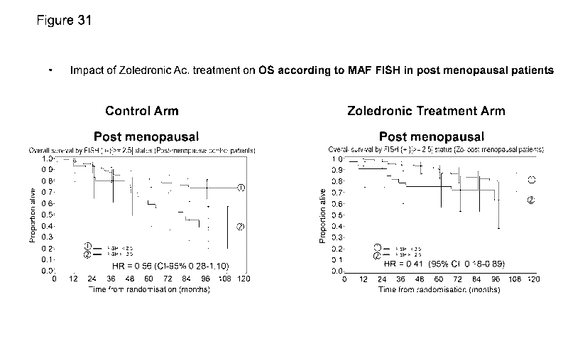
[0081] Figure 31. Predictive value of MAF FISH for the effect of
zoledronic acid
treatment on the OS outcome in post menopausal patients.
[0082] Figure 32. Predictive value of MAF FISH for the effect of
zoledronic acid
treatment on the OS outcome in non-post menopausal patients.
CA 03025264 2018-11-22
WO 2017/203468 PCT/IB2017/053094
14
DETAILED DESCRIPTION OF THE INVENTION
Definitions of general terms and expressions
[0083] "And/or" where used herein is to be taken as specific disclosure
of each of the two
specified features or components with or without the other. For example 'A
and/or B' is
to be taken as specific disclosure of each of (i) A, (ii) B and (iii) A and B,
just as if each is
set out individually herein.
[0084] The c-MAF gene (v-maf musculoaponeurotic fibrosarcoma oncogene
homologue
(avian) also known as MAF or MGC71685) is a transcription factor containing a
leucine
zipper which acts like a homodimer or a heterodimer. Depending on the DNA
binding
site, the encoded protein can be a transcriptional activator or repressor. The
DNA
sequence encoding c-MAF is described in the NCBI database under accession
number
NGO16440 (SEQ ID NO: 1)(coding)). The genomic sequence of c-MAF is set forth
in
SEQ ID NO:13. The methods of the present invention may utilize either the
coding
sequence or the genomic DNA sequence. Two messenger RNA are transcribed from
said
DNA sequence, each of the which will give rise to one of the two c-MAF protein
isoforms, the a isoform and the 0 isoform. The complementary DNA sequences for
each
of said isoforms are described, respectively, in the NCBI database under
accession
numbers NM 005360.4 (SEQ ID NO: 2) and NM 001031804.2 (SEQ ID NO: 3). Use of
the c-MAF gene to predict the prognosis of ER+ breast cancer can be found in
U.S. Appl.
No 13/878,114, which is incorporated herein by reference in its entirety. Use
of the c-
MAF gene to predict the prognosis of triple-negative and ER+ breast cancer is
described
in U.S. Appl. No. 14/391,085, which is incorporated herein by reference in its
entirety.
Use of the c-MAF gene to predict the prognosis of thyroid cancer is described
in U.S.
Prov. Appl. No. 61/801,769, which is incorporated herein by reference in its
entirety. Use
of the c-MAF gene to predict the prognosis of renal cell carcinoma is
described in U.S.
Prov. Appl. No. 14/776,390, which is incorporated herein by reference in its
entirety. The
use of a gene of interest, including c-MAF and the c-MAF gene locus, and
probes to the
gene locus, to determine the prognosis of an individual having breast cancer
is described
in U.S. Appl. No. 14/776,412, which is incorporated herein by reference in its
entirety.
Use of the c-MAF gene to predict the prognosis of lung cancer is found in U.S.
Appl. No.
14/405,724, which is incorporated herein by reference in its entirety. Use of
the c-MAF
gene to predict the prognosis of prostate cancer is found in U.S. Appl. Nos.
14/050,262
CA 03025264 2018-11-22
WO 2017/203468 PCT/IB2017/053094
and 14/435,128, which are incorporated herein by reference in their entirety.
Use of the c-
MAF gene to predict the prognosis of HER2+ cancer is found in U.S. App!. No.
15/027,946, which is incorporated herein by reference in its entirety. Use of
downstream
genes of c-MAF to predict the prognosis of cancer is found in U.S. App!. Nos.
15/014,916
and 14/776,453, which are incorporated herein by reference in its entirety.
[0085] As used herein, the term "basal-like" "basal-like subtype," "breast
cancer of the
basal-like subtype" and the like, as used herein, refers to a particular
subtype of breast
cancer characterized by the two negative receptors ER and HER2 and at least
one positive
receptor of the group consisting of CK5/6, CK14, CK17 and EGFR. Thus, all
sentences in
the present application which cite and refer to triple negative breast cancer
(ER, HER-2,
PgR) can also be cited and refer also to basal-like breast cancer wherein ER
and HER2
are negative and wherein at least one of CK5/6, CK14, CK17 and EGFR is
positive.
Alternatively, "basal-like" also refers to breast cancer characterized by a
gene expression
profile based on the up-regulation and/or down-regulation of the following ten
genes: (1)
Forkhead box CI (FOXC 1); (2) Melanoma inhibitory activity (MIA); (3) NDC80
homolog, kinetochore complex component (KNTC2); (4) Centrosomal protein 55kDa
(CEP55); (5) Anillin, actin binding protein (ANLN); (6) Maternal embryonic
leucine
zipper kinase (MELK); (7) G protein-coupled receptor 160 (GPR160); (8)
Transmembrane protein 45B (TMEM45B); (9) Estrogen receptor 1 (ESR1); (10)
Forkhead box Al (FOXA1). Because the gene expression profile used to classify
breast
cancer tumors as basal-like subtype does not include the estrogen receptor,
the
progesterone receptor or Her2, both triple negative and non-triple negative
breast cancers
may be classified as basal-like subtype.
[0086] As used herein, "Triple-negative breast cancer" refers to a breast
cancer which is
characterized by a lack of detectable expression of both ER and PR (preferably
when the
measures of expression of ER and PR are carried out by the method disclosed by
M.
Elizabeth H etal., Journal of Clinical Oncology, 28(16): 2784-2795, 2010) and
the tumor
cells are not amplified for epidermal growth factor receptor type 2 (HER2 or
ErbB2), a
receptor normally located on the cell surface. Tumor cells are considered
negative for
expression of ER and PR if less than 5 percent of the tumor cell nuclei are
stained for ER
and PR expression using standard immunohistochemical techniques. As used
herein,
tumor cells are considered negative for HER2 overexpression if they yield a
test result
CA 03025264 2018-11-22
WO 2017/203468 PCT/IB2017/053094
16
score of 0 or 1+, or 2+ when tested with a HercepTestTm Kit (Code K5204, Dako
North
America, Inc., Carpinteria, CA), a semi-quantitative immunohistochemical assay
using a
polyclonal anti-HER2 primary antibody or if they are HER2 FISH negative.
[0087] As used herein, "ER+ breast cancer" is understood as breast cancer
the tumor cells
of which express the estrogen receptor (ER). This makes said tumors sensitive
to
estrogen, meaning that the estrogen makes the cancerous breast tumor grow. In
contrast,
"ER- breast cancer" is understood as breast cancer the tumor cells of which do
not
express the estrogen receptor (ER). Among the ER+ breast cancer are included
luminal A
and B subtypes.
[0088] As used herein, "HER2+" refers to a breast cancer which is
characterized by
tumor cells with detectable expression of epidermal growth factor receptor
type 2 (HER2
or ErbB2) and/or amplification for the HER2 gene, a receptor normally located
on the
cell surface. As used herein, tumor cells are considered negative for HER2
overexpression if they yield a test result score of 0 or 1+, or 2+ when tested
with a
HercepTestTm Kit (Code K5204, Dako North America, Inc., Carpinteria, CA), a
semi-
quantitative immunohistochemical assay using a polyclonal anti-HER2 primary
antibody
or if they are HER2 FISH negative.
[0089] In the context of the present invention, a "post-menopausal"
subject is understood
to be a woman who has undergone menopause and has experienced sixty
consecutive
months without menstruation. See Coleman et al Lancet Oncol 2014; 15: 997-
1006. In
certain embodiments, a woman may confirm her postmenopausal status through the
measuring of follicle stimulating hormone (FSH).
[0090] In the context of the present invention, a "non post-menopausal"
subject is any
subject who has not gone through menopause and experienced sixty consecutive
months
without menstruation. "Non post-menopausal" subjects include premenopausal,
perimenopausal, and unknown menopausal status women.
[0091] In the context of the present invention, "metastasis" is understood
as the
propagation of a cancer from the organ where it started to a different organ.
It generally
occurs through the blood or lymphatic system. When the cancer cells spread and
form a
new tumor, the latter is called a secondary or metastatic tumor. The cancer
cells forming
the secondary tumor are like those of the original tumor. If a breast cancer,
for example,
spreads (metastasizes) to the bone, the secondary tumor is formed of malignant
breast
CA 03025264 2018-11-22
WO 2017/203468 PCT/IB2017/053094
17
cancer cells. The disease in the bone is metastatic breast cancer and not bone
cancer. In a
particular embodiment of the method of the invention, the metastasis is breast
cancer
which has spread (metastasized) to the bone.
[0092] In the context of the present invention, "recurrence" refers to the
return of breast
cancer following a period of time in which no cancer was detected. Breast
cancer may
reoccur locally in the breast or tissue surrounding the breast. Breast cancer
may also
reoccur in nearby lymph nodes or lymph nodes not in the surrounding area. When
the
breast cancer reoccurs by spreading to other tissues or travels through the
blood stream to
recur in bones or other organs, it is also referred to as metastasis. As used
herein,
recurrence also encompasses the risk of recurrence.
[0093] In the context of the present invention, "relapse" refers to the
situation when
symptoms have decreased, but the subject is not cancer free, and then cancer
returns.
Breast cancer may relapse locally in the breast or tissue surrounding the
breast. Breast
cancer may also relapse in nearby lymph nodes or lymph nodes not in the
surrounding
area. When the breast cancer relapses by spreading to other tissues or travels
through the
blood stream to recur in bones or other organs, it is also referred to as
metastasis. As used
herein, relapse also encompasses the risk of relapse.
[0094] As used herein, the term "disease free survival" refers to the
length of time after
primary treatment for a cancer ends that the patient survives without any
signs or
symptoms of that cancer. In some embodiments, disease free survival is
referred to as
DFS, relapse-free survival, or RFS.
[0095] As used herein, the term "overall survival" or "OS" refers to the
length of time
from either the date of diagnosis or the start of treatment for a cancer that
patients
diagnosed with the disease are still alive.
[0096] As used herein, the term "subject" or "patient" refers to all
animals classified as
mammals and includes but is not limited to domestic and farm animals, primates
and
humans, for example, human beings, non-human primates, cows, horses, pigs,
sheep,
goats, dogs, cats, or rodents. Preferably, the subject is a human man or woman
of any age
or race.
[0097] The terms "poor" or "good", as used herein to refer to a clinical
outcome, mean
that the subject will show a favorable or unfavorable outcome. As will be
understood by
those skilled in the art, such an assessment of the probability, although
preferred to be,
CA 03025264 2018-11-22
WO 2017/203468 PCT/IB2017/053094
18
may not be correct for 100% of the subjects to be diagnosed. The term,
however, requires
that a statistically significant portion of subjects can be identified as
having a
predisposition for a given outcome. Whether a portion is statistically
significant can be
determined readily by the person skilled in the art using various well known
statistic
evaluation tools, e.g., determination of confidence intervals, p-value
determination,
Student's t-test, Mann-Whitney test, etc. Details are found in Dowdy and
Wearden,
Statistics for Research, John Wiley & Sons, New York 1983. Preferred
confidence
intervals are at least about 50%, at least about 60%, at least about 70%, at
least about
80%, at least about 90% at least about 95%. . The p-values are, preferably,
0.05, 0.01,
0.005, or 0.0001 or less. More preferably, at least about 60 percent, at least
about 70
percent, at least about 80 percent or at least about 90 percent of the
subjects of a
population can be properly identified by the method of the present invention.
[0098] In the present invention "tumor sample" is understood as a sample
(e.g., tumor
tissue, circulating tumor cell, circulating tumor DNA) originating from the
primary breast
cancer tumor. Said sample can be obtained by conventional methods, for example
biopsy,
using methods well known by the persons skilled in related medical techniques.
The
methods for obtaining a biopsy sample include splitting a tumor into large
pieces, or
microdissection, or other cell separating methods known in the art. The tumor
cells can
additionally be obtained by means of cytology through aspiration with a small
gauge
needle. To simplify sample preservation and handling, samples can be fixed in
formalin
and soaked in paraffin or first frozen and then soaked in a tissue freezing
medium such as
OCT compound by means of immersion in a highly cryogenic medium which allows
rapid freezing.
[0099] In the context of the present invention, "functionally equivalent
variant of the c-
MAF protein" is understood as (i) variants of the c-MAF protein (SEQ ID NO: 4
or SEQ
ID NO: 5) in which one or more of the amino acid residues are substituted by a
conserved
or non-conserved amino acid residue (preferably a conserved amino acid
residue),
wherein such substituted amino acid residue may or may not be one encoded by
the
genetic code, or (ii) variants comprising an insertion or a deletion of one or
more amino
acids and having the same function as the c-MAF protein, i.e., to act as a DNA
binding
transcription factor. Variants of the c-MAF protein can be identified using
methods based
on the capacity of c-MAF for promoting in vitro cell proliferation as shown in
CA 03025264 2018-11-22
WO 2017/203468 PCT/IB2017/053094
19
international patent application W02005/046731(hereby incorporated by
reference in its
entirety), based on the capacity of the so-called inhibitor for blocking the
transcription
capacity of a reporter gene under the control of cyclin D2 promoter or of a
promoter
containing the c-MAF responsive region (MARE or c-MAF responsive element) in
cells
expressing c-MAF as described in W02008098351 (hereby incorporated by
reference in
its entirety), or based on the capacity of the so-called inhibitor for
blocking reporter gene
expression under the control of the IL-4 promoter in response to the
stimulation with
PMA/ionomycin in cells expressing NFATc2 and c-MAF as described in
US2009048117A (hereby incorporated by reference in its entirety).
[0100] The variants according to the invention preferably have sequence
similarity with
the amino acid sequence of any of the c-MAF protein isoforms (SEQ ID NO: 4 or
SEQ
ID NO: 5) of at about least 50%, at least about 60%, at about least 70%, at
least about
80%, at least about 90%, at least about 91%, at least about 92%, at least
about 93%, at
least about 94%, at least about 95%, at least about 96%, at least about 97%,
at about least
98% or at about least 99%. The degree of similarity between the variants and
the specific
c-MAF protein sequences defined previously is determined using algorithms and
computer processes which are widely known by the persons skilled in the art.
The
similarity between two amino acid sequences is preferably determined using the
BLASTP
algorithm [BLAST Manual, Altschul, S., et al., NCBI NLM NIH Bethesda, Md.
20894,
Altschul, S., et al., J. Mol. Biol. 215: 403-410 (1990)].
[0101] As used herein, "agent for avoiding or preventing bone remodelling"
refers to any
molecule capable of preventing, inhibiting, treating, reducing, or stopping
bone
degradation either by stimulating the osteoblast proliferation or inhibiting
the osteoclast
proliferation or fixing the bone structure. Agents for avoiding or prevent
bone
remodeling include agents for avoiding or preventing bone degradation and
include
agents for avoiding or preventing bone synthesis.
[0102] As used herein, a "c-MAF inhibitory agent" refers to any molecule
capable of
completely or partially inhibiting the c-MAF gene expression, both by
preventing the
expression product of said gene from being produced (interrupting the c-MAF
gene
transcription and/or blocking the translation of the mRNA coming from the c-
MAF gene
expression) and by directly inhibiting the c-MAF protein activity. C-MAF gene
expression inhibitors can be identified using methods based on the capacity of
the so-
CA 03025264 2018-11-22
WO 2017/203468 PCT/IB2017/053094
called inhibitor to block the capacity of c-MAF to promote the in vitro cell
proliferation,
such as shown in the international patent application W02005/046731 (the
entire contents
of which are hereby incorporated by reference), based on the capacity of the
so-called
inhibitor to block the transcription capacity of a reporter gene under the
control of the
cyclin D2 promoter or of a promoter containing the c-MAF response region (MARE
or c-
MAF responsive element) in cells which express c-MAF such as described in
W02008098351 (the entire contents of which are hereby incorporated by
reference) or
based on the capacity of the so-called inhibitor to block the expression of a
reporter gene
under the control of the IL-4 promoter in response to the stimulation with
PMA/ionomycin in cells which express NFATc2 and c-MAF such as described in
US2009048117A (the entire contents of which is hereby incorporated by
reference).
[0103] As used herein, Mammalian target of rapamycin (mTOR) or "mTor"
refers to
those proteins that correspond to EC 2.7.11.1. mTor enzymes are
serine/threonine protein
kinases and regulate cell proliferation, cell motility, cell growth, cell
survival, and
transcription.
[0104] As used herein, an "mTor inhibitor" refers to any molecule capable
of completely
or partially inhibiting the mTor gene expression, both by preventing the
expression
product of said gene from being produced (interrupting the mTor gene
transcription
and/or blocking the translation of the mRNA coming from the mTor gene
expression) and
by directly inhibiting the mTor protein activity. Including inhibitors that
have a dual or
more targets and among them mTor protein activity.
[0105] As used herein, "Src" refers to those proteins that correspond to
EC 2.7.10.2. Src
is a non-receptor tyrosine kinase and a proto-oncogene. Src may play a role in
cell
growth and embryonic development.
[0106] As used herein, a "Src inhibitor" refers to any molecule capable of
completely or
partially inhibiting the Src gene expression, both by preventing the
expression product of
said gene from being produced (interrupting the Src gene transcription and/or
blocking
the translation of the mRNA coming from the Src gene expression) and by
directly
inhibiting the Src protein activity.
[0107] As used herein, "Prostaglandin-endoperoxide synthase 2",
"cyclooxygenase-2" or
"COX-2" refers to those proteins that correspond to EC 1.14.99.1. COX-2 is
responsible
for converting arachidonic acid to prostaglandin endoperoxide H2.
CA 03025264 2018-11-22
WO 2017/203468 PCT/IB2017/053094
21
[0108] As used herein, a "COX-2 inhibitor" refers to any molecule capable
of completely
or partially inhibiting the COX-2 gene expression, both by preventing the
expression
product of said gene from being produced (interrupting the COX-2 gene
transcription
and/or blocking the translation of the mRNA coming from the COX-2 gene
expression)
and by directly inhibiting the COX-2 protein activity.
[0109] As used herein "outcome" or "clinical outcome" refers to the
resulting course of
disease and/or disease progression and can be characterized for example by
recurrence,
period of time until recurrence, relapse, metastasis, period of time until
metastasis,
number of metastases, number of sites of metastasis and/or death due to
disease. For
example a good clinical outcome includes cure, prevention of recurrence,
prevention of
metastasis and/or survival within a fixed period of time (without recurrence),
and a poor
clinical outcome includes disease progression, metastasis and/or death within
a fixed
period of time.
[0110] As used herein, "invasive disease free survival" or "IDFS" refers
to, in cancer, the
length of time after primary treatment for a cancer ends that the patient
survives without
any signs or symptoms of that cancer invading the same breast parenchyma as
the original
primary tumor or other tissues. In some embodiments, IDFS includes:
ipsilateral invasive
breast tumor recurrence, local or regional invasive breast cancer recurrence,
metastatic or
distant recurrence, death attributable to any cause, including breast cancer,
contralateral
invasive breast cancer, and second primary invasive cancer (non-breast but
excluding
basal-cell or squamous skin cancers). See Coleman et al Lancet Oncol 2014; 15:
997-
1006.
[0111] In the present invention, "diagnosis of metastasis in a subject
with breast cancer"
is understood as identifying a disease (metastasis) by means of studying its
signs, i.e., in
the context of the present invention by means of increased c-MAF gene
expression levels
(i.e., overexpression) in the breast cancer tumor tissue with respect to a
control sample.
[0112] In the present invention "prognosis of the tendency to develop
metastasis in a
subject with breast cancer" is understood as knowing based on the signs if the
breast
cancer that said subject has will metastasize in the future. In the context of
the present
invention, the sign is c-MAF gene overexpression in tumor tissue.
[0113] In the context of the present invention, it is understood that "a
subject has a
positive diagnosis for metastasis" when the breast cancer suffered by said
subject has
CA 03025264 2018-11-22
WO 2017/203468 PCT/IB2017/053094
22
metastasized to other organs of the body, in a particular embodiment, to the
bone. The
term is similarly used for recurrence and relapse.
[0114] The person skilled in the art will understand that the prediction
of the tendency for
a primary tumor to metastasize, relapse or reoccur is not intended to be
correct for all the
subjects to be identified (i.e., for 100% of the subjects). Nevertheless, the
term requires
enabling the identification of a statistically significant part of the
subjects (for example, a
cohort in a cohort study). Whether a part is statistically significant can be
determined in a
simple manner by the person skilled in the art using various well known
statistical
evaluation tools, for example, the determination of confidence intervals,
determination of
p values, Student's T test, Mann-Whitney test, etc. Details are provided in
Dowdy and
Wearden, Statistics for Research, John Wiley and Sons, New York 1983. The
preferred
confidence intervals are at least about 90%, at least about 95%, at least
about 97%, at
least 98% or at least 99%. The p values are preferably 0.1, 0.05, 0.01, 0.005
or 0.0001.
More preferably, at least about 60%, at least about 70%, at least about 80% or
at least
about 90% of the subjects of a population can be suitably identified by the
method of the
present invention.
[0115] As used herein, "poor prognosis" indicates that the subject is
expected e.g.
predicted to not survive and/or to have, or is at high risk of having,
recurrence, relapse, or
distant metastases within a set time period. The term "high" is a relative
term and, in the
context of this application, refers to the risk of the "high" expression group
with respect
to a clinical outcome (recurrence, distant metastases, etc.). A "high" risk
can be
considered as a risk higher than the average risk for a heterogeneous cancer
patient
population. In the study of Paik et al. (2004), an overall "high" risk of
recurrence was
considered to be higher than 15 percent. The risk will also vary in function
of the time
period. The time period can be, for example, five years, ten years, fifteen
years or even
twenty years of initial diagnosis of cancer or after the prognosis was made.
[0116] "Reference value", as used herein, refers to a laboratory value
used as a reference
for values/data obtained by laboratory examinations of patients or samples
collected from
patients. The reference value or reference level can be an absolute value; a
relative value;
a value that has an upper and/or lower limit; a range of values; an average
value; a
median value, a mean value, or a value as compared to a particular control or
baseline
value. A reference value can be based on an individual sample value, such as
for example,
CA 03025264 2018-11-22
WO 2017/203468 PCT/IB2017/053094
23
a value obtained from a sample from the subject being tested, but at an
earlier point in
time. The reference value can be based on a large number of samples, such as
from a
population of subjects of the chronological age matched group, or based on a
pool of
samples including or excluding the sample to be tested.
[0117] The term "treatment", as used herein, refers to any type of
therapy, which aims at
terminating, preventing, ameliorating or reducing the susceptibility to a
clinical condition
as described herein. In an embodiment, the term treatment relates to
prophylactic
treatment (i.e. a therapy to reduce the susceptibility to a clinical
condition), of a disorder
or a condition as defined herein. Thus, "treatment," "treating," and their
equivalent terms
refer to obtaining a desired pharmacologic or physiologic effect, covering any
treatment
of a pathological condition or disorder in a mammal, including a human. The
effect may
be prophylactic in terms of completely or partially preventing a disorder or
symptom
thereof and/or may be therapeutic in terms of a partial or complete cure for a
disorder
and/or adverse effect attributable to the disorder. That is, "treatment"
includes (1)
preventing the disorder from occurring or recurring in a subject, (2)
inhibiting the
disorder, such as arresting its development, (3) stopping or terminating the
disorder or at
least symptoms associated therewith, so that the host no longer suffers from
the disorder
or its symptoms, such as causing regression of the disorder or its symptoms,
for example,
by restoring or repairing a lost, missing or defective function, or
stimulating an inefficient
process, or (4) relieving, alleviating, or ameliorating the disorder, or
symptoms associated
therewith, where ameliorating is used in a broad sense to refer to at least a
reduction in
the magnitude of a parameter, such as inflammation, pain, or immune
deficiency.
[0118] As used herein, "sample" or "biological sample" means biological
material
isolated from a subject. The biological sample may contain any biological
material
suitable for determining the expression level of the c-MAF gene. The sample
can be
isolated from any suitable biological tissue or fluid such as, for example,
tumor tissue,
blood, blood plasma, serum, urine or cerebral spinal fluid (CSF).
[0119] As used herein, the term "expression level" of a gene as used
herein refers to the
measurable quantity of gene product produced by the gene in a sample of the
subject,
wherein the gene product can be a transcriptional product or a translational
product.
Accordingly, the expression level can pertain to a nucleic acid gene product
such as
mRNA or cDNA or a polypeptide gene product. The expression level is derived
from a
CA 03025264 2018-11-22
WO 2017/203468 PCT/IB2017/053094
24
subject's sample and/or a reference sample or samples, and can for example be
detected
de novo or correspond to a previous determination. The expression level can be
determined or measured, for example, using microarray methods, PCR methods
(such as
qPCR), and/or antibody based methods, as is known to a person of skill in the
art.
[0120] "Increased expression level" is understood as the expression level
when it refers to
the levels of the c-MAF gene greater than those in a reference sample or
control sample.
These increased levels can be caused without excluding other mechanisms by a
gene or
16q23 or 16q22-24 chromosomal locus amplification, copy gain or translocation.
Particularly, a sample can be considered to have high c-MAF expression level
when the
expression level in the sample isolated from the patient is at least about 1.1
times, 1.2
times, 1.3 times, 1.4 times, 1.5 times, 2 times, 2.1 times, 2.2 times, 2.3
times, 2.4 times,
2.5 times, 3 times, 4 times, 5 times, 10 times, 20 times, 30 times, 40 times,
50 times, 60
times, 70 times, 80 times, 90 times, 100 times or even more with respect to
the reference
or control. In embodiments, an "increased expression level" is a "high"
expression level.
An expression level that is "not increased" or "non increased" is any value
that is not
included in the definition of "increased" expression level, including a value
equal or the
reference or control level or a decreased expression level in comparison to a
reference or
control level.
[0121] "Decreased expression level" is understood as the expression level
when it refers
to the levels of the c-MAF gene less than those in a reference sample or
control sample.
This decreased level can be caused without excluding other mechanisms by a
gene or
16q23 or 16q22-24 chromosomal locus deletion. Particularly, a sample can be
considered
to have decreased c-MAF expression levels when the expression level in the
sample
isolated from the patient is at least about 1.1 times, 1.2 times, 1.3 times,
1.4 times, 1.5
times, 2 times, 2.1 times, 2.2 times, 2.3 times, 2.4 times, 2.5 times, 3
times, 4 times, 5
times, 10 times, 20 times, 30 times, 40 times, 50 times, 60 times, 70 times,
80 times, 90
times, 100 times or even less with respect to the reference or control. In
embodiments, a
"decreased expression level" is a "low" expression level.
[0122] As used herein, the term "gene copy number" refers to the copy
number of a
nucleic acid molecule in a cell. The gene copy number includes the gene copy
number in
the genomic (chromosomal) DNA of a cell. In a normal cell (non-tumoral cell),
the gene
copy number is normally two copies (one copy in each member of the chromosome
pair).
CA 03025264 2018-11-22
WO 2017/203468 PCT/IB2017/053094
The gene copy number sometimes includes half of the gene copy number taken
from
samples of a cell population.
[0123] In the present invention, "increased gene copy number" is
understood as when the
c-MAF gene copy number is more than the copy number that a reference sample or
control sample has. These increased gene copy number can be caused without
excluding
other mechanisms by a gene or 16q23 or 16q22-24 chromosomal locus
amplification,
copy gain or translocation. In particular, it can be considered that a sample
has an
increased c-MAF copy number when the copy number is more than 2 copies, for
example, 2.1, 2.2, 2.3, 2.4, 2.5, 2.6, 2.7, 2.8, 2.9, 3, 4, 5, 6, 7, 8, 9 or
10 copies, and even
more than 10 copies of the c-MAF gene. In embodiments, "increased gene copy
number"
is determined based on an average of copies per cells counted. In embodiments,
it can be
considered that a sample has an increased c-MAF copy number when the average
copy
number per cell counted is more than 2 copies, for example, 2.1, 2.2, 2.3,
2.4, 2.5, 2.6,
2.7, 2.8, 2.9, 3, 4, 5, 6, 7, 8, 9 or 10 copies, and even more than 10 copies
of the c-MAF
gene.
[0124] In the present invention, "decreased gene copy number" is
understood as when the
c-MAF gene copy number is less than the copy number that a reference sample or
control
sample has. These decreased gene copy number can be caused without excluding
other
mechanisms by a gene or 16q23 or 16q22-24 chromosomal locus deletions. In
particular,
it can be considered that a sample has a decreased c-MAF copy number when the
copy
number is less than 2 copies of the c-MAF gene.
[0125] In the present invention, a "not increased gene copy number" is
understood as
when the c-MAF gene copy number or the average c-MAF gene copy number is less
than
the copy number that a reference sample or positive for the increase sample
has. The not
increased gene copy number can be caused without excluding other mechanisms by
no
increase in gene or 16q23 or 16q22-24 chromosomal locus amplification, copy
gain or
translocation. In particular, it can be considered that a sample has not an
increased c-
MAF copy number or c-MAF average copy number when the copy number is less than
2.1, 2.2, 2.3, 2.4, 2.5, 2.6, 2.7, 2.8, 2.9 copies of the c-MAF gene.
[0126] The term "amplification of a gene" as understood herein refers to a
process
through which various copies of a gene or of a gene fragment are formed in an
individual
cell or a cell line. The copies of the gene are not necessarily located in the
same
CA 03025264 2018-11-22
WO 2017/203468 PCT/IB2017/053094
26
chromosome. The duplicated region is often called an "amplicon". Normally, the
amount
of mRNA produced, i.e., the gene expression level also increases in proportion
to the
copy number of a particular gene.
[0127] The term "gain" refers any chromosomal copy number increase from
the norm,
i.e., in a diploid organism, 3 copies of a gene in a cell would be a gain. In
some
embodiments, "gain" includes the term "copy gain", and is used synonymously
with
"copy number".
[0128] "Probe", as used herein, refers to an oligonucleotide sequence that
is
complementary to a specific nucleic acid sequence of interest. In some
embodiments, the
probes may be specific to regions of chromosomes that are known to undergo
translocations. In some embodiments, the probes have a specific label or tag.
In some
embodiments, the tag is a fluorophore. In some embodiments, the probe is a DNA
in situ
hybridization probe whose labeling is based on the stable coordinative binding
of
platinum to nucleic acids and proteins. In some embodiments, the probe is
described in
U.S. Patent Nos. 9,127,302 and 9,134,237, which are incorporated by reference
in their
entirety, or as described in Swennenhuis et al. "Construction of repeat-free
fluorescence
in situ hybridization probes" Nucleic Acids Research 40(3):e20 (2012).
[0129] "Tag" or "label", as used herein, refers to any physical molecule
that is directly or
indirectly associated with a probe, allowing the probe or the location of the
probed to be
visualized, marked, or otherwise captured.
[0130] "Translocation", as used herein, refers to the exchange of
chromosomal material
in unequal or equal amounts between chromosomes. In some cases, the
translocation is
on the same chromosome. In some cases, the translocation is between different
chromosomes. Translocations occur at a high frequency in many types of cancer,
including breast cancer and leukemia. Translocations can be either primary
reciprocal
translocations or the more complex secondary translocations. There are several
primary
translocations that involve the immunoglobin heavy chain (IgH) locus that are
believed to
constitute the initiating event in many cancers. (Eychene, A., Rocques, N.,
and
Puoponnot, C., A new MAFia in cancer. 2008. Nature Reviews: Cancer. 8: 683-
693.)
[0131] "Polyploid" or "polyploidy", as used herein, indicates that the
cell contains more
than two copies of a gene of interest. In some instances, the gene of interest
is MAF. In
some embodiments, polyploidy is associated with an accumulation of expression
of the
CA 03025264 2018-11-22
WO 2017/203468 PCT/IB2017/053094
27
gene of interest. In some embodiments, polyploidy is associated with genomic
instability.
In some embodiments, the genomic instability may lead to chromosome
translocations.
[0132] "Whole genome sequencing", as used herein, is a process by which
the entire
genome of an organism is sequenced at a single time. See, e.g., Ng., P.C. and
Kirkness,
E.F., Whole Genome Sequencing. 2010. Methods in Molecular Biology. 628: 215-
226.
[0133] "Exome sequencing", as used herein, is a process by which the
entire coding
region of the DNA of an organism is sequenced. In exome sequencing, the mRNA
is
sequenced. The untranslated regions of the genome are not included in exome
sequencing. See, e.g., Choi, M. et al., Genetic diagnosis by whole exome
capture and
massively parallel DNA sequencing. 2009. PNAS. 106(45): 19096-19101.
[0134] As used herein, "binding member" describes one member of a pair of
molecules
that bind one another. The members of a binding pair may be naturally derived
or wholly
or partially synthetically produced. One member of the pair of molecules has
an area on
its surface, or a cavity, which binds to and is therefore complementary to a
particular
spatial and polar organization of the other member of the pair of molecules.
Examples of
types of binding pairs are antigen-antibody, receptor-ligand and enzyme-
substrate. In
some embodiments, the binding member is an antibody. In some embodiments, the
binding member is an antibody that binds a c-MAF antigen.
[0135] As used herein, "CDR region" or "CDR" is intended to indicate the
hypervariable
regions of the heavy and light chains of the immunoglobulin as defined by
Kabat et al.,
(1991) Sequences of Proteins of Immunological Interest, 5th Edition. US
Department of
Health and Human Services, Public Service, NIH, Washington. An antibody
typically
contains 3 heavy chain CDRs, termed HCDR1, HCDR2, and HCDR3, and 3 light chain
CDRs, termed LCDR1, LCDR2 and LCDR3. The term CDR or CDRs is used here in
order to indicate one of these regions or several, or even the whole, of these
regions
which contain the majority of the amino acid residues responsible for the
binding by
affinity of the antibody for the antigen or the epitope which it recognizes.
Among the six
CDR sequences, the third CDR of the heavy chain (HCDR3) has a greatest size
variability i.e. greater diversity, essentially due to the mechanism known in
the art as
V(D)J rearrangement of the V, D and J gene segments of the germline
immunoglobulin
heavy chain gene locus. The HCDR3 may be as short as two amino acids or as
long as 26
amino acids, or may have any length in between these two extremes. CDR length
may
CA 03025264 2018-11-22
WO 2017/203468 PCT/IB2017/053094
28
also vary according to the length that can be accommodated by the particular
underlying
framework. Functionally, HCDR3 can play an important role in the determination
of the
specificity of the antibody (Segal et al., (1974) Proc Natl Acad Sci USA.
71(11): 4298-
302; Amit et al., (1986) Science 233(4765): 747-53; Chothia et al., (1987) J.
Mol. Biol.
196(4): 901-17; Chothia et al., (1989) Nature 342(6252): 877-83; Caton et al.,
(1990) J.
Immunol. 144(5): 1965-8; Sharon (1990a) PNAS USA. 87(12): 4814-7, Sharon
(1990b)
J. Immunol. 144: 4863-4869, Kabat et al., (1991) Sequences of Proteins of
Immunological Interest, 5th Edition. US Department of Health and Human
Services,
Public Service, NIH, Washington).
[0136] As used herein, "antibody", "antibody molecule", or "antibodies"
describes an
immunoglobulin whether naturally, or partly, or wholly synthetically produced.
The term
also covers any polypeptide or protein comprising an antibody antigen-binding
site. It
must be understood here that the invention does not relate to the antibodies
in natural
form, that is to say they are not in their natural environment but that they
have been able
to be isolated or obtained by purification from natural sources, or else
obtained by genetic
recombination, or by chemical synthesis, and that they can then contain
unnatural amino
acids. Antibody fragments that comprise an antibody antigen-binding site
include, but are
not limited to, molecules such as Fab, Fab', F(ab')2, Fab' ¨SH, scFv, Fv, dAb
and Fd.
Various other antibody molecules including one or more antibody antigen-
binding sites
have been engineered, including for example Fab2, Fab3, diabodies, triabodies,
tetrabodies, camelbodies, nanobodies and minibodies. Antibody molecules and
methods
for their construction and use are described in Hollinger & Hudson (2005)
Nature Biot.
23(9): 1126-1136.
[0137] As used herein, "antibody molecule" should be construed as covering
any binding
member or substance having an antibody antigen-binding site with the required
specificity and/or binding to antigen. Thus, this term covers functional
antibody
fragments and derivatives, including any polypeptide comprising an antibody
antigen-
binding site, whether natural or wholly or partially synthetic. Chimeric
molecules
comprising an antibody antigen-binding site, or equivalent, fused to another
polypeptide
(e.g. derived from another species or belonging to another antibody class or
subclass) are
therefore included. Cloning and expression of chimeric antibodies are
described for
CA 03025264 2018-11-22
WO 2017/203468 PCT/IB2017/053094
29
example in EP0120694A (Boss et al) and EP0125023A (Cabilly et al), which are
incorporated herein in their entirety.
[0138] As used herein, "functional fragment or variant" of, for example, a
binding
member of the present invention means a fragment or variant of a binding
member that
retains at least some function of a full binding member (e.g., the ability to
specifically
bind to antigen such as Maf).
[0139] "Tumor tissue sample" is understood as the tissue sample
originating from the
breast cancer tumor, including but not limited to circulating tumor cells and
circulating
tumor DNA. Said sample can be obtained by conventional methods, for example
biopsy,
using methods well known by the persons skilled in related medical techniques.
[0140] "Osteolytic bone metastasis" refers to a type of metastasis in
which bone
resorption (progressive loss of the bone density) is produced in the proximity
of the
metastasis resulting from the stimulation of the osteoclast activity by the
tumor cells and
is characterized by severe pain, pathological fractures, hypercalcaemia,
spinal cord
compression and other syndromes resulting from nerve compression.
Method for designing customized therapy of the invention in patients with
breast tumors
[0141] The present invention is directed to identifying subjects suffering
from breast
cancer who will benefit from treatment with particular agents and/or
therapies. In some
embodiments, the invention is directed to identifying subjects suffering from
breast
cancer who will not benefit from treatment with particular agents and
therapies. In some
embodiments, the subjects have a high expression level, copy number,
amplification, gain
and/or translocation of c-MAF. In certain embodiments, the subjects have a low
expression level, copy number, amplification, gain and/or translocation of c-
MAF. In
particular embodiments, the cancer is triple-negative breast cancer. In other
embodiments, the cancer is ER+ breast cancer. In further embodiments, the
cancer is ER-
breast cancer. In still further embodiments, the cancer is HER2+ breast
cancer. In some
embodiments, the cancer is a basal-like breast cancer. In one embodiment, the
subjects
are post-menopausal. In an embodiment, the subjects are non-post menopausal.
As
described U.S. Appl. No. 14/391,085, U.S. Prov. Appl. No. 61/801,769, U.S.
Prov. Appl.
No. 14/776,390, U.S. Appl. No. 14/776,412, U.S. Appl. No. 14/405,724, U.S.
Appl. No.
14/050,262, U.S. Appl. No. 14/435,128, U.S. Appl. No. 15/027,946 U.S. Appl.
No.
15/014,916, and U.S. Appl. No. 14/776,453, each of which is incorporated
herein by
CA 03025264 2018-11-22
WO 2017/203468 PCT/IB2017/053094
reference in its entirety, the levels of c-MAF can be used to diagnosis
metastasis, relapse
or recurrence, or to predict the tendency of a tumor to undergo metastasis,
relapse or
recurrence. Therefore, as described in the present invention, given that the c-
MAF gene
overexpression in breast cancer cells is related to the presence of
metastasis, relapse or
recurrence, the c-MAF gene expression levels allow making decisions in terms
of the
most suitable therapy for the subject suffering said cancer. In an embodiment,
the
invention comprises quantifying only the c-MAF gene expression level as a
single
marker, i.e., the method does not involve determining the expression level of
any
additional marker.
[0142] Thus, in one embodiment the invention relates to an in vitro method
for designing
a customized therapy for a subject with breast cancer, which comprises a)
quantifying the
c-MAF gene expression level, copy number, amplification, or gain in a tumor
sample of
said subject and b) comparing the expression level, copy number, amplification
or gain
obtained with the expression level, copy number, amplification or gain of said
gene in a
control sample, wherein the therapy is determined based on the c-MAF gene
expression
level, copy number, amplification or gain in the subject. In some embodiments,
the
subject has a high c-MAF gene expression level. In other embodiments, the
subject has a
low c-MAF gene expression level. In certain embodiments, the subject is
administered an
agent that avoids and/or prevents bone remodelling, including agents that
avoid or
prevent bone degradation. In embodiments, the subject is administered an agent
that
treats the cancer. In further embodiments, the subject is administered a c-MAF
inhibitory
agent. In particular embodiments, the agent that avoids and/or prevents bone
remodelling
or the c-MAF inhibitory agent is any agent disclosed in U.S. Publ. Nos.
2014/0057796
and 2015/0293100 and U.S. Appl. No. 15/027,946, which are incorporated herein
by
reference in their entireties.
[0143] In one embodiment, the invention relates to an in vitro method for
designing a
customized therapy for a subject having breast cancer which comprises i)
quantifying the
c-MAF gene expression level, copy number, amplification, or gain in a sample
of said
subject and ii) comparing the expression level, copy number, amplification, or
gain
obtained in i) with a reference value, wherein if the expression level, copy
number,
amplification, or gain is not increased with respect to said reference value,
then said
subject is susceptible to receive a therapy aiming to prevent and/or treat
bone remodeling
CA 03025264 2018-11-22
WO 2017/203468 PCT/IB2017/053094
31
or improves disease free survival or overall survival. In some embodiments,
the subject is
non-postmenopausal. In other embodiments, the subject is postmenopausal. In an
embodiment, the subject is administered the agent aiming to prevent and/or
treat bone
remodelling. In embodiments, the subject is administered an agent that
improves disease
free survival or overall survival. In further embodiments, the subject is
administered a c-
MAF inhibitory agent.
[0144] In another embodiment, the invention relates to an in vitro method
for designing a
customized therapy for a non-postmenopausal subject having breast cancer which
comprises i) quantifying the c-MAF gene expression level, copy number,
amplification,
or gain in a sample of said subject and ii) comparing the expression level,
copy number,
amplification, or gain obtained in i) with a reference value, wherein if the
expression
level, copy number, amplification, or gain is increased with respect to said
reference
value, then said subject is not susceptible to receive a therapy aiming to
prevent and/or
treat bone remodeling and/or improves disease free survival or overall
survival. In some
embodiments, the subject is not administered the agent aiming to prevent
and/or treat
bone remodeling and/or improves disease free survival or overall survival.
[0145] In another embodiment, the invention relates to an in vitro method
for designing a
customized therapy for a postmenopausal subject having breast cancer which
comprises i)
quantifying the c-MAF gene expression level, copy number, amplification, or
gain in a
sample of said subject and ii) comparing the expression level, copy number,
amplification, or gain obtained in i) with a reference value, wherein if the
expression
level, copy number, amplification, or gain is increased with respect to said
reference
value, then said subject is susceptible to receive a therapy aiming to prevent
and/or treat
bone remodeling and/or improves disease free survival or overall survival. In
some
embodiments, the subject is administered the agent aiming to prevent and/or
treat bone
remodeling and/or the therapy to improve disease free survival or overall
survival.
[0146] In another embodiment, the invention relates to an in vitro method
for designing a
customized therapy for a postmenopausal subject having breast cancer which
comprises i)
quantifying the c-MAF gene expression level, copy number, amplification, or
gain in a
sample of said subject and ii) comparing the expression level, copy number,
amplification, or gain obtained in i) with a reference value, wherein if the
expression
level, copy number, amplification, or gain is not increased with respect to
said reference
CA 03025264 2018-11-22
WO 2017/203468 PCT/IB2017/053094
32
value, then said subject is not susceptible to receive a therapy aiming to
prevent and/or
treat bone remodeling and/or improves disease free survival or overall
survival. In some
embodiments, the subject is not administered the agent aiming to prevent
and/or treat
bone remodeling and/or improves disease free survival or overall survival.
[0147] In an embodiment, the invention relates to a method for the
treatment of bone
metastasis in a subject having breast cancer and having decreased c-MAF levels
in a
metastatic tumor sample with respect to a control sample comprising
administering an
agent capable of preventing or inhibiting bone remodeling and or improve
disease free
survival or overall survival, wherein the agent capable of avoiding or
preventing bone
remodeling or improving disease free survival or overall survival is selected
from the
group consisting of: a bisphosphonate, a RANKL inhibitor, PTH, PTHLH inhibitor
(including neutralizing antibodies and peptides), a PRG analog, strontium
ranelate, a
DKK-1 inhibitor, a dual MET and VEGFR2 inhibitor, an estrogen receptor
modulator, an
EGFR inhibitor, calcitonin, Radium-223, a CCR5 antagonist, a Src kinase
inhibitor, a
COX-2 inhibitor, an mTor inhibitor, and a cathepsin K inhibitor. In some
embodiments,
the subject is non-postmenopausal. In other embodiments, the subject is
postmenopausal.
[0148] In another embodiment, the invention relates to a method for the
treatment of bone
metastasis in a postmenopausal subject having breast cancer and having
increased c-MAF
levels in a metastatic tumor sample with respect to a control sample
comprising
administering an agent capable of preventing or inhibiting bone remodeling
and/or an
agent that improves disease free survival or overall survival, wherein the
agent capable of
avoiding or preventing bone remodeling and/or improving disease free survival
or overall
survival is selected from the group consisting of: a bisphosphonate, a RANKL
inhibitor,
PTH, PTHLH inhibitor (including neutralizing antibodies and peptides), a PRG
analog,
strontium ranelate, a DKK-1 inhibitor, a dual MET and VEGFR2 inhibitor, an
estrogen
receptor modulator, an EGFR inhibitor, calcitonin, Radium-223, a CCR5
antagonist, a Src
kinase inhibitor, a COX-2 inhibitor, an mTor inhibitor, and a cathepsin K
inhibitor.
[0149] In certain embodiments, the subject is administered an agent that
avoids and/or
prevents bone remodelling, including agents that avoid or prevent bone
degradation. In
embodiments, the subject is administered an agent that avoids or prevents bone
degradation.
CA 03025264 2018-11-22
WO 2017/203468 PCT/IB2017/053094
33
[0150] Once the c-MAF gene expression level, copy number, amplification or
gain in the
sample have been measured and compared with the control sample, the expression
level,
copy number, amplification or gain of said gene, in combination with the
menopausal
status of the subject indicates whether the subject is susceptible to
receiving therapy
aiming to prevent (if the subject has yet to undergo metastasis) and/or treat
metastasis (if
the subject has already experienced metastasis), relapse or recurrence and or
a therapy or
agent intended to avoid or prevent bone remodelling.
[0151] In some embodiments, a copy number of MAF or average copy number of
MAF
per cell as measured using FISH > 2.1, 2.2, 2.3, 2.4, 2.5, 2.6, 2.7, 2.8, 2.9
or 3.0 is
considered a high value. In embodiments, the MAF FISH value is > 2.2. In
certain
embodiments, the MAF FISH value is > 2.3. In other embodiments, the MAF FISH
value
is > 2.4. In further embodiments, the MAF FISH value is > 2.5. In other
embodiments,
the copy number of c-MAF as measured using FISH is < 2.1, 2.2, 2.3, 2.4, 2.5,
2.6, 2.7,
2.8, 2.9 copies of the c-MAF gene.
[0152] In a particular embodiment, the subject has metastasis or a
prognosis to undergo
metastasis. In some embodiments, the metastasis is a bone metastasis. In a
further
embodiment, the bone metastasis is osteolytic metastasis.
[0153] In some embodiments, the method comprises in a first step
quantifying the c-MAF
gene expression level, copy number, gain or amplification in a tumor sample in
a subject
suffering from breast cancer.
[0154] In some embodiments, the sample is a primary tumor tissue sample of
the subject.
In a second step, the c-MAF gene expression level, copy number, amplification
or gain
obtained in the tumor sample of the subject is compared with the expression
level, copy
number, amplification or gain of said gene in a control sample. The
determination of the
c-MAF gene expression levels, copy number, amplification or gain must be
related to
values of a control sample or reference sample. Depending on the type of tumor
to be
analyzed, the exact nature of the control sample may vary. Thus, in some
embodiments,
the reference sample is a tumor tissue sample of a subject with breast cancer
that has not
metastasized, relapsed or reoccurred or that corresponds to the median value
of the c-
MAF gene expression levels, copy number, amplification or gain measured in a
tumor
tissue collection in biopsy samples of subjects with breast cancer which has
not
metastasized, relapsed or reoccurred.
CA 03025264 2018-11-22
WO 2017/203468 PCT/IB2017/053094
34
[0155] In one embodiment, the methods of the invention comprise in a
second step
comparing the c-MAF gene expression level, copy number, amplification or gain
obtained in the tumor sample (including but not limited to a primary tumor
biopsy,
circulating tumor cells and circulating tumor DNA) from the subject with the
expression
level of said gene in a control sample.
[0156] The determination of the c-MAF gene expression level, copy number,
amplification or gain must be correlated with values of a control sample or
reference
sample. Depending on the type of tumor to be analyzed, the exact nature of the
control
sample may vary. Thus, in the event that a diagnosis is to be evaluated, then
the reference
sample is a tumor tissue sample from a subject with breast cancer that has not
metastasized or that corresponds to the median value of the c-MAF gene
expression
levels measured in a tumor tissue collection in biopsy samples from subjects
with breast
cancer which have not metastasized.
[0157] Said reference sample is typically obtained by combining equal
amounts of
samples from a subject population. Generally, the typical reference samples
will be
obtained from subjects who are clinically well documented and in whom the
absence of
metastasis is well characterized. In such samples, the normal concentrations
(reference
concentration) of the biomarker (c-MAF gene) can be determined, for example by
providing the mean concentration over the reference population. Various
considerations
are taken into account when determining the reference concentration of the
marker.
Among such considerations are the age, weight, sex, general physical condition
of the
patient and the like. For example, equal amounts of a group of at least about
2, at least
about 10, at least about 20, at least about 25, at least about 50, at least
about 75, at least
about 100, at least about 250, at least about 500, to more than 1000 subjects,
classified
according to the foregoing considerations, for example according to various
age
categories, are taken as the reference group. The sample collection from which
the
reference level is derived will preferably be formed by subjects suffering
from the same
type of cancer as the patient object of the study (e.g., breast cancer).
Similarly, the
reference value within a cohort of patients can be established using a
receiving operating
curve (ROC) and measuring the area under the curve for all de sensitivity and
specificity
pairs to determine which pair provides the best values and what the
corresponding
reference value is. ROC is a standard statistical concept. A description can
be found in
CA 03025264 2018-11-22
WO 2017/203468 PCT/IB2017/053094
Stuart G. Baker "The Central Role of Receiver Operating Characteristic (ROC)
curves in
Evaluating Tests for the Early Detection of Cancer" Journal of The National
Cancer
Institute (2003) Vol 95, No. 7, 511-515.
[0158] Once this median or reference value has been established, the level
of this marker
expressed in tumor tissues from patients with this median value can be
compared and thus
be assigned, for example, to the "increased" expression level. Due to the
variability
among subjects (for example, aspects referring to age, race, etc.) it is very
difficult (if not
virtually impossible) to establish absolute reference values of c-MAF
expression. Thus, in
particular embodiments the reference values for "increased" or "reduced"
expression of
the c-MAF expression are determined by calculating the percentiles by
conventional
means which involves performing assays in one or several samples isolated from
subjects
whose disease is well documented by any of the methods mentioned above the c-
MAF
expression levels. The "reduced" levels of c-MAF can then preferably be
assigned to
samples wherein the c-MAF expression levels are equal to or lower than 50th
percentile in
the normal population including, for example, expression levels equal to or
lower than the
60th percentile in the normal population, equal to or lower than the 70th
percentile in the
normal population, equal to or lower than the 80th percentile in the normal
population,
equal to or lower than the 90th percentile in the normal population, and equal
to or lower
than the 95th percentile in the normal population. The "increased" c-MAF gene
expression
levels can then preferably be assigned to samples wherein the c-MAF gene
expression
levels are equal to or greater than the 50th percentile in the normal
population including,
for example, expression levels equal to or greater than the 60th percentile in
the normal
population, equal to or greater than the 70th percentile in the normal
population, equal to
or greater than the 80th percentile in the normal population, equal to or
greater than the
90th percentile in the normal population, and equal to or greater than the
95th percentile in
the normal population.
[0159] In a particular embodiment, the degree of amplification or gain of
the c-MAF gene
can be determined by means of determining the amplification or gain of a
chromosome
region containing said gene. Preferably, the chromosome region the
amplification or gain
of which is indicative of the existence of amplification or gain of the c-MAF
gene is the
locus 16q22-q24 which includes the c-MAF gene. The locus 16q22-q24 is located
in
chromosome 16, in the long arm of said chromosome and in a range between band
22 and
CA 03025264 2018-11-22
WO 2017/203468 PCT/IB2017/053094
36
band 24. This region corresponds in the NCBI database with the contigs NT
010498.15
and NTO10542.15. In another preferred embodiment, the degree of amplification
or gain
of the c-MAF gene can be determined by means of using a probe specific for
said gene.
[0160] In some embodiments, the amplification or gain is in region at the
16q23 locus. In
some embodiments, the amplification or gain is in any part of the chromosomal
region
between Chr. 16 - 79,392,959 bp to 79,663,806 bp (from centromere to
telomere). In
some embodiments, the amplification or gain is in the genomic region between
Chr. 16 -
79,392,959 bp to 79,663,806 bp, but excluding DNA repeating elements. In some
embodiments, amplification or gain is measured using a probe specific for that
region.
[0161] In an embodiment, the c-MAF gene is amplified with respect to a
reference gene
copy number when the c-MAF gene copy number is higher than the copy number
that a
reference sample or control sample has. In one example, the c-MAF gene is said
to be
"amplified" if the genomic copy number or the average genomic copy number of
the c-
MAF gene is increased by at least about 2- (i.e., 6 copies), 3- (i.e., 8
copies), 4-, 5-, 6-, 7-,
8-, 9-, 10-, 15-, 20-, 25-, 30-, 35-, 40-, 45-, or 50-fold in a test sample
relative to a control
sample. In another example, a c-MAF gene is said to be "amplified" if the
genomic copy
number or the average genomic copy number of the c-MAF gene per cell is at
least about
2.1, 2.2, 2.3, 2.4, 25., 2.6, 2.7, 2.8, 2.9, 3, 4, 5, 6, 7, 8,9, 10, 11, 12,
13, 14, 15, 16, 17, 18,
19, 20, 21, 22, 23, 24, 25, 26, 27, 28, 29, 30, and the like.
[0162] In some embodiments, when copy number is measured, the control
sample refers
to a tumor sample of a subject with breast cancer who has not suffered
metastasis or that
correspond to the median value of the c-MAF gene copy number measured in a
tumor
tissue collection in biopsy samples of subjects with breast cancer who have
not suffered
metastasis. Said reference sample is typically obtained by combining equal
amounts of
samples from a subject population. If the c-MAF gene copy number is increased
with
respect to the copy number of said gene in the control sample, then subject
has a positive
diagnosis for metastasis or a greater tendency to develop metastasis. In
another
embodiment, the reference gene copy number is the gene copy number in a sample
of
breast cancer from a subject who has not suffered bone metastasis.
[0163] In another embodiment, the amplification or gain is determined by
means of in
situ hybridization or PCR.
CA 03025264 2018-11-22
WO 2017/203468 PCT/IB2017/053094
37
[0164] In another embodiment and as described in the present invention,
given that the
chr16q22-24, including the c-MAF gene, is amplified in breast cancer cells is
related to
the presence of metastasis, relapse or recurrence the chr16q22-24, including
the c-MAF
gene, amplification or gain allow making decisions in terms of the most
suitable therapy
for the subject suffering said cancer.
[0165] The determination of the amplification of the c-MAF gene needs to
be correlated
with values of a control sample or reference sample that correspond to the
level of
amplification of the c-MAF gene measured in a tumor tissue sample of a subject
with
breast cancer who has not suffered metastasis or that correspond to the median
value of
the amplification of the c-MAF gene measured in a tumor tissue collection in
biopsy
samples of subjects with breast cancer who have not suffered metastasis. Said
reference
sample is typically obtained by combining equal amounts of samples from a
subject
population.
[0166] In general, the typical reference samples will be obtained from
subjects who are
clinically well documented and in whom the absence of metastasis is well
characterized.
The sample collection from which the reference level is derived will
preferably be made
up of subjects suffering the same type of cancer as the patient object of the
study. Once
this median value has been established, the level of amplification of c-MAF in
tumor
tissues of patients can be compared with this median value, and thus, if there
is
amplification, the subject has a positive diagnosis of metastasis or a greater
tendency to
develop metastasis.
[0167] In another aspect, the invention relates to an in vitro method for
designing a
customized therapy for a patient suffering from breast cancer, which comprises
determining if the c-MAF gene is translocated in a sample of said subject.
[0168] In some embodiments, the translocated gene is from the region at
the 16q23 locus.
In some embodiments, the translocated gene is from any part of the chromosomal
region
between Chr. 16 - 79,392,959 bp to 79,663,806 bp (from centromere to
telomere). In
some embodiments, the translocated gene is from the genomic region between
Chr. 16 -
79,392,959 bp to 79,663,806 bp, but excluding DNA repeating elements. In some
embodiments, the translocation is measured using a probe specific for that
region.
[0169] In a particular embodiment, the translocation of the c-MAF gene can
be
determined by means of determining the translocation of a chromosome region
containing
CA 03025264 2018-11-22
WO 2017/203468 PCT/IB2017/053094
38
said gene. In one embodiment, the translocation is the t(14,16) translocation.
In another
embodiment, the chromosome region that is translocated is from locus 16q22-
q24. The
locus 16q22-q24 is located in chromosome 16, in the long arm of said
chromosome and in
a range between band 22 and band 24. This region corresponds in the NCBI
database with
the contigs NT 010498.15 and NT 010542.15. In an, the c-MAF gene translocates
to
chromosome 14 at the locus 14q32, resulting in the translocation
t(14,16)(q32,q23). This
translocation places the MAF gene next to the strong enhancers in the IgH
locus, which,
in some cases, leads to overexpression of MAF. (Eychene, A., Rocques, N., and
Puoponnot, C., A new MAFia in cancer. 2008. Nature Reviews: Cancer. 8: 683-
693.)
[0170] In an embodiment, the translocation of the c-MAF gene can be
determined by
means of using a probe specific for said translocation.
[0171] One embodiment of the invention comprises a method in which in a
first step it is
determined if the c-MAF gene is translocated in a sample of a subject. In an
embodiment,
the sample is a tumor tissue sample.
[0172] In a particular embodiment, a method of the invention for the
prognosis of the
tendency to develop bone metastasis in a subject with breast cancer comprises
determining the c-MAF gene copy number in a sample of said subject wherein the
c-MAF
gene is translocated and comparing said copy number with the copy number of a
control
or reference sample, wherein if the c-MAF copy number is greater with respect
to the c-
MAF copy number of a control sample, then the subject has a greater tendency
to develop
bone metastasis.
[0173] In some embodiments, the amplification, gain and copy number of the
c-MAF
gene are determined after translocation of the c-MAF gene is determined. In
some
embodiments, a probe is used to determine if the cell is polyploid for the c-
MAF gene. In
some embodiments, a determination of polyploidy is made by determining if
there are
more than 2 signals from the gene of interest. In some embodiments, polyploidy
is
determined by measuring the signal from the probe specific for the gene of
interest and
comparing it with a centromeric probe or other probe.
Method of predicting Survival, including IDFS, using c-MAF
[0174] The present invention is directed to predicting the IDFS of a
subject suffering
from breast cancer. In certain embodiments, the subjects have a high
expression level,
copy number, amplification, or gain of c-MAF. In other embodiments, the
subjects have
CA 03025264 2018-11-22
WO 2017/203468 PCT/IB2017/053094
39
a low expression level, copy number, amplification, or gain of c-MAF. In some
embodiments, the cancer is triple-negative breast cancer. In other
embodiments, the
cancer is ER+ breast cancer. In further embodiments, the cancer is ER- breast
cancer. In
certain embodiments, the cancer is basal-like breast cancer. In still further
embodiments,
the cancer is HER2+ breast cancer. In some embodiments, the subjects are post-
menopausal. In other embodiments, the subjects are non-post menopausal.
[0175] In some embodiments, the invention is directed to an in vitro
method for
predicting the IDFS of a patient with breast cancer which comprises i)
quantifying the
expression level, copy number, amplification, or gain of the c-MAF gene in a
sample of
said subject and ii) comparing the expression level, copy number,
amplification, or gain
obtained in step i) with a reference value, wherein increased expression
level, copy
number, amplification, or gain of said gene with respect to said reference
value is
indicative of a poor IDFS prognosis.
[0176] In an embodiment, the invention is directed to an in vitro method
for predicting
the IDFS of a patient with breast cancer which comprises determining the c-MAF
gene
expression level, copy number, amplification, or gain in a sample of said
subject relative
to a reference wherein an increase of the c-MAF gene expression level, copy
number,
amplification, or gain with respect to said reference is indicative of a poor
IDFS
prognosis.
[0177] In a further embodiment, the invention is directed to an in vitro
method for
predicting the IDFS, excluding bone recurrence, of a patient with breast
cancer which
comprises determining the c-MAF gene expression level, copy number,
amplification, or
gain in a sample of said subject relative to a reference wherein an increase
of the c-MAF
gene expression level, copy number, amplification, or gain with respect to
said reference
is indicative of a poor IDFS prognosis, excluding bone recurrence.
[0178] In some embodiments, the copy number of MAF or average copy number
of MAF
per cell as measured using FISH > 2.1, 2.2, 2.3, 2.4, 2.5, 2.6, 2.7, 2.8, 2.9
or 3.0 is
considered a high value. In certain embodiments, the MAF FISH value is > 2.2.
In other
embodiments, the MAF FISH value is > 2.3. In further embodiments, the MAF FISH
value is > 2.4. In still further embodiments, the MAF FISH value is > 2.5.
[0179] In some embodiments, the c-MAF status of the subject predicts the
overall
survival or the duration of the disease-free survival of the subject. In
certain
CA 03025264 2018-11-22
WO 2017/203468 PCT/IB2017/053094
embodiments, the c-MAF status in any of the embodiments herein includes 16q23
or
16q22-24 chromosomal locus amplification, copy gain or translocation or lack
thereof, or
16q23 or 16q22-24 chromosomal locus deletions. In particular embodiments, a
subject
with an increase in their c-MAF gene expression level, copy number,
amplification, or
gain with respect to a reference has a shorter disease free survival than a
subject without
an increase of the c-MAF gene expression level, copy number, amplification, or
gain with
respect to a reference. In embodiments, the disease free survival of a subject
with an
increase in their c-MAF gene expression level, copy number, amplification, or
gain with
respect to a reference is at least about 1 month, 2 months, 3 months, 4
months, 5 months,
6 months, 7 months, 8 months, 9 months, 10 months, 11 months, 12 months, 1
year,
eighteen months, 3 years, 4 years, 5 years, 6 years, 7 years, 8 years, 9
years, 10 years or
more than 10 years less than the disease free survival of a subject without an
increase of
the c-MAF gene expression level, copy number, amplification, or gain with
respect to a
reference. In certain embodiments, a subject with an increase in their c-MAF
gene
expression level, copy number, amplification, or gain with respect to a
reference has a
shorter overall survival than a subject without an increase of the c-MAF gene
expression
level, copy number, amplification, or gain with respect to a reference. In
embodiments,
the overall survival of a subject with an increase in their c-MAF gene
expression level,
copy number, amplification, or gain with respect to a reference is at least
about 1 month,
2 months, 3 months, 4 months, 5 months, 6 months, 7 months, 8 months, 9
months, 10
months, 11 months, 12 months, 1 year, eighteen months, 3 years, 4 years, 5
years, 6 years,
7 years, 8 years, 9 years, 10 years or more than 10 years less than the
disease free survival
of a subject without an increase of the c-MAF gene expression level, copy
number,
amplification, or gain with respect to a reference. In embodiments, the
subject is post
menopausal. In other embodiments, the subject is non-post-menopausal. In some
embodiments, the subject is premenopausal.
[0180] In embodiments, the disease free survival of a subject without an
increase of the c-
MAF gene expression level, copy number, amplification, or gain with respect to
a
reference is longer after treatment with a bone modifying agent and/or an
agent that
avoids or prevents bone degradation, i.e. zoledronic acid, than the disease
free survival of
a subject with an increase in their c-MAF gene expression level, copy number,
amplification, or gain with respect to a reference. In embodiments, the
disease free
CA 03025264 2018-11-22
WO 2017/203468 PCT/IB2017/053094
41
survival of a subject without an increase in their c-MAF gene expression
level, copy
number, amplification, or gain with respect to a reference is at least about 1
month, 2
months, 3 months, 4 months, 5 months, 6 months, 7 months, 8 months, 9 months,
10
months, 11 months, 12 months, 1 year, eighteen months, 3 years, 4 years, 5
years, 6 years,
7 years, 8 years, 9 years, 10 years or more after treatment with zoledronic
acid than the
disease free survival of a subject with an increase of the c-MAF gene
expression level,
copy number, amplification, or gain with respect to a reference after
treatment with a
bone modifying agent and/or an agent that avoids or prevents bone degradation,
i.e.
zoledronic acid. In embodiments, the overall survival of a subject without an
increase of
the c-MAF gene expression level, copy number, amplification, or gain with
respect to a
reference is longer after treatment a bone modifying agent and/or an agent
that avoids or
prevents bone degradation, i.e. zoledronic acid, than the overall survival of
a subject with
an increase in their c-MAF gene expression level, copy number, amplification,
or gain
with respect to a reference. In embodiments, the overall survival of a subject
without an
increase in their c-MAF gene expression level, copy number, amplification, or
gain with
respect to a reference is at least about 1 month, 2 months, 3 months, 4
months, 5 months,
6 months, 7 months, 8 months, 9 months, 10 months, 11 months, 12 months, 1
year,
eighteen months, 3 years, 4 years, 5 years, 6 years, 7 years, 8 years, 9
years, 10 years or
more after treatment with zoledronic acid than the overall survival of a
subject with an
increase of the c-MAF gene expression level, copy number, amplification, or
gain with
respect to a reference after treatment with zoledronic acid. In embodiments,
the subject is
post menopausal. In other embodiments, the subject is non-post-menopausal. In
some
embodiments, the subject is premenopausal.
[0181] In embodiments, the disease free survival of a subject with an
increase of the c-
MAF gene expression level, copy number, amplification, or gain with respect to
a
reference is shorter after treatment a bone modifying agent and/or an agent
that avoids or
prevents bone degradation, i.e. zoledronic acid, than the disease free
survival of a subject
without an increase in their c-MAF gene expression level, copy number,
amplification, or
gain with respect to a reference. In embodiments, the disease free survival of
a subject
with an increase in their c-MAF gene expression level, copy number,
amplification, or
gain with respect to a reference is at least about 1 month, 2 months, 3
months, 4 months,
months, 6 months, 7 months, 8 months, 9 months, 10 months, 11 months, 12
months, 1
CA 03025264 2018-11-22
WO 2017/203468 PCT/IB2017/053094
42
year, eighteen months, 3 years, 4 years, 5 years, 6 years, 7 years, 8 years, 9
years, 10
years or more than 10 years less after treatment a bone modifying agent and/or
an agent
that avoids or prevents bone degradation, i.e. zoledronic acid, than the
disease free
survival of a subject without an increase of the c-MAF gene expression level,
copy
number, amplification, or gain with respect to a reference after treatment a
bone
modifying agent and/or an agent that avoids or prevents bone degradation, i.e.
zoledronic
acid. In embodiments, the overall survival of a subject with an increase of
the c-MAF
gene expression level, copy number, amplification, or gain with respect to a
reference is
shorter after treatment a bone modifying agent and/or an agent that avoids or
prevents
bone degradation, i.e. zoledronic acid, than the overall survival of a subject
without an
increase in their c-MAF gene expression level, copy number, amplification, or
gain with
respect to a reference. In embodiments, the overall survival of a subject with
an increase
in their c-MAF gene expression level, copy number, amplification, or gain with
respect to
a reference is at least about 1 month, 2 months, 3 months, 4 months, 5 months,
6 months,
7 months, 8 months, 9 months, 10 months, 11 months, 12 months, 1 year,
eighteen
months, 3 years, 4 years, 5 years, 6 years, 7 years, 8 years, 9 years, 10
years or more than
years less after treatment a bone modifying agent and/or an agent that avoids
or
prevents bone degradation, i.e. zoledronic acid, than the overall survival of
a subject
without an increase of the c-MAF gene expression level, copy number,
amplification, or
gain with respect to a reference after treatment a bone modifying agent and/or
an agent
that avoids or prevents bone degradation, i.e. zoledronic acid. In
embodiments, the
subject is post menopausal. In other embodiments, the subject is non-post-
menopausal.
In some embodiments, the subject is premenopausal.
[0182] In embodiments, the disease free survival of non-post menopausal
subject with an
increase of the c-MAF gene expression level, copy number, amplification, or
gain with
respect to a reference is shorter after treatment a bone modifying agent
and/or an agent
that avoids or prevents bone degradation, i.e. zoledronic acid, than the
disease free
survival of a subject without an increase in their c-MAF gene expression
level, copy
number, amplification, or gain with respect to a reference. In embodiments,
the disease
free survival of a non-post menopausal subject with an increase in their c-MAF
gene
expression level, copy number, amplification, or gain with respect to a
reference is at
least about 1 month, 2 months, 3 months, 4 months, 5 months, 6 months, 7
months, 8
CA 03025264 2018-11-22
WO 2017/203468 PCT/IB2017/053094
43
months, 9 months, 10 months, 11 months, 12 months, 1 year, eighteen months, 3
years, 4
years, 5 years, 6 years, 7 years, 8 years, 9 years, 10 years or more than 10
years less after
treatment a bone modifying agent and/or an agent that avoids or prevents bone
degradation, i.e. zoledronic acid, than the disease free survival of a subject
without an
increase of the c-MAF gene expression level, copy number, amplification, or
gain with
respect to a reference after treatment a bone modifying agent and/or an agent
that avoids
or prevents bone degradation, i.e. zoledronic acid.
[0183] In embodiments, the overall survival of a subject with an increase
of the c-MAF
gene expression level, copy number, amplification, or gain with respect to a
reference is
shorter after treatment a bone modifying agent and/or an agent that avoids or
prevents
bone degradation, i.e. zoledronic acid, than the disease free survival of a
subject without
an increase in their c-MAF gene expression level, copy number, amplification,
or gain
with respect to a reference. In embodiments, the overall survival of a subject
with an
increase in their c-MAF gene expression level, copy number, amplification, or
gain with
respect to a reference is at least about 1 month, 2 months, 3 months, 4
months, 5 months,
6 months, 7 months, 8 months, 9 months, 10 months, 11 months, 12 months, 1
year,
eighteen months, 3 years, 4 years, 5 years, 6 years, 7 years, 8 years, 9
years, 10 years or
more less after treatment a bone modifying agent and/or an agent that avoids
or prevents
bone degradation, i.e. zoledronic acid, than the overall survival of a subject
without an
increase of the c-MAF gene expression level, copy number, amplification, or
gain with
respect to a reference after treatment a bone modifying agent and/or an agent
that avoids
or prevents bone degradation, i.e. zoledronic acid. In embodiments, the
subject is post
menopausal. In other embodiments, the subject is non-post-menopausal. In some
embodiments, the subject is premenopausal.
[0184] In embodiments, the predictive power of MAF for the OS or DFS of a
subject is
based on the menopausal status of the subject. In some embodiments, MAF is
predictive
in postmenopausal, unknown and perimenopausl subjects at risk of a shorter DFS
or
worst OS. In other embodiments, in premenopausal subjects, MAF positive
subjects are
those at less risk and are more likely to have a longer DFS and better OS.
[0185] In embodiments, the MAF status of the subject is predictive of the
treatments that
should be received by the subject. In embodiments, the c-MAF status in any of
the
embodiments herein includes 16q23 or 16q22-24 chromosomal locus amplification,
copy
CA 03025264 2018-11-22
WO 2017/203468 PCT/IB2017/053094
44
gain or translocation or lack thereof, or 16q23 or 16q22-24 chromosomal locus
deletions.
In embodiments, post-menopausal patients with an increase of the c-MAF gene
expression level, copy number, amplification, or gain with respect to a
reference (and are
therefore at a high risk of a bad DFS or OS outcome) may be administered any
treatment
disclosed herein. In some embodiments, post menopausal patients with an
increase of the
c-MAF gene expression level, copy number, amplification, or gain with respect
to a
reference (and are therefore at a high risk of a bad DFS or OS outcome) may be
treated by
extending their hormonal treatment beyond the five year time prescribed by the
use of
hormonal treatments as the standard of care. In certain embodiments, the
hormonal
treatment is Tamoxifen and/or aromatase inhibitors. Patients without an
increase of the c-
MAF gene expression level, copy number, amplification, or gain with respect to
a
reference should not be administered a treatment disclosed herein.
[0186] In a particular embodiment, the subject has metastasis or a
prognosis to undergo
metastasis. In some embodiments, the metastasis is a bone metastasis. In a
further
embodiment, the bone metastasis is osteolytic metastasis.
[0187] In some embodiments, the sample is a primary tumor tissue sample of
the subject.
In a second step, the c-MAF gene expression level, copy number, amplification
or gain
obtained in the tumor sample of the subject is compared with the expression
level, copy
number, amplification or gain of said gene in a control sample. The
determination of the
c-MAF gene expression levels, copy number, amplification or gain must be
related to
values of a control sample or reference sample. Depending on the type of tumor
to be
analyzed, the exact nature of the control sample may vary. Thus, in some
embodiments,
the reference sample is a tumor tissue sample of a subject with breast cancer
that has not
metastasized, relapsed or reoccurred or that corresponds to the median value
of the c-
MAF gene expression levels, copy number, amplification or gain measured in a
tumor
tissue collection in biopsy samples of subjects with breast cancer which has
not
metastasized, relapsed or reoccurred.
[0188] In one embodiment, the methods of the invention comprise in a
second step
comparing the c-MAF gene expression level, copy number, amplification or gain
obtained in the tumor sample (including but not limited to a primary tumor
biopsy,
circulating tumor cells and circulating tumor DNA) from the subject with the
expression
level of said gene in a control sample.
CA 03025264 2018-11-22
WO 2017/203468 PCT/IB2017/053094
[0189] Once the c-MAF gene expression level, copy number, amplification or
gain in a
tumor tissue sample, a circulating tumor cell or circulating tumor DNA from a
subject
with breast cancer has been measured and compared with the control sample, if
the
expression level of said gene is increased with respect to its expression
level in the
control sample, then it can be concluded that said subject has a positive
diagnosis for
metastasis or a greater tendency to develop metastasis.
[0190] The determination of the c-MAF gene expression level, copy number,
amplification or gain must be correlated with values of a control sample or
reference
sample. Depending on the type of tumor to be analyzed, the exact nature of the
control
sample may vary. Thus, in the event that a diagnosis is to be evaluated, then
the reference
sample is a tumor tissue sample from a subject with breast cancer that has not
metastasized or that corresponds to the median value of the c-MAF gene
expression
levels measured in a tumor tissue collection in biopsy samples from subjects
with breast
cancer which have not metastasized.
[0191] Said reference sample is typically obtained by combining equal
amounts of
samples from a subject population. Generally, the typical reference samples
will be
obtained from subjects who are clinically well documented and in whom the
absence of
metastasis is well characterized. In such samples, the normal concentrations
(reference
concentration) of the biomarker (c-MAF gene) can be determined, for example by
providing the mean concentration over the reference population. Various
considerations
are taken into account when determining the reference concentration of the
marker.
Among such considerations are the age, weight, sex, general physical condition
of the
patient and the like. For example, equal amounts of a group of at least about
2, at least
about 10, at least about 20, at least about 25, at least about 50, at least
about 75, at least
about 100, at least about 250, at least about 500, to more than 1000 subjects,
classified
according to the foregoing considerations, for example according to various
age
categories, are taken as the reference group. The sample collection from which
the
reference level is derived will preferably be formed by subjects suffering
from the same
type of cancer as the patient object of the study (e.g., breast cancer).
Similarly, the
reference value within a cohort of patients can be established using a
receiving operating
curve (ROC) and measuring the area under the curve for all de sensitivity and
specificity
pairs to determine which pair provides the best values and what the
corresponding
CA 03025264 2018-11-22
WO 2017/203468 PCT/IB2017/053094
46
reference value is. ROC is a standard statistical concept. A description can
be found in
Stuart G. Baker "The Central Role of Receiver Operating Characteristic (ROC)
curves in
Evaluating Tests for the Early Detection of Cancer" Journal of The National
Cancer
Institute (2003) Vol 95, No. 7, 511-515.
[0192] Once this median or reference value has been established, the level
of this marker
expressed in tumor tissues from patients with this median value can be
compared and thus
be assigned, for example, to the "increased" expression level. Due to the
variability
among subjects (for example, aspects referring to age, race, etc.) it is very
difficult (if not
virtually impossible) to establish absolute reference values of c-MAF
expression. Thus, in
particular embodiments the reference values for "increased" or "reduced"
expression of
the c-MAF expression are determined by calculating the percentiles by
conventional
means which involves performing assays in one or several samples isolated from
subjects
whose disease is well documented by any of the methods mentioned above the c-
MAF
expression levels. The "reduced" levels of c-MAF can then preferably be
assigned to
samples wherein the c-MAF expression levels are equal to or lower than 50th
percentile in
the normal population including, for example, expression levels equal to or
lower than the
60th percentile in the normal population, equal to or lower than the 70th
percentile in the
normal population, equal to or lower than the 80th percentile in the normal
population,
equal to or lower than the 90th percentile in the normal population, and equal
to or lower
than the 95th percentile in the normal population. The "increased" c-MAF gene
expression
levels can then preferably be assigned to samples wherein the c-MAF gene
expression
levels are equal to or greater than the 50th percentile in the normal
population including,
for example, expression levels equal to or greater than the 60th percentile in
the normal
population, equal to or greater than the 70th percentile in the normal
population, equal to
or greater than the 80th percentile in the normal population, equal to or
greater than the
90th percentile in the normal population, and equal to or greater than the
95th percentile in
the normal population.
[0193] In a particular embodiment, the degree of amplification or gain of
the c-MAF gene
can be determined by means of determining the amplification or gain of a
chromosome
region containing said gene. Preferably, the chromosome region the
amplification or gain
of which is indicative of the existence of amplification or gain of the c-MAF
gene is the
locus 16q22-q24 which includes the c-MAF gene. The locus 16q22-q24 is located
in
CA 03025264 2018-11-22
WO 2017/203468 PCT/IB2017/053094
47
chromosome 16, in the long arm of said chromosome and in a range between band
22 and
band 24. This region corresponds in the NCBI database with the contigs NT
010498.15
and NTO10542.15. In an embodiment, the degree of amplification or gain of the
c-MAF
gene can be determined by means of using a probe specific for said gene.
[0194] When copy number is measured, the control sample refers to a tumor
sample of a
subject with breast cancer who has not suffered metastasis or that correspond
to the
median value of the c-MAF gene copy number measured in a tumor tissue
collection in
biopsy samples of subjects with breast cancer who have not suffered
metastasis. Said
reference sample is typically obtained by combining equal amounts of samples
from a
subject population. If the c-MAF gene copy number is increased with respect to
the copy
number of said gene in the control sample, then subject has a positive
diagnosis for
metastasis or a greater tendency to develop metastasis. In embodiments, the
copy number
is determined as the average copy number per cell.
[0195] In some embodiments, the amplification or gain is in region at the
16q23 locus. In
some embodiments, the amplification or gain is in any part of the chromosomal
region
between Chr. 16 - 79,392,959 bp to 79,663,806 bp (from centromere to
telomere). In
some embodiments, the amplification or gain is in the genomic region between
Chr. 16 -
79,392,959 bp to 79,663,806 bp, but excluding DNA repeating elements. In some
embodiments, amplification or gain is measured using a probe specific for that
region.
[0196] In an embodiment, the c-MAF gene is amplified with respect to a
reference gene
copy number when the c-MAF gene copy number is higher than the copy number
that a
reference sample or control sample has. In one example, the c-MAF gene is said
to be
"amplified" if the genomic copy number or the average genomic copy number of
the c-
MAF gene is increased by at least about 2-, 3-, 4-, 5-, 6-, 7-, 8-, 9-, 10-,
15-, 20-, 25-, 30-,
35-, 40-, 45-, or 50-fold in a test sample relative to a control sample. In
another example,
a c-MAF gene is said to be "amplified" if the genomic copy number or the
average
genomic copy number of the c-MAF gene per cell is at least about 2.1, 2.2,
2.3, 2.4, 25.,
2.6, 2.7, 2.8, 2.9, 3, 4, 5, 6, 7, 8,9, 10, 11, 12, 13, 14, 15, 16, 17, 18,
19, 20, 21, 22, 23,
24, 25, 26, 27, 28, 29, 30, and the like.
[0197] In another embodiment, the reference gene copy number is the gene
copy number
in a sample of breast cancer from a subject who has not suffered bone
metastasis.
CA 03025264 2018-11-22
WO 2017/203468 PCT/IB2017/053094
48
[0198] In another embodiment, the amplification or gain is determined by
means of in
situ hybridization or PCR.
[0199] In another embodiment and as described in the present invention,
given that the
chr16q22-24, including the c-MAF gene, is amplified in breast cancer cells is
related to
the presence of metastasis, relapse or recurrence the chr16q22-24, including
the c-MAF
gene, amplification or gain allow making decisions in terms of the most
suitable therapy
for the subject suffering said cancer.
[0200] The determination of the amplification of the c-MAF gene needs to
be correlated
with values of a control sample or reference sample that correspond to the
level of
amplification of the c-MAF gene measured in a tumor tissue sample of a subject
with
breast cancer who has not suffered metastasis or that correspond to the median
value of
the amplification of the c-MAF gene measured in a tumor tissue collection in
biopsy
samples of subjects with breast cancer who have not suffered metastasis. Said
reference
sample is typically obtained by combining equal amounts of samples from a
subject
population.
[0201] In general, the typical reference samples will be obtained from
subjects who are
clinically well documented and in whom the absence of metastasis is well
characterized.
The sample collection from which the reference level is derived will
preferably be made
up of subjects suffering the same type of cancer as the patient object of the
study. Once
this median value has been established, the level of amplification of c-MAF in
tumor
tissues of patients can be compared with this median value, and thus, if there
is
amplification, the subject has a positive diagnosis of metastasis or a greater
tendency to
develop metastasis.
[0202] In another aspect, the invention relates to determining if the c-
MAF gene is
translocated in a sample of said subject.
[0203] In some embodiments, the translocated gene is from the region at
the 16q23 locus.
In some embodiments, the translocated gene is from any part of the chromosomal
region
between Chr. 16 - 79,392,959 bp to 79,663,806 bp (from centromere to
telomere). In
some embodiments, the translocated gene is from the genomic region between
Chr. 16 -
79,392,959 bp to 79,663,806 bp, but excluding DNA repeating elements. In some
embodiments, the translocation is measured using a probe specific for that
region.
CA 03025264 2018-11-22
WO 2017/203468 PCT/IB2017/053094
49
[0204] In a particular embodiment, the translocation of the c-MAF gene can
be
determined by means of determining the translocation of a chromosome region
containing
said gene. In one embodiment, the translocation is the t(14,16) translocation.
In another
embodiment, the chromosome region that is translocated is from locus 16q22-
q24. The
locus 16q22-q24 is located in chromosome 16, in the long arm of said
chromosome and in
a range between band 22 and band 24. This region corresponds in the NCBI
database with
the contigs NTO10498.15 and NTO10542.15. In an embodiment, the c-MAF gene
translocates to chromosome 14 at the locus 14q32, resulting in the
translocation
t(14,16)(q32,q23). This translocation places the MAF gene next to the strong
enhancers
in the IgH locus, which, in some cases, leads to overexpression of MAF.
(Eychene, A.,
Rocques, N., and Puoponnot, C., A new MAFia in cancer. 2008. Nature Reviews:
Cancer. 8: 683-693.)
[0205] In an embodiment, the translocation of the c-MAF gene can be
determined by
means of using a probe specific for said translocation.
[0206] One embodiment of the invention comprises a method in which in a
first step it is
determined if the c-MAF gene is translocated in a sample of a subject. In an
embodiment,
the sample is a tumor tissue sample.
[0207] In a particular embodiment, a method of the invention for the
prognosis of the
tendency to develop bone metastasis in a subject with breast cancer comprises
determining the c-MAF gene copy number in a sample of said subject wherein the
c-MAF
gene is translocated and comparing said copy number with the copy number of a
control
or reference sample, wherein if the c-MAF copy number is greater with respect
to the c-
MAF copy number of a control sample, then the subject has a greater tendency
to develop
bone metastasis.
[0208] Methods for determining whether the c-MAF gene or the chromosome
region
16q22-q24 is translocated are widely known in the state of the art and include
those
described previously for the amplification of c-MAF. Said methods include,
without
limitation, in situ hybridization (ISH) (such as fluorescence in situ
hybridization (FISH),
chromogenic in situ hybridization (CISH) or silver in situ hybridization
(SISH)), genomic
comparative hybridization or polymerase chain reaction (such as real time
quantitative
PCR). For any ISH method, the amplification, the gain, the copy number, or the
translocation can be determined by counting the number of fluorescent points,
colored
CA 03025264 2018-11-22
WO 2017/203468 PCT/IB2017/053094
points or points with silver in the chromosomes or in the nucleus. In other
embodiments,
the detection of copy number alterations and translocations can be detected
through the
use of whole genome sequencing, exome sequencing or by the use of any PCR
derived
technology. For instance, PCR can be performed on samples of genomic DNA to
detect
translocation. In one embodiment, quantitative PCR is used. In one embodiment,
PCR is
performed with a primer specific to the c-MAF gene and a primer specific to
the IGH
promoter region; if a product is produced, translocation has occurred.
[0209] In some embodiments, the amplification, gain and copy number of the
c-MAF
gene are determined after translocation of the c-MAF gene is determined. In
some
embodiments, a probe is used to determine if the cell is polyploid for the c-
MAF gene. In
some embodiments, a determination of polyploidy is made by determining if
there are
more than 2 signals from the gene of interest. In some embodiments, polyploidy
is
determined by measuring the signal from the probe specific for the gene of
interest and
comparing it with a centromeric probe or other probe.
Methods of measuring c-MAF expression, copy number, amplification, gain and
translocation
[0210] In some embodiments, the c-MAF gene expression level, copy number,
amplification, gain or translocation is measured using any method known in the
art or
described herein.
[0211] The c-MAF protein expression level can be quantified by any
conventional
method that allows detecting and quantifying said protein in a sample from a
subject. By
way of non-limiting illustration, said protein levels can be quantified, for
example, by
using antibodies with c-MAF binding capacity (or a fragment thereof containing
an
antigenic determinant) and the subsequent quantification of the complexes
formed. The
antibodies used in these assays may or may not be labeled. Illustrative
examples of
markers that can be used include radioactive isotopes, enzymes, fluorophores,
chemiluminescence reagents, enzyme substrates or cofactors, enzyme inhibitors,
particles,
dyes, etc. There is a wide range of known assays that can be used in the
present invention
which use unlabeled antibodies (primary antibody) and labeled antibodies
(secondary
antibody); these techniques include Western-blot or Western transfer, ELISA
(enzyme-
linked immunosorbent assay), RIA (radioimmunoassay), competitive ETA
(competitive
enzyme immunoassay), DAS-ELISA (double antibody sandwich ELISA),
CA 03025264 2018-11-22
WO 2017/203468 PCT/IB2017/053094
51
immunocytochemical and immunohistochemical techniques, techniques based on the
use
of protein microarrays or biochips including specific antibodies or assays
based on
colloidal precipitation in formats such as dipsticks. Other ways for detecting
and
quantifying said c-MAF protein include affinity chromatography techniques,
ligand
binding assays, etc. When an immunological method is used, any antibody or
reagent that
is known to bind to the c-MAF protein with a high affinity can be used for
detecting the
amount thereof. This would include, but is not limited to, the use of an
antibody, for
example, polyclonal sera, supernatants of hybridomas or monoclonal antibodies,
antibody
fragments, Fv, Fab, Fab' and F(ab')2, scFv, humanized diabodies, triabodies,
tetrabodies,
antibodies, nanobodies, alphabodies, stapled peptides, and cyclopeptides.
There are
commercial anti-c-MAF protein antibodies on the market which can be used in
the
context of the present invention, such as for example antibodies ab427,
ab55502,
ab55502, ab72584, ab76817, ab77071 (Abcam plc, 330 Science Park, Cambridge CB4
OFL, United Kingdom), the 075444 monoclonal antibody (Mouse Anti-Human MAF
Azide free Monoclonal antibody, Unconjugated, Clone 6b8) of AbD Serotec, etc.
There
are many commercial companies offering anti-c-MAF antibodies, such as Abnova
Corporation, Bethyl Laboratories, Bioworld Technology, GeneTex, etc.
[0212] In some embodiments, the c-MAF protein levels are detected by an
antigen
binding member or fragment thereof. In some embodiments, the binding member is
an
antigen binding molecule or fragment thereof that binds to human c-MAF,
wherein the
antibody binding molecule or fragment thereof comprises a heavy chain CDR1 at
least
about 70%, about 75%, about 80%, about 85%, about 90%, about 95%, about 99% or
about 100% identical to the amino acid sequence of SEQ ID NO: 21, and/or a
heavy
chain CDR2 at least about 70%, about 75%, about 80%, about 85%, about 90%,
about
95%, about 99% or about 100% identical to the amino acid sequence of SEQ ID
NO: 22,
and/or a heavy chain CDR3 at least about 70%, about 75%, about 80%, about 85%,
about
90%, about 95%, about 99% or about 100% identical to the amino acid sequence
of SEQ
ID NO: 23; and/or comprising a light chain CDR1 at least about 70%, about 75%,
about
80%, about 85%, about 90%, about 95%, about 99% or about 100% identical to the
amino acid sequence of SEQ ID NO: 18, and/or a light chain CDR2 at least about
70%,
about 75%, about 80%, about 85%, about 90%, about 95%, about 99% or about 100%
identical to the amino acid sequence of SEQ ID NO: 19 and/or a light chain
CDR3 at
CA 03025264 2018-11-22
WO 2017/203468 PCT/IB2017/053094
52
least about 70%, about 75%, about 80%, about 85%, about 90%, about 95%, about
99%
or about 100% identical to the amino acid sequence of SEQ ID NO: 20.
[0213] In some embodiments, the antibody or fragment thereof comprises a
VH domain
with a sequence that is at least about 70%, at least about 75%, at least about
80%, at least
about 85%, at least about 90%, at least about 95%, at least about 99%, at
least about 99%,
or at least about 100% identical to the amino acid sequence of SEQ ID NO: 15.
[0214] In some embodiments, the antigen binding molecule or fragment
thereof
comprises a VL domain with a sequence that is at least about 70%, at least
about 75%, at
least about 80%, at least about 85%, at least about 90%, at least about 95%,
at least about
99%, at least about 99%, or at least about 100% identical to the amino acid
sequence of
SEQ ID NO: 17.
[0215] In some embodiments, the antibody or fragment thereof comprises a
heavy chain
sequence that is at least about 70%, at least about 75%, at least about 80%,
at least about
85%, at least about 90%, at least about 95%, at least about 99%, at least
about 99%, or at
least about 100% identical to the amino acid sequence of SEQ ID NO: 14.
[0216] In some embodiments, the antibody or fragment thereof comprises a
light chain
sequence that is at least about 70%, at least about 75%, at least about 80%,
at least about
85%, at least about 90%, at least about 95%, at least about 99%, at least
about 99%, or at
least about 100% identical to the amino acid sequence of SEQ ID NO: 16.
[0217] In some embodiments, the antigen binding molecule or fragment
thereof is an
antibody. In some embodiments, the antibody is a rabbit antibody, a mouse
antibody, a
chimeric antibody or a humanized antibody. In one aspect, the present
invention is
directed to a binding member, functional fragment, antibody or variant thereof
that
specifically binds to the epitope encoded by SEQ ID NO: 24. In some
embodiments, the
antibody is any antibody described in Int'l. Appl. No. PCT/M2015/059562, which
is
incorporated herein by reference in its entirety.
[0218] In a particular embodiment, the c-MAF protein levels are quantified
means of
western blot, immunohistochemistry, ELISA or a protein array.
[0219] As understood by the person skilled in the art, the gene expression
levels can be
quantified by measuring the messenger RNA levels of said gene or of the
protein encoded
by said gene. In some embodiment, the gene expression level can be quantified
by any
means known in the art.
CA 03025264 2018-11-22
WO 2017/203468 PCT/IB2017/053094
53
[0220] For this purpose, the biological sample can be treated to
physically or
mechanically break up the tissue or cell structure, releasing the
intracellular components
into an aqueous or organic solution for preparing nucleic acids. The nucleic
acids are
extracted by means of commercially available methods known by the person
skilled in the
art (Sambroock, J., et al., "Molecular cloning: a Laboratory Manual", 3rd ed.,
Cold
Spring Harbor Laboratory Press, N.Y., Vol. 1-3.)
[0221] Thus, the c-MAF gene expression level can be quantified from the
RNA resulting
from the transcription of said gene (messenger RNA or mRNA) or, alternatively,
from the
complementary DNA (cDNA) of said gene. Therefore, in a particular embodiment
of the
invention, the quantification of the c-MAF gene expression levels comprises
the
quantification of the messenger RNA of the c-MAF gene or a fragment of said
mRNA,
complementary DNA of the c-MAF gene or a fragment of said cDNA or the mixture
thereof.
[0222] Virtually any conventional method can be used within the scope of
the invention
for detecting and quantifying the mRNA levels encoded by the c-MAF gene or of
the
corresponding cDNA thereof. By way of non-limiting illustration, the mRNA
levels
encoded by said gene can be quantified using conventional methods, for
example,
methods comprising mRNA amplification and the quantification of said mRNA
amplification product, such as electrophoresis and staining, or alternatively,
by Southern
blot and using suitable probes, Northern blot and using specific probes of the
mRNA of
the gene of interest (c-MAF) or of the corresponding cDNA thereof, mapping
with 51
nuclease, RT-PCR, hybridization, microarrays, etc., preferably by means of
real time
quantitative PCR using a suitable marker. Likewise, the cDNA levels
corresponding to
the mRNA encoded by the c-MAF gene can also be quantified by means of using
conventional techniques; in this case, the method of the invention includes a
step for
synthesizing the corresponding cDNA by means of reverse transcription (RT) of
the
corresponding mRNA followed by the amplification and quantification of said
cDNA
amplification product. Conventional methods for quantifying expression levels
can be
found, for example, in Sambrook et al., 2001. (cited ad supra). These methods
are known
in the art and a person skilled in the art would be familiar with the
normalizations
necessary for each technique. For example, the expression measurements
generated using
multiplex PCR should be normalized by comparing the expression of the genes
being
CA 03025264 2018-11-22
WO 2017/203468 PCT/IB2017/053094
54
measured to so called "housekeeping" genes, the expression of which should be
constant
over all samples, thus providing a baseline expression to compare against or
other control
genes whose expression are known to be modulated with cancer.
[0223] In a particular embodiment, the c-MAF gene expression levels are
quantified by
means of quantitative polymerase chain reaction (PCR) or a DNA, RNA array, or
nucleotide hybridization technique.
[0224] In addition, the c-MAF gene expression level can also be quantified
by means of
quantifying the expression levels of the protein encoded by said gene, i.e.,
the c-MAF
protein (c-MAF) [NCBI, accession number 075444], or any functionally
equivalent
variant of the c-MAF protein. There are two c-MAF protein isoforms, the a
isoform
(NCBI, NP 005351.2) made up of 403 amino acids (SEQ ID NO: 4) and the f3
isoform
(NP 001026974.1) made up of 373 amino acids (SEQ ID NO: 5). The c-MAF gene
expression level can be quantified by means of quantifying the expression
levels of any of
the c-MAF protein isoforms. Thus, in a particular embodiment, the
quantification of the
levels of the protein encoded by the c-MAF gene comprises the quantification
of the c-
MAF protein.
[0225] Methods for determining whether the c-MAF gene or the chromosome
region
16q22-q24 is amplified are widely known in the state of the art. Said methods
include,
without limitation, in situ hybridization (ISH) (such as fluorescence in situ
hybridization
(FISH), chromogenic in situ hybridization (CISH) or silver in situ
hybridization (SISH)),
genomic comparative hybridization or polymerase chain reaction (such as real
time
quantitative PCR). For any ISH method, the amplification, gain or the copy
number can
be determined by counting the number of fluorescent points, colored points or
points with
silver in the chromosomes or in the nucleus.
[0226] The fluorescence in situ hybridization (FISH) is a cytogenetic
technique which is
used for detecting and locating the presence or absence of specific DNA
sequences in
chromosomes. FISH uses fluorescence probes which only bind to some parts of
the
chromosome with which they show a high degree of sequence similarity. In a
typical
FISH method, the DNA probe is labeled with a fluorescent molecule or a hapten,
typically in the form of fluor-dUTP, digoxigenin-dUTP, biotin-dUTP or hapten-
dUTP
which is incorporated in the DNA using enzymatic reactions, such as nick
translation or
PCR. The sample containing the genetic material (the chromosomes) is placed on
glass
CA 03025264 2018-11-22
WO 2017/203468 PCT/IB2017/053094
slides and is denatured by a formamide treatment. The labeled probe is then
hybridized
with the sample containing the genetic material under suitable conditions
which will be
determined by the person skilled in the art. After the hybridization, the
sample is viewed
either directly (in the case of a probe labeled with fluorine) or indirectly
(using
fluorescently labeled antibodies to detect the hapten).
[0227] In the case of CISH, the probe is labeled with digoxigenin, biotin
or fluorescein
and is hybridized with the sample containing the genetic material in suitable
conditions.
[0228] Methods for determining whether the c-MAF gene or the chromosome
region
16q22-q24 is translocated are widely known in the state of the art and include
those
described previously for the amplification of c-MAF. Said methods include,
without
limitation, in situ hybridization (ISH) (such as fluorescence in situ
hybridization (FISH),
chromogenic in situ hybridization (CISH) or silver in situ hybridization
(SISH)), genomic
comparative hybridization or polymerase chain reaction (such as real time
quantitative
PCR). For any ISH method, the amplification, the gain, the copy number, or the
translocation can be determined by counting the number of fluorescent points,
colored
points or points with silver in the chromosomes or in the nucleus. In other
embodiments,
the detection of copy number alterations and translocations can be detected
through the
use of whole genome sequencing, exome sequencing or by the use of any PCR
derived
technology. For instance, PCR can be performed on samples of genomic DNA to
detect
translocation. In one embodiment, quantitative PCR is used. In one embodiment,
PCR is
performed with a primer specific to the c-MAF gene and a primer specific to
the IGH
promoter region; if a product is produced, translocation has occurred.
[0229] Any marking or labeling molecule which can bind to a DNA can be
used to label
the probes used in the methods of the invention, thus allowing the detection
of nucleic
acid molecules. Examples of labels for the labeling include, although not
limited to,
radioactive isotopes, enzyme substrates, cofactors, ligands, chemiluminescence
agents,
fluorophores, haptens, enzymes and combinations thereof Methods for labeling
and
guideline for selecting suitable labels for different purposes can be found,
for example, in
Sambrook et al. (Molecular Cloning: A Laboratory Manual, Cold Spring Harbor,
New
York, 1989) and Ausubel et al. (In Current Protocols in Molecular Biology,
John Wiley
and Sons, New York, 1998).
CA 03025264 2018-11-22
WO 2017/203468 PCT/IB2017/053094
56
[0230] In some embodiments, a probe of the invention is a dual color
probe. In some
embodiments, a probe of the invention is a dual fusion probe. In some
embodiments, a
probe of the invention is a dual color, dual fusion probe. In some
embodiments, two
separate probes are used.
[0231] In another embodiment, one of the following probes is used to
measure the c-
MAF gene (including translation of the c-MAF gene): the Vysis LSI IGH/MAF Dual
Color dual fusion probe (http://www.abbottmolecular.com/us/products/analyte-
specific-
reagent/fish/vysis-lsi-igh-maf-dual-color-dual-fusion-probe.html; last
accessed
11/5/2012), which comprises a probe against 14q32 and 16q23; a Kreatech
diagnostics
MAF/IGH gt(14;16) Fusion probe
(https://www.leicabiosystems.com/fileadmin/img uploads/kreatech/ifu/PI-KI-
10610 D2.1.pdf; last accessed 05/18/2017), an Abnova MAF FISH probe
(http://www.abnova.com/products/products detail.asp?Catalog id=FA0375; last
accessed
11/5/2012), a Cancer Genetics Italia IGH/MAF Two Color, Two Fusion
translocation
probe (http://www.cancergeneticsitalia.com/dna-fish-probe/ighmaf/; last
accessed
11/5/2012), a Creative Bioarray IGH/MAF-t(14;16)(q32;q23) FISH probe
(http://www.creative-bioarray.com/products.asp?cid=35&page=10; last accessed
11/5/2012), a Amp Laboratories multiple myeloma panel by FISH
(http://www.aruplab.com/files/technical-
bulletins/Multiple%20Myeloma%20%28MM%29%20by%20FISH.pdf; last accessed
11/5/2012), an Agilent probe specific to 16q23 or 14q32
(http://www.genomics.agilent.com/ProductSearch.aspx?chr=16&start=79483700&end=7
9754340; last accessed 11/5/2012;
http://www.genomics.agilent.com/ProductSearch.aspx?Pageid=3000&ProductID=637;
last accessed 11/5/2012), a Dako probe specific to 16q23 or 14q32
(http://www.dako.com/us/ar42/psg42806000/baseproducts
surefish.htm?setCountry=true
&purl=ar42/psg42806000/baseproducts surefish.htm?undefined&submit=Accept%20cou
ntry; last accessed 11/5/2012), a Cytocell IGH/MAF Translocation, Dual Fusion
Probe
(http://www.zentech.be/uploads/docs/products info/prenatalogy/cytocell%202012-
2013%20catalogue%5B3%5D.pdf; last accessed 11/5/2012), a Metasystems XL IGH /
MAF Translocation ¨ Dual Fusion Probe (http://www.metasystems-
international .com/index.php?option=comj oodb&view=article&j
oobase=5&id=12%3Ad-
CA 03025264 2018-11-22
WO 2017/203468 PCT/IB2017/053094
57
5029-100-og&Itemid=272; last accessed 11/5/2012), a Zeiss FISH Probes XL, 100
1,
IGH / MAFB (https://www.micro-
shop.zeiss.com/?s=440675675dedc6&1=en&p=uk&f=r&i=5000&o=&h=25&n=l&sd=00
0000-0528-231-uk; last accessed 11/5/2012) or a Genycell Biotech IGH/MAF Dual
Fusion Probe
(http://www.google.com/url?sa=t&rct=j&q=&esrc=s&source=web&cd=l&ved=OCCQQ
FjAA&url=http%3A%2F%2Fwww.genycell.es%2Fimages%2Fproductos%2Fbrochures
%2Flphmie6 86.ppt&ei=MhGYU0i3GKWHOQG1t4DoDw&usg=AFQjCNEqQMbT8v
QGjJbi9riEf31VgoFTFQ&sig2=V5IS8juEMVHB18Mv2Xx Ww; last accessed
11/5/2012)
[0232] In some embodiments, the label on the probe is a fluorophore. In
some
embodiments, the fluorophore on the probe is orange. In some embodiments, the
fluorophore on the probe is green. In some embodiments, the fluorophore on the
probe is
red. In some cases, the fluorophore on the probe is yellow. In some
embodiments, one
probe is labeled with a red fluorophore, and one with a green fluorophore. In
some
embodiments, one probe is labeled with a green fluorophore and one with an
orange
fluorophore. In some cases, the fluorophore on the probe is yellow. For
instance, if the
MAF-specific probe is labeled with a red fluorophore, and the IGH-specific
probe is
labeled with a green fluorophore, if white is seen it indicates that the
signals overlap and
translocation has occurred.
[0233] In some embodiments, the fluorophore is SpectrumOrange. In some
embodiments, the fluorophore is SpectrumGreen. In some embodiments, the
fluorophore
is DAPI. In some embodiments, the fluorophore is PlatinumBright405 In some
embodiments, the fluorophore is PlatinumBright415. In some embodiments, the
fluorophore is PlatinumBright495. In some embodiments, the fluorophore is
PlatinumBright505. In some embodiments, the fluorophore is PlatinumBright550.
In
some embodiments, the fluorophore is PlatinumBright547. In some embodiments,
the
fluorophore is PlatinumBright570. In some embodiments, the fluorophore is
PlatinumBright590. In some embodiments, the fluorophore is PlatinumBright647.
In
some embodiments, the fluorophore is PlatinumBright495/550. In some
embodiments, the
fluorophore is PlatinumBright415/495/550. In some embodiments, the fluorophore
is
DAPI/PlatinumBright495/550. In some embodiments, the fluorophore is FITC. In
some
CA 03025264 2018-11-22
WO 2017/203468 PCT/IB2017/053094
58
embodiments, the fluorophore is Texas Red. In some embodiments, the
fluorophore is
DEAC. In some embodiments, the fluorophore is R6G. In some embodiments, the
fluorophore is Cy5. In some embodiments, the fluorophore is FITC, Texas Red
and
DAPI. In some embodiments, a DAPI counterstain is used to visualize the
translocation,
amplification, gain or copy number alteration.
Agents and therapies for use in methods for treatment or prevention of breast
cancer
[0234] In some embodiments, the methods of the invention herein include
treating
subjects with agents for avoiding or preventing bone remodelling. As used
herein, an
"agent for avoiding or preventing bone remodelling" refers to any molecule
capable of
treating or stopping bone degradation either by stimulating the osteoblast
proliferation or
inhibiting the osteoclast proliferation, including agents for avoiding or
preventing bone
degradation. In embodiments, the agent for avoiding or preventing bone
remodeling is a
bone modifying agent and/or an agent that avoids or prevents bone degradation.
Illustrative examples of agents used for avoiding and/or preventing bone
degradation
include, although not limited to:
- Parathyroid hormone (PTH) and Parathyroid like hormone (PTHLH) inhibitors
(including blocking antibodies) or recombinant forms thereof (teriparatide
corresponding to the amino acids 7-34 of PTH). This hormone acts by
stimulating
the osteoclasts and increasing their activity.
- Strontium ranelate: is an alternative oral treatment, and forms part of
the group of
drugs called "dual action bone agents" (DABAs) because they stimulate the
osteoblast proliferation and inhibit the osteoclast proliferation.
"Estrogen receptor modulators" (SERM) refers to compounds which interfere or
inhibit the binding of estrogens to the receptor, regardless of the mechanism.
Examples of estrogen receptor modulators include, among others, estrogens
progestagen, estradiol, droloxifene, raloxifene, lasofoxifene, TSE-424,
tamoxifen,
idoxifene, L Y353381, LY117081, toremifene, fluvestrant, 447-(2,2-dimethy1-1-
oxopropoxy-4-methy1-24442-(1-piperidinyl)ethoxy]pheny1]-2H-1-benzopyran-3-
y1]-pheny1-2,2-dimethylpropanoate 4,4'dihydroxybenzophenone-2,4-
dinitrophenyl-hydrazone and 5H646.
- Calcitonin: directly inhibits the osteoclast activity through the
calcitonin receptor.
The calcitonin receptors have been identified on the surface of the
osteoclasts.
CA 03025264 2018-11-22
WO 2017/203468 PCT/IB2017/053094
59
Bisphosphonates: are a group of medicinal products used for the prevention and
the treatment of diseases with bone resorption and reabsorption such as
osteoporosis and cancer with bone metastasis, the latter being with or without
hypercalcaemia, associated to breast cancer. Examples of bisphosphonates which
can be used in the therapy designed by means of the fifth method of the
invention
include, although not limited to, nitrogenous bisphosphonates (such as
pamidronate, neridronate, olpadronate, alendronate, ibandronate, risedronate,
incadronate, zoledronate or zoledronic acid, etc.) and non-nitrogenous
bisphosphonates (such as etidronate, clodronate, tiludronate, etc.).
"Cathepsin K inhibitors" refers to compounds which interfere in the cathepsin
K
cysteine protease activity. Non-limiting examples of cathepsin K inhibitors
include 4-amino-pyrimidine-2-carbonitrile derivatives (described in the
International patent application WO 03/020278 under the name of Novartis
Pharma GMBH), pyrrolo-pyrimidines described in the publication WO 03/020721
(Novartis Pharma GMBH) and the publication WO 04/000843 (ASTRAZENECA
AB) as well as the inhibitors described in the publications PCT WO 00/55126 of
Axys Pharmaceuticals, WO 01/49288 of Merck Frosst Canada & Co. and Axys
Pharmaceuticals.
"DKK-1(Dickkopf-1) inhibitor" as used herein refers to any compound which is
capable of reducing DKK-1 activity. DKK-1 is a soluble Wnt pathway antagonist
expressed predominantly in adult bone and upregulated in myeloma patients with
osteolytic lesions. Agents targeting DKK-1 may play a role in preventing
osteolytic bone disease in multiple myeloma patients. BHQ880 from Novartis is
a
first-in-class, fully human, anti-DKK-1 neutralizing antibody. Preclinical
studies
support the hypothesis that BHQ880 promotes bone formation and thereby
inhibits tumor-induced osteolytic disease (Ettenberg S. et al., American
Association for Cancer Research Annual Meeting. April 12-16, 2008; San Diego,
Calif. Abstract).
"Dual MET and VEGFR2 inhibitor" as used herein refers to any compound which
is a potent dual inhibitor of the MET and VEGF pathways designed to block MET
driven tumor escape. MET is expressed not only in tumor cells and endothelial
cells, but also in osteoblasts (bone-forming cells) and osteoclasts (bone-
removing
CA 03025264 2018-11-22
WO 2017/203468 PCT/IB2017/053094
cells). HGF binds to MET on all of these cell types, giving the MET pathway an
important role in multiple autocrine and paracrine loops. Activation of MET in
tumor cells appears to be important in the establishment of metastatic bone
lesions. At the same time, activation of the MET pathway in osteoblasts and
osteoclasts may lead to pathological features of bone metastases, including
abnormal bone growth (i.e., blastic lesions) or destruction (i.e., lytic
lesion. Thus,
targeting the MET pathway may be a viable strategy in preventing the
establishment and progression of metastatic bone lesions. Cabozantinib
(Exelixis,
Inc), formerly known as XL184 (CAS 849217-68-1), is a potent dual inhibitor of
the MET and VEGF pathways designed to block MET driven tumor escape. In
multiple preclinical studies cabozantinib has been shown to kill tumor cells,
reduce metastases, and inhibit angiogenesis (the formation of new blood
vessels
necessary to support tumor growth). Another suitable dual inhibitors are E7050
(N[2-Fluoro-4-({244-(4-methylpiperazin-1-yl)piperidin-1-yl]
carbonylaminopyridin-4-y1} oxy) pheny1]-N'-(4-fluorophenyl) cyclopropane-1,1-
dicarboxamide (2R,3R)-tartrate) (CAS 928037-13-2) or Foretinib (also known as
GSK1363089, XL880, CAS 849217-64-7).
"RANKL inhibitors" as used herein refer to any compound which is capable of
reducing the RANK activity. RANKL is found on the surface of the osteoblast
membrane of the stroma and T-lymphocyte cells, and these T-lymphocyte cells
are the only ones which have demonstrated the capacity for secreting it. Its
main
function is the activation of the osteoclasts, cells involved in the bone
resorption.
The RANKL inhibitors can act by blocking the binding of RANKL to its receptor
(RANK), blocking the RANK-mediated signaling or reducing the expression of
RANKL by blocking the transcription or the translation of RANKL. RANKL
antagonists or inhibitors suitable for use in the present invention include,
without
limitation:
o a suitable RANK protein which is capable of binding RANKL and
which
comprises the entire or a fragment of the extracellular domain of a RANK
protein. The soluble RANK may comprise the signal peptide and the
extracellular domain of the murine or human RANK polypeptides, or
CA 03025264 2018-11-22
WO 2017/203468 PCT/IB2017/053094
61
alternatively, the mature form of the protein with the signal peptide
removed can be used.
o Osteoprotegerin or a variant thereof with RANKL-binding capacity.
o RANKL-specific antisense molecules
o Ribozymes capable of processing the transcribed products of RANKL
o Specific anti-RANKL antibodies. "Anti-RANKL antibody or antibody
directed against RANKL" is understood herein as all that antibody which
is capable of binding specifically to the ligand of the activating receptor
for the nuclear factor KB (RANKL) inhibiting one or more RANKL
functions. The antibodies can be prepared using any of the methods which
are known by the person skilled in the art. Thus, the polyclonal antibodies
are prepared by means of immunizing an animal with the protein to be
inhibited. The monoclonal antibodies are prepared using the method
described by Kohler, Milstein et al. (Nature, 1975, 256: 495). Antibodies
suitable in the context of the present invention include intact antibodies
which comprise a variable antigen binding region and a constant region,
fragments "Fab", "F(ab")2" and "Fab', Fv, scFv, diabodies and bispecific
antibodies.
o Specific anti-RANKL nanobodies. Nanobodies are antibody-derived
therapeutic proteins that contain the unique structural and functional
properties of naturally-occurring heavy-chain antibodies. The Nanobody
technology was originally developed following the discovery that
camelidae (camels and llamas) possess fully functional antibodies that lack
light chains. The general structure of nanobodies is
FR1-CDR1-FR2-CDR2-FR3 -CDR3 -FR4
o wherein FR1 to FR4 are the framework regions 1 to 4 CDR1 to CDR3 are
the complementarity determining regions 1 to 3. These heavy-chain
antibodies contain a single variable domain (VHH) and two constant
domains (CH2 and CH3). Importantly, the cloned and isolated VHH
domain is a perfectly stable polypeptide harboring the full antigen-binding
capacity of the original heavy-chain antibody. These newly discovered
VHH domains with their unique structural and functional properties form
CA 03025264 2018-11-22
WO 2017/203468 PCT/IB2017/053094
62
the basis of a new generation of therapeutic antibodies which Ablynx has
named Nanobodies.
[0235] In one embodiment, the RANKL inhibitor is selected from the group
consisting of
a RANKL specific antibody, a RANKL specific nanobody and osteoprotegerin. In a
specific embodiment, the anti-RANKL antibody is a monoclonal antibody. In a
yet more
specific embodiment, the anti-RANKL antibody is Denosumab (Pageau, Steven C.
(2009). mAbs 1(3): 210-215, CAS number 615258-40-7) (the entire contents of
which
are hereby incorporated by reference). Denosumab is a fully human monoclonal
antibody
which binds to RANKL and prevents its activation (it does not bind to the RANK
receptor). Various aspects of Denosumab are covered by U.S. Pat. Nos.
6,740,522;
7,411,050; 7,097,834; 7,364,736 (the entire contents of each of which are
hereby
incorporated by reference in their entirety). In another embodiment, the RANKL
inhibitor an antibody, antibody fragment, or fusion construct that binds the
same epitope
as Denosumab.
[0236] In an embodiment, the anti-RANKL nanobody is any of the nanobodies
as
described in W02008142164, (the contents of which are incorporated in the
present
application by reference). In an embodiment, the anti-RANKL antibody is the
ALX-0141
(Ablynx). ALX-0141 has been designed to inhibit bone loss associated with post-
menopausal osteoporosis, rheumatoid arthritis, cancer and certain medications,
and to
restore the balance of healthy bone metabolism.
[0237] In an embodiment, the agent preventing the bone degradation is
selected from the
group consisting of a bisphosphonate, a RANKL inhibitor, PTH and PTHLH
inhibitor or
a PRG analog, strontium ranelate, a DKK-1 inhibitor, a dual MET and VEGFR2
inhibitor, an estrogen receptor modulator, Radium-223, calcitonin, and a
cathepsin K
inhibitor. In an embodiment the agent preventing the bone degradation is a
bisphosphonate. In an embodiment, the bisphosphonate is zoledronic acid.
[0238] In one embodiment, a CCR5 antagonist is administered to prevent or
inhibit
metastasis of the primary breast cancer tumor to bone or relapse or
recurrence. In one
embodiment, the CCR5 antagonist is a large molecule. In another embodiment,
the
CCR5 antagonist is a small molecule. In some embodiments, the CCR5 antagonist
is
Maraviroc. In some embodiments, the CCR5 antagonist is Vicriviroc. In some
aspects,
the CCR5 antagonist is Aplaviroc. In some aspects, the CCR5 antagonist is a
CA 03025264 2018-11-22
WO 2017/203468 PCT/IB2017/053094
63
spiropiperidine CCR5 antagonist. (Rotstein D.M. et al. 2009. Spiropiperidine
CCR5
antagonists. Bioorganic & Medicinal Chemistry Letters. 19 (18): 5401-5406. In
some
embodiments, the CCR5 antagonist is INCB009471 (Kuritzkes, D.R. 2009. HIV-1
entry
inhibitors: an overview. Curr. Opin. HIV AIDS. 4(2): 82-7).
[0239] In an embodiment the dual MET and VEGFR2 inhibitor is selected from
the
group consisting of Cabozantinib, Foretinib and E7050.
[0240] In embodiments, the MAF status is predictive of the treatments that
should be
received by the subject. In embodiments, the c-MAF status in any of the
embodiments
herein includes 16q23 or 16q22-24 chromosomal locus amplification, copy gain
or
translocation or lack thereof, or 16q23 or 16q22-24 chromosomal locus
deletions. In
embodiments, post-menopausal patients with an increase of the c-MAF gene
expression
level, copy number, amplification, or gain with respect to a reference (and
are therefore at
a high risk of a bad DFS or OS outcome) may be administered any treatment
disclosed
herein. In some embodiments, post menopausal patients with an increase of the
c-MAF
gene expression level, copy number, amplification, or gain with respect to a
reference
(and are therefore at a high risk of a bad DFS or OS outcome) may be treated
by
extending their hormonal treatment beyond the five year time prescribed by the
use of
hormonal treatments as the standard of care. In certain embodiments, the
hormonal
treatment is Tamoxifen and/or aromatase inhibitors. Patients without an
increase of the c-
MAF gene expression level, copy number, amplification, or gain with respect to
a
reference should not be administered a treatment disclosed herein.
[0241] In another aspect, the treatment is an mTor inhibitor. In some
aspects, the mTor
inhibitor is a dual mTor/PI3kinase inhibitor. In some aspects, the mTor
inhibitor is used
to prevent or inhibit metastasis, relapse or recurrence. In some aspects the
mTor inhibitor
is selected from the group consisting of: ABI009 (sirolimus), rapamycin
(sirolimus),
Abraxane (paclitaxel), Absorb (everolimus), Afinitor (everolimus), Afinitor
with Gleevec,
A5703026 (pimasertib), Axxess (umirolimus), AZD2014, BEZ235, Biofreedom
(umirolimus), BioMatrix (umirolimus), BioMatrix flex (umirolimus), CC115,
CC223,
Combo Bio-engineered Sirolimus Eluting Stent ORBUSNEICH (sirolimus), Curaxin
CBLC102 (mepacrine), DE109 (sirolimus), D53 078, Endeavor DES (zotarolimus),
Endeavor Resolute (zotarolimus), Femara (letrozole), Hocena (antroquinonol),
INK128,
Inspiron (sirolimus), IPI504 (retaspimycin hydrochloride), KRN951 (tivozanib),
ME344,
CA 03025264 2018-11-22
WO 2017/203468 PCT/IB2017/053094
64
MGA031 (teplizumab), MiStent SES (sirolimus), MKC1, Nobori (umirolimus),
0SI027,
0VI123 (cordycepin), Palomid 529, PF04691502, Promus Element (everolimus),
PWT33597, Rapamune (sirolimus), Resolute DES (zotarolimus), RG7422, 5AR245409,
SF1126, 5GN75 (vorsetuzumab mafodotin), Synergy (everolimus), Taltorvic
(ridaforolimus), Tarceva (erlotinib), Torisel (temsirolimus), Xience Prime
(everolimus),
Xience V (everolimus), Zomaxx (zotarolimus), Zortress (everolimus),
Zotarolimus
Eluting Peripheral Stent MEDTRONIC (zotarolimus), AP23841, AP24170, ARmTOR26,
BN107, BN108, Canstatin GENZYME (canstatin), CU906, EC0371, EC0565, KI1004,
L0R220, NV128, Rapamycin ONCOIMMUNE (sirolimus), 5B2602, Sirolimus PNP
SAMYANG BIOPHARMACEUTICALS (sirolimus), T0P216, VLI27, V55584,
WYE125132, XL388, Advacan (everolimus), AZD8055, Cypher Select Plus Sirolimus
eluting Coronary Stent (sirolimus), Cypher Sirolimus eluting coronary stent
(sirolimus),
Drug Coated Balloon (sirolimus), E-Magic Plus (sirolimus), Emtor (sirolimus),
Esprit
(everolimus), Evertor (everolimus), HBF0079, LCP-Siro (sirolimus), Limus
CLARIS
(sirolimus), mTOR Inhibitor CELLZOME, Nevo Sirolimus eluting Coronary Stent
(sirolimus), nPT-mTOR, Rapacan (sirolimus), Renacept (sirolimus), ReZolve
(sirolimus),
Rocas (sirolimus), SF1126, Sirolim (sirolimus), Sirolimus NORTH CHINA
(sirolimus),
Sirolimus RANBAXY (sirolimus), Sirolimus WATSON (sirolimus) Siropan
(sirolimus) ,
Sirova (sirolimus), Supralimus (sirolimus), Supralimus-Core (sirolimus),
Tacrolimus
WATSON (tacrolimus), TAFA93, Temsirolimus ACCORD (temsirolimus),
Temsirolimus SANDOZ (temsirolimus), T0P216, Xience Prime (everolimus), Xience
V
(everolimus). In a specific aspect the mTor inhibitor is Afinitor (everolimus)
(http://www.afinitor.com/index.j sp?usertrack.filter
applied=true&NovaId=40294620643
38207963; last accessed 11/28/2012). In another aspect, mTor inhibitors can be
identified
through methods known in the art. (See, e.g., Zhou, H. et al. Updates of mTor
inhibitors.
2010. Anticancer Agents Med. Chem. 10(7): 571-81, which is herein incorporated
by
reference). another aspect, mTor inhibitors can be identified through methods
known in
the art. (See, e.g., Zhou, H. et al. Updates of mTor inhibitors. 2010.
Anticancer Agents
Med. Chem. 10(7): 571-81, which is herein incorporated by reference). In some
aspects,
the mTor inhibitor is used to treat or prevent or inhibit metastasis in a
patient that is
positive for a hormone receptor. (See. e.g., Baselga, J., el al., Everolimus
in
Postmenopausal Hormone-Receptor Positive Advanced Breast Cancer. 2012. N.
Engl. J.
CA 03025264 2018-11-22
WO 2017/203468 PCT/IB2017/053094
Med. 366(6): 520-529). In some aspects, the mTor inhibitor is used to treat or
prevent or
inhibit metastasis in a patient with advanced breast cancer. In some aspects,
the mTor
inhibitor is used in combination with a second treatment. In some aspects, the
second
treatment is any treatment described herein.
[0242] In another aspect, the treatment is a Src kinase inhibitor. In some
aspects, the Src
inhibitor is used to prevent or inhibit metastasis, relapse or recurrence. In
some aspects,
the Src kinase inhibitor is selected from the group: AZD0530 (saracatinib),
Bosulif
(bosutinib), ENMD981693, KDO20, KX01, Sprycel (dasatinib), Yervoy
(ipilimumab),
AP23464, AP23485, AP23588, AZD0424, c-Src Kinase Inhibitor KISSEI, CU201,
KX2361, SKS927, SRN004, SUNK706, TG100435, TG100948, AP23451, Dasatinib
HETERO (dasatinib), Dasatinib VALEANT (dasatinib), Fontrax (dasatinib), Src
Kinase
Inhibitor KINEX, VX680,(tozasertib lactate), XL228, and SUNK706. In some
embodiments, the Src kinase inhibitor is dasatinib. In another aspect, Src
kinase
inhibitors can be identified through methods known in the art (See, e.g., Sen,
B. and
Johnson, F.M. Regulation of Src Family Kinases in Human Cancers. 2011. J.
Signal
Transduction. 2011: 14 pages, which is herein incorporated by reference). In
some
aspects, the Src kinase inhibitor is used to treat or prevent or inhibit
metastasis, relapse or
recurrence in a patient that is positive for the SRC-responsive signature
(SRS). In some
aspects, the patient is SRS+ and ER-. (See. e.g.,Zhang, CH.-F, et al. Latent
Bone
Metastasis in Breast Cancer Tied to Src-Dependent survival signals. 2009.
Cancer Cell.
16: 67-78, which is herein incorporated by reference.) In some aspects, the
Src kinase
inhibitor is used to treat or prevent or inhibit metastasis in a patient with
advanced breast
cancer. In some aspects, the Src kinase inhibitor is used in combination with
a second
treatment. In some aspects, the second treatment is any treatment described
herein.
[0243] In another aspect, the treatment is a COX-2 inhibitor. In some
aspects, the COX-2
inhibitor is used to prevent or inhibit metastasis, relapse or recurrence. In
some aspects,
the COX-2 inhibitor is selected from the group: ABT963, Acetaminophen ER
JOHNSON
(acetaminophen), Acular X (ketorolac tromethamine), BAY1019036 (aspirin),
BAY987111 (diphenhydramine, naproxen sodium), BAY11902 (piroxicam),
BCIBUCH001 (ibuprofen), Capoxigem (apricoxib), CS502, CS670 (pelubiprofen),
Diclofenac HPBCD (diclofenac), Diractin (ketoprofen), GW406381, HCT1026
(nitroflurbiprofen), Hyanalgese-D (diclofenac), HydrocoDex (acetaminophen,
CA 03025264 2018-11-22
WO 2017/203468 PCT/IB2017/053094
66
dextromethorphan, hydrocodone), Ibuprofen Sodium PFIZER (ibuprofen sodium),
Ibuprofen with Acetaminophen PFIZER (acetaminophen, ibuprofen), Impracor
(ketoprofen), IP880 (diclofenac), IP940 (indomethacin), ISV205 (diclofenac
sodium),
JNS013 (acetaminophen, tramadol hydrochloride), Ketoprofen TDS (ketoprofen),
LTNS001 (naproxen etemesil), Mesalamine SALIX (mesalamine), Mesalamine SOFAR
(mesalamine), Mesalazine (mesalamine), ML3000 (licofelone), MRX7EAT
(etodolac),
Naproxen IROKO (naproxen), NCX4016 (nitroaspirin), NCX701
(nitroacetaminophen),
Nuprin SCOLR (ibuprofen), OMS103HP (amitriptyline hydrochloride, ketoprofen,
oxymetazoline hydrochloride), Oralease (diclofenac), OxycoDex
(dextromethorphan,
oxycodone), P54, PercoDex (acetaminophen, dextromethorphan, oxycodone), PL3100
(naproxen, phosphatidyl choline), P5D508, R-Ketoprofen (ketoprofen), Remura
(bromfenac sodium), R0X828 (ketorolac tromethamine), RP19583 (ketoprofen
lysine),
RQ00317076, SDX101 (R-etodolac), TD5943 (diclofenac sodium), TDT070
(ketoprofen), TPR100, TQ1011 (ketoprofen), TT063 (S-flurbiprofen), UR8880
(cimicoxib), V0498TA01A (ibuprofen), VT122 (etodolac, propranolol), XP2OB
(acetaminophen, dextropropoxyphene), XP21B (diclofenac potassium), XP21L
(diclofenac potassium), Zoenasa (acetylcysteine, mesalamine), Acephen, Actifed
Plus,
Actifed-P, Acular, Acular LS, Acular PF, Acular X, Acuvail, Advil, Advil
Allergy Sinus
,Advil Cold and Sinus ,Advil Congestion Relief ,Advil PM, Advil PM Capsule,
Air
Salonpas, Airtal, Alcohol-Free NyQuil Cold & Flu Relief, Aleve, Aleve ABDI
IBRAHIM, Aleve-D, Alka-Seltzer ,Alka-Seltzer BAYER, Alka-Seltzer Extra
Strength,
Alka- Seltzer Lemon-Lime, Alka- Seltzer Original, Alka- Seltzer Plus, Alka-
Seltzer plus
Cold and Cough, Alka-Seltzer plus Cold and Cough Formula, Alka-Seltzer Plus
Day and
Night Cold Formulaõ Alka-Seltzer Plus Day Non-Drowsy Cold Formula, Alka-
Seltzer
Plus Flu Formula, Alka-Seltzer Plus Night Cold Formula, Alka-Seltzer Plus
Sinus
Formula, Alka-Seltzer Plus Sparkling Original Cold Formula, Alka-Seltzer PM,
Alka-
Seltzer Wake-Up Call, Anacin, Anaprox, Anaprox MINERVA, Ansaid, Apitoxin,
Apranax, Apranax abdi, Arcoxia, Arthritis Formula Bengay, Arthrotec, Asacol,
Asacol
HD, Asacol MEDUNA ARZNEIMITTEL, Asacol ORIFARM, Aspirin BAYER, Aspirin
Complex, Aspirin Migran, AZD3582, Azulfidine, Baralgan M, BAY1019036,
BAY987111, BAY11902, BCIBUCH001, Benadryl Allergy, Benadryl Day and Night,
Benylin 4 Flu, Benylin Cold and Flu, Benylin Cold and Flu Day and Night,
Benylin Cold
CA 03025264 2018-11-22
WO 2017/203468 PCT/IB2017/053094
67
and Sinus Day and Night, Benylin Cold and Sinus Plus, Benylin Day and Night
Cold and
Flu Relief, Benylinl All-In-One, Brexin, Brexin ANGELINI, Bromday, Bufferin,
Buscopan Plus, Caldolor, Calmatel, Cambia, Canasa, Capoxigem, Cataflam,
Celebrex,
Celebrex ORIFARM, Children's Advil Allergy Sinus, Children's Tylenol,
Children's
Tylenol Cough and Runny Nose, Children's Tylenol plus cold, Children's Tylenol
plus
Cold and Cough, Children's Tylenol plus cold and stuffy nose, Children's
Tylenol plus
Flu, Children's Tylenol plus cold & allergy, Children's Tylenol plus Cough &
Runny
Nose, Children's Tylenol plus Cough & Sore Throat, Children's Tylenol plus
multi
symptom cold, Clinoril, Codral Cold and Flu, Codral Day and Night Day Tablets,
Codral
Day and Night Night Tablets, Codral Nightime, Colazal, Combunox, Contac Cold
plus
Flu, Contac Cold plus Flu Non-Drowsy, Coricidin D, Coricidin HBP Cold and Flu,
Coricidin HBP Day and Night Multi-Symptom Cold, Coricidin HBP Maximum Strength
Flu, Coricidin HBP Nighttime Multi-Symptom Cold, Coricidin II Extra Strength
Cold
and Flu, CS502, CS670, Daypro, Daypro Alta, DDSO6C, Demazin Cold and Flu,
Demazin Cough, Cold and Flu, Demazin day/night Cold and Flu, Demazin PE Cold
and
Flu, Demazin PE day/night Cold and Flu, Diclofenac HPBCD, Dimetapp Day Relief,
Dimetapp Multi-Symptom Cold and Flu, Dimetapp Night Relief, Dimetapp Pain and
Fever Relief, Dimetapp PE Sinus Pain, Dimetapp PE Sinus Pain plus Allergy,
Dipentum,
Diractin, Disprin Cold 'n' Fever, Disprin Extra, Disprin Forte. Disprin Plus,
Dristan Cold,
Dristan Junior, Drixoral Plus, Duexis, Dynastat, Efferalgan, Efferalgan Plus
Vitamin C,
Efferalgan Vitamin C, Elixsure IB, Excedrin Back and Body, Excedrin Migraine,
Excedrin PM, Excedrin Sinus Headache, Excedrin Tension Headache, Falcol,
Fansamac,
Feldene, FeverAll, Fiorinal, Fiorinal with Codeine, Flanax, Flector Patch,
Flucam,
Fortagesic, Gerbin, Giazo, Gladio, Goody's Back and Body Pain, Goody's Cool
Orange,
Goody's Extra Strength, Goody's PM, Greaseless Bengay, GW406381, HCT1026, He
Xing Yi, Hyanalgese-D, HydrocoDex, Ibuprofen Sodium PFIZER, Ibuprofen with,
Acetaminophen PFIZER, Icy Hot SANOFI AVENTIS, Impracor, Indocin, Indomethacin
APP PHARMA, Indomethacin MYLAN, Infants' Tylenol, IP880, IP940, Iremod,
ISV205, JNS013, Jr. Tylenol, Junifen, Junior Strength Advil, Junior Strength
Motrin,
Ketoprofen TDS, Lemsip Max, Lemsip Max All in One, Lemsip Max All Night,
Lemsip
Max Cold and Flu, Lialda, Listerine Mouth Wash, Lloyds Cream, Lodine, Lorfit
P,
Loxonin, LTNS001, Mersyndol, Mesalamine SALIX, Mesalamine SOFAR, Mesalazine,
CA 03025264 2018-11-22
WO 2017/203468 PCT/IB2017/053094
68
Mesasal GLAXO, Mesasal SANOFI, Mesulid, Metsal Heat Rub, Midol Complete, Midol
Extended Relief, Midol Liquid Gels, Midol PM, Midol Teen Formula, Migranin
COATED TABLETS, ML3000, Mobic, Mohrus, Motrin, Motrin Cold and Sinus Pain,
Motrin PM, Movalis ASPEN, MRX7EAT, Nalfon, Nalfon PEDINOL, Naprelan,
Naprosyn, Naprosyn RPG LIFE SCIENCE, Naproxen IROKO, NCX4016, NCX701,
NeoProfen LUNDBECK, Nevanac, Nexcede, Niflan, Norgesic MEDIC'S, Novalgin,
Nuprin SCOLR, Nurofen, Nurofen Cold and Flu, Nurofen Max Strength Migraine,
Nurofen Plus, Nuromol, NyQuil with Vitamin C, Ocufen, OMS103HP, Oralease,
Orudis
ABBOTT JAPAN, Oruvail, Osteluc, OxycoDex, P54, Panadol, Panadol Actifast,
Paradine, Paramax, Parfenac, Pedea, Pennsaid, Pentasa, Pentasa ORIFARM, Peon,
Percodan, Percodan-Demi, PercoDex, Percogesic, Perfalgan, PL2200, PL3100,
Ponstel,
Prexige, Prolensa, P5D508, R-Ketoprofen, Rantudil, Relafen, Remura, Robaxisal,
Rotec,
Rowasa, R0X828, RP19583, RQ00317076, Rubor, Salofalk, Salonpas, Saridon,
SDX101, Seltouch, sfRowasa, Shinbaro, Sinumax, Sinutab, Sinutab, sinus, Spalt,
Sprix,
Strefen, Sudafed Cold and Cough, Sudafed Head Cold and Sinus, Sudafed PE Cold
plus
Cough, Sudafed PE Pressure plus Pain, Sudafed PE, Severe Cold, Sudafed PE
Sinus Day
plus Night Relief Day Tablets, Sudafed PE Sinus Day plus Night Relief Night
Tablets,
Sudafed PE Sinus plus Anti-inflammatory Pain Relief, Sudafed Sinus Advance,
Surgam,
Synalgos-DC, Synflex, Tavist allergy/sinus/headache, TD5943, TDT070, Theraflu
Cold
and Sore Throat, Theraflu Daytime Severe Cold and Cough, Theraflu Daytime
Warming
Relief,Theraflu Warming Relief Caplets Daytime Multi-Symptom Cold, Theraflu
Warming Relief Cold and Chest Congestion, Thomapyrin, Thomapyrin C, Thomapyrin
Effervescent, Thomapyrin Medium, Tilcotil, Tispol, Tolectin, Toradol, TPR100,
TQ1011,
Trauma-Salbe, Trauma-Salbe Kwizda, Treo, Treximet, Trovex, TT063, Tylenol,
Tylenol
Allergy Multi-Symptom, Tylenol Back Pain, Tylenol Cold & Cough Daytime,
Tylenol
Cold & Cough Nighttime, Tylenol Cold and Sinus Daytime, Tylenol Cold and Sinus
Nighttime, Tylenol Cold Head Congestion Severe, Tylenol Cold Multi Symptom
Daytime, Tylenol Cold Multi Symptom Nighttime Liquid, Tylenol Cold Multi
Symptom
Severe, Tylenol Cold Non-Drowsiness Formula, Tylenol Cold Severe Congestion
Daytime, Tylenol Complete Cold, Cough and Flu Night time, Tylenol Flu
Nighttime,
Tylenol Menstrual, Tylenol PM, Tylenol Sinus Congestion & Pain Daytime,
Tylenol
Sinus Congestion & Pain Nighttime, Tylenol Sinus Congestion & Pain Severe,
Tylenol
CA 03025264 2018-11-22
WO 2017/203468 PCT/IB2017/053094
69
Sinus Severe Congestion Daytime, Tylenol Ultra Relief, Tylenol with Caffeine
and
Codeine phosphate, Tylenol with Codeine phosphate, Ultra Strength Bengay
Cream,
Ultracet, UR8880, V0498TA01A, Vicks NyQuil Cold and Flu Relief, Vicoprofen,
Vimovo, Voltaren Emulgel, Voltaren GEL, Voltaren NOVARTIS CONSUMER
HEALTH GMBH, Voltaren XR, VT122, Xefo, Xefo Rapid, Xefocam, Xibrom, XL3,
Xodol, XP20B, XP21B, XP21L, Zipsor, and Zoenasa. In another aspect, COX-2
inhibitors can be identified through methods known in the art (See, e.g.,
Dannhardt, G.
and Kiefer, W. Cyclooxygenase inhibitors- current status and future prospects.
2001.
Eur. J. Med. Chem. 36: 109-126, which is herein incorporated by reference). In
some
aspects, the COX-2 inhibitor is used to treat or prevent or inhibit metastasis
in a patient
with advanced breast cancer. In some aspects, the COX-2 inhibitor is used in
combination with a second treatment. In some aspects, the second treatment is
any
treatment described herein. In some aspects, the COX-2 inhibitor is used in
combination
with a second treatment selected from the group consisting of: Denosumab,
Zometa
(http ://www.us.zometa. com/index sp?usertrack.filter
applied=true&NovaId=293537693
4467633633; last accessed 12/2/2012), Carbozantinib or Cabozantinib, Antibody
or
peptide blocking PTHLH (parathyroid hormone like hormone) or PTHrP
(parathyroid
hormone related protein).
[0244] In one embodiment, the treatment is Radium 223. In an embodiment
the Radium
223 therapy is Alpharadin (aka, Xofigo) (radium-223 dichloride). Alpharadin
uses alpha
radiation from radium-223 decay to kill cancer cells. Radium-223 naturally
self-targets to
bone metastases by virtue of its properties as a calcium-mimic. Alpha
radiation has a
very short range of 2-10 cells (when compared to current radiation therapy
which is based
on beta or gamma radiation), and therefore causes less damage to surrounding
healthy
tissues (particularly bone marrow). With similar properties to calcium, radium-
223 is
drawn to places where calcium is used to build bone in the body, including the
site of
faster, abnormal bone growth. Radium-223, after injection, is carried in the
bloodstream
to sites of abnormal bone growth. The place where a cancer starts in the body
is known as
the primary tumor. Some of these cells may break away and be carried in the
bloodstream
to another part of the body. The cancer cells may then settle in that part of
the body and
form a new tumor. If this happens it is called a secondary cancer or a
metastasis. The aim
CA 03025264 2018-11-22
WO 2017/203468 PCT/IB2017/053094
with radium-223 is to selectively target this secondary cancer. Any radium-223
not taken-
up in the bones is quickly routed to the gut and excreted.
[0245] In some embodiments, the treatment is a CDK4/6 inhibitor. In
particular
embodiments, the CDK4/6 inhibitor is selected from any known CDK4/6
inhibitors. In
still further embodiments, the CDK4/6 inhibitor is Palbociclib (PD-0332991),
Ribociclib
(LEE011), or Abemaciclib (LY2835219). The use of CDK4/6 inhibitors is
described in
Finn et al. Breast Cancer Research 18:17 (2016).
[0246] Alternatively a combined treatment can be carried out in which more
than one
agent from those mentioned above are combined to treat and/or prevent the
metastasis,
relapse or recurrence or said agents can be combined with other supplements,
such as
calcium or vitamin D or with a hormone treatment.
[0247] In embodiments, MAF positive postmenopausal patients at high risk
of DFS or
bad OS outcome are treated in the adjuvant setting with any therapy to improve
the
outcome of the patients. These therapies include any therapy disclosed herein,
including
agents for avoiding or preventing bone remodeling, agents to improve disease
free
survival or overall survival, c-MAF inhibitory agents, chemotherapy, hormone
therapy,
m-Tor inhibitors, CDK4/6 inhibitors, Radium-223, a CCR5 antagonist, a Src
kinase
inhibitor, or a COX-2 inhibitor and combinations thereof. Patients who are not
MAF
positive should not be administered such agents or therapies.
[0248] When the cancer has metastasized, systemic treatments including but
not limited
to chemotherapy, hormone treatment, immunotherapy, or a combination thereof
are used.
Additionally, radiotherapy and/or surgery can be used. The choice of treatment
generally
depends on the type of primary cancer, the size, the location of the
metastasis, the age, the
general health of the patient and the types of treatments used previously.
[0249] The systemic treatments are those that reach the entire body:
Chemotherapy is the use of medicaments to destroy cancer cells. The
medicaments are generally administered through oral or intravenous route. In
other embodiments, the treatment is chemotherapy. In some embodiments, the
chemotherapy is any chemotherapy that is known in the art. In particular
embodiments, the chemotherapy is adjuvant chemotherapy. In certain
embodiments, the chemotherapy is a taxane. In further embodiments, the taxane
is Paclitaxel (Taxol), docetaxel (Taxotere) or Cabazitaxel. The medicaments
are
CA 03025264 2018-11-22
WO 2017/203468 PCT/IB2017/053094
71
generally administered through oral or intravenous route. Sometimes,
chemotherapy is used together with radiation treatment. Hormone therapy is
based
on the fact that some hormones promote cancer growth. For example, estrogen in
women produced by the ovaries sometimes promotes the breast cancer growth.
There are several ways for stopping the production of these hormones. A way is
to
remove the organs producing them: the ovaries in the case of women, the
testicles
in the case of the men. More frequently, medicaments to prevent these organs
from producing the hormones or to prevent the hormones from acting on the
cancer cells can be used. In embodiments, the treatment is hormone therapy. In
certain embodiments, the hormone therapy is Tamoxifen and/or an aromatase
inhibitor.
Immunotherapy is a treatment that aids the immune system itself of the patient
to
combat cancer. There are several types of immunotherapy which are used to
treat
metastasis patients. These include but are not limited to cytokines,
monoclonal
antibodies and antitumor vaccines.
[0250] The agents for avoiding or preventing bone remodelling are
typically administered
in combination with a pharmaceutically acceptable carrier.
[0251] The term "carrier" refers to a diluent or an excipient whereby the
active ingredient
is administered. Such pharmaceutical carriers can be sterile liquids such as
water and oil,
including those of a petroleum, animal, plant or synthetic origin such peanut
oil, soy oil,
mineral oil, sesame oil and the like. Water or aqueous saline solutions and
aqueous
dextrose and glycerol solutions, particularly for injectable solutions, are
preferably used
as carriers. Suitable pharmaceutical carriers are described in "Remington's
Pharmaceutical Sciences" by E.W. Martin, 1995. Preferably, the carriers of the
invention
are approved by the state or federal government regulatory agency or are
listed in the
United States Pharmacopeia or other pharmacopeia generally recognized for use
thereof
in animals and more particularly in human beings.
[0252] The carriers and auxiliary substances necessary for manufacturing
the desired
pharmaceutical dosage form of the pharmaceutical composition of the invention
will
depend, among other factors, on the pharmaceutical dosage form chosen. Said
pharmaceutical dosage forms of the pharmaceutical composition will be
manufactured
according to the conventional methods known by the person skilled in the art.
A review of
CA 03025264 2018-11-22
WO 2017/203468 PCT/IB2017/053094
72
the different methods for administering active ingredients, excipients to be
used and
processes for producing them can be found in "Tratado de Farmacia Galenica",
C. Fauli i
Trillo, Luzan 5, S.A. 1993 Edition. Examples of pharmaceutical compositions
include any
solid composition (tablets, pills, capsules, granules, etc.) or liquid
composition (solutions,
suspensions or emulsions) for oral, topical or parenteral administration.
Furthermore, the
pharmaceutical composition may contain, as deemed necessary, stabilizers,
suspensions,
preservatives, surfactants and the like.
[0253] For use in medicine, the bone remodelling agents can be found in
the form of a
prodrug, salt, solvate or clathrate, either isolated or in combination with
additional active
agents and can be formulated together with a pharmaceutically acceptable
excipient.
Excipients preferred for use thereof in the present invention include sugars,
starches,
celluloses, rubbers and proteins. In a particular embodiment, the
pharmaceutical
composition of the invention will be formulated in a solid pharmaceutical
dosage form
(for example tablets, capsules, pills, granules, suppositories, sterile
crystal or amorphous
solids that can be reconstituted to provide liquid forms etc.), liquid
pharmaceutical dosage
form (for example solutions, suspensions, emulsions, elixirs, lotions,
ointments etc.) or
semisolid pharmaceutical dosage form (gels, ointments, creams and the like).
The
pharmaceutical compositions of the invention can be administered by any route,
including
but not limited to the oral route, intravenous route, intramuscular route,
intraarterial route,
intramedularry route, intrathecal route, intraventricular router, transdermal
route,
subcutaneous route, intraperitoneal route, intranasal route, enteric route,
topical route,
sublingual route or rectal route. A review of the different ways for
administering active
ingredients, of the excipients to be used and of the manufacturing processes
thereof can
be found in Tratado de Farmacia Galenica, C. Fauli i Trillo, Luzan 5, S.A.,
1993 Edition
and in Remington's Pharmaceutical Sciences (A.R. Gennaro, Ed.), 20th edition,
Williams
& Wilkins PA, USA (2000). Examples of pharmaceutically acceptable carriers are
known
in the state of art and include phosphate buffered saline solutions, water,
emulsions such
as oil/water emulsions, different types of wetting agents, sterile solutions,
etc. The
compositions comprising said carriers can be formulated by conventional
processes
known in the state of the art.
[0254] The bone remodelling-avoiding and preventing agents or the
pharmaceutical
compositions containing them can be administered at a dose of less than 10 mg
per
CA 03025264 2018-11-22
WO 2017/203468 PCT/IB2017/053094
73
kilogram of body weight, preferably less than at least about 50, 40, 30, 20,
10, 5, 2, 1, 0.5,
0.1, 0.05, 0.01, 0.005, 0.001, 0.0005, 0.0001, 0.00005 or 0.00001 mg per kg of
body
weight. The unit dose can be administered by injection, inhalation or topical
administration. In particular embodiments, the agent is administered at its
approved dose.
[0255] The dose depends on the severity and the response of the condition
to be treated
and it may vary between several days and months or until the condition
subsides. The
optimal dosage can be determined by periodically measuring the concentrations
of the
agent in the body of the patient. The optimal dose can be determined from the
EC50
values obtained by means of previous in vitro or in vivo assays in animal
models. The unit
dose can be administered once a day or less than once a day, preferably less
than once
every 2, 4, 8 or 30 days. Alternatively, it is possible to administer a
starting dose followed
by one or several maintenance doses, generally of a lesser amount than the
starting dose.
The maintenance regimen may involve treating the patient with a dose ranging
between
0.01 [tg and 1.4 mg/kg of body weight per day, for example 10, 1, 0.1, 0.01,
0.001, or
0.00001 mg per kg of body weight per day. The maintenance doses are preferably
administered at the most once every 5, 10 or 30 days. The treatment must be
continued
for a time that will vary according to the type of disorder the patient
suffers, the severity
thereof and the condition of the patient. After treatment, the progress of the
patient must
be monitored to determine if the dose should be increased in the event that
the disease
does not respond to the treatment or the dose is reduced if an improvement of
the disease
is observed or if unwanted side effects are observed.
Kits of the invention
[0256] In another aspect the invention relates to a kit for determining a
therapy for a
subject suffering from breast cancer, the kit comprising: a) means for
quantifying the
expression level, copy number, amplification, gain or translocation of c-MAF
in a sample
of said subject; b) means for comparing the quantified expression level, copy
number,
amplification, gain or translocation of c-MAF in said sample to a reference c-
MAF
expression level; and c) means for determining a therapy or excluding a
therapy from
consideration for said subject based on the comparison of the quantified
expression level
to the reference expression level.
[0257] Means for quantifying the expression level of c-MAF in a sample of
said subject
have been previously described in detail including 16q23 and 16q22-24 locus
CA 03025264 2018-11-22
WO 2017/203468 PCT/IB2017/053094
74
amplification, gain and/or translocation. In some embodiments, the means for
quantifying the c-MAF expression is any antibody, antigen binding molecule or
fragment
described herein. In some embodiments, the antibody is any antibody described
in Int'l
Appl. No. PCT/IB2015/059562, which is incorporated herein by reference in its
entirety.
[0258] In a preferred embodiment, means for quantifying expression
comprise a set of
probes and/or primers that specifically bind and/or amplify the c-MAF gene.
[0259] All the particular embodiments of the methods of the present
invention are
applicable to the kits of the invention and to their uses.
Method for classifying a subject suffering from breast cancer.
[0260] In another aspect, the invention relates to a method for
classifying a subject
suffering from breast cancer into a cohort, comprising: a) determining the
expression
level, copy number, amplification, gain or translocation of c-MAF in a sample
of said
subject; b) comparing the expression level, copy number, amplification, gain
or
translocationl of c-MAF in said sample to a predetermined reference level of c-
MAF
expression; and c) classifying said subject into a cohort based on said
expression level,
copy number, amplification, gain or translocation of c-MAF in the sample.
[0261] In some embodiments, the c-MAF gene expression level is used to
stratify patients
into groups for treatment based on their c-MAF expression level. In
embodiments,
patients with a high c-MAF expression level receive a different treatment than
patients
with a low c-MAF expression level. In embodiments, the patients are futher
stratified
based on their menopausal status. In embodiments, the patients are stratified
based on
whether they are post-menopausal or non-post-menopausal. In certain
embodiments, the
subjects are administered different treatments based on their c-MAF expression
levels
and/or their post-menopausal or non-post-menopausal status. In some
embodiments, the
stratified patients are administered an agent that avoids or prevents bone
remodelling. In
embodiments, the agent that avoids or prevents bone remodeling is an agent
that avoids or
prevents bone degradation. In further embodiments, the agent that avoids or
prevents
bone degradation is zoledronic acid. In other embodiments, the c-MAF gene
expression
level is used to select patients for treatment. In some embodiments, the
patients are
stratified into groups for clinical trials.
CA 03025264 2018-11-22
WO 2017/203468 PCT/IB2017/053094
[0262] Means for quantifying the expression level of c-MAF in a sample of
said subject
have been previously described in detail including 16q23 and 16q22-24 locus
amplification, gain and/or translocation.
[0263] In another preferred embodiment said cohort is for conducting a
clinical trial.
[0264] In a preferred embodiment, the sample is a tumor tissue sample.
[0265] The following examples illustrate the invention and do not limit
the scope thereof.
EXAMPLES
Example 1: Validation of c-MAF as a Metastasis Marker.
[0266] The IHC and FISH assays used to test c-MAF in the AZURE samples
were
analytically validated. An overview of the assay validation parameters can be
seen in
Figure 1.
[0267] The MAF FISH assay was produced by Kreatech for Inbiomotion based
on
KREATECH proprietary FISH technology. The probe set contains two probes: a MAF
16q23 probe plus a D16Z3 probe as control of chromosome 16 centromeric region.
The
assay was validated using a 16q23/D16Z3 probe (Inbiomotion) and Poseidon
Tissue
Digestion Kit from Kreatech. The scoring criteria were defined in a fact sheet
and as
follows: two FISH evaluable results per patient, and the highest value was
selected. The
scoring algorithm was as follows: 20 cells counted for target and centromere
amplification, if the gene count is >2 and <3, then 50 cells were counted.
[0268] The MAF IHC assay was based on a recombinant monoclonal antibody
(described
in Int'l Appl. No. PCT/IB2015/059562, which is incorporated herein by
reference in its
entirety). The antibody was selected based on IHC. The assay was validated
using MAF
RecMab (Inbiomotion) with DAKO AS LINK platform and protocol on control
specimens provided by Inbiomotion. The scoring criterion was defined upfront
in a fact
sheet, and there was one single IHC Hscore per patient. The scoring algorithm
was the
H-score.
[0269] An overview of the AZURE clinical trial (Coleman et al N Eng J Med
2011; 365:
1396-1405 and AZURE Current Controlled Trials number, ISRCTN79831382 and
ClinicalTrials.gov identifier NCT00072020) study design, whose patients were
used to
validate MAF, is provided in Figure 2. MAF was validated in a retrospective
analysis of
the AZURE trial using patient tumor sample prospectively collected under
regulatory
CA 03025264 2018-11-22
WO 2017/203468 PCT/IB2017/053094
76
compliant conditions. Out of the 3360 patients recruited, 1,769 donated tumor
tissue
(52.4%). There were 13 TMAs (tissue micro array) (150 patient samples each)
(1,769
patients). There were 4 replicas of each TMA using different tissue cores
(6,326
(4xpatient)). One TMA had only 1 replica and two TMAs had three replicas.
Based on
the H&E analysis (hematoxilyin and eosin) (Figure 3) (6,326): 3978 cores were
evaluable
(63%) and 2348 were nonevaluable.
[0270] For the FISH assay, there were 2,067 FISH evaluable cores (56%).
There were
865 patients (49%) with two FISH evaluable cores (26% of the AZURE patients)
and
1,202 patients had a single FISH score (68%). 567 patients were nonevaluable
by FISH
in any of the 4 replicas (32%).
[0271] For the IHC assay, a pathologist evaluation and a VisoPharm
computer assisted
evaluation were performed. For the pathologist evaluation, 2,232 cores were
evaluated
(59% evaluable for HScore). There were 1390 patients with an IHC HScore (74%),
representing 39% of the total AZURE patients. There were 460 patients that
were non-
evaluable by IHC in any of the four replicas. In the VisioPharm computer
assisted IHC
staining evaluation, 1299 IHC patients were evaluated out of 1309 scored by
pathologists
for HScore and mean staining per nuclei. The MAF positivity rate can be seen
in Figure
4A and 4B.
[0272] The cut-off optimized FISH data can be seen in Figure 5 and were
calculated as
described in Vipery et al JCO 2014 DOI: 10.1200/JC0.2013.53.3604.
[0273] With regard to molecular variables, for FISH analysis: MAF copy
number:
numerical and categorical (+/- cut-off >=2.5) variable; % of nucleic MAF
amplified
(MAF CN>2):numberical+categorical (cut-off TBD). For the IHC analysis: IHC H-
Score: numerical+categorical (cut-off=200), IHC OD: numerical +categorical
(cut-off
tBD). The following clinical variables were analyzed: disease-free survival
(DFS),
invasive disease-free survival (IDFS), overall survival (OS), first recurrence
in bone, bone
recurrence at any time, time to first DFS event in bone, time to first DFS
event not in
bone, response to zoledronic acid treatment.
[0274] In analyzing the MAF FISH prognostic value, the patients from the
control and
treatment arms were pooled for the initial analysis. Optimized cut-offs for
each variable
to be analyzed were used when indicated. Death as a competing event was used
in time
to bone metastasis, anytime. The following clinical variables were analyzed:
time to bone
CA 03025264 2018-11-22
WO 2017/203468 PCT/IB2017/053094
77
metastasis (anytime), time to bone metastasis (as first event), IDFS
(including ipsilateral
invasive breast tumor recurrence, regional invasive breast cancer recurrence,
metastatic
disease-breast cancer, death attributable to any cause, including breast
cancer,
contralateral invasive breast cancer, second primary non-breast invasive
cancer), and
overall survival.
[0275] Figure 6 shows that the risk for of bone metastasis was 40% higher
in MAF FISH
positive patients (>=2.3)(p= 0.007) when all patients are analyzed (death as a
competing
event is used in time to bone metastasis (anytime)). Figure 7 shows that there
was a 42%
higher risk for bone as the first metastasis site in MAF FISH positive
patients (>=2.3)
(p=0.02, multivariate analysis). As seen in Figure 8, the risk for IDFS was
38% higher in
MAF FISH positive patients (>=2.2) (p=0.0002, multivariate analysis) (there is
a very
early separation by two years then the curves parallel). Figure 9 shows that
overall
survival was 33% lower in MAF FISH positive patients (>=2.2) (p=0.02,
multivariate
analysis) (there is an early separation by three years for the overall
survival).
[0276] As seen in Figure 10, there was a shorter time to bone as the first
recurrence in
MAF FISH positive patients (>=2.3) of the control arm, with a significant
difference in
the multivariate analysis (HR=0.47, p=0.013).
[0277] As seen in Figure 11, there was a trend to a shorter time to
recurrence in untreated
MAF positive patients (>=2.2) (HR=0.72, p=0.08, multivariate analysis)
compared with
untreated MAF not positive patients. Figure 12 shows the time to IDFS
(excluding bone
recurrence) by FISH in AZURE control patients only. An optimized cutoff of 2.2
was
used.
[0278] In summary, the predefined cut off to stratify patients according
to their MAF
FISH level was very close to the optimized computer based defined cutoffs. The
threshold effect allows for the delineation of clear groups for appropriate
(related)
treatment (or avoidance of treatment). Based on the prognosis of the MAF FISH
positive
patients of the control arm, we saw a shorter time to bone as the first
metastasis
(HR=0.53, multivariate p=0.03) and a trend to a shorter time to recurrence
(invasive
disease) (HR=0.72, p=0.08).
CA 03025264 2018-11-22
WO 2017/203468 PCT/IB2017/053094
78
Example 2: Evaluation of the zoledronic acid treatment effect according to MAF
FISH
stratification.
[0279] The control and zoledronic acid treatment arms from the AZURE study
described
in Example 1 were evaluated to determine the effect of zoledronic acid
treatment on
MAF-stratified patients.
[0280] Figure 13 (Coleman et al Lancet Oncol 2014; 15: 997-1006, Figure 3)
shows the
time to bone metastasis in patients in the control arm and in the zoledronic
acid treatment
arm of the Azure trial. Figure 14 shows an evaluation of the time to bone
metastasis as a
first event. As can be seen in Figure 14, there was a shorter time to bone as
first
recurrence in MAF FISH positive (>=2.3) patients of the control arm, with a
significant
difference in the multivariate analysis (HR=0.47, p=0.013, multivariate
analysis).
Zoledronic acid treatment reduced the differences in incidence of bone as
first site of
recurrence between MAF positive and non-positive patients, and there was no
significant
difference in the risk of bone metastasis at any time in MAF positive compared
to MAF
non-positive patients treated with zoledronic acid.
[0281] Figure 15 (Coleman et al Lancet Oncol 2014; 15: 997-1006, Figure 2)
shows an
analysis of disease (DFS) and invasive disease (IDFS) free survival between
the control
arm and the zoledronic acid treated patients in the AZURE trial.
[0282] Figure 16 shows the time to distant recurrence between the control
arm and the
zoledronic acid treated patients. There was a trend to a shorter time to
distant recurrence
in untreated MAF positive patients (>=2.2) (HR=0.72, p=0.08, multivariate
analysis).
There was a significantly shorter time to recurrence (invasive disease) in MAF
positive
patients in the zoledronic acid treatment arm (HR=0.52, p<0.001, multivariate
analysis).
Treatment with zoledronic acid worsened IDFS compared to untreated MAF
positive
patients.
[0283] Figure 17 shows the time to a bone metastatic event (anytime)
according to
treatment. Death as a competing event is used in time to bone metastasis
(anytime).
There was a non-significant increased risk of bone metastasis in MAF FISH
positive
patients (>=2.3) of the control arm (HR=0.72, p=0.18). Zoledronic acid
treatment
significantly reduced the risk of bone metastasis at any time in MAF FISH not
positive
patients (<2.3) (HR=0.52, p=0.01) compared to MAF FISH positive patients.
[0284] Figure 18 shows the time to a bone metastatic event (anytime)
according to MAF
copy number (according to pre-specified MAF cut off of 2.5). Death as a
competing
CA 03025264 2018-11-22
WO 2017/203468 PCT/IB2017/053094
79
event is used in time to bone metastasis (anytime). Zoledronic acid treatment
significantly reduced the risk of bone metastasis in MAF FISH not positive
patients
(HR=0.65, p=0.03). Zoledronic acid treatment showed a trend to an increased
risk of
bone metastasis in MAF positive patients. The difference was non-significant
(HR=1.54,
p=0.22).
[0285] Figure 19 shows the IDFS by menopausal status of the AZURE trial
when patients
are not stratified according to MAF (Coleman et al Lancet Oncol 2014; 15: 997-
1006,
Figure 5).
[0286] Figure 20 shows the time to a bone metastatic event (anytime) using
death as a
competiting event according to MAF copy number (data according to a pre-
specified cut
off of 2.5) in postmenopausal patients. The treatment outcome in MAF positive
postmenopausal patients (>2.5) showed a trend to reduce the number of bone
metastasis
events (HR=0.46, p=0.26, with a limited number of events). The treatment
outcome in
MAF non-positive postmenopausal patients treated with zoledronic acid was less
effective than in MAF positive postmenopausal patients (HR=0.63 vs HR=0.46,
with a
limited number of events) suggesting a clear benefit of Zoledronic treatment
to prevent
bone metastasis in the MAF positive postmenopausal patients.
[0287] Figure 21 shows the time to a bone metastatic event (anytime)
according to MAF
copy number (data according to a pre-specified cut off of 2.5) in non-post
menopausal
patients. There was a significantly worse zoledronic acid treatment outcome in
MAF
positive non-post-menopausal patients causing an increase in bone metastatic
events
(HR=2.44, p=0.045). There was a trend to a better outcome with zoledronic acid
treatment in MAF non-positive non-post-menopausal patients (HR=0.66, p=0.08).
[0288] Figure 22 shows the IDFS of the zoledronic acid treatment arm and
the control
arm, excluding bone metastasis of post-menopausal women. As seen in Figure 22,
the
treatment of post-menopausal patients with zoledronic acid significantly
improved the
IDFS (excluding bone) of MAF FISH positive patients (>=2.2) reducing the
number of
invasive disease events, and there was no difference in the IDFS (excluding
bone) of
MAF non-positive patients.
[0289] Figure 23 shows the IDFS of the zoledronic acid treatment arm and
the control
arm, excluding bone metastasis of non-post-menopausal women. As seen in Figure
23,
the treatment of non-post-menopausal women with zoledronic acid significantly
worsens
CA 03025264 2018-11-22
WO 2017/203468 PCT/IB2017/053094
the IDFS (excluding bone) of MAF FISH positive patients (>=2.2), and no
difference was
seen in the IDFS (excluding bone) of the MAF FISH non-positive patients.
[0290] Figure 24 shows the overall survival (OS) by treatment arm.
Treatment of MAF
FISH positive patients with zoledronic acid significantly impacted the OS.
[0291] Figure 25 shows the prognosis of disease free survival (DFS) in the
AZURE
control arm. As can be seen in Figure 25, there is a significantly lower
disease free
survival in untreated MAF positive post-menopausal patients. With regard to
disease free
survival: the significance of FISH status and menopausal status interaction
covariate in
multivariate analysis (in control patients); Chi2 = 6.23, p-value = 0.013.
Figure 26 shows
the prognosis of overall survival in AZURE control arm patients. There is a
trend to a
shorter OS in untreated MAF positive post-menopausal patients. With regard to
OS: the
significance of FISH status and menopausal status interaction covariate in
multivariate
analysis (in control patients); Chi2 = 3.62, p-value = 0.057. Figure 27 shows
the impact
of zoledronic acid treatment on DFS according to the MAF FISH value. As can be
seen
in Figure 27, zoledronic acid treatment produces a differential DFS outcome
between
MAF FISH positive and negative patients, and these differences take place in
post and
non-post menopausal women.
[0292] Figure 28 shows the impact of zoledronic acid treatment on DFS
according to the
MAF FISH value on postmenopausal patients. As can be seen in Figure 28,
zoledronic
acid treatment produces a better DFS outcome in MAF negative post menopausal
patients
(HR = 0.56, (95% CI., 0.33-0.95). Figure 29 shows the impact of zoledronic
acid
treatment on the DFS of non-post-menopoausal women. As can be seen in Figure
29,
zoledronic acid treatment produces the worst DFS outcome in MAF positive non-
post-
menopausal patients. Figure 30 shows the impact of zoledronic acid treatment
on overall
survival according to the MAF FISH value. As can be seen in Figure 30,
zoledronic acid
treatment produces a significantly shorter overall survival in MAF positive
patients.
These differences take place in post and non-post menopausal women. Figure 31
shows
the impact of zoledronic acid treatment on overall survival according to MAF
FISH levels
in post menopausal patients. As can be seen in Figure 31, zoledronic acid
treatment
shows a trend to a better overall survival outcome in MAF negative post
menopausal
patients. HR = 0.56, (95% CI., 0.31-1.01), but a larger effect in zoledronic
FISH positive
patients. Figure 32 shows the impact of zoledronic acid treatment on the
overall survival
CA 03025264 2018-11-22
WO 2017/203468 PCT/IB2017/053094
81
of non-postmenopausal women according to MAF FISH value. As can be seen in
Figure
32, zoledronic acid treatment produces the worst overall survival outcome in
MAF
positive non-post menopausal patients.
[0293] A summary of the predictive value of the gene of interest (GOI) MAF
on the risk
of the patients for DFS and OS broken according to menopausal status is seen
in Table 1.
Table 1. Hazard ratio for predictive power of MAF based on menopausal status
Hazard ratio Lower limit of Upper limit of
(HR) 95% CI for 95% CI for
HR HR
GOI status: negative v. positive for pre- 3.134 0.913 10.760
menopausal patients
GOI status: negative v. positive for less 0.667 0.202 2.200
than or equal to 5 years since
menopause patients
GOI status: negative v. positive for 0.552 0.280 1.089
more than 5 years since menopause
patients
GO status: negative v. positive for 0.656 0.121 3.559
menstrual status unknown patients
[0294] As can be seen, MAF is predictive in postmenopausal, unknown and
perimenopausl patients at risk of a shorter DFS or worst OS. However in
premenopausal
women, MAF positive patients are those at less risk and are more likely to
have a longer
DFS and better OS.
[0295] In summary, there is a significant increased risk of bone
metastasis as first site of
recurrence in MAF FISH positive v. non positive patients of the control arm.
(HR=0.47,
p=0.013 with a cutoff =2.3) and this difference is reduced upon treatment with
Zoledronic
acid. In addition, Zoledronic acid treatment significantly reduced the risk of
bone
metastasis at any time on MAF FISH non positive patients (HR=0.65, p=0.03,
cutoff=2.5). Zoledronic acid treatment shows an increased risk of bone
metastasis at any
time on MAF positive patients. The difference is non-significant (HR=1.54,
p=0.22,
cutoff= 2.5). This effect is driven by menopausal status and shows the largest
effect in the
CA 03025264 2018-11-22
WO 2017/203468 PCT/IB2017/053094
82
non-postmenopausal group. Zoledronic improves the outcome of MAF FISH positive
postmenopausal patients significantly. However, Zoledronic acid worsens the
outcome of
MAF FISH positive non-postmenopausal patients. The effect is dependent on an
increase
in invasive disease (reduced IDFS) upon treatment with Zoledronic acid
(suggesting that
prevention of metastasis to the bone may facilitate metastasis elsewhere in
non
postmenopausal patients and eventually lead to metastasis to the bone as a
secondary
event).
[0296] MAF FISH positive patients who are not treated with zoledronic acid
have a
higher risk of bone metastasis and Invasive Disease (reduced IDFS including
and
excluding bone events). In patients treated with zoledronic acid, MAF positive
patients
have a worse outcome compared to untreated patients in terms of bone
metastasis at any
time, IDFS (including an excluding bone events) and overall survival. MAF
negative
patients treated with zoledronic acid have a better outcome compared to
untreated patients
with regard to bone metastasis at any time risk. With regard to post-
menopausal women,
there is a better outcome with regard to IDFS (excluding bone) in MAF positive
patients
treated with zoledronic acid. In non-postmenopausal women there is a worse
outcome
with regard to IDFS (excluding bone) in MAF positive patients treated with
zoledronic
acid.
[0297] All publications, patents, patent applications, internet sites, and
accession
numbers/database sequences including both polynucleotide and polypeptide
sequences
cited herein are hereby incorporated by reference herein in their entirety for
all purposes
to the same extent as if each individual publication, patent, patent
application, internet
site, or accession number/database sequence were specifically and individually
indicated
to be so incorporated by reference.