Note: Descriptions are shown in the official language in which they were submitted.
CA 03025347 2018-11-22
WO 2017/205742 PCT/US2017/034681
ANTI-CD40 ANTIBODIES AND THEIR USES
1. CROSS REFERENCE TO RELATED APPLICATIONS
[0001] This application claims the benefit under 35 U.S.C. 119(e) of U. S.
Provisional Application
no. 62/342,417, filed May 27, 2016, the contents of which are incorporated
herein in its entirety by
reference thereto.
2. SEQUENCE LISTING
[0002] The instant application contains a Sequence Listing which has been
submitted electronically
in ASCII format and is hereby incorporated by reference in its entirety. Said
ASCII copy, created on
May 17, 2017, is named 381493-285W0_SL.txt and is 108,670 bytes in size.
3. TECHNICAL FIELD
[0003] The present application pertains to, among other things, novel anti-
CD40 antibodies,
compositions including the new antibodies, nucleic acids encoding the
antibodies, and methods of
making and using the same.
4. BACKGROUND
[0004] Cancer therapies comprise a wide range of therapeutic approaches,
including surgery,
radiation, and chemotherapy. While the various approaches allow a broad
selection of treatments to
be available to the medical practitioner to treat the cancer, existing
therapeutics suffer from a number
of disadvantages, such as a lack of selectivity of targeting cancer cells over
normal, healthy cells, and
the development of resistance by the cancer to the treatment.
[0005] Recent approaches based on targeted therapeutics, which interfere with
cellular processes of
cancer cells preferentially over normal cells, have led to chemotherapeutic
regimens with fewer side
effects as compared to non-targeted therapies such as radiation treatment.
[0006] Cancer immunotherapy, in particular the development of agents that
activate T cells of the
host's immune system to prevent the proliferation of or kill cancer cells, has
emerged as a promising
therapeutic approach to complement existing standards of care. See, e.g.,
Miller, et al. Cancer Cell,
27, 439-449 (2015). Such immunotherapy approaches include the development of
antibodies used to
modulate the immune system to kill cancer cells. For example, anti-PD-1
blocking antibodies
pembrolizumab (Keytruda0) and nivolumab (Opdivo0) have been approved in the US
and the
European Union to treat diseases such as unresectable or metastatic melanoma
and metastatic non-
-1-
CA 03025347 2018-11-22
WO 2017/205742 PCT/US2017/034681
small cell lung cancer. Efforts to inhibit immunosuppressive proteins such as
CTLA-4 have led to the
development and clinical evaluation of anti-CTLA-4 antibodies, such as
tremelimumab and
ipilimumab (Yervoy0).
[0007] There remains a need for alternative approaches and additional cancer
treatments to
complement existing therapeutic standards of care.
5. SUMMARY
[0008] Human CD40 (SEQ ID NO:40) is a tumor necrosis factor (TNF) receptor
superfamily
member (TNF superfamily member 5) expressed on antigen-presenting cells such
as B cells, dendritic
cells (DC), and monocytes, and nonimmune cells, including certain types of
tumor cells. When
activated by human CD40 ligand (SEQ ID NO:41), human CD40 activates antigen-
presenting cells
and induces responses from both innate and adaptive immune systems. Agonistic
CD40 agents can
be used to induce the immune system to prevent proliferation of and/or kill
tumor cells, and thus
provide effective therapeutic treatment of solid tumors.
[0009] The present disclosure provides anti-CD40 antibodies and binding
fragments thereof that
specifically bind to human CD40 (SEQ ID NO:40). The amino acid sequences of
exemplary CDRs,
as well as the amino acid sequence of the VH and VL regions of the heavy and
light chains of
exemplary anti-CD40 antibodies are provided in the Detailed Description below.
[0010] The VH and VL chains of the anti-CD40 antibodies described herein
afford an allosteric
agonistic receptor response that can activate human CD40 in the presence or
absence of CD40 ligand
(CD4OL), without competing with the CD4OL-CD40 binding interaction. Moreover,
the present anti-
CD40 antibodies, by interacting with CD40, can enhance CD4OL binding to CD40.
[0011] The anti-CD40 antibodies may include modifications and/or mutations
that alter the
properties of the antibodies, such as increase half-life, increase or decrease
ADCC, or increase
agonistic activity, as is known in the art.
[0012] Nucleic acids comprising nucleotide sequences encoding the anti-CD40
antibodies of the
disclosure are provided herein, as are vectors comprising nucleic acids.
Additionally, prokaryotic and
eukaryotic host cells transformed with a vector comprising a nucleotide
sequence encoding a
disclosed anti-CD40 antibody are provided herein, as well as eukaryotic (such
as mammalian) host
cells engineered to express the nucleotide sequences. Methods of producing
antibodies, by culturing
-2-
CA 03025347 2018-11-22
WO 2017/205742 PCT/US2017/034681
host cells and recovering the antibodies are also provided, and discussed
further in the Detailed
Description below.
[0013] In another aspect, the present disclosure provides compositions
including the anti-CD40
antibodies described herein. The compositions generally comprise one or more
anti-CD40 antibody
as described herein, and/or salts thereof, and one or more excipients,
carriers or diluents.
[0014] The present disclosure provides methods of treating subjects, such as
human subjects,
diagnosed with a solid tumor with an anti-CD40 antibody. The method generally
involves
administering to the subject an amount of an anti-CD40 antibody described
herein effective to
provide therapeutic benefit. The subject may be diagnosed with any one of a
number of solid tumors
that may be newly diagnosed, relapsed, or relapsed and refractory. An anti-
CD40 antibody is
typically administered as an intravenous infusion or intratumoral injection at
doses ranging from
about 0.001 mg/kg to about 4 mg/kg. An anti-CD40 antibody is typically
administered as an
intravenous infusion or intratumoral injection twice a week, once a week, once
every two weeks, once
every three weeks, once every four weeks, once every five weeks, once every
six weeks, once every
seven weeks, or once every eight weeks.
[0015] The anti-CD40 antibodies may be administered as single therapeutic
agents (monotherapy) or
adjunctive to or with other therapeutic agents typically, but not necessarily,
those used for the
treatment of a solid tumor. Therapeutic agents typically will be used at their
approved dose, route of
administration, and frequency of administration, but may be used at lower
dosages.
[0016] The anti-CD40 antibodies may be administered via a variety of routes or
modes of
administration, including but not limited to, intravenous infusion and/or
injection, subcutaneous
injection, and intratumoral injection. The amount administered will depend
upon the route of
administration, the dosing schedule, the type of cancer being treated, the
stage of the cancer being
treated, and other parameters such as the age and weight of the patient, as is
well known in the art.
Specific exemplary dosing schedules expected to provide therapeutic benefit
are provided in the
Detailed Description.
[0017] Based on data presented herein, it is expected that the anti-CD40
antibodies described herein
will provide therapeutic benefit to subjects diagnosed with a solid tumor.
-3-
CA 03025347 2018-11-22
WO 2017/205742 PCT/US2017/034681
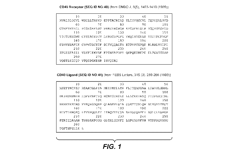
6. BRIEF DESCRIPTION OF THE FIGURES
[0018] FIG. 1 shows the amino acid sequences of human CD40 receptor (SEQ ID
NO:40) and
human CD40 ligand (SEQ ID NO:41).
[0019] FIGS. 2A-2G provide amino acid sequences for VH and VL of exemplary
mouse and
humanized anti-CD40 antibodies. FIG. 2A shows the VH and VL amino acid
sequences for muAbl
through muAb3; FIG. 2B shows the VII and VL amino acid sequences for muAb4
through muAb7;
FIG. 2C shows the VII and VL amino acid sequences for muAb8 through muAbl0;
FIG. 2D shows the
VH and VL amino acid sequences for humanized antibodies of muAb6 and muAb8;
FIG. 2E shows the
VH and VL amino acid sequences for humanized antibodies of muAb8 and muAb9;
FIG. 2F shows the
VH and VL amino acid sequences for further humanized antibodies of muAb9; and
FIG. 2G shows the
VH and VL amino acid sequences for additional humanized antibodies of muAb9.
[0020] FIG. 3 provides the results of competition experiments on human CD40
between CD4OL and
exemplary anti-CD40 antibodies. Upper left shows exemplary anti-CD40
antibodies that compete
with CD4OL; upper right shows antibodies that do not significantly compete
with CD4OL; lower left
shows antibodies that enhance CD4O-CD4OL interaction; lower right shows
effects of anti-CD40
antibody huAb9 A2I and CP-870,893. Y-axis depicts optical density (OD) at 650
nm; x-axis depicts
antibody dose ("Sample") in jtg/mL.
[0021] FIG. 4 shows the binding of fluorochrome-conjugated human CD40 at a
fixed concentration
of 1 ug/mL on Jurkat cells expressing human CD4OL in the presence of
increasing amounts of
antibody huAb9 A2I, CP-870,893, human IgGL ("huIgGl") isotype or human IgG2
("huIgG2")
isotype. Y-axis depicts mean fluorescence intensity ("MFI") representing the
binding; x-axis depicts
antibody dose ("Sample") in ug/mL.
[0022] FIGS. 5A-5B show the effects of an antibody dose ("Sample") at 3 ug/mL
or media only on
NFKB signal from HEK293 blue CD40 NFKB reporter cells mixed with Jurkat D1.1
cells (1:1 ratio).
Antibody huAb9 A2I, CP-870,893, human IgGL ("huIgGl") isotype or human IgG2
("huIgG2")
isotype, or media only, was added to the individual sample. FIG. 5A depicts
the NFKB signal in
cultures containing CD4OL negative ("CD4OL-") Jurkat D1.1 cells. FIG. 5B
depicts the NFKB signal
in cultures containing CD4OL positive ("CD40L+") Jurkat D1.1 cells. Y-axis
depicts OD at 625 nm;
x-axis depicts antibody or media only treatment ("Sample").
-4-
CA 03025347 2018-11-22
WO 2017/205742 PCT/US2017/034681
[0023] FIGS. 6A-6B show the binding of antibody doses ("Sample") in ug/mL of
huAb9-5 with wild
type huIgGI, or with V273Y or V273E variant, or CP-870,893, in CHO cells
expressing CD16F,
CD16V, CD32a, CD32b, or CD64. FIG. 6A shows binding of an anti-CD40 antibody
on CHO cells
expressing CD16F (upper left), CD16V (upper right), CD32a (lower left), or
CD32b (lower right).
FIG. 6B shows binding of an anti-CD40 antibody on CHO cells expressing CD64. Y-
axis depicts
mean fluorescence intensity (MFI) representing the binding; x-axis depicts
antibody dose ("Sample")
in ug/mL.
[0024] FIG. 7 shows the antibody-dependent cell-mediated cytotoxicity (ADCC)
of constant region
variants V273E or V273Y for antibody huAb9-5 as compared with huAb9-5 with the
wild type
human IgGI in RL cells. Y-axis depicts percent cytotoxicity in RL cells; x-
axis depicts antibody dose
("Sample") in jtg/mL.
[0025] FIG. 8 shows the effect of antibody huAb6-1 (upper left), huAb9-5
(lower left), huAb8-1
(upper right) with wild type human IgGI, or a constant region variant V273E or
V273Y, on B cell
proliferation. Lower right graph shows B cell proliferation effects of huAb9
A2I with human IgGI
V273E variant or CP-870,893. Y-axis depicts B cell proliferation in counts per
minute (CPM); x-axis
depicts antibody dose ("Sample") in ug/mL.
[0026] FIG. 9 shows the effect of antibody huAb6-1 (upper left), huAb9-5
(lower left) and huAb8-1
(upper right) with wild type human IgGI, or a constant region variant V273E or
V273Y, on dendritic
cell (DC) activation as measured by IL-12p70 production in pg/mL. Lower right
graph shows DC
activation of huAb9 A2I with human IgGI V273E variant or CP-870,893. Y-axis
depicts IL-12p70 in
pg/mL; x-axis depicts antibody dose ("Sample") in ug/mL.
[0027] FIG. 10 shows the effect of a V273Y variant of huAb6-1, huAb8-1, or
huAb9-5 on DC and
T-cell co-cultures as measured by interferon-gamma (IFN-y) production in
pg/mL.
[0028] FIG. 11 shows the effect of antibody huAb6-1 (upper), huAb9-5 (middle)
or huAb9 A2I
(lower) on tumor volume (mm3) in a prophylactic PC3 mouse model.
[0029] FIG. 12 shows in vivo effects following intratumoral (IT) or
intraperitoneal (IP) delivery of
anti-CD40 antibody 1C10, or mIgG1 isotype, in a mouse model carrying
bilaterally established CT26
syngeneic tumors. IT dosing was administered to one tumor at one flank, with
no injection to the
tumor at the other flank.
-5-
CA 03025347 2018-11-22
WO 2017/205742
PCT/US2017/034681
[0030] FIG. 13 shows effects on tumor volume (mm3) following dosing two times
a week of anti-
CD40 antibody 1C10 at 0.6 mg/kg, an anti-PD-1 antibody at 10 mg/kg, or
combination treatment of
both 1C10 and the anti-PD-1 antibody in a CT26 mouse syngeneic model.
[0031] FIG. 14 shows ALT (upper left), TNFa ("TNFa", lower left), or IL-6
(lower right) levels 24
hours after dosing of anti-CD40 antibody 1C10 ("anti-CD40"), anti-PD-1
antibody ("anti-PD-1") or
combination treatment ("anti-CD40 + anti-PD-1") in a CT26 mouse syngeneic
model. Upper right
graph shows spleen weight 4 days post-dosing.
7. DETAILED DESCRIPTION
[0032] The present disclosure concerns antibodies and fragments that
specifically bind human CD40
(SEQ ID NO:40), compositions comprising the antibodies, polynucleotides
encoding anti-CD40
antibodies, host cells capable of producing the antibodies, methods and
compositions useful for
making the antibodies and binding fragments, and various methods of using the
same.
[0033] As will be appreciated by skilled artisans, antibodies are "modular" in
nature. Throughout
the disclosure, various specific embodiments of the various "modules"
composing the antibodies are
described. As specific non-limiting examples, various specific embodiments of
VH CDRs, VH chains,
VL CDRs and VL chains are described. It is intended that all of the specific
embodiments may be
combined with each other as though each specific combination were explicitly
described individually.
7.1. Abbreviations
[0034] The antibodies, binding fragments, ADCs and polynucleotides described
herein are, in many
embodiments, described by way of their respective polypeptide or
polynucleotide sequences. Unless
indicated otherwise, polypeptide sequences are provided in N¨>C orientation;
polynucleotide
sequences in 5'¨>3' orientation. For polypeptide sequences, the conventional
three or one-letter
abbreviations for the genetically encoded amino acids may be used, as noted in
TABLE 1, below.
TABLE 1
Encoded Amino Acid Abbreviations
Amino Acid Three Letter Abbreviation One-
Letter Abbreviation
Alanine Ala A
Arginine Arg
Asparagine Asn
-6-
CA 03025347 2018-11-22
WO 2017/205742
PCT/US2017/034681
TABLE 1
Encoded Amino Acid Abbreviations
Amino Acid Three Letter Abbreviation One-
Letter Abbreviation
Aspartic acid Asp
Cysteine Cys
Glutamic acid Glu
Glutamine Gin
Glycine Gly
Histidine His
Isoleucine Ile
Leucine Leu
Lysine Lys
Methionine Met
Phenylalanine Phe
Proline Pro
Serine Ser
Threonine Thr
Tryptophan Trp
Tyrosine Tyr
Valine Val V
[0035] Certain sequences are defined by structural formulae specifying amino
acid residues
belonging to certain classes (e.g., aliphatic, hydrophobic, etc.). The various
classes to which the
genetically encoded amino acids belong as used herein are noted in TABLE 2,
below. Some amino
acids may belong to more than one class. Cysteine, which contains a sulfhydryl
group, and proline,
which is conformationally constrained, are not assigned classes.
-7-
CA 03025347 2018-11-22
WO 2017/205742
PCT/US2017/034681
TABLE 2
Encoded Amino Acid Classes
Class Amino Acids
Aliphatic A, I, L,V
Aromatic F, Y, W
Non-Polar M, A, I, L, V
Polar N, Q, S, T
Basic H, K, R
Acidic D, E
Small A, G
[0036] The abbreviations used for the various exemplary antibodies disclosed
herein are provided in
TABLE 3, below:
TABLE 3
Antibody Abbreviations
Clone/Name Abbreviation VH Sequence (FIGS. 2A-2G) Vi.
Sequence (FIGS. 2A-2G)
AD163.9.3 muAbl muAbl VH SEQ ID NO:101 muAbl Vi. SEQ
ID NO:151
AD166.4.4 muAb2 muAb2 VH SEQ ID NO:102 muAb2 VL SEQ
ID NO:152
AD175.14.11 muAb3 muAb3 VH SEQ ID NO:103 muAb3 VL SEQ
ID NO:153
AD163.10.7 muAb4 muAb4 VH SEQ ID NO:104 muAb4 VL SEQ
ID NO:154
AD165.1.2 muAb5 muAb5 VH SEQ ID NO:105 muAb5 VL SEQ
ID NO:155
AD163.162.1 muAb6 muAb6 VH SEQ ID NO:106 muAb6 VL SEQ
ID NO:156
AD163.27.12 muAb7 muAb6 VH SEQ ID NO:106 muAb7 VL SEQ
ID NO:157
AD163.7.2 muAb8 muAb8 VH SEQ ID NO:107 muAb8 VL SEQ
ID NO:158
AD164.14.6 muAb9 muAb9 VH SEQ ID NO:108 muAb9 VL SEQ
ID NO:159
AD164.76.3 muAblO muAb10VH SEQ ID NO:109 muAblO VL SEQ
ID NO:160
-8-
CA 03025347 2018-11-22
WO 2017/205742 PCT/US2017/034681
TABLE 3
Antibody Abbreviations
Clone/Name Abbreviation VH Sequence (FIGS. 2A-2G) Vi. Sequence
(FIGS. 2A-2G)
Humanized muAb6 #1 huAb6-1 huAb6-1 VH SEQ ID NO:110 huAb6-1 VL SEQ
ID NO:161
Humanized muAb6 #2 huAb6-2 huAb6-2 VH SEQ ID NO:111 huAb6-1 Vi. SEQ
ID NO:161
Humanized muAb6 #3 huAb6-3 huAb6-3 VH SEQ ID NO:112 huAb6-1 Vi. SEQ
ID NO:161
Humanized muAb8 #1 huAb8-1 huAb8-1 VH SEQ ID NO:113 huAb8-1 VL SEQ
ID NO:162
Humanized muAb8 #2 huAb8-2 huAb8-2 VH SEQ ID NO:114 huAb8-1 Vi. SEQ
ID NO:162
Humanized muAb8 #3 huAb8-3 huAb8-3 VH SEQ ID NO:115 huAb8-1 VL SEQ
ID NO:162
Humanized muAb9 #1 huAb9-1 huAb9-1 VH SEQ ID NO:116 huAb9-1 VL SEQ
ID NO:163
Humanized muAb9 #2 huAb9-2 huAb9-2 VH SEQ ID NO:117 huAb9-1 Vi. SEQ
ID NO:163
Humanized muAb9 #3 huAb9-3 huAb9-3 VH SEQ ID NO:118 huAb9-1 VL SEQ
ID NO:163
Humanized muAb9 #4 huAb9-4 huAb9-1 VH SEQ ID NO:116 huAb9-4 Vi. SEQ
ID NO:164
Humanized muAb9 #5 huAb9-5 huAb9-2 VH SEQ ID NO:117 huAb9-4 Vi. SEQ
ID NO:164
Humanized muAb9 #6 huAb9-6 huAb9-3 VH SEQ ID NO:118 huAb9-4 Vi. SEQ
ID NO:164
Humanized muAb9 #7 huAb9-7 huAb9-7 VH SEQ ID NO:119 huAb9-7 Vi. SEQ
ID NO:165
Humanized muAb9 #8 huAb9-8 huAb9-8 VH SEQ ID NO:120 huAb9-7 Vi. SEQ
ID NO:165
Humanized muAb9 #9 huAb9-9 huAb9-9 VH SEQ ID NO:121 huAb9-9 Vi. SEQ
ID NO:166
Rehumanized muAb9 huAb9 rehu#1 huAb9 SEQ
ID NO:122 huAb9 VK1 VL SEQ ID NO:167
version #1 rehuVH4 VH
Rehumanized muAb9 huAb9 rehu#2 huAb9 SEQ ID NO:122 huAb9 SEQ
ID NO:168
version #2 rehuVH4 VH rehuVK2 Vi.
Rehumanized muAb9 huAb9 rehu#3 huAb9 SEQ ID NO:123 huAb9 SEQ
ID NO:169
version #3 rehuVH3 VH rehuVK1 Vi.
Humanized muAb9 A2I huAb9 A2I
huAb9-2VH SEQ ID NO:117 huAb9A2I Vi. SEQ ID NO:170
Humanized muAb9 A2V huAb9 A2V huAb9-2VH SEQ
ID NO:117 huAb9A2V Vi. SEQ ID NO:171
-9-
CA 03025347 2018-11-22
WO 2017/205742 PCT/US2017/034681
7.2. Definitions
[0037] Unless otherwise defined herein, scientific and technical terms used in
connection with the
present disclosure shall have the meanings that are commonly understood by
those of ordinary skill in
the art.
7.3. Anti-CD40 Antibodies and Binding Fragments
[0038] In one aspect, the disclosure concerns antibodies and/or binding
fragments thereof that
specifically bind human CD40 receptor (also known as tumor necrosis factor
receptor superfamily
member 5, TNFRSF5, Bp50, and CD4OL receptor).
[0039] As used herein, the term "antibody" (Ab) refers to an immunoglobulin
molecule that
specifically binds to a particular antigen- here, CD40. In some embodiments,
the anti-CD40
antibodies of the disclosure bind to human CD40 and thereby modulate, e.g.,
activate, the immune
system. The resulting immune system response inhibits proliferation of cells
such as tumor cells, and
in some instances are cytotoxic to the tumor cells. Anti-CD40 antibodies of
the disclosure comprise
complementarity determining regions (CDRs), also known as hypervariable
regions, in both the light
chain and the heavy chain variable domains. The more highly conserved portions
of variable
domains are called the framework (FR). As is known in the art, the amino acid
position/boundary
delineating a hypervariable region of an antibody can vary, depending on the
context and the various
definitions known in the art. Some positions within a variable domain may be
viewed as hybrid
hypervariable positions in that these positions can be deemed to be within a
hypervariable region
under one set of criteria while being deemed to be outside a hypervariable
region under a different set
of criteria. One or more of these positions can also be found in extended
hypervariable regions. The
disclosure provides antibodies comprising modifications in these hybrid
hypervariable positions. The
variable domains of native heavy and light chains each comprise four FR
regions, largely by adopting
a 13-sheet configuration, connected by three CDRs, which form loops
connecting, and in some cases
forming part of, the 13-sheet structure. The CDRs in each chain are held
together in close proximity
by the FR regions and, with the CDRs from the other chain, contribute to the
formation of the target
binding site of antibodies. See Kabat etal., Sequences of Proteins of
Immunological Interest
(National Institute of Health, Bethesda, Md. 1987). As used herein, numbering
of immunoglobulin
amino acid residues is done according to the immunoglobulin amino acid residue
numbering system
of Kabat etal. unless otherwise indicated.
-10-
CA 03025347 2018-11-22
WO 2017/205742 PCT/US2017/034681
[0040] The antibodies of the disclosure may be polyclonal, monoclonal,
genetically engineered,
and/or otherwise modified in nature, including but not limited to chimeric
antibodies, humanized
antibodies, human antibodies, primatized antibodies, single chain antibodies,
etc. In various
embodiments, the antibodies comprise all or a portion of a constant region of
an antibody. In some
embodiments, the constant region is an isotype selected from: IgA (e.g., IgAl
or IgA2), IgD, IgE, IgG
(e.g., IgGi, IgG2, IgG3 or IgG4), and IgM. In specific embodiments, the anti-
CD40 antibodies
described herein comprise an IgGi. In other embodiments, the anti-CD40
antibodies comprise an
IgG2. In yet other embodiments, the anti-CD40 antibodies comprise an IgG4. As
used herein, the
"constant region" of an antibody includes the natural constant region,
allotypes or natural variants,
such as D356E and L358M, or A431G in human IgGi. See, e.g., Jefferis and
Lefranc, MAbs, 1(4):
332-338 (Jul-Aug 2009).
[0041] The light constant region of an anti-CD40 antibody may be a kappa (K)
light region or a
lambda () region. A2,, light region can be any one of the known subtypes,
e.g., i,2,, 2, 2,,3, or 2,4. In
some embodiments, the anti-CD40 antibody comprises a kappa (K) light region.
[0042] The term "monoclonal antibody" as used herein is not limited to
antibodies produced through
hybridoma technology. A monoclonal antibody is derived from a single clone,
including any
eukaryotic, prokaryotic, or phage clone, by any means available or known in
the art. Monoclonal
antibodies useful with the present disclosure can be prepared using a wide
variety of techniques
known in the art including the use of hybridoma, recombinant, and phage
display technologies, or a
combination thereof In many uses of the present disclosure, including in vivo
use of the anti-CD40
antibodies in humans, chimeric, primatized, humanized, or human antibodies can
suitably be used.
[0043] The term "chimeric" antibody as used herein refers to an antibody
having variable sequences
derived from a non-human immunoglobulin, such as a rat or a mouse antibody,
and human
immunoglobulin constant regions, typically chosen from a human immunoglobulin
template.
Methods for producing chimeric antibodies are known in the art. See, e.g.,
Morrison, 1985, Science
229(4719):1202-7; Oi etal., 1986, BioTechniques 4:214-221; Gillies etal.,
1985, J. Immunol.
Methods 125:191-202; U.S. Pat. Nos. 5,807,715; 4,816,567; and 4,816,397.
[0044] "Humanized" forms of non-human (e.g., murine) antibodies are chimeric
immunoglobulins
that contain minimal sequences derived from non-human immunoglobulin. In
general, a humanized
antibody will comprise substantially all of at least one, and typically two,
variable domains, in which
all or substantially all of the CDR regions correspond to those of a non-human
immunoglobulin and
-11-
CA 03025347 2018-11-22
WO 2017/205742 PCT/US2017/034681
all or substantially all of the FR regions are those of a human immunoglobulin
sequence. The
humanized antibody can also comprise at least a portion of an immunoglobulin
constant region (Fc),
typically that of a human immunoglobulin consensus sequence. Methods of
antibody humanization
are known in the art. See, e.g., Riechmann etal., 1988, Nature 332:323-7; U.S.
Patent Nos:
5,530,101; 5,585,089; 5,693,761; 5,693,762; and 6,180,370 to Queen etal.;
EP239400; PCT
publication WO 91/09967; U.S. Patent No. 5,225,539; EP592106; EP519596;
Padlan, 1991, Mol.
Immunol., 28:489-498; Studnicka etal., 1994, Prot. Eng. 7:805-814; Roguska
etal., 1994, Proc. Natl.
Acad. Sci. 91:969-973; and U.S. Patent No. 5,565,332.
[0045] "Human antibodies" include antibodies having the amino acid sequence of
a human
immunoglobulin and include antibodies isolated from human immunoglobulin
libraries or from
animals transgenic for one or more human immunoglobulin and that do not
express endogenous
immunoglobulins. Human antibodies can be made by a variety of methods known in
the art including
phage display methods using antibody libraries derived from human
immunoglobulin sequences. See
U.S. Patent Nos. 4,444,887 and 4,716,111; and PCT publications WO 98/46645; WO
98/50433; WO
98/24893; WO 98/16654; WO 96/34096; WO 96/33735; and WO 91/10741. Human
antibodies can
also be produced using transgenic mice which are incapable of expressing
functional endogenous
immunoglobulins but which can express human immunoglobulin genes. See, e.g.,
PCT publications
WO 98/24893; WO 92/01047; WO 96/34096; WO 96/33735; U.S. Patent Nos.
5,413,923; 5,625,126;
5,633,425; 5,569,825; 5,661,016; 5,545,806; 5,814,318; 5,885,793; 5,916,771;
and 5,939,598. In
addition, companies such as LakePharma, Inc. (Belmont, CA) or Creative BioLabs
(Shirley, NY) can
be engaged to provide human antibodies directed against a selected antigen
using technology similar
to that described above. Fully human antibodies that recognize a selected
epitope can be generated
using a technique referred to as "guided selection." In this approach, a
selected non-human
monoclonal antibody, e.g., a mouse antibody, is used to guide the selection of
a completely human
antibody recognizing the same epitope (see, Jespers etal., 1988, Biotechnology
12:899-903).
[0046] "Primatized antibodies" comprise monkey variable regions and human
constant regions.
Methods for producing primatized antibodies are known in the art. See, e.g.,
U.S. Patent Nos.
5,658,570; 5,681,722; and 5,693,780.
[0047] Anti-CD40 antibodies of the disclosure include full-length (intact)
antibody molecules that
are capable of specifically binding CD40, e.g., human CD40 (SEQ ID NO:40).
-12-
CA 03025347 2018-11-22
WO 2017/205742 PCT/US2017/034681
[0048] Also disclosed are anti-CD40 binding fragments that are capable of
specifically binding
human CD40. Examples of antibody binding fragments include by way of example
and not
limitation, Fab, Fab', F(ab1)2, Fv fragments, single chain Fv fragments and
single domain fragments.
[0049] A Fab fragment contains the constant and variable domains of the light
chain and the first
constant domain (CH1) and the variable domain of the heavy chain. Fab'
fragments differ from Fab
fragments by the addition of a few residues at the carboxyl terminus of the
heavy chain CH1 domain
including one or more cysteines from the antibody hinge region. F(ab')
fragments are produced by
cleavage of the disulfide bond at the hinge cysteines of the F(ab1)2 pepsin
digestion product.
Additional chemical couplings of antibody fragments are known to those of
ordinary skill in the art.
Fab and F(ab1)2 fragments lack the Fc fragment of an intact antibody, clear
more rapidly from the
circulation of animals, and may have less non-specific tissue binding than an
intact antibody (see,
e.g., Wahl etal., 1983, J. Nucl. Med. 24:316).
[0050] An "Fv" fragment is the minimum fragment of an antibody that contains a
complete target
recognition and binding site. This region consists of a dimer of one heavy and
one light chain
variable domain in a tight, non-covalent association (VH-VL dimer). It is in
this configuration that the
three CDRs of each variable domain interact to define a target binding site on
the surface of the
VH-VL dimer. Often, the six CDRs confer target binding specificity to the
antibody. However, in
some instances even a single variable domain (or half of an Fv comprising only
three CDRs specific
for a target) can have the ability to recognize and bind target, although at a
lower affinity than the
entire binding site.
[0051] "Single-chain Fv" or "scFv" antibody binding fragments comprise the VH
and VL domains of
an antibody, where these domains are present in a single polypeptide chain.
Generally, the Fv
polypeptide further comprises a polypeptide linker between the VH and VL
domains which enables the
scFy to form the desired structure for target binding.
[0052] "Single domain fragments" are composed of a single VII or VL domains
which exhibit
sufficient affinity to human CD40. In a specific embodiment, the single domain
fragment is a
camelized fragment (See, e.g., Riechmann, 1999, Journal of Immunological
Methods 231:25-38).
[0053] The anti-CD40 antibodies of the disclosure include derivatized
antibodies. For example, but
not by way of limitation, derivatized antibodies are typically modified by
glycosylation, acetylation,
pegylation, phosphorylation, amidation, derivatization by known
protecting/blocking groups,
proteolytic cleavage, linkage to a cellular ligand or other protein. Any of
numerous chemical
-13-
CA 03025347 2018-11-22
WO 2017/205742 PCT/US2017/034681
modifications can be carried out by known techniques, including, but not
limited to, specific chemical
cleavage, acetylation, formylation, metabolic synthesis of tunicamycin, etc.
Additionally, the
derivative can contain one or more non-natural amino acids, e.g., using Ambryx
technology (See, e.g.,
Wolfson, 2006, Chem. Biol. 13(10):1011-2).
[0054] The anti-CD40 antibodies or binding fragments may be antibodies or
fragments whose
sequences have been modified to alter at least one constant region-mediated
biological effector
function. For example, in some embodiments, an anti-CD40 antibody may be
modified to reduce at
least one constant region-mediated biological effector function relative to
the unmodified antibody,
e.g., reduced binding to one or more of the Fc receptors (FcyR) such as FcyRI,
FcyRIIA, FcyRIIB,
FcyRIIIA and/or FcyRIIIB. FcyR binding can be reduced by mutating the
immunoglobulin constant
region segment of the antibody at particular regions necessary for FcyR
interactions (See, e.g.,
Canfield and Morrison, 1991, J. Exp. Med. 173:1483-1491; and Lund etal., 1991,
J. Immunol.
147:2657-2662). Reduction in FcyR binding ability of the antibody can also
reduce other effector
functions which rely on FcyR interactions, such as opsonization, phagocytosis
and antigen-dependent
cellular cytotoxicity ("ADCC").
[0055] The anti-CD40 antibody or binding fragment described herein include
antibodies that have
been modified to acquire or improve at least one constant region-mediated
biological effector
function relative to an unmodified antibody, e.g., to enhance FcyR
interactions (See, e.g., US Patent
Appl. No. 2006/0134709). For example, an anti-CD40 antibody of the disclosure
can have a constant
region that binds FcyRI, FcyRIIA, FcyRIIB, FcyRIIIA and/or FcyRIIIB with
greater affinity than the
corresponding wild type constant region.
[0056] Thus, antibodies of the disclosure may have alterations in biological
activity that result in
increased or decreased opsonization, phagocytosis, or ADCC. Such alterations
are known in the art.
For example, modifications in antibodies that reduce ADCC activity are
described in U.S. Patent No.
5,834,597. An exemplary ADCC lowering variant corresponds to "mutant 3" (also
known as
shown in FIG. 4 of U.S. Patent No. 5,834,597) in which residues 234 and 237
(using EU numbering)
are substituted with alanines. A mutant 3 (also known as "M3") variation may
be used in a number of
antibody isotypes, e.g., IgG2.
[0057] In some embodiments, the anti-CD40 antibodies of the disclosure have
low levels of, or lack,
fucose. Antibodies lacking fucose have been correlated with enhanced ADCC
activity, especially at
low doses of antibody. See Shields etal., 2002, J. Biol. Chem. 277:26733-
26740; Shinkawa etal.,
-14-
CA 03025347 2018-11-22
WO 2017/205742 PCT/US2017/034681
2003, J. Biol. Chem. 278:3466-73. Methods of preparing fucose-less antibodies
include growth in rat
myeloma YB2/0 cells (ATCC CRL 1662). YB2/0 cells express low levels of FUT8
mRNA, which
encodes a-1,6-fucosyltransferase, an enzyme necessary for fucosylation of
polypeptides.
[0058] The anti-CD40 antibodies of the disclosure can comprise modified (or
variant) CH2 domains
or entire Fc domains that include amino acid substitutions that increase
binding to FcyRIIB and/or
reduced binding to FcyRIIIA as compared to the binding of a corresponding wild-
type CH2 or Fc
region. Variant CH2 or variant Fc domains have been described in U.S. Patent
Appl. No.
2014/0377253. A variant CH2 or variant Fc domain typically includes one or
more substitutions at
position 263, position 266, position 273, and position 305, wherein the
numbering of the residues in
the Fc domain is that of the EU index as in Kabat. In some embodiments, the
anti-CD40 antibodies
comprise one or more substitutions selected from V263L, V266L, V273C, V273E,
V273F, V273L,
V273M, V273S, V273Y, V305K, and V305W, relative to the wild-type CH2 domain.
In specific
embodiments, the one or more substitutions of the CH2 domain are selected from
V263L, V273E,
V273F, V273M, V2735, and V273Y, relative to the CH2 domain of a human IgGi.
For example, the
one or more substitutions of an IgGi CH2 domain can be V273E. In another
specific embodiment,
the anti-CD40 antibody of the disclosure comprises a variant IgGi CH2 region
comprising the amino
acid substitution V263L.
[0059] Other examples of variant CH2 or variant Fc domains that can afford
increased binding to
FcyRIIB and/or reduced binding to FcyRIIIA as compared to the binding of a
corresponding wild-
type CH2 or Fc region include those found in Vonderheide, et al. Clin. Cancer
Res., 19(5), 1035-1043
(2013), such as 5267E or 5267E/L328F in human IgGi.
[0060] In some embodiments, the anti-CD40 antibodies or binding fragments
include modifications
that increase or decrease their binding affinities to the fetal Fc receptor,
FcRn, for example, by
mutating the immunoglobulin constant region segment at particular regions
involved in FcRn
interactions (see, e.g., WO 2005/123780). In particular embodiments, an anti-
CD40 antibody of the
IgG class is mutated such that at least one of amino acid residues 250, 314,
and 428 of the heavy
chain constant region is substituted alone, or in any combinations thereof,
such as at positions 250
and 428, or at positions 250 and 314, or at positions 314 and 428, or at
positions 250, 314, and 428,
with positions 250 and 428 a specific combination. For position 250, the
substituting amino acid
residue can be any amino acid residue other than threonine, including, but not
limited to, alanine,
cysteine, aspartic acid, glutamic acid, phenylalanine, glycine, histidine,
isoleucine, lysine, leucine,
-15-
CA 03025347 2018-11-22
WO 2017/205742 PCT/US2017/034681
methionine, asparagine, proline, glutamine, arginine, serine, valine,
tryptophan, or tyrosine. For
position 314, the substituting amino acid residue can be any amino acid
residue other than leucine,
including, but not limited to, alanine, cysteine, aspartic acid, glutamic
acid, phenylalanine, glycine,
histidine, isoleucine, lysine, methionine, asparagine, proline, glutamine,
arginine, serine, threonine,
valine, tryptophan, or tyrosine. For position 428, the substituting amino acid
residues can be any
amino acid residue other than methionine, including, but not limited to,
alanine, cysteine, aspartic
acid, glutamic acid, phenylalanine, glycine, histidine, isoleucine, lysine,
leucine, asparagine, proline,
glutamine, arginine, serine, threonine, valine, tryptophan, or tyrosine. An
exemplary substitution
known to modify Fc effector function is the Fc substitution M428L, which can
occur in combination
with the Fc substitution T250Q. Additional specific combinations of suitable
amino acid
substitutions are identified in Table 1 of U.S. Patent No. 7,217,797. Such
mutations increase binding
to FcRn, which protects the antibody from degradation and increases its half-
life.
[0061] An anti-CD40 antibody may have one or more amino acids inserted into
one or more of its
CDRs, for example as described in Jung and Pluckthun, 1997, Protein
Engineering 10:9, 959-966;
Yazaki etal., 2004, Protein Eng. Des Sel. 17(5):481-9. Epub 2004 Aug 17; and
U.S. Pat. Appl. No.
2007/0280931.
[0062] Anti-CD40 antibodies with affinity for human CD40 may be desirable for
therapeutic and
diagnostic uses. Accordingly, the present disclosure contemplates antibodies
having binding affinity
to human CD40. In specific embodiments, the anti-CD40 antibodies that bind
human CD40 with an
affinity of at least about 1000 nM, but may exhibit higher affinity, for
example, at least about 900
nM, 800 nM, 700 nM, 600 nM, 500 nM, 400 nM, 300 nM, 250 nM, 200 nM, 150 nM,
100 nM, 90
nM, 80 nM, 70 nM, 60 nM, 50 nM, 40 nM, 30 nM, 25 nM, 20 nM, 15 nM, 10 nM, 7
nM, 6 nM, 5
nM, 4 nM, 3 nM, 2 nM, 1 nM, 0.1 nM, 0.01 nM, or even higher. In some
embodiments, the
antibodies bind human CD40 with an affinity in the range of about 1 pM to
about 1000 nM, or an
affinity ranging between any of the foregoing values.
[0063] Affinity of anti-CD40 antibodies for human CD40 can be determined using
techniques well
known in the art or described herein, such as for example, but not by way of
limitation, ELISA,
isothermal titration calorimetry (ITC), surface plasmon resonance, or
fluorescent polarization assay.
[0064] Anti-CD40 antibodies generally comprise a heavy chain comprising a
variable region (VH)
having three complementarity determining regions ("CDRs") referred to herein
(in N¨>C order) as
VH CDR#1, VH CDR#2, and VH CDR#3, and a light chain comprising a variable
region (VL) having
-16-
CA 03025347 2018-11-22
WO 2017/205742 PCT/US2017/034681
three complementarity determining regions referred to herein (in N¨>C order)
as VL CDR#1,
VL CDR#2, and VL CDR#3. The amino acid sequences of exemplary CDRs, as well as
the amino
acid sequence of the VH and VL regions of the heavy and light chains of
exemplary anti-CD40 are
provided herein. Specific embodiments of anti-CD40 antibodies include these
exemplary CDRs
and/or VH and/or VL sequences, as well as antibodies that compete for binding
human CD40 with
such antibodies.
[0065] In some embodiments, the amino acid sequences of the CDRs of an anti-
CD40 antibody are
selected from the following sequences:
CDR Sequence (N¨>C) Identifier
VH CDR#1: GYTFTSYWIT (SEQ ID NO:1)
GYTFTGYWIQ (SEQ ID NO:2)
GYTFTDYYMN (SEQ ID NO:3)
GFTFSDYYMS (SEQ ID NO:4)
GYSIT (SEQ ID NO:5)
GYTFTSYWMH (SEQ ID NO:6)
GYTFTDYYIN (SEQ ID NO:7)
GYSITSNYYWN (SEQ ID NO:8)
GYSISSNYYWN (SEQ ID NO:9)
GYDITSNYYWN (SEQ ID NO:10)
VH CDR#2: EINPGSGSTNYNEKFKS (SEQ ID NO:11)
EILPGGDHTKYNEKFRG (SEQ ID NO:12)
DINPNNGGTSYNQKFKG (SEQ ID NO:13)
FIRNKANGYTTEFSASVKG (SEQ ID NO:14)
YIRHDGTNNYNPSLKN (SEQ ID NO:15)
NIDPSNGETHYNQKFKD (SEQ ID NO:16)
WIFPGSGSVYCNEQFKG (SEQ ID NO:17)
YIRYDGSNNYNPSLKN (SEQ ID NO:18)
NIDPSNGETHYAQKFQG (SEQ ID NO:19)
WIFPGSGSVYSNEQFKG (SEQ ID NO:20)
YIRYDGSNNYNPSLKS (SEQ ID NO:21)
YIRYDGSNNYNPSLKG (SEQ ID NO:22)
-17-
CA 03025347 2018-11-22
WO 2017/205742
PCT/US2017/034681
CDR Sequence (N¨>C) Identifier
VH CDR#3: NRGTGDY (SEQ ID NO:31)
VGGDY (SEQ ID NO:32)
RGGLGRGTYALDY (SEQ ID NO:33)
YGGLRQGWYFDV (SEQ ID NO:34)
LDY (SEQ ID NO:35)
ERIYYSGSTYDGYFDV (SEQ ID NO:36)
SLGKFAY (SEQ ID NO:37)
VL CDR#1: RSSQSLVHSYGNTYLH (SEQ ID NO:51)
RSSQSLVNSNENTYLH (SEQ ID NO:52)
RASQDISNYLN (SEQ ID NO:53)
RASQDIRNYLN (SEQ ID NO:54)
RSSQSLENSYGNTFLN (SEQ ID NO:55)
SASS SLSYMH (SEQ ID NO:56)
KASQSVVTAVA (SEQ ID NO:57)
RSSQSLENTNGNTFLN (SEQ ID NO:58)
RSSQSLENSNGNTFLN (SEQ ID NO:59)
VL CDR#2: KVSNRIS (SEQ ID NO:61)
KVFNRYS (SEQ ID NO:62)
YTSRLHL (SEQ ID NO:63)
YTSRLHS (SEQ ID NO:64)
RVSNRFC (SEQ ID NO:65)
DTSKLAS (SEQ ID NO:66)
SASNRYT (SEQ ID NO:67)
RVSNRFS (SEQ ID NO:68)
RISNRFS (SEQ ID NO:69)
-18-
CA 03025347 2018-11-22
WO 2017/205742 PCT/US2017/034681
CDR Sequence (N¨>C) Identifier
VL CDR#3: SQSTHVPYT (SEQ ID NO:81)
FQSTHVPWT (SEQ ID NO:82)
QQGNTLPLT (SEQ ID NO:83)
QQGKTLPWT (SEQ ID NO:84)
LQVTHVPYT (SEQ ID NO:85)
QQWSSNPWT (SEQ ID NO:86)
QQYSSYPYT (SEQ ID NO:87)
LQVTHVPFT (SEQ ID NO:88)
[0066] In some embodiments, each CDR of an anti-CD40 antibody, independently
of the others, is
selected to correspond in sequence to the respective CDR of an antibody
provided in TABLE 3. In
some embodiments, an anti-CD40 antibody is an IgG, and has a VH and VL
corresponding in sequence
to the VH and VL of an antibody provided in TABLE 3.
[0067] In some embodiments, an anti-CD40 antibody comprises a VH chain
corresponding in
sequence to any one of SEQ ID NOS:101, 102, 103, 104, 105, 106, 107, 108, or
109; and a VL chain
corresponding in sequence to any one of SEQ ID NOS:151, 152, 153, 154, 155,
156, 157, 158, 159,
or 160. In some embodiments, an anti-CD40 antibody comprises a VH chain
corresponding in
sequence to SEQ ID NO:101 and a VL chain corresponding in sequence to SEQ ID
NO:151. In some
embodiments, an anti-CD40 antibody comprises a VH chain corresponding in
sequence to SEQ ID
NO:102 and a VL chain corresponding in sequence to SEQ ID NO:152. In some
embodiments, an
anti-CD40 antibody comprises a VH chain corresponding in sequence to SEQ ID
NO:103 and a VL
chain corresponding in sequence to SEQ ID NO:153. In some embodiments, an anti-
CD40 antibody
comprises a VH chain corresponding in sequence to SEQ ID NO:104 and a VL chain
corresponding in
sequence to SEQ ID NO:154. In some embodiments, an anti-CD40 antibody
comprises a VH chain
corresponding in sequence to SEQ ID NO:105 and a VL chain corresponding in
sequence to SEQ ID
NO:155. In some embodiments, an anti-CD40 antibody comprises a VH chain
corresponding in
sequence to SEQ ID NO:106 and a VL chain corresponding in sequence to SEQ ID
NO:156. In some
embodiments, an anti-CD40 antibody and comprises a VH chain corresponding in
sequence to SEQ
ID NO:106 and a VL chain corresponding in sequence to SEQ ID NO:157. In some
embodiments, an
anti-CD40 antibody and comprises a VH chain corresponding in sequence to SEQ
ID NO:107 and a
-19-
CA 03025347 2018-11-22
WO 2017/205742 PCT/US2017/034681
VL chain corresponding in sequence to SEQ ID NO:158. In some embodiments, an
anti-CD40
antibody and comprises a VH chain corresponding in sequence to SEQ ID NO:108
and a VL chain
corresponding in sequence to SEQ ID NO:159. In some embodiments, an anti-CD40
antibody
comprises a VH chain corresponding in sequence to SEQ ID NO:109 and a VL chain
corresponding in
sequence to SEQ ID NO:160.
[0068] Specific exemplary embodiments of anti-CD40 antibodies with the above
CDRs are
described herein. In some embodiments, an anti-CD40 antibody has the CDRs of
SEQ ID NOS: 1,
11, 31, 51, 61, and 81. In some embodiments, an anti-CD40 antibody has the
CDRs of SEQ ID NOS:
2, 12, 32, 52, 62, and 82. In some embodiments, an anti-CD40 antibody has the
CDRs of SEQ ID
NOS: 3, 13, 33, 53, 63, and 83. In some embodiments, an anti-CD40 antibody has
the CDRs of SEQ
ID NOS: 4, 14, 34, 54, 64, and 84. In some embodiments, an anti-CD40 antibody
has the CDRs of
SEQ ID NOS: 5, 15, 35, 55, 65, and 85. In some embodiments, an anti-CD40
antibody has the CDRs
of SEQ ID NOS: 6, 16, 36, 56, 66, and 86. In some embodiments, an anti-CD40
antibody has the
CDRs of SEQ ID NOS: 6, 19, 36, 56, 66, and 86. In some embodiments, an anti-
CD40 antibody has
the CDRs of SEQ ID NOS: 7, 17, 37, 57, 67, and 87. In some embodiments, an
anti-CD40 antibody
has the CDRs of SEQ ID NOS: 7, 20, 37, 57, 67, and 87. In some embodiments, an
anti-CD40
antibody has the CDRs of SEQ ID NOS: 8, 18, 35, 58, 68, and 88. In some
embodiments, an anti-
CD40 antibody has the CDRs of SEQ ID NOS: 9, 21, 35, 58, 68, and 88. In some
embodiments, an
anti-CD40 antibody has the CDRs of SEQ ID NOS: 10, 22, 35, 58, 68, and 88.
[0069] In some embodiments, an anti-CD40 antibody is suitable for
administration to humans. In a
specific embodiment, the anti-CD40 antibody is humanized. In another specific
embodiment, the
amino acid sequences of the CDRs of the anti-CD40 antibody are selected from:
CDR Sequence (N->C) Identifier
VH CDR#1: GYTFTSYWMH (SEQ ID NO:6)
GYTFTDYYIN (SEQ ID NO:7)
GYSITSNYYWN (SEQ ID NO:8)
GYSISSNYYWN (SEQ ID NO:9)
GYDITSNYYWN (SEQ ID NO:10)
-20-
CA 03025347 2018-11-22
WO 2017/205742 PCT/US2017/034681
CDR Sequence (N¨>C) Identifier
VH CDR#2: WIFPGSGSVYCNEQFKG (SEQ ID NO:17)
YIRYDGSNNYNPSLKN (SEQ ID NO:18)
NIDPSNGETHYAQKFQG (SEQ ID NO:19)
WIFPGSGSVYSNEQFKG (SEQ ID NO:20)
YIRYDGSNNYNPSLKS (SEQ ID NO:21)
YIRYDGSNNYNPSLKG (SEQ ID NO:22)
VH CDR#3: LDY (SEQ ID NO:35)
ERIYYSGSTYDGYFDV (SEQ ID NO:36)
SLGKFAY (SEQ ID NO:37)
VL CDR#1: SASSSLSYMH (SEQ ID NO:56)
KASQSVVTAVA (SEQ ID NO:57)
RSSQSLENTNGNTFLN (SEQ ID NO:58)
VL CDR#2: DTSKLAS (SEQ ID NO:66)
SASNRYT (SEQ ID NO:67)
RVSNRFS (SEQ ID NO:68)
VL CDR#3: QQWSSNPWT (SEQ ID NO:86)
QQYSSYPYT (SEQ ID NO:87)
LQVTHVPFT (SEQ ID NO:88)
[0070] In some embodiments, an anti-CD40 antibody comprises a VH chain
corresponding in
sequence to any one of SEQ ID NOS:110, 111, 112, 113, 114, 115, 116, 117, 118,
119, 120, 121, 122,
or 123; and a VL chain corresponding in sequence to any one of SEQ ID NOS:161,
162, 163, 164,
165, 166, 167, 168, 169, 170, or 171. In some embodiments, an anti-CD40
antibody comprises a VH
chain corresponding in sequence to SEQ ID NO:110 and a VL chain corresponding
in sequence to
SEQ ID NO:161. In some embodiments, an anti-CD40 antibody comprises a VH chain
corresponding
in sequence to SEQ ID NO:111 and a VL chain corresponding in sequence to SEQ
ID NO:161. In
some embodiments, an anti-CD40 antibody comprises a VH chain corresponding in
sequence to SEQ
ID NO:112 and a VL chain corresponding in sequence to SEQ ID NO:161. In some
embodiments, an
anti-CD40 antibody comprises a VH chain corresponding in sequence to SEQ ID
NO:113 and a VL
chain corresponding in sequence to SEQ ID NO:162. In some embodiments, an anti-
CD40 antibody
comprises a VH chain corresponding in sequence to SEQ ID NO:114 and a VL chain
corresponding in
-21-
CA 03025347 2018-11-22
WO 2017/205742 PCT/US2017/034681
sequence to SEQ ID NO:162. In some embodiments, an anti-CD40 antibody
comprises a VH chain
corresponding in sequence to SEQ ID NO:115 and a VL chain corresponding in
sequence to SEQ ID
NO:162. In some embodiments, an anti-CD40 antibody comprises a VH chain
corresponding in
sequence to SEQ ID NO:116 and a VL chain corresponding in sequence to SEQ ID
NO:163. In some
embodiments, an anti-CD40 antibody comprises a VH chain corresponding in
sequence to SEQ ID
NO:117 and a VL chain corresponding in sequence to SEQ ID NO:163. In some
embodiments, an
anti-CD40 antibody comprises a VH chain corresponding in sequence to SEQ ID
NO:118 and a VL
chain corresponding in sequence to SEQ ID NO:163. In some embodiments, an anti-
CD40 antibody
comprises a VH chain corresponding in sequence to SEQ ID NO:116 and a VL chain
corresponding in
sequence to SEQ ID NO:164. In some embodiments, an anti-CD40 antibody
comprises a VH chain
corresponding in sequence to SEQ ID NO:117 and a VL chain corresponding in
sequence to SEQ ID
NO:164. In some embodiments, an anti-CD40 antibody comprises a VH chain
corresponding in
sequence to SEQ ID NO:119 and a VL chain corresponding in sequence to SEQ ID
NO:165. In some
embodiments, an anti-CD40 antibody comprises a VH chain corresponding in
sequence to SEQ ID
NO:120 and a VL chain corresponding in sequence to SEQ ID NO:165. In some
embodiments, an
anti-CD40 antibody comprises a VH chain corresponding in sequence to SEQ ID
NO:121 and a VL
chain corresponding in sequence to SEQ ID NO:166. In some embodiments, an anti-
CD40 antibody
comprises a VH chain corresponding in sequence to SEQ ID NO:117 and a VL chain
corresponding in
sequence to SEQ ID NO:167. In some embodiments, an anti-CD40 antibody
comprises a VH chain
corresponding in sequence to SEQ ID NO:117 and a VL chain corresponding in
sequence to SEQ ID
NO:168. In some embodiments, an anti-CD40 antibody comprises a VH chain
corresponding in
sequence to SEQ ID NO:117 and a VL chain corresponding in sequence to SEQ ID
NO:169. In some
embodiments, an anti-CD40 antibody comprises a VH chain corresponding in
sequence to SEQ ID
NO:117 and a VL chain corresponding in sequence to SEQ ID NO:170. In some
embodiments, an
anti-CD40 antibody comprises a VH chain corresponding in sequence to SEQ ID
NO:117 and a VL
chain corresponding in sequence to SEQ ID NO:171. In some embodiments, an anti-
CD40 antibody
comprises a VH chain corresponding in sequence to SEQ ID NO:118 and a VL chain
corresponding in
sequence to SEQ ID NO:164. In some embodiments, an anti-CD40 antibody
comprises a VH chain
corresponding in sequence to SEQ ID NO:122 and a VL chain corresponding in
sequence to SEQ ID
NO:167. In some embodiments, an anti-CD40 antibody comprises a VH chain
corresponding in
sequence to SEQ ID NO:122 and a VL chain corresponding in sequence to SEQ ID
NO:168. In some
-22-
CA 03025347 2018-11-22
WO 2017/205742 PCT/US2017/034681
embodiments, an anti-CD40 antibody comprises a VH chain corresponding in
sequence to SEQ ID
NO:123 and a VL chain corresponding in sequence to SEQ ID NO:169.
[0071] In some embodiments, the anti-CD40 antibodies compete for binding human
CD40 in in vitro
assays with a reference antibody. In some embodiments, the anti-CD40
antibodies compete for
binding human CD40 on cells expressing human CD40. The reference antibody may
be any of the
anti-CD40 antibodies described herein. In some embodiments, the reference
antibody is an antibody
provided in TABLE 3. In specific embodiments, the reference antibody is
selected from antibody
AD163.9.3 ("muAbl"); antibody AD166.4.4 ("muAb2"); antibody AD175.14.11
("muAb3");
antibody AD163.10.7 ("muAb4"); antibody AD165.1.2 ("muAb5"); antibody
AD163.162.1
("muAb6"); antibody AD163.27.12 ("muAb7"); antibody AD163.7.2 ("muAb8");
antibody
AD164.14.6 ("muAb9"); and antibody AD164.76.2 ("muAbl0"). In some embodiments,
the
reference antibody is a humanized version of an antibody provided in TABLE 3.
In some
embodiments, the reference antibody is a humanized version of muAb6, muAb8, or
muAb9. In a
specific embodiment, the reference antibody is huAb9-2. In another embodiment,
the reference
antibody is huAb9-5. In another specific embodiment, the reference antibody is
huAb9 A2I.
[0072] Post-translational modifications to the sequences of an anti-CD40
antibody may occur, such
as cleavage of one or more (e.g., 1, 2, 3, or more) amino acid residues on the
C-terminal end of the
antibody heavy chain.
[0073] In some embodiments, an anti-CD40 antibody comprises a heavy chain
according to any one
of SEQ ID NOS: 130-135, and a light chain according to SEQ ID NOS: 140-142. In
certain
embodiments, an anti-CD40 antibody comprises a heavy chain according to SEQ ID
NOS: 130 or
131, and a light chain according to SEQ ID NO: 140. In certain embodiments, an
anti-CD40 antibody
comprises a heavy chain according to SEQ ID NOS: 132 or 133, and a light chain
according to SEQ
ID NO: 140. In certain embodiments, an anti-CD40 antibody comprises a heavy
chain according to
SEQ ID NOS: 134 or 135, and a light chain according to SEQ ID NO: 140. In
certain embodiments,
an anti-CD40 antibody comprises a heavy chain according to SEQ ID NOS: 132 or
133, and a light
chain according to SEQ ID NO: 141. In certain embodiments, an anti-CD40
antibody comprises a
heavy chain according to SEQ ID NOS: 132 or 133, and a light chain according
to SEQ ID NO: 142.
[0074] The anti-CD40 antibodies described herein generally bind specifically
to human CD40.
Cross reactivity of the antibodies for binding to CD40 from other species, for
example, from monkey,
e.g., cynomolgus monkey, may offer advantages, such as the ability to test in
monkey animal models
-23-
CA 03025347 2018-11-22
WO 2017/205742 PCT/US2017/034681
for biological activity. Such animal model testing may be used to screen anti-
CD40 antibodies to
select for properties, e.g., favorable pharmacokinetics. In some embodiments,
the anti-CD40
antibodies bind to cynomolgus CD40.
[0075] Assays for competition include, but are not limited to, a radioactive
material labeled
immunoassay (RIA), an enzyme-linked immunosorbent assay (ELISA), a sandwich
ELISA,
fluorescence activated cell sorting (FACS) assays, and surface plasmon
resonance assays.
[0076] In conducting an antibody competition assay between a reference
antibody and a test
antibody (irrespective of species or isotype), one may first label the
reference with a detectable label,
such as a fluorophore, biotin or an enzymatic (or even radioactive) label to
enable subsequent
identification. In this case, cells expressing human CD40 are incubated with
unlabeled test antibody,
labeled reference antibody is added, and the intensity of the bound label is
measured. If the test
antibody competes with the labeled reference antibody by binding to an
overlapping epitope, the
intensity will be decreased relative to a control reaction carried out without
test antibody.
[0077] In a specific embodiment of this assay, the concentration of labeled
reference antibody that
yields 80% of maximal binding ("conc80%") under the assay conditions (e.g., a
specified density of
cells) is first determined, and a competition assay carried out with 10X
c0nc80% of unlabeled test
antibody and c0nc80% of labeled reference antibody.
[0078] The inhibition can be expressed as an inhibition constant, or K, which
is calculated according
to the following formula:
Ki = IC50/ (1 + [reference Ab concentrationl/Ka),
where IC50 is the concentration of test antibody that yields a 50% reduction
in binding of the
reference antibody and Ka is the dissociation constant of the reference
antibody, a measure of its
affinity for human CD40. Antibodies that compete with anti-CD40 antibodies
disclosed herein can
have a Ki from 10 pM to 1000 nM under assay conditions described herein.
[0079] In various embodiments, a test antibody is considered to compete with a
reference antibody if
it decreases binding of the reference antibody by at least about 20% or more,
for example, by at least
about 20%, 30%, 40%, 50%, 60%, 70%, 80%, 90%, 95% or even more, or by a
percentage ranging
between any of the foregoing values, at a reference antibody concentration
that is 80% of maximal
binding under the specific assay conditions used, and a test antibody
concentration that is 10-fold
higher than the reference antibody concentration.
-24-
CA 03025347 2018-11-22
WO 2017/205742 PCT/US2017/034681
[0080] The anti-CD40 antibodies described herein are capable of agonizing
human CD40 (SEQ ID
NO:40) by activating human CD40 via at least two mechanisms of action. In some
embodiments, an
anti-CD40 antibody binds human CD40 in the absence of CD4OL (SEQ ID NO:41),
and enhances the
signaling of human CD40. In some embodiments, an anti-CD40 antibody binds the
human CD4OL-
CD40 bound complex, and enhances the signaling of human CD40. In some
embodiments, an anti-
CD40 antibody competes for binding human CD40 (SEQ ID NO:40) with a control
antibody selected
from a humanized antibody listed in TABLE 3, and activates human CD40
independent of human
CD40 ligand (SEQ ID NO:41), i.e., in the absence or presence of CD4OL.
[0081] The effect of the anti-CD40 antibodies on human CD4O-CD4OL interaction
can be
determined by assays known in the art, such as the CD4OL competitive assay
described in Example 2.
A ratio of an 0D450 measured in samples containing anti-CD40 antibodies to an
0D450 taken from
isotype control antibody samples (an "0D450 ratio") can be used to determine
the effect of an anti-
CD40 antibody on human CD4OL binding to human CD40. A 0D450 ratio of 1
indicates no effect;
less than 1 indicates competition with CD4OL; greater than 1 indicates an
enhancement of CD4OL
binding with CD40. In some embodiments, the anti-CD40 antibody increases
(i.e., enhances) binding
of human CD4OL (SEQ ID NO:41) to human CD40 (SEQ ID NO:40) as determined by
0D450 ratio.
An enhancement of CD4OL binding to CD40 by 0D450 ratio is at least about 1.2,
such as about 1.3,
1.4, 1.5, 1.6, 1.7, 1.8, 1.9, 2.0, 2.2, 2.4, 2.5, 2.6, 2.8, 3.0 or greater.
[0082] A specific assay and assay conditions useful for assessing whether an
antibody competes for
binding human CD40 with a reference antibody as described herein is provided
in Example 2.
Antibody competition can be determined by a surface plasmon resonance assay as
described in
Example 2, or in competitive binding protocol described in Section 8.4.3.
[0083] While an agonistic anti-CD40 antibody activates the immune system to
exert an antitumor
effect, broad systemic immune activation across all cell types may lead to
undesired side effects.
Accordingly, in some embodiments, an anti-CD40 antibody activates a dendritic
cell-mediated
immune response selectively over a B-cell immune response as compared to a
reference anti-CD40
antibody. In some embodiments, the reference anti-CD40 antibody is CP-870,893.
In some
embodiments, an anti-CD40 antibody has a similar activity, e.g., a production
of IL-12p70 at a given
dose, within about 200%, such as within about 180%, 150%, 130%, 110%, 100%,
90%, 80%, 70%,
60%, 50%, 40%, 30%, 25%, 20%, 15%, 10%, or about 5%, in activating a dendritic
cell as compared
to the production of IL-12p70 at the same dose of a reference anti-CD40
antibody in the assay
-25-
CA 03025347 2018-11-22
WO 2017/205742 PCT/US2017/034681
described in Section 8.1.3. In some embodiments, an anti-CD40 antibody has a
lower potency in
activating a B cell as compared to a reference anti-CD40 antibody. The B cell
EC50 ratio of the anti-
CD40 antibody to the reference anti-CD40 antibody can be greater than about
1.5, such as about 2, 3,
4, 5, 6, 8, 10, 15, 20, 30, 40, 50 or greater, in the assay described in
Section 8.5.3. In some
embodiments, an anti-CD40 antibody has a similar activity in activating a
dendritic cell and a lower
potency in activating a B cell as compared to a reference anti-CD40 antibody.
7.4. Polynucleotides Encoding the Anti-CD40 Antibodies, Expression
Systems and Methods of Making the Antibodies
[0084] The present disclosure encompasses nucleic acid molecules encoding
immunoglobulin light
and heavy chain genes for anti-CD40 antibodies, vectors comprising such
nucleic acids, and host cells
capable of producing the anti-CD40 antibodies of the disclosure.
[0085] An anti-CD40 antibody of the disclosure can be prepared by recombinant
expression of
immunoglobulin light and heavy chain genes in a host cell. To express an
antibody recombinantly, a
host cell is transfected with one or more recombinant expression vectors
carrying DNA fragments
encoding the immunoglobulin light and heavy chains of the antibody such that
the light and heavy
chains are expressed in the host cell and, optionally, secreted into the
medium in which the host cells
are cultured, from which medium the antibodies can be recovered. Standard
recombinant DNA
methodologies are used to obtain antibody heavy and light chain genes,
incorporate these genes into
recombinant expression vectors and introduce the vectors into host cells, such
as those described in
Molecular Cloning; A Laboratory Manual, Second Edition (Sambrook, Fritsch and
Maniatis (eds),
Cold Spring Harbor, N. Y., 1989), Current Protocols in Molecular Biology
(Ausubel, F.M. etal., eds.,
Greene Publishing Associates, 1989) and in U.S. Patent No. 4,816,397.
[0086] To generate nucleic acids encoding such anti-CD40 antibodies, DNA
fragments encoding the
light and heavy chain variable regions are first obtained. These DNAs can be
obtained by
amplification and modification of germline DNA or cDNA encoding light and
heavy chain variable
sequences, for example using the polymerase chain reaction (PCR). Germline DNA
sequences for
human heavy and light chain variable region genes are known in the art (See,
e.g., the "VBASE"
human germline sequence database; see also Kabat, E. A. etal., 1991, Sequences
of Proteins of
Immunological Interest, Fifth Edition, U.S. Department of Health and Human
Services, NIH
-26-
CA 03025347 2018-11-22
WO 2017/205742 PCT/US2017/034681
Publication No. 91-3242; Tomlinson etal., 1992, J. Mol. Biol. 22T:116-198; and
Cox etal., 1994,
Eur. J. Immunol. 24:827-836; the contents of each of which are incorporated
herein by reference).
[0087] Once DNA fragments encoding anti-CD40 antibody-related VH and VL
segments are
obtained, these DNA fragments can be further manipulated by standard
recombinant DNA
techniques, for example to convert the variable region genes to full-length
antibody chain genes, to
Fab fragment genes or to a scFv gene. In these manipulations, a VL- or VH-
encoding DNA fragment
is operatively linked to another DNA fragment encoding another protein, such
as an antibody constant
region or a flexible linker. The term "operatively linked," as used in this
context, is intended to mean
that the two DNA fragments are joined such that the amino acid sequences
encoded by the two DNA
fragments remain in-frame.
[0088] The isolated DNA encoding the VH region can be converted to a full-
length heavy chain gene
by operatively linking the VH-encoding DNA to another DNA molecule encoding
heavy chain
constant regions (CH 1, CH2, CH3 and, optionally, CH4). The sequences of human
heavy chain
constant region genes are known in the art (See, e.g., Kabat, E.A., etal.,
1991, Sequences of Proteins
of Immunological Interest, Fifth Edition, U.S. Department of Health and Human
Services, NIH
Publication No. 91-3242) and DNA fragments encompassing these regions can be
obtained by
standard PCR amplification. The heavy chain constant region can be an IgGI,
IgG2, IgG3, IgG4, IgA,
IgE, IgM or IgD constant region, but in certain embodiments is an IgGI or
IgG4. For a Fab fragment
heavy chain gene, the VH-encoding DNA can be operatively linked to another DNA
molecule
encoding only the heavy chain CH1 constant region.
[0089] The isolated DNA encoding the VL region can be converted to a full-
length light chain gene
(as well as a Fab light chain gene) by operatively linking the VL-encoding DNA
to another DNA
molecule encoding the light chain constant region, CL. The sequences of human
light chain constant
region genes are known in the art (See, e.g., Kabat, et al., 1991, Sequences
of Proteins of
Immunological Interest, Fifth Edition, U.S. Department of Health and Human
Services, N1H
Publication No. 91-3242) and DNA fragments encompassing these regions can be
obtained by
standard PCR amplification. The light chain constant region can be a kappa or
lambda constant
region, but in certain embodiments is a kappa constant region. To create a
scFv gene, the VH- and
VL-encoding DNA fragments are operatively linked to another fragment encoding
a flexible linker,
e.g., encoding the amino acid sequence (Gly4¨Ser)3 (SEQ ID NO:200), such that
the VH and VL
sequences can be expressed as a contiguous single-chain protein, with the VL
and VH regions joined
-27-
CA 03025347 2018-11-22
WO 2017/205742 PCT/US2017/034681
by the flexible linker (See, e.g., Bird etal., 1988, Science 242:423-426;
Huston etal., 1988, Proc.
Natl. Acad. Sci. USA 85:5879-5883; McCafferty etal., 1990, Nature 348:552-
554).
[0090] To express the anti-CD40 antibodies of the disclosure, DNAs encoding
partial or full-length
light and heavy chains, obtained as described above, are inserted into
expression vectors such that the
genes are operatively linked to transcriptional and translational control
sequences. In this context, the
term "operatively linked" is intended to mean that an antibody gene is ligated
into a vector such that
transcriptional and translational control sequences within the vector serve
their intended function of
regulating the transcription and translation of the antibody gene. The
expression vector and
expression control sequences are chosen to be compatible with the expression
host cell used. The
antibody light chain gene and the antibody heavy chain gene can be inserted
into separate vectors or,
more typically, both genes are inserted into the same expression vector.
[0091] The antibody genes are inserted into the expression vector by standard
methods (e.g., ligation
of complementary restriction sites on the antibody gene fragment and vector,
or blunt end ligation if
no restriction sites are present). Prior to insertion of the anti-CD40
antibody-related light or heavy
chain sequences, the expression vector can already carry antibody constant
region sequences. For
example, one approach to converting the anti-CD40 monoclonal antibody-related
VH and VL
sequences to full-length antibody genes is to insert them into expression
vectors already encoding
heavy chain constant and light chain constant regions, respectively, such that
the VH segment is
operatively linked to the CH segment(s) within the vector and the VL segment
is operatively linked to
the CL segment within the vector. Additionally or alternatively, the
recombinant expression vector
can encode a signal peptide that facilitates secretion of the antibody chain
from a host cell. The
antibody chain gene can be cloned into the vector such that the signal peptide
is linked in-frame to the
amino terminus of the antibody chain gene. The signal peptide can be an
immunoglobulin signal
peptide or a heterologous signal peptide (i.e., a signal peptide from a non-
immunoglobulin protein).
[0092] In addition to the antibody chain genes, the recombinant expression
vectors of the disclosure
carry regulatory sequences that control the expression of the antibody chain
genes in a host cell. The
term "regulatory sequence" is intended to include promoters, enhancers and
other expression control
elements (e.g., polyadenylation signals) that control the transcription or
translation of the antibody
chain genes. Such regulatory sequences are described, for example, in Goeddel,
Gene Expression
Technology: Methods in Enzymology 185, Academic Press, San Diego, CA, 1990. It
will be
appreciated by those skilled in the art that the design of the expression
vector, including the selection
-28-
CA 03025347 2018-11-22
WO 2017/205742 PCT/US2017/034681
of regulatory sequences may depend on such factors as the choice of the host
cell to be transformed,
the level of expression of protein desired, etc. Suitable regulatory sequences
for mammalian host cell
expression include viral elements that direct high levels of protein
expression in mammalian cells,
such as promoters and/or enhancers derived from cytomegalovirus (CMV) (such as
the CMV
promoter/enhancer), Simian Virus 40 (5V40) (such as the 5V40
promoter/enhancer), adenovirus,
(e.g., the adenovirus major late promoter (AdMLP)) and polyoma. For further
description of viral
regulatory elements, and sequences thereof, see, e.g., U.S. Patent No.
5,168,062 by Stinski, U.S.
Patent No. 4,510,245 by Bell etal., and U.S. Patent No. 4,968,615 by Schaffner
etal.
[0093] In addition to the antibody chain genes and regulatory sequences, the
recombinant expression
vectors of the disclosure can carry additional sequences, such as sequences
that regulate replication of
the vector in host cells (e.g., origins of replication) and selectable marker
genes. The selectable
marker gene facilitates selection of host cells into which the vector has been
introduced (See, e.g.,
U.S. Patents Nos. 4,399,216, 4,634,665 and 5,179,017, all by Axel etal.). For
example, typically the
selectable marker gene confers resistance to drugs, such as G418, hygromycin
or methotrexate, on a
host cell into which the vector has been introduced. Suitable selectable
marker genes include the
dihydrofolate reductase (DHFR) gene (for use in DHFR- host cells with
methotrexate
selection/amplification) and the neo gene (for G418 selection). For expression
of the light and heavy
chains, the expression vector(s) encoding the heavy and light chains is
transfected into a host cell by
standard techniques. The various forms of the term "transfection" are intended
to encompass a wide
variety of techniques commonly used for the introduction of exogenous DNA into
a prokaryotic or
eukaryotic host cell, e.g., electroporation, lipofection, calcium-phosphate
precipitation, DEAE-
dextran transfection and the like.
[0094] It is possible to express the antibodies of the disclosure in either
prokaryotic or eukaryotic
host cells. In certain embodiments, expression of antibodies is performed in
eukaryotic cells, e.g.,
mammalian host cells, of optimal secretion of a properly folded and
immunologically active antibody.
Exemplary mammalian host cells for expressing the recombinant antibodies of
the disclosure include
Chinese Hamster Ovary (CHO cells) (including DHFR- CHO cells, described in
Urlaub and Chasin,
1980, Proc. Natl. Acad. Sci. USA 77:4216-4220, used with a DHFR selectable
marker, e.g., as
described in Kaufman and Sharp, 1982, Mol. Biol. 159:601-621), NSO myeloma
cells, COS cells and
5P2 cells. When recombinant expression vectors encoding antibody genes are
introduced into
mammalian host cells, the antibodies are produced by culturing the host cells
for a period of time
-29-
CA 03025347 2018-11-22
WO 2017/205742 PCT/US2017/034681
sufficient to allow for expression of the antibody in the host cells or
secretion of the antibody into the
culture medium in which the host cells are grown. Antibodies can be recovered
from the culture
medium using standard protein purification methods. Host cells can also be
used to produce portions
of intact antibodies, such as Fab fragments or scFv molecules. It is
understood that variations on the
above procedure are within the scope of the present disclosure. For example,
it can be desirable to
transfect a host cell with DNA encoding either the light chain or the heavy
chain (but not both) of an
anti-CD40 antibody of this disclosure.
[0095] Recombinant DNA technology can also be used to remove some or all of
the DNA encoding
either or both of the light and heavy chains that is not necessary for binding
to human CD40. The
molecules expressed from such truncated DNA molecules are also encompassed by
the antibodies of
the disclosure.
[0096] For recombinant expression of an anti-CD40 antibody of the disclosure,
the host cell can be
co-transfected with two expression vectors of the disclosure, the first vector
encoding a heavy chain
derived polypeptide and the second vector encoding a light chain derived
polypeptide. The two
vectors can contain identical selectable markers, or they can each contain a
separate selectable
marker. Alternatively, a single vector can be used which encodes both heavy
and light chain
polypeptides.
[0097] Once a nucleic acid encoding one or more portions of an anti-CD40
antibody, further
alterations or mutations can be introduced into the coding sequence, for
example to generate nucleic
acids encoding antibodies with different CDR sequences, antibodies with
reduced affinity to the Fc
receptor, or antibodies of different subclasses.
[0098] The anti-CD40 antibodies of the disclosure can also be produced by
chemical synthesis (e.g.,
by the methods described in Solid Phase Peptide Synthesis, 211d ed., 1984 The
Pierce Chemical Co.,
Rockford, Ill.). Variant antibodies can also be generated using a cell-free
platform (See, e.g., Chu et
al., Biochemia No. 2, 2001 (Roche Molecular Biologicals) and Murray et al.,
2013, Current Opinion
in Chemical Biology, 17:420-426).
[0099] Once an anti-CD40 antibody of the disclosure has been produced by
recombinant expression,
it can be purified by any method known in the art for purification of an
immunoglobulin molecule, for
example, by chromatography (e.g., ion exchange, affinity, and sizing column
chromatography),
centrifugation, differential solubility, or by any other standard technique
for the purification of
-30-
CA 03025347 2018-11-22
WO 2017/205742 PCT/US2017/034681
proteins. Further, the anti-CD40 antibodies of the present disclosure can be
fused to heterologous
polypeptide sequences described herein or otherwise known in the art to
facilitate purification.
[0100] Once isolated, the anti-CD40 antibody can, if desired, be further
purified, e.g., by high
performance liquid chromatography (see, e.g., Fisher, Laboratory Techniques In
Biochemistry And
Molecular Biology, Work and Burdon, eds., Elsevier, 1980), or by gel
filtration chromatography on a
SuperdexTm 75 column (Pharmacia Biotech AB, Uppsala, Sweden).
7.5. Pharmaceutical Compositions
[0101] The anti-CD40 antibodies described herein may be in the form of
compositions comprising
the antibody and one or more carriers, excipients and/or diluents. The
compositions may be
formulated for specific uses, such as for veterinary uses or pharmaceutical
uses in humans. The form
of the composition (e.g., dry powder, liquid formulation, etc.) and the
excipients, diluents and/or
carriers used will depend upon the intended uses of the antibody and, for
therapeutic uses, the mode
of administration.
[0102] For therapeutic uses, the compositions may be supplied as part of a
sterile, pharmaceutical
composition that includes a pharmaceutically acceptable carrier. This
composition can be in any
suitable form (depending upon the desired method of administering it to a
subject, e.g., a human
subject, i.e., patient). The pharmaceutical composition can be administered to
a subject by a variety
of routes such as orally, transdermally, subcutaneously, intranasally,
intravenously, intramuscularly,
intratumorally, intrathecally, topically or locally. The most suitable route
for administration in any
given case will depend on the particular antibody, the subject, and the nature
and severity of the
disease and the physical condition of the subject. Typically, the
pharmaceutical composition will be
administered intravenously or subcutaneously.
[0103] Pharmaceutical compositions can be conveniently presented in unit
dosage forms containing
a predetermined amount of an anti-CD40 antibody described herein per dose. The
quantity of anti-
CD40 antibody included in a unit dose will depend on the disease being
treated, as well as other
factors as are well known in the art. Such unit dosages may be in the form of
a lyophilized dry
powder containing an amount of antibody suitable for a single administration,
or in the form of a
liquid. Dry powder unit dosage forms may be packaged in a kit with a syringe,
a suitable quantity of
diluent and/or other components useful for administration. Unit dosages in
liquid form may be
conveniently supplied in the form of a syringe pre-filled with a quantity of
the anti-CD40 antibody
suitable for a single administration.
-31-
CA 03025347 2018-11-22
WO 2017/205742 PCT/US2017/034681
[0104] The pharmaceutical compositions may also be supplied in bulk form
containing quantities of
anti-CD40 antibody suitable for multiple administrations.
[0105] Pharmaceutical compositions may be prepared for storage as lyophilized
formulations or
aqueous solutions by mixing an antibody having the desired degree of purity
with optional
pharmaceutically-acceptable carriers, excipients or stabilizers typically
employed in the art (all of
which are referred to herein as "carriers"), i.e., buffering agents,
stabilizing agents, preservatives,
isotonifiers, non-ionic detergents, antioxidants, and other miscellaneous
additives. See, Remington's
Pharmaceutical Sciences, 16th edition (Osol, ed. 1980). Such additives should
be nontoxic to the
recipients at the dosages and concentrations employed.
[0106] Buffering agents help to maintain the pH in the range which
approximates physiological
conditions. They may be present at a wide variety of concentrations, but will
typically be present in
concentrations ranging from about 2 mM to about 50 mM. Suitable buffering
agents for use with the
present disclosure include both organic and inorganic acids and salts thereof
such as citrate buffers
(e.g., monosodium citrate-disodium citrate mixture, citric acid-trisodium
citrate mixture, citric acid-
monosodium citrate mixture, etc.), succinate buffers (e.g., succinic acid-
monosodium succinate
mixture, succinic acid-sodium hydroxide mixture, succinic acid-disodium
succinate mixture, etc.),
tartrate buffers (e.g., tartaric acid-sodium tartrate mixture, tartaric acid-
potassium tartrate mixture,
tartaric acid-sodium hydroxide mixture, etc.), phosphate buffers (e.g.,
phosphoric acid-monosodium
phosphate mixture, phosphoric acid-disodium phosphate mixture, monosodium
phosphate -disodium
phosphate mixture, etc.), gluconate buffers (e.g., gluconic acid-sodium
gluconate mixture, gluconic
acid-sodium hydroxide mixture, gluconic acid-potassium gluconate mixture,
etc.), oxalate buffer
(e.g., oxalic acid-sodium oxalate mixture, oxalic acid-sodium hydroxide
mixture, oxalic acid-
potassium oxalate mixture, etc.), lactate buffers (e.g., lactic acid-sodium
lactate mixture, lactic acid-
sodium hydroxide mixture, lactic acid-potassium lactate mixture, etc.) and
acetate buffers (e.g., acetic
acid-sodium acetate mixture, acetic acid-sodium hydroxide mixture, etc.).
Additionally, fumarate
buffers, histidine buffers and trimethylamine salts such as 2-amino-2-
hydroxymethyl-propane-1,3-
diol (i.e., Tris, THAM, or tris(hydroxymethyl)aminomethane) can be used.
[0107] Isotonicifiers sometimes known as "stabilizers" can be added to ensure
isotonicity of liquid
compositions of the present disclosure and include polyhydric sugar alcohols,
for example trihydric or
higher sugar alcohols, such as glycerin, erythritol, arabitol, xylitol,
sorbitol and mannitol. Stabilizers
refer to a broad category of excipients which can range in function from a
bulking agent to an
-32-
CA 03025347 2018-11-22
WO 2017/205742 PCT/US2017/034681
additive which solubilizes the therapeutic agent or helps to prevent
denaturation or adherence to the
container wall. Typical stabilizers can be polyhydric sugar alcohols
(enumerated above); amino acids
such as arginine, lysine, glycine, glutamine, asparagine, histidine, alanine,
ornithine, leucine, 2-
phenylalanine, glutamic acid, threonine, etc., organic sugars or sugar
alcohols, such as lactose,
trehalose, stachyose, mannitol, sorbitol, xylitol, ribitol, myoinositol,
galactitol, glycerol and the like,
including cyclitols such as inositol; polyethylene glycol; amino acid
polymers; sulfur containing
reducing agents, such as urea, glutathione, thioctic acid, sodium
thioglycolate, thioglycerol, a-
monothioglycerol and sodium thiosulfate; low molecular weight polypeptides
(e.g., peptides of 10
residues or fewer); hydrophilic polymers, such as polyvinylpyrrolidone
monosaccharides, such as
xylose, mannose, fructose, glucose; disaccharides such as lactose, maltose,
sucrose and trehalose; and
trisaccacharides such as raffinose; and polysaccharides such as dextran.
Stabilizers may be present in
amounts ranging from 0.5 to 10 weight% per weight of anti-CD40 antibody.
[0108] Non-ionic surfactants or detergents (also known as "wetting agents")
may be added to help
solubilize the glycoprotein as well as to protect the glycoprotein against
agitation-induced
aggregation, which also permits the formulation to be exposed to shear surface
stressed without
causing denaturation of the protein. Suitable non-ionic surfactants include
polysorbates (20, 80, etc.),
poloxamers (184, 188 etc.), and pluronic polyols. Non-ionic surfactants may be
present in a range of
about 0.05 mg/mL to about 1.0 mg/mL.
[0109] A specific exemplary embodiment of an aqueous composition suitable for
administration via
intravenous infusion comprises 10 mg/mL of anti-CD40 antibody, 15 mM histidine
buffer, pH 6.0,
8.0% (w/v) sucrose, and 0.05% (w/v) polysorbate 80. In certain embodiments,
the anti-CD40
antibody is any one of the humanized antibodies described in TABLE 3. The
composition may be in
the form of a lyophilized powder that, upon reconstitution with 2.0 mL sterile
water or other solution
suitable for injection or infusion (for example, 0.9% saline, Ringer's
solution, lactated Ringer's
solution, etc.) provides the above aqueous composition. The composition, or
other embodiments of
compositions, may also be in the form of a syringe or other device suitable
for injection and/or
infusion pre-filled with a quantity of composition suitable for a single
administration of the anti-
CD40 antibody.
-33-
CA 03025347 2018-11-22
WO 2017/205742 PCT/US2017/034681
7.6. Methods of Use
7.6.1. Therapeutic benefit
[0110] Data provided herein demonstrate that anti-CD40 antibodies described
herein that agonize
CD40 in the presence of tumor cells exert potent anti-tumor activity against
these solid tumors in vivo.
Accordingly, the anti-CD40 antibodies, binding fragments, and/or
pharmaceutical compositions
comprising the anti-CD40 antibodies may be used therapeutically to treat solid
tumors.
[0111] Generally, the methods involve administering to a human patient having
a solid tumor an
effective amount of an anti-CD40 antibody that agonizes CD40, and kills and/or
reduces proliferation
of tumor cells to provide therapeutic benefit. Solid tumors that may be
treated with the anti-CD40
antibody include, but are not limited to, adrenal cancers, bone cancers, brain
cancers, breast cancers,
colorectal cancers, esophageal cancers, eye cancers, gastric cancers, head and
neck cancers, kidney
cancers, liver cancers, lung cancers (e.g., non-small cell lung cancer,
mesothelioma), head and neck
cancers (e.g., squamous cell carcinoma of the head and neck), lymphomas (e.g.,
B cell lymphomas),
melanomas (e.g., advanced malignant melanoma, cutaneous melanoma), oral
cancers, ovarian
cancers, penile cancers, prostate cancers, pancreatic cancers, skin cancers,
testicular cancers, thyroid
cancers, uterine cancers, and vaginal cancers. In some embodiments, the solid
tumor is head and
neck cancer, lung cancer, melanoma or pancreatic cancer.
[0112] The cancer may be newly diagnosed and naïve to treatment, or may be
relapsed, refractory, or
relapsed and refractory, or a metastatic form of a solid tumor. Indeed, in
vivo data in mouse PC3
prophylactic models (FIG. 12) show that the anti-CD40 antibodies are effective
in reducing tumor
size in comparison to dosing with isotype antibody.
[0113] Without wishing to be limited by theory, it is believed that an anti-
CD40 antibody activates
the immune system by agonizing CD40. The subsequent immune response then
exerts an antitumor
effect on adjacent tumor cells, without regard to CD40 expression levels.
Accordingly, an anti-CD40
antibody of the disclosure is expected to be effective against CD40-positive
or CD40-negative solid
tumors.
[0114] An anti-CD40 antibody of the disclosure may be administered alone
(monotherapy) or
adjunctive to, or with, other anti-cancer therapies and/or targeted or non-
targeted anti-cancer agents.
Whether administered as monotherapy or adjunctive to, or with, other therapies
or agents, an amount
-34-
CA 03025347 2018-11-22
WO 2017/205742 PCT/US2017/034681
of anti-CD40 antibody is administered such that the overall treatment regimen
provides therapeutic
benefit.
[0115] By therapeutic benefit is meant that the use of anti-CD40 antibodies to
treat cancer in a
patient results in any demonstrated clinical benefit compared with no therapy
(when appropriate) or to
a known standard of care. Clinical benefit can be assessed by any method known
to one of ordinary
skill in the art. In one embodiment, clinical benefit is assessed based on
objective response rate
(ORR) (determined using RECIST version 1.1), duration of response (DOR),
progression-free
survival (PFS), and/or overall survival (OS). In some embodiments, a complete
response indicates
therapeutic benefit. In some embodiments, a partial response indicates
therapeutic benefit. In some
embodiments, stable disease indicates therapeutic benefit. In some
embodiments, an increase in
overall survival indicates therapeutic benefit. In some embodiments,
therapeutic benefit may
constitute an improvement in time to disease progression and/or an improvement
in symptoms or
quality of life. In other embodiments, therapeutic benefit may not translate
to an increased period of
disease control, but rather a markedly reduced symptom burden resulting in
improved quality of
life. As will be apparent to those of skill in the art, a therapeutic benefit
may be observed using the
anti-CD40 antibodies alone (monotherapy) or adjunctive to, or with, other anti-
cancer therapies
and/or targeted or non-targeted anti-cancer agents.
[0116] Typically, therapeutic benefit is assessed using standard clinical
tests designed to measure the
response to a new treatment for cancer. To assess the therapeutic benefits of
the anti-CD40
antibodies described herein one or a combination of the following tests can be
used: (1) the Response
Evaluation Criteria In Solid Tumors (RECIST) version 1.1, (2) the Eastern
Cooperative Oncology
Group (ECOG) Performance Status, (3) immune-related response criteria (irRC),
(4) disease
evaluable by assessment of tumor antigens, (5) validated patient reported
outcome scales, and/or (6)
Kaplan-Meier estimates for overall survival and progression free survival.
[0117] Assessment of the change in tumor burden is an important feature of the
clinical evaluation of
cancer therapeutics. Both tumor shrinkage (objective response) and time to the
development of
disease progression are important endpoints in cancer clinical trials.
Standardized response criteria,
known as RECIST (Response Evaluation Criteria in Solid Tumors), were published
in 2000. An
update (RECIST 1.1) was released in 2009. RECIST criteria are typically used
in clinical trials where
objective response is the primary study endpoint, as well as in trials where
assessment of stable
disease, tumor progression or time to progression analyses are undertaken
because these outcome
-35-
CA 03025347 2018-11-22
WO 2017/205742 PCT/US2017/034681
measures are based on an assessment of anatomical tumor burden and its change
over the course of
the trial. TABLE 4 provides the definitions of the response criteria used to
determine objective tumor
response to a study drug, such as the anti-CD40 antibodies described herein.
TABLE 4
Response Criteria
Complete Response Disappearance of all target lesions. Any pathological lymph
nodes
(CR) (whether target or non-target) must have reduction in short
axis to <10 mm.
Partial Response At least a 30% decrease in the sum of diameters of target
lesions, taking as
(PR) reference the baseline sum diameters.
Progressive Disease At least a 20% increase in the sum of diameters of target
lesions, taking as
(PD) reference the smallest sum on study (this includes the
baseline sum if that is
the smallest on study). In addition to the relative increase of 20%, the sum
must also demonstrate an absolute increase of at least 5 mm. (Note: the
appearance of one or more new lesions is also considered progression).
Stable Disease Neither sufficient shrinkage to qualify for PR nor
sufficient increase to
(SD) qualify for PD, taking as reference the smallest sum
diameters while on
study.
[0118] Secondary outcome measures that can be used to determine the
therapeutic benefit of the
anti-CD40 antibodies described herein include, Objective Response Rate (ORR),
Progression Free
Survival (PFS), Overall Survival (OS), Duration of Overall Response (DOR), and
Depth of Response
(DpR). ORR is defined as the proportion of the participants who achieve a
complete response (CR)
or partial response (PR). PFS is defined as the time from the first dose date
of an anti-CD40 antibody
to either disease progression or death, whichever occurs first. OS is defined
as the length of time
from either the date of diagnosis or the start of treatment for a disease,
that patients diagnosed with
the disease are still alive. DOR is defined as the time from the participant's
initial CR or PR to the
time of disease progression. DpR is defined as the percentage of tumor
shrinkage observed at the
maximal response point compared to baseline tumor load. Clinical endpoints for
both ORR and PFS
can be determined based on RECIST 1.1 criteria described above.
[0119] The ECOG Scale of Performance Status shown in TABLE 5 is used to
describe a patient's
level of functioning in terms of their ability to care for themselves, daily
activity, and physical ability.
-36-
CA 03025347 2018-11-22
WO 2017/205742 PCT/US2017/034681
The scale was developed by the Eastern Cooperative Oncology Group (ECOG), now
part of the
ECOG-ACRIN Cancer Research Group, and published in 1982.
TABLE 5
Grade ECOG Performance Status
0 Fully active, able to carry on all pre-disease performance without
restriction
1 Restricted in physically strenuous activity but ambulatory and
able to carry out
work of a light or sedentary nature, e.g., light house work, office work
2 Ambulatory and capable of all selfcare but unable to carry out any
work activities;
up and about more than 50% of waking hours
3 Capable of only limited selfcare; confined to bed or chair more
than 50% of
waking hours
4 Completely disabled; cannot carry on any selfcare; totally
confined to bed or chair
Dead
[0120] Another set of criteria that can be used to characterize fully and to
determine response to
immunotherapeutic agents, such as antibody-based cancer therapies, is the
immune-related response
criteria (irRC), which was developed for measurement of solid tumors in 2009,
and updated in 2013
(Wolchok, et al. Clin. Cancer Res. 2009; 15(23): 7412-7420 and Nishino, et al.
Clin. Cancer Res.
2013; 19(14): 3936-3943, each of which is incorporated by reference in its
entirety). The updated
irRC criteria are typically used to assess the effect of an immunotherapeutic
agent, such as an anti-
CD40 antibody described herein, on tumor burden, and defines response
according to TABLE 6.
TABLE 6
Response Criteria
Complete Response Disappearance of all target lesions in two consecutive
observations not less
(CR) than 4 weeks apart
Partial Response At least a 30% decrease in the sum of the longest
diameters of target
(PR) lesions, taking as reference the baseline sum diameters.
-37-
CA 03025347 2018-11-22
WO 2017/205742 PCT/US2017/034681
TABLE 6
Response Criteria
Progressive Disease At least a 20% increase in the sum of diameters of target
lesions, taking as
(PD) reference the smallest sum on study (this includes the
baseline sum if that is
the smallest on study). (Note: the appearance of one or more new lesions is
not considered progression. The measurement of new lesions is included in
the sum of the measurements).
Stable Disease Neither sufficient shrinkage to qualify for PR nor
sufficient increase to
(SD) qualify for PD, taking as reference the smallest sum
diameters while on
study.
[0121] One exemplary therapeutic benefit resulting from the use of anti-CD40
antibodies described
herein to treat solid tumors, whether administered as monotherapy or
adjunctive to, or with, other
therapies or agents, is a complete response. Another exemplary therapeutic
benefit resulting from the
use of anti-CD40 antibodies described herein to treat solid tumors, whether
administered as
monotherapy or adjunctive to, or with, other therapies or agents, is a partial
response.
[0122] Validated patient reported outcome scales can also be used to denote
response provided by
each patient through a specific reporting system. Rather than being disease
focused, such outcome
scales are concerned with retained function while managing a chronic
condition. One non-limiting
example of a validated patient reported outcome scale is PROMISO (Patient
Reported Outcomes
Measurement Information System) from the United States National Institutes of
Health. For example,
PROMISO Physical Function Instrument for adult cancer patients can evaluate
self-reported
capabilities for the functioning of upper extremities (e.g., dexterity), lower
extremities (e.g., walking
or mobility), and central regions (e.g., neck, back mobility), and includes
routine daily activities, such
as running errands.
[0123] Kaplan-Meier curves (Kaplan and Meier, J. Am. Stat. Assoc. 1958;
53(282): 457-481) can
also be used to estimate overall survival and progression free survival for
cancer patients undergoing
anti-CD40 antibody therapy in comparison to standard of care.
7.6.2. Adjunctive Therapies
[0124] The anti-CD40 antibodies may be used adjunctive to, or with, other
agents or treatments
having anti-cancer properties. When used adjunctively, the anti-CD40 antibody
and other agent(s)
-38-
CA 03025347 2018-11-22
WO 2017/205742 PCT/US2017/034681
may be formulated together in a single, combination pharmaceutical
formulation, or may be
formulated and administered separately, either on a single coordinated dosing
regimen or on different
dosing regimens. Agents administered adjunctive to or with the anti-CD40
antibodies will typically
have complementary activities to the anti-CD40 antibodies such that the
antibodies and other agents
do not adversely affect each other.
[0125] Agents that may be administered adjunctive to or with an anti-CD40
antibody include, but are
not limited to, alkylating agents, angiogenesis inhibitors, antibodies,
antimetabolites, antimitotics,
antiproliferatives, antivirals, aurora kinase inhibitors, apoptosis promoters
(for example, Bc1-2 family
inhibitors), activators of death receptor pathway, Bcr-Abl kinase inhibitors,
BiTE (Bi-Specific T cell
Engager) antibodies, antibody drug conjugates, biologic response modifiers,
cyclin-dependent kinase
inhibitors, cell cycle inhibitors, cyclooxygenase-2 inhibitors, DVDs, leukemia
viral oncogene
homolog (ErbB2) receptor inhibitors, growth factor inhibitors, heat shock
protein (HSP)-90
inhibitors, histone deacetylase (HDAC) inhibitors, hormonal therapies,
immunologicals, inhibitors of
inhibitors of apoptosis proteins (IAPs), intercalating antibiotics, kinase
inhibitors, kinesin inhibitors,
Jak2 inhibitors, mammalian target of rapamycin (mTor) inhibitors, microRNAs,
mitogen-activated
extracellular signal-regulated kinase inhibitors, non-steroidal anti-
inflammatory drugs (NSAIDs),
poly ADP (adenosine diphosphate)-ribose polymerase (PARP) inhibitors, platinum
chemotherapeutics, Bruton's tyrosine kinase (BTK) inhibitors (e.g., ibrutinib,
acalabrutinib), polo-like
kinase (Plk) inhibitors, phosphoinositide-3 kinase (PI3K) inhibitors,
proteasome inhibitors, purine
analogs, pyrimidine analogs, receptor tyrosine kinase inhibitors,
retinoids/deltoids plant alkaloids,
small inhibitory ribonucleic acids (siRNAs), topoisomerase inhibitors,
ubiquitin ligase inhibitors, and
the like, as well as combinations of one or more of these agents.
[0126] Examples of immunologicals include, but are not limited to,
interferons, immune checkpoint
inhibitors, and other immune-enhancing agents. Interferons include interferon
alpha, interferon
alpha-2a, interferon alpha-2b, interferon beta, interferon gamma-la,
ACTIMMUNEO (interferon
gamma-lb) or interferon gamma-nl, combinations thereof and the like. Immune
check point
inhibitors include antibodies that target PD-1 (e.g., pembrolizumab and
nivolumab), PD-Li (e.g.,
durvalumab, atezolizumab, avelumab, MEDI4736, MSB0010718C and MPDL3280A), and
CTLA4
(cytotoxic lymphocyte antigen 4; e.g., ipilimumab, tremelimumab). Immune-
enhancing agents
include anti-0X40 agonist antibodies that activate T cells. In certain
embodiments, a humanized anti-
CD40 antibody shown in TABLE 3 is administered adjunctive to pembrolizumab. In
other certain
-39-
CA 03025347 2018-11-22
WO 2017/205742 PCT/US2017/034681
embodiments, a humanized anti-CD40 antibody shown in TABLE 3 is administered
adjunctive to
nivolumab.
[0127] An anti-CD40 antibody may also be used to enhance the efficacy of
radiation therapy.
Examples of radiation therapy include external beam radiation therapy,
internal radiation therapy (i.e.,
brachytherapy) and systemic radiation therapy.
7.7. Dosages and Administration Regimens
[0128] The amount of anti-CD40 antibodies administered will depend upon a
variety of factors,
including but not limited to, the particular type of solid tumor treated, the
stage of the solid tumor
being treated, the mode of administration, the frequency of administration,
the desired therapeutic
benefit, and other parameters such as the age, weight and other
characteristics of the patient, etc.
Determination of dosages effective to provide therapeutic benefit for specific
modes and frequency of
administration is within the capabilities of those skilled in the art.
[0129] Dosages effective to provide therapeutic benefit may be estimated
initially from in vivo
animal models or clinical. Suitable animal models for a wide variety of
diseases are known in the art.
[0130] The anti-CD40 antibodies disclosed herein may be administered by any
route appropriate to
the condition to be treated. In some embodiments, the anti-CD40 antibody is
any one of the
humanized antibodies listed in TABLE 3. An anti-CD40 antibody will typically
be administered
parenterally, i.e., infusion, subcutaneous, intramuscular, intravenous (IV),
intradermal, intrathecal,
bolus, intratumoral (IT) injection or epidural ((Shire etal., 2004, 1 Pharm.
Sciences 93(6):1390-
1402)). In one embodiment, an anti-CD40 antibody is provided as a lyophilized
powder in a vial. The
vials may contain 21 mg of anti-CD40 antibody. Prior to administration, the
lyophilized powder is
reconstituted with sterile water for injection (SWFI) or other suitable medium
to provide a solution
containing 10 mg/mL anti-CD40 antibody. In some embodiments, the resulting
reconstituted solution
is further diluted with saline or other suitable medium and administered via
an IV infusion twice
every 7 days, once every 7 days, once every 14 days, once every 21 days, once
every 28 days, once
every 35 days, once every 42 days, once every 49 days, or once every 56 days.
In some
embodiments, for the first cycle, the infusion occurs over 90 minutes. In some
embodiments,
subsequent infusions are over 60 minutes. In other embodiments, the resulting
reconstituted solution
is further diluted with saline or other suitable medium and administered via
an IT injection twice
every 7 days, once every 7 days, once every 14 days, once every 21 days, once
every 28 days, once
every 35 days, once every 42 days, once every 49 days, or once every 56 days.
-40-
CA 03025347 2018-11-22
WO 2017/205742 PCT/US2017/034681
[0131] In one exemplary embodiment, an anti-CD40 antibody is any one of the
humanized
antibodies listed in TABLE 3. The anti-CD40 antibody is administered as an IV
infusion once every
7 days at 0.005 mg/kg, 0.01 mg/kg, 0.02 mg/kg, 0.03 mg/kg, 0.04 mg/kg, 0.05
mg/kg, 0.06 mg/kg,
0.08 mg/kg, 0.1 mg/kg, 0.2 mg/kg, 0.3 mg/kg, 0.4 mg/kg, 0.5 mg/kg, 0.6 mg/kg,
0.8 mg/kg, 1.0
mg/kg, 1.1 mg/kg, 1.2 mg/kg, 1.3 mg/kg, 1.4 mg/kg, 1.5 mg/kg, 1.6 mg/kg, 1.7
mg/kg, 1.8 mg/kg, 1.9
mg/kg, 2.0 mg/kg, 2.2 mg/kg, 2.4 mg/kg, 2.6 mg/kg, 2.8 mg/kg, 3.0 mg/kg, 3.2
mg/kg, 3.4 mg/kg, 3.6
mg/kg, 3.8 mg/kg, or 4.0 mg/kg.
[0132] In another exemplary embodiment, an anti-CD40 antibody is any one of
the humanized
antibodies listed in TABLE 3. The anti-CD40 antibody is administered as an IV
infusion once every
14 days at 0.005 mg/kg, 0.01 mg/kg, 0.02 mg/kg, 0.03 mg/kg, 0.04 mg/kg, 0.05
mg/kg, 0.06 mg/kg,
0.08 mg/kg, 0.1 mg/kg, 0.2 mg/kg, 0.3 mg/kg, 0.4 mg/kg, 0.5 mg/kg, 0.6 mg/kg,
0.8 mg/kg, 1.0
mg/kg, 1.1 mg/kg, 1.2 mg/kg, 1.3 mg/kg, 1.4 mg/kg, 1.5 mg/kg, 1.6 mg/kg, 1.7
mg/kg, 1.8 mg/kg, 1.9
mg/kg, 2.0 mg/kg, 2.2 mg/kg, 2.4 mg/kg, 2.6 mg/kg, 2.8 mg/kg, 3.0 mg/kg, 3.2
mg/kg, 3.4 mg/kg, 3.6
mg/kg, 3.8 mg/kg, or 4.0 mg/kg.
[0133] In another exemplary embodiment, an anti-CD40 antibody is any one of
the humanized
antibodies listed in TABLE 3. The anti-CD40 antibody is administered as an IV
infusion once every
28 days at 0.005 mg/kg, 0.01 mg/kg, 0.02 mg/kg, 0.03 mg/kg, 0.04 mg/kg, 0.05
mg/kg, 0.06 mg/kg,
0.08 mg/kg, 0.1 mg/kg, 0.2 mg/kg, 0.3 mg/kg, 0.4 mg/kg, 0.5 mg/kg, 0.6 mg/kg,
0.8 mg/kg, 1.0
mg/kg, 1.1 mg/kg, 1.2 mg/kg, 1.3 mg/kg, 1.4 mg/kg, 1.5 mg/kg, 1.6 mg/kg, 1.7
mg/kg, 1.8 mg/kg, 1.9
mg/kg, 2.0 mg/kg, 2.2 mg/kg, 2.4 mg/kg, 2.6 mg/kg, 2.8 mg/kg, 3.0 mg/kg, 3.2
mg/kg, 3.4 mg/kg, 3.6
mg/kg, 3.8 mg/kg, or 4.0 mg/kg.
[0134] In another exemplary embodiment, an anti-CD40 antibody is any one of
the humanized
antibodies listed in TABLE 3. The anti-CD40 antibody is administered as an IT
injection once every
7 days at 0.001 mg/kg, 0.002 mg/kg, 0.003 mg/kg, 0.004 mg/kg, 0.005 mg/kg,
0.006 mg/kg, 0.007
mg/kg, 0.008 mg/kg, 0.009 mg/kg, 0.01 mg/kg, 0.015 mg/kg, 0.02 mg/kg, 0.03
mg/kg, 0.04 mg/kg,
0.05 mg/kg, 0.06 mg/kg, 0.08 mg/kg, 0.1 mg/kg, 0.2 mg/kg, 0.3 mg/kg, 0.4
mg/kg, 0.5 mg/kg, 0.6
mg/kg, 0.8 mg/kg, 1.0 mg/kg, 1.1 mg/kg, 1.2 mg/kg, 1.3 mg/kg, 1.4 mg/kg, 1.5
mg/kg, 1.6 mg/kg, 1.7
mg/kg, 1.8 mg/kg, 1.9 mg/kg, or 2.0 mg/kg.
[0135] In another exemplary embodiment, an anti-CD40 antibody is any one of
the humanized
antibodies listed in TABLE 3. The anti-CD40 antibody is administered as an IT
injection once every
14 days at 0.001 mg/kg, 0.002 mg/kg, 0.003 mg/kg, 0.004 mg/kg, 0.005 mg/kg,
0.006 mg/kg, 0.007
-41-
CA 03025347 2018-11-22
WO 2017/205742 PCT/US2017/034681
mg/kg, 0.008 mg/kg, 0.009 mg/kg, 0.01 mg/kg, 0.015 mg/kg, 0.02 mg/kg, 0.03
mg/kg, 0.04 mg/kg,
0.05 mg/kg, 0.06 mg/kg, 0.08 mg/kg, 0.1 mg/kg, 0.2 mg/kg, 0.3 mg/kg, 0.4
mg/kg, 0.5 mg/kg, 0.6
mg/kg, 0.8 mg/kg, 1.0 mg/kg, 1.1 mg/kg, 1.2 mg/kg, 1.3 mg/kg, 1.4 mg/kg, 1.5
mg/kg, 1.6 mg/kg, 1.7
mg/kg, 1.8 mg/kg, 1.9 mg/kg, or 2.0 mg/kg.
[0136] In another exemplary embodiment, an anti-CD40 antibody is any one of
the humanized
antibodies listed in TABLE 3. The anti-CD40 antibody is administered as an IT
injection once every
28 days at 0.001 mg/kg, 0.002 mg/kg, 0.003 mg/kg, 0.004 mg/kg, 0.005 mg/kg,
0.006 mg/kg, 0.007
mg/kg, 0.008 mg/kg, 0.009 mg/kg, 0.01 mg/kg, 0.015 mg/kg, 0.02 mg/kg, 0.03
mg/kg, 0.04 mg/kg,
0.05 mg/kg, 0.06 mg/kg, 0.08 mg/kg, 0.1 mg/kg, 0.2 mg/kg, 0.3 mg/kg, 0.4
mg/kg, 0.5 mg/kg, 0.6
mg/kg, 0.8 mg/kg, 1.0 mg/kg, 1.1 mg/kg, 1.2 mg/kg, 1.3 mg/kg, 1.4 mg/kg, 1.5
mg/kg, 1.6 mg/kg, 1.7
mg/kg, 1.8 mg/kg, 1.9 mg/kg, or 2.0 mg/kg.
[0137] When administered adjunctive to or with other agents, such as other
chemotherapeutic agents,
the anti-CD40 antibodies may be administered on the same schedule as the other
agent(s), or on a
different schedule. When administered on the same schedule, the anti-CD40
antibody may be
administered before, after, or concurrently with the other agent. In some
embodiments where an anti-
CD40 antibody is administered adjunctive to, or with, standards of care, the
anti-CD40 antibody may
be initiated prior to commencement of the standard therapy, for example a day,
several days, a week,
several weeks, a month, or even several months before commencement of standard
of care therapy.
[0138] In one exemplary embodiment, an anti-CD40 antibody is used adjunctive
to nivolumab
(OPDIV00) to treat non-small cell lung cancer. In some embodiments, the anti-
CD40 antibody is
any one of the humanized antibodies listed in TABLE 3. The anti-CD40 antibody
is administered via
IV infusion once every 7 days at 0.005 mg/kg, 0.01 mg/kg, 0.02 mg/kg, 0.03
mg/kg, 0.04 mg/kg, 0.05
mg/kg, 0.06 mg/kg, 0.08 mg/kg, 0.1 mg/kg, 0.2 mg/kg, 0.3 mg/kg, 0.4 mg/kg, 0.5
mg/kg, 0.6 mg/kg,
0.8 mg/kg, 1.0 mg/kg, 1.1 mg/kg, 1.2 mg/kg, 1.3 mg/kg, 1.4 mg/kg, 1.5 mg/kg,
1.6 mg/kg, 1.7 mg/kg,
1.8 mg/kg, 1.9 mg/kg, 2.0 mg/kg, 2.2 mg/kg, 2.4 mg/kg, 2.6 mg/kg, 2.8 mg/kg,
3.0 mg/kg, 3.2 mg/kg,
3.4 mg/kg, 3.6 mg/kg, 3.8 mg/kg, or 4.0 mg/kg. Nivolumab is administered by
intravenous infusion
at a dose of 3 mg/kg over 60 minutes once every two weeks. The adjunctive anti-
CD40 antibody /
nivolumab therapy is continued until disease progression or no longer
tolerated by the patient.
[0139] In another exemplary embodiment, an anti-CD40 antibody is used
adjunctive to nivolumab
(OPDIV00) to treat non-small cell lung cancer. In some embodiments, the anti-
CD40 antibody is
any one of the humanized antibodies listed in TABLE 3. The anti-CD40 antibody
is administered via
-42-
CA 03025347 2018-11-22
WO 2017/205742 PCT/US2017/034681
IV infusion once every 14 days at 0.005 mg/kg, 0.01 mg/kg, 0.02 mg/kg, 0.03
mg/kg, 0.04 mg/kg,
0.05 mg/kg, 0.06 mg/kg, 0.08 mg/kg, 0.1 mg/kg, 0.2 mg/kg, 0.3 mg/kg, 0.4
mg/kg, 0.5 mg/kg, 0.6
mg/kg, 0.8 mg/kg, 1.0 mg/kg, 1.1 mg/kg, 1.2 mg/kg, 1.3 mg/kg, 1.4 mg/kg, 1.5
mg/kg, 1.6 mg/kg, 1.7
mg/kg, 1.8 mg/kg, 1.9 mg/kg, 2.0 mg/kg, 2.2 mg/kg, 2.4 mg/kg, 2.6 mg/kg, 2.8
mg/kg, 3.0 mg/kg, 3.2
mg/kg, 3.4 mg/kg, 3.6 mg/kg, 3.8 mg/kg, or 4.0 mg/kg. Nivolumab is
administered by intravenous
infusion at a dose of 3 mg/kg over 60 minutes once every two weeks. The
adjunctive anti-CD40
antibody / nivolumab therapy is continued until disease progression or no
longer tolerated by the
patient.
[0140] In another exemplary embodiment, an anti-CD40 antibody is used
adjunctive to nivolumab
(OPDIV00) to treat non-small cell lung cancer. In some embodiments, the anti-
CD40 antibody is
any one of the humanized antibodies listed in TABLE 3. The anti-CD40 antibody
is administered via
IV infusion once every 28 days at 0.005 mg/kg, 0.01 mg/kg, 0.02 mg/kg, 0.03
mg/kg, 0.04 mg/kg,
0.05 mg/kg, 0.06 mg/kg, 0.08 mg/kg, 0.1 mg/kg, 0.2 mg/kg, 0.3 mg/kg, 0.4
mg/kg, 0.5 mg/kg, 0.6
mg/kg, 0.8 mg/kg, 1.0 mg/kg, 1.1 mg/kg, 1.2 mg/kg, 1.3 mg/kg, 1.4 mg/kg, 1.5
mg/kg, 1.6 mg/kg, 1.7
mg/kg, 1.8 mg/kg, 1.9 mg/kg, 2.0 mg/kg, 2.2 mg/kg, 2.4 mg/kg, 2.6 mg/kg, 2.8
mg/kg, 3.0 mg/kg, 3.2
mg/kg, 3.4 mg/kg, 3.6 mg/kg, 3.8 mg/kg, or 4.0 mg/kg. Nivolumab is
administered by intravenous
infusion at a dose of 3 mg/kg over 60 minutes once every two weeks. The
adjunctive anti-CD40
antibody / nivolumab therapy is continued until disease progression or no
longer tolerated by the
patient.
[0141] In another exemplary embodiment, an anti-CD40 antibody is used
adjunctive to nivolumab
(OPDIV00) to treat non-small cell lung cancer. In some embodiments, the anti-
CD40 antibody is
any one of the humanized antibodies listed in TABLE 3. The anti-CD40 antibody
is administered as
an IT injection once every 7 days at 0.001 mg/kg, 0.002 mg/kg, 0.003 mg/kg,
0.004 mg/kg, 0.005
mg/kg, 0.006 mg/kg, 0.007 mg/kg, 0.008 mg/kg, 0.009 mg/kg, 0.01 mg/kg, 0.015
mg/kg, 0.02 mg/kg,
0.03 mg/kg, 0.04 mg/kg, 0.05 mg/kg, 0.06 mg/kg, 0.08 mg/kg, 0.1 mg/kg, 0.2
mg/kg, 0.3 mg/kg, 0.4
mg/kg, 0.5 mg/kg, 0.6 mg/kg, 0.8 mg/kg, 1.0 mg/kg, 1.1 mg/kg, 1.2 mg/kg, 1.3
mg/kg, 1.4 mg/kg, 1.5
mg/kg, 1.6 mg/kg, 1.7 mg/kg, 1.8 mg/kg, 1.9 mg/kg, or 2.0 mg/kg. Nivolumab is
administered by
intravenous infusion at a dose of 3 mg/kg over 60 minutes once every two
weeks. The adjunctive
anti-CD40 antibody / nivolumab therapy is continued until disease progression
or no longer tolerated
by the patient.
-43-
CA 03025347 2018-11-22
WO 2017/205742 PCT/US2017/034681
[0142] In another exemplary embodiment, an anti-CD40 antibody is used
adjunctive to nivolumab
(OPDIV00) to treat non-small cell lung cancer. In some embodiments, the anti-
CD40 antibody is
any one of the humanized antibodies listed in TABLE 3. The anti-CD40 antibody
is administered as
an IT injection once every 14 days at 0.001 mg/kg, 0.002 mg/kg, 0.003 mg/kg,
0.004 mg/kg, 0.005
mg/kg, 0.006 mg/kg, 0.007 mg/kg, 0.008 mg/kg, 0.009 mg/kg, 0.01 mg/kg, 0.015
mg/kg, 0.02 mg/kg,
0.03 mg/kg, 0.04 mg/kg, 0.05 mg/kg, 0.06 mg/kg, 0.08 mg/kg, 0.1 mg/kg, 0.2
mg/kg, 0.3 mg/kg, 0.4
mg/kg, 0.5 mg/kg, 0.6 mg/kg, 0.8 mg/kg, 1.0 mg/kg, 1.1 mg/kg, 1.2 mg/kg, 1.3
mg/kg, 1.4 mg/kg, 1.5
mg/kg, 1.6 mg/kg, 1.7 mg/kg, 1.8 mg/kg, 1.9 mg/kg, or 2.0 mg/kg. Nivolumab is
administered by
intravenous infusion at a dose of 3 mg/kg over 60 minutes once every two
weeks. The adjunctive
anti-CD40 antibody / nivolumab therapy is continued until disease progression
or no longer tolerated
by the patient.
[0143] In another exemplary embodiment, an anti-CD40 antibody is used
adjunctive to nivolumab
(OPDIV00) to treat non-small cell lung cancer. In some embodiments, the anti-
CD40 antibody is
any one of the humanized antibodies listed in TABLE 3. The anti-CD40 antibody
is administered as
an IT injection once every 28 days at 0.001 mg/kg, 0.002 mg/kg, 0.003 mg/kg,
0.004 mg/kg, 0.005
mg/kg, 0.006 mg/kg, 0.007 mg/kg, 0.008 mg/kg, 0.009 mg/kg, 0.01 mg/kg, 0.015
mg/kg, 0.02 mg/kg,
0.03 mg/kg, 0.04 mg/kg, 0.05 mg/kg, 0.06 mg/kg, 0.08 mg/kg, 0.1 mg/kg, 0.2
mg/kg, 0.3 mg/kg, 0.4
mg/kg, 0.5 mg/kg, 0.6 mg/kg, 0.8 mg/kg, 1.0 mg/kg, 1.1 mg/kg, 1.2 mg/kg, 1.3
mg/kg, 1.4 mg/kg, 1.5
mg/kg, 1.6 mg/kg, 1.7 mg/kg, 1.8 mg/kg, 1.9 mg/kg, or 2.0 mg/kg. Nivolumab is
administered by
intravenous infusion at a dose of 3 mg/kg over 60 minutes once every two
weeks. The adjunctive
anti-CD40 antibody / nivolumab therapy is continued until disease progression
or no longer tolerated
by the patient.
[0144] In another exemplary embodiment, an anti-CD40 antibody is used
adjunctive to
pembrolizumab (Keytruda0) to treat non-small cell lung cancer. In some
embodiments, the anti-
CD40 antibody is any one of the humanized antibodies listed in TABLE 3. The
anti-CD40 antibody
is administered via IV infusion once every 7 days at 0.005 mg/kg, 0.01 mg/kg,
0.02 mg/kg, 0.03
mg/kg, 0.04 mg/kg, 0.05 mg/kg, 0.06 mg/kg, 0.08 mg/kg, 0.1 mg/kg, 0.2 mg/kg,
0.3 mg/kg, 0.4
mg/kg, 0.5 mg/kg, 0.6 mg/kg, 0.8 mg/kg, 1.0 mg/kg, 1.1 mg/kg, 1.2 mg/kg, 1.3
mg/kg, 1.4 mg/kg, 1.5
mg/kg, 1.6 mg/kg, 1.7 mg/kg, 1.8 mg/kg, 1.9 mg/kg, 2.0 mg/kg, 2.2 mg/kg, 2.4
mg/kg, 2.6 mg/kg, 2.8
mg/kg, 3.0 mg/kg, 3.2 mg/kg, 3.4 mg/kg, 3.6 mg/kg, 3.8 mg/kg, or 4.0 mg/kg.
Pembrolizumab is
administered by intravenous infusion at a dose of 2 mg/kg over 30 minutes once
every three weeks.
-44-
CA 03025347 2018-11-22
WO 2017/205742 PCT/US2017/034681
The adjunctive anti-CD40 antibody / pembrolizumab therapy is continued until
disease progression or
no longer tolerated by the patient.
[0145] In another exemplary embodiment, an anti-CD40 antibody is used
adjunctive to
pembrolizumab (Keytruda0) to treat non-small cell lung cancer. In some
embodiments, the anti-
CD40 antibody is any one of the humanized antibodies listed in TABLE 3. The
anti-CD40 antibody
is administered via IV infusion once every 14 days at 0.005 mg/kg, 0.01 mg/kg,
0.02 mg/kg, 0.03
mg/kg, 0.04 mg/kg, 0.05 mg/kg, 0.06 mg/kg, 0.08 mg/kg, 0.1 mg/kg, 0.2 mg/kg,
0.3 mg/kg, 0.4
mg/kg, 0.5 mg/kg, 0.6 mg/kg, 0.8 mg/kg, 1.0 mg/kg, 1.1 mg/kg, 1.2 mg/kg, 1.3
mg/kg, 1.4 mg/kg, 1.5
mg/kg, 1.6 mg/kg, 1.7 mg/kg, 1.8 mg/kg, 1.9 mg/kg, 2.0 mg/kg, 2.2 mg/kg, 2.4
mg/kg, 2.6 mg/kg, 2.8
mg/kg, 3.0 mg/kg, 3.2 mg/kg, 3.4 mg/kg, 3.6 mg/kg, 3.8 mg/kg, or 4.0 mg/kg.
Pembrolizumab is
administered by intravenous infusion at a dose of 2 mg/kg over 30 minutes once
every three weeks.
The adjunctive anti-CD40 antibody / pembrolizumab therapy is continued until
disease progression or
no longer tolerated by the patient.
[0146] In another exemplary embodiment, an anti-CD40 antibody is used
adjunctive to
pembrolizumab (Keytruda0) to treat non-small cell lung cancer. In some
embodiments, the anti-
CD40 antibody is any one of the humanized antibodies listed in TABLE 3. The
anti-CD40 antibody
is administered via IV infusion once every 28 days at 0.005 mg/kg, 0.01 mg/kg,
0.02 mg/kg, 0.03
mg/kg, 0.04 mg/kg, 0.05 mg/kg, 0.06 mg/kg, 0.08 mg/kg, 0.1 mg/kg, 0.2 mg/kg,
0.3 mg/kg, 0.4
mg/kg, 0.5 mg/kg, 0.6 mg/kg, 0.8 mg/kg, 1.0 mg/kg, 1.1 mg/kg, 1.2 mg/kg, 1.3
mg/kg, 1.4 mg/kg, 1.5
mg/kg, 1.6 mg/kg, 1.7 mg/kg, 1.8 mg/kg, 1.9 mg/kg, 2.0 mg/kg, 2.2 mg/kg, 2.4
mg/kg, 2.6 mg/kg, 2.8
mg/kg, 3.0 mg/kg, 3.2 mg/kg, 3.4 mg/kg, 3.6 mg/kg, 3.8 mg/kg, or 4.0 mg/kg.
Pembrolizumab is
administered by intravenous infusion at a dose of 2 mg/kg over 30 minutes once
every three weeks.
The adjunctive anti-CD40 antibody / pembrolizumab therapy is continued until
disease progression or
no longer tolerated by the patient.
[0147] In another exemplary embodiment, an anti-CD40 antibody is used
adjunctive to
pembrolizumab (Keytruda0) to treat non-small cell lung cancer. In some
embodiments, the anti-
CD40 antibody is any one of the humanized antibodies listed in TABLE 3. The
anti-CD40 antibody
is administered as an IT injection once every 7 days at 0.001 mg/kg, 0.002
mg/kg, 0.003 mg/kg, 0.004
mg/kg, 0.005 mg/kg, 0.006 mg/kg, 0.007 mg/kg, 0.008 mg/kg, 0.009 mg/kg, 0.01
mg/kg, 0.015
mg/kg, 0.02 mg/kg, 0.03 mg/kg, 0.04 mg/kg, 0.05 mg/kg, 0.06 mg/kg, 0.08 mg/kg,
0.1 mg/kg, 0.2
mg/kg, 0.3 mg/kg, 0.4 mg/kg, 0.5 mg/kg, 0.6 mg/kg, 0.8 mg/kg, 1.0 mg/kg, 1.1
mg/kg, 1.2 mg/kg, 1.3
-45-
CA 03025347 2018-11-22
WO 2017/205742 PCT/US2017/034681
mg/kg, 1.4 mg/kg, 1.5 mg/kg, 1.6 mg/kg, 1.7 mg/kg, 1.8 mg/kg, 1.9 mg/kg, or
2.0 mg/kg.
Pembrolizumab is administered by intravenous infusion at a dose of 2 mg/kg
over 30 minutes once
every three weeks. The adjunctive anti-CD40 antibody / pembrolizumab therapy
is continued until
disease progression or no longer tolerated by the patient.
[0148] In another exemplary embodiment, an anti-CD40 antibody is used
adjunctive to
pembrolizumab (Keytruda0) to treat non-small cell lung cancer. In some
embodiments, the anti-
CD40 antibody is any one of the humanized antibodies listed in TABLE 3. The
anti-CD40 antibody
is administered as an IT injection once every 14 days at 0.001 mg/kg, 0.002
mg/kg, 0.003 mg/kg,
0.004 mg/kg, 0.005 mg/kg, 0.006 mg/kg, 0.007 mg/kg, 0.008 mg/kg, 0.009 mg/kg,
0.01 mg/kg, 0.015
mg/kg, 0.02 mg/kg, 0.03 mg/kg, 0.04 mg/kg, 0.05 mg/kg, 0.06 mg/kg, 0.08 mg/kg,
0.1 mg/kg, 0.2
mg/kg, 0.3 mg/kg, 0.4 mg/kg, 0.5 mg/kg, 0.6 mg/kg, 0.8 mg/kg, 1.0 mg/kg, 1.1
mg/kg, 1.2 mg/kg, 1.3
mg/kg, 1.4 mg/kg, 1.5 mg/kg, 1.6 mg/kg, 1.7 mg/kg, 1.8 mg/kg, 1.9 mg/kg, or
2.0 mg/kg.
Pembrolizumab is administered by intravenous infusion at a dose of 2 mg/kg
over 30 minutes once
every three weeks. The adjunctive anti-CD40 antibody / pembrolizumab therapy
is continued until
disease progression or no longer tolerated by the patient.
[0149] In another exemplary embodiment, an anti-CD40 antibody is used
adjunctive to
pembrolizumab (Keytruda0) to treat non-small cell lung cancer. In some
embodiments, the anti-
CD40 antibody is any one of the humanized antibodies listed in TABLE 3. The
anti-CD40 antibody
is administered as an IT injection once every 28 days at 0.001 mg/kg, 0.002
mg/kg, 0.003 mg/kg,
0.004 mg/kg, 0.005 mg/kg, 0.006 mg/kg, 0.007 mg/kg, 0.008 mg/kg, 0.009 mg/kg,
0.01 mg/kg, 0.015
mg/kg, 0.02 mg/kg, 0.03 mg/kg, 0.04 mg/kg, 0.05 mg/kg, 0.06 mg/kg, 0.08 mg/kg,
0.1 mg/kg, 0.2
mg/kg, 0.3 mg/kg, 0.4 mg/kg, 0.5 mg/kg, 0.6 mg/kg, 0.8 mg/kg, 1.0 mg/kg, 1.1
mg/kg, 1.2 mg/kg, 1.3
mg/kg, 1.4 mg/kg, 1.5 mg/kg, 1.6 mg/kg, 1.7 mg/kg, 1.8 mg/kg, 1.9 mg/kg, or
2.0 mg/kg.
Pembrolizumab is administered by intravenous infusion at a dose of 2 mg/kg
over 30 minutes once
every three weeks. The adjunctive anti-CD40 antibody / pembrolizumab therapy
is continued until
disease progression or no longer tolerated by the patient.
[0150] As will be appreciated by those of skill in the art, the recommended
dosages for the various
agents described above may need to be adjusted to optimize patient response
and maximize
therapeutic benefit.
-46-
CA 03025347 2018-11-22
WO 2017/205742 PCT/US2017/034681
8. EXAMPLES
[0151] The following Examples, which highlight certain features and properties
of the exemplary
embodiments of the anti-CD40 antibodies described herein are provided for
purposes of illustration,
and not limitation.
Example 1: Generation of Mouse anti-Human CD40 Antibodies
[0152] Monoclonal antibodies were generated by immunizing Balb/C mice or SJL
mice
intraperitoneally with mouse 3T12 cells overexpressing human CD40. Spleens
were harvested, and
splenocytes were fused with the multiple myeloma cell line NSO. Hybridomas
were selected using
aminopterin. Selected hybridomas expressing anti-CD40 antibodies with
agonistic activities were
screened and subcloned to isolate individual clones.
[0153] To screen for antibodies with agonistic activity, a panel of functional
assays was developed,
including NFKB pathway stimulation, monocytes activation, dendritic cell (DC)
activation and CD40
ligand (CD4OL) competition. In these assays, anti-human CD40 G28-5 (mouse
IgG1) (Biolegend)
was included as positive control and an isotype matched mouse antibody (mIgG1)
as negative control.
8.1.1. HEK293 blue CD40 NficB reporter assay
[0154] HEK293 blue CD40 cell line (InVivogen) stably expressing human CD40 and
a NFKB
reporter gene were maintained in DMEM, 10% heat-inactivated fetal bovine serum
(FBS),
supplemented with 30 ug/mL Blasticidin and 100 ug/mL Zeocin. Activation of
CD40 on the surface
of HEK293 blue CD40 cells triggers a signaling cascade leading to the
activation of NFKB and the
subsequent secretion of embryonic alkaline phosphatase (SEAP). Incubation of
hybridoma
supernatants containing agonistic anti-CD40 with 2.5x10⁵/mL of HEK293 blue
CD40 cells stimulated
production of SEAP which was measured by a colorimetic enzyme assay. The level
of SEAP thus
corresponded to the activity of anti-CD40 in the hybridoma supernatants.
8.1.2. Monocyte activation assay
[0155] The monocyte activity assay was performed using the monocytic cell line
THP1-XBlue cells
(InVivogen). This cell line stably expresses an NFKB and AP-1-inducible SEAP
reporter gene and
was maintained in RPMI 1640 with 10% heat-inactivated FBS and 200 ug/mL
Zeocin. In the assay,
5x10⁵/mL THP1-XBlue cells were first primed with 40 ng/mL IFNy for 24 hours,
and were
subsequently incubated with testing samples for an additional 24 hours.
Agonistic anti-CD40-
induced SEAP activity was monitored by enzymatic assay.
-47-
CA 03025347 2018-11-22
WO 2017/205742 PCT/US2017/034681
8.1.3. Primary dendritic cell IL-12p70 production assay
[0156] Anti-CD40 clones were also screened for their ability to activate
monocyte-derived dendritic
cells (moDC). Activation was monitored by IL-12p70 production. Human
peripheral blood
mononuclear cells (PBMC) were first isolated on a Ficoll gradient. Briefly,
whole blood from healthy
human donors, diluted with an equal volume of PBS, was added to a Leucosep
(Greiner Bio One)
tube, containing Ficoll-Paque Plus below the frit (15 mL). The blood was then
centrifuged at 1,000g
for 15 minutes without brake. PBMC were collected and washed once with PBS,
centrifuged at 1,300
rpm for 5 minutes at room temperature, and washed once with RPMI 1640. Cells
were re-suspended
in culture medium (RPMI1640+10% heat-inactivated FBS). Monocytes were
subsequently isolated
from PBMC with an enrichment kit from StemCell and were cultured in StemSep
serum free medium
supplemented with 10 ng/mL GM-CSF and 20 ng/mL IL-4 at 37 C, 5% CO2 for 6
days. Fresh GM-
CSF and IL-4 were added to the culture at day 3 to help maintain DC
differentiation. After 6 days in
culture, monocyte-derived immature DC were subject to FACS analysis to verify
immature DC
phenotype: Lin-, CD80/CD86+, HLA-DR+, or CD11c+. Immature moDC were primed
with IFNy
and stimulated with samples containing an anti-CD40 antibody for 48 hours in
StemSep serum free
medium supplemented with GM-CSF and IL-4. The culture supernatant was
harvested and assayed
for IL-12p70 production by a commercially available ELISA kit. The screening
results and
representative activity are summarized in Table 1-1.
[0157] Table 1-1 shows the range of agonistic anti-CD40 activity across
isolated hybridomas. All of
the new clones demonstrated monocyte activation comparable to literature CD40
antibody G28-5
(see, e.g., Bishop, G. A. Journal of Immunology 188, 4127-4129 (2012)). Clones
AD166.4.4 and
AD175.14.11 demonstrated monocyte activation but did not show dendritic cell
activation. The
remainder of the clones displayed monocyte activation comparable to G28-5, as
well as enhanced
dendritic cell activation as compared with G28-5.
Table 1-1
Summary of agonistic anti-CD40 clone screen
Monocyte activation' moDC activation
Clone
(THP1-Xblue, 0D655) (IL-12p70, pg/mL)
AD163.7.2 0.13 905.3
AD163.9.3 0.19 2216.3
AD163.10.7 0.16 1318.8
-48-
CA 03025347 2018-11-22
WO 2017/205742 PCT/US2017/034681
Table 1-1
Summary of agonistic anti-CD40 clone screen
Monocyte activation' moDC activation
Clone
(THP1-Xblue, 0D655) (IL-12p70, pg/mL)
AD163.27.12 0.12 1514.5
AD163.162.1 0.11 2155.5
AD164.14.6 0.15 878
AD164.76.3 0.16 769.8
AD165.1.2 0.14 719.8
AD166.4.4 0.24 0
AD175.14.11 0.2 0
G28-5 0.12 138.8
muIgG1 0.06 0
'Monocyte activation is SEAP activity released from THP1-XBlue cells recorded
at 0D655
[0158] The cDNA sequences encoding the heavy and light chain variable regions
often monoclonal
antibodies were cloned from hybridomas AD163.9.3, AD166.4.4, AD175.14.11,
AD163.10.7,
AD165.1.2, AD163.162.1, AD163.27.12, AD163.7.2, AD164.14.6, and AD164.76.3,
respectively,
using standard PCR techniques and were sequenced using standard DNA sequencing
techniques. The
full corresponding antibody amino acid sequences encoded by the DNA are shown
in FIGS. 2A-2C.
Example 2: Epitope Classification of Mouse anti-Human CD40 Antibodies
[0159] BIAcore analysis and an ELISA method were used to classify mouse anti-
human CD40
agonistic antibodies based on their ability to compete with each other or CD40
ligand (CD4OL) for
binding to CD40.
[0160] BIAcore analysis was performed using a BIAcore T100 instrument at 12
C. A goat anti-
mouse Fc antibody (Pierce, cat# 31170) was first immobilized on CMS sensor
chip followed by
capture of the first test antibody on the surface. After blocking by 50 pg/mL
of mouse isotype
antibody cocktail, the flow cells were injected with the soluble form of the
extracellular domain of
human CD40 (Creative BioMart, cat# CD402221H). Subsequently, the second test
antibody or
CD4OL (PeproTech, cat# 0308145) was injected to measure their binding to the
complex of CD40
and the first test antibody. As shown in Table 2-1, the anti-CD40 antibodies
of the disclosure were
-49-
CA 03025347 2018-11-22
WO 2017/205742
PCT/US2017/034681
classified into three epitope groups. Epitope group 1 was exemplified by
AD163.7.2 ("muAb8") and
AD175.14.11 ("muAb3"), which blocked CD4OL binding to CD40. Epitope group 2
was determined
from clones AD163.162.1 ("muAb6") and AD163.27.12 ("muAb7"), which did not
compete with
CD4OL or to antibodies in epitope group 1. The third epitope group was
exemplified by clones
AD163.9.3 ("muAbl"), AD166.4.4 ("muAb2"), and AD165.1.2 ("muAb5"), which did
not compete
with CD4OL binding to CD40, but did compete with antibodies in epitope groups
1 and 2.
TABLE 2-1
Epitope classification by BIAcore analysis
first second antibody
antibody
muAb6 muAb 1 muAb2 muAb7 muAb5 muAb8 muAb3 CD4OL
muAb6 X X X X
muAb 1 X X X X X X
muAb2 X X X X X X
muAb7 X X X X
muAb5 X X X X X X
muAb8 Y X X Y X X X
muAb3 Y X X Y X X X
X: second antibody unable to bind; Y: simultaneous binding
[0161] An ELISA assay was developed to measure the effects of the anti-CD40
antibodies on the
human CD4O-CD4OL interaction. Briefly, a CD40-human Fc (huFc) fusion protein
(Creative
BioMart) was mixed with an anti-CD40 antibody or an isotype control antibody
and added to 96-well
plates coated with HA-tagged CD4OL (R&D Systems). The binding of the CD40-huFc
complex to
the plate-bound CD4OL was detected by HRP conjugated anti-human Fc antibody
(Jackson
ImmunoResearch). After development with TMB (3, 3', 5, 5'-
tetramethylbenzidine) substrate, the
plates were read at 0D450.
[0162] The effect of the anti-CD40 antibodies on CD4O-CD4OL interaction was
determined by
calculating the ratio of the 0D450 in the samples containing anti-CD40
antibodies to the 0D450 in the
sample containing the isotype control antibody ("0D450 ratio"). 0D450 ratios
of < 0.1 showed
inhibition of human CD4OL binding to human CD40. 0D450 ratios between 0.1 and
1 showed partial
-50-
CA 03025347 2018-11-22
WO 2017/205742 PCT/US2017/034681
inhibition of CD4OL binding to CD40. 0D450 ratios > 1 showed enhanced binding
of human CD4OL
to human CD40, thereby enhancing CD40 signaling.
[0163] The data are summarized in Table 2-2. Antibodies muAb8 and muAb3
blocked CD40
binding to CD4OL exhibited a ratio less than 0.1. Antibodies muAb6 and muAb7
showed a ratio
around 0.5, suggesting a modest effect on CD4O-CD4OL interaction. Antibodies
muAbl, muAb2,
muAb4, muAb5, muAb9, and muAblO exhibited an 0D450 ratio of about 1 or greater
than 1,
indicating either no effect or an effect that promoted CD40 binding to CD4OL.
TABLE 2-2
Competition with CD40 for CD4OL Binding
0D450 with anti-CD40
Antibody antibody 0D450 Ratio
muAb8 0.066 0.08
muAb3 0.068 0.09
muAb7 0.328 0.41
muAb6 0.455 0.57
muAbl 1.779 2.22
muAb4 1.343 1.68
muAb9 1.883 2.35
muAblO 2.107 2.63
muAb5 0.989 1.24
muAb2 1.025 1.28
Example 3: Humanization of Mouse anti-Human CD40 Antibodies
[0164] Humanization of the antibody V region was carried out as outlined by
Queen, C. et al. (Proc.
Natl. Acad. Sci. USA, 1989; 86:10029-10033). The canonical structures of the
CDRs were
determined according to Huang et al. (Methods, 2005; 36:35-42). Human variable
germline
sequences with the same or most similar CDR canonical structures were
identified, and appropriate
human VH, VL, and J segment sequences were selected to provide the frameworks
for the anti-CD40
variable region. At framework positions in which the computer model suggested
significant contact
with the CDRs, the amino acids from the murine anti-CD40 V regions were
substituted for the
original human framework amino acids (back-mutations). The constant regions of
human IgGI with
-51-
CA 03025347 2018-11-22
WO 2017/205742 PCT/US2017/034681
natural variants D356E and L358M in the heavy chain, and a kappa light chain
were used unless
otherwise specified. Full amino acid sequences of the VH and VL regions of the
humanized antibodies
are shown in FIGS. 2D-2G.
[0165] Anti-CD40 clone AD163.162.1 ("muAb6") was humanized according to the
method
described above. The humanized versions of muAb6 were huAb6-1, huAb6-2 and
huAb6-3.
Antibody huAb6-1 carried VH (SEQ ID NO: 110) framework back-mutations: M48I,
V67A, I69L,
and A71V. Antibody huAb6-2 carried VH (SEQ ID NO: 111) framework back-
mutations M48I and
A71V. Antibody huAb6-3 carried VH (SEQ ID NO: 112) framework back-mutations
M48I and
A71V, as well as VH CDR germlining changes N60A, K64Q and D65G to increase
identity to human
germline sequence. Antibodies huAb6-1, huAb6-2 and huAb6-3 carried VL (SEQ ID
NO: 161)
framework back-mutations: A435, L46R, L47W and F71Y.
[0166] The humanized versions of anti-CD40 clone AD163.7.2 ("muAb8") were
huAb8-1, huAb8-2
and huAb8-3 (FIGS. 2D-2E). Antibody huAb8-1 carried VH (SEQ ID NO: 113)
framework back-
mutations: M48I, V67A, I69L, A71V, K73R, Y91F, and R945. Antibody huAb8-2
carried VH (SEQ
ID NO: 114) framework back-mutations: M48I, V67A, I69L, A71V, K73R, Y91F, and
R945; as well
as VH CDR C595 mutation. Antibody huAb8-3 carried VH (SEQ ID NO: 115)
framework back-
mutations: M48I, A71V and R945. Antibodies huAb8-1, huAb8-2 and huAb8-3 all
carried VL (SEQ
ID NO: 162) framework back-mutations: A435, and Y87F.
[0167] Anti-CD40 clone AD164.14.6 ("muAb9") was humanized to provide huAb9-1,
huAb9-2,
huAb9-3, huAb9-4, huAb9-5 and huAb9-6. Antibodies huAb9-1 and huAb9-4
displayed VH (SEQ ID
NO: 116) framework back-mutations: I48M, V67I and V71R. Antibodies huAb9-2 and
huAb9-5
carried VH (SEQ ID NO: 117) framework back-mutations: I48M and V71R.
Antibodies huAb9-3
and huAb9-6 carried VH (SEQ ID NO: 118) framework back-mutations: I48M and
V71R, as well as
additional two CDR germline changes T305 and N655 to improve identity to human
germline
sequence. Antibodies huAb9-1, huAb9-2 and huAb9-3 carried VL (SEQ ID NO: 163)
framework
back-mutations: I2A, Y36F and Y87F. Antibodies huAb9-4, huAb9-5 and huAb9-6
carried VL (SEQ
ID NO: 164) framework back-mutation I2A. Clone AD164.14.6 was further modified
to remove a
signal peptide cleavage site found at the second position of the light chain,
by reverting the
framework back-mutation I2A of the VL. Antibodies huAb9 A2I and huAb9 A2V
carried VH (SEQ
ID NO:117) and VL containing framework revert mutations A2I (SEQ ID NO:170)
and A2V (SEQ ID
NO:171), respectively, prevented the formation of an undesired cleavage
product.
-52-
CA 03025347 2018-11-22
WO 2017/205742 PCT/US2017/034681
[0168] Humanized antibodies in the present Example were generated with a human
IgG1 heavy
chain constant region and kappa light chain constant region. The C-terminal
lysine may be partially
cleaved by post-translational processing after protein expression of the human
IgG1 heavy chain.
Accordingly, huAb9-5 had a heavy chain according to SEQ ID NOS:130 or 131 and
a light chain
according to SEQ ID NO:140. Antibody huAb9-5 also was produced with V273E and
V273Y amino
acid mutations in the heavy chain constant region, corresponding to a heavy
chain according to SEQ
ID NOS:132 or 133 and SEQ ID NOS:134 or 135, respectively, and a light chain
according to SEQ
ID NO:140. Antibodies huAb9 A21 and huAb9 A2V were generated with a human IgG1
V273E
heavy chain constant region. Accordingly, huAb9 A21 had a heavy chain
according to SEQ ID
NOS:132 or 133 and a light chain according to SEQ ID NO:141. Analogously,
huAb9 A2V had a
heavy chain according to SEQ ID NOS:132 or 133 and a light chain according to
SEQ ID NO:142.
Example 4: Characterization of the Humanized anti-Human CD40
Antibodies
[0169] To ensure the humanized anti-CD40 antibodies retained the agonistic and
other desired
properties of the parental murine antibodies, a panel of characterization
assays was performed to
determine NFKB activation, CD40 binding kinetics, species cross-reactivity and
epitope classes of the
humanized antibodies of the disclosure.
8.4.1. Nficl3 activation
[0170] NFKB activation by humanized anti-CD40 antibody of the invention was
evaluated in
HEK293 blue CD40 NFKB reporter cells. The activation was represented as SEAP
(secreted
embryonic alkaline phosphatase) reporter gene activity measured at 0D655. The
maximal 0D655
measured and the concentration for half-maximal activation (EGO are summarized
in Table 4-1.
Table 4-1
Nficl3 activation in HEK293 blue CD40 NFKB reporter cells
Humanized ECso Maximal activation
antibody (p,g/mL) (0D655)
huAb6-1 1.09 0.26
huAb6-2 1.21 0.20
huAb6-3 4.31 0.32
huAb8-1 0.14 0.45
-53-
CA 03025347 2018-11-22
WO 2017/205742 PCT/US2017/034681
Table 4-1
Nficl3 activation in HEK293 blue CD40 Nficl3 reporter cells
Humanized ECso Maximal activation
antibody (p,g/mL) (0D655)
huAb8-3 0.07 0.46
huAb9-1 0.19 0.26
huAb9-2 0.29 0.27
huAb9-3 0.18 0.27
huAb9-4 5.55 0.61
huAb9-5 1.13 0.67
huAb9-6 5.95 0.54
8.4.2. CD40 binding kinetics and species cross-reactivity
[0171] The binding affinities of the disclosed humanized anti-CD40 antibodies
were analyzed by
both BIAcore and flow cytometry analysis.
[0172] CD40 binding kinetics was analyzed by BIAcore assay with a BIAcore T200
instrument.
Briefly, a goat anti-mouse Fc antibody (Pierce, cat# 31170) or goat anti-human
Fc (Pierce,
cat#31125) was immobilized on a CMS sensor chip, followed by capture of the
anti-CD40 antibodies
on the test surface. Subsequently, the soluble form of the extracellular
domain of human CD40
(Creative BioMart, cat# CD402221H) or cynomolgus (cyno) CD40 (Creative
BioMart, cat# CD40-
8824C) was injected, and the binding and dissociation were measured.
[0173] Surface plasmon resonance data indicated that humanized huAb8-1, huAb9-
5, huAb9 A2I,
and huAb9 A2V antibodies retained similar binding affinities (KD) as that of
their parental clones
AD163.7.2 ("muAb8") or clone AD164.14.6 ("muAb9"), and showed similar binding
to human or
cynomolgus CD40 (Table 4-2).
-54-
CA 03025347 2018-11-22
WO 2017/205742
PCT/US2017/034681
TABLE 4-2
Affinity measured by BIAcore*
Human CD40 Cynomolgus CD40
Antibody ka(11Ms) kd KD (M)
ka(11Ms) kd KD (M)
muAb6 7.6E+04 4.8E-03 6.3E-08 Not determined
muAb7 7.5E+04 4.8E-03 6.4E-08 Not determined
muAb8 1.0E+06 4.2E-03 4.1E-09 Not determined
muAb3 6.9E+04 1.2E-02 1.7E-07 Not determined
muAbl 4.7E+05 1.6E-02 3.5E-08 Not determined
muAb2 2.8E+06 9.6E-03 3.5E-09 Not determined
muAb4 2.3E+06 1.7E-01 7.6E-08 Not determined
muAb5 2.3E+06 2.0E-03 8.8E-10 Not determined
muAblO 2.6E+06 3.4E-02 1.3E-08 Not determined
muAb9 2.8E+06 5.2E-02 1.9E-08 Not determined
huAb8-1 7.7E+05 2.3E-03 3.0E-09 1.0E+06 7.1E-03
7.1E-09
huAb9-5 1.7E+06 2.6E-02 1.5E-08 1.8E+06
2.4E-02 1.3E-08
huAb9 A2I 1.3E+06 2.2E-01 1.7E-07 1.7E+06
2.5E-01 1.5E-07
huAb9 A2V 1.5E+06 2.6E-01 1.7E-07 1.9E+06
2.7E-01 1.4E-07
*Numbers refer to scientific notation, e.g., 3.0E-09 = 3.0 X 10⁻⁹.
[0174] The humanized anti-CD40 antibodies were also evaluated for binding to
cell-surface CD40
on HEK293 cells stably transfected with human or cynomolgus CD40, as well as B
cells derived from
cynomolgus or human PBMC. Humanized anti-CD40 antibodies were incubated with
HEK293
transfectants for 15 minutes on ice, and the binding was detected with a
fluorescence-conjugated anti-
human secondary antibody (Jackson ImmunoResearch). FACS analysis of the cells
confirmed that
the humanized antibodies bound to human and cynomolgus CD40 stable cell lines.
In contrast, no
binding was observed in similar experiments performed with mouse, rat or dog
CD40.
[0175] The anti-CD40 antibodies were also assessed for their ability to bind
to primary human and
cynomolgus CD40-expressing cells. PBMCs isolated from human or cynomolgus
blood were
incubated with anti-CD40 antibodies conjugated to the fluorescence dye CF640R.
After FACS
analysis, the data were analyzed by FlowJo (FlowJo, LLC) software. These
results demonstrated that
-55-
CA 03025347 2018-11-22
WO 2017/205742 PCT/US2017/034681
the humanized antibodies bound to primary CD40-positive cells derived from
both human and
cynomolgus PBMC.
8.4.3. Epitope classification
[0176] Flow cytometry analysis and an ELISA method were used to classify
humanized agonistic
anti-CD40 antibodies based on their ability to compete with each other or CD40
ligand (CD4OL) for
binding to CD40.
[0177] A flow cytometry analysis was developed to assess whether an antibody
competes for binding
human CD40 with another antibody. In this assay, CP-870,893, prepared from a
fully human IgG2
anti-human CD40 antibody clone 21.4.1 (see, Gladue, RP. et al., Cancer
Immunol. Immunother.
2011; 60:1009-17 and US Patent No. 7,618,633), was used as the reference
antibody. The heavy and
light chains, respectively, of CP-870,893 were:
QVQLVQSGAEVKKPGASVKVSCKASGYTFTGYYMHWVRQAPGQGLEWMGWINPDSGGTN
YAQKFQGRVTMTRDTSISTAYMELNRLRSDDTAVYYCARDQPLGYCTNGVCSYFDYWGQG
TLVTVSSASTKGPSVFPLAPCSRSTSESTAALGCLVKDYFPEPVTVSWNSGALTSGVHTFPAV
LQSSGLYSLSSVVTVPSSNFGTQTYTCNVDHKPSNTKVDKTVERKCCVECPPCPAPPVAGPS
VFLFPPKPKDTLMISRTPEVTCVVVDVSHEDPEVQFNWYVDGVEVHNAKTKPREEQFNSTFR
VVSVLTVVHQDWLNGKEYKCKVSNKGLPAPIEKTISKTKGQPREPQVYTLPPSREEMTKNQ
VSLTCLVKGFYPSDIAVEWESNGQPENNYKTTPPMLDSDGSFFLYSKLTVDKSRWQQGNVFS
CSVMHEALHNHYTQKSLSLSPGK (SEQ ID NO: 181), and
DIQMTQSPSSVSASVGDRVTITCRASQGIYSWLAWYQQKPGKAPNLLIYTASTLQSGVPSRFS
GSGSGTDFTLTISSLQPEDFATYYCQQANIFPLTFGGGTKVEIKRTVAAPSVFIFPPSDEQLKSG
TASVVCLLNNFYPREAKVQWKVDNALQSGNSQESVTEQDSKDSTYSLSSTLTLSKADYEKH
KVYACEVTHQGLSSPVTKSFNRGEC (SEQ ID NO: 182).
[0178] All anti-CD40 human or humanized antibodies, including huAb6-1, huAb8-
1, huAb9 A2I,
and CP-870,893, were labeled with Alexa Fluor (AF)-488. Each fluorescence-
labeled antibody with
fixed concentration at 1 ug/mL was separately mixed with increasing amount of
other unlabeled anti-
CD40 antibodies ranging from 0.5 ng/mL up to 50 ug/mL, and incubated with
HEK293 cells stably
expressing human CD40. The binding of fluorescence-labeled antibody was then
monitored by flow
cytometry.
-56-
CA 03025347 2018-11-22
WO 2017/205742 PCT/US2017/034681
[0179] The competitive antibodies identified by dose-dependent reduction of
mean fluorescence
intensity (MFI) and non-competitive antibodies identified by constant MFI were
summarized in Table
4-3. While huAb6-1 did not compete for binding human CD40 with huAb8-1, both
huAb9 A2I and
the CP-870,893 competed with huAb6-1 and huAb8-1. Additionally, huAb9 A2I and
CP-870,893
competed with each other.
TABLE 4-3
Epitope classification by flow cytometry analysis
first antibody second antibody
huAb6-1 huAb8-1 huAb9 A2I CP-870,893
huAb6-1 Y X X
huAb8-1 Y X X
huAb9 A2I X X X
CP-870,893 X X X
X: mutually competitive; Y: mutually non-competitive
[0180] Epitope classification of the humanized antibodies of the invention was
also confirmed by an
ELISA assay measuring the binding of anti-CD40 antibody and CD40 complex to
plate-bound
CD4OL as described in Example 2. In this assay, CP-870,893 as prepared above
was used as a
reference anti-CD40 antibody. Increasing amounts of anti-CD40 antibodies or
human IgGI (huIgGI)
control antibody were incubated with 1 ug/mL of CD40-huFc fusion protein, and
added to a plate
coated with CD4OL. As shown in FIG. 3, humanized antibodies huAb8-1 and huAb8-
3 blocked
interaction of CD40 and CD4OL (upper left); huAb6-1 and huAb6-2 showed minimal
effect on
CD4O-CD4OL interaction (upper right); and huAb9-5 and huAb9-6 promoted CD40
binding to
CD4OL (lower left). Humanized antibody huAb9 A2I also promoted CD40 binding to
CD4OL as
compared to CP-870,893, which showed minimal effect on CD4O-CD4OL interaction
(lower right).
[0181] The results were consistent with those obtained in a flow cytometry
based assay with cells
expressing CD4OL (FIG. 4). CD40L+ Jurkat cells were incubated with
fluorochrome Alexa Fluor
488-conjugated soluble human CD40 protein at a constant concentration of 1
jtg/mL. The binding of
fluorochrome-conjugated CD40 to Jurkat cell surface CD4OL was measured by flow
cytometry
analysis in the presence of humanized antibody huAb9 A2I and reference anti-
CD40 antibody CP-
870,893. Enhanced fluorescence intensity was detected upon increased amount of
huAb9 A2I in the
-57-
CA 03025347 2018-11-22
WO 2017/205742 PCT/US2017/034681
sample, but not with reference antibody CP-870,893. These results suggested
that huAb9 A2I
promoted CD40 binding to CD40L+ Jurkat cells while the reference CP-870,893
did not.
[0182] The functional impact of huAb9 A2I on CD40 signaling driven by CD4OL
was also
determined in an assay comprising both CD40 and CD4OL expressing cells. CD40-
expressing cells
(HEK293 blue CD40 NFKB reporter cells described in Section 8.1.1) were mixed
with CD4OL- or
CD40L+ Jurkat cells at the ratio of 1:1, and incubated with either huAb9 A2I,
CP-870,893 or control
antibody at 3 ug/mL. The CD40 signaling was measured by SEAP reporter activity
through a
colorimetric assay as described in Section 8.4.1. When CD40 reporter cells
were co-cultured with
CD4OL- Jurkat cells (FIG. 5A), CD40 signaling was significantly enhanced only
after addition of
either huAb9 A2I or CP-870,893, but not with treatment of control antibody or
with no addition.
Although both antibodies activated CD40, huAb9 A2I was not significantly more
potent than CP-
870,893 in stimulating CD40 under these conditions. When CD40 reporter cells
were co-cultured
with CD40L+ Jurkat cells (FIG. 5B), cell surface CD4OL activated CD40 as
indicated by SEAP
reporter activity. Treatment with CP-870,893 did not further enhance CD40
activity signaling with
SEAP reporter activity similar to the control huIgG1 or huIgG2 isotype, or no
antibody (media only)
treatment. In contrast, treatment with huAb9 A2I further increased CD40
signaling with reporter
activity significantly greater than CP-870,893 and the control treatments
(p<0.001).
[0183] These data indicated that when cell surface CD40 was activated by a
saturated amount of cell
surface CD4OL, huAb9 A2I further enhanced CD40 activation by effecting greater
downstream
NFKB signaling as compared with an equivalent amount of known anti-CD40
antibody CP-870,893.
Example 5: Fc Region Variants of anti-CD40 Antibodies
[0184] Greater agonistic activity of CD40 can be achieved through modifying
the Fc region to
enhance FeyRIM binding (Li and Ravetch, Science, 2011; 333:1030-1034; and
White, et al., J.
Immunol, 2011; 187:1754-1763). Two mutations, V273E and V273Y, at position 273
in the human
IgG1 constant region were introduced into the humanized anti-CD40 antibodies
huAb6-1, huAb8-1,
huAb9-5, and huAb9 A2I. The impact of the Fc mutations on binding to Fey
receptors was monitored
by FACS analysis and by antibody-dependent cell-mediated cytotoxicity (ADCC).
The agonistic
activities of humanized anti-CD40 with Fc modification were monitored through
activation of NF-kB
reporter, B cells, monocyte-derived DC and T cells.
-58-
CA 03025347 2018-11-22
WO 2017/205742 PCT/US2017/034681
8.5.1. Fcy receptor binding and ADCC function
[0185] Increasing amounts of anti-CD40 human IgGI antibodies and their Fc
variants were incubated
with CHO cells stably expressing different human Fcy receptors, including
FcyRI (CD64), FcyRHA
(CD32a), FcyRIIB (CD32b), and FcyRIIIA (CD16) with either F or V polymorphism.
The binding
was detected with a fluorescence-conjugated anti-human F(ab')2 specific
secondary antibody
(Jackson ImmunoResearch). The mutations V273E or V273Y reduced binding to
FcyRIIIA (CD16F
or V) while maintaining FcyRI (CD64) binding, and enhancing FcyRHA (CD32a) or
FcyRIIB
(CD32b) binding (FIGS. 6A-6B).
[0186] The ADCC of the Fc variants of the humanized anti-CD40 antibodies was
measured using a
standard protocol (Law et al., 2005, Cancer Res. 65:8331-8). In an
illustrative example, ADCC was
reduced with constant region variants V273E or V273Y as compared with wild
type IgGI for
antibody huAb9-5 in RL cells (FIG. 7).
8.5.2. Enhanced agonistic activity upon FcyR binding
[0187] To evaluate the impact of Fcy receptor binding on agonistic activity of
anti-CD40, huAb9
A2I with huIgGI V273E mutation was used to treat HEK293 blue CD40 NFKB
reporter cells co-
cultured with CHO cells stably expressing different human Fcy receptors, and
NFKB activity was
monitored. As shown in Table 5-1, agonistic activity of CP-870,893 in
stimulating NFKB activation
was independent of Fcy receptor binding, while agonistic activity of huAb9 A2I
was found to be
dependent on Fcy receptor binding. The potency of huAb9 A2I was ten-fold
higher in stimulating
NFKB activity when reporter cells were co-cultured with CHO cells expressing
CD32a, CD32b or
CD64 than when co-cultured with CHO cells without Fcy receptor expression, or
expressing CD16V
or CD16F.
Table 5-1
NFKB activity (EC50, nM)
CHO (FcyR) huAb9 A2I (huIgGi V273E) CP-870,893 (huIgG2)
FcyR negative 0.72 0.03
CD16F 0.39 0.03
-59-
CA 03025347 2018-11-22
WO 2017/205742 PCT/US2017/034681
Table 5-1
NFKB activity (EC50, nM)
CHO (FcyR) huAb9 A2I (huIgGi V273E) CP-870,893 (huIgG2)
CD16V 0.30 0.02
CD32a 0.08 0.02
CD32b 0.07 0.02
CD64 0.01 0.02
8.5.3. Fc variants on B cell proliferation
[0188] The impact of Fc V273E or V273Y mutation on agonistic activity of anti-
CD40 was also
evaluated with B cell proliferation assay. In this assay, human B cells were
enriched by B cell
enrichment kit (StemCell Technologies) through negative selection. The
purified B cells were seeded
into 96 well plates at 5 x 10⁵/m1, 200 uL per well in AIM-V serum free medium
(GIBCO). Serially
diluted anti-CD40 antibodies were added and cultured with B cells for 6 days.
In the last 16 hours of
culture, 1 uCi of ItTdR were added to each well of the culture and B cell
proliferation was
determined by ItTdR incorporation. The radioactivity associated with H3TdR
incorporation was
recorded by a scintillation counter as count per minute (CPM). Compared to the
corresponding
human IgG1 wild-type antibodies, the anti-CD40 (huAb6-1, huAb8-1 and huAb9-5)
human IgG1 Fc
variants V273E and V273Y showed enhanced B cell activation (FIG. 8). However,
when compared
to CP-870,893, huAb9 A2I (human IgGI V273E) showed about ten-fold lower
potency in stimulating
B cell proliferation (lower right graph).
8.5.4. Fc variants on DC IL-12p70 production
[0189] The impact of Fc V273E or V273Y mutation on agonistic activity of anti-
CD40 was further
evaluated with DC activation assay using IL-12p70 as read-out. In this assay,
immature DCs were
first derived from monocytes purified from human PBMC and treated with IL4 and
GM-CSF. DC
maturation and IL-12p70 production were induced by anti-CD40 after priming
with IFNy. The
V273E or V273Y Fc mutated versions enhanced potency on DC activation by
enhancing IL-12p70
production. As illustrated in FIG. 9, huAb6-1 (upper left), huAb8-1 (upper
right), and huAb9-5
(lower left) with huIgGI Fc variants V273E or V273Y showed increased IL-12p70
production as
-60-
CA 03025347 2018-11-22
WO 2017/205742 PCT/US2017/034681
compared with their corresponding antibodies having wild type huIgGi. For
huAb6-1 and huAb8-1,
the variant with huIgGI V273Y mutation was more effective at enhancing in
vitro IL-12p70
production than the one with the V273E mutation. For huAb9-5, variants with
huIgGI V273E or
V273Y showed similar potency. In the case of huAb9 A2I (lower right graph),
the variant with
huIgGI V273E mutation demonstrated similar potency as CP-870,893 in
stimulating DC to produce
IL-12p70.
8.5.5. Fc variants on T cell activation in allogeneic DC and T cell co-culture
[0190] To demonstrate anti-CD40 could drive T cell activation through
stimulating antigen
presenting cells such as DC, anti-CD40 Fc variants were tested in allogeneic
DC and T cell co-
culture. In this assay, dendritic cells (5 x 10³) were first derived from
monocytes using the method
described above, then mixed with 1 x 10⁵ T cells purified from a different
donor. Various amounts of
anti-CD40 antibody huAb6-1, huAb8-1, or huAb9-5 with either the wild-type
human IgGI constant
region or their Fc variants V273Y were added to the DC and T cell co-culture.
After 4 days
incubation, supernatants were collected and IFN-y was measured by ELISA.
[0191] FIG. 10 illustrates exemplary antibodies that showed enhanced IFN-y
production in the co-
culture with cells from two different donor-pairs. In each instance, the V273Y
variants of huAb6-1,
huAb8-1, and huAb9-5 demonstrated T cell activation as evidenced by
significant increases in IFN-y
as compared with an isotype control huIgGI antibody.
Example 6: In vivo Antitumor Activity of anti-CD40 Antibodies
[0192] The humanized anti-CD40 antibodies huAb6-1, huAb9-5, and huAb9 A2I with
wild-type
human IgGI or Fc variants were assessed for their ability to inhibit tumor
growth in NSG mice
bearing the prostate PC3 tumors in the presence of human immune cells.
[0193] NSG mice were inoculated subcutaneously with a mixture of PC3 cells (1
x 106), purified T
cells (5 x 10⁵), and autologous DCs (1 x 10⁵). A single dose of the anti-CD40
antibodies or control
antibodies at 1 mg/kg was injected intraperitoneally immediately after
inoculation. Tumor volumes
were measured every other day with calipers. Anti-CD40 antibodies including
huAb6-1, huAb9-5,
huAb9 A2I and their Fc variants V273E or V273Y reduced tumor growth as
compared to isotype
control antibody in the PC3 model as shown in FIG. 11.
-61-
CA 03025347 2018-11-22
WO 2017/205742 PCT/US2017/034681
Example 7: Proof-of-concept Studies In Mouse Syngeneic Tumor Model
[0194] Due to the lack of mouse CD40 binding of exemplary anti-CD40 antibodies
of the disclosure,
evaluation of pharmacologic effects in mouse was performed using a murine CD40
agonist antibody
1C10 muIgGi. In analogy to the huIgGI Fc V273E mutation discussed above, 1C10
with murine
IgGI (muIgGI) Fc demonstrated strong binding to muFcyRIIB and minimal binding
to muFcyRI and
muFcyRIV, the functional equivalent to human FcyRIII. Similar to the anti-CD40
antibodies of the
disclosure, 1C10 muIgGI demonstrated potency in stimulating mouse splenic B
cell activation in vitro
and in vivo. Therefore, anti-murine CD40 antibody 1C10 with murine IgGI
constant region was used
as a proof of concept molecule to explore potential clinical development
paths, which included using
intratumoral delivery or combination therapy with co-administration of an anti-
PD-1 antibody.
8.7.1. Intratumoral administration
[0195] A CT26 syngeneic model in which mice harbored bilateral subcutaneous
tumors was used to
investigate intratumoral administration. Viable cells (1 x 10⁵) per mouse were
inoculated
subcutaneously into the right and left hind flanks of female Balb/c mice on
Day 0. Animals were
randomized into groups on Day 12 with ten mice per group. The mean tumor
volume of the right
flank at initiation of dosing was about 85 mm3. Three animals from each group
were sacrificed 24 hr
after the first dose for assessment of serum ALT. The remaining animals were
monitored for growth
of both tumors. Tumor volume was determined twice weekly. Measurements of the
length (L), width
(W) and height (H) of the tumor were taken via electronic caliper and the
volume was calculated
according to the following equation: LxWxH/ 2. Antibody dosing began
immediately following
randomization.
[0196] When anti-CD40 antibody 1C10 was directly injected into one tumor (3
mg/kg, 2 to 3 times a
week), the growth of tumors at both the injected site ("IT dosed") as well as
at a distal site ("Not IT
dosed") was reduced, suggesting the establishment of systemic anti-tumor
immunity (FIG. 12). Liver
toxicity was monitored by liver enzyme ALT measured by VetScan (Abaxis Inc.,
Union City, CA).
Intratumoral (IT) dosing of anti-CD40 antibody 1C10 incurred lower ALT
elevation than systemic
intraperitoneal (IP) dosing.
8.7.2. Combination therapy with co-administration of an anti-PD-1 antibody
[0197] CT26 tumor was established by inoculating 1 x 10⁵ viable cells per
mouse subcutaneously
into the right flank of female BALB/c mice on Day 0. Animals were randomized
into groups on Day
-62-
CA 03025347 2018-11-22
WO 2017/205742 PCT/US2017/034681
15. Anti-CD40 antibody 1C10 (0.6 mg/kg) in combination with a proprietary anti-
PD1 muIgG2a
antibody (10 mg/kg) were dosed IP twice a week in a syngeneic CT26 mouse
model. The combined
administration regimen exhibited significant anti-tumor activity (7 out of 10
mice showed tumor
regression vs. 0 in control and 1 out of 10 in each of anti-PD1 or anti-CD40
treated group),
supporting the development of this combination (FIG. 13).
[0198] Treatment of anti-CD40 antibody 1C10 was performed at 0.6 mg/kg, which
was a sub-
therapeutic dose for monotherapy in this model. In some cases, maintaining a
level of anti-tumor
efficacy at such doses of each monoclonal antibody may afford reduced
toxicity, as evidenced, e.g.,
by liver enzyme levels. In this experiment, the combination treatment did not
increase liver enzyme
levels, spleen weight, or cytokine levels, such as TNFa or IL-6 (FIG. 14).
Liver enzyme levels were
measured by VetScan, and cytokine levels were measured with MILLIPLEX Map
Mouse Cytokine
Kit (EMD Millipore).
[0199] All publications, patents, patent applications and other documents
cited in this application are
hereby incorporated by reference in their entireties for all purposes to the
same extent as if each
individual publication, patent, patent application or other document were
individually indicated to be
incorporated by reference for all purposes.
[0200] While various specific embodiments have been illustrated and described,
it will be
appreciated that various changes can be made without departing from the spirit
and scope of the
invention(s).
-63-