Note: Descriptions are shown in the official language in which they were submitted.
CA 03025798 2018-11-27
WO 2017/210017 PCT/US2017/033984
1
MEDICAL DEVICES, SYSTEMS AND METHODS UTILIZING PERMANENT
MAGNET AND MAGNETIZABLE FEATURE
FIELD
[0001] Principles and embodiments of the present disclosure relate generally
to devices,
systems and methods including a permanent magnet and a magnetizable feature.
BACKGROUND
[0002] Traditionally, penetration of a needle and catheter tubing through skin
tissue to reach
the vein during catheter insertion is invisible to clinicians. For this
reason, they must rely on
their first-hand experience with needle insertion in combination with tactile
sense to
successfully identify the location of the vein. This may be a difficult task
when attempting to
access a small vein in a deep location under the skin, increasing risk of
excess pain and/or
injury to the patient.
[0003] Emerging procedural guidance systems utilize a combination of
ultrasound and
magnetic technologies to provide visualization of subdermal anatomy and device
position in
the in-plane and out-of-plane orientations. This combination of ultrasound and
magnetic
methods also allows for the projection or anticipation of the insertion device
position relative
to the patient's anatomy, and thereby improves the likelihood of successfully
accessing the
vasculature and completing the invasive procedure.
[0004] One leading technology targets the cannula as the portion of the
invasive device for
magnetization, while another leading technology uses a permanent magnet
located on the
needle hub of the device. Although a permanent magnet offers a more reliable
magnetic field
as it is not subject to the variation of the clinician magnetizing the needle
at the point of use, it
does rely more on a calculated projection of the needle tip location. The
system that relies on
magnetizing the cannula prior to insertion can more reliably measure the
actual tip location,
but this method is subject to variability on consistently magnetizing the
cannula as it relies on
the clinician to place the needle into a magnetic device to magnetize the
needle. Both of these
systems utilize a magnetic field generated by a portion of the cannula
subassembly, and
therefore, is not able to measure or predict relative motion between the
needle hub and catheter
adapter subassemblies. Understanding the relative position and motion of these
two
subassemblies can be used to inform a clinician of procedurally important
states of the
CA 03025798 2018-11-27
WO 2017/210017 PCT/US2017/033984
2
insertion process, such as when the needle tip reaches the vein, when the
catheter tip reaches
the vein, when the catheter is advanced to cover the needle tip ("hooding the
catheter") and
thereby safe for further advancement. It would be desirable to provide medical
devices, system
and methods that could be used with devices, systems and methods to provide
improved
.. visualization during penetration of a needle through a patient's skin
tissue.
SUMMARY
[0005] Various embodiments are listed below. It will be understood that the
embodiments
listed below may be combined not only as listed below, but in other suitable
combinations in
accordance with the scope of the disclosure. A first aspect pertains to a
medical device
comprising a catheter assembly, the catheter assembly including a catheter
adapter
subassembly and a needle subassembly, wherein one of the catheter adapter
subassembly and
the needle subassembly includes a permanent magnet element, and the other of
the catheter
subassembly and the needle subassembly includes a magnetizable feature.
[0006] A second aspect pertains to a system for determining relative position
of a catheter
adapter subassembly and a needle subassembly comprising a catheter having a
catheter distal
tip and a needle having a needle distal tip; a permanent magnet element
associated with one of
the catheter adapter subassembly and needle subassembly; a magnetizable
feature associated
with the other of the catheter adapter subassembly and the needle subassembly;
and
magnetometers positioned with respect to the catheter adapter subassembly and
the needle
subassembly, the magnetometers configured to determine relative movement of
the catheter
adapter subassembly and needle subassembly.
[0007] A third aspect pertains to a method for determining a relative position
of a catheter tip
and a needle cannula tip, the method comprising providing a catheter adapter
subassembly
including catheter and a needle subassembly including a needle, the catheter
having a catheter
distal tip and the needle having a needle distal tip; associating a permanent
magnet element
with one of the catheter and the needle; associating a magnetizable feature
with the other of
the catheter and the needle; obtaining a measured position of the permanent
magnet; obtaining
a measured position of the magnetizable feature to obtain a calculated
position of the catheter
distal tip and a calculated position of the needle distal tip; and comparing
the calculated
position of the catheter distal tip with the calculated position of the needle
distal tip to
determine the relative position of the catheter distal tip and the needle
distal tip.
CA 03025798 2018-11-27
WO 2017/210017
PCT/US2017/033984
3
[0008] A fourth aspect pertains to a catheter adapter subassembly comprising a
magnetic
feature selected from the group consisting of a metal mandrel for connecting
catheter tubing to
the hub, a catheter tubing adhesive, a blood control component of the catheter
adapter
subassembly, and a magnetic wedge on the catheter adapter body.
BRIEF DESCRIPTION OF THE DRAWINGS
[0009] Fig. 1 is a perspective view of a catheter assembly that can be
utilized according to an
embodiment;
[0010] Fig. 2 is an exploded perspective view of the catheter assembly shown
in Fig. 1;
[0011] Fig. 3 is a top plan view of the catheter assembly shown in Fig. 1;
[0012] Fig. 4 is a top plan view of a catheter assembly according to an
embodiment;
[0013] Fig. 5 shows the catheter assembly of Figure 4 with the needle
subassembly and
catheter adapter subassembly separated;
[0014] Fig. 6A is a top plan view showing a portion of a needle subassembly
with the needle
disconnected from the needle chamber and a magnetic feature;
[0015] Fig. 6B is a top plan view showing a portion of an alternative
embodiment of a needle
subassembly with the needle disconnected from the needle chamber and a
magnetic feature;
[0016] Fig. 6C is a top plan view showing a portion of an alternative
embodiment of a needle
subassembly with the needle disconnected from the needle chamber and a
magnetic feature;
[0017] Fig. 6D is a top plan view showing a portion of an alternative
embodiment of a needle
subassembly with the needle disconnected from the needle chamber and a
magnetic feature;
[0018] Fig. 6E is a top plan view showing a portion of an alternative
embodiment of a needle
subassembly with the needle disconnected from the needle chamber and a
magnetic feature;
[0019] Fig. 7 is a top plan view of an embodiment of a catheter assembly
according to an
embodiment;
[0020] Fig. 8 is a top plan view of an embodiment of a catheter assembly
according to an
embodiment;
[0021] Fig. 9 shows the catheter assembly of Figure 8 with the needle
subassembly and
catheter adapter subassembly separated;
[0022] Fig. 10A is a top plan view of a catheter adapter subassembly according
to an
embodiment;
CA 03025798 2018-11-27
WO 2017/210017 PCT/US2017/033984
4
[0023] Fig. 10B is a top plan view of a catheter adapter subassembly according
to an
embodiment;
[0024] Fig. 10C is a top plan view of a catheter adapter subassembly according
to an
embodiment;
[0025] Fig. 10D is a top plan view of a catheter adapter subassembly according
to an
embodiment;
[0026] Fig. 11 is a perspective view of a catheter assembly showing optional
features;
[0027] Fig. 12A is a top plan view of an embodiment of a catheter assembly;
[0028] Fig. 12B shows the catheter assembly of Figure 12A in a first position;
[0029] Fig. 12C shows the catheter assembly of Figure 12A with the needle
subassembly and
catheter adapter subassembly moved with respect to each other;
[0030] Fig. 12D shows the catheter assembly of Figure 12A with the needle
subassembly and
catheter adapter subassembly moved further apart with respect to each other;
and
[0031] Fig. 13 shows an embodiment of a system.
DETAILED DESCRIPTION
[0032] Before describing several exemplary embodiments, it is to be understood
that the
disclosure is not limited to the details of construction or process steps set
forth in the following
description. The disclosure is capable of other embodiments and of being
practiced or being
carried out in various ways.
[0033] Reference throughout this specification to one embodiment," "certain
embodiments,"
"various embodiments," one or more embodiments" or an embodiment" means that a
particular feature, structure, material, or characteristic described in
connection with the
embodiment is included in at least one embodiment. Thus, the appearances of
the phrases such
as in one or more embodiments," "in certain embodiments," "in various
embodiments," "in
one embodiment" or in an embodiment" in various places throughout this
specification are not
necessarily referring to the same embodiment. Furthermore, the particular
features, structures,
materials, or characteristics may be combined in any suitable manner in one or
more
embodiments.
[0034] Reference will now be made to figures wherein like structures will be
provided with
like reference designations. It is understood that the drawings are
diagrammatic and schematic
CA 03025798 2018-11-27
WO 2017/210017 PCT/US2017/033984
representations of exemplary embodiments, and are neither limiting nor
necessarily drawn to
scale.
[0035] The present disclosure relates to medical devices, systems and methods
for enhancing
visualization of an invasive procedure requiring procedural guidance, such as
providing
5 enhanced visualization of a vascular access device during an invasive
insertion procedure. In
one or more embodiments, a catheter assembly is provided which includes a
catheter adapter
subassembly and a needle subassembly. The catheter adapter subassembly
includes either a
permanent magnet element or magnetizable feature and the needle subassembly
includes a
permanent magnet element or a magnetizable feature. Thus, in one embodiment,
the catheter
adapter subassembly includes a permanent magnet and the needle subassembly
includes a
magnetizable feature. In another embodiment, the catheter adapter subassembly
includes a
magnetizable feature and the needle subassembly includes a permanent magnet.
[0036] For clarity it is to be understood that the word 'proximal" refers to a
direction relatively
closer to a clinician using the device to be described herein, while the word
"distal" refers to a
direction relatively further from the clinician. For example, the end of a
needle placed within
the body of a patient is considered a distal end of the needle, while the
needle end remaining
outside the body is a proximal end of the needle. "Magnetic feature" refers to
a feature that
includes a permanent magnet and/or a magnetizable material that has been
magnetized by an
externally applied magnetic field such that the magnetic feature can be
detected by an
ultrasound system. A "magnetizable feature" refers to an element that can
become magnetized
and is detectable by an ultrasound system as described further herein.
[0037] Referring now to Figures 1-3, an exemplary embodiment of a catheter
assembly 10 is
shown, including a catheter adapter subassembly 12 and a needle subassembly
14. The
catheter adapter subassembly 12 comprises a catheter adapter 16, catheter
tubing 18 and a
securement element 22, and the needle subassembly 14 further includes a needle
20, connected
to a needle hub 24, at a hub distal end 23 and a vent plug 26. In other
embodiments not shown,
the needle 20 can be retracted into the needle hub 24 after the needle 20 has
been used to
prevent accidental needle sticks of a patient or a clinician.
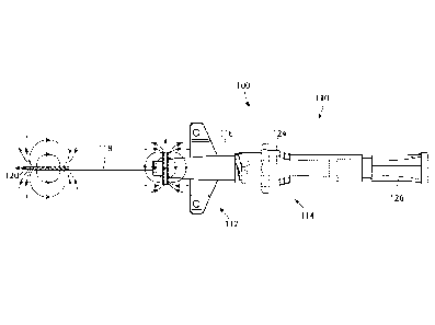
[0038] Referring now to Figs. 4 and 5, an embodiment of a medical device 100
comprising a
catheter assembly 110 is shown. The catheter assembly 110 includes a catheter
adapter
subassembly 112 and a needle subassembly 114. The catheter adapter subassembly
112 further
includes a catheter adapter 116, catheter hub (not shown) and catheter tubing
118. The needle
CA 03025798 2018-11-27
WO 2017/210017
PCT/US2017/033984
6
subassembly 114 further includes a needle 120, connected to a needle hub (not
shown),
disposed within a needle hub 124 and a vent plug 126. In the embodiment shown
in Figures 4
and 5, the catheter adapter subassembly 112 includes a permanent magnet
element 132 and the
needle subassembly 114 includes a magnetizable feature 130, in particular on
the needle 120.
According to an alternative embodiment (not shown), this configuration is
reversed wherein
the permanent magnet element 132 is on the needle subassembly 114, in
particular on the
needle 120, and the magnetizable feature 130 is on the catheter adapter
subassembly 112.
[0039] The use of a permanent magnet element on the catheter adapter
subassembly 112 and a
magnetizable feature on the needle subassembly 114 provides the ability to
calculate the
catheter tip position and the needle tip position based on known geometry
relative to the
position of permanent magnet element 132 on the catheter adapter subassembly
112 from
which a calculated catheter tip position and a calculated needle tip position
can be determined.
The permanent magnet element 132 provides a static magnetic field, while the
magnetizable
feature 130 on the needle 120 can be magnetized with an externally applied
magnetic field
prior to insertion of the needle 120 into the patient.
[0040] In the embodiment shown in Figures 4 and 5, the magnetizable feature
130 is on the
needle 120, and the catheter adapter subassembly 112 includes the permanent
magnet element
132. The magnetizable feature 130 on the needle 120 can be provided in a
variety of ways. In
one embodiment, the needle 120 is made from a magnetizable material, for
example, a steel
material that has a magnetic permeability that permits the needle 120 to be
magnetized by
application of an external magnetic field. Stainless steel that is typically
used to manufacture
hypodermic needles for medical use, for example, type 304 stainless steel, may
not have the
magnetic permeability to be magnetized and used in a device according one or
more
embodiments. Type 304 stainless steel is an austenitic steel comprising at
least 18%
chromium, 8% nickel, and a maximum of 0.08% carbon. Type 316 stainless steel
is also used
in the manufacture of hypodermic needles, and type 316 stainless steel is also
austenitic and
non-magnetic. The nickel content of type 316 stainless steel is typically
higher than type 304
stainless steel, and type 316 stainless steel also includes the addition of
molybdenum.
According to one or more embodiments, the needle 120 is made from martensitic
or ferritic
stainless steels, for example, type 420 or type 430 stainless steel.
[0041] In one or more embodiments, the magnetizable feature 130 on the needle
comprises a
separate feature on the needle 120. Referring now to Fig. 6A, in one
embodiment, needle
CA 03025798 2018-11-27
WO 2017/210017 PCT/US2017/033984
7
adhesive 140 is placed on a proximal end 121 of the needle 120, which can be
used to secure
the needle 120 to the hub within the needle chamber 24. The needle adhesive
140 can be any
suitable adhesive such as a curable glue containing magnetizable nanoparticles
such as
magnetizable metal nanoparticles or magnetizable metal oxide nanoparticles.
The magnetizable
metal can include iron, cobalt, nickel and alloys of iron, cobalt, and nickel.
According to one
or more embodiments, the size of the magnetic nanoparticles is in the range of
about 1
nanometer (nm) to about 100 nm. In one embodiment, adhesive is a light-curable
glue, and in
another embodiment, the adhesive is a heat-curable glue.
[0042] Referring now to Fig. 6B, an embodiment is shown in which the
magnetizable feature
is a needle ferrule 142 adjacent the distal tip 123 of the needle 120. The
needle ferrule 142 is
made from a magnetizable metal such as martensitic or ferritic stainless
steels, for example,
type 420 or type 430 stainless steel. The needle ferrule 142 provides at least
a localized area of
increased outer diameter. As used herein, the term "ferrule" refers to a
separate member
attached to the shank portion the needle 120, providing at least a localized
area of increased
outer diameter. The term "ferrule" includes a construction wherein the ferrule
comprises an
integral part of the needle, defining a one-piece monolithic construction
composed of both the
needle 120 and the needle ferrule 142, as well as a construction in which the
needle ferrule 142
is a piece added to the needle by crimping the needle ferrule 142 onto the
shank of the needle
120.
[0043] Referring now to Figure 6C, an embodiment is shown in which the
magnetizable
feature is a spot weld 144 adjacent the distal tip 123 of the needle 120. The
spot weld 144 can
be made from a magnetizable metal such as martensitic or ferritic stainless
steels, for example,
type 420 or type 430 stainless steel.
[0044] Referring now to Figure 6D, an embodiment is shown in which the
magnetizable
feature is a needle safety element, for example, a metal clip, specifically, a
metal cannula
safety clip 146 adjacent the distal tip 123 of the needle 120. The metal clip
146 can be made
from a magnetizable metal such as martensitic or ferritic stainless steels,
for example, type 420
or type 430 stainless steel. In other embodiments, the needle safety element
can be embodied
in other forms, for example, a spring, a plastic housing including a
magnetizable feature, or
other suitable safety elements. According to one or more embodiments, the
safety element
can be made from a materials that are not magnetic or magnetizable and include
a
magnetizable or magnetic material.
CA 03025798 2018-11-27
WO 2017/210017 PCT/US2017/033984
8
[0045] Referring now to Figure 6E, an embodiment is shown in which the
magnetizable
feature is a notch 148 in the needle 120, adjacent the distal tip 123 of the
needle 120. The
notch 148 can include an insert made from a magnetizable metal such as
martensitic or ferritic
stainless steels, for example, type 420 or type 430 stainless steel. The
insert fits inside the
notch 148 to completely or partially fill the notch 148. According to one or
more
embodiments, the insert can be a permanent magnet, magnetic adhesive or other
magnetic
material. The notch can be partially filled to occupy one-half the length of
the notch 148.
[0046] Referring now to Figure 7, an embodiment of a medical device 200
comprising a
catheter assembly 210 is shown. The catheter assembly 210 includes a catheter
adapter
subassembly 212 and a needle subassembly 214. The catheter adapter subassembly
212
includes a catheter adapter 216, catheter hub (not shown) and catheter tubing
218, and the
needle subassembly 214 further includes a needle 220 connected to the needle
hub 224,
disposed within a needle hub 224 and a vent plug 226. In the embodiment shown
in Figure 7,
the catheter adapter subassembly 212 includes a permanent magnet element 232
and the needle
.. subassembly 214 includes a magnetizable feature 230, in particular on the
needle 220. In the
specific embodiment shown, the catheter adapter subassembly 212 includes the
catheter tubing
218 and a catheter adapter 216, and a magnetic adhesive 240 attaches the
catheter tubing 218
to the catheter adapter 216. The magnetic adhesive 240 can be any suitable
adhesive such as a
curable glue containing magnetizable nanoparticles such as magnetizable metal
nanoparticles
or magnetizable metal oxide nanoparticles. The magnetizable metal can include
iron, cobalt,
nickel and alloys of iron, cobalt, and nickel. According to one or more
embodiments, the size
of the magnetic nanoparticles is in the range of about 1 nanometer (nm) to
about 100 nm. In
one embodiment, adhesive is a light-curable glue, and in another embodiment,
the adhesive is a
heat-curable glue.
[0047] Referring now to Figures 8 and 9, an embodiment of a medical device 300
comprising a
catheter assembly 310 is shown. The catheter assembly 310 includes a catheter
adapter
subassembly 312 and a needle subassembly 314. The catheter adapter subassembly
312
includes a catheter adapter 316, catheter hub (not shown) and catheter tubing
318, and the
needle subassembly 314 further includes a needle 320 connected to the needle
hub 324,
disposed within a needle chamber 324 and a vent plug 326. In the embodiment
shown in
Figures 8 and 9, the catheter adapter subassembly 312 includes a magnetizable
feature 330 and
the needle subassembly 314 includes a permanent magnet element 332.
CA 03025798 2018-11-27
WO 2017/210017 PCT/US2017/033984
9
[0048] Figures 10A-10D show various configurations for providing the
magnetizable feature
330 on the catheter adapter subassembly 312. In Figure 10A, a securement
element in the form
of a mandrel 342, which can be a conical mandrel for connecting the catheter
tubing 318 to the
catheter adapter 316, can be the magnetizable feature. According to one or
more
embodiments, the mandrel 342 is includes or is manufactured from martensitic
or ferritic
stainless steels, for example, type 420 or type 430 stainless steel. It will
be understood that in
Figure 10A, the mandrel 342 is protruding from the catheter adapter 316. In
other
embodiments (not shown), the mandrel 342 can be recessed within the catheter
adapter 316.
[0049] In Figure 10B, the securement element is shown in the form of a
catheter tubing
adhesive 340 is shown on the catheter tubing 318, which can be used to provide
the
magnetizable feature. The catheter tubing adhesive 340 can be any suitable
adhesive such as a
curable glue containing magnetizable nanoparticles such as magnetizable metal
nanoparticles
or magnetizable metal oxide nanoparticles. The magnetizable metal can include
iron, cobalt,
nickel and alloys of iron, cobalt, and nickel. According to one or more
embodiments, the size
of the magnetic nanoparticles is in the range of about 1 nanometer (nm) to
about 100 nm. In
one embodiment, adhesive is a light-curable glue, and in another embodiment,
the adhesive is a
heat-curable glue.
[0050] Figure 10C shows an embodiment in which a blood control component 346
shown
exploded from the catheter adapter subassembly 312 to provide the magnetizable
feature. In
.. the embodiment shown, the blood control component is a spring that includes
a magnetic
element or magnetizable material. According to one or more embodiments, the
blood control
component 346 includes martensitic or ferritic stainless steels, for example,
type 420 or type
430 stainless steel. The blood control component (metal spring for instance)
moves with the
catheter adapter until fully advanced. It will be appreciated that in use the
blood control
component 346 in the form of a spring would be located within the catheter
adapter 316, and
may not be visible, unless the catheter adapter was made from transparent
material.
[0051] Figure 10D shows an embodiment in which a magnetic element 348 on the
catheter
adapter 316 provides the magnetizable feature. According to one or more
embodiments, the
magnetic element 348 is includes or is made from martensitic or ferritic
stainless steels, for
example, type 420 or type 430 stainless steel. A magnetic wedge can provide a
controlled
position on the catheter adapter subassembly 312 to provide a fixed
measurement datum in a
fixed location relative to the catheter distal tip and a wedge having a highly
oriented grain
CA 03025798 2018-11-27
WO 2017/210017 PCT/US2017/033984
structure due to the cold forming used during is fabrication is also
beneficial in providing a
measurement datum. In one or more embodiments, the various alternatives
discussed with
respect to Figures 10A-10D may not have a position that is as precisely
controlled. In one or
more embodiments, the wedge, spring, and safety clip, would rely on catheter
tip calculated
5 projection rather than positional measurement.
[0052] In specific embodiments that include a magnetic adhesive, the adhesive
can include an
additive selected from a paramagnetic additive, a ferromagnetic additive and
combinations
thereof. The additive, according to one or more embodiments, includes a
component selected
from powdered iron, magnetic iron oxide, magnetic titanium oxide, magnetic
powdered steel,
10 .. and a magnetic iron alloy, and mixtures thereof. In specific
embodiments, the magnetic iron
alloy includes one or more of nickel, zinc, and copper. In specific
embodiments, the additive
further comprises a component selected from chromium, magnesium, molybdenum
and
combinations thereof.
[0053] In one or more embodiments, the needle subassembly includes the
permanent magnet
element, and the catheter adapter subassembly includes the magnetizable
feature, wherein the
magnetizable feature includes magnetizable catheter tubing. In one or more
embodiments, at
least a portion of the polyurethane tubing comprises a magnetizable
composition which is
magnetizable by an externally applied magnetic field, the magnetizable
composition
comprising a magnetic material dispersed in the polyurethane. In certain
embodiments, the
magnetic composition is dispersed in the polymeric material, for example,
polyurethane, which
forms the tubing. In a specific embodiment, the magnetizable composition
comprises an inner
layer surrounding the lumen of the catheter with an outer layer of non-
magnetizable polymeric
material, for example, polyurethane. In an alternative specific embodiment,
the layer of
magnetizable composition is an outer layer surrounding an inner layer of non-
magnetizable
polyurethane. In one or more embodiments, the magnetizable composition forms
longitudinal
segments of the catheter separated by longitudinal segments of non-
magnetizable polymeric
material, for example, polyurethane.
[0054] In any of the foregoing embodiments of the catheter, the magnetizable
composition
may further comprise a radiopaque component. Alternatively, in any of the
foregoing
embodiments, a non-magnetizable portion of catheter may comprise a radiopaque
component
[0055] It will be understood that the permanent magnet element or a magnetized
magnetizable
feature for the embodiments described above, the orientation of the magnetic
field can vary.
CA 03025798 2018-11-27
WO 2017/210017 PCT/US2017/033984
11
The permanent magnet element can have north and south poles on axis with the
catheter tubing
and with the needle. Alternatively, permanent magnet element or magnetized
magnetizable
feature can have north and south poles off axis with the catheter tubing and
with the needle, for
example, the north and south poles can be oriented perpendicular to the
longitudinal axis of the
catheter tubing and the needle. For example, in Figure 5, the magnetizable
feature 130 is
shown as being magnetized with the north pole 130N and south pole 130S of the
magnetizable
feature 130 oriented parallel of the longitudinal axis of the needle 120. The
permanent magnet
element 132 associated with the catheter adapter subassembly 112 is shown with
the north pole
132N and south pole 132S oriented perpendicular to the longitudinal axis of
the catheter tubing
.. 118. In the configuration shown in Figure 9, the permanent magnet element
332 and the
magnetizable feature 330, which has been magnetized, are shown with the poles
330N, 330S,
332N and 332S oriented parallel to the longitudinal axis of the needle 320 and
the catheter
tubing 318. Other variants are possible such as the permanent magnet element
and the
magnetizable feature which has been magnetized having their north and south
poles both
oriented perpendicular or orthogonal to the longitudinal axis of the needle
and the catheter
tubing.
[0056] An example of a vascular access device including a catheter according
to any of the
foregoing embodiments described above is illustrated in Fig. 11. The vascular
access device
500 shown in Figure 11 comprises a catheter adapter subassembly 512 including
a catheter
adapter body 516 and a catheter tubing 518 and a permanent magnet element 532.
A needle
(not shown) within the catheter tubing includes magnetizable feature 530,
which has been
magnetized by application of an external magnetic field and can be any of the
magnetizable
features described herein. Magnetizing the magnetizable feature 530 with an
externally
applied magnetic field creates a magnetic field 515 in the region of
magnetizable feature 530.
[0057] The vascular access device 500 may include a lateral access port 556
and may be
connected to a section of an extension tube 560 for establishing fluid
communication between
an IV fluid source and the catheter tubing 518. In one or more embodiments,
the extension
tube 560 is built-in to reduce contamination and mechanical phlebitis by
eliminating
manipulation at the insertion site. In one or more embodiments, the extension
tube 560 is
compatible with high pressure injection. In one or more embodiments, the
extension tube 560
provides continuous confirmation of vessel access during advancement of the
catheter into the
patient vein.
CA 03025798 2018-11-27
WO 2017/210017 PCT/US2017/033984
12
[0058] In one or more embodiments, a needle 511 of a needle subassembly 514 is
inserted into
the lumen (not show) of the catheter tubing 518. The needle subassembly 514 is
shown as
including finger grips 584 positioned at the sides of the needle subassembly
514 to facilitate
various insertion techniques. In one or more embodiments, bumps may be present
on the
finger grip to indicate where to the user may grip the device for needle
removal. In one or
more embodiments, a thumb pad 585, having a gently convex surface, is provided
at the
proximal end of the needle subassembly 514. A flange 586, having a gently
convex surface, is
provided at the proximal end of the needle subassembly 514 to provide a finger
pad. A wing
member 570, thumb pad 585 and flange 586 may be utilized by the user during
insertion,
permitting the user to elect which insertion technique to employ.
[0059] In one or more embodiments, the needle subassembly 514 includes a
needle shield 580.
The needle shield 580 may be a design adapted to secure the tip of the needle
within the shield
after use. In one or more embodiments, the needle shield 580 may be activated
passively.
The needle tip is completely covered by the needle shield 580 in a fixed
position. In one or
more embodiments, a ferrule, crimp or other structure may be included near the
tip for
engagement with a needle shield in certain applications.
[0060] A push tab 581 may be provided to facilitate catheter advancement
during insertion.
The push tab 581 also allows for one-handed or two-handed advancement. In one
or more
embodiments, the push tab 581 is removed with the needle shield 580. A clamp
582 may also
be included on the extension tubing to prevent blood flow when replacing the
access port.
[0061] In one or more embodiments, the vascular access device 500 further
includes a first luer
access 572 and a second luer access 573 in fluid communication with the
extension tube 560, a
blood control split septum 574 associated with the first luer access 572, and
an air vent 576
associated with the second luer access 573. Split septum 574 allows for a
reduction in
catheter-related bloodstream infection (CRBSI) while providing unrestricted
flow and a
straight fluid path and functions as a blood control septum. In one or more
embodiments, the
split septum 574 may be located in an internal cavity of the catheter adapter
or on the distal end
of the catheter adapter. In yet another embodiment, the split septum 574 may
be located on a
distal end of the extension tube 560. The air vent 576 allows air to escape
from the system
during insertion, providing continuous confirmation of vascular access while
preventing
leakage of blood from the system during insertion. In one or more embodiments,
the air vent
576 may be at the distal end of extension tube 560.
CA 03025798 2018-11-27
WO 2017/210017 PCT/US2017/033984
13
[0062] Another aspect of the disclosure pertains to a system for determining
catheter tip
location when the catheter tubing is inserted in a patient. According to one
or more
embodiments, a system provides a way to independently measure the cannula
tubing tip
location by measuring the location and vector of the permanent magnet, and
calculating and
.. predicting the catheter tip location relative to the position of the
magnetic sensor(s) on an
ultrasound probe and the ultrasound information transmitted from the sensors
on the ultrasound
probe. A permanent magnet on a device with north and south poles on axis with
the catheter
and needle and a known geometrical relationship to one or more features fixed
on the catheter
assembly provides a measurement datum that is measureable by the ultrasound
probe magnetic
sensors. From the measurement datum based on the one or more features on the
catheter
assembly, the direction vector and position of the catheter tip, needle tip or
other features can
be calculated. A magnetized magnetizable needle or feature on the needle can
then be used to
independently measure the position feature and calculate the position of the
needle tip. The
calculated position of the needle tip or feature on the needle can then be
compared relative to
.. the calculated position of the catheter tip to provide more specific
information related to the
catheter placement process, such as needle and catheter tip position relative
to the patient's
anatomy. This information can be used to determine (a) if the catheter is
properly seated and
ready for insertion (i.e., no over the bevel condition), (b) when the needle
tip is in the "hooded"
position (needle tip just inside of the catheter tip), and (c) and (d) when
the catheter is
advanced to specific distances and at angles suggesting successful placement
in the vein.
[0063] Referring now to Figures 12A-D, an embodiment of a medical device 600
comprising a
catheter assembly 610 is shown. The catheter assembly 610 includes a catheter
adapter
subassembly 612 and a needle subassembly 614. The catheter subassembly 612
includes a
catheter adapter 616, catheter hub (not shown) and catheter tubing 618 having
a distal catheter
tip, and the needle subassembly 614 further includes a needle 620 having a
needle distal tip
623 connected to a needle hub 624, and a vent plug 626. In the embodiment
shown in Figures
12A-D, the catheter adapter subassembly 612 includes a permanent magnet
element 632 and
the needle subassembly 614 includes a magnetizable feature 630. Fig. 12B shows
the catheter
assembly 610 in 12A in when the needle distal tip 623 is in the "hooded"
position where the
needle distal tip 623 is just inside of the catheter distal tip 619. Since the
dimensions of the
components of the needle subassembly 614 are fixed and known, placement of the
permanent
magnet element 632 provides a known geometrical relationship, for example,
distance and
CA 03025798 2018-11-27
WO 2017/210017 PCT/US2017/033984
14
angular position, with respect to one or more features fixed on the catheter
assembly, which
provides a measurement datum 633.
[0064] Referring now to Figure 12C, the catheter adapter subassembly 612 has
been advanced
in distal direction (toward the patient and away from the clinician), and the
measurement
datum 633 can be used to determine the distance and angular movement of the
needle 620 with
respect to the measurement datum 633. Similarly, if the catheter tubing 618 or
other part of the
catheter adapter subassembly 612 includes a magnetizable feature, and the
needle subassembly
614 includes a permanent magnet, the distance and the angular movement of the
catheter
tubing 618 can be determined with respect to the measurement datum. Figure 12C
shows that
the needle 620 has moved a distance D1, and the magnetizable feature 630 has
moved a
distance D1 from the catheter distal tip 619. In Figure 12D, the needle
subassembly 614 has
moved in a proximal direction (towards the clinician) for a distance D2, and
the magnetizable
feature 630 is now at a distance D2 from the catheter distal tip 619. Each
sequential movement
of either a permanent magnet element or magnetized magnetizable feature on a
needle and/or
the cannula can be measured and tracked using an ultrasound system.
[0065] The location of the magnetized magnetic feature or permanent magnet on
a needle or
cannula tubing can be accomplished by using a magnetometer to determine the
strength of the
magnetic field and its direction. As used herein, "magnetometer" refers to a
device that
detects a magnetic field. In specific embodiments, magnetometers may measure
the strength of
a magnetic field. When invasive needle or catheter is magnetic and produces a
known
magnetic field B at a given distance x through tissue of permeability pr, a
mathematical
correlation between the two i.e. x = f(B, pr) can be derived. In an
embodiment, three different
magnetometers are arranged in a three-dimensional grid array, orthogonal to
each other, are
used, and a three-dimensional (3D) correlation can be derived where I = f(B,
pi), where i = x or
y or z along three axes. Such correlation can be extended to an array of 3-
dimensional (3-D)
magnetometers to obtain the precise distance to the magnetized catheter or
vascular access
device from the array of 3D magnetometers. If the location of the array of 3D
magnetometers
is known in reference to the ultrasound sensor, then the precise location of
the magnetized
device with respect to the ultrasound sensor can be calculated. An inferred
image of the device
can then be created and superimposed over the ultrasound image and displayed.
An exemplary
magnetic sensing method using magnetometers and a lookup table instead of a
mathematical
function to determine the location of a magnetized invasive device from the
magnetic field
CA 03025798 2018-11-27
WO 2017/210017 PCT/US2017/033984
strength measured outside the body using magnetometers is shown and described
in United
States Patent Application Publication Number US20140257080 Al. The method
described in
US20140257080 Al can be adapted as described herein, for example, a three-
dimensional (3D)
correlation is from a mathematical function, and the correlation is extended
to an array of 3-
5 dimensional (3-D) magnetometers, one of the magnetometers outside the
patient's body, to
obtain the precise distance to the magnetized catheter or vascular access
device from the array
of 3D magnetometers. Another exemplary method of referencing the magnetometers
with
respect to an ultrasound probe is described in PCT Patent Application
Publication Number
W02013034175 Al, which can be adapted as described herein. For example, as
shown in Fig.
10 13, an ultrasound system 700 is shown including a catheter adapter
subassembly 712
comprising a magnetizable feature 732 that has been magnetized as described
herein is shown
inside of a patient's body 800. It will be appreciated that the sizes shown
are not to proportion
and the sizes of the catheter adapter subassembly 712 and the magnetizable
feature 732 are
exaggerated in size to illustrate these elements more clearly. A magnetometric
detector 711
15 .. comprising an array of magnetometers 720 (which can be housed in a probe
of a ultrasound
system, not shown) arranged in a 3-D array can be used to sense the magnetic
field 714
together with the terrestrial magnetic field and any other background magnetic
field. The
magnetometric detector 711 is in communication with an ultrasound processor
730 adapted to
determine from the detected field the position and orientation of the
magnetizable feature 732
relative to the magnetometric detector 711. This magnetically detected
position is then
displayed on a display 750 together with the ultrasound image.
[0066] The ultrasound system 700 can be a standard two dimensional B-mode
ultrasound
system with a standard ultrasound probe modified by the provision of the
magnetometric
detector 711. The ultrasound processor 730, which can be connected to the
ultrasound probe
via a cable 735, sends electrical signals to the magnetometric detector 711 to
cause it to
generate ultrasound pulses and interpreting the raw data received from the
transducer probe
housing the magnetometric detector 711, which represents echoes from the
patient's body, to
assemble it into an image of the patient's tissue.
[0067] The magnetometric detector 711 can be attached to the ultrasound probe
and can be
battery powered or powered from the ultrasound system. In specific
embodiments,
positioning elements are provided on the magnetometric detector 711 to ensure
that it is always
attached in the same well-defined position and orientation. The magnetometric
detector 711
CA 03025798 2018-11-27
WO 2017/210017 PCT/US2017/033984
16
can connected by a wireless connection to a base unit 740 which is in wireless
or wired (e.g.
USB) communication with the ultrasound processor 730 and the display 750. The
base unit
740 can be integrated with, or some of its functions performed by, the
ultrasound processor
730 or the magnetometric detector 711.
.. [0068] The base unit 740 receives normalized measurements from
magnetometric detector 711
and calculates the position, or optionally the position and orientation, of
magnetizable feature
732. The base unit 740 can also receive additional information such as the
state of charge of
the magnetometric detector's battery and information can be sent from the base
unit 740 to the
magnetometric detector 711, such as configuration information. The base unit
740 forwards the
results of its calculations, i.e. the position and, optionally, orientation,
to the ultrasound
processor 730 for inclusion in the displayed ultrasound image of an image of
the catheter.
[0069] In one or more embodiments, the base unit 740 can be integrated into
the ultrasound
system 700 with the ultrasound processor 730 and the magnetometric detector
711 being in
direct communication with the ultrasound system 700 either via wireless link
or using the same
physical cable 735.
[0070] Thus, in one or more embodiments, the magnetizable feature is
magnetized using any
suitable device that can produce an magnetic field to magnetize a needle or
medical device to
produce a magnetic field B at a distance x through tissue of permeability pr,
and the correlation
is calculated as x = f(B, pr). In one or more embodiments, three magnetometers
720 are placed
orthogonally to each other are used to derive a 3-dimensional correlation I =
f(Bõ pr), wherein i
= x or y or z along three axes. In a specific embodiment, the distance from
the magnetizable
feature to the 3-dimensional array of magnetometers is calculated. In a
further specific
embodiment, location of the array of magnetometers in reference to an
ultrasound sensor of an
ultrasound imaging system is used to calculate a location of the magnetizable
feature with
respect to the ultrasound sensor. In another specific embodiment, the method
comprises
displaying an image of the magnetizable feature.
[0071] As described above with respect to Figures 12A-D, providing a permanent
magnet on
the needle subassembly and a magnetizable feature on the catheter subassembly
(or a reverse
configuration in which the magnetizable feature is on the needle subassembly
(e.g., the needle
or needle hub) and the permanent magnet is on the catheter subassembly)
relative positions of
a catheter tip and a needle cannula tip can be determined by utilizing an
ultrasound system
including a three dimensional array of magnetometers. Relative positional
changes of the
CA 03025798 2018-11-27
WO 2017/210017 PCT/US2017/033984
17
catheter adapter subassembly and needle subassembly can be determined in three
axes, x, y and
z, as well relative changes in angular motion oi of the catheter adapter
subassembly and the
needle subassembly based on based on a known geometrical relationship to one
or more
features fixed on the catheter adapter assembly or needle subassembly, which
provides a
measurement datum that is measureable by the ultrasound probe magnetic
sensors. From the
measurement datum based on the one or more features, the direction vector and
position of the
catheter tip or other features can be calculated based on a 3-dimensional
correlation I = f(Bõ
pi), wherein i = x or y or z along three axes or predict relative motion
between the needle hub
and catheter adapter subassemblies. Understanding the relative position and
motion of these
two subassemblies can be used to inform a clinician of procedurally important
states of the
insertion process, such as when the needle tip reaches the vein, when the
catheter tip reaches
the vein, when the catheter is advanced to cover the needle tip ("hooding the
catheter") and
thereby safe for further advancement.
[0072] Another aspect of the disclosure comprises methods that can be
practiced according to
any of the previously described systems. A method for determining a relative
position of a
catheter tip and a needle cannula tip, the method includes providing a
catheter having a
catheter distal tip and a needle having a needle distal tip, associating a
permanent magnet
element with one of the catheter and the needle, associating a magnetizable
feature with the
other of the catheter and the needle, obtaining a measured position of the
permanent magnet,
obtaining a measured position of the magnetizable feature to obtain a
calculated position of the
catheter distal tip, and comparing the calculated position of the catheter
distal tip with the
calculated position of the needle distal tip to determine the relative
position of the catheter
distal tip and the needle distal tip. In one embodiment, the needle includes
the magnetizable
feature and the catheter includes the permanent magnet and the relative
position of the catheter
distal tip and the needle distal tip indicates that the catheter is properly
seated on the needle. In
another embodiment, the relative position of the catheter distal tip and the
needle distal tip
indicates that the catheter is in a hooded position on the needle. In another
embodiment, the
relative position of the catheter distal tip and the needle distal tip
indicates that the catheter
distal tip is advanced a specific distance or angle.
[0073] In one embodiment of the method, the catheter adapter subassembly
includes the
magnetizable feature and the needle subassembly includes the permanent magnet,
and relative
movement of the catheter adapter subassembly and needle subassembly is
determined by a
CA 03025798 2018-11-27
WO 2017/210017 PCT/US2017/033984
18
three-dimensional array of magnetometers positioned in proximity to at least
one of the
permanent magnet the magnetizable feature. In one embodiment of the method,
the method
includes magnetizing the magnetizable feature by applying an external magnetic
field to the
magnetizable feature. In one embodiment, the three-dimensional array of
magnetometers is
part of an ultrasound system, and the ultrasound system derives a three-
dimensional correlation
to obtain a distance from the grid array to the magnetizable feature or
permanent magnet. In
another embodiment, the three-dimensional correlation is determined by the
function I = f(B,
pi), where i = x or y or z along three axes, x, y and z are distances in three
planes, B is a known
magnetic field produced by the permanent magnet or magnetizable feature, and
pr is magnetic
permeability.
[0074] In another embodiment of the method, the catheter adapter subassembly
includes the
permanent magnet and the needle subassembly includes the magnetizable feature,
and relative
movement of the catheter adapter subassembly and needle subassembly is
determined by a
three-dimensional array of magnetometers positioned in proximity to at least
one of the
permanent magnet the magnetizable feature. In one embodiment, the method
includes
magnetizing the magnetizable feature by applying an external magnetic field to
the
magnetizable feature. According to another embodiment, the three-dimensional
array of
magnetometers is part of an ultrasound system, and the ultrasound system
derives a three-
dimensional correlation to obtain a distance from the grid array to the
magnetizable feature or
permanent magnet. In one embodiment, the three-dimensional correlation is
determined by the
function I = f(B, pr), where i = x or y or z along three axes, x, y and z are
distances in three
planes, B is a known magnetic field produced by the permanent magnet or
magnetizable
feature, and pr is magnetic permeability.
[0075] Another aspect of the disclosure pertains to a catheter adapter
subassembly comprising
a magnetic feature selected from the group consisting of a metal mandrel for
connecting
catheter tubing to the hub, a catheter tubing adhesive, a blood control
component of the
catheter adapter subassembly, and a magnetic wedge on the catheter adapter
body. The
catheter adapter subassembly may further comprise magnetic catheter tubing.
According to an
embodiment, the metal mandrel comprises austenitic stainless steel.
[0076] Although the disclosure herein provided a description with reference to
particular
embodiments, it is to be understood that these embodiments are merely
illustrative of the
principles and applications of the disclosure. It will be apparent to those
skilled in the art that
CA 03025798 2018-11-27
WO 2017/210017
PCT/US2017/033984
19
various modifications and variations can be made to the devices, methods and
systems
described in the of the present disclosure without departing from the spirit
and scope thereof.
Thus, it is intended that the present disclosure include modifications and
variations that are
within the scope of the appended claims and their equivalents.