Note: Descriptions are shown in the official language in which they were submitted.
CA 03026520 2018-12-04
CA Application
National Entry of PCT/JP2017/021558
Blakes Ref.: 14934/00002
Description
Title of Invention: SITE-SPECIFIC RADIOISOTOPE-LABELED ANTIBODY
USING IgG-BINDING PEPTIDE
Technical Field
[0001]
The present invention relates to an IgG-binding peptide comprising a ligand
capable of
binding to a radioactive metal nuclide, an IgG-binding peptide labeled with a
radioactive
metal nuclide, a conjugate of the IgG-binding peptide and IgG, and a
radionuclide imaging
agent or a diagnostic agent for cancer comprising the IgG-binding peptide or
the conjugate,
etc.
Background Art
[0002]
Antibodies have conventionally been often utilized in the detection of target
molecules
in various research and development activities, and are also of great
industrial importance as
detection reagents or diagnostic drugs. The antibodies have also received
attention as drugs
for the treatment of diseases because of their specificity for target
molecules.
[0003]
In order to impart a function to antibodies, labeling with radioisotopes is
practiced via
iodation or addition of a chelating compound (Non Patent Literature 1), etc.
These
modifications have been typically performed so far via a lysine amino group, a
cysteine thiol
group, and an activated carboxyl group, etc. contained in antibodies. These
modifications
are specific for the functional groups, but are not site-specific. Therefore,
these approaches
have the problems of, for example, reduction in the activity of antibodies due
to the
modification or the like of the antigen-binding sites of the antibodies, and
difficult control of
the number of compounds to be bound.
[0004]
1
23521198.1
CA 03026520 2018-12-04
CA Application
National Entry of PCT/JP2017/021558
Blakes Ref.: 14934/00002
In order to overcome these problems, antibody modification has been practiced
using
antibodies having a particular site-specifically introduced functional group.
For example,
modification at a particular site is achieved by introducing a non-natural
amino acid (Non
Patent Literatures 2 to 4) or free cysteine (Non Patent Literatures 5 and 6)
to the particular site
by genetic manipulation. Although site-specific antibody modification
techniques are under
development as mentioned above, these methods often require engineering
antibodies
themselves and are not always advantageous in light of reduction in the
functions of the
antibodies and high development cost in association with the engineering.
Citation List
Non Patent Literature
[0005]
Non Patent Literature 1: Rodwell, J. D. et al., Proceedings of the National
Academy of
Sciences of the United States of America, 1986, 83, pp. 2632-2636
.. Non Patent Literature 2: Axup, J. Y. et al., Proceedings of the National
Academy of Sciences
of the United States of America, 2012, 109, pp. 16101-16106
Non Patent Literature 3: Tian, F. et al., Proceedings of the National Academy
of Sciences of
the United States of America, 2014, 111, pp. 1766-1771
Non Patent Literature 4: Zimmerman, E. S. et al., Bioconjugate chemistry,
2014, 25, pp. 351-
361
Non Patent Literature 5: Shen, B. Q. et al., Nature biotechnology, 2012, 30,
pp. 184-189
Non Patent Literature 6: Bernardes, G. J. et al., Nature protocols, 2013, 8,
pp. 2079-2089
Summary of Invention
.. [0006]
Accordingly, there is a demand for methods that can modify antibodies
specifically
and conveniently.
[0007]
2
23521198.1
CA 03026520 2018-12-04
CA Application
National Entry of PCT/JP2017/021558
Blakes Ref.: 14934/00002
In order to solve the problems described above, the present inventors have
conducted
detailed studies on the position of each amino acid in an IgG-binding peptide
in a bound state
and the positional relationship of each amino acid with IgG Fc, on the basis
of the X-ray
crystallography of a conjugate of the IgG-binding peptide and the IgG Fc. The
present
inventors have further found that: an IgG-binding peptide site-specifically
modified with a
cross-linking agent can be prepared by introducing an amino acid capable of
binding to the
cross-linking agent to a peptide and modifying the amino acid with the cross-
linking agent;
and IgG can be modified using this IgG-binding peptide site-specifically
modified with a
cross-linking agent. Moreover, the present inventor has found that a conjugate
of an IgG-
binding peptide labeled with a radioactive metal nuclide and IgG can be used
as a diagnostic
agent for cancer. On the basis of the findings, the present invention has been
completed.
[0008]
Therefore, the present invention encompasses the following embodiments.
(1) A peptide which comprises an amino acid sequence consisting of 13 to 17
amino
acid residues represented by the following formula I:
(X1_3)-C-(X2)-H-(Xaa1)-G-(Xaa2)-L-V-W-C-(X1_3) (I)
wherein each X is independently any amino acid residue other than cysteine,
C is a cysteine residue,
H is a histidine residue,
Xaal is a lysine residue, a cysteine residue, an aspartic acid residue, a
glutamic acid residue,
2-aminosuberic acid, or diaminopropionic acid,
G is a glycine residue,
Xaa2 is a glutamic acid residue, a glutamine residue, or an asparagine
residue,
L is a leucine residue,
V is a valine residue, and
W is a tryptophan residue,
wherein the peptide is capable of binding to human IgG, and comprises a ligand
capable of
binding to a radioactive metal nuclide.
3
23521198.1
CA 03026520 2018-12-04
CA Application
National Entry of PCT/JP2017/021558
Blakes Ref.: 14934/00002
(2) A peptide which comprises an amino acid sequence consisting of 13 to 17
amino acid
residues represented by the following formula I:
(X1_3)-C-(X2)-H-(Xaa1)-G-(Xaa2)-L-V-W-C-(X1_3) (I)
wherein each X is independently any amino acid residue other than cysteine,
C is a cysteine residue,
H is a histidine residue,
Xaa1 is a lysine residue, a cysteine residue, an aspartic acid residue, a
glutamic acid residue,
2-aminosuberic acid, or diaminopropionic acid,
G is a glycine residue,
Xaa2 is a glutamic acid residue, a glutamine residue, or an asparagine
residue,
L is a leucine residue,
V is a valine residue, and
W is a tryptophan residue,
wherein the peptide is capable of binding to human IgG, and is labeled with a
radioactive
metal nuclide.
(3) A peptide which comprises an amino acid sequence consisting of 13 amino
acid residues
represented by the following formula V:
D-C-(Xaa2)-(Xaa3)-(Xaa4)-(Xaa1)-G-(Xaa5)-L-(Xaa6)-W-C-T (V)
wherein
D is an aspartic acid residue,
C is a cysteine residue,
G is a glycine residue,
L is a leucine residue,
W is a tryptophan residue,
T is a threonine residue,
Xaal is a lysine residue, a cysteine residue, an aspartic acid residue, a
glutamic acid residue,
2-aminosuberic acid, or diaminopropionic acid,
Xaa2 is an alanine residue, a serine residue or a threonine residue,
Xaa3 is a tryptophan residue or a tyrosine residue,
4
23521198.1
CA 03026520 2018-12-04
CA Application
National Entry of PCT/JP2017/021558
Blakes Ref.: 14934/00002
Xaa4 is a histidine residue, an arginine residue, a serine residue or a
threonine residue,
Xaa5 is a glutamic acid residue, a glutamine residue, an asparagine residue,
an arginine
residue, or an aspartic acid residue, and
Xaa6 is an isoleucine residue or a valine residue,
wherein the peptide is capable of binding to human IgG, and wherein a
radioactive metal
nuclide is bound to the peptide via a ligand.
(4) A conjugate of the peptide and IgG, wherein the conjugate is formed
through the cross-
linking reaction of the above described peptide modified with the cross-
linking agent with the
IgG.
(5) A radionuclide imaging agent or a diagnostic agent for cancer comprising
the peptide
according to (2) or (3), or the conjugate according to (4), wherein a
radioactive metal nuclide
is bound to the peptide.
(6) A method for determining the presence or absence of cancer in a subject,
comprising the
steps of:
reacting a sample obtained from the subject with the peptide according to (2)
or (3), or
the conjugate according to (4), wherein a radioactive metal nuclide is bound
to the peptide;
measuring a level or presence of radioactivity derived from the radioactive
metal
nuclide in the sample; and
determining the presence or absence of cancer in the subject on the basis of
the level or
presence of radioactivity.
(7) A method for determining the presence or absence of cancer in a subject,
comprising the
steps of:
administering the peptide according to (2) or (3), or the conjugate according
to (4) to
the subject, wherein a radioactive metal nuclide is bound to the peptide;
measuring a level or presence of radioactivity derived from the radioactive
metal
nuclide in the subject; and
determining the presence or absence of cancer in the subject on the basis of
the level or
presence of radioactivity.
5
23521198.1
CA 03026520 2018-12-04
CA Application
National Entry of PCT/JP2017/021558
Blakes Ref.: 14934/00002
This description includes all or part of the contents disclosed in Japanese
Patent
Applications No. 2016-117395, and No.2016-227025, to which the present
application claims
the priority.
[0009]
The IgG-binding peptide of the present invention can be easily bound with a
radioactive metal nuclide. Therefore, IgG can be labeled specifically and
conveniently with
the radioactive metal nuclide by using the IgG-binding peptide of the present
invention.
Furthermore, the IgG-binding peptide of the present invention eliminates the
need of altering
the sequence of the antibody molecule and therefore does not cause reduction
in the functions
of the antibody molecule associated with genetic engineering. Moreover, the
IgG-binding
peptide of the present invention eliminates the need of reaction of directly
labeling IgG with a
radioactive metal nuclide, which has conventionally been required, and does
not cause
reduction in the functions of the antibody caused by the reaction.
Brief Description of Drawings
[0010]
[Figure 11 Figure 1(A) shows the structure of a conjugate of an IgG-binding
peptide (C35A-
3/15: DCAYHRGELVWCT (SEQ ID NO: 33)) and human IgG Fc. The IgG-binding
peptide is depicted as a space-filling model, the IgG Fc is depicted as a
ribbon model, and the
sugar chain of the Fc is depicted as a wire model. Figure 1(B) shows a model
of the cross-
linked structure between an IgG-binding peptide (C35A-3/15(R8K): DCAYHKGELVWCT
(SEQ ID NO: 34)) modified with DSG and IgG Fc. The main chain of the peptide
is
depicted as a ribbon model. Peptide-Lys8 represents the lysine residue at
position 6 of
C35A-3/15(R8K), and peptide-Tyr6-Gly9 represents the tyrosine residue at
position 4 to the
glycine residue at position 7 of C35A-3/15(R8K). Fc-Lys248 represents Lys248
of Fc
according to the EU numbering, and Fc-Pro247-Asp249 represents Pro247 to
Asp249 of Fc
according to the EU numbering.
[Figure 2] Figure 2 shows results of SDS-PAGE (A) and Western blot (B) of
mixtures of
labeled IgG-binding peptides and various proteins. In the figure, DSG
represents that an
6
23521198.1
CA 03026520 2018-12-04
CA Application
National Entry of PCT/JP2017/021558
Blakes Ref.: 14934/00002
IgG-binding peptides reacted with DSG (disuccinimidyl glutarate) were
subjected, and DSS
represents that an IgG-binding peptides reacted with DSS (disuccinimidyl
suberate) were
subjected. In the figure, hIgG represents human IgG, hIgA represents human
IgA, and HSA
represents human serum albumin.
[Figure 3] Figure 3 shows results of study for reaction molar ratio (A) and
reaction time (B)
by ELISA for the reaction between a labeled IgG-binding peptide and IgG. DSS
R8K 0 min
represents that Tris-HCl (pH 7.0) was added to a labeling IgG-binding peptide
at a 10-fold
molar ratio to IgG, and the mixture was added to wells after blocking of a NHS
group. No
DSS R8K represents that a DSS-unbound biotinylated IgG-binding (R8K) peptide
was used.
no pep represents a control without the addition of the peptide.
[Figure 4] Figure 4 shows results of measuring the reactivity of a labeled IgG-
binding peptide
with each protein (hIgA, hIgG, and BSA (bovine serum albumin)) by use of size
exclusion
chromatography. Figure 4(A) shows results of measuring the reactivity of an
IgG-binding
peptide modified with DSS. Figure 4(B) shows results of measuring the
reactivity of an
IgG-binding peptide modified with DSG.
[Figure 5] Figure 5(A) shows results of liquid chromatography after adding a
DSG-modified
IgG-binding peptide dissolved in DMF to a human IgG Fc solution at a molar
ratio of 0.5, 1.0,
2.0, or 5.0, stirring the mixture, and then allowing them to react at room
temperature. Figure
5(B) shows change in the amounts of production of an unreacted form (peak 2),
an adduct of
one peptide (peak 3), and an adduct of two peptides (peak 4) when human IgG
and a DSG-
modified IgG-binding peptide were reacted at each molar ratio.
[Figure 61 Figure 6 shows change in the amounts of production of an unreacted
form (peak 2),
an adduct of one peptide (peak 3), and an adduct of two peptides (peak 4) 1,
5, 10, or 30
minutes after adding a DSG-modified IgG-binding peptide dissolved in DMF at a
molar ratio
of 1.0 to a human IgG Fc solution prepared at pH 4.0 (A), pH 5.5 (B), or pH
7.0 (C), stirring
the mixture, and then allowing them to react at room temperature.
[Figure 7] Figure 7 shows SPECT/CT images (A) and CT images (B) including a
tumor site 6
hours after administration of [1111n1 -labeled trastuzumab-1, and SPECT/CT
images (C)
including a tumor site 4 hours after administration of [1111n]-labeled
trastuzumab-2.
7
23521198.1
CA 03026520 2018-12-04
CA Application
National Entry of PCT/JP2017/021558
Blakes Ref.: 14934/00002
[Figure 8] Figure 8 shows SPECT/CT images including a tumor site 24 hours (A)
and 48
hours (B) after administration of [111In1-labeled trastuzumab-1, and SPECT/CT
images
including a tumor site 24 hours (C) and 48 hours (D) after administration of
[1111n]-labeled
trastuzumab-2.
[Figure 9] Figure 9 shows SPECT/CT images including the liver 6 hours after
administration
of [111-
1 ] labeled trastuzumab-1 (A) and 4 hours after administration of [1111n]-
labeled
trastuzumab-2 (B).
[Figure 101 Figure 10 shows a synthesis scheme of an IgG-binding peptide
having a SS cross-
linked structure via dichloropropanone, prepared in Example 9.
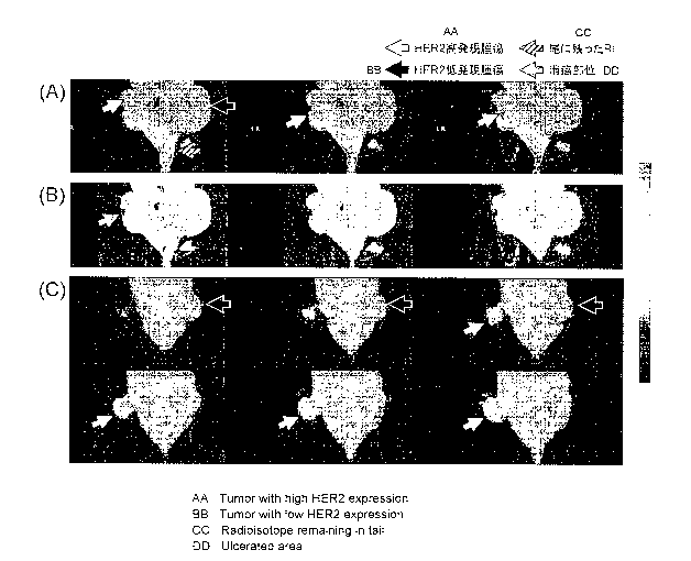
[Figure 111 Figure 11 shows PET images including a tumor site 6 hours, 24
hours, and 48
hours after administration of [89Zr]-labeled trastuzumab-1 or -2. The solid
line arrows depict
a tumor tissue with high expression of HER2, and the broken line arrows depict
a tumor tissue
with low expression of HERZ.
Description of Embodiments
[0011]
<IgG-binding peptide>
In one aspect, the present invention relates to an IgG-binding peptide
comprising a
ligand capable of binding to a radioactive metal nuclide. The position of the
ligand in the
IgG-binding peptide is not particularly limited. For example, the ligand can
be linked to the
N terminus or the C terminus, preferably the N terminus, of the IgG-binding
peptide. The
method for linking the ligand to the peptide is well known to those skilled in
the art. In the
case of linking the ligand to, for example, the N terminus of the IgG-binding
peptide, a
reactive group such as N-hydroxysuccinimide ester (NHS), an isothiocyano group
(ITC),
sulfonic acid chloride, carboxylic acid chloride, ethylene oxide, alkyl
chloride, an aldehyde
group, or carboxylic anhydride can be attached to the ligand and reacted with
the N-terminal
amino group of the IgG-binding peptide. Alternatively, the IgG-binding peptide
comprising
such a ligand may be directly synthesized by a well-known synthesis method or
the like.
[0012]
8
23521198.1
CA 03026520 2018-12-04
CA Application
National Entry of PCT/JP2017/021558
Blakes Ref.: 14934/00002
Examples of the ligand capable of binding to a radioactive metal nuclide that
may be
contained in the IgG-binding peptide of the present invention include, but are
not limited to,
chelating agents, for example, diethylenetriaminepentaacetic acid (DTPA),
deferoxamine,
1,4,7,10-tetraazacyclododecane-1,4,7,10-tetraacetic acid (DOTA), 1,4,7-
triazacyclononane-
1,4,7-triacetic acid (NOTA), ethylenediaminetetraacetic acid (EDTA),
ethylenediaminediacetic acid, triethylenetetraminehexaacetic acid (TTHA),
1,4,8,11-
tetraazacyclotetradecane-1,4,8,1 1-tetraacetic acid (TETA), dipyridoxyl
diphosphate (DPDP),
TPPS4, ethylenebishydroxyphenylglycine (EHPG), hexamethylenediaminetetraacetic
acid,
dimethylphosphinomethane (DMPE), methylenediphosphoric acid,
dimercaptosuccinic acid
(DMPA), and derivatives thereof.
[0013]
In one embodiment, a radioactive metal nuclide is bound to the IgG-binding
peptide.
Examples of the radioactive metal nuclide include 111/n (indium), 89Zr
(zirconium), 67/68Ga
(gallium), 99mTc (technetium), and 64Cu (copper), preferably 111In and 89Zr.
The radioactive
metal nuclide to be bound to the IgG-binding peptide can be selected depending
on the
purposes of the IgG-binding peptide and a conjugate of the IgG-binding peptide
and IgG
mentioned later. For example, 1111n, 89zr, 64Cu, 67/68u,-,a,
and 99mTc can be used for the
detection or diagnosis of cancer. For example, 89Zr and 64Cu can be used for
PET (positron
emission tomography), and 111In and 99mTc can be used for SPECT (single photon
emission
computed tomography).
[0014]
The radioactive metal nuclide may be bound directly to the IgG-binding
peptide, but is
preferably bound to the IgG-binding peptide via a ligand such as the chelating
agent. Those
skilled in the art can appropriately select a preferred combination of the
radioactive metal
nuclide and the ligand (see e.g., Hiroshi Sakurai and Yo Yokoyama ed., Housha
Yakuhingaku
Gairon (General Introduction to Radiation Medicine Science in English).
Examples thereof
include: 111In and DTPA; 89Zr and deferoxamine; 64Cu and DOTA or NOTA; 99mTc
and
dimethylphosphinomethane (DMPE), DTPA, methylenediphosphoric acid,
dimercaptosuccinic acid (DMPA), dithiosemicarbazone, or diaminoethanediol; and
67/68Ga
9
23521198.1
CA 03026520 2018-12-04
CA Application
National Entry of PCT/JP2017/021558
Blakes Ref.: 14934/00002
and deferoxamine or a DTPA derivative etc., preferably 111In and DTPA; 89Zr
and
deferoxamine; and 64Cu and DOTA or NOTA, more preferably 111In and DTPA; and
89Zr and
deferoxamine, further preferably 111In and DTPA.
[0015]
The IgG binding peptide of the present invention is described in detail below.
The "IgG" used in the present specification refers to IgG of a mammal, for
example, a
primate (such as a human and a chimpanzee), a laboratory animal (such as a
rat, a mouse, and
a rabbit), a livestock animal (such as a pig, cattle, a horse, sheep, and a
goat), or a pet animal
(such as a dog and a cat), preferably human IgG (IgG1, IgG2, IgG3 or IgG4). In
the present
specification, the IgG is more preferably human IgG1, IgG2, or IgG4, or rabbit
IgG,
particularly preferably human IgG1, IgG2, or IgG4.
[0016]
In one aspect, the present invention relates to a peptide which comprises an
amino acid
sequence consisting of 13 to 17 amino acid residues represented by the
following formula I
and is capable of binding to human IgG:
(X1_3)-C-(X2)-H-(Xaa1)-G-(Xaa2)-L-V-W-C-(X1_3) (I)
wherein each X is independently any amino acid residue other than cysteine,
C is a cysteine residue,
H is a histidine residue,
Xaa1 is a lysine residue, a cysteine residue, an aspartic acid residue, a
glutamic acid residue,
2-aminosuberic acid, or diaminopropionic acid,
G is a glycine residue,
Xaa2 is a glutamic acid residue, a glutamine residue, or an asparagine
residue,
L is a leucine residue,
V is a valine residue, and
W is a tryptophan residue.
[0017]
In the above formula, the term "X1_3" at the N terminus or the C terminus
means 1 to 3
consecutive independently selected arbitrary amino acid residues X other than
cysteine (C or
23521198.1
CA 03026520 2018-12-04
CA Application
National Entry of PC1/JP2017/021558
Blakes Ref.: 14934/00002
Cys). The constituting amino acid residues are the same or different residues
and preferably
consist of a sequence in all of the 3 residues are different from one another.
Likewise, X2
means two consecutive independently selected arbitrary amino acid residues X
other than
cysteine (C or Cys). The constituting amino acid residues are the same or
different residues
and preferably consist of a sequence in which the two consecutive amino acid
residues are
different residues.
[0018]
The two cysteine residues in the formula I can form a disulfide bond to form a
cyclic
peptide. The peptide of the formula I usually has a disulfide bond formed
between the two
cysteine residues on outer sides (other than Xaal, when Xaa 1 is cysteine).
Alternatively, in
the peptide of the formula I, sulfide groups in the two cysteine residues on
the outer sides may
be linked via a linker represented by the following formula:
[Formula 11
0
Iz,
In the above formula, the broken line moieties mean binding moieties to the
sulfide groups.
The linker is more stable against reduction reaction or the like than usual
disulfide bonds.
Therefore, this linker may be preferably used when using radioactive metal
nuclides which
may destabilize disulfide bond such as zirconium.
[0019]
This peptide can be prepared by a method comprising the step of mixing a
peptide
containing two or more, preferably two cysteine residues with a compound
represented by the
following formula:
[Formula 21
0
wherein 121 and R2 are each independently any halogen atom
to obtain a peptide in which sulfide groups in the two or more, preferably two
cysteine
residues are linked via a linker represented by the following formula:
11
23521198.1
CA 03026520 2018-12-04
CA Application
National Entry of PCT/JP2017/021558
Blakes Ref.: 14934/00002
[Formula 3]
0
1\ z
In the above formula, the broken line moieties mean binding moieties to the
sulfide groups.
[0020]
In the compound, R1 and R2 are each selected from the group consisting of
preferably
F, Cl, Br, and I, more preferably Cl, Br, and I. R1 and R2 are preferably the
same. More
preferably, both of R1 and R2 are Cl.
[0021]
Conditions for the mixing step in this method are not particularly limited as
long as the
conditions result in linking reaction between the cysteine residues of the
peptide. The
reaction can be performed, for example, by mixing the peptide and the compound
at room
temperature (such as approximately 15 C to 30 C) in an appropriate buffer, for
example, a
buffer solution containing guanidium chloride. The mixing step may be
performed by the
addition of a catalyst that accelerates the linking reaction in an appropriate
amount, if
necessary.
[0022]
The mixing ratio between the peptide and the compound in the mixing step of
this
method is not particularly limited. The molar ratio between the peptide and
the compound
can be, for example, 1:0.2 to 1:10, preferably 1:0.5 to 1:5 or 1:1 to 1:2.
[0023]
The mixing time (reaction time) in the mixing step is not limited as long as
the mixing
time results in the linking reaction between the cysteine residues of the
peptide. The mixing
time can be set to, for example, 1 minute to 5 hours, preferably 10 minutes to
2 hours or 15
minutes to 1 hour.
[0024]
This method may further comprise, if necessary, the step of purifying the
peptide
having linked cysteine residues by separating impurities, for example,
unreacted peptides and
compounds, from the mixture after the step described above. This step can be
performed by
12
23521198.1
CA 03026520 2018-12-04
CA Application
National Entry of PCT/JP2017/021558
Blakes Ref.: 14934/00002
a method known in the art, for example, chromatography such as gel filtration
chromatography, ion-exchange column chromatography, affinity chromatography,
reverse-
phase column chromatography, or HPLC.
[0025]
Peptides represented by the formula I' and the formula I" are given below,
wherein the
amino acid residues X in the amino acid sequence of the peptide of the formula
I are defined
in more detail.
[0026]
Specifically, the peptide represented by the formula I' comprises an amino
acid
sequence consisting of 13 to 17 amino acid residues represented by
(X1_3)-C-(X1)-Y-H-(Xaa1)-G-N-L-V-W-C-(X1_3) (I)
wherein each X is independently any amino acid residue other than cysteine,
C is a cysteine residue,
Y is a tyrosine residue,
H is a histidine residue,
Xaal is a lysine residue, a cysteine residue, an aspartic acid residue, a
glutamic acid residue,
2-aminosuberic acid, or diaminopropionic acid,
G is a glycine residue,
N is an asparagine residue,
L is a leucine residue,
V is a valine residue, and
W is a tryptophan residue.
[0027]
The peptide represented by the formula I" comprises an amino acid sequence
consisting of 13 to 17 amino acid residues represented by
(X1_3)-C-A-(X1)-H-(Xaa1)-G-E-L-V-W-C-(X1_3) (I")
wherein each X is independently any amino acid residue other than cysteine,
C is a cysteine residue,
A is an alanine residue,
13
23521198.1
CA 03026520 2018-12-04
CA Application
National Entry of PCT/JP2017/021558
Blakes Ref.: 14934/00002
H is a histidine residue,
Xaal is a lysine residue, a cysteine residue, an aspartic acid residue, a
glutamic acid residue,
2-aminosuberic acid, or diaminopropionic acid,
G is a glycine residue,
E is a glutamic acid residue,
L is a leucine residue,
V is a valine residue, and
W is a tryptophan residue.
[0028]
Also, a peptide represented by the formula II is given below, wherein the
amino acid
residues X in the amino acid sequence of the peptide of the formula I are
defined in more
detail.
[0029]
Specifically, the peptide represented by the formula II comprises an amino
acid
sequence consisting of 13 to 17 amino acid residues represented by
(X1_3)-C-(Xaa3)-(Xaa4)-H-(Xaa1)-G-(Xaa2)-L-V-W-C-(X1_3) (II)
wherein each X is independently any amino acid residue other than cysteine,
C is a cysteine residue,
H is a histidine residue,
Xaal is a lysine residue, a cysteine residue, an aspartic acid residue, a
glutamic acid residue,
2-aminosuberic acid, or diaminopropionic acid,
G is a glycine residue,
Xaa2 is a glutamic acid residue, a glutamine residue, or an asparagine
residue,
L is a leucine residue,
V is a valine residue,
W is a tryptophan residue,
Xaa3 is an alanine residue, a serine residue or a threonine residue, and
Xaa4 is a tyrosine residue or a tryptophan residue.
14
23521198.1
CA 03026520 2018-12-04
CA Application
National Entry of PCT/JP2017/021558
Blakes Ref.: 14934/00002
[0030]
In the amino acid sequences of the peptides of the formula I', the formula I"
and the
formula II described above, when the peptide is 17 amino acid residues, amino
acid residues
X from 1st, 2nd, 16th, and 17th positions from the N terminus may be deleted.
Such a
peptide may be 13 amino acids length.
[0031]
The phrase "when the peptide is 17 amino acid residues" used in the present
specification is used, for the sake of convenience, to number 17 residues,
which is the largest
amino acid length for the peptide of formula I, from the 1st to 17th residues
in order from the
N terminus, etc., when the amino acid residues of the peptide are indicated by
amino acid
positions.
[0032]
Also, a peptide represented by the formula III is shown below, wherein the
amino acid
residues X in the amino acid sequence of the peptide of the formula I are
defined in more
detail.
[0033]
Specifically, the peptide represented by the formula III comprises an amino
acid
sequence consisting of 13 to 17 amino acid residues represented by
(X1_3)-C-A-Y-H-(Xaa1)-G-E-L-V-W-C-(X1_3) (III)
wherein each X is independently any amino acid residue other than cysteine,
C is a cysteine residue,
A is an alanine residue,
Y is a tyrosine residue,
H is a histidine residue,
Xaa1 is a lysine residue, a cysteine residue, an aspartic acid residue, a
glutamic acid residue,
2-aminosuberic acid, or diaminopropionic acid,
G is a glycine residue,
E is a glutamic acid residue or a glutamine residue,
L is a leucine residue,
23521198.1
CA 03026520 2018-12-04
CA Application
National Entry of PCT/JP2017/021558
Blakes Ref.: 14934/00002
V is a valine residue, and
W is a tryptophan residue.
[0034]
In the amino acid sequence of the peptide of the formula III described above,
when the
peptide is 17 amino acid residues, amino acid residues X from 1st, 2nd, 16th,
and 17th
positions from the N terminus may be deleted. Such a peptide may be 13 amino
acids length.
[0035]
Each of the amino acid residues other than cysteine (C), i.e., amino acid
residues from
the 1st to 3rd, 5th, 6th, and 15th to 17th positions from the N terminus (when
the peptide is 17
amino acid residue), in the amino acid sequence of the peptide of each formula
described
above, is preferably selected from those described below. In this context,
each capital
alphabet is a single-letter code of an amino acid:
1st amino acid residue = S, G, F, R or none,
2nd amino acid residue = D, G, A, S, P, homocysteine or none,
3rd amino acid residue = S. D, T, N, E or R,
15th amino acid residue = S, T or D,
16th amino acid residue = H, G, Y, T, N, D, F, homocysteine or none,
17th amino acid residue = Y, F, H, M or none,
5th amino acid residue = A or T, and
6th amino acid residue = Y or W.
[0036]
Also, a peptide represented by the formula IV is shown below, wherein the
amino acid
residues X in the amino acid sequence of the peptide of the formula I are
defined in more
detail.
[0037]
Specifically, the peptide represented by the formula IV comprises an amino
acid
sequence consisting of 13 amino acid residues represented by
D-C-(Xaa3)-(Xaa4)-H-(Xaal)-G-(Xaa2)-L-V-W-C-T (IV)
wherein
16
23521198.1
CA 03026520 2018-12-04
CA Application
National Entry of PCT/JP2017/021558
Blakes Ref.: 14934/00002
D is an aspartic acid residue,
C is a cysteine residue,
H is a histidine residue,
Xaal is a lysine residue, a cysteine residue, an aspartic acid residue, a
glutamic acid residue,
2-aminosuberic acid, or diaminopropionic acid,
G is a glycine residue,
Xaa2 is a glutamic acid residue, a glutamine residue, or an asparagine
residue,
L is a leucine residue,
V is a valine residue,
W is a tryptophan residue,
T is a threonine residue,
Xaa3 is an alanine residue or a threonine residue, and
Xaa4 is a tyrosine residue or a tryptophan residue
[0038]
Several specific examples of the peptide of the formula I are listed below in
1) to 18),
though the peptide of the formula I is not limited to them, as a matter of
course:
1) DCAYH(Xaal)GELVWCT (SEQ ID NO: 1),
2) GPDCAYH(Xaal)GELVWCTFH (SEQ ID NO: 2),
3) RCAYH(Xaa1)GELVWCS (SEQ ID NO: 3),
4) GPRCAYH(Xaa1)GELVWCSFH (SEQ ID NO: 4),
5) SPDCAYH(Xaal.)GELVWCTFH (SEQ ID NO: 5),
6) GDDCAYH(Xaa1)GELVWCTFH (SEQ ID NO: 6),
7) GPSCAYH(Xaal)GELVWCTFH (SEQ ID NO: 7),
8) GPDCAYH(Xaa1)GELVWCSFH (SEQ ID NO: 8),
9) GPDCAYH(Xaa1)GELVWCTHH (SEQ ID NO: 9),
10) GPDCAYH(Xaa1)GELVWCTFY (SEQ ID NO: 10),
11) SPDCAYH(Xaa1)GELVWCTFY (SEQ ID NO: 11),
12) SDDCAYH(Xaa1)GELVWCTFY (SEQ ID NO: 12),
13) RGNCAYH(Xaal)GQLVWCTYH (SEQ ID NO: 13),
17
23521198.1
CA 03026520 2018-12-04
CA Application
National Entry of PCT/JP2017/021558
Blakes Ref.: 14934/00002
14) G(Xaa2)DCAYH(Xaal)GELVWCT(Xaa2)H (SEQ ID NO: 36),
15) DCTYH(Xaal)GNLVWCT (SEQ ID NO: 14),
16) DCAYH(Xaa1)GNLVWCT (SEQ ID NO: 15),
17) DCTYH(Xaa1)GELVWCT (SEQ ID NO: 16), and
18) DCAWH(Xaa1)GELVWCT (SEQ ID NO: 17),
wherein Xaa1 is a lysine residue, a cysteine residue, an aspartic acid
residue, a glutamic acid
residue, 2-aminosuberic acid, or diaminopropionic acid, and Xaa2 is
homocysteine, and
preferably, the two homocysteine residues form a disulfide bond.
[0039]
Preferred specific examples of the peptide of the formula I include
1) DCAYH(Xaa1)GELVWCT (SEQ ID NO: 1),
2) GPDCAYH(Xaal)GELVWCTFH (SEQ ID NO: 2),
13) RGNCAYH(Xaa1)GQLVWCTYH (SEQ ID NO: 13), and
14) G(Xaa2)DCAYH(Xaal)GELVWCT(Xaa2)H (SEQ ID NO: 36), and as a particularly
preferable example include 2) GPDCAYH(Xaa1)GELVWCTFH (SEQ ID NO: 2), wherein
Xaa1 is a lysine residue, Xaa2 is homocysteine, and preferably, the two
cysteine residues
and/or the two homocysteine residues form a disulfide bond.
[0040]
Further, in one aspect, the IgG binding peptide of the present invention
comprises, as a
primary structure in the broad sense, an amino acid sequence consisting of 13
amino acid
residues represented by the following formula V:
D-C-(Xaa2)-(Xaa3)-(Xaa4)-(Xaa1)-G-(Xaa5)-L-(Xaa6)-W-C-T (V)
wherein
D is an aspartic acid residue,
C is a cysteine residue,
G is a glycine residue,
L is a leucine residue,
W is a tryptophan residue,
T is a threonine residue,
18
23521198.1
CA 03026520 2018-12-04
CA Application
National Entry of PCT/JP2017/021558
Blakes Ref.: 14934/00002
Xaa1 is a lysine residue, a cysteine residue, an aspartic acid residue, a
glutamic acid residue,
2-aminosuberic acid, or diaminopropionic acid,
Xaa2 is an alanine residue, a serine residue or a threonine residue,
Xaa3 is a tryptophan residue or a tyrosine residue,
Xaa4 is a histidine residue, an arginine residue, a serine residue or a
threonine residue,
Xaa5 is a glutamic acid residue, a glutamine residue, an asparagine residue,
an arginine
residue, or an aspartic acid residue, and
Xaa6 is an isoleucine residue or a valine residue.
[0041]
The two cysteine residues in the formula V can form a disulfide bond to form a
cyclic
peptide. The peptide of the formula V usually has a disulfide bond formed
between the two
cysteine residues on the outer sides (other than Xaa1, when Xaa 1 is
cysteine). Alternatively,
in the peptide of the formula V, sulfide groups in the two cysteine residues
on the outer sides
may be linked via a linker represented by the following formula:
[Formula 4]
0
In the above formula, the broken line moieties mean binding moieties to the
sulfide groups.
The linker is more stable against reduction reaction or the like than usual
disulfide bonds.
Therefore, this linker may be preferably used when using radioactive metal
nuclides which
may destabilize disulfide bond such as zirconium. This peptide can be prepared
by the
method described herein.
[0042]
Several specific examples of the peptide of the formula V are listed below in
18) to 29),
though the peptide of the formula V is not limited to them, as a matter of
course:
18) DCTYT(Xaal)GNLVWCT (SEQ ID NO: 18),
19) DCAYT(Xaal)GNLVWCT (SEQ ID NO: 19),
20) DCSYT(Xaal)GNLVWCT (SEQ ID NO: 20),
21) DCTWT(Xaa1)GNLVWCT (SEQ ID NO: 21),
19
23521198.1
CA 03026520 2018-12-04
CA Application
National Entry of PCT/JP2017/021558
Blakes Ref.: 14934/00002
22) DCTYH(Xaa1)GNLVWCT (SEQ ID NO: 22),
23) DCTYR(Xaa1)GNLVWCT (SEQ ID NO: 23),
24) DCTYS(Xaal)GNLVWCT (SEQ ID NO: 24),
25) DCTYT(Xaa1)GNLVWCT (SEQ ID NO: 25),
26) DCTYT(Xaal)GELVWCT (SEQ ID NO: 26),
27) DCTYT(Xaal)GRLVWCT (SEQ ID NO: 27),
28) DCTYT(Xaal)GDLVWCT (SEQ ID NO: 28), and
29) DCTYT(Xaa1)GNLIWCT (SEQ ID NO: 29),
wherein Xaa1 is a lysine residue, a cysteine residue, an aspartic acid
residue, a glutamic acid
residue, 2-aminosuberic acid, or diaminopropionic acid.
[0043]
As mentioned above, the peptide of each formula described above according to
the
present invention has at least two separate cysteine (C) residues in its amino
acid sequence,
and the cysteine residues are located to be able to form a disulfide bond
between the cysteine
residues. Preferably, the peptide is a cyclic peptide having a disulfide bond
formed between
the two cysteine residues, and may have one or two any amino acid residues
other than
cysteine at the N terminus and the C terminus of each cysteine residue. When
the peptide
has one or two amino acid residues at the N terminal side and the C terminal
side of each
cysteine residue, each of the amino acid residues of 1st, 2nd, 16th, and 17th
positions from the
N terminus (when the peptide is 17 amino acid residue) is as listed above.
[0044]
As described above, in the peptide of the present invention, Xaa1 is a protein-
constituting amino acid such as a lysine residue, a cysteine residue, an
aspartic acid residue, or
a glutamic acid residue, or a non-protein-constituting amino acid such as
diaminopropionic
acid or 2-aminosuberic acid, and is preferably a lysine residue. It is
preferred that Xaal is
modifiable with a cross-linking agent described below. In the present
specification, the
"non-protein-constituting amino acid" refers to an amino acid that is not used
to constitute a
protein in an organism. For enhancing site specificity in the modification of
the peptide of
the present invention with a cross-linking agent, it is preferred that the
peptide of the present
23521198.1
CA 03026520 2018-12-04
CA Application
National Entry of PCT/JP2017/021558
Blakes Ref.: 14934/00002
invention has no or little same residue as Xaal (e.g., has only one or two
same residues as
Xaal) in its sequence. When Xaal is, for example, a lysine residue, it is
preferred that the
peptide of the present invention has no or little lysine residue at a site
other than Xaal in its
sequence.
.. [0045]
The peptide of the present invention has approximately 10 or more times,
preferably
approximately 50 or more times, more preferably approximately 200 or more
times higher
binding affinity for human IgG compared with other human immunoglobulins (IgA,
IgE, and
IgM). A dissociation constant (Kd) as to the binding of the peptide of the
present invention
.. to human IgG can be determined by surface plasmon resonance spectroscopy
(using, for
example, BIACORE system) and is, for example, 1 x 10⁻¹ M to less than 1 x 10⁻³
M,
preferably less than 1 x 10⁴ M, more preferably less than 1 x 10⁻⁵ M.
[0046]
The IgG-binding peptide of the present invention binds to the Fc domain of
IgG. As
shown in Examples mentioned later, the IgG-binding peptide of the present
invention is
placed, at the Xaal, in proximity to a particular region of IgG Fc, i.e., a
Lys248 residue
(hereinafter, also simply referred to as "Lys248" in the present
specification; which
corresponds to the 18th residue of human IgG CH2 (SEQ ID NO: 30)) or a Lys246
residue
(hereinafter, also simply referred to as "Lys246" in the present
specification; which
corresponds to the 16th residue of human IgG CH2 (SEQ ID NO: 30)), preferably
Lys248,
according to the Eu numbering in human IgG Fc.
[0047]
The peptide of the present invention can be produced by, for example, a
conventional
peptide synthesis method such as a liquid-phase synthesis method or a solid-
phase synthesis
method, or peptide synthesis using an automatic peptide synthesizer (Kelley et
al., Genetics
Engineering Principles and Methods, Setlow, J.K. eds., Plenum Press NY. (1990)
Vol. 12, p.
1-19; S tewart et al., Solid-Phase Peptide Synthesis (1989) W.H. Freeman Co.;
Houghten,
Proc. Natl. Acad. Sci. USA (1985) 82: p. 5132; and "Shin Seikagaku Jikken Koza
(New
Biochemical Experimental Lecture Series in English) 1, Protein IV" (1992), ed.
by The
21
23521198.1
CA 03026520 2018-12-04
CA Application
National Entry of PCT/JP2017/021558
Blakes Ref.: 14934/00002
Japanese Biochemical Society, Tokyo Kagaku Dojin Co., Ltd.). Alternatively,
the peptide
may be produced by, for example, a gene recombination method using a nucleic
acid
encoding the peptide of the present invention, or a phage display method. For
example, the
peptide of interest is produced by incorporating DNA encoding the amino acid
sequence of
the peptide of the present invention into an expression vector, transferring
it to host cells, and
then culturing them. The produced peptide can be collected or purified by a
routine method,
for example, chromatography such as gel filtration chromatography, ion-
exchange column
chromatography, affinity chromatography, reverse-phase column chromatography,
or HPLC,
ammonium sulfate fractionation, ultrafiltration, and/or immunoadsorption.
[0048]
In the peptide synthesis, for example, amino acids are prepared such that the
functional
groups, except for an a-amino group and an a-carboxyl group for use in bonds,
of these
amino acids (regardless of being natural or non-natural) are protected.
Peptide bond
formation reaction is performed between the a-amino group of one amino acid
and the a-
carboxyl group of another. Usually, the carboxyl group of an amino acid
residue positioned
at the C terminus of the peptide is immobilized onto a solid phase via an
appropriate spacer or
linker. The protective group at the amino terminus of the dipeptide thus
obtained is
selectively removed, and a peptide bond is formed between the deprotected
amino group and
the a-carboxyl group of the subsequent amino acid. A peptide having protected
side groups
is produced by continuously performing such operation. Finally, all of the
protective groups
are removed, and the peptide is separated from the solid phase. Details about
the type of the
protective group, the protection method, and the peptide bond method are
described in the
literatures described above.
[0049]
The production by the gene recombination method can be performed by a method
which involves, for example, inserting DNA encoding the peptide of the present
invention
into an appropriate expression vector, transferring the vector to appropriate
host cells,
culturing the cells, and collecting the peptide of interest from the inside of
the cells or the
extracellular fluid. The vector is not limited and is, for example, a vector
such as a plasmid,
22
23521198.1
CA 03026520 2018-12-04
CA Application
National Entry of PCT/JP2017/021558
Blakes Ref.: 14934/00002
a phage, a cosmid, a phagemid, or a virus. Examples of the plasmid vector
include, but are
not limited to, E. coli-derived plasmids (such as pET22b(+), pBR322, pBR325,
pUC118,
pUC119, pUC18, pUC19, and pBluescript), Bacillus subtilis-derived plasmids
(such as
pUB110 and pTP5), and yeast-derived plasmids (such as YEp13 and YCp50).
Examples of
the phage vector include, but are not limited to, T7 phage display vectors
(such as T7Select
10-3b, T7Select 1-1b, T7Select 1-2a, T7Select 1-2b, T7Select 1-2c (Novagen)),
and phage
vectors (such as Charon 4A, Charon 21A, EMBL3, EMBL4, Xgt10, kgt11, ?ZAP,
kZAPII).
Examples of the virus vector include, but are not limited to, animal viruses
such as retrovirus,
adenovirus, adeno-associated virus, vaccinia virus, and hemagglutinating virus
of Japan, and
insect viruses such as baculovirus. Examples of the cosmid vector include, but
are not
limited to, Lorist 6, Charomid 9-20, and Charomid 9-42. The phagemid vector is
not limited,
and, for example, pSKAN, pBluescript, pBK, and pComb3H are known. The vector
may
contain a control sequence that permits expression of the DNA of interest, a
selective marker
for the selection of a vector containing the DNA of interest, a multicloning
site for insertion of
the DNA of interest, and the like. Such a control sequence includes, for
example, a promoter,
an enhancer, a terminator, a S-D sequence or a ribosomal binding site, a
replication origin,
and a poly-A site. For example, an ampicillin resistance gene, a neomycin
resistance gene, a
kanamycin resistance gene, or a dihydrofolate reductase gene can be used as
the selective
marker. The host cells to which the vector is transferred are, for example,
cells of a
bacterium such as E. coli or Bacillus subtilis, yeast cells, insect cells,
animal cells (such as
mammalian cells), or plant cells. The transformation or transfection of these
cells includes,
for example, a calcium phosphate method, electroporation, a lipofection
method, a particle
gun method, and a PEG method. The culture of the transformed cells is
performed according
to an ordinary method for use in the culture of host organisms. For example, a
culture
solution for a microbe such as E. coli or yeast cells contains a carbon
source, a nitrogen source,
and inorganic salts, etc. utilizable by the host microbe. For facilitating
collecting the peptide
of the present invention, it is preferred that the peptide produced by
expression should be
secreted into the outside of the cells. This can be performed by linking DNA
encoding a
peptide sequence that permits secretion of the peptide from the cells, to the
5 end of DNA
23
23521198.1
CA 03026520 2018-12-04
CA Application
National Entry of PCT/11)2017/021558
Blakes Ref.: 14934/00002
encoding the peptide of interest. The fusion peptide transferred to the cell
membrane is
cleaved by signal peptidase so that the peptide of interest is secreted and
released into the
medium. Alternatively, the peptide of interest accumulated in the cells may be
collected.
In this case, the cells are disrupted physically or chemically, and the
peptide of interest is
collected by use of a protein purification technique.
[0050]
Hence, the present invention further relates to a nucleic acid encoding the
peptide of
the present invention. In this context, the nucleic acid includes DNA or RNA
(such as
mRNA).
[0051]
When the IgG-binding peptide of the present invention is fused with another
protein,
the IgG-binding peptide and another protein may be separately prepared and
then fused using
a linker, if necessary, or may be prepared as a fusion protein with an
optionally added
appropriate linker by a gene recombination method. In this case, the fusion
protein is
preferably prepared so as not to impair the binding activity of the IgG-
binding peptide of the
present invention against IgG.
[0052]
<Peptide modified with cross-linking agent>
In one aspect, the IgG-binding peptide according to the present invention is
preferably
modified with a cross-linking agent.
[0053]
As described above, the IgG-binding peptide of the present invention is
placed, at the
Xaal, in proximity to a particular region of IgG Fc, i.e., Lys248 or Lys246,
preferably Lys248,
according to the Eu numbering in human IgG Fe, as shown in Examples mentioned
later.
Thus, a cross-linked structure can be site-specifically formed between the
Xaal of the IgG-
binding peptide and Lys248 or Lys246, preferably Lys248, of IgG Fe, by
modifying Xaal of
the IgG-binding peptide of the present invention with a cross-linking agent,
followed by
cross-linking reaction of the peptide with IgG. Various compounds can be
introduced
specifically and conveniently to IgG by modifying Xaal of the IgG-binding
peptide of the
24
23521198.1
CA 03026520 2018-12-04
CA Application
National Entry of PCF/JP2017/021558
Blakes Ref.: 14934/00002
present invention with a cross-linking agent and the various compounds,
followed by cross-
linking reaction of the peptide with the IgG, as described above. According to
the present
invention, compounds can be introduced via the IgG-binding peptide. Therefore,
compounds having various structures can be introduced to IgG. Furthermore, the
method of
the present invention has high yields of products to be obtained and does not
involve the
engineering of antibodies themselves. Therefore, the method of the present
invention also
has the advantage that the method is unlikely to reduce the functions of the
antibodies.
[0054]
The IgG-binding peptide of the present invention can also be used for IgG of a
non-
human animal, preferably a mammal. In this case, those skilled in the art who
have read the
present specification can easily identify a site in IgG to which the IgG-
binding peptide of the
present invention binds, for example, by aligning the sequence of human IgG
with the
sequence of IgG of a different animal.
[0055]
In the present invention, the "cross-linking agent" is a chemical substance
for linking
the IgG-binding peptide of the present invention to IgG Fc via a covalent
bond. The cross-
linking agent of the present invention can be appropriately selected by those
skilled in the art
and can be a compound having at least two sites capable of binding to the
desired amino acids
(such as a lysine residue, a cysteine residue, an aspartic acid residue, a
glutamic acid residue,
2-aminosuberic acid, or diaminopropionic acid, and arginine). Examples thereof
include, but
are not limited to: cross-linking agents containing preferably two or more
succinimidyl groups,
such as DSG (disuccinimidyl glutarate) and DSS (disuccinimidyl suberate);
cross-linking
agents containing preferably two or more imidic acid moieties, such as DMA
(dimethyl
adipimidate dihydrochloride), DMP (dimethyl pimelimidate dihydrochloride), and
DMS
(dimethyl suberimidate dihydrochloride); and cross-linking agents having a SS
bond, such as
DTBP (dimethyl 3,3'-dithiobispropionimidate dihydrochloride) and DSP
(dithiobis(succinimidyl propionate)).
23521198.1
CA 03026520 2018-12-04
CA Application
National Entry of PCT/JP2017/021558
Blakes Ref.: 14934/00002
[0056]
The IgG-binding peptide of the present invention may be modified with an
additional
functional substance, for example, an antibody such as IgA or VHH, a labeling
agent and/or
an additional drug. The linking of the IgG-binding peptide to the additional
functional
substance can be performed by a method known to those skilled in the art, for
example, the
reaction between an azide group and dibenzocyclooctyne or the reaction between
a maleimide
group and a sulfhydryl group. The IgG can be detected or quantified via the
labeling agent,
when the IgG-binding peptide of the present invention labeled with a labeling
agent forms a
conjugate with IgG. Examples of the labeling agent include, but are not
limited to, the
.. radioactive metal nuclides described above, fluorescent dyes,
chemiluminescent dyes, biotin,
fluorescent proteins such as GFP (green fluorescent protein), luminescent
proteins, and
enzymes such as peroxidase. As a preferred example, the labeling agent is a
fluorescent dye
including fluorescein and fluorescein derivatives such as FITC, rhodamine and
rhodamine
derivatives such as tetramethylrhodamine, and Texas Red. In the case of
modifying the
peptide of the present invention with an additional drug, examples of the drug
include, but are
not limited to: anticancer agents such as auristatin, maytansine, emtansine,
doxorubicin,
bleomycin, and their derivatives; and targeting agents such as drugs that
permit transfer to the
central nerve through binding to a receptor on the blood-brain barrier, and
drugs that permit
transfer of an antibody into cancer cells or the like through binding to the
cells..
[0057]
The IgG-binding peptide modified with a cross-linking agent according to the
present
invention can be produced, for example, by reacting the IgG-binding peptide
obtained
according to the method described in the above section <IgG-binding peptide>
with the cross-
linking agent. In this case, the side chain of the amino acid residue Xaal in
the IgG-binding
peptide needs to be specifically modified. This can be achieved by selecting,
for example,
the type of the Xaal and its combination with the cross-linking agent. For
example, the
cross-linking agent containing succinimidyl groups, such as DSS or DSG, reacts
with primary
amines present at the side chain of a lysine residue and the N terminus of a
polypeptide.
Therefore, the N terminus of the IgG-binding peptide is blocked, and then, the
IgG-binding
26
23521198.1
CA 03026520 2018-12-04
CA Application
National Entry of PCT/JP2017/021558
Blakes Ref.: 14934/00002
peptide can be reacted with DSS or DSG to specifically modify only the side
chain of the
lysine residue with the DSS or the DSG. Such a combination of the amino acid
residue with
the cross-linking agent can be appropriately selected by those skilled in the
art.
[0058]
The IgG-binding peptide modified with a cross-linking agent according to the
present
invention can also be produced by peptide synthesis using, for example, an
amino acid residue
modified with the cross-linking agent. Likewise, in the case of modifying the
IgG-binding
peptide with a labeling agent and/or an additional drug, the IgG-binding
peptide modified
with the labeling agent and/or the additional drug may be prepared by peptide
synthesis using
an amino acid residue thus modified.
[0059]
Also, the IgG-binding peptide of the present invention may be modified by, for
example, N-terminal PEGylation (polyethylene glycol addition) and/or C-
terminal amidation,
to improve the stability of the IgG-binding peptide, etc. For the PEGylation,
the number of
PEG molecules is not particularly limited. For example, 1 to 50 molecules, 1
to 20
molecules, 2 to 10 molecules, 2 to 6 molecules, or 4 molecules of PEG can be
added thereto.
[0060]
<Cross-linking reaction>
In one aspect, the present invention relates to a method for producing a
conjugate of an
IgG-binding peptide and IgG, comprising the step of mixing the IgG-binding
peptide modified
with a cross-linking agent according to the present invention with the IgG.
This step can
cause cross-linking reaction between the IgG-binding peptide modified with a
cross-linking
agent and the IgG. The cross-linking reaction can occur site-specifically,
particularly,
between the amino acid residue Xaal of the IgG-binding peptide and Lys248 or
Lys246,
preferably Lys248, of IgG Fc.
[0061]
Conditions for the mixing step are not particularly limited as long as the
conditions
result in the cross-linking reaction between the IgG-binding peptide of the
present invention
and the IgG. For example, the IgG-binding peptide of the present invention and
the IgG can
27
23521198.1
CA 03026520 2018-12-04
CA Application
National Entry of PCT/JP2017/021558
Blakes Ref.: 14934/00002
be reacted by mixing at room temperature (such as approximately 15 C to 30 C)
in an
appropriate buffer. The mixing step may be performed by the addition of a
catalyst that
accelerates the cross-linking reaction in an appropriate amount, if necessary.
[0062]
The mixing ratio between the IgG-binding peptide of the present invention and
the IgG
in the mixing step is not particularly limited. The molar ratio between the
IgG-binding
peptide of the present invention and the IgG can be set to, for example, 1:1
to 20:1, preferably
2:1 to 20:1 or 5:1 to 10:1.
[0063]
The mixing time (reaction time) in the mixing step is not limited as long as
the mixing
time results in the cross-linking reaction between the IgG-binding peptide of
the present
invention and the IgG. The mixing time can be, for example, 1 minute to 5
hours, preferably
10 minutes to 2 hours or 15 minutes to 1 hour.
[0064]
The method for producing a conjugate of the IgG-binding peptide of the present
invention and IgG may further comprise, if necessary, the step of purifying
the conjugate by
separating impurities, for example, unreacted IgG-binding peptides and IgG,
and reagents,
from the mixture after the step described above. This step can be performed by
a method
known in the art, for example, chromatography such as gel filtration
chromatography, ion-
exchange column chromatography, affinity chromatography, reverse-phase column
chromatography, or HPLC.
[0065]
<Conjugate>
In one aspect, the present invention relates to a conjugate of the IgG-binding
peptide of
the present invention and IgG. The conjugate can be formed through the cross-
linking
reaction described above. Accordingly, the present invention preferably
relates to a
conjugate of the IgG-binding peptide and IgG, wherein the amino acid residue
Xaa1 of the
IgG-binding peptide is site-specifically linked to Lys248 or Lys246,
preferably Lys248, of
IgG Fc via a cross-linking agent.
28
23521198.1
CA 03026520 2018-12-04
CA Application
National Entry of PCT/JP2017/021558
Blakes Ref.: 14934/00002
[0066]
Since the conjugate of the present invention is formed through site-specific
cross-
linking reaction, the cross-linking reaction is unlikely to negatively
influence the activity of
IgG. Also, new functionality can be imparted to IgG by linking the modified
IgG-binding
peptide to the IgG. For example, IgG linked to the IgG-binding peptide with a
radioactive
metal nuclide such as 111In, 89Zr, 64CU, 67/68Ga, or 99mTc added thereto can
be used for the
detection or diagnosis of cancer. In this case, the IgG can be appropriately
selected
depending on the type of the cancer. For example, trastuzumab can be used for
breast cancer,
and cetuximab can be used for colorectal cancer.
[0067]
<Radionuclide imaging agent or diagnostic agent for cancer, and radionuclide
imaging
method or method for determining presence or absence of cancer>
In one aspect, the present invention relates to a radionuclide imaging agent
or a
diagnostic agent for cancer comprising the IgG-binding peptide bound with a
radioactive
metal nuclide, the IgG-binding peptide modified with a cross-linking agent, or
the conjugate
of the IgG-binding peptide modified with a cross-linking agent and IgG.
[0068]
The radionuclide imaging agent of the present invention can be used for
measuring the
distributions and/or pharmacokinetics of various substances in vivo. For
example, the
radionuclide imaging agent comprising the conjugate of the IgG-binding peptide
of the
present invention and IgG can be used for measuring the distributions of
antigens such as
inflammatory markers targeted by IgG, and the pharmacokinetics of the IgG
antibody itself.
[0069]
Examples of the cancer targeted by the diagnostic agent for cancer of the
present
invention include, but are not limited to, breast cancer, liver cancer,
pancreatic cancer,
prostate cancer, ovary cancer, colorectal cancer (e.g., colon cancer), stomach
cancer, uterine
cervical cancer, brain tumor, myeloma, osteosarcoma, lung cancer, leukemia and
malignant
lymphoma.
29
23521198.1
CA 03026520 2018-12-04
CA Application
National Entry of PCT/JP2017/021558
Blakes Ref.: 14934/00002
[0070]
The radionuclide imaging agent or the diagnostic agent for cancer of the
present
invention can be administered by oral administration or parenteral
administration (such as
intravenous injection, intramuscular injection, subcutaneous administration,
intraperitoneal
administration, rectal administration, or transmucosal administration). The
radionuclide
imaging agent or the diagnostic agent for cancer of the present invention can
be in an
appropriate dosage form depending on the administration route. Specifically,
the
radionuclide imaging agent or the diagnostic agent for cancer of the present
invention can be
prepared as various forms of preparations including granules, tablets, pills,
capsules, syrups,
emulsions, suspensions, injections for intravenous injection, intraarterial
injection, or
intramuscular injection, drops, agents for external use, and suppositories.
The administration
method and the dosage form can be appropriately selected by those skilled in
the art
depending on the sex, age, body weight, symptoms, etc. of a patient.
[0071]
The radionuclide imaging agent or the diagnostic agent for cancer of the
present
invention can be formulated according to a routine method (see, for example,
Remington's
Pharmaceutical Science, latest edition, Mark Publishing Company, Easton, USA)
and may
also contain a pharmaceutically acceptable carrier or additive.
[0072]
Examples of the carrier and the pharmaceutical additive that may be contained
in the
radionuclide imaging agent or the diagnostic agent for cancer of the present
invention include
water, pharmaceutically acceptable organic solvents, collagen, polyvinyl
alcohol,
polyvinylpyrrolidone, carboxyvinyl polymers, carboxymethylcellulose sodium,
sodium
polyacrylate, sodium alginate, water-soluble dextran, carboxymethyl starch
sodium, pectin,
methylcellulose, ethylcellulose, xanthan gum, gum arabic, casein, agar,
polyethylene glycol,
diglycerin, glycerin, propylene glycol, Vaseline, paraffin, stearyl alcohol,
stearic acid, human
serum albumin (HSA), mannitol, sorbitol, lactose, and surfactants acceptable
as
pharmaceutical additives.
23521198.1
CA 03026520 2018-12-04
CA Application
National Entry of PCT/JP2017/021558
Blakes Ref.: 14934/00002
[0073]
Actual additives are selected alone or in appropriate combination from among
those
described above according to the dosage form of the radionuclide imaging agent
or the
diagnostic agent for cancer of the present invention, though the additives are
not limited to
them. For example, for use as a preparation for injection, the IgG-binding
protein of the
present invention or the conjugate of the IgG-binding protein and IgG is
dissolved in a
solution, for example, saline, a buffer solution, or a glucose solution, to
which an agent
preventing adsorption onto containers, for example, Tween 80, Tween 20,
gelatin, or human
serum albumin, is added. The resulting mixture can be used. Alternatively, a
freeze-dried
product may be used for a dosage form that is reconstituted by thawing before
use. For
example, a sugar alcohol and/or a saccharide, such as mannitol or glucose, can
be used as a
stabilizer for the freeze drying.
[0074]
The effective dose and dosing interval of the radionuclide imaging agent or
the
diagnostic agent for cancer of the present invention can be appropriately
selected depending
on the sex, age, body weight, and symptoms, etc. of a patient.
[0075]
In one aspect, the present invention relates to a method for determining the
presence or
absence of cancer in a subject, comprising the steps of:
reacting a sample obtained from the subject with the IgG-binding peptide or
the
conjugate of the IgG-binding peptide and IgG described in the present
specification, wherein a
radioactive metal nuclide is bound to the IgG-binding peptide;
measuring a level or presence of radioactivity derived from the radioactive
metal
nuclide in the sample; and
determining the presence or absence of cancer in the subject on the basis of
the level or
presence of radioactivity.
[0076]
In one aspect, the present invention relates to a method for detecting an
antigen or IgG
in a subject, comprising the steps of:
31
23521198.1
CA 03026520 2018-12-04
CA Application
National Entry of PCT/JP2017/021558
Blakes Ref.: 14934/00002
reacting a sample obtained from the subject with the conjugate of the IgG-
binding
peptide and IgG described in the present specification, wherein a radioactive
metal nuclide is
bound to the IgG-binding peptide;
measuring a level or presence of radioactivity derived from the radioactive
metal
nuclide in the sample; and
detecting the antigen or IgG on the basis of the level or presence of
radioactivity.
The distribution and/or pharmacokinetics of the antigen or IgG in the subject
can be predicted
by detecting the antigen or IgG in the sample.
[0077]
Examples of the sample for use in this method include tissues and biological
samples.
Examples of the tissues include tissues of lesion sites, for example, the
breast, the liver, the
pancreas, the prostate, the ovary, the large intestine (e.g., the colon), the
stomach, the uterine
cervix, bone marrow, and lymph nodes. For example, biopsy samples of these
tissues can be
used. Examples of the biological samples include blood, plasma, lymph, tissue
fluid, urine,
and cells, for example, peripheral blood cells, trichogen cells, buccal cells,
nasal cells,
intestinal cells, vaginal cells, mucosal cells, and expectoration (which may
include alveolar
cells and tracheal cells, etc.), preferably blood and plasma.
[0078]
The method for measuring the level or presence of radioactivity is not
particularly
limited, and any method known to those skilled in the art can be used. For
example, image
analysis such as SPECT/CT may be conducted, or the level or presence of
radioactivity may
be measured using a detector such as a scintillation counter.
[0079]
The step of determining or detecting the presence or absence of cancer in the
subject
on the basis of the level or presence of radioactivity is not particularly
limited, and any
method known to those skilled in the art can be used. For example, it can be
determined that
the subject is likely to have cancer when the level of radioactivity in the
sample derived from
the subject subjected to the method of the present invention is significantly
higher than that of
32
23521198.1
CA 03026520 2018-12-04
CA Application
National Entry of PCT/JP2017/021558
Blakes Ref.: 14934/00002
a plurality of, for example, 2 or more, 3 or more, or 4 or more, preferably 5
or more samples
derived from subjects confirmed to have no cancer.
[0080]
In one aspect, the present invention relates to a method for determining the
presence or
absence of cancer in a subject, comprising the steps of:
administering the IgG-binding peptide or the conjugate of the IgG-binding
peptide and
IgG described in the present specification to the subject, wherein a
radioactive metal nuclide
is bound to the IgG-binding peptide;
measuring a level or presence of radioactivity derived from the radioactive
metal
nuclide in the subject; and
determining the presence or absence of cancer in the subject on the basis of
the level or
presence of radioactivity.
[0081]
In one aspect, the present invention relates to a method for detecting an
antigen or IgG
in a subject, comprising the steps of:
administering the conjugate of the IgG-binding peptide and IgG described in
the
present specification to the subject, wherein a radioactive metal nuclide is
bound to the IgG-
binding peptide;
measuring a level or presence of radioactivity derived from the radioactive
metal
nuclide in the subject; and
detecting the antigen or IgG on the basis of the level or presence of
radioactivity.
This method is preferably a radionuclide imaging method. The distribution
and/or
pharmacokinetics of the antigen or IgG in the subject can be predicted by this
method.
[0082]
In this method, the administration method is the same as that described about
the
radionuclide imaging agent or the diagnostic agent for cancer of the present
invention, so that
the description is omitted. Also, the step of measuring a level or presence of
radioactivity
and the step of determining the presence or absence of cancer in the subject
are as described
above, so that the description is omitted.
33
23521198.1
CA 03026520 2018-12-04
CA Application
National Entry of PCT/JP2017/021558
Blakes Ref.: 14934/00002
[0083]
In the present specification, examples of the organism species of the subject
include:
primates such as humans and chimpanzees; laboratory animals such as rats,
mice, and rabbits;
livestock animals such as pigs, cattle, horses, sheep, and goats; and pet
animals such as dogs
and cats. A human is preferred.
Examples
[0084]
Although the present invention will be described in further detail with
reference to
Examples below, the scope of the present invention shall not be limited by
these Examples.
[0085]
[Example 1: X-ray crystallography of conjugate of IgG-binding peptide and IgG]
<Method>
(1) Preparation of IgG-binding peptide solution
A cyclic homocysteine peptide having the sequence of
G(HC)DCAYHRGELVWCT(HC)H-NH2 (SEQ ID NO: 31, wherein HC represents
homocysteine, and the two Cys residues at positions 4 and 14 and the two
homocysteine
residues at positions 2 and 16 respectively formed intramolecular disulfide
bonds) was
prepared according to a routine method by the solid-phase peptide synthesis
method based on
the Fmoc method. A powder of 0.8 mg of the prepared IgG-binding peptide was
dissolved
in 24 1.1.1_, of 100% dimethyl sulfoxide (Wako Pure Chemical Industries, Ltd.)
to prepare an
IgG-binding peptide solution.
[0086]
(2) Preparation of conjugate of Fc and IgG-binding peptide
The hinge moiety of human IgG (Chugai Pharmaceutical Co., Ltd.) was cleaved
using
.. papain (manufactured by F. Hoffmann-La Roche, Ltd.) at 37 C in a 20 mmol/L
phosphate
buffer solution (pH 7.0) containing 10 mM EDTA and 1 mM L-cysteine.
Subsequently,
human IgG Fc was purified by gradient elution of 0 to 0.3 M NaC1 in a 20 mM
sodium acetate
buffer solution (pH 5.0) at a flow rate of 1 mL/min using a cation-exchange
column (TSI(gel
SP5-PW (Tosoh Corp.)). 63 !AL of a solution (0.1 M sodium chloride (Wako Pure
Chemical
34
23521198.1
CA 03026520 2018-12-04
CA Application
National Entry of PCT/JP2017/021558
Blakes Ref.: 14934/00002
Industries, Ltd.) and 0.04 M 2-morpholinoethanesulfonic acid (Wako Pure
Chemical
Industries, Ltd.) (pH 6.0)) containing 16 mg/mL human IgG Fc was mixed with 2
1AL of the
IgG-binding peptide solution prepared in the preceding section (1) to prepare
a Fc/IgG-
binding peptide conjugate solution.
.. [0087]
(3) Preparation of crystal of Fc/IgG-binding peptide conjugate
Crystals of the Fc/IgG-binding peptide conjugate were obtained by the sitting
drop
vapor diffusion method. Specifically, 0.3 pt of the Fc/IgG-binding peptide
conjugate
solution prepared in the preceding section (2) and 0.3 1AL of a crystallizing
agent (20%
polyethylene glycol 3350 (Sigma-Aldrich Co. LLC) and 0.2 M potassium iodide
(Wako Pure
Chemical Industries, Ltd.) (pH 6.9)) were mixed on Si wells of Intelli-Plate
for
Crystallization (manufactured by VERITAS Corp.) using Hydra II+ (manufactured
by Matrix
Technologies Corp.), which is a robot for crystallization, to prepare
crystallized drops. 70
IAL of the crystallizing agent was dispensed thereto as a reservoir solution.
The plate was
hermetically sealed using PowerSeal CRISTAL VIEW (manufactured by Greiner Bio-
One
Co., Ltd.) and then left standing for approximately 2 weeks in a thermostat
bath of 20 C to
obtain crystals.
[0088]
(4) Collection of X-ray diffraction intensity data on crystal of Fc/IgG-
binding peptide
conjugate
The crystals obtained in the preceding section (3) were transferred to a
stabilizing
mother liquor (22% polyethylene glycol 3350, 0.2 M potassium iodide, 0.1 M
sodium
chloride, 25% glycerol (w/v), and 0.04 M 2-morpholinoethanesulfonic acid (pH
6.0)) and
rapidly frozen under stream of nitrogen gas of -170 C, and X-ray diffraction
data was
.. determined by the oscillation method. The assay was carried out at an X-ray
wavelength of
1 angstrom and an angle of oscillation of 1 /frame. Next, the diffraction
intensity data was
processed at a resolution of 3.0 angstroms using a diffraction intensity data
processing
program HKL2000 (manufactured by HKL Research Inc.). As a result, the space
group of
the crystals was P21, and the lattice constants were a = 66.1 angstroms, b =
60.5 angstroms, c
23521198.1
CA 03026520 2018-12-04
CA Application
National Entry of PCT/JP2017/021558
Blakes Ref.: 14934/00002
= 69.5 angstroms, ci. = = 900, and p = 101.30. The obtained data had 99.9%
completeness
and 13.8% Rmerge.
[0089]
(5) Determination of crystal structure of Fc/IgG-binding peptide conjugate
The phase determination of DCAYHRGELVWCT (SEQ ID NO: 33) by the molecular
replacement method was attempted using the diffraction intensity data obtained
in the
preceding section (4) and a program Phaser included in CCP4 (Collaborative
Computational
Project Number 4). A Fc moiety model registered as PDB accession code: 1DN2 in
the
Protein Data Bank (PDB, URL: http://www.rcsb.org/pdb/) was utilized as a
search model for
the molecular replacement method. As a result, a model of one molecule in an
asymmetric
unit was able to be found. Next, structure refinement using a structure
refinement program
Refmac5 included in CCP4 and model correction using a model construction
program X-tal
view were repetitively carried out to obtain the crystal structure of the
conjugate of the Fc and
the IgG-binding peptide (DCAYHRGELVWCT (SEQ ID NO: 33)). The density of
electrons corresponding to the IgG-binding peptide was observed in the peptide-
binding site
of the Fc. The R factor serving as an index for the accuracy of the determined
crystal
structure was 0.216. The Rfree factor calculated from structural factors
corresponding to 5%
of the total reflection, which was excluded from calculation at the stage of
refinement, was
0.317.
[0090]
(6) Preparation of cross-linked structure model
On the basis of the structure in the X-ray crystallography, a cross-linked
structure
model was prepared on computational science software MOE (Molecular Operating
Environment). After substitution of the 6th amino acid of DCAYHRGELVWCT (SEQ
ID
NO: 33) by Lys, a cross-linked structure via DSG or DSS was converted to a
model in a form
having a linkage between the E amino group of this Lys and the s amino group
of Lys at
position 248 of the antibody Fc.
[0091]
<Results>
36
23521198.1
CA 03026520 2018-12-04
CA Application
National Entry of PC1/JP2017/021558
Blakes Ref.: 14934/00002
As shown in Figure 1A, the IgG-binding peptide seemed to bind to the boundary
region between CH2 and CH3 domains overlapping with a binding site for protein
A, and
bind to IgG in a manner similar to a previously reported IgG-binding peptide
Fe-Ill (DeLano,
W. L. et al., Science, 2000, 287, pp. 1279-1283). The characteristic
interaction between the
IgG-binding peptide and Fe is the salt linkage of the guanidino group of the
side chain of the
8th residue Arg in the IgG-binding peptide to the carboxylic acid of the side
chain of Glu380
(based on the EU numbering; the same also applies hereinbelow) in the Fe at
2.91 angstroms.
The side chain of this Glu380 forms an intramolecular salt linkage network
through the salt
linkage to Lys248 in human IgG Fe. Arg8 of the IgG-binding peptide and Lys248
of Fe
were positioned close to each other via the interaction with Glu380 of the Fe.
Accordingly,
the 8th residue Arg of the IgG-binding peptide was changed to Lys, and the
cross-linkage
between Lys8 of the peptide and the side chain amino group of Lys248 of the
antibody via a
cross-linking agent was discussed in a form similar to this salt linkage
network structure. A
model of a cross-linked structure via DSG (disuccinimidyl glutarate) or DSS
(disuccinimidyl
suberate) was actually prepared on the basis of the conjugate structure of the
IgG-binding
peptide and human IgG Fe. As a result, the introduction of the cross-linking
agent seemed to
be possible without causing the spatial distortion of the main chain structure
of the antibody
(Figure 1B).
[0092]
[Example 2: Preparation and properties of peptide for labeling]
<Method>
An amino-PEG4-added synthetic peptide GPDCAYHXGELVWCTFH (SEQ ID NO:
2) (C-terminally amidated) with the amino group modified with biotin or 5/6
TAMURA
succinimidyl ester (AnaSpec, Inc.) (fluorescent dye) was synthesized according
to a routine
method by the Fmoc solid-phase synthesis method. After removal of protective
groups, an
intramolecular S-S bond was formed under oxidative conditions in an aqueous
solution of pH
8.5. The peptide having the intramolecular S-S bond was purified using reverse-
phase
HPLC by gradient elution of 10% to 60% acetonitrile containing 0.1% TFA at a
flow rate of
1.0 ml/min.
37
23521198.1
CA 03026520 2018-12-04
CA Application
National Entry of PCT/JP2017/021558
Blakes Ref.: 14934/00002
[0093]
100 111_, of a DMF solution containing 1 mM of the purified IgG-binding
peptide was
mixed with 100 piL of an acetonitrile solution of 100 mM DSS or DSG (Thermo
Fisher
Scientific Inc.), and the mixture was then reacted overnight at room
temperature. The
reaction product was diluted 2.5-fold with 0.1% TFA and then injected to 1.1.
Bondasphere 5
C18 100 angstroms (3.9 mm in diameter x 150 mm) manufactured by Waters Corp.,
followed
by elution in a gradient of 4% to 60% acetonitrile containing 0.1% TFA. The
addition of the
cross-linking agent to the obtained product was confirmed by elution in a
gradient of 4% to
60% acetonitrile containing 0.1% formic acid on LC-Mass spectrometry (Acquity
SOD UPLC
system, Waters Corp.) connected with BEH300 C18 (1.7 jim, 2.1 mm in diameter x
50 mm)
column, and the subsequent measurement of the molecular weights of peaks.
[0094]
The affinity analysis of the obtained labeled reagent peptide was conducted by
a
method described below after addition of 1 M Tris-HC1 (pH = 7.0) in an amount
of 1/10 and
.. hydrolysis of the NHS group through reaction for 15 minutes. 0.4 M EDC (1-
ethy1-3-(3-
dimethylaminopropy1)-carbodiimide) and 0.1 M sulfo-NHS (sulfo-N-
hydroxysuccinimide)
were mixed in equal amounts and then injected onto a CM5 sensor chip loaded in
BlAcore
T200 (GE Healthcare Japan Corp.) for 7 minutes at a flow rate of 10 ill/m1 to
activate the
sensor chip. IgG was immobilized thereonto in an amount of 4000 to 5000 in
terms of RU
value under conditions of pH 4.0 (10 mM sodium acetate). While a HBS-EP buffer
solution
(0.01 M HEPES, 0.15 M NaCl, 0.005% Tween 20, and 3 mM EDTA, pH 7.0) was used,
binding reaction was monitored by the injection of the peptide at a
concentration of 10 nM to
2 fiM for 180 seconds at a flow rate of 50 pl/ml. Then, dissociation reaction
was assayed by
washing with a buffer solution for 600 seconds. Binding parameters were
analyzed using
BIAevalution T100 software.
[0095]
<Results>
In order to study whether the introduction of the cross-linked structure would
influence
the specificity and affinity of the IgG-binding peptide, the binding activity
of the IgG-binding
38
23521198.1
CA 03026520 2018-12-04
CA Application
National Entry of PCT/JP2017/021558
Blakes Ref.: 14934/00002
peptide having the introduced cross-linked structure against IgG was measured
by SPR
analysis (Table 1). The affinity of the IgG-binding peptide in which the 8th
residue arginine
was substituted by lysine (hereinafter, also referred to Type I(R8K)) for
human IgG was 131
nM (Kd), which was decreased by 10 times as compared with the affinity of the
IgG-binding
peptide before the substitution (hereinafter, also referred to as Type I). The
affinity of the
Type I(R8K) peptide bound to each cross-linking agent for human IgG was
approximately
330 nM (Kd) (Type I(R8K)-DSG-OH) and approximately 390 nM (Kd) (Type I(R8K)-
DSS-
OH), showing no large decrease in affinity due to the binding of the cross-
linking agent. All
of the peptides had affinity of 1.1M or lower in terms of Kd value, suggesting
sufficiently
specific labeling is achieved.
[0096]
[Table 1]
KD (nM)
Peptide Sequence ka kd 1:1 Equilibrium
binding value
Type I GPDCAYHRGELVWCTFH-NH2 1.57E+06 0.0144 9.1
10
Type l(R8K) GDDCAYHKGELVWCTFH-NH2 1.25E+06 0.195 156
131
Type l(R8K)-DSG-OH GDDCAYHK(DSG-OH)GELVWCTFH-NH2 3.29E+05 0.1036 315 330
Type l(R8K)-DSS-OH GDDCAYHK(DSS-OH)GELVWCTFH-NH2 1.68E+05 0.06136 365
389
Affinity of hydrolysates of Type l(R8K) and each cross-linking agent-bound
peptide (all of the peptides used were
N-terminally blocked with biotinylated PEG4). Type l(R8K)-DSG-OH and Type
l(R8K)-DSS-OH represent products
obtained by the hydrolysis of the NHS group of the introduced cross-linking
agent in Type l(R8K).
[0097]
[Example 3: Specific modification of human IgG-Fc with IgG-binding peptide]
<Method>
A labeled reagent peptide was prepared in the same way as in Example 2 by
modifying
a N-terminally biotin-PEG4-added IgG-binding peptide (Type I(R8K)) with DSS or
DSG.
This peptide was reacted with human IgG Fc to study the labeling reaction of
the human IgG
Fc. Specifically, an IgG-binding peptide (R8K) (200 pmo1/5 ttL in 0.1% TFA)
reacted with
an excess of DSS or DSG in the same way as in Example 2 was purified with a
reverse-phase
39
23521198.1
CA 03026520 2018-12-04
CA Application
National Entry of PCT/JP2017/021558
Blakes Ref.: 14934/00002
column, followed by the removal of acetonitrile under reduced pressure. Then,
the purified
product was neutralized by the addition of 0.5 M Na2HPO4 in an amount of
approximately 1/8
and immediately added at a molar ratio of 10 times to a protein sample (hIgG
(Chugai
Pharmaceutical Co., Ltd.), hIgA (Athens Research & Technology, Inc.), HAS
(Sigma-Aldrich
.. Co. LLC), or serum (collected from a healthy person)) (40 pmo1/5 I, for
each sample; the
serum used was diluted 10-fold with PBS). After adjustment of the final amount
to 20 L
with PBS, the mixture was left at room temperature for 5 minutes. Then, the
reaction was
terminated by the addition of 1 1.11 of 1 M Tris-HC1 (pH =7.0). Then, 6.7 I
of 4 x SDS
sample solution and 1.4 I of 2-mercaptoethanol (final concentration: 5%) were
added thereto,
and the mixture was treated at 95 C for 10 minutes, followed by SDS-PAGE using
a precast
gel SuperSep(TM) Ace, 5-20% (Wako Pure Chemical Industries, Ltd.). The gel
after the
electrophoresis was transferred to a PMDF membrane at 35 mA for 60 minutes
using Hoefer
Semiphor TE70 transblot system. Then, the membrane was blocked with 0.5% BSA.
The
protein labeled with the biotinylated peptide was detected using SA-conjugated
HRP (diluted
1000-fold, Vector Laboratories, Inc.) and a chemiluminescent reagent
(ImmunoStar(R) Basic,
Wako Pure Chemical Industries, Ltd.).
[0098]
<Results>
As shown in Figure 2B, a band considered to be derived from the conjugate was
observed only in the reaction with IgG in Western blotting, demonstrating that
both of the
IgG-binding peptides reacted with DSG or DSS selectively bind to IgG without
binding to
IgA, HAS, and proteins other than IgG in serum.
[0099]
[Example 4: Study on conditions for reaction of IgG-binding peptide with IgG]
<Method>
(1) Study on reaction molar ratio
A 0.1 M NaHCO3 solution containing each protein (IgG (Chugai Pharmaceutical
Co.,
Ltd.), IgA (Athens Research & Technology, Inc.), or bovine gelatin (Wako Pure
Chemical
Industries, Ltd.)) (50 ng (0.33 pmol)/ 1/well) was added to wells of a 96-well
microplate
23521198.1
CA 03026520 2018-12-04
CA Application
National Entry of PCT/JP2017/021558
Blakes Ref.: 14934/00002
(Nunc(R) MaxiSorp), and the plate was left overnight at room temperature to
adsorb each
protein onto the surface of the plate. After blocking with 0.5% BSA, a
biotinylated IgG-
binding peptide modified with DSG (molar ratio: 0, 1, 2, 5, or 10), prepared
in the same way
as in Example 2 was added to each well. After 1 hour, the reaction was
terminated by the
addition of 1 M Tris-HCl (pH 7.0) at 3 AL/well. SA-HRP (Vector Laboratories,
Inc.) diluted
2000-fold with 0.5% BSA was added thereto at 50 AL/well and reacted at room
temperature
for 1 hour. Then, the plate was washed five times with 0.1% PBST. Then, a TMB
solution
(Wako Pure Chemical Industries, Ltd.) was used in the color development of
HRP. After 5-
minute chromogenic reaction, the absorbance at 450 nm was measured using an
ELISA plate
reader (model 680 microplate reader (Bio-Rad Laboratories, Inc.)).
[0100]
(2) Study on reaction time
The biotinylated IgG-binding peptide modified with DSG was added at a molar
ratio of
2 to hIgG (50 ng) immobilized overnight at 4 C with a 50 ng/50 AL solution.
After each
reaction time (0 to 60 minutes), the reaction was terminated by the addition
of 3 AL of 1 M
Tris-HC1 (pH 7.0). The binding was detected in the same way as in (A).
[0101]
<Results>
Reaction efficiency based on different numbers of moles for reaction with the
antibody
and reaction times was studied by ELISA using the labeled IgG-binding peptide
modified
with DSS (Figure 3). Specifically, the IgG-binding peptide immobilized on a
plastic plate
was reacted at varying molar ratios from 1 to 10 with hIgG. As a result,
saturation was seen
at a molar ratio of almost 5, suggesting that the addition of the peptide
reagent at a molar ratio
of approximately 5 suffices for antibody labeling (Figure 3A). Very weak
binding was seen
in a biotinylated IgG-binding (R8K) peptide unmodified with DSS (NO DSS R8K).
This
may be derived from the binding activity of a peptide bound via a noncovalent
bond. Even
though an excess of the labeled IgG-binding peptide reagent was added, the
binding to other
proteins (hIgA, bovine gelatin, or BSA used as a blocking agent) was not
detected.
41
23521198.1
CA 03026520 2018-12-04
CA Application
National Entry of PCT/JP2017/021558
Blakes Ref.: 14934/00002
[0102]
Next, the reaction time was studied when IgG and the IgG-binding peptide were
reacted at a molar ratio of 1:2. As a result, saturation was seen after
approximately 15
minutes, suggesting that the reaction almost completed in 15 minutes (Figure
3B).
[0103]
These results indicated that the IgG-binding peptide of the present invention
modified
with a cross-linking agent specifically binds to IgG in a short time.
[0104]
[Example 5: Labeling of Fc with fluorescent IgG-binding peptide]
<Method>
IgG (Chugai Pharmaceutical Co., Ltd.), IgA (Athens Research & Technology,
Inc.), or
BSA (Sigma-Aldrich Co. LLC) (15 pig: 100 pmol in terms of IgG) and a DSG-cross-
linked
peptide or a DSS-crosslinked peptide (500 pmol) prepared according to Example
2 were
reacted at room temperature for 60 minutes in 200 4. The reaction was
terminated by the
addition of 10 1.LL of 1 M Tris-HC1 (pH = 7.0). Then, size exclusion
chromatography was
performed using Superdex(TM) 200 10/30GL 1.0 cm in diameter x 30 cm (GE
Healthcare
Japan Corp.); flow rate: 0.3 ml/min; running buffer: PBS pH 7.4. Assay was
conducted
using a fluorescence detector RF-10A (Shimadzu Corp.) (excitation light: 541
nm,
fluorescence: 565 nm).
[0105]
<Results>
The labeled IgG-binding peptide reacted with DSS or DSG was reacted with each
protein at a molar ratio of 1:5 to the protein at room temperature for 60
minutes, and analyzed
by size exclusion chromatography. Use of the labeled IgG-binding peptides (DSS
or DSG)
exhibited the specificity of reactivity with IgG at the same level in both
cases. The
fluorescent labeling of other proteins such as hIgA and BSA was not detected
(Figure 4).
These results demonstrated that human IgG can be fluorescently labeled with
high specificity
using any of the prepared IgG-binding peptides.
42
23521198.1
CA 03026520 2018-12-04
CA Application
National Entry of PCT/JP2017/021558
Blakes Ref.: 14934/00002
[0106]
[Example 6: Analysis of Fe modified with IgG-binding peptide (pH 4.5)]
<Method>
An IgG-binding peptide (RGNCAYHXGQLVWCTYH (SEQ ID NO: 35), wherein X
represents lysine) (4 mM) modified with DSG in the same way as in Example 2,
dissolved in
DMF was added in an amount of 0.5, 1.0, 2.0, or 5.0 1.1L (molar ratio: 0.5,
1.0, 2.0, or 5.0) to
200 pL of a human IgG (Chugai Pharmaceutical Co., Ltd.) Fc solution (20 vtM,
0.1 M acetate
buffer solution, pH 4.5), and the mixture was rapidly stirred and then reacted
at room
temperature for 15 minutes. The reaction was terminated by the addition of 10
1., of 1 M
Tris-HCl (pH 7.0). 50 1., of the reaction product was injected to NGC
Chromatography
system (Bio-Rad Laboratories, Inc.) connected with Shodex IEC SP-825 column,
followed by
gradient elution from a 25 mM acetate buffer (pH 4.5) to a 25 mM acetate
buffer (pH 4.5)
containing 1 M NaCl. The protein elution was monitored on the basis of
absorbance at 215
nm. Each obtained peak was separated and subjected to molecular weight
measurement by
LC/MS.
[0107]
L of the obtained fraction of the peak was injected to Shimadzu LCMS-8030
connected with Waters ACQUITY UPLC BEH C8 (1.7 pm, 2.1 mm x 100 mm) column,
followed by gradient elution from 4% acetonitrile containing 0.1% formic acid
to 60%
20 acetonitrile containing 0.1% formic acid. The eluted peaks were
subjected to mass
spectrometry, and the masses were calculated by deconvolution from polyvalent
ion peaks
using analytical software.
[0108]
<Results>
The DSG-modified IgG-binding peptide (4 mM, Biotin-PEG4-
RGNCAYHXGQLVWCTYH-NH2; molecular weight: 2760, wherein X represents DSG-
modified lysine, and the two Cys residues formed an intramolecular SS bond)
was reacted at a
molar ratio of 0.5, 1.0, 2.0, or 5.0 with human IgG1 Fe. As a result, as shown
in Figure 5A,
a peak at the original elution position of human IgG1 Fe (peak 2) and two
peaks (peaks 3 and
43
23521198.1
CA 03026520 2018-12-04
CA Application
National Entry of PCT/JP2017/021558
Blakes Ref.: 14934/00002
4) appeared (peak 1 seemed to be derived from the DSG-modified IgG-binding
peptide). In
order to identify these molecular species, LCMS analysis was conducted. IgG1
Fc before
the reaction was eluted at peak 1 in an ion-exchange chromatogram and produced
a value of
55084 in LCMS analysis. As a result of conducting the LCMS analysis of peaks
2, 3, and 4
after the reaction, values of 55087, 57735 (55087 + 2648), and 60384 (55087 +
5297),
respectively, were obtained. This demonstrated that peak 2 after the reaction
was derived
from unreacted Fc, and peaks 3 and 4 were derived from Fc bound with one
peptide and tow
peptides, respectively.
[0109]
Figure 5B is a graph showing change in the amounts of production of the
unreacted
form (peak 2), the adduct of one peptide (peak 3), and the adduct of two
peptides (peak 4) in
reaction at each molar ratio. For example, even the reaction at a molar ratio
of 1:1 produced
20% or less of the unreacted form, and the reaction at a molar ratio of 1:2
produced 10% or
less of the unreacted form, demonstrating very high yields. Even at an
excessive molar ratio
of 1:5, the production ratio of the adduct of two peptides was relatively
increased, whereas Fc
with a larger number of peptides added thereto was not detected on an ion-
exchange
chromatogram, demonstrating that this labeling reaction is very specific.
[0110]
[Example 7: Influence of pH and reaction time on reaction of Fc with IgG-
binding peptide]
<Method>
1.0 ILIL (molar ratio: 1.0) of the DSG-modified IgG-binding peptide (4 mM)
dissolved
in DMF, prepared in Example 5 was added to 200 iaL of a human IgG Fc solution
prepared at
pH 4.0 (25 mM acetate buffer solution), pH 5.5 (25 mM acetate buffer
solution), or pH 7.0
(PBS), and the mixture was rapidly stirred and then reacted at room
temperature. 1, 5, 10, or
30 minutes after the start of the reaction, the reaction was terminated by the
addition of 10 !IL
of 1 M Tris-HCl (pH 7.0). 50 1.11., of the reaction product was injected to
NGC
Chromatography system (Bio-Rad Laboratories, Inc.) connected with Shodex IEC
SP-825
column, followed by gradient elution from a 25 mM acetate buffer (pH 4.5) to a
25 mM
acetate buffer (pH 4.5) containing 1 M NaCl. The protein elution was monitored
on the
44
23521198.1
CA 03026520 2018-12-04
CA Application
National Entry of PCT/JP2017/021558
Blakes Ref.: 14934/00002
basis of absorbance at 215 nm. On the basis of the obtained chromatogram, the
percentage
of each peak was calculated.
[0111]
<Results>
As shown in Figure 6, labeling reaction proceeded rapidly at all of pH 4.0, pH
5.5, and
pH 7.0 tested, demonstrating that 90% or more of the reaction completed within
1 minute.
At pH 4.0, the amount of the unreacted form remaining exceeded 40%, and the
reaction yield
was low. Particularly, the yield of the adduct of two peptides (peak 4) was
approximately
15% and was low as compared with other pH cases (35-40%). At pH 5.5 and 7.0,
the yield
of the unreacted form was also as low as the 10% level, demonstrating
efficient reaction. As
for the difference between pH 5.5 and 7.0, a tendency to slightly decrease the
yield of peak 4
was seen at pH 7Ø
[0112]
[Example 8: Labeling of IgG-binding peptide with radioactive metal nuclide and
detection of
cancer using the same- 1]
1) Preparation of DTPA-containing IgG-binding peptide
An amino-PEG4-added IgG-binding peptide GPDCAYHKGELVWCTFH (SEQ ID
NO: 37, wherein two Cys residues formed an intramolecular SS bond, and the C
terminus was
amidated) with the N-terminal amino group modified with DTPA-tetra(tBu)ester
(manufactured by CheMatech) was synthesized according to a routine method by
the Fmoc
solid-phase synthesis method. After deprotection, the purified DTPA-IgG-
binding peptide
was dissolved in 20 [IL (19 mM) of DMSO. To the peptide solution, 20 p.L of
DSG (500
mM) dissolved in acetonitrile and 0.2 pt of pyridine (final concentration:
0.5%) were added,
and the mixture was reacted at 50 C for 3 hours. The whole amount was diluted
with 10 ml
of 15% acetonitrile containing 0.1% TFA and centrifuged. Then, the supernatant
was
injected to InertSustain(R) C18 column (7.6 mm 1 x 250 mm, GL Sciences Inc.),
followed by
elution in a gradient of 15% to 80% acetonitrile containing 0.1% TFA. The
eluate was
subjected to mass spectrometry, and the substance of interest (DSG-modified
DTPA-PEG4-
23521198.1
CA 03026520 2018-12-04
CA Application
National Entry of PCT/JP2017/021558
Blakes Ref.: 14934/00002
added IgG-binding peptide) was collected. After solvent removal, the residue
was freeze-
dried.
[0113]
2) Preparation of radioactive nuclide-labeled antibody "DTPA-modified
trastuzumab"
comprising DTPA-containing IgG-binding peptide bound to trastuzumab
The DSG-modified DTPA-PEG4-added IgG-binding peptide prepared in the preceding
section 1) was dissolved at a concentration of 5.0 mM in DMSO. 1.36 kiL of
this solution
and 1 mL of anti-HER2 human antibody (trastuzumab) (Chugai Pharmaceutical Co.,
Ltd.)
(6.8 M) dissolved in a 10 mM acetate buffer solution (pH 5.5) were mixed and
reacted at
room temperature for 30 minutes (molar ratio between the peptide and the
antibody = 1:1).
The DTPA-modified human antibody (antibody-drug conjugate, ADC) thus prepared
was
purified by gradient elution of 0 M to 0.5 M NaCl containing a 10 mM Tris-HC1
buffer
solution (pH 7.0) on an anion-exchange column Shodex QA825 (8.0 mm x 75 mm,
Shodex).
Two peaks (peaks A and B) other than unreacted antibodies were taken and then
desalted and
concentrated by centrifugation at 3000 g on Vivaspin (10000 Da cutoff,
Sartorius AG). The
mass of the obtained sample was measured using MALDI-TOF-MAS autoflex speed
TOF/TOF-KG (Bruker Daltonics). The mass of the peak A was increased by 2716
(theoretical value: 2722) as compared with the original anti-HER2 human
antibody, and the
mass of the peak B was increased by 5398 (theoretical value: 5444) as compared
with the
original anti-HER2 human antibody. Therefore, one DTPA-PEG4-added IgG-binding
peptide (anti-HER2 antibody-DTPA*1) and two DTPA-PEG4-added IgG-binding
peptides
(anti-HER2 antibody- DTPA*2) were confirmed to be introduced therein,
respectively.
[0114]
3) 111In labeling of DTPA-modified trastuzumab and confirmation of
radiochemical purity of
.. [1111n1-labeled DTPA-modified trastuzumab
The DTPA-modified trastuzumab prepared in the preceding section 2) was
accurately
weighed with a micropipette and placed in an Eppendorf tube (capacity: 1.5
mL). An acetic
acid (0.15 M)-ammonia buffer solution (pH 5.5) containing 10 mM citric acid
was added
thereto. The whole amount of the DTPA-modified trastuzumab solution was
extracted using
46
23521198.1
CA 03026520 2018-12-04
CA Application
National Entry of PCT/JP2017/021558
Blakes Ref.: 14934/00002
Myjector and placed in a colorless glass vial (capacity: 15 mL). A [111In]C13
solution in the
same amount as the whole amount of the DTPA-modified trastuzumab solution was
extracted
using Myjector and placed in the colorless glass vial, and the mixture was
well mixed. The
number of moles of the DTPA-modified trastuzumab was adjusted to 20 to 100
times that of
[111In]. This mixture was reacted at room temperature for 30 minutes. After
the completion
of reaction, the amount of radioactivity was measured using a radioisotope
dose calibrator.
[0115]
5 mL each of a 1 M citric acid solution and a 1 M trisodium citrate solution
was
accurately weighed using a micropipette and transferred to a 100 mL volumetric
flask. The
amount of the solution was accurately adjusted to 100 mL by the addition of
pure water. 1
mL of a 1% EDTA solution was accurately weighed using a micropipette and
transferred to
the 100 mL volumetric flask obtained in step 1, and the mixture was well
mixed. This mixed
solution was used as a developing solvent (prepared in use). The developing
solvent was
placed in a development container until approximately 1 cm from the bottom of
the container.
3 [IL of a test substance was accurately weighed using a micropipette and
added dropwise to
an origin set to 2 cm from the lower end of filter paper. Immediately after
the dropwise
addition, the filter paper was dried. A solvent front was set to 10 cm from
the origin and
placed in the developing solvent such that the lower end was dipped in the
developing solvent.
The filter paper was dried immediately after the developing solvent reached
the upper end of
the solvent front. Radioactivity remaining in the filter paper was measured
using a radio-
thin-layer chromatography analyzer (conditions: counting time: 20 min, energy
range: 125-
285 keV, binning: 2), and the radiochemical purity [%] of the [1111n]-labeled
DTPA-modified
trastuzumab was calculated from the ratio of the peak area of the origin.
[0116]
Table 2 shows results of studying labeling using two DTPA-modified trastuzumab
antibodies modified with a different number of DTPA-bound peptides per
trastuzumab
molecule. As shown in Table 1, all the DTPA-modified trastuzumab antibodies
were able to
be labeled with 111In.
47
23521198.1
CA 03026520 2018-12-04
CA Application
National Entry of P0/JP2017/021558
Blakes Ref.: 14934/00002
[0117]
[Table 2]
The number of DTPA-bound
Sample Amount of added Radiochemical
peptides with which one trastuzumab
name radioactivity purity
molecule was modified
1-1 1 5.76MBq 79.12%
2-1 1 6.57MBq 18.99%
1-11 2 6.57M Bq 93.99%
2-11 2 6.67MBq 97.94%
[0118]
4) Evaluation of binding ability and specificity of 1111n-labeled trastuzumab
forHER2
A cell line SK-OV-3 with high expression of HER2 (human-derived ovary cancer
cells) and a cell line MDA-MB-231 with low expression of HER2 (human-derived
breast
cancer cells) (both obtained from American Type Culture Collection) were each
collected
using a trypsin-EDTA mixed solution and adjusted to 1.5 x 10⁷ cells/mL with a
serum-free
medium to prepare a cell suspension. 200 pit of the cell suspension was
accurately weighed
using a micropipette, placed in a microtube (capacity: 1.5 mL), and cooled on
ice. The
number of samples was 3 for each experiment.
[0119]
The 1111n-labeled trastuzumab prepared in the preceding section3) was diluted
with a
serum-free medium and adjusted such that the radioactivity concentration was
10 to 200
kBq/mL.
[0120]
500 j.iL of the serum-free medium containing the 1111n-labeled trastuzumab
with the
adjusted radioactivity concentration was accurately weighed using a
micropipette, added to
the microtube containing the cell suspension, and well mixed with the cell
suspension. This
mixture was reacted for 1 hour on ice. After the completion of reaction,
centrifugation
(centrifugal acceleration: 5000 g, temperature: 4 C, time: 5 min) was
performed in order to
wash off 1111n-labeled trastuzumab nonspecifically adhering to the cells of
each line without
the mediation of HER2, and the supernatant was removed using a micropipette.
Then, the
cells were resuspended by the addition of 1 mL of a cold phosphate buffer
solution and
48
23521198.1
CA 03026520 2018-12-04
CA Application
National Entry of PCT/JP2017/021558
Blakes Ref.: 14934/00002
centrifuged (centrifugal acceleration: 5000 g, temperature: 4 C, time: 5 min).
This washing
operation was repeated three times. Finally, the supernatant was completely
removed using
a micropipette such that only pellets remained.
[0121]
500 !IL of the serum-free medium containing the 1111n-labeled trastuzumab was
accurately weighed using a micropipette and placed in a plastic tube
(capacity: 1.5 mL). A
plastic tube alone was provided as a blank (used for measuring a background
value in order to
calculate a net value in the calculation expression given below). The number
of samples was
3 for each experiment. The amount of radioactivity of each sample was measured
using an
auto-well gamma counter (measurement conditions: energy range: 111-252 keV,
preset time:
60 sec). The rate of binding (%) of the [1111n]-labeled trastuzumab to the
cells of each line
was calculated according to the following calculation expression using the
measurement value.
[0122]
[Expression 1]
Binding rate(%)
Count value of the pellets
Count value of the added serum ¨ free medium containing the [1111n] ¨ labeled
trastuzumab
x 100
"All of the count values represent the net values (determined by subtracting
the background
value from the measurement value, followed by decay correction).
[0123]
The results are shown in Table 3. All the [1111n]-labeled trastuzumab
antibodies were
confirmed to have the ability to bind to HER2 and specificity.
[0124]
[Table 3]
Binding rate (%) (n = 3, mean SD)
Sample name
SK-OV-3 MDA-MB-231
1-1 59.94 1.94 3.03 0.10
2-1 76.68 2.08 3.83 0.59
1-11 54.17 1.63 2.68 0.17
49
23521198.1
CA 03026520 2018-12-04
CA Application
National Entry of PCT/JP2017/021558
Blakes Ref.: 14934/00002
2-H 80.00 3.11 4.29 0.50
[0125]
5) Confirmation of binding ability and specificity of ]
labeled trastuzumab for HER2 in
tumor and specificity thereof by SPECT imaging analysis
SK-OV-3 (cell line with high expression of HER2) and MDA-MB-231 (cell line
with
low expression of HER2) were transplanted to the right and left lower limbs of
each mouse
(BALB/c, nu/nu, 19 weeks old, female, n = 1) to prepare a cancer-bearing
model, which was
then subjected to SPECT/CT imaging. The SPECT imaging employed two [1111n]-
labeled
labeled
trastuzumab antibodies (trastuzumab modified with one DTPA-bound peptide
molecule or
two DTPA-bound peptide molecules per trastuzumab molecule).
[0126]
(Preparation of 1111n-labeled trastuzumab)
The 1111n-labeled trastuzumab used in the subsequent experiments was prepared
by the
method described in the preceding section "3) 111In labeling of DTPA-modified
trastuzumab
and confirmation of radiochemical purity of ]
labeled DTPA-modified trastuzumab" and
purified by the ultrafiltration method. The antibody having one DTPA molecule
in one
molecule was designated as [111In1-labeled trastuzumab-1 and the antibody
having two DTPA
molecules in one molecule was designated as [1111n]-labeled trastuzumab-2.
[0127]
(Preparation of cancer-bearing model)
The cell line SK-OV-3 with high expression of HER2 was transplanted to the
left
lower limb of each BALB/c nu/nu nude mouse, and the cell line MDA-MB-231 with
low
expression of HER2 was transplanted to the right lower limb thereof to prepare
a cancer-
bearing model. The cells of these lines were transplanted to the same one
individual. The
nude mouse was purchased from Japan SLC, Inc., and six 6-week-old female mice
were used.
On the day of imaging, the tumor volume of each mouse was measured, and two
models
having a tumor volume appropriate for the imaging experiment were selected.
The cancer
cells of each line used in these mice were obtained from American Type Culture
Collection.
23521198.1
CA 03026520 2018-12-04
CA Application
National Entry of PCT/JP2017/021558
Blakes Ref.: 14934/00002
[0128]
(SPECT/CT imaging)
(SPECT imaging)
13 weeks after the transplantation, the [1111n1-
labeled labeled trastuzumab-1 solution (3.83
MBq) or the [1111n1 -labeled trastuzumab-2 (2.64 MBq) solution was
administered to each
model from the tail vein. From 4 hours after the administration, imaging was
carried out
using a SPECT/CT camera (product name: FX3000 Pre-Clinical Imaging System).
Then,
imaging was carried out at the time points of 24 hours and 48 hours after the
administration.
[0129]
(CT imaging)
CT imaging was carried out prior to the SPECT imaging in order to confirm that
tumor
tissues fell within the field of view of SPECT imaging at the same time. Each
model was
anesthetized with isoflurane, and then mounted to an animal bed with the
anesthesia
maintained. The model was placed in a CT apparatus, irradiated with X ray, and
positioned
such that its tumor was at the center in the field of view. The CT imaging was
carried out
under imaging conditions given below to obtain CT images (raw files) of tumor.
Project count: 200 views
Frames averaged: 1 frame/view
Detector binning: 2 x 2
X-ray tube current: Default (150 A)
X-ray tube voltage: 60 kV
Exposure time: 230 ms
Magnification: 1.8
[0130]
The obtained raw files were reconstituted using Trifoil Console
(reconstitution
conditions: Half Res), and the reconstituted images were further converted
using image
display software to prepare DICOM files. These files were read using image
analysis
software (product name: PMOD 3.6) so that the images were displayed and used
in the
subsequent analysis.
51
23521198.1
CA 03026520 2018-12-04
CA Application
National Entry of PCT/JP2017/021558
Blakes Ref.: 14934/00002
[0131]
(SPECT/CT image analysis)
At each time point after the administration of the [1111n]-labeled trastuzumab-
1 solution
or the [ labeled trastuzumab-2 solution, merged images of SPECT and CT
were prepared
and displayed at the coronal plane.
[0132]
Table 4 shows the details of the model used in the in vivo experiment and
information
on the administered ['111n]-labeled trastuzumab-1 and [1111n} labeled
trastuzumab-2.
[0133]
[Table 4]
pin]-labeled [111*.labeled
Administered radioactive drug
Trastuzumab-1 Trastuzumab-2
The number of DTPA-bound peptides with
1 2
which one trastuzumab molecule was modified
Animal (sex) BALB/c nu/nu mouse (y)
Age 19 weeks old
Body weight 17.60g 18.13g
Tumor volume
873mm3X 205mm3
(SK-OV-3, cell line with high expression of HER2)
Tumor volume
650mm3 133mm3
(MDA-MB-231, cell line with low expression of HER2)
3.83MBq 2.64MBq
Dose of administrated radioactivity
(liquid volume: 0.14mL) (liquid volume: 0.14mL)
Radiochemical purity 91.41% 65.18%
*: Ulcer was observed in the tumor mass.
[0134]
The imaging conditions of SPECT are shown in Table 5, and the image
reconstitution
conditions are shown in Table 6.
52
23521198.1
CA 03026520 2018-12-04
CA Application
National Entry of PCT/JP2017/021558
Blakes Ref.: 14934/00002
[0135]
[Table 5]
Isotope Indium-111, high energy
Collimator MMP(Multiplexed Multi-Pinhole)16
Radius of rotation (ROR) 55mm
Projective limit 150 sec or 300 sec
Angle of rotation 90 degrees
[0136]
[Table 6]
Item Numeric value and conditions
Smoothing Middle
Resolution Middle
Iteration Middle
[0137]
After the SPECT/CT imaging, merged images of SPECT and CT were prepared and
displayed at the coronal plane. The results of SPECT/CT imaging of the mouse
to which
[111111]-labeled trastuzumab-1 or [1111n]-labeled trastuzumab-2 was
administered are shown in
Figures 7, 8 and 9.
[0138]
The SPECT/CT images taken 6 hours and 4 hours after the administration of
[111th[_
labeled trastuzumab-1 and [1111n]-labeled trastuzumab-2, respectively, are
shown in Figure 7
as slice images including tumor. [1111n]-labeled trastuzumab-1 and [MTh
']- labeled
trastuzumab-2 were confirmed to accumulate in a tumor tissue with high
expression of HER2,
whereas their accumulation in a tumor tissue with low expression of HER2 was
not observed.
[1111n]-
labeled nonspecific accumulation of [ j labeled trastuzumab-1 in the tail
was observed, but
was due to the extravascular leakage of a portion of the solution at the time
of administration.
[0139]
The SPECT/CT images taken 24 hours and 48 hours after the administration of
[111In]-
[111[n]
labeled trastuzumab-1 and -labeled trastuzumab-2 are shown in Figure 8 as
slice images
including tumor. The SPECT images were shown with scale values adjusted by
decay
correction carried out with reference to the time of imaging at the time point
of 24 hours after
53
23521198.1
CA 03026520 2018-12-04
CA Application
National Entry of PCT/JP2017/021558
Blakes Ref.: 14934/00002
m in] [111
the administration of [ -labeled trastuzumab-1. In]-
labeled trastuzumab-1 and
111
[ In]-
labeled trastuzumab-2 were confirmed to accumulate in a tumor tissue with high
expression of HER2, whereas their accumulation in a tumor tissue with low
expression of
HER2 was not observed. The accumulation of [111In1-labeled trastuzumab-1 to
the tumor
tissue with high expression of HER2 was higher than that of [1111n]-labeled
trastuzumab-2 at
all of the time points.
[0140]
The SPECT/CT images taken 6 hours and 4 hours after the administration of
[111/n1_
labeled trastuzumab-1 and [1111n]-
labeled labeled trastuzumab-2, respectively, are shown in Figure 9
as slice images including the liver. The accumulation of [111In1-labeled
trastuzumab-2 to the
liver at an early stage after the administration was higher than that of
[1111n]-labeled
trastuzumab-1. The position of the liver was determined from the thoracic
cavity located
thereabove.
[0141]
[Example 9: Preparation of IgG-binding peptide having SS cross-linked
structure via
dichloropropanone]
An N-terminally acetylated RRC (Acm-protected)-PEG4-added synthetic peptide
GPDCAYHXGELVWCTFH (SEQ ID NO: 2, wherein X represents lysine, and the C
terminus was amidated) was synthesized according to a routine method by the
Fmoc solid-
phase synthesis method on peptide synthesis beads (Rink-amide-Chemmatrix
resin, Biotage
Japan, Ltd.). After excision of the peptide from the resin and deprotection, a
peptide (Figure
10, a) was obtained. 65 mg (15.6 p.mol) of the obtained peptide was dissolved
in 5 mL of a
phosphate buffer solution (pH = 7.3) containing 6 M Gn=HC1. 1,3-Dichloro-2-
propanone
(2.9 mg, 23.4 pmol, 1.5 molar equivalents) dissolved in 120 1.AL of
acetonitrile was added
thereto, and the mixture was stirred at room temperature. After 1 hour, the
completion of the
reaction was confirmed by HPLC analysis, and the reaction solution was
directly purified by
HPLC to obtain a cyclized peptide (Figure 10, b, 33 mg, 7.8 j.tmol, yield:
50%). To this
cyclized peptide, silver acetate (30.8 mg, 184.5 mop suspended in a 90%
aqueous acetic acid
solution (8.8 mL) was added, and the mixture was stirred at room temperature
for 5 hours in
54
23521198.1
CA 03026520 2018-12-04
CA Application
National Entry of PCT/JP2017/021558
Blakes Ref.: 14934/00002
the dark. Dithiothreitol (DTT; 352 mg, 2.3 mmol) was added thereto, and the
resulting
precipitates were removed by centrifugation. The obtained supernatant was
purified by
HPLC to obtain a cyclized peptide (Figure 10, c, 20.5 mg, 5.2 Amol, yield:
67%).
[0142]
.. [Example 10: Labeling of IgG-binding peptide with radioactive metal nuclide
and detection of
cancer using the same - 21
<Method>
1) Preparation of deferoxamine-containing IgG-binding peptide
An amino-PEG4-added IgG-binding peptide GPDCAYHKGELVWCTFH (SEQ ID
NO: 37, wherein two Cys residues formed an intramolecular SS bond, and the C
terminus was
amidated) with the N-terminal amino group modified with deferoxamine-
tetra(tBu) ester
(manufactured by CheMatech) was synthesized according to a routine method by
the Fmoc
solid-phase synthesis method. After deprotection, the purified deferoxamine-
IgG-binding
peptide was dissolved in 40 pt (18 mM) of DMSO. To the peptide solution, 40 pt
of DSG
(500 mM) dissolved in acetonitrile and 0.5 AL of pyridine (final
concentration: 0.6%) were
added, and the mixture was reacted at 50 C for 3 hours. The whole amount was
diluted with
10 ml of 15% acetonitrile containing 0.1% TFA and centrifuged. Then, the
supernatant was
injected to InertSustain(R) C18 column (6.0 x 250 mm, GL Sciences Inc.),
followed by
elution in a gradient of 10% to 66% acetonitrile containing 0.1% TFA. The
eluate was
subjected to mass spectrometry, and the substance of interest (DSG-modified
deferoxamine-
PEG4-added IgG-binding peptide) was collected. After solvent removal, the
residue was
freeze-dried.
[0143]
2) Preparation of radioactive nuclide-labeled antibody "deferoxamine-modified
trastuzumab"
comprising deferoxamine-containing IgG-binding peptide bound to trastuzumab
The DSG-modified deferoxamine-PEG4-added IgG-binding peptide prepared in the
preceding section 1) was dissolved at a concentration of 13 mM in DMSO. 10 pt
of this
solution and 1 mL of anti-HER2 human antibody (trastuzumab) (Chugai
Pharmaceutical Co.,
Ltd.) (22 JIM) dissolved in a 10 mM acetate buffer solution (pH 5.5) were
mixed and reacted
23521198.1
CA 03026520 2018-12-04
CA Application
National Entry of PCT/JP2017/021558
Blakes Ref.: 14934/00002
at room temperature for 2 hours (molar ratio between the peptide and the
antibody = 6:1).
The deferoxamine-modified human antibody (antibody-drug conjugate, ADC) thus
prepared
was purified by gradient elution of 0 M to 0.5 M NaC1 containing a 10 mM Tris-
HC1 buffer
solution (pH 7.0) on an anion-exchange column 0A825 (8.0 mm x 75 mm, Shodex).
Two
peaks (peaks A and B) other than unreacted antibodies were taken and then
desalted and
concentrated by centrifugation at 3000 g on Vivaspin(R) (10000 Da cutoff,
Sartorius AG).
The mass of the obtained sample was measured using MALDI-TOF-MAS autoflex
speed
TOF/TOF-KG (Bruker Daltonics). The mass of the peak A was increased by 2716
(theoretical value: 2722) as compared with the original anti-HER2 human
antibody, and the
mass of the peak B was increased by 5398 (theoretical value: 5444) as compared
with the
original anti-HER2 human antibody. Therefore, one deferoxamine-PEG4-added IgG-
binding peptide and two deferoxamine-PEG4-added IgG-binding peptides were
confirmed to
be introduced in trastuzumab, respectively.
[0144]
3) 89Zr-labeling and purification of deferoxamine-modified trastuzumab
89Zr was dissolved at 200 MBq/200 IAL in a 1 M oxalic acid solution. To a
microtube,
200 1.11. of the 89Zr-oxalic acid solution and 90 !AL of 2 M sodium carbonate
were added, and
the microtube was left at room temperature for 3 minutes. To the microtube
thus left for 3
minutes, 1030 IAL of a 0.5 M HEPES buffer solution (pH 7.1-7.3) containing 5
mg/mL
gentisic acid was added with stirring. To 650 1AL of this solution, 300 1AL of
the
deferoxamine-modified trastuzumab (monovalent or divalent) prepared in the
preceding
section 2) was added, and the mixture was mixed. The reaction solution in the
reaction vial
was confirmed with pH test paper to have a pH of 6.8 to 7.2. After the pH
confirmation, the
reaction solution was reacted at room temperature for 1 hour. Then, the
solvent in a PD-10
column was replaced with 20 mL of 0.25 M sodium acetate (pH 5.4-5.6)
containing 5 mg/mL
gentisic acid. Then, the 89Zr-labeled solution was applied to the PD-10 column
(GE
Healthcare Japan Corp.). 1.5 mL of 0.25 M sodium acetate (pH 5.4-5.6)
containing 5
mg/mL gentisic acid was added thereto, and the eluate was discarded. 2 mL of
0.25 M
sodium acetate (pH 5.4-5.6) containing 5 mg/mL gentisic acid was added
thereto, and the
56
23521198.1
CA 03026520 2018-12-04
CA Application
National Entry of PCT/JP2017/021558
Blakes Ref.: 14934/00002
eluate was fractionated into 0.2 mL each of fractions. Fractions having
radioactivity were
collected, followed by concentration operation using a centrifugal column for
ultrafiltration
(Amicon Ultra, manufactured by Merck Millipore). The 89Zr-labeled solution was
developed
on 50 mM EDTA (pH 5.0) as a developing solvent using a reverse-phase modified
silica gel
thin-layer chromatography (TLC) plate and TLC silica gel 60 RP-18 F254s (Merck
Millipore).
After the development, the TLC plate was exposed to an imaging plate (Fujifilm
Corp.), and
an autoradiogram was obtained using a fluoro image analyzer (FLA-7000,
manufactured by
GE Healthcare Japan Corp.). The radiochemical purity [%] of the [89Zr]-labeled
trastuzumab
(monovalent or divalent) was calculated from the ratio of the peak area of the
origin in the
obtained autoradiogram.
[0145]
4) Administration and PET imaging of 89Zr-labeled trastuzumab
SK-OV-3 (cell line with high expression of HER2) and MDA-MB-231 (cell line
with
low expression of HER2) (both obtained from American Type Culture Collection)
were
transplanted to the right and left lower limbs of each mouse (BALB/c-nuinu,
female, 13
weeks old) to prepare a cancer-bearing model, which was then subjected to PET
imaging.
The PET imaging employed two [89Z1]-labe1ed trastuzumab antibodies
(trastuzumab modified
with one deferoxamine-binding peptide molecule or two deferoxamine-binding
peptide
molecules per trastuzumab molecule) prepared according to the preceding
section 3). The
antibody having one deferoxamine molecule in one molecule was designated as
[89Z1-labeled
trastuzumab-1 and the antibody having two deferoxamine molecules in one
molecule was
designated as [89Zr] -labeled trastuzumab-2.
[0146]
The [89Zr]-labeled trastuzumab-1 solution or the [89Zr]-labeled trastuzumab-2
solution
was administered to each model from the tail vein. From 6 hours after the
administration,
imaging was carried out using a PET camera (product name: Clairvivo CD PET).
Then,
imaging was carried out at the time points of 24 hours and 48 hours after the
administration.
The 3D-DRAMA method was used as a method for reconstituting PET images.
57
23521198.1
CA 03026520 2018-12-04
CA Application
National Entry of PCT/JP2017/021558
Blakes Ref.: 14934/00002
[0147]
Table 7 summarizes the details of the model used in the in vivo experiment and
information on the administered [89Zr]-labeled trastuzumab-1 and [89Zr]-
labeled trastuzumab-
2.
[0148]
[Table 7]
[89L1-labeled [Ha]-labeled
Administered radioactive drug
Trastuzumab-1 Trastuzumab-2
The number of deferoxamine-bound peptides with
1 2
which one trastuzumab molecule was modified
Animal (sex) BALB/c nu/nu mouse (y)
Age 13 weeks old
Body weight 18.8g 20.0g
Tumor volume (SK-OV-3) 150.8mm3 88.7mm3
Tumor volume (MDA-MB-231) 55.2mm3 116.2mm3
Dose of administered radioactivity 1.84MBq 4.28MBq
Radiochemical purity 100% 100%
[0149]
Table 8 shows an imaging time at each time point. The imaging time at each
time
point was set, taking into consideration the dose of each labeled form and the
decay of
radioactivity.
[0150]
[Table 8]
6 hr 24 hr 48 hr
[Ka]-labeled
30 min 30 min 60 min
Trastuzumab-1
[89Zr1-labeled
min 15 min 30 min
Trastuzumab-2
[0151]
<Results>
15 The PET images taken 6, 24 and 48 hours after the administration of each
of [89Zr]-
labeled trastuzumab-1 and [89Zr]-labeled trastuzumab-2 are shown in Figure 11
as slice
images including tumor. As shown in Figure 11, [89Zr]-labeled trastuzumab-1
and [89Zr]-
58
23521198.1
CA 03026520 2018-12-04
CA Application
National Entry of PCT/JP2017/021558
Blakes Ref.: 14934/00002
labeled trastuzumab-2 were confirmed to highly accumulate in a tumor tissue
with high
expression of HER2, as compared with a tumor tissue with low expression of
HER2.
[0152]
The IgG-binding peptide of the present invention can be easily bound to a
radioactive
metal nuclide. Therefore, IgG can be labeled specifically and conveniently
with the
radioactive metal nuclide without impairing the functions of the IgG. The IgG
labeled with
the radioactive metal nuclide can be used in the diagnosis of cancer.
All publications, patents and patent applications cited herein are
incorporated herein by
reference in their entirety.
59
23521198.1