Note: Descriptions are shown in the official language in which they were submitted.
Epic ardial Mapping
COPYRIGHT NOTICE
[0001] A portion of the disclosure of this patent document contains material
that is subject to copyright protection. The copyright owner has no objection
to the
facsimile reproduction by anyone of the patent document or the patent
disclosure,
as it appears in the Patent and Trademark Office patent file or records, but
other-
wise reserves all copyright rights whatsoever.
BACKGROUND OF THE INVENTION
1. Field of the Invention.
[0002] This invention relates to detecting, measuring or recording bioelec-
tric signals of the body. More particularly, this invention relates to
analysis of elec-
trical signals of the heart for diagnostic purposes.
2. Description of the Related Art.
[0003] The meanings of certain acronyms and abbreviations used herein
.. are given in Table 1.
Table 1 - Acronyms and Abbreviations
LAT Local Activation Time
PDM Potential Duration Map
FAM Fast Anatomical Mapping
[0004] Epicardial mapping of the wall of the heart to obtain functional elec-
troanatomic maps of the external surface of the wall is useful for diagnosing
certain
conditions, such as Brugada Syndrome. Typical maps of this sort include maps
of
local activation time (LAT), unipolar, bipolar and potential duration maps
(PDM).
The mapping can be performed by substantially the same methods as mapping of a
chamber of the heart, but in this case the mapping catheter is external to the
heart.
[0005] In order to perform the mapping a mapping catheter is touched at
multiple points on the external wall of the heart. Generally, only a portion
of the ex-
1
CA 3027142 2018-12-11
ternal wall is contacted, involving a minority of the surface area. The fast
anatomi-
cal mapping (FAM) algorithm is used for mapping the epicardial shape and the
ac-
quired points used for coloring the Map. Fast anatomical mapping is described,
for
example, in U.S. Patent Application Publication No. 2011/0152684 by Altmann et
al.,
whose disclosure is incorporated herein by reference. The FAM technique auto-
matically computes a surface that defines the extent of the movements of the
sensor
(or electrodes). Ideally, the surface would have no thickness, but in practice
the
surface bounds a volume within which, but not outside of which, the sensor (or
electrodes) was moved.
SUMMARY OF THE INVENTION
[0006] As noted above, The FAM algorithm does not generate an ideal
plane that is curved in 3-dimensional space and that accurately represents the
epi-
cardial surface, Rather, the FAM algorithm generates a closed 3-dimensional
shape
(having a volume), in the approximate form of a squashed banana or convex-
concave lens. Some of the acquired measurements map to rear-facing surfaces of
the FAM-produced volume, while others map to front-facing surfaces. This
produc-
es distortion in the spatial representation of the measurements. Moreover,
besides
containing spatial errors, electroanatomic maps based on the FAM-produced vol-
ume are misleading. The rear-facing portions of the shape are obscured by
front-
facing portions. Thus, the observer cannot see the results produced by measure-
ments taken at points that map to the rear-facing portions of the shape and
sees on-
ly results relating to front-facing points. This issue is solved by the
algorithm de-
scribed below.
[0007] There is provided according to embodiments of the invention a
method, which is carried out by inserting a catheter into a pericardial space
of a
heart, acquiring electrical signals at locations on an epicardial surface of
the heart,
including first locations and second locations, deriving first electroanatomic
data
regarding the first locations and second electroanatomic data regarding the
sec-
ond locations from the signals, acquiring a closed 3-dimensional image of the
2
CA 3027142 2018-12-11
heart, modeling the image as a 3-dimensional mesh of triangles, which includes
rear-facing triangles and front-facing triangles. The method is further
carried out
by placing the first locations and the second locations in registration with
the mesh
wherein the first locations align with a first portion of the front-facing
triangles and
the second locations align with a portion of the rear-facing triangles,
projecting the
second locations onto a second portion of the front-facing triangles, and
displaying
the first electroanatomic data on the first portion of the front-facing
triangles and
the second electroanatomic data on the second portion of the front-facing
triangles.
[0008] According to yet another aspect of the method, displaying includes
constructing an electroanatomic map of the first locations and the second
locations.
[0009] According to still another aspect of the method, projecting the sec-
ond locations includes identifying respective closest front-facing triangles
to the
portion of the rear-facing triangles, and associating the second locations
with the
closest front-facing triangles.
[0010] Another aspect of the method includes constructing first vectors from
the center of mass of the mesh to each of the triangles, constructing second
vectors
from each of the triangles toward the exterior of the mesh, calculating
respective
dot products of the first vectors and the second vectors, and identifying the
trian-
gles as front-facing triangles and rear-facing triangles when the dot products
are
positive and negative, respectively.
[0011] An additional aspect of the method includes deleting the rear-facing
triangles from the mesh after projecting the second locations.
[0012] According to one aspect of the method, acquiring a closed 3-
dimensional image is performed using a fast anatomical mapping algorithm.
[0013] According to a further aspect of the method, acquiring a closed 3-
dimensional image is performed prior to inserting a catheter.
[0014] There is further provided according to embodiments of the invention
an apparatus including a probe that is adapted for insertion into a
pericardial space
of a heart. The probe had an elongated body, a location sensor, an ultrasound
im-
aging transducer, at least one mapping electrode disposed on a distal portion
of
3
CA 3027142 2018-12-11
the body and a memory having programs stored therein. The apparatus includes a
display, and a processor linked to the display and which accesses the memory
to
execute the programs. The processor is connectable to receive inputs provided
by
the at least one mapping electrode and the ultrasound imaging transducer,
where-
in the programs cause the processor to perform the steps of:
[0015] acquiring electrical signals from the at least one mapping electrode
at locations on an epicardial surface of the heart, including first locations
and sec-
ond locations, wherein the first locations and the second locations are
determined
from readings of the location sensor, deriving first electroanatomic data
regarding
the first locations and second electroanatomic data regarding the second
locations
from the signals, acquiring a closed 3-dimensional image of the heart using
the ul-
trasound imaging transducer, modeling the image as a 3-dimensional mesh of tri-
angles, including rear-facing triangles and front-facing triangles, placing
the first
locations and the second locations in registration with the mesh wherein the
first
locations align with a first portion of the front-facing triangles and the
second loca-
tions align with a portion of the rear-facing triangles, projecting the second
loca-
tions onto a second portion of the front-facing triangles, and displaying the
first
electroanatomic data on the first portion of the front-facing triangles and
the second
electroanatomic data on the second portion of the front-facing triangles.
BRIEF DESCRIPTION OF THE SEVERAL VIEWS OF THE DRAWINGS
[0016] For a better understanding of the present invention, reference is
made to the detailed description of the invention, by way of example, which is
to
be read in conjunction with the following drawings, wherein like elements are
giv-
en like reference numerals, and wherein:
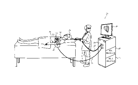
[0017] Fig. 1 is an illustration of a system, which is constructed and opera-
tive in accordance with a disclosed embodiment of the invention;
[0018] Fig. 2 is a flow chart of a method of electroanatomic mapping of the
epicardium in accordance with an embodiment of the invention;
4
CA 3027142 2018-12-11
[0019] Fig. 3 is a schematic illustration of a triangular mesh in accordance
with an embodiment of the invention;
[0020] Fig. 4 is an electroanatomic map of an epicardial surface of the heart
in accordance with an embodiment of the invention;
[0021] Fig. 5 illustrates a process of analyzing a triangular mesh in accord-
ance with an embodiment of the invention;
[0022] Fig. 6 is a set of diagrams of a portion of a triangular mesh that mod-
els an FAM-produced volume in accordance with an embodiment of the invention;
[0023] Fig. 7 illustrates projection of hidden portions of the electroanatomic
map shown in Fig. 4 in accordance with an embodiment of the invention;
[0024] Fig. 8 is an anterior view of a corrected version of the electroanatom-
ic map shown in Fig. 4 in accordance with an embodiment of the invention;
[0025] Fig. 9 is a posterior view of the surface shown in Fig. 4 in accordance
with an embodiment of the invention; and
[0026] Fig. 10 is a schematic diagram of an ablation and active current loca-
tion (ACL) circuit in accordance with an embodiment of the invention.
DETAILED DESCRIPTION OF THE INVENTION
[0027] In the following description, numerous specific details are set forth
in
order to provide a thorough understanding of the various principles of the
present
invention. It will be apparent to one skilled in the art, however, that not
all these
details are necessarily needed for practicing the present invention. In this
instance,
well-known circuits, control logic, and the details of computer program
instructions
for conventional algorithms and processes have not been shown in detail in
order
not to obscure the general concepts unnecessarily.
[0028] Documents incorporated by reference herein are to be considered
an integral part of the application except that, to the extent that any terms
are de-
fined in these incorporated documents in a manner that conflicts with
definitions
made explicitly or implicitly in the present specification, only the
definitions in the
present specification should be considered.
5
CA 3027142 2018-12-11
Overview.
[0029] Turning now to the drawings, reference is initially made to Fig. 1,
which is an illustration of a system 20, which is constructed and operative in
ac-
cordance with a disclosed embodiment of the invention. The system 20 is used
in
determining the position of a probe or catheter 22, used for the acquisition
of ana-
tomic and electrical data, and for tissue ablation using the catheter 22.
During ac-
quisition of an endocardial electrical map, the catheter 22 is placed into
chambers
of a heart 24 of a subject 26 using a known intravascular approach. For
obtaining an
epicardial electrical map, the catheter 22 may be percutaneously inserted into
the
pericardial cavity that surrounds the heart 24. Alternatively, the epicardial
electri-
cal map may be obtained non-invasively. Exemplary methods and devices for car-
diac mapping are described in U.S. Pat. Nos. 5,471,982, 5,391,199, 6,226,542,
6,301,496, and 6,892,091, and in PCT patent publications W094/06349,
W096/05768 and W097/24981, whose disclosures are incorporated herein by ref-
erence. U.S. Pat. No. 5,391,199, for example, describes a catheter including
both
electrodes for sensing cardiac electrical activity and miniature coils for
determin-
ing the position of the catheter relative to an externally-applied magnetic
field. Us-
ing this catheter data can be collected from a set of sampled points within a
short
period of time, by determining the electrical activity at a plurality of
locations and
determining the spatial coordinates of the locations.
[0030] The electrodes and transducers of distal end 44 of the catheter 22 are
connected by a cable through the insertion tube of the catheter 22 to a
control
unit 28 (Fig. 1), which controls other elements of the system 20, including an
image
processor 21, and an EKG processor 29. The processors access a memory to exe-
cute programs stored therein for performing procedures detailed below. The con-
trol unit 28 determines position coordinates of the catheter 22 relative to
specific
landmarks or features of the heart 24. The control unit 28 drives a display
40, which
shows the catheter position inside the body. The control unit 28 also drives
the ab-
lation transducers that are located generally at the tip of the catheter 22.
6
CA 3027142 2018-12-11
[0031] The catheter 22 is used in generating anatomic images or an epicar-
dial electrical map. The distal end of the catheter 22 comprises an ultrasound
imag-
ing device, which is typically a phased array of transducers, well known in
the art.
The ultrasound imaging device is operated, as is known in the art, so as to
capture
a 2-dimensional "fan" image in the plane of the scanning ultrasonic beam
(referred
to as the "beam plane" or "image plane"), which contains the longitudinal axis
of
the catheter. The transducers receive ultrasonic waves that are reflected from
ob-
jects in the beam plane and output signals in response to the reflected waves.
Typ-
ically, these signals are conveyed by wires running through the catheter 22 to
im-
age processor 21, which processes the signals in order to form and display
ultra-
sound images and 3-dimensional maps.
[0032] In some embodiments, the electrodes on the catheter can be used al-
ternately for mapping and for ablation. One system that embodies the above-
described features of the system 20 is the CARTO 3 System, available from Bio-
sense Webster, Inc., 33 Technology Drive, Irvine, CA 92618. This system may be
modified by those skilled in the art to embody the principles of the invention
de-
scribed herein.
[0033] In some embodiments of the invention, epicardial electrical maps
can be obtained noninvasively, using body-surface electrodes 31, of which
three
are shown representatively, it being known in the art that when using the
noninva-
sive technique, much larger arrays of electrodes are typically required in
order to
obtain accurate epicardial electrical maps. The electrodes 31 may conveniently
be
mounted in multi-electrode chest panels as described in any of the following
doc-
uments, all of which are herein incorporated by reference: Ransbury et aL,
U.S.
Patent Application Publication No. 2004/0015194; Sippensgroenewegen, U.S.
Patent Application Publication No. 2001/0056289; Ramanathan et al., in
Noninvasive
Electrocardiographic Imaging for Cardiac Electrophysiology and Arrhythmia,
Nature
Medicine, published online 14 March 2004; and Modre et al. Atrial Noninvasive
Activation Mapping of Paced Rhythm Data, J. Cardiovasc. Electrophysiology
14:712-
7
CA 3027142 2018-12-11
719 (July 2003), The electrodes 31 are connected to the control unit 28 by a
ca-
ble 33, and linked to the EKG processor 29.
[0034] Alternatively, the above-noted intrapericardial technique can be
used to generate an epicardial electrical map. This method is still less
invasive than
intravascular catheterization technique for obtaining endocardial electrical
maps.
The technique employs an epicardial contact probe as the catheter 22, which is
in-
serted through the chest wall into the pericardium, using known introduction
tech-
niques.
[0035] In either case, the epicardial electrical map typically shows the p0-
tentials on the epicardium, although it may also show endocardial potentials.
Nev-
ertheless, the term "epicardial electrical map" is employed herein, as the
data of
primary interest are obtained from outside the heart.
First Embodiment.
[0036] Reference is now made to Fig. 2, which is a flow chart of a method of
electroanatomic mapping of the epicardium in accordance with an embodiment of
the invention. The process steps are shown in a particular linear sequence in
Fig. 2
for clarity of presentation. However, it will be evident that many of them can
be
performed in parallel, asynchronously, or in different orders. Those skilled
in the
art will also appreciate that a process could alternatively be represented as
a num-
ber of interrelated states or events, e.g., in a state diagram. Moreover, not
all illus-
trated process steps may be required to implement the method.
[0037] At initial step 46 a mapping catheter is positioned at the epicardium.
At step 48 electrical readings are at mapped locations are taken as described
above.
[0038] At step 50 a closed 3-dimensional image representing a cardiac vol-
ume is generated using the above-described FAM technique. Then, at step 52 the
epicardial surface, including the mapped locations, is modeled as a triangular
mesh. Reference is now made to Fig. 3, which is a schematic illustration of a
trian-
gular mesh 54 including points 56 in accordance with an embodiment of the
invert-
tion. Although a geometric mesh is shown in Fig. 3 for clarity, the triangles
may be
8
CA 3027142 2018-12-11
advantageously implemented as a list or an array. The points 56 are registered
in
step 52, when in contact with the epicardial surface of the heart 24 (Fig. 1).
Typical-
ly during the mapping referred to above, image processor 21 initially stores 3-
dimensional coordinates of points 56 as measured in a 3-dimensional frame of
ref-
erence 58 defined by field generating coils (not shown). The image processor
21
then connects 3-dimensional coordinates of points 56, herein also termed 3-
dimensional vertices, by line segments 60 to produce a set of connected 3-
dimensional triangles, e.g., triangles 62, 64, 66. The procedures described in
commonly assigned U.S. Patent Application Publication Nos. 20150164356,
entitled
Dynamic Feature Rich Anatomical Reconstruction from a Point Cloud, and
20170221254, entitled High Definition Coloring of Heart Chambers, which are
herein
incorporated by reference, may be used to produce the mesh 54. Other suitable
algorithms include the ball-pivoting algorithm to produce the mesh 54.
Alternative-
ly, the mesh may be generated as a Delaunay triangulation. Elements of the
mesh
each have 3-dimensional coordinates.
[0039] Reverting to Fig. 2, at step 68 an electroanatomical map 70 is con-
structed on the mesh, using the readings taken at step 48. When such a map is
dis-
played, as shown in Fig. 4, only one side, i.e., the anterior epicardial
surface, is vis-
ible - posterior aspects are invisible in this view.
[0040] Continuing to refer to Fig. 2, in step 72 the triangles of the mesh are
analyzed and classified. From the perspective of an observer some of the
triangles
are front-facing, i.e., they face generally toward the observer, while others
face
away from the observer. The latter are referred to as rear-facing triangles.
The
terms "rear-facing" and "front-facing" are used arbitrarily herein to
distinguish dif-
ferent orientations of the triangles in the mesh, These terms have no physical
mean-
ings with respect to the actual configuration of the mesh.
[0041] Next, in step 74 for each measured point that maps to a rear-facing
triangle, the closest front-facing triangle to that point is identified.
[0042] Fig. 5 illustrates aspects of the process of step 74. A triangular
mesh 76 models a cardiac volume representing the epicardium that was created
by
9
CA 3027142 2018-12-11
the FAM algorithm. The mesh 76 is surrounded by an exterior that includes
center
of mass 82 and further includes an observer 92 on the opposite side of the
mesh 76.
Vectors 78, 80 are directed from center of mass 82 to triangles 84, 86 on the
mesh 76. Normal vectors 88, 90 are drawn from the triangles 84, 86 toward the
ex-
tenor of the volume, e.g., working in a clock-wise direction. The observer 92
look-
ing at a display screen containing the mesh 76 could see front-facing triangle
84
but could not see rear-facing triangle 86 as it is obscured by the front-
facing sur-
face of the mesh 76.
[0043] Projection of points that map to rear-facing triangles onto front-
facing
triangles is illustrated by Fig. 6, which is a set of diagrams of a portion of
a triangu-
lar mesh that models an FAM-produced volume. Four phases are shown in dia-
grams 94, 96, 98, 99. In diagram 94 rear-facing triangles 100, 102, 104 and
front-
facing triangles 106, 108, 110 were identified in step 72 (Fig. 2). The two
classes of
triangles are separated by interior 112 of the FAM-produced volume. Measured
points 114, 116 map to rear-facing triangles 100, 104, respectively. Measured
point 118 maps to front-facing triangle 108. In diagram 94 the pseudocolor
associ-
ated with point 118 is visible on a display, e.g., display 40 (Fig. 1). The
pseudocol-
ors associated with points 114, 116 are not visible on the display.
[0044] In diagram 96 front-facing triangles 106, 110 were identified in
step 74 (Fig. 2) as the closest front-facing triangles to points 114, 116,
respectively.
Rear-facing triangles are omitted in the searches to find the closest
triangles to the
measured points.
[0045] In diagram 98 the points 114, 116 and their data are now associated
with front-facing triangles 106, 110 respectively. The front-facing triangles
106, 110
are now shown in the pseudocolors that were previously presented on rear-
facing
triangles 100, 104.
[0046] In diagram 99 the rear-facing triangles 100, 102, 104 have been re-
moved. The front-facing triangles 106, 108, 110 now model the epicardial
surface as
a curved plane and displays a complete electroanatomic map of a portion of the
ep-
icardium. This is indicated in Fig. 2 as step 122, which can be understood
with ref-
CA 3027142 2018-12-11
erence to Fig. 7, Fig. 8 and Fig. 9. In step 122 portions of the map taken
from read-
ings on the rear-facing surface of the volume are projected onto the front
surface.
These appear in Fig. 7 as area 124 (outlined by a broken line). In Fig. 8 the
coloring
associated the front-facing triangles is combined with the coloring of the
projected
points of the rear-facing triangles to form a corrected map in Fig. 8, in
which the
areas corresponding to the rear-facing triangles are superimposed rather than
ob-
scured. The advantage of Fig. 8 is that the areas corresponding to both front-
and
rear-facing triangles can be appreciated in a single view. The measured points
need not encompass all rear-facing triangles, as indicated by area 120, which
is
outside area 124.
[0047] In some embodiments final step 126 is performed. Fig. 9 is a posteri-
or view of the map 70, which includes the area 120 that corresponds to rear-
facing
triangles of the mesh. The rear-facing triangles are removed from the mesh as
shown in diagram 99 (Fig. 6). The front-facing triangles that remain in the
mesh
now model a curved plane with no significant thickness, and which now presents
a
display of the electroanatomic map that includes previously hidden data.
[0048] The above-described method can also be used to project a map tak-
en from endocardial readings onto the front surface.
[0049] The algorithm described above is summarized by the pseudocode of
Listing 1.
Listing 1
Identify the center of mass for the mesh.
For each triangle in the mesh {
Construct a vector A* from the center of mass to the triangle.
Construct a directed vector 13 directed away from the center of mass and
normal to the surface of the triangle.
If the dot product (-A =) > 0 {
11
CA 3027142 2018-12-11
triangle is front-facing. It is considered in searches for a closest
triangle to a hidden measured point
If the dot product (A = 13.) < 0 {
/* the triangle is rear-facing */
omit this triangle from from consideration in the projection and map
reconstruction that follows.
For measured points that map to the rear-facing triangles {
Find the closest triangle /*it will be front-facing, since rear-facing
triangles have been excluded from the searches */.
project the measured point onto the closest front-facing triangle to
create a new coloring on the closest front-facing triangle based on
the data associated with the measured point.
Recolor the map according to the projected points
Second Embodiment
[0050] Typically an FAM-produced volume is generated and during the pa-
tient session in which epicardial readings are taken. In this embodiment the
FAM-
produced volume is generated from images pre-acquired at a different time from
the epicardial readings. The locations of the epicardial readings are then
placed in
registration with the FAM-produced volume by known methods, for example the
methods described in commonly assigned U.S. Patent Application Publication
Nos.
20130123773 entitled Integrative Atrial Fibrillation Ablation, 20160354049
entitled
Registration of Coronary Sinus Catheter Image and 20160120426 entitled
Registration
Maps Using Intra-Cardiac Signals, all of which are herein incorporated by
refer-
12
CA 3027142 2018-12-11
ence. The process described in the discussion of Fig. 2 beginning with step 52
can
then be performed using the FAM-produced volume.
Implementation Details.
[0051] Reference is now made to Fig. 10, which is a schematic diagram of an
ablation and active current location (ACL) circuit 244 for use with the system
shown
in Fig. 1. This arrangement is similar to that described in U.S. Patent
Application
Publications 2006/0173251, to Govari et al., and 2007/0038078, to Osadchy,
which
are herein incorporated by reference. The arrangement can be modified to oper-
ate in accordance with the principles of the present invention. A brief
description
follows for convenience of presentation. The (ACL) circuit 244 can be used to
de-
termine the mapped locations in step 48 (Fig. 2).
[0052] A plurality of body surface electrodes 246, which can be adhesive
skin patches, are coupled to a body surface 248 (e.g., the skin) of subject
250. The
body surface electrodes 246 are sometimes referred to herein as "patches". In
cardiac applications the body surface electrodes 246 are usually distributed
so as
to surround the heart, three on the chest of the subject and three on the
back. How-
ever, the number of the body surface electrodes 246 is not critical, and they
may
be placed at convenient locations on the body surface 248 in the general
vicinity of
the site of the medical procedure.
[0053] A control unit 252 includes current measurement circuitry 254 and
one or more catheter electrode transmitters 256 for driving a current through
one
or more of the electrodes 246 to one or more of the body surface electrodes
246 at
respective working frequencies. The control unit 252 is linked to a
positioning pro-
cessor (Fig. 1). The control unit 252 is linked to an ablator 258, which
comprises at
least one ablation generator 260. Currents through the body surface electrodes
246
and an ablator body surface electrode 262 flow in a circuit with the ablation
gener-
ator 260 and are measured by respective current measurement circuits that are
disposed within body electrode receivers 264, sometimes referred to herein as
"patch measurement circuits". The body electrode receivers 264 are typically
in-
corporated in the control unit 252. Alternatively, they may be affixed to the
body
13
CA 3027142 2018-12-11
surface electrodes 246. Catheter electrodes are represented as measurement
elec-
trodes 266 (circles) and a dual-purpose electrode 268 (ellipse). The dual-
purpose
electrode 268 functions as an ablation electrode and also serves as one of the
measurement electrodes.
[0054] The body surface electrodes 246 are connected to the body elec-
trode receivers 264 via a patch box 270, which protects the system from
ablation
and defibrillation currents. Typically the system is configured with six body
elec-
trode receivers 264. The patch box parasitic impedances 272 (Z), are measured
during production and thus known a priori. These impedances are discussed be-
.. low.
[0055] Typically, although only two measurement electrodes 266 are shown
for convenience, about 80 measurement electrodes are used for impedance meas-
urements. Typically there are one or two ablation electrodes. The coordinates
of a
catheter inside the body are determined in the positioning system by passing
cur-
rents between electrodes on the catheter and the body surface electrodes 246.
[0056] The control unit 252 may also control an ablation circuit, comprising
ablator 258, and the dual-purpose electrode 268. The ablator 258 is typically
dis-
posed externally to the control unit 252 and incorporates the ablation genera-
tor 260. It connects with the ablator body surface electrode 262 and to an
ablator
filter 276, which in this example is shown within the control unit 252.
However this
location is not essential. A switch 278 configures the ablator circuit for
different
modes of operation as described below. Voltage measurement circuitry is provid-
ed for determining the output of the catheter electrode transmitters 256. It
will be
noted from inspection that the ablation circuit is connected to one of the
catheter
electrode transmitters 256.
[0057] It will be appreciated by persons skilled in the art that the present
invention is not limited to what has been particularly shown and described
hereinabove. Rather, the scope of the present invention includes both
combinations and sub-combinations of the various features described
.. hereinabove, as well as variations and modifications thereof that are not
in the prior
14
CA 3027142 2018-12-11
art, which would occur to persons skilled in the art upon reading the
foregoing
description.
CA 3027142 2018-12-11