Note: Descriptions are shown in the official language in which they were submitted.
WO 2010/093861
PCT1US2010/024010
SYSTEMtMETHOD AND DEVICE FOR TISSUE-BASED DIAGNOSIS
Cross-Reference to Related Applications
[0001] This application claims the benefit of U.S. Provisional Patent
Application No.
61/152,585 filed February 13, 2009.
Background of the Invention
[0002] The biomolecular composition of human tissues, represented by a
multitude of
lipids, proteins, nucleic acids, and other miscellaneous molecules, is a
sensitive indicator of
local pathologies, such as cancer, allergies, and eczema, as well as several
systemic
diseases, such as cardiovascular disease. Alzheimer's disease, and diabetes.
in addition,
tissue molecular composition also holds critical information about the body's
exposure to
exogenous chemical and biological entities. However, this information is not
currently
used in diagnostic methods due to a lack of patient-friendly and standardized
methods for
routine sample collection from tissues. Instead, clinical diagnosis is
invariably performed
by visual observation and histopathological analysis of tissue biopsies, which
are highly
limited due to their qualitative nature, leading to increased misdiagnosis and
inappropriate
use. In addition to being invasive, current methods also fall short in
explaining a complete
molecular genesis of diseases, and fail to distinguish between diseases.
(00031 Prior approaches using physical and chemical methods for assessing
tissue fluid have
focused chiefly on extracting a few low molecular weight molecules that are
freely present in
the interstitial fluid, such as calcium and glucose. Use of tape stripping for
physically
harvesting superficially-lying tissue constituents with an adhesive tape has
been reported;
however this technique has been shown to be limited by inefficacy, lack of a
standardized
protocol, and high heterogeneity in tissue sampling.
Brief Summary of the Invention
[0004] In an aspect, the current invention describes system, method and
device, as well
as compositions useful in such systems, methods and devices, involving
application of
energy to a tissue of interest to generate a liquefied sample comprising
tissue constituents
so as to provide for rapid tissue sampling, as well as qualitative and/or
quantitative
1
BFC-TBD/PCT-CDAD1V
CA 3028277 2018-12-24
WO 2010/093861
PCT/US2010/024010
detection of analytes that may be part of tissue constituents (e.g., several
types of
hiomolecules, drugs, and microbes). Determination of tissue composition can be
used in a
variety of applications, including diagnosis or prognosis of diseases,
evaluating
bioavailability of therapeutics in different tissues following drug
administration, forensic
detection of drugs-of-abuse, evaluating changes in the tissue microenvironment
following
exposure to a harmful agent, tissue decontamination and various other
applications.
[0005] In another aspect, the current invention provides methods and devices
for
generating a liquefied tissue sample from a subject- living or diseased. The
device and
method involve applying energy and a liquefaction promoting medium to a tissue
of interest
of a subject, the applying producing a liquefied tissue sample, and collecting
the liquefied
tissue sample. In some embodiments, an analysis for the presence or absence of
at least one
analyte in the liquefied tissue sample is performed, wherein the analysis
facilitates diagnosis
of a condition of interest. ill certain embodiments, the analysis involves
generating an
analyte profile from the liquefied tissue sample and comparing the analyte
profile to a
reference analyte profile, wherein the comparing facilitates diagnosis of a
condition of
interest.
[0006] In some embodiments, the purpose of said tissue liquefaction is to
remove, or
decontaminate the tissue from undesired substances. Non-limiting examples of
such
undesired substances include chemicals, environmental contaminants, biological
toxins, and
in general substances that are considered toxic or hazardous to the body. In
certain
embodiments, the said method of decontamination is performed by continuously
moving
the tissue liquefaction device over tissue-of-interest until removal of
undesired substances
at a preferred level is attained.
[0007] In some embodiments, the liquefaction promoting agent comprises of one
of more of
sodium chloride, potassium chloride, sodium phosphate dibasic, potassium
phosphate
monobasie, [tris(hydroxymethypmethyl]amino}propanesulfonic acid, N,N-bis(2-
hydroxyethyDglyeine, tris (hydroxymethyl) methylamine, N-
tris(hydroxymethypmethylglycine, 4-2-hydroxyethyl- I -piperazineethanesulfonic
acid, 2-
[tris(hydroxymethyl)methyl]amino}ethanesulfonic acid, 3-(N-
rnorpholino)propanesulfonic
2
B FC-TB D/PCT-CDAD I V
CA 3028277 2018-12-24
WO 2010/093861
PCT/US2010/024010
acid, piperazine-N,N'-bis(2-ethanesulfonic acid), dimethylarsinic acid, saline
sodium citrate,
2- (N-morpholino)ethanesulfonic acid. In certain embodiments, the liquefaction
promoting
agent comprises of one or more of a protease inhibitor, an RNase inhibitor, or
a DNase
inhibitor. In certain embodiments, the liquefaction promoting agent comprises
at least one
of free radical scavenger, a defoaming agent, and a protein stabilizer. In
certain
embodiments, the liquefaction promoting agent comprises at least one of Brij-
30, 3-
(Decyl dimethyl ammonio) propane sulfonate (DPS), 3-(Dodecyl dimethyl ammonio)
propane sulfonate (DDPS), N-lauroyl sarcosine (NLS), Triton X-100, Sodium
Dodecyl
Sulfate, DMSO, fatty acids, azone, EDTA, or sodium hydroxide. In certain
embodiments,
the liquefaction promoting agent comprises a suspension of abrasive particles.
In certain
embodiments, the abrasive particles comprise silica or aluminum oxide.
[0008] In some embodiments, the energy is applied in the form of ultrasound,
mechanical,
optical, thermal, or electrical energy. In certain embodiments, the mechanical
energy is
applied by an abrasive material, In certain embodiments, the thermal energy is
applied in
the form of radio frequency energy. In certain embodiments, the optical energy
is applied in
the form of a laser.
[0009] In some embodiments, the liquefied tissue sample is generated for each
of a healthy
tissue of interest of the subject and a suspected diseased tissue of interest
of the subject, and
the analysis comprises comparing analytical results from the healthy tissue
sample with
analytical results from the suspected diseased tissue sample, wherein the
comparing
facilitates diagnosis of a condition of interest. In some embodiments, the
liquefied tissue
sample is generated for multiple tissue sites and the analysis comprises
comparing analytical
results from the multiple tissue sites, wherein said comparing facilitates
diagnosis of a
condition of interest. In some embodiments, the liquefied tissue sample is
collected from
multiple tissue sites, and the samples are combined to make a diagnosis.
[0010] In some embodiments, the liquefied tissue sample is collected by
aspiration. In
certain embodiments, the collecting is by retaining the liquefaction agent in
a housing placed
in contact with the tissue. In certain embodiments, the collecting is by
mechanized transfer
of the liquefied tissue sample in a housing located in the device.
3
BFC-TBD/PCT-CDADIV
CA 3028277 2018-12-24
W02010/093861
reCT/US2010/024010
[0011] In some embodiments, the liquefied tissue sample is mixed with a
substance whiCh
assists in further liquefaction and in stabilization of analytes of interest
for storage or
transportation. In certain embodiments, the transferred tissue sample from
that sample
container is mixed with the substances which are pre-stored in a container.
Examples include
a protein stabilizer such as protease inhibitor, a nucleic-acid stabilizer
such as EDTA, phenol,
nonspecific proteinase, an RNase inhibitor and a DNase inhibitor, a defoaming
agent, and
surfactants such as Triton X-100, Sodium Dodecyl Sulfate, and DMSO, and
abrasive particles
comprise silica or aluminum oxide.
[0012] In certain embodiments, the device evaluates the tissue of interest
prior, during, or
after liquefaction process. In certain embodiments, the evaluation is
performed by
electrochemical, biochemical, or optical means. In some embodiments, the
evaluation
involves measurement of tissue's electrical conductivity. In an exemplary
embodiment,
electrical conductivity is measured by a means applying an AC electrical
signal across the
tissue of interest. The said electrical signal has voltage between 0.1 mV and
10 V and
frequency between 1 Hz and 100 kHz.
[0013] In some embodiments, the device involves detecting certain tissue
constituents in the
liquefied tissue sample prior to analysis of an analyte of interest, such as a
disease marker. In
certain embodiments, the detecting is by electrochemical, biochemical, or
optical means. In
some embodiments the electrochemical means of detecting is an ion-elective
electrode. In
some embodiments the optical means of detecting is measuring the absorption or
scattering
coefficient of a liquid solution.
[0014] In some embodiments; the energy is applied to a tissue in the form of
ultrasound
with a mechanical index between 0.1 and 50. In certain embodiments, the energy
is applied
by contacting the tissue with a moving abrasive surface. In certain
embodiments, the energy
is applied to the tissue by contacting the tissue with a moving brushing
device comprising a
plurality of bristles. In certain embodiments, the energy is applied to the
tissue by
mechanical insertion of a patch bearing plurality of micro-needles into the
tissue; and further
injection of liquefaction medium through the micro-needles into the tissue. In
some
BFC-TBD/PCT=CDADIV
CA 3028277 2018-12-24
W02010/093861
PCT/US2010/024010
embodiments, additional energy is applied by moving the said micro-needle
patch after its
insertion into the tissue. In certain embodiments, the energy is applied to
the tissue by
mechanized stirring of the liquefaction agent. In certain embodiments, the
energy is applied
to the tissue by contacting the tissue with a high velocity jet comprising of
liquefaction
promoting medium, which may also contain abrasive particles in different
embodiments.
[0015] In some embodiments, the tissue comprises breast, prostate, eye,
vagina, bladder, nail,
hair, colon, testicles, or intestine. In certain embodiments, the tissue
comprises skin or a
mucosal membrane, In certain embodiments, the tissue comprises lung, brain,
pancreas, liver,
heart, bone, or aorta wall,
[0016] In some embodiments, the analyte comprises a small molecule, a drug or
metabolite
thereof, a polypeptide, a lipid, a nucleic acid, or a microbe. In certain
embodiments, the
analyte comprises an antibody, a cytokine, an illicit drug, or a cancer
biomarker.
[0017] In some embodiments, the liquefied tissue sample is held in a
container, and the
analyte profile is generated by integrating the liquid container with one or
more analytical
devices. In certain embodiments, the tissue liquefaction device contains a
means for
measuring the concentration of a calibrator analyte to provide a means for
calibrating the
analysis of the analyte.
[0018] In some embodiments, the device involves diagnosing allergic disease in
a subject,
and the device comprises means for analyzing the liquefied tissue sample for
the presence or
absence of lgE and IgG antibodies, cytokines such as IL4,1L5, IL10, IL-12,
IL13, IL-16,
GM-CSF, RANTES, MCP-4, CTACK/CCL27, IFN-g, TNFa, CD23, CD-40, Eotaxin-2, and
TARC, wherein the analysis facilitates diagnosis of allergic disease in the
subject.
[0019] In some embodiments, the device involves diagnosing cancer in a
subject, and the
device comprises means for analyzing the liquefied tissue sample for the
presence or absence
of one or more cancer markers, wherein the analysis facilitates diagnosis of
cancer in the
subject. In certain embodiments, the tissue of interest is breast, colon,
prostate, skin, testicle,
intestine, or mouth.
13FC-TIID/PCT-CDADIV
CA 3028277 2018-12-24
WO 2010/093861
PCT/US2010/024010
[0020] In some embodiments, the device involves diagnosing heart disease in a
subject, and
the device comprises means for analyzing the liquefied tissue sample for the
presence or
absence of one or more of cholesterol, triglycerides, lipoproteins, free fatty
acids, and
ceramides, wherein the analysis facilitates diagnosis of heart disease in the
subject.
[0021] In some embodiments, the device involves detecting the presence of an
illicit drug,
or metabolite thereof, in a subject, and the device comprises means for
analyzing the
liquefied tissue sample for the presence or absence of an illicit drug, or
metabolites
thereof, wherein the analysis provides for detection of illicit drugs in the
subject.
[00221 In some embodiments, the device involves detecting a microorganism in a
subject,
and the device comprises means for applying energy and a liquefaction medium
to a tissue of
interest in a subject and analyzing the liquefaction medium for the presence
or absence of a
microorganism., wherein the analysis provides for detection of the presence or
absence of a
microorganism.
[0023] Another aspect provides a method and device for liquefying a tissue of
a subject for
facilitating the passage of a drug across or into the tissue. The method and
device disclosed
above are applicable not only to collection of tissue constituents but also to
drug delivery.
The device and method involve applying energy and a liquefaction medium to a
tissue of
interest of a subject, and delivering a drug through or into the site of the
tissue to be liquefied.
The advantage of using the present invention is 1) to provide higher fluxes of
drugs into a
tissue, and 2) to allow greater control of fluxes into a tissue. Drugs which
would simply not
pass through the tissues such as the skin are forced through the tissues when
the method is
applied.
[0024] In some embodiments, the present invention offers a method for
delivering one or
more drugs through the tissue to be liquefied into the circulatory system,
which circumvents
degradation in the gastrointestinal tract and rapid metabolism by the liver
from which drugs
to be routinely administered either orally or by injection suffer. In certain
embodiments, the
current invention provides a method and device for delivering one or more
drugs locally to
6
BFC-TBD/PCT-CDADIV
CA 3028277 2018-12-24
WO 2010/093861
PCT/US2010/024010
the tissue of interest, thus limiting side effects to the healthy tissues. The
method and device
may also be applicable for enhancing transport to cellular membranes.
[0025J Some embodiments provide the following components: 1) an energy
generator; 2) a
liquefaction promoting medium; 3) a reservoir to hold drugs to be delivered
and/ or collect
the liquefied tissue sample.
[0026] A drug to be administered can be added into the liquefaction medium
prior or
during tissue liquefaction process. In an alternate embodiment, application of
energy is in
= combination of the liquefaction medium which does not contain a drug can
be used for
liquefying a tissue, and subsequently a drug in an appropriate carrier such as
a patch can be
applied on a site of the tissue to be liquefied.
[0027] The transport of drug into the tissue can be further enhanced by the
simultaneous or
subsequent application of a secondary driving force such as chemical
permeability or
transport enhancers, convection, osmotic pressure gradient, concentration
gradient,
iontophoresis, electroporation, magnetic field, ultrasound, or mechanical
pressure. The
driving force can be applied continuously over a period of time or at
intervals during the
period of liquefaction.
[0028] In some embodiments, the tissue to be administered comprises an organs
as well as
biological surfaces. In certain embodiment, the biological surfaces comprise a
biological
membrane and cellular membrane. In certain embodiment, the biological membrane
comprises skin or a mucosal membrane. In certain embodiments, the biological
membrane
comprises a buccal membrane, eye, vagina, colon, or intestine. In some
embodiment, the
tissue comprises a diseased tissue.
[0029] In one embodiment, a device is provided that can be used on a tissue to
obtain a
liquefied sample comprising an energy source operably coupled to the tissue,
and a chamber,
operably coupled to said tissue, capable of delivering liquefaction promoting
medium to
and/or collecting said liquefied sample from said tissue.
7
BFC-TBD/PCT-CDADI V
CA 3028277 2018-12-24
WO 2010/093861
PCT/US2010/024010
[0030] In another embodiment, the device can be used on a tissue which is a
part of a
living organism; and the tissue can be excised from the organism prior to
diagnosis.
[0031] In another embodiment, the device of claim 1 wherein the liquefied
tissue
sample is transferred to an assay for monitoring the presence or absence of at
least one
analyte.
[0032] In yet another embodiment, the chamber of the device can be a sponge-
bellow
assembly where the sponge is capable of storing said liquefaction promoting
medium and/ or
liquefied tissue sample.
[0033] In another embodiment, a device is provided comprising an energy source
operably
coupled to the tissue, and a chamber, operably coupled to said tissue, capable
of delivering
liquefaction promoting medium to and/or collecting said liquefied sample from
said tissue;
also comprises a tube/needle, connected to said chamber, capable of delivering
the
liquefaction promoting medium to and/or aspirating liquefied tissue sample
from the tissue.
[0034] In still another embodiment, a device is provided comprising an energy
source
operably coupled to the tissue, and a chamber, operably coupled to said
tissue, capable of
delivering liquefaction promoting medium to and/or collecting said liquefied
sample from
said tissue; also comprises a sample container, operably connected to said
chamber, capable
of storing aspirated liquefied tissue sample containing analytes, or
transferring said aspirated
liquefied tissue sample to an ancillary chamber; wherein the chamber is used
only to deliver
the liquefaction promoting medium to the chamber.
[0035] In another embodiment, a pressurized container and/or vacuum container
is part of
the device, which facilitates transfer of said liquefaction promoting medium
and/ or
liquefied tissue sample.
[0036] In one embodiment, the energy emitted from the energy source in the
device is in the
form of ultrasound, mechanical, optical, thermal, or electrical energy. In a
particular
embodiment, the mechanical energy is applied to the tissue by an abrasive
material, vacuum,
pressure or shear force, hi another embodiment, the thermal energy is applied
to the tissue in
8
FiFe-mum-r-cuaniv
CA 3028277 2018-12-24
WO 2010/093861
PCT/US2010/024010
the form of radio frequency energy. In another embodiment, the optical energy
is applied to
the tissue in the form of a laser.
[0037] In yet another embodiment, a device is provided comprising an energy
source
operably coupled to the tissue, and a chamber, operably coupled to said
tissue, capable of
delivering liquefaction promoting medium to and/or collecting said liquefied
sample from
said tissue further comprising a sample container, operably connected to said
chamber,
capable of storing aspirated liquefied tissue sample containing analytes, or
transferring said
aspirated liquefied tissue sample to an ancillary chamber; wherein the chamber
is used only
to deliver the liquefaction promoting medium to the chamber.
[0038] In another embodiment, a device is provided comprising an energy source
operably coupled to the tissue, and a chamber, operably coupled to said
tissue, capable
of delivering liquefaction promoting medium to and/or collecting said
liquefied sample
from said tissue, wherein the energy F!ource comprises of a pad connected to a
shaft.
[0039] In a more particular embodiment, the shaft has a pressure sensing unit,
which
maintains a predetermined pressure profile on to the tissue upon contact.
[0040] In another embodiment, the pad is selected from a group consisting of
an abrasive
surface and a patch comprising of a plurality of micro-needles.
[0041] In yet another embodiment, the device further comprises a plunger,
operably
connected to the top of the chamber.
[0042] In another embodiment, the device is divided into an upper and lower
unit, and
wherein the lower unit is detachable from said upper unit; wherein the upper
unit comprises
the energy source and the lower unit comprises the chamber.
[0043] In still another embodiment, the device .further comprises an
analytical unit operably
connected to the chamber, and where the analytic unit is capable of performing
temporal
monitoring of the tissue sample by electrochemical, biochemical or optical
means; or the
analytic unit is capable of analyzing the analytes within said liquefied
tissue sample,
9
aFc-Tao/PcT-oom.Av
CA 3028277 2018-12-24
WO 2010/093861
PCT/US2010/024010
[0044] In another embodiment, the device is connected to a diagnostic probe or
a catheter;
wherein the diagnostic probe is selected from a group consisting of endoscope,
colonoscope,
and laparoscope.
[0045] In still another embodiment, the use of the device results in situ
liquefaction of the
tissue sample.
[0046] In another embodiment, the device contains a liquefaction promoting
medium that
can preserve and enhance the detection of proteins, lipids and nucleic acids,
comprising: 3-
(decyl dimethyl ammonia) propane sulfonate (DPS) and polyethylene glycol
dodecyl ether
(Brij 30) dissolved in a buffered solution; and where the concentration of 3-
(decyl dimethyl
ammonio) propane sulfonate and polyethylene glycol dodecyl ether (B30) is
between 0.01-
10% (w/v); and where the 3-(decyl dirnethyl ammonio) propane sulfonate and
polyethylene
glycol dodecyl ether are present at a ratio of 50:50.
[0047] In yet another embodiment, the liquefaction promoting medium within the
device
is buffered in a solution comprising either phosphate-buffered saline, Tris-
buffered saline,
Tris- HCL or EDTA.
[0048] In another embodiment, liquefaction promoting medium within the device
comprises a nonionic surfactant selected from a Brij series surfactant, a
Triton-X
surfactant, and a Sorbitan surfactant; an anionic or a zwitterionic
surfactant; and a
hydrophilic solvent; wherein the medium has a total concentration of the
surfactants from
about 0.01%-10% (w/v).
[0048a] In another aspect, there is provided a device for at least partly
liquefying a tissue,
comprising a reservoir. An abrasive material is operatively coupled to the
reservoir, and the
reservoir is configured to transmit mechanical energy through the abrasive
material to the
tissue in the form of stirring, abrasion, pressure, or shear force. Also
provided is a
liquefaction promoting medium, comprising a non-ionic surfactant and a
zwitterionic
surfactant, the zwitterionic surfactant comprising one or more of: 3-(decyl
dimethyl
ammonia) propane sulfonate, 3-(dodecyl dimethyl ammonio) propane sulfonate,
BFC-TRD/PCT-CDADI V
CA 3028277 2018-12-24
=
WO 2010/093861
PCT/US2010/024010
myristyldirnethyl ammonio propane sultanate, hexadecyldimethyl ammonia propane
sulfonate, cocarnidopropyl betaine, oleyl betaine, cocamildopropyl
bydroxysultaine, or 3-(3-
cholamidopropy1)-dimethylammonio-1-propanesulfonate. The reservoir is
configured to be
operatively connected to the tissue and to apply the liquefaction promoting
medium to the
tissue.
(0048b] In another aspect, there is provided a device for at least partly
liquefying a tissue to
form a liquefied tissue and delivering a drug to the liquefied tissue,
comprising a reservoir.
An abrasive material is operatively coupled to the reservoir and the reservoir
is configured
to transmit mechanical energy through the abrasive material to the tissue in
the form of
stirring, abrasion, pressure, or shear force. Also provided is a liquefaction
promoting
medium, comprising a non-ionic surfactant and a zwitterionic surfactant; and a
drug. The
zwitterionic surfactant comprises one or more of: 3-(decyl dimethyl ammonio)
propane
sulfonate, 3-(dodecyl dimethyl arnmonio) propane sulfonate, myristyldirnethyl
amrnonio
propane sulfonate, hexadecyldimethyl ammonia propane sulfonate, cocamidopropyl
betaine,
oleyl betaine, cocarnidopropyl hydroxysultaine, or 3-(3-cholamidopropy1)-
dimethylammonio-1-propanesulfonate. The reservoir being configured to be
operatively
connected to the tissue and to apply to the tissue at least one of: the
liquefaction promoting
medium and the drug.
{0048c] In another aspect, there is provided a device for at least partly
liquefying a tissue,
comprisinga reservoir, An abrasive material is operatively coupled to the
reservoir. Also
provided is a liquefaction promoting medium. The liquefaction promoting medium
comprises a non-ionic surfactant and a zwitterionic surfactant, the non-ionic
surfactant
comprising one Or more of: polyethylene glycol dodecyl ether, polyoxyethylene
23-lauryl
ether, polyoxyethylene 2-cetyl ether, polyoxyethylene 10-cetyl ether,
polyoxyethylene 20-
cetyl ether, polyoxyethylene 2-stearyl ether, polyoxyethylene 10-stearyl
ether,
polyoxyethylene 20-stearyl ether, polyoxyethylene 2-oleyl ether,
polyoxyethylene 10-oley1
ether, polyoxyethylene 100-stearyl ether, or polyoxyethylene 21-stearyl ether.
The reservoir
11
B FC-TBD/PCT-CDAD I V
CA 3028277 2018-12-24
WO 2010/093861
PCT/US2010/024010
is configured to transmit energy through the abrasive material to the tissue,
and to be
operatively connected to the tissue to apply the liquefaction promoting medium
to the tissue.
[0048d] In another aspect, there is provided a device for at least partly
liquefying a tissue to
form a liquefied tissue and delivering a drug to the liquefied tissue,
comprising a reservoir.
An abrasive material is operatively coupled to the reservoir. Also provided is
a liquefaction
promoting medium, comprising a non-ionic surfactant and a zwitterionic
surfactant; and a
drug. The non-ionic surfactant comprises one or more of: polyethylene glycol
dodecyl
ether, polyoxyethylene 23-lauryl ether, polyoxyethylene 2-cotyl ether,
polyoxyethylene 10-
cetyl ether, polyoxyethylene 20-cetyI ether, polyoxyethylene 2-stearyl ether,
polyoxyethylene 10-stearyl ether, polyoxyethylene 20-stearyl ether,
polyoxyethylene 2-oleyI
ether, polyoxyethylene 10-oley1 ether, polyoxyethylene 100-stearyl ether, or
polyoxyethylene 21-steary1 ether. The reservoir is configured to transmit
energy through
the abrasive material to the tissue, and to be operatively connected to the
tissue to apply to
the tissue at least one of: the liquefaction promoting medium and the drug.
[0048e] In another aspect, there is provided a kit for at least partly
liquefying tissue,
cornprisinga liquefaction promoting medium (LPM). The LPM comprises a non-
ionic
surfactant, a zwitterionic surfactant. Also provided are an abrasive material
and instructions.
The instructions comprise directing a user to treat a tissue of a living
subject by:
applying the LPM together with the abrasive material to the tissue of the
living
subject; and
transmitting energy to the tissue of the living subject through the abrasive
material
in the presence of the LPM effective to cause at least partial dissolution of
one or more
components of the tissue of the living subject.
[0048f] In another aspect, there is provided a kit for at least partly
liquefying tissue, comprising
a liquefaction promoting medium (LPM). The LPM comprises an abrasive material
in a
concentration range in the LPM of 0.01-99% (w/v); and 3-(decyl dimethyl
ammonia) propane
sulfonate and polyethylene glycol dodecyl ether in a total surfactant
concentration in the LPM
12
Bre-TRO/PCT-CDADIV
CA 3028277 2018-12-24
WO 2010/093861
PCT/L182010/024010
of 0,01-20% (w/v);, Also provided are instructions, which comprise directing a
user to treat a
tissue of a living subject by:
applying the LPM to the tissue of the living subject, the tissue comprising at
least one of
skin and mucosal membrane; and
transmitting mechanical energy through the abrasive material to the tissue of
the living
subject in the presence of the LPM, and
the kit is effective to cause at least partial dissolution of one Or more
components of the tissue
of the living subject.
[0048g) In another aspect, there is provided a composition, comprising a non-
ionic surfactant; a
zwitterionic surfactant; and an abrasive material.
10048h) In another aspect, there is provided a method for at least partly
liquefying tissue,
comprising:
providing a liquefaction promoting medium (LPM), comprising: a non-ionic
surfactant; a
zwitterionic surfactant; and an abrasive material;
applying the LPM to a tissue of a living subject; and
transmitting mechanical energy through the abrasive material of the LPM to the
tissue of
the living subject effective to cause at least partial dissolution of one or
more components of
the tissue of the living subject.
[0049] These and other features of the invention will become apparent to those
persons
skilled in the art upon reading the details of the system, method and device
for tissue-based
diagnosis as more fully described below.
grief Description of the Drawings
[0050] The invention is best understood from the following detailed
description when read in
conjunction with the accompanying drawings. It is emphasized that, according
to common
practice, the various features of the drawings are not to-scale. On the
contrary, the
dimensions of the various features are arbitrarily expanded or reduced for
clarity. Included in
the drawings are the following figures:
13
BFC-1131)/PCT-CDAD1v
CA 3028277 2018-12-24
WO 2010/093861
PCT/US2010/024010
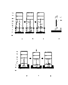
[0051] Figure 1 (Panels a-g) is a collection of cross-sectional drawings
illustrating structure,
components and functioning of various abrasive energy-based tissue
liquefaction devices.
Panels a-c and Panels e-g show the sequential working of two separate
liquefaction devices.
Panel d is a schematic representation of a pressure-sensitive motorized shaft
bearing an
abrasive head.
[0052] Figure 2 (Panels a-b) is a collection of cross-sectional drawings of
moveable
tissue liquefaction devices for continuous sampling of a large area of
tissues.
[0053] Figure 3 (Panels a-c) is a collection of cross-sectional drawings
illustrating structure
and components of various linear abrasive motion-based tissue liquefaction
devices. Panel
c is a schematic representation of a pressure-sensitive support shaft bearing
a gear.
[0054] Figure 4 (Panel a-g) is a collection of cross-sectional drawings
illustrating several
types of abrasive heads.
[0055] Figure 5 (Panels a-d) is a collection of cross-sectional device
drawings and
schematics for measuring tissue's electrical conductivity.
[0056] Figure 6 (Panels a-g) is a collection of cross-sectional drawings
illustrating
structure, components and fiinctioning of various microneedle-based tissue
liquefaction
devices.
[0057] Figure 7 (Panels a-c) is a collection of cross-sectional drawings of an
exemplary
abrasive energy-based tissue liquefaction device. Panel a shows various
assembly
components of the device. Panel b-d show sequential working steps of the
device including
transfer of the liquefaction medium to be placed in contact with the tissue
(Pane b-c), sample
generation by liquefaction (Panel c), and collection of the sample in a
container (panel d).
Panel e shows post- liquefaction retrieval of sampling container from the
device.
[0058] Figure 8 (Panels a-d) is a collection of cross-sectional drawings
illustrating
sequential working steps of an exemplary mieroneedle-based tissue liquefaction
device:
transfer of the liquefaction medium to be placed in contact with the tissue
(Pane a-b);
14
5pc-Tana2c-r-cuAn1v
CA 3028277 2018-12-24
WO 2010/093861
PCT/LIS2010/024010
sample generation by liquefaction (Panel c); and collection of the sample in a
container
(panel d).
[0059] Figure 9 (Panels a - d) is a collection of drawings illustrating a
sampling container.
Panels a¨d show the sequential working steps for transporting and/or analysis
of the
generated samples. Panel a shows substrates which selectively bind to analytes
of interest
are coated on the inside surface of the container, The analytes in the
liquefied tissue samples
are selectively captured by the coated substrates (Panel b). Upon sufficient
incubation ofthe
tissue sample, the sample is discarded while the analytes are held in the
container (Panel c).
The analytes are eluted by a buffer for subsequent analysis (Panel d).
[0060] Figure 10 (Panels a- c) is a collection of drawings illustrating the
screening
methodology for identifying unique surfactant formulations of LPMs. Panel a
ranks over
150 surfactant formulations in their ability to preserve protein bioactivity.
Panel b ranks best
formulations from Panel a on their tissue solubilization potential. Panel c
compares the best
LPM from entire screening- 0.5% (w/v) DPS-Brii30 with other conventional
surfactants in
their potential to sample functional proteins from skin tissue.
[0061] Figure 11 (Panel a-b) is a collection of drawings illustrating LPM-
assisted
preservation of bioactivities of various proteins (IgE-panel a; IgE, LDH and n-
gal ¨ panel b)
under mechanical stress of ultrasound exposure.
[0062] Figure 12 (Panel a -c) is a collection of drawings illustrating the
ability of ultrasonic
exposure in the presence ofLPM (saline solution of0.5% (w/v) DPS-B30) to
sample a
variety of functional disease biornarkers (IgE -Panel a; Cholesterol ¨ Panel
b; Bacteria-
Panel c) from skin tissue.
[0063] Figure 13 is a graph illustrating the effect of buffers in LPMs on the
compatibility
with quantitative PCR.
[0064] Figure 14 is a graph illustrating the influence of surfactant mixture
on the
compatibility with quantitative PCR.
BFc-moirci--coAotv
CA 3028277 2018-12-24
WO 2010/093861
PCT/US2010/024010
[0065] Figure 15 is a graph illustrating the effect of ultrasound intensity
and exposure time on
E. Colt viability. Samples were exposed to ultrasound at intensities of 1.7
W/cm2 (-) and 2.4
W/cm2 (1)_ Each point represents the mean value from three independent
samples.
[0066] Figure 16 is a photograph of agarose gel-electrophoresis of genomic DNA
from E.
con cells sonicated at different conditions in tris-HCI. Lane 1 molecular
standard; lane 2
Non-treated cell; lane 3 1.7 W/cm2, 2 min; lane 4 1.7 W/cm2 3 min; lane 5 2.4
W/cm2,3 min.
[0067] Figure 17 (Panels a and b) is a graph illustrating the number of
bacteria sampled
by ultrasound coupling with tris-HCl, swabbing, and surfactant scrub
technique, measured
by (a) culture assay and (b) quantitative PCR. Each point represents the mean
value from
five independent samples.
[0068] Figure 18 is a graph illustrating the effect of adding various
sensitivity enhancers in
LPM for enhanced detection of a model analyte- human lgE antibody, in it.
Sensitivity
enhancers used in the analysis are a mixture of 10% w/v BSA and 0.5% w/v Tween
20 in
phosphate- buffered saline (PBS) (open diamond); and a mixture of 10% w/v BSA
and 0.5%
w/v Tween 20 in tris-buffered saline (closed circle). Prior to analysis, each
of the sensitivity
enhancers was diluted at 1:10 ratio with LPM containing model analyte. As a
control, LPM
containing model analyte (open square) and a commonly-used analytical solvent
comprising
of a mixture of 1% w/v BSA and 0.05% w/v Tween 20 in iris-buffered saline
(solid square)
were used. The LPM was composed of a solution of 1% w/v mixture of NLS and
Brij 30 in
PBS. Error bars indicate the standard deviation.
[0069] Figure 19 (Panels a-b) is a collection of graphs illustrating delivery
of Inulin across
and of Acyclovir into pig skin in vitro after ultrasound application (a) or
abrasion with a
plurality of bristles (b).
Detailed Description of the Invention
DEFINITIONS
[0070] "Energy" as used herein means any appropriate energy that can be
applied to
tissue in the methods disclosed herein (e.g., liquefying tissue). Exemplary
types of
16
BFC-TBID/PCT-CDADIV
CA 3028277 2018-12-24
WO 2010/093861
PCT/US2010/024010
energy include mechanical energy (e.g., abrasion, shear, vacuum, pressure,
suction),
ultrasound, optical (e.g., laser), magnetic, thermal, and electrical energy.
[0071] An "analyte" as used herein means any biomolecule (e.g., polypeptide,
nucleic acid,
lipid, and the like), drug (e.g., therapeutic drugs, drugs-of-abuse, and the
like), small
molecule (e.g., natural moisturizing factors, nicotine, and the like, with the
understanding that
small molecules can also be drugs), warfare agent, environmental contaminant
(e.g.,
pesticides, etc.), microbe (e.g., bacterium, virus, fungus, yeast, and the
like) and the like that
is present in or on the tissue and can be extracted from the tissue of
interest (e.g,, skin, a
mucosal membrane, and the like) and detected, analyzed, and/or quantified.
[0072] The term "liquefaction" is used to describe the process by which tissue
and/or tissue
constituents are converted to a sufficiently soluble state through exposure to
sufficient
energy and, optionally, a liquefaction promoting medium, and can involve
conversion of at
least a portion of a tissue structure of interest to a liquid form. A tissue
sample that has been
subjected to liquefaction as sometimes referred to herein as a "liquefied"
sample.
[0073] The term "liquefaction-promoting medium" (LPIVI) is used to describe a
substance
that facilitates solubilization of one or more tissue constituents,
facilitates conversion of at
least a portion of a tissue structure into a liquid when exposed to energy,
and/or facilitates
preservation of bioactivity of one or more solubilized tissue constituents.
[0074] The term "liquefaction-promoting agent" (LPA) is used to describe a
component of
the liquefaction promoting medium, particularly an agent that promotes at
least solubilization
and/or preservation of bioactivity of one or more tissue constituents, and/or
analysis of
subsequent diagnostic assays.
[0075] A "calibration analyte" as used herein means any molecule naturally
present in a
tissue of interest at a known concentration, which can serve as a reference
analyte (e.g., as a
positive control to ensure a desired degree of liquefaction was achieved).
[0076] A "biomolecule" as used herein means any molecule or ion which has a
biological
origin or function. Non-limiting examples of biomolecules include proteins
(e.g., disease
17
B FC-1-13 D/PCT-CDA DI V
CA 3028277 2018-12-24
=
WO 2010/093861
PCT/US2010/024010
biomarkers such as cancer biomarkers, antibodies: IgE, IgG, IgA, IgD, or IgM,
and the like),
peptides, lipids (e.g., cholesterol, ceramides, or fatty acids), nucleic acids
(RNA and DNA),
small molecules (e.g., glucose, urea, creatine), small molecule drugs or
metabolites thereof,
microbes, inorganic molecules, elements, or ions (e.g., iron, Ca2+, K+, Na+,
and the like). In
some embodiments, the biomolecule is other than glucose and/or is other than a
cancer
marker.
[0077) The term "abused drug" or "drug-of-abuse" or "illicit drug" are used
interchangeably herein to refer to any substance which is regulated by a
governmental
(e.g. federally or state regulated) of which presence in a human tissue,
and/or presence
above a certain level in a human tissue, is illegal or can be harmful to a
human being.
Examples of abused drugs include: cocaine, heroin, methyl amphetamine, and
prescription drugs taken in excess of dosage, or taken without a prescription
(e.g.,
painkillers such as opioids),
[0078] The term "warfare agent" as used herein refers to any molecule,
compound, or
composition of either biological or chemical origin that may be used as a
weapon.
Examples of warfare agents include nerve gases (e.g. VX, Sarin), phosgene,
toxins,
spores (e.g., anthrax), and the like.
[0079] The term "environmental contaminant" as used herein includes any
molecule,
compound, or composition which can be detrimental to an individual, e.g., when
at
concentrations elevated above a risk threshold. Examples include water
pollutants (e.g.,
fertilizers, pesticides, fungicides, insecticides, herbicides, heavy metals,
halides), soil
pollutants (e.g., fertilizers, pesticides, fungicides, insecticides,
herbicides, heavy metals,
halides), air pollutants (e.g., NOx, S0x, greenhouse gases, persistent organic
pollutants
(POPs), particulate matter, smog).
[0080] The term "decontamination" as used herein includes removal from tissues
of
any unwanted or undesired molecule, compound, or composition which can be
detrimental to an individual. Examples include environmental contaminants (as
defined
above), toxic chemicals, and biological toxins.
18
BFC-TBD/PCT-CDAD1V
CA 3028277 2018-12-24
WO 2010/093861
PCT/if S2010/024010
[0081] The term "natural moisturizing factor" (NMFs) as used herein means any
one of
several types of small molecules, including but not limited to free amino
acids, lactate, and
urea, which are derivatives of fillagrin. NMFs can be used as analytes to
facilitate
assessment of general skin health (e.g., dry skin, flaky skin, normal skin,
etc.). The term
"mechanical index" as used herein means the ratio of the amplitude of peak
negative
pressure in an ultrasonic field and the square- root of the ultrasound
frequency
(Mechanical Index ¨ (Pressure (MPa)) / (Frequency (MHz)) A 0.5.
[0082] The term "drug delivery" as used herein means the delivery of one or
more
drugs into blood, lymph, interstitial fluid, a cell or tissue.
[0083] The term "sensitivity enhancer" as used herein means a substance or a
mixture of
substances that is mixed with LPM to stabilize liquefied tissne analytes and
facilitate their
analysis in terms of enhancing the sensitivity and specificity of the
diagnostic analytical
tests.
[0084] The term "blocking reagent" is used to describe a component which is
used to
prevent non specific binding of analytes to substrates used in a diagnostic
assay.
[0085] Before the present invention and specific exemplary embodiments of the
invention
are described, it is to be understood that this invention is not limited to
particular
embodiments described, as such may, of course, vary. It is also to be
understood that the
. terminology used herein is for the purpose of describing particular
embodiments only, and
is not intended to be limiting, since the scope of the present invention will
be limited only
by the appended claims.
[0086] Where a range of values is provided, it is understood that each
intervening value, to
the tenth of the unit of the lower limit unless the context clearly dictates
otherwise, between
the upper and lower limits of that range is also specifically disclosed. Each
smaller range
between any stated value or intervening value in a stated range and any other
stated or
intervening value in that stated range is encompassed within the invention.
The upper and
lower limits of these smaller ranges may independently be included or excluded
in the
19
BFC-TBD/PCT-CDADI V
CA 3028277 2018-12-24
WO 2010/093861
PCT/US2010/024010
range, and each range where either, neither, or both limits are included in
the smaller ranges
is also encompassed within the invention, subject to any specifically excluded
limit in the
stated range. Where the stated range includes one or both of the limits,
ranges excluding
either or both of those included limits are also included in the invention.
[0087] Unless defined otherwise, all technical and scientific terms used
herein have the same
meaning as commonly understood by one of ordinary skill in the art to which
this invention
belongs. Although any methods and materials similar or equivalent to those
described herein
can be used in the practice or testing of the present invention, some
potential and preferred
methods and materials are now described. All publications mentioned herein are
introduced
herein to disclose and describe the methods and/or materials in connection
with which the
publications are cited. It is understood that the present disclosure
supersedes any disclosure
of an introduced publication to the extent there is a contradiction.
[0088] It must be noted that as used herein and in the appended claims, the
singular forms
"a," "an," and "the" include plural referents unless the context clearly
dictates otherwise.
Thus, for example, reference to "a tissue" includes a plurality of such
tissues and reference
to "the liquid" includes reference to one or more liquids, and so forth. It is
further noted that
the claims may be drafted to exclude any optional clement. As such, this
statement is
intended to serve as antecedent basis for use of such exclusive terminology as
"solely,"
"only," and the like, in connection with the recitation of claim elements, or
use of a
"negative" limitation.
[0089] The publications discussed herein are provided solely for their
disclosure prior to
the filing date of the present application. Nothing herein is to be construed
as an admission
that the present invention is not entitled to antedate such publication by
virtue of prior
invention. Further, the dates of publication provided may be different from
the actual
publication dates which may need to be independently confirmed.
[0090] The current invention provides systems, methods and devices, as well as
compositions useful in such systems, methods and devices, involving
application of energy
to a tissue of interest to generate a liquefied sample comprising tissue
constituents so as to
BFC-TBD/PCT-CDADIV
CA 3028277 2018-12-24
WO 2010/093861
PCT/US2010/024010
provide for rapid tissue sampling, as well as qualitative and/or quantitative
detection of
analytes that may be part of tissue constituents (e.g., several types of
biomolecules, drugs,
and microbes- might want a paragraph to formally defined what you mean by
tissue
constituents). Determination of tissue composition or constituents can be used
in a variety
of applications, including diagnosis or prognosis of local as well as systemic
diseases,
evaluating bioavailability of therapeutics in different tissues following drug
administration,
forensic detection of drugs-of-abuse, evaluating changes in the tissue
microenvironment
following exposure to a harmful agent, decontamination, and various other
applications.
[0091] Another aspect provides a method and device for liquefying a tissue of
a subject for
facilitating the passage of a drug across or into the tissue. The method and
device disclosed
above are applicable not only to collection of tissue constituents but also to
drug delivery.
The device and method involve applying energy and a liquefaction medium to a
tissue
of interest of a subject, and delivering a drug through or into the site of
the tissue to be
liquefied. The advantage of using the present invention is 1) to provide
higher fluxes of
drugs into a tissue, and 2) to allow greater control of fluxes into a tissue.
Drugs which would
simply not pass through the tissues such as the skin and into the circulatory
system are
forced through the tissues when the method is applied.
[0092] Although the present invention may be described in conjunction with
human
applications, veterinary applications are within the contemplation and the
scope of the
present invention.
TISSUE DIAGNOSTICS
ENERGY APPLICATION DEVICES
[0093] The tissue liquefaction devices disclosed herein can be generally
described as having
an energy source/generator operably coupled to a reservoir unit/housing, where
the reservoir
houses a medium in which analytes are collected and which, in most
embodiments, facilitates
transfer of energy to the tissue of interest and can thus, where desired,
facilitate liquefaction
of a tissue sample. In use, the reservoir housing is placed in contact with
the subject's tissue
to make contact between the medium and the tissue, and the energy source is
activated. The
device can be operably coupled to additional energy sources, (e.g., abrasive
actuator,
21
RFC-TBD/PCT-CDADIV
CA 3028277 2018-12-24
W02010/093861
PCT/US2010/024010
piezoelectric transducer, suction or pressure), which can also be applied to
the tissue to
facilitate transfer of energy to the tissue. As energy is applied to the
tissue, constituents of
the tissue are solubilized by the energy and collected in the medium. The
medium can be
retained in the reservoir housing, or alternatively be transferred to a
separate container. The
reservoir housing or container can be operably coupled to a detection device
that can
quantitatively measure the tissue constituents present in the medium.
[0094] Energy can be applied to the tissue from a single energy source or as a
combination
of sources. Exemplary energy sources include mechanical (e.g., abrasion,
shear, vacuum,
pressure, and the like), piezoelectric transducer, ultrasound, optical (e.g.,
laser), thermal, and
electrical energy. The intensity of the energy applied, as well as the
duration of the energy
application, may be appropriately adjusted for the particular tissue of
interest and the
particular application of the method. The energy intensity and duration of
application may
also be appropriately adjusted based on the particular liquefaction promoting
medium (LPM)
used in connection with the energy. In some embodiments, an energy exposure
time of
greater than I minute, greater than 90 seconds, or greater than 2 minutes is
provided in order
to produce a suitable liquefied tissue sample. The magnitude of energy depends
on the
analyte of interest and the selection of LPM. Higher energies are required to
liquefy tissues
in the absence of surfactants or particles in the LPM. Use of high energies is
limited by their
adverse effects on the tissue or its constituents. A significant adverse
effect is injurious
tissue damage. In some embodiments, therefore, it might be necessary to
incorporate certain
device components that provide temporal monitoring (ideally, in real-time) of
the change in
tissue properties or the extent of tissue liquefaction such that, once safe
limit for energy
exposure is reached, the device can be stopped. The temporal evaluation can be
performed
prior, during, and after liquefaction process. In certain embodiments, the
temporal evaluation
is performed by electrochemical (e.g., tissue's electrical conductivity,
measurement of certain
ions by ion-selective electrodes, etc), biochemical (e.g., measurement of
certain tissue
components in the LPM by enzymatic assays such as ELISA and the like), or
optical (e.g,,
measurement of LPM turbidity by spectrophotometer, etc) means. In an exemplary
embodiment, tissue's temporal electrical conductivity is measured by applying
a pre-defined
AC electrical voltage across the tissue with a signal generator, and analyzing
the resultant
22
BFC-TBD/PCT-CDADIV
CA 3028277 2018-12-24
W02010/093861
PCT/US2010/024010
electrical current by a rnaltimeter. Another significant adverse effect of
high energy
exposure is attributed to temperature elevation in the tissue, also known as
thermal effects.
In some embodiments, therefore, it might be necessary to incorporate a
temperature sensing
element (e.g., a thermocouple) that allows monitoring of the temperature of
the tissue and/or
the LPM, facilitating the judgment of a safe amount of energy exposure to the
tissue,
[0095] The necessary energy level is significantly reduced by appropriate
selection of LPM.
For example, use of saline alone along with ultrasound resulted in recovery of
less than 0.1
mg protein per cm2 of skin. On the other hand, incorporation of surfactants
such as DPS,
NLS and Brij-30 at a concentration of 1%w/v in LPM increased protein recovery
to more
than 0.6 mg per cm2 of skin.
[0096] In certain embodiments, use of energy to liquefy tissue may lead to
reduction in
biological activity of solubilized tissue constituents, necessitating
selection of LPM which
adequately preserve the bioactivity of tissue's molecules as well as aid
tissue solubilization.
For example, incorporation of one or more surfactants such as UPS, NLS and
Brij-30 at a
concentration of 1%w/v in LPM facilitated complete preservation of the
bioactivity of
solubilized proteins and nucleic acids under ultrasonic energy exposure.
[0097] In certain embodiments, energy can be applied to a tissue using an
energy delivery
chamber that includes an energy producing element. The chamber, when placed on
the
tissue, will expose the tissue to the energy producing element and allow
energy to be
applied to the tissue with minimal interference. Such a chamber can contain
LPM and
provide for contact of the LPM with the tissue such that, upon application of
energy, tissue
constituents can be directly collected into the solution.
[0098] In certain embodiments, the energy delivery chamber containing the
1_,P1v1 may also
comprise a diagnostic device, for example, an analyte sensor, for detecting
and, optionally,
quantifying analytes that may be present in the LPM, These diagnostic devices
can serve as
chemical sensors, biosensors, or can provide other measurements to form a
complete
sampling and measurement system. An element having an internal channel for
fluid transfer
can be fabricated together with a sensor to form a disposable unit. The device
can also be
23
BFC-TBD/PCT-CDAD1V
CA 3028277 2018-12-24
WO 2010/093861
PCT/US2010/024010
adapted to include or be provided as a disposable unit that provides for
collection of analytes
in the LPM for analysis.
[0099] Alternatively, the diagnostic element can be located elsewhere (e.g.,
separate from
the energy device) and the contents of the energy delivery chamber in contact
with tissue
can be pumped using mechanical forces, capillary forces, ultrasound, vacuum,
or
electroosmotic forces into a sensing chamber and analyzed.
[00100] In certain embodiments, e.g., when evaluating topical formulations
or
determining pharmacological parameters, the unit can be constructed to
function as a closed
loop drug delivery unit, including drug delivery means, analyte recovery
means, sensing
means to measure the analyte, and control means to provide a signal to the
drug delivery
means.
[00101] An example of the general operation of an energy-assisted analyte
device is
described here. A portable disposable unit is inserted into a portable or
bench-top energy
generator. The energy generator may also include circuitry for tissue
resistance
measurements, analyte concentration measurements, and display of analyte
concentration
measurements. The system (e.g,, energy .applicator and disposable unit) is
placed against
the tissue, and energy is applied for a certain period of time, either alone
or as a
combination with other physical, mechanical, electrical, and chemical forces.
The tissue of
interest is liquefied, and analytes from the liquefied tissue are collected in
the disposable
unit and are measured using appropriate assays.
[00102] The preferred embodiment of the present invention and its
advantages are best
understood by referring to FIGS. 1 through 19 of the drawings, like numerals
being used for
like and corresponding parts of the various drawings.
[00103] Referring to FIGS. la through 1g. the structure, components and
functioning
of abrasive energy-based tissue liquefaction devices are shown. Panels a
through a of FIG.
I show the sequential working of a device that utilizes a rotary abrasive
component 101 as
means for applying energy to tissues for liquefaction. Liquefaction is
achieved by placing
and setting abrasive component 101 in motion against a tissue of interest 107.
Abrasive
24
EVC-T81)/PCT-00ADW
CA 3028277 2018-12-24
WO 2010/093861
PCT/US2010/024010
component 101 is attached to a shaft 102, which is further connected to a
rotary motor 103
in the device. In some embodiments, shaft 102 is designed to sense and control
the pressure
applied by abrasive component 101 on tissue 107. In an exemplary embodiment,
shaft 102
is constructed of shaft 1021 and shaft 1022 which are connected to each other
by a pressure-
sensitive spring 1023 (FIG. Id). In another embodiment, shaft 1021 and shaft
1022
sandwich between them a pressure-sensing piezoelectric crystal for monitoring
and
controlling applied pressure to tissue 107. A battery pack 104 powers motor
103, which can
subsequently set abrasive component 101 in rotary motion when directed by the
device
operator. Prior to liquefaction, abrasive component 101 is designed to be held
in isolation
against tissue 107 using a housing 105, and specifically, a thin sheet 106
located on the
base of housing 105 (FIG. la). Upon initiation of the liquefaction process,
LPM stored in a
cartridge 108 is transferred to the housing 105 (FIG. la), whereupon the LPM
contacts the
surface of the sheet material 106, followed by setting the abrasive component
101 in
motion against sheet 106. Material of sheet 106 is chosen such that it can be
quickly
abraded by abrasive component 101, allowing LPM and abrasive component 101 to
come
in contact with tissue 107 leading to tissue liquefaction (FIG. 1b). Non-
limiting examples of
sheet 106 include sheet of paper, rubber sheet, metal foil, plastic sheet, or
any water-soluble
sheet. Upon completion of liquefaction process, motor 103 stops and LPM
containing
tissue constituents is transferred to a sample container 110 (FIG. lc) [not
liquefied sample
not clearly shown in 110 so would need a revised figure]; or directly into a
pre-vacuumized
container (thus avoiding the need for suction pump 109 and container 110).
Where there is
no pre-vacuumized container, collection of the sample is facilitated by a
suction pump 109.
[00104] In some embodiments, certain device components are designed as
disposable units such that, after each use of the device, these components can
be replaced
to allow sterile usage. Such components may include housing 105, abrasive
component
101, cartridge 108, sample container 110, and other fluid-handling device
components, as
deemed necessary to maintain device sterility. Alternatively, in some
embodiments, the
whole device may be made disposable.
HFC-TriD/PCT-CDADIV
CA 3028277 2018-12-24
WO 2010/093861
PCT/US2010/024010
[00105] In certain embodiments, LPM storing cartridge 108 can be replaced
with a
sponge-bellow assembly for storage and release of LPM. Panels e through g of
FIG. I
show the sequential working of such a device. A flexible bellow-shaped housing
112
contains a sponge 111 filled with LPM (FIG. le). As the device is pushed
against tissue
107, sponge-bellow housing is squeezed to release LPM and abrasive component
101 is set
in motion (FIG. if). Upon completion of liquefaction process, motor 103 stops
and LPM
containing tissue constituents is transferred to a sample container 110 (FIG.
1g). Collection
of the sample is facilitated by a suction pump 109. Alternatively, in some
embodiments the
suction pump 109 and container 110 may be avoided by collecting the sample
into the
sponge by lifting the device back into its original position.
[00106] Referring to FIGS. 2a and 2b, the structure and components of
moveable
tissue liquefaction devices designed for continuous sampling of a large area
of tissue are
shown. Panel a of FIG. 2 show a device that utilizes a rotary abrasive
component 201 as
means for applying energy to tissues for liquefaction. Liquefaction is
achieved by placing
and setting abrasive component 201 in motion against a tissue of interest 207.
Abrasive
component 201 is attached to a shaft 202, which is further connected to a
rotary motor 203
in the device. In some embodiments, shaft 202 is designed to sense and control
the pressure
applied by abrasive component 201 on tissue 207. In an exemplary embodiment,
shaft 202
is constructed of two distinct shafts which are connected to each other by a
pressure-
sensitive spring or a pressure-sensing piezoelectric crystal for monitoring
and controlling
the applied pressure to tissue 207. A battery pack 204 powers motor 203, which
can
subsequently set abrasive component 201 in rotary motion when directed by the
device
operator. Once the device is placed against tissue 207, a continuous
liquefaction procedure
is initiated by performing three key processes ¨ LPM stored in a cartridge 208
is
continuously delivered to housing 212 at the device-tissue interface; abrasive
component
201 is set in motion against tissue 207; and liquefied tissue sample is
continuously collected
in a sample container 210 using a suction pump 209. The device can be moved
around such
that additional tissue surfaces are exposed to the device and liquefied. When
desired, the
liquefaction process can be stopped by switching-off motor 103 and cumulative
tissue
sample can be accessed from container 210.
26
BPC-TBD/PCT-CDADIV
CA 3028277 2018-12-24
WO 2010/093861
PCT/US2010/024010
[00107] In some embodiments, additional device components may be used for
preventing LPM leakage from housing 212 due to the motion of device over
tissue surface.
In an exemplary embodiment, suction pump 209 can be used to create a vacuum-
assisted
seal between tissue 207 and chamber 206 located in a flanged housing 205
around the
device.
[00108] Panel b of FIG. 2 show a device that utilizes a piezoelectric
element 251 as
means for applying mechanical energy to tissues for liquefaction.
Piezoelectric element 251
is placed in a housing 252 that interfaces with a tissue of interest 259, and
liquefaction is
achieved by activating piezoelectric element 251 with LPM present as a
coupling fluid
between tissue 259 and piezoelectric element 25 I. Piezoelectric element 251
is a transducer
of electrical energy, which is supplied to it by means of circuitry placed in
a flexible tubing
253. During liquefaction, LPM is supplied to housing 252 by a flexible tubing
254 using an
operator-controlled injection system 256. Liquefied tissue sample can be
simultaneously
collected from housing 252 into a sample container 257 using a flexible tubing
255. Sample
collection is facilitated by a suction pump 258 which is serially connected to
sample container
257. In some embodiments, suction pressure created in housing 252 by suction
pump 258
may provide for an effective seal between housing 252 and tissue 259 for
preventing LPM
leakage from housing 252 during liquefaction. In some embodiments, suction
pressure
created in housing 252 by suction pump 258 may provide for an additional
source of energy
for liquefaction.
[00109] In some embodiments, housing 252 may be moved to liquefy additional
tissue surfaces and collect a sample representing tissue constituents
accumulated from
various tissue surfaces. In such a device LPM is continuously supplied to
housing 252 by
tubing 254 and sample is continuously collected by tubing 255.
[00110] In certain embodiments, the device in FIG. 2b may operate without a
piezoelectric element 251. In this embodiment, the LPM which flows from a
tubing 254 into
the housing 252 makes contact with the tissue and liquefies the tissue.
Liquefied tissue is
collected from the housing by tubing 255. The housing may be moved
continuously or
27
13FC-TBD/PCT-CDADIV
CA 3028277 2018-12-24
WO 2010/093861
PCT[US2010/024010
intermittently to collect samples from a large tissue area. The device may
have additional
means that are practically necessary to allow the movement of the device on a
tissue,
liquefaction of tissue and collection of liquefied tissue. In certain
embodiments, either
pressure or vacuum but not both may be used to direct LPM towards the tissue
and collect
liquefied tissue.
{00111] In certain embodiments, liquefaction devices may be integrated
with a
diagnostic probe such as endoscope, colonoscope, laparoscope, and the like.
[00112] Referring to FIGS. 3a through 3c, the structure and components of
liquefaction devices that utilize an oscillating abrasive component as means
for applying
energy to tissues for liquefaction are shown. Referring to FIGS. 3a,
liquefaction is achieved
by placing and setting abrasive component 301 in motion against a tissue of
interest 311.
Linear motion can be achieved, for example, by a rack and pinion arrangement
(FIG. 3a),
Specifically, abrasive component 301 is attached to a rack 302, which slides
in a linear
oscillatory motion using a circular gear 303 (pinion). Gear 303 is driven in
oscillatory
circular motion by a motor 304. A battery pack 305 powers motor 304. In some
embodiments, motor 304 is a servo motor which may require an electronic
microchip
controller 306 to Produce oscillatory circular motion. Prior to liquefaction,
abrasive
component 301 is designed to be held in isolation against tissue 311 using a
housing 307,
and specifically, a thin sheet 308 located on the base of housing 307. LPM can
be pre-
stored in housing 307, for instance, so that it is in contact with 308. In
some embodiments,
1_,P1v1 may be transferred to housing 307 from a cartridge located elsewhere
in the device,
Liquefaction process is initiated by setting the abrasive component 301 in
linear motion
against sheet 308. Material of sheet 308 is chosen such that it can be quickly
abraded by
abrasive component 301, allowing LPM and abrasive component 301 to come in
contact
with tissue 311 leading to tissue liquefaction. Non-limiting examples of sheet
311 include
sheet of paper, rubber sheet, metal foil plastic sheet, or any water-soluble
sheet. Upon
completion of liquefaction process, motor 304 stops and LPM containing tissue
constituents
is transferred to a sample container 309. Collection of the sample is
facilitated by a suction
28
BFC-TBD/PCT-CDADIV
CA 3028277 2018-12-24
WO 2010/093861
PCTTUS2010/024010
pump 310. In certain embodiments, the sample may be directly collected in a
pre-
vacuumized container, avoiding the need of suction pump 310 and container 310.
[00113] In some embodiments, certain device components are designed as
disposable
units such that, after each use of the device, these components can be
replaced to allow
sterile usage. Such components may include housing 307, abrasive component
301, sample
container 309, and other fluid-handling device components, as deemed necessary
to
maintain device sterility. Alternatively, in some embodiments, the whole
device may be
made disposable.
[00114] In some embodiments, the linear oscillatory motion of abrasive
component
301 may be generated by other mechanism such as using linear motors, linear
motion
actuators, ball screw assembly, leadscrew assembly, jackscrew assembly, and
other devices
for translating rotational motion to linear motion.
[00115] In some embodiments, a single rack and pinion system as described
in FIG. 3a
may be replaced with an arrangement of multiple gears and a belt as
exemplified in FIG. 3b.
Specifically, a belt 327 (not clear where belt is on figure- need revised
figure) is mounted on
gears 321, 322, 323, 324, 325 and 326. An abrasive component 328 is attached
to belt 327
and is set in a linear oscillatory motion when gear 321 is driven by motor 304
in an
oscillatory rotation motion. While gears 321, 322 and 326 are fixed to the
housing of device,
gears 323, 324 and 325 are mounted on shaft 328. Shaft 328 is fixed to the
housing of
device. In some embodiments, shaft 328 has a flexible length such that, as
abrasive
component 328 is pressed against a non-flat tissue surface, shafts 328
attached with gears
323, 324 and 325 are able to adjust their lengths in order to make abrasive
component 328
contour with the non-flat tissue surface. Additionally, shaft 328 may be
designed to sense
and control the pressure applied by abrasive component 328 on tissue surface.
In an
exemplary embodiment, shaft 328 is constructed of shaft 3281 and shaft 3282
which are
connected to each other by a pressure-sensitive spring 3283 (FIG. 3c).
[00116] Referring to FIGS. 4a through 4g, several designs of abrasive
component
used in devices, methods and systems disclosed in this invention are
described. FIG. 4a
29
BPC-TRID/PC'1'-CDA DI V
CA 3028277 2018-12-24
WO 2010/093861
PCT/US2010/024010
illustrates an abrasive component comprising of a sheet of abrasive material
with uniform
thickness. Non- limiting examples of abrasive material with uniform thickness
include
fabric, abrasive crystals (e.g., quartz, metal, silica, silicon carbide, dust
and derivatives of
aluminum (such as A102), diamond dust, polymeric and natural sponge, and the
like, etc.
In some embodiments, it may be advantageous to design an abrasive component
with
heterogeneous abrasiveness, for example, those having spatial variation of
abrasiveness.
In an exemplary embodiment, abrasive component is a disc with a gradient of
abrasiveness that varies from high abrasiveness at disc's center to low
abrasiveness at the
disc periphery (FIG. 4b). In some embodiments, the shape of abrasive component
may be
varied to a non-planar geometry. In exemplary embodiments, FIG. 4c shows an
abrasive
component with a smooth and rounded tissue-facing surface (aspect ratio-
defined as the
ratio of height and width- may vary from 10 to 0.1), and FIG. 4d shows a
circular
ring-shaped abrasive component. FIGS. 4e through 4g show embodiments of
abrasive
components using brush as means for tissue abrasion. FIG. 4e illustrates an
abrasive
component comprising of a brush with bristles of uniform height and
abrasiveness. In
some embodiments, abrasive component comprises of a brush with bristles of
different
height and/or abrasiveness. FIG. 4f shows an exemplary embodiment of a
circular disc-
shaped brush with bristles of high abrasiveness at the center surrounded by
bristles with
low abrasiveness in the disc periphery. FIG. 4g shows an exemplary embodiment
of a
brush with bristles of different lengths forming a smooth and rounded tissue-
facing
surface (aspect ratio- defined as the ratio of height and width of abrasive
component-
may vary from 10 to 0.1).
[00117] Referring to FIGS. 5a through 5d, device components for measuring
a
tissue's electrical conductivity are disclosed. While high energy exposure
favorably
liquefies tissues, its use may lead to significant adverse effects such as
injurious tissue
damage. In some embodiments, therefore, it might be necessary to incorporate
certain
device components that provide temporal monitoring (ideally, in real-time) of
the change
in tissue properties, e.g., tissue's electrical conductivity, such that, once
safe limit for
energy exposure is reached, the device can be stopped. Temporal measurement
and
I3FC.TBD/PCT-CDADIV
CA 3028277 2018-12-24
W02010/093861
PCT/U82010/024010
monitoring of tissue's electrical conductivity during liquefaction process can
be done by
applying a pre-defined AC electrical voltage across the tissue of interest 503
using a
measurement electrode 501 placed on tissue 503 and a reference electrode 502
placed in the
vicinity of the region on tissue 503 that is being liquefied. The resultant
electrical current
across the two electrodes, as measured by an ammeter 504, can be taken as a
measure of
tissue's electrical conductivity. In some embodiments, measurement electrode
501 is
maintained in electrical contact with LPM, or directly with the region on
tissue 503 that is
being liquefied. In an embodiment, measurement electrode 501 is located as an
inner surface
lining of LPM housing 509 (FIG. 5a). In certain embodiments, measurement
electrode is a
sliding contact 506 that is fastened to a motorized shaft 510 immersed in LPM
(FIG. 5b).
Electrical current is transmitted by sliding contact 506 to an isolated stud
505 secured on the
device housing. In some embodiments, reference electrode 502 is an extension
of LPM
housing 509 and is placed in peripheral vicinity of the region on tissue 503
that is being
liquefied (FIG. 5a and FIG. 5b). In some embodiments, reference electrode is a
handheld
cylindrical electrode 507 that is electrically connected with the electrical
conductivity
measurement components located in the liquefaction device (FIG. 5c). In some
embodiments, reference electrode is a patch electrode 508 that is electrically
connected with
the electrical conductivity measurement components located in the liquefaction
device (FIG.
5d).
[00118] Referring to FIGS. 6a through 6g, structure, components and
functioning of
devices utilizing microneedle-based tissue liquefaction are disclosed.
Microneedle-based
devices apply energy to tissues through mechanical disruption of tissue
components which is
primarily accomplished by pushing microneedles into the tissue. FIG. 6a shows
the basic
design of a microneedle patch 601 bearing a multitude of microneedles 602
which are pre-
filled with LPM 613. Microneedle patch 601 can be inserted in the tissue of
interest allowing
disruption and dissolution of tissue components in LPM 613. I.,PM 613 can be
later
aspirated from patch 601 for diagnostic analysis.
[00119] Additional energy for liquefaction may be applied by post-
insertion motion of
microneedles inside the tissue. FIG. 6b illustrates a vibratory component 603
which may be
31
BFC-TBD/PCT-CDAD1V
CA 3028277 2018-12-24
WO 2010/093861
PCT/US2010/024010
secured on microneedle patch 601, which after insertion of patch 601 into
tissue can be
activated to vigorously shake microneedles 602 inside the tissue. Vibratory
component 603
contains a multitude of mechanical vibrators 6031 and a battery-operated
electronic circuit
board 6032 for powering and controlling the motion of mechanical vibrators in
desired
directions. In an exemplary embodiment, mechanical vibrators 6031 can be
vibrated in
directions parallel and perpendicular to the axis of microneedles 602.
[00120] In some embodiments, motion of microneedles post-insertion may be
produced
by the motion of each microneedle 602 with respect to patch 601. FIG. 6f
discloses an
electromagnet 612 placed on top of patch 601. Electromagnet 612 may be used to
produce
oscillatory motion of each microneedle 602 along its axis. This can be
achieved by fastening
a magnet 611 on top of each microneedle 602, such that magnet 611 responds to
an
alternating polarity profile of electromagnet 612 leading to oscillatory
linear motion of
microneedles 602. In certain embodiments rotary motion of microneedles may be
desired.
Electromagnets 6121, 6122, 6123 and 6124 are placed symmetrically around patch
601 (FIG.
6g). Magnet 611 attached on top of each microneedle 602 responds to
alternating polarity
profile of electromagnet 6121, 6122, 6123 and 6124 leading to rotary motion of
microneedles 602.
[00121] In Fig, 6b-6e, additional energy for liquefaction may be further
applied by
forced motion of LPM in tissue using active injection and withdrawal of LPM
through
microneedles. A housing 604 placed in the device may contain a compressed air
container
605 which can be utilized to force LPM contained in patch 601 to flow inside
tissue. A
suction pump 606 in housing 604 may be used to apply vacuum for withdrawing
LPM from
tissue. In some embodiments, compressed air container 605 and suction pump 606
may be
alternatively Used for repeated injection and withdrawal ofLPM from tissue for
enhanced
liquefaction. A battery- operated electronic circuit board 607 in housing 604
is used for
powering and controlling compressed air container 605 and suction pump 606. In
some
embodiments, suction pump 606 may be additionally connected to a sample
container to
aspirate and transfer liquefied tissue sample from patch 601 to the sample
container. In
certain embodiments, housing 604 may be replaced by a flexible elastic cap 608
(see Fig. 6d)
32
rwe-ran/ro-r-cuAniv
CA 3028277 2018-12-24
WO 2010/093861
PCT/US2010/024010
fitted on top of patch 601. Flexible cap 608 may be repeated pushed in and
pushed out, for
example, by pushing with a finger, such that LPM is repeatedly injected and
withdrawn from
the tissue through microneedIes 602.
[00122] Microneedles 602 may be coated with a substance 610 to enhance
tissue
liquefaction (FIO. 6e). In some embodiments, substance 610 is an abrasive
material which
may help in enhanced disruption of tissue constituents and their faster
dissolution in LPM.
In some embodiments, substance 610 is an enzyme which may cleave specific
tissue
components such as extracellular matrix for enhanced tissue liquefaction. In
some
eillbOdirrielltS, substance 610 is a molecule that specifically binds to
tissue analytes of
interest ,leading to enhanced recovery of the analyte from the tissue. In an
exemplary
embodiment, substance 610 is an antibody.
[00123] Referring to FIG. 6, in some embodiments, certain device
components may be
designed as disposable such that, after each use of the device, these
components can be
replaced to allow sterile usage. Such components may include microneedle patch
601,
microneedles 602, compressed air container 605, suction pump 606 and other
fluid-handling
device components, as deemed necessary to maintain device sterility.
Alternatively, in some
embodiments, the whole device may be made disposable.
LIQUEFACTION-PROMOTING MEDIUM (LPM)
= [00124] The LPM can be designed to serve one or more of the
following four purposes:
a) it facilitates dispersion of tissues into its constituents, b) it acts as a
medium to collect
liquefied tissue constituents, and c) it inhibits degradation of the sampled
constituents such
that their chemical or biological activity is retained (e.g., by preserving
various molecules'
structural conformation and by preserving the ability of sampled microbes to
multiply), and
d) ensure compatibility to the subsequent analytical techniques.
[00125] In general, LPM comprises a solvent, such as aqueous solutions
(e.g., Tris-FICI,
phosphate buffered saline, etc) or organic ("non-aqueous") liquids (e.g.,
DMSO, ethanol, and
the like), which may additionally contain a variety of liquefaction-promoting
agents,
including but not limited to surfactants (non-ionic, anionic, or cationic),
fatty acids, azone-
33
aFC-1130/I3CT-CDADIV
CA 3028277 2018-12-24
WO 2010/093861
PCT/I1S2010/024010
like molecules, chelating agents (e.g., EDTA, etc), inorganic compounds, and
abrasive
substances. "Liquefaction-promoting agent" as used herein refers to a
component of a LPM
which can facilitate liquefaction of a tissue sample and/or solubilization of
tissue constituents.
Depending on the tissue type and the analytes of interest, constituents of the
LPM can be
rationally selected based on the criteria described above, For example, a
delicate tissue, such
as mucosal membrane, can be liquefied by a saline solution with minimal or no
surfactants,
whereas keratinized tissues, such as skin, will require additional
constituents, such as
surfactants.
[00126] The liquefaction promoting agents within the LPM can comprise a
variety of
suitable components including, but not limited to: water, tris-HC1, saline
(phosphate-
buffered saline (PBS), and tris-buffered saline (TBS)), alcohols (including
ethanol and
isopropanol (e.g., in a concentration range of 10-100 % in aqueous solution)),
abrasive
substances, such as dust or derivatives of silica, aluminum oxide, or silicon
carbide (e.g., in
a concentration range of 0.01-99% (w/v) in water-based solution), surfactants,
such as Brij
(various chain lengths, e.g., Brij-30), 3-(Decyl dimethyl ammonia) propane
sulfonate (DPS),
3-(Dodecy) dimethyl ammonia) propane sulfonate (DDPS),N-lauroyl sarcosine
(NLS),
Triton X-100, Sodium Dodecyl Sulfate (SDS) and Sodium Lauryl Sulfate (SLS),
HCO-60
surfactant, Hydroxypolyethoxydodecane, Lauroyl sarcosine, Nonoxynol,
Octoxynol,
Phenylsulfonate, Pluronic, Polyoleates, Sodium laurate, Sodium oleate,
Sorbitan dilaurate,
Sorbitan dioleate, Sorbitan monolaurate, Sorbitan monooleates, Sorbitan
trilaurate, Sorbitan
trioleate, Span 20, Span 40, Span 85, Synperonic NP, Tweens, Sodium alkyl
sulfates, and
alkyl ammonium halides, (e.g., in concentrations ranging between 0,01-20% in
water-based
solution), DMSO (e.g., in a concentration range of between 0.01-20% in water-
based
solution), fatty acids such as linoleic acid (e.g., in a concentration range
of between 0,1-2%
in ethanol:water (50:50), azone (e.g., in a concentration range of 0.1-10% in
ethanol :water
(50:50), polyethylene glycol (e.g., in a concentration range of 10-50% in
water-based
solution), histamine (e.g., in a concentration range of 10-100 mg/ml in water-
based
solution), EDTA (e.g., in a concentration range of 1-100 inM), and sodium
hydroxide (e.g.,
in a concentration range of 1-100 mM). In some embodiments the LPM may contain
surfactants other than TWEEN, CTAB, SPAN, or Sodium Alkyl Sulfate. In some
34
BFC-TBD/PCT-CDADIV
CA 3028277 2018-12-24
WO 2010/093861
PCT/US2010/024010
embodiments, the LPM may contain surfactants other than cationic surfactants.
Where the
LPM includes a surfactant, the total concentration of the surfactant (w/v) in
the LPM can
range from at least 0.5%, to 10%, and can be, for example, about 0.5%, about
1%, about
1.5%, about 2%, about 2.5%, or about 3%.
[00127] The LPM can include agents that facilitate preservation
of bioactivity of an
analyte of interest. For example, the LPM can contain free radical scavengers
(e.g.,
antioxidants (e.g., polyphenol, beta-carotene, lutein, lycopene, selenium,
etc), vitamin A,
vitamin C, vitamin E, alpha-tocopherol, butylated hydroxytoluene, sodium
benzoate, sodium
formate, and the like); defoaming agents (e.g., silicone or non-silicone anti-
foaming agents
such as dimethylpolysiloxane, hydrocarbon oil, low fatty acid diglyceride, and
the like); and
shear protectants (e.g., polyethylene glycol, polyvinyl alcohol, pluronic F68,
and the like).
"Bioactivity" as used in the context of an analyte refers to a structural
conformation that
facilitates detection (e.g., such as an epitope bound by a specific antibody
or other structural
feature that is sensitive to denaturation), and may also include a biological
activity of an
= analyte (e.g., enzymatic activity).
[00128] LPM of particular interest are those that contain a
combination of surfactants
that when used in connection with the devices, methods and systems disclosed
herein
provides for a desired level of tissue constituents in the LPM while providing
for
preservation of bioactivity of analytes in the LPM, particularly so as to
provide for
maintenance of structural conformation of an analyte (e.g., avoid denaturation
of a protein
analyte).
[00129] Use of different combinations of surfactants including
combination of
nonionic surfactant, zwitterionic surfactant and anionic surfactant in the LPM
may provide
for both high levels of tissue constituents in the LPM and good preservation
of bioactivity
of an analyte contained in the LPM following use in devices, methods and
systems
described herein.
[00130] Non-limiting examples of non-ionic surfactants of
interest include Brij series
surfactants (e.g., Polyethylene glycol dodecyl ether (Brij 30),
Polyoxyethylene 23-lauryl
BFC-TBD/PCT-CDADIV
CA 3028277 2018-12-24
WO 2010/093861
PCT/US2010/024010
ether (Brij 35), Polyoxyethylene 2-cetyl ether (Brij 52), Polyoxyethylene 10-
cetyl ether (Brij
56), Polyoxyethylene 20-cetyl ether (Brij 58), Polyoxyethylene 2-stearyl ether
(Brij 72),
Polyoxyethylene 10-stearyl ether (Brij 76), Polyoxyethylene 20-stearyl ether
(Brij 78),
Polyoxyethylene 2-oley1 ether (Brij 92), Polyoxyethylene 10-oley1 ether (Brij
96),
Polyoxyethylene 100-stearyl ether (Brij 700), Polyoxyethylene 21-stearyl ether
(Brij 721),
and the like); Triton X (e.g., Triton X-15, Triton X-45, Triton X-100, Triton
X-114, Triton
X-165, Triton X-200, Triton X-207, Triton X-305, Triton X-405, and the like);
and. Sorbitan
(e.g., Span-20, Span-40, Span-60, Span-65, Span-80, Span-85, and the like).
[00131] Non-limiting examples of zwitterionic surfactants of interest
include 3-(Decyl
dimethyl ammonio) propane sulfonate, 3-(Dodecyl dimethyl ammonio) propane
sulfonate,
Myristyldimethyl ammonio propane sulfonate, Hexadecyldimethyl amtnonio propane
sulfonate, ChemBetaine C, ChemBetaine Oleyl, ChemBetaine CAS, and 3-(3-
cholamidopropy1)-dimethylammonio-1-propanesu1fonate.
(00132] Non-limiting examples of anionic surfactants of interest include N-
lauroyl
sarcosine, Sodium Cocoyl Sarcosinate, Sodium Myristoyi Sarcosinate, Isopropyl
Lauroylsarcosinate, Sodium Palmitoyl Sarcosinate, and Disodiurn
Lauroamphodiacetate
Lauroyl Sarcosinate.
[00133] In some embodiments, non-ionic surfactants are combined with
zwitterionic
surfactants. In certain embodiments, non-ionic surfactants are combined with
anionic
surfactants. In these embodiments, the ratio of non-ionic surfactant to
zwitterionic, or anionic
surfactant present in the LPM can be adjusted to achieve desired results. Non-
limiting ratios
of interest include 25:75 non-ionic; zwitterionic surfactant, 50:50 non-ionic:
zwitterionic
surfactant, 75:25 non-ionic: zwitterionic surfactant, 25:75 non-ionic:anionic
surfactant, 50:50
non-ionic:anionic surfactant, and 75:25 non-ionic:anionic surfactant. A
mixture of particular
interest is a 50:50 surfactant mixture of a Brij series surfactant (e.g. Brij-
30) and N-lauroyl
sarcosine (NLS). Another mixture of particular interest is a 50:50 surfactant
mixture of a Brij
series surfactant (e.g., Brij-30) and 3-(Decyl dimethyl ammonia) propane
sulfonate (DPS).
As illustrated in the Examples below, these combinations of surfactants, when
included in the
LPM at a total surfactant concentration of 0.5-1% (w/v), provided for
solubilization of a high
36
BFC-TBD/PCT-CDADIV
CA 3028277 2018-12-24
WO 2010/093861
PCT/US2010/024010
level of tissue constituents as assessed by total protein concentration, and
provided for
retention of bioactivity (as assessed by ELISA technique),
[00134] In some specific cases, for example, the collection of live
pathogens, different
LPM compositions can be used to achieve desired results. Saline and tris-Hcl
were used as
an LPM to provide for collection of a wide variety of skin-resident bacteria,
and additionally,
these microbes remained potent to multiply and grow ex vivo. In some
embodiments, an
LPM may contain an enrichment broth medium to supports growth of sampled
microbes.
Some of anaerobic bacteria are sensitive to an oxygen atmosphere. Thus, the
LPM for
collecting anaerobic bacteria may contain a nitrogen and hydrogen atmosphere.
It will be
evident to the ordinarily skilled artisan upon reading the present disclosure
that LPM
compositions varying in components can be readily produced for use in specific
applications.
[00135] LPM can also include stabilizers of analytes of interest, such as
protease-
inhibitors, RNase-inhibitors, and DNase-inhibitors, which can provide for
collection and at
least temporary storage of analytes with minimal or no detectable degradation
or loss of
bioa.ctivity. Other exemplary liquefaction-promoting agents are described in
U.S. Patent
No. 5,947,921. For example, the liquefaction- promoting agent can include
surfactants,
abrasive particles, and biomolecule stabilizers.
[00136] In one exemplary embodiment, the LPM is composed of a solution of
1% w/v
mixture of NLS and Brij-30 in sterile PBS. In another exemplary embodiment,
the LPM is
composed of a solution of 0.5% w/v mixture of DPS and Brij-30 in sterile PBS.
In certain
embodiments, specifically where the analytes are one or more proteins, the LPM
contains a
1-10% v/v protease inhibitor cocktail (e.g., catalog number: P8340, provided
by Sigma-
Aldrich, St. Louis, MO). In certain embodiments, the LPM is a saline solution.
In certain
embodiments, the LPM is a tris-Hel solution.
[00137] LPM can also include agents defined as "sensitivity enhancers",
which are used
to stabilize liquefied tissue analytes and facilitate their analysis in terms
of enhancing the
sensitivity and specificity of the diagnostic analytical tests. As deemed
necessary to achieve
37
11FC-TBD/PCI-CDADI V
CA 3028277 2018-12-24
WO 2010/093861
PCT/US2010/024010
these goals, the sensitivity enhancers on be added into LPM prior, during or
after tissue
liquefaction process, or prior or during the diagnostic analysis. For example,
the sensitivity
enhancer may be pre-stored in a container, and later the liquefied tissue
sample may be
mixed,
[00138] In typical embodiments, sensitivity enhancers are formulated of
substances
that synergistically act with specific components ofLPM (as disclosed above)
to enhance
the detection sensitivity and specificity of analytes of interest. In an
exemplary
embodiment, sensitivity enhancers are formulated of substances for preventing
non-specific
binding of protein analytes present in tissue sample to various diagnostic
assay substrates,
resulting in their sensitive and specific detection. In some embodiments,
sensitivity
enhancers are formulated to stabilize analytes of interest by deactivating
molecules such as
protease, RNase and DNase. In some specific cases, sensitivity enhancers may
be
formulated of substances that activate proteases to prevent non specific
biding of certain
analytes of interest with proteins present in the liquefied sample. In some
embodiments,
sensitivity enhancers are used to adjust the physiological state (for example,
pH) of the
liquefied samples to facilitate downstream analysis of analytes of interest.
[00139] In some embodiments, sensitivity enhancer may comprise of a
solvent, such as
aqueous solutions (e.g., phosphate buffered saline, tris-buffered saline, etc)
or organic liquids
("non-aqueous") liquids (e.g., DMSO, ethanol, phenol and the like), which may
additionally
contain but not limited to blocking reagents (e.g., Tween 20, Triton X-100,
bovine serum
albumin, non-fat dry milk, casein, caseinate, fish gelatin, sonicated-sperm-
nucleic acids and
the like), stabilizers such as protease, protease-inhibitors, RNasc-inhibitors
and DNase-
inhibitors, broth mediums. Depending on the type of tissue and analyte of
interest,
components of the sensitivity enhancer can be rationally chosen. In an
exemplary
embodiment, for detecting nucleic acids in liquefied keratinized tissue such
as skin, the
sensitivity enhancer comprises of 100 mM NaCI, 10 mM Tris CI (pH 8), 25 mM
EDTA (pH
8), 0.5% SDS, and 0.1 mg/m1 protease K. Herein, Protease K may not only
facilitates
liquefaction of the skin but may also stabilize nucleic acids by decomposing
DNase and
RNase- present in the sample as a tissue analyte.
38
Espc-Tiiorpo-r-coADiv
CA 3028277 2018-12-24
WO 2010/093861
PCT/US2010/024010
[00140] In some embodiments involving analyte detection by an
immunoassay,
sensitivity enhancer may comprise of a variety of suitable components
including, but not
limited to: solvent (e.g., water, a buffer solution (e.g., phosphate-buffered
saline, tris-1-1C1,
tris-buffered saline, etc), and the like), a stabilizer such as a protease
inhibitor, and a
blocking reagent such as Tween 20, Triton X-100, bovine serum albumin (e.g.,
in a
concentration range of 1-5%), non-fat dry milk (e.g., in a concentration range
of 0.1-0.5%),
= casein or caseinate (e.g., in a concentration range of 1-5%), fish
gelatin (e.g., in a
concentration range of 1-5%). In an exemplary embodiment, the sensitivity
enhancer for
immunoassays is composed of a solution of 10% BSA and 0.5% Tween 20 in Tris-
buffered
saline and is mixed with the tissue sample at ratio of 1:10.
[00141] In some embodiments involving detection of nucleic acids as an
analyte of
interest, sensitivity enhancer may comprise of various suitable components
including, but not
limited to: water, a buffer solution (e.g., TE, TAE, sodium citrate, etc), a
chelating agents
such as EDTA, a stabilizer (e.g., RNase-inhibitor, DNase-inhibitor, protease,
phenol,
ammonium sulfate, guanidine isothiocyanate, etc), a surfactant such as sodium
dodecyl
sulfate, and blocking reagents such as sonicated-sperm-nucleic acids, Tween
20, Triton X-
100, bovine serum albumin (e.g., in a concentration range of 1-5%), non-fat
dry milk (e.g., in
a concentration range of 0.1-0.5%), casein or caseinate (e.g., in a
concentration range of 1-
5%), fish gelatin (e.g., in a concentration range of 1-5%). In some
embodiments, where
detection of nucleic acids is desired by using polymerase-chain-reaction (PCR)
technology,
the LPM has to be chosen so as to avoid inclusion of PCR-inhibitors as LPA. In
exemplary
embodiments, PCR-compatible LPM is Tris- Hcl buffer, or EDTA buffer.
[00142] In some embodiments involving detection microbes as an analyte of
interest,
sensitivity enhancer may comprise an enrichment broth medium so as to
facilitate growth
of microbes ex vivo. Some of anaerobic bacteria are sensitive to an oxygen
atmosphere.
Thus, the sensitivity enhancer for collecting anaerobic bacteria may contain a
nitrogen and
hydrogen atmosphere.
39
BFC-TE3D/PCT-CDAL)iv
CA 3028277 2018-12-24
WO 2010/093861
PCT/US2010/024010
[00143] Other formulations of sensitivity enhancer for specific assay
system or
specific analyte of interest will be evident to the ordinarily skilled artisan
upon reading the
present disclosure.
[00144] In some embodiments, the thermal properties (e.g., temperature,
heat-capacity,
and the like) of the LPM can be manipulated before or during tissue
liquefaction so as to
reduce the adverse thermal effects of energy exposure on tissue and/or its
constituents. In
one embodiment, the temperature of the LPM is maintained low enough not to
induce
melting of the tissue constituents. In another exemplary embodiment, a pre-
cooled LPM
having temperature lower than the ambient temperature (about 25 C) can be used
for
ultrasound liquefaction. In another exemplary embodiment, the temperature of
the LPM can
be continuously reduced during energy exposure by transferring its heat to a
pre:cooled
liquid flowing through a heat-transfer jacket coupled to the LPM-containing
reservoir.
ANALYTES
[00145] A variety of analytes can be detected (qualitatively or
quantitatively) with the
devices, methods and systems disclosed herein and, optionally, characterized
to provide an
analyte profile of the tissue in question. Non-limiting examples include:
structural and
signaling proteins (e.g., keratins (e.g., basic keratins, acidic keratins), -
actin, interleukins,
chemokines, growth factors, colony-stimulating factors, interferons,
antibodies (IgE, IgG,
IgA, IgD, I gM), cancer biomarkers (e.g., CEA, and the like), heat shock
proteins (e.g., Hsp-
60, I-Isp-70, Iisp-90, etc.), and the like, lipids (e.g., cholesterol),
ceramides (e.g., ceramides
1-6), fatty acids, triglycerides, paraffin hydrocarbons, squalene, cholesteryl
esters,
cholesteryl diesters, free fatty acids, lanosterol, cholesterol, polar lipids
(e.g., glucosyl-
derivatives and phospholipids), and the like, nucleic acids (e.g., RNA and
DNA), small
molecules (e.g., free amino acids, lactate, exogenously delivered drug
molecules,
environmental contaminants, warfare agents; and the like) and microorganisms
(e.g. bacteria,
fungi, viruses and the like). These analytes are found within the tissue
itself, and may not be
solely present in the interstitial fluid around the tissue. The analyte may be
other than a
marker associated with interstitial fluid, such as a tumor marker. Thus, the
devices, methods
13FC-TEM/PCT-CDADIV
CA 3028277 2018-12-24
WO 2010/093861
PCT/US2010/024010
and systems disclosed herein can be adapted to detect tumor markers that are
present in
tissue structures, but which may or may not also be present in interstitial
fluid,
[00146] In a particular embodiment, antibodies against allergens and
cytokines are
liquefied (are these liquefied or is the tissue liquefied to produce these
soluble analytes) and
characterized to provide an allergy profile for the tissue and the subject in
question.
Specific types of antibodies include but are not limited to IgE and IgG
antibodies. Specific
types of cytokines include but are not limited to IL4, IL5, ILIO, IL-12, IL13,
IL-16, GM-
CSF, RANTES, MCP-4, CTACK/CCL27, 1FN-g, TNPa, CD23, CD-40, Eotaxin-2, and
TARC.
[00147] The analytes can be analyzed in many ways, which can be readily
selected by
the ordinarily skilled artisan in accordance with the analyte to be evaluated.
A reservoir or
collecting container can be applied to the site for collection of sample,
which is then
measured using analytical techniques. Application of energy can be optimized
to maximize
analyte recovery. It may be desirable for certain applications to maintain the
relative levels
of the analyte to other components of the sample. Exemplary assay methods
include but are
not limited to gel electrophoresis, agar plating, enzymatic testing, antibody-
based tests (e.g.,
western blot tests, Enzyme-Linked Immuno Sorbent Assay (EL1SA), lateral flow
assays,
and the like), thin layer chromatography, FIPLC, mass spectrometry, radiation-
based tests,
DNA/RNA electrophoresis, (LIVNis) spectrophotornetry, flow assays, and the
like.
[00148j A quantitative measurement of the presence of tissue constituents
in the
liquefied tissue sample can assess the extent of tissue liquefaction. Such an
internal
calibration can be accomplished by measuring one or more optical properties of
the liquefied
tissue sample such as absorbance, transmittance, scattering, or fluorescence
emission upon
being irradiated by a source emitting electromagnetic waves. Additional sample
parameters
such as gravimetric-weight, total protein content, pH, and electrical
conductance can be used
for calibrating the extent of liquefaction. Further, measurement of tissue
properties such as
thickness, rate of water loss, and electrical conductivity can be used. Direct
measurement of
the concentration of one or more sampled analytes such as 13-actin, p -
tubulin, GAPDH
(glyeeraldehyde 3-phosphate dehydrogenasc), LDH (lactate dehydrogenase), or
any other
41
BFC-TBD/PCT-CDADIV
CA 3028277 2018-12-24
WO 2010/093861
PCT/US2010/024010
abundantly-present biomolecule whose concentration is expected to remain
constant in the
tissue, can be used for calibrating the extent of tissue liquefaction.
Analytes could also be
quantified using immunological based assay (i.e. radioimmuno; Eliza; FACs).
Tissue Cells and Microorganisms
[00149] In addition to the analytes described above, whole cells of
tissue under analysis,
as well as a variety of microorganisms, can be detected in tissues of interest
using the
devices, methods and systems disclosed herein. Tissue cells and most
microorga,nisms are
much larger than the analytes described above, and their extraction from a
tissue of interest
can be accomplished using various embodiments of the current invention.
Pathogenic and
nonpathogenic bacteria, virus, protozoa, and fungi play well-known roles in
various
infectious diseases, and their detection can facilitate a diagnosis of a
disease caused by the
microorganism (e.g., tuberculosis, herpes, malaria, ringworm, etc.). The
disease state exhibits
either the presence of a novel microorganisms or an alteration in the
proportion of resident
microorganisms. When a subject is suspected of having an infection with such a
microorganism, the devices, methods and systems disclosed herein can be used
to quantify or
detect the presence or absence of a microorganism, and facilitate diagnosis of
the condition.
[00150] Non-pathogenic microorganisms are normally present in healthy
tissues
("normal flora"), and can play a role in many bodily functions and maintenance
of health of
a subject. Detection of these normal flora microorganisms (e.g., bacteria) in
a tissue of
interest can also be accomplished with the current method and device. A
subject's tissue can
be sampled and analyzed using the devices, methods and systems disclosed
herein to
examine the various microorganisms that are naturally present. When a subject
is suspected
to have an abnormal condition, tissue of the subject can be sampled according
to the devices,
methods and systems disclosed herein to detect the presence or absence of a
change ma
profile of non-pathogenic microorganisms relative to that of a normal, healthy
subject. A
change in this microorganism profile can facilitate diagnosis of a condition
of interest in the
subject.
[00151] In some embodiments, tissues can be liquefied to recover their
cells or
microorganisms residing therein. Application of the present devices, methods
and systems
42
BFC-TBD/PCT-CDADW
CA 3028277 2018-12-24
W02010/093861 PCT/U
S2010/02401 0
using energy provide for collection of bacteria from skin of a subject into a
collection
medium which may optionally contain an LPA. For example, application of
ultrasound
energy to the tissue of interest using tris-Hcl or PBS is sufficient to
collect bacterial
microflora. In general, use of a device utilizing this method involves
application of a
sufficient level of ultrasound energy so as to dislodge microorganisms from
the tissue and
enter the collection medium, which is then collected for subsequent analysis,
which may
include culturing the medium to determine whether certain microorganisms are
present,
directly assaying the medium (e.g., using ELISA techniques, e.g., involving
microorganism-
specific antibodies, e.g. involving a latex agglutination test, e.g., using a
nucleic-acid-based
diagnostic assay including the polymerase chain reaction hybridization, DNA
sequencing
method), or a combination of these approaches. Detection of microorganisms in
the
medium facilitates diagnosis of a condition of interest. Furtheri-nore, a high
yield collection
of microorganisms could shorten or eliminate a process to amplify the number
of nucleic
acids for diagnostics,
[00152] The invention described herein can also he used to collect
cells from the tissue.
Application of energy with an appropriate LPM that liquefies tissues without
disrupting cell
membranes can be used to harvest whole cells, including viable whole cells
from tissues.
LPM in this case may comprise chemicals including but not limited to ion
chelating agents
such as EDTA or enzymes such as trypsin to dislodge the cells. Similarly, with
changes in
parameters of energy and/or LPM as discuss above, the devices, methods and
systems of the
present disclosure can be used to collect nuclei or other cellular organelles.
TISSUE OF INTEREST
[00153] A variety of tissues are well suited to the devices, methods
and systems
disclosed herein. These tissues include but are not limited to skin, mucosal
membranes
(nasal, gut, colon, buccal, vagina etc.) or mucus, breast, prostate, eye,
intestine, bladder,
stomach, esophagus, nail, testicles, hair, lung, brain, pancreas, liver,
heart, bone, or aorta
= wall. In one embodiment, the tissue is skin, which can be skin of the
face, arms, hands, legs,
back, or any other location. While skin and rnucosal surfaces are highly
accessible for
performing liquefaction, liquefaction devices, methods and systems described
in this
43
BFC-TBD/PCT-CDADIV
CA 3028277 2018-12-24
WO 2010/093861
PCT/US2010/024010
disclosure can be designed to readily adapt to various internal tissues listed
above.
Exemplary devices specific to internal tissues that can find use in the
methods disclosed
herein include those disclosed in U.S. 5,704,361, U.S. 5,713,363, and U.S.
5,895,397.
[00154] In some embodiments, the tissue of interest is other than a tumor
or a tissue
suspected of being a tumor. Where the devices, methods and systems disclosed
herein are
applied to detection of a microorganism, the tissue of interest is one
suspected of containing
a microorganism (e.g., a tissue suspected of having an infection, particularly
a deep tissue
infection, e.g., infection of the dermal and/or subdermal layers of the skin,
including such
layers of mucosal membranes),
METHOD OF USE
[00155] The methods disclosed herein can be used for a broad range of
tissue
evaluations, including assessment of the presence or absence of an analyte(s)
of interest to
facilitate diagnosis of a condition of interest. In some embodiments, the
methods find use
where, for example, the patient presents with clinical signs and symptoms
suggestive of one
or more conditions, where the methods disclosed herein can facilitate a
differential diagnosis.
[00156] In certain embodiments, the current invention provides methods
that involve
comparing a test analyte profile generated from a patient sample to a
reference analyte
profile. A "reference analyte profile" or "analyte profile for a reference
tissue" generally
refers to qualitative or quantitative levels of a selected analyte or set of
1, 2, 3, 4, 5, 6, 7, 8, 9,
or more analytes, which are characteristic of a condition of interest.
Exemplary conditions
of interest for which a reference analyte profile may be provided include, but
are not limited
to, normal reference analyte profile (e.g., healthy tissue (i.e., absence of
disease), general
tissue health, acceptable or tolerated levels of an analyte (e.g., a drug,
environmental
contaminant, etc.), disease reference analyte profile (e.g., an analyte
profile characteristic
ofthe presence of, for example, microbial infection (e.g,, bacterial, viral,
fungal, or other
microbial infection), localized diseases in tissues (e.g., dermatitis,
psoriasis, cancers (prostate,
breast, lung, etc.), urticaria, etc.), systemic diseases manifested in tissues
(e.g., allergies,
diabetes, Alzheimer's disease, cardio- vascular diseases, and the like);
etc.), environmental
contaminant reference analyte profile (e.g., an analyte profile characteristic
of the presence of
44
aPC-TBD/PCT-CDAL)1 V
CA 3028277 2018-12-24
WO 2010/093861
PCT/US2010/024010
unacceptably high levels of an environmental contaminant (e.g., warfare agent,
pollens,
particulates, pesticides, etc.), drug reference analyte profile (e.g.. an
analyte profile
characteristic of therapeutic levels of a drug, drug-of-abuse (e.g., to
facilitate assessment of
drug-of-abuse), etc.); and the like. Reference analyte profiles may include
analytes that are
members of one or more classes of analytes (e.g., proteins (e.g., antibodies,
cancer
biomarkers, cytokines, cytoskeletal/cytoplasmic/extra-cellular proteins, and
the like), nucleic
acids (DNA, RNA), lipids (which include ceramides, cholesterol, phospholipids,
etc.),
biologically-derived small molecules, drugs (e.g., therapeutic drugs, drugs-,
of-abuse),
environmental contaminants, warfare agents, etc.) or members of a subclass of
analytes (e.g.,
antibodies, phospholipids). Reference analyte profiles of a given condition of
interest may be
previously known in the art or may be derived from the tissue using the
methods described in
this invention. Reference analyte profiles can be stored in electronic form
(e.g., in a
database) to provide for ready comparison to a test analyte profile to
facilitate analysis and
diagnosis.
[00157] A "test analyte profile" or "analyte profile for a tissue of
interest" refers to
qualitative or quantitative levels of a selected analyte or set of 1, 2, 3, 4,
5, 6, 7, 8, 9, 10 or
more analytes, to facilitate diagnosis or prognosis of a condition of
interest. A test analyte
profile may include analytes that are members of one or more classes of
analytes (e.g.,
proteins, nucleic acids, lipids, biologically-derived small molecules, drugs
(e.g., proteins
(e.g., antibodies, cancer biomarkers, cytokines, eytoskeletalicytoplasmidextra-
cellular
proteins, and the like), nucleic acids (DNA, RNA), lipids (which include
ceramides,
cholesterol, phospholipids, etc.), biologically-derived small molecules, drugs
(e.g.,
therapeutic drugs, drugs of abuse), environmental contaminants, warfare
agents, etc.) or
members of a subclass of analytes (e.g., antibodies, phospholipids). In
general, the analytes
selected for analysis to generate a test analyte profile are selected
according to analytes of a
desired reference analyte profile. Comparison of a test analyte profile to an
appropriate
reference analyte profile facilitates determining the presence or absence of
the condition or
state of interest, e.g., by assessing whether there is a substantial "match"
between a test
analyte profile and a reference analyte profile.
RFC-TBD/PCT-CDADIV
CA 3028277 2018-12-24
WO 2010/093861
PCT/US2010/024010
[00158] Methods for generating reference and test analyte profiles of a
selected analyte
or set of analytes can be accomplished using methods available in the .art,
and will be
selected according to the analyte(s) to be assessed.
[00159] The current methods can be used for a broad range of tissue
evaluations.
Energy- assisted tissue liquefaction can provide a quantitative evaluation and
profile of
normal tissue. Comparison of the normal tissue profile with a profile of
tissue under
investigation can facilitate diagnosis of changes in tissue microenvironment
(e.g. up/down-
regulation of several proteins, lipids, nucleic acids, small molecules, drugs,
etc) which can
indicate various diseased conditions such as allergies, cardio-vascular
disease, dermatitis, etc.
The methods can also be used as a tool for monitoring tissue recovery and
evaluating
therapeutic efficacy of various treatments (as in monitoring of therapy, which
can be
combined with modification of therapy as desired or needed). The analyte
profiling methods
can also provide tools for the personal-care industry for evaluation of
topical formulations
(e.g., as in cosmetics). This methodology can be utilized for determining
pharmacologic
parameters by liquefying tissues and detecting the drug molecules therein. In
a similar
manner, rapid and routine testing of chemicals, bio-hazardous contaminants,
and drugs-of-
abuse can also be quantitatively accomplished. The methods can also be used
for sensitive
detection and diagnosis of pathogenic microflora.
[00160] In certain embodiments; the current methods provide a profile of
normal
tissue, wherein normal tissue is defined by the absence of the abnormal tissue
condition of
interest. Energy is applied to the normal tissue, e.g., by ultrasound exposure
or abrasion, in
the presence of a liquefaction-promoting agent. Various tests are performed
upon the
liquefied tissue sample to isolate and identity the analytes present in the
tissue.
[00161] In certain embodiments, the methods can be applied to facilitate
diagnosis of
various tissue diseases which are characterized by a quantitative evaluation
of a change in
the tissue microenvironment. This evaluation is performed by comparing an
analyte
profile of a reference tissue (e.g., a reference analyte profile, which may be
stored in a
database) with the analyte profile of the tissue of interest (i.c., the test
analyte profile).
46
BFC,TBD/PCT-CDADIV
CA 3028277 2018-12-24
WO 2010/093861
PCT/US2010/024010
The quantitative presence or absence of a certain analyte or set of analytes
present in a
tissue under investigation, when compared to the quantitative presence or
absence of the
same analytes in a reference tissue will indicate the presence or absence of a
particular
disease, and thus facilitate diagnosis of the condition. The reference analyte
profile can
be one characteristic of tissue which is known to not he affected with the
disease in
question, or can be a reference analyte profile characteristic of the disease
in question for
the tissue in question.
[00162] In one embodiment, the tissue under investigation is skin and/or
mucosal
membranes, and the quantitative test analyte profile is compared to a
reference analyte
profile to determine the presence or absence of a disease such as allergy,
urticaria,
microbial infection, auto-immune disease, cardiovascular disease, or cancer.
[00163] In certain embodiments, this method can be used to monitor tissue
recovery.
This monitoring is performed by comparing an analyte profile of reference
tissue with
the analyte profile of tissue under investigation. The quantitative presence
or absence of
a certain analyte or composition of analytes present in a tissue under
investigation, when
compared to the quantitative presence or absence of the same analytes in a
reference
tissue can indicate whether or not the tissue is returning to its healthy
state. The
reference tissue is usually tissue that is in a healthy state.
[00164] In certain embodiments, the current methods can be used to
evaluate the
therapeutic effect of various treatments, including bioavailability of
therapeutics in
tissues of interest. The analyte in the liquefied tissue sample can be
quantified to indicate
how much of the analyte is present in the tissue. The quantitative presence or
absence of
a certain analyte or composition of analytes present in a tissue under
investigation, when
compared to the quantitative presence or absence of the same analytes in a
reference
tissue, can indicate whether or not the dosed therapeutic agent is staying in
the specific
tissue or body long enough to achieve its desired effect. The reference tissue
is usually tissue
that is in a healthy state.
47
BFC-TBD/PCT-CDAD1V
CA 3028277 2018-12-24
WO 2010/093861
PCT/US2010/024010
[00165] In certain embodiments, the methods disclosed herein can be used
to
evaluate therapeutic formulations on a tissue such as skin, specifically,
whether
component(s) of a formulation (e.g., lotions, creams, salves, and the like)
are being
absorbed by the tissue, and if the amount delivered is therapeutically
effective. In certain
embodiments, the methods disclosed herein can include a closed loop system, in
which the
same system can apply the therapeutic formulation, liquefy the analytes,
analyze the
analyte profile, and adjust the delivery of the formulation accordingly. The
reference
tissue in this case would be healthy tissue, or tissue at various levels of
recovery from the
condition that the therapeutic formulation was treating.
[00166] In certain embodiments, the current methods can be used to
determine the
analyte profile for use in determining pharmacological parameters or efficacy
of
pharmaceutical agents. The presence or absence of certain analytes (e.g.,
immune system
responders, cytokines) can be used to correlate certain dosages of
pharmaceutical agents to
biological parameters, including but not limited to bioavailability, AUC,
clearance, and half
life.
[00167] In certain embodiments, the methods disclosed herein can be used
to detect
the presence or absence of certain chemicals, including but not limited to bio-
hazardous
contaminants, warfare agents, illicit drugs, known pharmaceutical agents, and
the like.
Such methods find use in, for example, law enforcement, regulation of doping
in
competitive sports, evaluation of exposure and/or risk of disease as a result
of exposure to
toxins or contaminants, and the like.
(00168] In certain embodiments, the current methods can be used for
detecting or
diagnosing pathogenic microbes (e.g., bacteria, fungi, viruses, and the like).
Current
methodologies for microbial diagnostics in tissues, such as replica plating,
swabbing, and
washing, are unattractive due to large variability and low dispersion of
extracts, which
leads to decreased sensitivity and high protocol-dependency. Various tests can
be
performed upon the liquefied tissue sample to isolate and identify the
microbial analytes
present in the tissue. In certain embodiments, these tests include plating on
agar plates.
48
DFC-TBD/PCT-CDADI V
CA 3028277 2018-12-24
WO 2010/093861.
PCT/US2010/024010
DRILG_Duaym.
[00169] The present invention provides a method and device involving
liquefaction of
a tissue so as to control and enhance the flux of drugs into or through the
tissue. The method
includes the steps of 1) applying energy and a liquefaction promoting medium
to a tissue
where transport is desired of a subject; and 2) delivering one or more drugs
into or through
the tissue to be liquefied continuously or repeatedly. The method may further
include
reliquefy the tissue over the period of time during which transport occurs.
The method
comprising liquefying a tissue can perturb the barrier properties of a tissue
or biological
surface, leading to reducing the resistance to the drug's passage. The
advantage of the
present invention is that the rate and efficiency of transfer is both improved
and controlled.
Drugs which would simply not pass through the biological suifaces, or pass at
a rate which
is inadequate or variable over time, are forced into the biological surfaces
when energy in
combination of a LPM is applied. By controlling the mode, intensity and time
of energy
application and formulation of a LPM, the rate of transfer is controlled.
[00170] The transport of drugs can be modulated or enhanced by the
simultaneous or
subsequent application of a secondary driving force such as chemical
permeability or
transport enhancers, convection, osmotic pressure gradient, concentration
gradient,
iontophoresis, electroporation, magnetic field, ultrasound, or mechanical
pressure.
[00171] Enhancement of the disclosed method was demonstrated by the
following
non- limiting example employing 31-I-labelled Acyclovir and Inulin. The
required type,
length of time, and intensity of energy and formulation of a LPM are dependent
on a
number of factors including the type of tissues and the property of drugs,
which varies from
species to species, with age, injury or disease, and by location on the body.
Drug to be Administered
[00172] Drugs to be administered include a variety of bioactive agents,
but are
preferably proteins or peptides. Specific examples include insulin,
erythropoietin, and
interferon. Other substances, including nucleic acid molecules such as
antisense, siRNA
and genes encoding therapeutic proteins, synthetic organic and inorganic
molecules
including anti-inflammatories, antivirals, antifungals, antibiotics, local
anesthetics, and
49
BFC-TBD/PCT=CDADIV
CA 3028277 2018-12-24
WO 2010/093861
PCT/US2010/024010
saccharides, can also be administered. The drug will typically be administered
in an
appropriate pharmaceutically acceptable carrier having an absorption
coefficient similar
to water, such as an aqueous gel. Alternatively, a patch can be used as a
carrier. Drug
can be administered in a gel, ointment, lotion, or suspension.
[00173) In one embodiment, the drug is in the form of or encapsulated in
a delivery
device such as liposome, lipid vesicle, emulsion or polymeric nanoparticles,
microparticle, microcapsule, or microsphere (referred to collectively as
microparticles
unless otherwise stated). These can be formed of polymers such as polyhydroxy
acids,
polyorthoesters, polyanhydrides, and polyphosphazenes, or natural polymers
such as
collagen, polyarnino acids, albumin and other proteins, alginate and other
polysaccharides, and combinations thereof. The microparticles can be coated or
formed
of materials enhancing penetration, such as lipophilio materials or
hydrophilic molecules,
for example, polyalkylene oxide polymers, and conjugates, such as polyethylene
glycol.
Administration of Drug
[00174] The drugs are preferably administered, using the liquefaction
devices
mentioned, to the tissues at a site selected based on convenience to the
patient as well as
to achieve desired treatment results. A variety of tissues including
biological surfaces
are well suited to the current method. These tissues include but are not
limited to skin,
mucosal membranes (nasal, gut, colon, buccal, intestine, vagina, etc.). In one
embodiment, the method of the current invention is preferably administered to
the skin
of the face, arms, hands, legs, back, or any other location. While skin is
highly
accessible for performing liquefaction, the devices described in this
disclosure can be
designed to readily adapt to various internal membranes listed above.
[00175] In some embodiment, the tissue to be administered is a diseased
tissue such
as infectious organs, tissues that is inflamed, and solid tumors. In a certain
embodiment, the present invention comprises using the liquefying devices on
the healthy
tissues in the vicinity of and/or the diseased tissue, and delivering drugs
across the
healthy tissues and/or into the site of the diseases. Steroids such as
corticosteroids and
BI7C-TBD/PCT-CDADIV
CA 3028277 2018-12-24
WO 2010/093861
PCT/US2010/024010
many of chemotherapeutic agents including estramustine phosphate, paclitaxel,
and
vinblastine have potentially severe side effects. Hence, if given
systemically, they are
likely to cause undesirable side effects. This problem is overcome by
delivering these drugs
locally to the disease tissues. Other indications include delivery of drugs
into abnormal skin
such as psoriasis, atopic dermatitis, and scars.
[00176] In some embodiments, the current invention is used to enhance the
passage
of a compound such as a large molecular weight or polar molecule through the
tissue such
as skin, mucosal membranes (nasal, gut, colon, intestine, buccal, vagina
etc.). Greater
control and drug utilization are achieved by increasing the rate and
directional control of
the applied drug. The percentage of drug which quickly enters the bloodstream
is
increased accordingly and undesirable side effects are avoided. Drugs through
the tissues
stated above are infused into the bloodstream at an optimal rate,
Liquefaction Promoting Medium (LPM) for Drug Delivery
[00177] A LPM is also an important component for drug delivery. The design
of the
LPM for drug delivery is overlapping somewhat to that of the LPM for sample
collection.
The LPM can be designed to serve one or more of the following five purposes:
a) it couples
energy to a tissue, b) it facilitates liquefaction of the tissue, c) it
storages drugs to be
delivered into the tissue, d) it increases the solubility of the drugs, and e)
it inhibits
degradation of the drugs such that their biological or chemical activity is
retained.
[00178] The LPM may also contain a drug prior or during tissue
liquefaction process.
In an alternate embodiment, application of energy and the LPM which excludes a
drug can
be used for liquefying a tissue, and subsequently a drug in an appropriate
carrier such as a
patch can be applied on a site of the tissue to be liquefied.
KITS
[00179] The present disclosure also encompasses kits for practicing the
current
methods. The subject kits can include, for example, the entire energy
application device and
a liquefaction- promoting agent to liquefy tissues of interest, reagents for
conducting assays
51
131C-TBD/PCT-CDAD I V
CA 3028277 2018-12-24
WO 2010/093861
PCT/US2010/024010
to detect and analyze (qualitatively or quantitatively) the presence or
absence of tissue
analytes in the liquefied tissue sample generated through methods disclosed
herein. The
various components of the kit may be present in separate containers, or
certain compatible
components may be pre-combined into a single container, as desired.
[00180] In addition to the above-mentioned components, the kits typically
further
include instructions for using the components of the kit to practice the
methods. The
instructions for practicing the subject methods are generally recorded on a
suitable
recording medium. For example, the instructions may be printed on a substrate,
such as
paper or plastic, etc. As such, the instructions may be present in the kits as
a package insert,
in the labeling of the container of the kit or components thereof (i.e.,
associated with the
packaging or sub-packaging) etc. In other embodiments, the instructions are
present as an
electronic storage data file present on a suitable computer readable storage
medium, e.g.
CD-ROM, diskette, etc. In yet other embodiments, the actual instructions are
not present in
the kit, but means for obtaining the instructions from a remote source, e.g.
via the internet,
are provided. An example of this embodiment is a kit that includes a web
address where
the instructions can be viewed and/or from which the instructions can be
downloaded. As
with the instructions, this means for obtaining the instructions is recorded
on a suitable
substrate.
EXAMPLES
[00181] The following examples are put forth so as to provide those of
ordinary skill
in the art with a complete disclosure and description of how to make and use
the present
invention, and are not intended to limit the scope of what the inventors
regard as their
invention, nor are they intended to represent that the experiments below are
all or the only
experiments performed. Efforts have been made to ensure accuracy with respect
to
numbers used (e.g. amounts, temperature, etc.) but some experimental errors
and deviations
should be accounted for. Unless indicated otherwise, parts are parts by
weight, molecular
weight is weight average molecular weight, temperature is in degrees
Centigrade, and
pressure is at or near atmospheric.
52
BFC-TBD/PCT-CDA DI V
CA 3028277 2018-12-24
WO 2010/093861
PCT/US2010/024010
[00182] While the present invention has been described with reference to
the
specific embodiments thereof, it should be understood by those skilled in the
art that
various changes may be made and equivalents may be substituted without
departing
from the true spirit and scope of the invention. In addition, many
modifications may be
made to adapt a particular situation, material, composition of matter,
process, process
step, or steps, to the spirit and scope of the present invention. All such
modifications are
intended to be within the scope of the claims appended hereto.
EXAMPLE 1
Sampling of skin by an abrasive energy-based device
[00183] Referring to FIGS. 7a through 7e, an abrasive energy-based tissue
liquefaction device for sampling of skin tissue is described. Device is
assembled from
three components ¨ 751 (assembly of device housing 701 containing motor 704
and
electrical conductivity components 705 and 706); 752 (disposable assembly of
LPM
cartridge 708, collection container 707 and needle 709); and 753 (disposable
assembly of
LPM housing 712, abrasive pad 711 and shaft 710) (FIG. 7a). The assembled
device is
placed against a pre-identified region of interest on skin 713, such that
abrasive pad 711
is facing skin 713 (FIG. 7b). Sliding plunger 702 located on top of the device
is pushed
towards skin, which pushes needle 709 into LPM cartridge 708, breaking its
sterile seal
and transfers LPM into housing 712. Sliding plunger 702 also energizes motor
704
through battery pack 703, setting the shaft 710 and abrasive pad 711 in,rotary
motion
against skin tissue 713. As skin tissue is liquefied, tissue components are
dissolved in
LPM contained in housing 712. Electrical conductivity of skin tissue 713 is
also
simultaneously measured using sliding contact 705 fastened to shaft 710 as
measurement
electrode and reference electrode 706. Once the safe energy exposure limit is
reached as
determined by threshold electrical conductivity, motor 704 stops. Sliding
plunger 702 is
further pushed towards skin such that needle 709 punctures a pre-vacuumized
sample
container 707, which aspirates the sample from housing 712 in it (FIG. 7d).
The device
is removed from skin and disassembled. The device component 752 is further
dissembled and sample container 707 is processed for detection of analytes.
53
BK-TBD/PCT-CDADIV
CA 3028277 2018-12-24
WO 2010/093861
PCT/US2010/024010
EXAMPLE 2
Sampling of skin by a inicroneedle-based device
[00184] Referring to FIGS. 8a through 8d, a nucroneedle-based tissue
liquefaction
device for sampling of skin tissue is described. The device is placed against
a pre-
identified region of interest on skin 807, such that rnicroneedle bearing
patch 80 is facing
skin 807 (FIG. 8a). Sliding plunger 801 located on top of the device is pushed
towards skin
807 such that LPM soaked sponge 804 is squeezed and releases LPM into housing
803
(FIG. K). Consequently, rnicroneedtes in patch 805 and housing 803 at the skin
interface
are filled with LPM. To initiate liquefaction process, sliding plunger 801 is
further pushed
into skin tissue 807 leading to insertion of mieroneefiles 805 into skin
tissue 807 (FIG. 8c).
As skin tissue is liquefied, tissue components are dissolved in LPM contained
in housing
803. Upon completion of skin liquefaction, pre-vacuumized sample container 802
is
pushed towards skin tissue 807 such that needle 806 punctures sample container
802
resulting in aspiration of sample from housing 803 in it (FIG. 8d). The device
is removed
from skin and disassembled. Sample container 802 is retrieved for analyte
analysis and the
rest of the device components are disposed of.
EXAMPLE 3
Reservoir housing for capturing tissue analytes
[00185] Referring to Figure 9 (Panels a - d) a design for a reservoir
housing to
capture tissue analytes from liquefied tissue samples is described. The
reservoir housing
(901) is intended to be used with energy-application devices described herein,
as a
container to collect the liquefied tissue sample. The housing is coated with
capture
substrates (902) which selectively bind to tissue analytes (903) present in
the sample.
Upon sufficient incubation of the tissue sample, the sample is discarded while
the
analytes (903) are held in the housing. The analytes are eluted by an elution
buffer in the
housing for subsequent capture of the analytes as a separate sample (904).
Alternatively,
the housing can be integrated in an analytical tool for analyzing the bound
analytes (903).
EXAMPLE 4
54
13FC-TBD/PCT-CDADIV
CA 3028277 2018-12-24
WO 2010/093861
PCT/US2010/024010
Surfactant formulations for enhanced tissue solubilization and protein
functionality retention.
[00186] Unique surfactant formulations were identified that make up the
liquefaction
promoting medium (LPM) according to the definition disclosed in this text. A
library of
153 binary surfactant formulations was created using 19 surfactants belonging
to four
distinct categories: (i) anionic surfactants (sodium latuyl sulfate (SLS),
sodium laureth
sulfate (SLA), sodium tridecyl phosphate (TDP), sodium deoxycholate (SDC),
sodium
decanoyl sarcosinate (NDS), sodium lauroyl sarcosinate (NLS), sodium palmitoyl
sarcosinate (NPS)); (ii) cationic surfactants (octyl trimethyl ammonium
chloride (OTAB),
clodecyl trimethyl ammonium chloride (DDTAB), tetradecyl trimethyl ammonium
chloride
(TTAB)); (iii) zwitterionic surfactants (3-[(3-Cholamidopropyl) dimethyl
anunonio]1-
propane sulfonate (CHAPS), 3-(Decyl dimethyl anunonio) propane sulfonate
(DPS), 3-
(Dodecyl dimethyl ammonio) propane sulfonate (DDPS)); (iv) nonionic
surfactants
(Polyethylene glycol dodecyl ether (830), Polyoxyethylene 23-lauryl ether
(835),
Polyoxyethylene 10-cetyl ether (856), Polyoxyethylene 2-stearyl ether (872),
Polyethylene
glycol oley1 ether (893), Nonylphenol polyethylene glycol ether (N19)). Only a
handful of
surfactants from these categories (for example, nonionic surfactants) have
been traditionally
utilized for extracting functional tissue proteome. Additionally, these
surfactants are highly
limited in their ability to efficiently solubilize tissue constituents. As
such, across all
surfactant types, extraction potential and bioactivity preservation of tissue
constituents are
largely considered as mutually conflicting properties. By combining nonionic
surfactants
with other types of surfactants that have been previously described for their
high
solubilization ability (anionic, cationic and zwitterionic surfactants), we
show the discovery
of new families of surfactant formulations that simultaneously possess
superior
solubilization as well as non-denaturing capabilities.
[00187] The surfactant library was first screened for identifying non-
denaturing
surfactant formulations that retain protein bioactivity in extracts, and
subsequently ranked
for the ability of formulations to solubilize tissue proteins. Figure 10a
shows the potency of
153 surfactant formulations to preserve the specific functionality of a model
protein- IgE
BFC-TBD/PCT-CDADIV
CA 3028277 2018-12-24
WO 2010/093861
PCT/U82010/024010
antibody. Specifically, binding ability of IgE antibody with ovalbumin was
tested. The x-
axis in this figure represents the formulation index unique to each binary
formulation. The
y-axis represents % IgE bioactivity retention, defined as the fractional IgE
binding activity
in surfactant formulation compared with IgE binding activity when surfactants
in pure
solvent (phosphate buffered saline, PBS). The formulations spanned a wide
range of
denaturing potentials. Surprisingly, an increasing number of denaturing
surfactants upon
combination with gentler nonionic surfactant yielded a high synergistic gain
in IgE
functionality retention. Non-denaturing potential, averaged over all binary
surfactant
formulations was found to be significantly higher than their constituent
single surfactant
formulations (p < 0.006; two-tailed heteroscedastic student's t-test); further
demonstrating
unique synergic interactions.
[00188] Surfactant formulations exhibiting high bioactivity retention
(> 90%) were
further screened for their ability to extract tissue proteins in conjunction
with a brief
sonication treatment. Porcine skin was used as a model tissue for these
studies. While a
majority of formulations revealed an extraction potential close to 0.1 mg
protein per cm' of
skin tissue, only a couple of formulations achieved protein extraction
exceeding 0.3 ing/cm2
(Fig. lob).
[00189] The leading candidates screened from the surfactant library
generally resulted
in formulations that were exceptionally non-denaturing, yet more effective in
solubilizing
tissues than some of the most widely used extraction surfactants reported in
the literature.
Figure 10c compares the leading surfactant formulation-0.5% (w/v) DPS-B30,
with 1%
(w/v) SDS for skin sampling. Despite a moderate extraction agility (0.16 0.07
mg/cm2).
SDS is highly denaturing which results in a low yield of functional protein
recovery
(product of fractional bioactivity retained and total extracted protein). In
contrast, 0.5%
(w/v) DPS-B30 formulation not only extracts more skin proteins (0.48 0.12
nig/cm2) but
also preserves protein activity, amounting to an excess of 100-fold
enhancement in expected
functional protein recovery over SDS. Similarly, more than 10-folds of protein
recovery
were accomplished over commonly used non-denaturing surfactant 1% (w/v) Triton
X-100
and PBS.
EXAMPLE 5
=
56
= BPC-T80/PCT-CDADIv
CA 3028277 2018-12-24
WO 2010/093861
PCT/US2010/024010
Bioactivity retention under stress
[00190] We show that unique surfactant formulations, or LPMs (as
indentified by
method described in Example 1) additionally protect a variety of analytes
under stress. We
specifically show denaturing effects of mechanical energy such as ultrasound
exposure (a
commonly known denaturant to biomolecules), can be neutralized with the use of
unique
'surfactant formulations.
[00191] In separate experiments, a globular protein (IgE) and two
representative
enzymes -lactate dehydrogenase (LDH) and beta-galactosidase (13-Ga1) were
dissolved in
0.5% (w/v) DPS-B30 surfactant formulation and sonicated to determine retention
of protein
bioactivity over time. Proteins dissolved in saline (PBS) were prepared as
comparative
controls. A progressively sharp decrease in functionality was observed for IgE
dissolved in
PBS; however, 0.5% (w/v) DPS-B30 formulation, surprisingly, extended
protection to IgE
proteins towards ultrasonic denaturing stress (Fig. 11a). Irrespective of
ultrasound treatment,
IgE dissolved in SDS showed complete state of denaturation. Similar trends
were observed
on extended preservation of enzymatic activities for LDH and f3-Gal prepared
in 0.5% (w/v)
DPS-B30 formulation (Fig. 11b). Protein preparation in PBS resulted in
significant loss of
bioactivity (p < 0.006; two-tailed heteroscedastic student's t-test),
amounting to a fractional
bioactivity of 16.7% (IgE), 70.8% (LDH) and 68.7% (13-Gal) after 3 minutes of
sonication.
EXAMPLE 6
Tissue sampling and molecular diagnostics
[00192] The ability of ultrasonic exposure in the presence of LPM (saline
solution of
0.5% (w/v) DPS-B30) to sample a variety of functional disease biomarkers from
tissues was
demonstrated.
(00193] Sampling of allergy-specific IgE antibodies from the skin of mice
allergic to
egg was demonstrated. Six to eight weeks old female BALB/CJ mice were
purchased from
Charles River Labs (Wilmington, MA) and maintained under pathogen-free
conditions.
Allergic reaction was induced in mice by an epicutaneous exposure protocol.
After
anesthesia with 1.25-4% isofluorane in oxygen, the skin on the back of the
mice was shaved
and then tape stripped 10- times (Scotch Magic tape, 3M Health Care, St Paul,
MN) to
57
BFC-TBD/PCT-CDADIV
CA 3028277 2018-12-24
WO 2010/093861
PCT/US2010/024010
introduce a standardized skin injury, A gauze patch (1 cm x 1 cm) soaked with
100 p.L of
0.1% OVA was placed on the back skin and secured with a breathable elastic
cloth-based
adhesive tape. The patches were kept affixed for 1 week. The whole experiment
comprised
a total of three 1-week exposures with a 2-week interval between each exposure
week.
Sampling was performed by gluing a custom maae flanged chamber (skin exposure
area of
1.33 crn2) to the shaven skin area with a minimal. amount of cyanoacrylate-
based adhesive.
The chamber was filled with 1.8 ml of 0.5% (w/v) DPS-B30 surfactant
formulation and 20
kHz ultrasound was applied at 50% duty cycle, 2.4 W/cm2 for 5 minutes, Skin
biopsies of
ultrasound treated or untreated eczema skin sites were obtained, and skin
homogenate
samples were prepared as positive controls. Figure 12a shows that ultrasound-
assisted
sampling successfully sampled significantly more amount of allergy-specific
IgE antibodies
from allergic mice skin as compared to healthy mice. Expectedly, no difference
was seen in
the amount of lgG antibodies in the samples from allergic and healthy mice
skins.
[00194] Sampling of cholesterol from mouse skin was also demonstrated.
With
similar procedures as described in above paragraph, sk'n samples with the
ultrasound
procedure were collected. Skin homogenates were prepared as positive controls
from
biopsies collected from untreated skin. Skin cholesterol is an important
biomarker for
diagnosing cardiovascular disease [1]. Figure 12b shows that ultrasound-
assisted sampling
successfully samples cholesterol from skin and the amount sampled is
comparable to the
cholesterol present in skin homogenate.
[00195) Lastly, sampling of bacterial genome from porcine skin was
demonstrated.
Tissues, particularly skin and mucosal membranes are colonized by a diverse
set of
microorganisms including bacteria, fungi and viruses [2-5]. Accurate diagnosis
of
bacterial infection leads to appropriate patient management, providing
information on
prognosis and allowing the use of a narrow-spectrum antibiotics [6-8]. Thus,
definitive
microorganism detection is essential for diagnosis for treatment of infection
and trace-back
of disease outbreaks associated with microbial infections. Accurately
obtaining samples
which represent microorganisms on skin, however, is a major challenge [2]. The
most
practical method of collection would be swabbing because it is simple, quick
and
58
BF C-TB D/PCT-CDA DI V
CA 3028277 2018-12-24
W02010/093861
PCT/US2010/024010
noninvasive [3, 9]. However, swabbing has several limitations including poor
recoveries of
the microorganisms and lack of a standardized protocol, which suggests it
either does not
accurately represent the microorganisms on the skin or provide quantitative
data.
Ultrasound-assisted sampling can effectively address these limitations. In
particular, excised
porcine skin was sampled by swabbing with a cotton ball soaked in saline
(PBS), and by
ultrasound-assisted sampling with 0.5% (w/v) DPM-Brij30 as LPM in separate
experiments.
The bacterial genome was purified from each sample by standard phenol-
chloroform
extraction method. Briefly, samples were first incubated in a solution
consisting of 20 mM
Tris at p1-I 8.0 (BP154-1, Fisher Scientific), 2 mM EDTA(BP120-500, Fisher
Scientific),
1.2% Triton X-100 (BP151-100, Fisher Scientific), and 20 mg/ml lysozyrne
(62970-IG-F,
Sigma-Aldrich) for 30 min at 37 C [9]. Subsequently, samples were incubated
for 3 hours
at 37'C in a solution consisting of 0.1 mg/ml Proteinase K (P2308-25MG, Sigma-
Aldrich),
0.5% (w/v) sodium lauryl sulfate (5529, Fisher Scientific), and 100 mM sodium
chloride
(BP358-1, Fisher Scientific). Genomic DNA was then extracted with an equal
volume of
phenol (P4557, Sigma-Aldrich), followed by extraction with
phenol/chloroform/isoamyl
alcohol, 25:24..1 (P2069, Sigma-Aldrich). The DNA was precipitated by
incubation with
ethanol and centrifugation for 20 min. The DNA pellets were washed twice with
70%
ethanol, allowed to dry, and resuspended in 80 t1 of tris buffer. The amount
of bacteria
sampled by each methodology was evaluated by determining the presence of the
conserved
165 bacterial gene in each sample using quantitative-polyxnerase-chain-
reaction (qPCR).
Figure 12c shows that ultrasound-assisted sampling sampled at least 7-fold
higher amount
of bacterial genome from skin than the conventional cotton swabbing procedure.
EXAMPLE 7
Buffer design of LPMs compatible with nucleic-acid-based tests
[00196] To ensure compatibility of the liquefied tissue samples with
subsequent
analysis, the components of LPMs have to be carefully chosen. Compatibility of
several
LPM components with nucleic-acid-based analytical technique was tested.
Specifically, the
compatibility of LPM components with qPCR - the most common gene-based test,
was
evaluated by measuring the test's ability to amplify plasmid DNA added in
different LPMs.
59
BFC-TBD/PCT-CDADIV
CA 3028277 2018-12-24
WO 2010/093861
PCT/US2010/024010
[00197] Ten million copies of Luciferase plasmid (E1741, Promega Corp.)
were spiked
in 10 t.t1 of different solutions: (i) water, (ii) 0.91% (w/v) sodium chloride
(BP358-I, Fisher
Scientific) in water, (iii) PBS (P4417, Sigma-Aldrich), (iv) 10 mM Tris-HC1,
pH 7.9 (BPI 54-
1, Fisher Scientific), (v) 0.075 M sodium phosphate buffer, pH 7.9, derived
from sodium
phosphate monobasic monohydrate and sodium phosphate dibasic (S9638-25G, 87907-
100G,
Sigma- Aldrich), and (vi) 0.5 mM EDTA (BP120-500, Fisher Scientific) in water.
The
solutions were combined with 10 ul of PCR reaction buffer. Luciferase
amplification primers
were 5-GCC TGA AGT CTC TGA TTA AGT-3' for the forward primer and 5'-ACA CCT
GCG TCG AAG-3' for the reverse primer, creating an amplicon of 96 bp [10].
Amplification
reactions were performed in a 20 p.1 solution containing MgCl2 at I .5rnM,
primers at 0.2 p.M
(each), and 0.2 mM dNTPs in PCR buffer and 0.025 units/p.! of Taq polymerase
(10966-034,
Invitrogen) and SYBR-green (S-7563, Invitrogen) at 1:45,000. Aliquots of
plasmid DNA
were diluted in water to generate a standard curve. Analysis was performed on
iCycler PCR
machine (I3io-Rad Laboratories, Inc.) using optical grade 96-well plates.
Thermal cycle of the
reaction was set as follows: initial denaturation at 95 for 3 min, followed
by 40 cycles of
denaturation at 95 C for 30-see, 30-sec annealing at 60 C, and 30-sec
elongation at 72 C, all
followed by a final extension of 10 min at 72 C. For each sample, three
replicates were
performed. For each buffer, the compatibility .was calculated by comparing
with the control
(plasmid DNA in water).
[00198] Figure 13 shows that sodium chloride, PBS, and sodium phosphate
buffer was
incompatible as detection buffer for quantitative PCR assay as compared with
control.
However, use of tris-HC1 or EDTA as buffer increased the analytical assay's
detection
ability.
EXAMPLE 8
Compatibility of LPMs with nucleic-acid-based tests
[00199] Compatibility of various LPMs (disclosed in EXAMPLE 1) with
existing
nucleic- acid-based tests was tested. Specifically, plasmid DNA was mixed with
different
LPMs and the ability of qPCR to amplify DNA was assessed. LPMs were prepared
by
adding surfactants at various concentrations in 10 mM Tris-FIC1 buffer. To
mimic the
eFc-moipc-r-epAolv
CA 3028277 2018-12-24
WO 2010/093861
PCT/US2010/024010
process of tissue liquefaction as disclosed in this text, each LPM was mixed
with 0.2 mg/ml
of pig skin homogenate and ten million copies of Luciferase plasmic.). (E1741,
Promega
Corp.) were spiked per 101,11 of LPM. This solution was combined with 10 01 of
PCR
reaction buffer, qPCR was performed according to the protocol described in
Example 4,
Purified plasmid DNA were diluted in a tris-FIC1 solution to generate a
standard curve.
Compatibility of each LPM was calculated by determining the amount of plasmid
amplified
by qPCR and comparing it with the control buffer (plasmid DNA in tris-I-IC1
without
surfactant).
[00200] Figure 14 shows that Triton X-100, Brij 30, DMSO, OTAB, OTAB-Brij
30,
and DPS-Brij 30 were highly compatible with quantitative PCR; however LPIVis
consisting
of NLS or NLS-Brij30 failed to amplify the DNA. Notably, DPS-Brij 30 as a LPM
effectively samples biornolecules from tissues, retains protein activity and
is compatible
with analytical methods including ELISA, chromatography and qPCR. Therefore,
DPS-
Brij 30 is most desirable as liquefaction promoting media for analyzing
proteins, lipids and
nucleic acids, Triton X-100 and DMSO, which have been known as a facilitator
of PCR
[11], were consistently shown to effectively produce polymerase-chain
reactions; however,
they do not yield satisfactory tissue extraction.
EXAMPLE. 9
Identification of ultrasonic parameters for sampling viable and genetically-
intact microorganisms from tissues
[00201] This example describes a nonlethal condition of ultrasound to
efficiently
collect living microorganisms from tissues. Microorganisms can be collected
from tissue by
applying various form of energy to tissues; however use of high energies is
highly
detrimental to the viability of microorganisms. Therefore, it is essential to
find out
nonlethal conditions of energy application for sampling living microorganisms.
We describe
ultrasound exposure conditions for sampling viable and genetically-intact
bacteria from
skin.
61
BFC-TBD/PCT-CDADIV
CA 3028277 2018-12-24
WO 2010/093861
PCT/LTS2010/024010
[00202] Bacterial culture of E. Coll strain D1110a (18290-015,
Invitrogen) were
grown in Luria-Bertani (BP1426, Fisher Scientific) at 37 C, 250 rpm or as
solid culture on
Agar plates (37 C). Culture was harvested by centrifugation and the resulting
pellet was
suspended in LPM comprising of 10 mM Tris-HCI, pH 7.9 at a concentration of
109
cells/mi. E. Coll cells were quantified with a spectrophotometer
(Biophotometer,
Eppendorf), and a bacterial culture of 0.25 x 10⁵ cells/ml was considered to
correspond to
=
an optical density absorbance value of 0.25 at a wavelength of 600 nm. One ml
of the
resuspended cells was placed in a sterilized cylindrical container (internal
diameter 20 mm,
flat base, 1.3 mm wall thickness, 31 mm height). All experiments were
performed with a
600-Watt sonicator (Sallies St Materials, Newtown, CT) operating at a
frequency of 20
kHz at 50% duty cycle. The power setting and the time of ultrasound exposure
were varied
in this experiment. The transducer was lowered into the container until the
probe was
immersed in the fluid at a distance of 5 mm from the bottom. The transducer
was sterilized
by 70% ethanol between sonication procedures on different samples. After
sonication, 10-
fold serial dilutions of each sample were prepared in 10 mM Tris-HC1 (pH 7.9).
100 ul of
sample from each dilution step was plated onto Luria-Bertani agar and spread
with a sterile
spreader. The plates were incubated at 37 C for 24 h and viable bacterial
colony counts
were made on the surface of agar plates. Results were expressed as percentage
reduction in
viability relative to non-sonicated controls. To evaluate integrity of
bacterial genome in
samples exposed to ultrasound, electrophoresis was carried out. All samples
were
incubated at 56 C in Proteinase K (19131, Qiagen) and 0.5% (w/v) sodium lauryl
sulfate
(S529, Fisher Scientific). After 1-h incubation, total genomic DNA was
extracted by using
the DNeasy DNA Extraction Kit (69504, Qiagen). The standard protocol for the
kit was
followed for all subsequent steps. The purified genomic DNA was resuspended in
400
of Buffer AE and stored at - 20 C until analysis. The purified DNA was
electrophoresed for
90 min at 100 V in a 2% (w/v) Tris-acetate-EDTA-agarose gel. The gels were
stained with
SYBR Gold (SI1494, Invitrogen) and visualized under UV light.
[00203] Figure 15 shows that viability of E. ('oil exposed to
ultrasound at an intensity of
1.7 W/cm2 for up to 2 min was statistically insignificant to the viability of
non-treated E. coil
samples. This suggests that these ultrasonic liquefaction conditions can be
used for sampling
bacteria without a major loss of viability. However, samples sonicated at
higher power output
62
f3FC-1131:VPCT-CDADIV
CA 3028277 2018-12-24
WO 2010/093861
PCT/US2010/024010
exhibited a more rapid decrease with application time, and the cell
viabilities were
significantly different compared with non-treated cells. Even after 1 minute
exposure at a
higher intensity, viability was reduced to 3.6% (p < 0.05). This observation
is in agreement
with bacterial genome integrity as assessed by electrophoresis (Figure 16). No
damage to
bacterial genome was observed upon sonication for 2 minutes at 1.7 W/crn2
(conditions
shown to maintain cell viability); however, in contrast, the genomic DNA of K
coli cells
sonicated at intensities of 1.7 W/cm2 (32% viability) and 2.4 W/cm2 (8%
viability) for 3 min
were highly fragmented as can be seen by their migration to lower molecular
weight part of
the gel. These results suggest that collection of living bacteria should be
performed at an
ultrasound intensity of 1.7 W/cm2 for up to 2 min.
EXAMPLE 10
Detection of living microorganisms from tissues
[00204] A brief exposure of ultrasonic energy coupled with LPM (tris-HCI
buffer)
can sample viable bacteria from skin. Skin bacteria sampled by ultrasound were
quantified by the conventional colony counting assay as well as real-time
quantitative
PCR, and evaluated by comparing with standard sampling methods such as
swabbing and
the surfactant scrubbing technique.
[00205] In vitro experiments were performed on porcine skin to assess
sampling of
skin- resident bacteria. Pre-cut frozen full-thickness porcine skin harvested
from the lateral
abdominal region of Yorkshire pigs was procured in 10 cm x 25 cm strips from
Lampire
Biological Laboratories Inc., PA. The skin was stored at -70QC until the
experiment. Skin
pieces with no visible imperfections such as scratches and abrasions were
thawed at room
temperature and cut into small pieces (2.5 cm x 2.5 cm) and mounted on a Franz
diffusion
cell (Permegear, Hellertown, PA, USA). The receiver chamber of the diffusion
cells was
filled with phosphate buffered saline (PBS) (P4417, Sigma-Aldrich, St. Louis,
MO) and the
donor chamber (skin exposure area of 1.77 cm2) was filled with 1 ml of 10 mM
Tris-HCI
buffer (pH 7.9), which also acted as the coupling fluid between the ultrasound
transducer
and skin. The ultrasound transducer was placed at a distance of 5 mm from the
skin surface
and an ultrasonic intensity of 1.7 W/cm2 was applied for 2 minutes. The probe
was
63
1317C-TBD/PCT-CDADIV
CA 3028277 2018-12-24
WO 2010/093861
PCTTUS2010/024010
disinfected with 70% ethanol between experiments on different samples. As
comparative
controls, samples were obtained by swabbing the skin. Cotton swabs (B4320115,
BD
Diagnostics) were soaked in sterilized phosphate-buffered saline before use.
The area of the
sample site was standardized by holding a sterilized metal ring enclosing an
area of 3.3 cm2
onto the skin surface. The skin surface was rubbed gently and repeatedly for
approximately
20 seconds. Each swab was extracted with 1 ml of PBS. Skin bacteria were also
sampled by
the surfactant scrub technique of Williamson and Kligrnan [2, 12]. A sterile
metal ring was
firmly held against the skin surface and 1 ml of 0.1% Triton X-100 in 0.075 M
phosphate
buffer, 7.9, was pipette into it. The skin surface within the ring was
rubbed firmly for 1
min with a Teflon cell scraper and the resulting sample was collected in a
sterile centrifuge-
tube, The procedure was repeated at the same skin site for two additional
times and
samples were pooled together. Serial 10-fold dilutions of each sample were
prepared and
100 ul aliquots from each diluted sample were placed on Tryptic Soy agar
plates (90002,
BD Diagnostics) [12]. The plates were subsequently incubated under aerobic
conditions at
37 C for 24 hours and colonies were counted to obtain an estimate of
extraction efficiency
by calculating the colony-forming unit per unit area of sampled skin
(CFU/cm2). To
quantify total bacteria, real-time quantitative PCR was performed based on an
amplicon of
the 16S rRNA gene. All biological specimens were first incubated in a
preparation of
enzymatic lysis buffer (20 mM Tris at pH 8.0, 2 mM EDTA, 1.2% Triton X-100)
and
lysozyme (20 mg/mL) for 30 min at 37 C [9]. Subsequently, samples were
incubated for 1
hour at 56 C in Buffer AL and Proteinase K from the DNeasy DNA Extraction Kit
(Qiagen). The standard protocol for the kit was followed for all subsequent
steps. The DNA
eluted by Buffer AE was precipitated by incubation with equal volumes of
absolute
isopropanol and then centrifuging for 20 min. The DNA pellets were washed once
with 70%
ethanol, allowed to dry, and resuspended in 80 il of Buffer AE. Negative
controls were also
prepared using untreated sterile cotton swabs in PBS. Analysis of the 165
genes was
performed on the iCycler PCR machine (Bio-Rad Laboratories, Inc.) using
optical grade 96-
well plates. A portion of the bacterial 165 gene was amplified using forward
primer 63F
(5t-AGAGTTTGATCCTGGCTCAG-3') and reverse primer 355R (5'-
GACGGGCGGTGTGTRCA-3 [9, 131. A standard curve was constructed by amplifying
64
LIFC-TBDRCT-CDADIV
CA 3028277 2018-12-24
WO 2010/093861
PCT/US2010/024010
serial dilutions of genomic DNA from known quantities of E. Cali cells in 10
ul of Buffer
AE. 10 ul of purified DNA was mixed with 2 pmol of each primer and Platinum
PCR
Supermix (11784, Invitrogen) to a final reaction volume of 20 pl. Thermal
cycling was set
as follows; initial denaturation at 940 for 5 mill, followed by 32 cycles of a
30-sec 94 C
denaturation, 30-sec annealing at 66 C, and 30-sec elongation at 72 C, all
followed by a
final extension of 10 min at 72 C. For each sample, three replicates were
performed.
[00206] Figure 17 shows comparison of the sampling efficacies of
different
techniques. Figure 17a shows that ultrasonic sampling recovered approximately
17-fold
higher number of bacteria from skin than cotton swabbing (p < 0.05). Notably,
counts of the
total number of bacteria collected by ultrasound did not differ significantly
from the
positive control (surfactant scraping method). The effectiveness of ultrasonic
sampling was
further tested using quantitative real-time PCR based on amplifying the 165
rDNA bacterial
gene (Figure 17b). Consistently, ultrasound collected 1.7 x 10⁴ bacteria/cm2
which is
significantly higher than swabbing (4.5 x 1 d bacteria/cm2), and equivalent to
scrubbing
technique (1.6 x 1(1 bacteria/cm2).
EXAMPLE 11
Use of sensitivity enhancers to facilitate detection of human IgE in LPM
[00207] The ability of sensitivity enhancers to facilitate detection of a
model analyte
human IgE antibody, which was dissolved in a model LPM- 1% w/v NLS-Brij 30 in
a PBS,
was tested. ELISA assay was used to evaluate detection of human IgE antibody
in presence
or absence of sensitivity enhancers in LPM. Specifically, 1 microgram of
antibodies (A80-
108A, Bethyl laboratory, TX) with specific binding to human IgE antibodies was
coated per
well of a 96-well ELISA plate. Human IgE (RC80-108, Bethyl laboratory, TX) was
dissolved in the LPM with or without sensitivity enhancer at a concentration
of 0-100 ng/ml,
As a positive control, human IgE samples were prepared by dissolving in a
standard diluent
containing 1% w/v BSA and 0.05% w/v Tween 20 (P7949, Sigma-aldrich, MO) in 50
mM
Tris-buffered saline (T6664, Sigma-aldrich, MO) which is commonly used in
immunoassays. Two types of sensitivity enhancers were formulated: 10% BSA and
0.5%
Tween 20 in PBS and 10% BSA and 0.5% Tween 20 in 50 nilvf Tris-buffered
saline. Each of
BFC-TBD/PCT-CDA DI V
CA 3028277 2018-12-24
W02010/093861
PCT/US2010/024010
these sensitivity enhancers was separately added to LPM containing IgE in a
ratio of 1:10.
After 30-minutes incubation of ELISA plates with a standard blocking buffer,
these samples
were incubated in individual wells for 1 hour. After washing the wells, HRP-
conjugated-
secondary antibodies at a concentration of 1 microgram/nil were incubated in
each well for
1 hour. After washing, a 1-IRP-based chemiluminescence signal (induced by
substrates 54-
61-00, KPL, MD), signifying detection ability of IgE antibodies by ELISA, was
measured
for each test case using a spectrophotometer.
[00208] Figure 18 plots the chemiluminescence signal intensity from
various test eases
as a function of analyte concentration, Results show that LPM by itself was
not a suitable
detection reagent for ELISA assay as compared with positive control. However,
adding
sensitivity enhancers to LPM increased the analytical assay's detection
ability.
Additionally, tris-buffered saline was shown to elevate the signal intensity
as compared with
phosphate-buffered saline, when they are used as solvents to prepare
sensitivity enhancers.
These results demonstrated that LPM by itself is not efficient in facilitating
analyte detection
by ELISA; however, addition of sensitivity enhancers can significantly enhance
detection
ability of analytes by ELISA.
[00209] The preceding merely illustrates the principles of the invention.
It will be
appreciated that those skilled in the art will be able to devise various
aiTangements which,
although not explicitly described or shown herein, embody the principles of
the invention
and are included within its spirit and scope. Furthermore, all examples and
conditional
language recited herein are principally intended to aid the reader in
understanding the
principles of the invention and the concepts contributed by the inventors to
furthering the art,
and are to be construed as being without limitation to such specifically
recited examples and
conditions. Moreover, all statements herein reciting principles, aspects, and
embodiments of
the invention as well as specific examples thereof, are intended to encompass
both structural
and functional equivalents thereof. Additionally, it is intended that such
equivalents include
both currently known equivalents and equivalents developed in the future,
i.e., any elements
developed that perform the same function, regardless of structure. The scope
of the present
invention, therefore, is not intended to be limited to the exemplary
embodiments shown and
66
IFC-1-131)/PCT-CDADIV
CA 3028277 2018-12-24
WO 2010/093861
PCIMS2010/024010
described herein. Rather, the scope and spirit of the present invention is
embodied by the
appended claims.
EXAMPLE 12
Delivery of Inulin and Acyclovir into pig skin
(00210] Drug delivery experiments were performed on pig skin in vitro. Pre-
cut
frozen full-thickness porcine skin, harvested from the lateral abdominal
region of
Yorkshire pigs, was obtained from Lampire Biological Laboratory, Inc., PA. The
skin was
stored at -80 C freezer prior to the experiment. The skin was thawed at room
temperature,
and the skin with no visible imperfections such as scratches and abrasions
were cut into
small pieces (2.5 x 2.5 em). Skin pieces were mounted on to a Franz diffusion
cell
(FermeGear, Inc., PA). Before each experiment, the receiver compartment was
filled with
a LPM or phosphate buffer saline (PBS). A 1 %-w/v mixture of NLS and Brij 30
in PBS
was chosen as a model formulation of a LPM. Prior to each experiment, the
electrical
conductivity of the skin was measured to ensure its integrity. The skin was
considered
damaged if the initial conductivity was more than 2.2 microA/cm2. Ultrasound
was applied
using a sonicator (VCX 400, Sonics and Materials) operating at a frequency of
20 kHz at
an intensity of 2.4 W/cm2 for 5 minutes. After the LPM or PBS was removed, the
donor
compartment was filled with 10 microCi/m1 solution of lnulin (NET086L001MC,
PerkinElimer Life and Analytical Sciences, Inc., MA) in PBS. Samples were
taken from
the receiver compartments 24 hours after ultrasound application. In a separate
experiment,
a rotating abrasive surface (a circular brush with plastic bristles) was
introduced in the
donor chamber such that it directly contacted the skin sample. 10 microCi/m1
solution of
Acyclovir was placed on the skin for 24 hours. The skin was washed by a saline
and
dissolved in Solvable (PerkinElrner, MA). The concentrations of those samples
were
measured by a scintillation counter (Tri-Carb 2100 TR, Packard, CT). All
experiments
were conducted at room temperature, 22 C. Neither ultrasound nor abrasive
device was
applied on the controls. Error bars indicate the standard deviation,
[00211] 5 minutes of ultrasound irradiation in combination with the LPM
increased
drug transport, compared to that both by ultrasound alone and by the passive
diffusion on
67
BFC-TBD/PCT-CDADIV
CA 3028277 2018-12-24
WO 2010/093861
PCT/US2010/024010
intact skin, as shown in Figure 19(a). The same effect was observed when the
skin was
abraded with a moving brushing device comprising a plurality of bristles
(Figure 19(b)). In
summary, the examples using pig skin in vitro demonstrated that applying
energy with a
LPM is effective in enhancing the passage of molecules through or into
tissues. Parameters
such as power, time of application and a formulation of a LPM can be optimized
to suit the
individual situation, both with respect to the type of tissue and the
substances to be
transported.
[00212] Although the present invention has been described in connection
with the
preferred embodiments, it is to be understood that modifications and
variations may be
utilized without departing from the principles and scope of the invention, as
those
skilled in the art will readily understand. Accordingly, such modifications
may be
practiced within the scope of the following claims.
68
13FC-TBD/PCT-CDADIV
CA 3028277 2018-12-24