Note: Descriptions are shown in the official language in which they were submitted.
CA 03028422 2018-12-18
WO 2017/223233
PCT/US2017/038598
CELLS EXPRESSING PARATHYROID HORMONE 1 RECEPTOR AND USES
THEREOF
CROSS-REFERENCE TO RELATED APPLICATIONS
[0001] This
application claims the benefit of priority to U.S. Provisional
Application Nos. 62/353,993 filed June 23, 2016 and 62/445,636 filed January
12, 2017,
the disclosures of which are hereby expressly incorporated by reference in
their entirety.
FIELD OF THE INVENTION
[0002] The
present disclosure relates generally to the field of stem cells,
including the isolation of stem cells and the culturing of stem cells. In
particular, the
disclosure relates to the use of pluripotent stem cells expressing parathyroid
hormone
type 1 receptor for the use in improving fertility, promoting hair growth,
improving or
preventing skin conditions, improving or preventing bone disorders, improving
or
preventing macular degeneration, and improving or preventing autoimmune
disorders.
These stem cells are referred to as peripheral blood derived pluripotent stem
cells (PBD-
PSCs).
BACKGROUND
[0003] Human
stem cells are primitive, immature, unspecialized pluripotent
precursor cells with the ability to divide for indefinite periods and to
produce new or
specialized cells, and are capable of generating a variety of mature human
cell lineages.
Stem cells are found in nearly all tissues of the body, including bone marrow,
bone,
muscle, liver, brain, adipose tissue, blood, and skin.
[0004] Stem
cells are essential to the body because they act as a repair system
that enables regrowth and renewal of cells during injury, disease, or cellular
damage. For
example, fibroblasts, which are skin stem cells, repair skin damage, including
skin
lacerations. Osteoblasts, which are bone stem cells, repair bone damage,
including bone
fractures.
[0005] Stem
cells are capable of use in a variety of medical applications by
repopulating many types of tissues and restoring physiological and anatomical
-1-
CA 03028422 2018-12-18
WO 2017/223233
PCT/US2017/038598
functionality. Such uses include allogenic regenerative cell therapy,
autologous
regenerative cell therapy, tissue engineering, and regenerative drug therapy.
SUMMARY
[0006] The
present disclosure is directed to a pluripotent stem cell population,
which expresses the parathyroid hormone type 1 receptor (PTH1R) and is capable
of
differentiating into ectoderm, mesoderm, and endoderm when cultured, and is
referred to
herein as peripheral blood derived pluripotent stem cells (PBD-PSCs).
[0007] Some
embodiments relate to a population of cells comprising PBD-
PSC and such a cell population is identified, characterized, and/or isolated
by the
presence of the PTH1R. In some embodiments, the stem cell population
comprising PBD-
PSC is isolated from adipose tissue, bone marrow, peripheral blood, female
ovarian
follicular fluid, or male seminal plasma, or combinations thereof In some
embodiments,
the stem cell population comprising PBD-PSCs are isolated from mammals,
including
humans, domestic animals, or farm animals, such as dogs, cats, camels, horses,
cattle,
pigs, sheep, or goats.
[0008] In some
embodiments, the PBD-PSCs within the cell population that
are expressive of PTH1R are 2.5-4.5 p.m in diameter, such as 2.5, 2.6, 2.7,
2.8, 2.9, 3.0,
3.1, 3.2, 3.3, 3.4, 3.5, 3.6, 3.7, 3.8, 3.9, 4.0, 4.1, 4.2, 4.3, 4.4, or 4.5
p.m in diameter, or an
amount within a range defined by any two of the aforementioned values. In some
embodiments, the stem cell population comprising PBD-PSCs form embryoid-like
bodies
when cultured. In some embodiments, the stem cell population comprising PBD-
PSCs
differentiate into ectoderm, mesoderm, and/or endoderm germ layers when
cultured in the
appropriate induction media.
[0009] In some
embodiments, the isolated cell population comprising PBD-
PSCs is expressive of classical CD markers. In some embodiments, the PBD-PSCs
that
are expressive of PTH1R are positive for CD90 and/or CD133; positive/negative
for
CD29, CD34, CD105, and/or CD106; and/or negative for SSEA-3, CD200, and/or
CD45.
In some embodiments, the stem cell population comprising PBD-PSCs are
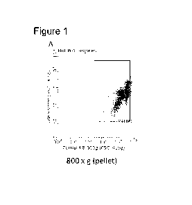
expressive of
5ox2 and 0ct4.
[0010] In some
embodiments, the PBD-PSCs are cultured with one or more
peptides. In some embodiments, the peptide is an extracellular matrix (ECM)
protein, a
cytokine, a growth factor, or an antigen. In some embodiments, the ECM protein
-2-
CA 03028422 2018-12-18
WO 2017/223233
PCT/US2017/038598
includes, for example, proteoglycans, non-proteoglycan polysaccharides, or
fibers, and
can include, for example, chondroitin sulfate, heparin sulfate, keratan
sulfate, hyaluronic
acid, agrin nidogen, collagen, elastin, entactin, fibronectin, laminin,
perlecan, total
protein, and/or protein fragments. In some embodiments, the cytokine(s)
is/are, for
example, lymphokines, interleukins, and chemokines, which can include, for
example, an
interleukin cytokine (IL-la, IL-1(3, IL-2, IL-3, IL-4, IL-5, IL-6, IL-7, IL-8,
IL-9, IL-10,
IL-11, IL-12, IL-13), an interferon (IFN-a, IFN-(3, and IFN-y), a tumor
necrosis factor
(TNFa and TNF-(3), a colony stimulating factor (GM-CSF and M-CSF), or a
combination
thereof In some embodiments, the growth factor includes, for example, an
epidermal
growth factor (EGF), a platelet derived growth factor (PDGF), a fibroblast
growth factor
(FGF and bFGF), a transforming growth factor (TGF-a and TGF-(3 1, 2, & 3), a
vascular
endothelial growth factor (VEGF), a hepatocyte growth factor (HGF), a
keratinocyte
growth factor (KGF), a nerve growth factor (NGF), erythropoietin (EPO), or an
insulin-
like growth factors (IGF-I and IGF-II), or a combination thereof In some
embodiments,
the PBD-PSCs are cultured with retinoic acid and/or with one or more of a
derivative of
retinoic acid.
[0011] In some
embodiments, the PBD-PSCs are transfected with one or more
heterologous genes encoding a peptide. In some embodiments, the peptide is an
extracellular matrix (ECM) protein, a cytokine, a growth factor, or an
antigen. In some
embodiments, the ECM protein includes, for example, proteoglycans, non-
proteoglycan
polysaccharides, or fibers, and can include, for example, chondroitin sulfate,
heparin
sulfate, keratan sulfate, hyaluronic acid, agrin nidogen, collagen, elastin,
entactin,
fibronectin, laminin, perlecan, total protein, and/or protein fragments. In
some
embodiments, the cytokine includes, for example, lymphokines, interleukins,
and
chemokines, which can include, for example, an interleukin cytokine (IL-la, IL-
1(3, IL-2,
IL-3, IL-4, IL-5, IL-6, IL-7, IL-8, IL-9, IL-10, IL-11, IL-12, IL-13), an
interferon (IFN-a,
IFN-(3, and IFN-y), a tumor necrosis factor (TNFa and TNF-(3), or a colony
stimulating
factor (GM-CSF and M-CSF), or a combination thereof In some embodiments, the
growth factor includes, for example, an epidermal growth factor (EGF), a
platelet derived
growth factor (PDGF), a fibroblast growth factor (FGF and bFGF), a
transforming growth
factor (TGF-a and TGF-(3 1, 2, & 3), a vascular endothelial growth factor
(VEGF), a
hepatocyte growth factor (HGF), a keratinocyte growth factor (KGF), a nerve
growth
-3-
CA 03028422 2018-12-18
WO 2017/223233
PCT/US2017/038598
factor (NGF), erythropoietin (EPO), or an insulin-like growth factors (IGF-I
and IGF-II),
or a combination thereof
[0012] In some
embodiments, PBD-PSCs are targeted to a specific region or
area within an organism, tissue, or organ. In some embodiments, the PBD-PSCs
are
attracted to a target region through the overexpression of parathyroid hormone
(PTH)
and/or the overexpression of parathyroid hormone-related protein (PTHrP) in
the area.
PTH and PTHrP act as chemoattractants to PBD-PSCs. In some embodiments, the
PBD-
PSCs are attracted to a region or area having a high concentration of PTH
and/or PTHrP.
In some embodiments, the high concentration or influx of PTH and/or PTHrP is a
result
of overexpression, native expression, and/or artificial placement.
[0013]
Embodiments also include an antibody or binding domain thereof
specific for a PTH1R on PBD-PSCs. In some embodiments, the antibody or binding
domain is a monoclonal antibody or binding fragment thereof or a poly clonal
antibody or
binding fragments thereof In some embodiments, the antibody or binding
fragment
thereof is a humanized antibody or a humanized binding fragment thereof
[0014]
Embodiments also include methods of isolating a pluripotent stem cell
population, wherein the stem cell population comprises PBD-PSCs that express
PTH1R.
In some embodiments, the method comprises contacting a sample comprising a
cell
population that comprises PBD-PSCs with an antibody or binding fragment
thereof
specific to PTH1R (preferably bound to a support or surface such as a bead,
membrane,
filter, container), so as to form a bound cell population (e.g., stem cell
population). In
some embodiments, the method further comprises isolating the antibody-bound
cell
population (e.g., stem cell population). In some embodiments, the method
comprises
releasing the bound cell population (e.g., stem cell population) from the
antibody or
binding fragment thereof In some embodiments, the method further comprises
contacting
a sample comprising the antibody/binding fragment thereof-bound cell
population (e.g.,
stem cell population) with magnetic beads, a surface, or support (e.g., the
magnetic beads,
surface, support, membrane, or filter can include immobilized or bound
antibodies or
binding fragments thereof specific for PTH1R). In some embodiments, the method
comprises applying a magnetic field and/or centrifugation to the sample,
thereby isolating
the antibody/binding fragment thereof-bound cell population (e.g., stem cell
population).
[0015] More
embodiments include methods of using PBD-PSCs for the
treatment or amelioration or inhibition of a disorder, disease or disease
state, wherein the
-4-
CA 03028422 2018-12-18
WO 2017/223233
PCT/US2017/038598
disorder, disease or disease state is infertility, a skin or hair disorder,
such as hair loss,
hair thinning, a bone disorder, a cancer, a neurological disorder, autoimmune
disorders,
or a combination thereof In some embodiments, the subject suffering from one
or more
disorders is identified by clinical or diagnostic evaluation for one or more
of the
aforementioned disorders, diseases or disease states and said individual is
administered
stem cell population comprising PBD-PSCs, wherein the stem cell population
comprising
PBD-PSCs is administered intravenously, intra-arterially, subcutaneously,
transdermally,
intravitreally, intraocularly, subconjunctivally, retrobulbarly, sub-orbitally
or topically, or
by a combination thereof In some embodiments, the treatment or amelioration of
the
disorder, disease or disease state results in the amelioration, improvement,
reversal, or
regression of the disorder. In some embodiments, intra-arterial injections
include
injection into the uterine, pancreatic, or ovarian artery, or combinations
thereof
[0016] Some
embodiments provided herein relate to a method of improving,
ameliorating, reversing, inhibiting or treating diabetes. In some embodiments,
the method
includes administering an effective amount of a composition. In some
embodiments, the
composition includes an isolated population of pluripotent stem cells isolated
as
described herein. In some embodiments, the composition includes the isolated
population
of pluripotent stem cells as described herein to a subject in need. In some
embodiments,
diabetes is Type 1 diabetes. In some embodiments, the composition is
administered intra-
arterially into the pancreatic artery. In some embodiments, the composition is
administered once every 12 weeks. In some embodiments, administration of the
composition reduces an amount of daily average insulin usage by the subject.
In some
embodiments, the daily average insulin usage is reduced by 2-10%, 5-20%, 10-
30%, 20-
40%, 30-50%, 40-60%, 50-70%, 60-80%, 70-90%, or 80-100% or within a range
defined
by any two of the aforementioned percentages.
[0017] Some
embodiments provided herein relate to a method of reducing the
average daily insulin dose in a subject suffering from diabetes. In some
embodiments, the
method includes administering an effective amount of a composition. In some
embodiments, the composition includes an isolated population of pluripotent
stem cells
isolated by the method as described herein. In some embodiments, the
composition
includes the isolated population of pluripotent stem cells as described herein
to a subject
in need. In some embodiments, diabetes is Type 1 diabetes. In some
embodiments, the
composition is administered intra-arterially into the pancreatic artery. In
some
-5-
CA 03028422 2018-12-18
WO 2017/223233
PCT/US2017/038598
embodiments, the composition is administered once every 12 weeks. In some
embodiments, administration of the composition reduces an amount of daily
average
insulin usage by the subject by 2-10%, 5-20%, 10-30%, 20-40%, 30-50%, 40-60%,
50-
70%, 60-80%, 70-90%, or 80-100% or within a range defined by any two of the
aforementioned percentages.
[0018] Some
embodiments provided herein relate to a method for growing an
isolated population of pluripotent stem cells on a matrix. In some
embodiments, the
method includes isolating a population of pluripotent stem cells from a sample
by the
method of as described herein, contacting a nanofiber matrix with the isolated
pluripotent
stem cells, and growing the isolated pluripotent stem cells on the nanofiber
matrix. In
some embodiments, a matrix including pluripotent stem cells is produced. In
some
embodiments, the nanofiber matrix includes polymer fibers, including, for
example,
polydimethylsiloxane, polyglycerol sebacate, polycaprolactone, polylactic
acid,
polyglycolic acid, cellulose, alginate, agar, agarose, collagen I, collagen
IV, hyaluronic
acid, fibrin, poly-L-lactide, or poly(lactic-co-glycolic acid). In some
embodiments, the
polymer fibers are electrospun. In some embodiments, the polymer fibers range
in
thickness from 200 to 700 p.m (e.g., 200, 300, 400, 500, 600, or 700 p.m or
within a range
defined by any two of the aforementioned thicknesses). In some embodiments,
the
pluripotent stem cells bind to and lay down in the matrix within 2-40 minutes
(e.g., 2, 3,
4, 5, 6, 7, 8, 9, 10, 15, 20, 25, 30, 35, or 40 minutes or an amount within a
range defined
by any two of the aforementioned values). In some embodiments, the matrix
comprising
pluripotent stem cells is used for injecting into joints or bones. In some
embodiments, the
matrix comprising pluripotent stem cells is used for covering a wound with or
without a
dressing, which may include a gel.
[0019]
Accordingly, some aspects described herein relate to the following
alternatives:
[0020] 1. A
pluripotent stem cell characterized in that it expresses parathyroid
hormone type 1 receptor (PTH1R), is 2.5 to 4.5 p.m in diameter, such as 2.5,
2.6, 2.7, 2.8,
2.9, 3.0, 3.1, 3.2, 3.3, 3.4, 3.5, 3.6, 3.7, 3.8, 3.9, 4.0, 4.1, 4.2, 4.3,
4.4, or 4.5 p.m in
diameter, or an amount within a range defined by any two of the aforementioned
values,
forms embryoid-like bodies when cultured, and is capable of differentiation
into
ectoderm, mesoderm, and endoderm upon culture.
-6-
CA 03028422 2018-12-18
WO 2017/223233
PCT/US2017/038598
[0021] 2. The
pluripotent stem cell of alternative 1, wherein the cell
population is cultured with one or more peptide.
[0022] 3. The
pluripotent stem cell of alternative 2, wherein the peptide is an
extracellular matrix (ECM) protein, a cytokine, a growth factor, or an
antigen.
[0023] 4. The
pluripotent stem cell of any of alternatives 1-3, wherein the
pluripotent stem cell is cultured with retinoic acid and/or with one or more
derivative of
retinoic acid.
[0024] 5. The
pluripotent stem cell of any of alternatives 1-4, wherein the
pluripotent stem cell is transfected with a heterologous gene encoding a
peptide.
[0025] 6. The
pluripotent stem cell of alternative 5, wherein the peptide is an
extracellular matrix (ECM) protein, a cytokine, a growth factor, or an
antigen.
[0026] 7. The
pluripotent stem cell of any one of alternatives 1-6, wherein the
pluripotent stem cell is isolated from peripheral blood, seminal fluid, and/or
ovarian
follicular fluid.
[0027] 8. An
isolated pluripotent stem cell population present in peripheral
blood, seminal fluid, and ovarian follicular fluid, which:
expresses parathyroid hormone type 1 receptor (PTH1R);
is 2.5 to 4.5 um in diameter, such as 2.5, 2.6, 2.7, 2.8, 2.9, 3.0, 3.1, 3.2,
3.3, 3.4, 3.5, 3.6, 3.7, 3.8, 3.9, 4.0, 4.1, 4.2, 4.3, 4.4, or 4.5 um in
diameter, or an
amount within a range defined by any two of the aforementioned values;
forms embryoid-like bodies when cultured; and
is capable of differentiation into ectoderm, mesoderm, and endoderm upon
culture.
[0028] 9. The
isolated pluripotent stem cell population of alternative 8,
wherein the cell population is:
positive for CD90 and CD133;
positive/negative for CD29, CD34, CD105, and CD106; and
negative for SSEA-3, CD200, and CD45.
[0029] 10. The
isolated pluripotent stem cell population of alternatives 8-9,
wherein the cell population expresses Sox2 and 0ct4.
[0030] 11. The
isolated pluripotent stem cell population of any of alternatives
8-10, wherein the isolated pluripotent stem cell population originates from
human,
equine, canine, or camel sources.
-7-
CA 03028422 2018-12-18
WO 2017/223233
PCT/US2017/038598
[0031] 12. The isolated pluripotent stem cell population of any of
alternatives
8-11, wherein the cell population is cultured with a peptide.
[0032] 13. The isolated pluripotent stem cell population of
alternative 12,
wherein the peptide is an extracellular matrix (ECM) protein, a cytokine, a
growth factor,
or an antigen.
[0033] 14. The isolated pluripotent stem cell population of any of
alternatives
8-13, wherein the cell population is cultured with retinoic acid and/or with
one or more
derivative of retinoic acid.
[0034] 15. The isolated pluripotent stem cell population of any of
alternatives
8-14, wherein the pluripotent stem cell is transfected with a heterologous
gene encoding a
peptide.
[0035] 16. The isolated pluripotent stem cell population of
alternative 15,
wherein the peptide is an extracellular matrix (ECM) protein, a cytokine, a
growth factor,
or an antigen.
[0036] 17. An antibody specific for parathyroid hormone type 1
receptor
(PTH1R) on a stem cell.
[0037] 18. The antibody of alternative 17, wherein the antibody is a
monoclonal antibody.
[0038] 19. The antibody of any of alternatives 17-18, wherein the
antibody is
a humanized antibody.
[0039] 20. A method for isolating a stem cell population as described
in any
one of alternatives 1-16, the method comprising:
contacting a sample comprising the stem cell population with an antibody
specific for PTH1R, to form an antibody-bound stem cell;
isolating the antibody-bound stem cell; and
releasing the stem cell from the antibody.
[0040] 21. The method of alternative 20, further comprising:
contacting a sample comprising the antibody-bound stem cell with
magnetic beads; and
applying a magnetic field, thereby isolating the antibody-bound stem cell.
[0041] 22. The method of any one of alternatives 20-21, wherein the
sample is
obtained from a sample selected from the group consisting of adipose tissue,
bone
-8-
CA 03028422 2018-12-18
WO 2017/223233
PCT/US2017/038598
marrow, peripheral blood, seminal fluid, ovarian follicular fluid, and
combinations
thereof
[0042] 23. The
method of any one of alternatives 20-22, wherein the isolated
stem cell population comprises stem cells that are 2.5 p.m in diameter and
expressive of
stem cell CD markers.
[0043] 24. The
method of any one of alternatives 20-23, wherein the isolated
stem cell population comprises stem cells that are isolated in quantities of
at least or equal
to 10,000, 50,000, 100,000, 500,000, 1,000,000, 5,000,000, or 10,000,000
cells/mL tissue
sample, or an amount within a range defined by any two of the aforementioned
values.
[0044] 25. A
method for isolating a pluripotent stem cell population as
described in any one of alternatives 1-16, the method comprising:
obtaining peripheral blood from an individual;
contacting the peripheral blood with an extracorporeal porous membrane
configured to capture the pluripotent stem cell population as described in
any one of alternatives 1-16;
applying centrifugal or gravity force or pressure to peripheral blood and
the membrane;
capturing the pluripotent stem cell population;
collecting pass-through blood; and
reinfusing the pass-through blood into the individual.
[0045] 26. The
method of alternative 25, wherein the captured pluripotent
stem cell population is prepared for subsequent reinfusion into the
individual.
[0046] 27. A
method for isolating a stem cell population as described in any
one of alternatives 1-16, the method comprising:
contacting a sample comprising the stem cell population with anti-CD45
antibody; and
removing cells unbound by anti-CD45 antibody.
[0047] 28. The
method of alternative 27, wherein the sample is peripheral
blood, plasma, or platelet lysate.
[0048] 29. The
method of any one of alternatives 27-28, wherein the isolated
stem cell population comprises stem cells that are isolated in quantities of
about 10,000,
50,000, 100,000, 500,000, 1,000,000, 5,000,000, or 10,000,000 cells/mL tissue
sample, or
an amount within a range defined by any two of the aforementioned values.
-9-
CA 03028422 2018-12-18
WO 2017/223233
PCT/US2017/038598
[0049] 30. A method of improving fertility in a subject comprising:
selecting a subject in need of improved fertility; and
administering a therapeutically effective amount of an isolated population
of pluripotent stem cells isolated by the method of any one of alternatives 20-
29
or the isolated population of pluripotent stem cells of any one of
alternatives 1-16
to said subject.
[0050] 31. The method of alternative 30, wherein the pluripotent stem
cells
are autologous.
[0051] 32. The method of any one of alternatives 30-31, wherein the
therapeutically effective amount is an amount sufficient to cause a detectable
improvement in ovarian function, oocyte quality, endometrial thickness,
endometrial
receptivity, or combinations thereof
[0052] 33. The method of any one of alternatives 30-32, wherein the
therapeutically effective amount of isolated population of pluripotent stem
cells is
administered by injection into the testicles or uterus.
[0053] 34. The method of any one of alternatives 30-32, wherein the
therapeutically effective amount of isolated population of pluripotent stem
cells is
administered intravenously to the subject into a uterine artery to promote
thickening and
receptivity of an endometrial wall.
[0054] 35. The method of alternative 34, wherein thickening of the
endometrial wall is increased by at least 10%, 20%, 30%, 40%, 50%, 60%, 70%,
or 80%,
or greater or within a range defined by any two of the aforementioned
percentages.
[0055] 36. The method of any one of alternatives 30-32, wherein the
therapeutically effective amount of isolated population of pluripotent stem
cells is
administered intravenously to the subject into an ovarian artery to increase
the number
and quality of eggs.
[0056] 37. The method of any one of alternatives 30-36, wherein the
pluripotent stem cells are isolated from a sample selected from the group
consisting of
adipose tissue, bone marrow, peripheral blood, seminal plasma, and ovarian
follicular
fluid, and/or combinations thereof
[0057] 38. A method of targeting a population of pluripotent stem
cells
isolated by the method of any one of alternatives 20-29 or the isolated
population of
-10-
CA 03028422 2018-12-18
WO 2017/223233
PCT/US2017/038598
pluripotent stem cells of any one of alternatives 1-16 to an area of interest
comprising
expressing parathyroid hormone-related protein in said area.
[0058] 39. A
method of increasing the production of dermal collagen and/or
elastin in an area of skin in a subject comprising administering a
therapeutically effective
amount of a composition comprising an isolated population of pluripotent stem
cells
isolated by the method of any one of alternatives 20-29 or comprising the
isolated
population of pluripotent stem cells of any one of alternatives 1-16.
[0059] 40. The
method of alternative 39, wherein the composition is
administered by one or more subcutaneous injections.
[0060] 41. The
method of any one of alternatives 39-40, wherein the increased
production of dermal collagen and/or elastin reduces fine lines, reduces
wrinkles,
increases radiance, or increases dermal tightness, and/or combinations thereof
[0061] 42. A
method for inhibiting hair loss or promoting hair growth
comprising administering an effective amount of a composition comprising an
isolated
population of pluripotent stem cells isolated by the method of any one of
alternatives 20-
26 or comprising the isolated population of pluripotent stem cells of any one
of
alternatives 1-16 to a subject in need.
[0062] 43. The
method of alternative 42, wherein the composition is
transdermally administered to the subject.
[0063] 44. The
method of alternative 42, wherein the composition is
subcutaneously injected to the subject.
[0064] 45. A
method of improving, ameliorating, reversing, or treating a bone
disorder comprising administering an effective amount of a composition
comprising an
isolated population of pluripotent stem cells isolated by the method of any
one of
alternatives 20-29 or comprising the isolated population of pluripotent stem
cells of any
one of alternatives 1-16 to a subject in need.
[0065] 46. The
method of alternative 45, wherein the bone disorder is a bone
injury.
[0066] 47. The
method of alternative 46, wherein the bone injury is a bone
fracture.
[0067] 48. The
method of alternative 47, wherein the bone fracture is a
spinous fracture.
-11-
CA 03028422 2018-12-18
WO 2017/223233
PCT/US2017/038598
[0068] 49. The
method of alternative 45, wherein the bone disorder is
osteoporosis.
[0069] 50. The
method of alternative 45, wherein the bone disorder is
osteopenia.
[0070] 51. The
method of any one of alternatives 45-50, wherein the
composition is subcutaneously injected to the subject.
[0071] 52. The
method of any one of alternatives 45-50, wherein the
composition is administered intravenously to the subject.
[0072] 53. The
method of any one of alternatives 45-50, wherein the
composition is administered intra-arterially to the subject.
[0073] 54. The
method of alternative 53, wherein the composition is
administered into one or more of the uterine, pancreatic, or ovarian artery.
[0074] 55. A
method of improving, ameliorating, inhibiting, reversing, or
treating a neurological disorder comprising administering an effective amount
of a
composition comprising an isolated population of pluripotent stem cells
isolated by the
method of any one of alternatives 20-29 or comprising the isolated population
of
pluripotent stem cells of any one of alternatives 1-16 to a subject in need.
[0075] 56. A
method of improving, ameliorating, inhibiting, reversing, or
treating metastatic carcinoma comprising administering an effective amount of
a
composition comprising an isolated population of pluripotent stem cells
isolated by the
method of any one of alternatives 20-29 or comprising the isolated population
of
pluripotent stem cells of any one of alternatives 1-16 to a subject in need.
[0076] 57. A
method of preventing, treating, inhibiting, preventing, or
ameliorating an autoimmune disorder comprising identifying a subject in need
and
administering to said subject an effective amount of a composition comprising
an isolated
population of pluripotent stem cells isolated by the method of any one of
alternatives 20-
29 or comprising the isolated population of cells comprising stem cells of any
one of
alternatives 1-16.
[0077] 58.
Peripheral blood derived pluripotent stem cells (PBD-PSCs) for
use as a medicament.
[0078] 59. A
method of improving, inhibiting, ameliorating, reversing, or
treating diabetes comprising administering an effective amount of a
composition
comprising an isolated population of pluripotent stem cells isolated by the
method of any
-12-
CA 03028422 2018-12-18
WO 2017/223233
PCT/US2017/038598
one of alternatives 20-29 or comprising the isolated population of pluripotent
stem cells
of any one of alternatives 1-16 to a subject in need.
[0079] 60. The method of alternative 59, wherein diabetes is Type 1
diabetes.
[0080] 61. The method of any one of alternatives 59-60, wherein the
composition is administered intra-arterially into the pancreatic artery.
[0081] 62. The method of any one of alternatives 59-61 wherein the
composition is administered once every 12 weeks.
[0082] 63. The method of any one of alternatives 59-62, wherein
administration of the composition reduces an amount of daily average insulin
usage by
the subject.
[0083] 64. The method of alternative 63, wherein the daily average
insulin
usage is reduced by about 2-10%, 5-20%, 10-30%, 20-40%, 30-50%, 40-60%, 50-
70%,
60-80%, 70-90%, or 80-100% or within a range defined by any two of the
aforementioned percentages.
[0084] 65. A method of reducing the average daily insulin dose in a
subject
suffering from diabetes, comprising administering an effective amount of a
composition
comprising an isolated population of pluripotent stem cells isolated by the
method of any
one of alternatives 20-29 or comprising the isolated population of pluripotent
stem cells
of any one of alternatives 1-16 to a subject in need.
[0085] 66. The method of alternative 65, wherein diabetes is Type 1
diabetes.
[0086] 67. The method of any one of alternatives 65-66, wherein the
composition is administered intra-arterially into the pancreatic artery.
[0087] 68. The method of any one of alternatives 65-67 wherein the
composition is administered once every 12 weeks.
[0088] 69. The method of any one of alternatives 65-68, wherein
administration of the composition reduces an amount of daily average insulin
usage by
the subject by about 2-10%, 5-20%, 10-30%, 20-40%, 30-50%, 40-60%, 50-70%, 60-
80%, 70-90%, or 80-100% or within a range defined by any two of the
aforementioned
percentages.
[0089] 70. A method for growing an isolated population of pluripotent
stem
cells on a matrix, comprising:
isolating a population of pluripotent stem cells from a sample by the
method of any one of alternatives 20-29;
-13-
CA 03028422 2018-12-18
WO 2017/223233
PCT/US2017/038598
contacting a nanofiber matrix with the isolated pluripotent stem cells; and
growing the isolated pluripotent stem cells on the nanofiber matrix;
wherein a matrix comprising pluripotent stem cells is produced.
[0090] 71. The
method of alternative 70, wherein the nanofiber matrix
comprises polymer fibers, including, for example, polydimethylsiloxane,
polyglycerol
sebacate, polycaprolactone, polylactic acid, polyglycolic acid, cellulose,
alginate, agar,
agarose, collagen I, collagen IV, hyaluronic acid, fibrin, poly-L-lactide,
and/or
poly(lactic-co-glycolic acid).
[0091] 72. The
method of alternative 71, wherein the polymer fibers are
el ectro spun.
[0092] 73. The
method of any one of alternatives 71-72, wherein the polymer
fibers are of a thickness that is at least or equal to 200, 300, 400, 500,
600, or 700 p.m or
within a range defined by any two of the aforementioned thicknesses.
[0093] 74. The
method of any one of alternatives 70-73, wherein the
pluripotent stem cells bind to and lay down in the matrix within about 2-40
minutes.
[0094] 75. The
method of any one of alternatives 70-74, wherein the matrix
comprising pluripotent stem cells is used for injecting into joints or bones.
[0095] 76. The
method of any one of alternatives 70-74, wherein the matrix
comprising pluripotent stem cells is used for covering a wound.
[0096] 77. A
method of improving, ameliorating, reversing, inhibiting, or
treating macular degeneration comprising administering an effective amount of
a
composition comprising an isolated population of pluripotent stem cells
isolated by the
method of any one of alternatives 20-29 or comprising the isolated population
of
pluripotent stem cells of any one of alternatives 1-16 to a subject in need.
[0097] The
method of alternative 77, wherein macular degeneration is dry or
wet macular degeneration.
[0098] The
method of any one of alternatives 77-78, wherein the composition
is administered by intravenous, intravitreal, intraocular, subconjunctival,
retrobulbar, or
sub-orbital injection.
[0099] The
method of any one of alternatives 77-79 wherein the composition
is administered once every 12 weeks.
[0100] The
method of any one of alternatives 77-80, wherein administration
of the composition improves visual acuity in the subject.
-14-
CA 03028422 2018-12-18
WO 2017/223233
PCT/US2017/038598
[0101] The
method of alternative 81, wherein the visual acuity is improved to
about 20/20, 20/30, 20/40, 20/50, 20/60, 20/70, 20/80, 20/90, or 20/100 or
within a range
defined by any two of the aforementioned amounts.
[0102] These
features, together with other features herein further explained,
will become obvious through a reading of the following description of the
drawings and
detailed description.
BRIEF DESCRIPTION OF THE DRAWINGS
[0103] In
addition to the features described above, additional features and
variations will be readily apparent from the following descriptions of the
drawings and
exemplary embodiments. It is to be understood that these drawings depict
typical
embodiments, and are not intended to be limiting in scope.
[0104] Figure 1
depicts the characterization of PBD-PSCs described herein.
The flow cytometric results of parathyroid hormone 1 receptor (PTH1R) positive
cells
after centrifugation and filtration at 800 x g (Panel A), 1000 x g (Panel 13),
and 1200 x g
(Panel C).
[0105] Figure 2
depicts the characterization of PBD-PSCs described herein
by flow cytometry. PTH1R positive cells from peripheral blood are shown in
Panel A.
Panel B compares PTH1R positive cells and CD133 positive cells. Panel C
compares
CD133 positive cells and CD90 positive cells. Panel D compares SSEA4 positive
cells
and CD45 positive cells.
[0106] Figure 3
shows the characterization of PTH1R positive cells from
ovarian follicular fluid after centrifugation and filtration. Panel A shows
the PTH1R
positive cells. Panel B compares PTH1R positive cells and CD133 positive
cells. Panel C
compares PTH1R positive cells and CD90 positive cells.
[0107] Figures
4A-4C depict the characterization of PTH1R positive cells
from ovarian follicular fluid and from peripheral blood. Figure 4A depicts
PTHR positive
cells, Figure 4B depicts CD133 positive cells, and Figure 4C depicts PTHR/CD90
positive cells.
[0108] Figures
5A-5E depict a micrograph of PBD-PSCs as described herein,
with antibody staining against PTH Receptor (Figure 5A), CD90 (Figure 5B),
CD133
(Figure 5C), SSEA-4 (Figure 5D), and CD45 (Figure 5E).
-15-
CA 03028422 2018-12-18
WO 2017/223233
PCT/US2017/038598
[0109] Figure 6
depicts a micrograph of the PBD-PSCs described herein. The
stem cells are 2.5-4.5 p.m in diameter and carry the pluripotent stem cell
marker Kyoto
probe 1.
[0110] Figure 7
is a micrograph showing that the PBD-PSCs form embryoid-
like bodies when cultured, providing evidence that these cells are very
primitive and
potent stem cells.
[0111] Figure 8
shows the gene expression of PBD-PSCs, using conventional
Rt-PCR for stem cell marker detection. Lane 1 is the DNA ladder, lane 2 is a
control
GAPDH1, lane 3 shows detection of Sox2, and lane 4 shows detection of 0ct4.
[0112] Figure 9
shows micrographs of the PBD-PSCs from different animal
sources; equine, canine, and camel.
[0113] Figure
10 illustrates the method of developing unique monoclonal
antibodies that target a unique marker to the PBD-PSCs. The monoclonal
antibody can be
used to select the specific pluripotent stem cells for diagnostic and
treatment purposes.
[0114] Figure
11 illustrates one embodiment for the process for the isolation
of the PBD-PSCs. The sample containing stem cells (tissue homogenate,
peripheral
blood, or other stem cell sources as described herein) is contacted with
monoclonal
antibodies developed as shown in Figure 10. Magnetic beads are added to the
mixture,
and a magnetic field is applied. A wash buffer is introduced, and the PBD-PSCs
are
isolated.
[0115] Figure
12 illustrates a method for isolating the PBD-PSCs in an
extracorporeal system. Peripheral blood from a subject is passed through an
extracorporeal membrane, preferably a porous membrane, configured to capture
the PBD-
PSCs. A centrifugal or gravity force is applied to the peripheral blood, such
that the PBD-
PSCs are captured and collected. Optionally, after the PBD-PSCs are removed
from the
peripheral blood, the pass-through blood is reinfused into the subject.
[0116] Figure
13 illustrates the processes involved in organismal aging.
Extrinsic influences act together with intrinsic influences causing genomic,
epigenomic,
and proteomic changes that result in functional decline. This in turn leads to
tissue
dysfunction and organismal aging.
[0117] Figure
14 shows the typical progression of stem cells. Young stem
cells are capable of producing healthy progeny. However, as stem cells age,
the resulting
-16-
CA 03028422 2018-12-18
WO 2017/223233
PCT/US2017/038598
progeny declines in function due to loss of lineage specificity, depletion due
to loss of
self-renewal, depletion due to senescence, and malignant transformation.
[0118] Figure
15 illustrates the negative correlation between the PTHR-
positive stem cells per mL of plasma (y-axis) by age of subject (x-axis).
[0119] Figure
16 illustrates the genetic and epigenetic alterations that occur
over time to any given stem cell population within an organism.
[0120] Figure
17 illustrates the properties of cancer stem cells (CSCs),
including the chemo and radiotherapy resistance of CSCs.
[0121] Figure
18 illustrates the factors in young blood versus old blood for
the ability to activate stem cells and rejuvenate organs in cells in old mice.
Factors in old
blood appear to inhibit regenerative capacity in young mice.
[0122] Figure
19 shows that over time and over the course of replication,
stem cells show increased effects of aging. However, the aging effects
experienced by
stem cells over chronological and replicative life span can be reversed by the
treatments
described herein.
[0123] Figures
20A-20D depict electron micrographs of PBD-PSCs obtained
from a healthy male, grown on a nanofiber matrix. The micrographs show the PBD-
PSCs
on the matrix over time, at time points of 0 minutes (Figure 20A), 5 minutes
(Figure
20B), 30 minutes (Figure 20C), and 120 minutes (Figure 20D).
[0124] Figure
21 shows an exemplary treatment of skin using a PBD-PSC
preparation as a mesotherapy agent to counter premature skin aging by
increasing the
presence of dermal collagen and elastin. Skin measurements of collagen and
elastin were
taken prior to treatment at day 0 and 30 days following treatment.
[0125] Figure
22 shows an exemplary treatment of hair follicles and scalp
using a PBD-PSC preparation so as to induce hair regrowth, and the figure
depicts follicle
growth prior to treatment and 10 weeks following treatment.
[0126] Figures
23A-23C show images following treatment of a spinous
process of a spinous process fracture using an embodiment of a PBD-PSC
treatment
method. Figure 23A shows the fracture prior to treatment. Figure 23B shows
treatment
of a coronal cervical fracture using a PBD-PSC treatment. The left image shows
the
fracture prior to treatment, and the right image shows the fracture 4 months
after PBD-
PSC treatment. Figure 23C shows treatment of a sagittal cervical fracture
using a PBD-
-17-
CA 03028422 2018-12-18
WO 2017/223233
PCT/US2017/038598
PSC treatment. The left image shows the fracture prior to treatment, and the
right image
shows the fracture 4 months after PBD-PSC treatment.
[0127] Figure
24 depicts the effects of osteoporosis. The left image depicts a
cutaway view of a normal, healthy bone. The right image depicts a cutaway view
of a
bone with osteoporosis, showing increased porosity and decreased bone density.
[0128] Figure
25 depicts the bone density of a subject suffering from severe
osteoporosis. The top panel shows the change in bone density in the lumbar
spine
following a single infusion of PBD-PSCs. The bottom panel shows the change in
bone
density in the left hip following a single infusion of PBD-PSCs. The y-axis
represents
bone density in g/cm2, and the x-axis represents time.
[0129] Figure
26 depicts the change in bone density and the T-score of a
subject with osteopenia. The top panel shows the change in bone density and T-
score in
the lumbar spine following a single PBD-PSC therapy. The bottom panel shows
the
change in bone density and T-score in the left hip following a single PBD-PSC
therapy.
[0130] Figure
27 shows an exemplary treatment of endometrial thinning
using a PBD-PSC intrauterine arterial injection. The pre-treatment image
depicts high
levels of endometrial thinning, whereas the post-treatment image shows that
the thinning
has largely dissipated.
[0131] Figure
28 shows an exemplary treatment of pulmonary metastases
using PBD-PSC. The figure shows a CT image of the pulmonary metastases at 30
days
(left) and 129 days (right) following transplantation of a PBD-PSC allograft,
and shows a
complete remission of metastases at 129 days after transplantation.
[0132] Figure
29 depicts a decrease in the average insulin usage following
doses of PBD-PSCs. Two doses of PBD-PSCs were administered to two subjects, a
17
year old male and a 16 year old female, at weeks 1 and 13. A decrease in the
average
daily insulin usage is observed following the PBD-PSC therapy.
DETAILED DESCRIPTION
[0133] In the
following detailed description, reference is made to the
accompanying drawings, which form a part hereof In the drawings, similar
symbols
typically identify similar components, unless context dictates otherwise. The
illustrative
embodiments described in the detailed description, drawings, and claims are
not meant to
be limiting. Other embodiments may be utilized, and other changes may be made,
without
-18-
CA 03028422 2018-12-18
WO 2017/223233
PCT/US2017/038598
departing from the spirit or scope of the subject matter presented herein. It
will be readily
understood that the aspects of the present disclosure, as generally described
herein, and
illustrated in the Figures, can be arranged, substituted, combined, separated,
and designed
in a wide variety of different configurations, all of which are explicitly
contemplated
herein.
[0134]
Peripheral blood derived pluripotent stem cells (PBD-PSCs) are very
small stem cells from 2.5-4.5 p.m in diameter, such as 2.5, 2.6, 2.7, 2.8,
2.9, 3.0, 3.1, 3.2,
3.3, 3.4, 3.5, 3.6, 3.7, 3.8, 3.9, 4.0, 4.1, 4.2, 4.3, 4.4, or 4.5 p.m in
diameter, or an amount
within a range defined by any two of the aforementioned values. PBD-PSCs are
very
primitive stem cells that have a high potency for differentiating into a
variety of
specialized lineages. Accordingly, PBD-PSCs have a high propensity for
applications in
the areas of regenerative medicine and in particular, for anti-aging
therapeutics. In
particular, PBD-PSCs are a powerful tool for a number of applications,
including, for
example: allogenic regenerative cell therapy, wherein donor cells are used for
differentiation and are capable of releasing growth factors for the repair of
damaged
tissue; autologous regenerative cell therapy, wherein the patient's own cells
are used for
reprogramming, expansion, and/or differentiation for the treatment of damaged
tissue by
permanently integrating into the tissue; and tissue engineering, wherein a
patient's own
cells are placed upon a scaffold to create a neo-tissue, which is then
engrafted onto
damaged tissue to repair the tissue. PBD-PSCs are characterized in that they
are highly
expressive of PTH1R. Accordingly, in some aspects, PBD-PSCs are also referred
to
herein as PTH1R-positive stem cells.
Definitions
[0135] Unless
defined otherwise, technical and scientific terms used herein
have the same meaning as commonly understood by one of ordinary skill in the
art to
which the present disclosure belongs. See, e.g. Singleton et al., Dictionary
of
Microbiology and Molecular Biology 2nd ed., J. Wiley & Sons (New York, NY
1994);
Sambrook et al., Molecular Cloning, A Laboratory Manual, Cold Springs Harbor
Press
(Cold Springs Harbor, NY 1989). For purposes of the present disclosure, the
following
terms are defined below.
[0136] The
articles "a" and "an" are used herein to refer to one or to more
than one (for example, to at least one) of the grammatical object of the
article. By way of
example, "an element" means one element or more than one element.
-19-
CA 03028422 2018-12-18
WO 2017/223233
PCT/US2017/038598
[0137] By
"about" is meant a quantity, level, value, number, frequency,
percentage, dimension, size, amount, weight or length that varies by as much
as 30, 25,
20, 15, 10, 9, 8, 7, 6, 5, 4, 3, 2 or 1% to a reference quantity, level,
value, number,
frequency, percentage, dimension, size, amount, weight or length.
[0138]
Throughout this specification, unless the context requires otherwise,
the words "comprise," "comprises," and "comprising" will be understood to
imply the
inclusion of a stated step or element or group of steps or elements but not
the exclusion of
any other step or element or group of steps or elements.
[0139] By
"consisting of' is meant including, and limited to, whatever follows
the phrase "consisting of" Thus, the phrase "consisting of' indicates that the
listed
elements are required or mandatory, and that no other elements may be present.
By
"consisting essentially of' is meant including any elements listed after the
phrase, and
limited to other elements that do not interfere with or contribute to the
activity or action
specified in the disclosure for the listed elements. Thus, the phrase
"consisting essentially
of' indicates that the listed elements are required or mandatory, but that
other elements
are optional and may or may not be present depending upon whether or not they
materially affect the activity or action of the listed elements.
[0140] In some
embodiments, the "purity" of any given agent (e.g., antibody,
polypeptide binding agent) in a composition may be specifically defined. For
instance,
certain compositions may comprise an agent that is at least 80, 85, 90, 91,
92, 93, 94, 95,
96, 97, 98, 99, or 100% pure, including all decimals in between, as measured,
for
example and by no means limiting, by high pressure liquid chromatography
(HPLC), a
well-known form of column chromatography used frequently in biochemistry and
analytical chemistry to separate, identify, and quantify compounds.
[0141] As used
herein, the terms "function" and "functional" and the like refer
to a biological, enzymatic, or therapeutic function.
[0142] The term
"isolated" is meant material that is substantially or essentially
free from components that normally accompany it in its native state. For
example, an
"isolated stem cell" or "isolated population of stem cells" as used herein,
includes a stem
cell or population of stem cells that has been purified from sample material,
including
other cells, debris, or extraneous sample material from its naturally-
occurring state,
Alternatively, an "isolated stem cell" or "isolated population of stem cells"
and the like,
as used herein, includes the in vitro, extracorporeal, or other isolation
and/or purification
-20-
CA 03028422 2018-12-18
WO 2017/223233
PCT/US2017/038598
of a cell or population of cells from its natural environment, and from
association with
other components of the sample or material in which it occurs. In some
embodiments,
isolated means that the component is not significantly associated with in vivo
substances.
[0143] The
practice of the present disclosure will employ, unless indicated
specifically to the contrary, conventional methods of molecular biology and
recombinant
DNA techniques within the skill of the art, many of which are described below
for the
purpose of illustration. Such techniques are explained fully in the
literature. See, e.g. ,
Sambrook, el al, Molecular Cloning: A Laboratory Manual (3rd Edition, 2000);
DNA
Cloning: A Practical Approach, vol. 1 & II (D. Glover, ed.); Oligonucleotide
Synthesis
(N. Gait, ed., 1984); Oligonucleotide Synthesis: Methods and Applications (P.
Herdewijn,
ed., 2004); Nucleic Acid Hybridization (B. Hames & S. Higgins, eds., 1985);
Nucleic
Acid Hybridization: Modern Applications (Buzdin and Lukyanov, eds., 2009);
Transcription and Translation (B. Hames & S. Higgins, eds., 1984); Animal Cell
Culture
(R. Freshney, ed., 1986); Freshney, R.I. (2005) Culture of Animal Cells, a
Manual of
Basic Technique, 5th Ed. Hoboken NJ, John Wiley & Sons; B. Perbal, A Practical
Guide
to Molecular Cloning (3rd Edition 2010); Farrell, R., RNA Methodologies: A
Laboratory
Guide for Isolation and Characterization (3rd Edition 2005).
[0144] As used
herein, the term "antibody" includes polyclonal antibodies,
monoclonal antibodies (including full length antibodies which have an
immunoglobulin
Fc region), antibody compositions with polyepitopic specificity, multispecific
antibodies
(e.g., bispecific antibodies, diabodies, and single-chain molecules, and
antibody
fragments (e.g., Fab or F(ab')2, and Fv). For the structure and properties of
the different
classes of antibodies, see e.g., Basic and Clinical Immunology, 8th Edition,
Daniel P.
Sties, Abba I. Terr and Tristram G. Parsolw (eds), Appleton & Lange, Norwalk,
Conn.,
1994, page 71 and Chapter 6.
[0145] In some
embodiments, an antibody against PBD-PSCs is provided. In
some embodiments, the antibody is a monoclonal or polyclonal antibody. In some
embodiments, the antibody is a humanized antibody. An "isolated antibody" is
an
antibody that is not associated with naturally-associated components,
including other
naturally-associated antibodies, that accompany it in its native state and is
free of other
proteins from the same species. Furthermore, the isolated antibody is
expressed by a cell
from a different species or does not occur in nature. The term human antibody
includes
all antibodies that have one or more variable and constant regions derived
from human
-21-
CA 03028422 2018-12-18
WO 2017/223233
PCT/US2017/038598
immunoglobulin sequences. A humanized antibody is an antibody that is derived
from a
non-human species, in which certain amino acids in the framework and constant
domains
of the heavy and light chains have been mutated so as to avoid or abrogate an
immune
response in humans. Alternatively, a humanized antibody may be produced by
fusing the
constant domains from a human antibody to the variable domains of a non-human
species. A chimeric antibody refers to an antibody that contains one or more
regions from
one antibody and one or more regions from one or more other antibodies. In
addition,
fragments of antibodies can be readily prepared. Thus, as described herein are
provided
antibodies and fragments thereof against PBD-PSCs.
[0146] As used
herein, the term "treatment" refers to an intervention made in
response to a disease, disorder or physiological condition manifested by a
subject,
particularly a subject suffering from an age-related disorder or a disorder
exhibition
degenerative tissue or cellular effects. Such disorders can include, but are
not limited to,
skin disorders, hair loss and hair disorders, diabetes, including Type 1
Diabetes, bone
disorders and injury including osteoporosis, osteopenia, and/or bone
fractures, infertility,
malignant cancers, autoimmune disorders, macular degeneration, and other
disorders
involving loss of regeneration, among others described herein and known in the
art. The
aim of treatment may include, but is not limited to, one or more of the
alleviation or
prevention of symptoms, slowing or stopping the progression or worsening of a
disease,
disorder, or condition and the remission of the disease, disorder or
condition. In some
embodiments, "treatment" refers to both therapeutic treatment and prophylactic
or
preventative measures. Those in need of treatment include those already
affected by a
disease or disorder or undesired physiological condition as well as those in
which the
disease or disorder or undesired physiological condition is to be prevented.
For example,
in some embodiments, treatments reduce, alleviate, or eradicate the symptom(s)
of the
disease(s). As used herein, the term "prevention" refers to any activity that
reduces the
burden of the individual later expressing disease symptoms. This can take
place at
primary, secondary and/or tertiary prevention levels, wherein: a) primary
prevention
avoids the development of symptoms/disorder/condition; b) secondary prevention
activities are aimed at early stages of the condition/disorder/symptom
treatment, thereby
increasing opportunities for interventions to prevent progression of the
condition/disorder/symptom and emergence of symptoms; and c) tertiary
prevention
reduces the negative impact of an already established
condition/disorder/symptom by, for
-22-
CA 03028422 2018-12-18
WO 2017/223233
PCT/US2017/038598
example, restoring function and/or reducing any condition/disorder/symptom or
related
complications.
[0147] The term
"therapeutically effective amount" is used to indicate an
amount of an active compound, or pharmaceutical agent, that elicits the
biological or
medicinal response indicated. For example, a therapeutically effective amount
of
compound can be the amount needed to prevent, alleviate or ameliorate symptoms
of
disease or prolong the survival of the subject being administered the therapy.
This
response may occur in a tissue, system, animal or human and includes
alleviation of the
signs or symptoms of the disease being treated. Determination of a
therapeutically
effective amount is well within the capability of those skilled in the art, in
view of the
disclosure provided herein. The therapeutically effective amount of the
compounds
disclosed herein required as a dose will depend on the route of
administration, the type of
animal, including human, being treated, and the physical characteristics of
the specific
animal under consideration. The dose can be tailored to achieve a desired
effect, but will
depend on such factors as weight, diet, concurrent medication and other
factors which
those skilled in the medical arts will recognize.
[0148] As used
herein, the term "osteoporosis" refers to the condition
characterized by reduced bone mass and disruption of bone architecture,
resulting in
increased bone fragility and increased fracture risk, and decreased
calcification or density
of bone. Osteoporosis is a thinning of the bones with reduction in bone mass
due to
depletion of calcium and bone protein. In osteoporotic patients, bone strength
is
abnormal, with a resulting increase in the risk of fracture. The fracture can
be in the form
of cracking (as in a hip fracture) or collapsing (as in a compression fracture
of the spine).
The spine, hips, and wrists are common areas of osteoporosis-induced bone
fractures,
although fractures also can occur in other skeletal areas. Unchecked
osteoporosis can lead
to changes in posture, physical abnormality and decreased mobility.
Osteoporosis can be
identified by bone mineral density measurements. As used herein, "osteopenia"
refers to
an imbalance between bone formation and bone resorption with the rate of
resorption
exceeding the rate of formation thereby negatively impacting the biological
and structural
integrity of the bone, resulting in decreased calcification or density of
bone.
[0149] As used
herein, the term "diabetes mellitus" refers to a disease caused
by a relative or absolute lack of insulin leading to uncontrolled carbohydrate
metabolism,
commonly simplified to "diabetes," though diabetes mellitus should not be
confused with
-23-
CA 03028422 2018-12-18
WO 2017/223233
PCT/US2017/038598
diabetes insipidus. As used herein, "diabetes" refers to diabetes mellitus,
unless otherwise
indicated. A "diabetic condition" includes pre-diabetes and diabetes. Type 1
diabetes
(sometimes referred to as "insulin-dependent diabetes" or "juvenile-onset
diabetes") is an
auto-immune disease characterized by destruction of the pancreatic 13 cells
that leads to a
total or near total lack of insulin. In type 2 diabetes (T2DM; sometimes
referred to as
"non-insulin-dependent diabetes" or "adult-onset diabetes"), the body does not
respond to
insulin, though it is present. As used herein, the term "metabolic condition"
is used to
refer to type 1 diabetes, type 2 diabetes, pre-diabetes, and diabetes
complications.
[0150] Symptoms
of diabetes include: excessive thirst (polydipsia); frequent
urination (polyuria); extreme hunger or constant eating (polyphagia);
unexplained weight
loss; presence of glucose in the urine (glycosuria); tiredness or fatigue;
changes in vision;
numbness or tingling in the extremities (hands, feet); slow-healing wounds or
sores; and
abnormally high frequency of infection. Diabetes may be clinically diagnosed
by a fasting
plasma glucose (FPG) concentration of greater than or equal to 7.0 mmol/L (126
mg/dL),
or a plasma glucose concentration of greater than or equal to 11.1 mmol/L (200
mg/dL) at
about two hours after an oral glucose tolerance test (OGTT) with a 75 g load.
[0151] As used
herein, the term "hair loss" refers to elimination of hair from
scalps or loosening or thinning of hair. The expression "preventing hair loss"
means
preventing and inhibiting such hair loss, and the expression "promoting hair
growth"
means promoting formation of new hair or keeping the existing hair growing
healthily.
[0152] "Skin
damage" or "skin disorder" as described herein, can refer to
damage to the skin that can be caused by aging, sun damage, cancer, skin
disorder or skin
diseases that can cause irritation of the skin. Without being limiting, the
"skin diseases"
and/or "skin disorders" can include rhytide, non-enzymatic glycosylation of
the skin, sun
damage, smoking damage, fibrosis of the skin, acne aestivalis (Mallorca acne),
acne
conglobate, acne cosmetica (cosmetic acne), acne fulminans (acute febrile
ulcerative
acne), acne keloidalis nuchae (acne keloidalis, dermatitis papillaris
capillitii, folliculitis
keloidalis, folliculitis keloidis nuchae, nuchal keloid acne), adult forehead
with scattered
red pimples, acne vulgaris, acne mechanica, acne medicamentosa, acne miliaris
necrotica
(acne varioliformis), acne vulgaris, acne with facial edema (solid facial
edema),
blepharophyma, erythrotelangiectatic rosacea (erythematotelangiectatic
rosacea, vascular
rosacea), excoriated acne (acne excoriee des jeunes filles, Picker's acne),
glandular
rosacea, gnathophyma, gram-negative rosacea, granulomatous facial dermatitis,
adult
-24-
CA 03028422 2018-12-18
WO 2017/223233
PCT/US2017/038598
male with a large, red, bulbous nose, rhinophyma, granulomatous perioral
dermatitis,
halogen acne, hidradenitis suppurativa (acne inversa, pyoderma fistulans
significa,
Verneuil's disease), idiopathic facial aseptic granuloma, infantile acne,
lupoid rosacea
(granulomatous rosacea, micropapular tuberculid, rosacea-like tuberculid of
Lewandowsky), lupus miliaris disseminatus faciei, metophyma, neonatal acne
(acne
infantum, acne neonatorum, neonatal cephalic pustulosis), occupational acne,
oil acne,
ocular rosacea (ophthalmic rosacea, ophthalmorosacea), otophyma, periorificial
dermatitis, persistent edema of rosacea (chronic upper facial erythematous
edema,
Morbihan's disease, rosaceous lymphedema), phymatous rosacea, pomade acne,
papulopustular rosacea (inflammatory rosacea), perifolliculitis capitis
abscedens et
suffodiens (dissecting cellulitis of the scalp, dissecting folliculitis,
perifolliculitis capitis
abscedens et suffodiens of Hoffman), perioral dermatitis, periorbital
dermatitis
(periocular dermatitis), pyoderma faciale (rosacea fulminans), rhinophyma,
rosacea (acne
rosacea), rosacea conglobate,
synovitis¨acne¨pustulosis¨hyperostosis¨osteomyelitis
syndrome (SAPHO syndrome), steroid rosacea, tar acne, skin cancer, tropical
acne,
psoriasis, including plaque psoriasis, guttate psoriasis, inverse psoriasis,
pustular
psoriasis, erythrodermic psoriasis, nail psoriasis, and/or psoriatic
arthritis, and/or
combinations and/or variations thereof In some embodiments described herein, a
method
of treating, inhibiting, preventing, or ameliorating a disease or disease
condition a subject
in need is provided. The subject can have a disease or disease condition
affecting the skin
as described herein.
[0153] "Hair
and scalp disorders" are diseases that affect the hair and scalp
and are also described herein. Diseases that affect hair and scalp can include
but are not
limited to: alopecia, androgenic alopecia, hirsutism, hair shaft disorders,
inflammation,
acromegaly, eczema, psoriasis, impetigo, atopic dermatitis, darier disease,
and/or
folliculitis. Common causes for scalp disorders can include but are not
limited to:
acromegaly, atopic dermatitis, darier disease, eczema, fragile X syndrome,
impetigo,
pachydermoperiostosis, psoriasis and/or Rosenthal-Kloepfer syndrome. In some
embodiments described herein, a method of treating, inhibiting, preventing, or
ameliorating a disease or disease condition in a subject in need is provided.
The subject
can have a disease affecting the skin and scalp. In some embodiments the
subject suffers
from alopecia, androgenic alopecia, hirsutism, hair shaft disorders,
inflammation,
acromegaly, eczema, psoriasis, impetigo, atopic dermatitis, darier disease,
and/or
-25-
CA 03028422 2018-12-18
WO 2017/223233
PCT/US2017/038598
folliculitis. In some embodiments, the subject suffers from acromegaly, atopic
dermatitis,
darier disease, eczema, fragile X syndrome, impetigo, pachydermoperiostosis,
psoriasis
and/or Rosenthal-Kloepfer syndrome. In some embodiments, the protocol includes
administering a formulation to the subject in need. In some embodiments, the
formulation
is within a hair cream, a hair gel, a scalp lotion, a shampoo, conditioner,
hair spray or a
hair mousse.
[0154] "Nail
diseases" are disorders or diseases that affect the nail, nail bed
or cuticle region and are also described herein. Diseases that affect the nail
and
surrounding skin area such as the cuticle can lead to infection or
inflammation that could
require medical assistance. Diseases that infect the nail, nail bed and/or
cuticle can
include but are not limited to: onychia, onchyocryptosis, onychodystophy,
onychogryposis, ony cholysis, ony chomadesis, ony chomy co si s, tinea
unguium,
onychophosis, onychoptosis, onchorrhexis, parony chi a, Koilony chi a,
subungual
hematoma, onychomatricoma, nail pemphigus, erythronychia and/or melanonychia.
In
some embodiments described herein, a method of treating, inhibiting,
preventing, or
ameliorating a disease or disease condition a subject in need is provided. The
subject can
have a disease or disease condition affecting the nails, nail bed and/or
cuticles. In some
embodiments the subject suffers from alopecia, androgenic alopecia, hirsutism,
hair shaft
disorders, inflammation, acromegaly, eczema, psoriasis, impetigo, atopic
dermatitis,
darier disease, and/or folliculitis. In some embodiments, the subject suffers
from onychia,
onchy ocryptosis, onychodystophy, onychogryposis, onycholysis, ony chomadesis,
onychomycosis, tinea unguium, onychophosis, onychoptosis, onchorrhexis,
paronychia,
Koilonychia, subungual hematoma, onychomatricoma, nail pemphigus,
erythronychia
and/or melanonychia. In some embodiments, the treating, inhibiting,
preventing, or
ameliorating includes administering a formulation to the subject in need. In
some
embodiments, the formulation is within a skin cream, a lotion, a cuticle cream
or a nail
polish.
[0155]
"Autoimmune disorders" or "autoimmune diseases" as used herein
describes diseases caused by an immune response against the body's own cells
or
tissues. Autoimmune disorders result in destruction of one or more types of
body tissues,
abnormal growth of an organ or organs, or changes in organ function or
functions. The
disorders may affect only one organ or tissue type or may affect multiple
organs and
tissue types. In addition, a person may experience one or more autoimmune
disorders at
-26-
CA 03028422 2018-12-18
WO 2017/223233
PCT/US2017/038598
the same time. Organs and tissues commonly affected by autoimmune disorders
include
blood components such as red blood cells, blood vessels, connective tissues,
endocrine
glands such as the thyroid or pancreas, muscles, joints, and/or skin.
[0156]
Autoimmune disorders are often categorized into two general types:
(1) systemic autoimmune diseases (e.g., disorders that damage many organs or
tissues),
and (2) localized autoimmune diseases (e.g., disorders that damage only a
single organ or
tissue). However, the effect of localized autoimmune diseases can be systemic
by
indirectly affecting other body organs and systems. Systemic autoimmune
diseases
include without limitation: rheumatoid arthritis, which can affect joints, and
possibly lung
and skin; lupus, including systemic lupus erythematosus (SLE), which can
affect skin,
joints, kidneys, heart, brain, and/or red blood cells, as well as other
tissues and organs;
scleroderma, which can affect skin, intestine, and/or lungs; Sjogren's
syndrome, which
can affect salivary glands, tear glands, and/or joints; Goodpasture's
syndrome, which can
affect lungs and/or kidneys; Wegener's granulomatosis, which can affect
sinuses, lungs,
and/or kidneys; polymyalgia rheumatica, which can affect large muscle groups,
and/or
temporal arteritis/giant cell arteritis, which can affect arteries of the head
and/or neck.
Localized autoimmune diseases include without limitation: Type 1 Diabetes
Mellitus,
which affects pancreas islets; Hashimoto's thyroiditis and/or Graves' disease,
which
affect the thyroid; celiac disease, Crohn's diseases, and/or ulcerative
colitis, which affect
the gastrointestinal tract; multiple sclerosis (MS) and Guillain-Barre
syndrome, which
affect the central nervous system; Addison's disease, which affects the
adrenal glands;
primary biliary sclerosis, sclerosing cholangitis, and/or autoimmune
hepatitis, which
affect the liver; and Raynaud's phenomenon, which can affect the fingers,
toes, nose,
ears. Additional examples of autoimmune disorders include: pernicious anemia;
Addison' s disease; dermatomyositis; myasthenia gravis (MG); Reiter' s
syndrome;
Pemphigus vulgaris; scleroderma and/or CREST syndrome; autoimmune hemolytic
anemia; autoimmune thrombocytopenic purpura; ankylosing spondylitis;
vasculitis;
and/or amyotrophic lateral sclerosis (Lou Gehrig's disease).
[0157] Symptoms
of autoimmune disorders can vary widely depending on the
type of disease. Commonly observed symptoms or disease states include:
fatigue,
dizziness, malaise, and/or fever. Other symptoms or disease states that may be
observed
in one or more autoimmune disorders include: chills, weight loss, skin rashes,
vasculitis,
polyarthralgia, patchy hair loss, oral and/or nasal sores, lymph-node
enlargement, gastric
-27-
CA 03028422 2018-12-18
WO 2017/223233
PCT/US2017/038598
problems, generalized pain, which may be located in the joints in the case of
arthritis,
enlarged glands, such as the thyroid in the case of Grave's disease, heart
palpitations,
dermal blisters and/or lesions, muscle weakness. In some embodiments, the
treating,
inhibiting, preventing or ameliorating includes administering a formulation to
the subject
to prevent, treat, or ameliorate an autoimmune disorder. In some embodiments,
the
treating, inhibiting, preventing, or ameliorating includes administering a
formulation for
the treatment of a symptom of the autoimmune disorder.
[0158] "Macular
degeneration" as used herein refers to deterioration of the
central portion of the retina, the macula, which can result in blurred vision
or loss of
vision in the center of the visual field. Macular degeneration can be
characterized as age-
related macular degeneration (AMD), and can include dry macular degeneration
or wet
macular degeneration (also referred to as neovascular or exudative macular
degeneration). In some embodiments, the treating, inhibiting, preventing, or
ameliorating
macular degeneration includes administering a formulation to the subject to
prevent, treat,
inhibit, or ameliorate macular degeneration. In some embodiments, the
formulation may
be administered intravenously. In some embodiments, intravenous administration
is
beneficial as a therapy for other diseases in addition to the treatment of
macular
degeneration. In some embodiments, the formulation may be administered into
the eye by
intravitreal, sub-orbital, retrobulbar, intraocular, subconjunctival, or other
ocular
injection. In some embodiments, the treating, inhibiting, preventing, or
ameliorating
includes administering a formulation for the treatment of a symptom of macular
degeneration. For administration in or around the eye, the formulation is
administered in
an amount of 5, 10, 20, 30, 40, 50, 60, 70, 80, 90, or 100 4 or an amount
within a range
defined by any two of the aforementioned volumes. For administration in or
around the
eye, the formulation is concentrated, such that for each administration, an
amount of
PBD-PSCs is 1,000, 5,000, 10,000, 50,000, 100,000, 500,000, 1,000,000,
2,000,000,
3,000,000, 4,000,000, 5,000,000, 6,000,000, 7,000,000, 8,000,000, 9,000,000,
10,000,000, 50,000,000, or 100,000,000 cells/4 or an amount that is within a
range
defined by any two of the aforementioned amounts of cells per 4 is
administered or
provided to a subject in need.
[0159] As used
herein, the term "subject" is an animal, such as a vertebrate,
preferably a mammal. The term "mammal" is defined as an individual belonging
to the
class Mammalia and includes, without limitation, humans, domestic and farm
animals,
-28-
CA 03028422 2018-12-18
WO 2017/223233
PCT/US2017/038598
and zoo, sports, or pet animals, such as sheep, dogs, horses, cats or cows. In
some
embodiments, the subject is mouse or rat. In some embodiments, the subject is
human. A
"subject" includes any animal that exhibits a symptom, or is at risk for
exhibiting a
symptom, of one or more disorder described herein. Suitable subjects
(patients) include
laboratory animals (such as mouse, rat, rabbit, or guinea pig), farm animals,
and domestic
animals or pets (such as a cat or dog). Non-human primates and/or, preferably,
human
patients, are included.
[0160]
"Pharmaceutically acceptable" carriers are ones which are nontoxic to
the cell or mammal being exposed thereto at the dosages and concentrations
employed.
"Pharmaceutically acceptable" carriers can be, but not limited to, organic or
inorganic,
solid or liquid excipients, which is suitable for the selected mode of
application such as
oral application or injection, and administered in the form of a conventional
pharmaceutical preparation, such as solid such as tablets, granules, powders,
capsules,
and/or liquid such as solution, emulsion, or suspension. Often the
physiologically
acceptable carrier is an aqueous pH buffered solution such as phosphate buffer
or citrate
buffer. The physiologically acceptable carrier may also comprise one or more
of the
following: antioxidants including ascorbic acid, low molecular weight (less
than about 10
residues) polypeptides, proteins, such as serum albumin, gelatin, or
immunoglobulins;
hydrophilic polymers such as polyvinylpyrrolidone, amino acids, or
carbohydrates
including glucose, mannose, or dextrins, chelating agents such as EDTA, sugar
alcohols
such as mannitol or sorbitol, salt-forming counter ions such as sodium, and
nonionic
surfactants such as Tweenrm, polyethylene glycol (PEG), and/or PluronicsTm.
Auxiliary,
stabilizer, emulsifier, lubricant, binder, pH adjustor controller, or isotonic
agent and other
conventional additives may also be added to the carriers.
[0161] The
pharmaceutically acceptable or appropriate carrier may include
other compounds known to be beneficial to an impaired situation of the GI
tract, (e.g.,
antioxidants, such as Vitamin C, Vitamin E, Selenium or Zinc); or a food
composition.
The food composition can be, but is not limited to, milk, yogurt, curd,
cheese, fermented
milks, milk based fermented products, ice-creams, fermented cereal based
products, milk
based powders, infant formulae, tablets, liquid bacterial suspensions, dried
oral
supplement, or wet oral supplement.
Stem Cells
-29-
CA 03028422 2018-12-18
WO 2017/223233
PCT/US2017/038598
[0162] "Stem
cells", as used herein, refers to cells which under suitable
conditions are capable of differentiating into other cell types having a
particular,
specialized function (e.g., "fully differentiated" cells) while under other
suitable
conditions are capable of self-renewing and remaining in an undifferentiated
multipotent
or pluripotent state as detailed below. A "cell" as used herein refers to a
single cell as
well as to a population of (e.g.,. more than one) cells. The population may be
a pure
population comprising one cell type. Alternatively, the population may
comprise more
than one cell type. The stem cells are preferably, PBD-PSCs obtained from, for
example,
peripheral blood, adipose tissue, bone marrow, female ovarian follicular
fluid, and/or
male seminal fluid.
[0163]
"Peripheral blood derived pluripotent stem cells" (or "PBD-PSCs"), as
used herein refers to a cell or a cell population characterized in that the
cells are 2.5-4.5
p.m in diameter, such as 2.5, 2.6, 2.7, 2.8, 2.9, 3.0, 3.1, 3.2, 3.3, 3.4,
3.5, 3.6, 3.7, 3.8, 3.9,
4.0, 4.1, 4.2, 4.3, 4.4, or 4.5 p.m in diameter, or an amount within a range
defined by any
two of the aforementioned values and are highly expressive of PTH1R. PBD-PSCs
are
also referred to herein as PTH1R positive stem cells, PTH1R positive cells, or
PTH1R
positive PSCs. PBD-PSCs are positive for CD90 and CD133; positive/negative for
CD29,
CD34, CD105, and CD106; and negative for SSEA-3, CD200, and CD45. In some
embodiments, the stem cells are expressive of 50X2 and OCT4. Although the name
suggests that the stem cells are derived from peripheral blood, PBD-PSCs can
be derived
from other sources as well, where they may be found in abundance, including
peripheral
blood, adipose tissue, bone marrow, female ovarian follicular fluid, and/or
male seminal
fluid. In some embodiments, PBD-PSCs are present in a cell population. In some
embodiments, the cell population includes additional stem cells and/or
endothelial cells
and/or other regenerative cells. In some embodiments, the PBD-PSCs are
included in cell
populations that have cells that are not regenerative cells, e.g., adipose
cells, skin cells,
bone cells, muscle cells, and/or cells from the female or male reproductive
system, such
as cells of the uterine wall or testis. In some embodiments, isolated PBD-PSCs
are added
back or mixed with a population of cells, which may include additional
regenerative cells
or may include non-regenerative cells e.g., adipose cells, skin cells, muscle
cells, bone
cells, or cells of the female or male reproductive system, such as uterine
cells or cells of
the testis.
-30-
CA 03028422 2018-12-18
WO 2017/223233
PCT/US2017/038598
[0164] The
cells may be derived from a peripheral blood sample by
centrifugation, filtration, lysing, and isolation techniques. Centrifugation
can take place at
100, 200, 300, 400, 500, 600, 700, 800, 900, 1000, 1100, 1200, 1300, 1400,
1500, 1600,
1700, 1800, 1900, 2000, 2100, 2200, 2300, 2400, or 2500G, or at a centrifugal
force that
is within a range defined by any two of the aforementioned values.
Centrifugation can
include various cycles to isolate a desired component of peripheral blood. For
example, a
first centrifugation step may be used to isolate a plasma layer, performed at
a low speed
centrifugation, such as for example, 100, 200, 300, 400 or 500G, or at a
centrifugal force
that is within a range defined by any two of the aforementioned values. The
isolated
plasma layer may then be subjected to higher speed centrifugation to isolate
platelets,
such as, for example, 800, 900, 1000, 1100, 1200G or greater, or at a
centrifugal force
that is within a range defined by any two of the aforementioned values. The
platelets
precipitate, and the precipitate can be isolated and suspended. The
resuspended platelets
can be subjected to filtration to remove debris or unwanted material, for
example, in a
filter having pores of 0.1, 0.2, 0.3, 0.4, 0.5, 0.6, 0.7, 0.8, 0.9, 1.0, 2, 3,
4, or 5 um pores, or
a size within a range defined by any two of the aforementioned values. The
platelet may
then be subjected to lysis to obtain a platelet lysate, which may be further
isolated or
purified by centrifugation and/or filtration. Lysis can take place through
various
techniques, including, for example, reagent-based lysis, physical disruption
(sonication).
The PBD-PSCs may be isolated from the platelet lysate by contacting the
platelet lysate
by negative selection (for example, contacting the platelet lysate with an
antibody, such
as anti-CD45 antibody, to bind cells other than PBD-PSCs) or positive
selection (for
example, contacting the platelet lysate with an antibody, such as anti-PTH
receptor
antibody, to bind PBD-PSCs).
[0165] The most
primitive germ cells in adult mammalian testis are the
spermatogonial stem cells (SSCs); whereas primordial follicles (PFs) are
considered the
fundamental functional unit in ovary. However, this central dogma has recently
been
modified with the identification of PBD-PSCs in the adult mammalian gonads.
These
stem cells are more primitive to SSCs and are also implicated during postnatal
ovarian
neo-oogenesis and primordial follicle assembly. PBD-PSCs are pluripotent in
nature and
are characterized by nuclear Oct-4A, cell surface SSEA-4, and other
pluripotent markers
such as Nanog, Sox2, and/or TERT. PBD-PSCs are considered to be the
descendants of
epiblast stem cells and possibly the primordial germ cells that persist into
adulthood and
-31-
CA 03028422 2018-12-18
WO 2017/223233
PCT/US2017/038598
undergo asymmetric cell division to replenish the gonadal germ cells
throughout life. The
role of PBD-PSCs during infertility, endometrial repair, superovulation, and
pathogenesis
of various reproductive diseases like PCOS, endometriosis, cancer is addressed
herein.
Some embodiments provided herein relate to new avenues for research to better
understand various reproductive processes and cancers. In some embodiments,
the PBD-
PSCs are relevant for regenerative medicine, translational research, and
clinical
applications in human reproduction.
[0166] Existing
dogma that a female is born with fixed number of eggs was
challenged by the detection of stem cells in adult mammalian ovary. Data has
accumulated in support of ovarian stem cells (OSCs) proliferation, maintenance
in
culture, formation of germ cell nests and differentiation into oocytes and
primordial
follicle assembly using different strategies.
[0167] Flow
cytometry analysis identified >8 pm OSCs, which are DDX1
positive and are considered equivalent to spermatogonial stem cells (SSCs) in
testis.
Analysis of both ovarian and testicular smears obtained after enzymatic
digestion has led
to the identification of PBD-PSCs. As indicated above, PBD-PSCs and OSCs/SSCs
differ
from each other in their size and OCT-4 expression. PBD-PSCs express
pluripotent
markers including nuclear OCT-4 whereas OSCs/SSCs express cytoplasmic OCT-4
indicating a differentiated state. PBD-PSCs can be studied by flow cytometry
as small
sized cells, which are LIN-/CD45-/Sca-1+. PBD-PSCs make up 0.02 0.008,
0.03 0.017 and 0.08 0.03 % of total cells in normal, chemoablated and
after FSH
treatment to chemoablated mouse ovary respectively.
[0168] Spinning
or centrifugation of cells obtained after enzymatic digestion
of ovarian tissue at a speed of 1000G (rather than 1200 rpm) throughout
processing
allows reliable detection of the PBD-PSCs by flow cytometry. Accordingly, in
some
procedures, PBD-PSCs are prepared by a method, wherein ovarian tissue is
enzymatically
digested or mechanically sheared so as to liberate the PBD-PSCs and said PBD-
PSCs are
isolated by centrifugation at 900, 950, 975, or 1000G or at a centrifugal
force that is
within a range defined by any two of the aforementioned values.
[0169]
"Parathyroid hormone type 1 receptor" (or "PTH1R") is a protein
expressed on PBD-PSCs that acts as a receptor for parathyroid hormone (PTH)
and for
parathyroid hormone related protein (PTHrP). PTH and PTHrP act as
chemoattractants to
-32-
CA 03028422 2018-12-18
WO 2017/223233
PCT/US2017/038598
PBD-PSCs, and are therefore useful for the targeting of PBD-PSCs to a
specific, desired
region within an organism.
[0170] "Cell
culture" or "cultured cell", as used herein, refer to cells or
tissues that are maintained, cultured, cultivated or grown in an artificial,
in vitro
environment e.g., in a media in a vessel. Included within this term are
continuous cell
lines (e.g. with an immortal phenotype), primary cell cultures, finite cell
lines (e.g., non-
transformed cells), and any other cell population maintained in vitro. In this
connection, a
primary cell is a cell, which is directly obtained from a tissue or organ of
an animal,
including a human, in the absence of culture. Typically, though not
necessarily, a primary
cell is capable of undergoing ten or fewer passages in vitro before senescence
and/or
cessation of proliferation.
[0171]
"Undifferentiated", as used herein, refers to cultured cells when a
substantial proportion (at least 20%, and possibly over 50% or 80%) of the
cells and their
derivatives in the population display morphological characteristics of
undifferentiated
cells, distinguishing them from differentiated cells of embryo or adult
origin. Cells are
recognized as proliferating in an undifferentiated state when they go through
at least 1
population doubling during a cultivation period of at least 3 weeks, while
retaining at
least about 50%, or the same proportion of cells bearing characteristic
markers or
morphological characteristics of undifferentiated cells after said cultivation
period.
[0172] "Cell
suspension" as used herein, refers to a culture of cells in which
the majority of the cells freely float in the medium, typically a culture
medium (system),
and the cells floating as single cells, as cell clusters and/or as cell
aggregates. In other
words, the cells survive and propagate in the medium without being attached to
a solid or
semi solid substrate. "Adherent cells" as used herein refers to a cell or cell
population that
adheres to a substrate or surface.
[0173] "Culture
system" as used herein, refers to culture conditions for
supporting the maintenance and propagation of PBD-PSCs. The term denotes a
combination of elements, which can include a basic medium (a cell culture
medium
usually comprising a defined base solution, which includes salts, sugars and
amino acids)
and a serum replacement supplement. The culture system may further comprise
other
elements such as, without being limited thereto, an extracellular matrix (ECM)
component, additional serum or serum replacements, a culture (nutrient) medium
and
other exogenously added factors, which together provide suitable conditions
that support
-33-
CA 03028422 2018-12-18
WO 2017/223233
PCT/US2017/038598
PBD-PSC growth, cell culture maintenance, cell differentiation, or expression
of various
molecules. In the relevant context, the term "culture system" also encompasses
the cells
cultured therein.
[0174] As used
herein, the PBD-PSC culture system includes stem cells
cultured in the presence of a protein, such as an extracellular matrix
protein, a cytokine, a
growth factor, or an antigen. Specific examples include, an epidermal growth
factor
(EGF), a platelet derived growth factor (PDGF), a fibroblast growth factor
(FGF and
bFGF), a transforming growth factor (TGF-a and TGF-(3 1, 2, & 3), a vascular
endothelial
growth factor (VEGF), a hepatocyte growth factor (HGF), a keratinocyte growth
factor
(KGF), a nerve growth factor (NGF), erythropoietin (EPO), an insulin-like
growth factors
(IGF-I and IGF-II), an interleukin cytokine (IL-la, IL-1(3, IL-2, IL-3, IL-4,
IL-5, IL-6, IL-
7, IL-8, IL-9, IL-10, IL-11, IL-12, IL-13), an interferon (IFN-a, IFN-(3, and
IFN-y), a
tumor necrosis factor (TNFa and TNF-(3), a colony stimulating factor (GM-CSF
and M-
CSF), insulin, parathyroid hormone, fibronectin, vitronectin, tenascin,
thrombospondin,
gelatin, fibrillin, merosin, anchorin, chondronectin, link protein, bone
sialoprotein,
osteocalcin, osteopontin, epinectin, hyaluronectin, undulin, epiligrin,
kalinin, collagenase,
collagen, elastin, laminin, agrin, nidogen, and/or entactin or variations
and/or
combinations thereof In some embodiments, the PBD-PSCs are cultured with
retinoic
acid and/or with one or more of a derivative of retinoic acid, or other small
molecule.
[0175] "Gene
products" as used herein refers to the product of a gene that is
transfected into a cell or population of cells. The gene that is transfected
can encode, for
example, a protein, such as an extracellular matrix protein, a cytokine, a
growth factor, or
an antigen. Specific examples include, an epidermal growth factor (EGF), a
platelet
derived growth factor (PDGF), a fibroblast growth factor (FGF and bFGF), a
transforming growth factor (TGF-a and TGF-(3 1, 2, & 3), a vascular
endothelial growth
factor (VEGF), a hepatocyte growth factor (HGF), a keratinocyte growth factor
(KGF), a
nerve growth factor (NGF), erythropoietin (EPO), an insulin-like growth
factors (IGF-I
and IGF-II), an interleukin cytokine (IL-la, IL-1(3, IL-2, IL-3, IL-4, IL-5,
IL-6, IL-7, IL-
8, IL-9, IL-10, IL-11, IL-12, IL-13), an interferon (IFN-a, IFN-(3, and IFN-
y), a tumor
necrosis factor (TNFa and TNF-(3), a colony stimulating factor (GM-CSF and M-
CSF),
insulin, parathyroid hormone, fibronectin, vitronectin, tenascin,
thrombospondin, gelatin,
fibrillin, merosin, anchorin, chondronectin, link protein, bone sialoprotein,
osteocalcin,
-34-
CA 03028422 2018-12-18
WO 2017/223233
PCT/US2017/038598
osteopontin, epinectin, hyaluronectin, undulin, epiligrin, kalinin,
collagenase, collagen,
elastin, laminin, agrin, nidogen, and/or entactin or variations and/or
combinations thereof
[0176] "Cell
marker", as used herein, refers to is any phenotypic feature of a
cell that can be used to characterize it or discriminate it from other cell
types. A marker
may be a protein (including secreted, cell surface, or internal proteins;
either synthesized
or taken up by the cell); a nucleic acid (such as an mRNA, or enzymatically
active nucleic
acid molecule) or a polysaccharide. Included are determinants of any such cell
components that are detectable by antibody, lectin, probe or nucleic acid
amplification
reaction that are specific for the cell type of interest. The markers can also
be identified
by a biochemical or enzyme assay that depends on the function of the gene
product.
Associated with each marker is the gene that encodes the transcript, and the
events that
lead to marker expression. A marker is said to be preferentially expressed in
an
undifferentiated or differentiated cell population, if it is expressed at a
level that is at least
times higher (in terms of total gene product measured in an antibody or PCR
assay) or 5
times more frequently (in terms of positive cells in the population). Markers
that are
expressed 10, 100, or 10,000 times higher or more frequently are increasingly
more
preferred.
[0177] In some
embodiments a stem cell population is provided, wherein the
stem cell population comprises PBD-PSCs. As shown in Figure 6, the PBD-PSCs
are 2.5-
4.5 [tm in diameter, such as 2.5, 2.6, 2.7, 2.8, 2.9, 3.0, 3.1, 3.2, 3.3, 3.4,
3.5, 3.6, 3.7, 3.8,
3.9, 4.0, 4.1, 4.2, 4.3, 4.4, or 4.5 p.m in diameter, or an amount within a
range defined by
any two of the aforementioned values. In addition, the cells are characterized
in that they
are expressive of PTH1R. The PBD-PSCs form embryoid-like bodies when cultured,
as
shown in Figure 7, and are capable of differentiation into ectoderm, mesoderm,
and
endoderm when cultured. In some embodiments, the cells are expressive of
classical CD
markers. PBD-PSCs are positive for CD90 and CD133; positive/negative for CD29,
CD34, CD105, and CD106; and negative for SSEA-3, CD200, and CD45. In addition,
as
shown in Figure 8, the PBD-PSCs are expressive of Sox2 and 0ct4. Furthermore,
the
PBD-PSCs are positive for SSEA-4 and CXCR-4. The PBD-PSCs are +/- for Lin
Cocktail, Sca-1, and Lgr5.
[0178] PBD-PSCs
are found in a variety of animal populations in addition to
human, including for example, equine, canine, and camel, as shown in Figure 9.
-35-
CA 03028422 2018-12-18
WO 2017/223233
PCT/US2017/038598
Furthermore, PBD-PSCs can optionally be cultured with one or more peptide,
such as an
ECM protein, a cytokine, a growth factor, or an antigen.
[0179] In some
embodiments, an antibody against PBD-PSCs is provided. In
some embodiments, the antibody is a monoclonal or polyclonal antibody. In some
embodiments, the antibody is a humanized antibody. An "isolated antibody" is
an
antibody that is not associated with naturally-associated components,
including other
naturally-associated antibodies, that accompany it in its native state and is
free of other
proteins from the same species. Furthermore, the isolated antibody is
expressed by a cell
from a different species or does not occur in nature. The term human antibody
includes
all antibodies that have one or more variable and constant regions derived
from human
immunoglobulin sequences. A humanized antibody is an antibody that is derived
from a
non-human species, in which certain amino acids in the framework and constant
domains
of the heavy and light chains have been mutated so as to avoid or abrogate an
immune
response in humans. Alternatively, a humanized antibody may be produced by
fusing the
constant domains from a human antibody to the variable domains of a non-human
species.
Methods of isolating PBD-PSCs
[0180] Some
embodiments are related to methods of isolating PBD-PSCs
from a sample of a subject. The subject can be a human, or other mammal, such
as a
domestic animal, companion animal, or farm animal. In some embodiments, the
subject is
a horse, dog, cat, or camel. In some embodiments, the sample is peripheral
blood, adipose
tissue, bone marrow, ovarian follicular fluid, or seminal fluid. In some
embodiments, the
sample is contacted with an antibody to the PBD-PSC. In some embodiments, the
antibody is an anti-PTH1R antibody. In some embodiments, the antibody is
associated
with a magnetic bead to assist in the isolation and separation of the antibody
from the
sample. In such cases, a magnetic field is applied to the sample, and the
magnetic bead,
associated with the antibody-PBD-PSC complex is separated from the sample
mixture. In
some embodiments, the sample is washed with a separation buffer, and the PBD-
PSCs are
isolated. In some embodiments, avidin or streptavidin and biotin are used to
attach the
magnetic bead to the antibody.
[0181] In some
embodiments, the PBD-PSCs are isolated extracorporeally. By
one such approach, the whole blood or plasma from a subject is obtained and
introduced
into an extracorporeal system comprising a membrane, support, or bead,
preferably a
-36-
CA 03028422 2018-12-18
WO 2017/223233
PCT/US2017/038598
porous membrane, configured to capture the PBD-PSCs e.g., a membrane, bead, or
support having the anti-PTH1R antibody or a binding portion thereof affixed or
immobilized to said membrane, support or bead. The anti-PTH1R antibody or a
binding
portion thereof affixed or immobilized to said membrane, support or bead can
be
incorporated into a cartridge, which is configured for introduction or
assembly into a
dialysis or blood recirculation device. In some embodiments, a centrifugal or
gravity
force or pressure is applied to the membrane, thereby causing the whole blood
to pass
through the membrane, support or bead but retaining the PBD-PSCs, which are
captured
by the membrane, support or bead. In some embodiments, the pass-through blood
is
reinfused into the subject. In some embodiments, the isolated PBD-PSCs are
prepared for
subsequent reinfusion or administration to the individual.
101821 In some
embodiments, the quantity of PBD-PSCs isolated from a
sample of a subject is 1,000, 5,000, 10,000, 50,000, 100,000, 500,000,
1,000,000,
2,000,000, 3,000,000, 4,000,000, 5,000,000, 6,000,000, 7,000,000, 8,000,000,
9,000,000,
10,000,000, 50,000,000, or 100,000,000 cells/mL of sample (peripheral blood,
tissue
homogenate, etc.), or an amount that is within a range defined by any two of
the
aforementioned quantities.
Methods of using PBD-PSCs
101831 Aspects
of the present invention concern the first medical use of PBD-
PSCs. Accordingly, some embodiments relate to PBD-PSCs for use as a
medicament.
Some embodiments provided herein relate to the use of PBD-PSCs for the
treatment or
amelioration of infertility, skin disorders, hair loss, malignant carcinomas,
bone disorders,
neurological disorders, and/or autoimmune disorders. In these methods, the PBD-
PSCs
and/or a cell population comprising the PBD-PSCs are administered or provided
to a
selected or identified subject e.g., a subject selected or identified for
receiving a
therapeutic that treats or ameliorates infertility, a skin disorder, hair
loss, a malignant
carcinoma, a bone disorder, neurological disorders, and/or autoimmune
disorders. The
selection and/or identification of such a subject in need can be accomplished
by clinical
evaluation and/or diagnostic test.
[0184] In some
embodiments, isolated PBD-PSCs are administered to a
subject who suffers from infertility e.g., a subject selected and/or
identified as being one
in need of an infertility treatment or a therapeutic that ameliorates
infertility. In some
embodiments, the selected or identified subject has previously undertaken in
vitro
-37-
CA 03028422 2018-12-18
WO 2017/223233
PCT/US2017/038598
fertilization and/or intracytoplasmic sperm injection (IVF/ICSI), but has been
unsuccessful. In some embodiments, the selected or identified subject does not
suffer
from polycystic ovary syndrome (PCOS). In some embodiments, the isolated PBD-
PSCs
or a cell population comprising PBD-PSCs are administered to said subject by
intra-
arterial injection into the uterine artery. In some embodiments, the PBD-PSCs
are
administered intravenously. In some embodiments, the PBD-PSC therapy increases
endometrial thickness, endometrial receptivity, oocyte count, oocyte quality,
ovarian
function, or estradiol quantity or combinations thereof
[0185] In some
embodiments, isolated PBD-PSCs or a cell population
comprising PBD-PSCs are administered to a subject who has been selected or
identified
as one that suffers from a skin disorder e.g., a subject selected and/or
identified as being
one in need of an skin disorder treatment or a therapeutic that ameliorates a
skin disorder.
In some embodiments, the subject is identified or selected as being one that
suffers from a
skin disorder such as skin aging that has resulted in wrinkles, loss of
elasticity, or loss of
dermal radiance. In some embodiments, the subject has been identified or
selected as
being one that suffers from loss of dermal collagen and/or loss of dermal
elastin or that
has or desires to remove fine lines and/or wrinkles. In some embodiments, the
PBD-PSCs
are administered topically via a cream or lotion. In some embodiments, the PBD-
PSCs
are administered subcutaneously or transdermally. In some embodiments, the PBD-
PSC
therapy increases the dermal collagen and elastin, and reduces fine lines,
reduces
wrinkles, increases radiance, or increases dermal tightness or combinations
thereof
[0186] In some
embodiments, isolated PBD-PSCs or a cell population
comprising PBD-PSCs are administered to a subject who has been selected or
identified
as one that suffers from a bone disorder e.g., a subject selected and/or
identified as being
one in need of a bone disorder treatment or a therapeutic that ameliorates a
bone disorder.
In some embodiments, the bone disorder is a bone injury, a bone fracture, a
spinous
fracture, osteopenia, or osteoporosis or combinations thereof In some
embodiments, the
composition is administered subcutaneously, intravenously, or intra-arterially
to the
identified or selected subject. In some embodiments, the administration of PBD-
PSCs
results in increased bone density, repaired bone damage, or combinations
thereof
[0187] In some
embodiments, isolated PBD-PSCs or a cell population
comprising PBD-PSCs are administered to a subject who has been selected or
identified
as one that suffers from a neurological disorder e.g., a subject selected
and/or identified
-38-
CA 03028422 2018-12-18
WO 2017/223233
PCT/US2017/038598
as being one in need of a treatment for a neurological disorder or a
therapeutic that
ameliorates a neurological disorder. In some embodiments, the neurological
disorder is
cerebellar ataxia. In some embodiments, the PBD-PSC is administered
subcutaneously,
intravenously, or intra-arterially to the subject. In some embodiments, the
administration
of PBD-PSCs results in an improvement of the neurological disorder.
[0188] In some
embodiments, isolated PBD-PSCs or a cell population
comprising PBD-PSCs are administered to a subject who has been selected or
identified
as one that suffers from a metastatic carcinoma or other malignancy e.g., a
subject
selected and/or identified as being one in need of a treatment for a
malignancy or
metastatic carcinoma or a therapeutic that ameliorates a malignancy or
metastatic
carcinoma. In some embodiments, the metastatic carcinoma is pulmonary
metastases. In
some embodiments, the PBD-PSC is administered subcutaneously, intravenously,
or
intra-arterially to the subject. In some embodiments, the administration of
PBD-PSCs
results in an improvement of the malignancy or metastatic carcinoma.
[0189] In some
embodiments, isolated PBD-PSCs or a cell population
comprising PBD-PSCs are administered to a subject who has been selected or
identified
as one that suffers from an autoimmune disorder, or who exhibits symptoms of
an
autoimmune disorder. In some embodiments, the autoimmune disorder is one or
more
disorder as identified or described herein. In some embodiments, the PBD-PSC
is
administered subcutaneously, intravenously, or intra-arterially to the
subject. In some
embodiments, the administration of PBD-PSCs results in an improvement of the
neurological disorder.
[0190] In some
embodiments, PBD-PSCs are isolated from a donor and
transplanted to a recipient who is in need of treatment with PBD-PSCs. In some
embodiments, the donor is a young subject. In some embodiments, the recipient
is an
older subject in need. In some embodiments, the recipient may be suffering
from a
disorder as described herein. In some embodiments, the recipient may be
suffering from
aging effects. In some embodiments, the donor cells may or may not match with
the
tissue or blood of the recipient cells, but the recipient is capable of
treatment without
consequence of an unmatched donor. Figures 13-19 generally depict the use of
donor
cells for providing anti-aging effects for a recipient.
[0191]
Accordingly, in some embodiments, the PBD-PSC preparation, made
in accordance with the teachings set forth herein, comprises purified PBD-
PSCs. In some
-39-
CA 03028422 2018-12-18
WO 2017/223233
PCT/US2017/038598
embodiments, the preparation further comprises a pharmaceutically acceptable
carrier, a
protein, such as an extracellular matrix protein, a cytokine, a growth factor,
or an antigen.
In some embodiments, the aforementioned preparations are useful for the
treatment,
prevention, or amelioration or inhibition of a skin disorder such as any one
or more of the
following disorders: rhytide, non-enzymatic glycosylation of the skin, sun
damage,
smoking damage, fibrosis of the skin, acne aestivalis (Mallorca acne), acne
conglobate,
acne cosmetica (cosmetic acne), acne fulminans (acute febrile ulcerative
acne), acne
keloidalis nuchae (acne keloidalis, dermatitis papillaris capillitii,
folliculitis keloidalis,
folliculitis keloidis nuchae, nuchal keloid acne), adult forehead with
scattered red
pimples, acne vulgaris, acne mechanica, acne medicamentosa, acne miliaris
necrotica
(acne varioliformis), acne vulgaris, acne with facial edema (solid facial
edema),
blepharophyma, erythrotelangiectatic rosacea (erythematotelangiectatic
rosacea, vascular
rosacea), excoriated acne (acne excoriee des jeunes filles, Picker's acne),
glandular
rosacea, gnathophyma, gram-negative rosacea, granulomatous facial dermatitis,
adult
male with a large, red, bulbous nose, rhinophyma, granulomatous perioral
dermatitis,
halogen acne, hidradenitis suppurativa (acne inversa, pyoderma fistulans
significa,
Verneuil's disease), idiopathic facial aseptic granuloma, infantile acne,
lupoid rosacea
(granulomatous rosacea, micropapular tuberculid, rosacea-like tuberculid of
Lewandowsky), lupus miliaris disseminatus faciei, metophyma, neonatal acne
(acne
infantum, acne neonatorum, neonatal cephalic pustulosis), occupational acne,
oil acne,
ocular rosacea (ophthalmic rosacea, ophthalmorosacea), otophyma, periorificial
dermatitis, persistent edema of rosacea (chronic upper facial erythematous
edema,
Morbihan's disease, rosaceous lymphedema), phymatous rosacea, pomade acne,
papulopustular rosacea (inflammatory rosacea), perifolliculitis capitis
abscedens et
suffodiens (dissecting cellulitis of the scalp, dissecting folliculitis,
perifolliculitis capitis
abscedens et suffodiens of Hoffman), perioral dermatitis, periorbital
dermatitis
(periocular dermatitis), pyoderma faciale (rosacea fulminans), rhinophyma,
rosacea (acne
rosacea), rosacea conglobate,
synovitis¨acne¨pustulosis¨hyperostosis¨osteomyelitis
syndrome (SAPHO syndrome), steroid rosacea, tar acne, skin cancer, tropical
acne,
psoriasis, including plaque psoriasis, guttate psoriasis, inverse psoriasis,
pustular
psoriasis, erythrodermic psoriasis, nail psoriasis, and/or psoriatic
arthritis, and/or
combinations and/or variations thereof
-40-
CA 03028422 2018-12-18
WO 2017/223233
PCT/US2017/038598
[0192] Figure
13 is a schematic diagram that depicts the general functional
decline in stem cells as a result of aging. Numerous influences work in
concert to result in
functional decline, including, for example, extrinsic and intrinsic
influences. Extrinsic
influences can include, for example, inflammatory cytokines or increased Wnt
activators.
Intrinsic influences include metabolic dysfunction. These influences work with
molecular
effectors, such as reactive oxygen species to result in genomic, epigenomic,
and/or
proteomic changes, resulting in a functional decline of stem cells, which
further
exacerbates the negative intrinsic influences. Furthermore, the functional
decline results
in tissue dysfunction and organismal aging, which further exacerbates the
extrinsic
influences, resulting in aging effects.
[0193] Figure
14 shows the progeny of stem cells from a perspective of young
stem cells that experience fewer negative influences as well as old stem
cells, which
experience greater numbers of intrinsic and extrinsic influences, resulting in
aging
effects. Figure 15 shows the strong trend towards negative correlation of the
quantity of
PBD-P S Cs with patients' age.
[0194] The
invention is generally disclosed herein using affirmative language
to describe the numerous embodiments. The invention also includes embodiments
in
which subject matter is excluded, in full or in part, such as substances or
materials,
method steps and conditions, protocols, or procedures. Thus, even though the
invention is
generally not expressed herein in terms of what the invention does not include
aspects
that are not expressly excluded in the invention are nevertheless disclosed
herein.
EXAMPLES
[0195] Some
aspects of the embodiments discussed above are disclosed in
further detail in the following examples, which are not in any way intended to
limit the
scope of the present disclosure. Those in the art will appreciate that many
other
embodiments also fall within the scope of the invention, as it is described
herein above
and in the claims.
Example 1
Preparation of Antibodies Against PBD-PSCs
[0196] The
following example demonstrates a method for the development of
monoclonal antibodies (mAb) against PBD-PSC.
-41-
CA 03028422 2018-12-18
WO 2017/223233
PCT/US2017/038598
[0197] Figure
10 provides the general procedure for the development of
monoclonal antibodies against PBD-PSCs. In step 1, a mouse is immunized with
the
PBD-PSC by injecting the mouse with the stem cells. The mouse spleen produces
plasma
cells that secrete antibodies against the stem cells. In step 2, the myeloma
cells unable to
produce antibodies are selected. In step 3, the mouse spleen is removed, and
plasma cells
from the spleen are isolated and mixed with the myeloma cells. Cell fusion is
induced to
produce hybridomas. In step 4, cells are transferred to hypoxanthin-
aminopterin-
thymidine (HAT) medium. The unfused plasma cells and unfused myeloma cells
die.
Finally, in step 5, hybridomas that produce antibodies specific to the
pluripotent stem
cells are selected and grown in bulk.
Example 2
Isolation of PBD-PSCs from Blood or Tissue Source
[0198] The
following example demonstrates a method for the isolation of
PBD-PSCs.
[0199] A sample
is obtained from a subject. The sample can include
peripheral blood, adipose tissue, bone marrow, ovarian follicular fluid, or
seminal plasma,
or combinations thereof As shown in Figure 11, the sample is mixed with a mAb
against
the PBD-PSC. The monoclonal antibody can be, for example, an antibody against
PTH1R. In some embodiments, magnetic beads can be included for the separation
of the
mAb-PBD-PSC complex. In some embodiments, avidin or streptavidin and biotin
are
used to attach the magnetic beads to the antibody. In this case, a magnetic
field is applied,
and the sample is washed to remove unbound supernatant. The PBD-PSC is
released from
the mAb with a wash buffer, and the cells are isolated.
Example 3
Isolation of PBD-PSCs from Peripheral Blood
[0200] The
following example demonstrates a method for the isolation of
PBD-PSCs from peripheral blood using an extracorporeal system.
[0201] A
subject, or donor, is connected to an extracorporeal device. The
whole blood is drawn from the donor, and directed to an extracorporeal device,
as
depicted in Figure 12. The device comprises a porous membrane through which
the blood
passes. The blood can pass through the membrane by way of centrifugal or
gravity force,
whereas the PBD-PSCs are isolated or captured on the membrane. In this way,
PBD-
PSCs are isolated from the whole blood. The pass-through blood may be
reinfused into
-42-
CA 03028422 2018-12-18
WO 2017/223233
PCT/US2017/038598
the individual. The PBD-PSCs are isolated in quantities of from 1,000, to
100,000,000
cells/mL sample. In some embodiments, the isolated PBD-PSCs are prepared for
subsequent reinfusion or administration to the individual.
Example 4
Growth of PBD-PSCs on a Nanofiber Matrix
[0202] The
following example demonstrates a method for growing PBD-PSCs
on a nanofiber matrix.
[0203] PBD-PSCs
were isolated from a 55 year old healthy male and grown
on a nanofiber matrix. Electron micrographs of the nanofiber matrix having PBD-
PSCs
grown thereon are shown in Figure 20A (0 min), Figure 20B (5 min; top, 1200 x
magnification; bottom, 5000 x magnification), Figure 20C (30 min; top, 1200 x
magnification; bottom, 5000 x magnification), and Figure 20D (120 min). The
PBD-PSCs
bind and lay down on the matrix within 5-30 minutes.
[0204] The
nanofiber matrix was prepared from electrospun polycaprolactone
(PCL) fibers, ranging in thickness from 200-700 p.m. The fibers were broken
down into
powder by use of laser. The combinatorial use of this matrix may include
injection into
joints and bones (such as fractures and dentistry applications) or for
placement on a
wound.
Example 5
Skin Treatment using PBD-PSCs
[0205] The
following example demonstrates a method of treating skin to
improve collagen and elastin using a PBD-PSC preparation.
[0206] A PBD-
PSC preparation was used to address the problem of aging
skin. Autologous PBD-PSC was obtained from the subject and used as a
mesotherapy
agent to counter premature skin aging by increasing dermal collagen and
elastin. As
shown in Figure 21, the amount of both collagen and elastin increased from pre-
treatment
(day 0) to 30 days post-treatment. In addition, as shown in Table 1, cosmetic
measures of
skin aging improved following treatment.
Table 1. Cosmetic Effect of PBD-PSCs for the Treatment of Aging Skin
% Patients Improved*
4 weeks (No/SI/Mo/Ma) 8 weeks (No/SI/Mo/Ma)
-43-
CA 03028422 2018-12-18
WO 2017/223233
PCT/US2017/038598
Fine Lines 0/23/77/0 0/0/80/20
Radiance 0/30/70/0 0/0/83/17
Tightness 0/43/57/0 0/0/83/17
*n=30
[0207] These
results demonstrate that a PBD-PSC preparation is useful for
improving the effects of aging skin by increasing the dermal collagen and
elastin, thereby
reducing fine lines, and increasing radiance and tightness.
Example 6
Hair Regrowth using PBD-PSCs
[0208] The
following example demonstrates a method of treating hair loss, or
improving hair growth using a PBD-PSC preparation.
[0209] An
autologous preparation of PBD-PSC was provided to a subject
suffering from hair loss. As shown in Figure 22, 10 weeks following the
treatment with
the PBD-PSC preparation, hair growth increased significantly. This result
demonstrates
the effectiveness of PBD-PSC preparations for use in improving hair growth.
The PBD-
PSC preparation comprises purified or isolated PBD-PSCs, and further comprises
a
pharmaceutically acceptable carrier. The preparation may further comprise a
protein,
growth factor, or other agent.
Example 7
PBD-PSCs for the Treatment of Bone Fractures
[0210] The
following example demonstrates that PBD-PSCs are useful for
treating bone fractures.
[0211] Figures
23A-23C depict an image of a bone fracture. The subject is a
50 year old patient with a non-healing C-7 spinous process fracture. The
fracture was
treated with a formulation comprising PBD-PSCs 9 months following the injury.
The
PBD-PSCs were injected intravenously and locally in the vicinity of the bone
fracture.
[0212] Figure
23B depicts a CT scan of a coronal cervical spine fracture. The
left image shows the fracture prior to treatment. The subject was treated with
a PBD-PSC
preparation, as described above with intravenous and local injections. Four
months
following treatment (right image), the fracture was completely healed. Figure
23C depicts
a CT scan of a sagittal cervical spine fracture. The left image shows the
fracture prior to
-44-
CA 03028422 2018-12-18
WO 2017/223233
PCT/US2017/038598
treatment. Four months following treatment (right image), the fracture was
completely
healed.
Example 8
PBD-PSCs for the Treatment of Osteoporosis
[0213] The
following example demonstrates that PBD-PSCs are useful for
treating osteoporosis.
[0214] PBD-PSC
preparations are useful for the treatment of osteoporosis.
Osteoporosis commonly occurs among the elderly due to greater than normal bone
loss.
In this example, an elderly female subject showed progressive post-menopausal
bone
loss, as shown in Table 2.
Table 2. Osteoporosis Bone Loss and Treatment with PBD-PSCs
79 months pre- 49 months 0 months 86 months
treatment pre-treatment pre-treatment post-
treatment
Treatment
Lumbar 0.908 0.834 0.792 0.800
Femur 0.767 0.672 0.629 0.695
All measurements in bone mineral density (g/cm2)
[0215] The
subject was treated with a formulation comprising PBD-PSCs.
Following treatment, the bone loss halted in both the lumbar and femur. In
fact, as shown
in the table, the bone density not only halted, but increased at 86 months
following
treatment with PBD-PSCs. These data demonstrate the usefulness of PBD-PSCs for
the
treatment of osteoporosis by improving bone density.
Example 9
PBD-PSCs for the Improvement in Osteoporosis
[0216] The
following example demonstrates an improvement in osteoporosis
in a 65 year old subject with administration of PBD-PSCs.
[0217] A 65
year old female patient with severe osteoporosis of the lumbar
spine and left hip showed a slight improvement in bone density of the lumbar
spine
following a single infusion of PBD-PSCs. As shown in Figure 25, the lumbar
bone
density improved from 0.641 to 0.647 (1.6%) g/cm2. The benefit of the PBD-PSC
infusion diminished slightly at nine months post-infusion (0.643 g/cm2),
indicating that a
repeat infusion at 6 month intervals may sustain or build on the benefit of
the initial
infusion. The left hip bone density dropped following the PBD-PSC infusion
from 0.650
-45-
CA 03028422 2018-12-18
WO 2017/223233
PCT/US2017/038598
to 0.619 (-4.77%) g/cm2. However, the bone density subsequently slowed in its
loss to
0.611 (-1.3%) g/cm2, indicating that the infusion arrested the sharp decline
in bone
density in the left hip.
Example 10
PBD-PSCs for the Treatment of Osteopenia
[0218] The
following example demonstrates that PBD-PSCs are useful for
treating osteopenia.
[0219] PBD-PSC
preparations are useful for the treatment of osteopenia.
Osteopenia is a condition in which bone mineral density is lower than normal,
with a T-
score between -1.0 and -2.5. In this example, a 75 year old female subject
with osteopenia
of the lumbar spine and left hip showed significant improvement in bone
mineral density
T-scores following a single PDB-PSC therapy. As shown in Figure 26, the T-
score in the
lumbar spine (top graph) increased from -2.4 to -1.5 (a 37.5% improvement)
with a
concomitant increase in bone density from 0.79 g/cm2 to 1.004 g/cm2. The T-
score in the
left hip (bottom graph) increased from -1.7 to -1.2 (a 29.4% improvement),
with a
concomitant increase in bone density from 0.66 g/cm2 to 0.851 g/cm2.
Example 11
PBD-PSCs for the Improvement of Fertility
[0220] The
following example demonstrates that PBD-PSCs are useful for
improving fertility.
[0221] To
determine whether PBD-PSCs are useful for improving fertility, an
autologous preparation of PBD-PSCs was prepared. The preparation was intra-
arterially
injected into the uterine artery of a subject having extensive endometrial
thinning. As
shown in Figure 27, following treatment with the PBD-PSCs, the endometrial
thinning
significantly improved. Thus, PBD-PSCs are useful for improving endometrial
thinning.
[0222] To
determine whether PBD-PSC therapy is useful for improving
fertility, a PBD-PSC therapy was used in subjects who previously had
experienced
infertility. Women who previously attempted in vitro
fertilization/intracytoplasmic sperm
injection (IVF/ICSI), and who did not have polycystic ovary syndrome (PCOS)
and with
at least four repeated IVF/ICSI failures were treated with PBD-PSC intravenous
therapy.
Table 3 summarizes the results of the PBD-PSC therapy.
-46-
CA 03028422 2018-12-18
WO 2017/223233
PCT/US2017/038598
Table 3. Intravenous PBD-PSC Therapy for Improving Fertility
Outcome PBD-PSC Previous
Treatment Outcome
Stimulated Cycles 33 198
Dominant Follicle Diameter (mm2) 19.6 + 0.2 18.6 + 0.2
No. of Follicles > 12 mm in diameter 12.6+ 1.8 10.8+ 0.9
Estradiol (pg/mL) 2418 +276 1878+ 144
Estradiol per > 12 mm follicle (pg/mm) 252 + 18 208 + 10
Endometrial thickness (mm) 10.9 + 0.3 10.6 + 0.4
Canceled Cycles 0 18
Oocyte retrieval (Cycles) 33 180
Embryo Transfer 33 162
Clinical Pregnancy (%) 7 (21.2%) 16 (8.0%)
Ongoing Pregnancy (%) 5(15.1%) 2(1.0%)
No. of Oocytes 7.3 + 2.6 5.6 + 1.7
No. of Fertilized Oocytes 5.4 + 1.1 3.7 + 0.9
No. of Embryos 3.3 + 1.0 1.8 + 0.6
No. of Superior Embryos 3.1 + 0.5 1.1 + 0.2
[0223] These
results demonstrate that the PBD-PSC therapy improves a
number of outcomes for women who previously experienced infertility with
previous
attempts with IVF/ICSI.
[0224]
Furthermore, in a study involving 226 subjects, women who attempted
IVF alone experienced lower successful IVF outcomes (41.3%) than those who
combined
PBD-PSC therapy with IVF (81.6%), as shown in Table 4.
Table 4. IVF/ICSI Outcomes in Women with or without PBD-PSC Therapy
Treatment Successful Outcome
IVF 41.3%
IVF + Lymphocyte immunotherapy 62.4%
IVF + Lymphocyte immunotherapy - PBD-PSC 81.6%
[0225] In
addition, an intrauterine artery PBD-PSC injection for endometrial
thinning in 17 infertile women with previous implantation failure show
increased
-47-
CA 03028422 2018-12-18
WO 2017/223233
PCT/US2017/038598
endometrial thickness and improved fertility outcomes, as shown in Table 5.
The average
endometrial thickness in the 17 women prior to treatment was 4.67 mm, and the
average
thickness following treatment was 7.75 mm (increase of 3.08 mm; 66% increase).
Table 5. Intravenous PBD-PSC Therapy for Increased Endometrial Thickness
Endometrial Thickness (mm)
Patient Conceived
Before After
Fk250971 4.7 6.6 No
JK231173 5.9 12.5 Yes
IR270674 3.4 7.3 No
CT261270 4.3 9.2 Yes
DF180671 3.3 9.7 Yes
MB240882 5.2 7.3 No
MH230573 5.7 8.5 No
CF191174 5.6 6.7 No
VP050872 5.6 6.6 Yes
RK310176 4.7 9.4 Yes
HS250973 4.9 8.3 No
JB251172 4.2 8 Yes
BL240968 5.4 6 No
KT290677 3.9 5.8 No
KL281174 4.3 6.6 No
MA130364 3.3 6.2 Yes
KR090278 5 7 No
[0226] Taken
together, these results demonstrate the statistically significant
effects of PBD-PSC therapy for the improvement of fertility. Specifically, the
therapy
resulted in a detectable improvement in ovarian function, oocyte number and
quality,
endometrial thickness, and endometrial receptivity.
Example 12
PBD-PSCs for the Treatment of Neurological Disorders
[0227] The
following example demonstrates that PBD-PSCs are useful in the
treatment of neurological disorders.
-48-
CA 03028422 2018-12-18
WO 2017/223233
PCT/US2017/038598
[0228] To
determine whether PBD-PSC therapy is effective for treating
neurological disorders, autologous stem cells were isolated from a subject
suffering with
progressive cerebellar ataxia. Cerebellar ataxia can occur as a result of many
diseases and
presents with symptoms of an inability to coordinate balance, gait, extremity
and eye
movement. Lesions to the cerebellum can cause dyssynergia, dysmetria,
dysdiadochokinesia, dysarthria, and ataxia of stance and gait. Deficits are
observed with
movements on the same side of the body as the lesion (ipsilateral). Clinicians
often use
visual observation of people performing motor tasks in order to look for signs
of ataxia.
There are many causes of cerebellar ataxia including, among others,
autoimmunity to
Purkinje cells or other neural cells in the cerebellum, SNC vasculitis,
multiple sclerosis,
infections, bleeding infarction, tumors, direct injury, toxins (e.g.,
alcohol), and genetic
disorders. After differentiation diagnosis, the current subject was thought to
have alcohol-
induced cerebellar ataxia. The subject had stopped drinking alcohol since
being
diagnosed. The subject was suffering from progressive to severe inability to
coordinate
balance and would have on average close to five falls a day. Prior intensive
rehabilitation
programs had failed to stop the progression and months of buspirone had little
to no
positive effect.
[0229] After
informed consent was given, the patient agreed to autologous
administration of PBD-PSC therapy. Peripheral blood was taken and over a 24
hour
period, PBD-PSCs were isolated by using magnetic antibody positive selection
of
PTH1R, CD90, and CD133 and negative selection of CD45. The cells where then
primed
18 hours with retinoic acid with the patient's own platelet rich lysate and
plasma. The
cells were administered by intravenous administration over a 45 minute period.
[0230] After
seven days, the patient noticed an improvement in her condition
by a decrease in her inability to coordinate balance, and had no fall during
the eighth day.
After an eight week period, the patient had experienced only one fall. No side
effects
from the therapy were observed. This example shows the effectiveness of PBD-
PSC
therapy for the treatment of cerebellar ataxia.
Example 13
PBD-PSCs for the Treatment of Malignancy
[0231] The
following example demonstrates that PBD-PSCs are useful in the
treatment of malignancy.
-49-
CA 03028422 2018-12-18
WO 2017/223233
PCT/US2017/038598
[0232] A 50
year old patient with refractory metastatic renal-cell carcinoma in
the lung, who had a suitable familiar donor received infusions of PBD-PSC
allograft from
a blood type identical sibling. The patient received three infusions of donor
pluripotent
stem cells every month for three months.
[0233] As shown
in Figure 28, 30 days following the transplantation,
pulmonary metastases (arrows) are readily visible. At 129 days following
transplantation,
the pulmonary metastases are largely dissipated. The patient has had a
complete response,
has remained in remission, and has experienced a complete regression of the
metastases.
[0234] This
example demonstrates the effectiveness of PBD-PSC therapy for
inducing sustained regression of metastatic renal-cell carcinoma in patients
who have had
no response to conventional immunotherapy.
Example 14
PBD-PSCs for the Treatment of Type 1 Diabetes
[0235] The
following example demonstrates that PBD-PSCs are useful in the
treatment of Type 1 diabetes.
[0236] A 17
year old male subject and a 16 year old female subject suffering
from Type 1 diabetes were given two infusions of PBD-PSCs, injected into the
pancreatic
artery. The first infusion was given at week 1, and the second infusion was
given at week
13.
[0237] Figure
29 shows the average daily insulin usage over time in weekly
intervals. As shown in Figure 29, the average daily insulin usage dropped
following the
initial PBD-PSC infusion, and again following the second PBD-PSC infusion.
[0238] This
example demonstrates the effectiveness of PBD-PSC therapy for
decreasing the average daily usage of insulin in subjects suffering from Type
1 diabetes.
Example 15
PBD-PSCs for the Treatment of Macular Degeneration
[0239] The
following example demonstrates that PBD-PSCs are useful in the
treatment of macular degeneration.
[0240] Two
hundred mL of peripheral blood was removed from a 71 year old
female patient suffering with dry macular degeneration using typical
venipuncture.
-50-
CA 03028422 2018-12-18
WO 2017/223233
PCT/US2017/038598
Sodium citrate was used as the anticoagulant. The patient's visual acuity test
score before
treatment was 20/200 in one eye and 20/500 in the other eye. The subject was
legally
blind and not able to drive a car. The PBD-PSCs were isolated using a
combination of
centrifugation (in swinging bucket rotor centrifuge) and filtration. The blood
was first
centrifuged for 10 minutes at 300 g. The plasma layer was removed and was
centrifuged
for another 10 minutes at 800 g. The plasma supernatant was centrifuged again
at 1100 g
for 10 minutes. The cell layer at the bottom was removed and placed in 5 mL of
sterile
saline and resuspended. The solution of suspended cells was then filtered
through a 5 p.m
filter. Twenty five 1,it of the supernatant (which still contains platelets)
was taken and
subjected to ultrasonication and 40-60 megahertz ultrasound for 20 minutes to
lyse the
platelets. The platelet lysate was then subjected to centrifugation for 2,500
g for 15
minutes to remove debris. The platelet rich lysate was further filtered
through a 0.22 p.m
filter for removal of debris. The platelet lysate was then added to the cells
that had been
suspended in saline and placed in a sterile culture plate of which the bottom
of the plate
had been coated with anti-human CD45 antibody. The plate was then placed into
a
refrigerated incubator at 4 C with 5% CO2 for 24 hours. The fluid was removed
after 24
hours, and subjected to another 5 p.m filtration. The filtrate (approx. 30 mL)
was then
injected intravenously into the patient over a 10 minute period. Analysis of
the filtrate
showed a total of approximately 9 million cells per mL of which the median
size of the
cells was 4.3 p.m in diameter. Flow cytometry analysis showed more than 90% of
the
cells were CD133 positive and PTH-receptor positive. Less than 1% of cells
were CD45
positive.
[0241] The
patient exhibited a visual acuity score three months following
administration of her platelet lysate activated-PBD-PSC cells of 20/40 in one
eye and
20/50 in the other eye. The subject was no longer legally blind and was given
new optics.
The subject can now drive a car and live her daily life unassisted. After six
months of
administration of her own PBD-PSCs her improvement in visual acuity was
sustained.
[0242] In at
least some of the previously described embodiments, one or more
elements used in an embodiment can interchangeably be used in another
embodiment
unless such a replacement is not technically feasible. It will be appreciated
by those
skilled in the art that various other omissions, additions and modifications
may be made
to the methods and structures described above without departing from the scope
of the
-51-
CA 03028422 2018-12-18
WO 2017/223233
PCT/US2017/038598
claimed subject matter. All such modifications and changes are intended to
fall within the
scope of the subject matter, as defined by the appended claims.
[0243] With
respect to the use of substantially any plural and/or singular terms
herein, those having skill in the art can translate from the plural to the
singular and/or
from the singular to the plural as is appropriate to the context and/or
application. The
various singular/plural permutations may be expressly set forth herein for
sake of clarity.
[0244] It will
be understood by those within the art that, in general, terms used
herein, and especially in the appended claims (e.g., bodies of the appended
claims) are
generally intended as "open" terms (e.g., the term "including" should be
interpreted as
"including but not limited to," the term "having" should be interpreted as
"having at
least," the term "includes" should be interpreted as "includes but is not
limited to," etc.).
It will be further understood by those within the art that if a specific
number of an
introduced claim recitation is intended, such an intent will be explicitly
recited in the
claim, and in the absence of such recitation no such intent is present. For
example, as an
aid to understanding, the following appended claims may contain usage of the
introductory phrases "at least one" and "one or more" to introduce claim
recitations.
However, the use of such phrases should not be construed to imply that the
introduction
of a claim recitation by the indefinite articles "a" or "an" limits any
particular claim
containing such introduced claim recitation to embodiments containing only one
such
recitation, even when the same claim includes the introductory phrases "one or
more" or
"at least one" and indefinite articles such as "a" or "an" (e.g., "a" and/or
"an" should be
interpreted to mean "at least one" or "one or more"); the same holds true for
the use of
definite articles used to introduce claim recitations. In addition, even if a
specific number
of an introduced claim recitation is explicitly recited, those skilled in the
art will
recognize that such recitation should be interpreted to mean at least the
recited number
(e.g., the bare recitation of "two recitations," without other modifiers,
means at least two
recitations, or two or more recitations). Furthermore, in those instances
where a
convention analogous to "at least one of A, B, and C, etc." is used, in
general such a
construction is intended in the sense one having skill in the art would
understand the
convention (e.g.," a system having at least one of A, B, and C" would include
but not be
limited to systems that have A alone, B alone, C alone, A and B together, A
and C
together, B and C together, and/or A, B, and C together, etc.). In those
instances where a
convention analogous to "at least one of A, B, or C, etc." is used, in general
such a
-52-
CA 03028422 2018-12-18
WO 2017/223233
PCT/US2017/038598
construction is intended in the sense one having skill in the art would
understand the
convention (e.g., " a system having at least one of A, B, or C" would include
but not be
limited to systems that have A alone, B alone, C alone, A and B together, A
and C
together, B and C together, and/or A, B, and C together, etc.). It will be
further
understood by those within the art that virtually any disjunctive word and/or
phrase
presenting two or more alternative terms, whether in the description, claims,
or drawings,
should be understood to contemplate the possibilities of including one of the
terms, either
of the terms, or both terms. For example, the phrase "A or B" will be
understood to
include the possibilities of "A" or "B" or "A and B."
[0245] In
addition, where features or aspects of the disclosure are described in
terms of Markush groups, those skilled in the art will recognize that the
disclosure is also
thereby described in terms of any individual member or subgroup of members of
the
Markush group.
[0246] As will
be understood by one skilled in the art, for any and all
purposes, such as in terms of providing a written description, all ranges
disclosed herein
also encompass any and all possible sub-ranges and combinations of sub-ranges
thereof
Any listed range can be easily recognized as sufficiently describing and
enabling the
same range being broken down into at least equal halves, thirds, quarters,
fifths, tenths,
etc. As a non-limiting example, each range discussed herein can be readily
broken down
into a lower third, middle third and upper third, etc. As will also be
understood by one
skilled in the art all language such as "up to," "at least," "greater than,"
"less than," and
the like include the number recited and refer to ranges which can be
subsequently broken
down into sub-ranges as discussed above. Finally, as will be understood by one
skilled in
the art, a range includes each individual member. Thus, for example, a group
having 1-3
articles refers to groups having 1, 2, or 3 articles. Similarly, a group
having 1-5 articles
refers to groups having 1, 2, 3, 4, or 5 articles, and so forth.
[0247] While
various aspects and embodiments have been disclosed herein,
other aspects and embodiments will be apparent to those skilled in the art.
The various
aspects and embodiments disclosed herein are for purposes of illustration and
are not
intended to be limiting, with the true scope and spirit being indicated by the
following
claims.
-53-