Note: Descriptions are shown in the official language in which they were submitted.
CA 03028629 2018-12-19
WO 2018/015868
PCT/IB2017/054307
1
An Oxazine Derivative for Use in the Prevention of Alzheimer's disease in at
Risk
Patients
Field of the Invention
The present invention relates to an oxazine derivative, and pharmaceutical
compositions
comprising such oxazine derivative, for use in the prevention of Alzheimer's
disease in a
patient at risk of developing clinical symptoms of Alzheimer's disease; and,
in particular,
where the patient at risk of developing clinical symptoms of Alzheimer's
disease carries one
or two copies of the ApoE4 allele.
Backdround to the Invention
Alzheimer's disease (AD) is one of the most prevalent neurological disorders
worldwide and
the most common and debilitating age-related condition, causing progressive
amnesia,
dementia, and ultimately global cognitive failure and death. Currently, the
only
pharmacological therapies available are symptomatic drugs such as
cholinesterase inhibitors
or other drugs used to control the secondary behavioral symptoms of AD.
Investigational
treatments targeting the AD pathogenic cascade include those intended to
interfere with the
production, accumulation, or toxic sequelae of amyloid-6 (4) species (Kramp
VP, Herrling
P, 2011). Strategies that target decreasing A6 by: (1) enhancing the amyloid
clearance with
an active or passive immunotherapy against A6; (2) decreasing production
through inhibition
of Beta-site-APP cleaving enzyme-1 (BACE-1, an enzyme involved in the
processing of the
amyloid precursor protein [APP]), are of potential therapeutic value.
Based on animal data and limited benefits in recent clinical trials targeting
dementia stages
of the disease, there is a growing belief that the A6-lowering therapies might
be most
effective in preventing or slowing the progression of AD in the preclinical
stages. This
approach allows participants to be treated before, or in the very earliest
stages of, symptoms
and disease onset, prior to plateau of fibrillary A6, extensive appearance of
tau
(neurofibrillary) pathology and irreversible synaptic or neuronal loss.
The E4 allele of the apolipoprotein E (ApoE4) gene is the main risk factor for
Alzheimer's
disease (AD). The APOE gene exists in three polymorphic alleles, E2, E3 and
E4, where E3 is
the most frequent. The APOE isoforms affect A6 clearance, aggregation and
deposition
differently; E2 seems to be protective whereas E4 carriers have enhanced
pathology and
accelerated age-dependent cognitive decline (for review see Liu CC etal.,
2013)).
Human ApoE is located on chromosome 19 (gene APOE, Uniprot P02649, gene codes
for
317 amino acids, including a pro-peptide of 18 amino acids), the mature form
is composed of
299 amino acids, and has 2 separate N-terminal and C-terminal domains joined
by a flexible
CA 03028629 2018-12-19
WO 2018/015868
PCT/IB2017/054307
2
linker. While the N-terminal domain contains the binding domain for receptor
binding (aa
136-150), the lipid binding domain (aa 240-260) is located in the C-terminal
part. Three
major isoforms (apoE2, -3 and -4) are known in humans, the allele frequency of
ApoE3
(having Cys at position 112 and Arg at position at position 158) is
approximately 50-90% in
humans. ApoE2 (with Cys at positions 112 and 158) has an allele frequency of 1-
5%, and
ApoE4 (with Arg at positions 112 and 158) has an allele frequency of 5-35% in
humans.
ApoE3 and 4 bind to the LDL receptor with high affinity, while ApoE2 (due to
the Cys-158)
has only low affinity.
ApoE4 homozygotes are estimated to represent about 2 to 3% of the general
population and
are at much higher risk of developing symptoms of AD, with a mean age of 68
years at
onset, than people with other APOE genotypes (Corder EH et al., 1993). By age
85, the
lifetime risk of symptomatic AD may be as high as 51% for male homozygotes and
60-68%
for female homozygotes. The corresponding percentage risks for 85 year old
ApoE4
heterozygotes are 23% and 30% for males and females respectively carrying an
ApoE3/4
genotypes and 20% and 27% for males and females respectively carrying an
ApoE2/4
genotype (Genin E et al., 2011). It is proposed that the presence of the ApoE4
gene
enhances the risk for AD by affecting AI3 clearance, aggregation, and
deposition (Liu CC et
al., 2013). It is expected that the presence of brain amyloid pathology in
ApoE4
heterozygotes significantly increases the risk of developing clinical symptoms
of AD
comparable to homozygotes.
Summary of the Invention
The compound N-(64(3R,6R)-5-amino-3,6-dimethy1-6-(trifluoromethyl)-3,6-dihydro-
2H-1,4-
oxazin-3-y1)-5-fluoropyridin-2-y1)-3-chloro-5-(trifluoromethyl)picolinamide,
referred to herein
as "Compound 1", is an orally active BACE inhibitor, previously described in
WO
2012/095469 Al, with an approximately 3-fold selectivity for BACE-1 over BACE-
2 and no
relevant off-target binding or activity.
Given the high rate of setback and disappointment in the field to date
(Cummings JL et al.,
2014), there is a high degree of uncertainty as to whether any experimental
disease-
modifying AD therapy will prove effective in at-risk patients. However, the
high degree of
effectiveness demonstrated herein by Compound 1 in: (i) lowering AI3 levels in
ApoE4
transgenic mice and human ApoE4 carriers, in the absence of undesirable side
effects, for
example hair discolouration; (ii) reducing amyloid-I3 deposition in the APP23
mouse model;
and, especially, (iii) in raising the ratio of A1342/440 in cerebrospinal
fluid, indicative of an
effect on the underlying AD pathology; strongly suggests that Compound 1 will
be effective
CA 03028629 2018-12-19
WO 2018/015868
PCT/IB2017/054307
3
in the prevention of AD in a patient at risk of developing clinical symptoms
of AD, and in
particular, those patients carrying one or two copies of the ApoE4 allele.
A Phase II/III clinical trial is described herein which has been designed to
demonstrate the
effectiveness of Compound 1 in the prevention of AD in cognitively unimpaired
ApoE4
homozygote patients or cognitively unimpaired, amyloid positive, ApoE4
heterozygote
patients. Based on current knowledge, the findings from this proposed clinical
trial and the
results described herein may be generalised and applicable to AD in at-risk
patients beyond
ApoE4 homozygotes and heterozygotes (for example in patients carrying
mutations in the
genes for amyloid precursor protein (APP), presenilin-1 and -2 (O'Brien RJ,
Wong PC, 2011)
or in Down Syndrome patients (Head E et al., 2012)) since a BACE inhibitor
therapy would
be expected to reduce and/or prevent amyloid plaque accumulation independent
of the
multiple potential causes of amyloid deposition.
In a first aspect of the invention, there is therefore provided the compound N-
(64(3R,6R)-5-
amino-3,6-dimethy1-6-(trifluoromethyl)-3,6-dihydro-2H-1,4-oxazin-3-y1)-5-
fluoro pyrid in-2-yI)-3-
chloro-5-(trifluoromethyl)picolinamide, or a pharmaceutically acceptable salt
thereof, for use
in the prevention of Alzheimer's disease in a patient at risk of developing
clinical symptoms
of Alzheimer's disease.
In a second aspect of the invention, there is provided a pharmaceutical
composition
comprising N-
(64(3R,6R)-5-amin o-3,6-d imethy1-6-(trifluoromethyl)-3,6-di hydro-2H-1 ,4-
oxazin-3-y1)-5-fluoropyridin-2-y1)-3-chloro-5-(trifluoromethyl)picolinamide,
or a
pharmaceutically acceptable salt thereof, for use in the prevention of
Alzheimer's disease in
a patient at risk of developing clinical symptoms of Alzheimer's disease.
In a third aspect of the invention, there is provided a method for the
prevention of
Alzheimer's disease in a patient at risk of developing clinical symptoms of
Alzheimer's
disease which method comprises administering to such patient a therapeutically
effective
amount of the compound N-(64(3R,6R)-5-amino-3,6-dimethy1-6-(trifluoromethyl)-
3,6-dihydro-
2H-1,4-oxazin-3-y1)-5-fluoropyridin-2-y1)-3-chloro-5-
(trifluoromethyl)picolinamide, or a
pharmaceutically acceptable salt thereof.
In a fourth aspect of the invention, there is provided a method for the
prevention of
Alzheimer's disease in a patient at risk of developing clinical symptoms of
Alzheimer's
disease which method comprises administering to such patient a pharmaceutical
composition comprising a therapeutically effective amount of the compound N-
(64(3R,6R)-5-
amino-3,6-dimethy1-6-(trifluoromethyl)-3,6-dihydro-2H-1,4-oxazin-3-y1)-5-
fluoropyridin-2-y1)-3-
chloro-5-(trifluoromethyl)picolinamide, or a pharmaceutically acceptable salt
thereof.
CA 03028629 2018-12-19
WO 2018/015868
PCT/IB2017/054307
4
In a fifth aspect of the invention, there is provided the use of the compound
N-(64(3R,6R)-5-
amino-3,6-dimethy1-6-(trifluoromethyl)-3,6-dihydro-2H-1,4-oxazin-3-y1)-5-
fluoropyridin-2-y1)-3-
chloro-5-(trifluoromethyl)picolinamide, or a pharmaceutically acceptable salt
thereof, for the
prevention of Alzheimer's disease in a patient at risk of developing clinical
symptoms of
Alzheimer's disease.
In a sixth aspect of the invention, there is provided the use of a
pharmaceutical composition
comprising the compound N-(64(3R,6R)-5-amino-3,6-dimethy1-6-(trifluoromethyl)-
3,6-
dihydro-2H-1,4-oxazin-3-y1)-5-fluoropyridin-2-y1)-3-chloro-5-
(trifluoromethyl)picolinamide, or a
pharmaceutically acceptable salt thereof, for the prevention of Alzheimer's
disease in a
patient at risk of developing clinical symptoms of Alzheimer's disease.
In a seventh aspect of the invention, there is provided the use of the
compound N-(6-
((3R,6R)-5-a mino-3 ,6-d imethy1-6-(trifluo romethyl)-3,6-di hydro-2H-1,4-
oxazin-3-yI)-5-
fluoropyridin-2-yI)-3-chloro-5-(trifluoromethyl)picolinamide, or a
pharmaceutically acceptable
salt thereof, for the manufacture of a medicament for the prevention of
Alzheimer's disease
in a patient at risk of developing clinical symptoms of Alzheimer's disease.
Description of the Invention
List of figures
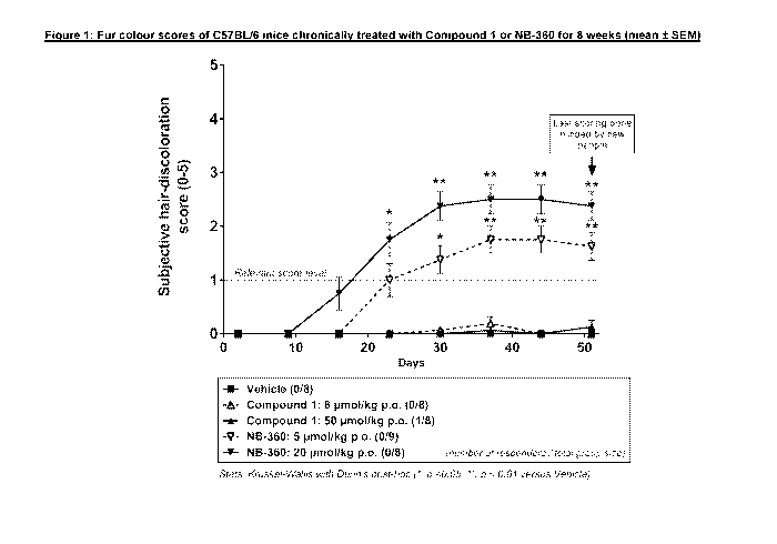
Figure 1: Fur colour scores of C57BL/6 mice chronically treated with
Compound 1 or
NB-360 for 8 weeks (mean SEM)
Figure 2: Reduction of brain AI340 in C57BL/6 mice upon treatment with
Compound
1 at 8 and 50 pmol/kg after last dose (mean SD, n=4 per group)
Figure 3: Effect of acute administration of Compound 1 on Forebrain AI340
levels in
APOE4-TR male and female mice (3-5 month-old, Mean SEM)
Figure 4: Effect of acute administration of Compound 1 on CSF AI340 levels
in
APOE4-TR male and female mice (3-5 month-old) (Mean SEM)
Figure 5: Effect of acute administration of Compound 1 on CSF AI342 levels
in
APOE4-TR male and female mice (3-5 month-old) (Mean SEM)
Figure 6: Compound 1 acute exposure in APOE4-TR male and female mice (3-5
month-old, Mean SD)
Figure 7: Brain PK/PD relationship (individual data)
Figure 8: Brain PK/PD relationship (Mean SD)
Figure 9: Effect of Compound 1 on CSF AI340 levels after two-week exposure
in
multiple ascending oral dose study in human subjects
CA 03028629 2018-12-19
WO 2018/015868
PCT/IB2017/054307
Figure 10: Effect of Compound 1 on CSF A840 levels in human subjects - `)/0
change
from baseline at 3 months (24 hours post last dose)
Figure 11: Effect of Compound 1 on A840 in Triton TX-100 extracted APP23
brains
Figure 12: Effect of Compound 1 on A842 in Triton TX-100 extracted APP23
brains
5 Figure 13: Effect of Compound 1 on sAPPa in Triton TX-100 extracted APP23
brains
Figure 14: Effect of Compound 1 on sAPP8 (Swe) in Triton TX-100 extracted
APP23
brains
Figure 15: Effects of Compound 1 treatment on A840 in the cerebrospinal fluid
of
APP23 mice
Figure 16: Effect of Compound 1 on formic acid soluble A840 in mouse (values
are
mean SEM)
Figure 17: Effect of Compound 1 on formic acid soluble A842 in mouse (values
are
mean SEM)
Figure 18: Effect of Compound 1 on formic acid soluble total A8 (40 + 42) in
mouse
(values are mean SEM)
Figure 19: Effect of Compound 1 on formic acid soluble A842/40 ratio in mouse
(values are mean SEM)
Figure 20: Effect of Compound 1 on plaque histology - number of small plaques
(data
normalized to total area)
Figure 21: Effect of Compound 1 on plaque histology - number of medium plaques
(data normalized to total area)
Figure 22: Effect of Compound 1 on plaque histology - number of large plaques
(data
normalized to total area)
Figure 23: Effect of Compound 1 on plaque histology ¨ total plaque area (data
normalized to total area)
Figure 24: Total GFAP positive area, normalized for total area. Shown are
means
SEM. Comparison was performed with Dunnett's multiple comparison test.
Figure 25: Plaque-associated GFAP positive area, normalized for total area.
Shown
are means SEM. Comparison was performed with Dunnett's multiple
comparison test.
Figure 26: Non-plaque-associated GFAP positive area, normalized for total
area.
Shown are means SEM. Comparison was performed with Dunnett's
multiple comparison test.
CA 03028629 2018-12-19
WO 2018/015868
PCT/IB2017/054307
6
Figure 27: Proximal GFAP positive area, normalized for total area. Shown are
means
SEM. Comparison was performed with Dunnett's multiple comparison
test.
Figure 28: Distal GFAP positive area, normalized for total area. Shown are
means
SEM. Comparison was performed with Dunnett's multiple comparison test.
Figure 29: Effect of Compound 1 treatment on total IBA1 positive area. Shown
are
distinct microglia populations, normalized by sample area. Shown are
means SEM. Comparison was performed with Dunnett's multiple
comparison test.
Figure 30: Effect of Compound 1 treatment on plaque-associated IBA1 positive
area.
Shown are distinct microglia populations, normalized by sample area.
Shown are means SEM. Comparison was performed with Dunnett's
multiple comparison test.
Figure 31: Effect of Compound 1 treatment on non-plaque-associated IBA1+ area.
Shown are distinct microglia populations, normalized by sample area.
Shown are means SEM. Comparison was performed with Dunnett's
multiple comparison test.
Figure 32: Effect of Compound 1 treatment on proximal IBA1+ area. Shown are
distinct microglia populations, normalized by sample area. Shown are
means SEM. Comparison was performed with Dunnett's multiple
comparison test.
Figure 33: Effect of Compound 1 treatment on distal IBA1+ area. Shown are
distinct
microglia populations, normalized by sample area. Shown are means
SEM. Comparison was performed with Dunnett's multiple comparison test.
Figure 34: Design of a two part, open-label, two-period, fixed-sequence study
in
healthy subjects to evaluate the PK of Compound 1 when given alone and
in combination with the strong CYP3A4 inhibitor itraconazole or the strong
CYP3A4 inducer rifampicin.
Figure 35: Fold change from baseline of CSF A842/440 ratio in response to
treatment with Compound 1 in non-ApoE4 carrier and ApoE4 carrier
healthy elderly subjects having an CSF A842/440 ratio < 0.09 at
baseline. Comparison was performed with Dunnett's multiple comparison
test.
Various Embodiments of the present invention are herein described.
Series A Embodiments of the First Aspect of the Invention
Embodiment Al: The compound N-(64(3R,6R)-5-amino-3,6-dimethy1-6-
(trifluoromethyl)-3,6-
dihydro-2H-1,4-oxazin-3-y1)-5-fluoropyridin-2-y1)-3-chloro-5-
(trifluoromethyl)picolinamide, or a
CA 03028629 2018-12-19
WO 2018/015868
PCT/IB2017/054307
7
pharmaceutically acceptable salt thereof, for use in the prevention of
Alzheimer's disease in
a patient at risk of developing clinical symptoms of Alzheimer's disease.
Embodiment A2: The compound, or a pharmaceutically acceptable salt thereof,
for the use
according to Embodiment Al, wherein the patient at risk of developing clinical
symptoms of
.. Alzheimer's disease carries a genetic predisposition for the development of
the clinical
symptoms of Alzheimer's disease or has Down syndrome.
Embodiment A3: The compound, or a pharmaceutically acceptable salt thereof,
for the use
according to Embodiment A2, wherein the patient carries a genetic
predisposition for the
development of the clinical symptoms of Alzheimer's disease and the genetic
predisposition
is:
(i) a mutation in the gene for amyloid precursor protein, presenilin-1 or
presenilin-2; or
(ii) the presence of one or two copies of the ApoE4 allele.
Embodiment A4: The compound, or a pharmaceutically acceptable salt thereof,
for the use
according to Embodiment A3, wherein the patient at risk of developing clinical
symptoms of
Alzheimer's disease carries one or two copies of the ApoE4 allele.
Embodiment A5: The compound, or a pharmaceutically acceptable salt thereof,
for the use
according to Embodiment A4, wherein the patient carries one copy of the ApoE4
allele.
Embodiment A6: The compound, or a pharmaceutically acceptable salt thereof,
for the use
according to Embodiment A4, wherein the patient carries two copies of the
ApoE4 allele.
Embodiment A7: The compound, or a pharmaceutically acceptable salt thereof,
for the use
according to any one of Embodiments Al to A6, wherein the patient is amyloid-
positive.
Embodiment A8: The compound, or a pharmaceutically acceptable salt thereof,
for the use
according to Embodiment A7, wherein the amyloid-positivity is determined by
PET or CSF
measurement.
.. Embodiment A9: The compound, or a pharmaceutically acceptable salt thereof,
for the use
according to any one of Embodiments A3 to A8, wherein the patient is between
60 and 75
years of age.
Embodiment A10: The compound, or a pharmaceutically acceptable salt thereof,
for the use
according to any one of Embodiments Al to A9, wherein the compound is used at
a daily
dose which results in at least a 70% lowering of AI3 1-40 in CSF following two
weeks of
compound exposure.
CA 03028629 2018-12-19
WO 2018/015868
PCT/IB2017/054307
8
Embodiment All: The compound, or a pharmaceutically acceptable salt thereof,
for the use
according to any one of Embodiments Al to A9, wherein the compound is used at
a daily
dose which results in at least a 50% lowering of A8 1-40 in CSF following two
weeks of
compound exposure.
Embodiment Al2: The compound, or a pharmaceutically acceptable salt thereof,
for the use
according to any one of Embodiments Al to A9, wherein the compound is used at
a dose of
between 10 and 30 mg per day.
Embodiment A13: The compound, or a pharmaceutically acceptable salt thereof,
for the use
according to any one of Embodiments Al to A9, wherein the compound is used at
a dose of
between 30 and 50 mg per day.
Embodiment A14: The compound, or a pharmaceutically acceptable salt thereof,
for the use
according to any one of Embodiments Al to A9, wherein the compound is used at
a dose of
mg per day.
Embodiment A15: The compound, or a pharmaceutically acceptable salt thereof,
for the use
15 according to any one of Embodiments Al to A9, wherein the compound is
used at a dose of
50 mg per day.
Embodiment A16: The compound, or a pharmaceutically acceptable salt thereof,
for the use
according to any one of Embodiments Al to A9, wherein the compound is used at
a daily
dose which results in a plasma steady state Cmax value of between 70 and 170
ng/ml.
.. Embodiment A17: The compound, or a pharmaceutically acceptable salt
thereof, for the use
according to any one of Embodiments Al to A9, wherein the compound is used at
a daily
dose which results in a plasma steady state Cmax value of between 200 and 500
ng/ml.
Embodiment A18: The compound N-(64(3R,6R)-5-amino-3,6-dimethy1-6-
(trifluoromethyl)-
3,6-dihydro-2H-1,4-oxazin-3-y1)-5-fluoropyridin-2-y1)-3-chloro-5-
(trifluoromethyl)picolinamide,
.. or a pharmaceutically acceptable salt thereof, for use in the prevention of
Alzheimer's
disease in a patient at risk of developing clinical symptoms of Alzheimer's
disease, wherein
the patient at risk of developing clinical symptoms of Alzheimer's disease
carries one or two
copies of the ApoE4 allele.
Embodiment A19: The compound N-(64(3R,6R)-5-amino-3,6-dimethy1-6-
(trifluoromethyl)-
3,6-dihydro-2H-1,4-oxazin-3-y1)-5-fluoropyridin-2-y1)-3-chloro-5-
(trifluoromethyl)picolinamide,
or a pharmaceutically acceptable salt thereof, for use in the prevention of
Alzheimer's
disease in a patient at risk of developing clinical symptoms of Alzheimer's
disease, wherein
CA 03028629 2018-12-19
WO 2018/015868
PCT/IB2017/054307
9
the patient at risk of developing clinical symptoms of Alzheimer's disease
carries one or two
copies of the ApoE4 allele, and wherein the compound is used at a dose of 15
or 50 mg per
day.
Embodiment A20: The compound for the use according to any one of Embodiments
Al to
A19, wherein the compound is in free form.
Embodiment A21: The compound, or a pharmaceutically acceptable salt thereof,
for the use
according to any one of Embodiments Al to A20, wherein the patient is not
simultaneously
treated with an inhibitor or inducer of CYP3A4.
Embodiment A22: The compound, or a pharmaceutically acceptable salt thereof,
for the use
according to any one of Embodiments Al to A20, wherein the patient is not
simultaneously
treated with a CYP3A4 inhibitor or inducer for a period longer than three
months.
Embodiment A23: The compound, or a pharmaceutically acceptable salt thereof,
for the use
according to Embodiment A21 or A22, wherein the CYP3A4 inhibitor is a strong,
moderate,
or weak inhibitor of CYP3A4; and the CYP3A4 inducer is a strong, moderate, or
weak
inducer of CYP3A4.
Embodiment A24: The compound, or a pharmaceutically acceptable salt thereof,
for the use
according to Embodiment A23, wherein the CYP3A4 inhibitor is a strong
inhibitor of
CYP3A4; and the CYP3A4 inducer is a strong inducer of CYP3A4.
Series B Embodiments of the Second Aspect of the Invention
Embodiment Bl: A pharmaceutical composition comprising the compound N-
(64(3R,6R)-5-
amino-3,6-dimethy1-6-(trifluoromethyl)-3,6-dihydro-2H-1,4-oxazin-3-y1)-5-
fluoropyridin-2-y1)-3-
chloro-5-(trifluoromethyl)picolinamide, or a pharmaceutically acceptable salt
thereof, for use
in the prevention of Alzheimer's disease in a patient at risk of developing
clinical symptoms
of Alzheimer's disease.
Embodiment B2: The pharmaceutical composition for the use according to
Embodiment Bl,
wherein the patient at risk of developing clinical symptoms of Alzheimer's
disease carries a
genetic predisposition for the development of the clinical symptoms of
Alzheimer's disease
or has Down syndrome.
Embodiment B3: The pharmaceutical composition for the use according to
Embodiment B2,
wherein the patient carries a genetic predisposition for the development of
the clinical
symptoms of Alzheimer's disease and the genetic predisposition is:
(i) a mutation in the gene for amyloid precursor protein, presenilin-1 or
presenilin-2; or
CA 03028629 2018-12-19
WO 2018/015868
PCT/IB2017/054307
(ii) the presence of one or two copies of the ApoE4 allele.
Embodiment B4: The pharmaceutical composition for the use according to
Embodiment B3,
wherein the patient at risk of developing clinical symptoms of Alzheimer's
disease carries
one or two copies of the ApoE4 allele.
5 Embodiment B5: The pharmaceutical composition for the use according to
Embodiment B4,
wherein the patient carries one copy of the ApoE4 allele.
Embodiment B6: The pharmaceutical composition for the use according to
Embodiment B4,
wherein the patient carries two copies of the ApoE4 allele.
Embodiment B7: The pharmaceutical composition for the use according to any one
of
10 Embodiments B1 to B6, wherein the patient is amyloid-positive.
Embodiment B8: The pharmaceutical composition for the use according to
Embodiment B7,
wherein the amyloid-positivity is determined by PET or CSF measurement.
Embodiment B9: The pharmaceutical composition for the use according to any one
of
Embodiments B3 to B8, wherein the patient is between 60 and 75 years of age.
Embodiment B10: The pharmaceutical composition for the use according to any
one of
Embodiments B1 to B9, wherein the compound is used at a daily dose which
results in at
least a 70% lowering of A8 1-40 in CSF following two weeks of compound
exposure.
Embodiment B11: The pharmaceutical composition for the use according to any
one of
Embodiments B1 to B9, wherein the compound is used at a daily dose which
results in at
least a 50% lowering of A8 1-40 in CSF following two weeks of compound
exposure.
Embodiment B12: The pharmaceutical composition for the use according to any
one of
Embodiments B1 to B9, wherein the compound is used at a dose of between 10 and
30 mg
per day.
Embodiment B13: The pharmaceutical composition for the use according to any
one of
Embodiments B1 to B9, wherein the compound is used at a dose of between 30 and
50 mg
per day.
Embodiment B14: The pharmaceutical composition for the use according to any
one of
Embodiments B1 to B9, wherein the compound is used at a dose of 15 mg per day.
Embodiment B15: The pharmaceutical composition for the use according to any
one of
Embodiments B1 to B9, wherein the compound is used at a dose of 50 mg per day.
CA 03028629 2018-12-19
WO 2018/015868
PCT/IB2017/054307
11
Embodiment B16: The pharmaceutical composition for the use according to any
one of
Embodiments B1 to B9, wherein the compound is used at a daily dose which
results in a
plasma steady state Cmax value of between 70 and 170 ng/ml.
Embodiment B17: The pharmaceutical composition for the use according to any
one of
Embodiments B1 to B9, wherein the compound is used at a daily dose which
results in a
plasma steady state Cmax value of between 200 and 500 ng/ml.
Embodiment B18: A pharmaceutical composition comprising the compound N-
(64(3R,6R)-5-
amino-3,6-dimethy1-6-(trifluoromethyl)-3,6-dihydro-2H-1,4-oxazin-3-y1)-5-
fluoropyridin-2-y1)-3-
chloro-5-(trifluoromethyl)picolinamide, or a pharmaceutically acceptable salt
thereof, for use
.. in the prevention of Alzheimer's disease in a patient at risk of developing
clinical symptoms
of Alzheimer's disease, wherein the patient at risk of developing clinical
symptoms of
Alzheimer's disease carries one or two copies of the ApoE4 allele.
Embodiment B19: A pharmaceutical composition comprising the compound N-
(64(3R,6R)-5-
amino-3,6-dimethy1-6-(trifluoromethyl)-3,6-dihydro-2H-1,4-oxazin-3-y1)-5-
fluoropyrid in-2-yI)-3-
.. chloro-5-(trifluoromethyl)picolinamide, or a pharmaceutically acceptable
salt thereof, for use
in the prevention of Alzheimer's disease in a patient at risk of developing
clinical symptoms
of Alzheimer's disease, wherein the patient at risk of developing clinical
symptoms of
Alzheimer's disease carries one or two copies of the ApoE4 allele, and wherein
the
compound is used at a dose of 15 or 50 mg per day.
Embodiment B20: The pharmaceutical composition for the use according to any
one of
Embodiments B1 to B19, wherein the compound is in free form.
Embodiment B21: The pharmaceutical composition for the use according to any
one of
Embodiments B1 to B20, wherein the patient is not simultaneously treated with
an inhibitor
or inducer of CYP3A4.
.. Embodiment B22: The pharmaceutical composition for the use according to any
one of
Embodiments B1 to B20, wherein the patient is not simultaneously treated with
a CYP3A4
inhibitor or inducer for a period longer than three months.
Embodiment B23: The pharmaceutical composition for the use according to
Embodiment
B21 or B22, wherein the CYP3A4 inhibitor is a strong, moderate, or weak
inhibitor of
CYP3A4; and the CYP3A4 inducer is a strong, moderate, or weak inducer of
CYP3A4.
CA 03028629 2018-12-19
WO 2018/015868
PCT/IB2017/054307
12
Embodiment B24: The pharmaceutical composition for the use according to
Embodiment
B23, wherein the CYP3A4 inhibitor is a strong inhibitor of CYP3A4; and the
CYP3A4 inducer
is a strong inducer of CYP3A4.
Series C Embodiments of the Third Aspect of the Invention
.. Embodiment Cl: A method for the prevention of Alzheimer's disease in a
patient at risk of
developing clinical symptoms of Alzheimer's disease which method comprises
administering
to such patient a therapeutically effective amount of the compound N-
(64(3R,6R)-5-amino-
3,6-dimethy1-6-(trifluoromethyl)-3 ,6-d ihydro-2H-1,4-oxazin-3-yI)-5-
fluoropyrid in-2-yI)-3-chloro-
5-(trifluoromethyl)picolinamide, or a pharmaceutically acceptable salt
thereof.
Embodiment C2: The method according to Embodiment Cl, wherein the patient at
risk of
developing clinical symptoms of Alzheimer's disease carries a genetic
predisposition for the
development of the clinical symptoms of Alzheimer's disease or has Down
syndrome.
Embodiment C3: The method according to Embodiment C2, wherein the patient
carries a
genetic predisposition for the development of the clinical symptoms of
Alzheimer's disease
and the genetic predisposition is:
(i) a mutation in the gene for amyloid precursor protein, presenilin-1 or
presenilin-2; or
(ii) the presence of one or two copies of the ApoE4 allele.
Embodiment C4: The method according to Embodiment C3, wherein the patient at
risk of
developing clinical symptoms of Alzheimer's disease carries one or two copies
of the ApoE4
allele.
Embodiment C5: The method according to Embodiment C4, wherein the patient
carries one
copy of the ApoE4 allele.
Embodiment C6: The method according to Embodiment C4, wherein the patient
carries two
copies of the ApoE4 allele.
Embodiment C7: The method according to any one of Embodiments Cl to C6,
wherein the
patient is amyloid-positive.
Embodiment C8: The method according to Embodiment C7, wherein the amyloid-
positivity is
determined by PET or CSF measurement.
Embodiment C9: The method according to any one of Embodiments C3 to C8,
wherein the
patient is over 60, 61, 62, 63, 64, 65, 66, 67, 68, 69, 70, 71, 72, 73, 74 or
75 years of age.
CA 03028629 2018-12-19
WO 2018/015868
PCT/IB2017/054307
13
Embodiment C10: The method according to any one of Embodiments C3 to C8,
wherein the
patient is between 60 and 75 years of age.
Embodiment C11: The method according to any one of Embodiments Cl to C10,
wherein
the compound is used at a daily dose which results in at least 10, 20, 30, 40,
50, 60, 70 or
.. 80% lowering of AI3 1-40 in CSF, blood, or plasma, following 2, 13, 26, 52,
78, 104, 130,
156, 182, 208, 234, 260, 286, 312, 338, 332, 390, or 416 weeks of compound
exposure.
Embodiment C12: The method according to any one of Embodiments Cl to C10,
wherein
the compound is used at a daily dose which results in at least a 70% lowering
of AI3 1-40 in
CSF, blood, or plasma, following 2, 13, 26, 52, 78, 104, 130, 156, 182, 208,
234, 260, 286,
312, 338, 332, 390, 0r416 weeks of compound exposure.
Embodiment C13: The method according to any one of Embodiments Cl to C10,
wherein
the compound is used at a daily dose which results in at least a 50% lowering
of AI3 1-40 in
CSF, blood, or plasma, following 2, 13, 26, 52, 78, 104, 130, 156, 182, 208,
234, 260, 286,
312, 338, 332, 390, 0r416 weeks of compound exposure.
Embodiment C14. The method according to any one of Embodiments Cl to C10,
wherein
the compound is used at a daily dose which results in a lowering of AI3 1-40
in CSF, blood or
plasma, in the range of 10, 20, 30, 40, 50, 60, 70 or 80% to 99, 97, 95, 93,
90, 87, 85, 80,
75, 70, 65, 60, 55, or 50%, following 2, 13, 26, 52, 78, 104, 130, 156, 182,
208, 234, 260,
286, 312, 338, 332, 390, or 416 weeks of compound exposure.
Embodiment C15. The method according to any one of Embodiments Cl to C10,
wherein
the compound is used at a daily dose which results in a lowering of AI3 1-40
in CSF, blood or
plasma, in the range of 40 to 70%, 45 to 65%, or 50 to 60%, or of at least 50%
in at least 80,
85, 90, 93, 95, 97, or 99% of the patients or in 80, 85, or 90 to 99, 97, 95,
or 93% of the
patients.
Embodiment C16. The method according to any one of Embodiments Cl to C10,
wherein
the compound is used at a daily dose which results in a lowering of AI3 1-40
in CSF, blood or
plasma, in the range of 65 to 95%, 75 to 90%, or 80 to 90%, or of at least 80%
in at least 80,
85, 90, 93, 95, 97, or 99% of the patients or in 80, 85, or 90 to 99, 97, 95,
or 93% of the
patients.
Embodiment C17: The method according to any one of Embodiments Cl to C10,
wherein
the compound is used at a dose of between 5 and 10; 10 and 15; 15 and 20; 20
and 25; 25
and 30; 30 and 35; 35 and 40; 45 and 50; 50 and 55 mg; 55 and 60 mg; 60 and
100 mg; 100
CA 03028629 2018-12-19
WO 2018/015868
PCT/IB2017/054307
14
and 200; 200 and 300 mg; 15 and 85 mg; 50 and 85 mg; 15 and 300 mg; 01 50 and
300 mg
per day.
Embodiment C18: The method according to any one of Embodiments Cl to C10,
wherein
the compound is used at a dose of between 10 and 30 mg per day.
Embodiment C19: The method according to any one of Embodiments Cl to C10,
wherein
the compound is used at a dose of between 30 and 50 mg per day.
Embodiment C20: The method according to any one of Embodiments Cl to C10,
wherein
the compound is used at a dose of 15 mg per day.
Embodiment C21: The method according to any one of Embodiments Cl to C10,
wherein
.. the compound is used at a dose of 50 mg per day.
Embodiment C22: The method according to any one of Embodiments Cl to C10,
wherein
the compound is used at a daily dose which results in a plasma steady state
Cmax value of
between 0 and 50; 50 and 100; 100 and 150; 150 and 200; 200 and 250; 250 and
300; 300
and 350; 350 and 400; 400 and 450; 450 and 500; 500 and 550; 550 and 600; 600
and 650;
or 650 and 700 ng/ml.
Embodiment C23: The method according to any one of Embodiments Cl to C10,
wherein
the compound is used at a daily dose which results in a plasma steady state
Cmax value of
between 70 and 170 ng/ml.
Embodiment C24: The method according to any one of Embodiments Cl to C10,
wherein
the compound is used at a daily dose which results in a plasma steady state
Cmax value of
between 200 and 500 ng/ml.
Embodiment C25: A method for the prevention of Alzheimer's disease in a
patient at risk of
developing clinical symptoms of Alzheimer's disease which method comprises
administering
to such patient a therapeutically effective amount of the compound N-(6-
((3R,6R)-5-amino-
.. 3,6-dimethy1-6-(trifluoromethyl)-3,6-dihydro-2H-1,4-oxazin-3-y1)-5-
fluoropyridin-2-y1)-3-chloro-
5-(trifluoromethyDpicolinamide, or a pharmaceutically acceptable salt thereof,
wherein the
patient at risk of developing clinical symptoms of Alzheimer's disease carries
one or two
copies of the ApoE4 allele.
Embodiment C26: A method for the prevention of Alzheimer's disease in a
patient at risk of
developing clinical symptoms of Alzheimer's disease which method comprises
administering
to such patient a therapeutically effective amount of the compound N-
(64(3R,6R)-5-amino-
3,6-dimethy1-6-(trifluoromethyl)-3,6-d ihydro-2H-1,4-oxazin-3-y1)-5-
fluoropyrid in-2-y1)-3-chloro-
CA 03028629 2018-12-19
WO 2018/015868
PCT/IB2017/054307
5-(trifluoromethyl)picolinamide, or a pharmaceutically acceptable salt
thereof, wherein the
patient at risk of developing clinical symptoms of Alzheimer's disease carries
one or two
copies of the ApoE4 allele, and wherein the compound is used at a dose of 15
or 50 mg per
day.
5 Embodiment C27: The method according to any one of Embodiments Cl to C26,
wherein
the compound is in free form.
Embodiment C28: The method according to any one of Embodiments Cl to C27
wherein
Compound 1 is comprised within a pharmaceutical composition.
Embodiment C29: The method according to any one of Embodiments Cl to C28,
wherein
10 the patient is not simultaneously treated with an inhibitor or inducer
of CYP3A4.
Embodiment C30: The method according to any one of Embodiments Cl to C28,
wherein
the patient is not simultaneously treated with a CYP3A4 inhibitor or inducer
for a period
longer than three months.
Embodiment C31: The method according to Embodiment C29 or C30, wherein the
CYP3A4
15 inhibitor is a strong, moderate, or weak inhibitor of CYP3A4; and the
CYP3A4 inducer is a
strong, moderate, or weak inducer of CYP3A4.
Embodiment C32: The method according to Embodiment C31, wherein the CYP3A4
inhibitor
is a strong inhibitor of CYP3A4; and the CYP3A4 inducer is a strong inducer of
CYP3A4.
Series D Embodiments of the Fifth Aspect of the Invention
Embodiment Dl: Use of the compound N-(64(3R,6R)-5-amino-3,6-dimethy1-6-
(trifluoromethyl)-3,6-dihydro-2H-1,4-oxazin-3-y1)-5-fluoropyridin-2-y1)-3-
chloro-5-
(trifluoromethyl)picolinamide, or a pharmaceutically acceptable salt thereof,
for the
prevention of Alzheimer's disease in a patient at risk of developing clinical
symptoms of
Alzheimer's disease.
Embodiment D2: The use according to Embodiment D1, wherein the patient at risk
of
developing clinical symptoms of Alzheimer's disease carries a genetic
predisposition for the
development of the clinical symptoms of Alzheimer's disease or has Down
syndrome.
Embodiment D3: The use according to Embodiment D2, wherein the patient carries
a
genetic predisposition for the development of the clinical symptoms of
Alzheimer's disease
and the genetic predisposition is:
(i) a mutation in the gene for amyloid precursor protein, presenilin-1 or
presenilin-2; or
CA 03028629 2018-12-19
WO 2018/015868
PCT/IB2017/054307
16
(ii) the presence of one or two copies of the ApoE4 allele.
Embodiment D4: The use according to Embodiment D3, wherein the patient at risk
of
developing clinical symptoms of Alzheimer's disease carries one or two copies
of the ApoE4
allele.
Embodiment D5: The use according to Embodiment D4, wherein the patient carries
one
copy of the ApoE4 allele.
Embodiment D6: The use according to Embodiment D4, wherein the patient carries
two
copies of the ApoE4 allele.
Embodiment D7: The use according to any one of Embodiments D1 to D6, wherein
the
patient is amyloid-positive.
Embodiment D8: The use according to Embodiment D7, wherein the amyloid-
positivity is
determined by PET or CSF measurement.
Embodiment D9: The use according to any one of Embodiments D3 to D8, wherein
the
patient is between 60 and 75 years of age.
Embodiment D10: The use according to any one of Embodiments D1 to D9, wherein
the
compound is used at a daily dose which results in at least a 70% lowering of
AI3 1-40 in CSF
following two weeks of compound exposure.
Embodiment D11: The use according to any one of Embodiments D1 to D9, wherein
the
compound is used at a daily dose which results in at least a 50% lowering of
AI3 1-40 in CSF
following two weeks of compound exposure.
Embodiment D12: The use according to any one of Embodiments D1 to D9, wherein
the
compound is used at a dose of between 10 and 30 mg per day.
Embodiment D13: The use according to any one of Embodiments D1 to D9, wherein
the
compound is used at a dose of between 30 and 50 mg per day.
Embodiment D14: The use according to any one of Embodiments D1 to D9, wherein
the
compound is used at a dose of 15 mg per day.
Embodiment D15: The use according to any one of Embodiments D1 to D9, wherein
the
compound is used at a dose of 50 mg per day.
CA 03028629 2018-12-19
WO 2018/015868
PCT/IB2017/054307
17
Embodiment D16: The use according to any one of Embodiments D1 to D9, wherein
the
compound is used at a daily dose which results in a plasma steady state Cmax
value of
between 70 and 170 ng/ml.
Embodiment D17: The use according to any one of Embodiments D1 to D9, wherein
the
compound is used at a daily dose which results in a plasma steady state Cmax
value of
between 200 and 500 ng/ml.
Embodiment D18: Use of the compound N-(64(3R,6R)-5-amino-3,6-dimethy1-6-
(trifluoromethyl)-3,6-dihydro-2H-1,4-oxazin-3-y1)-5-fluoropyridin-2-y1)-3-
chloro-5-
(trifluoromethyl)picolinamide, or a pharmaceutically acceptable salt thereof,
for the
prevention of Alzheimer's disease in a patient at risk of developing clinical
symptoms of
Alzheimer's disease, wherein the patient at risk of developing clinical
symptoms of
Alzheimer's disease carries one or two copies of the ApoE4 allele.
Embodiment D19: Use of the compound N-(64(3R,6R)-5-amino-3,6-dimethy1-6-
(trifluoromethyl)-3 ,6-d ihydro-2H-1,4-oxazin-3-y1)-5-fluoropyridin-2-y1)-3-
chloro-5-
(trifluoromethyl)picolinamide, or a pharmaceutically acceptable salt thereof,
for the
prevention of Alzheimer's disease in a patient at risk of developing clinical
symptoms of
Alzheimer's disease, wherein the patient at risk of developing clinical
symptoms of
Alzheimer's disease carries one or two copies of the ApoE4 allele, and wherein
the
compound is used at a dose of 15 or 50 mg per day.
Embodiment D20: The use according to any one of Embodiments D1 to D19, wherein
the
compound is in free form.
Embodiment D21: The use according to any one of Embodiments D1 to D20, wherein
the
compound is comprised within a pharmaceutical composition.
Embodiment D22: The use according to any one of Embodiments D1 to D21, wherein
the
patient is not simultaneously treated with an inhibitor or inducer of CYP3A4.
Embodiment D23: The use according to any one of Embodiments D1 to D21, wherein
the
patient is not simultaneously treated with a CYP3A4 inhibitor or inducer for a
period longer
than three months.
Embodiment D24: The use according to Embodiment D22 or D23, wherein the CYP3A4
inhibitor is a strong, moderate, or weak inhibitor of CYP3A4; and the CYP3A4
inducer is a
strong, moderate, or weak inducer of CYP3A4.
CA 03028629 2018-12-19
WO 2018/015868
PCT/IB2017/054307
18
Embodiment D25: The use according to Embodiment D24, wherein the CYP3A4
inhibitor is
a strong inhibitor of CYP3A4; and the CYP3A4 inducer is a strong inducer of
CYP3A4.
Series E Embodiments of the Seventh Aspect of the Invention
Embodiment El: Use of the compound N-(64(3R,6R)-5-amino-3,6-dimethy1-6-
(trifluoromethyl)-3,6-dihydro-2H-1,4-oxazin-3-y1)-5-fluoropyridin-2-y1)-3-
chloro-5-
(trifluoromethyl)picolinamide, or a pharmaceutically acceptable salt thereof,
for the
manufacture of a medicament for the prevention of Alzheimer's disease in a
patient at risk of
developing clinical symptoms of Alzheimer's disease.
Embodiment E2: The use according to Embodiment El, wherein the patient at risk
of
developing clinical symptoms of Alzheimer's disease carries a genetic
predisposition for the
development of the clinical symptoms of Alzheimer's disease or has Down
syndrome.
Embodiment E3: The use according to Embodiment E2, wherein the patient carries
a genetic
predisposition for the development of the clinical symptoms of Alzheimer's
disease and the
genetic predisposition is:
(i) a mutation in the gene for amyloid precursor protein, presenilin-1 or
presenilin-2; or
(ii) the presence of one or two copies of the ApoE4 allele.
Embodiment E4: The use according to Embodiment E3, wherein the patient at risk
of
developing clinical symptoms of Alzheimer's disease carries one or two copies
of the ApoE4
allele.
Embodiment E5: The use according to Embodiment E4, wherein the patient carries
one copy
of the ApoE4 allele.
Embodiment E6: The use according to Embodiment E4, wherein the patient carries
two
copies of the ApoE4 allele.
Embodiment E7: The use according to any one of Embodiments El to E6, wherein
the
patient is amyloid-positive.
Embodiment E8: The use according to Embodiment E7, wherein the amyloid-
positivity is
determined by PET or CSF measurement.
Embodiment E9: The use according to any one of Embodiments E3 to E8, wherein
the
patient is between 60 and 75 years of age.
CA 03028629 2018-12-19
WO 2018/015868
PCT/IB2017/054307
19
Embodiment E10: The use according to any one of Embodiments El to E9, wherein
the
compound is used at a daily dose which results in at least a 70% lowering of
A8 1-40 in CSF
following two weeks of compound exposure.
Embodiment El 1: The use according to any one of Embodiments El to E9, wherein
the
compound is used at a daily dose which results in at least a 50% lowering of
A8 1-40 in CSF
following two weeks of compound exposure.
Embodiment E12: The use according to any one of Embodiments El to E9, wherein
the
compound is used at a dose of between 10 and 30 mg per day.
Embodiment E13: The use according to any one of Embodiments El to E9, wherein
the
compound is used at a dose of between 30 and 50 mg per day.
Embodiment E14: The use according to any one of Embodiments El to E9, wherein
the
compound is used at a dose of 15 mg per day.
Embodiment E15: The use according to any one of Embodiments El to E9, wherein
the
compound is used at a dose of 50 mg per day.
Embodiment E16: The use according to any one of Embodiments El to E9, wherein
the
compound is used at a daily dose which results in a plasma steady state Cmax
value of
between 70 and 170 ng/ml.
Embodiment E17: The use according to any one of Embodiments El to E9, wherein
the
compound is used at a daily dose which results in a plasma steady state Cmax
value of
between 200 and 500 ng/ml.
Embodiment E18: Use of the compound N-(64(3R,6R)-5-amino-3,6-dimethy1-6-
(trifluoromethyl)-3,6-dihydro-2H-1,4-oxazin-3-y1)-5-fluoropyridin-2-y1)-3-
chloro-5-
(trifluoromethyl)picolinamide, or a pharmaceutically acceptable salt thereof,
for the
manufacture of a medicament for the prevention of Alzheimer's disease in a
patient at risk of
developing clinical symptoms of Alzheimer's disease, wherein the patient at
risk of
developing clinical symptoms of Alzheimer's disease carries one or two copies
of the ApoE4
allele.
Embodiment E19: Use of the compound N-(64(3R,6R)-5-amino-3,6-d imethy1-6-
(trifluoromethyl)-3 ,6-d ihydro-2H-1,4-oxazin-3-y1)-5-fluoropyridin-2-y1)-3-
chloro-5-
(trifluoromethyl)picolinamide, or a pharmaceutically acceptable salt thereof,
for the
manufacture of a medicament for the prevention of Alzheimer's disease in a
patient at risk of
developing clinical symptoms of Alzheimer's disease, wherein the patient at
risk of
CA 03028629 2018-12-19
WO 2018/015868
PCT/IB2017/054307
developing clinical symptoms of Alzheimer's disease carries one or two copies
of the ApoE4
allele, and wherein the compound is used at a dose of 15 or 50 mg per day.
Embodiment E20: The use according to any one of Embodiments El to E19, wherein
the
compound is in free form.
5 Embodiment E21: The use according to any one of Embodiments El to E20,
wherein the
medicament is a pharmaceutical composition.
Embodiment E22: The use according to any one of Embodiments El to E21, wherein
the
patient is not simultaneously treated with an inhibitor or inducer of CYP3A4.
Embodiment E23: The use according to any one of Embodiments El to E21, wherein
the
10 patient is not simultaneously treated with a CYP3A4 inhibitor or inducer
for a period longer
than three months.
Embodiment E24: The use according to Embodiment E22 or E23, wherein the CYP3A4
inhibitor is a strong, moderate, or weak inhibitor of CYP3A4; and the CYP3A4
inducer is a
strong, moderate, or weak inducer of CYP3A4.
15 Embodiment E25: The use according to Embodiment E24, wherein the CYP3A4
inhibitor is a
strong inhibitor of CYP3A4; and the CYP3A4 inducer is a strong inducer of
CYP3A4.
In a further invention, there is provided a method for the treatment or
prevention of
Alzheimer's disease which method comprises administering to a patient in need
thereof a
20 therapeutically effective amount of the compound N-(64(3R,6R)-5-amino-
3,6-dimethy1-6-
(trifluoromethyl)-3,6-dihydro-2H-1,4-oxazin-3-y1)-5-fluoropyridin-2-y1)-3-
chloro-5-
(trifluoromethyl)picolinamide, or a pharmaceutically acceptable salt thereof,
wherein the
patient is not simultaneously treated with an inhibitor or inducer of CYP3A4.
In one
embodiment, the patient is not simultaneously treated with an inhibitor or
inducer of CYP3A4
for a period longer than three months. In one embodiment, the patient is
simultaneously
treated with a CYP3A4 inhibitor or inducer for a period no longer than three
months. In one
embodiment, the CYP3A4 inhibitor is a strong, moderate, or weak inhibitor of
CYP3A4; and
the CYP3A4 inducer is a strong, moderate, or weak inducer of CYP3A4. In one
embodiment,
the CYP3A4 inhibitor is a strong inhibitor of CYP3A4; and the CYP3A4 inducer
is a strong
inducer of CYP3A4. In one embodiment, the patient is over 60, 61, 62, 63, 64,
65, 66, 67,
68, 69, 70, 71, 72, 73, 74 or 75 years of age. In one embodiment, the patient
is between 60
and 75 years of age. In one embodiment, the compound is used at a daily dose
which
CA 03028629 2018-12-19
WO 2018/015868
PCT/IB2017/054307
21
results in at least 10, 20, 30, 40, 50, 60, 70 or 80% lowering of AI3 1-40 in
CSF, blood, or
plasma, following 2, 13, 26, 52, 78, 104, 130, 156, 182, 208, 234, 260, 286,
312, 338, 332,
390, or 416 weeks of compound exposure. In one embodiment, the compound is
used at a
daily dose which results in at least a 70% lowering of AI3 1-40 in CSF, blood,
or plasma,
following 2, 13, 26, 52, 78, 104, 130, 156, 182, 208, 234, 260, 286, 312, 338,
332, 390, or
416 weeks of compound exposure. In one embodiment, the compound is used at a
daily
dose which results in at least a 50% lowering of AI3 1-40 in CSF, blood, or
plasma, following
2, 13, 26, 52, 78, 104, 130, 156, 182, 208, 234, 260, 286, 312, 338, 332, 390,
or 416 weeks
of compound exposure. In one embodiment, the compound is used at a dose of
between 5
and 10; 10 and 15; 15 and 20; 20 and 25; 25 and 30; 30 and 35; 35 and 40; 45
and 50; 50
and 55 mg; 55 and 60 mg; 60 and 100 mg; 100 and 200; 200 and 300 mg; 15 and 85
mg; 50
and 85 mg; 15 and 300 mg; 0r50 and 300 mg per day. In one embodiment, the
compound is
used at a dose of between 10 and 30 mg per day. In one embodiment, the
compound is
used at a dose of between 30 and 50 mg per day. In one embodiment, the
compound is
used at a dose of 15 mg per day. In one embodiment, the compound is used at a
dose of 50
mg per day. In one embodiment, the compound is used at a daily dose which
results in a
plasma steady state Cmax value of between 0 and 50; 50 and 100; 100 and 150;
150 and
200; 200 and 250; 250 and 300; 300 and 350; 350 and 400; 400 and 450; 450 and
500; 500
and 550; 550 and 600; 600 and 650; or 650 and 700 ng/ml. In one embodiment,
the
compound is used at a daily dose which results in a plasma steady state Cmax
value of
between 70 and 170 ng/ml. In one embodiment, the compound is used at a daily
dose which
results in a plasma steady state Cmax value of between 200 and 500 ng/ml. In a
further
embodiment, the compound is used in free form.
In a further invention, there is provided the compound N-(64(3R,6R)-5-amino-
3,6-dimethy1-6-
(trifluoromethyl)-3,6-dihydro-2H-1,4-oxazin-3-y1)-5-fluoropyridin-2-y1)-3-
chloro-5-
(trifluoromethyl)picolinamide, or a pharmaceutically acceptable salt thereof,
for use as a
medicament, wherein the patient treated with the medicament is not
simultaneously treated
with an inhibitor or inducer of CYP3A4. In another aspect of the further
invention, there is
provided the compound N-(64(3R,6R)-5-amino-3,6-dimethy1-6-(trifluoromethyl)-
3,6-dihydro-
2H-1 ,4-oxazin-3-y1)-5-fluoropyridin-2-y1)-3-ch loro-5-
(trifluoromethyl)picolinamide, or a
pharmaceutically acceptable salt thereof, for use in the treatment or
prevention of
Alzheimer's disease, wherein the patient is not simultaneously treated with an
inhibitor or
inducer of CYP3A4. In one embodiment of this further invention, the patient is
not
simultaneously treated with an inhibitor or inducer of CYP3A4 for a period
longer than three
months. In one embodiment of this further invention, the patient is
simultaneously treated
with a CYP3A4 inhibitor or inducer for a period no longer than three months.
In a further
CA 03028629 2018-12-19
WO 2018/015868
PCT/IB2017/054307
22
embodiment, the CYP3A4 inhibitor is a strong, moderate, or weak inhibitor of
CYP3A4; and
the CYP3A4 inducer is a strong, moderate, or weak inducer of CYP3A4. In a
further
embodiment, the CYP3A4 inhibitor is a strong inhibitor of CYP3A4; and the
CYP3A4 inducer
is a strong inducer of CYP3A4. In a further embodiment, the compound is used
at a dose of
15 or 50 mg per day. In a further embodiment, the compound is used in free
form. In another
embodiment, the compound is comprised within a pharmaceutical composition.
Definitions
As used herein, the term "Compound 1" or "Cmpd 1" refers to N-(6-((3R,6R)-5-
amino-3,6-
dimethy1-6-(trifluoromethyl)-3,6-d ihyd ro-2H-1,4-oxazin-3-y1)-5-fluoropyridin-
2-y1)-3-ch loro-5-
(trifluoromethyl)picolinamide and having the following structural formula:
F
CI OFiPLF
F
N `µfN NH2
0
F
In Example 1, using an alternative chemical naming format, "Compound 1" is
also referred to
as 3-chloro-5-trifluoromethyl-pyridine-2-carboxylic acid [64(3R,6R)-5-amino-
3,6-dimethy1-6-
trifluoromethy1-3,6-dihydro-2H-[1,4]oxazin-3-y1)-5-fluoro-pyridin-2-y1Famide.
The terms "Compound 1", "Cmpd 1" and its corresponding full chemical name are
used
interchangeably throughout the description of the invention. It is intended
that the term refers
to the compound in either free form or pharmaceutically acceptable salt form,
unless the
context clearly indicates that only one form of the compound is intended.
Compound 1 is
described in WO 2012/095469 Al, Example 34. WO 2012/095469 Al is incorporated
herewith by reference in its entirety, in particular the disclosure related to
the synthesis of
Example 34.
As used herein, the term "Alzheimer's disease" or "AD" encompasses both
preclinical and
clinical Alzheimer's disease unless the context makes clear that either only
preclinical
Alzheimer's disease or only clinical Alzheimer's disease is intended.
As used herein, the term "clinical Alzheimer's disease" or "clinical AD"
encompasses both
Mild Cognitive Impairment (MCI) due to AD and dementia due to AD, unless the
context
makes clear that either only MCI due to AD or dementia due to AD is intended.
CA 03028629 2018-12-19
WO 2018/015868 PCT/IB2017/054307
23
As used herein, the term "preclinical Alzheimer's disease" or "preclinical AD"
refers to the
presence of in vivo molecular biomarkers of AD in the absence of clinical
symptoms. The
National Institute on Aging and Alzheimer's Association provide a scheme,
shown in Table 1
below, which sets out the different stages of preclinical AD (Sperling et al.,
2011).
Table 1: Preclinical AD staging categories
A PET Markers of Evidence of
r3 (
Stage Description neuronal injury subtle
or CSF)
(tau, FDG, sMRI) cognitive change
Stage 1 Asymptomatic cerebral Positive Negative Negative
amyloidosis
Stage 2 Asymptomatic amyloidosis + Positive Positive
Negative
"downstream" neurodegeneration
Stage 3 Amyloidosis + neuronal injury + Positive Positive
Positive
subtle cognitive/behavioral decline
sMRI = structural magnetic resonance imaging
As used herein, the term "prevention of Alzheimer's disease" refers to the
prophylactic
treatment of AD; or delaying the onset or progression of AD. For example, the
onset or
progression of AD is delayed for at least 0.5, 1, 2, 3, 4, 5, 6, 7, 8, 9, or
10 years. In one
embodiment, "prevention of Alzheimer's disease" refers to the prophylactic
treatment of
preclinical AD; or delaying the onset or progression of preclinical AD. In a
further
embodiment, the onset or progression of preclinical AD is delayed for at least
0.5, 1, 2, 3, 4,
5, 6, 7, 8, 9, or 10 years. In another embodiment, "prevention of Alzheimer's
disease" refers
to the prophylactic treatment of clinical AD; or delaying the onset or
progression of clinical
AD. In a further embodiment, the onset or progression of clinical AD is
delayed for at least
0.5, 1, 2, 3, 4, 5, 6, 7, 8, 9, or 10 years.
Delay in the onset or progression of preclinical AD may be assessed by
measuring in vivo
molecular biomarkers relative to an initial baseline value, for example, by
measuring:
(a) a reduction in brain amyloid deposition. For example, by measuring a
change from
baseline in composite cortical amyloid standard uptake value ratio (SUVR)
using positron
emission tomography (PET) imaging. A suitable PET tracer for the measurement
of
SUVR ratios is 18F-florbetapir (((E)-
4-(2-(6-(2-(2-(2-([18F]-
fluoroethoxy)ethoxy)ethoxy)pyridin-3-yl)vinyI)-N-methyl benzenamine)). By this
method,
the development of amyloid accumulation over time in independent samples of
non-
demented individuals may be measured (Palmqvist S etal., 2015). SUVR
measurements
may be calculated in pre-defined cortical brain regions of interest (ROls)
referenced to
tracer uptake in a pre-defined reference region. Cortical ROls include areas
known to
have high amyloid deposition in AD, including, but not limited to, the
parietal, occipital,
lateral temporal and mesial temporal neocortical regions, as well as regions
typically
CA 03028629 2018-12-19
WO 2018/015868
PCT/IB2017/054307
24
affected in early AD (Vlassenko AG et al., 2012). In one embodiment, brain
amyloid
deposition relative to an initial baseline value is reduced to a rate of less
than 0, 1, 2, 3,
4, 5, 6, 7, 8, 9 or 10.0% per year of treatment.;
(b) an effect on the underlying tau pathology, more specifically using PET and
a suitable
Tau tracer, for example 18F-THK5351 (Harada R et al., 2016), to measure the
SUVR
change from baseline in brain Tau pathology or using cerebrospinal fluid (CSF)
to
measure total Tau and phosphorylated Tau (Forlenza OV et al., 2015). In one
embodiment, the levels of CSF Tau or phosphorylated Tau are reduced relative
to an
initial baseline value by at least 5, 10, 15, 20, 25, 30, 35, 40, 45 or 50%
per year of
treatment.;
(c) an effect on neuronal glucose metabolism, density and/or activity using
18F-FDG (2-
deoxy-2-[18F]fluoroglucose) PET (200 MBq for each scan). The 18F-FDG PET
signal in
AD-affected brain regions has been shown to be associated with cognitive
impairment,
subsequent cognitive decline and neuropathology in AD and to progress over
time in the
clinical and preclinical stages of AD, and is a disease and treatment efficacy
biomarker
(Foster NL et al., 2007). Data is analysed to determine the change in glucose
metabolism, relative to a selected reference region. In one embodiment, the
decrease in
neuronal glucose metabolism in an AD-affected brain region as determined by
18F-FDG
PET relative to an initial baseline value is limited to less than 5, 10, 15,
20, 25 or 30%
per year of treatment.;
(d) a slower decline in brain volume loss, as assessed by volumetric magnetic
resonance
imaging (vMRI) to measure a change from baseline in brain volume. vMRI can be
used
to measure a change in hippocampus, lateral ventricle, and total brain volume.
In one
embodiment, hippocampus volume loss is limited to less than 1, 2, or 3% per
year of
treatment.; or
(e) the CSF AI3 1-42/AI3 1-40 ratio over time, for example in subjects having
a baseline CSF
AI3 1-42/AI3 1-40 ratio below 0.09, indicative of cortical amyloid deposition,
as described
herein in Example 10. In one embodiment, CSF AI3 1-42/AI3 1-40 ratio increases
relative
to an initial baseline value by at least 10, 20, 30, 40, 50, 80, 100, 200 `)/0
over a period of
at least 3, 6, 9, 12, 18, 24, 0r36 months.
Delay in the onset or progression of preclinical AD may also be assessed
relative to an initial
baseline value using a sensitive cognitive measure to track changes in the
preclinical stages
of the disease, for example, using the Alzheimer's Prevention Initiative (API)
preclinical
composite cognitive (APCC) test battery. The APCC was developed as a sensitive
tool to
detect and track cognitive decline in individuals at risk to progress to the
clinical stages of
late onset AD (LOAD) (Langbaum JB etal., 2014).
CA 03028629 2018-12-19
WO 2018/015868
PCT/IB2017/054307
Delay in the onset of clinical AD may be assessed by measuring a delay in
cognitive and
functional impairment due to AD, for example, by measuring a delay in the time
to clinical
diagnosis of Mild Cognitive Impairment (MCI) due to AD and/or dementia due to
AD. The
core clinical diagnostic criteria proposed by the National Institute on Aging -
Alzheimer's
5 Association Working Group may, for example, be used for the diagnosis of
MCI (Albert MS
et al., 2011) or dementia (McKhann GM et al., 2011). The European Medicines
Agency
(EMA) in its "Draft guidelines on the clinical investigation of medicines for
the treatment of
AD and other dementias" (EMA/Committee for Medicinal Products for Human Use
(CHMP)/539931/2014) summarises the National Institute on Aging criteria for
the diagnosis
10 of MCI due to AD and AD dementia as set out below.
Diagnosis of MCI due to AD requires evidence of intra-individual decline,
manifested by:
a) A change in cognition from previously attained levels, as noted by self- or
informant
report and/or the judgment of a clinician.
b) Impaired cognition in at least one domain (but not necessarily episodic
memory) relative
15 to age-and education-matched normative values; impairment in more than
one cognitive
domain is permissible.
c) Preserved independence in functional abilities, although the criteria also
accept 'mild
problems' in performing instrumental activities of daily living (IADL) even
when this is
only with assistance (i.e. rather than insisting on independence, the criteria
allow for mild
20 dependence due to functional loss).
d) No dementia, which nominally is a function of c (above).
e) A clinical presentation consistent with the phenotype of AD in the absence
of other
potentially dementing disorders. Increased diagnostic confidence may be
suggested by
1) Optimal: A positive AI3 biomarker and a positive degeneration biomarker
25 2) Less optimal:
i. A positive AI3 biomarker without a degeneration biomarker
ii. A positive degeneration biomarker without testing for AI3 biomarkers
Diagnosis of AD dementia requires:
a) The presence of dementia, as determined by intra-individual decline in
cognition and
function.
b) Insidious onset and progressive cognitive decline.
c) Impairment in two or more cognitive domains; although an amnestic
presentation is
most common, the criteria allow for diagnosis based on nonamnestic
presentations (e.g.
impairment in executive function and visuospatial abilities).
d) Absence of prominent features associated with other dementing disorders.
CA 03028629 2018-12-19
WO 2018/015868
PCT/IB2017/054307
26
e) Increased diagnostic confidence may be suggested by the biomarker algorithm
discussed in the MCI due to AD section above.
Cognitive impairment and decline in the diagnosis of MCI due to AD and AD
dementia may
be measured using a sensitive cognitive measure to track changes in the
clinical stages of
the disease, for example, using:
a) the Clinical Dementia Rating (CDR) Scale - Sum of Boxes (SOB). The CDR is a
global
measure that evaluates cognition and functional performance and is widely used
in
clinical research in AD (Morris JC, 1993). The scale assesses six domains:
Memory,
Orientation, Judgment & Problem Solving, Community Affairs, Home & Hobbies,
and
Personal Care. Each domain is assigned a score, which are summed to obtain the
sum
of boxes (SOB) score.;
b) the Repeatable Battery for the Assessment of Neuropsychological Status
(RBANS). The
RBANS (Randolph C, 1998) is a clinical tool that was specifically designed for
both
diagnostic purposes and for tracking change in neurocognitive status over
time. One of
the key design goals of the battery is to detect and characterize very mild
dementia.; or
c) the Everyday Cognition Scale (ECog). The ECog measures cognitively-relevant
everyday abilities comprised of 39 items covering 6 cognitively-relevant
domains:
Everyday Memory, Everyday Language, Everyday Visuospatial Abilities, Everyday
Planning, Everyday Organization, and Everyday Divided Attention (Farias ST et
al.,
2008).
Suitable A6 biomarkers for use in the diagnosis of MCI due to AD and AD
dementia include,
for example, CSF A6 1-40, A6 1-42 or PET imaging of beta amyloid neuritic
plaques in the
brain, as described above.
Suitable degeneration biomarkers for use in the diagnosis of MCI due to AD and
AD
dementia are described above in relation to the in vivo molecular biomarkers
used to assess
delay in the onset or progression of preclinical AD and include, for example,
an effect on the
underlying tau pathology; an effect on neuronal glucose metabolism; or a
slower decline in
brain volume loss.
As used herein, the term "patient" refers to a human subject.
.. As used herein, the term "patient at risk of developing clinical symptoms
of Alzheimer's
disease" refers to:
(a) a human subject with a genetic predisposition for the development of the
clinical
symptoms of Alzheimer's disease, for example:
CA 03028629 2018-12-19
WO 2018/015868
PCT/IB2017/054307
27
i. subjects carrying mutations in the genes for amyloid precursor protein
(APP) or
presenilin-1 and -2 (O'Brien RJ, Wong PC, 2011), or
ii. subjects carrying one or two copies of the ApoE4 allele (Liu CC etal.,
2013);
(b) a human subject with Down Syndrome (Head E etal., 2012); or
(c) a human subject over 84 years of age.
As used herein, the term "amyloid-positive" refers to a patient who has
detectable levels of
accumulated AI3 in the brain. In one embodiment, a patient is "amyloid-
positive" if the
patient has detectable levels of accumulated AI3 in the brain based on an
assessment of AI3
in the CSF or amyloid PET imaging, or both. As used herein, the term "amyloid-
positivity
determined by PET" refers to an increased level of amyloid PET tracer
retention compared
to background. Suitable PET tracers for the measurement of amyloid-positivity
include 18F-
florbetapir (Palmqvist S et al., 2015), 18F-florbetaben (NeuraCeq) and 18F-
flutemetamol
(Vizamyl). For example, an SUVR of 1.1 or higher on a brain 18F-florbetapir
PET scan (260
MBq for each scan) may be used as an amyloid-positivity diagnostic threshold
(Schreiber S
etal., 2015). An SUVR of 1.2 or 1.3 could also be used as a threshold value.
As used herein, the term "amyloid-positivity determined by CSF measurement"
refers to a
reduced CSF AI3 1-42 value compared to that observed in a healthy control
group. For
example, amyloid-positivity may be determined by an AI3 1-42 value of 192 ng/L
or less in
CSF (Mattsson N et al., 2015). However, the CSF AI3 1-42 cut-off value used to
determine
amyloid-positivity will vary depending on the particular technique used
(Forlenza OV et al.,
2015). Amyloid positivity may also be determined by an AI3 1-42/AI3 1-40 ratio
of less than
0.09 in CSF (Janelidze S et al., 2016). In one embodiment, the AI3 1-42/AI3 1-
40 or
A1342/440 ratio is less than 0.20, 0.15, 0.10, 0.09, 0.08, 0.07, 0.06 or 0.05
or between 0.20
and 0.01, 0.15 and 0.01, 0.10 and 0.01, or 0.05 and 0.01. AI3 1-40 and AI3 1-
42 values may
be measured using standard immunoassay techniques, for example using a
monoclonal
single antibody sandwich enzyme-linked immunosorbent (ELISA) assay on the
Luminex
platform (Herskovitz AZ et al., 2013) or the Meso Scale Discovery (MSD) 96-
well MULTI-
ARRAY human/rodent (6E10) AI340 and 42 sandwich immunoassays (Meso Scale
Discovery, Rockville, MD, USA).
As used herein, the term "CYP3A4" refers to Cytochrome P450 3A4. CYP3A4 is an
enzyme
which plays a major role in the metabolism of a large variety of drugs (Luo G
etal., 2004).
As used herein, the term "inducer of CYP3A4" refers to a drug which causes
CYP3A4
activity levels to increase. Examples of CYP3A4 inducers include, but are not
limited to,
carbamazepine, phenytoin, rifampicin, and St John's wort. Techniques suitable
for the
measurement of CYP3A4 activity are well known (see, for example, Sevrioukova
IF and
CA 03028629 2018-12-19
WO 2018/015868
PCT/IB2017/054307
28
Poulos TL, 2015). "Strong", "moderate", and "weak" inducers of CYP3A4 are
drugs that
decrease the plasma area under the curve (AUC) of Compound 1 (calculated as
the area
under the curve from 0 to infinity (AUCinf)) by 80`)/0, 50`)/c, to <80%, and
20`)/c, to <50%,
respectively. In one embodiment, the "inducer of CYP3A4" is a "strong inducer
of CYP3A4."
Examples of strong inducers of CYP3A include, but are not limited to,
carbarnazopine,
enzalutamide, rbItotane, phenytoin, nfampin (also known as rifampicin), and St
John's wort
Examples of moderate inducers of CYP3A include, but are not limited to,
bosentan, efawenz,
etravinne, and modafinil. Examples of weak inducers of CYP3A include, but are
not limited to,
armodalinil and rufinamide. See
http://www.fda.gov/Drugs/DevelopmentApprovalProcess/
DevelopmentResou rces/Drug I nteractionsLabeling/ucm093664. htm#1ab1e3-3 (last
visited
October 11,2016).
As used herein, the term "inhibitor of CYP3A4" refers to a drug which causes
CYP3A4
activity levels to decrease. Techniques suitable for the measurement of CYP3A4
activity are
well known (see, for example, Sevrioukova IF and Poulos TL, 2015). Examples of
CYP3A4
inhibitors include, but are not limited to, clarithromycin, grapefruit juice,
and itraconazole.
"Strong", "moderate", and "weak" inhibitors of CYP3A4 are drugs that increase
the plasma
AUC of Compound 1 (calculated as the area under the curve from 0 to infinity
(AUCinf))
fold, to <5-fold, and 1.25 to <2-fold, respectively. In one embodiment the
"inhibitor of
CYP3A4" is a "strong inhibitor of CYP3A4." Examples of strong inhibitors of
CYP3A include,
but are not limited to, boceprevir, cobicistat, conivaptan, danoprevir and
ritonavir, elvitegravir
and ritonavir, grapefruit juice, indinavir and ritonavir, itraconazole,
ketoconazole, lopinavir
and ritonavir, paritaprevir and ritonavir and (ombitasvir and/or dasabuvir),
posaconazole,
ritonavir, saquinavir and ritonavir, telaprevir, tipranavir and ritonavir,
troleandomycin,
voriconazole, clarithromycin, diltiazem, idelalisib, nefazodone, and
nelfinavir. Examples of
moderate inhibitors of CYP3A include, but are not limited to, aprepitant,
cimetidine,
ciprofloxacin, clotrimazole, crizotinib, cyclosporine, dronedarone,
erythromycin, fluconazole,
fluvoxamine, imatinib, tofisopam, and verapamil. Examples of weak inhibitors
of CYP3A
include, but are not limited to, chlorzoxazone, cilostazol, fosaprepitant,
istradefylline,
ivacaftor, lomitapide, ranitidine, ranolazine, tacrolimus, and ticagrelor. See
http://www.fda.gov/Drugs/Deve lopme ntApprovalProcess/Developme ntResou
rces/Drug Inte ra
ctionsLabeling/ucm093664.htm#1ab1e3-2 (last visited October 11, 2016).
As used herein, term "simultaneously treated with an inhibitor or inducer of
CYP3A4" refers
to a situation where a patient is subjected to a therapeutic regimen with an
inhibitor or
inducer of CYP3A4 while also subjected to a therapeutic regimen with Compound
1. In one
embodiment, the patient is not simultaneously treated with an inhibitor or
inducer of CYP3A4
and Compound 1 for longer than 1,2, 3,4, 5,6, 7, 8, 9, 10, 11, 12, 13, 14, 15
or 16 weeks.
CA 03028629 2018-12-19
WO 2018/015868
PCT/IB2017/054307
29
In another embodiment, the patient is not simultaneously treated with an
inhibitor or inducer
of CYP3A4 and Compound 1 for longer than 1, 2, 3, 4, 5,7, 10, or 12 months. In
a certain
embodiment, the patient is not simultaneously treated with an inhibitor or
inducer of CYP3A4
and Compound 1 for longer than 3 months.
As used herein, the term "pharmaceutically acceptable salt" refers to salts
that retain the
biological effectiveness of the compound of this invention and which typically
are not
biologically or otherwise undesirable (Pharmaceutical Salts: Properties,
Selection, and Use,
2' Revised Edition (2011) P. Heinrich Stahl, Camille G. Wermuth).
As used herein, a "pharmaceutical composition" comprises the compound of this
invention,
or a pharmaceutically acceptable salt thereof, and at least one
pharmaceutically acceptable
carrier, in a solid form (typically a gelatin capsule) suitable for oral
administration.
The term "a therapeutically effective amount" of a compound of the present
invention refers
to an amount of the compound of the present invention that will elicit
inhibition of BACE-1 in
a patient as evidenced by a reduction in CSF or plasma AI3 1-40 levels
relative to an initial
baseline value.
For clarification, whenever a range is provided herein, said range is meant to
include the
endpoints. For example, a dose range between 30 and 50 mg per day comprises
also
doses of 30 and 50 mg per day.
List of abbreviations
Abbreviation Description
APP amyloid precursor protein
AI3 beta-amyloid peptide
aq. aqueous
AUC area under the curve, used to describe compound exposure
AUEC area under the effect curve, used to describe effect over
time
AI340 beta-amyloid peptide 40
AI342 beta-amyloid peptide 42
AI3 1-40 beta-amyloid peptide 1-40
AI3 1-42 beta-amyloid peptide 1-42
BACE-1 beta site APP cleaving enzyme-1
BACE-2 beta site APP cleaving enzyme -2
BACE beta site APP cleaving enzyme
BLQ below limit of quantification
Boc20 di-tert-butyl dicarbonate
concentration
CA 03028629 2018-12-19
WO 2018/015868
PCT/IB2017/054307
Abbreviation Description
CB clinical biochemistry
Cb cerebellum sample
Cl confidence interval
Comp. compound
conc. concentrated
Cpd compound
CPP Chemical and Pharmaceutical Profiling (department)
CSF Cerebrospinal fluid
CVP cava vena puncture blood sample
d day
DCM dichloromethane
DDI drug-drug interaction
DEA diethylamine
DMF N,N-dimethylformamide
DMSO dimethylsulfoxide
EDC 1-(3-dimethylaminopropyI)-3-ethylcarbodiimide
EDTA ethylenediamine tetraethyl acetate
ESI electrospray ionisation
Et0Ac ethyl acetate
FA formic acid
Fbr forebrain sample
g gram/gravitational acceleration
GFAP Glial fibrillary acidic protein (astrocyte marker)
h, hr hour
HOAt 1-hydroxy-7-azabenzotriazole
HPLC, LC high-performance liquid chromatography, liquid chromatography
IBA1 Ionized calcium binding adaptor molecule 1 (microglia marker)
ICso Inhibitory concentration 50
kg kilogram
LLOQ Lower limit of quantification
LSmeans least squares means
Me0H methanol
min minute(s)
ml milliliter
I-11 microliter
pM micromolar
pmol micromoles
MC methylcellulose
min minute
CA 03028629 2018-12-19
WO 2018/015868
PCT/IB2017/054307
31
Abbreviation Description
MS mass spectrometry
MSD MesoScale Discovery (Supplier of immunoassay kits)
NaCI sodium chloride
nM nanomolar
nmol nanomoles
NMR nuclear magnetic resonance spectrometry
ns not significant
PD pharmacodynamic
PET positron emission tomography
Pg picogram
PK pharmacokinetic
PMEL premelanosome protein
pmol picomoles
p.o. per os
q.d. or QD quaque die
q.s. quam satis
Rel. relative
Rf retention factor
rpm revolutions per minute
Rt retention time (min)
RT, it room temperature
SEM standard effort of the mean
SD standard deviation
Stats statistics
Swe Swedish, indicating the presence of the "Swedish" double
mutation in APP
t112 half-life
TBME tert-butyl-methyl-ether
THF tetrahydrofuran
tiss tissue
TLC thin layer chromatography
Tris tris-hydroxymethyl(aminomethane) buffer substance
TBS tris-buffered saline
TX-100 triton-X-100 (detergent, CAS No. 9002-93-1)
UPLC ultra performance liquid chromatography
vs versus
Examples
CA 03028629 2018-12-19
WO 2018/015868
PCT/IB2017/054307
32
The following Examples illustrate how Compound 1 may be prepared (Example 1);
demonstrate that Compound 1 is effective in reducing AI3 levels in wild type
mice in the
absence of an undesirable hair discolouration side effect observed with
comparator
compound NB-360 (Example 2); show the PK/PD effects of Compound 1 in an APOE4
transgenic mouse model (Example 3); show the PD effects of Compound 1 in a
First in
human clinical study (Example 4); demonstrate the safety and tolerability of
Compound 1 in
a 3-month clinical study (Example 5); show the effect of ApoE4 genotype on
Compound 1
PD response in the 3-month clinical study (Example 6); demonstrate the
therapeutic
effectiveness of Compound 1 in reducing amyloid plague number and area in the
APP23 AD
mouse model (Example 7); illustrate how a Compound efficacy study could be
performed in
ApoE4 homozygote at-risk patients (Example 8); show how the AUC of Compound 1
is
affected when given in combination with a strong inhibitor or inducer of
CYP3A4 (Example
9); and demonstrate how treatment with Compound 1 affects the underlying AD
pathology in
both ApoE4 carrier and non-carrier patients (Example 10).
Example 1: Preparation of Compound 1
The preparation of Compound 1 is described in WO 2012/095469 Al (Example 34).
Compound 1 may also be prepared as described below.
NMR Methodology
Proton spectra are recorded on a Bruker 400 MHz ultrashield spectrometer
unless otherwise
noted. Chemical shifts are reported in ppm relative to methanol (6 3.31),
dimethyl sulfoxide
(6 2.50), or chloroform (6 7.29). A small amount of the dry sample (2-5 mg) is
dissolved in an
appropriate deuterated solvent (0.7 mL). The shimming is automated and the
spectra
obtained in accordance with procedures well known to the person of ordinary
skill in the art.
General chromatography information
HPLC method H1 (Rtiii):
HPLC-column dimensions: 3.0 x 30 mm
HPLC-column type: Zorbax SB-C18, 1.8 pm
HPLC-eluent: A) water + 0.05 Vol.-% TFA; B) ACN + 0.05 Vol.-% TFA
HPLC-gradient: 30-100 `)/0 B in 3.25 min, flow = 0.7 ml / min
LCMS method H2 (RtH2):
HPLC-column dimensions: 3.0 x 30 mm
HPLC-column type: Zorbax SB-C18, 1.8 pm
CA 03028629 2018-12-19
WO 2018/015868
PCT/IB2017/054307
33
HPLC-eluent: A) water + 0.05 Vol.-% TFA, B) ACN + 0.05 Vol.-% TFA
HPLC-gradient: 10-100% B in 3.25 min, flow = 0.7 ml! min
UPLCMS method H3 (RtH3):
HPLC-column dimensions: 2.1 x50 mm
HPLC-column type: Acquity UPLC HSS T3, 1.8 pm
HPLC-eluent: A) water + 0.05 Vol.-% formic acid + 3.75 mM ammonium
acetate B) ACN + 0.04 Vol.-% formic acid
HPLC-gradient: 2-98 `)/0 B in 1.4 min, 98% B 0.75 min, flow = 1.2
ml! min
HPLC-column temperature: 50 C
LCMS method H4 (RtH4):
HPLC-column dimensions: 3.0 x 30 mm
HPLC-column type: Zorbax SB-C18, 1.8 pm
HPLC-eluent: A) water + 0.05 Vol.-% TFA; B) ACN + 0.05 Vol.-% TFA
HPLC-gradient: 70 - 100 % B in 3.25 min, flow = 0.7 ml! min
LCMS method H5 (RtH5):
HPLC-column dimensions: 3.0 x 30 mm
HPLC-column type: Zorbax SB-C18, 1.8 pm
HPLC-eluent: A) water + 0.05 Vol.-% TFA; B) ACN + 0.05 Vol.-% TFA
HPLC-gradient: 80 - 100 % B in 3.25 min, flow = 0.7 ml! min
LCMS method H6 (RtH6):
HPLC-column dimensions: 3.0 x 30 mm
HPLC-column type: Zorbax SB-C18, 1.8 pm
HPLC-eluent: A) water + 0.05 Vol.-% TFA; B) ACN + 0.05 Vol.-% TFA
HPLC-gradient: 40 - 100 % B in 3.25 min, flow = 0.7 ml! min
a) 2-Bromo-5-fluoro-4-triethylsilanyl-pyridine
A solution of diisopropylamine (25.3 g, 250 mmol) in 370 ml THF was cooled
with a dry-ice
acetone bath at -75 C. BuLi (100 ml, 250 mmol, 2.5 M in hexanes) was added
dropwise
while maintaining the temperature below -50 C. After the temperature of the
mixture had
reached -75 C again, a solution of 2-bromo-5-fluoropyridine (36.7 g, 208
mmol) in 45 ml
THF was added dropwise. The mixture was stirred for 1 h at -75 C.
Triethylchlorosilane
(39.2 g, 260 mmol) was added quickly. The temperature stayed below -50 C. The
cooling
bath was removed and the reaction mixture was allowed to warm to -15 C,
poured onto aq.
CA 03028629 2018-12-19
WO 2018/015868
PCT/IB2017/054307
34
NH4CI (10%). TBME was added and the layers were separated. The organic layer
was
washed with brine, dried with MgSO4.H20, filtered and evaporated to give a
brown liquid
which was distilled at 0.5 mm Hg to yield the title compound as a slightly
yellow liquid (b.p.
105-111 C). HPLC: RtH4 = 2.284 min; ESIMS: 290, 292 [(M+H)+, 1Br]; 1H-NMR
(400 MHz,
CDCI3): 8.14 (s, 1H), 7.40 (d, 1H), 1.00-0.82 (m, 15H).
b) 1-(6-Bromo-3-fluoro-4-triethylsilanyl-pyridin-2-yI)-ethanone
A solution of diisopropylamine (25.4 g, 250 mmol) in 500 ml THF was cooled to -
75 C. BuLi
(100 ml, 250 mmol, 2.5 M in hexanes) was added dropwise while maintaining the
temperature below -50 C. After the reaction temperature had reached -75 C
again, a
solution of 2-bromo-5-fluoro-4-triethylsilanyl-pyridine (56.04 g, 193 mmol) in
60 ml THF was
added dropwise. The mixture was stirred in a dry ice bath for 70 minutes. N,N-
dimethylacetamide (21.87 g, 250 mmol) was added quickly, the reaction
temperature rose to
-57 C. The reaction mixture was stirred in a dry ice bath for 15 min and then
allowed to
warm to -40 C. It was poured on a mixture of 2M aq. HCI (250 ml, 500 mmol),
250 ml water
and 100 ml brine. The mixture was extracted with TBME, washed with brine,
dried over
MgSO4.H20, filtered and evaporated to give a yellow oil which was purified on
a silica gel
column by eluting with hexane/0-5% TBME to yield 58.5 g of the title compound
as a yellow
liquid. TLC (Hex/TBME 99/1): Rf = 0.25; HPLC: RtH4 = 1.921 min; ESIMS: 332,
334 [(M+H)+,
1Br];1H-NMR (400 MHz, CDCI3): 7.57 (d, 1H), 2.68 (s, 3H), 1.00-0.84 (m, 15H).
C) (S)-2-(6-Bromo-3-fluoro-4-triethylsilanyl-pyridin-2-yI)-2-
trimethylsilanyloxy-
propionitrile
At first, the catalyst solution was prepared by dissolving water (54 mg, 3.00
mmol) in 100 ml
dry DCM ( 0.001% water). This wet DCM (44 ml, 1.32 mmol water content) was
added to
a well stirred solution of titanium(IV) butoxide (500 mg, 1.47 mmol) in 20 ml
dry DCM. The
resulting clear solution was refluxed for 1 h. This solution was then cooled
to it and 2,4-di-
tert-butyl-6-{[(E)-(S)-1-hydroxymethy1-2-methyl-propyliminoFmethyl}-phenol
[CAS 155052-
31-6] (469 mg, 1.47 mmol) was added. The resulting yellow solution was stirred
at it for 1 h.
This catalyst solution (0.023 M, 46.6 ml, 1.07 mmol) was added to a solution
of 1-(6-bromo-
3-fluoro-4-triethylsilanyl-pyridin-2-y1)-ethanone (35.53 g, 107 mmol) and
trimethylsilyl cyanide
(12.73 g, 128 mmol) in 223 ml dry DCM. The mixture was stirred for 2 days and
evaporated
to give 47 g of the crude title compound as an orange oil. HPLC: RtH5 = 2.773
min; ESIMS:
431, 433 [(M+H)+, 1Br]; 1H-NMR (400 MHz, CDCI3): 7.46 (d, 1H), 2.04 (s, 3H),
1.00 (t, 9H),
1.03-0.87 (m, 15H), 0.20 (s, 9H).
CA 03028629 2018-12-19
WO 2018/015868
PCT/IB2017/054307
d) (R)-1-Amino-2-(6-bromo-3-fluoro-4-triethylsilanyl-pyridin-2-y1)-propan-2-ol
hydrochloride
Borane dimethyl sulfide complex (16.55 g, 218 mmol) was added to a solution of
crude (S)-
2-(6-bromo-3-fluoro-4-triethylsilanyl-pyridin-2-y1)-2-trimethylsilanyloxy-
propionitrile (47 g, 109
5 mmol) in 470 ml THF. The mixture was refluxed for 2 h. The heating bath
was removed and
the reaction mixture was quenched by careful and dropwise addition of Me0H.
After the
evolution of gas had ceased, aq. 6M HCI (23.6 ml, 142 mmol) was added slowly.
The
resulting solution was evaporated and the residue was dissolved in Me0H and
evaporated
(twice) to yield 44.5 g of a yellow foam, pure enough for further reactions.
HPLC: RtH, =
10 2.617 min; ESIMS: 363, 365 [(M+H)+, 1Br]; 1H-NMR (400 MHz, CDCI3): 7.93
(s, br, 3H), 7.53
(d, 1H), 6.11 (s, br, 1H), 3.36-3.27 (m, 1H), 3.18-3.09 (m, 1H), 1.53 (s, 3H),
0.99-0.81 (m,
15H).
e) (R)-N-(2-(6-bromo-3-fluoro-4-(triethylsilyl)pyridin-2-y1)-2-hydroxypropy1)-
4-
15 nitrobenzenesulfonamide
To a solution of crude (R)-1-amino-2-(6-bromo-3-fluoro-4-triethylsilanyl-
pyridin-2-yI)-propan-
2-01 hydrochloride (43.5 g, 109 mmol) in 335 ml THF was added a solution of
NaHCO3
(21.02 g, 250 mmol) in 500 ml water. The mixture was cooled to 0-5 C and a
solution of 4-
nitrobenzenesulfonyl chloride (26.5 g, 120 mmol) in 100 ml THF was added in a
dropwise.
20 The resulting emulsion was stirred overnight while allowing the
temperature to reach rt. The
mixture was extracted with TBME. The organic layer was dried with MgSO4.H20,
filtered and
evaporated to give an orange resin which was purified on a silca gel column by
eluting with
Hexanes/10-20% Et0Ac to yield 37.56 g of the title compound as a yellow resin.
TLC
(Hex/Et0Ac 3/1): Rf = 0.34; HPLC: RtH4 = 1.678 min; ESIMS: 548, 550 [(M+H)+,
1Br]; 1H-
25 .. NMR (400 MHz, DMSO-d6): 8.40 (d, 2H), 8.06 (t, 1H), 7.97 (d, 2H), 7.45
(d, 1H), 5.42 (s,
1H), 3.23 (d, 2H), 1.44 (s, 3H) 0.97-0.81 (m, 15H); Chiral HPLC (Chiralpak AD-
H 1213, UV
210 nm): 90% ee.
f) 6-Bromo-3-fluoro-2-[(S)-2-methy1-1-(4-nitro-benzenesulfony1)-aziridin-2-y1]-
4-
30 triethylsilanyl-pyridine
A solution of triphenylphosphine (21.55 g, 82 mmol) and (R)-N-(2-(6-bromo-3-
fluoro-4-
(triethylsilyl)pyridin-2-y1)-2-hydroxpropy1)-4-nitrobenzenesulfonamide (37.56
g, 69 mmol) in
510 ml THF was cooled to 4 C. A solution of diethyl azodicarboxylate in
toluene (40% by
weight, 38.8 g, 89 mmol) was added in a dropwise while maintaining the
temperature below
35 .. 10 C. The cooling bath was removed and the reaction mixture was stirred
at rt for 1 h. The
reaction mixture was diluted with approx. 1000 ml toluene and THF was removed
by
evaporation at the rotavap. The resulting toluene solution of crude product
was pre-purified
CA 03028629 2018-12-19
WO 2018/015868
PCT/IB2017/054307
36
on a silca gel column by eluting with hexanes/5-17% Et0Ac. Purest fractions
were
combined, evaporated and crystallized from TBME/hexane to yield 29.2 g of the
title
compound as white crystals. HPLC: RtH4 = 2.546 min; ESIMS: 530, 532 [(M+H)+,
1Br]; 1H-
NMR (400 MHz, CDCI3): 8.40 (d, 2H), 8.19 (d, 2H), 7.39 (d, 1H), 3.14 (s, 1H),
3.02 (s, 1H),
2.01 (s, 3H) 1.03¨ 0.83 (m, 15H); a[D] -35.70 (c = 0.97, DCM).
g) 6-Bromo-3-fluoro-2-[(S)-2-methyl-1-(4-nitro-benzenesulfony1)-aziridin-2-y1]-
pyridine
Potassium fluoride (1.1 g, 18.85 mmol) was added to a solution of 6-bromo-3-
fluoro-2-[(S)-2-
methy1-1-(4-nitro-benzenesulfony1)-aziridin-2-y1]-4-triethylsilanyl-pyridine
(5 g, 9.43 mmol)
and AcOH (1.13 g, 9.43 mmol) in 25 ml THF. DMF (35 ml) was added and the
suspension
was stirred for 1 h at it. The reaction mixture was poured onto a mixture of
sat. aq. NaHCO3
and TBME. The layers were separated and washed with brine and TBME. The
combined
organic layers were dried over MgSO4.H20, filtered and evaporated to give a
yellow oil which
was crystallized from TBME/hexane to yield 3.45 g of the title compound as
white crystals.
HPLC: RtH6 = 2.612 min; ESIMS: 416, 418 [(M+H)+, 1Br]; 1H-NMR (400 MHz,
CDCI3): 8.41
(d, 2H), 8.19 (d, 2H), 7.48 (dd, 1H), 7.35 (t, 1H), 3.14 (s, 1H), 3.03 (s,
1H), 2.04 (s, 3H); a[D]
-35.70 (c = 0.89, DCM).
h) (R)-2-[(R)-2-(6-Bromo-3-fluoro-pyridin-2-yI)-2-(4-nitro-
benzenesulfonylamino)-
propoxy]-3,3,3-trifluoro-2-methyl-propionic acid ethyl ester
A solution of (R)-3,3,3-trifluoro-2-hydroxy-2-methyl-propionic acid ethyl
ester (11.93 g, 64.1
mmol) in DMF (158 ml) was evacuated/flushed with nitrogen twice. A solution of
KOtBu (6.21
g, 55.5 mmol) in DMF (17 ml) was added dropwise while maintaining a reaction
temperature
of ca 25 C using cooling with a water bath. After 15 min solid 6-bromo-3-
fluoro-2-[(S)-2-
methyl-1-(4-nitro-benzenesulfony1)-aziridin-2-y1Fpyridine (17.78 g, 42.7 mmol)
was added
and stirring was continued for 3 h. The reaction mixture was poured onto a
mixture of 1M
HCI (56 ml), brine and TBME. The layers were separated, washed with brine and
TBME.
The combined organic layers were dried over MgSO4H20, filtered and evaporated.
The
crude reaction product was purified via chromatography on silica gel
(hexanes/25-33%
TBME) to yield 16.93 g of the title compound as a yellow resin that was
contaminated with
an isomeric side-product (ratio 70:30 by 1H-NMR).
HPLC: RtH6 = 2.380 min; ESIMS: 602, 604 [(M+H)+, 1Br]; 1H-NMR (400 MHz,
CDCI3): 8.32
(d, 2H), 8.07 (d, 2H), 7.46 ¨ 7.41 (m, 1H), 7.30 ¨ 7.23 (m, 1H), 6.92 (s, 1H),
3.39 ¨ 4.30 (m,
2H), 3.95 (d, 1H), 3.84 (d, 1H), 1.68 (s, 3H), 1.56 (s, 3H), 1.40-1.34 (m, 3H)
+ isomeric side-
product.
CA 03028629 2018-12-19
WO 2018/015868
PCT/IB2017/054307
37
i) (R)-2-[(R)-2-(6-Bromo-3-fluoro-pyridin-2-yI)-2-(4-nitro-
benzenesulfonylamino)-
propoxy]-3,3,3-trifluoro-2-methyl-propionamide
A solution of (R)-2-[(R)-2-(6-bromo-3-fluoro-pyridin-2-yI)-2-(4-nitro-
benzenesulfonylamino)-
propoxy]-3,3,3-trifluoro-2-methyl-propionic acid ethyl ester (16.93 g, 28.1
mmol) in a
NH3/Me0H (7M, 482 ml) was stirred at 50 C in a sealed vessel for 26 h. The
reaction
mixture was evaporated and the residue was crystallized from DCM to yield 9.11
g of the title
compound as colorless crystals.
HPLC: RtH6 = 2.422 min; ESIMS: 573, 575 [(M+H)+, 1Br]; 1H-NMR (400 MHz,
CDCI3): 8.33
(d, 2H), 8.06 (d, 2H), 7.42 (dd, 1H), 7.30 ¨ 7.26 (m, 1H), 7.17 (s, br, 1H),
6.41 (s, 1H), 5.57
(s, br, 1H), 4.15 (m, 2H), 1.68 (s, 3H), 1.65 (s, 3H).
j) N-UR)-1-(6-Bromo-3-fluoro-pyridin-2-0-2-((R)-1-cyano-2,2,2-trifluoro-1-
methyl-
ethoxy)-1-methyl-ethyl]-4-nitro-benzenesulfonamide
A suspension of (R)-2-[(R)-2-(6-bromo-3-flu oro-pyrid in-2-
yI)-2-(4-n itro-
benzenesulfonylamino)-propoxy]-3,3,3-trifluoro-2-methyl-propionamide (8.43 g,
14.70 mmol)
and triethylamine (5.12 ml, 36.8 mmol) in 85 ml DCM was cooled to 0-5 C.
Trifluoroacetic
anhydride (2.49 ml, 17.64 mmol) was added dropwise over 30 min. Additional
triethylamine
(1.54 ml, 11.07 mmol) and trifluoroacetic anhydride (0.75 ml, 5.29 mmol) were
added to
complete the reaction. The reaction mixture was quenched by addition of 14 ml
aqueous
ammonia (25%) and 14 ml water. The emulsion was stirred for 15 min, more water
and DCM
were added and the layers were separated. The organic layer was dried with
MgS0.4 H20,
filtered and evaporated. Purification by column chromatography on a silica gel
(hexanes/10-
25% Et0Ac) gave 8.09 g of the title compound as a yellow resin.
HPLC: RtH6 = 3.120 min; ESIMS: 555, 557 [(M+H)+, 1Br]; 1H-NMR (400 MHz,
CDCI3): 8.35
(d, 2H), 8.11 (d, 2H), 7.50 (dd, 1H), 7.32 (dd, 1H), 6.78 (s, 1H), 4.39 (d
1H), 4.22 (d, 1H),
1.68 (s, 6H).
k) (2R,5R)-5-(6-Bromo-3-fluoro-pyridin-2-y1)-2,5-dimethy1-2-trifluoromethy1-
5,6-dihydro-
2H-[1,4]oxazin-3-ylamine
A solution of N-[(R)-1-(6-bromo-3-fluoro-pyridin-2-yI)-2-((R)-1-cyano-2,2,2-
trifluoro-1-methyl-
ethoxy)-1-methyl-ethy1]-4-nitro-benzenesulfonamide (9.18 g, 16.53 mmol) and N-
acetylcysteine (5.40 g, 33.10 mmol) in 92 ml ethanol was evacuated and flushed
with
nitrogen. K2CO3 (4.57 g, 33.1 mmol) was added and the mixture was stirred at
80 C for 3
days. The reaction mixture was concentrated in vacuo to about 1/4 of the
original volume
and partitioned between water and TBME. The organic layer was washed with 10%
aq.
K2CO3 solution, dried over Na2SO4, filtered and evaporated to give a yellow
oil. Column
CA 03028629 2018-12-19
WO 2018/015868
PCT/IB2017/054307
38
chromatography on silica (hexanes/14-50% (Et0Ac:Me0H 95:5)) gave 4.55 g of the
title
compound as an off-white solid.
HPLC: RtH2 = 2.741 min; ESIMS: 370, 372 [(M+H)+, 1Br]; 1H-NMR (400 MHz, DMSO-
d6):
7.71 ¨ 7.62 (m, 2H), 5.97 (s, br, 2H), 4.02 (d 1H), 3.70 (d, 1H), 1.51(s, 3H),
1.47 (s, 3H).
I) (2R, 5R)-5-(6-Amino-3-fluoro-pyridin-2-y1)-2,5-dimethy1-2-trifluoromethy1-
5,6-dihydro-
2H-[1 ,4]oxazin-3-y1 amine
A glass/stainless steel autoclave was purged with nitrogen, Cu2O (0.464 g,
3.24 mmol),
ammonia (101 ml, 25%, aq., 648 mmol, 30 equivalents) and (2R,5R)-5-(6-Bromo-3-
fluoro-
pyridin-2-y1)-2,5-dimethy1-2-trifluoromethy1-5,6-dihydro-2H41,4]oxazin-3-
ylamine (8 g, 21.6
mmol) in ethylene glycol (130 ml) was added. The autoclave was closed and the
suspension
heated up to 60 C and the solution was stirred for about 48 hours (max.
pressure 0.7 bar,
inside temperature 59-60 C). The reaction mixture was diluted with ethyl
acetate and water.
The organic phase was washed with water and 4 times with 12% aq. ammonia and
finally
with brine, dried over sodium sulfate, filtered and evaporated. The crude
product (7 g,
containing some ethylen glycol, quantitative yield) was used in the next step
without further
purification.
HPLC: RtH3= 0.60 min; ESIMS: 307 [(M-F1-1)].
m) [(2R, 5R)-5-(6-Amino-3-fluoro-pyridin-2-y1)-2,5-dimethy1-2-trifluoromethy1-
5,6-
dihydro-2H-0,41oxazin-3-y1]-carbamic acid tert-butyl ester
A solution of (2R, 5R)-5-(6-amino-3-fluoro-pyridin-2-y1)-2,5-dimethy1-2-
trifluoromethy1-5,6-
dihydro-2H-[1,4]oxazin-3-y1 amine (6.62 g, 21.6 mmol), Boc20 (4.72 g, 21.6
mmol) and
Hunig's base (5.66 ml, 32.4 mmol) in dichloromethane (185 ml) was stirred at
rt for 18 hours.
The reaction mixture was washed with sat. aq. NaHCO3 and brine. The aqueous
layers were
back extracted with dichloromethane and the combined organic layers were dried
over
sodium sulfate, filtered and evaporated to give a light green solid (14 g).
The crude product
was chromatographed over silicagel (cyclohexane:ethyl acetate 95:5 to 60:40)
to afford 7.68
g of the title compound.
TLC (cyclohexane:ethyl acetate 3:1): Rf = 0.21; HPLC: RtH3 = 1.14 min; ESIMS:
408
[(M+H)+]; 1H-NMR (400 MHz, CDCI3): 11.47 (br. s, 1H), 7.23 (dd, J=10.42, 8.78
Hz, 1H),
6.45 (dd, J=8.78, 2.64 Hz, 1H), 4.50 (br. s, 2H), 4.32 (d, J=2.38 Hz, 1H),
4.10 (d, J=11.80
Hz, 1H), 1.69 (s, 3H, CH3), 1.65 (s, 3H, CH3), 1.55 (s, 9H).
CA 03028629 2018-12-19
WO 2018/015868
PCT/IB2017/054307
39
n) ((2R, 5R)-5-{6-[(3-Chloro-5-trifluoromethyl-pyridine-2-carbonyl)-amino]-3-
fluoro-
pyridin-2-y1}-2,5-dimethyl-2-trifluoromethyl-5,6-dihydro-2H41,4]oxazin-3-y1)-
carbamic
acid tert-butyl ester
A mixture of [(2R, 5R)-5-(6-amino-3-fluoro-pyridin-2-y1)-2,5-dimethy1-2-
trifluoromethy1-5,6-
dihydro-2H-[1,4]oxazin-3-y1Fcarbamic acid tert-butyl ester (3.3 g, 8.12 mmol),
3-chloro-5-
trifluoromethylpicolinic acid (2.2 g, 9.74 mmol), HOAt (1.99 g, 14.62 mmol)
and EDC
hydrochloride (2.33 g, 12.18 mmol) was stirred in DMF (81 ml) at it for 48
hours. The
reaction mixture was diluted with ethyl acetate and washed with water and
brine, dried over
sodium sulfate, filtered and evaporated. The crude product (12 g) was
chromatographed
over silicagel (cyclohexane to cyclohexane:ethyl acetate 1:1) to yield 5.2 g
of the title
compound.
TLC (silica, cyclohexane:ethyl acetate 3:1): Rf=0.47; HPLC: RtH3 = 1.40 min;
ESIMS: 615,
616 [(M+H)+, ICI]; 1H-NMR (400 MHz, CDCI3): 11.68 (s, 1H), 10.41 (s, 1H), 8.81
(dd, J=1.82,
0.69 Hz, 1 H), 8.45 (dd, J=8.91, 3.14 Hz, 1 H), 8.19 (dd, J=1.88, 0.63 Hz, 1
H), 7.59 (dd,
J=9.79, 9.16
Hz, 1 H), 4.38 (d, J=2.13 Hz, 1 H), 4.18 (d, J=11.80 Hz, 1 H), 1.75 (s, 3H),
1.62 (s, 3H), 1.60
(s, 9H).
o) 3-Chloro-5-trifluoromethyl-pyridine-2-carboxylic acid [6-((3R,6R)-5-amino-
3,6-
dimethy1-6-trifluoromethy1-3,6-dihydro-2H-[1,4]oxazin-3-y1)-5-fluoro-pyridin-2-
y1]-amide
A mixture of ((2R, 5R)-5-{643-chloro-5-trifluoromethyl-pyridine-2-carbony1)-
amino]-3-fluoro-
pyridin-2-y1}-2,5-dimethy1-2-trifluoromethy1-5,6-dihydro-2H-[1,4]oxazin-3-y1)-
carbamic acid
tert-butyl ester (4.99 g, 8.13 mmol) and TFA (6.26 ml, 81 mmol) in
dichloromethane (81 ml)
was stirred at it for 18 hours. The solvent was evaporated and the residue
diluted with a
suitabable organic solvent, such as ethyl acetate and aq. ammonia. Ice was
added and the
organic phase was washed with water and brine, dried over sodium sulfate,
filtered and
evaporated to yield 3.78 g of the title compound.
HPLC: RtH3 = 0.87 min; ESIMS: 514, 516 [(M+H)+, ICI]; 1H-NMR (400 MHz, DMSO-
d6): 6
11.11(s, 1H), 9.06 (s, 1H), 8.69 (s, 1H), 8.13 (dd, J = 8.8, 2.6 Hz, 1H), 7.80
- 7.68 (m, 1H),
5.88 (br. s, 2H), 4.12 (d, J = 11.5 Hz, 1H), 3.72 (d, J = 11.4 Hz, 1H), 1.51
(s, 3H), 1.49 (s,
3H).
Example 2: Chronic dosinq of Compound 1 and comparator compound NB-360 in
wild-type mice
The studies described herein were carried out in commercial wildtype mice in
order to
investigate the chronic treatment effects of Compound 1, especially on
discoloration of the
fur, to determine an efficacious dose in wildtype mice, and to compare the
window between
CA 03028629 2018-12-19
WO 2018/015868
PCT/IB2017/054307
efficacy and fur colour changes with that of comparator BACE-1 inhibitor
compound NB-360
(N-(34(3R,6R)-5-amino-3,6-d imethy1-6-(trifluoromethyl)-3,6-d ihydro-2H-1,4-
oxazin-3-y1)-4-
fluoropheny1)-5-cyano-3-methylpicolinamide) (Neumann U etal., 2015; and
Shimshek DR et
al., 2016).
5 Animals
C57BL/6 mice were ordered at Charles River Laboratories, France.
Compound formulation and dosing
Compound 1 and NB-360 were formulated as a suspension. Vehicle, Compound 1 or
NB-
360 were given per os in a volume of 10 ml/kg once daily (mornings) for 8
weeks. Vehicle:
10 0.1 A, Tween80 in 0.5 % methylcellu lose in water.
Body weight and fur colour scoring
Body weight was taken 3 times per week (Monday, Wednesday, Friday). Subjective
scoring
of any hair colour changes was performed once weekly (Wednesday). Scores ( /0
of body
with grey fur): 0: No change; 1: Spots; 2: >30%; 3: >50%; 4:>75%; 5: 100%.
Animals were
15 photo-documented when fur color change was observed. Final fur color
scoring was
performed blinded and by a person not involved in the study.
Ex-vivo samples and sample harvest methodology
Blood samples were used to analyze whole-blood compound levels and were
obtained
either from tail-vein during the in-life part into EDTA tubes (CB300,
Sarstedt, Germany) or
20 from trunk blood at the day of necropsy into EDTA Eppendorf tubes
(Milian SA,
CatNoTOM-14, Fisher Scientific, Wohlen, Switzerland), or into serum tubes
(CB300Z,
Sarstedt, Numbrecht, Germany).
Plasma for amyloid-I3 (A13) analysis was collected by centrifugation of EDTA
blood (8000
rpm/6800xg, 15 min, 4 C) and collected into protein Lo-Bind Eppendorf tubes
(003
25 0108.116, Eppendorf, Hamburg, Germany).
After 20 min at room temperature, serum was separated by centrifugation
(8000xg, 15
min, 4 C) and collected into protein Lo-Bind Eppendorf tubes to check for
kidney
toxicity bio markers. All blood/plasma/serum samples were frozen on dry ice
and stored at -
80 C until analysis.
30 Brain was removed immediately after decapitation, rinsed with saline and
sectioned
sagitally down the midline. The left half of the cerebellum was used to
analyze compound
level and was placed into a glass tube (Chromacol, 125 x 5-SV T051, Welwyn
Garden
City, United Kingdom), weighed and frozen in dry-ice, the left half of the
forebrain (without
CA 03028629 2018-12-19
WO 2018/015868
PCT/IB2017/054307
41
olfactory bulb) was used for AI3 analysis, and was frozen on a metal plate on
dry ice and
placed into protein Lo-bind tube (003 0108.116, Eppendorf, Hamburg, Germany).
Ventral and dorsal skin were taken to analyze compound level, weighed and
frozen on
dry-ice.
Analysis of compound levels
Compound 1 and NB-360 levels in biological samples were quantified in blood,
brain and
skin by liquid chromatography/tandem mass spectrometry (HPLC/MS/MS). Brain
samples
were mixed with 2 volumes of KH2PO4 buffer and homogenized using the Covaris
device.
Skin samples were mixed with approx. 6-fold volumes of methanol/water and
homogenized
using a Precellys tube. Either 30 pL of blood, brain or skin homogenate were
spiked with a
structurally related internal standard and subsequently mixed with an at least
6-fold excess
volume acetonitrile for protein precipitation. The supernatant was injected
directly into the
LC/MS/MS system for analysis.
Table 2: Instrumental conditions for blood and brain samples
Analytical method HPLC/MS/MS
MS Sciex QTrap 5500;
Heated Electrospray Ionisation in positive ion mode.
MS/MS methods [CH5360]:
450.0 Da [M+1-1]+ ¨> 292.0 Da / CE 37 eV
Compound 1:
514.0 Da [M+H] 140.1/180.1 Da / CE 53/85 eV
Internal standard (IS):
400.0 Da [M+1-1]+ ¨> 131.0 Da / 55 40 eV
HPLC Flux Rheos Allegro (Thermo Scientific / Reinach BL,
Switzerland)
HPLC columns Phenomenex Kinetix C8 50*2.1mm, 2.6 uM (Phenomenex,
Torrance, CA, U.S.A.)
Buffer A // Buffer C A: H20 + 0.1 % formic acid // C: acetonitrile + 0.1 %
formic acid
Gradients Pump 1: 0.0 98% A, 2% C // 0.3' 70% A, 30% C // 7.5' 40%
A,
60% C // 9.5' 1%A, 99% C // 10.25' 1%A, 99% C// 10.3' 99% A,
1% C // 12.3' 99% A, 1% C. Flow 300-350 pl/min
Table 3: Instrumental conditions for skin samples
Analytical method HPLC/MS/MS
MS TSQ Quantum ultra (Thermo Scientific, Waltham, MA,
U.S.A.);
Heated Electrospray Ionisation in positive ion mode.
MS/MS methods [CH5360]:
450.0 Da [M+1-1]+ ¨> 116.9 Da / CE 43 eV
Compound 1:
514.0 Da [M+1-1]+ ¨> 375.0 Da / CE 28 eV
CA 03028629 2018-12-19
WO 2018/015868
PCT/IB2017/054307
42
Internal standard (IS)
400.0 Da [M+H] ¨> 131.0 Da / CE 40 eV
HPLC Flux Rheos Allegro (Thermo Scientific / Reinach BL,
Switzerland)
HPLC columns Synergie Hydro RP 50*2.0mm, 2.5 uM (Phenomenex,
Torrance,
CA, U.S.A.)
Buffer C // Buffer D C: H20 + 0.1 % formic acid // D: Methanol + 0.1 %
formic acid
Gradients Pump 1: 0.0 90% C, 10% D 110.1' 90% C, 10% D // 4.5' 20%
C,
80% D // 5.5' 2% C, 98% D // 5.6' 2% C, 98% D // 5.7' 90% C,
10% D // 7.0' 90% C, 10% D. Flow 400 pl/min
Acceptance criteria Calibration standards: Bias within the range 20% at
the LLOQ
in each run and at 15% at the other concentration levels. At least
3/4 of the
individual back-calculated values with at least one value at both
extremes of the standard curve fulfilling the acceptance criteria.
Quality Control samples: Bias within the range 30 % for at least
2/3 of the individual values. At least one value at each QC level
fulfilling the acceptance criteria.
Dynamic range: 0.4 to 12500 ng/mL for all compounds
Analysis of A1340 in mouse brain
Brain homogenization
Frozen mouse forebrains were weighed and homogenized in 9 volumes (w/v) of ice-
cold TBS-Complete (20 mM Tris-HCI pH 7.4, 137 mM NaCI, lx Complete [Protease
Inhibitor Cocktail Tablets: 1 836 145, Roche Diagnostics GmbH, Penzberg,
Germany]) by
sonication (90% duty cycle, output control 5, 40-55 pulses, [Sonifier 450,
Branson]). After
homogenization several 50 pl aliquots were prepared for analysis and were
stored at -80 C.
Preparation of synthetic Af31-40 solutions as standards
Human A13 peptide (1-40) trifluoroacetate salt (H 1194.1000, Bachem,
Bubendorf,
Switzerland) was used as calibration curve for A131-40. It was solubilized in
water-free
DMSO (41647, Fluka) at a concentration of 1 mg/ml for approximately 30 min at
room
temperature (RT) and then visually checked for complete solubilization.
x 5 pl aliquots and 100 pl aliquots of the remaining solution were prepared in
LoBind
15 tubes (0030 108.094, Eppendorf, Hamburg, Germany), overlaid with
nitrogen gas in order to
protect the A13 peptide from oxidation and stored at -80 C. For the
calibration curves a 5 pl
aliquot was used just once and then discarded.
Determination of A1340 in mouse brain
Endogenous A1340 in mice was determined with the Meso Scale Discovery (MSD) 96-
well
20 .. MULTI-ARRAY human/rodent (4G8) A1340 Ultrasensitive Assay (#K110FTE-3,
Meso Scale
CA 03028629 2018-12-19
WO 2018/015868
PCT/IB2017/054307
43
Discovery, Gaithersburg, USA). The assay was performed according to the
manufacturer's
instructions except for the calibration curve and the sample preparations.
TritonX-100 (TX-
100) soluble A1340 was extracted from forebrain with 1% TX-100 using a 50 pl
aliquot of
each 1:10 forebrain homogenate, mixed with 50 pl 2% TX-100 in TBS complete (20
mM
Tris-HCI pH 7.4, 137 mM NaCI, 1x Complete [Protease Inhibitor Cocktail
Tablets: 1 836 145,
Roche Diagnostics GmbH, Penzberg Germany]) to reach a final concentration of
1% TX-100
and a 1:20 forebrain dilution. The samples were incubated for 15 min on ice
and vortexed
every 5 min. The samples were ultra-centrifuged (100000xg, 4 C, 15 min) and 50
pl of the
clear supernatants were transferred to fresh tubes. For the A1340 assay the
supernatants
were further diluted 1:5 in 3% Blocker A solution (from kit) to a final
forebrain dilution of
1:100 and applied to the plate.
The calibration curve was prepared in a corresponding dilution of 1% Blocker A
solution
spiked with synthetic A131-40 peptide (1.56-100 pg/ml) except for non-
transgenic mouse
brain samples: In this case, the calibration curve was prepared in a
correspondingly diluted
APP knockout mouse forebrain spiked with synthetic A131-40 peptide (1.56-100
pg/ml). For
all samples and standards 25 pl were applied per well. For each determination
duplicate
wells were performed. The mean values from the duplicate wells were used for
calculations.
Since MSD did not provide quantification software, the relative units for
samples and
standards were imported into SOFTmax PRO 4.0 for calculation of standard
curves and
quantification of samples.
Results
Effects on body weight and fur colour in C57BL/6 mice chronically treated with
NB-360 or
Compound 1
Wild-type, naive mice (C57BL/6) were chronically treated for 8 weeks with
Compound 1 or
NB360 and body weight was measured every 3rd day (Monday, Wednesday, Friday).
No
overall significant body weight difference of the treatment group compared to
vehicle could
be observed as well as no significant difference at the end of the study at
day 56.
Nevertheless, for the treatment group a significant body weight gain (body
weight
comparison of day 0 to day 56) could be observed.
During the course of the study fur colour changes were observed in mice
treated with NB-
360. The black fur of C57BL/6 turned slowly grey in patches. These grey
patches were
visible on the ventral part of the animals while the dorsal part was
unaffected. The
appearance of grey patches was apparent after 3 weeks of treatment and present
at
different degrees in the high and low dose NB-360 group. A subjective scoring
system was
CA 03028629 2018-12-19
WO 2018/015868
PCT/IB2017/054307
44
implemented to quantitate the fur discoloration. All animals in the NB-360
group showed fur
discoloration. While the low dose NB-360 group (20 pmol/kg) showed only a
slight but
significant fur score change, the high dose NB-360 group (100 pmol/kg)
displayed a more
severe and profound fur color change, Figure 1. Noticeably, the spread and
increase of the
fur discoloration reached a plateau after 5 weeks of NB-360 treatment without
any
furthermore changes. Importantly, there was no apparent fur color change
detectable in the
Compound 1 treatment groups.
Exposure in blood and tissues
Compound 1 exposure in blood was determined at day 1 after the first dose, at
interim at day
14, and at the end of the study after the last dose. Exposure at the last day
was consistently
lower than at the beginning of the experiment. Exposure was reduced by around
35% for
Compound 1.
Exposure of Compound 1 over the last 24 hours, expressed as AUC0_24h in the
different
tissues, is summarized in Table 4. For blood, AUC was calculated form the data
at 1, 4, 7,
and 24 hours, as well as a 'mini' AUC only from the data at 4 and 24 hours.
Comparison of
the two values does not show a big difference. For tissue exposure, only data
at 4 and 24 h
were available. It was concluded that the 'mini' AUC sufficiently well
represented the tissue
exposure.
For both Compound 1 and NB-360, exposure in brain and skin was much higher
than in
blood, Table 4. In particular, the skin exposure was several fold higher than
blood exposure.
In addition, there seemed to be a higher exposure in ventral than in dorsal
parts of the skin,
especially for NB-360, Table 5. In all tissues, there was a good dose
proportionality of the
exposure.
Table 4: Compound AUC0_24h in various blood and brain tissues after last dose
(day 56)
Comp. Dose AUC blood AUC blood AUC brain AUC dorsal
skin AUC ventral skin
(pmol/kg) (PM. (pM=h)b (pmol/kg=h)b (pmol/kg=h)b (pmol/kg=h)b
NB-360 20 6.93 5.60 16.39 46.9c 104.81
100 35.89 25.26 114.45 294 712.16
Cmpd. 1 8 3.43 3.49 11.61 54.3 78.29
50 28.93 18.53 88.93 271.6 308.7
a AUC from 1,4,7, and 24 h time points; b AUC from 4 and 24 h time points; C
n=2 for 24h
Table 5: Tissue exposures normalized to blood exposure of compounds (mean of
low and
high dose)
CA 03028629 2018-12-19
WO 2018/015868
PCT/IB2017/054307
Comp. rel. AUC brain rel. AUC dorsal skin rel. AUC ventral skin
Ratio ventral/dorsal
NB-360 2.8 7.5 17.4 2.3
Cmpd. 1 3.2 12.6 16.8 1.3
Rel.: relative - normalized to AUC blood
Amvloid-beta lowering in mouse brain
At the last day of the study, groups of n=4 mice were sacrificed 4 hours and
24 hours after
having received the last dose. Forebrain was separated, and analyzed for 13-
amyloid peptide
5 1-40. Concentrations of AI340 for the vehicle and treatment groups are
summarized in Table
6 and visualised in Figure 2. The percent of reduction versus the
corresponding vehicle
treated group was calculated. Treatment resulted in a significant AI340
reduction 4 hours
after the last dose. Compound 1 showed still 25 % reduction of AI340 24 hours
after the last
dose relative to vehicle, but this was not significant. In the high dose
group, Compound 1
10 showed significantly lower levels of AI340 24 hours after the last dose.
The 50 pmoles/kg
Compound 1 dose group showed an almost flat profile, with 80-90% AI340
reduction over the
whole 24 hour time course.
Table 6: Effect of BACE inhibitor treatment on A1340 levels in mouse brain
(mean SD, n=4)
after last dose
dose time Average AI340 % significance Mini
(pmol/kg) brain (pg/g) reduction vs vehicle
AUEC
vs vehicle same time (%)
vehicle n.a. 4 1316.1 338.0 100 n.a.
vehicle n.a. 24 1469.6 152.8 100 n.a.
Compound 1 8 4 432.8 140.5 67.1 p < 0.01
46.2
Compound 1 8 24 1098.6 148.7 25.3 n.s.
Compound 1 50 4 93.4 83.7 92.9 p < 0.01
88.5
Compound 1 50 24 233.8 36.5 84.1 p < 0.01
15 n.s.: not significant
NB-360 is a dual BACE-1/BACE-2 inhibitor, as indicated by the BACE-1 and BACE-
2
enzyme inhibition in vitro assays (Neumann U etal., (2015)) which give a 1.0-
fold selectively
for BACE-1 over BACE-2. In the same assays, Compound 1 was found to have a 3
fold
selectivity for BACE-1 over BACE-2. In conclusion, moderate variations in
enzyme selectivity
20 and tissue distribution between Compound 1 and NB-360 are believed to
have an effect on
the occurrence of hair discoloration in chronic mouse studies. Despite being
active in vivo,
Compound 1 did not show signs of hair discoloration in mice.
Example 3: Acute PK/PD dose-response study of Compound 1 in APOE4-TR mice
CA 03028629 2018-12-19
WO 2018/015868
PCT/IB2017/054307
46
To investigate the effects of Compound 1 on APP metabolism in the human APOE4
context,
PK/PD studies in transgenic mice carrying the human APOE4 allele were
performed (mouse
Apoe gene was replaced by human APOE4; APOE4-TR; (Knouff C etal., 1999)).
In this study, male and female APOE4-TR animals at the age of 3-5 months were
treated
.. acutely with Compound 1 at different doses (3, 10, 30 u mol/kg) and
sacrificed at 4h and 24
after treatment.
Animals
Male and female transgenic homozygous APOE4-TR (B6.129P2-Apoetm3(APOE"4)Mae
N87
Taconic, Model 001549, 3-5 months old, n=48) were obtained from Taconic.
Dose Selection
Compound 1 was administered at 3, 10 and 30 pmol/kg.
Compound form, formulation and dosing
Compound 1 was formulated as a suspension. Vehicle or compound was given by
oral
administration in a volume of 10 ml/kg once. Vehicle: 0.1 `)/0 Tween80 in 0.5
%
.. Methylcellulose in water.
Table 7: Treatment Groups
Day 1
Treatment Dose at 0 h n =
+4h +24h
A Compound 1: 3 pmol/kg 12 Sacrifice (n=3 y n=3 c3) Sacrifice
(n=3 y n=3 c3)
Compound 1: 10 pmol/kg 12 Sacrifice (n=3 y n=3 (3) Sacrifice (n=3
y n=3 (3 )
Compound 1: 30 pmol/kg 12 Sacrifice (n=3 y n=3 (3 ) Sacrifice
(n=3 y n=3 (3 )
Vehicle (0.1 % Tween 80,
V 12 Sacrifice (n=3 y n=3 (3 ) Sacrifice (n=3 y n=3 (3 )
0.5 % MC)
Body weight
Body weight was taken once before dosing.
.. Ex vivo samples and sample harvest methodology
Blood samples were used to analyze whole blood compound levels and were
obtained from
trunk blood at the day of necropsy into EDTA Eppendorf tubes (Milian SA,
CatNoTOM-14,
Fisher Scientific, Wohlen, Switzerland), or into serum tubes (CB300Z,
Sarstedt, Numbrecht,
Germany).
CA 03028629 2018-12-19
WO 2018/015868
PCT/IB2017/054307
47
Plasma for amyloid-I3 (A13) analysis was collected by centrifugation of EDTA
blood (8000
rpm/6800xg, 15 min, 4 C) and collected into protein Lo-Bind Eppendorf tubes
(003
0108.116, Eppendorf, Hamburg, Germany).
All blood/plasma/serum samples were frozen on dry ice and stored at -80 C
until analysis.
Brain was removed immediately after decapitation, rinsed with saline and
sectioned
sagitally down the midline. The left cerebellum was used to analyze compound
level and
was placed into a glass tube (Chromacol, 125 x 5-SV T051, Welwyn Garden City,
United
Kingdom), weighed and frozen in dry-ice, the left half of the forebrain
(without olfactory
bulb) was used for AI3 analysis, and was frozen on a metal plate on dry ice
and placed into
protein Lo-bind tube (003 0108.116, Eppendorf, Hamburg, Germany). The right
brain was
fixed in 4% paraformaldehyde, washed in PBS and then embedded in paraffin for
possible
future histological analyses.
Tails were collected at the end of the study and stored at -20 C.
Table 8: Analysis of compound levels
Analytical method HPLC/MS/MS
Brain homogenization Brain samples were mixed with 2 volumes of
methanol/water (2/8 v/v)
method and homogenized using the Precellys device.
Sample preparation 30 pL of blood or brain homogenate were spiked with a
generic internal
method standard (labetalol) and subsequently mixed with 200 pL
acetonitrile
(protein precipitation). A 1 pL aliquot of the supernatant was injected into
the LC/MS/MS system for analysis.
MS AB Sciex Triple Quad 5500 (AB Sciex, Brugg AG,
Switzerland); Turbo
Ion Electrospray Ionisation in positive ion mode.
MS/MS methods 1.[Compound 1]:
508.2 Da [M+H] ¨> 139.1;180.1; 375.1 Da / CE 85;83;43 eV
2.[Labetalol]: (Internal Standard)
329.0 [M+H] ¨> 294.0 and 311.2 Da / CE 26 and 19 eV
HPLC Waters Acquity UPBINARY (Waters/ Dattwil AG,
Switzerland)
Synergi Kinetex C18, 50*2.1 mm, 2.6 pm (Phenomenex, Torrance, CA,
HPLC columns
U.S.A.)
A: H20 + 0.1 % formic acid // B: methanol/acetonitrile (1/1) + 0.1 %
Buffer A // Buffer C
formic acid
Gradients Pump 1: 0.0 98% A, 2% B // 2.5' 10% A, 90% B // 2.61%
A, 99% B // 3'
98% A, 2% B // 4' 98% A, 2% B.
Flow 0.0' ¨ 2.5' = 400 pl/min // 2.6' ¨ 4.0' = 500 pl/min
CA 03028629 2018-12-19
WO 2018/015868
PCT/IB2017/054307
48
LLOQ Compound 1: 0.4 ng/mL
Dynamic range: 0.4 to 12500 ng/mL (Compound 1)
Calibration standards Bias within the range -0.3 to -2.4% at the LLOQ and -
12.3 to 14.4% at
the other concentration levels
Quality Control Bias within the range of -4.3 to 14.4 % of all blood
quality control
samples. Bias within the range of 16.8 to 31.8 % of all brain quality
control samples: One brain QC was out of 30%
Acceptance criteria in Calibration standards: Bias within the range 30% at
the LLOQ and at
each run 20% at the other concentration levels. At least 3/4 of
the individual
back-calculated values with at least one value at both extremes of the
standard curve fulfilling the acceptance criteria.
Quality Control samples: Bias within the range 30 % for at least 2/3 of
the individual values. At least one value at each QC level fulfilling the
acceptance criteria.
Analysis of A1340 in mouse brain and A1340 and A1342 in CSF
Brain homogenization
Frozen mouse forebrains were weighed and homogenized in 9 volumes (w/v) of ice-
cold TBS-Complete (20 mM Tris-HCI pH 7.4, 137 mM NaCI, lx Complete [Protease
Inhibitor Cocktail Tablets: 1 836 145, Roche Diagnostics GmbH, Penzberg,
Germany]) by
sonication (90% duty cycle, output control 5, 40-55 pulses, [Sonifier 450,
Branson]). After
homogenization several 50 pl aliquots were prepared for analysis and were
stored at -80 C.
Preparation of synthetic A13 1-40 solutions as standards
Human A13 1-40 trifluoroacetate salt (H 1194.1000, Bachem, Bubendorf,
Switzerland) was
used as calibration curve for A131-40. It was solubilized in water-free DMSO
(41647,
Fluka) at a concentration of 1 mg/ml for approximately 30 min at room
temperature (RT)
and then visually checked for complete solubilization.
x 5 pl aliquots and 100 pl aliquots of the remaining solution were prepared in
LoBind
tubes (0030 108.094, Eppendorf, Hamburg, Germany), overlaid with nitrogen gas
in order to
15 protect the A13 peptide from oxidation and stored at -80 C. For the
calibration curves a 5 pl
aliquot was used just once and then discarded.
Determination of A1340 in mouse brain
Endogenous A1340 in mice was determined with the Meso Scale Discovery (MSD) 96-
well
MULTI-ARRAY human/rodent (4G8) A1340 Ultrasensitive Assay (#K110FTE-3, Meso
Scale
CA 03028629 2018-12-19
WO 2018/015868
PCT/IB2017/054307
49
Discovery, Gaithersburg, USA). The assay was performed according to the
manufacturer's
instructions except for the calibration curve and the sample preparations.
TritonX-100 (TX-
100) soluble A1340 was extracted from forebrain with 1% TX-100 using a 50 pl
aliquot of
each 1:10 forebrain homogenate, mixed with 50 pl 2% TX-100 in TBS complete (20
mM
Tris-HCI pH 7.4, 137 mM NaCI, lx Complete [Protease Inhibitor Cocktail
Tablets: 1 836 145,
Roche Diagnostics GmbH, Penzberg Germany]) to reach a final concentration of
1% TX-100
and a 1:20 forebrain dilution. The samples were incubated for 15 min on ice
and vortexed
every 5 min. The samples were ultra-centrifuged (100000xg, 4 C, 15 min) and 50
pl of the
clear supernatants were transferred to fresh tubes. For the A1340 assay the
supernatants
were further diluted 1:5 in 3% Blocker A solution (from kit) to a final
forebrain dilution of
1:100 and applied to the plate.
The calibration curve was prepared in a corresponding dilution of 1% Blocker A
solution
spiked with synthetic A131-40 peptide (1.56-100 pg/ml) except for non-
transgenic mouse
brain samples: In this case, the calibration curve was prepared in a
correspondingly diluted
APP knockout mouse forebrain spiked with synthetic A131-40 peptide (1.56-100
pg/ml). For
all samples and standards 25 pl were applied per well. For each determination
duplicate
wells were done. The mean values from the duplicate wells were used for
calculations. Since
MSD did not provide quantification software, the relative units for samples
and standards
were imported into SOFTmax PRO 4.0 for calculation of standard curves and
quantification
of samples.
Results
APOE4-TR mice (mouse Apoe gene was replaced by human APOE4) were acutely
treated
with three different doses (3, 10 and 30 pmol/kg) of the BACE inhibitor
Compound 1.
Animals were sacrificed 4h and 24h after last the last dose and forebrains
were separated.
.. Concentrations of A1340 and A1342 for the various groups are summarized in
Figures 3, 4 and
5; and Tables 9, 10 and 11. The percent of reduction versus the vehicle
treated group was
calculated. All treatments resulted in a significant and dose-dependent A1340
reduction at 4h
and 24h after the last dose, the effect ranged from 43-77% at 4h and 20-66% at
24h. For the
two lower dose groups (3 and 10 pmol/kg) the A1340 lowering effect was
significantly
reduced at 4h and 24, but substantially approximated baseline levels 24h after
the last dose.
The high dose of Compound 1 (30 pmol/kg) showed an almost flat profile with 77-
66% A1340
reduction over the whole 24h time course.
CA 03028629 2018-12-19
WO 2018/015868
PCT/IB2017/054307
Table 9: Effect of Compound 1 treatment on A1340 levels in APOE4-TR mouse
brain (n=6
(n=3 males, n=3 females))
Dose Dosing Time Average A(340 % reduction Significance Mini
AUEC
Regime brain (pmol/g)+SD vs vehicle vs vehicle
(%)
Vehicle Acute 4/24 0.200 0.034 n.a. n.a. n.a.
3 pmol/kg Acute 4 0.114 0.024 43 p<0.0001
32
3 pmol/kg Acute 24 0.160 0.033 20 p<0.05
10 pmol/kg Acute 4 0.074 0.014 63 p<0.0001
47
10 pmol/kg Acute 24 0.139 0.041 31 p<0.001
30 pmol/kg Acute 4 0.046 0.006 77 p<0.0001
72
30 pmol/kg Acute 24 0.068 0.021 66 p<0.0001
n.a.: not applicable, vehicle: all vehicles combined
5
Table 10: Effect of Compound 1 treatment on A1340 levels in APOE4-TR mouse CSF
(n=6
(n=3 males, n=3 females))
Dose Dosing Time Average A(340 CSF % reduction
Significance Mini AUEC
Regime (pmol/mI) SD vs vehicle vs vehicle (%)
Vehicle Acute 4/24 0.1826 0.0956 n.a. n.a. n.a.
3 pmol/kg Acute 4 0.1308 0.06381 28 ns
26
3 pmol/kg Acute 24 0.1402 0.07911 23 ns
10 pmol/kg Acute 4 0.07527 0.03356 59 p<0.05
37
10 pmol/kg Acute 24 0.1565 0.08348 14 ns
30 pmol/kg Acute 4 0.02533 0.01093 86 p<0.001
77
30 pmol/kg Acute 24 0.06083 0.03445 67 p<0.01
n.a.: not applicable, vehicle: all vehicles combined, ns: not significant
10 Table 11: Effect of Compound 1 treatment on AI342 levels in APOE4-TR
mouse CSF (n=6
(n=3 males, n=3 females))
Dose Dosing Time Average A(342 CSF % reduction Significance
Mini AUEC
Regime (pmol/mI) SD vs vehicle vs vehicle (%)
Vehicle Acute 4/24 0.1174 0.05610 n.a. n.a. n.a.
3 pmol/kg Acute 4 0.0524 0.02173 55 ns
38
3 pmol/kg Acute 24 0.09317 0.05304 21 ns
10 pmol/kg Acute 4 0.02318 0.00446 80 p<0.01
10 pmol/kg Acute 24 0.09438 0.09019 20 ns
30 pmol/kg Acute 4 0.0080 0.00395 93 p<0.001
81
30 pmol/kg Acute 24 0.03717 0.02438 68 p<0.05
n.a.: not applicable, vehicle: all vehicles combined, ns: not significant
PK data are shown in Figure 6 and Table 12 at 4h and 24h for acute dosing in
blood and
15 brain. Exposure of Compound 1 over 24h, expressed as AUC0_24h in blood
and brain is
summarized in Table 13. Compound 1 exposure in blood and brain was dose
proportional
CA 03028629 2018-12-19
WO 2018/015868
PCT/IB2017/054307
51
and displayed the expected minor decline in compound level after 24h, which
again, was
dose proportional. The compound exposure in brain was much higher than in
blood. The
brain blood ratio was similar for 3, 10 and 30 pmol/kg dose groups with 5, 3
and 4 at 4h and
9, 4, and 3 at 24h, respectively. The exposure ratio at 4h/24h was calculated
which allows
the comparison of the decline of compound exposure at the different doses
(Table 12).
Compound 1 had a moderate 2-5 fold exposure reduction, without big differences
between
the different doses and between blood and brain.
Table 12: Compound 1 levels in blood and brain of APOE4-TR mice (n=6 (n=3
males, n=3
females)
Treatment Dosing Time Mean Blood Exposure -- Mean Brain --
Exposure
(pmol/ml) ratios 4h/24h (pmol/g)
ratios 4h/24h
SD (Blood) SD (Brain)
3 pmol/kg Acute 4 105 8 512 216
4.2 2.4
3 pmol/kg Acute 24 25 7 210 262
pmol/kg Acute 4 317 50 995 151
3.6 3.1
10 pmol/kg Acute 24 88 25 324 109
30 pmol/kg Acute 4 985 224 3409 741
3.8 4.6
30 pmol/kg Acute 24 259 68 743 267
10 Table 13: Compound 1 AUC0_24h in blood and brain of APOE4-TR mice
Treatment Dosing AUCblood AUCbrain
(pM=h) (pmol/kg = h)
3 pmol/kg Acute 1.56 8.67
10 pmol/kg Acute 4.86 15.83
30 pmol/kg Acute 14.92 49.82
AUC from 4h and 24h time points
The brain pharmacokinetic/pharmacodynamic relationship for the individual
animals for all
dose groups is shown in Figure 7. There was a clear PK/PD relationship
apparent for
Compound 1; at low compound levels the A6 reduction efficacy was minimal
whereas at
high compound levels a maximum efficacy effect was detected.
Figure 8 displays the PK/PD relationship for the averaged values at the
different doses.
Again, the exposure dependent effect on A6 reduction is apparent, with a clear
minimal and
maximal efficacy effect.
Conclusion
Studies presented in this experimental example demonstrate that Compound 1 is
an orally-
available, centrally active and potent inhibitor of BACE in vivo in APOE4-TR
mice. APOE4-
TR mice that express human APOE4 from the mouse endogenous Apoe locus were
used to
CA 03028629 2018-12-19
WO 2018/015868
PCT/IB2017/054307
52
investigate PK/PD relationship of Compound 1. ApoE4 has been implicated to be
a high risk
factor for Alzheimer's disease and APOE4-TR mice resemble the ApoE4 effect in
the
Alzheimer's brain.
The PK properties of Compound 1 in APOE4-TR mice did not differ to those
observed in
wildtype mice. A dose-dependent Compound 1 exposure in blood and brain, with
much
higher brain levels, was observed. Furthermore, the exposure decrease after
24h was
similar to that observed in wildtype mice. Compound 1 at 30 pmol/kg resulted
in the maximal
effect on A8 reduction (>70%) in the brain of APOE4-TR, with similar extent
lasting over 24h
for acute dosing. The PK/PD relationship was very comparable to wildtype mice
and rats.
There was a slightly lower maximal efficacy effect on A8 reduction in the
brain apparent in
APOE4-TR mice at the highest dose (30 pmol/kg). This might be due to a lower
clearance
rate of amyloid-8 observed in APOE-4 TR mice (Castellano JM etal., 2011).
Example 4: First-in-human study
This study has been clinically completed and was a randomised, double-blind,
placebo-
controlled, single and multiple ascending oral dose study to primarily assess
the safety and
tolerability as well as the pharmacokinetics and pharmacodynamics of Compound
1 in
healthy adult and elderly subjects. The purpose of this study was to determine
the single and
multiple maximum tolerated dose of Compound 1 and to assess the
pharmacokinetic/pharmacodynamic (PK/PD) relationship using A8 in CSF as
primary PD
biomarker.
In healthy elderly subjects 60 years of age, the highest tested doses of 750
mg single dose
and 300 mg QD over two weeks were determined to be safe and tolerated.
Pharmacodynamic assessments using A8 concentrations in CSF as primary
biomarker of
drug action were also been applied in healthy elderly subjects. Dose-dependent
lowering of
440 concentrations was determined up to approximately 80% and 90% after single
and
multiple dosing, respectively (Tables 14 and 15, Figure 9).
CA 03028629 2018-12-19
WO 2018/015868
PCT/IB2017/054307
53
Table 14: A1340 in CSF - Summary of percent change from baseline across time
Single-dose
Time Cmpd 1 Cmpd 1 Cmpd 1
Cmpd 1
(hours) Placebo 10 mg 90 mg 300 mg 750
mg
12h N 12 6 6 8 8
Mean (SD) 1.6 (6.66) -11.6 (8.31) -14.2
(35.83) -35.8 (16.40) -36.2 (13.58)
Median 0.2 -12.5 -28.0 -44.1 -38.6
Min - Max -7-16 -21 -1 -36-58 -54 --13 -
53 --10
24h N 12 6 5 8 7
Mean (SD) -2.2 (12.84) -15.0 (10.21) -44.8
(12.48) -68.3 (10.65) -67.4 (15.54)
Median -2.3 -14.3 -49.2 -72.5 -70.6
Min - Max -22 - 22 -31 - -4 -55 - -25 -79 - -52 -
82 - -36
34h N 11 6 5 8 7
Mean (SD) 5.1 (14.83) -9.7 (14.72) -50.0 (8.87) -
78.3 (4.75) -79.1 (8.88)
Median 4.1 -12.5 -45.4 -79.3 -82.3
Mm-Max -22-29 -25-10 -60--41 -84--69 -
87--60
Table 15: A1340 in CSF Summary of percent change from baseline on Day 15 (24h
post last
dose)
Multiple-dose
Cmpd 1 Cmpd 1 Cmpd 1 Cmpd 1
Placebo 10 mg 30 mg 90 mg 300 mg
N 16 8 8 8 8
Mean (SD) -7.2 (6.04) -60.1 (9.91) -80.0 (3.89) -88.2 (3.07) -
93.6 (0.61)
Median -8.0 -61.5 -80.2 -89.1 -93.5
Min ; Max -18 ; 2 -77 ; -48 -86 ; -76 -92 ; -82 -94 ; -93
Example 5: 3-Month dose-ranoino safety and tolerability study
Compound 1 was administered to healthy elderly subjects 60 years or over in a
Phase I
clinical dose-ranging safety and tolerability study. This study is listed in
ClinicalTrials.gov
under the NCT02576639 Identifier code.
This randomized, double-blind, placebo-controlled study had a parallel-group
design and
Compound 1 was administered as once-daily, oral doses to five treatment groups
(Compound 1: 2mg, 10mg, 35mg or 85mg QD and placebo).
The primary purpose of this study was to expand on previous safety and
tolerability data
obtained over 2-week and 4-week duration in the first in human study and
thereby allow
CA 03028629 2018-12-19
WO 2018/015868
PCT/IB2017/054307
54
initiation of future long-term efficacy trials in subjects at risk of AD. In
addition, data relevant
for Pharmacokinetic/Pharmacodynamic modeling was obtained in order to support
dose
selection decisions for future efficacy studies.
In this study, Compound 1 was found to be safe and tolerated at once-daily
doses of 2, 10,
35 and 85mg over three months. The pharmacodynamics effects of Compound 1
administration on CSF AI3 levels are shown in Table 16 and Figure 10. The
extent of AI3
lowering was stable over time with PD steady-state being reached after
approximately 2-3
weeks.
Table 16: A13 in CSF at month 3 - Analysis of percent chanpe from baseline of
A1338, A1340,
and Af342
Difference (90% CI)
Parameter Treatment N LSmean (SE) Cmpd 1-Placebo P-value
Placebo 21 -2.41 (1.430)
Cmpd 1: 2mg 22 -20.58 (1.397) -18.17 (-
21.49, -14.85) <.001
A(338 (pg/mL) Cmpd 1: 10mg 21 -62.60 (1.430) -60.18 (-
63.54, -56.83) <.001
Cmpd 1: 35mg 23 -82.96 (1.366) -80.55 (-
83.83, -77.27) <.001
Cmpd 1: 85mg 20 -89.23 (1.470) -86.81 (-
90.22, -83.41) <.001
Placebo 21 -2.79 (1.389)
Cmpd 1: 2mg 22 -22.66 (1.355) -19.87 (-
23.09, -16.65) <.001
A(340 (pg/mL) Cmpd 1: 10mg 21 -62.96 (1.388) -60.18 (-
63.43, -56.92) <.001
Cmpd 1: 35mg 23 -83.19 (1.325) -80.41 (-
83.59, -77.22) <.001
Cmpd 1: 85mg 20 -90.40 (1.429) -87.62 (-
90.93, -84.30) <.001
Placebo 21 -2.76 (1.310)
Cmpd 1: 2mg 22 -24.00 (1.277) -21.24 (-
24.28, -18.21) <.001
A(342 (pg/mL) Cmpd 1: 10mg 21 -64.15 (1.308) -61.39 (-
64.47, -58.31) <.001
Cmpd 1: 35mg 23 -82.49 (1.250) -79.73 (-
82.73, -76.73) <.001
Cmpd 1: 85mg 20 -89.38 (1.348) -86.62 (-
89.76, -83.49) <.001
Adjusted Lsmeans, difference in Lsmeans, 90% Cl and P-value were obtained from
an ANCOVA model
with treatment as a fixed effect, and baseline AB level as covariat
The pharmacokinetic parameters of Compound 1 following three months (91 days)
daily
dosing at 2, 10, 35 and 85 mg are shown in Table 17.
CA 03028629 2018-12-19
WO 2018/015868
PCT/IB2017/054307
Table 17: Compound 1 pharmacokinetic parameters on Day 91
Cmax,ss (ng/mL)
10 mg qd. 35 mg qd. 85 mg qd.
Results 80 229 602
Mean (CV%) (36) (33) (25)
The Cmax,ss value represents the maximum plasma steady state concentration of
Compound 1 following 91 days of once daily (qd) dosing at the specified dose.
"CV%"
represents the percentage coefficient of variation.
5 Based on these results, a once daily dose of 15 mg of Compound 1 is
expected to result in a
plasma Cmax,ss value of between 70 and 170 ng/ml, and a once daily dose of 50
mg of
Compound 1 is expected to result in a plasma Cmax,ss value of between 200 and
500
ng/ml.
Based on the data presented in Example 4 and 5, pharmacometric modelling
predicts a daily
10 dose of 50 mg to reach 80% CSF AI340 lowering and a dose of 15 mg to
achieve 60% CSF
AI340 lowering, in 90% of the subjects.
Example 6: Effect of ApoE4 qenotype on response to treatment with Compound 1
In the completed first-in-human and 3-month dose-ranging safety and
tolerability clinical
studies described in Examples 5 and 6, AI3 concentrations in CSF were obtained
by means
15 of lumbar punctures before the first dose (baseline) and respectively
after 2 weeks and 3
months of multiple dosing. ApoE4 genotype also was obtained in the subjects
who
consented. The percent change from baseline in AI340 and AI342 concentrations
was
calculated in subjects who took the study treatment and had no major protocol
deviation with
potential impact on the evaluation of the pharmacodynamic effect. Tables 18 to
21 below
20 provide summary statistics of the percent change from baseline by
treatment group and
ApoE genotype (E4 heterozygotes versus E4 non-carriers). Only one subject with
CSF data
was E4 homozygote (from the 3-month dose-ranging safety and tolerability
study). This
subject was treated with placebo and showed 11% decrease in both AI340 and
AI342
concentrations and is not included in the tables below. The data shows that
there is no
25 difference in CSF AI340 and AI342 response to treatment with Compound 1
between ApoE4
carriers and non-carriers.
CA 03028629 2018-12-19
WO 2018/015868
PCT/IB2017/054307
56
Table 18: A1340 `)/0 change from baseline by ApoE genotype and Compound 1
treatment
group at 3 months
E4 heterozygotes E4 non-carriers
Placebo n 9 8
Mean (SD) -3 (7) -2 (5)
Median -2 -1
Range -19 ; 9 -13 ; 5
Compound 1: 2 mg n 10 12
Mean (SD) -21 (11) -24 (9)
Median -18 -22
Range -46 ; -3 -41 ; -10
Compound 1: 10 mg n 3 15
Mean (SD) -67 (7) -64 (6)
Median -64 -65
Range -75 ; -61 -73 ; -54
Compound 1: 35 mg n 8 12
Mean (SD) -84 (3) -82 (5)
Median -85 -83
Range -88 ; -79 -91 ; -72
Compound 1: 85 mg n 2 17
Mean (SD) -91 (0) -91 (2)
Median -91 -90
Range -91 ; -91 -94 ; -87
Table 19: A1342 % change from baseline by ApoE genotype and Compound 1
treatment
group at 3 months
E4 heterozygotes E4 non-carriers
Placebo n 9 8
Mean (SD) -2 (4) -2 (7)
Median -0 -1
Range -10 ; 4 -12 ; 8
Compound 1: 2 mg n 10 12
Mean (SD) -23 (9) -24 (9)
Median -23 -21
Range -44;-8 -42 ; -12
Compound 1: 10 mg n 3 15
Mean (SD) -66 (7) -65 (5)
Median -63 -66
Range -74 ; -62 -75 ; -56
Compound 1: 35 mg n 8 12
Mean (SD) -83 (5) -81 (6)
Median -85 -83
Range -89 ; -72 -88 ; -68
Compound 1; 85 mg n 2 17
Mean (SD) -88 (2) -90 (2)
Median -88 -90
Range -90 ; -87 -92 ; -84
CA 03028629 2018-12-19
WO 2018/015868
PCT/IB2017/054307
57
Table 20: A1340 % change from baseline by ApoE genotype and Compound 1
treatment
group at 2 weeks in first-in-human clinical study
E4 heterozygotes E4 non-carriers
Placebo n 5 9
Mean (SD) -12 (3) -6 (6)
Median -11 -4
Range -18 ; -9 -17 ; 2
Compound 1: 10 mg n 3 4
Mean (SD) -56 (10) -59 (7)
Median -53 -61
Range -67 ; -49 -64 ; -48
Compound 1: 30 mg n 0 7
Mean (SD) -80 (4)
Median -81
Range -86 ; -76
Compound 1: 90 mg n 2 5
Mean (SD) -90 (2) -89 (2)
Median -90 -90
Range -92 ; -89 -91 ; -86
Compound 1: 300 mg n 1 6
Mean (SD) -93 -94 (1)
Median -93 -94
Range -93 ; -93 -94 ; -93
Table 21: A1342 % change from baseline by ApoE genotype and Compound 1
treatment
group at 2 weeks in first-in-human clinical study
E4 heterozygotes E4 non-carriers
Placebo n 5 9
Mean (SD) -12 (4) -4 (10)
Median -9 -4
Range -17; -8 -18; 17
Compound 1: 10 mg n 3 4
Mean (SD) -59(11) -58(11)
Median -64 -59
Range -67 ; -47 -70 ; -46
Compound 1: 30 mg n 0 7
Mean (SD) -80 (6)
Median -82
Range -86 ; -72
Compound 1: 90 mg n 2 5
Mean (SD) -87 (2) -86 (5)
Median -87 -87
Range -89 ; -86 -90 ; -78
Compound 1: 300 mg n 1 6
Mean (SD) -89 -92 (1)
Median -89 -91
Range -89 ; -89 -93 ; -90
Example 7: Chronic therapeutic treatment of placlue-bearina male APP23 mice
with
the BACE inhibitor Compound 1
CA 03028629 2018-12-19
WO 2018/015868
PCT/IB2017/054307
58
Summary
Compound 1 was chronically administrated to APP23 transgenic mice at plaque
bearing age
(12 months) for 6 months, at two doses. Compared with a group that received
vehicle only,
the administration of Compound 1 at 0.03 g/kg food resulted in a slight, and
the
.. administration of 0.3g/kg food resulted in a strong reduction of amyloid-I3
40 and 42
compared to the vehicle group. The amount of AI3 in the mice brains was
similar to the mice
at baseline (12 months of age). Soluble AI3 in plasma and CSF were only
significantly
reduced in the high dose group. Plaque load as detected by
immunohistochemistry, was
also slightly (-20%) reduced in the low dose group, and strongly (-70%)
reduced in the high
dose group. The number of small, medium and large plaques responded equally to
the
treatment. The number of activated astrocytes was determined by GFAP staining.
Total
GFAP immunoreactivity was reduced by treatment with Compound 1 in a dose-
dependent
manner. While the majority of GFAP positive astrocytes was not associated with
plaques,
plaque associated astrocytes responded stronger to the Compound 1 treatment,
compared
to those distal from the plaques. Activated microglia cells were detected by
staining with
IBA1. The number of IBA1 positive microglia was dose dependently reduced by
Compound
1 treatment. Microglia in close vicinity to amyloid plaques were more reduced
by the
treatment compared to microglia distal from plaques.
In summary, Compound 1 treatment showed a dose-dependent reduction of brain
amyloid-I3
load, compared to untreated vehicle, and a correlating reduction of two
neuroinflammation
markers, the numbers of activated astrocytes and microglia cells in the mouse
brains.
Methods
Animals and dose selection
Male transgenic, heterozygous APP23 (B6,D2-Tg(Thy1App)235dz (Sturchler-Pierrat
C etal.,
1997), 12-14 months old, n=64) were treated with 0.3 g/kg or at 0.03 g/kg
Compound 1 in
food pellets.
Table 22: Treatment Groups
Group Treatment n =
A Compound 1:0.03 g/kg of food 18
Compound 1:0.3 g/kg of food 18
Vehicle/Control 18
Baseline 10
3 treatment groups, n=18 mice per treatment group; 1 baseline group, n=10
CA 03028629 2018-12-19
WO 2018/015868
PCT/IB2017/054307
59
Ex vivo samples and sample harvest methodolociv
Blood samples were used to analyze whole blood compound levels and were
obtained from
trunk blood at the day of necropsy into EDTA Eppendorf tubes (Milian SA,
CatNoTOM-14,
Fisher Scientific, Wohlen, Switzerland), or into serum tubes (CB300Z,
Sarstedt, Numbrecht,
Germany).
Plasma for amyloid-I3 (A13) analysis was collected by centrifugation of EDTA
blood (8000
rpm/6800xg, 15 min, 4 C) and collected into protein Lo-Bind Eppendorf tubes
(003
0108.116, Eppendorf, Hamburg, Germany).
All blood/plasma/serum samples were frozen on dry ice and stored at -80 C
until analysis.
Brain was removed immediately after decapitation, rinsed with saline and
sectioned
sagitally down the midline. The left half of the brain was used to analyze
compound level
and was placed into a glass tube (Chromacol, 125 x 5-SV T051, Welwyn Garden
City,
United Kingdom), weighed and frozen in dry-ice, the left half of the forebrain
(without
olfactory bulb) was used for AI3 analysis, and was frozen on a metal plate on
dry ice and
placed into protein Lo-bind tube (003 0108.116, Eppendorf, Hamburg, Germany).
Tails were collected at the end of the study and stored at -20 C.
Analysis of compound levels
Compound 1 levels in biological samples were quantified in blood and brain by
liquid
chromatography/tandem mass spectrometry (HPLC/MS/MS). Brain samples were mixed
with 2 volumes of KH2PO4 buffer and homogenized using the Covaris device.
Either 30
pL of blood or brain homogenate were spiked with a structurally related
internal standard
and subsequently mixed with an at least 6-fold excess volume acetonitrile for
protein
precipitation. The supernatant was injected directly into the LC/MS/MS system
for analysis.
CA 03028629 2018-12-19
WO 2018/015868
PCT/IB2017/054307
Table 23: Instrumental conditions for blood and brain samples
Analytical method HPLC/MS/MS
MS Sciex QTrap 5500;
Heated Electrospray Ionisation in positive ion mode.
MS/MS methods 514.0 Da [M+H] 140.1/180.1 Da / CE 53/85 eV
HPLC Flux Rheos Allegro (Thermo Scientific / Reinach BL,
Switzerland)
HPLC columns Phenomenex Kinetix C8 50*2.1mm, 2.6 uM (Phenomenex,
Torrance,
CA, U.S.A.)
Buffer A // Buffer C A: H20 + 0.1 % formic acid // C: acetonitrile + 0.1 %
formic acid
Gradients Pump 1: 0.0 98% A, 2% C // 0.3' 70% A, 30% C // 7.5' 40%
A, 60% C //
9.5' 1% A, 99% C // 10.25' 1% A, 99% C // 10.3' 99% A, 1% C // 12.3'
99% A, 1% C. Flow 300-350 pl/min
Acceptance criteria in Calibration standards: Bias within the range 20% at
the LLOQ and at
each run 15% at the other concentration levels. At least 3/4 of
the individual
back-calculated values with at least one value at both extremes of the
standard curve fulfilling the acceptance criteria.
Quality Control samples: Bias within the range 30 % for at least 2/3 of
the individual values. At least one value at each QC level fulfilling the
acceptance criteria.
Dynamic range: 0.4 to 12500 ng/mL
Analysis of A1340 and A1342 in mouse tissue
Brain homogenization
5 Frozen mouse forebrains were weighed and homogenized in 9 volumes (w/v)
of ice-
cold TBS-Complete (20 mM Tris-HCI pH 7.4, 137 mM NaCI, lx Complete [Protease
Inhibitor Cocktail Tablets: 1 836 145, Roche Diagnostics GmbH, Penzberg,
Germany]) by
sonication (90% duty cycle, output control 5, 40-55 pulses, [Sonifier 450,
Branson]). After
homogenization several 50 pl aliquots were prepared for analysis and were
stored at -80 C.
10 Preparation of synthetic A13 solutions as standards
Human A13 peptide (1-40) trifluoroacetate salt (H 1194.1000, Bachem,
Bubendorf,
Switzerland) was used as calibration curve for A131-40. It was solubilized in
water-free
DMSO (41647, Fluka) at a concentration of 1 mg/ml for approximately 30 min at
room
temperature (RT) and then visually checked for complete solubilization.
15 20 x 5 pl aliquots and 100 pl aliquots of the remaining solution were
prepared in LoBind
tubes (0030 108.094, Eppendorf, Hamburg, Germany), overlaid with nitrogen gas
in order to
protect the A13 peptide from oxidation and stored at -80 C. For the
calibration curves a 5 pl
aliquot was used just once and then discarded.
CA 03028629 2018-12-19
WO 2018/015868
PCT/IB2017/054307
61
Determination of Triton X-100 soluble A13 in APP23 mouse brain
Human A1340 and 42 in mice was determined with the Meso Scale Discovery (MSD)
96-well
MULTI-ARRAY human/rodent (6E10) A1340/42 Assay (Meso Scale Discovery,
Rockville, MD,
USA). The assay was performed according to the manufacturer's instructions
except for the
calibration curve and the sample preparations. TritonX-100 (TX-100) soluble
A1340 and 42
was extracted from forebrain with 1% TX-100 using a 50 pl aliquot of each 1:10
forebrain
homogenate, mixed with 50 pl 2% TX-100 in TBS complete (20 mM Tris-HCI pH 7.4,
137
mM NaCI, lx Complete [Protease Inhibitor Cocktail Tablets: 1 836 145, Roche
Diagnostics
GmbH, Penzberg Germany]) to reach a final concentration of 1% TX-100 and a
1:20
forebrain dilution. The samples were incubated for 15 min on ice and vortexed
every 5 min.
The samples were ultra-centrifuged (100000xg, 4 C, 15 min) and 50 pl of the
clear
supernatants were transferred to fresh tubes. The supernatants were further
diluted 1:5 in
3% Blocker A solution (from kit) to a final forebrain dilution of 1:100 and
applied to the plate.
The calibration curve was prepared in a corresponding dilution of 1% Blocker A
solution
spiked with synthetic A131-40 peptide (1.56-100 pg/ml) except for non-
transgenic mouse
brain samples: In this case, the calibration curve was prepared in a
correspondingly diluted
APP knockout mouse forebrain spiked with synthetic A131-40 peptide (1.56-100
pg/ml). For
all samples and standards 25 pl were applied per well. For each determination
duplicate
wells were done. The mean values from the duplicate wells were used for
calculations. The
relative units for samples and standards were imported into SOFTmax PRO 4.0
for
calculation of standard curves and quantification of samples.
Determination of formic acid soluble A1340 in APP23 mouse brain
Fifty microliter of forebrain homogenate was mixed with 116.6 p1100% formic
acid, resulting
in a final formic acid concentration of 70%. Samples were stored on ice and
vortexed every 5
minutes. For neutralization, 50 pl of the mixture were pipetted into a new
tube, and 950 pl of
1 M Tris base, containing lx Complete Protease inhibitor, was added. Tubes
were stored at
room temperature overnight and then centrifuged for 15 minutes at 14000 rpm in
an
Eppendorf Microzentrifuge at 4 C. From the top layer, 100 pl are removed and
mixed with
100 pl of 3% Blocker A solution (part of the MesoScale assay kit). This sample
was either
directly applied to the assay plate (dilution 1:1332) or further diluted in 1%
Blocker A
solution.
Analysis of A1340 in mouse CSF
Mouse CSF samples (3 pl) was diluted with 57 pL 1% Blocker A (MSD) and 25 pl
were
applied to the assay plate.
CA 03028629 2018-12-19
WO 2018/015868
PCT/IB2017/054307
62
Analysis of A1340 in mouse plasma
Plasma samples (30 pl) were mixed with 30 pl of 3 `)/0 Blocker A (MSD) and 25
pl were
applied to the assay plate.
Histological analysis of amyloid-beta plagues and activated astrocytes using
double
fluorescence immunohistochemistry
Amyloid plaques were stained using a rabbit anti-A13 primary antibody which
recognizes the
C-terminal part of the amyloid peptide (the antibody was raised as described
in Schrader-
Fischer G, Paganetti PA, 1996; Schrader-Fischer G etal., 1997). Activated
astrocytes were
detected using a commercial rabbit anti-GFAP (reference Z0334 from Dako
Schweiz GmbH,
.. Baar, Switzerland).
All stainings were performed using the fully automated instrument Ventana
Discovery Ultra
(Roche Diagnostics Schweiz AG, Rotkreuz, Switzerland). All chemicals were
provided by
Roche Diagnostic.
All study animals were used and brain tissue sections of 3 micrometers were
freshly cut and
collected on SuperFrost+ slides. The tissues sections were de-paraffinized and
rehydrated
under solvent-free conditions (EZprep solution) followed by antigen retrieval
(demasking)
performed by heat retrieval cycles for 32 min in an EDTA based buffer (CC1
solution).
Subsequently, slides were blocked for 4 min using the DISCOVERY Inhibitor
(reference
07017944001 (Roche)). The primary antibody diluted at 1/20000 in antibody
diluent was
manually added on tissue sections and incubated for lh at room temperature. A
short post-
fixation (glutaraldehyde at 0.05%) was performed before applying the multimer
UltraMap-anti
Rabbit HRP ready to use antibody (reference 05269717001) for 16 min.
Detection was performed using the DISCOVERY FITC following the manufacturer's
recommendations. Slides were then heat denaturated at 92 C for 20 min before a
manual
application of the second primary antibody (anti-GFAP diluted at 1/ 2000) and
incubated for
1h. UltraMap-anti-Rabbit HRP antibody was used again for 20 min to detect GFAP
in
combination with the DISCOVERY Rhodamine kit (reference 07259883001).
The slides were washed and mounted using Prolong Gold antifade reagent
(reference
P36931, ThermoFisher, Switzerland) and further scanned with the Hamamatsu
slide scanner
instrument (NanoZoomer 2.0 HT, scanning software NDP-Scan Vers. 2.5, Hamamatsu
Photonics France, Swiss Office, Solothurn, Switzerland) at the 40x objective.
The scanning
settings were as follows: the exposure time with the DAPI filter was set at
57m5 as well as
CA 03028629 2018-12-19
WO 2018/015868
PCT/IB2017/054307
63
for the FITC filter. The exposure time for the TRITC filter (detection of
Rhodamine) was set
at 14.2ms.
Analysis of amyloid-beta plagues and activated microglia cells using double
fluorescence
immunohistochemistry
Amyloid plaques were stained using the same antibody and the microglia cells
were
detected using a rabbit anti-IBA1 antibody (reference 019-19741) from Wako
Chemicals
GmbH (Neuss, Germany) and diluted at 1/200 in antibody diluent. The staining
protocol was
exactly similar to the protocol for amyloid-beta plaques and astrocytes. The
slides were
scanned with the same settings.
Image analysis
For the quantitative plaque evaluation based on image analysis, a proprietary
image analysis
platform (ASTORIA, Automated Stored Image Analysis) was developed based on MS
Visual
Studio 2010 and many functions from Matrox MIL V9 libraries (Matrox Inc,
Quebec,
Canada).
For the beta-amyloid plaque and neuroinflammation analysis, the following
sequence of
steps was performed:
- Slides were scanned with Hamamatsu Nanozoomer at 40x magnification. For each
fluorescence labelling (DAPI, FITC and TRITC), a separate image was created
- Manually outline ROls (regions of interest) for defining cortex in brain
sections for AI3
plaque assessment on the green FITC channel image, then use the resulting
outline also
for the other two channel images (copy resulting xml files)
- Run the in-house developed ImageScope (V12.1Ø5029, Aperio Inc., USA) plug-
in for
creation and export of *.tif image tiles (at 10x magnification) for each of
the 3
fluorescence channels
Image batch processing:
- Obtain the combined true color image (DAPI, FITC, TRITC) for each section
through
accessing each individual fluorescence channel image
- Segmentation of valid sample (within outlined ROI) from black unstained
background
- Apply adaptive thresholding technique for segmentation of objects in green
channel
image (FITC-labeled AI3 plaques)
CA 03028629 2018-12-19
WO 2018/015868
PCT/IB2017/054307
64
- Separation of touching objects for correct subsequent individual object
analysis after
elimination of too small debris showing signal in the green (FITC) channel
- Segmentation of TRITC-labeled objects in red channel (specific for GFAP or
lba1
staining indicating astrocytes or microglia) through morphological tophat
transformation
and thresholding
- Feature based object classification
4 object categories
- Unspecific debris (too faint, too small objects) to be excluded
- Small plaques (40 ... 1000 pixels)
- Medium plaques (1000 ... 6500 pixels)
- Large plaques (> 6500 pixels)
Computation of several morphometric and densitometric features for valid
plaques
- Number of plaques
- "Specific optical density" which has been described to reflect the amount of
protein
(antigen) concentration, based on measuring and using the staining intensity
of an
appropriate antibody in a non-linear way (Rahier etal., 1989; Ruifrok etal.,
2001)
- assessment of "plaque-associated GFAP or lba1" based on the ratio of
TRITC+ signal vs
plaque area, "proximal GFAP or lba1" based on ratio of TRITC+ signal within
dilation ring
around plaque
CA 03028629 2018-12-19
WO 2018/015868
PCT/IB2017/054307
Results
Table 24: Compound 1 levels in brain and blood
Dose (food pellet) Time (after study Tissue/Fluid Mean Amount (nM)
start) SD
0.03 g/kg 2 months Blood 134 49
0.3 g/kg 2 months Blood 1802 306
0.03 g/kg 4 months Blood 406 76
0.3 g/kg 4 months Blood 2597 346
0.03 g/kg 6 months Blood 207 28
0.3 g/kg 6 months Blood 1861 151
0.03 g/kg 6 months Brain 556 92
0.3 g/kg 6 months Brain 6152 1212
Blood concentrations of Compound 1 were determined after 2 and 4 months of
dosing, and
5 at the end of the study at 6 months. As shown in Table 24, there was
constant exposure
over the course of the study with acceptable variation between animals, 18% (8-
36%) on
average. Average Compound 1 blood concentration was 0.25 0.13 pM (mean SD)
for the
0.03 g/kg food dosing group, and 2.10 0.47 pM for the 0.3 g/kg dosing group,
in good
agreement with the 10-fold difference in compound dose. The exposure observed
in this
10 study roughly corresponded to a 5 and a 45 mg/kg daily oral dose of
Compound 1. The
brain/blood ratio, determined at the end of the experiment, was 2.7 for the
0.03 g/kg group,
and 3.3 for the 0.3 g/kg group.
Biochemical determination of APP metabolites: Triton TX-100-soluble APP
metabolites from
mouse brain
15 Brain homogenates were extracted with 1% Triton X-100 in buffer and the
resulting
supernatant was considered to represent soluble forms of APP metabolites. In
addition to
AI340 and 42, we determined the N-terminal APP fragments sAPPa (direct
cleavage product
of a-secretase) and sAPPI3 (Swe) (direct product of BACE1 cleavage). As shown
in Table
25, soluble AI340 and 42 moderately (less than 2-fold) increase over the
course of the study
20 in the non-treated groups. Since no change in the APP expression and AI3
generation is
known to happen during this age, it is assumed that the increased values in
the vehicle
group (18-20 mo old) arise from "leakage" out of the AI3 deposits (which
increase several
fold, see below). Also the values for the soluble APP metabolites sAPPa and p,
did not
change significantly in the non-treated groups.
CA 03028629 2018-12-19
WO 2018/015868
PCT/IB2017/054307
66
Mice treated with Compound 1 at the low dose (0.03 g Compound 1/kg food)
showed a
weak, but not significant reduction of soluble AI340 and 42 and a moderate
increase in
sAPPa (Tables 25 and 26, Figures 11, 12 and 13). Soluble APPI3 (Swe) was
significantly
reduced by 29% (Tables 25 and 26, Figure 14). Mice treated with the 0.3 g/kg
dose of
Compound 1 showed significant reduction of both AI3 and of sAPPI3 (Swe), as
well as a 3-
fold increase in sAPPa (Tables 25 and 26, Figures 11, 12 and 14).
Taken together, Compound 1 treatment resulted in a dose-dependent reduction of
all soluble
BACE1 cleavage products and in a dose dependent increase in sAPPa.
Table 25: Mouse brain A1340 and 42 levels following treatment with Compound 1
Treatment AI340 (ng/g tiss.) AI342 (ng/g tiss.) sAPPa (pg/g tiss.)
sAPPI3 (Swe)
group (n) Mean SEM Mean SEM Mean SEM (pg/g tiss.)
Mean SEM
Baseline (10) 152.3 4.6 46.4 1.3 207 28 96.8 7.9
0.03 g/kg 190.2 18.6 106.3 10.0 435 40 78.5 4.0
food (16)
0.3 g/kg food 120.8 18.9 30.3 4.2 1147 36 24.6 0.96
(13)
Vehicle (18) 213.8 17.3 133.0 17.7 302 30 110.5 6.4
Table 26: Comparison of changes between groups (Dunnett's multiple comparison
test)
Groups compared AI340 AI342 sAPPa sAPPI3
(Swedish)
0.03 g/kg vs -4.6% -20.1% +44% -29.0%
vehicle not significant not significant not
significant p < 0.0001
0.3 g/kg vs vehicle -43.7% -77.3% +279.8% -77.7%
p= 0.001 p < 0.0001 p < 0.0001 p < 0.0001
Vehicle vs +141% +186% +31.4% +12.4%
baseline not significant p<0.0001 not significant
not significant
APP metabolites in CSF
CSF was collected from all mice at necropsy. Samples from the baseline group
were stored
for approximately 6 months, and analyzed together with the rest of the samples
at the end of
the study. Data in Table 27 and Figure 15 show that CSF AI3 are highest in the
baseline
group (APP23 mice at 12 months of age), but drop in the vehicle group (APP23
mice at 18
months of age). Compared to this vehicle group, CSF AI340 is non-significantly
reduced in
the 0.03g/kg food Compound 1 treatment group, and significantly in the 0.3g/kg
food
CA 03028629 2018-12-19
WO 2018/015868
PCT/IB2017/054307
67
Compound 1 treatment group. The reason for the high baseline values is
currently not
known. It is hypothesized that this is an effect of long term storage, when
dissociation of
oligomeric forms of AI3 may lead to higher monomeric concentrations. CSF A13,
more than
Triton TX-100 solubilized AI3 from brain extracts, represents the steady-state
concentration
of soluble amyloid-I3 that directly responds to changes in AI3 generation. The
small and non-
significant treatment effects at the low Compound 1 dose (-4.6 to -20%), as
well as the
pronounced and significant effect at the high Compound 1 doses (-43.7 to -
77%), are very
comparable between the soluble AI3 species isolated from the brain tissue and
AI340 in CSF.
Table 27: Summary of results for CSF A1340
Treatment group (n) AI340 (ng/ml) Change vs significance
Mean SEM vehicle
Baseline (10) 48.8 3.15 +42.7% p<0.0001
0.03 g/kg food (16) 31.2 2.11 -8.6% not significant
0.3 g/kg food (12) 15.6 1.8 -54.3% p<0.0001
Vehicle (18) 34.2 1.16 n.a. n.a.
Formic acid soluble amyloid-beta peptides in forebrain
Treatment effects of Compound 1 on deposited forms of amyloid-f3 in the APP23
mouse
brains were investigated after extraction of insoluble AI3 species with formic
acid. As shown
in Tables 28 and 29 and Figures 16 to 19, a massive increase of deposited AI3
was
observed in the vehicle group, compared to baseline. AI342 increased more than
AI340
(AI342/40 ratio increased by 55% in the vehicle group), in agreement with its
higher
aggregation propensity. AI340 and AI342 showed a reduction after treatment
with the low
dose of Compound 1 of around 17%, compared to vehicles, but it did not reach
statistical
significance. The AI342/40 ratio of the extracted material did not change.
Strong and highly
significant (around 80% vs vehicle) reduction of deposited AI340 and AI342 was
observed in
the high Compound 1 treatment group, and the AI342/40 ratio returned to
baseline value of
0.07. In summary, the treatment with the high dose of Compound 1 almost
completely
blocked the increase of amyloid 3 in APP23 mice.
CA 03028629 2018-12-19
WO 2018/015868
PCT/IB2017/054307
68
Table 28: Formic acid soluble amyloid-beta peptides in mouse forebrain
Treatment A1340 (pg/g) A1342 (pg/g) Total A13 Mean Ratio
42/40
group (n) Mean SEM Mean SEM SEM Mean SEM
Baseline (10) 22.7 3.3 1.45 0.2 24.2 2.4 0.068 0.0097
0.03 g/kg food 170.9 14.8 18.6 1.9 189.5 15.5 0.103 0.0198
(16)
0.3 g/kg food 43.7 5.0 3.1 0.5 46.8 5.4 0.068 0.0155
(12)
Vehicle (18) 208.7 17.6 22.2 2.0 231.0 19.5 0.106 0.0136
Table 29: Group comparison and statistics (Dunnett's multiple comparison test)
Groups A1340 A1342 Total A13 A1342/40 ratio
compared
0.03 g/kg vs -18.1% -16.3% -17.9% -2.5%
vehicle not significant not significant not
significant not significant
0.3 g/kg vs - 79.2% -86.2% -79.8% -36.5%
vehicle p<0.0001 p<0.0001 p<0.0001 p<0.0001
Vehicle vs +820% +1348% +854% +55%
baseline p<0.0001 p<0.0001 p<0.0001 p<0.0001
Histological assessment of amyloid pathology and neuroinflammation: plague
numbers and
plague area
Amyloid plagues on APP23 brain slices were stained with an anti-A13 antibody
which
recognizes the C-terminal part of the amyloid peptide. For a more detailed
data analysis, the
various forms of amyloid-13 depositions in APP23 mice were classified into
"small", "medium"
and "large" plagues. Furthermore, total immuno-stained area was determined.
Quantification
results are shown in Tables 30 and 31 and Figures 20 to 23. The majority of
A13 deposits
were classified as "small" plagues, while the number of "medium" plagues was
10-fold and
the number of "large" plagues was 100-fold lower. The numbers of all forms of
plagues
increase by approximately 4-6 fold in the vehicle group during the duration of
the study, the
same was observed for the total plague area. Treatment with Compound 1 reduced
the
increase by about 25% in the low dose treatment group and about 60% in the
high dose
treatment group. The A13 increase in the vehicle group and the effects in the
0.3 g/kg food
Compound 1 treatment group are lower in the histological analysis, compared to
the
biochemical determination. The 2-dimensional histological analysis may not
fully recapitulate
the plague volume changes that in reality occur in all 3 dimensions.
CA 03028629 2018-12-19
WO 2018/015868 PCT/IB2017/054307
69
Table 30: Effect of Compound 1 treatment on plaque numbers and plaque area in
APP23
mice, normalized to total area (1000000 * mean SEM)
Treatment group Number of Number of Number of Total plaque
(n) small plaques medium plaques large plaques area
Baseline (10) 2.73 0.20 0.32 0.03 0.02 0.01 12.05
1.37
0.03 g/kg food (13) 10.06 1.55 1.06 0.14 0.09 0.02
18.56 2.59
0.3 g/kg food (12) 5.78 0.75 0.58 0.10 0.04 0.01 12.79
2.33
Vehicle (17) 14.43 1.37 1.38 0.10 0.12 0.01
22.33 1.86
Table 31: Group comparison and statistics
Groups compared Number of Number of Number of Total plaque
small plaques medium plaques large plaques area
0.03 g/kg vs -30.3% -22.9% -29.9% -26.4%
vehicle p<0.05 Not significant Not significant
p< 0.05
0.3 g/kg vs vehicle -60.0% -57.9% -68.2% -59.5%
p<0.0001 p<0.0001 p<0.0001 p<0.0001
Vehicle vs 428.6% 331.2% 500% 378.8%
baseline p<0.0001 p<0.0001 p<0.0001 p<0.0001
Effects on activated astrocvtes
GFAP (Glial Acidic Fibrillary Protein) is found in resting as well as in
activated astrocytes.
GFAP immunoreactivity is often used as a marker of astrocyte number and
activation. In
APP23 mice, the normalized GFAP positive area increased with mouse age
approximately
2-fold, and this increase was reduced by Compound 1 treatment in a dose-
dependent
manner (Tables 32 and 33 and Figures 24 to 28). GFAP immunoreactivity was
further
dissected with respect to association with amyloid plaques (performed
identically to IBA1
immunoreactivity). This analysis shows that the vast majority of GFAP
immunoreactivity is
non-plaque associated (distal), and only 10% is plaque associated or proximal.
The fraction
of plaque-associated and proximal GFAP immuno-reactivity was increased in the
vehicle
group, suggesting a dominating increase in astrocyte number/activation in the
close
neighborhood of amyloid plaques. The increase with aging was low for distant
and not
plaque associated GFAP positive staining. The effect of Compound 1 treatment
was also
distinct between the plaque-associated and the non-plaque associated GFAP
immunoreactivity: The effects on the plaque-associated/proximal GFAP
immunoreactivity
were stronger than on the non-plaque associated/distal staining. These data
suggest that
Compound 1 exerts its effect on GFAP staining primarily in the direct vicinity
of amyloid
plaques, most probably by way of an effect on the plaques themselves.
CA 03028629 2018-12-19
WO 2018/015868
PCT/IB2017/054307
Table 32: Effect of Compound 1 treatment on activated astrocytes, expressed as
GFAP
positive area, normalized to total area (100 * mean SEM)
Treatment group (n) Total plaque- non-plaque Proximal
Distal
GFAP+ associated associated GFAP+ area GFAP+
area GFAP+ GFAP+ area
area area
Baseline (10) 12.05 0.74 0.12 11.31
1.41 0.21 9.90 1.11
1.37 1.28
0.03 g/kg food (13) 18.56 2.29 0.47 16.27 4.11
0.86 12.16 1.52
2.59 2.19
0.3 g/kg food (12) 12.79 1.16 0.32 11.63
1.91 0.51 9.72 1.65
2.33 2.06
Vehicle (17) 22.33 3.26 0.33 19.07
5.71 0.56 13.36 1.25
1.86 1.62
Table 33: Treatment effects and statistics for normalized GFAP positive area
Groups Total GFAP+ plaque- non-plaque Proximal
Distal
compared area associated associated GFAP+
area GFAP+ area
GFAP+ area GFAP+ area
0.03 g/kg vs -16.9% -29.8% -14.6% -28.0% -8.9%
vehicle not not not not not
significant significant significant
significant significant
0.3 g/kg vs -42.7% -64.3% -39.0% -66.6% -27.2%
vehicle p<0.01 p<0.001 p<0.05 p<0.001 not
significant
Vehicle vs +85.3% +338.2% +68.6% +306.4% +34.9%
baseline p<0.01 p<0.0001 p<0.05 p<0.0001 not
significant
5
Effects on IBA1 positive microglia
IBA1 (Ionized calcium binding adaptor molecule 1) is a microglia/macrophage
specific
protein. IBA1 immunoreactivity is often used as a marker of microglia number
and activation.
In APP23 mice, the normalized IBA1 positive area increased with mouse age
approximately
10 5-fold, and this increase was reduced by Compound 1 treatment in a
dose-dependent
manner (Tables 34 and 35). IBA1 immunoreactivity was further dissected with
respect to
association with amyloid plaques. This analysis shows that approximately 75%
of IBA1
immunoreactivity is non-plaque associated (distal), and only 25% is plaque
associated or
proximal. The fraction of plaque-associated and proximal IBA1 immuno-
reactivity was
15 increased in the vehicle group. To a lesser extent, also distant and
not plaque associated
IBA1 positive staining increased with mouse age. The effect of Compound 1
treatment was
also distinct between the plaque-associated and the non-plaque associated IBA1
CA 03028629 2018-12-19
WO 2018/015868
PCT/IB2017/054307
71
immunoreactivity: The effects on the plaque-associated/proximal IBA1
immunoreactivity
were strong and significant. No significant effects were found on the non-
plaque
associated/distal staining. This is further illustrated in Figures 29 to 33,
showing the effects
of total IBA1 staining and plaque-associated IBA1 staining, relative to plaque
area. These
data suggest that Compound 1 exerts its effect on IBA1 staining primarily in
direct vicinity of
amyloid plaques, most probably via affecting the plaques themselves. Effects
on plaques do
not as strongly affect the microglia activation/number which is distant from
the plaques
(which is the majority of IBA1+ immunoreactivity.
Table 34: Effect of Compound 1 treatment on IBA1-positive microglia,
normalized by total
area (values are mean SEM)
Treatment group Total IBA1+ Plaque- Non-
plaque- Proximal Distal IBA1+
(n) area associated associated IBA1+ area
area
IBA1+ area IBA1+ area*
Baseline (7) 1.94 0.20 0.53 0.09 1.37 0.15
0.29 0.04 1.08 0.12
0.03 g/kg food (7) 5.76 0.83 1.79 0.51 3.98 0.54
1.23 0.22 2.74 0.42
0.3 g/kg food (6) 6.31 0.92 0.88 0.19 5.70 1.00
0.65 0.05 5.05 0.98
Vehicle (7) 9.24 1.38 3.00 0.57 6.20 1.04
2.34 0.49 3.86 0.75
Table 35: Treatment effects and statistics for normalized IBA1 positive area
Groups Total IBA1+ Plaque- Non-plaque- Proximal
Distal IBA1+
compared area associated associated IBA1+ area area
IBA1+ area IBA1+ area*
0.03 g/kg vs -37.4% -40.5% -39.2% -47.4% -28.9%
vehicle p<0.05 not significant not significant
p<0.05 not significant
0.3 g/kg vs -28.5% -70.6% -8.2% -72.2% +30.7%
vehicle not significant p<0.01 not significant
p<0.01 not significant
Vehicle vs +374.6% +389.3% +360.9% +598.4% + 287.1%
baseline p<0.001 p<0.001 p<0.001 p< 0.0001 p<0.05
Example 8: Summary of a randomised, double-blind, placebo-controlled, study to
evaluate the efficacy of Compound 1 in participants at risk for the onset of
clinical
symptoms of AD
In the clinical trial described herein, the identification of ApoE4
homozygotes is employed as
a prognostic enrichment strategy to select individuals with a greater
likelihood of having
substantial worsening in cognition, in a reasonable timeframe, that can be
practically
assessed within the setting of a clinical trial. This study is listed in
ClinicalTrials.gov under
the NCT02565511 Identifier code. In the alternative, this example may be
conducted with
cognitively unimpaired ApoE4 carriers (homozygotes; or heterozygotes with
additional
CA 03028629 2018-12-19
WO 2018/015868
PCT/IB2017/054307
72
enrichment for brain amyloid ("amyloid-positivity") determined, for example,
by PET or CSF
measurement), aged 60 to 75 years, at a once daily oral dose of 15 or 50 mg
Compound 1.
This study is listed in ClinicalTrials.gov under the NCT03131453 Identifier
code.
During the treatment duration of at least 5 years in the proposed clinical
trial, it is expected
that a significant proportion of the participants will be diagnosed with mild
cognitive
impairment (MCI) or dementia due to AD. The majority of the diagnoses are
expected to be
MCI, which is expected to precede diagnosis of dementia by 2-4 years.
Table 36: Summary of a randomised, double-blind, placebo-controlled, study to
evaluate the
efficacy of Compound 1 in participants at risk for the onset of clinical
symptoms of AD
Title A randomized, double-blind, placebo-controlled study to
evaluate the
efficacy of Compound 1 in participants at risk for the onset of clinical
symptoms of Alzheimer's disease (AD).
Study type Interventional.
Purpose and The purpose of this study is to determine the effects of the
therapy on
rationale cognition, global clinical status, and underlying pathology in
participants at
risk for the onset of clinical symptoms of AD. Cognitively unimpaired
individuals with APOE4 homozygote (HM) genotype and age 60 to
75 years, inclusive, are selected as they represent a population at
particularly high risk of progression to Mild Cognitive impairment (MCI) due
to AD and/or dementia due to AD.
Primary = To demonstrate the effects of Compound 1, vs. placebo on
Time-to-
Objective(s) event (TTE), with event defined as a diagnosis of MCI due
to AD or
dementia due to AD, whichever occurs first during the course of the
study.
= To demonstrate the effects of Compound 1 vs. placebo on cognition as
measured by the change from Baseline to Month 60 in the APCC (API
Preclinical Composite Cognitive Battery) test score (Langbaum JB et
al., 2014).
Secondary Key secondary objective
Objectives = To demonstrate the effects of Compound 1, vs. placebo on
global
clinical status as measured by the change from Baseline to Month 60
in Clinical Dementia Rating Scale Sum of Boxes (CDR-SOB) score
(Morris JC, 1993).
Secondary objectives
CA 03028629 2018-12-19
WO 2018/015868
PCT/IB2017/054307
73
= To demonstrate the safety and tolerability of Compound 1, vs. placebo
as measured by adverse events (AEs), and changes in the brain
structural MRI, laboratory tests, skin examinations, non-cognitive
neurological and psychiatric findings including Columbia Suicide
Severity Rating Scale (C-SSRS) (Posner K etal., 2011), vital signs and
electrocardiogram (ECG).
= To demonstrate the effects of Compound 1, vs. placebo on cognition
as measured by changes from Baseline to Month 60 on the Total Scale
score and individual neurocognitive domain index scores of the
Repeatable Battery for the Assessment of Neuropsychological Status
(RBANS) (Randolph C. 1998).
= To demonstrate the effects of Compound 1, vs. placebo on function as
measured by the change from Baseline to Month 60 in the Everyday
Cognition scale (ECog) total scores reported by the participant and
study partner, respectively (Farias ST etal., 2008).
= To demonstrate the effects of Compound 1, vs. placebo on AD-related
biomarkers (amyloid deposition and measures of neurodegeneration)
as measured by change from Baseline to Months 24 and 60 on:
O Binding of amyloid tracer 18F-florbetapir obtained using brain
positron emission tomography (PET) imaging,
O Volumetric MRI measurements, and
O CSF A ,8 1-40, A I3142, total tau and phospho-tauls, levels.
Study design The Treatment Epoch follows a randomized, double-blind,
placebo-
controlled, design in which participants receive the investigational
treatment or its matching placebo for at least 60 months up to a maximum
of 96 months and no longer than when the target number of events for the
TTE endpoint has been observed and confirmed.
Population The Treatment Epoch population will consist of male and female
participants at risk for the onset of clinical symptoms of AD, based on their
APOE4 HM genotype and age (60 to 75 years of age).
Randomization will be stratified by age group and region
CA 03028629 2018-12-19
WO 2018/015868
PCT/IB2017/054307
74
Inclusion = Male or female, age 60 to 75 years inclusive. Females must be
criteria considered post-menopausal and not of child bearing potential.
= Mini-Mental State Examination (MMSE) (Fo!stein MF etal., 1975) total
score 24 and cognitively unimpaired as evaluated by memory tests
performed at screening and as defined by:
o Homozygous APOE4 genotype,
o Participant's willingness to have a study partner
Exclusion = Current chronic treatment (>3 months) with strong CYP3A4
inducers or
criteria strong CYP3A4 inhibitors.
= Any disability that may prevent the participants from completing all
study
requirements.
= Current medical or neurological condition that might impact cognition or
performance on cognitive assessments.
= Advanced, severe progressive or unstable disease that may interfere
with the safety, tolerability and study assessments, or put the participant
at special risk.
= History of malignancy of any organ system, treated or untreated, within
the past 60 months.
= Indication for, or current treatment with ChEls and/or another AD
treatment (e.g. memantine).
= Brain MRI results showing findings unrelated to AD that, in the opinion
of the Investigator might be a leading cause to cognitive decline, might
pose a risk to the participant, or might prevent a satisfactory MRI
assessment for safety monitoring.
= Suicidal Ideation in the past six months, or Suicidal Behavior in the
past
two years.
= A positive drug screen at Screening, if, in the Investigators opinion,
this
is due to drug abuse.
= Significantly abnormal laboratory results at Screening, not as a result
of
a temporary condition.
= Current clinically significant ECG findings.
Investigational
Compound 1 and placebo:
and reference
therapy Arm #1: Compound 1, 50 mg capsule p.o. for once daily
administration
Arm #2: Placebo to Compound 1 p.o.
Participants will be dispensed medication supplies for 3-month treatment
CA 03028629 2018-12-19
WO 2018/015868
PCT/IB2017/054307
with Compound 1, 50 mg or placebo for once daily, oral intake for the
duration of the Treatment Epoch.
Efficacy = MCI due to AD or dementia due to AD (MCl/dementia) (diagnostic
assessments verification form)
= API Preclinical Composite Cognitive (APCC) Battery
= Repeatable Battery for the Assessment of Neuropsychological Status
(RBANS)
= Raven's Progressive Matrices (Raven JC at al., 1992; Raven JC, 2000)
= Mini Mental State Examination (MMSE)
= Clinical Dementia Rating Scale Sum of Boxes (CDR-SOB)
= Everyday Cognition Scale (ECog)
= Neuropsychiatric Inventory-Questionnaire (NPI-Q) (Kaufer etal., 2000)
= Geriatric Depression Scale (GDS) (Sheikh JI, Yesavage JA, 1986)
= Lifestyle questionnaire (Carlsson AC etal., 2013)
= Quality of Life (QOL ¨AD) (Logsdon RG etal., 1999 and Thorgrimsen L
etal., 2003)
Safety = Physical and Neurological examination (including skin
evaluation)
assessments = Vital signs
= Weight
= Laboratory evaluations
= Electrocardiogram (ECG)
= Safety brain MRI scans
= Adverse events and serious adverse events
= Columbia-Suicide Severity Rating Scale (C-SSRS)
Other = Pharmacokinetics
assessments = Biomarkers
o Imaging biomarkers
= Volumetric MRI
= Resting state functional MRI
= Amyloid PET
= FDG PET
o Fluid biomarkers
= CSF-based biomarkers
= Blood-based biomarkers (serum, plasma, blood for
pharmacogenomics (RNA) and pharmacogenetics (DNA))
CA 03028629 2018-12-19
WO 2018/015868
PCT/IB2017/054307
76
Data analysis The primary analysis comprises statistical tests of hypotheses
of both
primary endpoints. The statistical tests will compare active investigational
treatment versus matching placebo as control group. A pre-defined testing
strategy will be used to adjust the type I error rate for testing more than
one hypothesis.
Secondary endpoints (CDR-SOB, ECog, individual scales included in the
APCC battery and RBANS, PET, Volumetric MRI, Total tau,
phosphorylated tau in CSF) will all be analyzed using longitudinal models
such as a generalized linear mixed model (GLMM) for the CDR-SOB and a
mixed model repeat measure (MMRM) similar to the approach for the
primary endpoint APCC with treatment as factor and adjusting for
important covariates. For the secondary safety parameters (AEs, SAEs,
laboratory results, vital signs, ECG, safety brain MRI scans) descriptive
statistics will be provided.
Example 9: In human study of pharmacokinetics of Compound 1 when qiyen alone
and in combination with the stronq CYP3A4 inhibitor itraconazole or the stronq
CYP3A4 inducer rifampicin
In a drug-drug interaction (DDI) study in healthy volunteers, the effect of a
strong CYP3A4
inhibitor (itraconazole) and a strong CYP3A4 inducer (rifampicin) on the PK of
Compound 1
was evaluated. The DDI study design is outlined in Figure 34. Itraconazole, at
a dose of 200
mg q.d., increased mean AUC of Compound 1 2-3-fold and mean Cmax of Compound 1
by
25%, when given together with Compound 1 as compared to when Compound 1 was
given
alone (Table 37). Rifampicin, at a dose of 600 mg q.d., decreased mean AUC of
Compound
1 5-6-fold and mean Cmax of Compound 1 2.5-fold, when given together with
Compound 1
as compared to when Compound 1 was given alone (Table 38). In conclusion, the
effect of a
strong CYP3A4 inducer and a strong CYP3A4 inhibitor on Compound 1 exposure in
a Phase
1 study has shown that CYP3A4/5 is of major importance for the elimination of
Compound 1.
CA 03028629 2018-12-19
WO 2018/015868
PCT/IB2017/054307
77
Table 37: Pharmacokinetic results ¨ Statistical analysis of the effect of
itraconazole on the
plasma PK parameters of Compound 1: Compound 1 30 mg SD + itraconazole 200 mg
QD
vs Compound 1 30 mg SD
Adjusted Geometric mean
Parameter geometric ratio 90% CI for
[Unit] Treatment n* mean (Test/Reference) ratio
AUCinf Cmpd 1 30 mg SD 17 3560 3.05 [ 2.91 , 3.20]
(ng*hr/mL) Cmpd 1 30 mg SD + 17 10900
Itraconazole 200 mg
QD
AUClast Cmpd 1 30 mg SD 17 3150 2.20 [ 2.11 , 2.30]
(ng*hr/mL) Cmpd 1 30 mg SD + 17 6930
Itraconazole 200 mg
QD
Cmax Cmpd 1 30 mg SD 17 74.1 1.23 [ 1.18 , 1.29 ]
(ng/mL) Cmpd 1 30 mg SD + 17 91.3
Itraconazole 200 mg
QD
n* = number of subjects with non-missing values.
An ANOVA model with fixed effects for treatment and subject was fitted to each
log-
transformed PK parameter. Results were back transformed to obtain 'Adjusted
geo-
mean', 'Geo-mean ratio and '90% Cl.
Table 38: Pharmacokinetic results ¨ statistical analysis of the effect of
rifampicin on the
plasma PK parameters of Compound 1: Compound 1 100 mg SD + rifampicin 600 mg
QD vs
Compound 1 100 mg SD
Adjusted Geometric mean
Parameter geometric ratio
[Unit] Treatment n* mean (Test/Reference) 90% CI for ratio
AUCinf Cmpd 1 100 mg SD 13 10200 0.172 [ 0.152, 0.194]
(ng*hr/mL) Cmpd 1 100 mg SD + 13 1750
Rifampicin 600 mg QD
AUClast Cmpd 1 100 mg SD 13 8560 0.196 [ 0.176 , 0.219]
(ng*hr/mL) Cmpd 1 100 mg SD + 13 1680
Rifampicin 600 mg QD
Cmax Cmpd 1 100 mg SD 13 222 0.414 [ 0.365 , 0.470]
(ng/mL) Cmpd 1 100 mg SD + 13 92.2
Rifampicin 600 mg QD
n* = number of subjects with non-missing values.
An ANOVA model with fixed effects for treatment and subject was fitted to each
log-
transformed PK parameter. Results were back transformed to obtain 'Adjusted
geo-
mean', 'Geo-mean ratio' and '90% Cl.
CA 03028629 2018-12-19
WO 2018/015868
PCT/IB2017/054307
78
Example 10: Evaluation of chande in A1342/A1340 ratio from baseline in healthy
elderly
ApoE4 heterozydotes and ApoE4 non-carriers in response to treatment with
Compound 1
Increased A8 deposition in the brain can be determined by PET imaging of
cortical A8 using
an established PET tracer, for example 11C-Pittsburg compound B, 18F-
florbetaben, or 18F-
flutemetamol, and also as a decrease in CSF A8 1-42. Several studies have
shown high
concordance between PET imaging vs CSF A8 1-42 analysis for the detection of
amyloid-8
pathology in the brain (VVeigand SD et al., 2011; Barthel H et al., 2011;
Schipke CG et al.,
2017). The correlation suggests that the reduction in CSF A8 1-42 is a result
of increased
amyloid-8 deposition in the brain. In contrast to A8 1-42, the CSF
concentration of A8 1-40,
which is less prone to accumulate in cortical amyloid deposits, remains
practically constant,
even in patients with high cortical amyloid-8 load. In agreement with this, it
has been
demonstrated that a more robust PET-CSF correlation is obtained when the CSF
A8 1-
42/4 1-40 ratio is used (Pannee J etal., 2016; Janelidze S etal., 2016),
instead of A8 1-42
alone. While the use of the A8 1-42/4 1-40 ratio as a diagnostic tool for the
detection of
amyloid-8 pathology in the brain is well established, a change of this
parameter in response
to treatment with an anti-amyloid agent has not previously been described.
In the completed 3-month dose-ranging safety and tolerability clinical study
in healthy elderly
subjects described in Example 5, 440 and 442 concentrations in CSF were
obtained by
means of lumbar punctures before the first dose (baseline) and after 3 months
of multiple
dosing. It was found that a significant number of subjects have a baseline CSF
A842/440
below normal (below a cut-off of 0.09) indicative of cortical amyloid
deposition. The
percentage of subjects with a ratio below 0.09 is higher in the group of ApoE4
carriers
(33%), compared to the non-carriers (15%). This is in agreement with the
enhanced risk of
ApoE4 carriers developing amyloidosis.
The CSF A842/440 ratio at the end of a 3-month treatment with Compound 1 was
determined in subjects having a baseline CSF A842/440 ratio below 0.09, Figure
35. It was
found that treatment with Compound 1, in a dose-dependent manner, increased
the
A842/440 ratio, compared with baseline value in the same subject. An increased
A842/440 ratio was found in response to treatment for both carriers and non-
carriers of the
ApoE4 allele. Specifically, the 35 mg and 85 mg daily doses resulted in a 1.36
(p<0.01 vs.
placebo) and 1.46 (p<0.01 vs. placebo) fold increase of the A842/440 ratio.
This result is indicative of increased transport of 442 from the brain to CSF,
corresponding
to a reduced cortical amyloid-8 load in subjects treated with the higher doses
of Compound
1. The reduction in cortical amyloid-8 load demonstrates that Compound 1 is
capable of
CA 03028629 2018-12-19
WO 2018/015868
PCT/IB2017/054307
79
modifying the amyloid pathology characteristic of AD in both carriers and non-
carriers of the
ApoE4 allele and, therefore, is expected to be effective in the prevention of
AD in either of
these patient groups.
CA 03028629 2018-12-19
WO 2018/015868
PCT/IB2017/054307
References
Albert MS et al., (2011) The diagnosis of mild cognitive impairment due to
Alzheimer's
disease: recommendations from the national Institute on Aging ¨ Alzheimer's
Association
workgroups on diagnostic guidelines for Alzheimer's disease. Alzheimers
Dement; 7:270-9.
5 Barthel H etal., (2011) Cerebral amyloid-beta PET with florbetaben (18F)
in patients with
Alzheimer's disease and healthy controls: a multicentre phase 2 diagnostic
study. Lancet
Neurol., 10: 424-435.
Carlsson AC etal., (2013) Seven modifiable lifestyle factors predict reduced
risk for ischemic
cardiovascular disease and all-cause mortality regardless of body mass index:
A cohort
10 study. International Journal of Cardiology; 168: 946-952.
Castellano JM etal., (2011) Human apoE Isoforms Differentially Regulate Brain
Amyloid-b
Peptide Clearance. Sci Trans! Med; 3 (89): 89ra57).
Corder EH et al., (1993) Gene dose of apolipoprotein E type 4 allele and the
risk of
alzheimer's disease in late onset families. Science; 261(5123):921-3.
15 Cummings JL etal., (2014) Alzheimer's disease drug-development pipeline:
few candidates,
frequent failures. Alzheimer's Research & Therapy; 6:37.
Farias ST etal., (2008) The measurement of everyday cognition (ECog): Scale
development
and psychometric properties. Neuropsychology; 22(4):531-544.
Folstein MF et al., (1975) A practical method for grading the cognitive state
of patients for
20 the clinician. J. Psychiat. Res.; 12:189-198.
Forlenza OV etal., (2015) Cerebrospinal fluid biomarkers in Alzheimer's
disease: Diagnostic
accuracy and prediction of dementia. Alzheimers Dement. (Amst); 1(4):455-463.
Foster NL et al., (2007) FDG-PET Improves Accuracy in Distinguishing
Frontotemporal
Dementia and Alzheimer's Disease. Brain; 130(10):2616-2635.
25 Genin E et al., (2011) ApoE and Alzheimer disease: A major gene with
semi-dominant
inheritance. Mol. Psychiatry; 16(9):903-907.
Harada R etal., (2016) 18F-THK5351: A Novel PET Radiotracer for Imaging
Neurofibrillary
Pathology in Alzheimer Disease. J. Nucl. Med.; 57(2):208-214.
Head E et al., (2012) Alzheimer's Disease in Down Syndrome. Eur. J.
Neurodegener. Dis.;
30 1(3):353-364.
Herskovitz AZ et al., (2013) A Luminex assay detects amyloid 3 oligomers in
Alzheimer's
disease cerebrospinal fluid. PLoS ONE; 8(7):e67898.
doi:10.1371/journal.pone.0067898.
Janelidze S et al., (2016) Swedish BioFINDER study group, Hansson 0.CSF
A1342/440
and A1342/438 ratios: better diagnostic markers of Alzheimer disease. Ann Clin
Trans!
35 Neurol. 3(3):154-65.
CA 03028629 2018-12-19
WO 2018/015868
PCT/IB2017/054307
81
Jansen WJ et al., (2015) Prevalence of cerebral amyloid pathology in persons
without
dementia: a meta-analysis JAMA; 313(19):1924-38.
Kaufer et al., (200)) Validation of the NPI-Q, a brief clinical form of the
Neuropsychiatric
Inventory. J. Neuropsychiatry Clin. Neuroscience; 12: 233-239
Knouff C etal. (1999) Apo E structure determines VLDL clearance and
atherosclerosis risk
in mice. J. Clin. Invest.; 103(11): 1579-1586).
Kramp VP, Herrling P, (2011) List of drugs in development for
neurodegenerative diseases:
Update June 2010. Neurodegenerative Dis.; 8:44-94.
Langbaum JB et al., (2014) An empirically derived composite cognitive test
score with
improved power to track and evaluate treatments for preclinical Alzheimer's
disease.
Alzheimers Dement.; 10(6):666-74.
Liu CC et al., (2013) Apolipoprotein E and Alzheimer disease: risk, mechanisms
and
therapy. Nat. Rev. Neurol.; 9:106-118.
Logsdon RG et al., (1999) Quality of life in Alzheimer's disease: Patient and
caregiver
reports. Journal of Mental Health & Aging; 5(1):21-32.
Luo G et al., (2004) CYP3A4 Induction by Xenobiotics: Biochemistry,
Experimental Methods
and Impact on Drug Discovery and Development. Current Drug Metabolism; 5:483-
505
Mattsson N et al., (2015) Predicting Reduction of Cerebrospinal Fluid 3-
Amyloid 42 in
Cognitively Healthy Controls. JAMA Neurology; 72(5):554-560.
McKhann GM et al., (2011) The diagnosis of dementia due to Alzheimer's
disease:
Recommendations from the National Institute of Aging-Alzheimer's Association
workgroups
on diagnostic guidelines for Alzheimer's disease. Alzheimer's and Dementia;
7:263-269.
Medina M et al., (2014) New perspectives on the role of tau in Alzheimer's
disease.
Implications for therapy. Biochem. Pharmacol.; 88(4):540-547.
Morris JC, (1993) The Clinical Dementia Rating (CDR): Current version and
scoring rules.
Neurology; 43:2412-2414.
Neumann U. etal., (2015) A novel BACE inhibitor NB-360 shows a superior
pharmacological
profile and robust reduction of amyloid-3 and neuroinflammation in APP
transgenic mice.
Molecular Neurodegeneration; 10:44.
O'Brien RJ, Wong PC, (2011) Amyloid Precursor Protein Processing and
Alzheimer's
disease. Annu. Rev. Neurosci.; 34:185-204.
Palmqvist S et al., (2016) Cerebrospinal fluid analysis detects cerebral
amyloid-3
accumulation earlier than positron emission tomography. Brain; 139:1226-1236.
Pannee J et al., (2016) Reference measurement procedure for CSF amyloid beta
(A3)1-42
and the CSF A31-42 /A31-40 ratio - a cross-validation study against amyloid
PET. J
Neurochem; 139(4):651-658.
CA 03028629 2018-12-19
WO 2018/015868
PCT/IB2017/054307
82
Posner K et al., (2011) The Columbia-Suicide Severity Rating Scale: Initial
Validity and
Internal Consistency Findings From Three Multisite Studies With Adolescents
and Adults.
Am. J. Psychiatry; 168:1266-1277.
Rahier J et al., (1989) Determination of antigen concentration in tissue
sections by
immunodensitometry. Laboratory Investigations; 61:357-363.
Randolph C, (1998) The Repeatable Battery for the Assessment of
Neuropsychological
Status (RBANS). Pearson; San Antonio.
Raven JC etal., (1992) Standard progressive matrices-1992 edition; Raven
manual: Section
3. Oxford Psychologists Press; Oxford.
Raven JC, (2000) Standard Progressive Matrices-1998 Edition, updated 2000.
Manual for
Standard Progressive Matrices (Section 3): NCS Person, Inc.; San Antonio.
Ruifrok AC et al., (2001) Quantification of histochemical staining by color
deconvolution.
Anal Quant. Cytol. Histol.; 23:291-299.
Schipke CG et al., (2017) Correlation of florbetaben PET imaging and the
amyloid peptide
A1142 in cerebrospinal fluid. Psychiatry Research (265):98-101.
Schrader-Fischer G, Paganetti PA, (1996) Effect of alkalizing agents on the
processing of
the 13-amyloid precursor protein. Brain Research; 716(1-2):91-100.
Schrader-Fischer G et al., (1997) Insertion of Lysosomal Targeting Sequences
to the
Amyloid Precursor Protein Reduces Secretion of I3A4. Journal of Neurochemistry
68(4):1571-1580.
Schreiber S et al., (2015) Comparison of Visual and Quantitative Florbetapir F
18 Positron
Emission Tomography Analysis in Predicting Mild Cognitive Impairment Outcomes.
JAMA
Neurol.; 72(10):1183-1190.
Sevrioukova IF, Poulos TL, (2015) Current Approaches for Investigating and
Predicting
Cytochrome P450 3A4-Ligand Interactions. Adv. Exp. Med. Biol.; 851:83-105.
Sheikh JI, Yesavage JA, (1986) Geriatric Depression Scale (GDS). Recent
evidence and
development of a shorter version. In T.L. Brink (Ed.), Clinical Gerontology: A
Guide to
Assessment and Intervention (pp. 165-173). NY: The Haworth Press, Inc.
Shimshek DR et al., (2016) Pharmacological BACE1 and BACE2 inhibition induces
hair
depigmentation by inhibiting PMEL17 processing in mice. Sci. Rep.; 6:21917;
doi:
10.1038/srep21917.
Sperling RA et al., (2011) Toward defining the preclinical stages of
Alzheimer's disease:
Recommendations from the National Institute on Aging and the Alzheimer's
Association
workgroup. Alzheimers Dementia; 7(3):280-292..
Sturchler-Pierrat C et al., (1997) Two amyloid precursor protein transgenic
mouse models
with Alzheimer disease-like pathology. Proc. Natl. Acad. Sci. USA.;
94(24):13287-13292.
CA 03028629 2018-12-19
WO 2018/015868
PCT/IB2017/054307
83
Thorgrimsen L et al., (2003) Whose Quality of Life Is It Anyway? The Validity
and Reliability
of the Quality of Life-Alzheimer's Disease (Qol-AD) Scale. Alzheimer Dis.
Assoc. Disord.;
17:201-208.
Vlassenko AG et al., (2012) PET amyloid-beta imaging in preclinical
Alzheimer's disease
Biochim. Biophys. Acta; 1822(3):370-379.
Weigand SD et al., (2011), Transforming cerebrospinal fluid Abeta42 measures
into
calculated Pittsburgh Compound B units of brain Abeta amyloid. Alzheimer's
Dement: J.
Alzheimer's Assoc., 7:133-141.
All references, e.g., a scientific publication or patent application
publication, cited herein are
incorporated herein by reference in their entirety and for all purposes to the
same extent as if
each reference was specifically and individually indicated to be incorporated
by reference in
its entirety for all purposes. Although the foregoing invention has been
described in some
detail by way of illustration and example for purposes of clarity of
understanding, it will be
readily apparent to those of ordinary skill in the art in light of the
teachings of this invention
that certain changes and modifications may be made thereto without departing
from the spirit
or scope of the appended claims.