Note: Descriptions are shown in the official language in which they were submitted.
CA 03029308 2018-12-24
WO 2016/209997 PCT/US2016/038815
METHOD AND APPARATUS FOR MODULATION OF EFFECTOR ORGANS
[001] FIELD
[002] The present invention relates to method and apparatus for modulating and
regulating
autonomically-innervated effector organs, such as modulation and regulation of
bladder function.
[003] BACKGROUND
[004] The nervous system includes the Central Nervous System (CNS) and the
Peripheral
Nervous System (PNS), the latter including the Somatic Nervous System (SNS)
and Autonomic
Nervous System (ANS). The CNS includes the brain and the spinal cord. The
spinal cord is the
main communication route for signals between the body and the brain. The SNS
and ANS
overlap the CNS and PNS. There are 31 pairs of spinal nerves arising from
cervical (8), thoracic
(12), lumbar (5), sacral (5) and coccygeal (1) segments. The spinal nerves
contain both sensory
and motor fibers. Efferent nerves (as opposed to afferent nerves) are the
nerves leading from the
central nervous system to an effector organ, and efferent neural signals refer
to neural signals
from the brain that are transmitted via spinal cord pathways to effector
organs. Afferent nerves
are the nerves leading to the central nervous system, and afferent neural
signals refer to neural
signals being transmitted to the brain.
[005] The ANS consists of two divisions, the sympathetic nervous system and
the
parasympathetic nervous system, Figure 1, and is responsible for regulating
bodily functions
including heart rate, respiration, digestion, bladder tone, sexual response
and other functions.
Activation of the sympathetic nervous system results in preparation of the
body for stressful or
emergency situations, while activation of the parasympathetic nervous system
results in
conservation and restoration and controls body processes during normal
situations. For specific
organs that are innervated by the autonomic nervous system, it is well known
which spinal levels
are involved. Figure 2 shows segmental sympathetic and parasympathetic
innervation of various
organs. Parasympathetic innervation is either through the vagus nerve (cranial
nerve X) or at the
sacral levels (S2-S4). Sympathetic preganglionic neurons either synapse in the
sympathetic chain
ganglia or project through the sympathetic chain ganglia and synapse at
various ganglia such as
superior mesenteric ganglia or inferior mesenteric ganglia. The post-
ganglionic neuron then
projects to the end organ that it innervates. Parasympathetic pre-ganglionic
neurons (from
1
CA 03029308 2018-12-24
WO 2016/209997 PCT/US2016/038815
cranial nerve X and below) synapse very close to the organ they innervate and
usually in a nerve
plexus attached to the organ, and synapse with a post-ganglionic neuron that
sends projections to
the organ. The autonomic nervous system includes both sensory and motor
neurons.
[006] The ability to activate or inhibit either the sympathetic or
parasympathetic nervous
system would enable the regulation of numerous bodily functions and enable the
treatment of
specific disorders related to dysfunction of either the sympathetic or
parasympathetic system.
Normal functions that are potentially regulated by modulation of sympathetic
or parasympathetic
activity include modulating bronchodilation in the airways, modulating
vasoconstriction in the
skin and organs, stimulating gluconeogenesis and glucose release from the
liver, stimulating
secretion of epinephrine and norepinephrine by the adrenal gland, modulation
of sweating,
slowing or increasing heartrate and pumping efficiency, modulating tidal
volume and rate of
respiration, slowing or increasing intestinal processes involved with
digestion, modulating urine
production, modulating bladder contraction, modulating sphincter control,
stimulating erection
and sexual arousal, and numerous others. Beyond modulating normal functions,
there are
numerous disorders of the ANS that have been described and are referred to as
dysautonomias,
and is due to failure or disruption of either the sympathetic or
parasympathetic divisions of the
ANS. Specific such disorders include autoimmune autonomic ganglionopathy,
congenital
central hypoventilation syndrome, familiar dysautonomia, Holmes-Adie syndrome,
multiple
system atrophy, Shy-Drager syndrome, neurally mediated syncope, orthostatic
hypotension,
postural tachycardia syndrome, striatonigral degeneration and vasovagal
syncope. Elevated
sympathetic tone has been linked to disorders such as heart failure,
hypertension, obesity,
obstructive sleep apnea, diabetes, migraine, parkinsonian symptoms, septic
shock, primary
hyperhidrosis, complex regional pain syndrome and numerous others.
[007] As there are many disorders and dysfunctions associated with abnormal
regulation of
autonomically-innervated effector organs, the ability to regulate the
autonomic nervous system
would enable important new therapeutic strategies. We have developed novel
approaches to
modulating the autonomic nervous system using various implementations of trans-
spinal direct
current stimulation (tsDCS).
2
CA 03029308 2018-12-24
WO 2016/209997 PCT/US2016/038815
[008] The bladder is one example of an autonomically controlled organ. The
bladder functions
as a reservoir and is responsible for storing urine that has been formed by
the kidneys in the
process of eliminating metabolites and excess water from the blood. The stored
urine is released
via the urethra in the process of micturition.
[009] The pathways mediating neural control of bladder function are well
established and
include sympathetic, parasympathetic and somatic pathways. Referring to Figure
3, sympathetic
control of the bladder is from sympathetic efferents from T11-L2 that run via
the sympathetic
trunk and the splanchnic nerves to the inferior mesenteric ganglion. Post-
ganglionic fibers
contribute to the hypogastric plexus and reach the bladder where they synapse
on the detrusor
muscle, and also synapse on the sphincter vesicae at the bladder neck.
Parasympathetic control
is from parasympathetic fibers that arise from S2-S4 and travel via the pelvic
splanchnic nerves
to synapse on post-ganglionic neurons located in a dense plexus among the
detrusor smooth
muscle cells in the wall of the bladder. Post-ganglionic parasympathetic
fibers cause contraction
of the bladder detrusor muscle and relaxation of the sphincter vesicae. The
external urethral
sphincter (EUS) consists of striated muscle and is under voluntary control via
alpha motor
neurons in Onuf s nucleus in the ventral horns of S2-S4. Afferent responses
from bladder stretch
receptors enter the spinal cord at T11-L2 and also S2-S4 where they travel up
to brainstem areas.
Sensory fibers in the urethral wall respond to urinary flow by causing firing
of their cell bodies
located in dorsal root ganglia, which synapse on neurons in the spinal cord
dorsal horn. These
sensory fibers travel to the spinal cord via the pudendal nerve, and
transection of this sensory
nerve reduces bladder contraction strength and voiding efficiency.
1010] Urinary retention is an inability to empty the bladder completely and
can be acute or
chronic. Retention can be due to numerous issues, including constipation,
prostatic enlargement,
urethral strictures, urinary tract stones, tumors, and nerve conduction
problems. Such nerve
conduction problems are seen in brain and spinal cord injuries, diabetes,
multiple sclerosis,
stroke, pelvic surgery, heavy metal poisoning, aging and idiopathically. These
result in either
weak bladder contraction and/or excess sphincter activation. As such,
modulation strategies that
enable improved emptying of the bladder are of therapeutic interest.
3
CA 03029308 2018-12-24
WO 2016/209997 PCT/US2016/038815
[011] Urinary incontinence is loss of bladder control leading to mild leaking
all the way up to
uncontrollable wetting. It results from weak sphincter muscles, overactive
bladder muscles,
damage to nerves that control the bladder from diseases such as multiple
sclerosis and
Parkinson's disease, and can occur after prostate surgery. As such, modulation
strategies that
treat urinary incontinence are of therapeutic interest.
[012] Neurogenic bladder refers to bladder malfunction due to any type of
neurological
disorder, which can include stroke, multiple sclerosis, spinal cord injury,
peripheral nerve lesions
and numerous other conditions. Following a stroke, the brain often enters a
cerebral shock
phase, and the urinary bladder will be in retention (or detrusor areflexia).
Around 25% of stroke
patients develop acute urinary retention. Following the cerebral shock phase,
the bladder often
shows detrusor hyperreflexia, and the patient will have urinary frequency,
urgency and urge
incontinence. In multiple sclerosis, the most common urological dysfunction is
detrusor
hyperreflexia, occurring in as many as 50-90% of patients with MS. Detrusor
areflexia is seen in
20-30% of patients, so treatment must be individualized based on urodynamic
findings. In spinal
cord injuries occurring from motor vehicle or diving accidents, an initial
response of spinal
shock is seen in which patients experience flaccid paralysis below the level
of injury, and
experiences urinary retention consistent with detrusor areflexia. Spinal shock
phase lasts usually
6-12 weeks but may be prolonged. During this period, the urinary bladder often
must be drained
with either intermittent catheterization or an indwelling catheter. Following
the spinal shock
phase, bladder function returns, however with an increase in excitability, and
results in detrusor
hyperreflexia. Peripheral nerve lesions can be due to diabetes mellitus,
herpes zoster,
neurosyphilis, herniated lumbar disk disease, pelvic surgery and other
conditions, and can result
in detrusor areflexia. There is a continuing and unmet need for improved
ability to impose
beneficial control over behavior of end effectors. Embodiments of the present
invention are
variously directed to meeting such need.
[013] SUMMARY OF THE INVENTION
[014] As there are many disorders and dysfunctions related to the nervous
system, such as those
associated with abnormal regulation of autonomically-innervated effector
organs, the ability to
4
CA 03029308 2018-12-24
WO 2016/209997 PCT/US2016/038815
regulate related parts of the nervous system, such as the autonomic nervous
system, enables new
therapeutic strategies and interventions. We disclose novel systems, devices,
apparatuses and
methods for modulating parts of the nervous systems using various
implementations of trans-
spinal direct current stimulation (tsDCS) and we provide new therapeutic
strategies and
interventions for modulation of bladder and other organs using trans-spinal
direct current
stimulation.
[015] Therefore the present invention relates to methods and systems utilizing
trans-spinal
direct current stimulation for modulation of target effector organs.
Illustrative embodiments of
this disclosure are directed to application of tsDCS to modulation of effector
constituents of the
autonomic nervous system (ANS), and illustrative embodiments include method
and apparatus
for treatment of bladder dysfunctions. Such disclosure is by way of
illustration and not by way of
limitation of the scope of the present invention to other organs.
[016] We apply tsDCS in various configurations. In some embodiments, we use
tsDCS by
itself. In other embodiments, we use coordinated multi-site neurostimulation
that incorporates
tsDCS together with stimulation at other site(s) along the neural axis.
[017] In a double-stimulation configuration, we provide simultaneous spinal
tsDCS stimulation
together with a second stimulation. In one embodiment we provide tsDCS spinal
stimulation
combined with direct current peripheral stimulation of a nerve leading to a
targeted effector
organ. In an alternative double-stimulation configuration, we provide
simultaneous spinal
stimulation together with a second stimulation that modulates central
autonomic outflow.
[018] In a triple-stimulation configuration, we provide simultaneous
stimulation of cerebral,
spinal and peripheral sites serving target effector organs, e.g., organs such
as the bladder or
external urethral sphincter (BUS). Through such coordinated multi-site
neurostimulation, the
descending cortical signals are amplified by spinal-level tsDCS to drive
stronger responses at the
target effector organ. This approach effectively stimulates neural pathways
and enables delivery
of stronger cortical signals to drive stronger effector responses.
[019] In one embodiment, method and system for modulating function of the
autonomic
nervous system in a vertebrate being is provided, including a primary
stimulation component
which initiates central autonomic outflow, and a second stimulation component
which modulates
CA 03029308 2018-12-24
WO 2016/209997 PCT/US2016/038815
descending autonomic pathways at the level of the spinal cord. A further
embodiment includes a
primary stimulation component that includes either transcranial direct current
stimulation,
transcutaneous vagal nerve stimulation, transcranial magnetic stimulation,
cold/hot pressors, oral
or transdermal pharmaceutical agents, visual stimuli, auditory stimuli,
olfactory stimuli or other
forms of stimulation. In some embodiments, the secondary stimulation component
comprises
trans-spinal direct current stimulation and the autonomic outflow is either
sympathetic outflow or
parasympathetic outflow.
[020] A further method and system for modulating function of the autonomic
nervous system in
a vertebrate being is provided, including a primary stimulation component
which initiates central
autonomic outflow, a second stimulation component which modulates descending
autonomic
pathways at the level of the spinal cord, and a third peripheral stimulation
component which
stimulates a nerve leading to a target effector organ.
[021] In embodiments of the invention we incorporate a wearable tsDCS
controller that
modulates descending autonomic signals traversing the spinal cord. In some
embodiments, this
is combined with an implanted electrode that directly stimulates the nerve to
a targeted effector
organ. The implanted electrode is in wireless communication with the wearable
tsDCS
controller. This stimulation is selected as either excitatory or inhibitory in
practices of the
invention.
[022] This approach is sufficient for certain applications. In other
applications, it is beneficial
to directly modulate central autonomic outflow before spinal level modulation
via tsDCS. In
several practices of the invention, we increase or decrease sympathetic
outflow, or increase or
decrease parasympathetic outflow. Furthermore, in particular embodiments we
provide non-
invasive and non-pharmacological modulation of autonomic outflow for control
and treatment of
autonomically-related functions and disorders. In other embodiments, we
provide
pharmacological modulation of autonomic outflow for control and treatment of
autonomically-
related functions and disorders.
[023] We apply tsDCS in various configurations. In embodiments of the
invention, the
stimulation applied to the spine is a continuous constant current direct
current signal. For
practical reasons, this constant tsDCS signal is ramped at the beginning and
end of application to
6
CA 03029308 2018-12-24
WO 2016/209997 PCT/US2016/038815
reduce local induced stimulation artifacts. In some embodiments this is a
pulsed signal which
delivers an equivalent continuous constant-current signal to the stimulation
site.
[024] In various embodiments, the tsDCS spinal stimulation is applied with an
active electrode
at the spine being driven as either anode or cathode and cooperating with its
complimentary
return electrode to define the spinal circuit. The distal neural stimulation,
sometimes referred to
as peripheral direct current stimulation (pDCS) is applied with the distal
active electrode at a
nerve to the target effector organ being driven as either anode or cathode at
the opposite polarity
of the active spinal electrode, and also cooperating with the distal
complementary return
electrode to define the distal peripheral circuit between these electrodes.
These spinal and
peripheral stimulation circuits are energized and during such energized state
create a resulting
circuit between the active spinal electrode and the active neural electrode.
This forms an active
resulting anode-cathode pair, with the resulting current flow between this
energized pair during
the stimulation period favorably polarizing the connecting neural pathway down
to the nerve at
target effector organ. The result of applying such stimulation is to modulate
neural transmission
from spinal cord to the target effector organ, resulting in modulation of
function at the target
effector organ.
[025] BRIEF DESCRIPTION OF THE DRAWINGS
[026] The above illustrative and further embodiments are described below in
conjunction with
the following drawings, where specifically numbered components are described
and will be
appreciated to be thus described in all figures of the disclosure:
[027] Figure 1 shows the two divisions of the Autonomic Nervous System: the
sympathetic
nervous system and the parasympathetic nervous system;
[028] Figure 2: shows segmental sympathetic and parasympathetic innervation of
various
organs;
[029] Figure 3: shows well-known pathways mediating neural control of bladder
function;
[030] Figure 4A: shows illustrative stimulator devices in practice of
embodiments of the
invention;
7
CA 03029308 2018-12-24
WO 2016/209997 PCT/US2016/038815
[031] Figure 4B: shows common TMS magnetic stimulator with figure-eight probe
in practice
of embodiments of the invention;
[032] Figures 5A-D: show illustrative wearable and implantable components and
configurations, including a closed-loop system, in practice of embodiments of
the invention;
[033] Figure 6: shows surgical placement of cysostomy tube into the bladder to
enable
measurement of bladder pressures and urine output, in practice of embodiments
of the invention;
[034] Figure 7A: shows bladder pressures and the frequency of voiding and non-
voiding
contractions measured at baseline prior to stimulation with cathodal tsDCS, in
practice of
embodiments of the invention;
[035] Figure 7B: shows spinal to bladder tsDCS stimulation that initiated
bladder retention and
voiding reflex in a vertebrate being with severe chronic spinal cord injury,
in practice of
embodiments of the invention;
[036] Figure 7C: shows bladder reflexes in subjects with acute complete spinal
cord injury and
the effects of tsDCS, in practice of embodiments of the invention;
[037] Figure 8: shows treatment of patient with a condition of urinary
incontinence involving
detrusor hyperreflexia treated by application of tsDCS in a configuration that
decreases
parasympathetic tone, in practice of embodiments of the invention;
[038] Figure 9: shows return electrode is positioned within the bladder trans-
urethrally, in
practice of embodiments of the invention;
[039] Figures 10 and 11: show a subject with a condition of urinary
incontinence treated by
application of tsDCS in a configuration that increases sympathetic tone with
an anodal return
electrode abdominally positioned anteriorly (Fig.10) and at an and with the
return electrode
positioned within the bladder trans-urethrally (Fig.11), in practice of
embodiments of the
invention;
[040] Figure 12 shows spinal stimulations which increase parasympathetic
outflow to the
bladder combined with electrical stimulation of the parasympathetic
preganglionic fibers in
pelvic nerve, with cathodal tsDCS applied at S2-S4, in practice of embodiments
of the invention;
8
CA 03029308 2018-12-24
WO 2016/209997 PCT/US2016/038815
[041] Figure 13: shows spinal stimulations which increase parasympathetic
outflow to the
bladder combined with electrical inhibition of the pudendal nerve that
innervates the EUS using
implanted electrodes, with cathodal tsDCS applied at S2-S4, in practice of
embodiments of the
invention;
[042] Figure 14: shows spinal stimulations which increase parasympathetic
outflow to the
bladder combined with electrical stimulation of the pudendal nerve using
implanted electrodes,
with cathodal tsDCS applied at S2-S4, in practice of embodiments of the
invention;
[043] Figure 15: shows cathodal spinal stimulations increase sympathetic
outflow to the
bladder combined with implanted microstimulator electrodes which stimulate the
pudendal
nerve, with cathodal spinal stimulations at T11-L2, in practice of embodiments
of the invention;
[044] Figure 16: shows cathodal spinal stimulations which increase sympathetic
outflow to the
bladder combined with implanted electrodes which are applied to inhibit the
parasympathetic
preganglionic fibers of the pelvic splanchnic nerves, with cathodal spinal
stimulations at T11-L2,
in practice of embodiments of the invention;
1045] Figure 17: shows non-invasive tDCS coupled with tsDCS at the relevant
spinal level to
modulate autonomic outflow, with sympathetic outflow from the brain increased
by anodal tDCS
over the primary motor cortex and further increased at the spinal level of the
targeted effector
organ by cathodal tsDCS at the high thoracic level, in practice of embodiments
of the invention;
[046] Figure 18A-B: shows transcutaneous vagal nerve stimulation (tVNS) and an
embodiment
where auricular stimulation is combined with a wearable tsDCS controller, in
practice of
embodiments of the invention;
[047] Figure 19: shows pharmacological autonomic modulators, in practice of
embodiments of
the invention; and
[048] Figure 20: shows a triple-stimulation approach in practice of
embodiments of these
teachings, in practice of embodiments of the invention.
9
CA 03029308 2018-12-24
WO 2016/209997 PCT/US2016/038815
[049] DETAILED DESCRIPTION OF THE INVENTION
[050] The description is not to be taken in a limiting sense, but is made
merely for the purpose
of illustrating the general principles of these teachings, since the scope of
these teachings is best
defined by the appended claims.
[051] As used herein, the singular forms "a," "an," and "the" include the
plural reference unless
the context clearly dictates otherwise.
[052] The following definitions pertain to the present disclosure, with the
understanding that
such may be modified by context of use. For purposes of the teaching of the
present teachings:
[053] The term "nerves" may be referred to herein as including nerves,
neurons, motor neurons
and intemeurons and the like, and are generally referred to herein as "nerves"
or "neurons";
[054] The terms or concepts of nerve stimulation and neural stimulation are
used liberally and
interchangeably to describe applications of the stimulation of the teachings;
[0551 The terms neuromodulation, modulation, stimulation and regulation are
used
interchangeably as equivalents for purposes of this disclosure and indicate an
effect imposed
upon a target in practice of present teachings;
[056] The terms dysfunction, disorder, defect and abnormality are used
interchangeably as
equivalents for purposes of this disclosure and indicate the concept of
medically recognized
conditions suitable for medical intervention:
[057] The term effector organ refers to a neurally-innervated organ that
produces an effect in
response to nerve stimulation. Muscles are included within such definition for
purposes of this
disclosure. The effects of stimulation of the present teachings upon an
effector organ or muscle
may be discussed interchangeably for purposes of inclusive discussion of the
present teachings.
[058] The term "stimulation," as used herein, refers to either excitation or
inhibition of nerve
fibers, also referred to as up regulation or down regulation.
[059] The term "electrical stimulation," as used herein refers to the
production or introduction
of current into spinal nerve, neuron, circuit or pathway, whether by applying
a voltage or
magnetically inducing a current.
CA 03029308 2018-12-24
WO 2016/209997 PCT/US2016/038815
[060] Improved method and apparatus for neuromodulation and regulation of
effector organs
are disclosed herein below.
[061] In practice of embodiments of the invention, we provide benchtop,
wearable or
implantable systems for modulating the components of the nervous system,
including effector
organs. Strategies that provide spinal stimulation via tsDCS (mono-
stimulation), spinal
stimulation via tsDCS combined with either peripheral stimulation or
stimulation of central
autonomic outflow (double-stimulation), and spinal stimulation via tsDCS
combined with
peripheral stimulation and stimulation of cortex (e.g., motor cortex) or
central autonomic outflow
(triple-stimulation), are disclosed. In illustrative embodiments herein, we
disclose methods and
apparatus that apply these strategies to modulate the autonomic nervous system
and to regulate
autonomically-innervated effector organs such as the bladder. These strategies
treat nervous
system conditions, including bladder incontinence and bladder retention.
[062] In practice of embodiments of the invention, we provide benchtop,
wearable or
implantable systems for modulating the components of the nervous system,
including effector
organs. Strategies provide spinal stimulation by applying tsDCS on its own
(mono-stimulation),
or tsDCS spinal stimulation combined with peripheral stimulation (double-
stimulation), or
tsDCS spinal stimulation combined with cerebral stimulation (double-
stimulation), or tsDCS
spinal stimulation combined with two other stimulations, which may include
peripheral
stimulation and cerebral stimulation (triple-stimulation), are disclosed. In
illustrative
embodiments herein, we disclose methods and apparatus that apply these
strategies to modulate
the autonomic nervous system and to regulate autonomically-innervated effector
organs such as,
but not limited to, the bladder. These strategies treat nervous system
conditions, including
bladder incontinence and bladder retention.
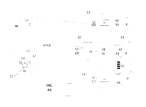
[063] Figure 4A shows illustrative stimulator devices 10,12, 14 which may be
utilized in
various practices of the invention. These devices include a tsDCS stimulation
device 10 which
may be used on its own to deliver a tsDCS mono-stimulation treatment or in
combination with
additional stimulation devices 12 and/or 14 to provide various double and
triple stimulation
treatments, in several embodiments of the invention.
11
CA 03029308 2018-12-24
WO 2016/209997 PCT/US2016/038815
[064] tsDCS stimulator device 10 delivers trans-spinal direct current
stimulation to a spinal
location neurally associated with a distal effector organ of interest, and
more particularly
associated with function of a target effector organ, such as the bladder. In
various embodiments
the stimulation supplied by stimulator device 10 provides monopolar, and an
essentially or
effectively continuous, constant, non-varying direct current stimulation of a
selected polarity, in
a range of 0.5 to 5 or 6 mA, typically 1-4.5mA. Stimulator device 10
illustrates a tsDCS
component in embodiments of the invention. In this illustration, device 10
includes a computing
and synchronizing unit 16, for provision of a system control function, and
including a signal
polarity and function controller 18, and having a system memory 19. The second
stimulator
device 12 provides a known transcranial direct current (tDCS) stimulation
source of pulsed or
constant direct current stimulation to the cortex area C, having a circuit 20
for signal computing
and synchronizing, and for control of signal polarity and function, integrated
with resident
memory 19. In an alternative embodiment, repetitive pulsed magnetic
stimulation (rTMS) is
provided to the cortical area C by a TMS magnetic stimulator 14A using a
figure-eight probe 22,
as shown in Figure 4B, as will be understood by a person skilled in the art,
[065] In an illustrative embodiment, pulsed electrical stimulation of the
motor cortex in an adult
ranges at 100 ¨ 400 mA, typically around 200mA, pulse width of 100 ¨ 300
microseconds,
typically around 200ms, 0.5 to 3 Hz repetition rate, operating voltage 400-
800. For a child, 70-
100 milliamps at 100 microseconds is a target. Magnetic stimulation is
alternatively applied, and
in an illustrative pulsed TMS embodiment, magnetic stimulation is delivered at
a rate of 0.5 to 3
Hz, 200 microsecond pulse width, reaching stimulation current levels
equivalent to the electrical
stimulation, as will be understood by a person skilled in the art. In one TMS
practice of the
invention, rTMS is applied with a magnetic flux density of 1.0 to 1.5 Tesla.
[066] The third stimulator device 14 is a source of direct current stimulation
to stimulate a
peripheral location of interest, typically for stimulation of a nerve leading
to a target effector
organ of interest, such as the bladder, and which may include non-varying or
pulsed direct
current stimulation. This stimulator device 14 includes a circuit 23 for
signal computing and
synchronizing and for control of signal polarity and function, with resident
memory. An
illustrative peripheral constant direct current stimulation is applied at
levels of 1-5mA for double
12
CA 03029308 2018-12-24
WO 2016/209997 PCT/US2016/038815
stimulation and with pulsed peripheral intensity typically ranges is from 5 to
40 mA for triple
stimulation. In a bladder treatment of the invention, continuous tsDCS is
applied to the Onufs
nucleus in the sacral region of the spinal cord, with typical intensity in the
range from 1-4.5 mA.
[067] All three of devices 10, 12, 14, are shown having an I/O component for
external signal
connection, such as with electrodes, 24,26, 30, 32, and 34, 36, respectively,
providing +/-
terminals for electrode connection. Each unit is also provided with a
communication component
40, which enables data links 42 for wired or wireless communication between
the devices or with
other external devices. In this illustration, all three devices 10, 12, 14
have a user interface with
microprocessor unit 44 and a power supply P, such as rechargeable batteries.
[068] The tsDCS stimulator device 10 is engaged on its own when tsDCS mono-
stimulation is
provided. For double stimulation, the tsDCS stimulator device 10 is engaged
along with another
stimulation source, such as provided by the cortical stimulator device 12 in
one practice or by the
peripheral stimulator device 14 in another practice of the invention. In one
practice double-
stimulation is provided by two independent or isolated circuits with the same
or paired
stimulation devices.
[069] As will be appreciated by a person skilled in the art, in several
embodiments, where
constant current stimulation is to be delivered to the patient, the two
cooperating stimulation
sources, such as devices 10 and 14 share a common ground in order to enable an
efficient control
function as the circuits attempt to maintain assigned signal levels over time
in the presence of
changing resistance of the current path(s) within the patient.
[070] In some embodiments, the tsDCS stimulator device 10 is engaged to
provide tsDCS in a
triple-stimulation embodiment, in cooperation with other two stimulation
sources, such as with
the cortical stimulator device 12 and the peripheral stimulator device 14. In
an illustrative
embodiment, the tsDCS triple-stimulation includes pulsed stimulation at the
cortex, constant
stimulation at the spine and pulsed stimulation at the peripheral location.
[071] Referring to Figure 4A, a person to be treated is shown from the back.
Three sets of
electrode connections are shown as would be used during an illustrative triple
stimulation
practice of the invention. Electrodes will be applied in locations discussed
below.
13
CA 03029308 2018-12-24
WO 2016/209997 PCT/US2016/038815
[072] As an illustration only, in a tsDCS triple stimulation embodiment, the
cortical stimulator
12 provides transcortical direct current (tDCS) stimulation as a source of
direct current to the
local cortical area C via active cortical electrode 34 and return (also called
"reference") electrode
36. The stimulation path 34-36 is defined between the two electrodes to
stimulate the local
cortex area C which is associated with the intended stimulation of a target
effector organ of
interest, such as bladder 21 (indicated by dotted symbol). In an alternative
embodiment,
repetitive pulsed magnetic stimulation (rTMS) is supplied to cortical area C
by a probe 22 of a
TMS magnetic stimulator 14A shown in Figure 4B, for application of known
pulsed cortical
stimulation, as will be understood by a person skilled in the art.
[073] The tsDCS stimulator 10 delivers trans-spinal direct current mono-
stimulation to a spinal
location 15 associated with neural outflow associated with a target effector
organ, such as at the
bladder. The spinal active electrode 24 is applied at spinal location 15 and a
return electrode 26
is located distal to the spinal area, such as at an anterior aspect of the
body. In this embodiment, a
spinal stimulation circuit 17 is defined between these two electrodes with the
stimulation current
traversing the spinal processes at that location as a stimulation path of
interest.
[074] The third stimulator 14 provides peripheral direct current stimulation
to stimulate a nerve
leading to a target effector organ or a nerve of the target effector organ,
such as the bladder 21.
In one embodiment, the stimulation signal is monopolar and pulsed. In another
embodiment the
stimulation signal is monopolar and constant.
1075] An illustrative embodiment of the invention includes method and system
having a single
tsDCS stimulation circuit, for mono-stimulation of the spinal cord, and
defined by placing an
electrode at the spinal location of interest and a return electrode on the
anterior aspect of the
body, thus defining a pathway of interest between these electrodes. In various
practices of the
invention, these electrodes are assigned as either anode or cathode and a
tsDCS stimulation
circuit is thus created for applying current between the electrodes and for
modulating spinal cord
excitability. The applied current is delivered having a desired signal
character and level. In
further embodiments of the invention, we apply these teachings in wearable and
implantable
embodiments.
14
CA 03029308 2018-12-24
WO 2016/209997 PCT/US2016/038815
[076] In a further embodiment of the invention, a wearable mono-stimulation
device is
provided. In this practice, there are two electrodes which are skin surface
type, serving as the
active spinal electrode and the spinal circuit return electrode. In one
embodiment, a surface of
the wearable device provides the spinal electrode and the device also connects
to a return
electrode, on the opposite side of the spinal cord, which is placed on the
skin surface such as on
the abdomen or iliac crest. In another embodiment, the reference electrode is
placed internal to
the bladder, such as by urethral catheter insertion, surgically, or the like.
The spinal location of
interest is selected based on spinal outflow to the target effector organ. In
another implantable
mono-stimulation device of the invention, there are two electrodes which are
implantable
electrodes, serving as the active spinal electrode and the return electrode.
In one embodiment,
the mono-stimulation device is fully implantable, with electrode leads from
the device to dorsal
spinal location and ventral location tunneled subcutaneously. The spinal
location of interest is
selected based on spinal outflow to the target effector organ.
[077] In a fully implantable subcutaneous double-stimulation embodiment of the
invention, two
circuits are supplied by four leads emanating from controller device. This
embodiment delivers
two simultaneous stimulations, a spinal stimulation and a peripheral
stimulation applied to a
nerve of the target effector organ. There are two separate stimulation current
paths with these
two circuits. But these circuits also interact to form a resulting stimulation
current path between
the active electrode at the spine of the spinal circuit and the electrode of
opposite polarity
positioned at the nerve of the target effector organ. This provides a
polarization flow down along
the neural path between the two described electrodes. In this double
stimulation embodiment,
the first current path is a tsDCS spinal circuit defined by placing an active
spinal electrode at the
spinal location of interest and a return electrode at a non-spinal location,
with the applied current
running between these electrodes. The second current path is a peripheral
circuit defined by
placing active and return electrodes on or in proximity to a nerve of the
target effector organ.
[078] In a further embodiment, a two-part semi-implantable stimulation device
is provided. A
first component is a wearable mono-stimulation device which includes an active
spinal electrode
applied by skin attachment and a return electrode. The second component is an
implanted
peripheral stimulator or microstimulator with two leads that has its own power
supply. Both
CA 03029308 2018-12-24
WO 2016/209997 PCT/US2016/038815
leads of the second component are in contact with or in close proximity to a
nerve of a target
effector organ. The wearable component can communicate wirelessly with the
implanted
component. When the wearable component turns on and issues its stimulation
signal, the
implanted stimulator responds and issues a stimulation signal to the target
effector organ, which
can be either excitatory or inhibitory.
[079] In a further embodiment of a wearable double-stimulation device, two
circuits are
supplied by four leads emanating from controller device. This embodiment
delivers two
simultaneous stimulations. The first stimulation is a spinal stimulation
delivered via active
spinal electrode applied by skin attachment and a return electrode. The second
stimulation
modulates central autonomic outflow, and can be either trans-cranial direct
current stimulation
(tDCS) or trans-cutaneous vagal nerve stimulation (fVNS). There are two
separate stimulation
current paths with these two circuits but they are electrically isolated from
each other.
[080] In a further embodiment, a two-part semi-implantable stimulation device
is provided. A
first component is a wearable double-stimulation device that provides a first
stimulation that is
spinal stimulation, and a second stimulation that modulates central autonomic
outflow. The
second component is an implanted peripheral stimulator or microstimulator with
two leads that
has its own power supply. Both leads of the second component are in contact
with or in close
proximity to a nerve of a target effector organ. The wearable component can
communicate
wirelessly with the implanted component. When the wearable component turns on
and issues its
stimulation signal, the implanted stimulator responds and issues a stimulation
signal to the target
effector organ, which can be either excitatory or inhibitory.
[081] Illustrative embodiments of the invention is set forth in Figure 5A-C
featuring wearable
and implantable components.
[082] In Figure 5A, a disk-shaped wearable system 100 is disclosed. As
illustrated in Figure
5A-B, system 100 includes a wearable tsDCS controller 102, shown affixed to
the patient at its
skin-side 104 optionally presenting an electrode surface 111. External
interaction with controller
102 is by buttons or touch screen or by wireless interaction with a portable
device or cell phone
103 for user intervention. Controller 102 directs action of implanted
control unit 106.
16
CA 03029308 2018-12-24
WO 2016/209997 PCT/US2016/038815
[083] Controller 102 incorporates a cognate circuit of device 10, Figure 4A,
including a
miniaturized version of computing and synchronizing unit 16, with memory 19,
for provision of
system control, and further including a signal polarity and function
controller 18, with
appropriate instruction loaded set in memory 19 for instruction of implanted
control unit 106.
Control unit 106 includes a rechargeable power supply (not shown), and
according to
instructions from controller 102, applies electrical stimulation to a local
peripheral nerve 108 that
innervates a target effector organ, such as the bladder. The stimulation can
be adjusted as
needed, and is provided as constant continuous non-varying direct current
stimulation, or can be
pulsed direct current stimulation, in various practices of the invention.
[084] In one embodiment, the implanted control unit 106 provides electrical
leads 109 to
deliver the stimulation signal to suitable electrode, shown as a cuff
electrode 110, which is
affixed at nerve 108. In one embodiment, controller 102 presents an electrode
surface 111 on the
skin side of the device for affixation of the device to the patient. This
electrode surface may
include electrically conductive adhesive to assist attachment to the patient,
and permits
application of tsDCS stimulation at that location. In further embodiments of
the invention,
system 100 further includes and cooperates with the implanted control unit
106, which in turn
drives single or multiple implanted electrodes, such as a cuff electrode 108
via leads 109. Cuff
electrode 108 is placed around a peripheral or autonomic nerve of interest 110
and stimulates the
nerve fibers to achieve either excitation or inhibition of the effector organ,
e.g., bladder. The
cuff electrode is made of soft, flexible materials such as silicone that
render an electrode flexible
and less prone to injure the peripheral nerve than common electrodes.
Alternatively, two
electrode leads representing the anode and cathode are positioned in contact
with or in close
proximity to the nerve.
[085] In another embodiment of the present teachings, a wearable tsDCS unit
that wirelessly
controls an implanted stimulator is combined with a sensor that detects a
relevant physiological
state to form a closed-loop system. The wearable tsDCS unit wirelessly
communicates with the
sensor, which could be either implanted or wearable, and activates tsDCS
spinal stimulation and
stimulation of an effector organ via the implanted stimulator, when it detects
a relevant
state. The sensor can be configured to detect blood pressure, heart rate, body
temperature,
17
CA 03029308 2018-12-24
WO 2016/209997 PCT/US2016/038815
respiration rate, skin turgor, skin conductivity, oxygenation state, bladder
pressure, urine
osmolarity, hemodynamic parameters, specific cardiac rhythms by EKG, urethral
pressure, anal
sphincter pressure, muscle contraction state by EMG, specific brain waves by
EEG, electrolytes,
specific proteins and signaling molecules in specific tissue compartments,
blood glucose
concentration, gastric pH, gastrointestinal motility sounds, environmental
cues such as specific
sights, sounds and signals, and other parameters depending on intended
application. The
neuromodulation system is thus activated upon sensing a specific state, and
inactivated when that
state no longer holds. In one embodiment of the present teachings, the system
also includes a
sensor configured to detect a predetermined parameter, such as those listed
herein above, and
configured to provide a sensed value of the predetermined parameter to the
controller
component. The controller component is further configured to initiate
stimulation, initiation of
stimulation determined by whether the sensed value is less than or exceeds a
predetermined
value denoting the specific state.
1086] A closed loop system 200 of the invention is shown in Figure 5C and is
configured to
operate autonomously in the background with reduced user interaction. As will
be appreciated
by a person skilled in the art, the system takes advantage of modern wireless
communications, as
shown, which is available to implanted medical systems. System 200 includes
tsDCS controller
102 and implanted control unit 106 with implanted electrode 108 at nerve 110,
and including an
implanted feedback device 112. The feedback device 112 is in wireless
communication with
controller 102, which then responsively instructs control unit 106 to adjust
or initiate or cease the
stimulation function as needed. The implanted stimulator, control unit 106,
stimulates nerve 110
via leads 109 and electrode 108, consistent with instructions from controller
102.
[087] In a bladder management embodiment, the implanted feedback device 112 is
a bladder
pressure sensor 112A. Bladder data from sensor 112A is wirelessly provided to
controller 102
which wirelessly instructs implanted control unit 106, or directly instructs
control unit 106, to
control stimulation of bladder nerve 108 via electrode 11, to reduce
incontinence or to reduce
urinary retention, for example.
[088] Controller unit 102 has human interface, common instruction memory
store, and logic
circuits, and or a microprocessor, for executing its control instructions to
control unit 106. In
18
CA 03029308 2018-12-24
WO 2016/209997 PCT/US2016/038815
turn, the control unit has a power supply which supplies the electrode
accordingly. Preferably the
power supply is wirelessly rechargeable.
[089] The implanted sensor 112A closes the loop with the device controller
circuit 102 in
system 200 such that the system automatically adjusts without user
intervention, according to
stored profiles. In one embodiment of bladder modulation, the implanted sensor
112A is a
bladder function sensor such as a bladder pressure sensor which detects
bladder pressure exerted
by urine volume in the bladder and enables and wirelessly informs the needed
neural stimulation
instruction to be issued from controller 102 to control unit 106 to initiate
stimulation and to
obtain a desired outcome, such as controlled voiding. In one embodiment, the
data from bladder
sensor 112A is directly acted upon by control unit 106.
[090] In a further application of the closed-loop system 200 of Figure 5C, we
combine
stimulation that modulates central autonomic outflow, in which a primary
stimulation modulates
either the sympathetic or parasympathetic branch of the autonomic nervous
system, with the
closed-loop system 200. Thus, cerebral and spinal stimulations are combined
with an implanted
stimulator that is under the control of the wearable tsDCS controller.
[091] It will be appreciated that embodiments of the present teachings feature
tsDCS spinal
stimulation. In many embodiments, this tsDCS stimulation is augmented with
stimulation of a
peripheral nerve leading to a target effector organ. In practices of these
teachings, peripheral
direct current stimulation (pDCS) is continuous, non-varying, steady-state
direct current
stimulation, while in other embodiments, stimulation of a peripheral nerve or
autonomic nerve
fiber associated with an effector organ may include pulsed electrical
stimulation, continuous
DCS, pulsed DCS, or other alternating signals. The present teachings also may
be practiced with
wireless microstimulators as known in the art.
[092] In practice of the invention, we apply tsDCS in various configurations.
A tsDCS
stimulation system provides tsDCS stimulation, which in various embodiments is
applied by
itself to favorably polarize a target neural pathway of interest. In other
embodiments, we use
coordinated multi-site neurostimulation that incorporates the tsDCS polarizing
stimulation
together with stimulation at other site(s) along the neural axis. We provide
this multi-site
19
CA 03029308 2018-12-24
WO 2016/209997 PCT/US2016/038815
stimulation by combination of tsDCS stimulation with at least one other
stimulation, which
includes cerebral stimulation and/or peripheral stimulation.
[093] In one embodiment of the present teachings, peripheral stimulation is
continuous steady-
state and non-varying. In another embodiment of the invention, excitation or
inhibition of a
stimulated autonomic nerve fiber depends on the frequency of the applied
electrical stimulation.
In one illustrative but non-limiting practice of the invention, inhibition of
parasympathetic fibers
is achieved with high-frequency monopolar electrical stimulation (greater than
about 50-100 Hz),
while excitation of parasympathetic fibers is achieved with low-frequency
monopolar electrical
stimulation (less than about 50-100 Hz). Similarly, inhibition of sympathetic
fibers is achieved
with high-frequency electrical stimulation (greater than about 50-100 Hz),
while excitation of
sympathetic fibers is achieved with low-frequency electrical stimulation (less
than about 50-100
Hz). In various embodiments we apply stimulation via skin surface electrodes
in a range up to
about 1-6 mA or more often at 1-4.5 mA.
[094] In embodiments of the present teachings, the tsDCS device is fully
implantable, with
electrode leads from the device to dorsal spinal location and ventral location
tunneled
subcutaneously. Electrode leads from the tsDCS device which function for
peripheral
stimulation are also tunneled subcutaneously with electrodes implanted on the
appropriate nerves
of the effector organ being modulated. In another embodiment, the tsDCS device
remains
external to the body and wearable, but has electrode leads for peripheral
stimulation that are
either surface mounted or implanted.
Illustrative mono-stimulation embodiments
[095] It will appreciated that the mono-stimulation process involves applying
a single source of
constant current stimulation and is typically delivered by the tsDCS
stimulator alone. In practice
of the present invention, we employ tsDCS to induce either an area of
increased or decreased
neural activation within the spinal cord.
[096] The present invention teaches methods and systems utilizing trans-spinal
direct current
stimulation for modulation of body functions, such as at effector organs.
Illustrative
embodiments of this disclosure are directed to application of such tsDCS to
modulation of
effector constituents of the autonomic nervous system (ANS). Illustrative
embodiments include
CA 03029308 2018-12-24
WO 2016/209997 PCT/US2016/038815
method and apparatus for treatment of bladder dysfunctions. This disclosure is
by way of
illustration and not by way of limitation of the scope of the present
invention.
[097] It will now be appreciated that in various practices of the invention,
tsDCS stimulation is
applied at the spinal location. At peripheral sites (or cerebral sites in the
case of transcutaneous
vagus nerve stimulation), stimulation can be of a broader variety within the
scope of the
invention. In several practices of the present invention, monopolar direct
current stimulation is
applied at specific points along the neural axis. Monopolar direct current
electrical stimulation is
applied and characterized as anodal or cathodal. In an embodiment of the
invention, this
characterization is indicative of the polarity of the current source as
applied between a spinal
location of interest and a return location. Depending upon the desired
outcome, the circuit may
be applied as anodal, positive, at the location of interest, and cathodal,
negative, at the return
location, or vice versa.
[098] Single and/or multiple monopolar direct current stimulation circuits are
engaged in
various embodiments. These monopolar stimulations are, characterized as being
anodal or
cathodal, have a polarizing effect over the stimulated pathways. This
polarization has significant
favorable modulatory effect upon the transmission efficiency of neural signals
flowing over a
neural pathway of interest. Monopolar stimulation applied to a neural pathway
has potential
polarizing and modulatory affects. In various practices of the invention, we
engage and harness
these effects accordingly.
[099] In an illustrative embodiment of the invention in awake healthy mice, a
two-electrode
mono-stimulation configuration of tsDCS was utilized, employing a stimulator,
with an active
cathode electrode on the lumbosacral spine (L6-S3), and a return anode
electrode on the
abdomen. To enable measurements of bladder function, we surgically placed a
cysostomy tube
(PESO tubing) into the bladder to enable measurement of bladder pressures and
urine output
(Figure 6). Bladder pressures, and the frequency of voiding and non-voiding
contractions were
measured at baseline prior to stimulation with cathodal tsDCS (Figure 7A). In
such
embodiments with cathodal tsDCS providing stimulation, there is a decrease in
the basal
pressure, increase in the amplitude of bladder contractions, increase in inter-
voiding contraction
interval, and increase the number and amplitude of non-voiding contractions.
In a series of
21
CA 03029308 2018-12-24
WO 2016/209997 PCT/US2016/038815
experiments, after 20 minutes of cathodal tsDCS, these effects were still
apparent. With such
stimulation, the bladder can contract more fully.
[0100] The same stimulation paradigm was also evaluated in awake mice with
chronic spinal
cord injury, with spinal cord lesioning at T10 level 30 days prior to
stimulation studies. In these
subjects, there is excessive bladder activity and non-voiding contractions,
with higher bladder
pressures as compared to healthy subjects, a condition of detrusor
hyperreflexia. Baseline
measurements were done in these subjects, followed by measurements during
cathodal tsDCS,
and 2 hours after 20 minutes of cathodal tsDCS. In awake subjects with chronic
spinal cord
injury, there is a decrease in the basal pressure, larger non-voiding
contractions, and a decreased
frequency of voiding contractions. Similar to awake healthy subjects, cathodal
tsDCS enables
the bladder of subjects with chronic spinal cord injury to contract more
fully.
[0101] In another embodiment relating to treatment of chronic spinal cord
injury in mice, a two-
electrode configuration of tsDCS was utilized, with an anodal electrode on the
lumbosacral spine
(L6-S3), and with, in one embodiment, the return electrode on the front of the
subject's
abdomen, and in another embodiment, with the return electrode at the bladder
wall via
transurethral insertion. Figure 7B shows spinal to bladder tsDCS stimulation
that initiated
bladder retention and voiding reflex in a vertebrate being with severe chronic
spinal cord injury.
The subject had demonstrated skin irritation caused by excessive urination due
to inability to
retain urine. The top provides cystometry traces showing intravesicle pressure
before, during,
and after stimulation with anode on the spine and cathode inside the bladder.
Note that there
were no reflexes before or after stimulation. Traces on the right are with
expanded time scale to
show the structure of the reflexes. The bottom trace shows cystometry traces
from the same
subject showing before, during stimulation 1 (anode inside the bladder),
stimulation 2 (cathode
inside the bladder), and after. An improved ability to retain urine is seen
even after stimulation is
switched off.
[0102] In further studies of mice with acute spinal cord injury, the same two-
electrode
configuration of tsDCS was utilized. In acute spinal cord injury, there is
spinal shock and
detrusor areflexia, during which period the bladder fills to high and
potentially dangerous
pressures, with voiding pressures significantly higher than normal subjects or
in subjects with
22
CA 03029308 2018-12-24
WO 2016/209997 PCT/US2016/038815
chronic spinal cord injury. This represents a significant health issue because
it can cause stretch
injuries to the bladder and upper urinary tract complications.
[0103] Figure 7C shows bladder reflexes in subjects with acute complete spinal
cord injury and
the effects of tsDCS. Baseline reflexes show very high voiding pressures that
were further
increased by spinal anode/cathode in bladder arrangement. This effect was
maintained for at
least 10 min after the current was turned off. When the polarity was switched,
with spinal
cathode and anode in bladder, this configuration immediately decreased the
voiding pressure and
decreased inter-voiding contraction interval, demonstrating that this
configuration has
therapeutically useful effects in subjects with detrusor areflexia following
acute spinal cord
injury.
[0104] These results are consistent with both notinal and spinal cord injured
mammals.
Excitability of small or moderate sized spinal neurons is increased by
cathodal tsDCS and
depressed by anodal tsDCS. Since autonomic preganglionic neurons are smaller
in size, they
follow this principle. We have found that cathodal tsDCS on the lumbosacral
region increases
the excitability of spinal parasympathetic preganglionic neurons hence
decreasing urine storage
reflexes and increasing voiding reflexes. Reverse polarity induces opposite
modulation, i.e.,
increasing urine storage reflexes and decreasing voiding reflexes. In such
practices, we have
found that placing the return electrode inside or around the bladder enhances
modulatory effects.
[0105] The described anodal spinal/cathodal bladder configuration is effective
in delaying the
bladder voiding reflex to allow for longer filling time. Moreover, the same
arrangement produces
efficient voiding that is evident in lowering the basal pressure after each
voiding cycle. In an
illustrative embodiment of the invention, this anodal spinal/cathodal bladder
configuration has an
inhibitory effect on the parasympathetic input to the bladder. Inhibiting the
parasympathetic
inputs causes relaxation of the bladder detrusor and contraction of the
sphincter vesicae. This
allows for longer inter-voiding contraction interval. In addition, this
configuration enables
increased sympathetic influence over parasympathetic. This treatment is
valuable in for
achieving conditions of low pressure storage and efficient bladder voiding. In
further practices of
the invention, we treat conditions of detrusor areflexia by switching the
polarities of the
electrodes applied to spinal and bladder locations.
23
CA 03029308 2018-12-24
WO 2016/209997 PCT/US2016/038815
[0106] In practice of the present invention, a patient with a condition of
urinary incontinence
involving detrusor hyperreflexia is treated by application of tsDCS in a
configuration that
decreases parasympathetic tone, Figure 8. Such a decrease in parasympathetic
tone results in
relaxation of the detrusor contraction and increased contraction of the
sphincter vesicae. In one
embodiment this is non-invasively achieved by anodal tsDCS at the level of S2-
S4 with a return
cathodal electrode positioned anteriorly at an abdominal location, such as the
skin superior to the
iliac bone. In another embodiment, the return electrode is positioned within
the bladder trans-
urethrally, Figure 9. In further practice of the invention these polarities
(i.e., the anodal and
cathodal assignments,) are reversed for treatment of conditions of urinary
retention. In this
embodiment, the configuration results in an increase in parasympathetic tone.
[0107] In further embodiments of the present invention, a subject with a
condition of urinary
incontinence is treated by application of tsDCS in a configuration that
increases sympathetic
tone, Figure 10. Such an increase in sympathetic tone results in relaxation
and expansion of the
detrusor muscle, constriction of the sphincter vesicae, and inhibition of
parasympathetic nerves
that trigger bladder contraction. This is non-invasively achieved by cathodal
tsDCS at the T11-
L2 spinal level with an anodal return electrode positioned anteriorly at an
abdominal location. In
variant of the embodiment, Figure 11, the return electrode is positioned
within the bladder trans-
urethrally. In further practice of the invention these polarities (i.e., the
anodal and cathodal
assignments,) are reversed for treatment of conditions of urinary retention,
which achieves a
decrease of sympathetic tone.
[0108] An embodiment of the invention includes method and system having a
single tsDCS
stimulation circuit, for mono-stimulation of the spinal cord, and defined by
placing an electrode
at the spinal location of interest and a return electrode on the anterior
aspect of the body, thus
defining a pathway of interest between these electrodes. In various practices
of the invention,
these electrodes are assigned as either anode or cathode and a tsDCS
stimulation circuit is thus
created for applying current between the electrodes and for modulating spinal
cord excitability.
The applied current is delivered having a desired signal character and level.
[0109] In a wearable mono-stimulation device embodiment of the invention,
there are two
electrodes which are skin surface type, serving as the active spinal electrode
and the return
24
CA 03029308 2018-12-24
WO 2016/209997 PCT/US2016/038815
electrode. In one embodiment, the surface of the wearable device provides the
spinal electrode
and the device also connects to a return electrode on the opposite side of the
spinal cord, which is
placed on the skin surface such as on the abdomen or iliac crest. In another
embodiment, the
reference electrode is placed internal to the bladder, such as by urethral
catheter insertion,
surgically, or the like. The spinal location of interest is selected based on
spinal outflow to the
target effector organ.
[0110] In an implantable mono-stimulation device of the invention, there are
two electrodes
which are implantable electrodes, serving as the active spinal electrode and
the return electrode.
In one embodiment, the mono-stimulation device is fully implantable, with
electrode leads from
the device to dorsal spinal location and ventral location tunneled
subcutaneously. The spinal
location of interest is selected based on spinal outflow to the target
effector organ.
[0111] Illustrative double-stimulation embodiments
[0112] Beyond strategies that utilize spinal stimulation via tsDCS on its own,
we also disclose
strategies that combine spinal stimulation via tsDCS with additional
stimulation.
[0113] We teach double-stimulation in various embodiments. Illustrative
embodiments include
two stimulators electrically tied together in as system for polarizing a
critical neural pathway; a
wearable mono-stimulation device communicating wirelessly with an implanted
microstimulator;
and two separate stimulators that are electrically isolated, as when there is
a cortical stimulation
using tDCS combined with spinal stimulation using tsDCS. Still other
configurations will occur
consistent with this disclosure that are also within the scope of the
invention.
[0114] In one double-stimulation embodiment of the invention, we provide
simultaneous tsDCS
spinal stimulation together with pulsed peripheral direct current stimulation
(pDCS) of a nerve
leading to a targeted effector organ. In one particular embodiment, a
resulting polarizing circuit
is defined between an active spinal tsDCS stimulation circuit and an active
pulsed pDCS
peripheral stimulation circuit.
[0115] In one embodiment of the present invention, the described spinal
stimulations which
increase parasympathetic outflow to the bladder are combined with electrical
stimulation of the
CA 03029308 2018-12-24
WO 2016/209997 PCT/US2016/038815
parasympathetic preganglionic fibers in pelvic nerve, Figure 12, with cathodal
tsDCS applied at
S2-S4. Stimulation of the pelvic splanchnic nerve results in contraction of
the bladder detrusor,
and relaxation of the sphincter vesicae, thereby further treating a condition
of urinary retention.
In further practice of the invention, these polarities (i.e., the anodal and
cathodal assignments,)
are reversed for treatment of conditions of urinary incontinence resulting in
a decrease in
parasympathetic tone.
[0116] Excessive activity in the somatic efferents innervating the striated
muscle of the external
urethral sphincter (EUS) results in contraction of the sphincter. In another
embodiment of the
present invention, the described spinal stimulations which increase
parasympathetic outflow to
the bladder are combined with electrical inhibition of the pudendal nerve that
innervates the EUS
using implanted electrodes, Figure 13, with cathodal tsDCS applied at S2-54.
This combination
results in contraction of the bladder detrusor, relaxation of the sphincter
vesicae, and relaxation
of the EUS, thereby further treating a condition of urinary retention. In
further practice of the
invention, these polarities (i.e., the anodal and cathodal assignments,) are
reversed for treatment
of conditions of urinary incontinence and the pudendal nerve innervating the
EUS is electrically
stimulated using implanted electrodes.
[0117] Stimulation of the sensory afferents that fire in response to urine
flow through urethra
results in increased strength of bladder contraction and voiding efficiency.
In another
embodiment of the present invention, the described spinal stimulations which
increase
parasympathetic outflow to the bladder are combined with electrical
stimulation of the pudendal
nerve using implanted electrodes, Figure 14, with cathodal tsDCS applied at S2-
S4.
[0118] In a further embodiment, cathodal spinal stimulations increase
sympathetic outflow to the
bladder as combined with implanted microstimulator electrodes which stimulate
the pudendal
nerve, Figure 15, with cathodal spinal stimulations at T11-L2. Increased
sympathetic tone
results in relaxation of the bladder detrusor and contraction of the sphincter
vesicae, while
stimulation of the pudendal nerve results in contraction of the external
urethral sphincter, thereby
further treating a condition of urinary incontinence. In further practice of
the invention, these
polarities (i.e., the anodal and cathodal assignments,) are reversed for
treatment of conditions of
urinary retention and the pudendal nerve innervating the EUS is electrically
inhibited using
26
CA 03029308 2018-12-24
WO 2016/209997 PCT/US2016/038815
implanted electrodes. In such embodiments, the implanted microstimulator
communicates with
and is controlled by a tsDCS controller that provides spinal stimulations,
that can be either a
wearable device, or an implanted subcutaneous device.
[0119] In a further embodiment, the cathodal spinal stimulations which
increase sympathetic
outflow to the bladder are combined with implanted electrodes which are
applied to inhibit the
parasympathetic preganglionic fibers of the pelvic splanchnic nerves, Figure
16, with cathodal
spinal stimulations at T11-L2,. Increased sympathetic tone results in
relaxation of the bladder
detrusor and contraction of the sphincter vesicae, while inhibition of the
pelvic splanchnic nerves
results in further relaxation of the bladder detrusor, thereby further
treating a condition of urinary
incontinence. In further practice of the invention, these polarities (i.e.,
the anodal and cathodal
assignments,) are reversed for treatment of conditions of urinary retention.
[0120] In a fully implantable subcutaneous double-stimulation embodiment of
the invention, two
circuits are supplied by four leads emanating from controller device. This
embodiment delivers
two simultaneous stimulations, a spinal stimulation and a peripheral
stimulation applied to a
nerve of the target effector organ. There are two separate stimulation current
paths with these
two circuits. But these circuits also interact to form a resulting stimulation
current path between
the anode of one circuit (i.e., active electrode at the spine of the spinal
circuit) and the active
cathode at the nerve of the neural circuit. This provides a polarization flow
down along the
neural path between the two active electrodes. In this double stimulation
embodiment, the first
current path is a tsDCS spinal circuit defined by placing an active spinal
electrode at the spinal
location of interest and a return electrode at a non-spinal location, with the
applied current
running across the tissues between these electrodes. The second current path
is a peripheral
circuit defined by placing active cathode and anode electrodes on or in
proximity to a nerve of
the target effector organ.
[0121] In a further embodiment, a two-part semi-implantable stimulation device
is provided. A
first component is a wearable mono-stimulation device which includes an active
spinal electrode
applied by skin attachment and a return electrode. The second component is an
implanted
peripheral stimulator or microstimulator with two leads that has its own power
supply. Both
leads of the second component are in contact with or in close proximity to a
nerve of a target
27
CA 03029308 2018-12-24
WO 2016/209997 PCT/US2016/038815
effector organ. The wearable component can communicate wirelessly with the
implanted
component. When the wearable component turns on and issues its stimulation
signal, the
implanted stimulator responds and issues a stimulation signal to the target
effector organ, which
can be either excitatory or inhibitory.
[0122] In a further embodiment of a wearable double-stimulation device, two
circuits are
supplied by four leads emanating from controller device. This embodiment
delivers two
simultaneous stimulations. The first stimulation is a spinal stimulation
delivered via active
spinal electrode applied by skin attachment and a return electrode. The second
stimulation
modulates central autonomic outflow, and can be either trans-cranial direct
current stimulation
(tDCS) or trans-cutaneous vagal nerve stimulation (tVNS). There are two
separate stimulation
current paths with these two circuits that are electrically isolated from each
other.
[0123] Triple-stimulation embodiments
[0124] We also herein describe strategies that combine spinal stimulation,
peripheral stimulation,
and stimulation of central autonomic outflow to modulate autonomic function.
The previously
disclosed strategies based on mono-stimulation and double-stimulation might be
sufficient for
certain applications. In other applications, it will be necessary or
beneficial to directly modulate
central autonomic outflow before spinal level modulation via tsDCS and
potential peripheral
stimulation. Non-invasive methods for modulating central autonomic outflow are
combined with
other sites of stimulation using a variety of approaches:
[0125] Transcranial direct current stimulation (tDCS) ¨ A number of different
tDCS montages
have been utilized to modulate the autonomic nervous system. Anodal tDCS over
the primary
motor cortex, with cathode return electrode over the contralateral
supraorbital area has been
reported to increase sympathetic activity (Clancy et al., Brain Stim., 2014,
7:97-104). Anodal
stimulation of the left dorsolateral prefrontal cortex (DLPFC) has been
reported to increase
parasympathetic activity, while anodal stimulation of the right DLPFC has been
reported to
increase sympathetic activity (Brunoni et al., Psychoneuroendocrinology,
2012). Other work has
reported that anodal tDCS over the temporal lobe results in increased
parasympathetic activity.
As such, non-invasive tDCS can be coupled with tsDCS at the relevant spinal
level to modulate
autonomic outflow. In one embodiment, sympathetic outflow from the brain is
increased by
28
CA 03029308 2018-12-24
WO 2016/209997 PCT/US2016/038815
anodal tDCS over the primary motor cortex and further increased at the spinal
level of the
targeted effector organ by cathodal tsDCS at the high thoracic level. This
embodiment is shown
in Figure 17, where cortical electrodes are shown combined with a wearable
tsDCS controller.
In another embodiment, sympathetic outflow from the brain is increased by
anodal tDCS of the
right DLPFC and further increased at the spinal level of the targeted effector
organ by cathodal
tsDCS. In yet another embodiment, parasympathetic outflow from the brain is
increased by
anodal tDCS over the temporal lobe and further increased at either the S2-S4
spinal level of the
targeted effector organ or the brainstem level of DMV by cathodal tsDCS.
[0126] Transcutaneous vagal nerve stimulation (tVNS) - The auricular branch of
the vagus nerve
supplies sensation to the posterior parts of the ear pinna, external auditory
canal and tympanic
membrane, Figure 18A. Nerve cell bodies are located in the superior (jugular)
ganglion of the
vagus, and they project to the nucleus of the tractus solitarius (NTS) in the
brainstem. Electrical
stimulation of the ear concha (tVNS) produces activation of NTS and its known
projections
(parabrachial nucleus, nucleus accumbens, hypothalamus, amygdala). The dorsal
motor nucleus
of the vagus (DMV) in the brainstem contains the cell bodies of the
parasympathetic neurons that
project down the vagus nerve as preganglionic efferent fibers. Direct
connections between the
NTS and DMV have been described, and it is established that NTS sends
projections to DMV.
Stimulation of the external ear tragus using electrical stimulation (10-50 mA,
30 Hz pulse
frequency, 200 microsecond pulse width) results in decreased sympathetic
discharge (Clancy et
al., Brain Stim., 2014, 7:817-877. In a practice of the present invention, we
utilize this non-
invasive methodology for decreasing sympathetic tone and coupling it with
anodal tsDCS at the
spinal level. Sympathetic outflow from the brain is reduced by tVNS and
further reduced at the
spinal level of the targeted effector organ by applied anodal tsDCS. This
embodiment is shown
in Figure 18B, where auricular stimulation is combined with a wearable tsDCS
controller.
[0127] Transcranial magnetic stimulation (TMS) ¨ TMS, both repetitive and
single pulse, has
been utilized in studies that modulate the autonomic nervous system. Targeted
sites include left
temporo-parietal cortex (Lai et al., 2010) and primary motor cortex M1
(Vernieri et al., 2009 and
Yozbatiran et al., 2009). TMS was found to exert changes on autonomic control
in these, and
other studies. Accordingly, in a further embodiment we combine TMS with tsDCS
at the spinal
29
CA 03029308 2018-12-24
WO 2016/209997 PCT/US2016/038815
level. While Figure 17 is illustrated showing cortical stimulation via tDCS,
it will be appreciated
that TMS is an alternative source of cortical stimulation in practices of the
present invention.
[0128] Cold/hot pressors ¨ It is known that immersion of a subject's hand in a
bucket of ice
water results in increased heart rate and pulse pressure, thought to be due to
increased
sympathetic tone activated by sensory afferents. As such, in practices of the
invention, we utilize
this approach as a methodology to initiate modulation of autonomic outflow. As
a bucket of ice
water is impractical, we utilize alternative methodologies to achieve this
effect. More
specifically, in one embodiment, this effect is delivered as a cooling/heating
pad that is affixed to
a theimosensitive area of skin such as the upper back, or in another
embodiment is presented as a
vest or glove with cooling/heating elements. This device is switched to either
"cold stimulation"
or "hot stimulation" to provide that sensation to the skin. To increase
sympathetic tone to a
specific effector organ, we combine activation of "cold stimulation" to the
subject's skin with
cathodal tsDCS at the relevant spinal level. To increase parasympathetic tone
to a specific
effector organ, we combine activation of "hot stimulation" to the subject's
skin with cathodal
tsDCS at the S2-S4 level (or DMV brainstem level). In this way, efferent
outflow through either
the sympathetic or parasympathetic system is activated depending on which
temperature
"setting" is used, and cathodal tsDCS amplifies the signals that are going to
autonomic neurons
in the spinal cord.
[0129] Pharmacological autonomic modulators ¨ Certain pharmacological agents
have
modulatory effects on the autonomic nervous system. Sympathomimetics increase
sympathetic
tone, and include amphetamines and phenylephrine. Sympatholytics decrease
sympathetic tone,
and include prazosin and yohimbine. Parasympathomimetics increase
parasympathetic tone, and
include muscarine, pilocarpine and choline esters. Parasympatholytics decrease
sympathetic
tone, and include scopalamine and atropine. Sympathomimetics can be given in
combination
with parasympatholytics, and parasympathomimetics can be given in combination
with
sympatholytics. Depending on specific molecular characteristics, these
pharmacological agents
can be given orally, subcutaneously, intramuscularly, transdermally,
intravenously or as depot
injections. Pharmacological autonomic modulators are shown in Figure 19.
CA 03029308 2018-12-24
WO 2016/209997 PCT/US2016/038815
[0130] As will be understood by a person skilled in the art, in practices of
the present invention,
we modulate autonomic outflow and use various strategies to monitor effect.
For example, in
various embodiments, we monitor readouts including heart rate, heart rate
variability,
microneurography recording muscle sympathetic nerve activity, blood pressure,
pulse pressure,
pupillary size, skin conductance, sympathetic skin response, respiratory rate,
cerebral vasomotor
reactivity, and body temperature, the utility of which will be understood by a
person skilled in
the art.
[0131] In another embodiment, various of the above described approaches of
modulating central
autonomic outflow, is combined with spinal stimulation and is further combined
with a third
peripheral stimulation, delivered at the level of the nerve leading to the
target effector organ, to
render a useful therapeutic effect. This triple-stimulation approach is shown
in Figure 20.
[0132] In a further embodiment, a two-part semi-implantable stimulation device
is provided. A
first component is a wearable double-stimulation device that provides a first
stimulation that is
spinal stimulation, and a second stimulation that modulates central autonomic
outflow. The
second component is an implanted peripheral stimulator or microstimulator with
two leads that
has its own power supply. Both leads of the second component are in contact
with or in close
proximity to a nerve of a target effector organ. The wearable component can
communicate
wirelessly with the implanted component. When the wearable component turns on
and issues its
stimulation signal, the implanted stimulator responds and issues a stimulation
signal to the target
effector organ, which can be either excitatory or inhibitory.
[0133] In various embodiments, effector organ stimulation via the nerve
leading to the effector
organ is achieved using energetic modalities, including electrical
stimulation, magnetic
stimulation, acoustic stimulation and others. In some instances, it is
desirable to directly
stimulate such nerve using electrical stimulation. In several embodiments of
the invention, the
electrical stimulation is applied at the nerve leading to smooth muscle,
skeletal muscle or is at a
ganglion or plexus associated with the targeted effector organ. In some
embodiments applied to
the autonomic system, stimulation is applied directly at the sympathetic trunk
or ganglia, celiac
ganglion, superior mesenteric ganglion, inferior mesenteric ganglion, or is
stimulated at the post-
ganglionic nerve. The parasympathetic nervous system has ganglia in close
proximity to or
31
CA 03029308 2018-12-24
WO 2016/209997 PCT/US2016/038815
located in the organs being innervated, and in some embodiments electrodes are
placed in
proximity to these parasympathetic ganglia to achieve the desired simulative
effect at the target
effector organ.
[0134] Peripheral pulse intensity typically ranges is from 5 to 40 mA. In one
triple stimulation
bladder embodiment, continuous tsDCS is applied to the Onufs nucleus in the
sacral region of
the spinal cord. The tsDCS is applied with typical intensity in the range from
2 to 5 mA.
[0135] Peripheral pulse intensity typically ranges is from 5 to 40 mA. In one
triple stimulation
bladder embodiment, continuous tsDCS is applied to the Onufs nucleus in the
sacral region of
the spinal cord. The tsDCS is applied with typical intensity in the range from
2 to 5 mA.
[0136] In treating bladder dysfunction, the desired subthreshold spinal tsDCS
and subthreshold
pDCS are established in view of the level at which the effector organ responds
to electrical
stimulation, which serves as the threshold indicator and value of merit. In an
embodiment, this
level is in a range of 2-5mA. In an illustrative embodiment, 3-4.5mA
stimulation at the spine
and 2-3mA via the cathetered active peripheral electrode or 2.5-3.5mA when
applied via
abdominal surface electrode, delivers the desired subthreshold peripheral
stimulation, assuming
the return electrode is placed at a bony location. If the peripheral return
electrode is located
closely associated with the bladder, such as by placement near the bladder or
into the bladder,
then the threshold is detected and adjusted accordingly, typically in the same
range.
[0137] The embodiments described herein provide the basis to treat neurogenic
bladder
conditions that result in either detrusor hyperreflexia or detrusor areflexia
with external devices,
wearable devices, or implanted devices that deliver the described
stimulations. It will be
appreciated by a person skilled in the art that the findings described herein
and reduced to
practice for bladder modulation using a tsDCS-based approach are directly
applicable to
controlling kidney, lung, heart, pancreas, gastrointestinal system, stomach,
anal sphincter and
other autonomically controlled effector organs and may be practiced
accordingly under the
principals disclosed herein. It will now be appreciated that we have
illustrated single, double, and
triple stimulation configurations and methods in practice of embodiments of
the invention.
32
CA 03029308 2018-12-24
WO 2016/209997 PCT/US2016/038815
[0138] Some of the above described approaches combine a primary stimulation
that modulates
either the sympathetic or parasympathetic branch of the autonomic nervous
system, with spinal
stimulation that amplifies the evoked response. A single constant tsDCS
stimulation impacting
the target effector organ is useful and successful in certain situations. In
other situations, a
double-stimulation approach is useful in situations where amplifying autonomic
outflow at the
spinal level is sufficient for a therapeutic effect. In other situations,
primary stimulation and
spinal stimulation is combined with a third stimulation, which is delivered at
the level of the
nerve leading to the targeted effector organ, to render a useful therapeutic
effect. Effector organ
stimulation via the nerve leading to the effector organ is achieved using
selected energetic
modalities, including electrical stimulation, magnetic stimulation, acoustic
stimulation and
others. In some instances, it is desirable to directly stimulate a nerve using
electrical stimulation.
The electrical stimulation is directed to the nerve leading to smooth muscle,
skeletal muscle or is
at a ganglion or plexus associated with the ANS. This is directly at the
sympathetic trunk or
ganglia, celiac ganglion, superior mesenteric ganglion, inferior mesenteric
ganglion, or is
stimulated at the post-ganglionic nerve. The parasympathetic nervous system
has ganglia in close
proximity to or located in the organs being innervated, and in some instances
electrodes might be
placed in proximity to these parasympathetic ganglia.
[0139] In another embodiment, stimulation of the motor cortex using TMS or
tDCS is combined
with spinal stimulation using tsDCS and peripheral stimulation of a nerve
leading to a striated
muscle under voluntary control. As it relates to bladder dysfunction, this
approach can be
utilized to strengthen the external urinary sphincter (EUS), which is a
striated muscle under
voluntary control. In a preferred embodiment, TMS is applied to the motor
cortex area
associated with the EUS, cathodal tsDCS is applied at the spinal level
corresponding to EUS, and
peripheral stimulation is applied to the pudendal nerve leading to the EUS
using an implanted
electrode. In one practice of this embodiment, wherein neural dysfunction of a
distal effector
organ (e.g., a urinary sphincter) is to be treated, the tsDCS spinal
stimulation is applied for the
duration of treatment (a "session") to the spine at the spinal location and
affecting a neural
pathway associated with neural control of that effector organ, and peripheral
and cortical
stimulations are applied to locations associated with that effector organ to
improve neural
33
CA 03029308 2018-12-24
WO 2016/209997 PCT/US2016/038815
communication to that target effector organ. In another embodiment, this
approach is applied to
the external anal sphincter.
[0140] In an illustrative triple stimulation embodiment of the invention,
pulsed stimulation and
cortical stimulation are applied in the presence of tsDCS at the spinal
location (neural spinal
junction) of interest (i.e., a neural spinal junction associated with cortical
control of a target
peripheral organ of interest, such as the bladder). The cortical, spinal and
peripheral stimulation
sites are connected by a common neural pathway. As applied to the neural
pathway, applied
peripheral stimulation pulses from a peripheral stimulator device (e.g.,
device 14) are
synchronized with applied cortical stimulation pulses from a cortical
stimulator 12 or 12A, such
that the peripheral pulses precede the cortical pulse in timing, in any one
cycle. In a typical
stimulation cycle, at least one peripheral pulse and preferably two, applied
to the peripheral
location of interest, e.g., a nerve associated with bladder sphincter control,
precede a following
cortical pulse, wherein such cortical electrical or magnetic stimulation pulse
is applied at a
cortical location of interest, such as at a cortical site associated with
control of the target organ,
e.g., control of bladder sphincter. Latencies of induced peripheral and
cortical pulses are
synchronized to give maximal evoked response (MEP), wherein latencies
typically range from
20 to 45 ms, and as will be appreciated by a person skilled in the art, the
timing of the applied
pulses is thus adjusted in view of these latencies in order to induce the
cortical and pulsed neural
signals on the neural pathway of interest as will flow to the spinal junction
and overlap at the
spinal junction together in the applied presence of the tsDCS stimulation, to
achieve the desired
triple stimulation. Peripheral pulse intensity typically ranges from 5 to 40
mA. In one triple
stimulation bladder embodiment, the tsDCS is applied to the Onufs nucleus in
the sacral region
of the spinal cord. tsDCS with typical intensity in the range from 2 to 5 mA.
[0141] It will be appreciated that in practice of an embodiment of the
invention, we limit
maximum current output for double-stimulation with two simultaneous skin-
surface DC
stimulations at or about 5mA for both spinal and peripheral stimulation
locations. In one
embodiment, an illustrative sponge rubber electrode has a skin contact area of
9cm2 resulting in
34
CA 03029308 2018-12-24
WO 2016/209997 PCT/US2016/038815
a maximum current density of 0.56mA/cm2. As will be appreciated by a persons
skilled in the
art, this is well below the reported safe upper limit for current density of
14.29 ma/cm2 as cited
in: Nitsche MA, Liebetanz D, Lang N, Tergau F, Paulus W., in Safety Criteria
For Transcranial
Direct Current Stimulation (TDCS) In Humans. Clin Neurophysiol
2003;114(11):2220e2."
[0142] It will be appreciated that the stimulation routines of the invention
utilizing cortical
stimulation, either direct electrical direct current stimulation or magnetic,
as in TMS, follow the
triple stimulation teachings of our co-pending US application serial number
14/665,220, filed
March 23, 2015, entitled: Method and System for Treatment of Neuromotor
Dysfunction, which
is a continuation of now issued US Patent 9,011,310, all having a common
inventor and assigned
to a common owner, and all incorporated herein by reference for all purposes
whatsoever.
[0143] It will be appreciated that the stimulation teachings of the invention
utilizing double
stimulation are an adaptation of the teachings of our co-pending US
application serial number
15/046,797, filed February 18, 2016, entitled: Trans-Spinal Direct Current
Modulation Systems,
which is a continuation of now issued US Patent 9,283,391, all having a common
inventor and
assigned to a common owner, and all incorporated herein by reference for all
purposes
whatsoever. In a further alternative illustrative embodiment of the invention,
pulsed implanted
stimulation is provided, as is known in the art for other pulsed peripheral
applications. Such
stimulation can be set to an output of up to 10.5V for pulses up 240
microseconds at 14 Hz, 0.3%
duty cycle, providing a set voltage amplitude and adjusting the current to
maintain the set
amplitude, with pulsed current up to 10mA. Voltage settings are set according
to what the patient
can tolerate, as will be appreciated by a person skilled in the art. The
current is dependent on the
electrode resistance, the electrode tissue interface (likely appreciable) and
the impedance of the
tissue itself, is illustratively at around lkohm.
[0144] In further embodiments of the invention we incorporate a wearable tsDCS
controller that
modulates descending autonomic signals traversing the spinal cord. In some
embodiments, this
is combined with an implanted electrode that directly stimulates the nerve to
a targeted effector
= CA 03029308 2018-12-24
WO 2016/209997
PCT/US2016/038815
organ. This stimulation is selected as either excitatory or inhibitory, and is
further embodiments
depends on stimulation frequency as well as pulse amplitude and duration. The
implanted
electrode is in wireless communication with the wearable tsDCS controller.
[0145] This approach is sufficient for certain applications. In other
applications, it is beneficial
to directly modulate central autonomic outflow before spinal level modulation
via tsDCS. In
practice of the invention, we increase or decrease sympathetic outflow, or
increase or decrease
parasympathetic outflow, as a person skilled in the art would appreciate.
Furthermore in
particular embodiments we provide non-invasive and non-pharmacological
modulating of
autonomic outflow for control and treatment of autonomically-related functions
and disorders.
[0146] COMPUTER
[0147] This disclosure includes description by way of example of a device
configured to execute
functions (hereinafter referred to as computing device) which may be used with
the presently
disclosed subject matter. The description of the various components of a
computing device is not
intended to represent any particular architecture or manner of interconnecting
the components.
Other systems that have fewer or more components may also be used with the
disclosed subject
matter. A communication device may constitute a form of a computing device and
may at least
include a computing device. The computing device may include an inter-connect
(e.g., bus and
system core logic), which can interconnect such components of a computing
device to a data
processing device, such as a processor(s) or microprocessor(s), or other form
of partly or
completely programmable or pre-programmed device, e.g., hard wired and or
application
specific integrated circuit ("ASIC") customized logic circuitry, such as a
controller or
microcontroller, a digital signal processor, or any other form of device that
can fetch instructions,
operate on pre-loaded/pre-programmed instructions, and/or followed
instructions found in hard-
wired or customized circuitry to carry out logic operations that, together,
perform steps of and
whole processes and functionalities as described in the present disclosure.
[0148] In this description, various functions, functionalities and/or
operations may be described
as being perfouned by or caused by software program code to simplify
description. However,
those skilled in the art will recognize what is meant by such expressions is
that the functions
36
CA 03029308 2018-12-24
WO 2016/209997 PCT/US2016/038815
result from execution of the program code/instructions by a computing device
as described
above, e.g., including a processor, such as a microprocessor, microcontroller,
logic circuit or the
like. Alternatively, or in combination, the functions and operations can be
implemented using
special purpose circuitry, with or without software instructions, such as
using Application-
Specific Integrated Circuit (ASIC) or Field-Programmable Gate Array (FPGA),
which may be
programmable, partly programmable or hard wired. The application specific
integrated circuit
("ASIC") logic may be such as gate arrays or standard cells, or the like,
implementing
customized logic by metalization(s) interconnects of the base gate array ASIC
architecture or
selecting and providing metalization(s) interconnects between standard cell
functional blocks
included in a manufacturer's library of functional blocks, etc. Embodiments
can thus be
implemented using hardwired circuitry without program software
code/instructions, or in
combination with circuitry using programmed software code/instructions.
[0149] Thus, the techniques are limited neither to any specific combination of
hardware circuitry
and software, nor to any particular tangible source for the instructions
executed by the data
processor(s) within the computing device. While some embodiments can be
implemented in fully
functioning computers and computer systems, various embodiments are capable of
being
distributed as a computing device including, e.g., a variety of forms and
capable of being applied
regardless of the particular type of machine or tangible computer-readable
media used to actually
effect the performance of the functions and operations and/or the distribution
of the performance
of the functions, functionalities and/or operations.
[0150] The interconnect may connect the data processing device to define logic
circuitry
including memory. The interconnect may be internal to the data processing
device, such as
coupling a microprocessor to on-board cache memory or external (to the
microprocessor)
memory such as main memory, or a disk drive or external to the computing
device, such as a
remote memory, a disc farm or other mass storage device, etc. Commercially
available
microprocessors, one or more of which could be a computing device or part of a
computing
device, include a PA-RISC series microprocessor from Hewlett-Packard Company,
an 80x86 or
Pentium series microprocessor from Intel Corporation, a PowerPC microprocessor
from IBM, a
37
CA 03029308 2018-12-24
WO 2016/209997 PCT/US2016/038815
Sparc microprocessor from Sun Microsystems, Inc, or a 68xxx series
microprocessor from
Motorola Corporation as examples.
[0151] The inter-connect in addition to interconnecting such as
microprocessor(s) and memory
may also interconnect such elements to a display controller and display
device, and/or to other
peripheral devices such as input/output (I/O) devices, e.g., through an
input/output controller(s).
Typical I/O devices can include a mouse, a keyboard(s), a modem(s), a network
interface(s),
printers, scanners, video cameras and other devices which are well known in
the art. The inter-
connect may include one or more buses connected to one another through various
bridges,
controllers and/or adapters. In one embodiment the I/O controller includes a
USB (Universal
Serial Bus) adapter for controlling USB peripherals, and/or an IEEE- 1394 bus
adapter for
controlling IEEE- 1394 peripherals.
[0152] The memory may include any tangible computer-readable media, which may
include but
are not limited to recordable and non-recordable type media such as volatile
and non-volatile
memory devices, such as volatile RAM (Random Access Memory), typically
implemented as
dynamic RAM (DRAM) which requires power continually in order to refresh or
maintain the
data in the memory, and non-volatile RAM (Read Only Memory), and other types
of non-volatile
memory, such as a hard drive, flash memory, detachable memory stick, etc. Non-
volatile
memory typically may include a magnetic hard drive, a magnetic optical drive,
or an optical
drive (e.g., a DVD RAM, a CD RAM, a DVD or a CD), or 'other type of memory
system which
maintains data even after power is removed from the system.
[0153] For the purposes of describing and defining the present teachings, it
is noted that the term
"substantially" is utilized herein to represent the inherent degree of
uncertainty that may be
attributed to any quantitative comparison, value, measurement, or other
representation. The term
"substantially" is also utilized herein to represent the degree by which a
quantitative
representation may vary from a stated reference without resulting in a change
in the basic
function of the subject matter at issue.
[0154] While these teachings have been described in terms of specific
embodiments, it is evident
in view of the foregoing description that numerous alternatives, modifications
and variations will
be apparent to those skilled in the art. Accordingly, these teachings are
intended to encompass all
38
CA 03029308 2018-12-24
WO 2016/209997 PCT/US2016/038815
such alternatives, modifications and variations which fall within the scope
and spirit of the
present teachings and the following claims. The foregoing description is
illustrative validation of
the present invention. It will now be appreciated that tsDCS stimulation
according to
embodiments of the invention can be practiced non-invasively or invasively
using direct current
stimulation to modulate spinal cord neurons. While these teachings have been
described in terms
of specific embodiments, it is evident in view of the foregoing description
that numerous
alternatives, modifications and variations will be apparent to those skilled
in the art.
Accordingly, these teachings are intended to encompass all such alternatives,
modifications and
variations which fall within the scope and spirit of the present teachings and
the following
claims.
[0155] What is claimed is:
39