Note: Descriptions are shown in the official language in which they were submitted.
DESCRIPTION
TITLE OF THE INVENTION
DIAGNOSTIC BIOMARKER FOR EXTRAHEPATIC BILE DUCT CANCER,
INTRAHEPATIC BILE DUCT CANCER OR GALLBLADDER CANCER
Technical Field
[0001]
The present invention relates to a method for
collecting data for diagnosing extrahepatic bile duct
carcinoma, intrahepatic bile duct carcinoma, or
gallbladder carcinoma (hereinafter, these carcinomas are
sometimes collectively referred to as "extrahepatic bile
duct carcinoma or the like"), and a kit for diagnosing
extrahepatic bile duct carcinoma or the like for use in
the method.
Background Art
[0002]
The bile duct is a duct guiding bile produced in the
liver into the duodenum and is roughly classified into the
intrahepatic bile duct in the liver and the extrahepatic
bile duct outside the liver. The extrahepatic bile duct
is linked to the gallbladder for temporarily storing and
concentrating bile via the gallbladder duct. The
extrahepatic bile duct at which the extrahepatic bile duct
and the gallbladder duct merge is called the common bile
duct, and the intrahepatic bile duct, the extrahepatic
bile duct, and the gallbladder are collectively referred
to as the biliary tract.
1
CA 3029926 2020-03-12
[0003]
Most bile duct carcinomas are cancerous biliary
epithelial cells covering the lumen; chemotherapy and
radiotherapy have a little effect on the carcinomas and
surgical resection with early detection is only curative
treatment. However, there is no symptom for early bile
duct carcinoma, for example, extrahepatic bile duct
carcinoma is often found in a state of advanced cancer
since symptoms, such as jaundice and itching, do not occur
until the bile duct is obstructed by the progress of the
carcinoma with bile flowed back into the blood vessel. On
the other hand, intrahepatic bile duct carcinoma does not
quite obstruct the extrahepatic bile duct, and thus,
carcinoma often progresses while remaining asymptomatic
without jaundice symptoms. According to statistics on
cancer death rates by site in Japan in 2014 disclosed by
the Center for Cancer Control and Information Services,
National Cancer Center, the number of people dying of
gallbladder/bile duct carcinoma amounted to 18,117, and
the 5-year relative survival rate by site in 2003 to 2005
was 22.5% for males and 19.9% for females, being the
second worst after pancreatic cancer. The bile duct is
closely related to important organs, such as the liver and
the pancreas, and thus, the metastasis of carcinoma to
these organs contributes to the aggravation of prognosis.
[0004]
Less-invasive abdominal ultrasonography and
hematological examination are generally used for the
diagnosis of biliary tract carcinoma (Non-patent Document
1). The rate of visualization of bile duct carcinoma by
2
CA 3029926 2020-03-12
abdominal ultrasonography ranges from 21 to 90%, and it is
considered problematic that the visualization rate reduces
when the occupation site is the lower bile duct.
Hematological examination using an increase in a tumor
marker, such as CEA or CA19-9, as an index is carried out,
but these tumor markers do not enable the early detection
of biliary tract carcinoma and have a problem with
diagnostic accuracy. Recently, a method has been reported
for detecting bile duct carcinoma using the expression
level of specific microRNA (miRNA) as an index (Patent
Document 1).
[0005]
Carcinoembryonic antigen (CEA) is known to be one of
the embryonal antigens produced from carcinoma cells and
be a glycoprotein having a molecular weight of around
200,000. CEA has 10 or more structurally similar
subfamilies, and CEACAM1 is known as one of them. CEACAM1
in serum is reported to be usable as a marker for
pancreatic carcinoma diagnosis (Non-patent Document 2),
but the relation between CEACAM1 and extrahepatic bile
duct carcinoma or the like has not previously been known.
Prior Art Documents
Patent Document
[0006]
[Patent Document 1] Japanese unexamined Patent Application
Publication No. 2015-139440
Non-patent Documents
[0007]
3
CA 3029926 2020-03-12
[Non-patent Document 1] "Evidence-based clinical practice
guidelines for the management of biliary tract cancers",
Edited by publishing committee of the clinical practice
guidelines for the management of biliary tract cancers,
Igaku Tosho Shuppan Co., Ltd., 2007, p. 38-39.
[Non-patent Document 2] Simeone, D. M. et al., Pancreas.
(2007) 34 (4): 436-443.
Summary of the Invention
Object to be Solved by the Invention
[0008]
An object of the present invention is to provide a
method for collecting highly accurate data for diagnosis,
useful in diagnosing the presence or absence of
extrahepatic bile duct carcinoma or the like, and a kit
for diagnosis.
Means to Solve the Object
[0009]
To solve the above objects, the present inventors
have analyzed the concentration of CEACAM1 in blood
samples collected from extrahepatic bile duct carcinoma,
intrahepatic bile duct carcinoma, and gallbladder
carcinoma patients, and as a result, have found that the
presence or absence of extrahepatic bile duct carcinoma,
intrahepatic bile duct carcinoma, or gallbladder carcinoma
can be determined with good accuracy using the CEACAM1
concentration as an index, thereby accomplishing the
present invention.
[0010]
Thus, the present invention is as follows.
4
CA 3029926 2020-03-12
[1] A method for collecting data for diagnosing
extrahepatic bile duct carcinoma, intrahepatic bile duct
carcinoma, or gallbladder carcinoma, comprising a step of
detecting the concentration of CEACAM1 in a blood sample
collected from a test subject.
[2] The method according to [1] above, further
comprising a step of comparing the concentration of
CEACAM1 in the blood sample collected from the test
subject with the concentration of CEACAM1 in a blood
sample derived from a non-carcinoma control subject,
wherein the concentration of CEACAM1 in the blood sample
collected from the test subject being higher than the
concentration of CEACAM1 in the blood sample derived from
the non-carcinoma control subject indicates that the test
subject has a high possibility of having extrahepatic bile
duct carcinoma, intrahepatic bile duct carcinoma, or
gallbladder carcinoma.
[3] The method according to [2] above, wherein the
non-carcinoma control subject is a healthy subject.
[4] The method according to [1] above, wherein:
when collecting data for diagnosing extrahepatic bile duct
carcinoma, the concentration of CEACAM1 in the blood
sample collected from the test subject being more than
63.9 ng/mL indicates that the test subject has a high
possibility of having extrahepatic bile duct carcinoma;
when collecting data for diagnosing intrahepatic bile duct
carcinoma, the concentration of CEACAM1 in the blood
sample collected from the test subject being more than
Date Recue/Date Received 2021-09-10
55.0 ng/mL indicates that the test subject has a high
possibility of having intrahepatic bile duct carcinoma;
and
when collecting data for diagnosing gallbladder carcinoma,
the concentration of CEACAM1 in the blood sample collected
from the test subject being more than 49.9 ng/mL indicates
that the test subject has a high possibility of having
gallbladder carcinoma.
[5] The method according to [4] above, wherein:
when collecting data for diagnosing extrahepatic bile duct
carcinoma, the concentration of CEACAM1 in the blood
sample collected from the test subject being more than
71.5 ng/mL indicates that the test subject has a high
possibility of having extrahepatic bile duct carcinoma;
and
when collecting data for diagnosing intrahepatic bile duct
carcinoma or gallbladder carcinoma, the concentration of
CEACAM1 in the blood sample collected from the test
subject being more than 63.9 ng/mL indicates that the test
subject has a high possibility of having intrahepatic bile
duct carcinoma or gallbladder carcinoma.
[6] The method according to [4] above, wherein the
concentration of CEACAM1 in the blood sample collected
from the test subject being more than 74.0 ng/mL indicates
that the test subject has a high possibility of having
extrahepatic bile duct carcinoma, intrahepatic bile duct
carcinoma, or gallbladder carcinoma.
[7] The method according to any one of [1] to [6]
above, wherein the blood sample is serum.
6
CA 3029926 2020-03-12
[8] The method according to any one of [1] to [7]
above, wherein the extrahepatic bile duct carcinoma is
extrahepatic bile duct carcinoma classified into stages I
to IIB.
[9] The method according to any one of [1] to [8]
above, further comprising a step of detecting the
concentration of CEA and/or CA19-9 in the blood sample
collected from the test subject.
[10]
[11] A kit for diagnosing extrahepatic bile duct
carcinoma, intrahepatic bile duct carcinoma, or
gallbladder carcinoma, comprising an antibody specifically
binding to CEACAM1 or a labeled product thereof.
[12] The kit according to [11] above, further
comprising an antibody specifically binding to CEA and/or
CA19-9 or a labeled product thereof.
[13] The kit according to [11] or [12] above,
wherein the extrahepatic bile duct carcinoma is
extrahepatic bile duct carcinoma classified into stages I
to IIB.
[0011]
Other embodiments of the present invention can
include a method for diagnosing extrahepatic bile duct
carcinoma, intrahepatic bile duct carcinoma, or
gallbladder carcinoma, comprising detecting the
concentration of CEACAM1 in a blood sample collected from
a test subject.
7
Date Recue/Date Received 2021-09-10
=
Effect of the Invention
[0012]
According to the present invention, highly accurate
data for diagnosis, useful in diagnosing the presence or
absence of extrahepatic bile duct carcinoma, intrahepatic
bile duct carcinoma, or gallbladder carcinoma can be
obtained, and thus extrahepatic bile duct carcinoma,
intrahepatic bile duct carcinoma, or gallbladder carcinoma
can be early detected and appropriately treated.
Brief Description of Drawings
[0013]
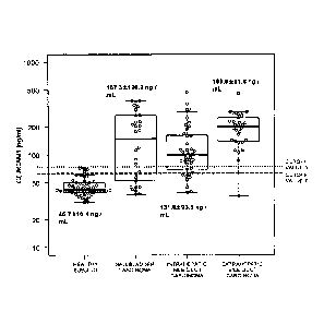
[Figure 1] Figure 1 is a graph showing the results of
measuring the CEACAM1 concentration of the sera of 3 types
of carcinoma patients (extrahepatic bile duct carcinoma,
intrahepatic bile duct carcinoma, and gallbladder
carcinoma) and healthy subject. In the figure, cutoff
value 2 (65.9 ng/mL) and cutoff value 3 (76.0 ng/mL)
indicate cutoff values for "CEACAM1 concentration average
value + 2SD" and "CEACAM1 concentration average value +
3SD" in healthy subject, respectively.
[Figure 2] Figure 2A is a graph showing the results of
measuring the CEACAM1 concentration of the sera of
extrahepatic bile duct carcinoma patients and healthy
individuals (subject). Figure 2B is a graph showing the
results of preparing receiver operating characteristic
(ROC) curves based on the results of the CEACAM1
concentration of the sera of extrahepatic bile duct
carcinoma patients and healthy subject.
8
CA 3029926 2020-03-12
[Figure 3] Figure 3A is a graph showing the results of
measuring the CEACAM1 concentration of the sera of
intrahepatic bile duct carcinoma patients and healthy
individuals (subject). Figure 3B is a graph showing the
results of preparing ROC curves based on the results of
the CEACAM1 concentration of the sera of intrahepatic bile
duct carcinoma patients and healthy subject.
[Figure 4] Figure 4A is a graph showing the results of
measuring the CEACAM1 concentration of the sera of
gallbladder carcinoma patients and healthy individuals
(subject). Figure 4B is a graph showing the results of
preparing ROC curves based on the results of the CEACAM1
concentration of the sera of gallbladder carcinoma
patients and healthy subject.
[Figure 5] Figure 5 is a graph showing the relationship
between the CEACAM1 concentration of the sera of
extrahepatic bile duct carcinoma patients (vertical axis)
and the stage of the carcinoma (horizontal axis). The
cutoff values 1 to 3 in the figure represent 73.5, 65.9,
and 76.0 ng/mL, respectively.
[Figure 6] Figure 6 is a graph showing the relationship
between the CEACAM1 concentration of the sera of
intrahepatic bile duct carcinoma patients (vertical axis)
and the stage of the carcinoma (horizontal axis). The
cutoff values 1 to 3 in the figure represent 57.0, 65.9,
and 76.0 ng/mL, respectively.
[Figure 7] Figure 7 is a graph showing the relationship
between the CEACAM1 concentration of the sera of
gallbladder carcinoma patients (vertical axis) and the
stage of the carcinoma (horizontal axis). The cutoff
9
CA 3029926 2020-03-12
=
values 1 to 3 in the figure represent 51.9, 65.9, and 76.0
ng/mL, respectively.
Mode of Carrying Out the Invention
[0014]
The method for collecting data for diagnosing
extrahepatic bile duct carcinoma or the like according to
the present invention is not particularly limited provided
that it is a method for collecting data for diagnosing
extrahepatic bile duct carcinoma or the like, comprising a
step of detecting (and if necessary, further quantifying)
the concentration of human CEACAM1 (carcinoembryonic
antigen-related cell adhesion molecule 1) (also referred
to as CD66a) in a blood sample collected from a test
subject (donor) (hereinafter sometimes referred to as
"collecting method of the present case"); examples of the
blood sample can include blood and serum or plasma
prepared from blood, and preferred is serum. The
collecting method of the present case is a method for
assisting the diagnosis of extrahepatic bile duct
carcinoma or the like by a physician and does not include
diagnostic action by a physician.
[0015]
The kit for diagnosing extrahepatic bile duct
carcinoma or the like according to the present invention
is not particularly limited provided that it is a kit for
use in diagnosing extrahepatic bile duct carcinoma or the
like (hereinafter sometimes referred to as a "kit for
diagnosis according to the present case"), comprising an
antibody specifically binding to human CEACAM1 in the
CA 3029926 2020-03-12
blood sample (anti-human CEACAM1 antibody), or a labeled
product thereof; the kit for diagnosis according to the
present case is a use invention of a kit for diagnosing
extrahepatic bile duct carcinoma or the like; and the kit
typically includes package inserts, such as an instruction
manual and a manual for diagnosing extrahepatic bile duct
carcinoma or the like, in addition to components generally
used in this type of kit for diagnosis, for example, a
carrier, a pH buffering agent, and a stabilizer.
[0016]
In the collecting method of the present case and the
kit for diagnosis according to the present case, the
carcinoma to be diagnosed may be at least one carcinoma
selected from carcinomas in the biliary tract (bile duct
and gallbladder), i.e., extrahepatic bile duct carcinoma,
intrahepatic bile duct carcinoma, and gallbladder
carcinoma, and the carcinoma also includes the 2 types of
carcinomas of extrahepatic bile duct carcinoma and
intrahepatic bile duct carcinoma, the 2 types of
carcinomas of extrahepatic bile duct carcinoma and
gallbladder carcinoma, the 2 types of carcinomas of
intrahepatic bile duct carcinoma and gallbladder carcinoma,
and the 3 types of carcinomas of extrahepatic bile duct
carcinoma, intrahepatic bile duct carcinoma, and
gallbladder carcinoma.
[0017]
11
Date Recue/Date Received 2021-09-10
The extrahepatic bile duct carcinoma may be a state
in which malignant cells (malignancy) occur in at least a
part of a bile duct portion outside the liver (e.g., the
perihilar bile duct or the distal bile duct); examples
thereof can include perihilar bile duct carcinoma at
stages I to IVB and distal bile duct carcinoma at stages
IA to IV; preferred is extrahepatic bile duct carcinoma
(perihilar bile duct carcinoma or distal bile duct
carcinoma) classified into stages I to II (IIB) since the
collecting method of the present case can also collect
data for diagnosing relatively early extrahepatic bile
duct carcinoma. The state of perihilar bile duct
carcinoma and distal bile duct carcinoma at each stage is
as shown in the following Tables 1 and 2, respectively.
[0018]
Table 1: Perihilar Bile Duct Carcinoma
Stage State
A state in which carcinoma cells remain in the bile duct wall
II A state in which carcinoma cells infiltrate beyond the bile
duct wall
but [1] do not infiltrate into other organs or [2] infiltrate only into the
hepatic parenchyma
IIIA A state in which carcinoma cells infiltrate into the portal
vein in the
bile duct infiltration dominant side or the hepatic artery
IIIB A state in which carcinoma cells metastasize to regional lymph
nodes
IVA A state in which carcinoma cells infiltrate into bilateral
secondary
intrahepatic bile duct branches, the portal trunk, or the left and right
branches
IVB A state in which carcinoma cells metastasize distally
[0019]
Table 2: Distal Bile Duct Carcinoma
Stage State
IA A state in which carcinoma cells remain in the bile duct wall
12
CA 3029926 2020-03-12
=
IB A state in which carcinoma cells infiltrate beyond the bile
duct wall
but do not infiltrate into other organs
I IA A state in which carcinoma cells infiltrate into the
gallbladder, liver,
pancreas, duodenum, or other surrounding organs, or blood
vessels, such as the portal trunk, the superior mesenteric vein, and
the inferior vena cava
IIB A state in which carcinoma cells metastasize to regional lymph
nodes
Ill A state in which carcinoma cells infiltrate into the common
hepatic
artery, the coeliac artery, or the superior mesenteric artery
IV A state in which carcinoma cells metastasize distally
[0020]
The intrahepatic bile duct carcinoma may be a state
in which malignant cells (malignancy) occur in at least a
part of a bile duct portion in the liver, and examples
thereof can include intrahepatic bile duct carcinoma at
stages I to IVB. The state of intrahepatic bile duct
carcinoma at each stage is as shown in the following Table
3.
[0021]
Table 3: Intrahepatic Bile Duct Carcinoma
Stage State
A state in which carcinoma cells are solitary and do not invade
vascular vessels
II A state in which carcinoma cells are solitary and do not invade
vascular vessels [1], or a state in which carcinoma cells are multiple
[2]
Ill A state in which carcinoma cells infiltrate into the visceral
peritoneum or surrounding organs
IVA A state in which carcinoma cells infiltrate around the bile
duct, or
metastasize to lymph nodes
IVB A state in which carcinoma cells metastasize distally
[0022]
The gallbladder carcinoma may be a state in which
malignant cells (malignancy) occur in at least a part of
13
CA 3029926 2020-03-12
the gallbladder, and examples thereof can include
gallbladder carcinoma at stages 0 to IVB. The state of
gallbladder carcinoma at each stage is as shown in the
following Table 4.
[0023]
Table 4: Gallbladder Carcinoma
Stage State
0 A state in which carcinoma cells remain in the gallbladder
mucosa
A state in which carcinoma cells infiltrate into the lamina propria
mucosae or the muscularis propria
II A state in which carcinoma cells infiltrate into the subserosa
or
connective tissues around the gallbladder bed muscular layer
IIIA A state in which carcinoma cells infiltrate into the serous
membrane, the hepatic parenchyma, and/or one surrounding organ
IIIB A state in which carcinoma cells metastasize to regional lymph
nodes
IVA A state in which carcinoma cells infiltrate into 2 or more
surrounding
organs other than the liver, or the portal trunk, the common hepatic
artery, or the proper hepatic artery
IVB A state in which carcinoma cells metastasize distally
[0024]
Examples of the test subject can include test
subjects for whom it is uncertain whether or not they have
carcinoma, and carcinoma patients for whom it is uncertain
whether they have extrahepatic bile duct carcinoma or the
like. Such test subjects and carcinoma patients include
test subjects and carcinoma patients who have had
extrahepatic bile duct carcinoma or the like in the past
and have experienced a complete cure of the carcinomas but
for whom it is uncertain whether they have extrahepatic
bile duct carcinoma or the like at testing.
[0025]
14
CA 3029926 2020-03-12
In the collecting method of the present case, the
CEACAM1 concentration of a blood sample collected from the
test subject being higher than the concentration of
CEACAM1 in a blood sample derived from the non-carcinoma
control subject indicates that the test subject has a high
possibility of having extrahepatic bile duct carcinoma,
intrahepatic bile duct carcinoma, or gallbladder carcinoma.
Thus, the collecting method of the present case preferably
further comprises a step of comparing the concentration of
CEACAM1 in a blood sample collected from the test subject
with the concentration of CEACAM1 in a blood sample
derived from the non-carcinoma control subject.
Comprising such a comparison step enables the collection
of data for diagnosing the test subject as having a high
possibility of having extrahepatic bile duct carcinoma or
the like when the CEACAM1 concentration of a blood sample
derived from the test subject is higher than the CEACAM1
concentration of the blood sample derived from the non-
carcinoma control subject, and enables the collection of
data for diagnosing the test subject as having a low
possibility of having extrahepatic bile duct carcinoma or
the like when the CEACAM1 concentration of a blood sample
derived from the test subject is not higher than the
CEACAM1 concentration of a blood sample derived from the
non-carcinoma control subject. In performing the
collecting method of the present case, as the CEACAM1
concentration of the blood sample derived from the non-
carcinoma control subject, one measured each time may be
used, or one measured in advance may be used. The blood
sample derived from the non-carcinoma control subject is
CA 3029926 2020-03-12
preferably one obtained by collecting the same type of
sample as the sample derived from the test subject and
then subjecting it to the same treatment as that for the
blood sample derived from the test subject.
[0026]
The "non-carcinoma control subject" herein may be a
subject not having carcinoma (a control for the test
subject); specific examples thereof can include a healthy
subject.
[0027]
In the collecting method of the present case, the
CEACAM1 concentration of a blood sample collected from the
test subject being higher than a threshold value (a cutoff
value) indicates that the test subject has a high
possibility of having extrahepatic bile duct carcinoma,
intrahepatic bile duct carcinoma, or gallbladder carcinoma.
The threshold value (cutoff value) cannot simply be
determined because of varying depending on the type of the
carcinoma to be diagnosed, the type of the blood sample,
the detection method, and the like; however, it is
typically about 40.0 ng/mL, preferably about 43.0 ng/mL,
more preferably about 47.0 ng/mL, still more preferably
about 50.0 ng/mL, yet more preferably about 51.9 ng/mL,
particularly preferably about 57.0 ng/mL, particularly
more preferably about 60.0 ng/mL, particularly still more
preferably about 63.0 ng/mL, particularly yet more
preferably about 65.9 ng/mL, and particularly preferably
about 70.0 ng/mL, particularly more preferably about 73.5
ng/mL, most preferably about 76.0 ng/mL.
16
Date Recue/Date Received 2021-09-10
[0028]
The range of "about" in the "about ** ng/mL"
typically means the range of 5 ng/mL, preferably the
range of 4 ng/mL, more preferably the range of 3 ng/mL,
still more preferably the range of 2 ng/mL, most
preferably the range of 1 ng/mL.
[0029]
In the collecting method of the present case, the
threshold value (cutoff value) for indicating that the
test subject has a high possibility of having extrahepatic
bile duct carcinoma when the carcinoma to be diagnosed is
extrahepatic bile duct carcinoma is preferably about 65.9
(specifically 63.9) ng/mL, more preferably about 73.5
(specifically 71.5) ng/mL, still more preferably about
76.0 (specifically 74.0) ng/mL. The threshold value
(cutoff value) for indicating that the test subject has a
high possibility of having intrahepatic bile duct
carcinoma when the carcinoma to be diagnosed is
intrahepatic bile duct carcinoma is preferably about 57.0
(specifically 55.0) ng/mL, more preferably about 65.9
(specifically 63.9) ng/mL, still more preferably about
76.0 (specifically 74.0) ng/mL. The threshold value
(cutoff value) for indicating that the test subject has a
high possibility of having gallbladder carcinoma when the
carcinoma to be diagnosed is gallbladder carcinoma is
preferably about 51.9 (specifically 49.9) ng/mL, more
preferably about 65.9 (specifically 63.9) ng/mL, still
more preferably about 76 (specifically 74.0) ng/mL.
[0030]
17
CA 3029926 2020-03-12
Specific examples of the CEACAM1 can include one or
more proteins selected from the following [group A
protein].
[Group A Protein]
(1) a protein consisting of the amino acid sequence
shown in SEQ ID NO: 1 (CEACAM1 isoform 2 [NCBI Reference
Sequence: NP 001020083]), or a protein consisting of an
amino acid sequence in which 1 or several amino acids are
deleted, substituted and/or added in the amino acid
sequence shown in SEQ ID NO: 1 and having a high
expression level in the test subjecs compared to that in
the healthy subject;
(2) a protein consisting of the amino acid sequence
shown in SEQ ID NO: 2 (CEACAM1 isoform 4 [NCBI Reference
Sequence: NP 001171742]), or a protein consisting of an
amino acid sequence in which 1 or several amino acids are
deleted, substituted and/or added in the amino acid
sequence shown in SEQ ID NO: 2 and having a high
expression level in the test subject compared to that in
the healthy subject;
(3) a protein consisting of the amino acid sequence
shown in SEQ ID NO: 3 (CEACAM1 isoform 3 [NCBI Reference
Sequence: NP 001171744]), or a protein consisting of an
amino acid sequence in which 1 or several amino acids are
deleted, substituted and/or added in the amino acid
sequence shown in SEQ ID NO: 3 and having a high
expression level in the test subject compared to that in
the healthy subject;
(4) a protein consisting of the amino acid sequence
shown in SEQ ID NO: 4 (CEACAM1 isoform 5 [NCBI Reference
18
CA 3029926 2020-03-12
Sequence: NP 001171745]), or a protein consisting of an
amino acid sequence in which 1 or several amino acids are
deleted, substituted and/or added in the amino acid
sequence shown in SEQ ID NO: 4 and having a high
expression level in the test subject compared to that in
the healthy subject;
(5) a protein consisting of the amino acid sequence
shown in SEQ ID NO: 5 (CEACAM1 isoform 6 [NCBI Reference
Sequence: NP 001192273]), or a protein consisting of an
amino acid sequence in which 1 or several amino acids are
deleted, substituted and/or added in the amino acid
sequence shown in SEQ ID NO: 5 and having a high
expression level in the test subject compared to that in
the healthy subject;
(6) a protein consisting of the amino acid sequence
shown in SEQ ID NO: 6 (CEACAM1 isoform 1 [NCB' Reference
Sequence: NP 001703]), or a protein consisting of an amino
acid sequence in which 1 or several amino acids are
deleted, substituted and/or added in the amino acid
sequence shown in SEQ ID NO: 6 and having a high
expression level in the test subject compared to that in
the healthy subject.
[0031]
The "amino acid sequence in which 1 or several amino
acids are deleted, substituted and/or added" means an
amino acid sequence in which typically 1 to 10, preferably
1 to 7, more preferably 1 to 6, still more preferably 1 to
5, yet more preferably 1 to 4, particularly preferably 1
to 3, particularly more preferably 1 to 2, most preferably
1 amino acid is deleted, substituted and/or added.
19
CA 3029926 2020-03-12
[0032]
In the collecting method of the present case, the
CEA and/or CA19-9 concentration of a blood sample derived
from the test subject being higher than the CEA and/or
CA19-9 concentration, respectively, of a blood sample
derived from the healthy subject indicates that the test
subject has a high possibility of having extrahepatic bile
duct carcinoma or the like.
Thus, to further enhance the
reliability of data for diagnosing extrahepatic bile duct
carcinoma or the like, the collecting method of the
present case preferably further comprises a step of
simultaneously, successively, or separately detecting the
concentration of CEA (carcinoembryonic antigen) (also
referred to as CD66e or CEACAM5) and/or CA19-9 in a blood
sample.
[0033]
In the collecting method of the present case, the
method for detecting/quantifying the concentration of
CEACAM1, CEA and/or CA19-9 in a blood sample
Date Recue/Date Received 2021-09-10
may be any method provided that it is a method capable of
specifically detecting a part or all of CEACAM1,
CEA and/or CA19-9 protein in a blood
sample; specific examples thereof can include a mass
spectrometric method for detecting peptides constituting
CEACAM1, CEA and/or CA19-9 protein and an immunoassay
method using an antibody specifically recognizing CEACAM1,
CEA and/or CA19-9 protein.
[0034]
Examples of the immunoassay method can suitably
include an immunohistochemical staining method, an ELISA
method, an EIA method, an RIA method, a western blotting
method, and flow cytometry. Flow cytometry can be
performed with a fluorescence activated cell sorter (FACS)
using an antibody specifically binding to CEACAM1, CEA, or
CA19-9 protein, labeled with a fluorescent substance (e.g.,
allophycocyanin [APC], phycoerythrin [PE], FITC
[fluorescein isothiocyanate], Alexa Fluor 488, Alexa Fluor
647, Alexa Fluor 700, PE-Texas Red, PE-Cy5, or PE-Cy7).
[0035]
To further enhance the reliability of data for
diagnosing extrahepatic bile duct carcinoma or the like,
the kit for diagnosis according to the present case
preferably further comprises an antibody specifically
binding to CEA and/or CA19-9 in a blood sample, or a
labeled product thereof.
[0036]
21
Date Recue/Date Received 2021-09-10
The antibody in the kit for diagnosis according to
the present case may be an antibody, such as a monoclonal
antibody, a polyclonal antibody, a human antibody, a
chimeric antibody, or a humanized antibody, and also
includes an antibody fragment consisting of a portion of
an antibody, such as F(aby)2, Fab, a diabody, Fv, ScFv, or
Sc(Fv)2.
[0037]
Examples of the labeling substance in the labeled
product in the kit for diagnosis according to the present
case can include enzymes, such as peroxidase (e.g.,
horseradish peroxidase), alkaline phosphatase, p-D-
galactosidase, glucose oxidase, glucose-6-phosphate
dehydrogenase, alcohol dehydrogenase, malate dehydrogenase,
penicillinase, catalase, apo-glucose oxidase, urease,
luciferase, Or acetylcholine esterase, fluorescent
substances, such as fluorescein
isothiocyanate,
phycobiliprotein, rare earth metal chelate, dansyl
chloride, and tetramethylrhodamine
isothiocyanate,
fluorescent proteins, such as green fluorescence protein
(GFP), cyan fluorescence protein (OFF), blue fluorescence
protein (BFP), yellow fluorescence protein (YFP), red
fluorescence protein (RFP), and luciferase, radioactive
isotopes, such as 3H, 14C, 1251 or 1311, biotin, avidin, or
chemiluminescent substances.
[0038]
The present invention will be more specifically
described below with reference to Examples. However, the
technical scope of the present invention is not intended
to be limited to these Examples.
22
CA 3029926 2020-03-12
Examples
[0039]
[Material]
Serum samples of extrahepatic bile duct carcinoma
patients were prepared according to an established method
by collecting blood from total 30 extrahepatic bile duct
carcinoma patients (24 males aged 40 to 84 and 6 females
aged 70 to 81), 4 for stage I, 1 for stage IB, 8 for stage
II, 3 for stage IIA, 4 for stage IIB, 1 for stage IIIA, 4
for stage IIIB, 3 for stage IVA, and 2 for stage IVB.
Serum samples of intrahepatic bile duct carcinoma
patients were prepared according to the established method
by collecting blood from total 40 intrahepatic bile duct
carcinoma patients (23 males aged 47 to 80 and 17 females
aged 55 to 85), 4 for stage I, 4 for stage II, 3 for stage
III, 3 for stage IVA, 1 for stage IVB, and 25 for "not
excised". The patients for "not excised" indicate
carcinoma patients who were found to have more advanced
carcinoma and many metastases by diagnostic imaging or the
like and thus were considered to be incapable of being
saved even by surgery and did not undergo surgery.
Serum samples of gallbladder carcinoma patients were
prepared according to the established method by collecting
blood from total 30 gallbladder carcinoma patients (13
males aged 57 to 86 and 17 females aged 46 to 80), 1 for
stage I, 5 for stage II, 1 for stage IIIA, 1 for stage
IIIB, 1 for stage IVA, 2 for stage IVB, and 19 for "not
excised".
23
CA 3029926 2020-03-12
Serum samples of healthy subject as controls were
prepared according to the established method by collecting
blood from 50 healthy subject (9 males aged 37 to 66 and
41 females aged 23 to 60).
[0040]
[Method]
The CEACAM-1 concentration of the serum samples was
measured using "Human CEACAM-1/CD66a DuoSet ELISA" (from
R&D Systems, Inc.) according to the protocol appended to
the product. The CEA concentration and the CA19-9
concentration of the serum samples as comparative controls,
which were measured according to an established method,
were obtained from SRL, Inc. Cutoff values for the
CEACAM1 concentration for distinguishing between patients
with 3 types of carcinomas (extrahepatic bile duct
carcinoma, intrahepatic bile duct carcinoma, and
gallbladder carcinoma) and healthy subject were calculated
based on the ROC curve using a statistical analysis
software (the pROC package of R software) in addition to
the "average 2 x standard deviation (SD)" (Figures 1 and
to 7) and "average 3 x SD" (Figures 1 and 5 to 7 and
Tables 5 to 7) of the CEACAM1 concentrations in the
healthy subject (Figures 1, 2B, 3B, 4B, and 5 to 7).
Hereinafter, the cutoff value calculated based on the ROC
curve is referred to as "cutoff value 1", and the cutoff
values calculated based on the "average 2 x SD" and the
"average 3 x SD" are referred to as "cutoff values 2 and
3", respectively.
[0041]
[Result]
24
CA 3029926 2020-03-12
1. Serum CEACAM-1 Concentration of Extrahepatic Bile
Duct Carcinoma, Intrahepatic Bile Duct Carcinoma, and
Gallbladder Carcinoma Patients
The CEACAM-1 concentration (average SD) of the
sera derived from the healthy subject was 45.7 10.1
(30.8 to 72.4 ng/mL) (Figures 2A, 3A, and 4A), whereas
those in the sera derived from patients with 3 types of
carcinomas (extrahepatic bile duct carcinoma, intrahepatic
bile duct carcinoma, and gallbladder carcinoma) were 198.9
81.8 (36.1 to 458.1 ng/mL) (Figure 2A), 131.8 93.6
(39.2 to 468.2 ng/mL) (Figure 3A), and 167.3 120.2 (37.4
to 383.2 ng/mL) (Figure 4A), respectively (Figure 1).
These results show that the CEACAM-1 concentration
of serum of patients with 3 types of carcinomas
(extrahepatic bile duct carcinoma, intrahepatic bile duct
carcinoma, and gallbladder carcinoma) is high compared to
that for healthy subject.
[0042]
2. Diagnosis of Extrahepatic Bile Duct Carcinoma,
Intrahepatic Bile Duct Carcinoma, and Gallbladder
Carcinoma
Then, cutoff values for distinguishing between
carcinoma patients and healthy subject were set to measure
sensitivity and specificity. Specifically, when the
cutoff value 1 for extrahepatic bile duct carcinoma was
set to 73.5 ng/mL, the sensitivity (the percentage of
test-positive patients in extrahepatic bile duct carcinoma
patients) and specificity (the percentage of test-negative
patients in patients not having extrahepatic bile duct
carcinoma [healthy subject]) for extrahepatic bile duct
CA 3029926 2020-03-12
carcinoma were 96.7% and 100%, respectively. Similarly,
when the cutoff value 2 for extrahepatic bile duct
carcinoma was 65.9 ng/mL, the sensitivity and the
specificity were 96.7% and 92.0%, respectively; when the
cutoff value 3 for extrahepatic bile duct carcinoma was
76.0 ng/mL, the sensitivity and the specificity were 96.7%
and 100%, respectively.
[0043]
When the cutoff value 1 for intrahepatic bile duct
carcinoma was set to 57.0 ng/mL, the sensitivity and the
specificity for intrahepatic bile duct carcinoma were
90.0% and 84.0%, respectively. Similarly, when the cutoff
value 2 for intrahepatic bile duct carcinoma was 65.9
ng/mL, the sensitivity and the specificity were 77.5% and
92.0%, respectively; when the cutoff value 3 for
intrahepatic bile duct carcinoma was 76.0 ng/mL, the
sensitivity and the specificity were 72.5% and 100.0%,
respectively.
[0044]
When the cutoff value 1 for gallbladder carcinoma
patients was set to 51.9 ng/mL, the sensitivity and the
specificity for gallbladder carcinoma were 76.7% and 82.0%,
respectively. Similarly, when the cutoff value 2 for
gallbladder carcinoma was 65.9 ng/mL, the sensitivity and
the specificity were 63.3% and 92.0%, respectively; when
the cutoff value 3 for gallbladder carcinoma was 76.0
ng/mL, the sensitivity and the specificity were 63.3% and
100.0%, respectively.
[0045]
26
CA 3029926 2020-03-12
These results show that when the concentration of
CEACAM1 in serum is measured and suitable cutoff values
are set, 3 types of carcinomas (extrahepatic bile duct
carcinoma, intrahepatic bile duct carcinoma, and
gallbladder carcinoma) can be diagnosed using the CEACAM1
concentration as an index. Particularly, for the
diagnosis of extrahepatic bile duct carcinoma, when around
73.5 to 76.0 ng/mL is set as a cutoff value, the
sensitivity and the specificity are as very high as 96.7%
and 100%, respectively, showing that extrahepatic bile
duct carcinoma can be diagnosed with good accuracy.
[0046]
3. Relation with Stage of Carcinoma
Then, the relation between the CEACAM1 concentration
and the stage of carcinoma was examined. The positive
rates (sensitivities) of 3 types of carcinomas
(extrahepatic bile duct carcinoma, intrahepatic bile duct
carcinoma, and gallbladder carcinoma) had high values
irrespective of the degree of progression of carcinoma
when any of the cutoff values 1 to 3 described above was
set (Figures 5 to 7). Particularly, the positive rate of
extrahepatic bile duct carcinoma had a high value (95% [=
100 x 19/20]) even for relatively early extrahepatic bile
duct carcinoma at stages I to IIB (Figure 5).
These results show that relatively early
extrahepatic bile duct carcinoma, intrahepatic bile duct
carcinoma, and gallbladder carcinoma can be diagnosed
using the concentration of CEACAM1 in serum as an index.
[0047]
27
CA 3029926 2020-03-12
4. Comparison and Combination with Other Carcinoma
Diagnosis Marker
Similar to CEACAM1, CEA and CA19-9 are known as
tumor markers. Accordingly, the CEA concentration and the
CA19-9 concentration of the sera of patients with 3 types
of carcinomas (extrahepatic bile duct carcinoma,
intrahepatic bile duct carcinoma, and gallbladder
carcinoma) were also measured for comparison with CEACAM1
for diagnostic accuracy.
As a result, in extrahepatic bile duct carcinoma
patients, the positive rate of CEACAM1 (96.7%) ("CEACAM1-
positive" in Table 5) was higher than the positive rate of
CEA (20.0%) ("CEA-positive" in Table 5) or the positive
rate of CA19-9 (60.0%) ("CA19-9" in Table 5). In addition,
23 of 24 extrahepatic bile duct carcinoma patients showing
CEA negativity were CEACAM1-positive ("CEA-negative +
CEACAM1-positive" in Table 5), and 9 of 10 extrahepatic
bile duct carcinoma patients showing CA19-9 negativity
were CEACAM1-positive ("CA19-9-negative + CEACAM1-
positive" in Table 5).
These results show that CEACAM1 is a marker capable
of diagnosing extrahepatic bile duct carcinoma with better
accuracy than CEA or CA19-9.
[0048]
Table 5: Extrahepatic Bile Duct Carcinoma
Marker Protein Positive Rate (%)
CEA-positive 20.0 (6/30)
CA19-9-positive 60.0 (15/25)
CEACAM1-positive 96.7 (29/30)
28
CA 3029926 2020-03-12
CEA-negative + CEACAM1-positive 95.8 (23/24)
CA19-9-negative + CEACAM1-positive 90.0 (9/10)
(CEA-negative + CA19-9-negative) + CEACAM1-positive 87.5 (7/8)
The "positive rate" indicates the percentage of test-positive patients in
extrahepatic bile duct carcinoma patients when the cutoff value for the
concentration of each marker protein in healthy subject is set to average 3
x
SD.
[0049]
In intrahepatic bile duct carcinoma patients, the
positive rate of a combination of CEACAM1 and CEA (84.2%)
("CEACAM1-positive + CEA-positive" in Table 6) and the
positive rate of a combination of CEACAM1 + 0A19-9 (86.2%)
("CEACAM1-positive + 0A19-9-positive" in Table 6) were
high compared to the positive rate of CEACAM1 alone
(72.5%) ("CEACAM1-positive" in Table 6). In addition, 15
of 22 intrahepatic bile duct carcinoma patients showing
CEA negativity were CEACAM1-positive ("CEA-negative +
CEACAM1-positive" in Table 6), and 3 of 7 intrahepatic
bile duct carcinoma patients showing CA19-9 negativity
were CEACAM1-positive ("CA19-9-negative + CEACAM1-
positive" in Table 6).
These results show that CEACAM1 combined with CEA or
CA19-9 is a marker capable of diagnosing intrahepatic bile
duct carcinoma with good accuracy compared to the case of
using CEACAM1 alone.
[0050]
29
CA 3029926 2020-03-12
Table 6: lntrahepatic Bile Duct Carcinoma
Marker Protein Positive Rate (%)
CEACAM1-positive 72.5 (29/40)
CEACAM1-positive + CEA-positive 84.2 (32/38)
CEACAM1-positive + CA19-9-positive 86.2 (25/29)
CEA-negative + CEACAM1-positive 68.2 (15/22)
CA19-9-negative + CEACAM1-positive 42.9 (3/7)
(CEA-negative + CA19-9-negative) + CEACAM1-positive 50.0 (3/6)
The "positive rate" indicates the percentage of test-positive patients in
intrahepatic bile duct carcinoma patients when the cutoff value for the
concentration of each marker protein in healthy subject is set to average 3
x
SD.
[0051]
In gallbladder carcinoma patients, the positive rate
of a combination of CEACAM1 and CEA (73.3%) ("CEACAM1-
positive + CEA-positive" in Table 7) and the positive rate
of a combination of CEACAM1 + CA19-9 (87.5%) ("CEACAM1-
positive + CA19-9-positive" in Table 7) were high compared
to the positive rate of CEACAM1 alone (63.3%) ("CEACAM1-
positive" in Table 7). In addition, 7 of 16 gallbladder
carcinoma patients showing CEA negativity were CEACAM1-
positive ("CEA-negative + CEACAM1-positive" in Table 7),
and 3 of 6 gallbladder carcinoma patients showing 0A19-9
negativity were CEACAM1-positive ("CA19-9-negative +
CEACAM1-positive" in Table 7).
These results show that CEACAM1 combined with CEA or
CA19-9 is a marker capable of diagnosing gallbladder
carcinoma with good accuracy compared to the case of using
CEACAM1 alone.
[0052]
CA 3029926 2020-03-12
Table 7: Gallbladder Carcinoma
Marker Protein Positive Rate ( /0)
CEACAM1-positive 63.3 (19/30)
CEACAM1-positive + CEA-positive 73.3 (22/30)
CEACAM1-positive + CA19-9-positive 87.5 (21/24)
CEA-negative + CEACAM1-positive 43.8 (7/16)
CA19-9-negative + CEACAM1-positive 50.0 (3/6)
(CEA-negative + CA19-9-negative) + CEACAM1-positive 40.0 (2/5)
The "positive rate" indicates the percentage of test-positive patients in
gallbladder carcinoma patients when the cutoff value for the concentration of
each marker protein in healthy subject is set to average 3 x SD.
Industrial Applicability
[0053]
The present invention is conducive to the diagnosis
and treatment of extrahepatic bile duct carcinoma,
intrahepatic bile duct carcinoma, or gallbladder carcinoma.
31
CA 3029926 2020-03-12