Note: Descriptions are shown in the official language in which they were submitted.
CA 03030755 2019-01-11
WO 2018/022651 PCT/US2017/043782
AUTOLOGOUS AND ALLOGENIC MACROPHAGES AND MONOCYTES FOR USE
IN THERAPEUTIC METHODS
CROSS ¨ REFERENCE
[0001] This application claims the benefit of U.S. Provisional Application No.
62/366,474, filed
July 25, 2016, U.S. Provisional Application No. 62/366,569, filed July 25,
2016, U.S.
Provisional Application No. 62/444,760, filed January 10, 2017, U.S.
Provisional Application
No. 62/456,448, filed February 8, 2017, and U.S. Provisional Application No.
62/457,681, filed
February 10, 2017, each of which are incorporated herein by reference in their
entireties.
SUMMARY OF THE INVENTION
[0002] Disclosed herein, in certain embodiments, are methods for treating a
pathogenic infection
in an individual in need thereof, comprising: administering to the individual
an innate immune
cell. In some embodiments, the innate immune cell is allogenic. In some
embodiments, the
innate immune cell is autologous. In some embodiments, the innate immune cell
is a monocyte.
In some embodiments, the innate immune cell is a macrophage. In some
embodiments, the
monocyte is produced by a method comprising isolating monocytes from a
population of
immune cells extracted from an individual. In some embodiments, the monocyte
is produced by
a method comprising differentiating a CD34+ hematopoietic stem cell from a
peripheral blood
sample, a cord blood sample, or a bone marrow sample into a monocyte
progenitor cell and
further differentiating the monocyte progenitor cell into the monocyte. In
some embodiments,
the monocyte is produced by a method comprising differentiating an embryonic
stem cell (ESC)
into a monocyte progenitor cell and further differentiating the monocyte
progenitor cell into the
monocyte. In some embodiments, the monocyte is produced by a method comprising
genetically reprogramming a somatic cell into an induced pluripotent stem cell
(iPSC) and
differentiating the iPSC into the monocyte. In some embodiments, the
macrophage is produced
by a method comprising isolating macrophages from a tissue or a population of
immune cells
extracted from an individual. In some embodiments, the macrophage is produced
by (a)
isolating monocytes from a population of immune cells extracted from an
individual; and (b)
differentiating the isolated monocytes into macrophages. In some embodiments,
the
macrophage is produced by differentiating a CD34+ hematopoietic stem cell from
a peripheral
blood sample, a cord blood sample, or a bone marrow sample into a macrophage
progenitor cell
and further differentiating the macrophage progenitor cell into the
macrophage. In some
embodiments, the macrophage is produced by differentiating an embryonic stem
cell (ESC) into
a macrophage progenitor cell and further differentiating the macrophage
progenitor cell into the
macrophage. In some embodiments, the macrophage is produced by genetically
reprogramming
1
CA 03030755 2019-01-11
WO 2018/022651 PCT/US2017/043782
a somatic cell into an induced pluripotent stem cell (iPSC) and
differentiating the iPSC into the
macrophage. In some embodiments, the pathogenic infection is a bacterial
infection. In some
embodiments, the pathogenic infection is a viral infection. In some
embodiments, the
pathogenic infection is a fungal infection. In some embodiments, the
pathogenic infection is a
parasitic infection. In some embodiments, the bacterial infection comprises
intracellular bacteria
or extracellular bacteria. In some embodiments, the bacterial infection
comprises gram negative
bacteria. In some embodiments, the bacterial infection comprises gram positive
bacteria. In
some embodiments, the bacterial infection comprises multi-drug resistant
bacteria, extensively
drug resistant bacteria, or pan-drug resistant bacteria. In some embodiments,
the bacterial
infection comprises bacterial that are resistant to an antibacterial selected
from the group
consisting of: penicillin, ampicillin, carbapenem, fluoroquinolone,
cephalosporin, tetracycline,
erythromycin, methicillin, gentamicin, vancomycin, imipenem, ceftazidime,
levofloxacin,
linezolid, daptomycin, ceftaroline, clindamycin, fluconazole, and
ciprofloxacin. In some
embodiments, the bacterial infection comprises bacteria selected from the
group consisting of:
Klebsiella pneumoniae, Clostridium difficile, Acinetobacer baumannii, Bacillus
anthracis,
Escherichia coli, Haemophilus influenza, Mycoplasma spp., Pseudomonas
aeruginosa,
Staphylococcus aureus, Streptococcus pyogenes, Enterobacteriaceae,
Enterococcus faecium,
Helicobacter pylori, Campylobacter spp., Salmonellae, Neisseria gonorrhoeae,
Streptococcus
pneumoniae, Haemophilus influenza, Shigella spp., Burkholderia cepacia,
Mycobacterium
tuberculosis, Neisseria meningitidis, non-tuberculous mycobacteria,
Streptococcus agalactiae,
and Vibrio cholerae. In some embodiments, the bacterial infection comprises
Clostridium
difficile bacteria. In some embodiments, the bacterial infection comprises
Klebsiella pneumoniae
bacteria. In some embodiments, the bacterial infection comprises Acinetobacter
baumannii
bacteria. In some embodiments, the bacterial infection comprises Pseudomonas
Aeruginosa
bacteria. In some embodiments, the bacterial infection comprises methacillin-
resistant
staphylococcus aureas (MRSA) bacteria. In some embodiments, the viral
infection comprises a
virus selected from the group consisting of: Herpes simplex virus (HSV),
varicella zoster virus,
cytomegalovirus (CMV), Epstein-Barr virus (EBV), Eastern equine encephalitis
(EEE), western
equine encephalitis (WEE), rubella virus, poliovirus, coxsackievirus, an
enterovirus, St. Louis
encephalitis (SLE), Japanese encephalitis, rubeola (measles) virus, mumps
virus, California
encephalitis, LaCrosse virus, human immunodeficiency virus (HIV), rabies
virus, and Influenza
A virus. In some embodiments, the fungal infection comprises a fungus selected
from the group
consisting of: Aspergillus, Bipolaris, Blastomyces, Candida, Cryptococcus,
Coccidioides,
Curvularia, Exophiala, Histoplasma, Mucorales, Ochroconis, Pseudallescheria,
Ramichloridium, Sporothrix, Zygomyctes, Pneumocystis, and Trichosporon. In
some
2
CA 03030755 2019-01-11
WO 2018/022651 PCT/US2017/043782
embodiments, the pathogenic infection is selected from: sepsis, pneumonia,
catheter-associated
infection, bacteremia, hospital-acquired infection, intensive care unit
infection, central line
bloodstream infection, surgical site infection, urinary tract infection,
ventilator associated
pneumonia, infections associated with combat-related injuries, and chronic
wound infections. In
some embodiments, the population of immune cells is extracted from a
peripheral blood sample,
a cord blood sample, or a bone marrow sample of the individual. In some
embodiments, the
peripheral blood sample is a mobilized peripheral blood sample or a non-
mobilized peripheral
blood sample. In some embodiments, differentiating the isolated monocytes into
macrophages
comprises contacting the isolated monocytes with granulocyte¨macrophage (GM-
CSF) or
macrophage (M-CSF) colony-stimulating factor. In some embodiments, the methods
further
comprise activating the innate immune cells by contacting the innate immune
cells with an
activator. In some embodiments, the activator is selected from: a small
molecule drug, an
endotoxin, a cytokine, a chemokine, an interleukin, a pattern recognition
receptor (PRR) ligand,
a toll-like receptor (TLR) ligand, an adhesion molecule, or any combinations
thereof In some
embodiments, the small molecule drug is phorbol myristate acetate. In some
embodiments, the
endotoxin is lipopolysaccharide (LPS) or delta endotoxin. In some embodiments,
the cytokine is
IL-4, IL-13, interferon gamma (IFNy), or tumor-necrosis factor (TNF). In some
embodiments,
the adhesion molecule is an integrin, an immunoglobulin, or a selectin. In
some embodiments,
the innate immune cell is genetically engineered to reduce or inhibit
production of an unwanted
protein, an unwanted amino acid sequence, an unwanted nucleic acid, or an
alloantigen. In some
embodiments, the unwanted protein is SIRP-a. In some embodiments, the unwanted
amino acid
sequence is immunoreceptor tyrosine-based inhibition motif (ITIM). In some
embodiments, the
innate immune cell is frozen.
[0003] Disclosed herein, in certain embodiments, are methods of treating a
pulmonary disease in
an individual in need thereof comprising: administering to the individual an
innate immune cell.
In some embodiments, the innate immune cell is allogenic. In some embodiments,
the innate
immune cell is autologous. In some embodiments, the innate immune cell is a
monocyte. In
some embodiments, the innate immune cell is a macrophage. In some embodiments,
the
monocyte is produced by a method comprising isolating monocytes from a
population of
immune cells extracted from an individual. In some embodiments, the monocyte
is produced by
a method comprising differentiating a CD34+ hematopoietic stem cell from a
peripheral blood
sample, a cord blood sample, or a bone marrow sample into a monocyte
progenitor cell and
further differentiating the monocyte progenitor cell into the monocyte. In
some embodiments,
the monocyte is produced by a method comprising differentiating an embryonic
stem cell (ESC)
into a monocyte progenitor cell and further differentiating the monocyte
progenitor cell into the
3
CA 03030755 2019-01-11
WO 2018/022651 PCT/US2017/043782
monocyte. In some embodiments, the monocyte is produced by a method comprising
genetically
reprogramming a somatic cell into an induced pluripotent stem cell (iPSC) and
differentiating
the iPSC into the monocyte. In some embodiments, the macrophage is produced by
a method
comprising isolating macrophages from a population of immune cells extracted
from an
individual. In some embodiments, the macrophage is produced by (a) isolating
monocytes from
a population of immune cells extracted from an individual; and (b)
differentiating the isolated
monocytes into macrophages. In some embodiments, the macrophage is produced by
differentiating an embryonic stem cell (ESC) into a macrophage progenitor cell
and further
differentiating the macrophage progenitor cell into the macrophage. In some
embodiments, the
macrophage is produced by genetically reprogramming a somatic cell into an
induced
pluripotent stem cell (iPSC) and differentiating the iPSC into the macrophage.
In some
embodiments, the pulmonary disease is a chronic pulmonary disease. In some
embodiments, the
pulmonary disease is chronic obstructive pulmonary disease (COPD), cystic
fibrosis, or asthma.
In some embodiments, the pulmonary disease is associated with a pathogenic
infection. In some
embodiments, the pathogenic infection is a bacterial infection. In some
embodiments, the
pathogenic infection is a viral infection. In some embodiments, wherein the
pathogenic
infection is a fungal infection. In some embodiments, the pathogenic infection
is a parasitic
infection. In some embodiments, the bacterial infection comprises
intracellular bacteria or
extracellular bacteria. In some embodiments, the bacterial infection comprises
gram negative
bacteria. In some embodiments, the bacterial infection comprises gram positive
bacteria. In
some embodiments, the bacterial infection comprises multi-drug resistant
bacteria, extensively
drug resistant bacteria, or pan-drug resistant bacteria. In some embodiments,
the bacterial
infection comprises bacterial that are resistant to an antibacterial selected
from the group
consisting of: penicillin, ampicillin, carbapenem, fluoroquinolone,
cephalosporin, tetracycline,
erythromycin, methicillin, gentamicin, vancomycin, imipenem, ceftazidime,
levofloxacin,
linezolid, daptomycin, ceftaroline, clindamycin, fluconazole, and
ciprofloxacin. In some
embodiments, the bacterial infection comprises bacteria selected from the
group consisting of:
Klebsiella pneumoniae, Clostridium difficile, Acinetobacer baumannii, Bacillus
anthracis,
Escherichia coil, Haemophilus influenza, Mycoplasma spp., Pseudomonas
aeruginosa,
Staphylococcus aureus, Streptococcus pyogenes, Enterobacteriaceae,
Enterococcus faecium,
Helicobacter pylori, Campylobacter spp., Salmonellae, Neisseria gonorrhoeae,
Streptococcus
pneumoniae, Haemophilus influenza, Shigella spp., Burkholderia cepacia,
Mycobacterium
tuberculosis, Neisseria meningitidis, non-tuberculous mycobacteria,
Streptococcus agalactiae,
and Vibrio cholerae. In some embodiments, the bacterial infection comprises
Clostridium
difficile bacteria. In some embodiments, the bacterial infection comprises
Klebsiella pneumoniae
4
CA 03030755 2019-01-11
WO 2018/022651 PCT/US2017/043782
bacteria. In some embodiments, the bacterial infection comprises Acinetobacter
baumannii
bacteria. In some embodiments, the bacterial infection comprises Pseudomonas
Aeruginosa
bacteria. In some embodiments, the bacterial infection comprises methicillin-
resistant
staphylococcus aureus (MRSA) bacteria. In some embodiments, the viral
infection comprises a
virus selected from the group consisting of: Herpes simplex virus (HSV),
varicella zoster virus,
cytomegalovirus (CMV), Epstein-Barr virus (EBV), Eastern equine encephalitis
(EEE), western
equine encephalitis (WEE), rubella virus, poliovirus, coxsackievirus, an
enterovirus, St. Louis
encephalitis (SLE), Japanese encephalitis, rubeola (measles) virus, mumps
virus, California
encephalitis, LaCrosse virus, human immunodeficiency virus (HIV), rabies
virus, and Influenza
A virus. In some embodiments, the fungal infection comprises a fungus selected
from the group
consisting of: Aspergillus, Bipolaris, Blastomyces, Candida, Cryptococcus,
Coccidioides,
Curvularia, Exophiala, Histoplasma, Mucorales, Ochroconis, Pseudallescheria,
Ramichloridium, Sporothrix, Zygomyctes, Pneumocystis, and Trichosporon. In
some
embodiments, the pathogenic infection is selected from: sepsis, pneumonia,
catheter-associated
infection, bacteremia, hospital-acquired infection, intensive care unit
infection, central line
bloodstream infection, surgical site infection, urinary tract infection, and
ventilator associated
pneumonia. In some embodiments, the population of immune cells is extracted
from a
peripheral blood sample, a cord blood sample, or a bone marrow sample of the
individual. In
some embodiments, the peripheral blood sample is a mobilized peripheral blood
sample or a
non-mobilized peripheral blood sample. In some embodiments, differentiating
the isolated
monocytes into macrophages comprises contacting the isolated monocytes with
granulocyte¨
macrophage (GM-CSF) or macrophage (M-CSF) colony-stimulating factor. In some
embodiments, the methods further comprise activating the innate immune cells
by contacting the
innate immune cells with an activator. In some embodiments, the activator is
selected from: a
small molecule drug, an endotoxin, a cytokine, a chemokine, an interleukin, a
pattern
recognition receptor (PRR) ligand, a toll-like receptor (TLR) ligand, an
adhesion molecule, or
any combinations thereof. In some embodiments, the small molecule drug is
phorbol myristate
acetate. In some embodiments, the endotoxin is lipopolysaccharide (LPS) or
delta endotoxin. In
some embodiments, the cytokine is IL-4, IL-13, interferon gamma (IFNy), or
tumor-necrosis
factor (TNF). In some embodiments, the adhesion molecule is an integrin, an
immunoglobulin,
or a selectin. In some embodiments, the innate immune cell is genetically
engineered to reduce
or inhibit production of an unwanted protein, an unwanted amino acid sequence,
an unwanted
nucleic acid, or an alloantigen. In some embodiments, the unwanted protein is
SIRP-a. In some
embodiments, the unwanted amino acid sequence is immunoreceptor tyrosine-based
inhibition
motif (ITIM). In some embodiments, the innate immune cell is frozen.
CA 03030755 2019-01-11
WO 2018/022651 PCT/US2017/043782
[0004] Disclosed herein, in certain embodiments, are methods of treating an
inflammatory
disease in an individual in need thereof comprising: administering to the
individual an innate
immune cell. In some embodiments, the innate immune cell is allogenic. In some
embodiments, the innate immune cell is autologous. In some embodiments, the
innate immune
cell is a monocyte. In some embodiments, the innate immune cell is a
macrophage. In some
embodiments, the monocyte is produced by a method comprising isolating
monocytes from a
population of immune cells extracted from an individual. In some embodiments,
the monocyte
is produced by a method comprising differentiating a CD34+ hematopoietic stem
cell from a
peripheral blood sample, a cord blood sample, or a bone marrow sample into a
monocyte
progenitor cell and further differentiating the monocyte progenitor cell into
the monocyte. In
some embodiments, the monocyte is produced by a method comprising
differentiating an
embryonic stem cell (ESC) into a monocyte progenitor cell and further
differentiating the
monocyte progenitor cell into the monocyte. In some embodiments, the monocyte
is produced
by a method comprising genetically reprogramming a somatic cell into an
induced pluripotent
stem cell (iPSC) and differentiating the iPSC into the monocyte. In some
embodiments, the
macrophage is produced by a method comprising isolating macrophages from a
population of
immune cells extracted from an individual. In some embodiments, the macrophage
is produced
by (a) isolating monocytes from a population of immune cells extracted from an
individual; and
(b) differentiating the isolated monocytes into macrophages. In some
embodiments, the
macrophage is produced by differentiating an embryonic stem cell (ESC) into a
macrophage
progenitor cell and further differentiating the macrophage progenitor cell
into the macrophage.
In some embodiments, the macrophage is produced by genetically reprogramming a
somatic cell
into an induced pluripotent stem cell (iPSC) and differentiating the iPSC into
the macrophage.
In some embodiments, the inflammatory disease is a chronic inflammatory
disease. In some
embodiments, the chronic inflammatory disease is atherosclerosis, rheumatoid
arthritis, lupus, or
type 1 diabetes. In some embodiments, the population of immune cells is
extracted from a
peripheral blood sample, a cord blood sample, or a bone marrow sample of the
individual. In
some embodiments, the peripheral blood sample is a mobilized peripheral blood
sample or a
non-mobilized peripheral blood sample. In some embodiments, differentiating
the isolated
monocytes into macrophages comprises contacting the isolated monocytes with
granulocyte¨
macrophage (GM-CSF) or macrophage (M-CSF) colony-stimulating factor. In some
embodiments, the methods further comprise activating the innate immune cells
by contacting the
innate immune cells with an activator. In some embodiments, the activator is
selected from: a
small molecule drug, an endotoxin, a cytokine, a chemokine, an interleukin, a
pattern
recognition receptor (PRR) ligand, a toll-like receptor (TLR) ligand, an
adhesion molecule, or
6
CA 03030755 2019-01-11
WO 2018/022651 PCT/US2017/043782
any combinations thereof. In some embodiments, the small molecule drug is
phorbol myristate
acetate. In some embodiments, the endotoxin is lipopolysaccharide (LPS) or
delta endotoxin. In
some embodiments, the cytokine is IL-4, IL-13, interferon gamma (IFNy), or
tumor-necrosis
factor (TNF). In some embodiments, the adhesion molecule is an integrin, an
immunoglobulin,
or a selectin. In some embodiments, the innate immune cell is genetically
engineered to reduce
or inhibit production of an unwanted protein, an unwanted amino acid sequence,
or an
alloantigen. In some embodiments, the unwanted protein is SIRP-a. In some
embodiments, the
unwanted amino acid sequence is immunoreceptor tyrosine-based inhibition motif
(ITIM). In
some embodiments, the innate immune cell is frozen.
[0005] Disclosed herein, in certain embodiments, are methods of treating an
autoimmune
disease in an individual in need thereof comprising: administering to the
individual an innate
immune cell. In some embodiments, the innate immune cell is allogenic. In some
embodiments, the innate immune cell is autologous. In some embodiments, the
innate immune
cell is a monocyte. In some embodiments, the innate immune cell is a
macrophage. In some
embodiments, the monocyte is produced by a method comprising isolating
monocytes from a
population of immune cells extracted from an individual. In some embodiments,
the monocyte
is produced by a method comprising differentiating a CD34+ hematopoietic stem
cell from a
peripheral blood sample, a cord blood sample, or a bone marrow sample into a
monocyte
progenitor cell and further differentiating the monocyte progenitor cell into
the monocyte. In
some embodiments, the monocyte is produced by a method comprising
differentiating an
embryonic stem cell (ESC) into a monocyte progenitor cell and further
differentiating the
monocyte progenitor cell into the monocyte. In some embodiments, the monocyte
is produced
by a method comprising genetically reprogramming a somatic cell into an
induced pluripotent
stem cell (iPSC) and differentiating the iPSC into the monocyte. In some
embodiments, the
macrophage is produced by a method comprising isolating macrophages from a
population of
immune cells extracted from an individual. In some embodiments, the macrophage
is produced
by (a) isolating monocytes from a population of immune cells extracted from an
individual; and
(b) differentiating the isolated monocytes into macrophages. In some
embodiments, the
macrophage is produced by differentiating an embryonic stem cell (ESC) into a
macrophage
progenitor cell and further differentiating the macrophage progenitor cell
into the macrophage.
In some embodiments, the macrophage is produced by genetically reprogramming a
somatic cell
into an induced pluripotent stem cell (iPSC) and differentiating the iPSC into
the macrophage.
In some embodiments, the autoimmune disease is rheumatoid arthritis, lupus, or
type 1 diabetes.
In some embodiments, the population of immune cells is extracted from a
peripheral blood
sample, a cord blood sample, or a bone marrow sample of the individual. In
some embodiments,
7
CA 03030755 2019-01-11
WO 2018/022651 PCT/US2017/043782
the peripheral blood sample is a mobilized peripheral blood sample or a non-
mobilized
peripheral blood sample. In some embodiments, differentiating the isolated
monocytes into
macrophages comprises contacting the isolated monocytes with
granulocyte¨macrophage (GM-
CSF) or macrophage (M-CSF) colony-stimulating factor. In some embodiments, the
methods
further comprise activating the innate immune cells by contacting the innate
immune cells with
an activator. In some embodiments, the activator is selected from: a small
molecule drug, an
endotoxin, a cytokine, a chemokine, an interleukin, a pattern recognition
receptor (PRR) ligand,
a toll-like receptor (TLR) ligand, an adhesion molecule, or any combinations
thereof In some
embodiments, the small molecule drug is phorbol myristate acetate. In some
embodiments, the
endotoxin is lipopolysaccharide (LPS) or delta endotoxin. In some embodiments,
the cytokine is
IL-4, IL-13, interferon gamma (IFNy), or tumor-necrosis factor (TNF). In some
embodiments,
the adhesion molecule is an integrin, an immunoglobulin, or a selectin. In
some embodiments,
the innate immune cell is genetically engineered to reduce or inhibit
production of an unwanted
protein, an unwanted amino acid sequence, or an alloantigen. In some
embodiments, the
unwanted protein is SIRP-a. In some embodiments, the unwanted amino acid
sequence is
immunoreceptor tyrosine-based inhibition motif (ITIM). In some embodiments,
the innate
immune cell is frozen.
[0006] Disclosed herein, in certain embodiments, are methods of treating an
immunodeficiency
in an individual in need thereof comprising: administering to the individual
an innate immune
cell. In some embodiments, the innate immune cell is allogenic. In some
embodiments, the
innate immune cell is autologous. In some embodiments, the innate immune cell
is a monocyte.
In some embodiments, the innate immune cell is a macrophage. In some
embodiments, the
monocyte is produced by a method comprising isolating monocytes from a
population of
immune cells extracted from an individual. In some embodiments, the monocyte
is produced by
a method comprising differentiating a CD34+ hematopoietic stem cell from a
peripheral blood
sample, a cord blood sample, or a bone marrow sample into a monocyte
progenitor cell and
further differentiating the monocyte progenitor cell into the monocyte. In
some embodiments,
the monocyte is produced by a method comprising differentiating an embryonic
stem cell (ESC)
into a monocyte progenitor cell and further differentiating the monocyte
progenitor cell into the
monocyte. In some embodiments, the monocyte is produced by a method comprising
genetically
reprogramming a somatic cell into an induced pluripotent stem cell (iPSC) and
differentiating
the iPSC into the monocyte. In some embodiments, the macrophage is produced by
a method
comprising isolating macrophages from a population of immune cells extracted
from an
individual. In some embodiments, the macrophage is produced by (a) isolating
monocytes from
a population of immune cells extracted from an individual; and (b)
differentiating the isolated
8
CA 03030755 2019-01-11
WO 2018/022651 PCT/US2017/043782
monocytes into macrophages. In some embodiments, the macrophage is produced by
differentiating an embryonic stem cell (ESC) into a macrophage progenitor cell
and further
differentiating the macrophage progenitor cell into the macrophage. In some
embodiments, the
macrophage is produced by genetically reprogramming a somatic cell into an
induced
pluripotent stem cell (iPSC) and differentiating the iPSC into the macrophage.
In some
embodiments, the population of immune cells is extracted from a peripheral
blood sample, a
cord blood sample, or a bone marrow sample of the individual. In some
embodiments, the
peripheral blood sample is a mobilized peripheral blood sample or a non-
mobilized peripheral
blood sample. In some embodiments, differentiating the isolated monocytes into
macrophages
comprises contacting the isolated monocytes with granulocyte¨macrophage (GM-
CSF) or
macrophage (M-CSF) colony-stimulating factor. In some embodiments, the methods
further
comprise activating the innate immune cells by contacting the innate immune
cells with an
activator. In some embodiments, the activator is selected from: a small
molecule drug, an
endotoxin, a cytokine, a chemokine, an interleukin, a pattern recognition
receptor (PRR) ligand,
a toll-like receptor (TLR) ligand, an adhesion molecule, or any combinations
thereof In some
embodiments, the small molecule drug is phorbol myristate acetate. In some
embodiments, the
endotoxin is lipopolysaccharide (LPS) or delta endotoxin. In some embodiments,
the cytokine is
IL-4, IL-13, interferon gamma (IFNy), or tumor-necrosis factor (TNF). In some
embodiments,
the adhesion molecule is an integrin, an immunoglobulin, or a selectin. In
some embodiments,
the innate immune cell is genetically engineered to reduce or inhibit
production of an unwanted
protein, an unwanted amino acid sequence, or an alloantigen. In some
embodiments, the
unwanted protein is SIRP-a. In some embodiments, the unwanted amino acid
sequence is
immunoreceptor tyrosine-based inhibition motif (ITIM). In some embodiments,
the innate
immune cell is frozen.
[0007] Disclosed herein, in certain embodiments, are methods of inducing or
enhancing
efferocytosis in an individual in need thereof comprising: administering to
the individual an
innate immune cell. In some embodiments, the innate immune cell is allogenic.
In some
embodiments, the innate immune cell is autologous. In some embodiments, the
innate immune
cell is a monocyte. In some embodiments, the innate immune cell is a
macrophage. In some
embodiments, the monocyte is produced by a method comprising isolating
monocytes from a
population of immune cells extracted from an individual. In some embodiments,
the monocyte is
produced by a method comprising differentiating a CD34+ hematopoietic stem
cell from a
peripheral blood sample, a cord blood sample, or a bone marrow sample into a
monocyte
progenitor cell and further differentiating the monocyte progenitor cell into
the monocyte. In
some embodiments, the monocyte is produced by a method comprising
differentiating an
9
CA 03030755 2019-01-11
WO 2018/022651 PCT/US2017/043782
embryonic stem cell (ESC) into a monocyte progenitor cell and further
differentiating the
monocyte progenitor cell into the monocyte. In some embodiments, the monocyte
is produced
by a method comprising genetically reprogramming a somatic cell into an
induced pluripotent
stem cell (iPSC) and differentiating the iPSC into the monocyte. In some
embodiments, the
macrophage is produced by a method comprising isolating macrophages from a
population of
immune cells extracted from an individual. In some embodiments, the macrophage
is produced
by (a) isolating monocytes from a population of immune cells extracted from an
individual; and
(b) differentiating the isolated monocytes into macrophages. In some
embodiments, the
macrophage is produced by differentiating an embryonic stem cell (ESC) into a
macrophage
progenitor cell and further differentiating the macrophage progenitor cell
into the macrophage.
In some embodiments, the macrophage is produced by genetically reprogramming a
somatic cell
into an induced pluripotent stem cell (iPSC) and differentiating the iPSC into
the macrophage.
In some embodiments, the population of immune cells is extracted from a
peripheral blood
sample, a cord blood sample, or a bone marrow sample of the individual. In
some embodiments,
the peripheral blood sample is a mobilized peripheral blood sample or a non-
mobilized
peripheral blood sample. In some embodiments, differentiating the isolated
monocytes into
macrophages comprises contacting the isolated monocytes with
granulocyte¨macrophage (GM-
CSF) or macrophage (M-CSF) colony-stimulating factor. In some embodiments, the
methods
further comprise activating the innate immune cells by contacting the innate
immune cells with
an activator. In some embodiments, the activator is selected from: a small
molecule drug, an
endotoxin, a cytokine, a chemokine, an interleukin, a pattern recognition
receptor (PRR) ligand,
a toll-like receptor (TLR) ligand, an adhesion molecule, or any combinations
thereof In some
embodiments, the small molecule drug is phorbol myristate acetate. In some
embodiments, the
endotoxin is lipopolysaccharide (LPS) or delta endotoxin. In some embodiments,
the cytokine is
IL-4, IL-13, interferon gamma (IFNy), or tumor-necrosis factor (TNF). In some
embodiments,
the adhesion molecule is an integrin, an immunoglobulin, or a selectin. In
some embodiments,
the innate immune cell is genetically engineered to reduce or inhibit
production of an unwanted
protein, an unwanted amino acid sequence, or an alloantigen. In some
embodiments, the
unwanted protein is SIRP-a. In some embodiments, the unwanted amino acid
sequence is
immunoreceptor tyrosine-based inhibition motif (ITIM). In some embodiments,
the innate
immune cell is frozen.
[0008] Disclosed herein, in certain embodiments, are methods of vaccinating an
individual in
need thereof comprising: administering to the individual (a) an isolated
antigen or an isolated
allergen, and (b) an innate immune cell. In some embodiments, the isolated
antigen or the
isolated allergen is expressed by the innate immune cell. In some embodiments,
the innate
CA 03030755 2019-01-11
WO 2018/022651 PCT/US2017/043782
immune cell is allogenic. In some embodiments, the innate immune cell is
autologous. In some
embodiments, the innate immune cell is a monocyte. In some embodiments, the
innate immune
cell is a macrophage. In some embodiments, the monocyte is produced by a
method comprising
isolating monocytes from a population of immune cells extracted from an
individual. In some
embodiments, the monocyte is produced by a method comprising differentiating a
CD34+
hematopoietic stem cell from a peripheral blood sample, a cord blood sample,
or a bone marrow
sample into a monocyte progenitor cell and further differentiating the
monocyte progenitor cell
into the monocyte. In some embodiments, the monocyte is produced by a method
comprising
differentiating an embryonic stem cell (ESC) into a monocyte progenitor cell
and further
differentiating the monocyte progenitor cell into the monocyte. In some
embodiments, the
monocyte is produced by a method comprising genetically reprogramming a
somatic cell into an
induced pluripotent stem cell (iPSC) and differentiating the iPSC into the
monocyte. In some
embodiments, the macrophage is produced by a method comprising isolating
macrophages from
a population of immune cells extracted from an individual. In some
embodiments, the
macrophage is produced by (a) isolating monocytes from a population of immune
cells extracted
from an individual; and (b) differentiating the isolated monocytes into
macrophages. In some
embodiments, the macrophage is produced by differentiating an embryonic stem
cell (ESC) into
a macrophage progenitor cell and further differentiating the macrophage
progenitor cell into the
macrophage. In some embodiments, the macrophage is produced by genetically
reprogramming
a somatic cell into an induced pluripotent stem cell (iPSC) and
differentiating the iPSC into the
macrophage. In some embodiments, the population of immune cells is extracted
from a
peripheral blood sample, a cord blood sample, or a bone marrow sample of the
individual. In
some embodiments, the peripheral blood sample is a mobilized peripheral blood
sample or a
non-mobilized peripheral blood sample. In some embodiments, differentiating
the isolated
monocytes into macrophages comprises contacting the isolated monocytes with
granulocyte¨
macrophage (GM-CSF) or macrophage (M-CSF) colony-stimulating factor. In some
embodiments, the methods further comprise activating the innate immune cells
by contacting the
innate immune cells with an activator. In some embodiments, the activator is
selected from: a
small molecule drug, an endotoxin, a cytokine, a chemokine, an interleukin, a
pattern
recognition receptor (PRR) ligand, a toll-like receptor (TLR) ligand, an
adhesion molecule, or
any combinations thereof. In some embodiments, the small molecule drug is
phorbol myristate
acetate. In some embodiments, the endotoxin is lipopolysaccharide (LPS) or
delta endotoxin. In
some embodiments, the cytokine is IL-4, IL-13, interferon gamma (IFNy), or
tumor-necrosis
factor (TNF). In some embodiments, the adhesion molecule is an integrin, an
immunoglobulin,
or a selectin. In some embodiments, the innate immune cell is genetically
engineered to reduce
11
CA 03030755 2019-01-11
WO 2018/022651 PCT/US2017/043782
or inhibit production of an unwanted protein, an unwanted amino acid sequence,
or an
alloantigen. In some embodiments, the unwanted protein is SIRP-a. In some
embodiments, the
unwanted amino acid sequence is immunoreceptor tyrosine-based inhibition motif
(ITIM). In
some embodiments, the innate immune cell is frozen.
[0009] Disclosed herein, in certain embodiments, are isolated and purified
macrophages. In
some embodiments, the isolated and purified macrophage is a Kupffer cell,
histiocyte, alveolar
macrophage, splenic macrophage, placental macrophage, peritoneal macrophage,
osteoclast,
adipose tissue macrophage (ATM), or sinusoidal lining cell. In some
embodiments, the isolated
and purified macrophage is produced by a method comprising isolating a
subpopulation of
macrophages from a population of immune cells extracted from an individual. In
some
embodiments, the isolated and purified macrophage is produced by a method
comprising (a)
isolating a subpopulation of macrophage progenitor cells from a population of
immune cells
extracted from an individual; and (b) differentiating the isolated macrophage
progenitor cells
into a plurality of macrophages ex vivo. In some embodiments, the isolated and
purified
macrophage is produced by differentiating an embryonic stem cell (ESC) into a
macrophage
progenitor cell and further differentiating the macrophage progenitor cell
into the macrophage.
In some embodiments, the isolated and purified macrophage is produced by
genetically
reprogramming a somatic cell into an induced pluripotent stem cell (iPSC) and
differentiating
the iPSC into the macrophage. In some embodiments, the isolated and purified
macrophage is
activated ex vivo. In some embodiments, the isolated and purified macrophage
is genetically
engineered to reduce or inhibit production of an unwanted protein, an unwanted
amino acid
sequence, or an alloantigen. In some embodiments, the unwanted protein is SIRP-
a. In some
embodiments, the unwanted amino acid sequence is immunoreceptor tyrosine-based
inhibition
motif (ITIM). In some embodiments, the isolated and purified macrophage is
frozen.
[0010] Disclosed herein, in certain embodiments, are isolated and purified
monocytes. In some
embodiments, the isolated and purified monocyte is produced by a method
comprising isolating
a subpopulation of monocytes from a population of immune cells extracted from
an individual.
In some embodiments, the isolated and purified monocyte is produced by
differentiating an
embryonic stem cell (ESC) into a monocyte progenitor cell and further
differentiating the
monocyte progenitor cell into the macrophage. In some embodiments, the
isolated and purified
monocyte is produced by genetically reprogramming a somatic cell into an
induced pluripotent
stem cell (iPSC) and differentiating the iPSC into the macrophage. In some
embodiments, the
isolated and purified monocyte is activated ex vivo. In some embodiments, the
isolated and
purified macrophage is genetically engineered to reduce or inhibit production
of an unwanted
protein, an unwanted amino acid sequence, or an alloantigen. In some
embodiments, the
12
CA 03030755 2019-01-11
WO 2018/022651 PCT/US2017/043782
unwanted protein is SIRP-a. In some embodiments, the unwanted amino acid
sequence is
immunoreceptor tyrosine-based inhibition motif (ITIM). In some embodiments,
the isolated and
purified monocyte is frozen.
[0011] Disclosed herein, in certain embodiments, are pharmaceutical
compositions comprising
an (a) isolated and purified macrophage; and (b) a pharmaceutically-acceptable
excipient. In
some embodiments,the pharmaceutical compositions further comprise a compound
that activates
the macrophage. In some embodiments, the compound that activates the
macrophage is selected
from: IL-4, IL-13, phorbol myristate acetate, lipopolysaccharide (LPS), IFNy,
tumor-necrosis
factor (TNF), or any combinations thereof. In some embodiments, the
pharmaceutical
compositions further comprise a cryoprotectant. In some embodiments, the
isolated and purified
macrophage is frozen.
[0012] Disclosed herein, in certain embodiments, are pharmaceutical
compositions comprising
an (a) isolated and purified monocyte; and (b) a pharmaceutically-acceptable
excipient. In some
embodiments, the pharmaceutical compositions further comprise a compound that
activates the
monocyte. In some embodiments, the compound that activates the monocyte is
selected from:
IL-4, IL-13, phorbol myristate acetate, lipopolysaccharide (LPS), IFNy, tumor-
necrosis factor
(TNF), or any combinations thereof In some embodiments, the pharmaceutical
compositions
further comprise a cryoprotectant. In some embodiments, the isolated and
purified monocyte is
frozen.
BRIEF DESCRIPTION OF THE DRAWINGS
[0013] The novel features of the subject matter disclosed herein are set forth
with particularity
in the appended claims. A better understanding of the features and advantages
of the subject
matter disclosed herein will be obtained by reference to the following
detailed description that
sets forth illustrative embodiments, in which the principles of the subject
matter disclosed herein
are utilized, and the accompanying drawings of which:
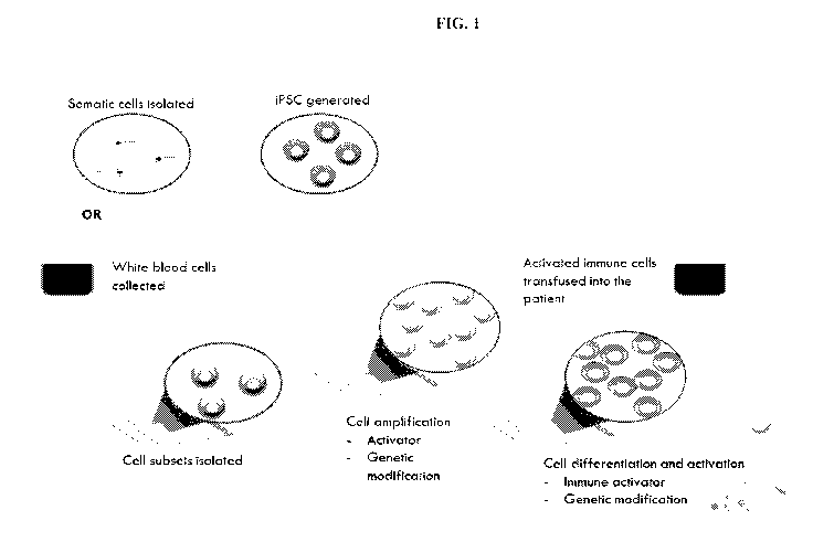
[0014] FIG. 1 illustrates the concept of the therapies described herein.
[0015] FIGS. 2A-C show mouse bone marrow-derived macrophages stimulated with
interferon
gamma (IFNy) (squares) with an enhanced ability to kill virulent bacterial
strains, as evidenced
by a decrease in intracellular bacterial burden (CFU = colony forming units).
FIG. 2A shows
the enhanced killing with the clinically relevant species Pseudomonas
aeruginosa. FIG. 2B
shows the enhanced killing with the clinically relevant species Acinetobacter
baumannii. FIG.
2C shows the enhanced killing with the clinically relevant multidrug resistant
clinical isolate of
Acinetobacter baumannii (ACI-3). Data shown in FIGS. 2A-C is an average of 6
technical
replicates from each of 4 biological replicates.
13
CA 03030755 2019-01-11
WO 2018/022651 PCT/US2017/043782
[0016] FIGS. 3A-C show human monocyte-derived macrophages increases the
killing of
multiple bacterial species. FIG. 3A shows the total bacterial burden over time
(t=20 hrs) before
and after exposure to human monocyte-derived macrophages stimulated with
interferon gamma
(IFNy) (squares). As evidenced by a decrease in intracellular bacterial burden
(CFU = colony
forming units), human monocyte-derived macrophages stimulated with interferon
gamma
(IFNy) had an enhanced ability to kill Pseudomonas aeruginosa. FIG. 3B shows
the number of
bacteria killed by monocyte-derived macrophages over the course of 2 hrs. The
monocyte-
derived macrophages obtained from different donors (n=14) and stimulated with
IFNy showed
enhanced killing across multiple clinically relevant species, with a
correlation between activities
against different bacterial species (p=0.002) FIG. 3C compares the number of
bacteria killed by
human monocyte-derived macrophages stimulated with IFNy and a control (non-
stimulated
human monocyte-derived macrophages). IFNy stimulated human monocyte-derived
macrophages to kill A. baumannii in a majority of young adult donors (8 of 10
donors).
[0017] FIG. 4 shows the infusion of mouse monocyte-derived macrophages
decreases organ
bacterial load in vivo. Mice injected intraperitoneally with Acinetobacter
baumanni were
subsequently injected with either Control (unstimulated; n=10 animals) or
Activated (IFNy
stimulated; n=9 animals) mouse-derived macrophages. Animals were sacrificed
and bacterial
load (CFU = colony forming units) was measured. Animals treated with
stimulated
macrophages showed significantly lower bacterial burden in multiple organs.
Data shown
represents technical triplicates from each organ.
DETAILED DESCRIPTION OF THE INVENTION
[0018] While preferred embodiments of the subject matter disclosed herein have
been shown
and described herein, it will be obvious to those skilled in the art that such
embodiments are
provided by way of example only. Numerous variations, changes, and
substitutions will now
occur to those skilled in the art without departing from the subject matter
disclosed herein. It
should be understood that various alternatives to the embodiments of the
subject matter
disclosed herein may be employed in practicing the subject matter disclosed
herein. It is
intended that the following claims define the scope of the subject matter
disclosed herein and
that methods and structures within the scope of these claims and their
equivalents be covered
thereby.
Definitions
[0019] Throughout this application, various embodiments of this invention may
be presented in
a range format. It should be understood that the description in range format
is merely for
convenience and brevity and should not be construed as an inflexible
limitation on the scope of
14
CA 03030755 2019-01-11
WO 2018/022651 PCT/US2017/043782
the invention. Accordingly, the description of a range should be considered to
have specifically
disclosed all the possible subranges as well as individual numerical values
within that range. For
example, description of a range such as from 1 to 6 should be considered to
have specifically
disclosed subranges such as from 1 to 3, from 1 to 4, from 1 to 5, from 2 to
4, from 2 to 6, from
3 to 6 etc., as well as individual numbers within that range, for example, 1,
2, 3, 4, 5, and 6. This
applies regardless of the breadth of the range.
[0020] The term "about" or "approximately" means within an acceptable error
range for the
particular value as determined by one of ordinary skill in the art, which will
depend in part on
how the value is measured or determined, i.e., the limitations of the
measurement system. For
example, "about" can mean within 1 or more than 1 standard deviation, per the
practice in the
art. Alternatively, "about" can mean a range of up to 20%, up to 10%, up to
5%, or up to 1% of
a given value. Alternatively, particularly with respect to biological systems
or processes, the
term can mean within an order of magnitude, preferably within 5-fold, and more
preferably
within 2-fold, of a value. Where particular values are described in the
application and claims,
unless otherwise stated the term "about" meaning within an acceptable error
range for the
particular value should be assumed.
[0021] The terms "subject," "individual," "host," "donor," and "patient" are
used
interchangeably herein to refer to a vertebrate, for example, a mammal.
Mammals include, but
are not limited to, murine, simians, humans, farm animals, sport animals, and
pets. Tissues,
cells, and their progeny of a biological entity obtained in vivo or cultured
in vitro are also
encompassed. Designation as a "subject," "individual," "host," "donor," or
"patient" does not
necessarily entail supervision of a medical professional.
[0022] The terminology used herein is for the purpose of describing particular
cases only and is
not intended to be limiting. As used herein, the singular forms "a", "an" and
"the" are intended
to include the plural forms as well, unless the context clearly indicates
otherwise. Furthermore,
to the extent that the terms "including", "includes", "having", "has", "with",
or variants thereof
are used in either the detailed description and/or the claims, such terms are
intended to be
inclusive in a manner similar to the term "comprising."
[0023] As used herein, the term "therapeutically effective amount" refers to
an amount of an
immunological cell or a pharmaceutical composition described herein that is
sufficient and/or
effective in achieving a desired therapeutic effect in treating a patient
having a pathogenic
disease. In some embodiments, a therapeutically effective amount of the immune
cell will avoid
adverse side effects.
[0024] As used herein, the term "pluripotent stem cells" (PSCs) refers to
cells capable, under
appropriate conditions, of producing different cell types that are derivatives
of all of the 3
CA 03030755 2019-01-11
WO 2018/022651 PCT/US2017/043782
germinal layers (i.e. endoderm, mesoderm, and ectoderm). Included in the
definition of
pluripotent stem cells are embryonic stem cells of various types including
human embryonic
stem (hES) cells, human embryonic germ (hEG) cells; non-human embryonic stem
cells, such as
embryonic stem cells from other primates, such as Rhesus stem cells, marmoset
stem cells;
murine stem cells; stem cells created by nuclear transfer technology, as well
as induced
pluripotent stem cells (iPSCs).
[0025] As used herein, the term "embryonic stem cells" (ESCs) refers to
pluripotent stem cells
that are derived from a blastocyst before substantial differentiation of the
cells into the three
germ layers (i.e. endoderm, mesoderm, and ectoderm). ESCs include any
commercially
available or well established ESC cell line such as H9, H1, H7, or SA002.
[0026] As used herein, the term "induced pluripotent stem cells" or "iPSCs"
refers to somatic
cells that have been reprogrammed into a pluripotent state resembling that of
embryonic stem
cells. Included in the definition of iPSCs are iPSCs of various types
including human iPSCs and
non-human iPSCs, such as iPSCs derived from somatic cells that are primate
somatic cells or
murine somatic cells.
[0027] As used herein, the term "allogenic" means the plurality of macrophages
are obtained
from a genetically non-identical donor. For example, allogenic macrophages are
extracted from
a donor and returned back to a different, genetically non-identical recipient.
[0028] As used herein, the term "autologous" means the plurality of
macrophages are obtained
from a genetically identical donor. For example, autologous macrophages are
extracted from a
patient and returned back to the same, genetically identical patient.
[0029] As used herein, the term "activated" and "stimulated" are used
interchangeably to
indicate that an immune cell (e.g. a macrophage or a monocyte) is exposed to
or contacted with
an activator.
[0030] As used herein, the term "activator" is any molecular entity that
drives a change in the
genome, transcriptome, proteome, or metabolome of a cell.
Macrophages and Monocytes
[0031] The emergence of pathogen resistance to multiple antimicrobial and
antibiotic agents has
become a significant public threat that places substantial clinical and
financial burden on health
care systems and patients. Recent statistic reports show pathogenic infections
are the largest
addressable hospital cost in the United States. In addition, pathogenic
infections and sepsis are
the leading cause of death in non-cardiac Intensive Care Units (ICUs). Thus,
there is a clear
need for alternative methods of management, prevention, and resolution of
pathogenic infections
and sepsis, including those caused by multi-, extensively, and pan-drug
resistant pathogens.
Disclosed herein, in certain embodiments, are methods of treating a pathogenic
infection in an
16
CA 03030755 2019-01-11
WO 2018/022651 PCT/US2017/043782
individual in need thereof comprising the administration of macrophages or
monocytes to the
individual.
[0032] Macrophages and monocytes are part of the innate immune system. The
innate immune
system is an important component of the overall immune system that provides
protection to the
host from foreign pathogens. Unlike the adaptive immune system, an innate
immune response
does not develop over time against a specific pathogenic antigen or epitope
the way an adaptive
immune response does. However, the innate immune system is quick to recognize
and respond
within the first few critical hours and days of exposure to a new pathogen.
The innate immune
system comprises a group of proteins and phagocytic cells, including
macrophages and
monocytes, which recognize conserved features of pathogens and become
activated when these
conserved features are encountered.
Macrophages
[0033] Macrophages are a type of white blood cell that engulfs and digests
pathogenic
organisms. Macrophages recognize foreign pathogens for uptake through several
mechanisms,
including both non-specific bulk endocytosis and through engagement of
specific receptors on
the cell surface that either bind to epitopes on the bacterial surface itself
or bind mammalian
proteins that have bound to the bacterial surface (antibodies, complement
proteins, or other
opsonins). Following internalization of a pathogen by the macrophage, the
pathogen becomes
encapsulated in a membrane bound compartment called the phagosome. The
phagosome is fused
with a lysosome to form a phagolysosome. The phagolysosome contains enzymes,
reactive
oxygen species, and other toxic molecules that break-down the pathogen.
Macrophages also
internalize and breakdown infected cells and cell debris from the site of an
active infection,
helping prevent further spread of the infection and limiting the area of
tissue damage.
[0034] Macrophages also play a role in innate immunity and adaptive immunity
by recruiting
other immune cells to the site of an infection. For example, macrophages
function as antigen
presenting cells to T cells. Following phagocytosis and degradation of a
pathogen, a macrophage
will present an antigen of the pathogen for helper T cells in the context of
the major
histocompatibility complex (MHC) class II proteins on the cell surface.
Analogously, viral
pathogens replicating within macrophages can also be degraded and presented on
the MHC class
I complex at the cell surface. Presentation of the antigen by the macrophage
together with
appropriate co-stimulatory proteins results in the activation of T cells and
subsequent production
of antibodies that target the antigen. Macrophages also recruit and activate
other immune cell
types by secreting soluble factors like cytokines and chemokines, which signal
to other
circulating immune cells to infiltrate the infected area and help fight the
infection.
17
CA 03030755 2019-01-11
WO 2018/022651 PCT/US2017/043782
[0035] Macrophages are either derived from the proliferation of specialized
tissue macrophage
populations (e.g. Kupffer cells) or differentiate from circulating peripheral-
blood monocytes,
which migrate into tissue in the steady state or in response to inflammation.
Monocytes develop
from myeloid progenitor cells in the bone marrow. Myeloid progenitor cells
give rise to
monoblasts which develop into pro-monocytes which then develop into monocytes.
The
monocytes are released from the bone marrow into the bloodstream. Once in the
bloodstream
they migrate into tissues, where they differentiate into macrophages or
dendritic cells.
[0036] Macrophages are activated via several different pathways. The classical
method of
activation results in macrophages that are produced during cell-mediated
immune responses.
Generally, the presence of interferon-y (IFNy) and/or tumor-necrosis factor
(TNF) in a tissue
results in a macrophage population that targets pathogens and secretes high
levels of pro-
inflammatory cytokines. IFNy is produced, for example by natural killer (NK)
cells in response
to stress and infections. The presence of IFNy activates macrophages to
secrete pro-
inflammatory cytokines, and to produce increased amounts of superoxide anions
and oxygen and
nitrogen radicals to increase their killing ability. Macrophages are also
classically activated by
certain molecular patterns commonly present in pathogenic organisms, such as
lipopolysaccharide (LPS) or the nucleic acid CpG. These molecules are
recognized by a class of
pattern-recognition receptors (PRRs) like the Toll-like receptors (TLRs),
leading to an
intracellular signaling cascade that ultimately turns on the macrophage
pathogen defense
response. Macrophages can also be alternatively activated by exposure to
cytokines, such as
IL-4 and IL-13. Alternatively activated macrophages produce soluble factors
such as IL-10 and
matrix metalloproteinases (MNIPs) that downregulate pro-inflammatory cytokines
like TNF and
promote wound healing by breaking down extracellular matrix proteins.
[0037] The production of IFNy by NK cells is transient and results in the
transient production of
macrophages primed to target pathogens. To assist in the activation of
macrophages, adaptive
immune cells, such as TH1 cells, are recruited. While T helper 1 (TH1) cells
are antigen
specific, macrophages activated in response to the TH1 cells can target any
pathogenic cells. In
some embodiments, the methods disclosed herein further comprise administering
NK cells to the
individual. In some embodiments, the methods disclosed herein further comprise
administering
TH1 cells to the individual in need thereof. In some embodiments, the TH1
cells are specific to
the unwanted pathogen. In some embodiments, the TH1 cells are not specific to
the unwanted
pathogen.
[0038] In certain instances, pro-inflammatory cytokines produced by
classically activated
macrophages are associated with damage to the host. IL-1, IL-6, and IL-23 are
produced by
classically activated macrophages. These cytokines result in the development
and expansion of
18
CA 03030755 2019-01-11
WO 2018/022651 PCT/US2017/043782
TH17 cells which produce IL-17. Excessive IL-17 levels in tissue are
associated with unwanted
inflammation and sometimes the progression of an autoimmune phenotype. TNF-
alpha and
TNFSF1A are additional cytokines produced by classically activated
macrophages. Chemokines
including IL-8/CXCL8, IP-10/CXCL10, MW-1 alpha/CCL3, MIP-1 beta/CCL4, and
RANTES/CCL5 are produced by classically activated macrophages. In some
embodiments, the
plurality of macrophages is genetically engineered to reduce or inhibit
production of an
unwanted cytokine. In some embodiments, the cytokine is selected from TNF, IL-
1, IL-6, IL-8,
IL-12, and IL-23.
[0039] In some embodiments, a macrophage for use in a method disclosed herein
is activated
before administration by exposure to IL-4, IL-13, interferon-y (IFNy), and/or
tumor-necrosis
factor (TNF) in cell culture, resulting in in vitro activated macrophages. In
some embodiments, a
macrophage for use in a method disclosed herein is activated before
administration by in vitro
exposure to IL-4, IL-13, interferon-y (IFNy), and/or tumor-necrosis factor
(TNF) followed by an
additional stimulant, such as bacterial lipopolysaccharide (LPS), resulting in
in vitro activated
macrophages. In some embodiments, a macrophage for use in a method disclosed
herein is
activated by exposure to IL-4, IL-13, interferon-y (IFNy), and/or tumor-
necrosis factor (TNF) in
the individual, resulting in in vivo activated macrophages. In some
embodiments, a macrophage
for use in a method disclosed herein is activated by exposure to IL-4, IL-13,
interferon-y (IFNy),
and/or tumor-necrosis factor (TNF), followed by an additional stimulant, such
as a pathogen or
pathogen-associated molecular pattern, in the individual, resulting in in vivo
activated
macrophages. In some embodiments, a macrophage for use in a method disclosed
herein is
activated by exposure to IL-4, IL-13, interferon-y (IFNy), and/or tumor-
necrosis factor (TNF),
followed by an additional stimulant, such as a TLR agonist, in the individual,
resulting in in vivo
activated macrophages. In some embodiments, a macrophage for use in a method
disclosed
herein is activated by exposure to IL-4, IL-13, interferon-y (IFNy), and/or
tumor-necrosis factor
(TNF), followed by an additional stimulant, such as a vaccine adjuvant, in the
individual,
resulting in in vivo activated macrophages.
Monocytes
[0040] Monocytes are produced in the bone marrow from monoblasts. Monocytes
circulate in
the bloodstream until they encounter a molecular signal that indicates damage
or infection in the
nearby tissue. They then migrate out of the blood into the damaged tissue.
Chemotaxis of
monocytes to a pathogen is controlled by multiple compounds, including
monocyte chemotactic
protein-1; monocyte chemotactic protein-3 (CCL7); Leukotriene B4; 5-HETE; 5-
oxo-ETE); and
N-Formylmethionineleucyl-phenylalanine.
19
CA 03030755 2019-01-11
WO 2018/022651 PCT/US2017/043782
[0041] Once in a tissue, monocytes can mature into macrophages or dendritic
cells. There are
several subsets of monocytes in humans as defined by their surface markers,
including classical
(CD I e+CD 1 6-), non-classical (CD14dimCD I 6+), and intermediate (CD 1 e'CD
I 6+). While
their downstream functional differences are still unclear, they each have the
capacity to
differentiate to macrophages under the correct stimulation conditions.
[0042] Monocytes themselves engage in phagocytosis and cytokine production.
Following
opsonization by an opsonin (e.g., an antibody, complement protein, or one of
several circulating
proteins (e.g., pentraxins, collectins, and ficolins)) monocytes are able to
engulf a pathogen.
Like macrophages, monocytes are able to phagocytose pathogens by binding
directly to pattern-
recognition receptors on the pathogen. Monocytes also use antibody-dependent
cell-mediated
cytotoxicity (ADCC) to kill pathogens.
[0043] In some embodiments, a monocyte for use in a method disclosed herein is
activated
before administration by exposure to IL-4, IL-13, interferon-y (IFNy), and/or
tumor-necrosis
factor (TNF) in cell culture, resulting in in vitro activated monocytes. In
some embodiments, a
monocyte for use in a method disclosed herein is activated before
administration by in vitro
exposure to IL-4, IL-13, interferon-y (IFNy), and/or tumor-necrosis factor
(TNF) followed by an
additional stimulant, such as bacterial lipopolysaccharide (LPS), resulting in
in vitro activated
monocytes. In some embodiments, a monocyte for use in a method disclosed
herein is activated
by exposure to IL-4, IL-13, interferon-y (IFNy), and/or tumor-necrosis factor
(TNF) in the
individual, resulting in in vivo activated monocytes. In some embodiments, a
monocyte for use
in a method disclosed herein is activated by exposure to IL-4, IL-13,
interferon-y (IFNy), and/or
tumor-necrosis factor (TNF), followed by an additional stimulant, such as a
pathogen or
pathogen-associated molecular pattern, in the individual, resulting in in vivo
activated
monocytes. In some embodiments, a monocyte for use in a method disclosed
herein is activated
by exposure to IL-4, IL-13, interferon-y (IFNy), and/or tumor-necrosis factor
(TNF), followed
by an additional stimulant, such as a TLR agonist, in the individual,
resulting in in vivo activated
monocytes. In some embodiments, a monocyte for use in a method disclosed
herein is activated
by exposure to IL-4, IL-13, interferon-y (IFNy), and/or tumor-necrosis factor
(TNF), followed
by an additional stimulant, such as a vaccine adjuvant, in the individual,
resulting in in vivo
activated monocytes.
Isolated and Purified Monocytes and Macrophages
[0044] Disclosed herein, in certain embodiments, are isolated and purified
innate immune cells.
Additionally disclosed herein, in certain embodiments, are pharmaceutical
compositions
comprising (a) isolated and purified innate immune cells; and (b) a
pharmaceutically-acceptable
excipient.
CA 03030755 2019-01-11
WO 2018/022651 PCT/US2017/043782
[0045] In some embodiments, the innate immune cells are macrophages. In some
embodiments,
the macrophages are Kupffer cells, histiocytes, alveolar macrophages, splenic
macrophages,
placental macrophages, peritoneal macrophages, osteoclasts, adipose tissue
macrophage (ATM),
or sinusoidal lining cells. In some embodiments, the macrophages are produced
by a method
comprising isolating a subpopulation of macrophages from a population of
immune cells
extracted from an individual. In some embodiments, the macrophages are
produced by a method
comprising (a) isolating a subpopulation of macrophage progenitor cells from a
population of
immune cells extracted from an individual; and (b) differentiating the
isolated macrophage
progenitor cells into a plurality of macrophages ex vivo. In some embodiments,
the macrophages
are produced by generating macrophage progenitor cells from embryonic stem
cells (ESCs) and
differentiating the macrophage progenitor cells into macrophages. In some
embodiments, the
macrophages are produced by reprogramming somatic cells into induced
pluripotent cells
(iPSCs), generating macrophage progenitor cells from the iPSCs, and
differentiating the
macrophage progenitor cells into macrophages.
[0046] In some embodiments, the innate immune cells are monocytes. In some
embodiments,
the monocytes are produced by a method comprising isolating a subpopulation of
monocytes
from a population of immune cells extracted from an individual. In some
embodiments, the
monocytes are produced by generating monocyte progenitor cells from embryonic
stem cells
(ESCs) and differentiating the monocyte progenitor cells into macrophages. In
some
embodiments, the monocytes are produced by reprogramming somatic cells into
induced
pluripotent cells (iPSCs), generating monocyte progenitor cells from the
iPSCs, and
differentiating the monocyte progenitor cells into macrophages.
[0047] In some embodiments, the innate immune cells are fresh, i.e., not
frozen or previously
frozen. In some embodiments, the innate immune cells are frozen and stored for
later use (for
example to facilitate transport) to generate frozen macrophages or monocytes.
In some
embodiments, the innate immune cells are administered to the individual after
being thawed. In
some embodiments, a pharmaceutical formulation disclosed herein comprises (a)
isolated and
purified innate immune cells; and (b) a cryoprotectant. In some embodiments,
the
cryoprotectant is selected from dimethylsulfoxide (DMSO), formamide, propylene
glycol,
ethylene glycol, glycerol, trehalose, 2-methyl-2,4-pentanediol, methanol,
butanediol, or any
combination thereof
[0048] In some embodiments, the innate immune cells are activated before
administration to the
individual. In some embodiments, the innate immune cells are not activated
before
administration to the individual. In some embodiments, the innate immune cells
are activated by
the immune system of the individual and the presence of a pathogen in the
individual. In some
21
CA 03030755 2019-01-11
WO 2018/022651 PCT/US2017/043782
embodiments, innate immune cells are co-administered with a compound that
activates the
innate immune cells in vivo. In some embodiments, a pharmaceutical formulation
disclosed
herein comprises (a) isolated and purified innate immune cells; and (b) a
compound that
activates the innate immune cells. In some embodiments, the compound that
activates innate
immune cells is selected from: IL-4, IL-13, phorbol myristate acetate,
lipopolysaccharide (LPS),
IFNy, tumor-necrosis factor (TNF), or any combinations thereof
[0049] In some embodiments, the innate immune cells are autologous to an
individual. In some
embodiments, the innate immune cells are allogenic. As used herein,
"autologous" means the
plurality of innate cells are obtained from the individual or a genetically
identical donor. As used
herein, "allogenic" means the plurality of innate cells are obtained from a
genetically non-
identical donor.
Isolation of Monocytes
[0050] In some embodiments, monocytes or monocyte progenitor cells are
isolated from a
human blood sample or a human bone marrow sample. In some embodiments,
monocyte
progenitor cells are differentiated into monocytes in vitro. In some
embodiments, the monocyte
progenitor cells are hematopoietic stem cells, CD34+ stem cells, common
myeloid progenitor
cells, or granulocyte-monocyte progenitor cells.
[0051] Any suitable means for isolating monocytes or monocyte progenitor cells
from an
individual is contemplated for use with the methods disclosed herein. Methods
to isolate
monocytes or monocyte progenitor cells from an individual include, but are not
limited to:
isolation by adherence, isolation by size sedimentation on Percoll, isolation
by flow sorting,
positive or negative bead-based selection using cell surface markers, or
isolation by counterflow
centrifugal elutriation.
[0052] In some embodiments, the monocytes or monocyte progenitor cells are
isolated from a
human blood sample. In some embodiments, the human blood sample is a
peripheral blood
sample. In some embodiments, the human blood sample is a cord blood sample. In
some
embodiments, the peripheral blood sample is a mobilized blood sample. In some
embodiments,
the cord blood sample is a mobilized blood sample. In some embodiments, the
peripheral blood
sample is a non-mobilized blood sample. In some embodiments, the cord blood
sample is a non-
mobilized blood sample.
[0053] Mobilization is a process where monocytes or monocyte progenitor cells
are stimulated
out of the bone marrow space into the bloodstream, making them available for
collection. Thus,
mobilization presents a less invasive alternative to a bone marrow harvest,
which is a surgical
procedure that is also used as a method to collect macrophage progenitor cells
from the bone
marrow of the donor. In some embodiments, mobilization is performed by
administrating to the
22
CA 03030755 2019-01-11
WO 2018/022651 PCT/US2017/043782
donor a drug, a cytokine, a hormone, a protein, or any combination thereof. In
some
embodiments, mobilization is performed by administrating to the donor
granulocyte colony-
stimulating factor (G-CSF), granulocyte macrophage colony-stimulating factor
(GM-CSF),
plerixafor, stem cell factor (SCF), a CXCR4 inhibitor, an S113 agonist, a VCAM
inhibitor, a
VLA-4 inhibitor, a parathyroid hormone, a proteosome inhibitor, growth
regulated protein beta
(Gro0), a HIF stabilizer, or any combination thereof
[0054] In some embodiments, leukapheresis is performed after obtaining the
human blood
sample. Leukapheresis is a procedure in which white blood cells are separated
from a blood
sample, allowing the return of red blood cells to the donor.
[0055] In some embodiments, a peripheral blood sample is obtained from the
individual. In
some embodiments, the peripheral blood sample is subjected to gradient
centrifugation to
generate a buffy coat fraction (i.e., the fraction of an anticoagulated blood
sample that contains
white blood cells). In some embodiments, the buffy coat fraction is subjected
to gradient
centrifugation in the presence of Ficoll to generate a peripheral blood
mononuclear cell (PBMC)
fraction. In some embodiments, the PBMC fraction is suspended in a suitable
solution (e.g.,
PBS-EDTA) and centrifuged to generate an isolated PBMC pellet. In some
embodiments, the
isolated PBMC pellet is suspended in a suitable solution (e.g., RPMI 1640
medium or X-VIVO)
to generate a solution of isolated PBMCs.
[0056] In some embodiments, monocytes or monocyte progenitor cells are
isolated from the
solution of isolated PBMCs. In some embodiments, the monocytes or monocyte
progenitor cells
are positively selected by using beads coated with antibody against common
surface markers.
Exemplary monocyte markers for use in cell sorting include, but are not
limited to CD2, CD31,
CD56, CD62L, CD192, CX3CR1, CXCR3, CXCR4, CD14, CD16, CD64, CD11b, CD115, Gr-
1, Ly-6C, CD204 or any combination thereof In some embodiments, the monocytes
or
monocyte progenitor cells are negatively selected by using beads coated with
antibodies against
common surface markers of cells other than monocytes or monocyte progenitors.
Exemplary
non-monocytic markers for use in negative selection include, but are not
limited to CD3, CD4,
CD8, CD19, CD20, BCR, TCR, IgD, IgM, CD56 or any combination thereof. In some
embodiments, the monocytes or monocyte progenitor cells are isolated by use of
cell sorting,
e.g. fluorescence activated cell sorting (FACS). Exemplary monocyte markers
for use in cell
sorting include, but are not limited to CD2, CD31, CD56, CD62L, CD192, CX3CR1,
CXCR3,
CXCR4, CD14, CD16, CD64, CD11b, CD115, Gr-1, Ly-6C, CD204 or any combination
thereof Exemplary hematopoietic stem cell markers include, but are not limited
to
2B4/CD244/SLAMF4, ABCG2, C1qR1/CD93, CD34, CD38, CD45, CD48/SLAMF2, CDCP1,
CXCR4, Flt-3/Flk-2, SCF R/c-kit, SLAM/CD150, or any combination thereof.
Exemplary
23
CA 03030755 2019-01-11
WO 2018/022651 PCT/US2017/043782
common myeloid progenitor cell markers include, but are not limited to CD34,
Flt-3/Flk-2, SCF
R/c-kit, IL-3 Ra, or any combination thereof Exemplary common granulocyte-
macrophage
progenitor cell markers include, but are not limited to CD34, CD38, IL-3 Ra,
or any
combination thereof.
[0057] Alternatively, in some embodiments, the solution of isolated PBMCs is
subjected to
gradient centrifugation in the presence of Percoll solution. In some
embodiments, the monocyte
or monocyte progenitor cell fraction is isolated, suspended in a suitable
solution (e.g., PBS-
EDTA) and centrifuged to generate an isolated monocyte pellet or monocyte
progenitor cell
pellet. The pellet is suspended in a suitable solution (e.g., RPMI 1640 medium
or X-VIVO) to
generate a solution of isolated monocytes or monocyte progenitor cells.
[0058] In some embodiments, the isolated monocytes or monocyte progenitor
cells are grown in
cell culture to generate isolated monocytes or monocyte progenitor cells. In
some embodiments,
the culture is grown in the presence of culture medium comprising RPMI 1640
medium or X-
VIVO. In some embodiments, the culture medium further comprises serum, for
example fetal
calf serum (FCS) or fetal bovine serum (FBS). In some embodiments, the culture
medium
comprises a suitable serum replacement that is safe for clinical use, for
example human AB
serum, human platelet lysate, or chemically defined optimized serum-free
medium. In some
embodiments, the culture medium further comprises an antibiotic including:
actinomycin D,
ampicillin, carbenicillin, cefotaxime, fosmidomycin, gentamicin, kanamycin,
neomycin,
penicillin streptomycin (Pen Strep), polymixyn B, or streptomycin.
[0059] In some embodiments, the isolated monocyte progenitor cells are
differentiated into
monocytes. In some embodiments, monocyte progenitor cells are differentiated
into monocytes
by contacting monocyte progenitor cells with IL-3, granulocyte macrophage
colony-stimulating
factor (GM-CSF), macrophage colony-stimulating factor (M-CSF), granulocyte
colony-
stimulating factor (G-CSF), stem cell factor (SCF), thrombopoietin (TPO), or
any combination
thereof In some embodiments, the monocyte progenitor cells are contacted with
any possible
combination of factors selected from the group comprising: IL-3, GM-CSF, M-
CSF, G-CSF,
SCF, and/or TPO. For example, in some embodiments, the monocyte progenitor
cells are
contacted with a combination of IL-3 and GM-CSF; a combination of IL-3 and M-
CSF; or a
combination of SCF, TPO, G-CSF, and GM-CSF.
[0060] In some embodiments, the monocyte progenitor cells are contacted with M-
CSF at a
concentration ranging from about 1 ng/ml to about 100 ng/ml; e.g. about 5
ng/ml, 10 ng/ml, 25
ng/ml, 50 ng/ml, 75 ng/ml, or 100 ng/ml. In some embodiments, the monocyte
progenitor cells
are contacted with M-CSF at a concentration ranging from about 50 ng/ml to
about 200 ng/ml;
from about 200 ng/ml to about 500 ng/ml; from about 500 ng/ml to about 1000
mg/ml.
24
CA 03030755 2019-01-11
WO 2018/022651 PCT/US2017/043782
[0061] In some embodiments, the monocyte progenitor cells are contacted with
GM-CSF at a
concentration ranging from about 1 ng/ml to about 100 ng/ml; e.g. about 5
ng/ml, 10 ng/ml, 25
ng/ml, 50 ng/ml, 75 ng/ml, or 100 ng/ml. In some embodiments, higher
concentrations of GM-
CSF are used, e.g., from about 50 ng/ml to about 200 ng/ml; from about 200
ng/ml to about 500
ng/ml; from about 500 ng/ml to about 1000 mg/ml.
[0062] In some embodiments, the monocyte progenitor cells are contacted with G-
CSF at a
concentration ranging from about 1 ng/ml to about 100 ng/ml; e.g. about 5
ng/ml, 10 ng/ml, 25
ng/ml, 50 ng/ml, 75 ng/ml, or 100 ng/ml. In some embodiments, higher
concentrations of G-CSF
are used, e.g., from about 50 ng/ml to about 200 ng/ml; from about 200 ng/ml
to about 500
ng/ml; from about 500 ng/ml to about 1000 mg/ml.
[0063] In some embodiments, the monocyte progenitor cells are contacted with
IL-3 at a
concentration ranging from about 1 ng/ml to about 100 ng/ml; e.g. about 5
ng/ml, 10 ng/ml, 25
ng/ml, 50 ng/ml, 75 ng/ml, or 100 ng/ml. In some embodiments, higher
concentrations of IL-3
are used, e.g., from about 50 ng/ml to about 200 ng/ml; from about 200 ng/ml
to about 500
ng/ml; from about 500 ng/ml to about 1000 mg/ml.
[0064] In some embodiments, the monocyte progenitor cells are contacted with
SCF at a
concentration ranging from about 1 ng/ml to about 100 ng/ml; e.g. about 5
ng/ml, 10 ng/ml, 25
ng/ml, 50 ng/ml, 75 ng/ml, or 100 ng/ml. In some embodiments, the monocyte
progenitor cells
are contacted with SCF at a concentration ranging from about 50 ng/ml to about
200 ng/ml;
from about 200 ng/ml to about 500 ng/ml; from about 500 ng/ml to about 1000
mg/ml.
[0065] In some embodiments, the monocyte progenitor cells are contacted with
TPO at a
concentration ranging from about 1 ng/ml to about 100 ng/ml; e.g. about 5
ng/ml, 10 ng/ml, 25
ng/ml, 50 ng/ml, 75 ng/ml, or 100 ng/ml. In some embodiments, the monocyte
progenitor cells
are contacted with TPO at a concentration ranging from about 50 ng/ml to about
200 ng/ml;
from about 200 ng/ml to about 500 ng/ml; from about 500 ng/ml to about 1000
mg/ml.
[0066] In some embodiments, the monocytes are administered to the individual.
In some
embodiments, the monocytes are fresh, i.e., not frozen. In some embodiments,
the monocytes
are frozen and stored for later use (for example to facilitate transport). In
some embodiments,
cell freezing or cryopreservation media and/or cryoprotective agents are
utilized to preserve the
monocytes during the freezing process. In some embodiments, the cryoprotectant
is selected
from dimethylsulfoxide (DMSO), formamide, propylene glycol, ethylene glycol,
glycerol,
trehalose, 2-methyl-2,4-pentanediol, methanol, butanediol, or any combination
thereof In some
embodiments, the frozen monocytes are administered to the individual after
being thawed.
CA 03030755 2019-01-11
WO 2018/022651 PCT/US2017/043782
Differentiation of Macrophages from Isolated Monocytes
[0067] In some embodiments, the isolated monocytes are differentiated into
macrophages in
culture. In some embodiments, the monocytes are contacted with
granulocyte¨macrophage
(GM-CSF) or macrophage colony-stimulating factor (M-CSF) to generate
differentiated
macrophages. In some embodiments, the concentration of M-CSF is from about 1
ng/ml to
about 100 ng/ml; e.g. about 5 ng/ml, 10 ng/ml, 25 ng/ml, 50 ng/ml, 75 ng/ml,
or 100 ng/ml. In
some embodiments, higher concentrations of M-CSF are used, e.g., from about 50
ng/ml to
about 200 ng/ml; from about 200 ng/ml to about 500 ng/ml; from about 500 ng/ml
to about 1000
mg/ml. In some embodiments, the concentration of GM-CSF is from about 1 ng/ml
to about
100 ng/ml; e.g. about 5 ng/ml, 10 ng/ml, 25 ng/ml, 50 ng/ml, 75 ng/ml, or 100
ng/ml. In some
embodiments, higher concentrations of GM-CSF are used, e.g., from about 50
ng/ml to about
200 ng/ml; from about 200 ng/ml to about 500 ng/ml; from about 500 ng/ml to
about 1000
mg/ml.
[0068] In some embodiments, the differentiated macrophages are isolated by use
of cell sorting,
e.g. fluorescence activated cell sorting (FACS). Exemplary macrophage markers
for use in cell
sorting include CD11b, CD68, CD163, F4/80, CD16, CD54, CD49e, CD38, Egr2,
CD71, TLR2,
TLR4, or any combination thereof.
Differentiation of Macrophages from Macrophage Progenitor Cells
[0069] In some embodiments, macrophage progenitor cells are differentiated
into macrophages
in culture. Any suitable means for differentiating the macrophage progenitor
cells into
macrophages is contemplated for use with the methods disclosed herein. In some
embodiments,
the macrophage progenitor cells are hematopoietic stem cells, CD34+
hematopoietic stem cells,
common myeloid progenitor cells, granulocyte-monocyte progenitor cells, or
monocytes. In
some embodiments, the macrophage progenitor cells are isolated from an
individual. In some
embodiments, the macrophage progenitor cells are isolated from a human blood
sample, a
human tissue, a human peritoneal fluid sample, or a human bone marrow sample.
In some
embodiments, the macrophage progenitor cells are isolated from a human
peripheral blood
sample or a human cord blood sample. In some embodiments, the peripheral blood
sample is a
mobilized blood sample. In some embodiments, the peripheral blood sample is a
non-mobilized
blood sample. In some embodiments, the cord blood sample is a mobilized blood
sample. In
some embodiments, the cord blood sample is a non-mobilized blood sample.
[0070] In some embodiments, mobilization is performed by administrating to the
donor a drug, a
cytokine, a hormone, a protein, or any combination thereof In some
embodiments, mobilization
is performed by administrating to the donor granulocyte colony-stimulating
factor (G-CSF),
granulocyte macrophage colony-stimulating factor (GM-CSF), plerixafor, stem
cell factor
26
CA 03030755 2019-01-11
WO 2018/022651 PCT/US2017/043782
(SCF), a CXCR4 inhibitor, an S113 agonist, a VCAM inhibitor, a VLA-4
inhibitor, a parathyroid
hormone, a proteosome inhibitor, growth regulated protein beta (Grof3), a HIF
stabilizer, or any
combination thereof
[0071] In some embodiments, the macrophage progenitor cells are contacted with
a cytokine, a
chemokine, a protein, a peptide, a small molecule, a growth factor, or a
nucleic acid molecule to
generate differentiated macrophages. In some embodiments, the cytokine is
macrophage
colony-stimulating factor (M-CSF) or stem cell factor (SCF). In some
embodiments, the small
molecule is FMS-like tyrosine kinase 3 ligand (F1t31). In some embodiments,
Flt31 functions as
a cytokine and a growth factor. In some embodiments, the protein is GM-CSF, IL-
3, or IL-6.
In some embodiments GM-CSF functions as a cytokine.
[0072] In some embodiments, the nucleic acid molecule is deoxyribonucleic acid
(DNA) or
ribonucleic acid (RNA). In some embodiments, the RNA molecule is a small RNA.
In some
embodiments, examples of small RNA include micro-RNA (miRNA), ribosomal RNA
(rRNA),
small nuclear RNA (snRNA), transfer RNA (tRNA), small nucleolar RNA (snoRNA),
Piwi-
interacting RNA (piRNA), tRNA-derived small RNA (tsRNA), small rDNA-derived
RNA
(srRNA), or 5S RNA. In some embodiments, the RNA molecule is a long RNA. In
some
embodiments, examples of long RNA include long non-coding RNA (lncRNA) or
messenger
RNA (mRNA). In some embodiments, the RNA molecule is a double stranded RNA
(dsRNA),
a circular RNA, a small interfering RNA (siRNA), antisense RNA (aRNA), cis-
natural antisense
transcript (cis-NAT), CRISPR RNA (crRNA), short hairpin RNA (shRNA), trans-
acting siRNA
(tasiRNA), repeat associated siRNA (rasiRNA), 7SK RNA (7SK), or enhancer RNA
(eRNA).
[0073] In some embodiments, the macrophage progenitor cells are contacted with
granulocyte¨
macrophage (GM-CSF), macrophage (M-CSF) colony-stimulating factor, FMS-like
tyrosine
kinase 3 ligand (F1t31), IL-3, IL-6, stem cell factor (SCF), or any
combination thereof
[0074] In some embodiments, the macrophage progenitor cells are contacted with
M-CSF at a
concentration ranging from about 1 ng/ml to about 100 ng/ml; e.g. about 5
ng/ml, 10 ng/ml, 25
ng/ml, 50 ng/ml, 75 ng/ml, or 100 ng/ml. In some embodiments, the macrophage
progenitor
cells are contacted with M-CSF at a concentration ranging from about 50 ng/ml
to about 200
ng/ml; from about 200 ng/ml to about 500 ng/ml; from about 500 ng/ml to about
1000 mg/ml.
[0075] In some embodiments, the macrophage progenitor cells are contacted with
GM-CSF at a
concentration ranging from about 1 ng/ml to about 100 ng/ml; e.g. about 5
ng/ml, 10 ng/ml, 25
ng/ml, 50 ng/ml, 75 ng/ml, or 100 ng/ml. In some embodiments, higher
concentrations of GM-
CSF are used, e.g., from about 50 ng/ml to about 200 ng/ml; from about 200
ng/ml to about 500
ng/ml; from about 500 ng/ml to about 1000 mg/ml.
27
CA 03030755 2019-01-11
WO 2018/022651 PCT/US2017/043782
[0076] In some embodiments, the macrophage progenitor cells are contacted with
Flt13 at a
concentration ranging from about 1 ng/ml to about 100 ng/ml; e.g. about 5
ng/ml, 10 ng/ml, 25
ng/ml, 50 ng/ml, 75 ng/ml, or 100 ng/ml. In some embodiments, higher
concentrations of Flt13
are used, e.g., from about 50 ng/ml to about 200 ng/ml; from about 200 ng/ml
to about 500
ng/ml; from about 500 ng/ml to about 1000 mg/ml.
[0077] In some embodiments, the macrophage progenitor cells are contacted with
IL-3 at a
concentration ranging from about 1 ng/ml to about 100 ng/ml; e.g. about 5
ng/ml, 10 ng/ml, 25
ng/ml, 50 ng/ml, 75 ng/ml, or 100 ng/ml. In some embodiments, higher
concentrations of IL-3
are used, e.g., from about 50 ng/ml to about 200 ng/ml; from about 200 ng/ml
to about 500
ng/ml; from about 500 ng/ml to about 1000 mg/ml.
[0078] In some embodiments, the macrophage progenitor cells are contacted with
IL-6 at a
concentration ranging from about 1 ng/ml to about 100 ng/ml; e.g. about 5
ng/ml, 10 ng/ml, 25
ng/ml, 50 ng/ml, 75 ng/ml, or 100 ng/ml. In some embodiments, higher
concentrations of IL-6
are used, e.g., from about 50 ng/ml to about 200 ng/ml; from about 200 ng/ml
to about 500
ng/ml; from about 500 ng/ml to about 1000 mg/ml.
[0079] In some embodiments, the macrophage progenitor cells are contacted with
SCF at a
concentration ranging from about 1 ng/ml to about 100 ng/ml; e.g. about 5
ng/ml, 10 ng/ml, 25
ng/ml, 50 ng/ml, 75 ng/ml, or 100 ng/ml. In some embodiments, higher
concentrations of SCF
are used, e.g., from about 50 ng/ml to about 200 ng/ml; from about 200 ng/ml
to about 500
ng/ml; from about 500 ng/ml to about 1000 mg/ml.
[0080] In some embodiments, the differentiated macrophages are isolated by use
of cell sorting,
e.g. fluorescence activated cell sorting (FACS). Exemplary macrophage markers
for use in cell
sorting include CD14, CD11b, CD68, CD163, CD16, CD54, CD49e, CD38, CD204,
Egr2,
CD71, TLR2, TLR4, or any combination thereof
Production of Macrophages and Macrophage-Like Cells from Stem Cells
[0081] In some embodiments, the macrophages are stem cell-derived macrophages
or stem cell-
derived macrophage-like cells. In some embodiments, the term "macrophage-like
cells" is
defined as cells that behave like macrophages, display macrophage markers,
function like
macrophages, and/or exhibit the same responses as macrophages. In some
embodiments,
macrophage-like cells express one or more markers selected from CD14, CD11b,
CD68,
CD163, CD16, CD54, CD49e, CD38, CD204, Egr2, CD71, toll like receptor ligand-2
(TLR2),
and TLR4.
[0082] In some embodiments, the stem cells are allogenic. In some embodiments,
the stem cells
are autologous. In some embodiments, the stem cells are embryonic stem (ES)
cells. In some
embodiments, the embryonic stem cells are HI ES cells. In some embodiments,
the embryonic
28
CA 03030755 2019-01-11
WO 2018/022651 PCT/US2017/043782
stem cells are H9 ES cells. In some embodiments, the embryonic stem cells are
non-human
embryonic stem cells. In some embodiments, stem cells are induced pluripotent
stem (iPS)
cells. In some embodiments, stem cells are somatic stem cells. In some
embodiments, stem
cells are pluripotent stem cells. In some embodiments, stem cells are
hematopoietic stem cells
(HSC). Any suitable means for deriving macrophages or macrophage-like cells
from stem cells
is contemplated for use with the methods disclosed herein.
ESC-Derived Macrophages
[0083] In some embodiments, embryonic stem (ES) cells are cultured on an
irradiated mouse
embryonic feeder (MEF) layer in cell culture medium in the presence of
leukemia inhibitory
factor (LIF). In some embodiments, the cell culture medium is a macrophage
differentiation
medium (MDM). In some embodiments, the MDM is obtained by culturing
fibroblasts and
harvesting the medium they are cultured in after reaching confluence. The ES
cells form ES cell
clusters and in order to induce embryoid body (EB) formation, the ES cell
clusters are detached
and cultured on a non-adherent cell culture dish without LIF. In some
embodiments, ES cells
are cultured in a medium supplemented with IL-3. Embryoid bodies (EBs) are
generated and
they are plated on a gelatin-coated cell culture dish in an adequate cell
culture medium. These
conditions induce the growth and development of different cell types. After at
least 4 days of
culture, supernatants of adherent EB contain floating macrophage progenitors.
In some
embodiments, the macrophage progenitors are collected and plated onto low
adherence cell
culture dishes. The macrophage progenitors are further cultured for up to 7
days and form an
adherent macrophage monolayer. In some embodiments, the macrophage progenitors
are
cultured in medium, such as RPMI-1640, supplemented with glutamine, fetal
bovine serum
(FBS), and macrophage differentiation medium. In some embodiments, the ES cell-
derived
macrophages are harvested from the monolayer by adding a lidocaine solution.
In some
embodiments, ES cell-derived macrophages express CD11b, CD68, CD163, F4/80,
CD16,
CD54, CD49e, CD38, Egr2, CD71, TLR-2, TLR-4, or a combination thereof.
iPSC-Derived Macrophages
[0084] In some embodiments, a plurality of somatic or adult cells is
retrovirally co-transduced
with 0ct3/4, 5ox2, c-Myc, Klf4, and Nanog genes in order to produce induced
pluripotent stem
(iPS) cells. In some embodiments, retroviral con-transduction with c-Myc or
Nanog is not
necessary to produce iPS cells. In some embodiments, the somatic or adult
cells used to
generated iPS cells are human somatic or human adult cells. In some
embodiments, the human
somatic or human adult cells used to generated iPS cells include fibroblasts,
keratinocytes,
peripheral blood cells, renal epithelial cells, monocytes, adipose cells, or
hepatocytes.
29
CA 03030755 2019-01-11
WO 2018/022651 PCT/US2017/043782
[0085] In some embodiments, any cells other than germ cells of mammalian
origin (e.g.,
humans, mice, monkeys, pigs, rats etc.) are used as starting material for the
production of iPS
cells. Examples include keratinizing epithelial cells (e.g., keratinized
epidermal cells), mucosal
epithelial cells (e.g., epithelial cells of the superficial layer of tongue),
exocrine gland epithelial
cells (e.g., mammary gland cells), hormone-secreting cells (e.g.,
adrenomedullary cells), cells
for metabolism or storage (e.g., liver cells), intimal epithelial cells
constituting interfaces (e.g.,
type I alveolar cells), intimal epithelial cells of the obturator canal (e.g.,
vascular endothelial
cells), cells having cilia with transporting capability (e.g., airway
epithelial cells), cells for
extracellular matrix secretion (e.g., fibroblasts), contractile cells (e.g.,
smooth muscle cells),
cells of the blood and the immune system (e.g., T lymphocytes), sense-related
cells (e.g., rod
cells), autonomic nervous system neurons (e.g., cholinergic neurons),
sustentacular cells of
sensory organs and peripheral neurons (e.g., satellite cells), nerve cells and
glia cells of the
central nervous system (e.g., astroglia cells), pigment cells (e.g., retinal
pigment epithelial cells),
progenitor cells thereof (tissue progenitor cells) and the like. There is no
limitation on the degree
of cell differentiation, the age of the animal from which cells are collected
and the like; even
undifferentiated progenitor cells (including somatic stem cells) and finally
differentiated mature
cells can be used alike as sources of somatic cells in the present invention.
Examples of
undifferentiated progenitor cells include tissue stem cells (somatic stem
cells) such as
neural stem cells, hematopoietic stem cells, mesenchymal stem cells, and
dental pulp stem cells.
[0086] Cell colonies displaying iPS cell morphology are cultured passaged on
an irradiated
mouse embryonic feeder (MEF) layer in an adequate cell culture medium. In some
embodiments, the cell colonies displaying iPS cell morphology are cultured in
the presence of
FGF2. The iPS cells are detached after some days in culture, and in order to
induce
differentiation, the iPS cells are cultured on a non-adherent cell culture
dish, i.e. in feeder-free
conditions, without any growth factors. In some embodiments, the human iPS
cells are cultured
in defined, feeder-free maintenance medium. In some embodiments, the feeder-
free
maintenance medium is mTeSRTml. In some embodiments, the human iPS cells are
cultured on
Matrigel. In some embodiments, the iPS cells are passaged and plated in medium
containing
Rho-kinase inhibitor Y-27632.
[0087] In some embodiments, embryoid bodies (EBs) are generated by seeding and
culturing
iPS cells in medium supplemented with BMP-4, stem cell factor, vascular
endothelial growth
factor (VEGF), and Y-27632. In some embodiments, the EBs are cultured for 4
days. The cells
are further expanded in macrophage differentiation media, which induces
differentiation of EBs
into macrophages. In some embodiments, the macrophage differentiation medium
comprises
macrophage colony stimulating factor (M-CSF), X-VIVOTm 15, IL-3, glutamax,
penicillin,
CA 03030755 2019-01-11
WO 2018/022651 PCT/US2017/043782
streptomycin, and fl-mercaptoethanol. In some embodiments, iPS cell-derived
macrophages
express wild type macrophage gene markers. In some embodiments, iPS cell-
derived
macrophages express CD14, CD11b, CD68, CD163, CD16, CD54, CD49e, CD38, CD204,
Egr2, CD71, TLR2, TLR4, or combinations thereof.
Production of Monocytes and Monocyte-Like Cells from Stem Cells
[0088] In some embodiments, the monocytes administered to the individual are
stem cell-
derived monocytes or stem cell-derived monocyte-like cells. In some
embodiments, the term
"monocyte-like cells" is defined as cells that behave like monocytes, display
monocyte markers,
function like monocytes, and/or exhibit the same responses as monocytes. In
some
embodiments, monocyte-like cells express one or more markers selected from
CD14, CD16,
CD36, CD163, Fc receptors CD32 and CD64, CD15, CD33, CD115, CD116, CCR5,
CX3CR1,
CD34, CCR2.
[0089] In some embodiments, the stem cells are allogenic. In some embodiments,
the stem cells
are autologous. In some embodiments, stem cells are embryonic stem (ES) cells.
In some
embodiments, the embryonic stem cells are Ell ES cells. In some embodiments,
the embryonic
stem cells are H9 ES cells. In some embodiments, the embryonic stem cells are
non-human
embryonic stem cells. In some embodiments, stem cells are induced pluripotent
stem (iPS) cells.
In some embodiments, stem cells are somatic stem cells. In some embodiments,
stem cells are
pluripotent stem cells. Any suitable means for deriving monocytes or monocyte-
like cells from
stem cells is contemplated for use with the methods disclosed herein.
ESC-Derived Monocytes
[0090] In some embodiments, a plurality of embryonic stem (ES) cells is
cultured on an
irradiated mouse embryonic feeder (MEF) layer in an adequate cell culture
medium. In some
embodiments, the embryonic stem cells are human embryonic stem cells. In some
embodiments, the human embryonic stem cells are Ell (NIH code WA01) or H9 (NIH
code
WA09). In some embodiments, the adequate cell culture medium is supplemented
with fetal
bovine serum. The ES cells form ES cell clusters and in order to induce
embryoid body (EB)
formation, the ES cell clusters are detached and cultured on a non-adherent
cell culture dish.
Embryoid bodies (EBs) are generated and they are plated on a gelatin-coated
cell culture dish in
an adequate cell culture medium. These conditions induce the growth and
development of
different cell types. After at least 5 days of culture, supernatants of
adherent EB contain floating
hematopoietic cells. In some embodiments, the EBs are differentiated into a
mixture of
hematopoietic cells by exposure to an adequate cell culture medium
supplemented with bone
morphogenetic protein 4 (BMP-4), vascular endothelial growth factor (VEGF),
interleukin-3
(IL-3), fetal liver tyrosine kinase 3 ligand (FLT3-L), stem cell factor (SCF),
and thrombopoietin.
31
CA 03030755 2019-01-11
WO 2018/022651 PCT/US2017/043782
In some embodiments, CD14+ cells are isolated from the mixture of
hematopoietic cells. In
some embodiments, CD14+ cells achieve terminal differentiation into a monocyte
lineage upon
exposure to monocyte-colony-stimulating factor (M-CSF), granulocyte-macrophage-
colony-
stimulating factor (GM-CSF), IL-3, and FLT3-L. In some embodiments, the ES
cell-derived
monocytes are collected and further expanded in vitro. In some embodiments,
the ES cell-
derived monocytes are harvested from the monolayer by adding a lidocaine
solution. In some
embodiments, hES cell-derived monocytes express wild type monocytic gene
markers. In some
embodiments, hES cell-derived monocytes express CD14, CD16, CD36, CD163, Fc
receptors
CD32 and CD64, CD15, CD33, CD115, CD116, CCR5, CX3CR1, CD34, CCR2, or
combinations thereof.
iPSC-Derived Monocytes
[0091] In some embodiments, a plurality of somatic or adult cells is
retrovirally co-transduced
with 0ct3/4, 5ox2, c-Myc, Klf4, and Nanog genes in order to produce induced
pluripotent stem
(iPS) cells. In some embodiments, retroviral con-transduction with c-Myc or
Nanog is not
necessary to produce iPS cells. In some embodiments, the somatic or adult
cells used to
generated iPS cells are human somatic or human adult cells. In some
embodiments, the human
somatic or human adult cells used to generated iPS cells include fibroblasts,
keratinocytes,
peripheral blood cells, renal epithelial cells, monocytes, adipose cells, or
hepatocytes.
[0092] In some embodiments, any cells other than germ cells of mammalian
origin (e.g.,
humans, mice, monkeys, pigs, rats etc.) are used as starting material for the
production of iPS
cells. Examples include keratinizing epithelial cells (e.g., keratinized
epidermal cells), mucosal
epithelial cells (e.g., epithelial cells of the superficial layer of tongue),
exocrine gland epithelial
cells (e.g., mammary gland cells), hormone-secreting cells (e.g.,
adrenomedullary cells), cells
for metabolism or storage (e.g., liver cells), intimal epithelial cells
constituting interfaces (e.g.,
type I alveolar cells), intimal epithelial cells of the obturator canal (e.g.,
vascular endothelial
cells), cells having cilia with transporting capability (e.g., airway
epithelial cells), cells for
extracellular matrix secretion (e.g., fibroblasts), contractile cells (e.g.,
smooth muscle cells),
cells of the blood and the immune system (e.g., T lymphocytes), sense-related
cells (e.g., rod
cells), autonomic nervous system neurons (e.g., cholinergic neurons),
sustentacular cells of
sensory organs and peripheral neurons (e.g., satellite cells), nerve cells and
glia cells of the
central nervous system (e.g., astroglia cells), pigment cells (e.g., retinal
pigment epithelial cells),
progenitor cells thereof (tissue progenitor cells) and the like. There is no
limitation on the degree
of cell differentiation, the age of the animal from which cells are collected
and the like; even
undifferentiated progenitor cells (including somatic stem cells) and finally
differentiated mature
cells can be used alike as sources of somatic cells in the present invention.
Examples of
32
CA 03030755 2019-01-11
WO 2018/022651 PCT/US2017/043782
undifferentiated progenitor cells include tissue stem cells (somatic stem
cells) such as
neural stem cells, hematopoietic stem cells, mesenchymal stem cells, and
dental pulp stem cells.
[0093] Cell colonies displaying iPS cell morphology are cultured passaged on
an irradiated
mouse embryonic feeder (MEF) layer in an adequate cell culture medium. In some
embodiments, the cell colonies displaying iPS cell morphology are cultured in
the presence of
FGF2. The iPS cells are detached after some days in culture, and in order to
induce
differentiation, the iPS cells are cultured on a non-adherent cell culture
dish, i.e. in feeder-free
conditions, without any growth factors. In some embodiments, the human iPS
cells are cultured
in defined, feeder-free maintenance medium. In some embodiments, the feeder-
free
maintenance medium is mTeSRTml. In some embodiments, the human iPS cells are
cultured on
Matrigel. In some embodiments, the iPS cells are passaged and plated in medium
containing
Rho-kinase inhibitor Y-27632.
[0094] In some embodiments, embryoid bodies (EBs) are generated by seeding and
culturing
iPS cells in medium supplemented with BMP-4, stem cell factor, vascular
endothelial growth
factor (VEGF), and Y-27632. In some embodiments, the EBs are cultured for 4
days. The cells
are further expanded in a monocyte differentiation medium, which induces
differentiation of
EBs into monocytes. In some embodiments, the monocyte differentiation medium
(MDM)
comprises a medium specifically developed to support differentiation of iPS
cells such as
STEMdiffrm APELTM medium supplemented with an antibiotic, monocyte (M-CSF)
colony-
stimulating factor, granulocyte-macrophage colony-stimulating factor (GM-CSF),
and IL-3. In
some embodiments, non-limiting examples of an antibiotic in the MDM are
penicillin,
streptomycin sulfate, gentamicin sulfate, neomycin sulfate, polymixin B
sulfate, or combinations
thereof Immature myeloid cells are first generated from the EBs exposed to the
MDM. Upon
longer exposure to the MDM, immature myeloid cells differentiate into
monocytes. In some
embodiments, hES cell-derived monocytes express wild type monocytic gene
markers. In some
embodiments, hES cell-derived monocytes express CD14, CD16, CD36, CD163, Fc
receptors
CD32 and CD64, CD15, CD33, CD115, CD116, CCR5, CX3CR1, CD34, CCR2, or
combinations thereof.
Macrophage and Monocyte Activators
[0095] In some embodiments, the innate immune cells described herein are
activated.
[0096] In some embodiments, a macrophage is activated via exposure to an
activator. In some
embodiments, a monocyte is activated via exposure to an activator. Any
suitable activator is
used. In some embodiments, any suitable method of screening a library of
compounds is used to
identify a macrophage or monocyte activator. In some embodiments, the
macrophage or
monocyte are activated via in vitro exposure to the activator. In some
embodiments, the
33
CA 03030755 2019-01-11
WO 2018/022651 PCT/US2017/043782
macrophage or monocyte are activated with the activator in vitro prior to
administration to the
individual. In some embodiments, exposure of the macrophage or the monocyte to
the activator
promotes production of a reactive oxygen species, a reactive nitrogen species,
or a combination
thereof
[0097] In some embodiments, the activator is a small molecule drug, an
endotoxin, a cytokine, a
chemokine, an interleukin, a pattern recognition receptor (PRR) ligand, a toll-
like receptor
(TLR) ligand, an adhesion molecule, or any combinations thereof. In some
embodiments, the
small molecule drug is phorbol myristate acetate. In some embodiments, the
cytokine is IL-4,
IL-13, interferon gamma (IFNy), or tumor-necrosis factor (TNF). In some
embodiments, the
endotoxin is lipopolysaccharide (LPS) or endotoxin delta. In some embodiments,
the adhesion
molecule is an integrin, an immunoglobulin, or a selectin.
[0098] In some embodiments, the activator is a toll-like receptor (TLR)
ligand, or a molecule
that activates downstream TLR signaling. In some embodiments, the TLR ligand
is a ligand that
binds to TLR-1, TLR-2, TLR-3, TLR-4, TLR-5, TLR-6, TLR-7, TLR-8, TLR-9, TLR-
10, TLR-
11, TLR-12, or TLR-13. In some embodiments, the TLR ligand is a ligand that
binds to TLR-3
or TLR-4. In some embodiments, the ligand of TLR-3 or TLR-4 is a pathogen-
associated
molecular pattern (PAMP). In some embodiments, the ligand that binds to TLR-3
is a double-
stranded RNA. In some embodiments, the ligand that binds to TLR-4 is a
lipopolysaccharide
(LPS).
Modification of Innate Immune Cells
[0099] In some embodiments, the innate immune cells disclosed herein are
modified to reduce
or inhibit production of an unwanted protein, an alloantigen, an unwanted
nucleic acid sequence,
or an unwanted amino acid sequence.
[00100] In some embodiments, the protein is signal regulatory protein alpha
(SIRPa). In
some embodiments, the protein contains an immunoreceptor tyrosine-based
inhibition motif
(ITIM). SIRPa is a membrane glycoprotein expressed mainly by myeloid cells.
SIRPa
recognizes and binds to CD47, which triggers intracellular signals through
SIRPa's cytoplasmic
domain. The cytoplasmic region of SIRPa contains four immunoreceptor tyrosine-
based
inhibition motifs (ITIMs) that become phosphorylated upon binding. The binding
of SIRPa to
CD47 results in inhibition of phagocytosis. Therefore, inhibition of SIRPa-
CD47 binding in
isolated innate immune cells, such as macrophages and monocytes, provides
increased
phagocytic capabilities of transplanted immune cells. In some embodiments,
reduction or
inhibition of SIRPa or ITIMs increase phagocytosis of an unwanted pathogen.
[00101] In some embodiments, the innate immune cells described herein, such as
macrophages
and monocytes, are modified to reduce expression of an alloantigen. The term
"alloantigens"
34
CA 03030755 2019-01-11
WO 2018/022651 PCT/US2017/043782
refers to antigens that differ between members of the same species, when the
donor and recipient
have different types of major histocompatibility complex (MHC) molecules. In
some
embodiments, the alloantigens are MHC antigens, blood group antigen, or minor
histocompatibility antigens.
[00102] In some embodiments, the plurality of innate immune cells, such as
macrophages or
monocytes, is genetically engineered to express a bacterial, fungal, or viral
antigen. In some
embodiments, the plurality of innate immune cells is genetically engineered to
overexpress
relevant receptors that bind to an opsonin. Any suitable method of genetic
engineering may be
used to produce the plurality of innate immune cells.
Nucleic Acid Vectors
[00103] In some embodiments, the unwanted nucleic acid sequence is a nucleic
acid molecule
that partially, substantially, or completely deletes, silences, inactivates,
or down-regulates a gene
encoding an unwanted protein or amino acid sequence (e.g., SIRPa or ITIM) or
alloantigen. In
some embodiments, the unwanted nucleic acid sequence is introduced into an
isolated
macrophage or monocyte via an expression vector, under the appropriate
conditions, to induce
or cause partial, substantial, or complete deletion, silencing, inactivation,
or down-regulation of
the gene encoding an unwanted protein or amino acid sequence (e.g., SIRPa or
ITIM) or
alloantigen.
[00104] In some embodiments, the unwanted nucleic acid sequence introduced
into an isolated
macrophage or monocyte via an expression vector, under the appropriate
conditions, encodes a
bacterial, viral, or fungal antigen. In some embodiments, the bacterial
antigen originates from
extracellular bacteria. In some embodiments, the bacterial antigen originates
from intracellular
bacteria. In some embodiments, a bacterial antigen is selected from the
bacterial genera
comprising: Actinomyces, Bacillus, Bartonella, Bordetella, Borrelia, Bruiella,
Campylobacter, ,
Chlamydia, Chlamydophila, Clostridium, Corynebacterium, Enterococcus,
Escherichia,
Francisella, Haemophilus, Helicobacter, , Legionella, Leptospira, Listeria,
Mycobacterium,
Mycoplasma, Neisseria, Pseudomonas, Rickettsia, Salmonella, Shigella,
Staphylococcus,
Streptococcus, Treponema, Ureaplasma, Vibrio, and Yersinia.
[00105] In some embodiments, a viral antigen is selected from the group
comprising: human
immunodeficiency virus (HIV), influenza, hepatitis, varicella, varicella
zoster, West Nile,
parvovirus, and human papilloma virus. In some embodiments, a fungal antigen
is selected from
the group comprising: Pneumocystis jirovecii, Candida, Aspergillus,
Blastomyces, Cryptococcus
gattii, Cryptococcus neformans, Histoplasma, and Coccidioides.
[00106] In some embodiments, the components or elements of a vector are
optimized such that
expression vectors are compatible with the macrophage or the monocyte.
CA 03030755 2019-01-11
WO 2018/022651 PCT/US2017/043782
[00107] In some embodiments, the macrophage or the monocyte is transformed
with a nucleic
acid, preferably an expression vector, containing a nucleic acid encoding
transcription activator-
like effector nucleases (TALEN). TALEN are restriction enzymes are designed to
specifically
cleave nucleic acid sequences encoding the unwanted protein or amino acid
sequence (e.g.,
SIRPa or ITIM) or alloantigen.
[00108] TALEN are produced by the fusion of a transcription activator-like
(TAL) effector
DNA-binding domain, which is derived from TALE proteins, to a nuclease or Fokl
DNA
cleavage domain. Fokl is a type ITS restriction endonuclease that is naturally
found in the gram-
negative bacteria Flavobacterium okeanokoites. TALE proteins originate from
the bacteria
genus Xanthomonas and contain DNA-binding domains, 33-35-amino-acid repeat
regions,
which are able to recognize a single base pair. This amino acid repeat region
in the TAL
effectors is readily customizable and determines binding specificity. TALEN
bind adjacent
DNA target sites and induce double-strand breaks between the target sequences.
[00109] In some embodiments, a macrophage or a monocyte is transfected with a
vector
comprising a nucleic acid sequence encoding TALEN, wherein the TALEN
specifically cleaves
a nucleic acid sequence encoding an unwanted protein or amino acid sequence
(e.g., SIRPa or
ITIM) or alloantigen and partially, substantially, or completely deletes,
silences, inactivates, or
down-regulates the unwanted protein, amino acid sequence, or alloantigen.
[00110] In some embodiments, the macrophage or a monocyte is transformed with
a nucleic
acid, preferably an expression vector, containing a nucleic acid encoding zinc
finger nucleases
(ZFN). In some embodiments, ZFN are restriction enzymes that can be designed
to specifically
cleave unwanted nucleic acid sequences encoding an unwanted protein or amino
acid sequence
(e.g., SIRPa or ITIM) or alloantigen.
[00111] ZFN are produced by the fusion of a Cys2¨His2 zinc finger DNA-binding
domain to a
DNA-cleavage domain. The DNA-cleavage domain is a Fokl type ITS restriction
endonuclease.
The Cys2¨His2 zinc finger DNA-binding domain is one of the most common DNA-
binding
motifs found in eukaryotes. An individual zinc finger comprises 30 amino acids
and is able to
contact three base pairs in the major groove of DNA. Zinc finger DNA-binding
domains contain
between 3 and 6 zinc finger repeats and can be customized to recognize 9 to 18
target base pairs.
In some embodiments, zinc finger DNA-binding domains are generated via a
modular assembly
process, wherein 3 individual zinc fingers are used to generate a 3-finger
array that can
recognize 9 target base pairs. In some embodiments, zinc finger DNA-binding
domains are
generated via a modular assembly process, wherein 2-finger modules are used.
In some
embodiments, zinc finger DNA-binding domains are generated via a modular
assembly process,
36
CA 03030755 2019-01-11
WO 2018/022651 PCT/US2017/043782
wherein 1-finger modules are used. ZFN dimers bind adjacent DNA target sites
and induce
double-strand breaks between the target sequences.
[00112] In some embodiments, a macrophage or a monocyte is transfected with a
vector
containing a nucleic acid sequence encoding a ZFN, wherein the ZFN
specifically cleaves a
nucleic acid sequence encoding an unwanted protein or amino acid sequence
(e.g., SIRPa or
ITIM) or alloantigen and partially, substantially, or completely deletes,
silences, inactivates, or
down-regulates the unwanted protein or amino acid sequence (e.g., TNF, IL-1,
IL-6, IL-8, IL-12,
and IL-23) or alloantigen.
[00113] In some embodiments, a macrophage or a monocyte is transformed with a
nucleic acid,
preferably an expression vector, encoding a nucleic acid encoding a crRNA,
tracrRNA, and a
Cas9 molecule. In some embodiments, a macrophage or a monocyte is transformed
with a
nucleic acid, preferably an expression vector, encoding a Cas9 molecule and a
nucleic acid
encoding a crRNA and tracrRNA.
[00114] The CRISPR/Cas system is originally an RNA-mediated bacterial immune
system that
provides a form of acquired immunity against viruses and plasmids; it
comprises three
components: a Cas9 (CRISPR associated protein 9) endonuclease, a crRNA (CRISPR
RNA),
and a tracrRNA (transactivating crRNA). Clustered regularly interspaced short
palindromic
repeats (CRISPR) are short repetitions of bacterial DNA followed by short
repetitions of spacer
DNA from viruses or plasmids. The Cas9 endonuclease contains two nuclease
domains and is
programmed by a crRNA and tracrRNA hybrid to cleave the target sequence.
[00115] In some embodiments, the crRNA sequence is substantially homologous to
a portion of
the nucleic acid sequence encoding an unwanted protein or amino acid sequence
(e.g., SIRPa or
ITIM) or alloantigen. In some embodiments the gRNA sequence is substantially
homologous to
a portion of the nucleic acid sequence encoding an unwanted protein or amino
acid sequence
(e.g., SIRPa or ITIM) or alloantigen. In some embodiments, the Cas9
endonuclease is
programmed by a crRNA and tracrRNA hybrid to cleave the nucleic acid sequence
encoding the
unwanted protein or amino acid sequence (e.g., SIRPa or ITIM) or alloantigen.
[00116] In some embodiments, a nucleic acid molecule that partially,
substantially, or
completely enhances, activates, or up-regulates a gene encoding a receptor
that binds to an
opsonin is introduced into an isolated macrophage or monocyte via an
expression vector, under
the appropriate conditions, to induce or cause partial, substantial, or
complete enhancement,
activation, or up-regulation of the gene encoding a receptor that binds to an
opsonin. In some
embodiments, the gene encodes an Fc receptor or a complement receptor 1. In
some
embodiments, the gene encodes a receptor that binds to an Fc region of an
antibody, C3b, C4b,
Cl q, pentraxin, collectin, ficolin, or combinations thereof
37
CA 03030755 2019-01-11
WO 2018/022651 PCT/US2017/043782
[00117] In some embodiments, the plurality of macrophages or monocytes is
transfected with a
nucleic acid molecule that partially, substantially, or completely enhances,
activates, or up-
regulates a gene encoding a receptor that binds to an opsonin. Any of a
variety of transfection
methods, including non-viral and viral transfection methods, known to the
skilled artisan is
applicable in the macrophage modification methods. For example, non-viral
transfection
methods available are chemical-based transfection, non-chemical-based
transfection, particle-
based transfection, or other hybrid methods. In some embodiments, chemical-
based transfection
methods include using calcium phosphate, cyclodextrin, cationic polymers such
as DEAE-
dextran or polyethylenimine, cationic liposomes, or dendrimers. In some
embodiments, non-
chemical-based transfection methods include using electroporation, cell
squeezing,
sonoporation, optical transfection, protoplast fusion, impalefection, or
hydrodynamic delivery.
In some embodiments, particle-based transfection methods include using a gene
gun where the
nucleic acid is conjugated to an inert solid nanoparticle such as gold,
magnetofection, carbon
nanofibers or silicon nanowires functionalized with the nucleic acid
molecules, or particle
bombardment. In some embodiments, other hybrid transfection methods include
nucleofection.
[00118] In some embodiments, the nucleic acid molecule is deoxyribonucleic
acid (DNA) or
ribonucleic acid (RNA). In some embodiments, the RNA molecule is a small RNA.
In some
embodiments, examples of small RNA include micro-RNA (miRNA), ribosomal RNA
(rRNA),
small nuclear RNA (snRNA), transfer RNA (tRNA), small nucleolar RNA (snoRNA),
Piwi-
interacting RNA (piRNA), tRNA-derived small RNA (tsRNA), small rDNA-derived
RNA
(srRNA), or 5S RNA. In some embodiments, the RNA molecule is a long RNA. In
some
embodiments, examples of long RNA include long non-coding RNA (lncRNA) or
messenger
RNA (mRNA). In some embodiments, the RNA molecule is a double stranded RNA
(dsRNA),
a circular RNA, a small interfering RNA (siRNA), antisense RNA (aRNA), cis-
natural antisense
transcript (cis-NAT), CRISPR RNA (crRNA), short hairpin RNA (shRNA), trans-
acting siRNA
(tasiRNA), repeat associated siRNA (rasiRNA), 7SK RNA (7SK), or enhancer RNA
(eRNA).
[00119] In some embodiments, the nucleic acid molecule that partially,
substantially, or
completely deletes, silences, inactivates, or down-regulates an unwanted
protein or amino acid
sequence (e.g., SIRPa or ITIM) or alloantigen is introduced into a macrophage
or a monocyte
using a viral vector, such as retrovirus-based vector, adenovirus-based
vector, lentivirus-based
vector, or adeno-associated virus-based vector. In some embodiments, a nucleic
acid molecule
that partially, substantially, or completely enhances, activates, or up-
regulates a gene encoding a
receptor that binds to an opsonin is introduced into an isolated macrophage or
monocyte using a
viral vector, such as retrovirus-based vector, adenovirus-based vector,
lentivirus-based vector, or
adeno-associated virus-based vector.
38
CA 03030755 2019-01-11
WO 2018/022651 PCT/US2017/043782
Methods of Treating Diseases
[00120] Disclosed herein, in certain embodiments, are methods of treating a
pathogenic
infection in an individual in need thereof, comprising: administering innate
immune cells
produced by any method described herein. Disclosed herein, in certain
embodiments, are
methods of treating a pulmonary disease in an individual in need thereof
comprising:
administering innate immune cells produced by any method described herein.
Disclosed herein,
in certain embodiments, are methods of treating an inflammatory disease in an
individual in
need thereof comprising administering innate immune cells produced by any
method described
herein. Disclosed herein, in certain embodiments, are methods of treating an
autoimmune
disease in an individual in need thereof comprising: administering innate
immune cells produced
by any method described herein. Disclosed herein, in certain embodiments, are
methods of
treating an immunodeficiency in an individual in need thereof comprising:
administering innate
immune cells produced by any method described herein. Disclosed herein, in
certain
embodiments, are methods of inducing or enhancing efferocytosis in an
individual in need
thereof comprising: administering innate immune cells produced by any method
described
herein. Disclosed herein, in certain embodiments, are methods of vaccinating
an individual in
need thereof comprising: administering to the individual (a) an isolated
antigen or isolated
allergen, and (b) innate immune cells produced by any method described herein.
[00121] In some embodiments, the innate immune cells comprise macrophages. In
some
embodiments, the macrophages are obtained by differentiating monocytes that
are isolated from
a blood sample or a bone marrow sample. In some embodiments, the macrophages
are obtained
by differentiating macrophage progenitor cells that are isolated from a blood
sample or a bone
marrow sample. In some embodiments, the macrophage progenitor cells are
hematopoietic stem
cells, CD34+ stem cells, common myeloid progenitor cells, granulocyte-monocyte
progenitor
cells, or monocytes. In some embodiments, the macrophages are isolated from a
human tissue
sample. In some embodiments, the macrophages are isolated from a human
peritoneal fluid
sample. In some embodiments, the macrophages are derived from pluripotent
cells. In some
embodiments, the macrophages are obtained by differentiating embryonic stem
cells (ESCs) into
macrophage progenitor cells and further differentiating the macrophage
progenitor cells into
macrophages. In some embodiments, the macrophages are obtained by genetically
reprogramming somatic cells into induced pluripotent stem cells (iPSCs) and
differentiating
iPSCs into macrophages. In some embodiments, the macrophages are Kupffer
cells, histiocytes,
alveolar macrophages, splenic macrophages, peritoneal macrophages, placental
macrophages,
osteoclasts, adipose tissue macrophage (ATM), or sinusoidal lining cells.
39
CA 03030755 2019-01-11
WO 2018/022651 PCT/US2017/043782
[00122] In some embodiments, the innate immune cells comprise monocytes. In
some
embodiments, the monocytes are isolated from a peripheral blood sample, a cord
blood sample,
or a bone marrow sample. In some embodiments, the monocytes are obtained by
differentiating
monocyte progenitor cells that are isolated from a blood sample or a bone
marrow sample. In
some embodiments, the monocyte progenitor cells are hematopoietic stem cells,
CD34+ stem
cells, common myeloid progenitor cells, or granulocyte-monocyte progenitor
cells. In some
embodiments, the monocytes are derived from pluripotent cells. In some
embodiments, the
monocytes are obtained by differentiating embryonic stem cells (ESCs) into
monocyte
progenitor cells and further differentiating the monocyte progenitor cells
into monocytes. In
some embodiments, the monocytes are obtained by genetically reprogramming
somatic cells
into induced pluripotent stem cells (iPSCs) and differentiating iPSCs into
monocytes.
[00123] In some embodiments, the innate immune cells are activated ex vivo
before
administration to the individual. In some embodiments, the innate immune cells
are activated in
vivo following administration to the individual, e.g., by the immune system of
the individual and
the presence of the unwanted pathogen. In some embodiments, the innate immune
cells are
activated in vivo following administration to the individual, e.g., by the
immune system of the
individual and the presence of a symbiotic pathogen.
[00124] In some embodiments, the innate immune cells are autologous. In some
embodiments,
the innate immune cells are allogenic.
[00125] In some embodiments, the innate immune cells are fresh, i.e., not
frozen or previously
frozen. In some embodiments, the innate immune cells are frozen and stored for
later use (for
example to facilitate transport). In some embodiments, the frozen innate
immune cells are
administered to the individual after being thawed.
[00126] In some embodiments, the innate immune cells are activated before
administration to
the individual. In some embodiments, the macrophages are not activated before
administration
to the individual. In some embodiments, the macrophages are activated by the
immune system
of the individual and the presence of the unwanted pathogen in the individual.
In some
embodiments, the macrophages are activated by the immune system of the
individual and the
presence of a symbiotic pathogen in the individual. In some embodiments,
macrophages are co-
administered with one or more compounds that activate the macrophages. For
example, the
macrophages are co-administered with phorbol myristate acetate,
lipopolysaccharide (LPS),
IFNy, tumor-necrosis factor (TNF), IL-4, IL-13, or any combinations thereof
[00127] In some embodiments, the individual is administered a pre-treatment
with opsonins
prior to administration of the innate immune cells. Exemplary opsonins for use
with the
methods described herein include, but are not limited to an antibody, a
complement protein, or a
CA 03030755 2019-01-11
WO 2018/022651 PCT/US2017/043782
circulating protein. In some embodiments, the antibody has an immunogloblulin
G (IgG) or
IgA isotype. In some embodiments, the complement protein is C3b, C4b, C5, or
Clq. In some
embodiments, the circulating protein is a pattern recognition receptor (PRR),
pentraxin,
collectin, or ficolin. In some embodiments, the individual is administered a
dose of an IgG
antibody, an IgA antibody, C3b, C4b, C5, Clq, pentraxin, collectin, ficolin,
or combinations
thereof, prior to the administration of the innate immune cells.
Pathogenic Diseases
[00128] In some embodiments, the innate immune cells are administered to the
individual
following diagnosis of a pathogenic infection. In some embodiments, the
pathogenic infection is
a viral infection. In some embodiments, the pathogen infection is a bacterial
infection. In some
embodiments, the pathogenic infection is a fungal infection. In some
embodiments, the
pathogenic infection is a parasitic infection.
[00129] In some embodiments, the innate immune cells are administered to the
individual to
prophylactically, for example if an individual is expected to be exposed to a
pathogen. In some
embodiments, the pathogen is a viral pathogen. In some embodiments, the
pathogen is a
bacterial pathogen. In some embodiments, the pathogen is a fungal pathogen. In
some
embodiments, the pathogenic infection is a parasitic infection.
[00130] In some embodiments, the pathogenic infection is a bacterial
infection. In some
embodiments, the pathogenic infection is a viral infection. In some
embodiments, the pathogenic
infection is a fungal infection. In some embodiments, the pathogenic infection
is a parasitic
infection.
[00131] In some embodiments, the pathogenic infection is a bacterial
infection. In some
embodiments, the bacterial infection is characterized by extracellular
bacteria. In some
embodiments, the bacterial infection is characterized by intracellular
bacteria. In some
embodiments, the bacterial infection is characterized by gram negative
bacteria. In some
embodiments, the bacterial infection is characterized by gram positive
bacteria.
[00132] In some embodiments, the bacteria are multi-drug resistant (MDR)
bacteria,
extensively drug resistant (XDR) bacteria, or pan-drug resistant (PDR)
bacteria. In some
embodiments, the term "multi-drug resistant bacteria" refers to bacteria that
are resistant to one
key antimicrobial agent. In some embodiments, the term "extensively-drug
resistant bacteria"
refers to bacteria that are resistant to multiple antimicrobial agents and
also likely to be resistant
to all, or almost all, approved antimicrobial agents. In some embodiments, the
term
"extensively-drug resistant bacteria" refers to bacteria that are resistant to
multiple antimicrobial
agents and also likely to be resistant to all, or almost all, antimicrobial
agents. In some
embodiments, the term "pan-drug resistant bacteria" refers to bacteria that
are resistant to all
41
CA 03030755 2019-01-11
WO 2018/022651 PCT/US2017/043782
antimicrobial agents. In some embodiments, the drug is an antibiotic. In some
embodiments,
the pathogenic infection is characterized by antibiotic resistant bacteria. In
some embodiments,
the antibiotic is penicillin, ampicillin, carbapenem, fluoroquinolone,
cephalosporin, tetracycline,
erythromycin, methicillin, gentamicin, vancomycin, imipenem, ceftazidime,
levofloxacin,
linezolid, daptomycin, ceftaroline, clindamycin, fluconazole, or
ciprofloxacin.
[00133] In some embodiments, the bacterial infection is characterized by the
presence of one or
more of the following bacterial genera: Klebsiella, Clostridium, Naegleria,
Acinetobacter,
Bacteroides, Borrelia, Brucella, Burkholderia, Campylobacter, Ehrlichia,
Enterobacteriaceae,
Enterococcus, Escherichia, Haemophilus, Helicobacter, Fusobacterium,
Leptospira, Listeria,
Mycobacterium, Mycoplasma, Neisseria, Nocardia, Prevotella, Rickettsia,
Salmonellae,
Shigella, Staphylococcus, Streptococcus, and Treponema. In some embodiments,
the pathogenic
infection is characterized by bacteria including: Klebsiella pneumoniae,
Clostridium difficile,
Naegleria fowleri, Acinetobacter baumannii, Borrelia burgdorferi,
Escheririchia coli,
Haemophilus influenza, Listeria monocytogenes, Mycobacterium tuberculosis,
Neisseria
meningitidis, Nocardia asteroids, Staphylococcus aureus, Streptococcus
agalactiae,
Streptococcus intermedius, Streptococcus pneumoniae, Treponema pallidum,
Enterococcus
faecium, Helicobacter pylori, Neisseria gonorrhoeae, Streptococcus pneumoniae,
Shigella spp.,
Burkholderia cepacia, Mycobacterium tuberculosis, and non-tuberculous
mycobacteria.
[00134] In some embodiments, the bacterial infection comprises a biofilm. As
used herein, the
term "biofilm" means a group of microbial cells that irreversibly adhere to
each other and to a
surface and are enclosed within an extracellular polymeric substrate (EPS)
composed mainly of
a polysaccharide material.
[00135] In some embodiments, the pathogenic infection is a viral infection. In
some
embodiments, the virus is a DNA virus or an RNA virus. In some embodiments,
the viral
infection is characterized by the presence of one or more of the following
virial families
including: Bunyaviridae, Flaviviridae, Herpesviridae, Orthomyxoviridae,
Papovaviridae,
Paramyxoviridae, Picornaviridae, Togaviridae, Retroviridae, and Rhabdoviridae.
In some
embodiments, the viral infection is characterized by a virus including: Herpes
simplex virus
(HSV), varicella zoster virus, cytomegalovirus (CMV), Epstein-Barr virus
(EBV), Eastern
equine encephalitis (EEE), western equine encephalitis (WEE), rubella virus,
poliovirus,
coxsackievirus, an enterovirus, St. Louis encephalitis (SLE), Japanese
encephalitis, rubeola
(measles) virus, mumps virus, California encephalitis, LaCrosse virus, human
immunodeficiency virus (HIV), rabies virus, and Influenza A virus.
[00136] In some embodiments, the pathogenic infection is a parasitic
infection. In some
embodiments, a macrophage activated in vitro by exposure to IL-4 and/or IL-13
is administered
42
CA 03030755 2019-01-11
WO 2018/022651 PCT/US2017/043782
as a method to treat a parasitic infection. In some embodiments, the parasite
is a helminth or a
protozoan. In some embodiments, the parasitic infection is characterized by
the presence of one
of the following parasite genera comprising: Angiostrongylus, Cysticercus,
Echinococcus,
Entamoeba, Gnathostoma, Paragnoimus, Plasmodium, Taenia, Toxoplasma,
Trypanosoma, and
Schistosoma. In some embodiments, the pathogenic infection is characterized by
a parasite
including: Angiostrongylus cantonesis, Entamoeba histolytica, Gnathostoma
spinigerum, Taenia
solium, Toxoplasma gondii, and Trypanosoma cruzi.
[00137] In some embodiments, the pathogenic infection is a fungal infection.
In some
embodiments, the fungal infection is characterized by the presence of one or
more of the
following fungal genera comprising: Aspergillus, Bipolaris, Blastomyces,
Candida,
Cryptococcus, Coccidioides, Curvularia, Exophiala, Histoplasma, Mucorales,
Ochroconis,
Pseudallescheria, Ramichloridium, Sporothrix, Zygomyctes, Pneumocystis, and
Trichosporon.
In some embodiments, the pathogenic injection is characterized by a fungus
including
Blastomyces dermatitidis, Candida albicans, Coccidioides immitis, Cryptococcus
gattii,
Cryptococcus neoformans, Curvalaria pallescens, Exophiala dermatitidis,
Histoplasma
capsulatum, Onchroconis gallopava, Psudallescheria boydii, Ramichloridium
mackenziei,
Sporothrix schenckii, Aspergillus fumigatus, Candida parapsilosis,
Coccidioides neoformans,
Pneumocystis carinii, and Trichosporon asahii. In some embodiments, the fungal
infection is
characterized by the presence of Aspergillus fungi. In some embodiments, the
fungal infection
is characterized by the presence of Candida fungi.
[00138] In some embodiments, the pathogenic infection is a hospital acquired
infection (HAI)
or a nosocomial infection. In some embodiments, the HAI is selected from: a
catheter-line
associated infection, a catheter-related bloodstream infection, a central line
bloodstream
infection, a catheter-associated urinary tract infection, ventilator
associated pneumonia. In some
embodiments, the central line bloodstream infection is an infection that
occurs when bacteria or
viruses enter the bloodstream through a central line. In some embodiments, a
central line is a
catheter or tube that is placed in fluidic connection with the bloodstream via
an opening through
a large vein in the neck, groin, or chest. In some embodiments, the central
line is a central
venous catheter.
[00139] In some embodiments, the pathogenic infection is selected from sepsis,
a urinary tract
infection, pneumonia, staphylococcal food poisoning, typhoid fever, vibrio
enteritis, viral
pneumonia, yellow fever, candidiasis, cholera, botulism, Clostridium difficile
colitis, gas
gangrene, food poisoning by Clostridium perfringens, tetanus, granuloma
inguinale
(donovanosis), primary amoebic meningoencephalitis (PAM), lyme disease,
brucellosis,
hemolytic-uremic syndrome, chancroid, Haemophilus influenzae infection,
leptospirosis,
43
CA 03030755 2019-01-11
WO 2018/022651 PCT/US2017/043782
listeriosis, buruli ulcer, leprosy, mycoplasma pneumonia, gonorrhea,
meningococcal disease,
neonatal conjunctivitis, nocardiosis, prevotella infection, epidemic typhus,
rickettsial infection,
rickettsial pox, Rocky Mountain spotted fever, typhus fever, cellulitis, or
syphilis.
[00140] In some embodiments, the pathogenic infection is an infection
associated with combat-
related injuries. Non-limiting examples of combat-related injuries include
extremity trauma,
extremity injuries, musculoskeletal injuries, soft tissue wounds, abdominal
injuries, traumatic
extremity amputations, traumatic lacerations, gunshot wounds, injuries caused
by explosions,
thoracic trauma, skin injuries, facial injuries, brain injuries, and/or
gastrointestinal injuries.
[00141] In some embodiments, the pathogenic infection is a chronic wound
infection. A
chronic wound is a wound that does not heal within an average time frame (e.g.
three months)
and does not follow the typical wound healing stages (e.g. the wound persists
in an
inflammatory state for an extended period of time). Chronic wounds are caused
by a variety of
factors including, but not limiting to ischemia, reperfusion injury, bacterial
colonization, poor
circulation, neuropathy, difficulty moving, systemic illnesses, repeated
trauma (e.g.
subcutaneous administration of heroin by heroin users), age, vasculitis,
immune suppression,
pyoderma gangrenosum, ischemic diseases, long term medical drug usage (e.g.
steroids), cancer
(e.g. squamous cell carcinoma), chronic fibrosis, edema, sickle cell disease,
and/or peripheral
artery disease (e.g. caused by atherosclerosis). In some embodiments, the
chronic wound is a
venous ulcer, a diabetic chronic wound, a pressure ulcer, a radiation
poisoning wound, and/or
ischemia.
[00142] In some embodiments, bacterial colonization causes a wound to become a
chronic
wound. In some embodiments, patients with chronic wound infections develop
drug resistant
bacterial strains. In some embodiments, patients with chronic wound infections
carry
methicillin-resistant Staphylococcus aureus. In some embodiments, patients
with chronic
wound infections carry multi-drug resistant bacteria, extensively drug
resistant bacteria, or pan-
drug resistant bacteria.
Pulmonary Diseases
[00143] In some embodiments, the pulmonary disease is associated with a
pathogenic infection.
In some embodiments, the pulmonary disease is a chronic pulmonary disease. In
some
embodiments, the pulmonary disease is an acute pulmonary disease. In some
embodiments the
pulmonary disease is chronic obstructive pulmonary disease (COPD), chronic
obstructive airway
disease (COAD), acute bronchitis, chronic bronchitis, emphysema, pulmonary
emphysema,
asthma, cystic fibrosis, allergic sinusitis, pulmonary hypertension,
pneumonia, tuberculosis,
pulmonary edema, pneumoconiosis, interstitial lung disease, sarcoidosis,
idiopathic pulmonary
fibrosis, pleural effusion, pneumothorax, mesothelioma, acute respiratory
distress syndrome
44
CA 03030755 2019-01-11
WO 2018/022651 PCT/US2017/043782
(ARDS), alpha-1 antitrypsin deficiency, asbestosis, bronchiectasis,
bronchiolitis, bronchiolitis
obliterans with organizing pneumonia (B OOP), bronchopulmonary dysplasia,
byssinosis,
chronic thromboembolic pulmonary hypertension (CTEPH), coccidioidomycosis,
cryptogenic
organizing pneumonia (COP), hantavirus pulmonary syndrome (HPS),
histoplasmosis, human
metapneumovirus (hNIPV), hypersensitivity pneumonitis, influenza, lung cancer,
lymphangioleiomyomatosis (LAM), middle eastern respiratory syndrome (MERS),
nontuberculosis mycobacteria, pertussis, primary ciliary dyskinesia (PCD),
pulmonary arterial
hypertension (PAH), pulmonary fibrosis (PF), respiratory syncytial virus
(RSV), severe acute
respiratory syndrome (SARS), or silicosis.
Inflammatory Diseases
[00144] In some embodiments, the inflammatory disease is a chronic
inflammatory disease. In
some embodiments, a macrophage activated in vitro by exposure to IL-4 and/or
IL-13 is
administered as a method to treat a chronic inflammatory disease. In some
embodiments, the
chronic inflammatory disease is atherosclerosis. In some embodiments, the
chronic
inflammatory disease is lupus. In some embodiments, the chronic inflammatory
disease is
rheumatoid arthritis. In some embodiments, the chronic inflammatory disease is
type 1 diabetes.
In some embodiments, the inflammatory disease includes osteoarthritis,
psoriatic arthritis,
Crohn's disease, colitis, dermatitis, diverticulitis, fibromyalgia, hepatitis,
irritable bowel
syndrome (IBS), systemic lupus erythematous (SLE), nephritis, Alzheimer's
disease,
Parkinson's disease, ulcerative colitis, cardiovascular disease, acne
vulgaris, celiac disease,
chronic prostatitis, diverticulitis, glomerulonephritis, hidradenitis
suppurativa, interstitial
cystitis, inflammatory bowel disease, otitis, pelvic inflammatory disease,
reperfusion injury,
rheumatic fever, transplant rejection, vasculitis, allergies and resulting
hypersensitivities,
myopathies such as systemic sclerosis, dermatomyositis, polymyositis, or
inclusion body
myositis, leukocyte defects such as Chediak-Higashi syndrome, chronic
granulomatous disease,
cancer-related inflammation, HIV and AIDS, or obesity.
Autoimmune Diseases
[00145] In some embodiments, the autoimmune disease is rheumatoid arthritis.
In some
embodiments, the autoimmune disease is lupus. In some embodiments, the
autoimmune disease
is type 1 diabetes. In some embodiments, the autoimmune disease is acute
disseminated
encephalomyelitis (ADEM), acute necrotizing hemorrhagic leukoencephalitis,
Addison's
disease, agammaglobulinemia, allergic asthma, allergic rhinitis, alopecia
areata, amyloidosis,
ankylosing spondylitis, anti-GBM/anti-TBM nephritis, antiphospholipid syndrome
(AP 5),
autoimmune aplastic anemia, autoimmunedysautonomia, autoimmune hepatitis,
autoimmune
hyperlipidemia, autoimmune immunodeficiency, autoimmune inner ear disease
(AIED),
CA 03030755 2019-01-11
WO 2018/022651 PCT/US2017/043782
autoimmune myocarditis, autoimmune pancreatitis, autoimmune retinopathy,
autoimmune
thrombocytopenic purpura (ATP), autoimmune thyroid disease, axonal & neuronal
neuropathies,
Balo disease, Behcet's disease, bullous pemphigoid, cardiomyopathy, Castlemen
disease, celiac
sprue (non-tropical), Chagas disease, chronic fatigue syndrome, chronic
inflammatory
demyelinating polyneuropathy (CIDP), chronic recurrent multifocal ostomyelitis
(CRMO),
Churg-Strauss syndrome, cicatricial pemphigoid/henign mucosal pemphigoid,
Crohn's disease,
Cogan's syndrome, cold agglutinin disease, congenital heart block, coxsackie
myocarditis,
CREST disease, essential mixed cryoglobulinemia, demyelinating neuropathies,
dermatomyositis, Devic's disease(neuromyelitis optica), discoid lupus,
Dressler's syndrome,
endometriosis, eosinophilic fasciitis, erythema nodosum, experimental allergic
encephalomyelitis, Evan's syndrome, fibromyalgia, fibrosing alveolitis, giant
cell arteritis
(temporal arteritis), glomerulonephritis, Good pasture's syndrome, Grave's
disease, Guillain-
Barre syndrome, Hashimoto's encephalitis, Hashimoto's thyroiditis, hemolytic
anemia, Henock-
Schoniein purpura, herpes gestationis, hypogammaglobulinemia, idiopathic
thrombocytopenic
purpura (ITP), IgA nephropathy, immunoregulatory lipoproteins, inclusion body
myositis,
insulin-dependent diabetes (type 1), interstitial cystitis, juvenile
arthritis, juvenile diabetes,
Kawasaki syndrome, Lambert-Eaton syndrome, leukocytoclastic vasculitis, lichen
planus, lichen
sclerosus, ligneous conjunctivitis, linear IgA disease (LAD), Lupus (SLE),
Lyme disease,
Meniere's disease, microscopic polyangitis, mixed connective tissue disease
(MCTD), Mooren's
ulcer, Mucha-Habermann disease, multiple sclerosis, myasthenia gravis,
myositis, narcolepsy,
neuromyelitis optica (Devic's), neutropenia, ocular cicatricial pemphigoid,
optic neuritis,
palindromic rheumatism, PANDAS (Pediatric Autoimmune Neuropsychiatric
Disorders
Associated with Streptococcus), paraneoplastic cerebellar degeneration,
paroxysmal nocturnal
hemoglobinuria (PNH), Parry Romberg syndrome, Parsonnage-Turner syndrome, pars
plantis
(peripheral uveitis), pemphigus, peripheral neuropathy, perivenous
encephalomyelitis,
pernicious anemia, POEMS syndrome, polyarteritis nodosa, type I, II & III
autoimmune
polyglandular syndromes, polymyalgia rheumatic, polymyositis, postmyocardial
infarction
syndrome, postpericardiotomy syndrome, progesterone dermatitis, primary
biliary cirrhosis,
primary sclerosing cholangitis, psoriasis, psoriatic arthritis, idiopathic
pulmonary fibrosis,
pyoderma gangrenosum, pure red cell aplasis, Raynaud's phenomena, reflex
sympathetic
dystrophy, Reiter's syndrome, relapsing polychondritis, restless legs
syndrome, retroperitoneal
fibrosis, rheumatic fever, rheumatoid arthritis, sarcoidosis, Schmidt
syndrome, scleritis,
scleroderma, Slogren's syndrome, sperm and testicular autoimmunity, stiff
person syndrome,
subacute bacterial endocarditis (SBE), sympathetic ophthalmia, Takayasu's
arteritis, temporal
arteritis/giant cell arteries, thrombocytopenic purpura (TPP), Tolosa-Hunt
syndrome, transverse
46
CA 03030755 2019-01-11
WO 2018/022651 PCT/US2017/043782
myelitis, ulcerative colitis, undifferentiated connective tissue disease
(UCTD), uveitis,
vasculitis, vesiculobullous dermatosis, vitiligo or Wegener's granulomatosis
or, chronic active
hepatitis, primary biliary cirrhosis, cadilated cardiomyopathy, myocarditis,
autoimmune
polyendocrine syndrome type I (APS-I), cystic fibrosis vasculitis, acquired
hypoparathyroidism,
coronary artery disease, pemphigus foliaceus, pemphigus vulgaris, Rasmussen
encephalitis,
autoimmune gastritis, insulin hypoglycemic syndrome (Hirata disease), Type B
insulin
resistance, acanthosis, systemic lupus erythematosus (SLE), pernicious anemia,
treatment-
resistant Lyme arthritis, polyneuropathy, demyelinating diseases, atopic
dermatitis, autoimmune
hypothyroidism, vitiligo, thyroid associated ophthalmopathy, autoimmune
coeliac disease,
ACTH deficiency, dermatomyositis, Sjogren syndrome, systemic sclerosis,
progressive systemic
sclerosis, morphea, primary antiphospholipid syndrome, chronic idiopathic
urticaria, connective
tissue syndromes, necrotizing and crescentic glomerulonephritis (NCGN),
systemic vasculitis,
Raynaud syndrome, chronic liver disease, visceral leishmaniasis, autoimmune Cl
deficiency,
membrane proliferative glomerulonephritis (MPGN), prolonged coagulation time,
immunodeficiency, atherosclerosis, neuronopathy, paraneoplastic pemphigus,
paraneoplastic
stiff man syndrome, paraneoplastic encephalomyelitis, subacute autonomic
neuropathy, cancer-
associated retinopathy, paraneoplastic opsoclonus myoclonus ataxia, lower
motor neuron
syndrome, and Lambert-Eaton myasthenic syndrome.
Immunodeficiency
[00146] In some embodiments, the individual is immunodeficient due to
administration of
chemotherapeutic agents. In some embodiments, the individual is
immunodeficient due to
radiation therapy. In some embodiments, the individual is immunodeficient due
to an
autoimmune disease. In some embodiments, the individual is immunodeficient due
to senility or
old age. In some embodiments, the individual is immunodeficient and
susceptible to acquire a
disease described herein. In some embodiments, the individual acquired a
disease described
herein due to an immunodeficiency.
Efferocytosis
[00147] Efferocytosis is the process by which phagocytes remove apoptotic or
necrotic cells.
Apoptotic cells actively recruit phagocytes by secreting chemotaxins, shifting
their surface
glycoprotein composition, changing the basal asymmetry of their lipid
membranes, and
displaying specific molecules on their surface such as phosphatidylserine
(PS). Efferocytosis
contributes to anti-inflammatory and tolerogenic processes, favoring tissue
repair and
suppressing inflammation. Impaired efferocytosis contributes to secondary
necrosis, sustained
inflammation, and/or autoimmunity provoked by release of pro-inflammatory cell
contents
during cell necrosis. Improper clearance of apoptotic cells contributes to the
establishment and
47
CA 03030755 2019-01-11
WO 2018/022651 PCT/US2017/043782
progression of certain diseases such as inflammatory diseases, autoimmune
diseases, pulmonary
diseases including asthma, COPD, and cystic fibrosis, obesity, type 2
diabetes, and
atherosclerosis. Thus, increasing or enhancing efferocytosis provides a method
of treating or
preventing an inflammatory disease, an autoimmune disease, or a pulmonary
disease in a subject
in need thereof.
[00148] In some embodiments, the individual has an inflammatory disease. In
some
embodiments, the individual has an autoimmune disease. In some embodiments,
the individual
has a neurodegenerative disease. Exemplary neurodegenerative diseases include,
but are not
limited to multiple sclerosis, Parkinson's disease, Alzheimer's disease,
dementia, Huntington's
disease, amyotrophic lateral sclerosis, and Batten disease. In some
embodiments, the individual
has asthma. In some embodiments, the individual has rheumatoid arthritis. In
some
embodiments, the individual has atherosclerosis the individual has. In some
embodiments, the
individual has COPD. In some embodiments, the individual has pulmonary
fibrosis.
Vaccine Adjuvant
[00149] In some embodiments, innate immune cells are administered to the
individual before,
after, or simultaneously with an isolated antigen or isolated allergen. In
some embodiments, the
innate immune cells are administered in the same dosage form as the isolated
antigen or
allergen. In some embodiments, the isolated antigen or the isolated allergen
is expressed by the
innate immune cell. In some embodiments, the isolated antigen or the isolated
allergen is
expressed by the macrophage. In some embodiments, the isolated antigen or the
isolated
allergen is expressed by the monocyte. In some embodiments, the innate immune
cells are
engineered to express the antigen or allergen. In some embodiments, the innate
immune cells
are administered as a vaccine adjuvant. In some embodiments, the individual
being
administered the innate immune cells as a vaccine adjuvant lacks an effective
innate immune
response. In some embodiments, the individual being administered the innate
immune cells as a
vaccine adjuvant is an elderly individual. In some embodiments, the innate
immune cells
increase vaccine efficiency when administered as a vaccine adjuvant.
Combination Therapies
[00150] Disclosed herein, in certain embodiments, are methods of treating a
pathogenic
infection in an individual in need thereof, comprising: administering innate
immune cells
produced by any method described herein, and an additional therapeutic agent.
Disclosed herein,
in certain embodiments, are methods of treating a pulmonary disease in an
individual in need
thereof comprising: administering innate immune cells produced by any method
described
herein, and an additional therapeutic agent. Disclosed herein, in certain
embodiments, are
methods of treating an inflammatory disease in an individual in need thereof
comprising
48
CA 03030755 2019-01-11
WO 2018/022651 PCT/US2017/043782
administering innate immune cells produced by any method described herein, and
an additional
therapeutic agent. Disclosed herein, in certain embodiments, are methods of
treating an
autoimmune disease in an individual in need thereof comprising: administering
innate immune
cells produced by any method described herein, and an additional therapeutic
agent. Disclosed
herein, in certain embodiments, are methods of treating an immunodeficiency in
an individual in
need thereof comprising: administering innate immune cells produced by any
method described
herein, and an additional therapeutic agent.
[00151] In some embodiments, the innate immune cells comprise macrophages. In
some
embodiments, the macrophages are obtained by differentiating monocytes that
are isolated from
a blood sample or a bone marrow sample. In some embodiments, the macrophages
are obtained
by differentiating macrophage progenitor cells that are isolated from a blood
sample or a bone
marrow sample. In some embodiments, the macrophage progenitor cells are
hematopoietic stem
cells, CD34+ stem cells, common myeloid progenitor cells, granulocyte-monocyte
progenitor
cells, or monocytes. In some embodiments, the macrophages are isolated from a
human tissue
sample. In some embodiments, the macrophages are isolated from a human
peritoneal fluid
sample. In some embodiments, the macrophages are derived from pluripotent
cells. In some
embodiments, the macrophages are obtained by differentiating embryonic stem
cells (ESCs) into
macrophage progenitor cells and further differentiating the macrophage
progenitor cells into
macrophages. In some embodiments, the macrophages are obtained by genetically
reprogramming somatic cells into induced pluripotent stem cells (iPSCs) and
differentiating
iPSCs into macrophages. In some embodiments, the macrophages are Kupffer
cells, histiocytes,
alveolar macrophages, splenic macrophages, placental macrophages, peritoneal
macrophages,
osteoclasts, adipose tissue macrophage (ATM), or sinusoidal lining cells.
[00152] In some embodiments, the innate immune cells comprise monocytes. In
some
embodiments, the monocytes are isolated from a peripheral blood sample, a cord
blood sample,
or a bone marrow sample. In some embodiments, the monocytes are obtained by
differentiating
monocyte progenitor cells that are isolated from a blood sample or a bone
marrow sample. In
some embodiments, the monocyte progenitor cells are hematopoietic stem cells,
CD34+ stem
cells, common myeloid progenitor cells, or granulocyte-monocyte progenitor
cells. In some
embodiments, the monocytes are derived from pluripotent cells. In some
embodiments, the
monocytes are obtained by differentiating embryonic stem cells (ESCs) into
monocyte
progenitor cells and further differentiating the monocyte progenitor cells
into monocytes. In
some embodiments, the monocytes are obtained by genetically reprogramming
somatic cells
into induced pluripotent stem cells (iPSCs) and differentiating iPSCs into
monocytes.
49
CA 03030755 2019-01-11
WO 2018/022651 PCT/US2017/043782
[00153] In some embodiments, the innate immune cells are activated ex vivo
before
administration to the individual. In some embodiments, the innate immune cells
are activated in
vivo following administration to the individual, e.g., by the immune system of
the individual and
the presence of the unwanted pathogen. In some embodiments, the innate immune
cells are
activated in vivo following administration to the individual, e.g., by the
immune system of the
individual and the presence of a symbiotic pathogen.
[00154] In some embodiments, the innate immune cells are autologous. In some
embodiments,
the innate immune cells are allogenic.
[00155] In some embodiments, the innate immune cells are fresh, i.e., not
frozen or previously
frozen. In some embodiments, the innate immune cells are frozen and stored for
later use (for
example to facilitate transport). In some embodiments, the frozen innate
immune cells are
administered to the individual after being thawed.
[00156] In some embodiments, the innate immune cells are activated before
administration to
the individual. In some embodiments, the macrophages are not activated before
administration
to the individual. In some embodiments, the macrophages are activated by the
immune system
of the individual and the presence of the unwanted pathogen in the individual.
In some
embodiments, the macrophages are activated by the immune system of the
individual and the
presence of a symbiotic pathogen in the individual. In some embodiments,
macrophages are co-
administered with one or more compounds that activate the macrophages. For
example, the
macrophages are co-administered with phorbol myristate acetate,
lipopolysaccharide (LPS),
IFNy, tumor-necrosis factor (TNF), IL-4, IL-13, or any combinations thereof
[00157] In some embodiments, a method of treating a disease or condition in an
individual in
need thereof, comprises: administering macrophages produced by any method
described herein,
and an additional therapeutic agent. In some embodiments, a method of treating
a disease or
condition in an individual in need thereof, comprise: administering monocytes
produced by any
method described herein, and an additional therapeutic agent. In some
embodiments, the
additional therapeutic agent is selected from a group comprising: an
antibiotic agent, an anti-
inflammatory agent, an anti-allergy agent, a chemotherapeutic, an
immunosuppressive agent, an
immunostimulant agent, a respiratory agent, a macrophage activator, a monocyte
activator, an
immune cell, and/or a combination thereof. In some embodiments, the innate
immune cell is
conjugated to the additional therapeutic agent. In some embodiments, the
macrophage is
conjugated to the additional therapeutic agent. In some embodiments, the
monocyte is
conjugated to the additional therapeutic agent.
CA 03030755 2019-01-11
WO 2018/022651 PCT/US2017/043782
Antibiotic Agents
[00158] In some embodiments, the additional therapeutic agent is an antibiotic
agent, an
antibacterial agent, an antiviral agent, an antifungal agent, or an anti-
parasitic agent.
[00159] In some embodiments, antibacterial agent is selected from the group
consisting of:
ceftobiprole, ceftaroline, clindamycin, dalbavancin, daptomycin, linezolid,
mupirocin,
oritavancin, tedizolid, telavancin, tigecycline, vancomycin, an antibiotic
agent belonging to the
aminolylcosides class of antibiotics, an antibiotic agent belonging to
carbapenems class of
antibiotics, ceftazidime, cefepime, ceftobiprole, an antibiotic agent
belonging to the
fluoroquinolones class of antiobiotics, piperacillin, tazobactam, ticarcillin,
clavulanic acid,
linezolid, an antibiotic agent belonging to the class of streptogramins class
of antiobiotics,
tigecycline, daptomycin, or any combinations thereof
[00160] In some embodiments, antiviral agent is selected from the group
consisting of:
abacavir, acyclovir, adefovir, amantadine, amprenavir, ampligen arbidol,
atazanavir, atripla,
balavir, cidofovir, combivir, dolutegravir, darunavir, delavirdine,
didanosine, docosanol,
edoxudine, efavirenz, emtricitabine, enfuvirtide, entecavir, ecoliever,
faciclovir, fomivirsen,
fosamprenavir, foscarnet, fofonet, fusion inhibitor, ganciclovir, ibacitabine,
imunovir,
idoxuridine, imiquimod, indinavir, inosine, integrase inhibitor, interferon
type I, interferon type
II, interferon type III, interferon, lamivudine, lopinavir, loviride,
maraviroc, moroxydine,
methisazone, nelfinavir, nevirapine, nexavir, nitazoxanide, nucleoside
analogues, novir,
oseltamivir, peginterferon alfa-2a, penciclovir, peramivir, pleconaril,
podophyllotoxin, a
protease inhibitor, raltegravir, a reverse transcriptase inhibitor, ribavirin,
rimantadine, ritonavir,
pyramidine, saquinavir, sofosbuvir, stavudine, an antiretroviral synergistic
enhancer, telaprevir,
tenofovir, tenofovir disoproxil, tipranavir, trifluridine, trizivir,
tromantadine, truvada,
valaciclovir, valganciclovir, vicriviroc, vidarabine, viramidine, zalcitabine,
zanamivir,
zidovudine, or any combinations thereof.
[00161] In some embodiments, an antifungal agent is antimycotic agent. In some
embodiments, the antifungal agent is selected from the group consisting of: a
polyene,
imidazone, triazole, thiazole, allylamine, or echinocandin classes of
antifungals. In some
embodiments, the antifungal agent is selected from the group consisting of:
benzoic acid,
ciclopirox olamine, flucytosine, griseofulvin, haloprogin, tolnaftate,
undecylenic acid, crystal
viole, balsam of Peru, clotrimazole, econazole, miconazole, terbinafine,
fluconazole,
ketoconazole, amphotericin, itraconazole, posaconazole, isavuconazonium,
voriconazole,
caspofungin, anidulafungin, micafungin, griseofulvin, terbinafine,
flucytosine, nystatin,
amphotericin B lipid complex, amorolfin, butenafine, naftifine, abafungin,
albaconazole,
efinaconazole, epoxiconazole, isavuconazole, propiconazole, ravuconazole,
terconazole,
51
CA 03030755 2019-01-11
WO 2018/022651 PCT/US2017/043782
bifonazole, butoconazole, fenticonazole, luliconazole, omoconazole,
oxiconazole, sertaconazole,
sulconazole, tioconazole, candicin, filipin, hamycin natamycin, rimocidin, or
any combinations
thereof
Anti-Inflammatory Agents
[00162] In some embodiments, the additional therapeutic agent is an anti-
inflammatory. In
some embodiments, the anti-inflammatory is selected from the group consisting
of:
acetaminophen, a nonsteroidal anti-inflammatory drug (NSAID), a cyclooxygenase
(COX)-1
inhibitor, a disease-modifying anti-rheumatic drug (DMARD), or a COX-2
inhibitor.
[00163] In some embodiments, the NSAID is bromfenac, diclofenac, diflunisal,
etodolac,
fenoprofen, flurbiprofen, ibuprofen, indomethacin, ketoprofen, ketorolac,
meclofenamate,
mefenamic acid, meloxicam, nabumetone, naproxen, nepafenac, oxaprozin,
phenylbutazone,
piroxicam, sulindac, tometin, or a combination thereof.
[00164] In some embodiments, the DMARD is hydroxychloroquine, sulfasalazine,
leflunomide,
methotrexate, minocycline, abatacept, adalimumab, anakinra, certolizumab,
etanercept,
etanercpt-szzs, golimumab, infliximab, rituximab, tocilizumab, azathioprine,
tofacitinib, or a
combination thereof In some embodiments, the COX-1 inhibitor is sulindac
sulfide,
pravadoline, indomethacin, naproxen, meclofenamate sodium, ibuprofen,
piroxicam, MK-886
sodium salt, (S)-ibuprofen, (S)-ketoprofen, (R)-ibuprofen, meloxicam,
resveratrol, diclofenac
sodium, flurbiprofen, aspirin, loganin, SC 560, fexofenadine HC1,
pterostilbene, acetaminophen,
FR 122047 HC1, tenisdap, cis-resveratrol, ketoprofen, ketorolac, NO-
indomethacin, (S)-(+)-
flurbiprofen, sedanolide, valeryl salicylate, licofelone, ampiroxicam,
naproxen sodium salt,
zaltoprofen, acetylsalicylic acid-d4, CAY10589, ZLJ-6, Y5121, diclofenac
diethylamine, TFAP,
MEG HC1, or any combination thereof
[00165] In some embodiments, the COX-2 inhibitor is celecoxib, 6-methoxy-2-
naphthylacetic
acid, acetylsalicylic acid-d4, N-(2-phenylethyl)indomethacin amide, N-(3-
pyridyl)indomethacin
amide, 5C236, indomethacin heptyl ester, CAY10589, ZLJ-6, Y5121, diclofenac
diethylamine,
MEG HC1, sulindac sulfide, pravadoline, naproxen, meclofenamate sodium,
ibuprofen,
piroxicam, (S)-ibuprofen, (S)-ketoprofen, (R)-ibuprofen, meloxicam, APHS,
diclofenac sodium,
flurbiprofen, fexofenadine HC1, pterostilbene, acetaminophen, etodolac,
ketoprofen, ketorolac,
NO-indomethacin, (S)-(+)-flurbiprofen, sedanolide, licofelone, N-(4-
acetamidophenyl)indomethacin amide, ampiroxicam, zaltoprofen, valdecoxib,
rofecoxib,
celecoxib, or any combination thereof.
Anti-Allergy Agents
[00166] In some embodiments, the additional therapeutic is an anti-allergy
agent. In some
embodiments, an anti-allergy agent is an antihistamine, a glucocorticoid,
epinephrine, a mast
52
CA 03030755 2019-01-11
WO 2018/022651 PCT/US2017/043782
cell stabilizer, an antileukotriene agent, an anticholinergic, or a
decongestant. In some
embodiments, the antihistamine is an H1-antihistamine, an Hz-antihistamine, an
H3-
antihistamine, an H4-antihistamine, or a histidine decarboxylase inhibitor. In
some
embodiments, the H1-antihistamine is an H1 antagonist or an H1 inverse
agonist. In some
embodiments, the H1 antagonists include acrivastine, azelastine, Benadryl,
diphenhydramine,
bilastine, bromodiphenhydramine, brompheniramine, buclizine, carbinoxamine,
cetirizine,
chlorodiphenhydramine, chlorphenamine, chlorpromazine, clemastine, cyclizine,
cyproheptadine, dexbrompheniramine, dexchlorpheniramine, dimenhydrinate,
dimetindene,
doxylamine, ebastine, embramine, fexofenadine, hydroxyzine, loratadine,
meclizine,
mirtazapine, olopatadine, orphenadrine, phenindamine, pheniramine,
phenyltoloxamine,
promethazine, quetiapine, rupatadine, tripelennamine, triprolidine, or any
combinations thereof.
In some embodiments, the H1 inverse agonists include cetirizine,
levocetirizine, desloratadine,
pyrilamine, or any combinations thereof In some embodiments, the Hz-
antihistamines include
cimetidine, famotidine, lafutidine, nizatidine, ranitidine, roxatidine,
tiotidine, or any
combinations thereof. In some embodiments, the H3-antihistamines include
clobenpropit, ABT-
239, ciproxifan, conessine, A-349, A-821, and thioperamide. In some
embodiments, the H4-
antihistamines include thioperamide, JNJ 7777120, and VUF-6002. In some
embodiments, the
histidine decarboxylase inhibitors include tritoqualine and catechin.
[00167] In some embodiments, the glucocorticoid is selected from the group
comprising:
alclometasone, AZD5423, beclometasone dipropionate, betamethasone
dipropionate,
budesonide, chlormadinone acetate, chloroprednisone, ciclesonide,
corticosteroid ester, cortisol,
cortisporin, cortivazol, cyproterone, cyproterone acetate, deflazacort,
delmadinone acetate, 11-
deoxycortisol, dexamethasone, 5a-dihydrocorticosterone, fludroxycortide,
flugestone, flugestone
acetate, flumetasone, flunisolide, fluocinonide, fluocortolone,
fluorometholone,
fluoxymesterone, fluticasone, fluticasone furoate, fluticasone propionate,
gestodene, a
glucocorticoid receptor modulator, hydrocortamate, hydrocortisone, 150-
hydroxycyproterone
acetate, 17a-hydroxyprogesterone, corticosteroid esters, mapracorat,
medrogestone,
medroxyprogesterone acetate, medrysone, megestrol acetate, membrane
glucocorticoid receptor,
meprednisone, methylprednisolone, metribolone, mometasone, mometasone,
furoate,
norgestomet, osaterone acetate, otobiotic, paramethasone, prebediolone
acetate, prednisolone,
prednisone, prednylidene, pregnenolone acetate, pregnenolone succinate,
proctosedyl,
progesterone, progesterone, promegestone, quingestrone, rimexolone, RU-28362,
segesterone
acetate, tetrahydrocorticosterone, tetrahydrogestrinone, tixocortol,
tobramycin/dexamethasone,
triamcinolone, and ulobetasol.
53
CA 03030755 2019-01-11
WO 2018/022651 PCT/US2017/043782
[00168] In some embodiments, the mast cell stabilizer is selected from the
group comprising:
02-adrenergic agonists, cromoglicic acid, cromoly, nedocromil, ketotifen,
methylxanthines,
olopatadine, omalizumab, pemirolast, quercetin, compound 13, R112, ER-27317,
U63A05,
WHI-131, hypothemycin, midostaurin, CP99994, K 1, Ro 20-1724, fullerenes,
siguazodan,
vacuolin-1, CMT-3, OR-1384, OR-1958, TLCK, TPCK, bromoenol lactone,
cerivastatin,
atorvastatin, fluvastatin, and nilotinib.
[00169] In some embodiments, the antileukotriene agent is selected from the
group comprising:
montelukast, zafirlukast, zileuton, pranlukast, ZD-2138, Bay X 1005, and MK-
0591.
[00170] In some embodiments, the anticholinergic is an antimuscarinic agent or
an antinicotinic
agent. In some embodiments, antimuscarinic agents are atropine, benzatropine,
biperide,
chlorpheniramine, dicyclomine, dimenhydrinate, diphenhydramine, doxepin,
doxylamine,
glycopyrrolate, ipratropium, orphenadrine, oxitropium, oxybutynin,
tolterodine, tiotropium, a
tricyclic antidepressant, tryhexypheniyl, scopolamine, solifenacin,
tropicamide, or any
combinations thereof. In some embodiments, antinicotinic agents are bupropion,
dextromethorphan, doxacurium, hexamethonium, mecamylamine, and tubocurarine.
[00171] In some embodiments, the decongestant is selected from the group
comprising:
ephedrine, levomethamphetamine, naphazoline, oxymetazoline, phenylephrine,
phenylpropanolamine, propylhexedrine, pseudoephedrine, synephrine,
tetryzoline, tramazoline,
xylometazoline, cafaminol, cyclopentamine, epinephrine, fenoxazoline,
levonordefrin,
mephentermine, metizoline, norepinephrine, tuaminoheptane, and tymazoline.
Chemotherapeutics
In some embodiments, the additional therapeutic agent is a chemotherapeutic
agent. In some
embodiments, the innate immune cells are administered prophylactically in
combination with
the chemotherapeutic agent in order to treat an immunodeficiency caused by the
chemotherapeutic agent. In some embodiments, the innate immune cells are
administered in
combination with the chemotherapeutic agent in order to treat an
immunodeficiency caused by
the chemotherapeutic agent. In some embodiments, the chemotherapeutic agent is
an alkylating
agent, an anthracycline, a cytoskeletal disruptor, an epothilone, a histone
deacetylase inhibitor, a
topoisomerase I inhibitor, a topoisomerase II inhibitor, a kinase inhibitor, a
nucleotide analog, a
precursor analog, a peptide antibiotic, a platinum-based agent, a retinoid, or
a vinca alkaloid. In
some embodiments, chemotherapeutic agents include: actinomycin, all-trans
retinoic acid,
azacitidine, azathioprine, bleomycin, bortezomib, carboplatin, capecitabine,
cisplatin,
chlorambucil, cyclophosphamide, cytarabine, daunorubicin, docetaxel,
doxifluridine,
doxorubicin, epirubicin, epothilone, etoposide, fluorouracil, gemcitabine,
hydroxyurea,
idarubicin, imatinib, irinotecan, mechlorethamine, mercaptopurine,
methotrexate, mitoxantrone,
54
CA 03030755 2019-01-11
WO 2018/022651 PCT/US2017/043782
oxaliplatin, paclitaxel, pemetrexed, teniposide, tioguanine, topotecan,
valrubicin, vinblastine,
vincristine, vindesine, and vinorelbine.
Immunosuppressive Agents
[00172] In some embodiments, the additional therapeutic agent is an
immunosuppressive agent.
In some embodiments, the immunosuppressive agent is a glucocorticoid, a
cytostatic agent, an
antibody, a drug acting on immunophilins, or a combination thereof. In some
embodiments, a
cytostatic agent inhibits cell division. In some embodiments, the cytostatic
agent is an
alkylating agent or an antimetabolite. In some embodiments, the alkylating
agent is nitrogen
mustard (cyclophosphamide), nitrosourea, or a platinum compound. In some
embodiments, the
antimetabolite is a folic acid analogue, such as methotrexate; a purine
analogue, such as
azathioprine and mercaptourine; a pyrimidine analogue, such as fluorouracil; a
protein synthesis
inhibitor; or a cytotoxic antibiotic, such as dactinomycin, anthracyclin,
mitomycin C, bleomycin,
or mithramycin. In some embodiments, the immunosuppressive agent is
azathioprine,
mycophenolate mofetil, cyclosporine, leflunomide, chlorambucil, or a
combination thereof
Immunostimulants
[00173] In some embodiments, the additional therapeutic agent is an
immunostimulant agent.
In some embodiments, the immunostimulant agent is specific immunostimulant or
a non-
specific immunostimulant. In some embodiments, the specific immunostimulant is
a vaccine, an
antigen, or a combination thereof In some embodiments, the non-specific
immunostimulant is
an adjuvant. In some embodiments, the immunostimulant is an endogenous
immunostimulant,
such as deoxycholic acid (DCA); a supplement, such as vitamin C, vitamin B6,
vitamin A, and
vitamin E; a synthetic immunostimulant, such as imiquimod and resiquimod;
colony stimulating
factors, such as filgrastim, pegfilgrastim, tbo-filgrastim, and sargramostim;
interferons, such as
interferon gamma, interferon beta, interferon alpha; interleukins, such as
aldesleukin and
oprelvekin; glatiramer; pegademase bovine; plerixafor; or any combination
thereof.
[00174] Respiratory Agents
In some embodiments, the additional therapeutic agent is a respiratory agent.
In some
embodiments, the respiratory agent is an antiasthmatic drug, a bronchodilator,
a glucocorticoid,
an antihistamine, an antitussive agent, a decongestant, an expectorant, a
leukotriene modifier, a
lung surfactant, a respiratory inhalant, a mast cell stabilizer, a
corticosteroid, a mulocytic agent,
a selective phosphodiesterase-4 inhibitor, an anti-IgE antibody, a leukotriene
receptor
antagonist, a repiratory stimulant, an oxygen antimicrobial, an antiviral, an
expectorant. In some
embodiments, the bronchodilator is albuterol, levalbuterol, salmeterol,
formoterol, or any
combination thereof In some embodiments, the corticosteroid is racemic
epinephrine,
fluticasasone, budesonide, or any combination thereof. In some embodimennts,
the mast cell
CA 03030755 2019-01-11
WO 2018/022651 PCT/US2017/043782
stabilizer or anti-IgE antibody is mometasone furoate, nedocromil, or any
combination thereof
In some embodiments, the leukotriene receptor antagonist is cromolyn sodium,
omalizumab, or
any combination thereof In some embodiments, the antihistamine is zafirlukast,
montelukast,
zileuton, or any combination thereof In some embodiments, the respiratory
stimulant is
loratidine, fexofenadine, cetirizine, epinephrine, or any combination thereof
In some
embodiments, the pulmonary surfactant is doxapram, theophylline, progesterone,
caffeine, or
any combination thereof In some embodiments, the oxygen antimicrobial is
colfosceril
palmitate, beractant, calfactant, poractant alpha, or any combination thereof.
In some
embodiments, the antiviral is pentamidine, or tobramycin, or any combination
thereof. In some
embodiments, the expectorant is ribavirin, zanamivir, guaifenesin,
varenicline, or any
combination thereof In some embodiments, the respiratory agent is almitrine,
amiphenazole,
AZD-5423, bemegride, BIMU8, budesonide/formoterol, BW373U86, CX-546,
dimefline,
doxapam, etamivan, GAL-021, leptacline, mepixanox, nikethamide,
pentylenetetrazol,
zacopride,
Macrophage and Monocyte Activators
[00175] In some embodiments, the activator is a small molecule drug, an
endotoxin, a cytokine,
a chemokine, an interleukin, a pattern recognition receptor (PRR) ligand, a
toll-like receptor
(TLR) ligand, an adhesion molecule, or any combinations thereof. In some
embodiments, the
small molecule drug is phorbol myristate acetate. In some embodiments, the
cytokine is IL-4,
IL-13, interferon gamma (IFNy), and/or tumor-necrosis factor (TNF). In some
embodiments,
the endotoxin is lipopolysaccharide (LPS) or endotoxin delta. In some
embodiments, the
adhesion molecule is an integrin, an immunoglobulin, or a selectin.
[00176] In some embodiments, the activator is a toll-like receptor (TLR)
ligand, or a molecule
that activates downstream TLR signaling. In some embodiments, the TLR ligand
is a ligand that
binds to TLR-1, TLR-2, TLR-3, TLR-4, TLR-5, TLR-6, TLR-7, TLR-8, TLR-9, TLR-
10, TLR-
11, TLR-12, or TLR-13. In some embodiments, the TLR ligand is a ligand that
binds to TLR-3
or TLR-4. In some embodiments, the ligand of TLR-3 or TLR-4 is a pathogen-
associated
molecular pattern (PAMP). In some embodiments, the ligand that binds to TLR-3
is a double-
stranded RNA. In some embodiments, the ligand that binds to TLR-4 is a
lipopolysaccharide
(LPS).
Immune Cells
[00177] In some embodiments, the additional therapeutic agent is an additional
immune cell or
an antibody that binds to the unwanted pathogen. In some embodiments, the
additional immune
cell is a T-cell, for example a helper T cell (Th cell). In some embodiments,
the Th cell is
specific to the unwanted pathogen. In some embodiments, the Th cell is not
specific to the
56
CA 03030755 2019-01-11
WO 2018/022651 PCT/US2017/043782
unwanted pathogen. In some embodiments, the additional immune cell is a
dendritic cell. In
some embodiments, the dendritic cell is exposed to an antigen of the unwanted
pathogen before
administration to the individual. In some embodiments, the additional immune
cell is a
monocyte. In some embodiments, the B cell is expresses a B-cell receptor that
binds to an
antigen of the unwanted pathogen.
Pharmaceutical Compositions
[00178] Disclosed herein, in certain embodiments, are pharmaceutical
compositions
comprising: (a) an isolated and purified innate immune cell; and (b) a
pharmaceutically-
acceptable excipient. Disclosed herein, in certain embodiments, are
pharmaceutical
compositions comprising: (a) an isolated and purified macrophage; and (b) a
pharmaceutically-
acceptable excipient. Disclosed herein, in certain embodiments, are
pharmaceutical
compositions comprising: (a) an isolated and purified monocyte; and (b) a
pharmaceutically-
acceptable excipient.
[00179] In some embodiments, the innate immune cell is isolated and purified
by any of the
methods disclosed herein. In some embodiments, the macrophage is isolated and
purified by any
of the methods disclosed herein. In some embodiments, the monocyte is isolated
and purified by
any of the methods disclosed herein. In some embodiments, a pharmaceutical
composition
includes one population of innate immune cells, or more than one, such as two,
three, four, five,
six or more populations of innate immune cells. In some embodiments, a
pharmaceutical
composition comprises a population of isolated and purified macrophages and a
population of
isolated and purified monocytes. In some embodiments, a pharmaceutical
composition
comprises a population of isolated and purified macrophages, a population of
isolated and
purified monocytes, and additional populations of isolated and purified innate
immune cells.
[00180] In some embodiments, the components of the pharmaceutical compositions
described
herein are administered either alone or in combination with pharmaceutically
acceptable
carriers, excipients, or diluents, in a pharmaceutical composition.
Pharmaceutical compositions
are formulated in a conventional manner using one or more pharmaceutically
acceptable inactive
ingredients that facilitate processing of the active compounds into
preparations that are used
pharmaceutically. Pharmaceutically-acceptable excipients included in the
pharmaceutical
compositions will have different purposes depending, for example, on the
subpopulation of
innate immune cells used and the mode of administration. Examples of generally
used
pharmaceutically-acceptable excipients include, without limitation: saline,
buffered saline,
dextrose, water-for-injection, glycerol, ethanol, and combinations thereof,
stabilizing agents,
solubilizing agents and surfactants, buffers and preservatives, tonicity
agents, bulking agents,
57
CA 03030755 2019-01-11
WO 2018/022651 PCT/US2017/043782
and lubricating agents. The formulations comprising populations of innate
immune cells are
prepared and cultured in the absence of any non-human components, such as
animal serum.
[00181] In some embodiments, the pharmaceutical compositions further comprise
a compound
that activates the innate immune cell. In some embodiments, the pharmaceutical
compositions
further comprise a compound that activates the macrophage. In some
embodiments, the
pharmaceutical compositions further comprise a compound that activates the
monocyte. In
some embodiments, the compound that activates the innate immune cell is
selected from: IL-4,
IL-13, phorbol myristate acetate, lipopolysaccharide (LPS), IFNy, tumor-
necrosis factor (TNF),
or any combinations thereof. In some embodiments, the compound that activates
the
macrophage is selected from: IL-4, IL-13, phorbol myristate acetate,
lipopolysaccharide (LPS),
IFNy, tumor-necrosis factor (TNF), or any combinations thereof In some
embodiments, the
compound that activates the monocyte is selected from: IL-4, IL-13, phorbol
myristate acetate,
lipopolysaccharide (LPS), IFNy, tumor-necrosis factor (TNF), or any
combinations thereof
[00182] In some embodiments, the pharmaceutical compositions further comprise
a
cryoprotectant. In some embodiments, the cryoprotectant is selected from
dimethylsulfoxide
(DMSO), formamide, propylene glycol, ethylene glycol, glycerol, trehalose, 2-
methy1-2,4-
pentanediol, methanol, butanediol, or any combination thereof
[00183] Pharmaceutical compositions comprising: (a) an isolated and purified
innate immune
cell; and (b) a pharmaceutically-acceptable excipient are administered to a
subject using modes
and techniques known to the skilled artisan. Exemplary modes include, but are
not limited to,
intravenous injection. Other modes include, without limitation, intratumoral,
intradermal,
subcutaneous (S.C., s.q., sub-Q, Hypo), intramuscular (i.m.), intraperitoneal
(i.p.), intra-arterial,
intramedullary, intracardiac, intra-articular (joint), intrasynovial (joint
fluid area), intracranial,
intraspinal, and intrathecal (spinal fluids), intraduodenal, intramedullary,
intraosseous,
intrathecal, intravascular, intravitreal, and epidural. Any known device
useful for parenteral
injection of infusion of the formulations can be used to effect such
administration.
[00184] In some embodiments, pharmaceutical compositions are formulated for
parenteral
administration by injection, e.g., by bolus injection or continuous infusion.
Formulations for
injection are presented in unit dosage form, e.g., in ampoules or in multi-
dose containers, with
an added preservative. In some embodiments, the compositions take such forms
as suspensions,
solutions, or emulsions in oily or aqueous vehicles, and contain formulatory
agents such as
suspending, stabilizing and/or dispersing agents. In some embodiments, the
compositions are
presented in unit-dose or multi-dose containers, for example sealed ampoules
and vials, and are
stored in powder form or in a freeze-dried (lyophilized) condition requiring
only the addition of
the sterile liquid carrier, for example, saline or sterile pyrogen-free water,
immediately prior to
58
CA 03030755 2019-01-11
WO 2018/022651 PCT/US2017/043782
use. In some embodiments, extemporaneous injection solutions and suspensions
are prepared
from sterile powders, granules and tablets of the kind previously described.
[00185] In some embodiments, pharmaceutical compositions for parenteral
administration
include aqueous and non-aqueous (oily) sterile injection solutions of the
active compounds
which contain antioxidants, buffers, bacteriostats, and solutes which render
the formulation
isotonic with the blood of the intended recipient; and aqueous and non-aqueous
sterile
suspensions which include suspending agents and thickening agents. Suitable
lipophilic solvents
or vehicles include fatty oils such as sesame oil, or synthetic fatty acid
esters, such as ethyl
oleate or triglycerides, or liposomes. In some embodiments, aqueous injection
suspensions
contain substances which increase the viscosity of the suspension, such as
sodium
carboxymethyl cellulose, sorbitol, or dextran. In some embodiments, the
suspension also
contains suitable stabilizers or agents which increase the solubility of the
compounds to allow
for the preparation of highly concentrated solutions.
[00186] It should be understood that in addition to the ingredients
particularly mentioned
above, the compounds and compositions described herein include other agents
conventional in
the art having regard to the type of formulation in question.
Methods of Dosing and Treatment Regimens
[00187] In certain embodiments, the compositions comprising the innate immune
cells and/or
the combination therapies described herein are administered for prophylactic
and/or therapeutic
treatments of diseases. In certain therapeutic applications, the compositions
are administered to a
patient already suffering from a disease or condition, in an amount sufficient
to cure or at least
partially arrest at least one of the symptoms of the disease or condition.
Amounts effective for
this use depend on the severity and course of the disease or condition,
previous therapy, the
patient's health status, weight, and response to the drugs, and the judgment
of the treating
physician. Therapeutically effective amounts are optionally determined by
methods including,
but not limited to, a dose escalation and/or dose ranging clinical trial.
[00188] In prophylactic applications, compositions comprising the innate
immune cells
described herein are administered to a patient susceptible to or otherwise at
risk of a particular
disease, disorder or condition. Such an amount is defined to be a
"prophylactically effective
amount or dose." In this use, the precise amounts also depend on the patient's
state of health,
weight, and the like. When used in patients, effective amounts for this use
will depend on the
severity and course of the disease, disorder or condition, previous therapy,
the patient's health
status and response to the drugs, and the judgment of the treating physician.
In one aspect,
prophylactic treatments include administering to an individual, who previously
experienced at
least one symptom of the disease being treated and is currently in remission,
a pharmaceutical
59
CA 03030755 2019-01-11
WO 2018/022651 PCT/US2017/043782
composition comprising an immune cell described herein, in order to prevent a
return of the
symptoms of the disease or condition.
[00189] In certain embodiments, an innate immune cell and an additional
therapeutic agent
described herein are administered at a dose lower than the dose at which
either the innate
immune cell or the additional therapeutic agent are normally administered as
monotherapy
agents. In certain embodiments, an innate immune cell and an additional
therapeutic agent
described herein are administered at a dose lower than the dose at which
either the innate
immune cell or the additional therapeutic agent are normally administered to
demonstrate
efficacy. In certain embodiments, an innate immune cell is administered at a
dose lower than
the dose at which it is normally administered as a monotherapy agent, when
administered in
combination with an additional therapeutic agent described herein. In certain
embodiments, an
innate immune cell is administered at a dose lower than the dose at which it
is normally
administered to demonstrate efficacy, when administered in combination with an
additional
therapeutic agent described herein. In certain embodiments, an additional
therapeutic agent is
administered at a dose lower than the dose at which it is normally
administered as a
monotherapy agent, when administered in combination with an innate immune
cell. In certain
embodiments, an additional therapeutic agent is administered at a dose lower
than the dose at
which it is normally administered to demonstrate efficacy, when administered
in combination
with an innate immune cell.
[00190] In certain embodiments, wherein the patient's condition does not
improve, upon the
doctor's discretion the administration of the compounds are administered
chronically, that is, for
an extended period of time, including throughout the duration of the patient's
life in order to
ameliorate or otherwise control or limit the symptoms of the patient's disease
or condition.
[00191] In certain embodiments wherein a patient's status does improve, the
dose of the
pharmaceutical composition being administered is temporarily reduced or
temporarily
suspended for a certain length of time (i.e., a "drug holiday"). In specific
embodiments, the
length of the drug holiday is between 2 days and 1 year, including by way of
example only, 2
days, 3 days, 4 days, 5 days, 6 days, 7 days, 10 days, 12 days, 15 days, 20
days, 28 days, or more
than 28 days. The dose reduction during a drug holiday is, by way of example
only, by 10%-
100%, including by way of example only 10%, 15%, 20%, 25%, 30%, 35%, 40%, 45%,
50%,
55%, 60%, 65%, 70%, 75%, 80%, 85%, 90%, 95%, and 100%.
[00192] Once improvement of the patient's conditions has occurred, a
maintenance dose is
administered if necessary. Subsequently, in specific embodiments, the dosage
or the frequency
of administration, or both, is reduced, as a function of the symptoms, to a
level at which the
CA 03030755 2019-01-11
WO 2018/022651 PCT/US2017/043782
improved disease, disorder or condition is retained. In certain embodiments,
however, the
patient requires intermittent treatment on a long-term basis upon any
recurrence of symptoms.
[00193] The amount of a given agent that corresponds to such an amount varies
depending
upon factors such as the particular compound, disease condition and its
severity, the identity
(e.g., weight, sex) of the individual in need of treatment, but nevertheless
is determined
according to the particular circumstances surrounding the case, including,
e.g., the specific agent
being administered, the route of administration, the condition being treated,
and the individual
being treated.
[00194] In some embodiments, the pharmaceutical compositions comprising an
innate immune
cell is administered at a dosage in the range of about 103 to about 1010
innate immune cells per
kg of body weight innate immune cells per kg of body weight, including all
integer values
within those ranges. In some embodiments, the pharmaceutical compositions
comprising a
macrophage is administered at a dosage in the range of about 103 to about 1010
macrophages per
kg of body weight, preferably about 105 to about 106 macrophages per kg of
body weight,
including all integer values within those ranges. In some embodiments, the
pharmaceutical
compositions comprising a monocyte is administered at a dosage in the range of
about 103 to
about 1010 monocytes per kg of body weight, preferably about 105 to about 106
monocytes per kg
of body weight, including all integer values within those ranges. In one
embodiment, the
desired dose is conveniently presented in a single dose or in divided doses
administered
simultaneously or at appropriate intervals, for example as two, three, four or
more sub-doses per
day. In some embodiments, the daily dosage or the amount of active in the
dosage form are
lower or higher than the ranges indicated herein, based on a number of
variables in regard to an
individual treatment regime. In various embodiments, the daily and unit
dosages are altered
depending on a number of variables including, but not limited to, the activity
of the compound
used, the disease or condition to be treated, the mode of administration, the
requirements of the
individual, the severity of the disease or condition being treated, and the
judgment of the
practitioner.
[00195] In any of the aforementioned aspects are further embodiments in which
the effective
amount of the pharmaceutical compound described herein is: (a) systemically
administered to
the subject; and/or (and/or (c) intravenously administered to the subject;
and/or (d) administered
by injection to the subject; and/or (f) administered non-systemically or
locally to the subject.
[00196] In any of the aforementioned aspects are further embodiments
comprising single
administrations of the effective amount of the pharmaceutical composition,
including further
embodiments in which (i) the pharmaceutical composition is administered once a
day; or (ii) the
61
CA 03030755 2019-01-11
WO 2018/022651 PCT/US2017/043782
pharmaceutical composition is administered to the individual multiple times
over the span of one
day.
[00197] In any of the aforementioned aspects are further embodiments
comprising multiple
administrations of the effective amount of the pharmaceutical composition,
including further
embodiments in which (i) the pharmaceutical composition is administered
continuously or
intermittently: as in a single dose; (ii) the time between multiple
administrations is every 6
hours; (iii) the compound is administered to the individual every 8 hours;
(iv) the compound is
administered to the individual every 12 hours; (v) the compound is
administered to the
individual every 24 hours. In further or alternative embodiments, the method
comprises a drug
holiday, wherein the administration of the compound is temporarily suspended
or the dose of the
compound being administered is temporarily reduced; at the end of the drug
holiday, dosing of
the compound is resumed. In one embodiment, the length of the drug holiday
varies from 2 days
to 1 year.
[00198] In certain instances, it is appropriate to administer at least one
pharmaceutical
composition described herein, in combination with one or more other
therapeutic agents.
[00199] In one embodiment, the therapeutic effectiveness of one of the
pharmaceutical
compositions described herein is enhanced by administration of an adjuvant
(i.e., by itself the
adjuvant has minimal therapeutic benefit, but in combination with another
therapeutic agent, the
overall therapeutic benefit to the patient is enhanced). Or, in some
embodiments, the benefit
experienced by a patient is increased by administering one of the
pharmaceutical compositions
described herein with another agent (which also includes a therapeutic
regimen) that also has
therapeutic benefit.
[00200] In one specific embodiment, a pharmaceutical composition described
herein, is co-
administered with a second therapeutic agent, wherein the pharmaceutical
composition
described herein, and the second therapeutic agent modulate different aspects
of the disease,
disorder or condition being treated, thereby providing a greater overall
benefit than
administration of either therapeutic agent alone.
[00201] In any case, regardless of the disease, disorder or condition being
treated, the overall
benefit experienced by the patient may be additive of the two therapeutic
agents or the patient
may experience a synergistic benefit.
[00202] In certain embodiments, different dosages of the pharmaceutical
composition disclosed
herein are utilized in formulating pharmaceutical composition and/or in
treatment regimens
when the compounds disclosed herein are administered in combination with one
or more
additional agent, such as an additional drug, an adjuvant, or the like.
Dosages of drugs and other
agents for use in combination treatment regimens are optionally determined by
means similar to
62
CA 03030755 2019-01-11
WO 2018/022651 PCT/US2017/043782
those set forth hereinabove for the actives themselves. Furthermore, the
methods of
prevention/treatment described herein encompasses the use of metronomic
dosing, i.e.,
providing more frequent, lower doses in order to minimize toxic side effects.
In some
embodiments, a combination treatment regimen encompasses treatment regimens in
which
administration of a pharmaceutical composition described herein, is initiated
prior to, during, or
after treatment with a second agent described herein, and continues until any
time during
treatment with the second agent or after termination of treatment with the
second agent. It also
includes treatments in which a pharmaceutical composition described herein,
and the second
agent being used in combination are administered simultaneously or at
different times and/or at
decreasing or increasing intervals during the treatment period. Combination
treatment further
includes periodic treatments that start and stop at various times to assist
with the clinical
management of the patient.
[00203] It is understood that the dosage regimen to treat, prevent, or
ameliorate the condition(s)
for which relief is sought, is modified in accordance with a variety of
factors (e.g. the disease,
disorder or condition from which the individual suffers; the age, weight, sex,
diet, and medical
condition of the individual). Thus, in some instances, the dosage regimen
actually employed
varies and, in some embodiments, deviates from the dosage regimens set forth
herein.
[00204] For combination therapies described herein, dosages of the co-
administered
pharmaceutical compositions vary depending on the type of co-drug employed, on
the specific
drug employed, on the disease or condition being treated and so forth. In
additional
embodiments, when co-administered with one or more other therapeutic agents,
the
pharmaceutical composition provided herein is administered either
simultaneously with the one
or more other therapeutic agents, or sequentially.
[00205] In combination therapies, the multiple therapeutic agents (one of
which is one of the
pharmaceutical compositions described herein) are administered in any order or
even
simultaneously. If administration is simultaneous, the multiple therapeutic
agents are, by way of
example only, provided in a single, unified form, or in multiple forms (e.g.,
as a single pill or as
two separate pills).
[00206] The pharmaceutical compositions described herein, or a
pharmaceutically acceptable
salt thereof, as well as combination therapies, are administered before,
during or after the
occurrence of a disease or condition, and the timing of administering the
pharmaceutical
composition containing a compound varies. Thus, in one embodiment, the
pharmaceutical
compositions described herein are used as a prophylactic and are administered
continuously to
individuals with a propensity to develop conditions or diseases in order to
prevent the
occurrence of the disease or condition. In another embodiment, the
pharmaceutical compositions
63
CA 03030755 2019-01-11
WO 2018/022651 PCT/US2017/043782
are administered to an individual during or as soon as possible after the
onset of the symptoms.
In specific embodiments, a pharmaceutical composition described herein is
administered as soon
as is practicable after the onset of a disease or condition is detected or
suspected, and for a
length of time necessary for the treatment of the disease. In some
embodiments, the length
required for treatment varies, and the treatment length is adjusted to suit
the specific needs of
each individual. For example, in specific embodiments, a compound described
herein or a
formulation containing the pharmaceutical composition is administered for at
least 2 weeks,
about 1 month to about 5 years.
EXAMPLES
[00207] The following examples are given for the purpose of illustrating
various embodiments
of the invention and are not meant to limit the present invention in any
fashion. The present
examples, along with the methods described herein are presently representative
of preferred
embodiments, are exemplary, and are not intended as limitations on the scope
of the invention.
Changes therein and other uses which are encompassed within the spirit of the
invention as
defined by the scope of the claims will occur to those skilled in the art.
Example 1 ¨ Generation of allogenic macrophages
[00208] A peripheral blood sample is obtained from the donor. The peripheral
blood sample is
subjected to gradient centrifugation to generate a buffy coat fraction. The
buffy coat fraction is
subjected to gradient centrifugation in the presence of Ficoll to generate a
peripheral blood
mononuclear cell (PBMC) fraction. The PBMC fraction is suspended in PBS-EDTA
and
centrifuged to generate an isolated PBMC pellet. The isolated PBMC pellet is
suspended in
RPMI 1640 medium to generate a solution of isolated PBMCs.
[00209] The solution of isolated PBMCs is subjected to gradient centrifugation
in the presence
of Percoll solution. The monocyte fraction is isolated, suspended in PBS-EDTA
and centrifuged
to generate an isolated monocyte pellet. The monocyte pellet is suspended in
RPMI 1640
medium to generate a solution of isolated monocytes.
[00210] The isolated monocytes are grown in cell culture with RPMI 1640 and
human AB
serum to generate a plurality of monocytes.
[00211] The plurality of monocytes is contacted with granulocyte¨macrophage (M-
CSF) to
generate differentiated macrophages.
[00212] The differentiated macrophages are contacted with IFNy and tumor-
necrosis factor
(TNF) to generate activated macrophages.
[00213] The activated macrophages are frozen and stored for future use.
64
CA 03030755 2019-01-11
WO 2018/022651 PCT/US2017/043782
Example 2 ¨ Allogenic treatment of bacterial infection
An individual presents with a fever. The physician diagnoses the individual as
having a bacterial
infection. The physician unfreezes and administers an allogenic supply of
activated
macrophages by IV infusion to the individual.
Example 3 - Autologous treatment of viral infection
[00214] An individual presents with a fever. The physician diagnoses the
individual as having a
viral infection.
[00215] The physician obtains a peripheral blood sample from the individual.
The peripheral
blood sample is subjected to gradient centrifugation to generate a buffy coat
fraction. The buffy
coat fraction is subjected to gradient centrifugation in the presence of
Ficoll to generate a
peripheral blood mononuclear cell (PBMC) fraction. The PBMC fraction is
suspended in PBS-
EDTA and centrifuged to generate an isolated PBMC pellet. The isolated PBMC
pellet is
suspended in X-VIVO to generate a solution of isolated PBMCs.
[00216] A plurality of monocytes is isolated from the solution of isolated
PBMCs. The solution
of isolated PBMCs is subjected to positive selection using magnetic microbeads
coated with
anti-CD14 antibody (CD14 MicroBeads) in order to enrich the monocyte
population. In other
words, the solution of isolated PBMCs is magnetically labeled with the CD14
MicroBeads and
loaded into a column, which is placed in the magnetic field of a separator.
The magnetically
labeled cells are retained within the column and the unlabeled cells run
through and are
depleted. In this manner, the magnetically retained CD14+ cells (i.e. the
monocytes) are eluted
as the positively selected cell fraction.
[00217] The monocyte fraction is isolated, suspended in PBS-EDTA and
centrifuged to
generate an isolated monocyte pellet. The monocyte pellet is suspended in X-
VIVO to generate
a solution of isolated monocytes.
[00218] The isolated monocytes are grown in cell culture with X-VIVO to
generate a plurality
of monocytes.
[00219] The plurality of monocytes is contacted with granulocyte¨macrophage
(GM-CSF) to
generate differentiated macrophages.
[00220] The differentiated macrophages are contacted with phorbol myristate
acetate,
lipopolysaccharide (LPS), tumor-necrosis factor (TNF), IFNy, or any
combinations thereof to
generate activated macrophages.
[00221] The activated macrophages are administered to the individual.
CA 03030755 2019-01-11
WO 2018/022651 PCT/US2017/043782
Example 4 - Autologous treatment of rheumatoid arthritis
[00222] An individual presents inflammation of the small joints of the hand
with increased joint
stiffness in the morning. The physician diagnoses the individual as having
rheumatoid arthritis.
The disease has progressed to a stage where significant scar tissue has
developed.
[00223] The physician obtains a peripheral blood sample from the individual. A
peripheral
blood sample is obtained from the donor. A plurality of macrophages is
generated according to
the protocol described in Example 1.
[00224] The plurality of macrophages is contacted with interferon-y (IFNy) to
generate
activated macrophages.
[00225] The activated macrophages are administered to the individual
periodically to increase
efferocytosis.
Example 5 ¨ Generation of allogenic monocytes
[00226] A peripheral blood sample is obtained from the donor. The peripheral
blood sample is
subjected to gradient centrifugation to generate a buffy coat fraction. The
buffy coat fraction is
subjected to gradient centrifugation in the presence of Ficoll to generate a
peripheral blood
mononuclear cell (PBMC) fraction. The PBMC fraction is suspended in PBS-EDTA
and
centrifuged to generate an isolated PBMC pellet. The isolated PBMC pellet is
suspended in
RPMI 1640 medium to generate a solution of isolated PBMCs.
[00227] The solution of isolated PBMCs is subjected to gradient centrifugation
in the presence
of Percoll solution. The monocyte fraction is isolated, suspended in PBS-EDTA
and centrifuged
to generate an isolated monocyte pellet. The monocyte pellet is suspended in
RPMI 1640
medium to generate a solution of isolated monocytes.
[00228] The isolated monocytes are grown in cell culture with RPMI 1640 to
generate a
plurality of monocytes.
[00229] The monocytes are frozen and stored for future use.
Example 6 ¨Treatment of bacterial infection with allogenic monocytes
[00230] An individual presents with a fever. The physician diagnoses the
individual as having a
bacterial infection. The physician unfreezes and administers an allogenic
supply of activated
monocytes by IV infusion to the individual.
Example 7 - Treatment of viral infection with autologous monocytes
[00231] An individual presents with a fever. The physician diagnoses the
individual as having a
viral infection.
[00232] The physician obtains a peripheral blood sample from the individual.
The peripheral
blood sample is subjected to gradient centrifugation to generate a buffy coat
fraction. The buffy
coat fraction is subjected to gradient centrifugation in the presence of
Ficoll to generate a
66
CA 03030755 2019-01-11
WO 2018/022651 PCT/US2017/043782
peripheral blood mononuclear cell (PBMC) fraction. The PBMC fraction is
suspended in PBS-
EDTA and centrifuged to generate an isolated PBMC pellet. The isolated PBMC
pellet is
suspended in X-VIVO to generate a solution of isolated PBMCs.
[00233] A plurality of monocytes is isolated from the solution of isolated
PBMCs. The solution
of isolated PBMCs is suspended in phosphate buffered saline (PBS) containing
10% AB serum
and is incubated for 10 minutes at 4 C in order to block the nonspecific
binding of monoclonal
antibodies (Mab) to surface Fc receptors. The PBMCs are then centrifuged to
form a pellet and
the pellet is resuspended in a solution containing a fluorescently-labeled
monocyte-specific Mab
(e.g. Alexa Fluor 488 anti-CD14 antibody). The monocytes are sorted and
isolated using a
flow cytometer with sorting capabilities.
[00234] The monocyte fraction is isolated, suspended in PBS-EDTA and
centrifuged to
generate an isolated monocyte pellet. The monocyte pellet is suspended in X-
VIVO to generate
a solution of isolated monocytes.
[00235] The isolated monocytes are grown in cell culture with X-VIVO to
generate a plurality
of monocytes.
[00236] The monocytes are administered to the individual.
Example 8 ¨ IFNy-stimulated mouse macrophages showed an increased killing of
multiple
bacterial species, including multi-drug resistant bacterial species
[00237] Bone marrow was obtained from the femurs of C57BL6 mice by flushing
with PBS
into a petri dish. The resulting cell slurry was passed through a cell
strainer to remove clumps
then subjected to centrifugation to pellet the cells. The pellet was
resuspended in red blood cell
lysis buffer followed by several rounds of centrifugation and PBS-wash to
remove
contaminating red blood cells. The isolated bone marrow cell pellet was
suspended in RPMI
1640 medium supplemented with 10% fetal bovine serum and 20 ng/mL M-CSF and
transferred
to a tissue culture dish. Cultures were fed with fresh media at day 4 and were
mature at day 7.
In the case of activated macrophages, cells were stimulated overnight on day 7
with interferon
gamma (IFNy) prior to use. To determine anti-bacterial function, fully
differentiated
macrophages +/- IFNy stimulation were first incubated with pathogens for lh at
37 C. At lh
post infection, gentamicin was added to kill any extracellular bacteria and
incubation was
continued for lh at 37 C. At this time, the cells were washed twice with PBS
and fresh
media containing gentamicin was added. Samples were taken at indicated times
and lysed to
release surviving intracellular bacteria. These were enumerated by limiting
dilution on agar
plates and the number of colony forming units (CFU; measure of live bacteria)
was determined
following incubation of the agar plates at 37 C for 18-24h. The results of
this experiment are
shown in FIGS. 2A-C. Mouse bone marrow-derived macrophages stimulated with
interferon
67
CA 03030755 2019-01-11
WO 2018/022651 PCT/US2017/043782
gamma (IFNy) (squares) exhibited an enhanced ability to kill virulent
bacterial strains, as
evidenced by a decrease in intracellular bacterial burden (CFU = colony
forming units) (FIGS.
2A-C). The enhanced killing was seen with the clinically relevant species
Pseudomonas
aeruginosa (FIG. 2A), Acinetobacter baumannii. (FIG. 2B), and a multidrug
resistant clinical
isolate of Acinetobacter baumannii (ACI-3) (FIG. 2C). Data shown in FIGS. 2A-C
is an
average of 6 technical replicates from each of 4 biological replicates.
Example 9 ¨ IFNy-stimulated human monocyte-derived macrophages showed an
increased
killing of multiple bacterial species
[00238] A peripheral blood sample was obtained from the donor. The peripheral
blood sample
was subjected to gradient centrifugation in the presence of Ficoll and
generated a peripheral
blood mononuclear cell (PBMC) fraction. The PBMC fraction was suspended in PBS-
EDTA
and centrifuged to generate an isolated PBMC pellet. The pellet was
resuspended in red blood
cell lysis buffer followed by several rounds of centrifugation and PBS-EDTA
wash to remove
contaminating red blood cells. The isolated PBMC pellet was suspended in Hanks
Balanced
Salt Solution (HBSS) ¨ EDTA and the PBMC were subjected to positive selection
using beads
coated with anti-CD14 antibody to enrich the monocyte population. The monocyte
fraction was
isolated, suspended in PBS-EDTA and centrifuged to generate an isolated
monocyte pellet. The
monocyte pellet was suspended in RPMI 1640 medium to generate a solution of
isolated
monocytes. The isolated monocytes were suspended in RPMI 1640 medium
supplemented with
10% fetal bovine serum and 125 ng/mL M-CSF and transferred to a tissue culture
dish. Cultures
were fed with fresh media at day 4 and are mature at day 7. In the case of
activated
macrophages, cells were stimulated overnight on day 7 with interferon gamma
(IFNy) prior to
use. To determine anti-bacterial function, fully differentiated macrophages +/-
IFNy
stimulation were first incubated with pathogens for lh at 37 C. At lh post
infection,
gentamicin was added to kill any extracellular bacteria and incubation was
continued for lh at
37 C. At this time, the cells were washed twice with PBS and fresh media
containing
gentamicin was added. Samples were taken at indicated times and lysed to
release surviving
intracellular bacteria. These were enumerated by limiting dilution on agar
plates and the number
of colony forming units (CFU; measure of live bacteria) was determined
following incubation of
the agar plates at 37 C for 18-24h.
[00239] The results of this experiment are shown in FIG. 3A-C. Human monocyte-
derived
macrophages stimulated with IFNy increased the killing of multiple bacterial
species. FIG. 3A
shows the total bacterial burden over time (t=20 hrs) before and after
exposure to human
monocyte-derived macrophages stimulated with interferon gamma (IFNy)
(squares). As
evidenced by a decrease in intracellular bacterial burden (CFU = colony
forming units), human
68
CA 03030755 2019-01-11
WO 2018/022651 PCT/US2017/043782
monocyte-derived macrophages stimulated with interferon gamma (IFNy) had an
enhanced
ability to kill Pseudomonas aeruginosa. FIG. 3B shows the number of bacteria
killed by
monocyte-derived macrophages over the course of 2 hrs. The monocyte-derived
macrophages
obtained from different donors (n=14) and stimulated with IFNy showed enhanced
killing across
multiple clinically relevant species, with a correlation between activities
against different
bacterial species (p=0.002). FIG. 3C compares the number of bacteria killed by
human
monocyte-derived macrophages stimulated with IFNy and a control (non-
stimulated human
monocyte-derived macrophages). IFNy stimulated human monocyte-derived
macrophages to
kill A. baumannii in a majority of young adult donors (8 of 10 donors).
Example 10 ¨ Infusion of mouse monocyte-derived macrophages decreased organ
bacterial
load in vivo
[00240] Two groups of 10 C57/black female mice were infected IP with 106 A.
baumannii. In
the treatment group each mouse was treated with 107 activated macrophages
delivered
intraperitoneally (i.p.) at t=lh post-infection. Macrophages were bone marrow-
derived
macrophages activated with 12 ng/ml IFN-y for 24 h prior to i.p. injection in
mice. Clinical
signs were monitored every 8 hours for a 22 hour period. Moribund mice
(clinical score being
equal to or greater than 4) were sacrificed and organs were harvested for CFU
counts (heart,
liver, spleen). No significant differences in clinical signs or survival were
noted between the
two groups of animals. However, one of the treated animals scored a 2 at 22 h
post infection.
This animal was sacrificed at 22 h post-injection.
[00241] The results of this study are shown in FIG. 4. FIG. 4 shows the
infusion of mouse
monocyte-derived macrophages decreases organ bacterial load in vivo. Mice
injected
intraperitoneally with Acinetobacter baumanni were subsequently injected with
either Control
(unstimulated; n=10 animals) or Activated (IFNy stimulated; n=9 animals) mouse-
derived
macrophages. Animals were sacrificed and bacterial load (CFU = colony forming
units) was
measured. Animals treated with stimulated macrophages showed significantly
lower bacterial
burden in multiple organs. Data shown represents technical triplicates from
each organ.
69