Note: Descriptions are shown in the official language in which they were submitted.
84996828
1
TITLE OF THE INVENTION
Dental X-ray Sensor Holder
BACKGROUND OF THE INVENTION
Field of the Invention
The invention relates generally to dental x-ray media/sensor holders (such as
Film, Phosphor
Plate or Digital Sensor) and more specifically it relates to a dental x-ray
sensor/media holder for securing
an x-ray sensor to a holder using suction created between the sensor and a
backing plate of the sensor
holder.
Related Art
Dental professionals have used x-ray imaging in their line of work for many
years. A traditional dental x-
ray acquisition involves exposing an x-ray film to x-ray energy after it has
passed through site to be
irradiated. The film is developed and an image is obtained. The dental x-ray
film must be positioned
relative to the target site in a predetermined and secure manner in order to
obtain a useful image. Many
numbers of x-ray film holders and positioning devices have long been known in
the art, including for
example, that shown in U.S. Pat. No. 3,473,026.
Many dental professionals have replaced traditional x-ray films with x-ray
sensors. An example of such a
sensor is shown for example in U.S. Pat. No. 6,652,141. Both X-ray sensors and
X-ray films are,
as is long known in the art, secured in a predetermined position during the x-
ray imaging procedure.
In a manner similar to the use of x-ray films, holding and positioning devices
have been developed
for x-ray sensors. Digital sensors often have attached electrical connection
cords such that the digital
sensor transfers data to a storage or display device such as a computer.
Date Recue/Date Received 2023-05-01
84996828
2
Phosphor imaging plates are also used in the dental industry. The imaging
plate is irradiated and the x-
ray shot is stored onto the imaging plate to be read later by a scanning
machine or the like and the data
is transferred to a storage or display device, such as a computer.
These and other type of devices that receive dental x-rays for dental purposes
are herein collectively
.. referred to as dental x-ray imaging media, x-ray sensosrs, sensors,
imagers, image media or the like. Any
such devices that are sensitive to such x-rays is within the scope of the
invention. It will be appreciated
from the above discussion that the different image media holders while all
accomplishing similar
purposes, all operate in different manners. The image media themselves are
different in shape, size and
configuration. For example, traditional x-ray films are often manufactured
inside an envelope before
being used with a patient. Phosphor imaging plates are often very thin, not
much thicker than a sheet of
paper or two and are placed into a barrier envelope before being used in an x-
ray procedure. Digital
sensors tend to be fairly thick in respective comparison due to the internal
energy sensing components
required for such devices. It is envisioned that in the future, other type of
dental imaging media will be
developed using similar or perhaps completely different technologies. These
all have at least some
commonality in that they generally must fit within the oral cavity and they
must be securely held in a
desired location during the x-ray procedure.
One way of securing dental imaging media in a desired location is by using
adhesives or one or more
straps as shown in U.S. Pat. No. 8,573,844. This allows dental professionals
to affix the media
to a holder which in turn is connected to an aiming ring through a positioning
arm, allowing for
the aiming ring to be in alignment with the imaging media and the holder.
Straps are not aesthetically
pleasing and moreover provide extra bulkiness which is not needed while some
adhesives are not
suitable for use in the oral cavity. Moreover, smaller holders can be produced
when these
securing means are eliminated. It is therefore desired to create a holder that
is small,
eliminates the use of adhesives or straps on imaging media and can be used for
different
sizes, shapes and configuration of imaging media. Given the large number of
different imaging media of
different sizes, shapes and configurations, and given that many different x-
ray procedures may be
required in the oral cavity, a dental practitioner will normally keep a large
number of imaging media
holders in order to be reasonably certain that a proper holder is available at
any given time for an x-ray
procedure. It takes time and effort to match holders to specific imaging media
and it is desired to
eliminate this drawback. A need exists therefore for a universal dental x-ray
imaging media holder that
will securely affix different shapes, sizes and configurations of such imaging
media. The present
invention provides a holder that meets these desires by securing the x-ray
imaging media to the holder
using suction created in an evacuation chamber that exists between the sensor
and the holder.
Date Recue/Date Received 2023-05-01
CA 03031504 2019-01-18
WO 2018/022582 PCT/US2017/043661
3
BRIEF SUMMARY OF THE INVENTION
The invention generally relates to a dental x-ray sensor holder which includes
a bite block with
a first end and a second end, a backing plate affixed to or formed
contiguously with the second end
of the bite block, and an opening to a cavity in the holder, wherein said
cavity leads to an
evacuation chamber for creating a vacuum between the holder and an X-ray
sensor.
There has thus been outlined, rather broadly, some of the features of the
invention in order
that the detailed description thereof may be better understood, and in order
that the present
contribution to the art may be better appreciated. There are additional
features of the invention
that will be described hereinafter.
In this respect, before explaining at least one embodiment of the invention in
detail, it is to
be understood that the invention is not limited in its application to the
details of construction or to
the arrangements of the components set forth in the following description or
illustrated in the
drawings. The invention is capable of other embodiments and of being practiced
and carried out in
various ways. Also, it is to be understood that the phraseology and
terminology employed herein
are for the purpose of the description and should not be regarded as limiting.
An object is to provide a Dental X-ray Sensor Holder for securing an x-ray
sensor to a holder
using suction created between the sensor and a backing plate of the sensor
holder.
Another object is to provide a Dental X-ray Sensor Holder that eliminates the
use of releasable
or pressure sensitive adhesives and/or one or more straps to affix an X-ray
sensor to a backing plate of a
sensor holder.
CA 03031504 2019-01-18
WO 2018/022582 PCT/US2017/043661
4
Another object is to provide a Dental X-ray Sensor Holder that has ridges that
allow an even
evacuation of air between the sensor in a sensor sheath and the backing plate
of a sensor holder to
create an even suction or vacuum effect to hold the sensor to the holder.
Another object is to provide a Dental X-ray Sensor Holder that is provided
with a swivel
connection to a suction line to allow for unhindered movement of the suction
line or holder when in the
oral cavity of a patient.
Another object is to provide a Dental X-ray Sensor Holder with a suction
tubing and an adapter
for connection to a saliva ejector of a suction system in a dental chair.
Another object is to provide a Dental X-ray Sensor Holder with a suction
tubing and an adapter
for direct connection to a suction line in a dental chair or vacuum pump
without the use of intermediate
connections such as a saliva ejector or aspirator hose.
Another object is to provide a Dental X-ray Sensor Holder that can hold any
size of sensor by
suction.
Another object is to provide a Dental X-ray Sensor Holder with an aiming arm
and a suction
tubing such that that the suction tubing is embedded inside the
aiming/positioning arm to make the
holder more compact.
Other objects and advantages of the present invention will become obvious to
the reader and it
is intended that these objects and advantages are within the scope of the
present invention. To the
accomplishment of the above and related objects, this invention may be
embodied in the form
illustrated in the accompanying drawings, attention being called to the fact,
however, that the
CA 03031504 2019-01-18
WO 2018/022582 PCT/US2017/043661
drawings are illustrative only, and that changes may be made in the specific
construction illustrated
and described within the scope of this application.
CA 03031504 2019-01-18
WO 2018/022582 PCT/US2017/043661
6
BRIEF DESCRIPTION OF THE DRAWINGS
Various other objects, features and attendant advantages of the present
invention will
become fully appreciated as the same becomes better understood when considered
in conjunction
with the accompanying drawings, in which like reference characters designate
the same or similar
parts throughout the several views, and wherein:
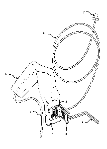
FIGURE 1 is a perspective view of a holder in use with an X-ray sensor.
FIGURE 2 illustrates a side view of an X-ray sensor securely attached to a
sensor holder.
FIGURE 3 is a perspective view of the preferred embodiment of the sensor
holder. Vertical ridges on the
backing plate of the sensor holder allow for even removal of air from the
space between the sensor and
the plate to create a strong pulling force on the sensor.
.. FIGURE 4 is a perspective view of the present invention illustrating a
second embodiment of the sensor
holder. In this embodiment the backing plate is shown as a substrate with no
elastomeric material
attached or bonded to it.
FIGURE 5 illustrates a perspective view of a third embodiment of the sensor
holder wherein semi
.. spherical protrusions are arranged on the substrate to provide an even
suctioning effect.
FIGURE 6 is a perspective view of Figure 4 showing the sensor holder with an
appropriately shaped foam
or foam tape to be attached or bonded to the substrate.
CA 03031504 2019-01-18
WO 2018/022582 PCT/US2017/043661
7
FIGURE 7 is a cross sectional view in accordance with the preferred embodiment
of the present
invention as used with an X-ray sensor showing an opening leading a cavity and
then to an evacuation
chamber between the holder and the sensor.
FIGURE 8 is a bottom view of the present invention illustrating an extended
swivel opening which leads
a cavity and then to an evacuation chamber.
FIGURE 9 illustrates a cross sectional view of the preferred embodiment of the
sensor with an extended
swivel opening for connection to a suction tubing.
FIGURE 10 is a perspective view of an adapter to be connected to a suction
tubing.
FIGURE 11 is a perspective view showing the adapter in connection with the
suction tubing.
FIGURE 12 is a perspective view illustrating another adapter constructed to
connect directly to a saliva
ejector.
FIGURE 13 is a perspective view showing the adapter of Figure 12 in connection
with the saliva ejector
of Figure 12.
FIGURE 14 is a side view illustrating the use of an X-ray sensor, a holder and
an aiming/positioning arm
in the oral cavity of a patient to take X-ray images.
CA 03031504 2019-01-18
WO 2018/022582
PCT/US2017/043661
8
FIGURE 15 is a side view of the present invention showing a suction tubing of
the holder embedded
inside the aiming/suction arm to make the holder more compact.
FIGURE 16 shows a side view of the second embodiment with a foam attached or
bonded to the
substrate of the holder.
CA 03031504 2019-01-18
WO 2018/022582 PCT/US2017/043661
9
DETAILED DESCRIPTION OF THE INVENTION
Turning now descriptively to the drawings, in which similar reference
characters denote
similar elements throughout the several views, the figures illustrate a sensor
holder having a bite
block with a first end and a second end, a backing plate affixed to or formed
contiguously with the
second end of the bite block, and an opening to a cavity in the holder,
wherein said cavity leads to
an evacuation chamber for creating a vacuum between the holder and an X-ray
sensor. The cavity,
opening and evacuation chamber may be of different configurations, shapes and
sizes.
FIGURE 1 and FIGURE 2 show a sensor securely attached to an X-ray holder.
Referring to Figure 1, an X-
ray sensor 1 is secured to the backing plate 3 of a sensor holder, the sensor
being enclosed in a
protective sheath 4. A suction tubing 6 is connected to the sensor holder.
This allows for the creation of
a vacuum between the backing plate of the sensor holder and the sensor/sheath
combination when
brought together with the suction effect holding the sensor to the backing
plate. A positioning /aiming
arm 8 is connected to the bite block of the sensor holder preferably through
slots or apertures in the
bite block and preferable in a friction fit manner. The backing plate is
preferably affixed to or formed
contiguously with the bite block. An adapter 7 allows for direct or indirect
connection to suction lines
present on most dental chairs (not shown) or other suction device to create a
vacuum that pulls the
sensor in tightly and holds it in place until the suction device is turned
off. The cable 5 of the X-ray
sensor allows for transmission of the sensor data to a receiver in a
conventional manner. As is known in
the art, the bite block of the X-ray sensor holder will be positioned in a
patient's oral cavity (not shown)
and the patient will be instructed to bite upon the block. This locates the
affixed or supported x-ray
image media during the ensuing dental imaging acquisition procedure. Bite
block 2 of the invention is of
any suitable and conventional design, shape or configuration but can also have
the unique inventive
aspect of being designed to receive an aiming/suction arm 21 which has a
suction tubing embedded in it
to make the holder more compact, as shown in Figure 15.
Referring to Figure 3, vertical ridges 12 on the backing plate 3 of the sensor
holder allow for an even
removal of air from an evacuation chamber 14 (shown in Figure 7) between the
sensor and the plate 3
CA 03031504 2019-01-18
WO 2018/022582
PCT/U52017/043661
to create a strong pulling force on the sensor. Air is evacuated from the
space between the sensor and
the backing plate 3 through the cavity 20. The sensor holder 10 has apertures
or slits 9 within which a
conventional aiming arm fits. In a preferred embodiment shown in FIGURE 3, an
elastomeric material is
injection overmolded onto the substrate 11 of the backing plate 3 to create a
soft outer surface for
5 contact with the sensor to enhance the suction effect. The substrate 11
of the backing plate 3 needs to
be a fairly stiff material to assure that the X-ray sensor/media(Film,
Phosphor Plate or Digital Sensor) is
in parallel alignment with the aiming ring(not shown). Different designs can
be realized for the ridges of
the backing plate. In all cases, the ridges 12, 13 should be such that an even
suction effect is created in
the entirety of the evacuation chamber between the X-ray sensor and the
backing plate. The ridges
10 .. could be a part of the substrate 11 or could be a part of the
elastomeric surface as in the case of
injection overmolding.
Figure 4 illustrates a different embodiment of the sensor holder 10a. The
backing plate is shown as a
substrate 11 with no elastomeric material attached or bonded to it. An
appropriately shaped
elastomeric material or foam can be bonded or attached to it, as shown in
Figure 6, to enhance the
suction effect that secures the sensor in place. Ridges 12a are arranged to
provide an evenly distributed
suctioning effect.
The soft surface in any embodiment could be, but is not limited to:
1. A permanent, injection overmolded, elastomeric material.
2. An appropriately shaped foam tape that could be permanently adhered to the
face of the
backing plate.
3. An appropriately shaped self-adhesive foam tape that would be single use
and would be
removable after use.
4. A removable molded elastomeric sleeve that fits tightly to the upright
sensor plate.
Ideally, the foam is a closed cell foam. Closed cell foams generally contain
gas bubbles formed
during the foam's expansion and cure. The bubbles are permanently locked in
place and this
CA 03031504 2019-01-18
WO 2018/022582 PCT/US2017/043661
11
enables it to resist liquids such as water and saliva and also act as an
insulator and air barrier,
enhancing the suction effect in the evacuation chamber.
As shown in FIGURE 5, spherical protrusions 12b can be arranged on the
substrate 11 to provide an even
suction effect.
Figure 7 is a cross sectional view of a preferred embodiment of the sensor
holder 10 in operation with a
suction tubing 6 to secure an X-ray sensor to the backing plate 3 of the
holder 10. Air is drawn out of the
evacuation chamber 14 in the sensor holder combination to create the vacuum
required to hold the
combination together. The suction tubing is connected to holder 10 through an
extended opening 17,
which leads through a cavity 20 to the evacuation chamber 14. The cavity 20
and/or evacuation
chamber 14 may be of different configurations, shapes and sizes.
The opening 17 can also be of different configurations, shapes and size . In
particular, Figure 8 shows a
bottom view of the sensor holder with a swivel opening 17a which extends in
the direction of the
longitudinal axis of the bite block 2. The swivel opening 17a can additionally
be seen in Figure 9. The
opening allows swivel movements of the suction tubing 6 when connected to
enable easy placement of
the holder in the mouth of a patient. Swivel opening 17a allows suction tubing
6 to be positioned on
either side of holder 10 to allow holder 10 to be used in different locations
in the oral cavity. Opening
17, tubing 6 and evacuation chamber 14 are in fluid communication.
As shown in Figure 10, the distal end 7 of the suction tubing 6 can be fitted
with an adapter 7 for
connection to a suction device such as a saliva ejector or aspirator hand
piece(not shown) which in turn
in connected to suction units such as the suction unit of a dental chair.
Direct connections to suction
units such as a vacuum pump can also be realized by using appropriate
adapters. 7a shows an adapter
to a suction line of a dental chair and 7b shows an adapter to a saliva
ejector. In Figure 12 the adapter
7b is constructed to connect directly and firmly to a saliva ejector 18 to
conduct a suction effect through
the sensor holder tubing 6 to the evacuation chamber. The adapter 7b receives
the saliva ejector
preferably through a friction fit manner. The operation of the preferred
embodiment of the holder
involves holding the bite block 2 in place by teeth 19 and creating a vacuum
between the sensor and the
CA 03031504 2019-01-18
WO 2018/022582
PCT/US2017/043661
12
backing plate 3 to secure the sensor throughout the image acquisition process
as shown in Figure 14. An
aiming/positioning arm 8 connected to an aiming ring (not shown) outside the
oral cavity ensures that
the sensor and the ring are in alignment. A suction tubing 6, connected on the
proximal end to the
opening in the holder and on the distal end to an adapter which in turn is
connected to a suction device,
.. provides a medium through which air is evacuated from the evacuation
chamber 14 when the suction
device (not shown) is engaged. Air is drawn out of the evacuation chamber to
create a vacuum, which
has the resultant effect of pulling the sensor securely to the backing plate.
The ridges 12,13 on the
backing plate ensure an even suction effect is created in the evacuation
chamber.
To make the holder even more compact the suction tubing can be embedded inside
the aiming/suction
.. arm 21 for connection to the cavity 20 or evacuation chamber 14. This
ensures a space saving holder
and reduces the mass of material in the oral cavity of the patient during
image acquisition.
What has been described and illustrated herein is a preferred embodiment of
the invention
along with some of its variations. The terms, descriptions and figures used
herein are set forth by
way of illustration only and are not meant as limitations. Those skilled in
the art will recognize that
many variations are possible within the spirit and scope of the invention in
which all terms are
meant in their broadest, reasonable sense unless otherwise indicated. Any
headings utilized within
the description are for convenience only and have no legal or limiting effect.
CA 03031504 2019-01-18
WO 2018/022582
PCT/US2017/043661
13
Reference Numerals
1 Sensor
2 Bite block
3 Backing plate
4 Sheath
5 Sensor cable
6 Suction tubing
7 Adapter
7a Direct connection to suction line
7b Connection to saliva ejector
7c Adapter connected to suction tube
7d Adapter connected to saliva ejector
8 Positioning/aiming arm
9 Slot/aperture
10 Holder with injection overmould
10a Bare Holder with vertical ridges on substrate
10b Bare Holder with spherical protrusions on substrate
11 Substrate
12 Ridge
12a Vertical ridges
12b Spherical protrusions
13 Horizontal ridge
14 Evacuation chamber
15 Foam/Foam tape
16 Adhesive
17 Opening
17a Swivel opening
CA 03031504 2019-01-18
WO 2018/022582
PCT/US2017/043661
14
18 Saliva ejector
19 Tooth
20 Cavity
21 Aiming/Suction arm