Note: Descriptions are shown in the official language in which they were submitted.
CA 03032078 2019-01-25
WO 2018/022311
PCT/US2017/041953
1
ULTRASONIC SURGICAL PROBE, ASSEMBLY, AND RELATED METHOD
BACKGROUND OF THE INVENTION
This invention relates to an ultrasonic surgical probe. This invention also
relates to an assembly of a probe and a sheath. The invention additionally
relates to
an associated method for operating on a surgical site, exemplarily to reduce
biofilm
on a wound site particularly a wound site that is being debrided to remove
necrotic
tissue.
Chronic wound infection represents a significant healthcare problem
worldwide. Often the end objective of wound healing is the objective for new
therapeutic options. Yet chronic wounds compromise a number of different and
complex conditions that each interferes with the healing process. For example,
a
chronic wound can comprise necrotic tissue in need of debridement, bacterial
infection in need of antimicrobial agents and compromised vasculature that
impedes
the normal healing process.
One element of the chronic wound infection condition that impedes healing is
the formation of biofilm. Biofilm is the result of planktonic bacteria forming
together
and secreting exopolysaccharide (EPS) to adhere and protect the colonizing
community. At the height of formation, EPS can make up between 75-90% of the
total biofilm composition (Regt). Biofilm inhibits healing by creating an
optimal
condition for bacteria to grow, while simultaneously preventing antimicrobial
agents
from direct access to bacteria.
Methods to remove biofilm include ultrasonic debridement, topical
antimicrobials, suction, and surface cleansing. Each of these methods alone
treat an
aspect of biofilm. For example, ultrasonic debridement of wounds has proven to
be
the most effective mechanism in disrupting and debulking a majority of the
biofilm
formation. Yet even in this preferred method, biofilm debris can be left
behind to
propagate. Suction alone has not proven to be effective in removing biofilm,
and can
potentially interfere with the operation of other methods like ultrasonic
debridement if
applied simultaneously.
US Patent No. 7,608,054 to Soring et al. describes a medical treatment
apparatus that combines an ultrasound sonotrode with a suction sheath. The
fixed
position between the tip of the suction and the tip of the sonotrode only
allows for one
simultaneous operation. In particular this approach is limited due to the
potential
interference of the suction tip during the ultrasonic debridement operation.
CA 03032078 2019-01-25
WO 2018/022311
PCT/US2017/041953
2
US Patent No, 7,522,955 B2 to Rontal et al. describes a method and apparatus
for ultrasonic cleaning of biofilm coated surfaces for sinus cavities within a
human
head. The method describes an ultrasonic application in combination with
irrigation
and suction that is designed to not remove any of the surrounding underlying
tissue.
This differs significantly from an ultrasonic debridement of a wound bed,
which
requires the removal of tissue in combination with biofilm. Thus the
ultrasonic probe
needs to operate in a cavitation mode at the surface of a wound, causing
destruction of
the biofilm.
Methods of mechanical removal of biofilm in wounds alone have proven to be
inadequate. What does not exist and what would be beneficial to the market is
an
ultrasonic probe or instrument assembly which permits implementation of an
improved method to remove biofilm and prevent it from reforming in order to
allow
wounds to heal.
SUMMARY OF THE INVENTION
The present invention aims to provide an improved ultrasonic probe and/or
instrument assembly, particularly permitting execution of an improved method
to
inhibit biofilm formation in order to allow wounds to heal more expeditiously.
An ultrasonic surgical probe comprises, in accordance with the present
invention, an elongate shaft having a distal end portion with a longitudinal
axis and a
probe head that is enlarged to extend laterally or transversely in two opposed
directions relative to the shaft and the axis. The head is formed with a
recess or cavity
facing laterally in a third direction relative to the shaft and the axis.
Where the head is
conceptualized as lying in a plane, owing to its lateral enlargement or
extension, the
recess or cavity faces in a direction perpendicular to that plane. The recess
or cavity
is defined in part by an inclined floor or base surface contiguous at a
proximal end
with the shaft. The recess or cavity is defined in part by a peripheral wall
extending
only partway around the recess or cavity, along a distal side and two lateral
sides
thereof. The cavity or recess is closed on a proximal side by the inclined
surface and
the shaft.
Pursuant to further features of the present invention, the recess or cavity is
further defined by an additional floor or base surface located distally of the
inclined
floor or base surface. The additional floor or base surface is planar or flat
and
oriented parallel to the axis. Preferably, the inclined floor or base surface
is also
planar or flat.
CA 03032078 2019-01-25
WO 2018/022311
PCT/US2017/041953
3
The distal, additional floor or base surface and the proximal, inclined floor
or
base surface are preferably adjacent and contiguous with one another.
Pursuant to further features of the present invention, the probe head is
provided in at least one of the inclined floor or base surface and the
additional floor or
base surface with at least one first opening spaced from the peripheral wall.
The
opening permits the egress of pressurized fluid from the recess or cavity into
a space
surrounding the probe head, exemplarily into a channel in a sheath that
surrounds the
probe. The pressurized fluid is liquid irrigant which serves to cool the
surfaces of the
probe head, particularly including those surfaces that contact tissue during
an
ultrasonic debridement or biofilm removal procedure. The pressurized fluid is
fed
into the recess or cavity via an axial channel or through bore in the probe
shaft. The
channel or through bore has an outlet port at least partially in or at the
inclined floor
or base surface.
Preferably, the probe head is provided with a second opening or through hole
in the peripheral wall at a distal end of the probe head. A groove may be
formed in
the floor of the recess or cavity, with the first opening or through hole
located in the
groove, the groove extending to the second opening or through hole.
The groove may be formed during manufacture during the drilling of a probe
blank to form the channel in the shaft. The inclined floor or base surface and
the
additional floor or base surface are subsequently formed by machining one side
of the
head of the blank. This machining opens a side of the channel in the head and
thereby
generates the groove.
Pursuant to other features of the present invention, the peripheral wall of
the
recess or cavity includes a cylindrical section on the distal side of the
recess or cavity
and further includes two linear sections on the lateral sides of the recess or
cavity.
The peripheral wall thus has a U-shaped plan or configuration.
In accordance with another feature of the present invention, the peripheral
wall
is provided with a beveled surface, on a side of the peripheral wall opposite
the recess
or cavity, and has a flat terminal edge in a plane parallel to the axis. The
flat terminal
edge or rim enables the peripheral wall to transmit ultrasonic vibratory
energy into the
tissues at a surgical site during a debridement or biofilm elimination
procedure.
It is to be noted that the multiple openings in the head, one in the floor and
one
in on the distal portion of the peripheral wall, facilitate the flow of liquid
irrigant in
part to optimize cooling of the peripheral wall and the tissues at the
surgical site, thus
CA 03032078 2019-01-25
WO 2018/022311
PCT/US2017/041953
4
reducing if not eliminating damage to the healthy tissue which remains after
the
debridement procedure. The floor geometry of the recess or cavity in the probe
head
acts to deflect and guide removed tissue fragments from the recess or cavity,
thereby
inhibiting if not completely preventing the clogging of the openings in the
probe head.
In addition, the flow of liquid irrigant or coolant into the recess or cavity
through the
channel or bore in the shaft and out through the openings in the floor and the
peripheral wall help move the separated tissue fragments along the floor
surfaces and
out of the recess or cavity. The floor structure of the recess or cavity
assists in
maintaining desired cooling for longer periods of time.
A surgical instrument assembly comprises, in accordance with the present
invention, an ultrasonic probe having an operative tip, an electromechanical
transducer operatively connected to the probe for generating an ultrasonic
standing
wave in the probe, and at least one sheath or sleeve disposed about the probe
and
defining at least a first suction port at a distal end of the probe, proximate
the
operative tip thereof, and a second suction port spaced from the distal end of
the probe.
The probe comprises (A) an elongate shaft having a distal end portion with a
longitudinal axis and (B) a probe head that is enlarged to extend laterally or
transversely in two opposed directions relative to the shaft and the axis, the
head
having a recess or cavity facing laterally in a third direction relative to
the shaft and
the axis. The recess or cavity is defined in part by an inclined floor or base
surface
contiguous at a proximal end with the shaft and in part by a peripheral wall
extending
only partway around the recess or cavity, along a distal side and two lateral
sides
thereof. The cavity or recess is closed on a proximal side by the inclined
surface and
the shaft.
The at least one sheath or sleeve is preferably exactly one sheath or sleeve.
Optionally, the sheath or sleeve is longitudinally slidable relative to the
probe to shift
between a distal position and a proximal position.
The inclined floor or base surface of the probe cavity is planar or flat and
the
recess or cavity is further defined by an additional floor or base surface
located
distally of the inclined floor or base surface, the additional floor or base
surface being
planar or flat and oriented parallel to the axis.
The probe head is provided with a first opening or through hole in the floor
of
the recess or cavity, spaced from the peripheral wall. The probe head is
provided with
a second opening or through hole in the peripheral wall. The probe head may be
CA 03032078 2019-01-25
WO 2018/022311
PCT/US2017/041953
provided in the floor of the recess or cavity with a groove. In that case, the
first
opening or through hole is located in the groove while the groove extends to
the
second opening or through hole.
The peripheral wall preferably includes a cylindrical section on the distal
side
5 of the recess or cavity and further includes two linear sections on the
lateral sides of
the recess or cavity, so that the peripheral wall has a U-shaped
configuration.
The peripheral wall is preferably provided with a beveled surface, on a side
of
the peripheral wall opposite the recess or cavity, and has a flat terminal
edge in a
plane parallel to the axis. The bevel tapers the peripheral wall towards the
flat
terminal edge or rim.
Pressurized coolant liquid enters the recess or cavity in the probe head from
the channel or bore in the instrument shaft and exits in part through the
openings in
the floor or base and the semi-cylindrical portion of the peripheral wall. The
diameters of the channel and the openings in the probe head, as well as the
pressure of
the fluid and the magnitude of applied suction must be taken into account in
optimizing the rate of coolant flow so that the entire probe head is
maintained within a
desirable temperature range.
A surgical method in accordance with the present invention utilizes an
ultrasonic surgical probe comprising an elongate shaft having a distal end
portion with
a longitudinal axis and further comprising a probe head that is enlarged to
extend
laterally or transversely in two opposed directions relative to the shaft and
the axis.
The head has a recess or cavity facing laterally in a third direction relative
to the shaft
and the axis, and the recess or cavity is defined in part by an inclined floor
or base
surface contiguous at a proximal end with the shaft. The recess or cavity is
also
defined in part by a peripheral wall extending only partway around the recess
or
cavity, along a distal side and two lateral sides thereof, the cavity or
recess being
closed on a proximal side by the inclined surface and the shaft. The method
comprises manipulating the probe to press the head and particularly at least a
portion
of the peripheral wall into tissue at a surgical site. While continuing to
press the head
and the portion of the peripheral wall into the tissue, one draws the probe
and
concomitantly the portion of the peripheral wall in a proximal direction
across the
tissue, substantially (mostly) parallel to the surgical site. During that
drawing process,
CA 03032078 2019-01-25
WO 2018/022311
PCT/US2017/041953
6
ultrasonic vibrations are conducted into the probe. Tissue is separated from
the
surgical site by the drawing of the probe and the ultrasonic vibrating
thereof. Upon
the separating of the tissue, the separated tissue is deflected or moved out
of the recess
or cavity in part by contact of the separated tissue with the inclined floor
or base
surface. During the drawing of the probe, an irrigation liquid is fed to the
recess or
cavity via a longitudinal channel or bore in the probe shaft. The irrigation
liquid
serves to cool the probe head and assists in moving the separated tissue out
of the
recess or cavity. Flow of the liquid irrigant/coolant is maintained in part
where the
probe head is formed with at least one opening or through hole through which
the
liquid is aspirated into a surrounding sheath or sleeve. The cross-sectional
areas of
the channel or bore and the opening(s) in the probe head,- as well as the
pressure f
liquid feed and the degree of suction are selected in concert to ensure
adequate
cooling of the probe head.
BRIEF DESCRIPTION OF THE DRAWINGS
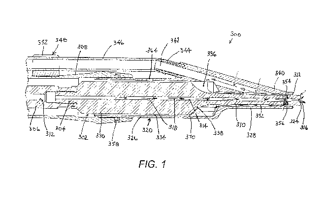
FIG. 1 is a partial longitudinal cross-sectional view of a device for
debriding
or removing biofilm from a wound site.
FIG. 2 is a schematic right side, top and front perspective view of the device
of FIG. 1.
FIG. 3 is an exploded right side, top and front perspective view of a probe
and
sheath included in the device of FIGS. 1 and 2.
FIG. 4 is an exploded left side, top, and rear perspective view of the probe
and
sheath of FIGS. 1-3.
FIG. 5 is a rear elevational view of the probe and sheath of FIGS. 1-4.
FIG. 6 is a partial cross-sectional view, similar to FIG. 1, taken along line
VI-
.. VI in FIG. 5.
FIG. 7 is a top plan view of an ultrasonic surgical probe in accordance with
the
present invention.
FIG. 8 is a front end elevational view of the probe of FIG. 7.
FIG. 9 is a right side elevational view of the probe of FIGS. 7 and 8.
FIG. 10 is a right, top and rear isometric view of a head of the probe of
FIGS.
7-9.
FIG. 11 is a right, bottom and front isometric view of the probe head of FIG.
10.
CA 03032078 2019-01-25
WO 2018/022311
PCT/US2017/041953
7
FIG. 12 is a right, top and front isometric view of the probe head of FIGS. 10
and 11.
FIG. 13 is atop, front and let side isometric view of the probe head of FIGS.
10-12.
FIG. 14 is a side elevational view of a sheath for an ultrasonic surgical
instrument assembly in accordance with the present invention.
FIG. 15 is a bottom, and right side isometric view of an instrument assembly
including the sheath of FIG. 14 and the probe of FIGS. 7-13.
FIG. 16 is a schematic and diagrammatic longitudinal cross-sectional view, on
an enlarged scale, of a distal end portion of the instrument assembly of FIGS.
14 and
15.
FIG. 17 is a schematic and diagrammatic longitudinal cross-sectional view
similar to FIG. 16, showing use of the instrument assembly in debriding or
removing
tissue and biofilm from a wound site, with directional arrows indicating
tissue
fragment transport.
FIG. 18 is a schematic and diagrammatic longitudinal cross-sectional view, on
a smaller scale, showing a larger distal end portion of the instrument
assembly of
FIGS. 14 and 15 than visible in FIGS. 16 and 17 and with directional arrows
indicating flow of liquid coolant or irrigant.
FIG. 19 is a schematic and diagrammatic longitudinal cross-sectional view
identical to FIG. 16, with directional arrows to represent irrigation liquid
flow paths.
DETAILED DESCRIPTION
As depicted in FIGS. 1-6, a surgical device 300 for debriding or removing
tissue and biofilm from a wound site comprises an ultrasonic probe 302 which
is
attached at a proximal end via threaded connector 304 to a driver 306 is
operatively
connected to a generator of vibratory energy, typically a piezoelectric
transducer array
(not shown). Both the driver 306 and the piezoelectric transducer are located
in a
handpiece which has a cover or housing (not shown) connected to a casing 308.
Probe 302 tapers down on a distal side to a distal end section 310. It is to
be noted
that the terms "horn" and "probe" are used synonymously.
Driver 306 and probe 302 are formed with mutually aligned axial channels or
bores 312 and 314 that define a lumen (not separately designated) for the
delivery of
irrigant to a distal end aperture 316 in probe horn section 310, as indicated
by flow
arrows 318.
CA 03032078 2019-01-25
WO 2018/022311
PCT/US2017/041953
8
Surgical device 300 further includes a rigid sheath 320 that is shiftably
mounted to casing 308 to vary a position of a distal tip 322 of the sheath
relative to a
distal tip or end face 324 of probe 302. Sheath 320 includes a cylindrical
rear section
326 and a rectangularly prismatic forward section 328, which correspond
geometrically to cross-sections of horn 310 and a proximal portion 330 of
probe, 302,
respectively.
Together with an outer surface (not designated) of probe horn 310, forward
sheath section 328 defines a forward or distal channel or conduit 332, which
is
rectangular in cross-section. Together with an outer surface (not designated)
of
proximal probe portion 330, rear sheath section 326 defines a rearward or
proximal
channel or conduit 334, which is circular in cross-section. At a distal end,
rearward
channel 334 expands to an enlarged space 336 owing to the tapering of the
probe at
338.
Sheath 320 is provided with an arm 340 that is connected at a forward or
distal
end to forward section 326 and is angled outwardly at a proximal side. Sheath
arm
340 includes a main aspiration channel 342 that communicates at a distal end
with
forward channel 332. At a more proximal location, aspiration channel 342 of
arm 340
communicates with rearward channel 334 and more particularly with enlarged
space
336. At a proximal end, arm 340 is provided with an undercut connector port
344
which receives a resilient aspiration tube 346 in a friction fit. Aspiration
tube 346 is
fastened to casing 308 via a pair of clips 348 each formed with a pair of
slotted
annular rings 350 and 352 for receiving casing 308 and aspiration tube 346,
respectively.
At a forward or distal end, probe horn 310 is formed with one or more
apertures or cross-bores 354 and 356 that communicate on an inner side with
channel
or lumen 314 and on an outer side with forward channel 332. At a rear end,
rear
section 326 of sheath 320 is inserted between proximal probe portion 330 and a
distal
end of casing 308. An 0-ring seal 358 is provided between casing 308 and an
outer
surface of sheath rear section 326.
A distal end of horn section 310 is formed into a probe head 360 that is
extended in a traverse dimension, orthogonally to a longitudinal axis of the
probe 302.
Head 360 may particularly take a form disclosed in U.S. Patent Application No.
14/172,566, Publication No. 2015/0216549, the disclosure of which is
incorporated by
reference herein. In particular, head 360 includes a plurality of teeth 362
arranged in
CA 03032078 2019-01-25
WO 2018/022311
PCT/US2017/041953
9
two mutually parallel rows along opposing edges or sides of the distal end
face 324 of
the probe head.
As indicated above, sheath 320 is slidable or longitudinally shiftable
relative
to probe 302 so as to be continuously adjustable as to axial or longitudinal
position
relative to probe head 360 anywhere from a fully extended position, where the
distal
tip 322 of sheath 320 is essentially coplanar with the distal end face 324 of
probe head
360, to a retracted position where at least the teeth 362 of probe head 360
are fully
exposed. 0-ring 358 enables the adjustable positioning of sheath 320.
Apertures or cross-bores 354 and 356 serves as bypass holes, regardless of the
relative longitudinal positioning of sheath 320 and probe 302. A vacuum under-
pressure applied to the internal spaces of sheath 320, i.e., aspiration
channel 342,
forward channel 332, and rearward channel 334, by a suction source (not shown)
enables the capturing and removal of most of the irrigant that is delivered
through
central channel 314 (flow arrows 318). Accumulation of irrigant within sheath
320,
especially when the device is used in a predominantly vertical orientation, is
prevented by the provision of two suction pathways, namely, between aspiration
channel 342 and each of the forward channel 332 and rearward channel 334.
Irrigant
not captured via a distal pathway is captured in a proximal pathway.
Where tissue fragments are small enough to be aspirated through the gap
between the probe 302 and the sheath 320, clogging is prevented by designing
the
aspiration pathway of channel 324 to gradually increase in cross-sectional
area from
the probe-sheath gap at the distal end of the instrument all the way to the
aspiration
line. A vent port 364 may be provided in the rear sheath section 326 to reduce
the
magnitude of vacuum-generated pull force acting on the tissue which is driven
towards and into the probe-sheath gap during debridement.
Matching or cooperating features 366 and 368 are respectively disposed on the
outer side of the probe 302 and the inside of rear sheath section 326, in
close
proximity to a nodal plane or the probe, to facilitate probe-sheath alignment.
This
minimizes the chances of a probe-sheath contact at the points of maximum
vibratory
motion (antinodes), particularly at end face 324 of probe head 360. Due to
their
placement at a location of minimal vibratory displacement, e.g., the junction
370
between cylindrical probe portion 330 and tapering probe section 338, the
alignment
features 366 and 368 allow for the probe-sheath contact necessary for
preventing or
minimizing the unwanted interaction in the area of maximum vibratory
displacement.
CA 03032078 2019-01-25
WO 2018/022311
PCT/US2017/041953
FIGS. 7-13 depict an ultrasonic surgical probe 102 that may be used instead of
probe 302 for debriding tissue or removing biofilm from a wound site. Probe
102
comprises an elongate shaft 104 having an enlarged proximal end portion 106
with a
screw-type coupling 108 for connection to a source of ultrasonic mechanical
vibratory
5 energy. Shaft 104 has a distal end portion 110 with a longitudinal axis
112 and a
probe head 114 that is enlarged to extend laterally or transversely in two
opposed
directions (arrows 116, 118) relative to shaft 104 and axis 112. Head 114 is
formed
with a recess or cavity 120 facing laterally in a third direction (arrow 122)
relative to
shaft 104 and axis 112. Where head 114 may be seen as lying in a plane defined
by
10 axis 112 and extension directions 116 and 118, recess or cavity 120
faces in direction
122 perpendicular to that plane. Recess or cavity 120 is defined in part by an
inclined
floor or base surface 124 contiguous at a proximal end with shaft 104. Recess
or
cavity 120 is further defined in part by a peripheral wall 126 extending only
partway
around the recess or cavity, with a cylindrical wall portion 128 along a
distal side of
head 114 and two linear or planar wall sections 130, 132 along lateral sides
thereof.
Peripheral wall 126 thus has a U-shaped plan or configuration. Recess or
cavity 120
is closed on a proximal side by inclined surface 124 and shaft 104.
Recess or cavity 120 is further defined by an additional floor or base surface
134 located distally of inclined floor or base surface 124. Additional floor
or base
surface 134 is planar or flat and oriented parallel to shaft axis 112.
Preferably,
inclined floor or base surface 124 is also planar or flat. Floor or base
surfaces 124 and
134 are preferably adjacent and contiguous with one another.
Probe head 114 is provided in at least one opening 136 in either inclined
floor
surface 124 and/or parallel floor surface 134. Opening 136 is spaced from the
peripheral wall 126. Opening 136 permits the egress of pressurized fluid from
recess
or cavity 120 into a space surrounding probe head 114, exemplarily into a
channel
138 in a sheath 140 that surrounds the probe 102 (see discussion above with
reference
to cross-bores 354 and 356 shown in FIG. 1 and description hereinbelow with
reference to FIGS. 14 et seq.). The pressurized fluid is a liquid irrigant for
cooling the
surfaces of the probe head 120 particularly including surfaces of peripheral
wall 126
and other surfaces that contact tissue at a surgical site during an ultrasonic
debridement or biofilm removal procedure. The pressurized fluid is fed into
recess or
cavity 120 via an axial channel or through bore 142 in probe shaft 104.
Channel or
CA 03032078 2019-01-25
WO 2018/022311
PCT/US2017/041953
11
through bore 142 has an outlet port 144 at least partially in or at inclined
floor or base
surface 124.
Preferably, probe head 114 is provided with a further opening or through hole
146 in peripheral wall 126 at a distal end of the probe head, particularly in
cylindrical
wall section 128. A groove 148 is formed in floor surfaces 124 and 134 of
recess or
cavity 120, with opening or through hole 136 located in the groove, the groove
extending to cylindrical wall section 128 and particularly to opening or
through hole
146 therein.
Groove 148 is typically formed during manufacture during a drilling of a
probe blank (not separately shown) to form channel or bore 142 in probe shaft
104.
Inclined floor or base surface 124 and the distal floor or base surface 134
are
subsequently formed by machining one side of the head of the blank. This
machining
opens a side of the channel in the head and thereby generates groove 148.
Peripheral wall 126 is provided with a beveled surface 150, on a side of the
.. peripheral wall opposite recess or cavity 120, and has a flat terminal edge
152 in a
plane parallel to axis 112 and the plane of head 114. The flat terminal edge
or rim
152 enables the peripheral wall 126 to transmit ultrasonic vibratory energy
into the
tissues at a surgical site during a debridement or biofilm elimination
procedure.
It is to be noted that the multiple openings 136, 146 in the probe head 114,
one
in the floor 124, 134 and one in on the distal portion 128 of peripheral wall
126,
facilitate the flow of liquid irrigant in part to optimize cooling of the
peripheral wall
and the tissues at the surgical site, thus reducing if not eliminating damage
to the
healthy tissue which remains after the debridement procedure.
The floor geometry of recess or cavity 120 in probe head 114 acts to deflect
and guide removed tissue fragments from the recess or cavity, thereby
inhibiting if not
completely preventing the clogging of the openings 136, 146 in the probe head.
In
addition, the flow of liquid irrigant or coolant into the recess or cavity 120
through the
channel or bore 142 in shaft 104 and out through the openings 136, 146 in the
floor
124, 134 and the peripheral wall 126 help move the separated tissue fragments
along
the floor surfaces 134, 124 and out of the recess or cavity 120. The floor
structure of
recess or cavity 120 assists in maintaining desired cooling for longer periods
of time.
FIGS. 14-19 depict a surgical instrument assembly incorporating probe 102
and including sheath or sleeve 140 disposed about probe 102. As shown in FIGS.
14
and 15, sheath 140 includes an eccentric suction arm 154 extending at an angle
away
CA 03032078 2019-01-25
WO 2018/022311
PCT/US2017/041953
12
from a distal end of the sheath. A connector 156 provided at a proximal end of
the
suction arm 154 is force-fit into a distal end of a length of vacuum tubing
158 that is
further attached to the sheath 140, as well as to a transducer housing 160 via
a
plurality of spring clips 162. FIG. 15 also shows a liquid conduit 164 coaxial
with
probe axis 112 and an electrical cable 166 that provides an ultrasonic-
frequency
power signal to the transducer disposed inside housing or handpiece 160.
As described hereinabove with reference to FIGS. 1-6, sheath or sleeve 140
defines at least a first suction port 168 at a distal end of the probe,
proximate the
operative tip or head 114 thereof, and a second suction port 170 (FIG. 18)
spaced
back from the distal end of the probe. The relationship between probe 102 and
sheath
or sleeve 140 is analogous to that described above with reference to FIGS. 1-
6.
Sheath or sleeve 140 is preferably exactly one sheath or sleeve longitudinally
slidable relative to probe 102 to shift between a distal position and a
proximal position.
Sheath or sleeve 140 has a distal end wall section 172 that is disposed
transversely or
perpendicularly to the probe axis 112 (which is co-linear or co-incident with
an axis
of sheath 140). Distal end wall section 172 facilitates collection and
extraction of
liquid irrigant that exits probe head 114 and more particularly cavity or
recess 120 via
opening or through hole 146 in cylindrical section 128 of peripheral wall 126.
The
purpose of irrigant collection and extraction is two-fold: to prevent the
irrigant from
overflowing a surgical site and the operating table and to facilitate
temperature control.
Pressurized coolant liquid enters recess or cavity 120 in probe head 114 from
channel or bore 142 in instrument shaft 104, as indicated by arrows 174 in
FIGS. 17-
19, and exits in part through opening 136 in floor or base surface 124 or 134,
as
represented by arrows 176, and opening 146 in semi-cylindrical portion 128 of
peripheral wall 126 (arrows 178). Some of the irrigant that enters recess or
cavity 120
from channel or bore 142 forms a slurry with tissue fragments that are removed
from
a surgical site 180 (FIG. 17) during a debridement or biofilm-removal
procedure in
which the ultrasonically vibrating probe head 114 and particularly rim or
surface 152
thereof is pressed into the tissue, as indicated by a first force arrow 182
(FIG. 17), and
then dragged in a proximal direction as represented by a second force arrow
184. In
this operation, the slurry of irrigant and tissue fragments moves in circular
flow
patterns 186 guided by inclined floor or base surface 124 of probe head 114.
The
tissue fragments are thus deflected out of the cavity 120 and away from
openings 136
and 146, thereby delaying if not preventing clogging of the openings and the
suction
CA 03032078 2019-01-25
WO 2018/022311
PCT/US2017/041953
13
path inside eccentric suction arm 154 of sheath 140. The diameters or cross-
sectional
areas of channel or bore 142 and of openings 136 and 146 in probe head 114, as
well
as the pressure of the fluid (174) and the magnitude of applied suction
(negative-sign
symbols 188) must be taken into account in optimizing the rate of coolant flow
so that
the entire probe head 114 is maintained within a desirable temperature range.
During the pressing (182) and drawing (184) of probe 144 during a
debridement or biofilm-removal procedure, irrigation liquid or coolant is fed
to recess
or cavity 120 via channel or bore 142 in probe shaft 104. The irrigation
liquid serves
to cool the probe head 114 and assists in moving the separated tissue via
slurry flow
186 out of recess or cavity 120. Flow of the liquid irrigant/coolant is
maintained in
part by openings or through holes 136, 146 through which the liquid is
aspirated into
sheath or sleeve 140.
Although the invention has been described in terms of particular embodiments
and applications, one of ordinary skill in the art, in light of this teaching,
can generate
additional embodiments and modifications without departing from the spirit of
or
exceeding the scope of the claimed invention. Accordingly, it is to be
understood that
the drawings and descriptions herein are proffered by way of example to
facilitate
comprehension of the invention and should not be construed to limit the scope
thereof.